WO2010122787A1 - 腹膜保護剤および腹膜保護剤の調製方法 - Google Patents
腹膜保護剤および腹膜保護剤の調製方法 Download PDFInfo
- Publication number
- WO2010122787A1 WO2010122787A1 PCT/JP2010/002865 JP2010002865W WO2010122787A1 WO 2010122787 A1 WO2010122787 A1 WO 2010122787A1 JP 2010002865 W JP2010002865 W JP 2010002865W WO 2010122787 A1 WO2010122787 A1 WO 2010122787A1
- Authority
- WO
- WIPO (PCT)
- Prior art keywords
- peritoneal
- cells
- protective agent
- peritoneum
- morphological
- Prior art date
- Legal status (The legal status is an assumption and is not a legal conclusion. Google has not performed a legal analysis and makes no representation as to the accuracy of the status listed.)
- Ceased
Links
Images





Classifications
-
- A—HUMAN NECESSITIES
- A61—MEDICAL OR VETERINARY SCIENCE; HYGIENE
- A61K—PREPARATIONS FOR MEDICAL, DENTAL OR TOILETRY PURPOSES
- A61K35/00—Medicinal preparations containing materials or reaction products thereof with undetermined constitution
- A61K35/12—Materials from mammals; Compositions comprising non-specified tissues or cells; Compositions comprising non-embryonic stem cells; Genetically modified cells
- A61K35/36—Skin; Hair; Nails; Sebaceous glands; Cerumen; Epidermis; Epithelial cells; Keratinocytes; Langerhans cells; Ectodermal cells
-
- A—HUMAN NECESSITIES
- A61—MEDICAL OR VETERINARY SCIENCE; HYGIENE
- A61P—SPECIFIC THERAPEUTIC ACTIVITY OF CHEMICAL COMPOUNDS OR MEDICINAL PREPARATIONS
- A61P1/00—Drugs for disorders of the alimentary tract or the digestive system
- A61P1/16—Drugs for disorders of the alimentary tract or the digestive system for liver or gallbladder disorders, e.g. hepatoprotective agents, cholagogues, litholytics
-
- A—HUMAN NECESSITIES
- A61—MEDICAL OR VETERINARY SCIENCE; HYGIENE
- A61P—SPECIFIC THERAPEUTIC ACTIVITY OF CHEMICAL COMPOUNDS OR MEDICINAL PREPARATIONS
- A61P41/00—Drugs used in surgical methods, e.g. surgery adjuvants for preventing adhesion or for vitreum substitution
-
- A—HUMAN NECESSITIES
- A61—MEDICAL OR VETERINARY SCIENCE; HYGIENE
- A61P—SPECIFIC THERAPEUTIC ACTIVITY OF CHEMICAL COMPOUNDS OR MEDICINAL PREPARATIONS
- A61P43/00—Drugs for specific purposes, not provided for in groups A61P1/00-A61P41/00
Definitions
- the present invention relates to a peritoneal protective agent comprising peritoneal mesothelial cells that act effectively on peritoneal adhesion formation, peritoneal thickening, peritoneal damage, and the like, and further relates to a method for preparing the peritoneal protective agent.
- the present invention also relates to a regenerative medical material containing the peritoneal protective agent.
- Peritoneal adhesions occur at the site of surgical operations in various fields such as gastroenterology and gynecology, and have a profound effect on prognosis.
- intestinal adhesions that occur after laparotomy, especially during gastrointestinal surgery, tend to decrease with the improvement of surgical technique and equipment, but ileus (intestinal obstruction) symptoms, etc. after several to ten years have passed since surgery. It develops and is one of the most serious complications after surgery.
- Adhesion between the intestinal wall and the intestinal tract during laparotomy also causes intestinal obstruction, and there is a tendency to re-operate or worsen the prognosis of postoperative patients (Non-patent Document 1).
- a special anti-adhesive agent at a site where adhesion is likely to occur in such complications, it is still not a complete prevention method and a new approach is required.
- EPS peritoneal sclerosis
- Seprafilm (R) (Kaken Pharmaceutical Co., Ltd., Genzyme), which contains sodium hyaluronate and carboxymethylcellulose, which are bioabsorbable materials in a ratio of 2: 1, has already been clinically used as an anti-adhesion agent with excellent safety. Used in. Seprafilm (R) becomes a gel in 24 to 48 hours after application, and is stored at the application site for approximately 7 days to physically isolate the damaged tissue and the surrounding tissue, thereby preventing adhesion. Demonstrate. In addition, since the anti-adhesion agent is discharged outside the body within 28 days after application, the surgical removal is unnecessary.
- the anti-adhesion agent is not flexible in a dry state, it cannot be used if the use site has a complicated shape, and is not suitable for surgery under a laparoscope. Moreover, even if such an adhesion preventing agent is used, it is difficult to prevent adhesion after the EPS operation of a peritoneal dialysis patient that occurs in the whole wall side and visceral peritoneum.
- Patent Documents 1 to 4 Many studies have been made on the prevention of adhesion formation, most of which are anti-adhesion materials made of biodegradable or biocompatible polymer materials (Patent Documents 1 to 4), or materials that serve as scaffolds for epithelial cells (Patent Document 5). There is something about.
- Non-patent Document 2 transplantation of mesothelial cells into the abdominal cavity has been shown to be effective in reducing peritoneal adhesion formation and remodeling of exposed mesothelial cells.
- Non-patent Document 3 peritoneal mesothelial cells have a remarkable self-renewal ability that cannot be explained only by the regeneration ability of progenitor cells, and mesothelial cells may be a promising option for regenerative medicine.
- Non-patent Document 4 transplantation of peritoneal mesothelial cells has also been shown to be considered as one of the treatment options for peritoneal dysfunction.
- many problems remain to be solved, such as the source of mesothelial cells used for transplantation.
- the present inventors focused on the fact that peritoneal mesothelial cells derived from patients are mixed in peritoneal dialysis drainage obtained from patients undergoing peritoneal dialysis. It has been found that a sufficient amount of peritoneal mesothelial cells can be easily collected by selecting epithelioid morphological cells among the cells in the peritoneal dialysis drainage. It was found that the obtained peritoneal mesothelial cells can function as a peritoneal protective agent, and the present invention was completed.
- this invention consists of the following. 1.
- a peritoneal protective agent comprising peritoneal mesothelial cells recovered from peritoneal dialysis drainage as an active ingredient. 2.
- the peritoneal protective agent according to item 1 above comprising at least 10 peritoneal mesothelial cells collected from peritoneal dialysis drainage. 3.
- the peritoneal protective agent according to 1 or 2 above, wherein the peritoneal mesothelial cells recovered from the peritoneal dialysis drainage are epithelial morphological cells. 4).
- the peritoneal protective agent according to 3 above, wherein the epithelial morphological cells express hepatocyte growth factor (HGF). 5). 5.
- HGF hepatocyte growth factor
- peritoneal protective agent according to any one of 1 to 4 above, wherein the peritoneal protection is protection against peritoneal adhesion formation, peritoneal thickening and / or peritoneal injury. 6). 6. The method for preparing a peritoneal protective agent according to any one of 1 to 5 above, further comprising the step of collecting cells from the collected peritoneal dialysis drainage and further selecting epithelial morphological cells. 7). 7. The method for preparing a peritoneal protective agent according to item 6 above, wherein the epithelioid morphological cells are selected by culturing cells recovered from peritoneal dialysis effluent, and selecting cells that are morphologically recognized as epithelial morphological cells. . 8).
- a sufficient amount of peritoneal mesothelial cells can be easily collected by the method for collecting peritoneal mesothelial cells of the present invention.
- the collected peritoneal mesothelial cells are effective against peritoneal protective action, i.e. peritoneal adhesion formation, peritoneal sclerosis, peritoneal damage, etc., and can be used as peritoneal protective agents as these inhibitors and therapeutic agents, Furthermore, it can be a regenerative medical material for damaged peritoneum.
- the peritoneal mesothelial cells obtained by the collection method of the present invention are collected from the peritoneal dialysis drainage, can be collected non-invasively, and the burden on the patient is light. In addition, since peritoneal dialysis drainage has been conventionally discarded, resources can be effectively utilized.
- the peritoneal mesothelial cells obtained by the collection method of the present invention are used as a peritoneal protective agent or a regenerative medical material containing the peritoneal protective agent, the peritoneal mesothelial cells can be used even if their application site has a complicated shape. Because it is possible to engraft and proliferate in the peritoneum, etc., and regenerate the peritoneum, functions such as peritoneal protection at sites that have been impossible in the past, i.e., prevention of peritoneal adhesion formation, peritoneal sclerosis prevention, and peritoneal regeneration are demonstrated. .
- the peritoneum protective agent and regenerative medical material of the present invention are derived from a living body and are highly safe in that they can be used without using artificially synthesized materials.
- FIG. 1 It is a schematic diagram showing a method for collecting peritoneal mesothelial cells of the present invention. Furthermore, it is a figure which also shows the form of an epithelioid morphological cell and a fibroblast-like cell.
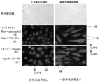
- Example 1 It is a photograph figure which shows the morphological characteristics and expression of various collagen by an optical microscope about the epithelioid morphological cell selected by the collection
- Example 1 It is a figure which shows the administration schedule and test schedule of an epithelioid morphological cell or a fibroblast-like cell in the transplantation test with respect to the peritoneal injury model using an immune tolerance mouse
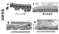
- Example 2 It is a photograph figure which shows the result which acts on the thickening of the wall side peritoneum at the time of transplanting an epithelioid form cell or a fibroblast-like cell to a peritoneum injury model.
- Example 2 It is a photograph which shows the result which acts on the thickening of the visceral peritoneum at the time of transplanting an epithelioid morphological cell or a fibroblast-like cell to a peritoneal injury model.
- Example 2 It is a figure which shows the result which acts on the number of adhesion sites in the wall side and the visceral peritoneum, and peritoneal thickening when an epithelioid morphological cell or a fibroblast-like cell is transplanted to a peritoneal injury model.
- Example 2 It is a photograph figure which shows the expression result of type I collagen or fibronectin in a peritoneum at the time of transplanting an epithelium-like morphological cell or a fibroblast-like cell to a peritoneal injury model.
- Example 3 It is a figure which shows the expression result of the hepatocyte growth factor (HGF) and the vascular endothelial growth factor (VEGF) in the transplanted cell at the time of transplanting an epithelioid morphological cell or a fibroblast-like cell to a peritoneal injury model.
- HGF hepatocyte growth factor
- VEGF vascular endothelial growth factor
- Example 4 It is a figure which shows the expression result of HGF in the wall side and visceral peritoneum at the time of transplanting an epithelioid morphological cell or a fibroblast-like cell to a peritoneal injury model.
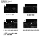
- Example 5 It is a figure which shows the expression result of VEGF in the wall side and visceral peritoneum at the time of transplanting an epithelioid morphological cell or a fibroblast-like cell to a peritoneal injury model. (Experimental example 5)
- the present invention relates to a peritoneal protective agent containing peritoneal mesothelial cells recovered from peritoneal dialysis drainage as an active ingredient.
- the peritoneal mesothelial cells collected from the peritoneal dialysis drainage are epithelial morphological cells, and the epithelial morphic cells express hepatocyte growth factor (HGF).
- HGF hepatocyte growth factor
- peritoneal protection is used in the concept of effectively acting on peritoneal disorders including peritoneal adhesion formation, peritoneal thickening and / or peritoneal injury
- the peritoneal protective agent of the present invention is used for peritoneal adhesion formation, peritoneal thickening and / or Or it can be used prophylactically and / or therapeutically for peritoneal injury.
- the peritoneal protective agent of the present invention can be prepared by a method including a step of collecting cells from the collected peritoneal dialysis drainage and further selecting epithelial morphological cells.
- the selection of the epithelioid morphological cells is performed by culturing cells collected from the peritoneal dialysis drainage, and selecting cells that are morphologically recognized as epithelial morphological cells.
- the selection of the epithelioid morphological cells is performed by selecting the cells collected from the peritoneal dialysis effluent using a marker specifically expressed in the epithelial morphological cells as an index.
- hepatocyte growth factor (HGF) is mentioned as a marker specifically expressed by epithelioid morphological cells.
- the method for collecting peritoneal mesothelial cells of the present invention includes a step of collecting cells contained in the collected peritoneal dialysis drainage and selecting epithelial morphological cells.
- Peritoneal dialysis is a type of artificial dialysis in which the peritoneum is used as a dialysis membrane, and is performed by injecting a sterilized dialysis solution into the abdominal cavity and removing waste products in the body through the peritoneum.
- dialysate perfusate
- the peritoneal dialysis drainage of the present invention refers to the drainage collected in a bag or the like after peritoneal dialysis as described above.
- the drainage fluid to be collected is not particularly limited as long as it is drained after peritoneal dialysis. For example, it is collected by continuous ambulatory peritoneal dialysis (CAPD) or automated peritoneal dialysis (APD). Drainage.
- peritoneal dialysis drainage waste from body fluids including blood, such as excess water in body fluids, nitrogen-containing wastes resulting from protein metabolism, etc., as well as dropping from the body due to stimulation of peritoneal dialysis fluid, etc.
- Cells particularly one layer of peritoneal mesothelial cells (PMC) covering the peritoneum.
- PMC peritoneal mesothelial cells
- Peritoneal mesothelial cells in peritoneal dialysis drainage are classified into epithelial cells and fibroblast cells.
- An epithelial cell is a cell that forms an epithelium on the surface of an animal tissue or body, has a two-dimensional layered structure, and has a function of keeping contents between cells or shielding the outside by tight bonding.
- fibroblasts are one type of cells that constitute connective tissue. Fibroblasts are said to produce fibrous substances such as type III and type IV collagen.
- the peritoneal mesothelial cells collected in the present invention are preferably epithelial cells among the cells.
- peritoneal mesothelial cells are intended to be used to improve peritoneal adhesions, peritoneal thickening or peritoneal damage, and thus eliminate fibroblast-like cells that produce fibrous substances such as collagen, It is important to select epithelioid morphological cells.
- the method for obtaining cells from the peritoneal dialysis drainage is not particularly limited.
- the peritoneal dialysis drainage is centrifuged to obtain a precipitate.
- epithelial morphological cells can be selected using as a marker a marker that is visually or specifically expressed in epithelial morphological cells.
- a step of culturing the obtained cells in a culture solution may be included before the selection step.
- the obtained cells are added to a culture solution in which dexamethasone, nicotinamide, HGF (hepatocyte growth factor) or b-FGF (basic fibroblast growth factor) is added to DMEM / F12 medium in addition to 10% FCS.
- Well suspended in a fibronectin-coated culture dish so that the number of cells is about 1 to 100 cells / well can be added to a cell culture vessel and cultured at 37 ° C. for about one month.
- the concentration of dexamethasone added can be 1 to 100 nM, preferably 20 to 75 nM, more preferably 62.5 nM, the concentration of nicotinamide can be 1 to 5 mM, preferably 2.5 mM, and the concentration of HGF Can be 1-30 ng / ml, preferably 1-10 ng / ml, more preferably 6.25 ng / ml, and the concentration of b-FGF is 1-30 ng / ml, preferably 1-10 ng / ml. Preferably it can be 6.25 ng / ml.
- KS medium DMEM / F12 + 10% FCS + ITS
- ITS-X insulin 5 ⁇ g / ml, transferrin 2.75 ⁇ g / ml, sodium selenite 3.35 ng / ml
- dexamethasone 62.5 nM
- nicotinamide 2.5 mM
- HGF 6.25 ng / m
- b-FGF 6.25 ng / ml
- the cells can be selected by observing the shape of the cells and selecting by a person skilled in the art.
- the epithelioid morphological cells can be selected by culturing the colonies obtained by the above culture by low-density culture or limiting dilution, and selecting paving stone-like cells from the cultured cells. See FIG. 1 for the morphology of epithelioid-like or fibroblast-like cells.
- a marker specifically expressed in epithelial morphological cells examples include hepatocyte growth factor (HGF), cytokeratin, and E-cadherin.
- HGF hepatocyte growth factor
- cytokeratin cytokeratin
- E-cadherin hepatocyte growth factor
- Cell selection using a marker as an index can be performed by FACS analysis using a flow cytometer.
- the peritoneal mesothelial cells obtained by the collection method of the present invention can be stored under conditions where the cells do not freeze, for example, at 1 to 4 ° C. within 10 hours, preferably within 6 hours, more preferably about 4 hours. It can also be stored frozen at ⁇ 80 ° C. or lower using a cell line cryopreservation medium.
- peritoneal mesothelial cells obtained by the collection method of the present invention effectively act on peritoneal adhesion formation, peritoneal thickening, peritoneal damage and the like.
- a preventive effect or a therapeutic effect can be expected for peritoneal adhesion formation by intraperitoneal organ surgery, peritoneal adhesion formation by gastrointestinal inflammation, peritonitis, etc., and peritoneal adhesion formation by peritoneal mesothelial cell detachment by peritoneal dialysis.
- a prophylactic effect or a therapeutic effect can be expected for peritoneal thickening caused by peritoneal dialysis fluid stimulation such as peritonitis or peritoneal dialysis.
- EPS peritoneal adhesions and encapsulating peritoneal sclerosis
- EMT epithelial-mesenchymal transition
- the peritoneal mesothelial cells of the present invention can supplement the dropped peritoneal mesothelial cells and suppress epithelial-mesenchymal transition.
- peritoneal mesothelial cells can regenerate damaged peritoneum and have a remarkable self-renewal ability that cannot be explained only by the regenerative capacity of progenitor cells (non- Patent Document 3), mesothelial cells are considered to be a promising option used in regenerative medicine.
- the present invention also extends to a peritoneal protective agent containing peritoneal mesothelial cells obtained by the above-described collection method, and a regenerative medical material containing the peritoneal protective agent.
- the peritoneal protective agent of the present invention can be stored frozen at ⁇ 80 ° C. or lower using a cryopreservation medium containing, for example, a KS medium.
- the peritoneal mesothelial cells obtained by the collection method of the present invention can be cryopreserved to construct a cell bank corresponding to the immune type and provide it as a peritoneal protective agent or a material for regenerative medical treatment of the peritoneum.
- Cells obtained by the above collection method, or cells obtained by the above collection method and cryopreserved and then thawed by a normal method can be used for cell transplantation of physiological saline, PBS, Hanks buffer, etc. It can be suspended in a solution to obtain a cell preparation.
- the cells contained in the cell preparation are required to contain at least 10 peritoneal mesothelial cells per ml.
- the number of cells can be at most 1 ⁇ 10 20 cells / ml, and preferably 10 to 1 ⁇ 10 7 cells / ml can be used.
- the peritoneal mesothelial cells used for the peritoneal protective agent are most preferably derived from autologous cells, but those derived from individuals with similar immunotypes as in the case of bone marrow transplantation. Can be used.
- peritoneal mesothelial cells other than self When peritoneal mesothelial cells other than self are applied, an immunosuppressant or the like may be used in combination as necessary.
- the peritoneum protective agent containing peritoneal mesothelial cells of the present invention or a regenerative medical material containing the peritoneal protective agent is a per se known treatment method for each symptom such as peritoneal adhesion, peritoneal thickening, peritoneal injury, etc. Can be used in combination with excision.
- Example 1 Method for collecting peritoneal mesothelial cells Informed consent was given to a peritoneal dialysis patient, 10 ml of peritoneal dialysis drainage was collected, and centrifuged at 1500 rpm for 5 minutes. The cells separated into the precipitate were suspended in KS medium and seeded on a culture dish coated with KS medium and fibronectin. After culturing at 37 ° C. for 4 to 7 days, the obtained cells were further diluted with a culture solution and cultured at 37 ° C. for 7 days by the limiting dilution method. Among the obtained cells, cobblestone-like epithelial morphological cells were selected. As a comparative example, fibroblast-like cells were also selected (see FIG. 1).
- Example 1 Characterization of the collected peritoneal mesothelial cells Each cell obtained by the method of Example 1 was observed with an optical microscope. Furthermore, cytokeratin (Santa Cruz: pan-cytokeratin antibody), type I collagen (Chemicon), vimentin (Sigma), and type IV collagen (Chemicon) expressed in each cell were fluorescently stained and observed. Cells with an epithelial character have many cytokeratin skeletal proteins, and cells with a fibroblast character have many vimentin skeletal proteins, and epithelial and fibroblastic cells can be seen by differences in the skeletal proteins. Generally, cells involved in fibrosis are considered to be fibroblasts.
- Example 2 Effect of peritoneal mesothelial cells on peritoneal sclerosis
- Immune-tolerant mice (nu / nu) were subjected to laparotomy, and the peritoneum (wall side) and intestinal peritoneum (sternal side) were rubbed 50 times each with gauze.
- Control mice in which peritoneal injury was formed by rubbing 100 times to construct a model of peritoneal injury was a sham-operated group (sham-ope group) in which immune-tolerant mice (nu / nu) females were only subjected to laparotomy.
- Example 2 Each mouse prepared by the method of Example 1 was incorporated into each mouse using a Vybrant (R) cell staining solution (Molecular probe), and DiO TM dye (green fluorescence) (Molecular probe) was incorporated. Fluorescent labeling was performed for tracking. 1 ml each of the cells suspended in PBS (number of cells: 1 ⁇ 10 7 cells / ml) was transplanted by intraperitoneal administration according to the administration schedule shown in FIG. On day 14 after the first cell transplantation, the mice were sacrificed and the morphological effects on the peritoneum were observed.
- Vybrant Vybrant
- DiO TM dye green fluorescence
- DiO TM dye can recognize up to 3 daughter cell division, but it becomes thinner at the time of further division and fluorescence recognition becomes impossible.
- DIO TM dye and PCNA which is a division marker, become both positive.
- both epithelial and fibroblastic cells were DiO TM dye positive, they engrafted in the peritoneum, and some of those cells were also positive for PCNA. It can be seen that the transplantation effect is exerted.
- Example 4 Cell Properties After Cell Transplantation With respect to each cell obtained by the method of Example 1, the expression of HGF and vascular endothelial growth factor (VEGF) was observed by fluorescent staining. As a result, HGF expression was observed from the epithelial morphological cells, and VEGF expression was mild. on the other hand. In fibroblast-like cells, almost no HGF expression was observed, confirming that VEGF was highly expressed (FIG. 8).
- VEGF vascular endothelial growth factor
- epithelial morphological cells adhere to and proliferate on the damaged site by epithelial morphological cell transplantation, and express HGF, a regeneration factor, to prevent peritoneal adhesion and sclerosis. It was thought to be.
- HGF a regeneration factor
- the fibroblast-like cells adhere and proliferate to the damaged site, produce type I collagen, promote peritoneal adhesion / sclerosis, and express VEGF, which is a vascular permeability factor. It was thought that this promoted an inflammatory reaction.
- peritoneal mesothelial cells can be easily collected by the method for collecting peritoneal mesothelial cells of the present invention.
- the collected peritoneal mesothelial cells effectively act on peritoneal adhesion formation, peritoneal thickening, peritoneal damage, etc., and can be used as peritoneal protective agents such as these preventives and therapeutic agents, and even damaged. It can be a regenerative medical material for the peritoneum.
- the peritoneal mesothelial cells obtained by the collection method of the present invention are collected from the peritoneal dialysis drainage, can be collected non-invasively, and the burden on patients and the like is light. Moreover, since the peritoneal dialysis drainage was conventionally discarded, resources can be effectively used.
- peritoneal protective agent When used as a peritoneal protective agent or a regenerative medicine material, even if the application site is complex, it is impossible in the past because peritoneal mesothelial cells can engraft and proliferate in the damaged peritoneum, etc. This prevents the formation of peritoneal adhesions, the prevention of peritoneal thickening, the peritoneal regeneration function, and the like.
- the peritoneal mesothelial cells obtained by the collection method of the present invention are derived from a living body, and the peritoneal protective agent containing the peritoneal mesothelial cells or the regenerative medical material containing the peritoneal protective agent is artificially synthesized. It is highly safe in that it can be used without using the prepared material.
- the peritoneal mesothelial cells to be applied are autologous. However, as in the case of bone marrow transplantation, those derived from individuals with similar immune types are also used. be able to.
- the peritoneal mesothelial cells obtained by the collection method of the present invention are cryopreserved to construct a cell bank according to the immune type and provide as a pharmaceutical composition as a peritoneal protective agent or a regenerative medical material containing a peritoneal protective agent You can also
Landscapes
- Health & Medical Sciences (AREA)
- Life Sciences & Earth Sciences (AREA)
- Veterinary Medicine (AREA)
- Chemical & Material Sciences (AREA)
- Public Health (AREA)
- General Health & Medical Sciences (AREA)
- Medicinal Chemistry (AREA)
- Engineering & Computer Science (AREA)
- Animal Behavior & Ethology (AREA)
- Pharmacology & Pharmacy (AREA)
- Nuclear Medicine, Radiotherapy & Molecular Imaging (AREA)
- General Chemical & Material Sciences (AREA)
- Chemical Kinetics & Catalysis (AREA)
- Bioinformatics & Cheminformatics (AREA)
- Organic Chemistry (AREA)
- Cell Biology (AREA)
- Biotechnology (AREA)
- Dermatology (AREA)
- Biomedical Technology (AREA)
- Surgery (AREA)
- Developmental Biology & Embryology (AREA)
- Immunology (AREA)
- Virology (AREA)
- Zoology (AREA)
- Epidemiology (AREA)
- Gastroenterology & Hepatology (AREA)
- Medicines Containing Material From Animals Or Micro-Organisms (AREA)
- Micro-Organisms Or Cultivation Processes Thereof (AREA)
- Materials For Medical Uses (AREA)
Abstract
本発明は、十分量の腹膜中皮細胞を簡便に採取する方法および採取された腹膜中皮細胞を提供する。該腹膜中皮細胞を有効成分として含む腹膜保護剤、または当該腹膜保護剤を含む再生医療用材料を提供する。腹膜透析を受けている患者から得た腹膜透析排液中の細胞を形態学的または細胞表面に発現するマーカーを指標として上皮様形態細胞を選別することにより、十分量の腹膜中皮細胞を簡便に採取しうる。得られた腹膜中皮細胞は、腹膜保護、即ち腹膜癒着形成、腹膜肥厚、腹膜損傷などに対して保護機能を有するため、腹膜保護剤または再生医療用材料をして利用することができる。
Description
本発明は、腹膜癒着形成、腹膜肥厚、腹膜損傷などに対して有効に作用する腹膜中皮細胞を有効成分とする腹膜保護剤に関し、さらに当該腹膜保護剤の調製方法に関する。また、当該腹膜保護剤を含む再生医療用材料に関する。
本出願は、参照によりここに援用されるところの日本出願特願2009-104704号優先権を請求する。
腹膜癒着は、消化器科、婦人科など各種領域の外科手術の跡に生じ、予後に大きな影響を与える。例えば、開腹手術後、特に消化器手術に際して生じる腸管の癒着は、手術手技および機器の向上に伴い少なくなる傾向にあるが、手術後、数年から十数年経過後にイレウス(腸閉塞)症状などを発症し、外科手術後の合併症の中でも最も重大な障害のひとつとなっている。開腹手術における腸壁や腸管の癒着は腸閉塞の原因ともなるため、再手術となったり、術後患者の予後を悪化させる傾向も散見される(非特許文献1)。このような合併症において癒着がおきると思われる部位に、特殊な癒着防止剤を使用する方法があるが、いまだ完全な予防法ではなく、新たなアプローチが求められている。
また、腹膜透析患者において致死的な合併症に被嚢性腹膜硬化症(EPS)がある。この合併症は、長年の腹膜透析液の刺激や腹膜炎の刺激により腹膜肥厚が進行し、腸閉塞が引き起こされ、場合によっては死に至る合併症である。本合併症においては、外科的に肥厚した腹膜を開腹切除するしかないが、手術後に再度癒着を引き起こすことが多く、いまだ有効な治療がない。
安全性に優れた癒着防止剤として、原材料が生体吸収性材料であるヒアルロン酸ナトリウムおよびカルボキシメチルセルロースを2:1の割合で含有するセプラフィルム(R)(科研製薬株式会社、Genzyme社)が既に臨床で使用されている。セプラフィルム(R)は貼付後24時間から48時間でゲル状となり、およそ7日間貼付部位に貯留して、損傷を受けた組織とその周囲の組織を物理的に隔離することで、癒着防止効果を発揮する。また、該癒着防止剤は貼付後28日以内に体外へ排出されるため、外科的除去は不要であることが特長点である。しかしながら、該癒着防止剤は乾燥状態では柔軟性がないため、使用部位が複雑な形状であると使用できず、また腹腔鏡下での手術時には適さないという問題点が挙げられている。また、そのような癒着防止剤を使用しても、壁側・臓側腹膜全体に発生する腹膜透析患者のEPS術後の癒着を予防することは難しい。
癒着形成防止について多く検討されているが、その殆どが生体分解性または生体適合性のポリマー材料などによる癒着防止材料(特許文献1~4)、または上皮細胞の足場となる材料(特許文献5)に関するものある。
近年、腹膜中皮細胞の移植とその可能性についていくつか報告がある。例えば、中皮細胞の腹腔内への移植は、腹膜癒着形成の減少や中皮細胞露出部のリモデリングに有効であることが示されている(非特許文献2)。また、腹膜中皮細胞は、前駆細胞の再生能のみからでは説明できない顕著な自己再生能力を有し、中皮細胞は再生医療に用いられる有望な選択肢になる可能性があることが報告されている(非特許文献3)。さらに、腹膜中皮細胞の移植は、腹膜機能不全に対する治療の選択肢の一つと考えられることも示されている(非特許文献4)。しかしながら、移植に用いる中皮細胞のソースなど、解決すべき課題が多く残されている。
Ann R Coll Surg Engl. Jan;72(1):60-63, 1990
Int J Artif Organs. 30(6):456-476, 2007
Int J Artif Organs. 30(6):501-512, 2007
Int J Artif Organs. 30(6):513-519, 2007
本発明は、十分量の腹膜中皮細胞を簡便に採取する方法および採取された腹膜中皮細胞を有効成分とする腹膜保護剤を提供することを課題とする。また、当該腹膜保護剤の調製方法並びに当該腹膜保護剤を含む再生医療用材料を提供することを課題とする。
本発明者らは、腹膜透析を受けている患者から得た腹膜透析排液中に、患者由来の腹膜中皮細胞が混入していると考えられることに着目し、鋭意研究を重ねた結果、腹膜透析排液中の細胞のうち、上皮様形態細胞を選別することで、十分量の腹膜中皮細胞を簡便に採取しうることを見出した。得られた腹膜中皮細胞は、腹膜保護剤として機能しうることを見出し、本発明を完成した。
即ち本発明は、以下よりなる。
1.腹膜透析排液から回収した腹膜中皮細胞を有効成分として含む、腹膜保護剤。
2.腹膜透析排液から回収した腹膜中皮細胞を1mlあたり少なくとも10個含む、前項1に記載の腹膜保護剤。
3.腹膜透析排液から回収した腹膜中皮細胞が、上皮様形態細胞である前項1または2に記載の腹膜保護剤。
4.上皮様形態細胞が、肝細胞増殖因子(HGF)を発現してなる前項3に記載の腹膜保護剤。
5.腹膜保護が、腹膜癒着形成、腹膜肥厚および/または腹膜損傷に対する保護である、前項1~4のいずれか1に記載の腹膜保護剤。
6.採取した腹膜透析排液から細胞を回収し、さらに上皮様形態細胞を選別する工程を含む前項1~5のいずれか1に記載の腹膜保護剤の調製方法。
7.前記上皮様形態細胞の選別が、腹膜透析排液から回収された細胞を培養し、形態学的に上皮様形態細胞と認められる細胞を選別することによる前項6に記載の腹膜保護剤の調製方法。
8.前記上皮様形態細胞の選別が、腹膜透析排液から回収された細胞について、上皮様形態細胞に特異的に発現されるマーカーを指標として選別することによる前項6に腹膜保護剤の調製方法。
9.上皮様形態細胞に特異的に発現されるマーカーが肝細胞増殖因子(HGF)である前項8に腹膜保護剤の調製方法。
10.採取した腹膜透析排液から細胞を回収し、さらに上皮様形態細胞を選別する工程を含む腹膜中皮細胞の採取方法。
11.前項10の方法により採取された腹膜中皮細胞。
12.前項11に記載の腹膜中皮細胞を含む腹膜保護剤。
13.前項1~5および前項12のいずれか1に記載の腹膜保護剤を含む再生医療用材料。
1.腹膜透析排液から回収した腹膜中皮細胞を有効成分として含む、腹膜保護剤。
2.腹膜透析排液から回収した腹膜中皮細胞を1mlあたり少なくとも10個含む、前項1に記載の腹膜保護剤。
3.腹膜透析排液から回収した腹膜中皮細胞が、上皮様形態細胞である前項1または2に記載の腹膜保護剤。
4.上皮様形態細胞が、肝細胞増殖因子(HGF)を発現してなる前項3に記載の腹膜保護剤。
5.腹膜保護が、腹膜癒着形成、腹膜肥厚および/または腹膜損傷に対する保護である、前項1~4のいずれか1に記載の腹膜保護剤。
6.採取した腹膜透析排液から細胞を回収し、さらに上皮様形態細胞を選別する工程を含む前項1~5のいずれか1に記載の腹膜保護剤の調製方法。
7.前記上皮様形態細胞の選別が、腹膜透析排液から回収された細胞を培養し、形態学的に上皮様形態細胞と認められる細胞を選別することによる前項6に記載の腹膜保護剤の調製方法。
8.前記上皮様形態細胞の選別が、腹膜透析排液から回収された細胞について、上皮様形態細胞に特異的に発現されるマーカーを指標として選別することによる前項6に腹膜保護剤の調製方法。
9.上皮様形態細胞に特異的に発現されるマーカーが肝細胞増殖因子(HGF)である前項8に腹膜保護剤の調製方法。
10.採取した腹膜透析排液から細胞を回収し、さらに上皮様形態細胞を選別する工程を含む腹膜中皮細胞の採取方法。
11.前項10の方法により採取された腹膜中皮細胞。
12.前項11に記載の腹膜中皮細胞を含む腹膜保護剤。
13.前項1~5および前項12のいずれか1に記載の腹膜保護剤を含む再生医療用材料。
本発明の腹膜中皮細胞の採取方法により、十分量の腹膜中皮細胞を簡便に採取することができる。採取された腹膜中皮細胞は、腹膜保護作用、即ち腹膜癒着形成、腹膜硬化、腹膜損傷など対して有効に作用し、これらの防止剤や治療剤としての腹膜保護剤として使用することができ、さらには損傷を受けた腹膜の再生医療用材料となりうる。本発明の採取方法により得られる腹膜中皮細胞は、腹膜透析排液から回収されるものであり、非侵襲的に回収することができ、患者にとって負担が軽いものである。また、腹膜透析排液は従来廃棄処理していたものであるので、資源の有効活用ができる。
本発明の採取方法により得られる腹膜中皮細胞を腹膜保護剤、または当該腹膜保護剤を含む再生医療用材料として用いる場合に、それらの適用部位が複雑な形状であっても腹膜中皮細胞が障害腹膜等に生着・増殖し、腹膜再生が可能であるため、従来不可能であった部位での腹膜保護作用、即ち腹膜癒着形成防止、腹膜硬化防止、腹膜再生などの機能が発揮される。
また、本発明の腹膜保護剤や再生医療用材料は、生体由来のものであり、人工的に合成した材料を用いることなく使用することができる点で安全性の高いものである。
本発明は、腹膜透析排液から回収した腹膜中皮細胞を有効成分として含む、腹膜保護剤に関する。また、腹膜透析排液から回収した腹膜中皮細胞が、上皮様形態細胞であり、当該上皮様形態細胞は、肝細胞増殖因子(HGF)を発現してなる。本発明において、腹膜保護は、腹膜癒着形成、腹膜肥厚および/または腹膜損傷を含む腹膜の障害に有効に作用する概念で用いられ、本発明の腹膜保護剤は、腹膜癒着形成、腹膜肥厚および/または腹膜損傷に対して、予防的および/または治療的に用いることができる。
本発明の腹膜保護剤は、採取した腹膜透析排液から細胞を回収し、さらに上皮様形態細胞を選別する工程を含む方法により調製することができる。前記上皮様形態細胞の選別は、腹膜透析排液から回収された細胞を培養し、形態学的に上皮様形態細胞と認められる細胞を選別することによる。または、前記上皮様形態細胞の選別は、腹膜透析排液から回収された細胞について、上皮様形態細胞に特異的に発現されるマーカーを指標として選別することによる。ここで、上皮様形態細胞に特異的に発現されるマーカーとしては、肝細胞増殖因子(HGF)が挙げられる。
本発明の腹膜中皮細胞の採取方法は、採取した腹膜透析排液に含まれる細胞を収集し、上皮様形態細胞を選別する工程を含む。
腹膜透析とは腹膜を透析膜として行う人工透析の一種であり、殺菌した透析液を腹腔内に注入し、体内の老廃物を腹膜を介して除去することにより行われる。実際には、腹腔内に挿入されたカテーテルから透析液(灌流液)を注入し、数時間後にバッグなどに排液を回収することにより行われる。本発明の腹膜透析排液は、前述のように腹膜透析後にバッグなどに回収される排液をいう。回収する排液は、腹膜透析後の排液であればよく、特に限定されないが、例えば連続携行式腹膜透析(Continuous Ambulatory Peritoneal Dialysis:CAPD)や 自動腹膜透析(Automated Peritoneal Dialysis:APD)により回収される排液が挙げられる。
腹膜透析排液には、血液等を含む体液からの老廃物、例えば体液中の過剰の水分、タンパク質の代謝の結果生じた窒素含有老廃物等のほか、腹膜透析液の刺激などにより生体から脱落した細胞、特に腹膜上を覆う一層の腹膜中皮細胞(peritoneal mosothelial cells:PMC)が含まれる。腹膜透析排液中の腹膜中皮細胞は、上皮細胞(epithelial cells)と線維芽細胞(fibroblast cells)などに分類される。ここで上皮細胞とは、動物の組織や体の表面で上皮を形成する細胞で、二次元的な層状構造をとり、細胞間は密着結合によって内容物を保ったり外部と遮蔽する機能を有する。一方、線維芽細胞とは、結合組織を構成する細胞の1種である。線維芽細胞は、III型、IV型コラーゲンなどの線維性物質を産生するといわれている。本発明で回収される腹膜中皮細胞は、前記細胞のうち上皮細胞が好適である。本発明において、腹膜中皮細胞は、腹膜癒着、腹膜肥厚あるいは腹膜損傷を改善するために用いることを目的とするため、上記コラーゲンなどの線維性物質を産生する線維芽細胞様細胞を排除し、上皮様形態細胞を選別することが重要である。
本発明において、腹膜透析排液からの細胞の取得方法は、特に限定されないが、例えば腹膜透析排液を遠心分離し、沈殿物を取得することに達成される。腹膜透析排液から遠心分離等により細胞を採取するまでの工程では、細胞が凍結しない条件で可能な限り低温、かつ迅速に処理することが好ましく、例えば1~4℃で、10時間以内、好ましくは6時間以内、より好ましくは4時間以内に処理するのが好適である。
本発明において、上皮様形態細胞は、目視的または上皮様形態細胞に特異的に発現されるマーカーを指標として選別することができる。上記選別工程の前に、取得した細胞を培養液中で培養する工程を含んでもよい。培養する場合は、取得した細胞を、DMEM/F12培地に10%FCSのほか、デキサメタゾン、ニコチナマイド、HGF(hepatocyte growth factor)やb-FGF(basic fibroblast growth factor)を添加した培養液などに、96ウェル フィブロネクチンコート培養皿に細胞数1~100個/ウェル程度になるように懸濁したものを、細胞培養用容器に加え、37℃で一か月程度培養することができる。添加するデキサメタゾンの濃度は1~100nM、好ましくは20~75nM、より好ましくは62.5nMとすることができ、ニコチナマイドの濃度は1~5mM、好ましくは2.5mMとすることができ、HGFの濃度は1~30ng/ml、好ましくは1~10ng/ml、より好ましくは6.25ng/mlとすることができ、b-FGFの濃度は1~30ng/ml、好ましくは1~10ng/ml、より好ましくは6.25ng/mlとすることができる。具体的な培養液としては、例えばKS培地(DMEM/F12+10%FCS+ITS(GIBCO(R) ITS-X:インスリン5μg/ml、トランスフェリン2.75μg/ml、亜セレン酸ナトリウム3.35ng/ml)+デキサメタゾン(62.5nM)+ニコチナマイド(2.5mM)+HGF(6.25ng/m)+b-FGF(6.25ng/ml))を用いて培養することができる。培養可能であれば、上記の培養方法に限定されることはない。
上皮様形態細胞を目視的に選別する方法としては、細胞の形状を観察し、いわゆる当業者が選別する方法で選別することができる。具体的には、上記培養により得られたコロニーを低密度培養または限界希釈法にて培養し、培養細胞から敷石状の細胞を選別することで、上皮様形態細胞を選別することができる。上皮様形態細胞または線維芽様細胞の形態については、図1を参照されたい。また、上皮様形態細胞に特異的に発現されるマーカーを指標として選別する場合、そのようなマーカーとしては、例えば肝細胞増殖因子(HGF)、サイトケラチン、E-カドヘリンを挙げることができ、より好ましくはHGFが挙げられる。マーカーを指標とする細胞の選別は、フローサイトメーターを用いたFACS解析などにより行うことができる。
本発明の採取方法により得られた腹膜中皮細胞は、細胞が凍結しない条件、例えば1~4℃で、10時間以内、好ましくは6時間以内、より好ましくは4時間程度保存することができる。また、細胞株の凍結保存用培地を用いて-80℃以下で凍結保存することもできる。
本発明の採取方法により得られた腹膜中皮細胞は、腹膜癒着形成、腹膜肥厚、腹膜損傷などに対して有効に作用する。例えば腹腔内臓器手術による腹膜癒着形成、消化管炎症や腹膜炎等による腹膜癒着形成、さらに腹膜透析による腹膜中皮細胞の脱落等による腹膜癒着形成などに対して、予防効果または治療効果が期待できる。また、腹膜炎や腹膜透析などの腹膜透析液の刺激等による腹膜肥厚に対しても予防効果または治療効果が期待できる。例えば、腹膜透析患者に多く見られる合併症のひとつに、腹膜癒着や被嚢性腹膜硬化症(EPS)が挙げられるが、これらは上皮様細胞である腹膜中皮細胞の脱落や、中皮細胞から上皮間葉系細胞へと移行する上皮間葉移行(epitherial-mesenchymal transition:EMT)などが原因と考えられる。このような場合にも、本発明の腹膜中皮細胞は、脱落した腹膜中皮細胞を補い、上皮間葉移行を抑制することができる。さらには、腹膜中皮細胞は、損傷した腹膜を再生可能である他、前駆細胞の再生能のみからでは説明できない顕著な自己再生能力を有していることが報告されていることからも(非特許文献3)、中皮細胞は再生医療に用いられる有望な選択肢になる可能性があると考えられる。
本発明は、上記採取方法により得られた腹膜中皮細胞を含む腹膜保護剤、および当該腹膜保護剤を含む再生医療用材料にも及ぶ。本発明の腹膜保護剤は、例えばKS培地を含む凍結保存用培地を用いて-80℃以下で凍結保存することができる。本発明の採取方法で得られた腹膜中皮細胞を凍結保存することにより、免疫タイプに応じたセルバンクを構築し、腹膜保護剤や腹膜の再生医療用材料として提供することもできる。上記採取方法得られた細胞、あるいは上記採取方法で得られ、凍結保存されたのち、通常の方法で融解処理された細胞は、生理食塩水、PBSやハンクス緩衝液などの細胞移植に使用可能な溶液に懸濁して細胞製剤とすることができる。細胞製剤に含まれる細胞は、1mlあたり少なくとも10個の腹膜中皮細胞を含むことが必要とされる。また、多くとも1×10²⁰個/mlの細胞数とすることができ、好適には10~1×107個/mlの細胞数で使用することができる。
腹膜中皮細胞を含む腹膜保護剤、または当該腹膜保護剤を含む再生医療用材料は、腹膜癒着や腹膜炎や腹膜肥厚などの腹膜損傷が危惧される部位や、あるいは既に癒着または腹膜損傷をきたしている部位に、予防的・治療的に用いることができる。拒絶反応を抑制する観点から、腹膜保護剤に用いる腹膜中皮細胞は自己由来であるのが最も好適であるが、骨髄移植の場合と同様に免疫タイプがよく近似している個体由来のものも使用することができる。自己以外の腹膜中皮細胞を適用する場合は、必要に応じて免疫抑制剤などを適宜併用してもよい。また、本発明の腹膜中皮細胞を含む腹膜保護剤または当該腹膜保護剤を含む再生医療用材料は、腹膜癒着、腹膜肥厚、腹膜損傷等の各症状に対する自体公知の処理方法、例えば腹膜硬化部切除術などと併用することができる。
以下、本発明を実施例によりさらに具体的に説明するが、本発明は下記の実施例の範囲に限定されることはない。
(実施例1)腹膜中皮細胞の採取方法
腹膜透析患者にインフォームドコンセントを行い、腹膜透析排液10mlを採取し、1500rpm×5分間遠心分離を行った。沈殿に分離された細胞をKS培地に懸濁し、KS培地およびフィブロネクチンコートした培養皿に播種した。37℃で4~7日培養後、得られた細胞をさらに培養液を用いて希釈し、限外希釈法にて37℃で7日間培養した。得られた細胞のうち、敷石状の上皮様形態細胞を選別した。比較例として、線維芽細胞様細胞も選別した(図1参照)。
腹膜透析患者にインフォームドコンセントを行い、腹膜透析排液10mlを採取し、1500rpm×5分間遠心分離を行った。沈殿に分離された細胞をKS培地に懸濁し、KS培地およびフィブロネクチンコートした培養皿に播種した。37℃で4~7日培養後、得られた細胞をさらに培養液を用いて希釈し、限外希釈法にて37℃で7日間培養した。得られた細胞のうち、敷石状の上皮様形態細胞を選別した。比較例として、線維芽細胞様細胞も選別した(図1参照)。
(実験例1)採取した腹膜中皮細胞の性状分析
実施例1の方法により得られた各細胞を光学顕微鏡により観察した。さらに各細胞に発現したサイトケラチン(Santa Cruz社:pan-cytokeratin抗体)、タイプIコラーゲン(Chemicon社)、ならびにビメンチン(Sigma社)、タイプIVコラーゲン(Chemicon社)を蛍光染色し、観察した。上皮の性格を示す細胞はサイトケラチンの骨格タンパクが多く、線維芽細胞の性格を示す細胞はビメンチンの骨格タンパクが多く、その骨格タンパクの違いでも上皮系、線維芽系細胞がわかる。一般に、線維化に関与する細胞は線維芽細胞でと考えられている。サイトケラチン(緑)、タイプIコラーゲン(赤)、ならびにビメンチン(緑)、タイプIVコラーゲン(赤)の蛍光染色はKEYENCE社のオールインワン蛍光顕微鏡(BZ-8000)を用いて観察した。上記の結果、線維芽細胞様細胞には硬化進展に関与するタイプIコラーゲンおよびタイプIVコラーゲンの発現が観察されたのに対し、上皮様形態細胞ではコラーゲンの発現は認められなかった(図2)。
実施例1の方法により得られた各細胞を光学顕微鏡により観察した。さらに各細胞に発現したサイトケラチン(Santa Cruz社:pan-cytokeratin抗体)、タイプIコラーゲン(Chemicon社)、ならびにビメンチン(Sigma社)、タイプIVコラーゲン(Chemicon社)を蛍光染色し、観察した。上皮の性格を示す細胞はサイトケラチンの骨格タンパクが多く、線維芽細胞の性格を示す細胞はビメンチンの骨格タンパクが多く、その骨格タンパクの違いでも上皮系、線維芽系細胞がわかる。一般に、線維化に関与する細胞は線維芽細胞でと考えられている。サイトケラチン(緑)、タイプIコラーゲン(赤)、ならびにビメンチン(緑)、タイプIVコラーゲン(赤)の蛍光染色はKEYENCE社のオールインワン蛍光顕微鏡(BZ-8000)を用いて観察した。上記の結果、線維芽細胞様細胞には硬化進展に関与するタイプIコラーゲンおよびタイプIVコラーゲンの発現が観察されたのに対し、上皮様形態細胞ではコラーゲンの発現は認められなかった(図2)。
(実験例2)腹膜硬化に対する腹膜中皮細胞の作用
免疫寛容マウス(nu/nu)雌を開腹手術し、腹膜(壁側)および腸管腹膜(臓側)をガーゼにて各50回擦り、計100回擦過することで腹膜障害を形成させ、腹膜障害モデルを構築した対照マウスは、免疫寛容マウス(nu/nu)雌を開腹手術のみ行ったシャムオペ群(sham-ope群)とした。各マウスに、実施例1の方法で調製した各細胞をVybrant(R) 細胞染色溶液(Molecular probe社)を使用し、DiOTM色素(緑色蛍光)(Molecular probe社)をとりこませ、移植後でも追跡できるよう蛍光ラベルを行った。その細胞を各々PBSに懸濁したもの(細胞数1×10⁷個/ml)1mlを、図3に示す投与スケジュールで腹腔内投与により移植した。第1回の細胞移植後14日目にマウスを屠殺し、腹膜に及ぼす形態的作用を観察した。
免疫寛容マウス(nu/nu)雌を開腹手術し、腹膜(壁側)および腸管腹膜(臓側)をガーゼにて各50回擦り、計100回擦過することで腹膜障害を形成させ、腹膜障害モデルを構築した対照マウスは、免疫寛容マウス(nu/nu)雌を開腹手術のみ行ったシャムオペ群(sham-ope群)とした。各マウスに、実施例1の方法で調製した各細胞をVybrant(R) 細胞染色溶液(Molecular probe社)を使用し、DiOTM色素(緑色蛍光)(Molecular probe社)をとりこませ、移植後でも追跡できるよう蛍光ラベルを行った。その細胞を各々PBSに懸濁したもの(細胞数1×10⁷個/ml)1mlを、図3に示す投与スケジュールで腹腔内投与により移植した。第1回の細胞移植後14日目にマウスを屠殺し、腹膜に及ぼす形態的作用を観察した。
上記の結果、上皮様形態細胞を移植した群(n=3)では、シャムオペ群(n=3)と同様に、腹膜(壁側)および腸管腹膜(臓側)での腹膜癒着および肥厚化は認められなかったが、線維芽細胞様細胞を移植した群(n=4)では、腹膜障害を起こした部位で腹膜癒着が認められた(図4、図5)。各腹膜(壁側、臓側)について、上皮様形態細胞または線維芽細胞様細胞を移植した群における腹膜癒着部位の数および腹膜の厚さを測定した結果を図6に示した。この結果からも、上皮様形態細胞を移植した群では、シャムオペ群と同様に、腹膜癒着および肥厚化が抑えられていることが観察された。
(実験例3)各細胞移植後の腹膜組織の性状
実験例2の方法で、腹膜障害モデルに上皮様形態細胞または線維芽細胞様細胞を移植した後の各腹膜組織について、核内増殖抗原(PCNA)(Pharmingen社)、タイプIコラーゲン(Chemicon社)、フィブロネクチン(Chemicon社)の発現を蛍光染色により観察した。
上記の結果、腹膜障害群で、タイプIコラーゲンの発現が最も多く認められ、線維芽細胞様細胞を移植した群でもやや多く発現していることが観察されたのに対し、上皮様形態細胞を移植した群では、シャムオペ群と同様に、タイプIコラーゲンの発現が抑制されていた(図7)。DiOTM色素では3娘細胞分裂まで認識できるが、それ以上の分裂時には薄くなり蛍光認識できなくなる。本色素を認識した細胞が分裂が激しい場合は、DIOTM色素と分裂マーカーであるPCNAが両陽性になる。細胞移植群において、上皮形態細胞も線維芽形態細胞もDiOTM色素陽性であり、それらが腹膜に生着し、それらの細胞の一部がPCNAも陽性となったため、生着細胞が分裂増殖し、移植効果が発揮されていることがわかる。
実験例2の方法で、腹膜障害モデルに上皮様形態細胞または線維芽細胞様細胞を移植した後の各腹膜組織について、核内増殖抗原(PCNA)(Pharmingen社)、タイプIコラーゲン(Chemicon社)、フィブロネクチン(Chemicon社)の発現を蛍光染色により観察した。
上記の結果、腹膜障害群で、タイプIコラーゲンの発現が最も多く認められ、線維芽細胞様細胞を移植した群でもやや多く発現していることが観察されたのに対し、上皮様形態細胞を移植した群では、シャムオペ群と同様に、タイプIコラーゲンの発現が抑制されていた(図7)。DiOTM色素では3娘細胞分裂まで認識できるが、それ以上の分裂時には薄くなり蛍光認識できなくなる。本色素を認識した細胞が分裂が激しい場合は、DIOTM色素と分裂マーカーであるPCNAが両陽性になる。細胞移植群において、上皮形態細胞も線維芽形態細胞もDiOTM色素陽性であり、それらが腹膜に生着し、それらの細胞の一部がPCNAも陽性となったため、生着細胞が分裂増殖し、移植効果が発揮されていることがわかる。
(実験例4)各細胞移植後の細胞の性状
実施例1の方法により得られた各細胞について、HGFおよび血管内皮増殖因子(VEGF)の発現を蛍光染色により観察した。
上記の結果、上皮様形態細胞からHGFの発現が観察され、VEGFの発現は軽度であった。一方。線維芽細胞様細胞では、HGFの発現は殆ど認められず、VEGFは高度に発現していることが確認された(図8)。
実施例1の方法により得られた各細胞について、HGFおよび血管内皮増殖因子(VEGF)の発現を蛍光染色により観察した。
上記の結果、上皮様形態細胞からHGFの発現が観察され、VEGFの発現は軽度であった。一方。線維芽細胞様細胞では、HGFの発現は殆ど認められず、VEGFは高度に発現していることが確認された(図8)。
(実験例5)各細胞移植後の腹膜組織中の細胞の性状
実験例2の方法で上皮様形態細胞または線維芽細胞様細胞を移植した後の各腹膜組織について、HGFおよびVEGFの発現を蛍光染色により観察した。
上記の結果、移植後の上皮様形態細胞からHGFの発現が観察されたが、線維芽細胞様細胞からのHGFの発現は殆ど認められなかった(図9)。一方、上皮様形態細胞からのVEGFの発現は軽度であったが、線維芽細胞様細胞からVEGFが高度に発現していることが確認された(図10)。
実験例2の方法で上皮様形態細胞または線維芽細胞様細胞を移植した後の各腹膜組織について、HGFおよびVEGFの発現を蛍光染色により観察した。
上記の結果、移植後の上皮様形態細胞からHGFの発現が観察されたが、線維芽細胞様細胞からのHGFの発現は殆ど認められなかった(図9)。一方、上皮様形態細胞からのVEGFの発現は軽度であったが、線維芽細胞様細胞からVEGFが高度に発現していることが確認された(図10)。
実験例3~5の結果より、上皮様形態細胞の移植により上皮様形態細胞が障害部位に接着・増殖して障害腹膜上を覆い、再生因子であるHGFを発現し、腹膜癒着・硬化を防止すると考えられた。一方、線維芽細胞様細胞の移植では、線維芽細胞様細胞が障害部位に接着・増殖し、タイプIコラーゲンを産生し、腹膜癒着・硬化を促進し、かつ血管透過因子であるVEGFを発現することにより、炎症反応を助長することとなると考えられた。
以上詳述したように、本発明の腹膜中皮細胞の採取方法により、十分量の腹膜中皮細胞を簡便に採取することができる。採取された腹膜中皮細胞は、腹膜癒着形成、腹膜肥厚、腹膜損傷など対して有効に作用し、これらの防止剤や治療剤などの腹膜保護剤として使用することができ、さらには損傷を受けた腹膜の再生医療用材料となりうる。本発明の採取方法により得られる腹膜中皮細胞は、腹膜透析排液から回収されるものであり、非侵襲的に回収することができ、患者等の負担が軽いものである。また、腹膜透析排液は、従来は廃棄処理していたものであるので、資源の有効活用ができる。
腹膜保護剤や再生医療用材料として利用する場合、適用部位が複雑な形状であっても、腹膜中皮細胞が障害腹膜等に生着・増殖し、腹膜再生が可能であるため、従来不可能であった部位での腹膜癒着形成防止、腹膜肥厚防止、腹膜再生機能などが発揮される。
また、本発明の採取方法により得られる腹膜中皮細胞は生体由来のものであり、該腹膜中皮細胞を含む腹膜保護剤、または当該腹膜保護剤を含む再生医療用材料は、人工的に合成した材料を用いることなく使用することができる点で安全性の高いものである。
拒絶反応を抑制する観点から、適用される腹膜中皮細胞は自己由来であるのが最も好適であるが、骨髄移植の場合と同様に免疫タイプがよく近似している個体由来のものも使用することができる。本発明の採取方法で得られた腹膜中皮細胞を凍結保存することにより、免疫タイプに応じたセルバンクを構築し、腹膜保護剤としての医薬組成物や腹膜保護剤を含む再生医療用材料として提供することもできる。
Claims (13)
- 腹膜透析排液から回収した腹膜中皮細胞を有効成分として含む、腹膜保護剤。
- 腹膜透析排液から回収した腹膜中皮細胞を1mlあたり少なくとも10個含む、請求項1に記載の腹膜保護剤。
- 腹膜透析排液から回収した腹膜中皮細胞が、上皮様形態細胞である請求項1または2に記載の腹膜保護剤。
- 上皮様形態細胞が、肝細胞増殖因子(HGF)を発現してなる請求項3に記載の腹膜保護剤。
- 腹膜保護が、腹膜癒着形成、腹膜肥厚および/または腹膜損傷に対する保護である、請求項1~4のいずれか1に記載の腹膜保護剤。
- 採取した腹膜透析排液から細胞を回収し、さらに上皮様形態細胞を選別する工程を含む請求項1~5のいずれか1に記載の腹膜保護剤の調製方法。
- 前記上皮様形態細胞の選別が、腹膜透析排液から回収された細胞を培養し、形態学的に上皮様形態細胞と認められる細胞を選別することによる請求項6に記載の腹膜保護剤の調製方法。
- 前記上皮様形態細胞の選別が、腹膜透析排液から回収された細胞について、上皮様形態細胞に特異的に発現されるマーカーを指標として選別することによる請求項6に腹膜保護剤の調製方法。
- 上皮様形態細胞に特異的に発現されるマーカーが肝細胞増殖因子(HGF)である請求項8に腹膜保護剤の調製方法。
- 採取した腹膜透析排液から細胞を回収し、さらに上皮様形態細胞を選別する工程を含む腹膜中皮細胞の採取方法。
- 請求項10の方法により採取された腹膜中皮細胞。
- 請求項11に記載の腹膜中皮細胞を含む腹膜保護剤。
- 請求項1~5および請求項12のいずれか1に記載の腹膜保護剤を含む再生医療用材料。
Priority Applications (1)
| Application Number | Priority Date | Filing Date | Title |
|---|---|---|---|
| JP2011510219A JP5731969B2 (ja) | 2009-04-23 | 2010-04-21 | 腹膜保護剤および腹膜保護剤の調製方法 |
Applications Claiming Priority (2)
| Application Number | Priority Date | Filing Date | Title |
|---|---|---|---|
| JP2009-104704 | 2009-04-23 | ||
| JP2009104704 | 2009-04-23 |
Publications (1)
| Publication Number | Publication Date |
|---|---|
| WO2010122787A1 true WO2010122787A1 (ja) | 2010-10-28 |
Family
ID=43010913
Family Applications (1)
| Application Number | Title | Priority Date | Filing Date |
|---|---|---|---|
| PCT/JP2010/002865 Ceased WO2010122787A1 (ja) | 2009-04-23 | 2010-04-21 | 腹膜保護剤および腹膜保護剤の調製方法 |
Country Status (2)
| Country | Link |
|---|---|
| JP (1) | JP5731969B2 (ja) |
| WO (1) | WO2010122787A1 (ja) |
Cited By (1)
| Publication number | Priority date | Publication date | Assignee | Title |
|---|---|---|---|---|
| CN109810939A (zh) * | 2019-04-09 | 2019-05-28 | 武汉轻工大学 | 一种猪腹膜间皮细胞的培养方法 |
-
2010
- 2010-04-21 JP JP2011510219A patent/JP5731969B2/ja active Active
- 2010-04-21 WO PCT/JP2010/002865 patent/WO2010122787A1/ja not_active Ceased
Non-Patent Citations (10)
| Title |
|---|
| BERTRAM P ET AL.: "Intraperitoneal transplantation of isologous mesothelial cells for prevention of adhesions.", EUR. J. SURG., vol. 165, 1999, pages 705 - 709 * |
| DI PAOLO N ET AL.: "Implant of autologous mesothelial cells in animals and a peritoneal dialysis patient.", INT. J. ARTIF. ORGANS, vol. 12, 1989, pages 485 - 501 * |
| DI PAOLO N ET AL.: "State of the art on autologous mesothelial transplant in animals and humans.", INT. J. ARTIF. ORGANS, vol. 30, 2007, pages 456 - 476 * |
| FOLEY-COMER A J ET AL.: "Evidence for incorporation of free-floating mesothelial cells as a mechanism of serosal healing.", J. CELL SCI., vol. 115, 2002, pages 1383 - 1389 * |
| HEKKING L H P ET AL.: "Feasibility of mesothelial transplantation during experimental peritoneal dialysis and peritonitis.", INT. J. ARTIF. ORGANS, vol. 30, 2007, pages 513 - 519 * |
| HITOSHI SUGIYAMA ET AL.: "Fukumaku Shogai ni Taisuru Saisei Chiryo", THERAPEUTICS & ENGINEERING, vol. 22, 2 April 2010 (2010-04-02), pages 71 * |
| MUTSAERS S ET AL.: "Future directions in mesothelial transplantation research.", INT. J. ARTIF. ORGANS, vol. 30, 2007, pages 557 - 561 * |
| RAMPINO T ET AL.: "Hepatocyte growth factor/ scatter factor released during peritonitis is active on mesothelial cells.", AM. J. PATHOL., vol. 159, 2001, pages 1275 - 1285 * |
| SHINJI KITAMURA ET AL.: "Fukumaku Toseki Haieki kara Saishu shita Hito Fukumaku Chuhi Saibo o Shiyo shita Fukumaku Shogai eno Saibo Ryoho no Kokoromi", JOURNAL OF JAPANESE SOCIETY FOR DIALYSIS THERAPY, vol. 42, no. 1, 8 May 2009 (2009-05-08), pages 485 * |
| YANEZ-MO M ET AL.: "Peritoneal dialysis and epithelial-to-mesenchymal transition of mesothelial cells.", NEW ENGL. J. MED., vol. 348, 2003, pages 403 - 413 * |
Cited By (1)
| Publication number | Priority date | Publication date | Assignee | Title |
|---|---|---|---|---|
| CN109810939A (zh) * | 2019-04-09 | 2019-05-28 | 武汉轻工大学 | 一种猪腹膜间皮细胞的培养方法 |
Also Published As
| Publication number | Publication date |
|---|---|
| JPWO2010122787A1 (ja) | 2012-10-25 |
| JP5731969B2 (ja) | 2015-06-10 |
Similar Documents
| Publication | Publication Date | Title |
|---|---|---|
| JP7027484B2 (ja) | 瘻の治療における脂肪組織由来間質幹細胞の使用 | |
| JP5731969B2 (ja) | 腹膜保護剤および腹膜保護剤の調製方法 | |
| Sheta et al. | Structural and functional renovation of urinary bladders after amniotic membrane implantation in dogs | |
| HK1239729B (en) | Use of adipose tissue-derived stromal stem cells in treating fistula | |
| HK1239730B (en) | Use of adipose tissue-derived stromal stem cells in treating fistula | |
| HK1239731B (en) | Use of adipose tissue-derived stromal stem cells in treating fistula | |
| HK1239731A1 (en) | Use of adipose tissue-derived stromal stem cells in treating fistula | |
| HK1239730A1 (en) | Use of adipose tissue-derived stromal stem cells in treating fistula | |
| MX2008000001A (en) | Use of adipose tissue-derived stromal stem cells in treating fistula |
Legal Events
| Date | Code | Title | Description |
|---|---|---|---|
| 121 | Ep: the epo has been informed by wipo that ep was designated in this application |
Ref document number: 10766848 Country of ref document: EP Kind code of ref document: A1 |
|
| DPE1 | Request for preliminary examination filed after expiration of 19th month from priority date (pct application filed from 20040101) | ||
| ENP | Entry into the national phase |
Ref document number: 2011510219 Country of ref document: JP Kind code of ref document: A |
|
| NENP | Non-entry into the national phase |
Ref country code: DE |
|
| 122 | Ep: pct application non-entry in european phase |
Ref document number: 10766848 Country of ref document: EP Kind code of ref document: A1 |