WO2010044686A1 - Akr1c3 as a biomarker, methods of selecting and treating patients based upon an akr1c3 profile and compounds for use therein - Google Patents
Akr1c3 as a biomarker, methods of selecting and treating patients based upon an akr1c3 profile and compounds for use therein Download PDFInfo
- Publication number
- WO2010044686A1 WO2010044686A1 PCT/NZ2009/000227 NZ2009000227W WO2010044686A1 WO 2010044686 A1 WO2010044686 A1 WO 2010044686A1 NZ 2009000227 W NZ2009000227 W NZ 2009000227W WO 2010044686 A1 WO2010044686 A1 WO 2010044686A1
- Authority
- WO
- WIPO (PCT)
- Prior art keywords
- akrl
- patient
- cancer
- activated
- agent
- Prior art date
- Legal status (The legal status is an assumption and is not a legal conclusion. Google has not performed a legal analysis and makes no representation as to the accuracy of the status listed.)
- Ceased
Links
- 0 *CCN(CC*)c(cc(CC(N(*)*)=O)c([N+]([O-])=O)c1)c1[N+]([O-])=O Chemical compound *CCN(CC*)c(cc(CC(N(*)*)=O)c([N+]([O-])=O)c1)c1[N+]([O-])=O 0.000 description 1
- CNDDNFNRYIWPDJ-UHFFFAOYSA-N CCCN(CCC)c(c(CC(O)=O)cc([N+]([O-])=O)c1)c1[N+]([O-])=O Chemical compound CCCN(CCC)c(c(CC(O)=O)cc([N+]([O-])=O)c1)c1[N+]([O-])=O CNDDNFNRYIWPDJ-UHFFFAOYSA-N 0.000 description 1
- OFXCRXLTJJLOIB-UHFFFAOYSA-N CCCN(CCC)c(ccc([N+]([O-])=O)c1)c1[N+]([O-])=O Chemical compound CCCN(CCC)c(ccc([N+]([O-])=O)c1)c1[N+]([O-])=O OFXCRXLTJJLOIB-UHFFFAOYSA-N 0.000 description 1
- STCOQWRCNUZHNZ-UHFFFAOYSA-N CCCN(CCC1CC1)c(c(C(O)=O)cc([N+]([O-])=O)c1)c1[N+]([O-])=O Chemical compound CCCN(CCC1CC1)c(c(C(O)=O)cc([N+]([O-])=O)c1)c1[N+]([O-])=O STCOQWRCNUZHNZ-UHFFFAOYSA-N 0.000 description 1
- CDVNZMKTJIBBBV-UHFFFAOYSA-N Cc(c(C(O)=O)cc([N+]([O-])=O)c1)c1[N+]([O-])=O Chemical compound Cc(c(C(O)=O)cc([N+]([O-])=O)c1)c1[N+]([O-])=O CDVNZMKTJIBBBV-UHFFFAOYSA-N 0.000 description 1
Classifications
-
- C—CHEMISTRY; METALLURGY
- C07—ORGANIC CHEMISTRY
- C07F—ACYCLIC, CARBOCYCLIC OR HETEROCYCLIC COMPOUNDS CONTAINING ELEMENTS OTHER THAN CARBON, HYDROGEN, HALOGEN, OXYGEN, NITROGEN, SULFUR, SELENIUM OR TELLURIUM
- C07F9/00—Compounds containing elements of Groups 5 or 15 of the Periodic Table
- C07F9/02—Phosphorus compounds
- C07F9/06—Phosphorus compounds without P—C bonds
- C07F9/08—Esters of oxyacids of phosphorus
- C07F9/09—Esters of phosphoric acids
- C07F9/091—Esters of phosphoric acids with hydroxyalkyl compounds with further substituents on alkyl
-
- A—HUMAN NECESSITIES
- A61—MEDICAL OR VETERINARY SCIENCE; HYGIENE
- A61K—PREPARATIONS FOR MEDICAL, DENTAL OR TOILETRY PURPOSES
- A61K31/00—Medicinal preparations containing organic active ingredients
-
- A—HUMAN NECESSITIES
- A61—MEDICAL OR VETERINARY SCIENCE; HYGIENE
- A61K—PREPARATIONS FOR MEDICAL, DENTAL OR TOILETRY PURPOSES
- A61K31/00—Medicinal preparations containing organic active ingredients
- A61K31/13—Amines
- A61K31/135—Amines having aromatic rings, e.g. ketamine, nortriptyline
- A61K31/136—Amines having aromatic rings, e.g. ketamine, nortriptyline having the amino group directly attached to the aromatic ring, e.g. benzeneamine
-
- A—HUMAN NECESSITIES
- A61—MEDICAL OR VETERINARY SCIENCE; HYGIENE
- A61K—PREPARATIONS FOR MEDICAL, DENTAL OR TOILETRY PURPOSES
- A61K31/00—Medicinal preparations containing organic active ingredients
- A61K31/13—Amines
- A61K31/145—Amines having sulfur, e.g. thiurams (>N—C(S)—S—C(S)—N< and >N—C(S)—S—S—C(S)—N<), Sulfinylamines (—N=SO), Sulfonylamines (—N=SO2)
-
- A—HUMAN NECESSITIES
- A61—MEDICAL OR VETERINARY SCIENCE; HYGIENE
- A61K—PREPARATIONS FOR MEDICAL, DENTAL OR TOILETRY PURPOSES
- A61K31/00—Medicinal preparations containing organic active ingredients
- A61K31/16—Amides, e.g. hydroxamic acids
- A61K31/165—Amides, e.g. hydroxamic acids having aromatic rings, e.g. colchicine, atenolol, progabide
- A61K31/166—Amides, e.g. hydroxamic acids having aromatic rings, e.g. colchicine, atenolol, progabide having the carbon of a carboxamide group directly attached to the aromatic ring, e.g. procainamide, procarbazine, metoclopramide, labetalol
-
- A—HUMAN NECESSITIES
- A61—MEDICAL OR VETERINARY SCIENCE; HYGIENE
- A61K—PREPARATIONS FOR MEDICAL, DENTAL OR TOILETRY PURPOSES
- A61K31/00—Medicinal preparations containing organic active ingredients
- A61K31/66—Phosphorus compounds
- A61K31/661—Phosphorus acids or esters thereof not having P—C bonds, e.g. fosfosal, dichlorvos, malathion or mevinphos
-
- A—HUMAN NECESSITIES
- A61—MEDICAL OR VETERINARY SCIENCE; HYGIENE
- A61K—PREPARATIONS FOR MEDICAL, DENTAL OR TOILETRY PURPOSES
- A61K45/00—Medicinal preparations containing active ingredients not provided for in groups A61K31/00 - A61K41/00
- A61K45/06—Mixtures of active ingredients without chemical characterisation, e.g. antiphlogistics and cardiaca
-
- A—HUMAN NECESSITIES
- A61—MEDICAL OR VETERINARY SCIENCE; HYGIENE
- A61P—SPECIFIC THERAPEUTIC ACTIVITY OF CHEMICAL COMPOUNDS OR MEDICINAL PREPARATIONS
- A61P35/00—Antineoplastic agents
-
- C—CHEMISTRY; METALLURGY
- C07—ORGANIC CHEMISTRY
- C07C—ACYCLIC OR CARBOCYCLIC COMPOUNDS
- C07C237/00—Carboxylic acid amides, the carbon skeleton of the acid part being further substituted by amino groups
- C07C237/28—Carboxylic acid amides, the carbon skeleton of the acid part being further substituted by amino groups having the carbon atom of at least one of the carboxamide groups bound to a carbon atom of a non-condensed six-membered aromatic ring of the carbon skeleton
- C07C237/32—Carboxylic acid amides, the carbon skeleton of the acid part being further substituted by amino groups having the carbon atom of at least one of the carboxamide groups bound to a carbon atom of a non-condensed six-membered aromatic ring of the carbon skeleton having the nitrogen atom of the carboxamide group bound to an acyclic carbon atom of a hydrocarbon radical substituted by oxygen atoms
-
- C—CHEMISTRY; METALLURGY
- C07—ORGANIC CHEMISTRY
- C07C—ACYCLIC OR CARBOCYCLIC COMPOUNDS
- C07C309/00—Sulfonic acids; Halides, esters, or anhydrides thereof
- C07C309/63—Esters of sulfonic acids
- C07C309/64—Esters of sulfonic acids having sulfur atoms of esterified sulfo groups bound to acyclic carbon atoms
- C07C309/65—Esters of sulfonic acids having sulfur atoms of esterified sulfo groups bound to acyclic carbon atoms of a saturated carbon skeleton
- C07C309/66—Methanesulfonates
-
- C—CHEMISTRY; METALLURGY
- C12—BIOCHEMISTRY; BEER; SPIRITS; WINE; VINEGAR; MICROBIOLOGY; ENZYMOLOGY; MUTATION OR GENETIC ENGINEERING
- C12Q—MEASURING OR TESTING PROCESSES INVOLVING ENZYMES, NUCLEIC ACIDS OR MICROORGANISMS; COMPOSITIONS OR TEST PAPERS THEREFOR; PROCESSES OF PREPARING SUCH COMPOSITIONS; CONDITION-RESPONSIVE CONTROL IN MICROBIOLOGICAL OR ENZYMOLOGICAL PROCESSES
- C12Q1/00—Measuring or testing processes involving enzymes, nucleic acids or microorganisms; Compositions therefor; Processes of preparing such compositions
- C12Q1/26—Measuring or testing processes involving enzymes, nucleic acids or microorganisms; Compositions therefor; Processes of preparing such compositions involving oxidoreductase
-
- G—PHYSICS
- G01—MEASURING; TESTING
- G01N—INVESTIGATING OR ANALYSING MATERIALS BY DETERMINING THEIR CHEMICAL OR PHYSICAL PROPERTIES
- G01N33/00—Investigating or analysing materials by specific methods not covered by groups G01N1/00 - G01N31/00
- G01N33/48—Biological material, e.g. blood, urine; Haemocytometers
- G01N33/50—Chemical analysis of biological material, e.g. blood, urine; Testing involving biospecific ligand binding methods; Immunological testing
- G01N33/53—Immunoassay; Biospecific binding assay; Materials therefor
- G01N33/573—Immunoassay; Biospecific binding assay; Materials therefor for enzymes or isoenzymes
-
- G—PHYSICS
- G01—MEASURING; TESTING
- G01N—INVESTIGATING OR ANALYSING MATERIALS BY DETERMINING THEIR CHEMICAL OR PHYSICAL PROPERTIES
- G01N33/00—Investigating or analysing materials by specific methods not covered by groups G01N1/00 - G01N31/00
- G01N33/48—Biological material, e.g. blood, urine; Haemocytometers
- G01N33/50—Chemical analysis of biological material, e.g. blood, urine; Testing involving biospecific ligand binding methods; Immunological testing
- G01N33/53—Immunoassay; Biospecific binding assay; Materials therefor
- G01N33/575—Immunoassay; Biospecific binding assay; Materials therefor for cancer
- G01N33/5758—Immunoassay; Biospecific binding assay; Materials therefor for cancer involving compounds serving as markers for tumours, cancers or neoplasias, e.g. cellular determinants, receptors, heat shock/stress proteins, A-protein, oligosaccharides or metabolites
-
- G—PHYSICS
- G01—MEASURING; TESTING
- G01N—INVESTIGATING OR ANALYSING MATERIALS BY DETERMINING THEIR CHEMICAL OR PHYSICAL PROPERTIES
- G01N2333/00—Assays involving biological materials from specific organisms or of a specific nature
- G01N2333/90—Enzymes; Proenzymes
- G01N2333/902—Oxidoreductases (1.)
- G01N2333/90209—Oxidoreductases (1.) acting on NADH or NADPH (1.6), e.g. those with a heme protein as acceptor (1.6.2) (general), Cytochrome-b5 reductase (1.6.2.2) or NADPH-cytochrome P450 reductase (1.6.2.4)
-
- G—PHYSICS
- G01—MEASURING; TESTING
- G01N—INVESTIGATING OR ANALYSING MATERIALS BY DETERMINING THEIR CHEMICAL OR PHYSICAL PROPERTIES
- G01N2800/00—Detection or diagnosis of diseases
- G01N2800/52—Predicting or monitoring the response to treatment, e.g. for selection of therapy based on assay results in personalised medicine; Prognosis
Definitions
- AKR1C3 AS A BIOMARKER, METHODS OF SELECTING AND TREATING PATIENTS BASED UPON AN AKRIC3 PROFILE AND COMPOUNDS FOR USE
- the present invention relates generally to the field of biomarkers, and more specifically to methods and procedures to determine drug sensitivity in patients to allow the identification of individualized patient profiles which will aid in treating diseases and disorders.
- the invention also provides methods of treatment which are based upon such biomarker-related profiles, as well as compounds suitable for use in such methods. BACKGROUND OF THE INVENTION
- Cancer is a disease with extensive histoclinical heterogeneity. Although conventional histological and clinical features have been correlated to prognosis, the same apparent prognostic type of tumors varies widely in its responsiveness to therapy and consequent survival of the patient. New prognostic and predictive markers, which would facilitate an individualization of therapy for each patient, are needed to accurately predict patient response to treatments, such as small molecule or biological molecule drugs, in the clinic. The problem may be solved by the identification of new parameters that could better predict the patient's sensitivity to treatment. The classification of patient samples is a crucial aspect of cancer diagnosis and treatment.
- the association of a patient's response to a treatment with specific biomarkers can open up new opportunities for treatment development in non-responding patients, or distinguish a treatment's indication among other treatment choices because of higher confidence in die efficacy. Further, the pre-selection of patients who are likely to respond well to a medicine, drug, or combination therapy may reduce the number of patients needed in a clinical study or accelerate the time needed to complete a clinical development program (M. Cockett et al., Current Opinion in Biotechnology, 11:602- 609 (2000)).
- HSDs hydroxysteroid dehydrogenases
- ARR aldo-keto reductase superfamily
- HSDs function as prereceptor switches by activating/deactivating steroid hormones via redox chemistry.
- the occupancy of androgen receptors in the prostate may be regulated by reducing the highly potent androgen 5 ⁇ -dihydrotestosterone to the inactive metabolite 3 ⁇ -androstanediol.
- reduction of 5 ⁇ -dihydroprogesterone to 3 ⁇ , 5 ⁇ -tetrahydroprogesterone produces an allosteric regulator of the GABA receptor in the brain.
- AKR1C2 human type 3 3 ⁇ -hydroxysteroid dehydrogenase
- AKRl C3 contains high 17 ⁇ -HSD activity and is involved in the peripheral formation of androgens and estrogens, reactions that may be important in prostate and breast cancer.
- AKRl C3 also exhibits prostaglandin synthase activity.
- AKRl C3 has also been identified as a suppressor of cell differentiation in myeloid cells, and has been suggested as an antineoplastic target (e.g. in HL-60 cells, see Desmond et al. Cancer Res. 63, 505-512, (2003)). Overexpression of AKR1C3 resulted in diminished sensitivity to the differentiation promoter ATRA. Inhibition of the activity of the enzyme, such as by competitive inhibition, has been suggested as a potential cancer therapy.
- NSAIDs Non-Steroidal Antiinflammatory Drugs
- AKRl C3 may also have a role in cancer therapy other than through inhibition.
- the applicants have determined that AKRl C3 is capable of exhibiting nitroreductase activity and activating nitroaromatic prodrugs to release a cytotoxic effector.
- AKRlC3-activated nitroaromatic prodrugs are the derivativeobenzamide mustards (such as those described in WO 2004/033415 and WO 2005/042471), with 2-((2- bronioemyl)-2- ⁇ [(2-hydroxyemyl)arnino]carbonyl ⁇ -4,6-dinitroanilino)ethyl methanesulfonate (PR- 104A, released from pre-prodrug 2-[(2-brornoe ⁇ yl)-2,4-dinitxo-6-[[[2-(phosphonooxy)ethyl]arnino]- carbonyl]anilino]ethyl methanesulfonate (PR-104)) being a representative example.
- the invention provides an assay method for predicting the response of a patient with cancer to treatment with a therapeutic agent, said method comprising: (a) determining at least one AKRl C3 profile selected from the group consisting of an
- AKRl C3 nucleic acid profile an AKRl C3 protein profile and combinations thereof in a sample from said patient
- the sample is of or from a tumour and the response being predicted is whether the tumour is responsive or non-responsive to treatment with said agent.
- the tumour is a solid tumour.
- the tumour sample may be from a treatment naive patient or from a patient pre-treated with, and optionally resistant to, at least one other therapeutic agent.
- the tumour sample is obtained from the patient shortly or immediately prior to the assay being performed.
- the pre-treatment is with an antifolate or antimetabolite anti-cancer agent or a platinum or anthracycline anti-cancer agent.
- the sample is a non-tumour sample. Samples from outside the tumour can be tested, for example, to predict whether the therapeutic agent will have an acceptable toxicity profile in normal tissue.
- the non-tumour sample can be from the bone marrow, brain, lung, liver, kidney, colon, small intestine, mammary gland, uterus, cervix, bladder, prostate or testis, or can be blood, plasma or serum.
- the AKRl C3 profile of samples from both a tumour and non- tumour source is determined. The patient can then be identified as suitable or non-suitable for treatment with the therapeutic agent based upon a comparison of the AKRl C3 profiles from the respective tumour and non-tumour sources.
- the therapeutic agent to which a response is predicted is an AICRl C3-activated prodrug or a pre-prodrug thereof.
- the prodrug is PR-104A
- the pre-prodrug is PR-104
- a positive AKRl C3 profile being predictive that the patient will be responsive to treatment with PR-104A or PR- 104.
- die prodrug is a compound of formula I as defined below or a pharmaceutically acceptable salt thereof
- the pre-prodrug is a compound of formula II as defined below or a pharmaceutically acceptable salt thereof, with a positive AKRl C3 profile being predictive that the patient will be responsive to treatment with the prodrug, the pre-prodrug, or the pharmaceutically acceptable salt thereof.
- the therapeutic agent to which a response is predicted is a non-AKRlC3- activated anti-cancer agent with a negative AKRl C3 profile being predictive that the patient will be responsive to treatment with said anti-cancer agent.
- die invention provides a method of treating a patient with cancer which comprises die step of treating a patient predicted to be a responder to treatment widi an AKRl C3- activated therapeutic agent widi an amount of an AKRlC3-activated agent sufficient to produce a therapeutic effect.
- the AKRlC3-activated therapeutic agent is PR-104A or PR- 104.
- the AKRl C3-activated therapeutic agent is a compound of formula I as defined below, a compound of formula II as defined below, or a pharmaceutically acceptable salt of a compound of formula I or of formula II.
- the AKRl C3-activated agent can be administered alone or, as is preferred, in combination with at least one other anti-cancer agent.
- the invention provides a method for predicting the response of a patient with cancer to treatment with a therapeutic agent, said mediod comprising: (a) providing at least one AKRl C3 profile selected from the group consisting of an AKRl C3 nucleic acid profile, an AKRl C3 protein profile and combinations thereof; and
- the invention provides a method of treating a patient with cancer which comprises the step of treating said patient when predicted to be a non-responder to treatment with an AKRl C3-activated agent with a therapeutic amount of at least one anti-cancer agent other than an AKRlC3-activated agent, or with an amount of a combination of an AKRl C3-activated agent and a Non-Steroidal Anti-Inflammatory Drug (NSAID) sufficient to produce a therapeutic effect.
- NSAID Non-Steroidal Anti-Inflammatory Drug
- a particularly preferred combination for treating said patient is a combination of PR-104 and naproxen.
- the AI ⁇ RlC3-activated agent/NSAID combination can also be further combined with at least one other anti-cancer agent.
- the invention provides a method of cancer treatment which includes the step of treating a patient with cancer having a positive AKRl C3 profile with an amount of an AKRl C3 -activated agent sufficient to produce a therapeutic effect.
- the AKRlC3-activated agent can be administered alone or, as is preferred, in combination with at least one other anti-cancer agent.
- the patient is treated with PR-104A, PR-104, a compound of formula I as defined below, a compound of formula II as defined below, or a pharmaceutically acceptable salt thereof, alone or in combination with at least one other anti-cancer agent.
- the invention provides a method of cancer treatment which comprises the step of treating a patient with cancer having a negative AICRl C3 profile with a therapeutic amount of an anti-cancer agent other than an AKRlC3-activated agent or with an amount of a combination of an AKRlC3-activated agent and a NSAID sufficient to produce a therapeutic effect.
- the invention provides a method of treating a patient resistant to treatment with-a non-AKRl C-activated anti-cancer agent which comprises the step of treating said patient with an amount of an AKRlC3-activated agent sufficient to produce a therapeutic effect.
- said patient is treated with PR-104A or PR-104 or with a compound of formula I or II as defined below, or a pharmaceutically acceptable salt thereof.
- the invention provides an assay method for optimising dose efficacy in a patient with cancer receiving or who is to receive an AKRlC3-activated therapeutic agent, said method comprising:
- AKRl C3 profile selected from the group consisting of an AKRl C3 nucleic acid profile, an AKRl C3 protein profile and combinations thereof in a tumour sample from said patient as well as in at least one non-tumour sample from said patient;
- the recommended dose is zero.
- the invention provides an assay method for optimising dose efficacy in a patient with cancer receiving or who is to receive an AKRl C3-activated therapeutic agent, said method comprising:
- AKRl C3 profile selected from the group consisting of an AKRl C3 nucleic acid profile, an AKRl C3 protein profile and combinations thereof in a tumour sample from said patient as well as in at least one non-tumour sample from said patient;
- the invention provides a method of identifying an AKRl C3 -activated prodrug comprising a bioreductive trigger component and an active drug component, which method comprises the steps of:
- the invention provides a prodrug comprising a bioreductive trigger and an active drug component, wherein said bioreductive trigger is activated by AKRl C3 enzyme to release the active drug, and wherein said prodrug is identified or identifiable by a method as defined above.
- the active drug component is a cytotoxic agent capable of producing a therapeutic effect when released in, or delivered to, a tumour.
- the invention provides a kit for use in an assay method as defined above, which kit comprises:
- the invention further provides a pharmaceutical composition comprising a compound of formula I as defined above, a compound of formula II as defined above, or a pharmaceutically acceptable salt thereof.
- the invention further provides a method of treating a patient with cancer which comprises the step of administering to a patient in need thereof a therapeutically-effective amount of a compound of formula I as defined above, a compound of formula II as defined above, or a pharmaceutically acceptable salt thereof.
- the invention further relates to use of a compound of formula I as defined above, a compound of formula II as defined above, or a pharmaceutically acceptable salt thereof in the manufacture of a composition for treating cancer in a patient in need thereof.
- the invention also contemplates a compound of formula I as defined above, a compound of formula II as defined above, or a pharmaceutically acceptable salt thereof for treating cancer in a patient in need thereof.
- Figure 1 Mechanism of activation of PR-104 by one-and two-electron transfer.
- AKRl C3 is able to reduce PR-104A to its cytotoxic metabolites under aerobic conditions. A-Aerobic metabolism was determined by an LC/MS/MS assay for PR- 104H ad PR-104M. B-Detection of expressed AKR enzymes and NQOl in HCT 116 cells by Western blotting.
- Figure 3 AKR1C3 protein levels correlate with aerobic metabolism of PR-104A in a panel of
- a - Cell lines are shown in rank order of aerobic metabolism of PR-104A to PR- 104H/M
- FIG. 4 Effect of AKRl C3 expression on sensitivity of human tumour cell lines to bioreductive drugs in vitro and in vivo.
- A- 1C50 assays were carried out on HCTl 16WT and HCTl 16 AKRl C3 cells, expression of AKRl C3 sensitises HCTl 16 cells to PR-104A but not to other bioreductive drugs.
- B Tumour growth delay experiment showing increased sensitivity to PR- 104, but not to cyclophosphamide (CPA) in AKJIl C3 expressing HCTl 16 tumours.
- C Expression of AKRl C3 in human tumour xenografts by Western blotting.
- B Graph showing the intensity of AKRl C3 staining for 2700 tumors across 19 tumour types. A conservative cut-off was used so only diffuse staining (score 6 and 3) or strong moderate staining (score 5) was included as a positive result.
- C A breakdown of lung cancer subtypes (positive scoring represents a score of 6, 5 or 4).
- Tissue are; 1, adrenal; 2, bladder; 3, bone marrow; 4, eye; 5, breast; 6, cerebellum; 7, cerebral cortex; 8 fallopian tube; 9, esophagus; 10, stomach; 11, small intestine; 12, colon; 13, rectum; 14, heart; 15, kidney; 16, liver; 17, lung; 18, ovary; 19, pancreas; 20, parathyroid; 21, pituitary; 22, placenta; 23, prostate; 24, skin; 25, spinal cord; 26, spleen; 27, muscle; 28, testis; 29, thymus; 30, thyroid; 31, tonsil; 32, cervix; 33, endometrium.
- B IC50 of bioreductive prodrugs following 4h aerobic exposure of HCTl 16 WT and HCTl 16 AKR1C3 (clones #1 and #2).
- Figure 7 Pharmacological inhibition of aerobic reduction of PR-104A in vitro and mice.
- Anti-cancer effects include, but are not limited to, anti-tumour effects, the response rate, the time to disease progression and the survival rate.
- Anti-tumour effects include but are not limited to inhibition of tumour growth, tumour growth delay, regression of tumour, shrinkage of tumour, increased time to regrowth of tumour on cessation of treatment and slowing of disease progression.
- Effective amount means an amount of a compound that, when administered to a subject for treating a cancer, is sufficient to effect such treatment for the cancer.
- the “effective amount” will vary depending on the cancer to be treated, the compound to be administered, the severity of the cancer treated, the age and relative health of the subject, the route and form of administration, whether the treatment is monotherapy or combination therapy, the judgement of the attending clinician, and other factors.
- “Pharmaceutically acceptable” means that which is useful in preparing a pharmaceutical composition that is generally safe, non-toxic, and neither biologically nor otherwise undesirable and includes that which is acceptable for veterinary as well as human pharmaceutical use.
- “Pharmaceutically acceptable salts” of a compound means salts that are pharmaceutically acceptable, as defined herein, and that possess the desired pharmacological activity of the parent compound.
- Such salts include: acid addition salts formed with inorganic acids such as hydrochloric acid, hydrobromic acid, sulfuric acid, nitric acid, phosphoric acid and the like; or formed with organic acids such as acetic acid, methanesulfonic acid, maleic acid, tartaric acid, citric acid and the like; or salts formed when an acidic proton present in the parent compound either is replaced by a metal ion, e.g.
- Acceptable organic bases include ethanolamine, diethanolamine, N- methylglucamine, triethanolamine and the like.
- Acceptable inorganic bases include aluminium hydroxide, calcium hydroxide, potassium hydroxide, sodium carbonate and sodium hydroxide.
- subject is intended to refer to an animal, preferably a mammal, more preferably a mammalian companion animal or human. Preferred companion animals include cats, dogs and horses.
- mammalian subjects include an agricultural animal, including a horse, a pig, a sheep, a goat, a cow, a deer, or a fowl, or a laboratory animal, including a monkey, a rat, or a mouse.
- an agricultural animal including a horse, a pig, a sheep, a goat, a cow, a deer, or a fowl
- a laboratory animal including a monkey, a rat, or a mouse.
- treat and its derivatives should be interpreted in their broadest possible context. The term should not be taken to imply that a subject is treated until total recovery. Accordingly, “treat” broadly includes maintaining a subject's disease progression or symptoms at a substantially static level, increasing a subject's rate of recovery, amelioration and/or prevention of the onset of the symptoms or severity of a particular condition, or extending a patient's quality of life. The term “treat” also broadly includes the maintenance of good health for sensitive individuals and building stamina for disease prevention.
- Warm blooded animal means any member of the mammalia class including, but not limited to humans, non-human primates such as chimpanzees and other apes and monkey species, farm animals such as cattle, horses, sheep, goats, and swine; domestic animals such as rabbits, dogs and cats; laboratory animals including rodents, such as rats, mice and guinea pigs, and the like. DESCRIPTION OF THE INVENTION
- aldo-ketoreductase family 1 member C3 (17- beta-hydroxysteroid dehydrogenase type 5; 3-alpha-hydroxysteroid dehydrogenase type 2; Dihydrodiol dehydrogenase 3; Prostaglandin F synthase; ACC No. NM-003739; Prot ID P42330; Gene ID 8644, also known as AKRl C3; DD3; DDX; HAKRB; HAKJIe; HAl 753; HSDl 7B5; hlu PGFS; KlAAOl 19) and its role as a biomarker.
- the invention is based upon the surprising finding that AKRl C3 has nitroreductase activity. This latter activity delivers the capability to AKRl C3 to bioreduce and therefore activate nitroaromatic prodrugs which, upon activation, release one or more cytotoxic species in vivo.
- This finding relating to AKRl C3 will be apparent to diose persons skilled in the art.
- One such application is in a method of screening candidate nitroaromatic prodrugs to determine which are reduced (activated) by AKRl C3 and therefore capable of releasing one or more cytotoxic species in vivo.
- the primary and preferred application is in methods of screening patients to assess their predicted responder/non-responder status with respect to AKRlC3-activated prodrug therapy in treating cancer, with "cancer” having its widest possible meaning to include any member of a class of diseases characterised by the uncontrolled growth of abberant cells.
- cancer having its widest possible meaning to include any member of a class of diseases characterised by the uncontrolled growth of abberant cells.
- the screening approach of the invention involves determining at least one AKRl C3 profile for said patient. That profile may be an AKRl C3 nucleic acid profile, an AKRl C3 protein profile or a combination of these.
- a number of approaches are available to determine an AKRl C3 nucleic acid profile for a patient. Such approaches include determining a genotypic profile, a gene expression profile or a combination. Any other art-established approach to determining a nucleic acid profile (including those which determine gene copy numbers or methylation profiles, for example) can also be used. Similarly, an AKRl C3 protein profile can be established using any art- known - technique. Examples include determining an AKRl C3 expression profile or an AKRl C3 activity profile.
- An activity profile may involve, where the AKRlC3-activated prodrug is PR-104A for example, measuring the reduction of PR-104A to PR-104H or PR-104M.
- the selected profile is an AKRl C3 nucleic acid profile
- the AKRl C3 nucleic acid assayed for can be the AKRl C3 gene or any representative part thereof.
- a "gene” in this context is a segment of DNA involved in producing a polypeptide chain, in this case the AKRl C3 protein.
- the gene can include, without limitation, regions preceding and following the coding region such as the promoter and 3'-untranslated region, as well as the introns.
- the nucleic acid assayed for can be ribonucleic acid transcribed from the gene during production of the polynucleotide, such as mRNA.
- the AKRl C3 gene has a number of polymorphisms (Jakobsson, J et al., The P ⁇ karmacogenonomics journal (2007) 7, 282-289). All polymorphisms are included as assay targets. Individual polymorphisms can be targeted by an assay approach which focuses on the (or a) polymorphic site. Those skilled in the art will recognise that the nature of the polymorphism, such as the allele or genotype present at a polymorphic site can also be determined indirectly, e.g., by an analysis of one or more polymorphisms in linkage disequilibrium with an allele at the polymorphic site. Such approaches are described in more detail below.
- AKRl C3 gene can co-ordinate with expression of other genes, including those in the AKR superfamily. While less desirable than an assay for nucleic acid which is direcdy part of the AKRl C3 gene, an assay which targets such co-ordinated gene expression is in no way excluded.
- the AKRl C3 protein assayed for will generally be the 323 amino acid 37 kDa protein having wild-type functionality, or any part thereof including any product of in vivo proteolytic digestion. Polymorphic forms (such as the GIn 5 His variant) are however not excluded and neither are fragments (including digestion fragments) of such polymorphic forms. It will be appreciated that retention of the nitroreductive functionality of the protein or fragment and identification of that capability is critical to assigning either a "predicted responder" or "predicted non-responder” status to a patient.
- the result of the assay whether via an AKRl C3 nucleic acid profile, an AKRl C3 protein profile or both will be to assign a "predicted responder" or "predicted non-responder” status to a patient.
- Patients whose samples generally a turnout biopsy, preferably taken shordy or immediately prior to the assay) are AICRl C3 positive will be predicted to be responders whereas patients whose samples are AKRl C3 negative will be predicted to be non-responders.
- Predicted responders will be considered suitable for therapy involving administration of an AKRl C3-activa ted prodrug.
- Such prodrugs are any compound which is capable, upon exposure to functional AKRl C3 enzyme, of releasing a cytotoxic species to have an anti-tumour effect.
- activation by AKRl C3 may be the only mechanism.
- Activation by AKRl C3 need not, however, be the sole activation mechanism for all such compounds.
- any compound which is activated by both AKRl C3 and a nitroreductase expressed within, or proximate to, hypoxic regions of tumour remains an "AKRl C3-activated" compound or drug as that term is used in this invention.
- AKRl C3-activated prodrugs include the nitroaromatic prodrugs described in WO 2004/033415 and their pre-prodrug forms described in WO 2005/042471.
- PR-104A and PR-104 Specific representative examples are PR-104A and PR- 104.
- Prodrugs PR- 104 and PR-104A are also able to be activated by 1 -electron nitroreduction under hypoxia.
- prodrugs of formula I are:
- Pre-prodrugs of formula II are:
- X and Y are different with X being selected from Cl, Br, I, and OSO 2 R 1 , and Y being selected from Cl, Br, I and OSO 2 R 1 ;
- Z represents, at any available ring position, -CO- or -SO 2 -;
- R is methyl or ethyl;
- R 1 is C 1 — C 6 alkyl; and n is 1 - 6.
- Non-responders are candidates for treatment widi other cancer therapeutics (particularly those which are AKRl C-independent) or with AKRl C3-activated agents which are also activated via another mechanism.
- Predicted non-responders to AKRl C3 can therefore be treated with PR- 104 or PR-104A alone or in combination(s) with other agents, including other cancer therapeutics.
- a patient who has predicted non-responder status may have some level of AKRl C3 expression, albeit less than enough to be predicted to respond therapeutically to treatment with an AKRl C3-activated prodrug on its own.
- an AKRlC3-independent (or negated) treatment approach is preferred, either by anti-cancer agents which are not activated or otherwise affected by AKRl C3, or by a combination approach where it remains desirable to administer an AKRl C3-activated prodrug.
- One preferred combination approach is to administer PR-104 or PR-104A together with a Non-Steroidal Anti-Inflammatory Drug (NSAID).
- NSAID Non-Steroidal Anti-Inflammatory Drug
- This combination can be administered alone or itself in combination with other cancer therapeutics.
- Combinations which include both PR-104/PR- 104A and an NSAID are employed where it is viewed as necessary or desirable to functionally eliminate any residual AKRl C3 activation from PR-104/PR-104A when treating the patient.
- NSAIDs that inhibit AKRl C3 are well known in the art. While any appropriate NSAID can be employed in this approach, it is preferred that the NSAID be naproxen.
- AKRl C3 negative Patients whose tumours are considered AKRl C3 negative are also candidates for treatment with therapeutics to which resistance is induced through co-expression of other genes in the AKR family (such as AKRlCl, AKRl C2, AKRlBlO and NQOl).
- Representative cancer therapeutics to treat AKRl C3 negative patients include antifolates (Selga et al., Biochemical Pharmacology 75 (2008)414-426), platinums (such as cis-platin and carboplatin) and anthracyclines (such as oracin (6- [2-(2-hydroxyethyl)-aminoethyl]-5,l l-dioxo-5,6-dihydro-ll i-/-indeno[l,2-c]isoquinoline), doxorubicin and daunorubicin).
- antifolates Selga et al., Biochemical Pharmacology 75 (2008)414-426
- platinums such as cis-platin and carboplatin
- anthracyclines such as oracin (6- [2-(2-hydroxyethyl)-aminoethyl]-5,l l-dioxo-5,6-dihydro-ll i-/-
- AKRl C3 RNA transcripts have been detected in the brain, lung, liver, kidney, colon small intestine, mammary gland, uterus, cervix, bladder, prostate and testis, and most of these areas will be tumour-free in the patient concerned.
- Levels of AKRl C3 in normal tissues not implicated in the cancer or in blood, serum or plasma can therefore also be determined for the purpose of predicting whether or not treatment with a particular AKRl C3-activated prodrug will have an acceptable toxicity outcome for a patient.
- results of such an assay can be used, usually in conjunction with the results of a parallel tumour- focussed assay, to predict the likely cytotoxicity of the prodrug within the patient generally, and to either eliminate the patient as suitable for treatment (where the non-specific toxicity is unacceptably high) or to assist with optimising the dose of the prodrug to be administered to the patient.
- the results of assays described herein can be used to calculate a likely maximum tolerated dose for an individual, having regard to the level or activity of AKRl C3 in normal tissue (e.g., from the individual) compared to tumour tissue.
- a variety of techniques can be used for genotypic analysis of a nucleic acid sample in determining a genotypic profile according to the mediods of the present invention. For example, en2ymatic amplification of nucleic acid from a sample can be conveniently used to obtain nucleic acid for subsequent analysis. However, the presence or absence of a nucleic acid sequence can also be determined directly from a nucleic acid sample without enzymatic amplification (e.g., using hybridization techniques). Geno typing of nucleic acid, whether amplified or not, can be performed using any of various techniques known to one of skill in the art. Useful techniques include, without limitation, polymerase chain reaction (PCR)-based analysis, sequence analysis, and electrophoretic analysis, which can be used alone or in combination.
- PCR polymerase chain reaction
- PCR amplification of nucleic acids
- primers for PCR analysis can be designed based on the sequence flanking the nucleic acid of interest.
- a PCR primer can contain between about 15 to about 60 nucleotides (e.g., 15-50,15-40, or 15-30 nucleotides) of a sequence upstream or downstream of the nucleic acid of interest.
- Such primers generally are designed to have sufficient guanine and cytosine content to attain a high melting temperature which allows for a stable annealing step in the amplification reaction.
- Several computer programs, such as Primer Select are available to aid in the design of PCT primers.
- Sequence analysis can also be useful for genotyping a gene.
- sequence analysis includes any manual or automated process by which the order of nucleotides in a nucleic acid is determined.
- sequence analysis can be used to determine the nucleotide sequence of a sample of DNA.
- the term encompasses, without limitation, chemical and enzymatic methods such as dideoxy enzymatic methods including, for example, Maxam-Gilbert and Sanger sequencing as well as variations thereof.
- the term also encompasses, without limitation, capillary array DNA sequencing, which relies on capillary electrophoresis and laser-induced fluorescence detection and can be performed using instruments such as the MegaBACE 1000 or ABI 3700.
- the term encompasses thermal cycle sequencing (Sears et al., Biote ⁇ niques, 13:626-633 (1992)); solid-phase sequencing (Zimmerman et al., Methods MoI. Cell. Biol., 3:39-42 (1992); and sequencing with mass spectrometry, such as matrix - assisted laser desorption/ionization time-of-flight mass spectrometry (MALDI-TOF MS; Fu et al, Nature Biotech, 16:381-384 (1998).
- thermal cycle sequencing Sears et al., Biote ⁇ niques, 13:626-633 (1992)
- solid-phase sequencing Zimmerman et al., Methods MoI. Cell. Biol., 3:39-42 (1992)
- sequencing with mass spectrometry such as matrix - assisted laser desorption/ionization time-of-flight mass spectrometry (MALDI-TOF MS; Fu et al, Nature Biotech, 16:381-384 (1998).
- the term further includes, without limitation, sequencing by hyridization (SBH), which relies on an array of all possible short obligonucleotides to identify a segment of sequence (Chee et al, Science, 274:610-614 (1996); Drmanac et al., Science, 260:1649-1652 (1993); Drmanac et al., Nature Biotech., 16:54-58 (1998)). See, in general, Ausubel et al., Current Protocols in Molecular Biology, Chapter 7 and Supplement 47, John Wiley & Sons, Inc., New York (1999). A number of sequencing methods and platforms are particularly suited to large-scale implementation, and are amenable to use in the methods of the invention.
- SBH sequencing by hyridization
- pyrosequencing methods such as that utilised in the GS FLX pyrosequencing platform available from 454 Life Sciences (Branford, CT) which can generate 100 million nucleotide data in a 7.5 hour run with a single machine
- solid-state sequencing methods such as that utilised in the SOLiD sequencing platform (Applied Biosystems, Foster City, CA).
- a number of methods currendy used for detection of polymorphsims involve site-specific and/or allele-specif ⁇ c hybridisation. These methods are largely reliant on the discriminatory binding of oligonucleotides to target sequences containing the SNP of interest.
- the techniques of Illumina (San Diego, CA), Affymetrix (Santa Clara, CA.) and Nanogen Inc. (San Diego, Calif.) are particularly well-known, and utilize the fact that DNA duplexes containing single base mismatches are much less stable than duplexes that are perfectly base-paired. The presence of a matched duplex is usually detected by fluorescence.
- a number of whole-genome genotyping products and solutions amenable or adaptable for use in the present invention are now available, including those available from the above companies.
- the method utilises a single-step hybridization involving two hybridization events: hybridization of a first portion of the target sequence to a capture probe, and hybridization of a second portion of said target sequence to a detection probe. Both hybridization events happen in the same reaction, and the order in which hybridisation occurs is not critical.
- electrophoretic analysis can be useful for genotyping a gene.
- electrophoretic analysis includes a process whereby charged molecules are moved through a stationary medium under the influence of an electric field. Electrophoretic migration separates nucleic acids primarily on the basis-of their charge, which is in proportion to their size, with smaller molecules migrating more quickly.
- the term includes, without limitation, analysis using slab gel electrophoresis such as agarose or polyacrylamide gel electrophoresis, or capillary electrophoresis.
- Capillary electrophoretic analysis generally occurs inside a small-diameter quartz capillary in the presence of high (kilovolt- level) separating voltages with separation times of a few minutes.
- nucleic acids are conveniently detected by UV absorption or fluorescent labeling, and single-base resolution can be obtained on fragments up to several hundred base pairs in length.
- Such methods of electrophoretic analysis, and variations thereof, are well known-in the art, as described, for example, in Ausubel et al., Current Protocols in Molecular Biology, Chapter 2 and Supplement 45, John Wiley & Sons, Inc., New York (1999).
- genotyping techniques include, without limitation, automated sequencing and RNAase mismatch techniques (Winter et al., Proc. Natl. Acad. Sd., 82:7575-7579 (1985). See, in general, Birren et al., Genome Anayl sis: A Laboratory Manual, Volume 1 (Analyzing DNA), New York, Cold Spring Harbor Laboratory Press (1997).
- an AKRl C3 nucleic acid profile that comprises data on one or more polymorphisms, such as one or more polymorphisms associated with increased or decreased expression or activity of AKRl C3, can be generated by direcdy determining the one or more polymorphisms, or by detection of one or more other polymorphisms which are in linkage disequilibrium with one or more of said polymorphisms.
- Linkage disequilibrium is a phenomenon in genetics whereby two or more mutations or polymorphisms are in such close genetic proximity that they are co-inherited. This means that in genotyping, detection of one polymorphism as present implies the presence of the other.
- the one or more polymorphisms in linkage disequilibrium with the one or more polymorphisms are in greater than about 60% linkage disequilibrium, are in about 70% linkage disequilibrium, about 75%, about 80%, about 85%, about 90%, about 91%, 92%, 93%, 94%, 95%, 96%, 97%, 98%, 99%, or about 100% linkage disequilibrium with the one or more polymorphisms.
- a gene expression profile is typically evaluated in vitro on a sample collected from a subject in comparison to a normal or reference sample. Determination of a transcriptional expression profile can be accomplished, e.g., using hyridization techniques well-known to those skilled in the art such as Northern analysis and slot blot hybridization or by performing reverse-transcriptase (RT)- PCR amplification followed by gel electrophoresis.
- hyridization techniques well-known to those skilled in the art such as Northern analysis and slot blot hybridization or by performing reverse-transcriptase (RT)- PCR amplification followed by gel electrophoresis.
- PCR amplification techniques are described in Ausubel et al., Current Protocols in Molecular Biology, John Wiley & Sons, Inc., New York (1999); Theophilus et at, "PCR Mutation Detection Protocols," Humana Press (2002); and Innis et al., “PCR Applications: Protocols for Functional Genomics," 1 st Edition, Academic Press (1999).
- General nucleic acid hybridization methods are described in Anderson, “Nucleic Acid Hybridization,” BIOS Scientific Publishers (1999).
- Amplification or hybridization of a plurality of transcribed nucleic acid sequences can also be performed using mRNA or cDNA sequences arranged in a microarray.
- Microarray methods are generally described in Hardiman, “Microarrays Methods and Applications: Nuts & Bolts,” DNA Press (2003) and Baldi et al., “DNA Microarrays and Gene Expressions: From Experiments to Data Analysis and Modeling,” Cambridge University Press (2002).
- 5,777,888 discloses the utility of microarray gene expression profiles to evaluate the target specificity of a candidate drug by comparison of an expression profile obtained from cells treated with the candidate drug to a database of expression profiles obtained from cells treated with known drugs.
- U.S. Pat. No. 6,218,122 provides methods of monitoring the disease state of a subject and determining the effect of a therapy upon the subject through the use of gene expression profiles (see, also, U.S. Pat. No. 6,266,093).
- a protein biomarker can be analyzed using an immunoassay.
- a protein expression profile can also be evaluated using electrophoresis, e.g., Western blotting, as well as any other technique known to those skilled in the art.
- Immunoassay techniques and protocols are generally described in Price and Newman, "Principles and Practice of Immunoassay,” 2nd Edition, Grove's Dictionaries (1997); and Gosling. "Immunoassays: A Practical Approach," Oxford University Press (2000).
- the presence or amount of the protein biomarker is typically determined using antibodies specific for the biomarker and detecting specific binding.
- Any suitable immunoassay can be utilized for determining the presence of level of one or more protein biomarkers in a sample.
- a variety of immunoassay techniques, including competitive and non-competitive immunoassays, can be used (see, e.g., Self et al., Curr. Opin. Biote ⁇ noL, 7:60-65 (1996)).
- immunoassay encompasses techniques including, without limitation, enzyme immunoassays (EIA) such as enzyme multiplied immunoassay technique (EMIT), enzyme-linked immunosorbent assay (ELISA), IgM antibody capture ELISA (MAC ELISA), and microparticle enzyme immunoassay (MEIA); capillary electrophoresis immunoassays (CEIA); radioimmunoassays (RIA); immunoradiometric assays (IRMA); fluorescence polarization immunoassays (FPIA); and chemiluminescence assays (CL).
- EIA enzyme multiplied immunoassay technique
- ELISA enzyme-linked immunosorbent assay
- MAC ELISA IgM antibody capture ELISA
- MEIA microparticle enzyme immunoassay
- CEIA capillary electrophoresis immunoassays
- RIA radioimmunoassays
- IRMA immunoradiometric assays
- FPIA fluor
- Immunoassays can also be used in conjunction with laser induced fluorescence (see, e.g. Schmalzing et al., Electmphoresis, 18:2184-93 (1997); BaoJ. Chromatogr. B. Biomed. Sa., 699:463-80 (1997)).
- Liposome immunoassays such as flow-injection liposome immunoassays and liposome immunosensors, are also suitable for use in the present invention (see, e.g., Rongen et al.,/. Immunol. Methods, 204:105-133 (1997)).
- Nephelometry assays in which the formation of protein/antibody complexes results in increased light scatter that is converted to a peak rate signal as a function of the marker concertration, are suitable for use in the methods of the present invention.
- Nephelometry assays are commercially available from Beckman Coulter (Brea, Calif.;- Kit #449-430) and can be performed using a Behring Nephelometer Analyzer (Fink et al.,/. Clin. Chem. Clin. Bio ⁇ em., 27:261-276 (1989)).
- Direct labels include fluorescent or luminescent tags, metals dyes, radionuclides, and the like, attached to the antibody.
- An antibody labelled with iodine-125 ( 125 1.) can be used for determining the level of one or more biomarkers in a sample.
- a chemiluminescence assay using a chemiluminescent antibody specific for the biomarker is suitable for sensitive, non-radioactive detection of biomarker levels.
- An antibody labelled with fluorochrome is also suitable for determining the level of one or more biomarkers in a sample.
- fluorochromes examples include, without limitation, DAPI, fluorescein, Hoechst 33258, R-phycocyanin, B-phycoerythrin, R- phycoerythrin, rhodamine, Texas red, and lissamine.
- Indirect labels include various enzymes well known in the art, such as horseradish peroxidase (HRP), alkaline phosphatase (AP), ⁇ -galactosidase, urease, and the like.
- a horseradish-peroxidase detection system can be used, for example, with the chromogenic substrate tetramethyl-benzidine (TMB), which yields a soluble product in the presence of hydrogen peroxide that is detectable at 450 nm.
- TMB tetramethyl-benzidine
- An alkaline phosphatase detection system can be used with the chromogenic substrate p-nitrophenyl phosphate, for example, which yields a soluble product readily detectable at 405 nm.
- a ⁇ -galactosidase detection system can be used with the chromogenic substrate o-nitrophenyl- ⁇ -D-galactopyranoside (ONPG), which yields a soluble product detectable at 410 nm.
- An urease detection system can be used with a substrate such as urea-bromocresol purple (Sigma Immunochemicals; St. Louis, Mo.).
- a signal from the direct or indirect label can be analyzed, for example, using a spectrophotometer to detect color from a chromogenic substrate; a radiation counter to detect radiation such as a gamma counter for detection of 125 1; or a fluorometer to detect fluorescence in the presence of light of a certain wavelength.
- a quantitative analysis of the amount marker levels can be made using a spectrophotometer such as an EMAX Microplate Reader (Molecular Devices; Menlo Park, Calif.) in accordance with the manufacturer's instructions.
- the assays of the present invention can be automated or performed robotically, and the signal from multiple samples can be detected simultaneously.
- Antigen capture assays can be useful in the methods of the present invention.
- an antibody directed to a biomarker of interest is bound to a solid phase and sample is added such that the biomarker is bound by the antibody. After unbound proteins are removed by washing, the amount of bound marker can be quantitated using, for example, a radioimmunoassay (see, e.g., Harlow and Lane, Antibodies: A Laboratory Manual, Cold Spring Harbor Laboratory, New York (1988)).
- Sandwich enzyme immunoassays can also be useful in the methods of the present invention. For example, in a two-antibody sandwich assay, a first antibody is bound to a solid support, and the biomarker is allowed to bind to the first antibody.
- the amount of the biomarker is quantitated by measuring the amount of a second antibody that binds the biomarker.
- the antibodies can be immobilized onto a variety of solid supports, such as magnetic or chromatographic matrix particles, the surface of an assay plate (e.g., microliter wells), pieces of a solid substrate material or membrane (e.g., plastic, nylon, paper), and the like.
- An assay strip can be prepared by coating the antibody or a plurality of antibodies in an array on a solid support. This strip can then be dipped into the test sample and processed quickly through washes and detection steps to generate a measurable signal, such as a colored spot.
- Quantrtiative Western blotting also can be used to detect or determine the level of one or rr ⁇ ore proteinaceous biomarkers in a sample.
- Western blots can be quantitated by well-known methods such as scanning densitometry or phosphorimaging. In certain instances, autoradiographs of the blots are analyzed using a scanning densitometer (Molecular Dynamics; Sunnyvale, Calif.) and normalized to a positive control. Values are reported, for example, as a ratio between the actual value to the positive control (densitometric index). Such methods are well known in the art as described, e.g., in Parra et alj. Vase. Surg., 28:669-675 (1998).
- immunohistochemistry or “IHC” encompasses techniques that utilize the visual detection of fluorescent dyes or enzymes coupled (i.e., conjugated) to antibodies that react with the biomarker using fluorescent microscopy or light microscopy and includes, without limitation, direct fluorescent antibody, indirect fluorescent antibody (IFA), anticomplement immunofluorescence, avidinbiotin immunofluorescence, and immunoperoxidase assays.
- IHC immunohistochemistry
- An IFA assay for example, is useful for determining whether a sample is positive for a particular marker of interest, the level of that marker, and/or die staining pattern of that marker.
- concentration of the marker in a sample can be quantiated, e.g., through endpoint titration or through measuring the visual intensity of fluorescence compared to a known reference standard.
- IHC can be performed according to the following protocol: (1) slides containing the sample (e.g., tumor tissue are deparaffinized with xylene/70% ethanol into phosphate buffered saline (PBS) at pH 7.4; (2) the slides are then immersed in 10 mM citric acid at pH 6.0 microwaved for about 37 minutes, and cooled down at room temperature (RT) for bout 30-60 minutes; (3) endogenous peroxidases ate quenched for about 10 minutes in 1 part 30% H 2 O 2 and 9 parts methanol and the slides are washed 3 times for 3 minutes in PBS; (4) the slides are blocked with blocking reagent at RT for about 30 minutes; (5) antibodies against the biomarker of interest are added and the slides are incubated at 4° C.
- PBS phosphate buffered saline
- the slides are washed in PBS at RT for about 30 minutes, changing the wash buffer every 5 minutes; (7) secondary antibodies such as biotinylated antibodies are added and the slides are incubated at RT for about 60 minutes; (8) the slides are washed in PBS at RT for about 30 minutes, changing the wash buffer every 5 minutes; (9) streptavidin is added and the slides are incubated at RT for about 30 minutes; (10) 3,3 — diaminobenzidine (DAB) is added, the slides are incubated for 5 minutes, the DAB is neutralized with bleach, and the slides are washed for 5 minutes with water; (11) the slides are counterstained with methylgreen for 3 minutes washed 3 times with water; (12) the slides are dipped in 95% ethanol, followed by a 100% ethanol and xylene series; and (13) a coverslip is placed on the slide.
- DAB diaminobenzidine
- IHC protocols for determining the presence or level of specific antigens of interest are known in the art. These include the IHC protocols described in, e.g., Ishikawa et al, Cancer Res., 65:9176-9184 (2005) for TGF- ⁇ and amphiregulin; Cappuzzo et al.,/. Clin. Oncol., 23:5007-5018 (2005) for HER2; Cappuzzo et al., /. Natl. Caner Inst., 97:643-655 (2005) for EGFR; Abrams et al., MoI. Cancer. Then, 2:471-478 (2003) for c-KIT and PDG-FRB; and Lee et A., Anal. Quant.
- Tissue staining can be visualized using peroxidase-based immunostaining kits available from Vector Laboratories (Burlingame, Calif.) and DAKO (Glostrup, Denmark).
- the presence or level of a biomarker can also be determined by detecting or quantifying the amount of the purified marker. Purification of the marker can be achieved, for example, by high pressure liquid chromatography (HPLC), alone or in combination with mass spectrometry (e.g., MALDI/MS, MALDI-TOF/MS, tandem MS, etc.).
- Qualitative or quantitative detection of a biomarker can also be determined by well-known methods including, without limitation, Bradford assays, Coomassie blue staining, silver staining, assays for radiolabeled protein, and mass spectrometry.
- the activity of a protein biomarker can also be determined by art-known techniques, including by one or more of the analytical techniques discussed above.
- Antibodies against AKRl C3 are commercially available.
- a suitable example is the anti — AKRl C3 antibody, clone NPG. G6. A6 (Lin et al., Steroids, 2004 69:795-801, Sigma product A6229).
- the invention is not limited to the use of existing antibodies but contemplates generation of new reagents.
- the generation and selection of antibodies not already commercially available for detecting or determining the level of protein biomarkers generally may be accomplished several ways.
- one way is to purify polypeptides of interest using e.g., solid phase peptide synthesis methods well known in the art. See, e.g., Guide to Protein Purification, Murray P. Deutcher, ed., Meth. En ⁇ ymol, Vol. 182, 1990; Solid Phase Peptide Synthesis, Greg B. Fields, ed., Meth. En ⁇ ymol, Vol. 289, 1997; Kiso et al., Chem. Pharm. Bull., 38:1192-99 (1990); Mostafavi et al., Biomed. Pept.
- polypeptides Nucleic Acids, 1:225-60, (1995); Fujiwara et al., Chem. Pharm. Bull., 44:1326-31 (1996).
- the selected polypeptides may then be injected, for example, into mice or rabbits, to generate polyclonal or monoclonal antibodies.
- mice or rabbits One skilled in the art will recognize that many procedures are available for the production of antibodies, for example, as described in ⁇ ntibodies, A Laboratory Manual, Harlow and Lane, Eds., Cold Spring Harbour Laboratory, Cold Spring Harbour, N.Y. (1998).
- binding fragments or Fab fragments which mimic antibodies can also be prepared from genetic information by various procedures (see, e.g., Antibody Engineering: A Practical Approach, Borrebaeck, Ed., Oxford University Press, Oxford (1995);/. Immunol, 149:3914- 3920 (1992)).
- Hypoxia-activated prodrug PR- 104 is a water-soluble phosphate ester which is converted in ⁇ vo to the corresponding alcohol, PR-104A.
- This 3,5-dinitrobenzamide-2-nitrogen mustard is activated by reduction to the corresponding 5-hydroxylamine (PR-104H) and 5-amine (PR-104M) (Patterson et al., Clin Can Res 2007, 13:3922-32, see Figure 1).
- neoplastic cell lines display atypical aerobic cell sensitivity that appears to correlate with NQOl expression in vitro (Guise et al., Biochem Pharmacol, 2007, 74:810- 20). NQOl cDNA expression does not however increase aerobic cytotoxicity or PR-104A metabolism. Thus DT-diaphorase is not the aerobic PR-104A reductase but appears to be co- ordinately regulated with it.
- Aims ⁇ To identify the reductase(s) responsible for the reduction of PR-104A under aerobic conditions. ⁇ To determine the expression of these reductase(s) in human cancers.
- RNA expression profiles covering 38,500 probes (Affymetrix HG-Ul 33 Plus 2.0) were obtained for 23 human neoplastic cell lines.
- Oxidoreductase genes were selected, using Gene Ontology, and analysed by hierarchical clustering .
- To identify candidate PR-104A reductases the correlation coefficient (Kendall TauJ between gene expression and aerobic PRl 04 metabolism (to PR104H/M) was calculated for each probeset; correlation p-values were adjusted using the false discovery rate (FDR) correction for multiple comparisons.
- FDR false discovery rate
- Entry vectors encoding AKR family members/NQOl were purchased from Invitrogen's sequence verified ultimate ORF library.
- Candidate genes were cloned into a Gateway compatible expression vector behind the EF-I ⁇ promoter.
- the bicistronic mRNA also encoded a protein conferring resistance to puromycin to enable selection of transfected cells.
- PR-104A metabolites by LC /MS /MS.
- Cells were exposed to PR-104A in 24 well plates for 1 hr using a 37°C humidified incubator (20% O2, 5% CO2)
- PR-104A metabolites were extracted from the media and cell monolayer by addition of two volumes of methanol containing tetradeuterated (d4)-PR-104H internal standard and stored at -80 0 C.
- Samples (75 ⁇ L) of standards and unknowns were diluted with 25 ⁇ L water and assayed by LC/MS/MS (Agilent 6410).
- Antibodies Anti-AKR1C3 (Sigma, A6229). Anti-AKRIBIO (Abnova, H00057016-M01). Anti-V5-HRP (Invitrogen, 46-0708). Anti-actin (Chemicon, MAB1501R). Anti-NQOl (kindly provided by Prof David Ross, University of Colorado, Denver). Cytotoxicity assay. Growth inhibition of human HCTl 16 WT and HCTl 16 AKRl C3 carcinoma cells was determined from IC50 values for 4 hr drug exposure under oxic conditions, with staining by sulforhodamine B 5 days later.
- Tumour growth delay assay Antitumour activity of PR-104 and cyclophosphamide in HCTl 16 WT and HCTl 16 AKRl C3 tumours was evaluated. Tumours were grown in CD-I nude mice (SC inoculation) and activity was assessed by tumour growth inhibition. Mice were IP dosed at the MTD with PR-104 (550mg/kg) or cyclophosphamide (62mg/kg) using a q4dx3 schedule. Tumour volumes were monitored until tumour volume increased 4-fold with respect to day 1 treatment volume. Clonogenic survival assay. Tumours were grown in CD-I nude mice (SC inoculation). Mice were IP dosed with PR-104 (348mg/kg).
- tumours were excised, weighed, minced with surgical scissors and dissociated enzymatically in magnetically stirred suspensions.
- Cell suspensions were diluted, plated and incubated at 37°C for 10-14 days before staining with methylene blue. Colonies containing >50 cells were counted.
- Commercial tumour microarrays TMAs. TMAs were purchased from Imgenex Inc. (Sano).
- oxidoreductase genes identified a cluster of cell lines (SiHa, HT29, HCT8, H460, A549, Skov3) which also had high sensitivity to PR-104A in aerobic IC50 assays (not shown) and high rates of aerobic PR-104A reduction to PR-104H and PR-104M (see Fig 3A).
- ARR aldo-keto reductase
- PR-104A can be reduced by AKR1C3 under aerobic conditions ⁇
- candidate AKR enzymes were expressed in metabolism-null HCTl 16 cells.
- Expression of proteins was checked using Invitrogen's TAG on demandTM system and * "-' - through specific antibodies for AKRl C3, AKRlBlO and NQOl (representative actin loading shown) (Fig 2B).
- AKRl C3 ⁇ Expression of AKRl C3 correlates well with aerobic metabolism of PR-104A to its cytotoxic metabolites in human tumour cell lines ( Figure 3). The correlation observed with AKR1C3 appears to be stronger than that observed for NQOl or AKRlBlO.
- Tumour xenografts expressing AKRl C3 are more sensitive to PR-104 compared to ' tumours with low AKRl C3 expression (Fig 4B-E).
- ⁇ AKRl C3 was shown to metabolise PR-104A in ⁇ tro.
- AKRl C3 expression varies between cancer types. Cancer populations with high expression of AKR1C3 present as indications for PR-104 treatment.
- Patients with cancer can be screened to determine their individual levels of AKRl C3 expression and identified as likely responders or non-responders to treatment with PR- 104/PR-104A.
- TMAs human tumor biopsy tissue microarrays
- Table 2 A total of 3932 individual cores representing 19 cancer types (2490 cases) were analyzed across 38 TMAs with an average of 207 cores per disease (median 174; range 31-452).
- Methodology optimisation was carried out and cross-validated against paired frozen samples by western blot (Fig. S8).
- TMAs tissue microarrays
- AKR1C3 expression in human tumor surgical samples is heterogeneous.
- a scoring system illustrated in Fig. 7A, gave higher ranking to uniform over focal staining - with scores of 4, 5 and 6 considered 'positive'.
- Expression of AKRl C3 was present in most tumor types (Fig 7B).
- HCC showed the highest frequency of positive cores with most (58%) staining strongly in all cells (Score 6).
- Other disease types with >50% positive cores included bladder, renal and gastric carcinomas. A summary of all scores is shown in Table 3.
- Table 3 Summary of AKR1C3 immunohistochemical staining score frequencies.
- Esophagus 133 201 4.5 30.8 3.0 38.3 22.4 8.5 9.0 21.9 61.7
- Lymphoma 36 45 0 4.4 0.0 4.4 15.6 0.0 22.2 57.8 95.6
- Adrenal 20 21 23.8 4.8 19.0 47.6 23.8 4.8 4.8 19.0 52.4
- Thyroid 15 15 0 0.0 6.7 6.7 6.7 6.7 20.0 60.0 93.3
- AKR1C3 expression in normal tissues A survey of 33 normal tissue cores identified small intestine and kidney as containing moderate numbers of cells with strong AKRl C3
- HCC cores (Fig. 7D). Independently, 23 normal tissue sections were analyzed and demonstrated strong positive AKRl C3 staining in 7 tissues; stomach, small intestine, colon, pancreas, kidney, uterus and ovary, with weak/diffuse staining in the majority of liver cells (Table 1). Most specimens demonstrated both nuclear and cytoplasmic staining but adrenal and liver demonstrated cytoplasmic staining only. Thus the full section histopathology analysis was broadly consistent with the TMA scoring.
- AKRl C3 Over expression of AKRl C3 has been documented in carcinomas of the breast (24, 37, 38), prostate (39-41), endometrium (42, 43), and kidney (44). Overall, the IHC analysis demonstrates the intensity of AKRl C3 expression is strikingly elevated in certain neoplasia relative to normal tissues (Figure 7D).
- AKRl C3 activation of certain anti-cancer agents and their cytotoxicity between AKR1C3 activation and the responsiveness of a subject to a particular anti-cancer agent, or between AKRl C3 activation and the susceptibility (or otherwise) of a subject to undesirable side effects including undesirable cytotoxicity, each have application in the design and/or screening of candidate therapeutics.
- the candidate compounds of the present invention can be obtained using any of the numerous approaches in combinatorial library methods known in the art.
- libraries include: small molecule libraries, peptide libraries, peptoid libraries (libraries of molecules having the functionalities of peptides, but with a novel, non-peptide backbone that is resistant to enzymatic degradation); spatially addressable parallel solid phase or solution phase libraries; synthetic libraries obtained by deconvolution or affinity chromatography selection; and the "one-bead one- compound” libraries.
- libraries comprising compounds of formulae I and II are specifically contemplated. Examples of methods for synthesis of molecular libraries are well known in the art.
- Libraries of compounds may be presented in any manner amenable to identification of the compounds comprised therein, and may include libraries presented in solution, on beads, chips, bacteria (e.g., U.S. Patent No. 5,223,409), spores (e.g., U.S. Patent No. 5,223,409), plasmids, or phage (e.g., U.S. Patent No. 5,223,409).
- a system containing an AKRl C3 gene or an AKRl c3 gene product, such as AKRl C3, is contacted with a candidate compound and the level of activation is evaluated relative to that in the absence of an AKRl C3 gene or an AKRl c3 gene product.
- Activation may be assessed by determining the presence or level of a candidate compound or of an activated product thereof, directly or indirectly.
- methods of chemical analysis such as mass spectroscopy, NMR, HPLC, elemental analysis, and the like, including those analytical methods exemplified herein, may be utilized to identify an activated product directly.
- one or more activities of a candidate compound or its activated product can be used to indirectly identify candidate compounds susceptible to activation by AKRl C3. For example, a comparison is made between the cytotoxicity of a candidate compound in the presence or absence of AKRl C3.
- AKRl C3 contemplates prior contact as well as contemporaneous contact, such that candidate compounds may be contacted with AKRl C3 prior to contacting the candidate compound with a cell, whether or not the AKRl C3 is subsequently removed.
- candidate compound may be contacted with the cell and AKRl C3 simultaneously.
- the cells may naturally express AKRl C3, or may be modified to express recombinant AKRl C3, for example, may have one or more AKRl C3 genes or fragments thereof, including fragments comprising one or more AKRl C3 polymorphisms, and one or more AKRl C3 genes or fragments thereof fused to a marker gene(s).
- a parent cell line known to exhibit AKRl C3 activity is used to generate a daughter cell line deficient in AKRl C3 activity, or vice versa, by methods well known in the art. Both parent and daughter cells are contacted with a candidate compound, whereupon cytotoxicity of the compound in each cell line is assessed. A greater degree of cytotoxicity in a cell line exhibiting AKRl C3 activity compared to a cell line lacking AKRl C3 activity is indicative of a candidate compound that is susceptible to AKRl C3 activation.
- a candidate compound may be contacted with AKRl C3 enzyme, whether in vitro or in ⁇ vo, prior to or during contacting the candidate compound with a target cell (whether a normal cell or a tumour cell).
- a difference in the cytotoxicity exhibited by the candidate compound that has not been contacted with AKRl C3 versus that exhibited by the candidate compound that has been contacted with AKRl C3 is indicative of the susceptibility of the candidate compound to AKRl C3 activation.
- HCT-116 NXT cell ATCC CCL-247
- HCT-116 AKR1C3 cells engineered to express human aldo-keto reductase 1C3 (AKRl C3; NM_003739) were passaged as monolayers in minimal essential media ( ⁇ MEM; Gibco, Invitrogen Corporation, Grand Island, NY, USA) supplemented with 5% FBS, (GIBCO NZ Ltd, Auckland, New Zealand) without antibiotics for ⁇ 3 months from frozen stocks confirmed to be mycoplamsa free by PCR-ELISA (Roche Diagnostics
- the drugs were then diluted along the plate in 3-fold serial dilutions using a 12- channel pipette (Biolab Limited, Albany, Auckland, New Zealand). Plates were returned to the 37°C, 5% CO 2 incubator and left for 4 hrs. After the incubation period cells were washed three times and plates were filled with 200 ⁇ l of ⁇ MEM containing 5% FCS and 1% penicillin/streptomycin ⁇ Gibco, Invitrogen Corporation, Grand Island, NY, USA), and left for a further 5 days in a 37 0 C, 5% CO 2 incubator. After five days cells were fixed by adding 67 ⁇ l of cold 40% trichloroacetic acid
- the stain was solubilised by the addition of lOO ⁇ l of 1OmM unbuffered Tris (AppliChem GmbH, Darmstadt, Germany), left for lhr in the dark shaking at 150 rpm (Barns tead-Labline; Barnstead International, Dubuque, 10, USA). Plates were then read on an ELx 808 Absorbance Microplate Reader (Bio-Tek Instruments, Winooski, VT, USA). Wavelengths on the plate reader were set at 490nm for measurement filter and 450nm for reference filter (the reference filter was subtracted from the measurement filter to give final absorbance: 490-450).
- IC 50 value (KC4 microplate data analysis software V3.4, Bio- Tek), where 50% suppression of cell growth has occurred relative to untreated controls.
- N 2-6 independent experiments IC 50 value (Mean ⁇ 1 SD) were determined for each HCTl 16 population and intra-experimental sensitivity ratio calculated. The results are shown in Table 6 below.
- SN 29236 is at least as sensitive to AKRl C3 activation as compound 11 (PR-104A).
- AKR1C3 expression sensitises cells to PR-104A but not to other bioreductive drugs.
- AKR1C3 expression enhances the cytotoxicity of PR-104A
- two clones from a pool of HCTl 16 cells transfected with AKRl C3 were isolated. Clonogenic survival curves showed clone #1 to be 10-fold more sensitive to PR-104A than the parental cells under aerobic conditions, and was further sensitised (44-fold) under anoxia (Fig. 6A).
- aerobic IC 50 assays the sensitivity of clone #1 and #2, relative to the parental line, to PR-104A and 10 other bioreductive agents including 6 other nitro compounds, 3 quinones and a tertiary amine N- oxide (Fig. 6B and Table 7 below) were compared.
- the present invention enables the suitability of a particular treatment regimen to a particular patient (and vice versa) to be determined.
- the discussion herein refers to aspects of the invention useful to predict or determine a subject's response to one or more cancer therapeutics, such as one or more AKRlC3-activated agents
- these aspects of the invention are also useful in determining a subject's suitability for a treatment regime, preferably in determining a subject's suitability to treatment with one or more anti-cancer agents, including one or more AKRl C3- activated agents, or in determining a subject's suitability to treatment widi a combination of dierapeutic agents, such as one or more AKRl C3-activated agents and one or more NSAIDs.
- the methods of the invention allow an AKRl C3 profile to be determined for a particular patient.
- the profile will usually be determined in a tumour sample from the patient with detection of a high level of AKRl C3 expression (for example) being predictive that a patient will likely therapeutically respond to an AKRl C3-activated prodrug. That patient will be identified as having "likely responder status” or as having a "responder profile”.
- detection of a low level of ARKl C3 expression (again, for example) will be predictive that the patient will be unlikely to therapeutically respond to an AKRl C3-activated prodrug based therapy.
- the latter patient will be identified as having "likely non-responder status" or as having a "non-responder profile”.
- AKRl C3 profile such as an AKRl C3 profile
- AKRl C3 profile comprising data relating to AKRl C3 levels or activity in normal tissue and in tumour tissue can be used to select a particular anti-cancer agent, such as a particular AKRl C3- activated prodrug.
- a particular anti-cancer agent such as a particular AKRl C3- activated prodrug.
- an AKRl C3 profile can be used to determine a suitable dosage regime for a subject.
- an AKRl C3 profile comprising data relating to AKRl C3 levels or activity in normal tissue and in tumour tissue may indicate an enhanced response of said subject to an increased dose, or increasing doses, of an anti-cancer agent, such as an AKRl C3- activated therapeutic agent.
- die subject may exhibit high levels of AKRl C3 in tumour tissue, but no or very low levels of AKRl C3 in non-tumour tissues.
- the AKRl C3-activated prodrug can be administered as a monotherapy or in combination with at least one other anticancer agent.
- the other anti-cancer drug(s) will primarily be selected based upon the cancer type. For example, where the cancer is NSCLC and the AKRlC3-activated prodrug is PR- 104, a suitable combination agent is docetaxel. Similarly, where the cancer is hepatocarcinoma and the AKRl C3-activated prodrug is again PR- 104, the combination agent is sorafenib.
- the treatment selected may vary dependent upon the precise AKRl C3 profile determined.
- the patient can be treated with any appropriate anticancer agent (selected in accordance with cancer type).
- That agent can be an agent which is not activated by AKRl C3 or can be an AKRlC3-activated agent with another mechanism of activation (e.g. hypoxic activation).
- Such agents can be administered alone or in combination as appropriate.
- the therapeutic selection criteria change.
- Either one or more anti-cancer agents without significant AKRlC3-activation capability are selected, or a dual-mechanism AKRlC3-activated prodrug such as PR-104 is administered in combination with an agent which effectively ablates or inhibits AKRl C3 activity such as a Non-Steroidal Anti-Inflammatory Drug (NSAID).
- NSAID Non-Steroidal Anti-Inflammatory Drug
- the NSAID for example, naproxen
- the combination will desirably be administered together with at least one further anti-cancer agent selected with reference to the cancer to be treated.
- the combination of PR-104, naproxen and docetaxel can be employed in treating NSCLC, and the combination of PR- 104, naproxen and sorafenib employed in treating hepatocarcinoma.
- compositions of the invention suitable for administration to a subject may be formulated as a medicament or pharmaceutical, and appropriate formulations may be prepared by an art skilled worker with regard to that skill and the teaching of this specification.
- compositions useful herein may be formulated to allow for administration to a subject by any chosen route, including but not limited to oral or parenteral (including topical, or subcutaneous, intramuscular, intradermal, and intravenous injections or infusion techniques) administration.
- routes of administration to a subject will typically take into account the purpose for which the composition is being administered, and die nature of the composition (such as the active agents within the composition).
- any mode of administration may be suitable for any composition of the invention, including administration by multiple routes, including different routes for different agents. Therefore, inhalation (nasal or buccal inhalation) and vaginal and rectal administration of any composition of the invention is also contemplated. Intramedullar, epidural, intra-articular, and intra-pleural administration of any composition of the invention is also contemplated. Administration of a composition of the invention, optionally with at least one additional therapeutic factor, by a first administration route accompanied by separate, simultaneous or sequential administration of one or more other agents, including one or more other therapeutic agents, by a second administration route is also contemplated; for example, intravenous administration of a composition of the invention accompanied by oral administration of the at least one additional therapeutic agent.
- a pharmaceutical composition according to the invention may be formulated with an appropriate pharmaceutically acceptable carrier (including excipients, diluents, auxiliaries, and combinations thereof) selected with regard to the intended route of administration and standard pharmaceutical practice. See for example, Remington's Pharmaceutical Sciences, 16th edition, Osol, A. Ed., Mack Publishing Co., 1980.
- compositions of the invention may also be formulated as a dosage form.
- a dosage form useful herein may be administered orally as a powder, liquid, tablet or capsule.
- Suitable dosage forms may contain additional agents as required, including emulsifying, antioxidant, flavouring or colouring agents, or have an enteric coating. Suitable enteric coatings are known. Enteric coatings surrounding the active ingredients and prevent the release of the active ingredients in the stomach but allow release after the dosage form has left the stomach.
- Dosage forms useful herein may be adapted for immediate, delayed, modified, sustained, pulsed or controlled release of the active components.
- Suitable formulations may contain additional agents as required, including emulsifying, antioxidant, flavouring or colouring agents.
- Capsules can contain any standard pharmaceutically acceptable materials such as gelatin or cellulose. Tablets can be formulated in accordance with conventional procedures by compressing mixtures of the active ingredients with a solid carrier and a lubricant. Examples of solid carriers include starch and sugar bentonite. Active ingredients can also be administered in a form of a hard shell tablet or a capsule containing a binder, e.g., lactose or mannitol, a conventional filler, and a tabletting agent.
- Liquid pharmaceutical compositions may comprise a liquid carrier such as water, petroleum, animal or vegetable oils, mineral oil or synthetic oil. Physiological saline solution, dextrose or other saccharide solutions or glycols such as ethylene glycol, propylene glycol or polyethylene glycol may be included.
- compositions can also be administered via the parenteral route.
- parenteral dosage forms include aqueous solutions, isotonic saline or 5% glucose of the active agent, or other well-known pharmaceutically acceptable excipient. Solubilising agents well-known to those familiar with the art, can be utilized as pharmaceutical excipients for delivery of the therapeutic agent.
- the pharmaceutical composition may conveniently be in the form of a parenterally acceptable aqueous solution which is pyrogen-free and has a suitable pH, isotonicity and stability.
- a parenterally acceptable aqueous solution which is pyrogen-free and has a suitable pH, isotonicity and stability.
- Those of skill in the art are able to prepare suitable solutions using, for example, isotonic vehicles such as sodium chloride injection, Ringer's injection, and Lactated Ringer's injection.
- Preservatives, stabilisers, buffers, antioxidants and/or other additives may be included as required.
- Injectable dosage forms may be formulated as liquid solutions or suspensions. Solid forms suitable for solution in, or suspension in, liquid prior to injection may also be prepared. 5 The dosage form may also be emulsified. Anti-cancer therapeutics, and when present the at least one additional therapeutic factor, may be mixed with carriers such as, for example, water, saline, dextrose, glycerol, ethanol, or the like and combinations thereof.
- Sustained-release preparations may be prepared by methods well-known in the art. Suitable examples of sustained-release preparations include semi-permeable matrices of solid 10. hydrophobic polymers containing the therapeutic agents described herein, such as one or more AKRlC3-activated agent, and when present the at least one additional therapeutic agent.
- the matrices may be in the form of shaped articles, e.g., films, or microcapsules.
- sustained-release matrices examples include polyesters, hydrogels (for example, poly(2-hydroxyethyl- methacrylate), or poly(vinylalcohol)), polylactides (see US 3,773,919), copolymers of L-glutamic 15 acid and ethyl-L-glutamate, non-degradable ethylene-vinyl acetate, and degradable lactic acid- glycolic acid copolymers such as the LUPRON DEPOTTM (injectable microspheres composed of lactic acid-glycolic acid copolymer and leuprolide acetate).
- polyesters for example, poly(2-hydroxyethyl- methacrylate), or poly(vinylalcohol)
- polylactides see US 3,773,919
- copolymers of L-glutamic 15 acid and ethyl-L-glutamate non-degradable ethylene-vinyl acetate
- degradable lactic acid- glycolic acid copolymers such
- the composition is in the form of a powder, a tablet, a caplet, a pill, a hard or soft capsule or a lozenge, or in the form of a cachet, a dispensable powder, granules, a 20 suspension, an elixir, a liquid, a drink, or any other form that can be added to food or drink, including for example water or fruit juice.
- the composition is an enteral product, a solid enteral product or a liquid enteral product.
- composition further comprises one or more constituents (such as antioxidants) which prevent or reduce degradation of the composition during storage or after 5 administration.
- constituents such as antioxidants
- compositions of the invention can be provided and administered in forms suitable for once-a-day dosing.
- an acetate, phosphate, citrate or glutamate buffer may be added allowing a pH of the final composition to be from about 5.0 to about 9.5; optionally a carbohydrate or polyhydric alcohol tonicifier and, a preservative selected from the group 30 consisting of m-cresol, benzyl alcohol, methyl, ethyl, propyl and butyl parabens and phenol may also be added.
- Water for injection, tonicifying agents such as sodium chloride, as well as other excipients may also be present, if desired.
- formulations are isotonic or substantially isotonic to avoid irritation and pain at the site of administration.
- buffer when used with reference to 35 hydrogen-ion concentration or pH, refer to the ability of a system, particularly an aqueous solution, to resist a change of pH on adding acid or alkali, or on dilution with a solvent.
- Characteristic of buffered solutions which undergo small changes of pH on addition of acid or base, is the presence either of a weak acid and a salt of the weak acid, or a weak base and a salt of the weak base.
- An example of the former system is acetic acid and sodium acetate. The change of pH is slight as long as the amount of hydroxyl ion added does not exceed the capacity of the buffer system to neutralize it.
- Maintaining the pH of the formulation in die range of approximately 5.0 to about 9.5 can enhance die stability of the parenteral formulation of the present invention.
- Other pH ranges include, about 5.5 to about 9.0, or about 6.0 to about 8.5, or about 6.5 to about 8.0, or, preferably, about 7.0 to about 7.5.
- the buffer used in die certain embodiments of the present invention may be selected from any of the following, for example, an acetate buffer, a phosphate buffer or glutamate buffer, die most preferred buffer being a phosphate buffer.
- Carriers or excipients can also be used to facilitate administration of the compositions and formulations of die invention.
- carriers and excipients include calcium carbonate, calcium phosphate, various sugars such as lactose, glucose, or sucrose, or types of starch, cellulose derivatives, gelatin, polyethylene glycols and physiologically compatible solvents.
- a stabilizer may be included in the formulations of the invention, but will generally not be needed. If included, however, a stabilizer useful in die practice of the invention is a carbohydrate or a polyhydric alcohol.
- the polyhydric alcohols include such compounds as sorbitol, mannitol, glycerol, xylitol, and polypropylene/ethylene glycol copolymer, as well as various polyediylene glycols (PEG) of molecular weight 200, 400, 1450, 3350, 4000, 6000, and 8000).
- the carbohydrates include, for example, mannose, ribose, trehalose, maltose, inositol, lactose, galactose, arabinose, or lactose.
- Anti-microbial agents in bacteriostatic or fungistatic concentrations are generally added to preparations contained in multiple dose containers.
- a preservative is, in the common pharmaceutical sense, a substance diat prevents or inhibits microbial growth and may be added to a pharmaceutical formulation for this purpose to avoid consequent spoilage of the formulation by microorganisms. While the amount of the preservative is not great, it may nevertheless affect the overall stability of the active agent(s).
- die preservative for use in the practice of the invention can range from 0.005 to 1.0% (w/v), the preferred range for each preservative, alone or in combination with others, is: benzyl alcohol (0.1-1.0%), or m-cresol (0.1-0.6%), or phenol (0.1-0.8%) or combination of methyl (0.05- 0.25%) and ethyl or propyl or butyl (0.005%-0.03%) parabens.
- the parabens are lower alkyl esters of para-hydroxybenzoic acid.
- the parenteral formulation may be thickened with a thickening agent such as a methylcellulose.
- a thickening agent such as a methylcellulose.
- the formulation may be prepared in an emulsified form, either water in oil or oil in water. Any of a wide variety of pharmaceutically acceptable emulsifying agents may be employed including, for example, acacia powder, a non-ionic surfactant or an ionic surfactant.
- aqueous suspensions such as synthetic and natural gums, e.g., tragacanth, acacia, alginate, dextran, sodium carboxymethylcellulose, methylcellulose, polyvinyl-pyrrolidone or gelatin.
- Such additional ingredients may include wetting agents, oils (e.g., a vegetable oil such as sesame, peanut or olive), analgesic agents, emulsifiers, antioxidants, bulking agents, tonicity modifiers, metal ions, oleaginous vehicles, proteins (e.g., human serum albumin, gelatin or proteins) and a zwitterion (e.g., an amino acid such as betaine, taurine, arginine, glycine, lysine and histidine).
- oils e.g., a vegetable oil such as sesame, peanut or olive
- analgesic agents emulsifiers, antioxidants, bulking agents, tonicity modifiers, metal ions, oleaginous vehicles
- proteins e.g., human serum albumin, gelatin or proteins
- a zwitterion e.g., an amino acid such as betaine, taurine, arginine, glycine, lysine and histidine.
- the dose of the composition administered, the period of administration, and the general administration regime may differ between subjects depending on the responder status of the subject, in addition to such variables as the severity of symptoms of a subject, the type of disorder to be treated, the mode of administration chosen, and the age, sex and/or general health of a subject.
- the dosage required may depend on the choice of the route of administration, the nature of the formulation, the nature of the subject's illness, the subject's size, weight, surface area, age, and sex, other drugs being administered, the judgment of the attending physician, and the nature of the anti-cancer agent. Wide variations in the needed dosage are to be expected in view of the variety of compounds available and the different efficiencies of various routes of administration.
- oral administration would be expected to require higher dosages than administration by intravenous injection. Variations in these dosage levels can be adjusted using standard empirical routines for optimization as is well understood in the art.
- the amount of anti-cancer agent administered may be between about 20% and 100% of the maximum tolerated dose of the subject.
- administration may include a single dose, such as a single daily dose, or administration of a number of discrete divided doses as may be appropriate. It will also be understood that a person of ordinary skill in the art will be able without undue experimentation, having regard to that skill and this disclosure, to determine an effective dosage regime (including dose and timing of administration) for a given condition.
- Ketoconazole, cimetidine, isoniazid, metyrapone, quinidine, furafylline, celecoxib, naproxen (NAP), diethyldithiocarbamate (DITC) and 1-aminobenzotriazole (ABT) were from
- Tissue solubiliser Soluene-350
- liquid scintillants Emulsifier-SafeTM, Hionic-Fluor
- Cell lines were passaged in ⁇ MEM supplemented with 5% fetal bovine serum without antibiotics for ⁇ 3 mo from frozen stocks confirmed to be Mycoplasma- free by PCR-ELISA (Roche Diagnostics). Cell lines were obtained from ATCC (Manassas, VA) except for the gift of A549 cells (Dr. Martin Brown, Stanford U.) and SiHa (Dr. David Cowan, Ontario Cancer
- AKR1C3 in which human AKRl C3 is expressed from an EF-I ⁇ promoter in die Gateway compatible F527-V5 plasmid, is as described above. Plasmids containing cDNAs of the murine genes AKR1C6 (MC203314), AKR1C12 (MC203332), AKR1C14 (MC206345) and AKRl C21 (MC203832) were purchased from Origene (Rockville, MD, USA). Custom primers were used to amplify the gene sequences and attach flanking regions compatible with Gateway ® cloning technology.
- AKRl C 18, AKRl Cl 9 and AKRl C20 were custom synthesised with Gateway compatible flanking regions by GenScript (Piscataway, NJ, USA) and supplied in the pUC57 plasmid. Sequences generated by GenScript were codon optimised for translation in human cells.
- Murine AKRs were cloned into the Gateway ® compatible F527-V5 vector as described previously for members of the human AKR family 2 (**). Plasmids were transfected into HCTl 16 cells using Fugene ⁇ transfection reagent and stable populations selected with puromycin as described previously (Guise, Wang, et al. 2007
- HCT116 AKRi ⁇ cells A549 or HCT116 AKRi ⁇ cells, and HCTl 16 cells transfected with murine AKRs, using a modification of the previous method described above.
- Cells (5 x 10 5 cells per well in 24-well plate) were pre-incubated at 37°C with inhibitors as required for 2 h, followed by addition of PR- 104A to 100 ⁇ M (with maintenance of the inhibitor concentrations). Plates were incubated aerobically for a further 1 h, samples extracted with methanol containing PR-104H-d 4 internal standard as above, and stored at -80 0 C for analysis by LC-MS/MS. Cytotoxicity of PR-104A in the HCTl 16 cell lines was determined in aerobic monolayers, by clonogenic assay after exposure for 2 hr, as described herein. Results Screening Candidate Inhibitors of PR-104A Aerobic Metabolism
- Celecoxib 278 >300 5.45 200 2 ⁇ 0.01
- the invention provides compounds of formulae I and II as described below. These and other nitroaromatic compounds described herein are suitable for use in the practise of the • invention as described herein.
- Y being selected from Cl, Br, I, and from OSO 2 R 1 ,
- Z represents at any available ring position -CO- or -SO 2 -, R is methyl or ethyl;
- Certain exemplary compounds are those of formulae Ia or Ha:
- Still further exemplary compounds are those of formulae Ic or Hc:
- Exemplary specific compounds of formula I are depicted in Table 9 below and include the following: 2-((2-Bromoethyl)-2- ⁇ [(2-hydroxyethyl)(methyl)amino]carbonyl ⁇ -4,6- dinitroanilino) ethyl methanesulfonate (12); 2-[bis(2-bromoethyl)amino]-iV-ethyl-iV-(6- hydroxyhexyl)-3,5-dinitrobenzamide (107); 2-((2-chloroethyl)-2- ⁇ [(2- hydroxyemyl)(niemyl)arr]ino]carbonyl ⁇ -4,6-dinitroariilino)ethyl methanesulfonate (108); 2-((2- chloroethyl)-2- ⁇ [(3-hydroxypropyl)(rnemyl)arnino]carbonyl ⁇ -4,6-dinitroanilino)ethyl me
- the preferred dinitrobenzamide mustard prodrug compounds of formula I and II may be prepared employing methods analogous to those described in the literature (Atwell et al, PCT Int. Appl. WO 2008030112 Al; Yang et al, Tetrahedron, 2007, 63, 5470-5476; Atwell et al, J. Med. Chem. 2007, 50, 1197-1212; Denny et al, PCT Int. Appl. WO 2005042471 Al; Denny et al, PCT Int. Appl. WO 2004033415 Al; Fnedlos et al, J. Med. Chem.
- the preferred dinitrobenzamide mustard prodrug compounds of formula I may be prepared as shown in scheme 1 from the known. 3-chloro-2,6-dinitrobenzoic acid (Palmer et al, J. Med. Chem. 1996, 39, 2518-2528), or the commercially available 2-chloro- 3,5-dinitrobenzoic acid and 5-chloro-2,4-dinitrobenzoic acid, respectively.
- the asymmetric haloge ⁇ /alkyl sulfonate mustards (VIII) can be prepared from their symmetrical counterparts by reaction with approximately one equivalent of a silver alkylsulfonate salt in an appropriate solvent, such as acetonitrile.
- the preferred dinitrobenzamide mustard prodrug compounds of formula I may be prepared as shown in scheme 2 from the known. 3- chloro-2,6-dinitrobenzoic acid (Palmer et al, J. Med. Chem. 1996, 39, 2518-2528), or the commercially available 2-chloro-3,5-dinitrobenzoic acid and 5-chloro-2,4-dinitrobenzoic acid, respectively. Reaction with tert-butyl acetate in the presence of perchloric acid provides the respective tert-butyl esters (IX). Reaction of these with diethanolamine in dioxane then affords die respective diols (X).
- the preferred asymmetric dinitrobenzamide mustard prodrug compounds of formula I may also be prepared following the methodology of Yang et al (Tetrahedron, 2007, 63, 5470-5476) as shown in scheme 3.
- the above described tert- butyl esters (IX) can be reacted with aziridine ethanol in the presence of a metal halide (LiCl, LiBr or NaI) to provide the haloethyl half-mustards (XIV), which can be derivatised to the halo/alkylsulfonate mustards (XV) by reaction with the appropriate alkylsulfonic anhydride in the presence of pyridine and catalytic dimethylaminopyridine (DMAP).
- asymmetric halogen/alkyl sulfonate mustards can be prepared from their symmetrical counterparts by reaction with approximately one equivalent of a silver alkylsulfonate salt in an appropriate solvent, such as acetonitrile.
- phosphates of formula II may be prepared as shown in scheme 5 from the preferred prodrug compounds of formula I by reaction of these alcohol derivatives with di- tert-butyl diisopropylphosphoramidite utilizing lH-tetrazole as die base, followed by oxidation with eidier r ⁇ -chloroperoxybenzoic acid (r ⁇ -CPBA) or 70% aqueous hydrogen peroxide, to provide the di-/ ⁇ -butylphosphate ester intermediates (XXII).
- Acid mediated hydrolysis employing trifluoroacetic acid (TFA) in dichloromethane, then provides the phosphates of formula II as their free acids.
- the asymmetric bromo/methyl sulfonate mustard (12) was prepared from diis by reaction with one equivalent of silver mesylate in acetonitrile. Reaction of alcohol 2 with di-tertbutyldiisopropylphosphoramidite utilizing lH-tetrazole as the base, followed by oxidation widi 70% aqueous hydrogen peroxide, provided the di-tertbutylphosphate ester intermediate (33). Acid mediated hydrolysis, employing trifluoroacetic acid (TFA) in dichloromethane, then provided the phosphate (39) of formula II as the free acid.
- TFA trifluoroacetic acid
- the compounds of the Formulae I and II of the present invention can be used in the treatment of cancer of the human or animal body.
- the treatment may be of any cancer type that includes hypoxic regions, as the mustards of Formula II are reduced by enzymes present in such regions.
- the cancers treated may be solid tumours, such as ovarian, colon, brain, thyroid, pancreas, bladder, breast, prostate, lung (such as small cell lung tumour cells and large cell lung carcinoma), cervical and skin cancer.
- the cancer may be leukaemia, multiple myeloma or lymphoma. All of these cancers present with hypoxic regions, particularly where tumours are growing or have become large.
- the compounds of the invention can be administered in the form of pharmaceutical compositions, containing one or more compounds of the invention in combination with one or more pharmaceutically acceptable carriers.
- the pharmaceutically acceptable carrier(s) should be non-toxic and not interfere with the efficacy of the active ingredient.
- the precise nature of the carrier will depend on the route of administration, which can be oral, or parenteral, including intravenous, cutaneous, subcutaneous, intramuscular, intravascular or by infusion.
- the preparation of pharmaceutical compositions comprising one or more compounds of the invention may be performed by methods well known in the art, such as those described above.
- the exact dose of the compound to be administered will be at the discretion of the physician, taking into account the type of cancer, the therapeutic approach (monotherapy or combination therapy) and the overall condition and needs of the patient. Typical doses and administration schedules will be determined by experience in clinical trials. Total doses are expected to be in the range from about 0.1 to 200 mg/kg per subject, such as about 10 mg/kg per subject. The amount of compound administered may be between about 20% and 100% of the maximum tolerated dose of the subject.
- the compounds of Formula I and II can be used as single agents or in combination with one or more other cytotoxic or other therapeutic agents or therapies, especially those that are relatively ineffective against hypoxic cells, such as radiation therapy. Where such other agents and/or radiotherapy are administered in combination with a compound of the invention, the radiation and/or other agents may be administered before, during or after administration of the compound of Formula I and II.
- cytotoxicity of compounds 11 (PR-104A) and 12 was assessed as follows.
- Parental HCT-116 WT cell (ATCC CCL-247) and HCT-Il 6 ⁇ KR1C3 cells engineered to express human aldo-keto reductase 1C3 (AKRl C3; NM_003739) were passaged as monolayers in minimal essential media ( ⁇ MEM; Gibco, In ⁇ trogen Corporation, Grand Island, NY, USA) supplemented with 5% FBS, (GIBCO NZ Ltd, Auckland, New Zealand) without antibiotics for ⁇ 3 months from frozen stocks confirmed to be mycoplamsa free.by PCR-ELISA (Roche Diagnostics Mannheim, Germany).
- cells were washed three times and plates were filled with 200 ⁇ l of ⁇ MEM containing 5% FCS and 1 % penicillin/streptomycin ⁇ Gibco, In ⁇ trogen Corporation, Grand Island, NY, USA), and left for a further 5 days in a 37 0 C, 5% CO 2 incubator. After five days cells were fixed by adding 67 ⁇ l of cold 40% trichloroacetic acid (Merck KGaA, Darmstadt, Germany) to each well, to give a final concentration of 10%. Plates were held at 4 0 C for 1 hour. After 1 hr, plates were rinsed in tap water 3-4 times, with excess water being drained prior to staining.
- ⁇ MEM containing 5% FCS and 1 % penicillin/streptomycin ⁇ Gibco, In ⁇ trogen Corporation, Grand Island, NY, USA
- representative compound 12 is at least as sensitive to AKRl C3 activation as compound 11 (PR- 104A).
- Carboxylates Selective for Human Aldo-Keto Reductase Isoforms Potential Antineoplastic Agents That Work Independently of Cyclooxygenase Isozymes" MoI Pharmacol. 2005 Jan;67(l):60-8. Epub 2004 Oct 8.
Landscapes
- Health & Medical Sciences (AREA)
- Chemical & Material Sciences (AREA)
- Life Sciences & Earth Sciences (AREA)
- Organic Chemistry (AREA)
- General Health & Medical Sciences (AREA)
- Medicinal Chemistry (AREA)
- Engineering & Computer Science (AREA)
- Animal Behavior & Ethology (AREA)
- Immunology (AREA)
- Pharmacology & Pharmacy (AREA)
- Public Health (AREA)
- Veterinary Medicine (AREA)
- Molecular Biology (AREA)
- Epidemiology (AREA)
- Biomedical Technology (AREA)
- Biochemistry (AREA)
- Urology & Nephrology (AREA)
- Hematology (AREA)
- Physics & Mathematics (AREA)
- Biotechnology (AREA)
- Analytical Chemistry (AREA)
- Microbiology (AREA)
- Proteomics, Peptides & Aminoacids (AREA)
- Wood Science & Technology (AREA)
- Zoology (AREA)
- Pathology (AREA)
- General Physics & Mathematics (AREA)
- Food Science & Technology (AREA)
- Cell Biology (AREA)
- Nuclear Medicine, Radiotherapy & Molecular Imaging (AREA)
- Genetics & Genomics (AREA)
- General Chemical & Material Sciences (AREA)
- Biophysics (AREA)
- General Engineering & Computer Science (AREA)
- Bioinformatics & Cheminformatics (AREA)
- Chemical Kinetics & Catalysis (AREA)
- Pharmaceuticals Containing Other Organic And Inorganic Compounds (AREA)
Abstract
The invention is based on the finding that the enzyme AKR1C3 is capable of exhibiting nitroreductase activity and activating nitroaromatic prodrugs to release the cytotoxic effector. AKR1C3-activated nitroaromatic prodrugs include dinitrobenzamide mustards such as 2-((2-bromoethyl)-2-{[(2-hydroxyethyl)amino]carbonyl}-4,6-dinitroanilino)ethyl methanesulfonate (PR-104A), and the corresponding pre-prodrug 2-[(2-(bromoethyl)-2,4-dinitro-6-[[[2- (phosphonooxy)ethyl]amino]carbonyl]anilino]ethyl methanesulfonate (PR-104). The invention provides methods and procedures to determine drug sensitivity in patients to allow the identification of individualized patient profiles which will aid in treating diseases and disorders, such as cancer. The invention also provides methods of treatment which are based upon such biomarker-related profiles, as well as compounds suitable for use in such methods and compositions comprising "such compounds.
Description
AKR1C3 AS A BIOMARKER, METHODS OF SELECTING AND TREATING PATIENTS BASED UPON AN AKRIC3 PROFILE AND COMPOUNDS FOR USE
THEREIN
FIELD OF THE INVENTION The present invention relates generally to the field of biomarkers, and more specifically to methods and procedures to determine drug sensitivity in patients to allow the identification of individualized patient profiles which will aid in treating diseases and disorders. The invention also provides methods of treatment which are based upon such biomarker-related profiles, as well as compounds suitable for use in such methods. BACKGROUND OF THE INVENTION
Cancer is a disease with extensive histoclinical heterogeneity. Although conventional histological and clinical features have been correlated to prognosis, the same apparent prognostic type of tumors varies widely in its responsiveness to therapy and consequent survival of the patient. New prognostic and predictive markers, which would facilitate an individualization of therapy for each patient, are needed to accurately predict patient response to treatments, such as small molecule or biological molecule drugs, in the clinic. The problem may be solved by the identification of new parameters that could better predict the patient's sensitivity to treatment. The classification of patient samples is a crucial aspect of cancer diagnosis and treatment. The association of a patient's response to a treatment with specific biomarkers can open up new opportunities for treatment development in non-responding patients, or distinguish a treatment's indication among other treatment choices because of higher confidence in die efficacy. Further, the pre-selection of patients who are likely to respond well to a medicine, drug, or combination therapy may reduce the number of patients needed in a clinical study or accelerate the time needed to complete a clinical development program (M. Cockett et al., Current Opinion in Biotechnology, 11:602- 609 (2000)).
The ability to predict drug sensitivity in patients is particularly challenging because drug responses reflect not only properties intrinsic to the target cells, but also a host's metabolic properties. Efforts to use genetic information to predict drug sensitivity have primarily focused on individual genes that have broad effects, such as the multidrug resistance genes, mdrl and mrpl (P. Sonneveld, /. Intern. Med., 247:521 -534 (2000)).
More recent studies demonstrate that gene expression information generated by microarray analysis of human tumors can predict clinical outcome (LJ. van't Veer et al., Nature, 415:530-536 (2002); T. Sorlie et al., P. N. A. S. USA, 98:10869-10874 (2001); M. Shipp et al., Nature Medicine, 8(l):68-74 (2002); G. Glinsky et al., The Journal of CHn. Invest., 113(6):913-923 (2004)). These findings
bring hope that cancer treatment will be vastly improved by better predicting the response of individual tumors to therapy.
Needed are new and alternative methods and procedures to determine drug sensitivity in patients to allow the development of individualized patient profiles which are necessary to treat diseases and disorders based on patient response at a molecular level.
There exist hydroxysteroid dehydrogenases (HSDs) that belong to the aldo-keto reductase superfamily (AKR) (Fang, J. -M.; Lin, C. -H.; Bradshaw, C. W.; Wong, C. -H. /. Chem. Soc. Perkin Trans. 1 1995, 967-978). There are four human isozymes, designated as AKRlCl, AKR1C2, AKRl C3, and AKRl C4, which exhibit different expression levels in various tissues (Penning, T. M.; Burczynski, M. E.; Jez, J. M.; Hung, C. -F.; Lin, H. -K; Ma, H.; Moore, M.; Palackal, N.; Ratnam, K. Biochem. J. 2000, 351, 67-77). It has been proposed that these HSDs function as prereceptor switches by activating/deactivating steroid hormones via redox chemistry. For example, the occupancy of androgen receptors in the prostate may be regulated by reducing the highly potent androgen 5α-dihydrotestosterone to the inactive metabolite 3α-androstanediol. Similarly, reduction of 5α-dihydroprogesterone to 3α, 5α-tetrahydroprogesterone (allopregnanolone) produces an allosteric regulator of the GABA receptor in the brain. Both reactions are catalyzed by human type 3 3α-hydroxysteroid dehydrogenase (AKR1C2). By contrast, AKRl C3 contains high 17β-HSD activity and is involved in the peripheral formation of androgens and estrogens, reactions that may be important in prostate and breast cancer. Moreover, AKRl C3 also exhibits prostaglandin synthase activity.
AKRl C3 has also been identified as a suppressor of cell differentiation in myeloid cells, and has been suggested as an antineoplastic target (e.g. in HL-60 cells, see Desmond et al. Cancer Res. 63, 505-512, (2003)). Overexpression of AKR1C3 resulted in diminished sensitivity to the differentiation promoter ATRA. Inhibition of the activity of the enzyme, such as by competitive inhibition, has been suggested as a potential cancer therapy. The capacity of Non-Steroidal Antiinflammatory Drugs (NSAIDs) to protect against certain tumours has been suggested to be due to the influence of NSAIDs on inhibition of AKRl C3 coupled with the wide tissue distribution of die enzyme.
Applicants have surprisingly found that AKRl C3 may also have a role in cancer therapy other than through inhibition. In particular, the applicants have determined that AKRl C3 is capable of exhibiting nitroreductase activity and activating nitroaromatic prodrugs to release a cytotoxic effector. Examples of such AKRlC3-activated nitroaromatic prodrugs are the dintrobenzamide mustards (such as those described in WO 2004/033415 and WO 2005/042471), with 2-((2- bronioemyl)-2-{[(2-hydroxyemyl)arnino]carbonyl}-4,6-dinitroanilino)ethyl methanesulfonate (PR-
104A, released from pre-prodrug 2-[(2-brornoeΛyl)-2,4-dinitxo-6-[[[2-(phosphonooxy)ethyl]arnino]- carbonyl]anilino]ethyl methanesulfonate (PR-104)) being a representative example. This finding provides the opportunity, amongst others, of using AKRl C3 as a biomarker of drug sensitivity in patients to such prodrugs and to facilitate individualisation of therapy on a patient-by-patient basis. It is towards this end that the present invention is directed, or to at least provide the public with a useful choice.
SUMMARY OF THE INVENTION
In a first aspect, the invention provides an assay method for predicting the response of a patient with cancer to treatment with a therapeutic agent, said method comprising: (a) determining at least one AKRl C3 profile selected from the group consisting of an
AKRl C3 nucleic acid profile, an AKRl C3 protein profile and combinations thereof in a sample from said patient; and
(b) identifying the patient as either suitable or non-suitable for such treatment based upon said at least one profile. Preferably, the sample is of or from a tumour and the response being predicted is whether the tumour is responsive or non-responsive to treatment with said agent. Preferably, the tumour is a solid tumour.
The tumour sample may be from a treatment naive patient or from a patient pre-treated with, and optionally resistant to, at least one other therapeutic agent. Preferably, the tumour sample is obtained from the patient shortly or immediately prior to the assay being performed.
Preferably, where said patient has been pre-treated, the pre-treatment is with an antifolate or antimetabolite anti-cancer agent or a platinum or anthracycline anti-cancer agent.
Alternatively, the sample is a non-tumour sample. Samples from outside the tumour can be tested, for example, to predict whether the therapeutic agent will have an acceptable toxicity profile in normal tissue.
The non-tumour sample can be from the bone marrow, brain, lung, liver, kidney, colon, small intestine, mammary gland, uterus, cervix, bladder, prostate or testis, or can be blood, plasma or serum. In one embodiment, the AKRl C3 profile of samples from both a tumour and non- tumour source is determined. The patient can then be identified as suitable or non-suitable for treatment with the therapeutic agent based upon a comparison of the AKRl C3 profiles from the respective tumour and non-tumour sources.
Preferably, the therapeutic agent to which a response is predicted is an AICRl C3-activated prodrug or a pre-prodrug thereof.
Preferably the prodrug is PR-104A, and the pre-prodrug is PR-104, with a positive AKRl C3 profile being predictive that the patient will be responsive to treatment with PR-104A or PR- 104. Alternatively die prodrug is a compound of formula I as defined below or a pharmaceutically acceptable salt thereof, or the pre-prodrug is a compound of formula II as defined below or a pharmaceutically acceptable salt thereof, with a positive AKRl C3 profile being predictive that the patient will be responsive to treatment with the prodrug, the pre-prodrug, or the pharmaceutically acceptable salt thereof. Alternatively, the therapeutic agent to which a response is predicted is a non-AKRlC3- activated anti-cancer agent with a negative AKRl C3 profile being predictive that the patient will be responsive to treatment with said anti-cancer agent.
In another aspect, die invention provides a method of treating a patient with cancer which comprises die step of treating a patient predicted to be a responder to treatment widi an AKRl C3- activated therapeutic agent widi an amount of an AKRlC3-activated agent sufficient to produce a therapeutic effect.
Preferably, the AKRlC3-activated therapeutic agent is PR-104A or PR- 104.
Alternatively, the AKRl C3-activated therapeutic agent is a compound of formula I as defined below, a compound of formula II as defined below, or a pharmaceutically acceptable salt of a compound of formula I or of formula II.
The AKRl C3-activated agent can be administered alone or, as is preferred, in combination with at least one other anti-cancer agent.
In another aspect, the invention provides a method for predicting the response of a patient with cancer to treatment with a therapeutic agent, said mediod comprising: (a) providing at least one AKRl C3 profile selected from the group consisting of an AKRl C3 nucleic acid profile, an AKRl C3 protein profile and combinations thereof; and
(b) identifying the patient as either suitable or non-suitable for such treatment based upon said at least one profile.
In yet another aspect, the invention provides a method of treating a patient with cancer which comprises the step of treating said patient when predicted to be a non-responder to treatment with an AKRl C3-activated agent with a therapeutic amount of at least one anti-cancer agent other than an AKRlC3-activated agent, or with an amount of a combination of an AKRl C3-activated agent and a Non-Steroidal Anti-Inflammatory Drug (NSAID) sufficient to produce a therapeutic effect.
A particularly preferred combination for treating said patient is a combination of PR-104 and naproxen.
The AI<RlC3-activated agent/NSAID combination can also be further combined with at least one other anti-cancer agent. In still another aspect the invention provides a method of cancer treatment which includes the step of treating a patient with cancer having a positive AKRl C3 profile with an amount of an AKRl C3 -activated agent sufficient to produce a therapeutic effect.
The AKRlC3-activated agent can be administered alone or, as is preferred, in combination with at least one other anti-cancer agent. Preferably, the patient is treated with PR-104A, PR-104, a compound of formula I as defined below, a compound of formula II as defined below, or a pharmaceutically acceptable salt thereof, alone or in combination with at least one other anti-cancer agent.
In a still further aspect the invention provides a method of cancer treatment which comprises the step of treating a patient with cancer having a negative AICRl C3 profile with a therapeutic amount of an anti-cancer agent other than an AKRlC3-activated agent or with an amount of a combination of an AKRlC3-activated agent and a NSAID sufficient to produce a therapeutic effect.
In still yet a further aspect the invention provides a method of treating a patient resistant to treatment with-a non-AKRl C-activated anti-cancer agent which comprises the step of treating said patient with an amount of an AKRlC3-activated agent sufficient to produce a therapeutic effect.
Preferably, said patient is treated with PR-104A or PR-104 or with a compound of formula I or II as defined below, or a pharmaceutically acceptable salt thereof.
In yet another aspect the invention provides an assay method for optimising dose efficacy in a patient with cancer receiving or who is to receive an AKRlC3-activated therapeutic agent, said method comprising:
(a) determining at least one AKRl C3 profile selected from the group consisting of an AKRl C3 nucleic acid profile, an AKRl C3 protein profile and combinations thereof in a tumour sample from said patient as well as in at least one non-tumour sample from said patient;
(b) recommending a dose of said AKRl C3 activated therapeutic agent based upon comparison of the AKRl C3 profiles of said tumour sample and said at least one non-tumour sample.
In one embodiment, the recommended dose is zero.
In yet another aspect the invention provides an assay method for optimising dose efficacy in a patient with cancer receiving or who is to receive an AKRl C3-activated therapeutic agent, said method comprising:
(a) providing at least one AKRl C3 profile selected from the group consisting of an AKRl C3 nucleic acid profile, an AKRl C3 protein profile and combinations thereof in a tumour sample from said patient as well as in at least one non-tumour sample from said patient;
(b) recommending a dose of said AKRl C3 activated therapeutic agent based upon comparison of the AKRl C3 profiles of said tumour sample and said at least one non-tumour sample. In still another aspect, the invention provides a method of identifying an AKRl C3 -activated prodrug comprising a bioreductive trigger component and an active drug component, which method comprises the steps of:
(a) contacting a candidate AKRl C3 - activated prodrug with an effective amount of a functional AKRl C3 enzyme; and (b) detecting the release or non-release of the active drug component from the prodrug, with release of the active drug component confirming the candidate as an AKRl C3 — activated prodrug.
In still a further aspect, the invention provides a prodrug comprising a bioreductive trigger and an active drug component, wherein said bioreductive trigger is activated by AKRl C3 enzyme to release the active drug, and wherein said prodrug is identified or identifiable by a method as defined above.
In a preferred embodiment, the active drug component is a cytotoxic agent capable of producing a therapeutic effect when released in, or delivered to, a tumour.
In a further aspect, the invention provides a kit for use in an assay method as defined above, which kit comprises:
(a) at least one immunologic or nucleic acid reagent specific for AKRl C3; and
(b) a set of instructions for use of the reagent(s) to determine the AKRl C3 profile of a patient and to predict whether said patient is suitable or non-suitable for treatment with a therapeutic agent. In a further aspect, the invention provides compounds of formula I and formula II as shown below:
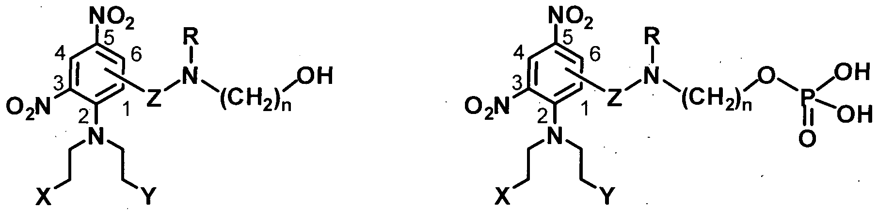
I Il wherein X and Y are different with X being selected from Cl, Br, I, and OSO2R1, and Y being selected from Cl, Br, I, and from OSO2R1, Z represents at any available ring position -CO- or -SO2-, R is methyl or ethyl; R1 is C1 6 alkyl; and n = 1-6, and pharmaceutically acceptable salts thereof.
The invention further provides a pharmaceutical composition comprising a compound of formula I as defined above, a compound of formula II as defined above, or a pharmaceutically acceptable salt thereof.
The invention further provides a method of treating a patient with cancer which comprises the step of administering to a patient in need thereof a therapeutically-effective amount of a compound of formula I as defined above, a compound of formula II as defined above, or a pharmaceutically acceptable salt thereof.
The invention further relates to use of a compound of formula I as defined above, a compound of formula II as defined above, or a pharmaceutically acceptable salt thereof in the manufacture of a composition for treating cancer in a patient in need thereof.
The invention also contemplates a compound of formula I as defined above, a compound of formula II as defined above, or a pharmaceutically acceptable salt thereof for treating cancer in a patient in need thereof.
While the invention is broadly defined above, it will be appreciated by those skilled in the art that it is not limited thereto and that it also includes further aspects as described below with reference to the following description and experimental teaching as well as by reference to the drawings.
It is intended that reference to a range of numbers disclosed herein (for example, 1 to 10) also incorporates reference to all rational numbers within that range (for example, 1, 1.1, 2, 3, 3.9, 4, 5, 6, 6.5, 7, 8, 9 and 10) and also any range of rational numbers within that range (for example, 2 to 8, 1.5 to 5.5 and 3.1 to 4.7) and, therefore, all sub-ranges of all ranges expressly disclosed herein are
hereby expressly disclosed. These are only examples of what is specifically intended and all possible combinations of numerical values between the lowest value and the highest value enumerated are to be considered to be expressly stated in this application in a similar manner.
In this specification where reference has been made to patent specifications, other external documents, or other sources of information, this is generally for the purpose of providing a context for discussing the features of the invention. Unless specifically stated otherwise, reference to such external documents is not to be construed as an admission that such documents, or such sources of information, in any jurisdiction, are prior art, or form part of the common general knowledge in die art. DESCRIPTION OF THE DRAWINGS
Figure 1: Mechanism of activation of PR-104 by one-and two-electron transfer.
Figure 2: AKRl C3 is able to reduce PR-104A to its cytotoxic metabolites under aerobic conditions. A-Aerobic metabolism was determined by an LC/MS/MS assay for PR- 104H ad PR-104M. B-Detection of expressed AKR enzymes and NQOl in HCT 116 cells by Western blotting. Figure 3: AKR1C3 protein levels correlate with aerobic metabolism of PR-104A in a panel of
23 human tumour cell lines.
A - Cell lines are shown in rank order of aerobic metabolism of PR-104A to PR- 104H/M
B-Detection of AKRl C3, AKRlBlO and NQOl in human cancer cell lines by
Western blotting in the same cell lines. Figure 4: Effect of AKRl C3 expression on sensitivity of human tumour cell lines to bioreductive drugs in vitro and in vivo. A- 1C50 assays were carried out on HCTl 16WT and HCTl 16 AKRl C3 cells, expression of AKRl C3 sensitises HCTl 16 cells to PR-104A but not to other bioreductive drugs.
B - Tumour growth delay experiment showing increased sensitivity to PR- 104, but not to cyclophosphamide (CPA) in AKJIl C3 expressing HCTl 16 tumours. C — Expression of AKRl C3 in human tumour xenografts by Western blotting.
D — Detection of AKRl C3 in the same tumour xenografts by immunohistochemistry.
E — Correlation of AKRl C3 expression with donogenic cell kill of human tumour xenografts after in vivo treatment with PR- 104 monotherapy (348 mg/kg ip).
Figure 5: Expression levels of AKRl C3 in human tumour biopsies. Immunohistochemistry was carried out on an array of human tumour biopsies using a specific AKRl C3 antibody.
A — Each positively stained biopsy was assigned a score between 1 to 6 based on intensity and frequency of staining. Biopsies which did not stain for AKRl C3 were assigned a score of zero.
B — Graph showing the intensity of AKRl C3 staining for 2700 tumors across 19 tumour types. A conservative cut-off was used so only diffuse staining (score 6 and 3) or strong moderate staining (score 5) was included as a positive result. C - A breakdown of lung cancer subtypes (positive scoring represents a score of 6, 5 or 4).
D - Representative stained tissue microarray showing AKRl C3 expression in liver cancer. Alternate rows are duplicate cores from the same tumor (LVCl 501; 36 cases, duplicate cores) and normal tissue (MBO661; 33 tissues; duplicate cores). Tissue are; 1, adrenal; 2, bladder; 3, bone marrow; 4, eye; 5, breast; 6, cerebellum; 7, cerebral cortex; 8 fallopian tube; 9, esophagus; 10, stomach; 11, small intestine; 12, colon; 13, rectum; 14, heart; 15, kidney; 16, liver; 17, lung; 18, ovary; 19, pancreas; 20, parathyroid; 21, pituitary; 22, placenta; 23, prostate; 24, skin; 25, spinal cord; 26, spleen; 27, muscle; 28, testis; 29, thymus; 30, thyroid; 31, tonsil; 32, cervix; 33, endometrium.
Figure 6 Effect of AKRl C3 expression on sensitivity of tumor cell lines to PR-104.
A - Clonogenic survival curves of HCTl 16WT and AKRl C3 over-expressing cells (Clone #1) exposed to PR-104A for 2h under aerobic and hypoxic conditions. Mean ± SD for two experiments. B — IC50 of bioreductive prodrugs following 4h aerobic exposure of HCTl 16 WT and HCTl 16 AKR1C3 (clones #1 and #2). Figure 7 Pharmacological inhibition of aerobic reduction of PR-104A in vitro and mice.
(A) Inhibition by NSAIDs of aerobic PR-104A reduction in HCT116AKR1C1 cell cultures. Inhibitors were added 2 hr before PR-104A (100 μM), and PR-104H assayed by LC-MS/MS 1 hr later. Values are means ± SEM for 3. The control value was 90.8 ± 5.2 pmol/106 cells.
(B) Effect of pretreatment of mice with naproxen (NAP; 100 mg/kg, i.p.) 30 min before PR-104 (326 mg/kg, i.v.) on levels of PR-104A and its reduced metabolites in H460 tumours, plasma, liver and bone marrow assayed 15 min after PR-104 Values
are means ± SEM for 5 mice. ***, p<0.001 (PR-104H) and **, p<0.01 (PR-104M) vs. PR- 104 only group by Student's t-test.
(C) Antitumour activity of PR-104 (326 mg/kg. i.v.) against H460 tumour xenografts in mice, by ex vivo assay of clonogenic cell survival 18 hr after treatment, and effect of pretreatment with naproxen (100 mg/kg. i.p.) 30 min before PR-104. Values are mean ± SEM for 6 mice. ***, p<0.001 vs. PR-104 only group.
(D) Effect of pretreatment of mice with 1 -aminobenzotriazole (ABT; 100 mg/kg, i.p.) 4 hr before PR-104 (326 mg/kg, Lv.) on levels of PR-104A, reduced metabolites and the oxidative dealkylation product PR-104S, in plasma and liver assayed 15 min after PR-104 Values are means ± SEM for 5 mice.
(E) Inhibition by NSAIDs of aerobic PR-104A reduction in H460 cell cultures. Inhibitors were added 2 hr before PR-104A (100 μM), and PR-104H assayed by LC- MS/MS 1 hr later. Values are means ± SEM for 3. The control value was 90.8 ± 5.2
 (F) Plasma pharmacokinetics of naproxen in mice.
DEFINITIONS
"Anti-cancer effects" include, but are not limited to, anti-tumour effects, the response rate, the time to disease progression and the survival rate. "Anti-tumour" effects include but are not limited to inhibition of tumour growth, tumour growth delay, regression of tumour, shrinkage of tumour, increased time to regrowth of tumour on cessation of treatment and slowing of disease progression.
The term "comprising" as used in this specification means "consisting at least in part of.
When interpreting statements in this specification that include that term, the features, prefaced by that term in each statement, all need to be present but other features can also be present. Related terms such as "comprise" and "comprised" are to be interpreted in the same manner.
"Effective amount" means an amount of a compound that, when administered to a subject for treating a cancer, is sufficient to effect such treatment for the cancer. The "effective amount" will vary depending on the cancer to be treated, the compound to be administered, the severity of the cancer treated, the age and relative health of the subject, the route and form of administration, whether the treatment is monotherapy or combination therapy, the judgement of the attending clinician, and other factors.
"Pharmaceutically acceptable", means that which is useful in preparing a pharmaceutical composition that is generally safe, non-toxic, and neither biologically nor otherwise undesirable and includes that which is acceptable for veterinary as well as human pharmaceutical use.
"Pharmaceutically acceptable salts" of a compound means salts that are pharmaceutically acceptable, as defined herein, and that possess the desired pharmacological activity of the parent compound. Such salts include: acid addition salts formed with inorganic acids such as hydrochloric acid, hydrobromic acid, sulfuric acid, nitric acid, phosphoric acid and the like; or formed with organic acids such as acetic acid, methanesulfonic acid, maleic acid, tartaric acid, citric acid and the like; or salts formed when an acidic proton present in the parent compound either is replaced by a metal ion, e.g. an alkali metal ion, an alkaline earth ion, or an aluminium ion; or coordinates with an organic or inorganic base. Acceptable organic bases include ethanolamine, diethanolamine, N- methylglucamine, triethanolamine and the like. Acceptable inorganic bases include aluminium hydroxide, calcium hydroxide, potassium hydroxide, sodium carbonate and sodium hydroxide. The term "subject" is intended to refer to an animal, preferably a mammal, more preferably a mammalian companion animal or human. Preferred companion animals include cats, dogs and horses. Other mammalian subjects include an agricultural animal, including a horse, a pig, a sheep, a goat, a cow, a deer, or a fowl, or a laboratory animal, including a monkey, a rat, or a mouse.
The term "treat" and its derivatives should be interpreted in their broadest possible context. The term should not be taken to imply that a subject is treated until total recovery. Accordingly, "treat" broadly includes maintaining a subject's disease progression or symptoms at a substantially static level, increasing a subject's rate of recovery, amelioration and/or prevention of the onset of the symptoms or severity of a particular condition, or extending a patient's quality of life. The term "treat" also broadly includes the maintenance of good health for sensitive individuals and building stamina for disease prevention.
"Warm blooded animal" means any member of the mammalia class including, but not limited to humans, non-human primates such as chimpanzees and other apes and monkey species, farm animals such as cattle, horses, sheep, goats, and swine; domestic animals such as rabbits, dogs and cats; laboratory animals including rodents, such as rats, mice and guinea pigs, and the like. DESCRIPTION OF THE INVENTION
The focus of this invention is on the enzyme aldo-ketoreductase family 1 , member C3 (17- beta-hydroxysteroid dehydrogenase type 5; 3-alpha-hydroxysteroid dehydrogenase type 2; Dihydrodiol dehydrogenase 3; Prostaglandin F synthase; ACC No. NM-003739; Prot ID P42330; Gene ID 8644, also known as AKRl C3; DD3; DDX; HAKRB; HAKJIe; HAl 753; HSDl 7B5; hlu
PGFS; KlAAOl 19) and its role as a biomarker. More specifically, the invention is based upon the surprising finding that AKRl C3 has nitroreductase activity. This latter activity delivers the capability to AKRl C3 to bioreduce and therefore activate nitroaromatic prodrugs which, upon activation, release one or more cytotoxic species in vivo. The applications of this finding relating to AKRl C3 will be apparent to diose persons skilled in the art. One such application is in a method of screening candidate nitroaromatic prodrugs to determine which are reduced (activated) by AKRl C3 and therefore capable of releasing one or more cytotoxic species in vivo. However, the primary and preferred application is in methods of screening patients to assess their predicted responder/non-responder status with respect to AKRlC3-activated prodrug therapy in treating cancer, with "cancer" having its widest possible meaning to include any member of a class of diseases characterised by the uncontrolled growth of abberant cells. This latter application offers the prospect of individualising treatment on a patient-by-patient basis.
The screening approach of the invention involves determining at least one AKRl C3 profile for said patient. That profile may be an AKRl C3 nucleic acid profile, an AKRl C3 protein profile or a combination of these.
A number of approaches are available to determine an AKRl C3 nucleic acid profile for a patient. Such approaches include determining a genotypic profile, a gene expression profile or a combination. Any other art-established approach to determining a nucleic acid profile (including those which determine gene copy numbers or methylation profiles, for example) can also be used. Similarly, an AKRl C3 protein profile can be established using any art- known - technique. Examples include determining an AKRl C3 expression profile or an AKRl C3 activity profile.
An activity profile may involve, where the AKRlC3-activated prodrug is PR-104A for example, measuring the reduction of PR-104A to PR-104H or PR-104M. Where the selected profile is an AKRl C3 nucleic acid profile, the AKRl C3 nucleic acid assayed for can be the AKRl C3 gene or any representative part thereof. A "gene" in this context is a segment of DNA involved in producing a polypeptide chain, in this case the AKRl C3 protein. The gene can include, without limitation, regions preceding and following the coding region such as the promoter and 3'-untranslated region, as well as the introns. Equally, the nucleic acid assayed for can be ribonucleic acid transcribed from the gene during production of the polynucleotide, such as mRNA.
The AKRl C3 gene has a number of polymorphisms (Jakobsson, J et al., The P ^karmacogenonomics journal (2007) 7, 282-289). All polymorphisms are included as assay targets. Individual polymorphisms can be targeted by an assay approach which focuses on the (or a)
polymorphic site. Those skilled in the art will recognise that the nature of the polymorphism, such as the allele or genotype present at a polymorphic site can also be determined indirectly, e.g., by an analysis of one or more polymorphisms in linkage disequilibrium with an allele at the polymorphic site. Such approaches are described in more detail below. Expression of the AKRl C3 gene can co-ordinate with expression of other genes, including those in the AKR superfamily. While less desirable than an assay for nucleic acid which is direcdy part of the AKRl C3 gene, an assay which targets such co-ordinated gene expression is in no way excluded.
When the AKRl C3 profile is an AKRl C3 protein profile, the AKRl C3 protein assayed for will generally be the 323 amino acid 37 kDa protein having wild-type functionality, or any part thereof including any product of in vivo proteolytic digestion. Polymorphic forms (such as the GIn 5 His variant) are however not excluded and neither are fragments (including digestion fragments) of such polymorphic forms. It will be appreciated that retention of the nitroreductive functionality of the protein or fragment and identification of that capability is critical to assigning either a "predicted responder" or "predicted non-responder" status to a patient.
The result of the assay, whether via an AKRl C3 nucleic acid profile, an AKRl C3 protein profile or both will be to assign a "predicted responder" or "predicted non-responder" status to a patient. Patients whose samples (generally a turnout biopsy, preferably taken shordy or immediately prior to the assay) are AICRl C3 positive will be predicted to be responders whereas patients whose samples are AKRl C3 negative will be predicted to be non-responders. Predicted responders will be considered suitable for therapy involving administration of an AKRl C3-activa ted prodrug.
Such prodrugs are any compound which is capable, upon exposure to functional AKRl C3 enzyme, of releasing a cytotoxic species to have an anti-tumour effect. For some prodrugs, activation by AKRl C3 may be the only mechanism. Activation by AKRl C3 need not, however, be the sole activation mechanism for all such compounds. For example, any compound which is activated by both AKRl C3 and a nitroreductase expressed within, or proximate to, hypoxic regions of tumour remains an "AKRl C3-activated" compound or drug as that term is used in this invention.
Representative examples of AKRl C3-activated prodrugs include the nitroaromatic prodrugs described in WO 2004/033415 and their pre-prodrug forms described in WO 2005/042471.
Specific representative examples are PR-104A and PR- 104. Prodrugs PR- 104 and PR-104A are also able to be activated by 1 -electron nitroreduction under hypoxia.
Other prodrugs can be identified using art-standard screening techniques involving functional AKRl C3 enzyme. Examples of such screening techniques are discussed herein.
Further specific examples of such prodrugs are the prodrugs of formula I, the pre-prodrugs of formula II, and their respective pharmaceutically acceptable salts. Prodrugs of formula I are:

I
Pre-prodrugs of formula II are:

In both formulae I and II, X and Y are different with X being selected from Cl, Br, I, and OSO2R1, and Y being selected from Cl, Br, I and OSO2R1;
Z represents, at any available ring position, -CO- or -SO2-; R is methyl or ethyl;
R1 is C1 — C6 alkyl; and n is 1 - 6.
Compounds of formula I and formula II and dieir pharmaceutically acceptable salts are separate aspects of diis invention. Their synthesis and properties are more fully described below. Non-responders are candidates for treatment widi other cancer therapeutics (particularly those which are AKRl C-independent) or with AKRl C3-activated agents which are also activated via another mechanism. Predicted non-responders to AKRl C3 can therefore be treated with PR- 104 or PR-104A alone or in combination(s) with other agents, including other cancer therapeutics.
It is however envisaged that a patient who has predicted non-responder status may have some level of AKRl C3 expression, albeit less than enough to be predicted to respond therapeutically to treatment with an AKRl C3-activated prodrug on its own. In the case of such patients, it may be beneficial to ensure that a partial or low-level activation by AKRl C3 does not occur (in order to avoid cytotoxin release elsewhere in the body, for example). In such cases, an
AKRlC3-independent (or negated) treatment approach is preferred, either by anti-cancer agents which are not activated or otherwise affected by AKRl C3, or by a combination approach where it remains desirable to administer an AKRl C3-activated prodrug.
One preferred combination approach is to administer PR-104 or PR-104A together with a Non-Steroidal Anti-Inflammatory Drug (NSAID). This combination can be administered alone or itself in combination with other cancer therapeutics. Combinations which include both PR-104/PR- 104A and an NSAID are employed where it is viewed as necessary or desirable to functionally eliminate any residual AKRl C3 activation from PR-104/PR-104A when treating the patient.
NSAIDs that inhibit AKRl C3 are well known in the art. While any appropriate NSAID can be employed in this approach, it is preferred that the NSAID be naproxen.
Patients whose tumours are considered AKRl C3 negative are also candidates for treatment with therapeutics to which resistance is induced through co-expression of other genes in the AKR family (such as AKRlCl, AKRl C2, AKRlBlO and NQOl). Representative cancer therapeutics to treat AKRl C3 negative patients include antifolates (Selga et al., Biochemical Pharmacology 75 (2008)414-426), platinums (such as cis-platin and carboplatin) and anthracyclines (such as oracin (6- [2-(2-hydroxyethyl)-aminoethyl]-5,l l-dioxo-5,6-dihydro-ll i-/-indeno[l,2-c]isoquinoline), doxorubicin and daunorubicin).
The applications of the finding regarding the ability of AKRl C3 to catalyse nitroaromatic groups are not of course restricted to the methods above. For example, AKRl C3 RNA transcripts have been detected in the brain, lung, liver, kidney, colon small intestine, mammary gland, uterus, cervix, bladder, prostate and testis, and most of these areas will be tumour-free in the patient concerned. Levels of AKRl C3 in normal tissues not implicated in the cancer or in blood, serum or plasma can therefore also be determined for the purpose of predicting whether or not treatment with a particular AKRl C3-activated prodrug will have an acceptable toxicity outcome for a patient. The results of such an assay can be used, usually in conjunction with the results of a parallel tumour- focussed assay, to predict the likely cytotoxicity of the prodrug within the patient generally, and to either eliminate the patient as suitable for treatment (where the non-specific toxicity is unacceptably high) or to assist with optimising the dose of the prodrug to be administered to the patient. For example, the results of assays described herein can be used to calculate a likely maximum tolerated dose for an individual, having regard to the level or activity of AKRl C3 in normal tissue (e.g., from the individual) compared to tumour tissue.
1. ASSAYS AND TOOLS Genotypic Profiling
A variety of techniques can be used for genotypic analysis of a nucleic acid sample in determining a genotypic profile according to the mediods of the present invention. For example, en2ymatic amplification of nucleic acid from a sample can be conveniently used to obtain nucleic acid for subsequent analysis. However, the presence or absence of a nucleic acid sequence can also be determined directly from a nucleic acid sample without enzymatic amplification (e.g., using hybridization techniques). Geno typing of nucleic acid, whether amplified or not, can be performed using any of various techniques known to one of skill in the art. Useful techniques include, without limitation, polymerase chain reaction (PCR)-based analysis, sequence analysis, and electrophoretic analysis, which can be used alone or in combination.
Use of PCR for the amplification of nucleic acids is well known in the art (see, eg Mullis et al., The Polymerase Chain Reaction, Birkauser, Boston (1994). Further protocols are provided in Innis et al, "PCR Applications". Protocols for Functional Genomics", 1st Edition, Academic Press (1999). Any of a variety of different primers can be used to PCR amplify a subject's nucleic acid.
One skilled in the art understands that primers for PCR analysis can be designed based on the sequence flanking the nucleic acid of interest. As a non-limiting example, a PCR primer can contain between about 15 to about 60 nucleotides (e.g., 15-50,15-40, or 15-30 nucleotides) of a sequence upstream or downstream of the nucleic acid of interest. Such primers generally are designed to have sufficient guanine and cytosine content to attain a high melting temperature which allows for a stable annealing step in the amplification reaction. Several computer programs, such as Primer Select, are available to aid in the design of PCT primers.
Sequence analysis can also be useful for genotyping a gene. As used herein, the term "sequence analysis" includes any manual or automated process by which the order of nucleotides in a nucleic acid is determined. As an example, sequence analysis can be used to determine the nucleotide sequence of a sample of DNA. The term encompasses, without limitation, chemical and enzymatic methods such as dideoxy enzymatic methods including, for example, Maxam-Gilbert and Sanger sequencing as well as variations thereof. The term also encompasses, without limitation, capillary array DNA sequencing, which relies on capillary electrophoresis and laser-induced fluorescence detection and can be performed using instruments such as the MegaBACE 1000 or ABI 3700. As additional non-limiting examples, the term encompasses thermal cycle sequencing (Sears et al., Bioteώniques, 13:626-633 (1992)); solid-phase sequencing (Zimmerman et al., Methods MoI. Cell. Biol., 3:39-42 (1992); and sequencing with mass spectrometry, such as matrix - assisted laser desorption/ionization time-of-flight mass spectrometry (MALDI-TOF MS; Fu et al, Nature
Biotech, 16:381-384 (1998). The term further includes, without limitation, sequencing by hyridization (SBH), which relies on an array of all possible short obligonucleotides to identify a segment of sequence (Chee et al, Science, 274:610-614 (1996); Drmanac et al., Science, 260:1649-1652 (1993); Drmanac et al., Nature Biotech., 16:54-58 (1998)). See, in general, Ausubel et al., Current Protocols in Molecular Biology, Chapter 7 and Supplement 47, John Wiley & Sons, Inc., New York (1999). A number of sequencing methods and platforms are particularly suited to large-scale implementation, and are amenable to use in the methods of the invention. These include pyrosequencing methods, such as that utilised in the GS FLX pyrosequencing platform available from 454 Life Sciences (Branford, CT) which can generate 100 million nucleotide data in a 7.5 hour run with a single machine, and solid-state sequencing methods, such as that utilised in the SOLiD sequencing platform (Applied Biosystems, Foster City, CA).
A number of methods currendy used for detection of polymorphsims involve site-specific and/or allele-specifϊc hybridisation. These methods are largely reliant on the discriminatory binding of oligonucleotides to target sequences containing the SNP of interest. The techniques of Illumina (San Diego, CA), Affymetrix (Santa Clara, CA.) and Nanogen Inc. (San Diego, Calif.) are particularly well-known, and utilize the fact that DNA duplexes containing single base mismatches are much less stable than duplexes that are perfectly base-paired. The presence of a matched duplex is usually detected by fluorescence. A number of whole-genome genotyping products and solutions amenable or adaptable for use in the present invention are now available, including those available from the above companies.
The majority of methods to detect or identify polymorphisms by site-specific hybridisation require target amplification by methods such as PCR to increase sensitivity and specificity (see, for example U.S. Pat. No. 5,679,524, PCT publication WO 98/59066, PCT publication WO 95/12607). US Patent Application publication number 20050059030 (incorporated herein in its entirety) describes a method for detecting a single nucleotide polymorphism in total human DNA without prior amplification or complexity reduction to selectively enrich for the target sequence, and without the aid of any enzymatic reaction. The method utilises a single-step hybridization involving two hybridization events: hybridization of a first portion of the target sequence to a capture probe, and hybridization of a second portion of said target sequence to a detection probe. Both hybridization events happen in the same reaction, and the order in which hybridisation occurs is not critical.
In addition electrophoretic analysis can be useful for genotyping a gene. The term "electrophoretic analysis," as used herein in reference to one or more nucleic acids such as amplified fragments, includes a process whereby charged molecules are moved through a stationary medium under the influence of an electric field. Electrophoretic migration separates nucleic acids primarily
on the basis-of their charge, which is in proportion to their size, with smaller molecules migrating more quickly. The term includes, without limitation, analysis using slab gel electrophoresis such as agarose or polyacrylamide gel electrophoresis, or capillary electrophoresis. Capillary electrophoretic analysis generally occurs inside a small-diameter quartz capillary in the presence of high (kilovolt- level) separating voltages with separation times of a few minutes. Using capillary electrophoretic analysis, nucleic acids are conveniently detected by UV absorption or fluorescent labeling, and single-base resolution can be obtained on fragments up to several hundred base pairs in length. Such methods of electrophoretic analysis, and variations thereof, are well known-in the art, as described, for example, in Ausubel et al., Current Protocols in Molecular Biology, Chapter 2 and Supplement 45, John Wiley & Sons, Inc., New York (1999).
Other molecular techniques useful for genotypic analysis, in this case of a polymorphic site in a gene associated with variations in the level or activity of AKRl C3, or with one or more AKRl C3 polymorphisms, are also known in the art and useful in the methods of the present invention. Such genotyping techniques include, without limitation, automated sequencing and RNAase mismatch techniques (Winter et al., Proc. Natl. Acad. Sd., 82:7575-7579 (1985). See, in general, Birren et al., Genome Anayl sis: A Laboratory Manual, Volume 1 (Analyzing DNA), New York, Cold Spring Harbor Laboratory Press (1997).
Those skilled in the art will recognise diat an AKRl C3 nucleic acid profile that comprises data on one or more polymorphisms, such as one or more polymorphisms associated with increased or decreased expression or activity of AKRl C3, can be generated by direcdy determining the one or more polymorphisms, or by detection of one or more other polymorphisms which are in linkage disequilibrium with one or more of said polymorphisms. Linkage disequilibrium is a phenomenon in genetics whereby two or more mutations or polymorphisms are in such close genetic proximity that they are co-inherited. This means that in genotyping, detection of one polymorphism as present implies the presence of the other. (Reich DE et al; Linkage disequilibrium in the human genome, Nature 2001, 411:199-204.) Various degrees of linkage disequilibrium are possible. Preferably, the one or more polymorphisms in linkage disequilibrium with the one or more polymorphisms are in greater than about 60% linkage disequilibrium, are in about 70% linkage disequilibrium, about 75%, about 80%, about 85%, about 90%, about 91%, 92%, 93%, 94%, 95%, 96%, 97%, 98%, 99%, or about 100% linkage disequilibrium with the one or more polymorphisms. Those skilled in the art will appreciate that linkage disequilibrium may also, when expressed with reference to die deviation of the observed frequency of a pair of alleles from the expected, be denoted by a capital D. Accordingly, the phrase "two alleles are in LD" usually means that D does not equal 0. Contrariwise, "linkage equilibrium" denotes the case D = O. When utilising this
nomenclature, the one or more polymorphisms in LD with the one or more polymorphisms specified herein are preferably in LD of greater than about D'=0.6, of about D'=0.7, of about D'=0.75, of about D'=0.8, of about D'=0.85, of about D'=0.9, of about D'=0.91, of about D'=0.92, of about D'=0.93, of about D'=0.94, of about D'=0.95, of about D'=0.96, of about D'=0.97, of about D'=0.98, of about D'=0.99, or about D'=1.0. (Devlin and Risch 1995; A comparison of linkage disequilibrium measures for fine-scale mapping, Genomics 29: 311-322).
In view of the above, one skilled in the art will readily appreciate that the methods of the present invention for determining a genotypic profile in a sample can be practiced using one or any combination of the well-known techniques described above or other techniques known in the art. Gene Expression Profiling
A gene expression profile is typically evaluated in vitro on a sample collected from a subject in comparison to a normal or reference sample. Determination of a transcriptional expression profile can be accomplished, e.g., using hyridization techniques well-known to those skilled in the art such as Northern analysis and slot blot hybridization or by performing reverse-transcriptase (RT)- PCR amplification followed by gel electrophoresis. Applicable PCR amplification techniques are described in Ausubel et al., Current Protocols in Molecular Biology, John Wiley & Sons, Inc., New York (1999); Theophilus et at, "PCR Mutation Detection Protocols," Humana Press (2002); and Innis et al., "PCR Applications: Protocols for Functional Genomics," 1st Edition, Academic Press (1999). " General nucleic acid hybridization methods are described in Anderson, "Nucleic Acid Hybridization," BIOS Scientific Publishers (1999). Amplification or hybridization of a plurality of transcribed nucleic acid sequences (e.g. mRNA or cDNA) can also be performed using mRNA or cDNA sequences arranged in a microarray. Microarray methods are generally described in Hardiman, "Microarrays Methods and Applications: Nuts & Bolts," DNA Press (2003) and Baldi et al., "DNA Microarrays and Gene Expressions: From Experiments to Data Analysis and Modeling," Cambridge University Press (2002).
The measurement of gene expression profiles using microarrays also has many important applications to the monitoring of disease states and therapies (see, e.g., U.S. Pat. Nos. 6,218,122 and 6,222,093), the identification of drug targets, the identification of pathways of drug action, and drug design (see, e.g., U.S. Pat. Nos. 6,303,291, 6,165,709, 6,146,830, 5,965,352, and 5,777,888). For example, van't Veer et al., supra, identified "good prognosis" and "poor prognosis" gene expression signatures that could be used to predict the clinical outcome of breast cancer patients. Similarly, U.S. Pat. No. 5,777,888 discloses the utility of microarray gene expression profiles to evaluate the target specificity of a candidate drug by comparison of an expression profile obtained from cells treated with the candidate drug to a database of expression profiles obtained from cells treated with
known drugs. U.S. Pat. No. 6,218,122 provides methods of monitoring the disease state of a subject and determining the effect of a therapy upon the subject through the use of gene expression profiles (see, also, U.S. Pat. No. 6,266,093).
In view of the above, one skilled in the art will readily appreciate that the methods of the present invention for determining a gene expression profile from a sample of a subject can be practiced using one or any combination of the well-known techniques described above or other techniques known in the art.
Protein Expression Profiling
A variety of techniques can be used to detect the presence or level of an expressed protein for determining a protein expression profile according to the methods of the present invention. For example, a protein biomarker can be analyzed using an immunoassay. A protein expression profile can also be evaluated using electrophoresis, e.g., Western blotting, as well as any other technique known to those skilled in the art. Immunoassay techniques and protocols are generally described in Price and Newman, "Principles and Practice of Immunoassay," 2nd Edition, Grove's Dictionaries (1997); and Gosling. "Immunoassays: A Practical Approach," Oxford University Press (2000). The presence or amount of the protein biomarker is typically determined using antibodies specific for the biomarker and detecting specific binding.
Any suitable immunoassay can be utilized for determining the presence of level of one or more protein biomarkers in a sample. A variety of immunoassay techniques, including competitive and non-competitive immunoassays, can be used (see, e.g., Self et al., Curr. Opin. BioteώnoL, 7:60-65 (1996)). The term immunoassay encompasses techniques including, without limitation, enzyme immunoassays (EIA) such as enzyme multiplied immunoassay technique (EMIT), enzyme-linked immunosorbent assay (ELISA), IgM antibody capture ELISA (MAC ELISA), and microparticle enzyme immunoassay (MEIA); capillary electrophoresis immunoassays (CEIA); radioimmunoassays (RIA); immunoradiometric assays (IRMA); fluorescence polarization immunoassays (FPIA); and chemiluminescence assays (CL).
Immunoassays can also be used in conjunction with laser induced fluorescence (see, e.g. Schmalzing et al., Electmphoresis, 18:2184-93 (1997); BaoJ. Chromatogr. B. Biomed. Sa., 699:463-80 (1997)). Liposome immunoassays, such as flow-injection liposome immunoassays and liposome immunosensors, are also suitable for use in the present invention (see, e.g., Rongen et al.,/. Immunol. Methods, 204:105-133 (1997)). In addition, nephelometry assays, in which the formation of protein/antibody complexes results in increased light scatter that is converted to a peak rate signal as a function of the marker concertration, are suitable for use in the methods of the present invention. Nephelometry assays are commercially available from Beckman Coulter (Brea, Calif.;- Kit #449-430)
and can be performed using a Behring Nephelometer Analyzer (Fink et al.,/. Clin. Chem. Clin. Bioώem., 27:261-276 (1989)).
Specific immunological binding of the antibody to the biomarker can be detected directly or indirectly. Direct labels include fluorescent or luminescent tags, metals dyes, radionuclides, and the like, attached to the antibody. An antibody labelled with iodine-125 (1251.) can be used for determining the level of one or more biomarkers in a sample. A chemiluminescence assay using a chemiluminescent antibody specific for the biomarker is suitable for sensitive, non-radioactive detection of biomarker levels. An antibody labelled with fluorochrome is also suitable for determining the level of one or more biomarkers in a sample. Examples of fluorochromes include, without limitation, DAPI, fluorescein, Hoechst 33258, R-phycocyanin, B-phycoerythrin, R- phycoerythrin, rhodamine, Texas red, and lissamine. Indirect labels include various enzymes well known in the art, such as horseradish peroxidase (HRP), alkaline phosphatase (AP), β-galactosidase, urease, and the like. A horseradish-peroxidase detection system can be used, for example, with the chromogenic substrate tetramethyl-benzidine (TMB), which yields a soluble product in the presence of hydrogen peroxide that is detectable at 450 nm. An alkaline phosphatase detection system can be used with the chromogenic substrate p-nitrophenyl phosphate, for example, which yields a soluble product readily detectable at 405 nm. Similarly, a β-galactosidase detection system can be used with the chromogenic substrate o-nitrophenyl-β-D-galactopyranoside (ONPG), which yields a soluble product detectable at 410 nm. An urease detection system can be used with a substrate such as urea-bromocresol purple (Sigma Immunochemicals; St. Louis, Mo.).
A signal from the direct or indirect label can be analyzed, for example, using a spectrophotometer to detect color from a chromogenic substrate; a radiation counter to detect radiation such as a gamma counter for detection of 1251; or a fluorometer to detect fluorescence in the presence of light of a certain wavelength. For detection of enzyme-linked antibodies, a quantitative analysis of the amount marker levels can be made using a spectrophotometer such as an EMAX Microplate Reader (Molecular Devices; Menlo Park, Calif.) in accordance with the manufacturer's instructions. If desired, the assays of the present invention can be automated or performed robotically, and the signal from multiple samples can be detected simultaneously.
Antigen capture assays can be useful in the methods of the present invention. For example, in an antigen capture assay, an antibody directed to a biomarker of interest is bound to a solid phase and sample is added such that the biomarker is bound by the antibody. After unbound proteins are removed by washing, the amount of bound marker can be quantitated using, for example, a radioimmunoassay (see, e.g., Harlow and Lane, Antibodies: A Laboratory Manual, Cold Spring Harbor Laboratory, New York (1988)). Sandwich enzyme immunoassays can also be useful in the
methods of the present invention. For example, in a two-antibody sandwich assay, a first antibody is bound to a solid support, and the biomarker is allowed to bind to the first antibody. The amount of the biomarker is quantitated by measuring the amount of a second antibody that binds the biomarker. The antibodies can be immobilized onto a variety of solid supports, such as magnetic or chromatographic matrix particles, the surface of an assay plate (e.g., microliter wells), pieces of a solid substrate material or membrane (e.g., plastic, nylon, paper), and the like. An assay strip can be prepared by coating the antibody or a plurality of antibodies in an array on a solid support. This strip can then be dipped into the test sample and processed quickly through washes and detection steps to generate a measurable signal, such as a colored spot. Quantrtiative Western blotting also can be used to detect or determine the level of one or rrϊore proteinaceous biomarkers in a sample. Western blots can be quantitated by well-known methods such as scanning densitometry or phosphorimaging. In certain instances, autoradiographs of the blots are analyzed using a scanning densitometer (Molecular Dynamics; Sunnyvale, Calif.) and normalized to a positive control. Values are reported, for example, as a ratio between the actual value to the positive control (densitometric index). Such methods are well known in the art as described, e.g., in Parra et alj. Vase. Surg., 28:669-675 (1998).
Alternatively, a variety of immunohistochemistry (IHC) techniques can be used to determine the level of one or more proteinaceous biomarkers in a sample. As used herein, die term "immunohistochemistry" or "IHC" encompasses techniques that utilize the visual detection of fluorescent dyes or enzymes coupled (i.e., conjugated) to antibodies that react with the biomarker using fluorescent microscopy or light microscopy and includes, without limitation, direct fluorescent antibody, indirect fluorescent antibody (IFA), anticomplement immunofluorescence, avidinbiotin immunofluorescence, and immunoperoxidase assays. An IFA assay, for example, is useful for determining whether a sample is positive for a particular marker of interest, the level of that marker, and/or die staining pattern of that marker. The concentration of the marker in a sample can be quantiated, e.g., through endpoint titration or through measuring the visual intensity of fluorescence compared to a known reference standard.
Any IHC technique known to one of skill in the art is suitable for use in the assay methods of the present invention. As a non-limiting example, IHC can be performed according to the following protocol: (1) slides containing the sample (e.g., tumor tissue are deparaffinized with xylene/70% ethanol into phosphate buffered saline (PBS) at pH 7.4; (2) the slides are then immersed in 10 mM citric acid at pH 6.0 microwaved for about 37 minutes, and cooled down at room temperature (RT) for bout 30-60 minutes; (3) endogenous peroxidases ate quenched for about 10 minutes in 1 part 30% H2O2 and 9 parts methanol and the slides are washed 3 times for 3
minutes in PBS; (4) the slides are blocked with blocking reagent at RT for about 30 minutes; (5) antibodies against the biomarker of interest are added and the slides are incubated at 4° C. overnight; (6) the slides are washed in PBS at RT for about 30 minutes, changing the wash buffer every 5 minutes; (7) secondary antibodies such as biotinylated antibodies are added and the slides are incubated at RT for about 60 minutes; (8) the slides are washed in PBS at RT for about 30 minutes, changing the wash buffer every 5 minutes; (9) streptavidin is added and the slides are incubated at RT for about 30 minutes; (10) 3,3 — diaminobenzidine (DAB) is added, the slides are incubated for 5 minutes, the DAB is neutralized with bleach, and the slides are washed for 5 minutes with water; (11) the slides are counterstained with methylgreen for 3 minutes washed 3 times with water; (12) the slides are dipped in 95% ethanol, followed by a 100% ethanol and xylene series; and (13) a coverslip is placed on the slide.
Examples of IHC protocols for determining the presence or level of specific antigens of interest are known in the art. These include the IHC protocols described in, e.g., Ishikawa et al, Cancer Res., 65:9176-9184 (2005) for TGF-α and amphiregulin; Cappuzzo et al.,/. Clin. Oncol., 23:5007-5018 (2005) for HER2; Cappuzzo et al., /. Natl. Caner Inst., 97:643-655 (2005) for EGFR; Abrams et al., MoI. Cancer. Then, 2:471-478 (2003) for c-KIT and PDG-FRB; and Lee et A., Anal. Quant. CytoL Histol., 27:202-210 (2005) for PTEN. Tissue staining can be visualized using peroxidase-based immunostaining kits available from Vector Laboratories (Burlingame, Calif.) and DAKO (Glostrup, Denmark). The presence or level of a biomarker can also be determined by detecting or quantifying the amount of the purified marker. Purification of the marker can be achieved, for example, by high pressure liquid chromatography (HPLC), alone or in combination with mass spectrometry (e.g., MALDI/MS, MALDI-TOF/MS, tandem MS, etc.). Qualitative or quantitative detection of a biomarker can also be determined by well-known methods including, without limitation, Bradford assays, Coomassie blue staining, silver staining, assays for radiolabeled protein, and mass spectrometry.
The activity of a protein biomarker can also be determined by art-known techniques, including by one or more of the analytical techniques discussed above.
In view of the above, one skilled in the art will readily appreciate that the methods of the present invention for determining an AKRl C3 protein profile from a sample of a subject can be practiced using one or any combination of the well-known techniques described herein or other techniques known in the art.
Selection of Antibodies
Antibodies against AKRl C3 are commercially available. A suitable example is the anti — AKRl C3 antibody, clone NPG. G6. A6 (Lin et al., Steroids, 2004 69:795-801, Sigma product A6229). < However, the invention is not limited to the use of existing antibodies but contemplates generation of new reagents.
The generation and selection of antibodies not already commercially available for detecting or determining the level of protein biomarkers generally may be accomplished several ways. For example, one way is to purify polypeptides of interest using e.g., solid phase peptide synthesis methods well known in the art. See, e.g., Guide to Protein Purification, Murray P. Deutcher, ed., Meth. En^ymol, Vol. 182, 1990; Solid Phase Peptide Synthesis, Greg B. Fields, ed., Meth. En^ymol, Vol. 289, 1997; Kiso et al., Chem. Pharm. Bull., 38:1192-99 (1990); Mostafavi et al., Biomed. Pept. Proteins Nucleic Acids, 1:225-60, (1995); Fujiwara et al., Chem. Pharm. Bull., 44:1326-31 (1996). The selected polypeptides may then be injected, for example, into mice or rabbits, to generate polyclonal or monoclonal antibodies. One skilled in the art will recognize that many procedures are available for the production of antibodies, for example, as described in Λntibodies, A Laboratory Manual, Harlow and Lane, Eds., Cold Spring Harbour Laboratory, Cold Spring Harbour, N.Y. (1998). One skilled in the art will also appreciate that binding fragments or Fab fragments which mimic antibodies can also be prepared from genetic information by various procedures (see, e.g., Antibody Engineering: A Practical Approach, Borrebaeck, Ed., Oxford University Press, Oxford (1995);/. Immunol, 149:3914- 3920 (1992)).
Those skilled in the art will recognize that many approaches can be taken in producing antibodies or binding fragments and screening and selecting for affinity and specificity for the various polypeptides, but these approaches do not change the scope of the present invention.
This aspect of the invention will now be described in more detail with reference to the following non-limiting experimental section.
EXAMPLE 1. PART A Introduction
Hypoxia-activated prodrug PR- 104 is a water-soluble phosphate ester which is converted in ήvo to the corresponding alcohol, PR-104A. This 3,5-dinitrobenzamide-2-nitrogen mustard is activated by reduction to the corresponding 5-hydroxylamine (PR-104H) and 5-amine (PR-104M) (Patterson et al., Clin Can Res 2007, 13:3922-32, see Figure 1).
It has been observed that certain neoplastic cell lines display atypical aerobic cell sensitivity that appears to correlate with NQOl expression in vitro (Guise et al., Biochem Pharmacol, 2007, 74:810-
20). NQOl cDNA expression does not however increase aerobic cytotoxicity or PR-104A metabolism. Thus DT-diaphorase is not the aerobic PR-104A reductase but appears to be co- ordinately regulated with it.
Aims ■ To identify the reductase(s) responsible for the reduction of PR-104A under aerobic conditions. ■ To determine the expression of these reductase(s) in human cancers.
Experimental design
Microarray-based RNA expression profiles covering 38,500 probes (Affymetrix HG-Ul 33 Plus 2.0) were obtained for 23 human neoplastic cell lines. Oxidoreductase genes were selected, using Gene Ontology, and analysed by hierarchical clustering . To identify candidate PR-104A reductases the correlation coefficient (Kendall TauJ between gene expression and aerobic PRl 04 metabolism (to PR104H/M) was calculated for each probeset; correlation p-values were adjusted using the false discovery rate (FDR) correction for multiple comparisons. Expression of AKRs and NOOl in HCTl 16 cells. Cloning utilised Invitrogen's GatewayTM system. Entry vectors encoding AKR family members/NQOl were purchased from Invitrogen's sequence verified ultimate ORF library. Candidate genes were cloned into a Gateway compatible expression vector behind the EF-I α promoter. In addition to the gene of interest, the bicistronic mRNA also encoded a protein conferring resistance to puromycin to enable selection of transfected cells.
Determination of PR-104A metabolites by LC /MS /MS. Cells were exposed to PR-104A in 24 well plates for 1 hr using a 37°C humidified incubator (20% O2, 5% CO2) PR-104A metabolites were extracted from the media and cell monolayer by addition of two volumes of methanol containing tetradeuterated (d4)-PR-104H internal standard and stored at -800C. Samples (75 μL) of standards and unknowns were diluted with 25 μL water and assayed by LC/MS/MS (Agilent 6410).
Antibodies. Anti-AKR1C3 (Sigma, A6229). Anti-AKRIBIO (Abnova, H00057016-M01). Anti-V5-HRP (Invitrogen, 46-0708). Anti-actin (Chemicon, MAB1501R). Anti-NQOl (kindly provided by Prof David Ross, University of Colorado, Denver). Cytotoxicity assay. Growth inhibition of human HCTl 16 WT and HCTl 16 AKRl C3 carcinoma cells was determined from IC50 values for 4 hr drug exposure under oxic conditions, with staining by sulforhodamine B 5 days later.
Tumour growth delay assay. Antitumour activity of PR-104 and cyclophosphamide in HCTl 16 WT and HCTl 16 AKRl C3 tumours was evaluated. Tumours were grown in CD-I nude
mice (SC inoculation) and activity was assessed by tumour growth inhibition. Mice were IP dosed at the MTD with PR-104 (550mg/kg) or cyclophosphamide (62mg/kg) using a q4dx3 schedule. Tumour volumes were monitored until tumour volume increased 4-fold with respect to day 1 treatment volume. Clonogenic survival assay. Tumours were grown in CD-I nude mice (SC inoculation). Mice were IP dosed with PR-104 (348mg/kg). 24 hours later, tumours were excised, weighed, minced with surgical scissors and dissociated enzymatically in magnetically stirred suspensions. Cell suspensions were diluted, plated and incubated at 37°C for 10-14 days before staining with methylene blue. Colonies containing >50 cells were counted. Commercial tumour microarrays (TMAs). TMAs were purchased from Imgenex Inc. (San
Diego, CA) and Pantomics Inc (San Francisco, CA). A total number of 2574 cases (4016 cores) were analysed, from 40 TMAs (36 neoplasia, 1 metastasis, 3 normal tissue). Immunohistochemistry was carried out using a validated monoclonal anti-AKRlC3 antibody, clone NP6.G6.A6 (Lin et al., Steroids, 2004, 69:795-801, Sigma product A6229) and cores were scored for intensity and frequency of staining.
Identification of an aerobic PR-104A reductase
■ Microarray-based RNA expression profiles covering 38,500 genes (54,675 probes) were obtained for 23 human neoplastic cell lines. Unsupervised hierarchical clustering of
> oxidoreductase genes identified a cluster of cell lines (SiHa, HT29, HCT8, H460, A549, Skov3) which also had high sensitivity to PR-104A in aerobic IC50 assays (not shown) and high rates of aerobic PR-104A reduction to PR-104H and PR-104M (see Fig 3A).
■ A correlation analysis was performed to evaluate the relationship between gene expression and aerobic PR-104A metabolism.
■ Amongst the annotated oxidoreductase genes, 38 showed strong positive correlations (r2 > 0.68) between aerobic metabolism of PR-104A (Fig 3A) and normalised transcript abundance. Prominent among these were the members of the aldo-keto reductase (AKR) gene family (AKRlCl, AKR1C2, AKR1C3, AKR1C4 and AKRlBlO all showed r2 values > 0.75).
PR-104A can be reduced by AKR1C3 under aerobic conditions ■ Based on the microarray analysis, candidate AKR enzymes were expressed in metabolism-null HCTl 16 cells.
■ Expression of proteins was checked using Invitrogen's TAG on demand™ system and * "-' - through specific antibodies for AKRl C3, AKRlBlO and NQOl (representative actin loading shown) (Fig 2B).
■ Only cells expressing human AKR1C3 showed increased metabolism of PR-104A to its 5 cytotoxic metabolites (by LC /MS /MS) (Fig 2A).
Expression of AKR1C3 in human cancer cell lines
■ Expression of AKRl C3 correlates well with aerobic metabolism of PR-104A to its cytotoxic metabolites in human tumour cell lines (Figure 3). The correlation observed with AKR1C3 appears to be stronger than that observed for NQOl or AKRlBlO.
10 AKR1C3 expression sensitises tumour cells to PR-104
■ Overexpression of AKRl C3 sensitises HCTl 16 cells to PR-104A but not to other bioreductive drugs in ήtro (Fig 4A).
■ Tumour xenografts expressing AKRl C3 are more sensitive to PR-104 compared to ' tumours with low AKRl C3 expression (Fig 4B-E).
15 Clinical impact of AKR1C3 expression
■ Population level analysis of human tumour biopsies shows AKRl C3 expression varies within and across different tumour types (Fig 5).
■ Cancer populations which show high up-regulation of AKRl C3, such as hepatoma, represent better candidate populations for PR-104 therapy.
20 Conclusions
■ Microarray analysis of a panel of 23 human tumour cell lines identified the AKR family members as being up-regulated in cancer cell lines known for high aerobic metabolism of PR-104A.
■ AKRl C3 was shown to metabolise PR-104A in ήtro.
25 ■ Expression of AKRl C3 in a metabolism-null background increased sensitivity of the cells to PR-104A in ήtro and in ήvo. Human tumour xenografts which express AKRl C3 show greater clonogenic cell kill in response to PR-104.
■ Population level analysis of human tumour biopsies showed AKRl C3 expression varies between cancer types.
Cancer populations with high expression of AKR1C3 present as indications for PR-104 treatment.
Patients with cancer can be screened to determine their individual levels of AKRl C3 expression and identified as likely responders or non-responders to treatment with PR- 104/PR-104A.
PART B
Materials and Methods
This Part further illustrates the ability of the AKRlC3-based assay to differentiate between samples, both of tumors and normal tissues. AKR1C3 expression in human tumor biopsy tissue microarrays (TMAs). TMAs were sourced as shown in Table 2. A total of 3932 individual cores representing 19 cancer types (2490 cases) were analyzed across 38 TMAs with an average of 207 cores per disease (median 174; range 31-452). Methodology optimisation was carried out and cross-validated against paired frozen samples by western blot (Fig. S8). Slides were immunostained for AKRl C3 as for tumor xenografts and cores were scored for staining intensity and proportion of AKRl C3 positive neoplastic cells by a certified pathologist using a semi-quantitative measure on a 7-point scale ranging from negative (0) to diffuse strong staining (6). This measure was applied to the neoplastic cell element of the tumours within the tissue microarrays and the epithelial elements within the normal tissue microarray. An unrelated series of normal tissue, NSCLC and breast cancer sections were evaluated independently by Mosaic Laboratories, LLC.
Table 2. List of high-density tissue microarrays (TMAs)


The procedure was by IHC analysis of AKRl C3 and was performed using manual detection at RT. Specimens were sectioned at 3-4 micron thickness, mounted onto positive-charged glass slides, dried, baked, deparaffinized, and rehydrated. Following rehydration, tissue sections were incubated in Envision Peroxidase (DakoCytomation) for 5 minutes to quench endogenous peroxidase. Slides then underwent pretreatment using Reveal buffer (Biocare Medical) for 3 minutes in a decloaker set to 125°C followed by a rinse in Splash-T Buffer (0.5%, Mosaic Laboratories). Slides were incubated with anti-AKRlC3 antibody (mouse monoclonal, clone NP6.G6.A6 Cat No. A6229, Sigma Aldrich, Australia) diluted in Dako Diluent (DakoCytomation) for 30 minutes. Slides were then rinsed in Splash-T Buffer for 5 minutes followed by detection using the Envision+ Mouse HRP Detection Kit (DakoCytomation) for 30 minutes. Slides were rinsed with Splash-T Buffer for 5
minutes followed by incubation with DAB (DakoCytomation) for 10 minutes. Slides were rinsed with water, counterstained with hematoxylin (DakoCytomation), blued in ammonia water, dehydrated through graded alcohols, cleared in xylene, and coverslipped. Staining was evaluated by a pathologist and/or the study director and evaluation of reactivity involved a combination of the following: Cellular localization of staining, staining intensity, subcellular localization, and percentage of cells staining in the primary component of the tissue type of interest.
Results
The results are summarised, in part, in Figures 7A-7D. AKR1C3 expression in human tumor surgical samples is heterogeneous. A scoring system, illustrated in Fig. 7A, gave higher ranking to uniform over focal staining - with scores of 4, 5 and 6 considered 'positive'. Expression of AKRl C3 was present in most tumor types (Fig 7B). HCC showed the highest frequency of positive cores with most (58%) staining strongly in all cells (Score 6). Other disease types with >50% positive cores included bladder, renal and gastric carcinomas. A summary of all scores is shown in Table 3. Examination of sub-types of lung carcinoma demonstrated marked heterogeneity, with expression restricted to NSCLC while small cell lung carcinoma was negative; expression was also present in a high proportion (54%) of lung tumor metastases (Fig. 7C). To confirm these tissue microarray observations, a series of lung and breast cancer tissue sections were analyzed by an independent laboratory using the protocol above; 48% (10/21) of NSCLC and 14% (3/21) of breast cancer were classified as highly positive for AKRl C3 (Table 4 and 5, respectively).
Table 3: Summary of AKR1C3 immunohistochemical staining score frequencies.
ΓMA score (AKR1C3)
Cases Cores % Positive D Negative
Cancer type N = N = 6 5 3 Total 4 1 0 Total
Hepatoma 152 282 58.2 25.2 8.2 91.5 5.3 1.1 1.1 1.1 8.5
Bladder 43 71 9.9 46.5 9.9 66.2 23.9 2.8 2.8 4.2 33.8
Renal 158 298 22.5 28.2 11.1 61.7 11.1 8.1 5.0 14.1 38.3
Gastric 95 153 14.4 39.9 4.6 58.8 17.6 3.3 13.7 6.5 41.2
Cervix 106 174 8.6 29.3 6.3 44.3 13.2 12.6 10.9 19.0 55.7
Colon 159 295 4.7 27.1 9.5 41.4 22.0 12.9 14.2 9.5 58.6
NSCLC 265 452 13.1 23.0 4.2 40.3 25.2 8.4 15.7 10.4 59.7
Rectal 150 289 8.7 27.0 3.5 39.1 34.9 12.1 8.0 5.9 60.9
Pancreas 30 49 4.1 34.7 0.0 38.8 38.8 10.2 8.2 4.1 61.2
Esophagus 133 201 4.5 30.8 3.0 38.3 22.4 8.5 9.0 21.9 61.7
Head & Neck 381 419 5.7 25.1 3.8 34.6 28.4 10.7 13.4 12.9 65.4
Brain 161 160 8.1 11.9 10.0 30.0 5.0 5.0 11.9 48.1 70.0
Ovary 107 174 10.9 16.1 2.3 29.3 24.7 6.3 16.1 23.6 70.7
Sarcoma 107 152 4.6 15.1 3.9 23.7 13.2 7.2 9.2 47.4 77.0
Endometrium 90 157 5.1 12.1 0.6 17.8 33.8 5.1 18.5 24.8 82.2
Prostate 142 239 2.1 9.6 0.0 11.7 11.7 2.9 16.3 57.3 88.3
Lymphoma 36 45 0 4.4 0.0 4.4 15.6 0.0 22.2 57.8 95.6
Breast 158 291 0.3 2.7 1.7 4.8 25.1 5.8 23.7 40.5 95.2
SCLC 17 31 0 0.0 0.0 0.0 16.1 3.2 12.9 67.7 100
Adrenal 20 21 23.8 4.8 19.0 47.6 23.8 4.8 4.8 19.0 52.4
Gall bladder 16 16 25.0 12.5 0.0 37.5 50.0 0.0 0.0 12.5 62.5
Melanoma 22 20 5.0 15.0 5.0 25.0 25.0 5.0 10.0 35.0 75.0
Thyroid 15 15 0 0.0 6.7 6.7 6.7 6.7 20.0 60.0 93.3
Testis 11 12 0 0.0 8.3 8.3 25.0 0.0 16.7 50.0 91.7
Table 4. Pathology review of AKR1C3 immunohistochemical staining in NSCLC.

SCL = subcellullar localisation, (N — nuclear, C — cytosolic, M = mitochondrial, order indicates ), MAX SI = maximum staining intensity, NAT normal adjacent tissue, NA = not available, NS = not seen κ κ
Table 5. Pathology review of AKR1C3 immunohistochemical staining in breast cancer.
O
4-
O
H o o
 O NAT O O K) κ>

AKR1C3 expression in normal tissues. A survey of 33 normal tissue cores identified small intestine and kidney as containing moderate numbers of cells with strong AKRl C3
• immunoreactivity (score 4), with mixed staining seen in the liver core (score 5 and 4) (Fig. 7D).
Occasional weak positivity (score 1) was seen in bone marrow cell TMAs (morphology undetermined). Notably, the intensity of normal tissue staining was substantially less than seen with positive neoplasia samples, as illustrated by a side-by-side macro comparison with a set of
HCC cores (Fig. 7D). Independently, 23 normal tissue sections were analyzed and demonstrated strong positive AKRl C3 staining in 7 tissues; stomach, small intestine, colon, pancreas, kidney, uterus and ovary, with weak/diffuse staining in the majority of liver cells (Table 1). Most specimens demonstrated both nuclear and cytoplasmic staining but adrenal and liver demonstrated cytoplasmic staining only. Thus the full section histopathology analysis was broadly consistent with the TMA scoring.
Conclusions
A population analysis of AKRl C3 expression in tumor cores (2490 cases) demonstrated that it is strongly and frequently up-regulated in some carcinomas (Figure 7B). Across 19 tumor types the highest frequency of strongly positive biopsies was in HCC, with many other positive tumor types including bladder, renal, gastric, cervix, colon and NSCLC (Figure 7B and Table 2).
Over expression of AKRl C3 has been documented in carcinomas of the breast (24, 37, 38), prostate (39-41), endometrium (42, 43), and kidney (44). Overall, the IHC analysis demonstrates the intensity of AKRl C3 expression is strikingly elevated in certain neoplasia relative to normal tissues (Figure 7D).
2. SCREENING FOR AKR1C3-ACTIVATED COMPOUNDS
The relationship identified herein between the AKRl C3 activation of certain anti-cancer agents and their cytotoxicity, between AKR1C3 activation and the responsiveness of a subject to a particular anti-cancer agent, or between AKRl C3 activation and the susceptibility (or otherwise) of a subject to undesirable side effects including undesirable cytotoxicity, each have application in the design and/or screening of candidate therapeutics.
Methods for determining the susceptibility of a particular cancer therapeutic, in particular a nitroaromatic compound such as those of formula I or II as defined herein, to AKRl C3 activation are apparent to those skilled in the art and particular methods are exemplified herein in the examples.
The candidate compounds of the present invention can be obtained using any of the numerous approaches in combinatorial library methods known in the art. Such libraries include: small molecule libraries, peptide libraries, peptoid libraries (libraries of molecules having the functionalities of peptides, but with a novel, non-peptide backbone that is resistant to enzymatic
degradation); spatially addressable parallel solid phase or solution phase libraries; synthetic libraries obtained by deconvolution or affinity chromatography selection; and the "one-bead one- compound" libraries. It will be appreciated that libraries comprising compounds of formulae I and II are specifically contemplated. Examples of methods for synthesis of molecular libraries are well known in the art.
Libraries of compounds may be presented in any manner amenable to identification of the compounds comprised therein, and may include libraries presented in solution, on beads, chips, bacteria (e.g., U.S. Patent No. 5,223,409), spores (e.g., U.S. Patent No. 5,223,409), plasmids, or phage (e.g., U.S. Patent No. 5,223,409). To identify compounds activated by AKRl C3, a system (a cell system or a cell- free system) containing an AKRl C3 gene or an AKRl c3 gene product, such as AKRl C3, is contacted with a candidate compound and the level of activation is evaluated relative to that in the absence of an AKRl C3 gene or an AKRl c3 gene product.
Activation may be assessed by determining the presence or level of a candidate compound or of an activated product thereof, directly or indirectly. For example, methods of chemical analysis, such as mass spectroscopy, NMR, HPLC, elemental analysis, and the like, including those analytical methods exemplified herein, may be utilized to identify an activated product directly. Alternatively, one or more activities of a candidate compound or its activated product can be used to indirectly identify candidate compounds susceptible to activation by AKRl C3. For example, a comparison is made between the cytotoxicity of a candidate compound in the presence or absence of AKRl C3. Those skilled in the art will recognise that the presence or absence of AKRl C3 contemplates prior contact as well as contemporaneous contact, such that candidate compounds may be contacted with AKRl C3 prior to contacting the candidate compound with a cell, whether or not the AKRl C3 is subsequently removed. Of course, the candidate compound may be contacted with the cell and AKRl C3 simultaneously. In a cell system, e.g., a system containing tumor cells, the cells may naturally express AKRl C3, or may be modified to express recombinant AKRl C3, for example, may have one or more AKRl C3 genes or fragments thereof, including fragments comprising one or more AKRl C3 polymorphisms, and one or more AKRl C3 genes or fragments thereof fused to a marker gene(s).
For example, in one embodiment a parent cell line known to exhibit AKRl C3 activity is used to generate a daughter cell line deficient in AKRl C3 activity, or vice versa, by methods well known in the art. Both parent and daughter cells are contacted with a candidate compound, whereupon cytotoxicity of the compound in each cell line is assessed. A greater degree of cytotoxicity in a cell line exhibiting AKRl C3 activity compared to a cell line lacking AKRl C3
activity is indicative of a candidate compound that is susceptible to AKRl C3 activation.
Conversely, no difference (preferably no significant difference) in cytotoxicity observed in the two cell lines is indicative of a candidate compound having reduced (or no) susceptibility to AKRl C3 activation. Alternatively, a candidate compound may be contacted with AKRl C3 enzyme, whether in vitro or in ήvo, prior to or during contacting the candidate compound with a target cell (whether a normal cell or a tumour cell). Again, a difference in the cytotoxicity exhibited by the candidate compound that has not been contacted with AKRl C3 versus that exhibited by the candidate compound that has been contacted with AKRl C3 is indicative of the susceptibility of the candidate compound to AKRl C3 activation.
Assays to determine cytotoxicity are well known in the art, and examples are provided herein.
The following examples may assist.
Screening of compounds 11 (PR-104A) and DNBM analog SN 29236 for AKR1C3 activity
Parental HCT-116NXT cell (ATCC CCL-247) and HCT-116AKR1C3 cells engineered to express human aldo-keto reductase 1C3 (AKRl C3; NM_003739) were passaged as monolayers in minimal essential media (αMEM; Gibco, Invitrogen Corporation, Grand Island, NY, USA) supplemented with 5% FBS, (GIBCO NZ Ltd, Auckland, New Zealand) without antibiotics for <3 months from frozen stocks confirmed to be mycoplamsa free by PCR-ELISA (Roche Diagnostics
Mannheim, Germany). Cells were removed from T75 flask (Becton Dickinson Biosciences, Bedford, MA) with 1 ml of warm 0.05% trypsin/EDTA (Invitrogen Corporation, Carlsbad, CA, USA), counted (Z2 Coulter Particle Count and cell Analyzer, Beckman Coulter, Fullerton, CA, USA) and diluted to give 500/well (in 100 μl) and left to attach for at >4 hrs (37°C, humidified incubator, 5% CO2). Frozen stocks of compound 11 and SN 29236 (2-((2-bromoethyl)-2-{[(2- hydroxypropyl)arnino]carbonyl}-4,6-dinitroanilino)ethyl methanesulfonate, prepared as compound IIb5 in WO 2004/033415) (in DMSO) were thawed and diluted to required concentration with αMEM. Aliquots were diluted 1:100 so organic solvent concentrations (DMSO) in the cell cultures did not exceed 1%. 50 μl of diluted drug stock was added directly to the top-well, where solubility limitations required, 150 μl of drug solution was added directly to aspirated wells. The drugs were then diluted along the plate in 3-fold serial dilutions using a 12- channel pipette (Biolab Limited, Albany, Auckland, New Zealand). Plates were returned to the 37°C, 5% CO2 incubator and left for 4 hrs. After the incubation period cells were washed three times and plates were filled with 200 μl of αMEM containing 5% FCS and 1% penicillin/streptomycin {Gibco, Invitrogen Corporation, Grand Island, NY, USA), and left for a further 5 days in a 37 0C, 5%
CO2 incubator. After five days cells were fixed by adding 67μl of cold 40% trichloroacetic acid
{Merck KGaA, Darmstadt, Germany) to each well, to give a final concentration of 10%. Plates were held at 4 0C for 1 hour. After 1 hr, plates were rinsed in tap water 3-4 times, with excess water being drained prior to staining. After the plates were rinsed, 50μl of 0.4% sulforhodamine B (SRB; Sigma-Aldrich, St. Louis, MO. USA) in 1% acetic acid {Merck KGaA, Darmstadt, Germany) was added to each well and left for 30 minutes in the dark. Plates were rinsed in 1% acetic acid and excess fluid was drained. The stain was solubilised by the addition of lOOμl of 1OmM unbuffered Tris (AppliChem GmbH, Darmstadt, Germany), left for lhr in the dark shaking at 150 rpm (Barns tead-Labline; Barnstead International, Dubuque, 10, USA). Plates were then read on an ELx 808 Absorbance Microplate Reader (Bio-Tek Instruments, Winooski, VT, USA). Wavelengths on the plate reader were set at 490nm for measurement filter and 450nm for reference filter (the reference filter was subtracted from the measurement filter to give final absorbance: 490-450). The data was used to calculate the IC50 value (KC4 microplate data analysis software V3.4, Bio- Tek), where 50% suppression of cell growth has occurred relative to untreated controls. N = 2-6 independent experiments IC50 value (Mean ± 1 SD) were determined for each HCTl 16 population and intra-experimental sensitivity ratio calculated. The results are shown in Table 6 below.

11 59 ± 4.5 2.5 ± 0.8 23 ± 5.5
SN 29236 34 ± 1.9 0.63 ± 0 55.5 ± 4.5
As can be seen, SN 29236 is at least as sensitive to AKRl C3 activation as compound 11 (PR-104A). AKR1C3 expression sensitises cells to PR-104A but not to other bioreductive drugs.
To test whether AKR1C3 expression enhances the cytotoxicity of PR-104A, two clones from a pool of HCTl 16 cells transfected with AKRl C3 were isolated. Clonogenic survival curves showed clone #1 to be 10-fold more sensitive to PR-104A than the parental cells under aerobic conditions, and was further sensitised (44-fold) under anoxia (Fig. 6A). Using aerobic IC50 assays the sensitivity of clone #1 and #2, relative to the parental line, to PR-104A and 10 other bioreductive agents including 6 other nitro compounds, 3 quinones and a tertiary amine N- oxide (Fig. 6B and Table 7 below) were compared. This confirmed the ~ 10-fold sensitization to PR-104A, and showed that this activation by AKR1C3 is PR-104A-specific among the
bioreductive drugs tested. This demonstrates that candidate compounds can be readily differentiated as either "AKRIC3-activated" or "non-AKRlC3-activated".
Table 7 - Sources of cytotoxic drugs, and solvents used for formulation

3. TREATMENT REGIMENS
It will be apparent to those skilled in the art diat the present invention enables the suitability of a particular treatment regimen to a particular patient (and vice versa) to be determined. Where the discussion herein refers to aspects of the invention useful to predict or determine a subject's response to one or more cancer therapeutics, such as one or more AKRlC3-activated agents, it will be appreciated that these aspects of the invention are also useful in determining a subject's suitability for a treatment regime, preferably in determining a subject's suitability to treatment with one or more anti-cancer agents, including one or more AKRl C3- activated agents, or in determining a subject's suitability to treatment widi a combination of dierapeutic agents, such as one or more AKRl C3-activated agents and one or more NSAIDs. The methods of the invention allow an AKRl C3 profile to be determined for a particular patient. The profile will usually be determined in a tumour sample from the patient with detection of a high level of AKRl C3 expression (for example) being predictive that a patient will likely therapeutically respond to an AKRl C3-activated prodrug. That patient will be identified as having "likely responder status" or as having a "responder profile". Conversely, detection of a low level of ARKl C3 expression (again, for example) will be predictive that the patient will be unlikely to therapeutically respond to an AKRl C3-activated prodrug based therapy. The latter patient will be identified as having "likely non-responder status" or as having a "non-responder profile".
Testing of a tumour sample from the patient will usually be enough to allow the patient to be assigned a status. However, there may be circumstances where it would be desirable to also test a sample from non-tumour tissue from the patient. For example, if the level of AKRl C3 expression is significant but less than has been seen with other patients, the benefit the patient may receive from treatment with an AKRlC3-activated agent may need to be considered against any detriment die patient may suffer due to activation of the prodrug by an AKRl C3 in normal tissue. In such circumstances, it may be prudent to also determine the AKRl C3 profile from other samples taken from the patient outside the tumour site or sites. A comparison of the relative AKRl C3 profiles can then determine whether the patient is properly a likely responder. Likewise, those skilled in the art will appreciate that an AKRl C3 profile, such as an
AKRl C3 profile comprising data relating to AKRl C3 levels or activity in normal tissue and in tumour tissue can be used to select a particular anti-cancer agent, such as a particular AKRl C3- activated prodrug. Those skilled in the art will recognise that the differential susceptibility of such compounds to activation by AKRl C3 enables die determination of a desirable treatment regimen, including a desirable dosage regimen that maximises therapeutic efficacy while minimising toxicity outcomes.
It will be appreciated that an AKRl C3 profile can be used to determine a suitable dosage regime for a subject. For example, an AKRl C3 profile comprising data relating to AKRl C3 levels or activity in normal tissue and in tumour tissue may indicate an enhanced response of said subject to an increased dose, or increasing doses, of an anti-cancer agent, such as an AKRl C3- activated therapeutic agent. In one example, die subject may exhibit high levels of AKRl C3 in tumour tissue, but no or very low levels of AKRl C3 in non-tumour tissues. It is determined that the maximum tolerated dose of an AKRlC3-activated uierapeutic agent for such an individual is significandy elevated, as the risk of systemic toxicity or non-tumour toxicity is gready reduced. Accordingly, a dosage regimen in which the subject is administered increased dose(s) of AKRl C3-activated therapeutic agent is proposed.
Where a patient is identified as having likely responder status, the AKRl C3-activated prodrug can be administered as a monotherapy or in combination with at least one other anticancer agent. Where a combination approach is elected, the other anti-cancer drug(s) will primarily be selected based upon the cancer type. For example, where the cancer is NSCLC and the AKRlC3-activated prodrug is PR- 104, a suitable combination agent is docetaxel. Similarly, where the cancer is hepatocarcinoma and the AKRl C3-activated prodrug is again PR- 104, the combination agent is sorafenib.
Where a patient is identified as having likely non-responder status, the treatment selected may vary dependent upon the precise AKRl C3 profile determined. For example, where no or
almost no AKRl C3 expression is detected, the patient can be treated with any appropriate anticancer agent (selected in accordance with cancer type). That agent can be an agent which is not activated by AKRl C3 or can be an AKRlC3-activated agent with another mechanism of activation (e.g. hypoxic activation). Such agents can be administered alone or in combination as appropriate.
However, where the patient is a likely non-responder due to insufficient AKRl C3 expression in the tumour or too high a level of AKRl C3 expression elsewhere in their body, the therapeutic selection criteria change. Either one or more anti-cancer agents without significant AKRlC3-activation capability are selected, or a dual-mechanism AKRlC3-activated prodrug such as PR-104 is administered in combination with an agent which effectively ablates or inhibits AKRl C3 activity such as a Non-Steroidal Anti-Inflammatory Drug (NSAID). The NSAID (for example, naproxen) will remove die AKRlC3-activation factor from the treatment equation and leave PR- 104 as a hypoxia-specific prodrug. Again, in such circumstances, the combination will desirably be administered together with at least one further anti-cancer agent selected with reference to the cancer to be treated. For example, the combination of PR-104, naproxen and docetaxel can be employed in treating NSCLC, and the combination of PR- 104, naproxen and sorafenib employed in treating hepatocarcinoma.
Compositions of the invention suitable for administration to a subject may be formulated as a medicament or pharmaceutical, and appropriate formulations may be prepared by an art skilled worker with regard to that skill and the teaching of this specification.
The pharmaceutical compositions useful herein may be formulated to allow for administration to a subject by any chosen route, including but not limited to oral or parenteral (including topical, or subcutaneous, intramuscular, intradermal, and intravenous injections or infusion techniques) administration. Those skilled in the art will appreciate that the route of administration to a subject will typically take into account the purpose for which the composition is being administered, and die nature of the composition (such as the active agents within the composition).
It should be understood that any mode of administration may be suitable for any composition of the invention, including administration by multiple routes, including different routes for different agents. Therefore, inhalation (nasal or buccal inhalation) and vaginal and rectal administration of any composition of the invention is also contemplated. Intramedullar, epidural, intra-articular, and intra-pleural administration of any composition of the invention is also contemplated. Administration of a composition of the invention, optionally with at least one additional therapeutic factor, by a first administration route accompanied by separate, simultaneous or sequential administration of one or more other agents, including one or more
other therapeutic agents, by a second administration route is also contemplated; for example, intravenous administration of a composition of the invention accompanied by oral administration of the at least one additional therapeutic agent.
Thus, a pharmaceutical composition according to the invention may be formulated with an appropriate pharmaceutically acceptable carrier (including excipients, diluents, auxiliaries, and combinations thereof) selected with regard to the intended route of administration and standard pharmaceutical practice. See for example, Remington's Pharmaceutical Sciences, 16th edition, Osol, A. Ed., Mack Publishing Co., 1980.
The compositions of the invention may also be formulated as a dosage form. A dosage form useful herein may be administered orally as a powder, liquid, tablet or capsule. Suitable dosage forms may contain additional agents as required, including emulsifying, antioxidant, flavouring or colouring agents, or have an enteric coating. Suitable enteric coatings are known. Enteric coatings surrounding the active ingredients and prevent the release of the active ingredients in the stomach but allow release after the dosage form has left the stomach. Dosage forms useful herein may be adapted for immediate, delayed, modified, sustained, pulsed or controlled release of the active components. Suitable formulations may contain additional agents as required, including emulsifying, antioxidant, flavouring or colouring agents.
Capsules can contain any standard pharmaceutically acceptable materials such as gelatin or cellulose. Tablets can be formulated in accordance with conventional procedures by compressing mixtures of the active ingredients with a solid carrier and a lubricant. Examples of solid carriers include starch and sugar bentonite. Active ingredients can also be administered in a form of a hard shell tablet or a capsule containing a binder, e.g., lactose or mannitol, a conventional filler, and a tabletting agent. Liquid pharmaceutical compositions may comprise a liquid carrier such as water, petroleum, animal or vegetable oils, mineral oil or synthetic oil. Physiological saline solution, dextrose or other saccharide solutions or glycols such as ethylene glycol, propylene glycol or polyethylene glycol may be included.
Pharmaceutical compositions can also be administered via the parenteral route. Examples of parenteral dosage forms include aqueous solutions, isotonic saline or 5% glucose of the active agent, or other well-known pharmaceutically acceptable excipient. Solubilising agents well-known to those familiar with the art, can be utilized as pharmaceutical excipients for delivery of the therapeutic agent.
For parenteral injection, the pharmaceutical composition may conveniently be in the form of a parenterally acceptable aqueous solution which is pyrogen-free and has a suitable pH, isotonicity and stability. Those of skill in the art are able to prepare suitable solutions using, for example, isotonic vehicles such as sodium chloride injection, Ringer's injection, and Lactated Ringer's
injection. Preservatives, stabilisers, buffers, antioxidants and/or other additives may be included as required.
Injectable dosage forms may be formulated as liquid solutions or suspensions. Solid forms suitable for solution in, or suspension in, liquid prior to injection may also be prepared. 5 The dosage form may also be emulsified. Anti-cancer therapeutics, and when present the at least one additional therapeutic factor, may be mixed with carriers such as, for example, water, saline, dextrose, glycerol, ethanol, or the like and combinations thereof.
Sustained-release preparations may be prepared by methods well-known in the art. Suitable examples of sustained-release preparations include semi-permeable matrices of solid 10. hydrophobic polymers containing the therapeutic agents described herein, such as one or more AKRlC3-activated agent, and when present the at least one additional therapeutic agent. The matrices may be in the form of shaped articles, e.g., films, or microcapsules. Examples of sustained-release matrices include polyesters, hydrogels (for example, poly(2-hydroxyethyl- methacrylate), or poly(vinylalcohol)), polylactides (see US 3,773,919), copolymers of L-glutamic 15 acid and ethyl-L-glutamate, non-degradable ethylene-vinyl acetate, and degradable lactic acid- glycolic acid copolymers such as the LUPRON DEPOT™ (injectable microspheres composed of lactic acid-glycolic acid copolymer and leuprolide acetate).
In one embodiment the composition is in the form of a powder, a tablet, a caplet, a pill, a hard or soft capsule or a lozenge, or in the form of a cachet, a dispensable powder, granules, a 20 suspension, an elixir, a liquid, a drink, or any other form that can be added to food or drink, including for example water or fruit juice. In one embodiment the composition is an enteral product, a solid enteral product or a liquid enteral product.
In one embodiment the composition further comprises one or more constituents (such as antioxidants) which prevent or reduce degradation of the composition during storage or after 5 administration.
The compositions of the invention can be provided and administered in forms suitable for once-a-day dosing. For example, an acetate, phosphate, citrate or glutamate buffer may be added allowing a pH of the final composition to be from about 5.0 to about 9.5; optionally a carbohydrate or polyhydric alcohol tonicifier and, a preservative selected from the group 30 consisting of m-cresol, benzyl alcohol, methyl, ethyl, propyl and butyl parabens and phenol may also be added. Water for injection, tonicifying agents such as sodium chloride, as well as other excipients, may also be present, if desired. For parenteral administration, formulations are isotonic or substantially isotonic to avoid irritation and pain at the site of administration.
The terms buffer, buffer solution and buffered solution, when used with reference to 35 hydrogen-ion concentration or pH, refer to the ability of a system, particularly an aqueous
solution, to resist a change of pH on adding acid or alkali, or on dilution with a solvent.
Characteristic of buffered solutions, which undergo small changes of pH on addition of acid or base, is the presence either of a weak acid and a salt of the weak acid, or a weak base and a salt of the weak base. An example of the former system is acetic acid and sodium acetate. The change of pH is slight as long as the amount of hydroxyl ion added does not exceed the capacity of the buffer system to neutralize it.
Maintaining the pH of the formulation in die range of approximately 5.0 to about 9.5 can enhance die stability of the parenteral formulation of the present invention. Other pH ranges, for example, include, about 5.5 to about 9.0, or about 6.0 to about 8.5, or about 6.5 to about 8.0, or, preferably, about 7.0 to about 7.5.
The buffer used in die certain embodiments of the present invention may be selected from any of the following, for example, an acetate buffer, a phosphate buffer or glutamate buffer, die most preferred buffer being a phosphate buffer.
Carriers or excipients can also be used to facilitate administration of the compositions and formulations of die invention. Examples of carriers and excipients include calcium carbonate, calcium phosphate, various sugars such as lactose, glucose, or sucrose, or types of starch, cellulose derivatives, gelatin, polyethylene glycols and physiologically compatible solvents. A stabilizer may be included in the formulations of the invention, but will generally not be needed. If included, however, a stabilizer useful in die practice of the invention is a carbohydrate or a polyhydric alcohol. The polyhydric alcohols include such compounds as sorbitol, mannitol, glycerol, xylitol, and polypropylene/ethylene glycol copolymer, as well as various polyediylene glycols (PEG) of molecular weight 200, 400, 1450, 3350, 4000, 6000, and 8000). The carbohydrates include, for example, mannose, ribose, trehalose, maltose, inositol, lactose, galactose, arabinose, or lactose. Anti-microbial agents in bacteriostatic or fungistatic concentrations are generally added to preparations contained in multiple dose containers.
A preservative is, in the common pharmaceutical sense, a substance diat prevents or inhibits microbial growth and may be added to a pharmaceutical formulation for this purpose to avoid consequent spoilage of the formulation by microorganisms. While the amount of the preservative is not great, it may nevertheless affect the overall stability of the active agent(s). While die preservative for use in the practice of the invention can range from 0.005 to 1.0% (w/v), the preferred range for each preservative, alone or in combination with others, is: benzyl alcohol (0.1-1.0%), or m-cresol (0.1-0.6%), or phenol (0.1-0.8%) or combination of methyl (0.05- 0.25%) and ethyl or propyl or butyl (0.005%-0.03%) parabens. The parabens are lower alkyl esters of para-hydroxybenzoic acid. A detailed description of each preservative is set forth in
"Remington's Pharmaceutical Sciences" as well as Avis et al., Pharmaceutical Dosage Forms:
Parenteral Medications, Vol. 1 (1992).
If desired, the parenteral formulation may be thickened with a thickening agent such as a methylcellulose. The formulation may be prepared in an emulsified form, either water in oil or oil in water. Any of a wide variety of pharmaceutically acceptable emulsifying agents may be employed including, for example, acacia powder, a non-ionic surfactant or an ionic surfactant.
It may also be desirable to add suitable dispersing or suspending agents to the pharmaceutical formulation. These may include, for example, aqueous suspensions such as synthetic and natural gums, e.g., tragacanth, acacia, alginate, dextran, sodium carboxymethylcellulose, methylcellulose, polyvinyl-pyrrolidone or gelatin.
It is possible that other ingredients may be present in the parenteral pharmaceutical formulation of the invention. Such additional ingredients may include wetting agents, oils (e.g., a vegetable oil such as sesame, peanut or olive), analgesic agents, emulsifiers, antioxidants, bulking agents, tonicity modifiers, metal ions, oleaginous vehicles, proteins (e.g., human serum albumin, gelatin or proteins) and a zwitterion (e.g., an amino acid such as betaine, taurine, arginine, glycine, lysine and histidine). Such additional ingredients, of course, should not adversely affect the overall stability of the pharmaceutical formulation of the present invention.
As will be appreciated, the dose of the composition administered, the period of administration, and the general administration regime may differ between subjects depending on the responder status of the subject, in addition to such variables as the severity of symptoms of a subject, the type of disorder to be treated, the mode of administration chosen, and the age, sex and/or general health of a subject. For example, the dosage required may depend on the choice of the route of administration, the nature of the formulation, the nature of the subject's illness, the subject's size, weight, surface area, age, and sex, other drugs being administered, the judgment of the attending physician, and the nature of the anti-cancer agent. Wide variations in the needed dosage are to be expected in view of the variety of compounds available and the different efficiencies of various routes of administration. For example, oral administration would be expected to require higher dosages than administration by intravenous injection. Variations in these dosage levels can be adjusted using standard empirical routines for optimization as is well understood in the art. The amount of anti-cancer agent administered may be between about 20% and 100% of the maximum tolerated dose of the subject.
It should be appreciated that administration may include a single dose, such as a single daily dose, or administration of a number of discrete divided doses as may be appropriate. It will also be understood that a person of ordinary skill in the art will be able without undue
experimentation, having regard to that skill and this disclosure, to determine an effective dosage regime (including dose and timing of administration) for a given condition.
The following non-limiting examples may assist with respect to the co-therapy approach to be employed with respect to likely non-responders with residual AKRl C3 activity. Experimental
1. Co-therapy using NSAIDs and compounds of the invention
The viability of co-therapeutic treatment regimens using NSAIDs, in particular naproxen, in conjunction with AKRlC3-activated therapeutic agents was investigated.
Materials and methods Ketoconazole, cimetidine, isoniazid, metyrapone, quinidine, furafylline, celecoxib, naproxen (NAP), diethyldithiocarbamate (DITC) and 1-aminobenzotriazole (ABT) were from
Sigma-Aldrich (St. Louis, MO). Indomethacin, ibuprofen, mefenamic acid, diclofenac and flurbiprofen were synthesised in-house. Acetonitrile (HPLC grade) were obtained from Merck
(Darmstadt, Germany). Tissue solubiliser (Soluene-350) and liquid scintillants (Emulsifier-Safe™, Hionic-Fluor) were purchased from PerkinElmer, (Torrance, CA, USA). Other chemicals were all of analytical grade or higher purity.
CeIl Lines and Expression of Murine AKRs in HCT116 Cells
Cell lines were passaged in αMEM supplemented with 5% fetal bovine serum without antibiotics for <3 mo from frozen stocks confirmed to be Mycoplasma- free by PCR-ELISA (Roche Diagnostics). Cell lines were obtained from ATCC (Manassas, VA) except for the gift of A549 cells (Dr. Martin Brown, Stanford U.) and SiHa (Dr. David Cowan, Ontario Cancer
Institute). Construction of the cell line HCT116AKR1C3, in which human AKRl C3 is expressed from an EF-I α promoter in die Gateway compatible F527-V5 plasmid, is as described above. Plasmids containing cDNAs of the murine genes AKR1C6 (MC203314), AKR1C12 (MC203332), AKR1C14 (MC206345) and AKRl C21 (MC203832) were purchased from Origene (Rockville, MD, USA). Custom primers were used to amplify the gene sequences and attach flanking regions compatible with Gateway® cloning technology. AKRl C 18, AKRl Cl 9 and AKRl C20 were custom synthesised with Gateway compatible flanking regions by GenScript (Piscataway, NJ, USA) and supplied in the pUC57 plasmid. Sequences generated by GenScript were codon optimised for translation in human cells. Murine AKRs were cloned into the Gateway® compatible F527-V5 vector as described previously for members of the human AKR family2(**). Plasmids were transfected into HCTl 16 cells using Fugeneό transfection reagent and stable populations selected with puromycin as described previously (Guise, Wang, et al. 2007
PR-104A Cytotoxicity and Metabolism in Aerobic Cell Cultures Reductive metabolism of PR-104A was assessed using confluent monolayers of H460,
A549 or HCT116AKRiα cells, and HCTl 16 cells transfected with murine AKRs, using a modification of the previous method described above. Cells (5 x 10⁵ cells per well in 24-well plate) were pre-incubated at 37°C with inhibitors as required for 2 h, followed by addition of PR- 104A to 100 μM (with maintenance of the inhibitor concentrations). Plates were incubated aerobically for a further 1 h, samples extracted with methanol containing PR-104H-d4 internal standard as above, and stored at -800C for analysis by LC-MS/MS. Cytotoxicity of PR-104A in the HCTl 16 cell lines was determined in aerobic monolayers, by clonogenic assay after exposure for 2 hr, as described herein. Results Screening Candidate Inhibitors of PR-104A Aerobic Metabolism
The effects of NSAIDs with known AKR1C3 inhibitory activity on PR-104A reduction in aerobic H460 cells (Fig. 7E) and in an AKRlC3-overexpressing HCTl 16 cell Line (Fig. 7A) were evaluated. The IC50 values, summarised in Table 8, showed mefenamic acid, naproxen and indomethacin to be the most potent inhibitors in both cell lines, consistent with the reported sensitivity of AKRl C3 to these inhibitors in cell-free systems (see references in Table 8). Relative
to achievable total plasma concentrations of these NSAIDs in humans, naproxen appeared to be the most promising AKRl C3 inhibitor (Table 8).
TABLE 8. Inhibition of PR-104A reduction in HCTllό*™103 and H460 tumour cell lines assessed by LC-MS/MS assay of PR-104H, and comparison with achievable plasma concentrations in humans

Naproxen 45 78 0.48, 1.4 1000 408 5.2
Mefenamic
25 64 0.39 1000 83 1.3 acid
Celecoxib 278 >300 5.45 200 2 <0.01
Diclofenac 145 222 2.6 75 7 0.03
Flurbiprofen 66 1 15 7.8 100 57 0.5
*Published IC5U values {Gobec, Brozic, et al. 2005 19609 /id} {Bauman, Rudnick, et al. 2005 19643 /id} {Byrns, Steckelbroeck, et al. 2008 20818 /id} for purified recombinant AKRl C3, determined by measuring the rate of change of NADPH absorbance at 340 nm at a range of inhibitor concentrations with the indicated substrates. **Values from http://www.RxList.com
The plasma pharmacokinetics of naproxen in mice appears not to have been reported, but in a pilot study a single i.p. dose of 400 mg/kg provided plasma concentrations ≥ 2.5 mM between 15 and 120 min after dosing (Fig. 7F). Although protein binding is expected to be much greater in mouse plasma than in the cellular IC50 assays, the effect of naproxen on the metabolism of PR-104A in mice was further investigated as described below. Effects of Naproxen on Reductive Activation of PR-104 in Mice
Inhibition of PR-104 reductive metabolism in normal tissues and H460 tumours in mice was tested by dosing with naproxen (i.p., 100mg/kg) 30min prior to PR-104 (i.v., 326 mg/kg). Plasma and tissue samples were collected 15 min later, naproxen was assayed by HPLC, and PR- 104A and its reduced metabolites by LC-MS/MS (Fig. 8B). Naproxen concentrations were 563 μM in plasma and 165-195 μM in tumour, liver and bone marrow. Naproxen had no effect on
PR-104A levels, but significantly lowered concentrations of metabolites PR-104H and PR-104M in tumour, with a 66% inhibition of total reduced metabolites. Consistent with this, in separate animals naproxen significandy (p<0.001) inhibited the activity of PR-104 against H460 tumours as assessed using an ex vivo clonogenic cell survival assay (Fig. 8C). In contrast to the suppression of reductive activation in H460 tumours, naproxen had no effect on normal tissue levels of PR- 104H or PR-104M (Fig. 8B). ABT, a pan CYP inhibitor {Ortiz de Montelkno & Mathews 1981 18798 /id}, inhibits reductive metabolism of PR-104A in mice. Administration of ABT (100 mg/kg, i.p.) 4 h before PR-104, conditions that have been demonstrated to provide effective CYP inhibition {Balani, Ii, et al. 2004 18714 /id}, had no effect on levels of PR-104H or PR- 104M in liver or plasma. As a positive control, oxidative dealkylation of PR-104A to the semi- mustard metabolite PR-104S was also monitored in this experiment; levels of PR-104S were reduced by >99.8% in liver and 99.0% in plasma, confirming CYP inhibition in this model. Thus reductive metabolism of PR-104A in mouse liver appears not to be due to CYPs.
These results show that NSAIDs, and particularly naproxen, are effective in inhibiting AKRl C3 activation of therapeutic agents suitable for use in the present invention, including inhibiting AKRlC3-mediated activation of PR-104. 4. COMPOUNDS OF THE INVENTION
The invention provides compounds of formulae I and II as described below. These and other nitroaromatic compounds described herein are suitable for use in the practise of the • invention as described herein.

I Il wherein X and Y are different with X being selected from Cl, Br, I, and OSO2R1, and
Y being selected from Cl, Br, I, and from OSO2R1,
Z represents at any available ring position -CO- or -SO2-, R is methyl or ethyl;
R1 is Cj.6 alkyl; and n = 1-6, and pharmaceutically acceptable salts and derivatives thereof.
Certain exemplary compounds are those of formulae Ia or Ha:

Ia Ma
Other exemplary compounds are those of formulae Ib or lib:

Still further exemplary compounds are those of formulae Ic or Hc:

Exemplary specific compounds of formula I are depicted in Table 9 below and include the following: 2-((2-Bromoethyl)-2-{[(2-hydroxyethyl)(methyl)amino]carbonyl}-4,6- dinitroanilino) ethyl methanesulfonate (12); 2-[bis(2-bromoethyl)amino]-iV-ethyl-iV-(6- hydroxyhexyl)-3,5-dinitrobenzamide (107); 2-((2-chloroethyl)-2-{[(2- hydroxyemyl)(niemyl)arr]ino]carbonyl}-4,6-dinitroariilino)ethyl methanesulfonate (108); 2-((2- chloroethyl)-2-{[(3-hydroxypropyl)(rnemyl)arnino]carbonyl}-4,6-dinitroanilino)ethyl methanesulfonate (109); 2-((2-chloroethyl)-2- { [(4-hydroxybutyl) (methyl) amino] carbonyl} -4,6- dinitroanilino)ethyl methanesulfonate (110); 2-((2-chloroethyl)-2-{[(5- hydroxypentyl)(rnethyl)anτino]carbonyl}-4,6-dinitroanilino)ethyl methanesulfonate (111); 2-((2- chloroemyl)-2-{[(6-hydroxyhexyl)(rnemyl)arriino]carbonyl}-4,6-dinitroanilino)ethyl methanesulfonate (112); 2-((2-chloroethyl)-2-{[ethyl(2-hydroxyethyl)amino]carbonyl}-4,6- dinitroanilino) ethyl methanesulfonate (113); 2-((2-chloroethyl)-2-{[ethyl(3- hydroxypropyl)arnino]carbonyl}-4,6-dinitroanilino)ethyl methanesulfonate (114); 2-((2- chloroemyl)-2-{[emyl(4-hydroxybutyl)arnkio]carbonyl}-4,6-dinitroanilino)ethyl methanesulfonate (115); 2-((2-chloroemyl)-2-{[eώyl(5-hydroxypentyl)arrύno]carbonyl}-4,6-dinitroanilino)ethyl methanesulfonate (116); 2-((2-chloroethyl)-2- { [ethyl(6-hydroxyhexyl)amino]carbonyl} -4,6-
dinitroanilino) ethyl methanesulfonate (117); 2-((2-bromoethyl)-2-{[(3- hydroxypropyl)(methyl)arnino]carbonyl}-4,6-dinitroanilino)ethyl methanesulfonate (118); 2-((2- bromoethyl)-2- { [(4-hydroxybutyl) (methyl) amino] carbonyl} -4,6-dinitroanilino)ethyl methanesulfonate (119); 2-((2-bromoethyl)-2-{[(5-hydroxypentyl)(methyl)amino]carbonyl}-4,6- dinitroanilino) ethyl methanesulfonate (120); 2-((2-bromoethyl)-2-{[(6- hyckoxyhexyl)(rnethyl)aπiino]carbonyl}-4,6-dinitroanilino)ethyl methanesulfonate (121); 2-((2- bromoethyl)-2- { [ethyl(2-hydroxyethyl)amino]carbonyl} -4,6-dinitroanilino)ethyl methanesulfonate (122); 2-((2-brornoemyl)-2-{[ethyl(3-hydroxypropyl)arnino]carbonyl}-4,6-dimtroanilino)ethyl methanesulfonate (123); 2-((2-bromoethyl)-2-{[ethyl(4-hydroxybutyl)amino]carbonyl}-4,6- dinitroanilino) ethyl methanesulfonate (124); 2-((2-bromoethyl)-2-{[ethyl(5- hydroxypentyl)anτino]carbonyl}-4,6-dinitroanilino)ethyl methanesulfonate (125); 2-((2- bromoethyl)-2- { [ethyl(6-hydroxyhexyl)amino] carbonyl} -4,6-dinitroanilino)ethyl methanesulfonate (126).
An exemplary specific compound of formula II depicted in Table 9 below is 2-[(2- Brornoethyl)-2-({rnethyl[2-(phosphonooxy)ethyl]arnino}carbonyl)-4,6-dinitroanilino]ethyl methanesulfonate (39).

Preparation of dinitrobenzamide mustard prodrugs of the invention
The preferred dinitrobenzamide mustard prodrug compounds of formula I and II may be prepared employing methods analogous to those described in the literature (Atwell et al, PCT Int. Appl. WO 2008030112 Al; Yang et al, Tetrahedron, 2007, 63, 5470-5476; Atwell et al, J. Med. Chem. 2007, 50, 1197-1212; Denny et al, PCT Int. Appl. WO 2005042471 Al; Denny et al, PCT Int. Appl. WO 2004033415 Al; Fnedlos et al, J. Med. Chem. 1997, 40, 1270-1275; Atwell et al, Anti-Cancer Drug Design, 1996, 11, 553-567; Palmer et al, J. Med. Chem. 1996, 39, 2518-2528; Palmer et al, J. Med. Chem. 1994, 37, 2175-84.)
In general terms, the preferred dinitrobenzamide mustard prodrug compounds of formula I may be prepared as shown in scheme 1 from the known. 3-chloro-2,6-dinitrobenzoic acid (Palmer et al, J. Med. Chem. 1996, 39, 2518-2528), or the commercially available 2-chloro- 3,5-dinitrobenzoic acid and 5-chloro-2,4-dinitrobenzoic acid, respectively. Reaction with JV,N- bis(2-chloroethyl) amine hydrochloride in dioxane employing triethylamine as base provides the dichloro mustard acids (IV). Conversion of these to their respective acid chlorides and then reaction with secondary hydroxyalkyl amines, that are themselves commercially available or readily prepared using the reductive amination method of Saavedra (J. Org. Chem. 1985, 50, 2271), provides the dinitrobenzamides (V). Lithium bromide or sodium iodide mediated halogen exchange then provides the symmetrical dibromo and diiodo mustards Vl and VII, respectively. The asymmetric halogeή/alkyl sulfonate mustards (VIII) can be prepared from their symmetrical counterparts by reaction with approximately one equivalent of a silver alkylsulfonate salt in an appropriate solvent, such as acetonitrile.
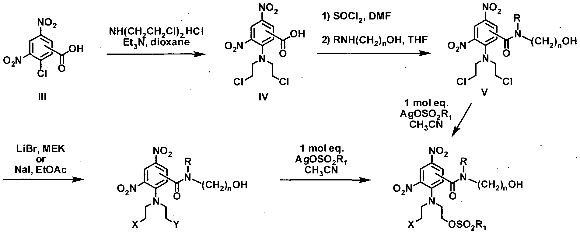
Vh X = Y = Br VIIh X = CI, Br, I VIh X = Y = I
Scheme 1
As an alternative method, in general terms, the preferred dinitrobenzamide mustard prodrug compounds of formula I may be prepared as shown in scheme 2 from the known. 3- chloro-2,6-dinitrobenzoic acid (Palmer et al, J. Med. Chem. 1996, 39, 2518-2528), or the commercially available 2-chloro-3,5-dinitrobenzoic acid and 5-chloro-2,4-dinitrobenzoic acid, respectively. Reaction with tert-butyl acetate in the presence of perchloric acid provides the respective tert-butyl esters (IX). Reaction of these with diethanolamine in dioxane then affords die respective diols (X). Conversion of these to the bis-alkylsulfonates (XI) can be achieved utilizing the appropriate alkylsulfonic anhydride in the presence of pyridine and catalytic dimethylaminopyridine (DMAP). Trifluoroacetic acid mediated tert-butyl ester hydrolysis,
followed by conversion of the intermediate acids (XH) to their respective acid chlorides, employing oxalyl chloride in the presence of magnesium oxide, and then subsequent reaction of these acid chlorides secondary hydroxyalkyl amines, that are themselves commercially available or readily prepared using the reductive amination method of Saavedra (J. Org. Chem. 1985, 50, 2271), provides the bis-alkylsulfonate dinitrobenzamides (XIII). Lithium chloride, lithium bromide or sodium iodide mediated partial halogen exchange, followed by the appropriate chromatography, then provides the asymmetric halogen/alkyl sulfonate mustards (VI).


XIII VIIh X = CI, Br, I
Scheme 2
As an alternative method, in general terms, the preferred asymmetric dinitrobenzamide mustard prodrug compounds of formula I may also be prepared following the methodology of Yang et al (Tetrahedron, 2007, 63, 5470-5476) as shown in scheme 3. The above described tert- butyl esters (IX) can be reacted with aziridine ethanol in the presence of a metal halide (LiCl, LiBr or NaI) to provide the haloethyl half-mustards (XIV), which can be derivatised to the halo/alkylsulfonate mustards (XV) by reaction with the appropriate alkylsulfonic anhydride in the presence of pyridine and catalytic dimethylaminopyridine (DMAP). Tert-butyl ester deprotection, acid chloride formation and amide coupling with secondary hydroxyalkyl amines, as described above, then provides the asymmetric halogen/alkyl sulfonate mustards of formula I.

IX XIV: X = Cl, Br, I XV: X = Cl, Br, I
Scheme 3 Preparation of dinitrobenzenesulfonamide mustard prodrugs of the invention In general terms, the preferred dinitrobenzenesulfonamide mustard prodrug compounds of formula I may be prepared as shown in scheme 4 from the known. 2-chloro-3,5- dinitrobenzenesulfonyl chloride and 5-chloro-2,4-dinitrobenzenesulfonyl chloride (McNaIIy et al, 1944, US 2358465; Herbert and Holliman, Tetrahedron, 1965, 21, 663-75), respectively. Reaction with secondary hydroxyalkyl amines, that are themselves commercially available or readily prepared using the reductive amination method of Saavedra (J. Org. Chem. 1985, 50, 2271), provides the dinitrobenzenesulfonamides (XVII). Chlorine displacement with NJSf-bis(2- chloroethyl) amine hydrochloride in dioxane employing triethylamine as base, provides the dichloro mustards (XVIII). Lithium bromide or sodium iodide mediated halogen exchange then provides the symmetrical dibromo and diiodo mustards (XIX and XX, respectively). The asymmetric halogen/alkyl sulfonate mustards (XXI) can be prepared from their symmetrical counterparts by reaction with approximately one equivalent of a silver alkylsulfonate salt in an appropriate solvent, such as acetonitrile.


XlX: X = Y = Br XXI: X = Cl, Br, I XX: X = Y = I
Scheme 4
Pteparation of phosphates of dinitrobenzamide and dinitrobenzenesulfonamide mustard prodrugs of the invention
In general terms, phosphates of formula II may be prepared as shown in scheme 5 from the preferred prodrug compounds of formula I by reaction of these alcohol derivatives with di- tert-butyl diisopropylphosphoramidite utilizing lH-tetrazole as die base, followed by oxidation with eidier rø-chloroperoxybenzoic acid (rø-CPBA) or 70% aqueous hydrogen peroxide, to provide the di-/^-butylphosphate ester intermediates (XXII). Acid mediated hydrolysis, employing trifluoroacetic acid (TFA) in dichloromethane, then provides the phosphates of formula II as their free acids.

Scheme 5
Schemes 6 below illustrates the preparation of dinitrobenzamide mustard prodrug compounds of formula I and II according to the invention. Reaction of the commercially available 2-chloro-3,5-dinitrobenzoic acid (21) with JVrN-bis(2-chloroethyl)amine hydrochloride in dioxane employing triethylamine as base provides the dichloro mustard acid (22). Conversion of this to the acid chloride and then reaction with 2-(methylamino)ethanol, provides the dinitrobenzamide (23). Lithium bromide mediated halogen exchange tben provides the symmetrical dibromo mustard (2). The asymmetric bromo/methyl sulfonate mustard (12) was prepared from diis by reaction with one equivalent of silver mesylate in acetonitrile. Reaction of alcohol 2 with di-tertbutyldiisopropylphosphoramidite utilizing lH-tetrazole as the base, followed by oxidation widi 70% aqueous hydrogen peroxide, provided the di-tertbutylphosphate ester intermediate (33). Acid mediated hydrolysis, employing trifluoroacetic acid (TFA) in dichloromethane, then provided the phosphate (39) of formula II as the free acid.
THF


22 23

AgOMs i 2: X = Y = Br 33 39
CH3CN !_»> 12: X = Br; Y = OSO2CH3
Scheme 6
Preparation of comparative compound (U), unsubstituted at the carboxamide nitrogen, was as described by Denny et al (PCT Int. Appl. WO 2005042471 Al; PCT Int. Appl. WO 2004033415 Al). Therapeutic uses of the compounds of the invention
The compounds of the Formulae I and II of the present invention can be used in the treatment of cancer of the human or animal body. In particular embodiments, the treatment may be of any cancer type that includes hypoxic regions, as the mustards of Formula II are reduced by enzymes present in such regions. For example, the cancers treated may be solid tumours, such as ovarian, colon, brain, thyroid, pancreas, bladder, breast, prostate, lung (such as small cell lung tumour cells and large cell lung carcinoma), cervical and skin cancer. Alternatively, the cancer may be leukaemia, multiple myeloma or lymphoma. All of these cancers present with hypoxic regions, particularly where tumours are growing or have become large.
The compounds of the invention can be administered in the form of pharmaceutical compositions, containing one or more compounds of the invention in combination with one or more pharmaceutically acceptable carriers. The pharmaceutically acceptable carrier(s) should be non-toxic and not interfere with the efficacy of the active ingredient. The precise nature of the carrier will depend on the route of administration, which can be oral, or parenteral, including intravenous, cutaneous, subcutaneous, intramuscular, intravascular or by infusion. The preparation of pharmaceutical compositions comprising one or more compounds of the invention may be performed by methods well known in the art, such as those described above.
The exact dose of the compound to be administered will be at the discretion of the physician, taking into account the type of cancer, the therapeutic approach (monotherapy or combination therapy) and the overall condition and needs of the patient. Typical doses and administration schedules will be determined by experience in clinical trials. Total doses are expected to be in the range from about 0.1 to 200 mg/kg per subject, such as about 10 mg/kg per subject.
The amount of compound administered may be between about 20% and 100% of the maximum tolerated dose of the subject.
The compounds of Formula I and II can be used as single agents or in combination with one or more other cytotoxic or other therapeutic agents or therapies, especially those that are relatively ineffective against hypoxic cells, such as radiation therapy. Where such other agents and/or radiotherapy are administered in combination with a compound of the invention, the radiation and/or other agents may be administered before, during or after administration of the compound of Formula I and II.
The following non-limiting examples may exist: EXPERIMENTAL
1. Chemical synthesis of exemplary compounds of the invention
Combustion analyses were performed by the Microchemical Laboratory, University of Otago, Dunedin, NZ. Melting points were determined using either an Electrothermal Model 9200 and are as read. 1H NMR spectra were measured either on a Bruker Avance-400 spectrometer and are referenced to Me4Si. High resolution mass spectra were recorded on a Varian VG-70SE spectrometer at nominal 5000 resolution. Mass spectrometry was performed on a ThermoFinnigan MSQ single quadrupole mass spectrometer. Mass detection was performed with an APCI source, using simultaneous positive and negative ion acquisition. Unless otherwise indicated, compounds were purified by flash column chromatography on Silica gel 60 support (Scharlau, 230-400 mesh ASTM), using the indicated eluants.
1.1 Preparation of the N-alkyl carboxamide mustard alcohols
A mixture of 2-chloro-3,5-dinitrobenzoic acid (21) (25.0 g, 10.10 mol), bis(2- chloroethyl) amine hydrochloride (36.2 g, 0.20 mol) and Et3N (42.3 mL, 0.30 mol) in dioxane (400 mL) was stirred at 25 0C for 72 h, then concentrated below 30 0C to 150 mL and diluted with excess IN aqueous HCl. Prolonged cooling at 0 0C provided a solid which was collected by filtration and chromatographed on silica gel, eluting with EtOAc/petroleum ether (3:1). The product thus obtained was triturated with iPr2O to give 2-[bis(2-chloroethyl)amino]-3,5- dimtrobenzoic acid (22) (26.9 g, 75%) as a yellow solid: mp (MeOH/H2O) 142-143 0C; 1H NMR [(CDj)2SO] δ 14-16 (br s, 1 H), 8.81 (dj = 2.8 Hz, 1 H), 8.59 (d J = 2.8 Hz, 1 H), 3.70 (tj = 6.7 Hz, 4 H), 3.46 (t, / = 6.7 Hz, 4 H).
A stirred solution of 2-[bis(2-chloroethyl)amino]-3,5-dinitrobenzoic acid (22) (15.0 g, 42.6 mmol) in SOCl2 (100 mL) and DMF (5 drops) was heated under reflux for 3 h, then cooled to room temperature and the excess SOCl2 was removed under reduced pressure. The resulting crude 2-[bis(2-chloroethyl)amino]-3,5-dinitrobenzoyl chloride was dissolved in THF (50 mL) and added slowly to a stirred solution of 2-(methylamino)ethanol (9.60 g, 12.8 mmol) in THF (80 mL)
at -10 0C. The mixture was stirred at -10 0C for 5 min, then acidified with 0.5 N aqueous HCl
(110 niL), concentrated to half volume under reduced pressure and extracted with EtOAc (2x).
The combined organic phases were washed with brine, dried and evaporated, and the residue was chromatographed on silica gel, eluting with EtOAc, followed by crystallisation from EtOAc/iPr2O, to give 2-[bis(2-chloroethyl)amino]-ΛT-(2-hydroxyethyl)-N-methyl-3,5τ dinitrobenzamide (23) (15.82 g, 91%): mp 136-137 0C; 1H NMR [(CD3)2SO] δ (mixture of rotamers) 8.71, 8.69 (2d, / = 2.8 Hz, 1 H), 8.39, 8.34 (d, / = 2.8 Hz, 1 H), 4.90-4.81 (m, 1 H), 3.73-3.29, 3.18-3,11 (2m, 12 H), 3.03, 2.94 (2s, 3 H). Anal. Calcd for C14H18Cl2N4O6: C, 41.1; H, 4.4; N, 13.7; Cl, 17.3%; found: C, 41.4; H, 4.6; N, 13.8; Cl, 17.3%. A mixture of 23 (20.0 g, 49 mmol) and LiBr (63.7 g, 730 mmol) in dry 3-methyl-2- butanone (methyl ethyl ketone; MEK) (150 mL) was stirred at reflux for 3 h, then concentrated under reduced pressure to remove most of the solvent. The residue was partitioned between EtOAc and water and the organic layer was washed with 10% aqueous NaI, dried and evaporated under reduced pressure. The residue was treated with further LiBr (63.7 g, 730 mmol) as above and worked up to provide crude product. A solution of this in EtOAc was filtered through a plug of silica gel, followed by crystallisation from EtOAc, to give 2-[bis(2-bromoethyl)amino]-A/-(2- hydroxyethyl)-Λ/-methyl-3,5-dinitrobenzamide (2) (17.44 g, 72%) as a yellow solid: mp 130-132 0C; 1H NMR [(CD^)2SO] (mixture of rotamers) δ 8.71, 8.69 (2dJ = 2.8 Hz, 1 H), 8.40, 8.34 (2dJ = 2.8 Hz, 1 H), 4.85 (br s, 1 H), 3.70-3.33, 3.18-3.11 (2m, 12 H), 3.03, 2.95 (2s, 3 H). Anal. Calcd for C14H18Br2N4On: C, 33.8; H, 3.6; N, 11.3%; found: C, 34.2; H, 3.9; N, 11.4%.
1.1.1. 2-((2-Bromoethyl)-2- { [(2-hydroxyethyl) (methyl)amino]carbonyl} -4,6-dinitroamlino) ethyl methanesulfonate (12)
A solution of 2 (10.06 g, 20.2 mmol) in MeCN (75 mL) was treated with AgOMs (3.69 g, 18.2 mmol) under reflux with stirring for 1 h. The mixture was concentrated under reduced pressure and the residue was extracted with EtOAc, evaporated, and chromatographed on silica gel. Elution with EtOAc/petroleum ether (2:1) gave unreacted 2 (3.59 g), while elution with EtOAc, concentration of the eluate to small volume and dilution with petroleum ether gave 2- ((2-brornoethyl)-2-{[(2-hydroxyethyl)(rneΛyl)arnino]carbonyl}-4,6-dinitroanilino)ethyl methanesulfonate (12) (3.48 g, 34%) as a yellow gum; 1H NMR [(CD3)2SO] (mixture of rotamers) δ 8.71, 8.69 (2dJ = 2.8 Hz, 1 H), 8.40, 8.34 (2d, J = 2.8 Hz, 1 H), 4.87 (br s, 1 H), 4.31-4.19 (m, 2 H), 3.71-3.10 (m, 13 H), 3.03, 2.95 (2s, 3 H). HRMS(FAB) calcd for C15H22 79BrN4O9S [M+H]+ m/z 513.0219: found 513.0273. 1.2 Preparation of the N-alkyl carboxamide mustard phosphates
1.2.1. 2-[(2-Bromoethyl)-2-({methyl[2-(phosphonooxy)ethyl]amino}carbonyl)-4,6- dinitroanilino] ethyl methanesulfonate (39)
A stirred solution of 2-((2-bromoethyl)-2-{[(2-hydroxyethyl)(methyl)amino]carbonyl}-4,6- dinitroanilino) ethyl methanesulfonate (12) (2.00 g, 3.90 mmol) in DMF (5 mL) at 10 0C was treated with lH-tetrazole (17.3 mL, 5.85 mmol; 3% w/w solution in MeCN), followed by the slow addition of di-tø?-butyl dϋsopropylphosphoramidite (95%, 1.68 mL, 5.06 mmol). The mixture was warmed to room temperature for 2 h, then cooled to 0 0C and treated with a solution of 70% aqueous H2O2 (0.9 mL) in THF (1 mL). The mixture was warmed to 15 0C for 20 min, then poured into ice-water and extracted with EtOAc (2x). The combined organic phases were washed with 5% aqueous Na2S2O5, water (3x), dried, and concentrated under reduced pressure below 30 0C. The residue was chromatographed on silica gel, eluting with EtOAc/petroleum ether (4:1). The eluate was concentrated to small volume and diluted with petroleum ether to give 2-[(2-bromoethyl)-2-(6-/ι?^-butoxy-2,8,8-trimethyl-6-oxido-5,7-dioxa-2- a2a-6-phosphanon-l-anoyl)-4,6-dinitroanilino]ethyl methanesulfonate (33) (1.96 g, 71%) as an unstable yellow gum; 1H NMR [(CDO2SO] (mixture of rotamers) δ 8.72, 8.70 (2d, / = 2.7 Hz, 1 H), 8.34, 8.29 (2d, / = 2.7 Hz, 1 H), 4.33-4.23, 4.19-4.04, 3,89-3.30 (3m, 12H), 3.16, 3.14 (2s, 3 H), 3.06, 2.97 (2s, 3 H), 1.44, 1.40, 1.37 (3s, 18 H). HRMS(FAB) CaIc. for C23H39 79BrN4O12PS [M+H]+ m/z 705.1206; found 705.2111.
A stirred solution of 2-[(2-bromoethyl)-2-(6-/^-butoxy-2,8,8-trimethyl-6-oxido-5,7-dioxa- 2-aza-6-ρhosphanon-l-anoyl)-4,6-dinitroanilino]ethyl methanesulfonate (33) (1.56 g, 2.21 mmol) in CH2Cl2 (8 mL) was treated with TFA (8 mL) for 1 h at room temperature, then concentrated under reduced pressure below 30 0C. The resulting gum was dissolved in CH2Cl2 (30 mL) and the filtered solution was refrigerated at 5 0C for 16 h. The separated yellow solid was collected, washed with CH2Cl2, and dried under high vacuum to give 2-[(2-bromoethyl)-2-({methyl[2- (phosphonooxy)ethyl]amino}carbonyl)-4,6-dinitroanilino]ethyl methanesulfonate (39) (1.16 g, 88%): mp 137-138 0C; 1H NMR [(CD3)2SO] (mixture of rotamers) δ 8.72, 8.68 (2d, / = 2.8 Hz, 1 H), 8.41, 8.31 (2dJ = 2.8 Hz, 1 H), 4.35-4.20 (m, 2 H), 4.18-3.27 (m, 10 H), 3.15, 3.14 (2s, 3 H), 3.05-2.97 (2s, 3 H), P(OH)2 signals not seen. HRMS(FAB) CaIc. for C15H22 79BrN4O12PS [M+H]+ m/z 592.9954; found 592.9953. Anal. Calcd for C15H22BrN4O12PS: C, 30.4; H, 3.7; N, 9.4; P, 5.2%; found: C, 30.5; H, 3.6; N, 9.3; P, 5.3%.
Cytotoxic efficacy of exemplary compounds of the invention
The cytotoxicity of compounds 11 (PR-104A) and 12 was assessed as follows. Cellular cytotoxicity determinations for parental HCT116 cell populations and a clonal population stably expressing aldo-keto reductase 1C3 (AKR1C3)
Parental HCT-116WT cell (ATCC CCL-247) and HCT-Il 6ΛKR1C3 cells engineered to express human aldo-keto reductase 1C3 (AKRl C3; NM_003739) were passaged as monolayers in minimal essential media (αMEM; Gibco, Inήtrogen Corporation, Grand Island, NY, USA) supplemented with 5% FBS, (GIBCO NZ Ltd, Auckland, New Zealand) without antibiotics for <3 months from frozen stocks confirmed to be mycoplamsa free.by PCR-ELISA (Roche Diagnostics Mannheim, Germany). Cells were removed from T75 flask (Becton Dickinson Biosciences, Bedford, MA) with 1 ml of warm 0.05% trypsin/EDTA (Invitrogen Corporation, Carlsbad, CA, USA), counted (Z2 Coulter Particle Count and cell Analyzer, Beckman Coulter, Fullerton, CA, USA) and diluted to give 500/well (in 100 μl) and left to attach for at ≥4 hrs (37°C, humidified incubator, 5% CO2). Frozen stocks of compounds 11 and 12 (in DMSO) were thawed and diluted to required concentration with αMEM. Aliquots were diluted 1:100 so organic solvent concentrations (DMSO) in the cell cultures did not exceed 1%. 50 μl of diluted drug stock was added directly to the top-well, where solubility limitations required, 150 μl of drug solution was added directly to aspirated wells. The drugs were then diluted along die plate in 3-fold serial dilutions using a 12-channel pipette (Biolab Umited, Albany, Auckland, New Zealand). Plates were returned to the 37°C, 5% CO2 incubator and left for 4 hrs. After the incubation period cells were washed three times and plates were filled with 200 μl of αMEM containing 5% FCS and 1 % penicillin/streptomycin {Gibco, Inήtrogen Corporation, Grand Island, NY, USA), and left for a further 5 days in a 37 0C, 5% CO2 incubator. After five days cells were fixed by adding 67μl of cold 40% trichloroacetic acid (Merck KGaA, Darmstadt, Germany) to each well, to give a final concentration of 10%. Plates were held at 4 0C for 1 hour. After 1 hr, plates were rinsed in tap water 3-4 times, with excess water being drained prior to staining. After die plates were rinsed, 50μl of 0.4% sulforhodamine B (SRB; SigmaAldrich, St. Louis, MO. USA) in 1% acetic acid (Merck KGaA, Darmstadt, Germany) was added to each well and left for 30 minutes in die dark. Plates were rinsed in 1% acetic acid and excess fluid was drained. The stain was solubilised by the addition of lOOμl of 1OmM unbuffered Tris (AppliChem GmbH, Darmstadt, Germany), left for lhr in the dark shaking at 150 rpm (Barnstezd-LabhneyBarnstead International, Dubuque, 10, USA). Plates were then read on an ELx 808 Absorbance Microplate Reader (Bio-Tek Instruments, Winooski, VT, USA). Wavelengths on the plate reader were set at 490nm for measurement filter and 450nm for reference filter (the reference filter was subtracted from the measurement filter to give final absorbance: 490-450). The data was used to calculate the IC50 value (KC4 microplate data analysis software V3.4, Bio-Tek), where 50% suppression of cell growth has occurred relative to untreated controls. N = 2-6 independent experiments IC50 value (Mean ± 1 SD) were determined for each HCTl 16 population and intra-experimental sensitivity ratio calculated. The results are shown in Table 10 below.

11 59 ± 4.5 2.5 ± 0.8 23 ± 5.5
12 93 ± 15 2.0 33.5
„ As can be seen, representative compound 12 is at least as sensitive to AKRl C3 activation as compound 11 (PR- 104A).
All publications referred to in this specification are herein incorporated by reference. Although the present invention has been described in some detail by way of illustration and example above, it will be understood by those persons of ordinary skill in the art that changes and modifications may be made without departing from the spirit or scope of the invention.
REFERENCES
Abrams et al., Mo/. Cancer. Ther., 2:471-478 (2003)
Anderson, "Nucleic Acid Hybridization," BIOS Scientific Publishers (1999) Atwell et al, Anti-Cancer Drug Design, 1996, 11 , 553-567 Atwell et-alj. Med. Chem. 2007, 50, 1197-1212 Ausubel et al., Current Protocols in Molecular Biology, Chapter 2 and Supplement 45, John Wiley & Sons, Inc.,
New York (1999)
" Baldi et al., "DNA Microarrays and Gene Expressions: From Experiments to Data Analysis and Modeling," Cambridge University Press (2002)
BaoJ. Chromatogr. B. Biomed. Sd., 699:463-80 (1997) Bauman, D R.; et al. "Development of Nonsteroidal Anti-Inflammatory Drug Analogs and Steroid
Carboxylates Selective for Human Aldo-Keto Reductase Isoforms: Potential Antineoplastic Agents That Work Independently of Cyclooxygenase Isozymes" MoI Pharmacol. 2005 Jan;67(l):60-8. Epub 2004 Oct 8.
Birren et al., Genome Anayl sis: A laboratory Manual, Volume 1 (Analyzing DNA), New York, Cold Spring
Harbor Laboratory Press (1997)
Borrebaeck, Ed., Antibody Engineering: A Practical Approach, Oxford University Press, Oxford (1995) Byrns, M C, et al. "An indomethacin analogue, N-(4-chlorobenzoyl)-melatonin, is a selective inhibitor of aldo-keto reductase 1C3 (type 2 3α-HSD, type 5 17β-HSD, and prostaglandin F synthase), a potential target for the treatment of hormone dependent and hormone independent malignancies" Biochemical Pharmacology, Volume 75, Issue 2, 15 January 2008, Pages 484-493 Cappuzzo et al. J. Clin. Oncol, 23:5007-5018 (2005) Cappuzzo et al.,/. Natl. Canerlnst., 97:643-655 (2005) Chee et al., Science, 274:610-614 (1996)
Cockett, M et al., Current Opinion in Biotechnology, 11:602-609 (2000) Desmond et al. Cameras. 63, 505-512, (2003)
Deutcher, Murray P. ed., Guide to Protein Purification, Meth. En^ymol, Vol. 182, 1990; Devlin and Risch 1995; A comparison of linkage disequilibrium measures for fine-scale mapping, Genomics 29: 311-322
Drmanac et al., Nature Biotech., 16:54-58 (1998) Drmanac et al., Science, 260:1649-1652 (1993) Fang, J. -M. et al. J. Chem. Soc. Perkin Trans. 1 1995, 967-978 Fields, Greg B. ed., Solid Phase Peptide Synthesis, Meth. En^ymol, Vol. 289, 1997; Fink et al., /. Clin. Chem. Clin. Biocbem., 27:261-276 (1989) Friedlos et al., }. Med. Chem. 1997, 40, 1270-1275 Fu et al, MALDI-TOF MS; Nature Biotech, 16:381-384 (1998)
Fujiwara et al., Chem. Pharm. Bull., 44:1326-31 (1996)
Glinsky, G. et al., The Journal of Clin. Invest., 113(6):913-923 (2004)
Gobec, S, et al., "Nonsteroidal anti-inflammatory drugs and their analogues as inhibitors of aldo-keto reductase AKRl C3: New lead compounds for the development of anticancer agents" Bioorgank <& Medicinal Chemistry Letters, Volume 15, Issue 23, 1 December 2005, Pages 5170-5175
Gosling. "Immunoassays: A. Practical Approach," Oxford University Press (2000)
Guise et al., Biochem Pharmacol, 2007, 74:810-20
Hardiman, "Microarrays Methods and Applications: Nuts & Bolts," DNA Press (2003)
Harlow and Lane, Eds., Antibodies, A laboratory Manual, Cold Spring Harbour Laboratory, Cold Spring Harbour, N.Y. (1998)
Herbert and Holliman, Tetrahedron, 1965, 21, 663-75
Innis et al., "PCR Applications: Protocols for Functional Genomics," 1st Edition, Academic Press (1999)
Ishikawa et al., Cameras., 65:9176-9184 (2005)
/. Immunol, 149:3914-3920 (1992) J. Org. Chem. 1985, 50, 2271
Jakobsson,J et al., The Pharmacogenonomics Journal (2007) 7, 282-289
Kiso et al., Chem. Pharm. Bull, 38:1192-99 (1990)
Lee et al., Anal. Quant. CytoL Histol, 27:202-210 (2005)
Lin et al., Steroids, 2004 69:795-801 Mostafavi et al., Biomed. Pept. Proteins Nucleic Acids, 1:225-60, (1995)
Mullis et al., The Polymerase Chain Reaction, Birkauser, Boston (1994)
Ortiz de Montellano PR, Mathews JM. "Autocatalytic alkylation of the cytochrome P-450 prosthetic haem group by 1-aminobenzotriazole. Isolation of an NN-bridged benzyne-protoporphyrin IX adduct." Biochem J. 1981 Jun l;195(3):761-4. Palmer et al, J. Med. Chem. 1994, 37, 2175-84
Palmer et al, J. Med. Chem. 1996, 39, 2518-2528
Parra et alj. Vase. Surg., 28:669-675 (1998)
Patterson et al., CHn Can Res 2007, 13:3922-32
Penning, T. M.; Burczynski et al. Biochem. J. 2000, 351, 67-77 Price and Newman, "Principles and Practice of Immunoassay " 2nd Edition, Grove's Dictionaries (1997)
Reich DE et al; Linkage disequilibrium in the human genome, Nature 2001, 411:199-204
Rongen et al.J. Immunol. Methods, 204:105-133 (1997)
Schmalzing et al., Electrophoresis, 18:2184-93 (1997)
Sears et al., Biotechniques, 13:626-633 (1992) Self et al., Curr. Opin. Biotechnol, 7:60-65 (1996)
Selga et al., Biochemical Pharmacology 75 (2008)414-426
Shipp, M. et al., Nature Medicine, 8(l):68-74 (2002)
Sonneveld, P, /. Intern. Med, 247:521-534 (2000)
Sorlie, T. et al., P. M A. S. USA, 98:10869-10874 (2001) Theophilus et at., "PCR Mutation Detection Protocols," Humana Press (2002)
Van't Veer L J et al., Nature, 415:530-536 (2002)
Winter et al., Proc. Natl. Acad. Sd., 82:7575-7579 (1985)
Yang et al, Tetrahedron, 2007, 63, 5470-5476
Zimmerman et al., Methods MoI. Cell. Biol, 3:39-42 (1992)
Claims
1. An assay method for predicting the response of a patient with cancer to treatment with a therapeutic agent, said method comprising:
(a) determining at least one AKRl C3 profile selected from the group consisting of an AKRl C3 nucleic acid profile, an AKRl C3 protein profile and combinations thereof in a sample from said patient; and
(b) identifying the patient as either suitable or non-suitable for such treatment based upon said at least one profile.
2. The method according to claim 1 wherein die sample is of or from a tumour and the response being predicted is whether the tumour is responsive or non-responsive to treatment with said agent.
3. The method according to claim 1 wherein the sample is a non-tumour sample.
4. The method according to claim 1 , claim 2, or claim 3 wherein the therapeutic agent to which a response is predicted is an AKRl C3-activated prodrug or a pre-prodrug thereof.
5. The method according to claim 4 wherein the prodrug is PR-104A.
6. The method according to claim 4 wherein the prodrug is a compound of formula I:

I or a pharmaceutically acceptable salt thereof or the pre-prodrug is-a compound of formula II:
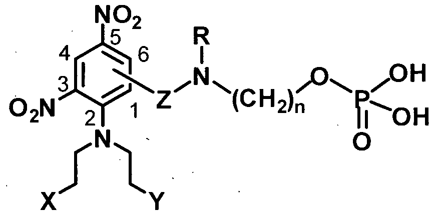
Il or a pharmaceutically acceptable salt thereof.
7. A method of treating a patient with cancer which comprises the step of treating a patient predicted to be a responder to treatment with an AKRlC3-activated therapeutic agent with an amount of an AKRlC3-activated agent sufficient to produce a therapeutic anticancer effect.
8. The method according to claim 7 wherein the AKRlC3-activated therapeutic agent is PR- 104A or PR-104.
9. The method according to claim 7 wherein the AKRl C3-activated therapeutic agent is a compound of formula I:

I or a pharmaceutically acceptable salt thereof, or a compound of formula II:
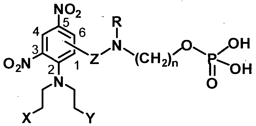
Il or a pharmaceutically acceptable salt thereof.
10. A method of treating a patient with cancer which comprises the step of treating said patient when predicted to be a non-responder to treatment with an AKRlC3-activated agent with an anti-cancer agent other than an AKRl C3-activated agent, or with an amount of a combination of an AKRl C3-activated agent and a Non-Steroidal Anti- Inflammatory Drug (NSAID) sufficient to produce a therapeutic effect.
11. A method of cancer treatment which includes the step of treating a patient with cancer having a positive AKRl C3 profile with an amount of AKRlC3-activated agent sufficient to produce a therapeutic effect.
12. The method according to claim 11 wherein the patient is treated with PR-104A or PR- 104.
13. The method according to claim 11 wherein the patient is treated with a compound of formula I: 
I or a pharmaceutically acceptable salt thereof or a compound of formula II:

Il or a pharmaceutically acceptable salt thereof.
14. A method of cancer treatment which comprises the step of treating a patient with cancer having a negative AKRl C3 profile with an anti-cancer agent other than an AKRl C3- activated agent, or with a combination of an AKRl C3-activated agent and an NSAID.
15. A method of treating a patient with AKRlCl, AKRl C2, AKRlBlO or NQOl-induced resistance to chemotherapy wherein said patient is resistant to treatment with an antifolate anti-cancer agent or a platinum or anthracycline anti-cancer agent which comprises the step of treating said patient with an amount of an AKRl C3-activated agent sufficient to produce a therapeutic anti-cancer effect.
16. The method according to claim 15 wherein said patient is treated with PR-104A or PR- 104.
17. The method according to claim 15 wherein said patient is treated with a compound of formula I:

I or a pharmaceutically acceptable salt thereof or a compound of formula II: 
or a pharmaceutically acceptable salt thereof.
18. An assay method for optimising dose efficacy in a patient with cancer receiving or who is to receive an AKRlC3-activated therapeutic agent, said method comprising:
(a) determining at least one AKRl C3 profile selected from the group consisting of an AKRl C3 nucleic acid profile, an AKRl C3 protein profile and combinations thereof in a tumour sample from said patient as well as in at least one non-tumour sample from said patient;
(b) recommending a dose of said AKRl C3 activated therapeutic agent based upon comparison of the AKRl C3 profiles of said tumour sample and said at least one non- tumour sample.
19. The method according to claim 18 wherein the recommended dose is zero.
20. A method of identifying an AKRl C3 -activated prodrug comprising a bioreductive trigger component and an active drug component, which method comprises the steps of:
(a) contacting a candidate AKRlC3-activated prodrug with an effective amount of a functional AKRl C3 enzyme; and
(b) detecting the release or non-release of die active drug component from the prodrug, widi release of the active drug component confirming the candidate as an AKRl C3 - activated prodrug.
21. A prodrug comprising a bioreductive trigger and an active drug component, wherein said bioreductive trigger is activated by AKRl C3 enzyme to release the active drug, and wherein said prodrug is identified or identifiable by a method as claimed in claim 20.
22. The prodrug according to claim 21 wherein the active drug component is a cytotoxic agent capable of producing a therapeutic effect when released in, or delivered to, a tumour.
23. A kit for use in an assay method as defined above, which kit comprises:
(a) at least one immunologic or nucleic acid reagent specific for AKRl C3; and
(b) a set of instructions for use of the reagent(s) to determine the AKRl C3 profile of a patient and to predict whether said patient is suitable or non-suitable for treatment with a therapeutic agent.
24. Compounds of formula I and formula II as shown below:

I Il wherein- X and Y are different with X being selected from Cl, Br, I, and OSO2R1, and Y being selected from Cl, Br, I, and OSO2R1, Z represents at any available ring position - CO- or -SO2-, R is methyl or ethyl; R1 is C1-6 alkyl; and n = 1-6, and pharmaceutically acceptable salts thereof.
25. The compound of claim 24 wherein X is Br or Cl.
26. The compound of claim 24 or 25 wherein Y is OSO2CH3.
27. A pharmaceutical composition comprising a compound as claimed in any one of claims 24 to 26.
28. A method of treating a patient with cancer which comprises the step of administering to a patient in need thereof a therapeutically-effective amount of a compound as claimed in any one of claims 24 to 26.
29. Use of a compound as claimed in any one of claims 24 to 26 in the manufacture of a composition for treating cancer in a patient in need thereof.
30. A compound as claimed in any one of claims 24 to 26 for treating cancer in a patient in need thereof.
Applications Claiming Priority (4)
| Application Number | Priority Date | Filing Date | Title |
|---|---|---|---|
| NZ572123 | 2008-10-17 | ||
| NZ57213308 | 2008-10-17 | ||
| NZ57212308 | 2008-10-17 | ||
| NZ572133 | 2008-10-17 |
Publications (1)
| Publication Number | Publication Date |
|---|---|
| WO2010044686A1 true WO2010044686A1 (en) | 2010-04-22 |
Family
ID=42106697
Family Applications (1)
| Application Number | Title | Priority Date | Filing Date |
|---|---|---|---|
| PCT/NZ2009/000227 Ceased WO2010044686A1 (en) | 2008-10-17 | 2009-10-19 | Akr1c3 as a biomarker, methods of selecting and treating patients based upon an akr1c3 profile and compounds for use therein |
Country Status (1)
| Country | Link |
|---|---|
| WO (1) | WO2010044686A1 (en) |
Cited By (10)
| Publication number | Priority date | Publication date | Assignee | Title |
|---|---|---|---|---|
| WO2016161342A3 (en) * | 2015-04-02 | 2016-11-10 | Threshold Pharmaceuticals, Inc. | Nitrobenzyl derivatives of anti-cancer agents |
| US10131683B2 (en) | 2014-07-17 | 2018-11-20 | Molecular Templates, Inc. | TH-302 solid forms and methods related thereto |
| CN110057944A (en) * | 2019-05-24 | 2019-07-26 | 广东中烟工业有限责任公司 | A kind of amino acid derivatization method and its application |
| US10364261B2 (en) | 2015-03-10 | 2019-07-30 | Obi Pharma, Inc. | DNA alkylating agents |
| US10409869B2 (en) | 2012-10-29 | 2019-09-10 | Obi Pharma, Inc. | (R)- and (S)-1-(3-(3-N,N-dimethylaminocarbonyl)phenoxyl-4-nitrophenyl)-1-ethyl-N,N'-bis (ethylene)phosphoramidate, compositions and methods for their use and preparation |
| WO2019190331A1 (en) * | 2018-03-29 | 2019-10-03 | Achilles Medical Limited | Prodrug compounds activated by akr1c3 and their use for treating hyperproliferative disorders |
| US10668047B2 (en) | 2015-06-24 | 2020-06-02 | Molecular Templates, Inc. | Aziridine containing DNA alkylating agents |
| WO2021110085A1 (en) * | 2019-12-03 | 2021-06-10 | 深圳艾欣达伟医药科技有限公司 | Method for associating with expression level of akr1c3 enzyme via content of prostaglandin, and use of screening for drug administration |
| US20220119429A1 (en) * | 2019-05-13 | 2022-04-21 | Ascentawits Pharmaceuticals, Ltd. | Fluorine-containing compound and anti-cancer medical use thereof |
| JP2023540283A (en) * | 2020-09-02 | 2023-09-22 | アセンタウィッツ ファーマシューティカルズ リミテッド | AKR1C3 detection method, AKR1C3 detection diagnostic kit and use thereof |
Citations (4)
| Publication number | Priority date | Publication date | Assignee | Title |
|---|---|---|---|---|
| WO1993011099A1 (en) * | 1991-11-28 | 1993-06-10 | Cancer Research Campaign Technology Limited | Nitroaniline derivatives and their use as antitumor agents |
| WO2008030112A1 (en) * | 2006-09-04 | 2008-03-13 | Auckland Uniservices Limited | Processes of preparing asymmetric dinitrobenzamide mustard compounds, intermediate compounds useful therein and products obtained therefrom |
| WO2008033041A1 (en) * | 2006-09-11 | 2008-03-20 | Auckland Uniservices Limited | Cancer treatment |
| WO2008124824A1 (en) * | 2007-04-10 | 2008-10-16 | Myriad Genetics, Inc. | Dosages and methods for the treatment of cancer |
-
2009
- 2009-10-19 WO PCT/NZ2009/000227 patent/WO2010044686A1/en not_active Ceased
Patent Citations (4)
| Publication number | Priority date | Publication date | Assignee | Title |
|---|---|---|---|---|
| WO1993011099A1 (en) * | 1991-11-28 | 1993-06-10 | Cancer Research Campaign Technology Limited | Nitroaniline derivatives and their use as antitumor agents |
| WO2008030112A1 (en) * | 2006-09-04 | 2008-03-13 | Auckland Uniservices Limited | Processes of preparing asymmetric dinitrobenzamide mustard compounds, intermediate compounds useful therein and products obtained therefrom |
| WO2008033041A1 (en) * | 2006-09-11 | 2008-03-20 | Auckland Uniservices Limited | Cancer treatment |
| WO2008124824A1 (en) * | 2007-04-10 | 2008-10-16 | Myriad Genetics, Inc. | Dosages and methods for the treatment of cancer |
Non-Patent Citations (7)
Cited By (21)
| Publication number | Priority date | Publication date | Assignee | Title |
|---|---|---|---|---|
| US10409869B2 (en) | 2012-10-29 | 2019-09-10 | Obi Pharma, Inc. | (R)- and (S)-1-(3-(3-N,N-dimethylaminocarbonyl)phenoxyl-4-nitrophenyl)-1-ethyl-N,N'-bis (ethylene)phosphoramidate, compositions and methods for their use and preparation |
| US10131683B2 (en) | 2014-07-17 | 2018-11-20 | Molecular Templates, Inc. | TH-302 solid forms and methods related thereto |
| US10654876B2 (en) | 2014-07-17 | 2020-05-19 | Molecular Templates, Inc. | TH-302 solid forms and methods related thereto |
| US10766914B2 (en) | 2015-03-10 | 2020-09-08 | Obi Pharma, Inc. | DNA alkylating agents |
| US10364261B2 (en) | 2015-03-10 | 2019-07-30 | Obi Pharma, Inc. | DNA alkylating agents |
| US10829437B2 (en) | 2015-04-02 | 2020-11-10 | Obi Pharma, Inc. | Nitrobenzyl derivatives of anti-cancer agents |
| CN112142692A (en) * | 2015-04-02 | 2020-12-29 | 深圳艾欣达伟医药科技有限公司 | Nitrobenzyl derivative anticancer agents |
| US11535585B2 (en) | 2015-04-02 | 2022-12-27 | Obi Pharma, Inc. | Nitrobenzyl derivatives of anti-cancer agents |
| WO2016161342A3 (en) * | 2015-04-02 | 2016-11-10 | Threshold Pharmaceuticals, Inc. | Nitrobenzyl derivatives of anti-cancer agents |
| JP2018511612A (en) * | 2015-04-02 | 2018-04-26 | アセンタ ファーマシューティカルズ リミテッド | Nitrobenzyl derivatives of anticancer agents |
| US10668047B2 (en) | 2015-06-24 | 2020-06-02 | Molecular Templates, Inc. | Aziridine containing DNA alkylating agents |
| CN111918864A (en) * | 2018-03-29 | 2020-11-10 | 阿基利斯医疗有限公司 | Prodrug compounds activated by AKR1C3 and their use in the treatment of hyperproliferative disorders |
| WO2019190331A1 (en) * | 2018-03-29 | 2019-10-03 | Achilles Medical Limited | Prodrug compounds activated by akr1c3 and their use for treating hyperproliferative disorders |
| EP3774743A4 (en) * | 2018-03-29 | 2021-11-24 | Achilles Medical Limited | PRODUCT-LIKE COMPOUNDS ACTIVATED BY AKR1C3 AND THEIR USE FOR THE TREATMENT OF HYPERPROLIFERATIVE DISORDERS |
| US11661404B2 (en) | 2018-03-29 | 2023-05-30 | Achilles Medical Limited | Prodrug compounds activated by AKR1C3 and their use for treating hyperproliferative disorders |
| CN111918864B (en) * | 2018-03-29 | 2024-03-05 | 阿基利斯医疗有限公司 | Prodrug compounds activated by AKR1C3 and their use in the treatment of hyperproliferative disorders |
| US20220119429A1 (en) * | 2019-05-13 | 2022-04-21 | Ascentawits Pharmaceuticals, Ltd. | Fluorine-containing compound and anti-cancer medical use thereof |
| US12595275B2 (en) * | 2019-05-13 | 2026-04-07 | Ascentawits Pharmaceuticals, Ltd. | Fluorine-containing compound and anti-cancer medical use thereof |
| CN110057944A (en) * | 2019-05-24 | 2019-07-26 | 广东中烟工业有限责任公司 | A kind of amino acid derivatization method and its application |
| WO2021110085A1 (en) * | 2019-12-03 | 2021-06-10 | 深圳艾欣达伟医药科技有限公司 | Method for associating with expression level of akr1c3 enzyme via content of prostaglandin, and use of screening for drug administration |
| JP2023540283A (en) * | 2020-09-02 | 2023-09-22 | アセンタウィッツ ファーマシューティカルズ リミテッド | AKR1C3 detection method, AKR1C3 detection diagnostic kit and use thereof |
Similar Documents
| Publication | Publication Date | Title |
|---|---|---|
| WO2010044686A1 (en) | Akr1c3 as a biomarker, methods of selecting and treating patients based upon an akr1c3 profile and compounds for use therein | |
| JP6906554B2 (en) | Biomarkers that identify patients responding to treatment and treatment of such patients | |
| CN103562406B (en) | Methods and compositions for predicting response to eribulin | |
| EP3372591B1 (en) | Cadherin-11 inhibitors and methods of use thereof | |
| JP2019531699A (en) | Diagnosis and treatment method of cancer by expression state and mutation state of NRF2 and downstream target gene of the gene | |
| KR20150079949A (en) | Synthetic lethality and the treatment of cancer | |
| JP2008533053A (en) | Histone deacetylase inhibitors sensitize cancer cells to epidermal growth factor receptor inhibitors | |
| KR20220123064A (en) | Cancer treatment with CDK12/13 inhibitors | |
| US20210371935A1 (en) | Identification of pde3 modulator responsive cancers | |
| US20100028259A1 (en) | Use of organic cation transporters for cancer diagnosis and therapy | |
| JP2004533603A (en) | PIN1 as a marker for abnormal cell growth | |
| JP2021532158A (en) | Tetrahydroquinolino derivatives for the treatment of metastatic and chemotherapy-resistant cancers | |
| WO2016109471A1 (en) | Bufalin phosphate prodrugs and methods of use thereof | |
| JP2020505392A (en) | Compositions and methods for treating androgen-independent cancer | |
| WO2016077347A1 (en) | Mir-214 as a diagnostic and prognostic biomarker specific for ulcerative colitis and a mir-214 inhibitor for treatment of same | |
| CN114214418A (en) | Biomarker for evaluating sensitivity of lung cancer patient to proton radiotherapy and application thereof | |
| AU2014266278B2 (en) | Therapeutic effect prediction method for colorectal cancer patient in whom expression of TK1 protein has increased | |
| CA2697887A1 (en) | Modulating and/or detecting activation induced deaminase and methods of use thereof | |
| Pinheiro et al. | Targeting metabolic reprogramming as an anti-cancer strategy: aiming at monocarboxylate transporters | |
| US20140227172A1 (en) | Diagnosis and treatment of prostate cancer | |
| US11892454B2 (en) | Method for the diagnosis of etoposide prodrug treatable cancer | |
| JP2024509887A (en) | EP300 degraders and their use in neuroblastoma | |
| US20120264803A1 (en) | Method for predicting therapeutic effects of chemotherapy on hepatocellular carcinoma patients | |
| US20190227072A1 (en) | Biomarkers for Carcinogenesis and Uses Thereof | |
| US20170218373A1 (en) | Composition for diagnosis of liver metastasis of colorectal cancer and the use thereof |
Legal Events
| Date | Code | Title | Description |
|---|---|---|---|
| 121 | Ep: the epo has been informed by wipo that ep was designated in this application |
Ref document number: 09820808 Country of ref document: EP Kind code of ref document: A1 |
|
| NENP | Non-entry into the national phase |
Ref country code: DE |
|
| 122 | Ep: pct application non-entry in european phase |
Ref document number: 09820808 Country of ref document: EP Kind code of ref document: A1 |