WO2010029799A1 - Microscope device and fluorescent observing method using same - Google Patents
Microscope device and fluorescent observing method using same Download PDFInfo
- Publication number
- WO2010029799A1 WO2010029799A1 PCT/JP2009/060644 JP2009060644W WO2010029799A1 WO 2010029799 A1 WO2010029799 A1 WO 2010029799A1 JP 2009060644 W JP2009060644 W JP 2009060644W WO 2010029799 A1 WO2010029799 A1 WO 2010029799A1
- Authority
- WO
- WIPO (PCT)
- Prior art keywords
- fluorescence
- observation
- image information
- observed
- control
- Prior art date
Links
- 238000000034 method Methods 0.000 title claims abstract description 42
- 238000001514 detection method Methods 0.000 claims description 71
- 238000002073 fluorescence micrograph Methods 0.000 claims description 46
- 230000005284 excitation Effects 0.000 claims description 45
- 238000003384 imaging method Methods 0.000 claims description 38
- 238000005286 illumination Methods 0.000 claims description 23
- 230000004936 stimulating effect Effects 0.000 claims description 6
- 230000001678 irradiating effect Effects 0.000 claims description 4
- 230000004069 differentiation Effects 0.000 claims description 2
- 230000010354 integration Effects 0.000 claims description 2
- 210000004027 cell Anatomy 0.000 description 110
- 230000003287 optical effect Effects 0.000 description 57
- 238000010586 diagram Methods 0.000 description 22
- 230000006870 function Effects 0.000 description 21
- 230000000638 stimulation Effects 0.000 description 21
- 238000012545 processing Methods 0.000 description 18
- 239000011347 resin Substances 0.000 description 18
- 229920005989 resin Polymers 0.000 description 18
- 238000011156 evaluation Methods 0.000 description 17
- 239000007789 gas Substances 0.000 description 17
- 150000002500 ions Chemical class 0.000 description 15
- 230000008859 change Effects 0.000 description 12
- 239000000243 solution Substances 0.000 description 11
- 239000000126 substance Substances 0.000 description 11
- PNDZEEPOYCVIIY-UHFFFAOYSA-N indo-1 Chemical compound CC1=CC=C(N(CC(O)=O)CC(O)=O)C(OCCOC=2C(=CC=C(C=2)C=2N=C3[CH]C(=CC=C3C=2)C(O)=O)N(CC(O)=O)CC(O)=O)=C1 PNDZEEPOYCVIIY-UHFFFAOYSA-N 0.000 description 9
- 230000000007 visual effect Effects 0.000 description 8
- 239000007850 fluorescent dye Substances 0.000 description 7
- IJGRMHOSHXDMSA-UHFFFAOYSA-N Atomic nitrogen Chemical compound N#N IJGRMHOSHXDMSA-UHFFFAOYSA-N 0.000 description 6
- 238000004422 calculation algorithm Methods 0.000 description 6
- 238000005259 measurement Methods 0.000 description 6
- 238000004458 analytical method Methods 0.000 description 5
- 230000007423 decrease Effects 0.000 description 5
- 239000011521 glass Substances 0.000 description 5
- 210000002865 immune cell Anatomy 0.000 description 5
- 239000007788 liquid Substances 0.000 description 5
- CURLTUGMZLYLDI-UHFFFAOYSA-N Carbon dioxide Chemical compound O=C=O CURLTUGMZLYLDI-UHFFFAOYSA-N 0.000 description 4
- 210000000170 cell membrane Anatomy 0.000 description 4
- 230000000052 comparative effect Effects 0.000 description 4
- 230000005484 gravity Effects 0.000 description 4
- 239000000523 sample Substances 0.000 description 4
- 230000003044 adaptive effect Effects 0.000 description 3
- 238000013528 artificial neural network Methods 0.000 description 3
- 239000004020 conductor Substances 0.000 description 3
- 238000001816 cooling Methods 0.000 description 3
- 239000006059 cover glass Substances 0.000 description 3
- 230000007613 environmental effect Effects 0.000 description 3
- 230000002068 genetic effect Effects 0.000 description 3
- 230000002452 interceptive effect Effects 0.000 description 3
- 230000003834 intracellular effect Effects 0.000 description 3
- 244000005700 microbiome Species 0.000 description 3
- 230000002123 temporal effect Effects 0.000 description 3
- XKRFYHLGVUSROY-UHFFFAOYSA-N Argon Chemical compound [Ar] XKRFYHLGVUSROY-UHFFFAOYSA-N 0.000 description 2
- MYMOFIZGZYHOMD-UHFFFAOYSA-N Dioxygen Chemical compound O=O MYMOFIZGZYHOMD-UHFFFAOYSA-N 0.000 description 2
- 210000001744 T-lymphocyte Anatomy 0.000 description 2
- 230000002411 adverse Effects 0.000 description 2
- 230000004888 barrier function Effects 0.000 description 2
- 239000012472 biological sample Substances 0.000 description 2
- 229910002092 carbon dioxide Inorganic materials 0.000 description 2
- 239000001569 carbon dioxide Substances 0.000 description 2
- 239000003153 chemical reaction reagent Substances 0.000 description 2
- 229910001873 dinitrogen Inorganic materials 0.000 description 2
- 229910001882 dioxygen Inorganic materials 0.000 description 2
- 229910052736 halogen Inorganic materials 0.000 description 2
- 150000002367 halogens Chemical class 0.000 description 2
- 230000003993 interaction Effects 0.000 description 2
- 239000000203 mixture Substances 0.000 description 2
- 229910052757 nitrogen Inorganic materials 0.000 description 2
- 239000011148 porous material Substances 0.000 description 2
- 229910052724 xenon Inorganic materials 0.000 description 2
- FHNFHKCVQCLJFQ-UHFFFAOYSA-N xenon atom Chemical compound [Xe] FHNFHKCVQCLJFQ-UHFFFAOYSA-N 0.000 description 2
- 241000931526 Acer campestre Species 0.000 description 1
- 239000004925 Acrylic resin Substances 0.000 description 1
- 229920000178 Acrylic resin Polymers 0.000 description 1
- 241000894006 Bacteria Species 0.000 description 1
- 229920002799 BoPET Polymers 0.000 description 1
- 108090000371 Esterases Proteins 0.000 description 1
- 239000005041 Mylar™ Substances 0.000 description 1
- 241000223785 Paramecium Species 0.000 description 1
- OAICVXFJPJFONN-UHFFFAOYSA-N Phosphorus Chemical compound [P] OAICVXFJPJFONN-UHFFFAOYSA-N 0.000 description 1
- 241000700605 Viruses Species 0.000 description 1
- 230000009471 action Effects 0.000 description 1
- 230000004913 activation Effects 0.000 description 1
- 229910052786 argon Inorganic materials 0.000 description 1
- 230000008901 benefit Effects 0.000 description 1
- 230000004397 blinking Effects 0.000 description 1
- 238000004364 calculation method Methods 0.000 description 1
- 238000011088 calibration curve Methods 0.000 description 1
- 239000003990 capacitor Substances 0.000 description 1
- 238000006243 chemical reaction Methods 0.000 description 1
- 230000000295 complement effect Effects 0.000 description 1
- 150000001875 compounds Chemical class 0.000 description 1
- 230000003247 decreasing effect Effects 0.000 description 1
- 238000009795 derivation Methods 0.000 description 1
- 238000013461 design Methods 0.000 description 1
- 238000002059 diagnostic imaging Methods 0.000 description 1
- 230000010339 dilation Effects 0.000 description 1
- 238000006073 displacement reaction Methods 0.000 description 1
- 238000010494 dissociation reaction Methods 0.000 description 1
- 230000005593 dissociations Effects 0.000 description 1
- 239000003814 drug Substances 0.000 description 1
- 230000009977 dual effect Effects 0.000 description 1
- 238000005516 engineering process Methods 0.000 description 1
- 238000002474 experimental method Methods 0.000 description 1
- 239000000284 extract Substances 0.000 description 1
- 238000000605 extraction Methods 0.000 description 1
- 238000005562 fading Methods 0.000 description 1
- 238000012632 fluorescent imaging Methods 0.000 description 1
- 230000036737 immune function Effects 0.000 description 1
- 230000006698 induction Effects 0.000 description 1
- 239000012212 insulator Substances 0.000 description 1
- 239000010416 ion conductor Substances 0.000 description 1
- 210000000265 leukocyte Anatomy 0.000 description 1
- 238000013178 mathematical model Methods 0.000 description 1
- 230000007246 mechanism Effects 0.000 description 1
- QSHDDOUJBYECFT-UHFFFAOYSA-N mercury Chemical compound [Hg] QSHDDOUJBYECFT-UHFFFAOYSA-N 0.000 description 1
- 229910052753 mercury Inorganic materials 0.000 description 1
- 238000012986 modification Methods 0.000 description 1
- 230000004048 modification Effects 0.000 description 1
- 239000002245 particle Substances 0.000 description 1
- 230000002093 peripheral effect Effects 0.000 description 1
- 230000008569 process Effects 0.000 description 1
- 238000003672 processing method Methods 0.000 description 1
- 230000009467 reduction Effects 0.000 description 1
- 230000004044 response Effects 0.000 description 1
- 238000012552 review Methods 0.000 description 1
- 239000004065 semiconductor Substances 0.000 description 1
- GGCZERPQGJTIQP-UHFFFAOYSA-N sodium;9,10-dioxoanthracene-2-sulfonic acid Chemical compound [Na+].C1=CC=C2C(=O)C3=CC(S(=O)(=O)O)=CC=C3C(=O)C2=C1 GGCZERPQGJTIQP-UHFFFAOYSA-N 0.000 description 1
- 239000002887 superconductor Substances 0.000 description 1
- 239000012085 test solution Substances 0.000 description 1
- 238000001931 thermography Methods 0.000 description 1
- 231100000331 toxic Toxicity 0.000 description 1
- 230000002588 toxic effect Effects 0.000 description 1
- 230000017105 transposition Effects 0.000 description 1
- XLYOFNOQVPJJNP-UHFFFAOYSA-N water Substances O XLYOFNOQVPJJNP-UHFFFAOYSA-N 0.000 description 1
Images


Classifications
-
- G—PHYSICS
- G02—OPTICS
- G02B—OPTICAL ELEMENTS, SYSTEMS OR APPARATUS
- G02B21/00—Microscopes
- G02B21/0004—Microscopes specially adapted for specific applications
- G02B21/0088—Inverse microscopes
-
- G—PHYSICS
- G01—MEASURING; TESTING
- G01N—INVESTIGATING OR ANALYSING MATERIALS BY DETERMINING THEIR CHEMICAL OR PHYSICAL PROPERTIES
- G01N21/00—Investigating or analysing materials by the use of optical means, i.e. using sub-millimetre waves, infrared, visible or ultraviolet light
- G01N21/62—Systems in which the material investigated is excited whereby it emits light or causes a change in wavelength of the incident light
- G01N21/63—Systems in which the material investigated is excited whereby it emits light or causes a change in wavelength of the incident light optically excited
- G01N21/64—Fluorescence; Phosphorescence
- G01N21/645—Specially adapted constructive features of fluorimeters
- G01N21/6456—Spatial resolved fluorescence measurements; Imaging
- G01N21/6458—Fluorescence microscopy
-
- G—PHYSICS
- G02—OPTICS
- G02B—OPTICAL ELEMENTS, SYSTEMS OR APPARATUS
- G02B21/00—Microscopes
- G02B21/0004—Microscopes specially adapted for specific applications
- G02B21/002—Scanning microscopes
- G02B21/0024—Confocal scanning microscopes (CSOMs) or confocal "macroscopes"; Accessories which are not restricted to use with CSOMs, e.g. sample holders
- G02B21/0052—Optical details of the image generation
- G02B21/0076—Optical details of the image generation arrangements using fluorescence or luminescence
-
- G—PHYSICS
- G02—OPTICS
- G02B—OPTICAL ELEMENTS, SYSTEMS OR APPARATUS
- G02B21/00—Microscopes
- G02B21/16—Microscopes adapted for ultraviolet illumination ; Fluorescence microscopes
-
- G—PHYSICS
- G02—OPTICS
- G02B—OPTICAL ELEMENTS, SYSTEMS OR APPARATUS
- G02B21/00—Microscopes
- G02B21/34—Microscope slides, e.g. mounting specimens on microscope slides
-
- G—PHYSICS
- G02—OPTICS
- G02B—OPTICAL ELEMENTS, SYSTEMS OR APPARATUS
- G02B21/00—Microscopes
- G02B21/24—Base structure
- G02B21/26—Stages; Adjusting means therefor
Definitions
- the present invention relates to a microscope apparatus used for fluorescence observation of a living tissue or the like and a fluorescence observation method for an object to be observed using the microscope apparatus.
- the optical microscope is mainly used when observing a living tissue.
- the laser confocal microscope is used when a part of a living cell is irradiated with light, the irradiation position is moved, and a part of the cell is observed.
- Fluorescence observation is one of the main observation techniques of optical microscopes.
- a fluorescent dye is attached to ions or molecules in cells of living tissue, and the fluorescent dye is irradiated with excitation light to observe the fluorescence emitted by the excited fluorescent dye.
- the wavelengths of fluorescence and excitation light are different. Accordingly, analysis of fluorescence enables detection of intracellular molecules and measurement of concentration. For this reason, fluorescence observation has the following advantages. (i) It can be observed by the size of ions and molecules. (ii) Only specific ions and molecules can be observed. (iii) Since the brightness of the fluorescence changes according to the ion or molecular concentration, these concentrations can be measured.
- the cells When observing moving cells using a fluorescence microscope capable of fluorescence observation, the cells may go out of the field of view of the microscope and the observation may be interrupted.
- the method (1) lower the magnification of the objective lens to widen the field of view, (2) suppress the movement of cells mechanically or chemically, (3) move the stage and move the cell position to the center of the field of view To.
- the method (1) has a lower spatial resolution, and the method (2) may adversely affect cells.
- Patent Documents 1 and 2 disclose an optical microscope having a mechanism for tracking an observation object in order to observe a minute observation object. These optical microscopes can control the cell position and observation region using transmitted light and perform transmitted light recording.
- Patent Documents 3 to 5 disclose fluorescence microscopes that can control the cell position and observation region using fluorescence and perform fluorescence recording.
- Non-Patent Documents 1 to 5 report a method of observing a Paramecium moving as a relatively large object to be observed while fixing it in the field of view.
- Non-Patent Document 6 Among cells that can move freely in the culture solution, those that are regarded as important in the fields of medicine and biology include immune cells that have a function of protecting the human body from bacteria and viruses (see Non-Patent Document 6). ).
- the cells that do not adhere much to the surface of the slide glass and are floating are classified as floating cells.
- the floating cells in the petri dish always change positions due to the convection of the culture solution, gravity, the petri dish walls and bottom, the water surface, and the interaction between cells.
- a method of tracking one cell using the method of the back reported in Non-Patent Document 1 can be considered.
- Hashimoto “High-speed autofocusing of a cell using diffraction patterns”, Optics Express, Vol.14, No.9, pp.3952-3960, 2006 Hiromasa Oku, Makoto Ishii, Masatoshi Ishikawa: Micro Visual Feedback System, IEICE Journal D-II, Vol.J84-D-II, No.6, pp.994-1002, 2001 Hiromasa Oku, Masatoshi Ishikawa: Structure of variable focus lens for high-speed vision chip that can respond in kilohertz order, optics, Vol.31, No.10, pp.758-764, 642002 Koichi Hashimoto: Active sensing using microorganisms, SORST joint symposium (6) Abstracts, pp.
- the fluorescence observation parameters are currently determined by the observer according to empirical rules. In other words, the current situation is that it is not determined according to objective evaluation. In this case, even if the position, distribution, type, etc. of the cells change, it is difficult for the observer to adjust the fluorescence observation parameters flexibly. If an observer intends to observe many cells and excessively expands the excitation light irradiation range and irradiation time, the cells and the fluorescent dye will be adversely affected. In addition, a large amount of fluorescent image data is acquired, and a long time is required for analysis.
- Such a control method is mainly intended to correct a shift that occurs when a cell that does not move originally is observed for a long time.
- the present invention uses a microscope apparatus that can automatically control the position and observation area of an observation object during fluorescence observation by using transmitted light from the observation object for control.
- An object of the present invention is to provide a fluorescence observation method for the observed object used.
- a microscope apparatus of the present invention includes a stage on which an observation object is placed, a first light source that irradiates the observation object with illumination light, and excitation for exciting fluorescence in the observation object.
- a second light source for irradiating light an image information detection unit for detecting image information of the object to be observed obtained by the illumination light, and a fluorescence for detecting image information by the fluorescence generated in the object to be observed by the excitation light
- the fluorescence observation area of the observation object is determined based on the image information detection unit, the motion model of the observation object, and the image information of the observation object input from the image information detection unit, and input from the image information detection unit
- a control unit that obtains image information of the object to be observed and fluorescence image information input from the fluorescence image information detection unit at predetermined intervals in the fluorescence observation region.
- a motion model focuses on a specific dimension, a combination of specific dimensions, or a weighted combination of specific dimensions among the physical or chemical properties of the object to be observed, and the observed object on that dimension. It means a model that aims to predict the spatio-temporal changes of objects. For example, it means a mathematical model representing a spatio-temporal change of any one of the position, velocity, distribution, type, shape, ion concentration, molecular concentration, etc. of the object to be observed.
- the control unit preferably controls the fluorescence observation region such as classical control such as PID control, modern control such as optimal control and sub-optimal control, H ⁇ control, sample value control, finite time settling control, adaptive control, etc. It is determined according to intelligent control such as post-modern control theory, neural network control, fuzzy control, genetic algorithm control.
- PID control is control in which the deviation between the target value and the output value is minimized in accordance with a control law expressed by any of a term proportional to the deviation, an integral term of the deviation, a differential term of the deviation, or a combination thereof. It is a technique.
- the optimal control is a control method for deriving an optimal control law by creating an evaluation function indicating the efficiency of fluorescence observation and minimizing or maximizing it.
- the sub-optimal control is a control method for deriving an optimal control law locally by creating an evaluation function indicating the efficiency of fluorescence observation and locally minimizing or maximizing it.
- the control unit preferably determines the center position of the fluorescence observation region according to the motion model of the object to be observed.
- the control unit preferably includes a first light source control unit for controlling the first light source and a second light source control unit for controlling the second light source.
- the stage is preferably a three-dimensional stage that moves the position of the object to be observed.
- the fluorescence image information detection unit preferably includes wavelength selection means for separating fluorescence having at least one wavelength.
- a first pinhole disposed between the second light source and the object to be observed and a second pinhole disposed between the fluorescence and the fluorescence image information detection unit are provided.
- a pinhole driving unit that moves and / or rotates the first pinhole or the second pinhole is provided.
- an objective lens disposed between the first light source and the object to be observed and a drive unit for the objective lens are provided.
- an imaging lens disposed between the light generated by the object to be observed and the image information detection unit, and an imaging lens driving unit are provided.
- an imaging lens disposed between the fluorescence and the fluorescence image information detection unit, and an imaging lens driving unit are provided.
- an environment control unit that accommodates an object to be observed and is filled with an atmospheric gas.
- the environment control unit controls the type of atmospheric gas and the temperature of the atmospheric gas. Nitrogen gas (single), oxygen gas (single), carbon dioxide (single), air (mixture), and a mixed gas thereof can be used as the atmospheric gas.
- the environment control unit preferably includes a storage unit that can store a plurality of objects to be observed.
- an observation object stimulating means for stimulating the observation object is provided. For example, electrical stimulation, magnetic stimulation, mechanical stimulation, ultrasonic stimulation, thermal stimulation, chemical stimulation, light stimulation, or the like can be used as the stimulus given to the object to be observed.
- the object stimulating means includes, for example, an electrode, an electric current, a battery, a resistor, a capacitor, a magnet, a coil, a superconductor, a complete conductor, an electric conductor,
- An electrical stimulation control device made of a semiconductor, an insulator, a dielectric, a piezoelectric, a pyroelectric, a ferroelectric, an ion conductor, or the like can be used.
- a mechanical stimulus control device including a short needle, a probe, an actuator, a piezoelectric body, a centrifuge, a weight, a spring and the like can be used.
- an ultrasonic stimulation control device such as an electrostrictive vibrator or a magnetostrictive vibrator can be used to give an ultrasonic stimulus to the object to be observed.
- a thermal stimulus control device including a heater, a cooler, a heat conductor, a thermometer, a thermography, or the like can be used.
- a chemical stimulus control device including a pipette, a pump, a syringe, a propeller, a screw, and a microfluidic device that changes the concentration or state of the chemical substance in time and space is used be able to.
- a light stimulus control device such as a mirror, a prism, a filter, a lamp, a laser, or a maser can be used to give a light stimulus to the object to be observed.
- these stimulation control devices and their components are complementary to each other.
- the photostimulation control device can change the concentration or state of chemical substances inside and outside the cell by applying light to the caged compound and decompose it, and can also be a chemical stimulation control device.
- the light stimulus control device can warm the measurement liquid with light, and can also be a thermal stimulus control device.
- a fluorescence observation method of the present invention includes a first stage for determining a fluorescence observation region of an observation object based on image information of the observation object and a motion model of the observation object, and a fluorescence observation region. A second stage of acquiring fluorescence image information at a predetermined position.
- the first-stage fluorescence observation area is preferably controlled by classical control such as PID control, modern control such as optimal control and sub-optimal control, H ⁇ control, sample value control, finite time settling control, adaptive control, etc. It is decided according to intelligent control such as post modern control theory, neural network control, fuzzy control, genetic algorithm control.
- the center position of the fluorescence observation region is determined based on at least one of the image information of the object to be observed and the motion model of the object to be observed.
- image information of the object to be observed is acquired. It is preferable that each step is repeated a predetermined number of times for each predetermined position in the fluorescence observation region.
- the parameter of the motion model is preferably composed of any one of the position, velocity, distribution, type, ion concentration, molecular concentration, etc. of the object to be observed.
- the fluorescence observation region is determined based on the image information from the object to be observed and the motion model of the object to be observed, the fluorescence observation can be automatically performed.
- the fluorescence observation parameters can be adjusted flexibly and automatically in response to a dynamic change of the object to be observed.
- FIG. 2 is a schematic diagram illustrating a configuration of an environment control unit
- FIG. 4 is a schematic diagram showing another configuration of an environment control unit capable of observing a plurality of objects to be observed, where (A) is a plan view and (B) is a cross-sectional view taken along line XX in (A). It is a figure which shows an example of the cell distribution immediately after an observation start.
- a fluorescence observation procedure it is the schematic diagram which compared the evaluation function J of the Example using control amount U (k), and the evaluation function J of the comparative example at the time of fixing a fluorescence observation area
- variety In a fluorescence observation procedure, it is the schematic diagram which compared the evaluation function J of the Example using control amount U (k), and the evaluation function J of the comparative example at the time of fixing a fluorescence observation area
- variety In a fluorescence observation procedure, it is the schematic diagram which compared the evaluation function J of the Example using control amount U (k), and the evaluation function J
- FIG. 1 is a schematic diagram showing the configuration of the microscope apparatus 1 according to the first embodiment of the present invention.
- the microscope apparatus 1 according to the first embodiment controls the stage 3, the optical system 10, the position of the stage 3, and the like on which the object 2 is placed and the position of the object 2 can be freely moved.
- the optical system 10 irradiates an illumination light source 4 for irradiating the observation object 2 with illumination light in order to detect an image or position of the observation object 2 and excitation light for exciting fluorescence generated from the observation object 2.
- the excitation light source 5 Generated by the excitation light source 5, the light path of the transmitted light T obtained from the observation object 2 by irradiation with the light from the illumination light source 4, the optical path for guiding the excitation light 5 A to the observation object 2, and the observation object 2.
- a fluorescence image information detection unit 17 that detects fluorescence image information of the observation object 2 emitted from the tube unit 15.
- the observation object 2 may be, for example, a cell that is a biological sample.
- the observation object 2 is placed on the stage 3, and the position of the observation object 2 is controlled by the control unit 20 described later.
- This stage 3 is a so-called electric stage, which is an XY stage that is driven and controlled on a two-dimensional plane (XY plane) on which the object 2 is placed, or an XYZ stage that is driven and controlled in three dimensions. There may be.
- Three-dimensional driving may be performed by a manipulator (see Non-Patent Document 3).
- the position control in the XY-axis direction of the observation region may be performed by controlling the irradiation position of the illumination light source 4 or the excitation light source 5 and the position where a pinhole described later is disposed.
- the position control in the Z-axis direction of the observation area may be performed by controlling positions where an objective lens, an imaging lens, and a pinhole, which will be described later, are arranged (see Patent Document 6, Non-Patent Documents 2 and 4).
- the illumination light source 4 When the illumination light source 4 is a light source for transmitted light, the illumination light source 4 may be anything as long as it emits light including a wavelength that the object to be observed 2 absorbs or reflects. As such an illumination light source 4, various lamps such as halogen lamps, light emitting diodes, and various lasers can be used. In order to prevent the transmitted light T from interfering with the excitation light 5A and the fluorescence F, it is desirable that the same wavelength band as the excitation light 5A and the fluorescence F in the light output from the illumination light source is cut by the optical filter 8 or the like. Illumination light emitted from the illumination light source 4 to the object to be observed 2 becomes transmitted light T or reflected light from the object to be observed 2 and is detected by the image information detection unit 16. The image from the observation object 2 is image information such as a so-called bright field image and dark field image.
- the excitation light source 5 may be any light source that can excite the observation object 2 itself or a phosphor contained in the observation object 2.
- a pump such as a xenon lamp or a mercury lamp, or various lasers such as an argon laser can be used as the excitation light 5A.
- a pump such as a xenon lamp or a mercury lamp, or various lasers such as an argon laser can be used as the excitation light 5A.
- the excitation light source 5 may be introduced into the object to be observed 2 via a light projection tube (not shown).
- the lens barrel portion 15 includes first and second beam splitters 18, 19, in order to form an optical path through which the light generated in the observation object 2 by the light from the illumination light source 4, the excitation light 5A, and the fluorescence F pass.
- An optical component including the objective lens 6 and the imaging lens 7 is included.
- an example of an inverted microscope apparatus is shown.
- the light generated in the observation object 2 by the light from the illumination light source 4 may be reflected light instead of the transmitted light T.
- the first and second beam splitters 18 and 19 may be arranged in a port (not shown) provided in the lens barrel 15.
- the beam splitters 18 and 19 can use dichroic mirrors or the like that can separate the wavelength of the illumination light source 4 and the wavelength of the fluorescence F generated from the observation object 2. In the following description, the beam splitters 18 and 19 are described as dichroic mirrors.
- the first dichroic mirror 18 Since the first dichroic mirror 18 has an action of reflecting short wavelength light and transmitting long wavelength light, the excitation light 5A is reflected and incident on the observation object 2 above the paper surface. On the other hand, the transmitted light T from the object to be observed 2 is transmitted through the first dichroic mirror 18 by using light having a wavelength longer than the wavelength of the excitation light 5A.
- the second dichroic mirror 19 is disposed below the first dichroic mirror 18.
- the transmitted light T from the object 2 that has passed through the first dichroic mirror 18 passes through the second dichroic mirror 19 and the imaging lens 7, and is emitted from the lens barrel 15 to the image information detector 16.
- a filter (not shown) between the imaging lens 7 and the image information detection unit 16, the transmitted light T can be incident on the image information detection unit 16 without interfering with the excitation light 5A and the fluorescence F. Good.
- Fluorescence F from the observation object 2 that has passed through the first dichroic mirror 18 is reflected by the second dichroic mirror 19, passes through the imaging lens 12, and then is emitted to the fluorescence image information detection unit 17.
- a filter (not shown) between the imaging lens 12 and the fluorescence image information detection unit 17, the fluorescence F is incident on the fluorescence image information detection unit 17 without interfering with the excitation light 5A and the transmitted light T. May be.
- the image information detection unit 16 includes a detector that allows the light from the illumination light source 4 to acquire image information by transmitted light T or reflected light from the object 2 to be observed.
- An imaging lens 7 is provided on the optical axis of the transmitted light T and immediately before the image information detection unit 16 that detects the transmitted light T.
- An image sensor such as a CCD image sensor or a CMOS image sensor can be used as the detector. Further, these image sensors may be cooled by a cooling device using, for example, liquid nitrogen or a Peltier element so as to improve the S / N ratio (signal to noise ratio) and reduce noise.
- the detector may include a computer that performs image processing. Further, an eyepiece lens may be provided for visual observation.
- the fluorescent image information detection unit 17 includes a detector that can acquire a fluorescent image from the observation object 2.
- This detector can use a silver salt camera or an image sensor depending on the purpose.
- As the image sensor a CCD image sensor or a CMOS image sensor can be used as in the case of the image information detector 16. Further, these image sensors may be cooled by a cooling device using liquid nitrogen or a Peltier element so as to improve the S / N ratio (signal to noise ratio) and reduce noise.
- the imaging lens 12 may be provided on the optical path of the fluorescence F and immediately before the fluorescence image information detection unit 17 that detects the fluorescence F. Further, an eyepiece lens may be provided for visual observation.
- the imaging lens 7 for the image information detection unit 16 and the imaging lens 12 for the fluorescence image information detection unit 17 may be different lenses.
- the magnification of the imaging lens 7 for the image information detection unit 16 may be decreased and the magnification of the imaging lens 12 for the fluorescence image information detection unit 17 may be increased.
- the field of view of the transmitted light T image acquired by the image information detection unit 16 can be widened, and the fluorescence image information detection unit 17 can obtain a more magnified fluorescence image of the object 2 to be observed.
- the optical path of the transmitted light T and the optical paths of the excitation light 5A and the fluorescence F are shown not to overlap, but these optical paths may be matched.
- these deviations may be measured in advance and used as an offset in position control and observation area control of the observation object 2. By applying the offset, it is possible to correct the deviation of the visual field center position and the focal position.
- the control unit 20 receives image information from the transmitted light T and reflected light of the object 2 via the image information detection unit 16.
- the control unit 20 performs processing of the image information and control of the stage 3 based on the processing result of the image information.
- the control unit 20 is composed of an electronic computer.
- a personal computer 21 can be used as such an electronic computer.
- the personal computer 21 includes a display device 22 that displays an image of the object 2 to be observed.
- the personal computer 21 controls the start and end of shooting in the image information detection unit 16 and the optical image information detection unit 17, the shooting speed, the number of shooting pixels, the number of binnings, the gain, the number of bits, and the like.
- the control unit 20 receives image information of the transmitted light T transmitted through the object 2 to be observed by the optical system 10, and the control unit 20 controls the position of the object 2 on the stage 3 based on this image information.
- the fluorescence observation region of the observation object 2 is determined based on the image information of the observation object 2 input from the image information detection unit 16 and the motion model of the observation object 2, and the stage 3 is
- the image information of the observation object 2 input from the image information detection unit 16 and the fluorescence image information input from the fluorescence image information detection unit 17 are acquired by moving the images at predetermined intervals in the fluorescence observation region.
- the parameters of the motion model are any one of the position, velocity, distribution, type, shape, ion concentration, and molecular concentration of the observation object 2 or a combination thereof.
- the control unit 20 determines the fluorescence observation region of the observation object 2 based on the image information of the transmitted light of the observation object 2 input from the image information detection unit 16.
- a second stage for determining the center position of the fluorescence observation area according to the motion model of the object to be observed 2 and a third stage for performing fluorescence observation and bright field observation in the fluorescence observation area at a predetermined interval; And the acquisition of the fluorescence information of the observation object 2 can be controlled.
- the fluorescence observation region can be determined according to intelligent control such as an optimal control law described later.
- the image information detection unit 16 acquires an image of transmitted light of the object to be observed 2 and then extracts an image feature amount from the image information.
- Image features are extracted from the region where the observation target exists by binarizing the image information, and at this time, by using the self-window method that performs extraction only in the vicinity of the region where the observation target was present immediately before, Only an observation target can be extracted without being confused by an obstacle (see Non-Patent Document 1).
- 0, 1, 2 moments are calculated from the above binary data as transmitted light image feature quantities.
- This image feature amount is read by the personal computer 21 of the control unit 20 to obtain the center of gravity and direction of the object 2 to be observed.
- the motion of the observation object 2 may be predicted by a Kalman filter, a particle filter, or the like.
- a target value of the motor rotation angle of the stage 3 is determined so that the center of gravity of the object to be observed 2 moves to the center of the visual field, and this motor is feedback controlled.
- an XYZ stage can be used for the stage 3.
- a control method using any one of proportionality, integration, differentiation, or a combination thereof can be used.
- a specific position shifted from the center of gravity of the object to be observed 2 is moved to the center of the visual field.
- a target value of the motor rotation angle of the stage 3 may be determined and this motor may be feedback controlled.
- the transmitted light image feature amount may be other than the self-window method, and may be extracted from a diffraction image of the observation object 2 (see Non-Patent Document 2).
- the displacement information of the XYZ stage 3 is recorded in the personal computer 21. Thereby, the trajectory of the observation object 2 can be obtained.
- the transmitted light T is used in order to stably capture the entire image of the object 2 to be tracked with high brightness.
- the same light source as the illumination light source 4 can be used.
- the image from the observed object 2 is a reflected light image. If the transmitted light T from the illumination light source 4 is excessively applied to the object 2 to be observed, the temperature of the object 2 to be observed may be increased. For this reason, the control part 20 of this embodiment can adjust the intensity
- fluorescent observation method Next, a fluorescence observation method using the microscope apparatus 1 of the present invention will be described.
- a fluorescent reagent is carried or injected into the observation object 2.
- a fluorescent reagent such as Indo-1 (AM body, Dojindo Laboratories) may be used and loaded at an appropriate concentration for a predetermined time. Indo-1 (AM body) does not emit fluorescence F, but passes through the cell membrane of microorganisms.
- Indo-1 (AM body) that has passed through the cell membrane is converted into a deAM body by the enzyme esterase in the body. That is, when Indo-1 (AM body) passes through the cell membrane, it changes to Indo-1 that emits fluorescence F but cannot pass through the cell membrane. Indo-1 emits fluorescence F having different wavelengths depending on whether it binds to Ca 2+ ions or dissociates.
- the amount of Indo-1 in each state of binding and dissociation varies depending on the Ca 2+ concentration. That is, as the Ca 2+ concentration increases, the fluorescence F due to Indo-1 in the bound state increases, and conversely, the fluorescence F due to Indo-1 in the dissociated state decreases.
- the Ca 2+ concentration can be obtained by comparing the intensity of the fluorescence F at each wavelength. Specifically, when the microscope apparatus 40 described later is used, the two-wavelength fluorescence observation image is divided for each wavelength, and the fluorescence intensity ratio calculated for each pixel is substituted into a calibration curve prepared in advance. By doing this, the Ca 2+ concentration can be determined.
- Control algorithm for fluorescence observation area The control for reducing the number of fluorescence observations and increasing the number of cells capable of fluorescence observation using the microscope apparatus 1 will be described. This control is given as a temporal change in the Z-axis direction of the stage 3 for the fluorescence observation, that is, the position of the imaging lens 7, more specifically, a temporal change in the fluorescence observation region width.
- FIG. 2 is a diagram showing a model of the fluorescence observation region. A portion indicated by diagonal lines in FIG. The observation area is equally divided. The width d to be divided (here, d> 0) is determined by the object to be observed. Next, fluorescence observation is performed at each point in the observation region. Then, after waiting for several tens of seconds, the observation area is reset and fluorescence observation is performed. These observations are repeated T times (where T> 0).
- EN (k) which is a value obtained by multiplying the time e (> 0) for irradiating the excitation light 5A in one fluorescence observation by the number N (k) of observation points (where k> 0), This is the total time during which the excitation light 5A is irradiated in one fluorescence observation.
- e can be expanded variably, it is fixed here for simplicity.
- the region width 2x (k) of the fluorescence observation for k ⁇ ⁇ 0, 1,..., T ⁇ 1 ⁇ is given by the following equation (1).
- the fluorescence observation time is updated with x (k) as x (k + 1) when shifting from the kth trial to the (k + 1) th fluorescence F.
- J (T) As an evaluation function for fluorescence observation, J (T) given by the following equation (2) is introduced.
- q (> 0) and r (> 0) are weights relating to the time required for fluorescence observation and the number of observed cells, respectively.
- y (k) is the number of cells whose fluorescence is observed within the fluorescence observation region width 2x (k).
- the right side becomes smaller as the number of fluorescence observations N (k) decreases, and decreases as the number of cells y (k) observed in fluorescence increases.
- N (k) decreases
- y (k) also decreases.
- the problem can be replaced with the problem of searching for the fluorescence observation parameter N (k) that minimizes the evaluation function J (T). From equation (1), since N (k) and x (k) have the relationship of equation (1), the optimum control side of x (k) is determined below.
- Equation (3) means that x (k + 1) at the next time is obtained by adding a control amount u (k) to x (k) at the current time.
- control amount u (k) is the movement of the cell 2 along the optical axis direction of the microscope apparatus 1. This is because the cell 2 is not present in the fluorescence observation region due to the movement of the microscope apparatus 1 in the optical axis direction. Therefore, a model for predicting the movement of the cell 2 on the optical axis of the microscope apparatus 1 is used. Then, the relationship between the fluorescence observation region width 2x (k) and the number of cells existing in the region y (k) is obtained.
- M be the total number of cells.
- the number for identifying the cell 2 is i. i is a range of i ⁇ ⁇ 1, 2,..., M ⁇ .
- the position z i (k) (> 0) in the optical axis direction of the microscope apparatus 1 of the cell 2 is represented by the following expression (5).
- v 0 is a constant representing the population average of the moving speed of the cell 2.
- ⁇ k ⁇ and ⁇ ij are Kronecker deltas.
- T represents transposition.
- the initial position z i (0) is a random variable whose mean given by the following equation (7) and the variance given by the following equation (8) are known. Assume that v 0 and ⁇ i (k) are independent. here,
- the time update expression of the variance P i (k) of z i (k) is given by the following expression (9) from the above expression (5).
- the population average z bar (k) of the cell position is given by the following equation (10), and the population average P (k) of the cell position variance is given by the following equation (11).
- the distribution of the cells 2 can be regarded as a normal distribution by the central limit theorem. Therefore, it is assumed that the distribution of the positions of the cells 2 in the optical axis direction of the microscope apparatus 1 at k is given by the probability density function h (z (k)) of the following equation (12).
- the cell number distribution f (k, n (k)) in the optical axis direction of the microscope apparatus 1 at k is assumed to be M times the probability density function h (n (k)) as shown in the following equation (15). This is because h (n (k)), which is a normal distribution, is integrated when n (k) is integrated in the interval from ⁇ to + ⁇ , but is integrated in the same interval of f (k, n (k)). This is because the value is M from the definition.
- the number of cells y (k) existing within the observation region width 2x (k) in the optical axis direction of the microscope apparatus 1 is given by the following equation (16).
- Equation (16) regarding y (k) is linearly approximated with respect to x (k)
- equation (17) is obtained.
- c (k) is a coefficient applied to x (k) and is given by the following equation (18). From equation (18), it can be seen that c (k)> 0 when x (k ⁇ 1)> 0. This means that the number of cells existing in the region increases monotonically with respect to the observation width, and is physically appropriate.
- s (k) is a term that does not include x (k), and is given by the following equation (19).
- the optimum control amount u * (k) in the equation (24) is rewritten using the equation (17)
- the optimum control amount u * (k) is given by the following equation (25).
- the optimum control amount u * (k) indicates that the observed value y (k) of the cell number is fed back.
- the number of cells is counted, it may be counted even when the cell 2 exists at a position shifted from the focus. In this case, since measurement is performed when the cell 2 exists at a position where the cell 2 does not actually exist, a measurement error occurs. Therefore, a method for estimating the cell position is indispensable, and the Becke line method or the like can be applied.
- the cell position is estimated using the Becke method, the cell number measurement error when the cell 2 exists at a position deviated from the focus can be reduced.
- the transmitted light image processing can be performed by the following procedure.
- (i) The observation region of the test solution in which the cells 2 are suspended is set in the optical axis direction of the microscope apparatus 1, and the observation object 2 is imaged with a camera or the like provided in the image information detection unit 16. This is taken as an observation image 1.
- the observation image 1 is a bright field image.
- An image closer to the objective lens 6 is taken by a predetermined distance than the observation image of (i). This is taken as an observation image 2.
- the predetermined distance can be, for example, on the order of 1 ⁇ m.
- (iii) The difference between the observation image 1 and the observation image 2 is taken.
- Dilation processing is performed using a circular mask of pixels having a predetermined radius, and the separated pixels of the same cell 2 are combined into one.
- Reduction processing is performed using the circular mask of the pixel in (v), and small noise other than the cell 2 is removed.
- the above image processing can be performed by the following procedures (1) to (7) using the image information detection unit 16, the control unit 20, and the stage 3 of the microscope apparatus 1.
- the personal computer 21 issues an instruction.
- the D / A board receiving the instruction from the personal computer 21 outputs a voltage.
- Move stage 3. Capture an image obtained through the objective lens 6 with a camera.
- the captured image is sent to the personal computer 21.
- the personal computer 21 performs image processing.
- the personal computer 21 issues an instruction to the D / A board.
- the number of cells in each image is measured by image processing, and this number of cells becomes the number of cells at each observation point in the observation region.
- the cell distribution in the observation area is obtained.
- z bar (k) is calculated, and the amount of change in z bar (k) with respect to time change can be obtained as the population average v 0 of the cell moving speed.
- FIG. 3 is an example of a flowchart showing a fluorescence observation procedure using the microscope apparatus 1 according to the first embodiment of the present invention.
- step ST1 bright field observation in an observation region is performed at d ( ⁇ m) intervals (hereinafter referred to as d intervals) with the observation object 2 as a predetermined interval.
- the observation object 2 is, for example, a T cell that is a kind of immune cell.
- step ST2 the average position and variance of the observation object 2 in the observation region are calculated.
- step ST3 the fluorescence observation region is determined according to the number of objects 2 to be observed.
- step ST4 fluorescence observation and bright field observation of the fluorescence observation region are performed at intervals d.
- the observation interval of the bright field observation may be narrowed or the observation area may be expanded as compared with the fluorescence observation.
- step ST5 it waits for a predetermined time, for example, 20 seconds.
- the standby time for bright field observation may be shortened as compared with fluorescence observation.
- step ST6 the fluorescence observation region is determined according to the optimal control law.
- step ST7 the center position of the fluorescence observation region is determined according to the motion model of the object to be observed 2.
- step ST8 fluorescence observation and bright field observation of the fluorescence observation region are performed at intervals of d.
- the d interval can be set to 300 ⁇ m, for example.
- the observation interval of the bright field observation may be narrowed or the observation area may be expanded as compared with the fluorescence observation.
- step ST9 it is determined whether or not to end the fluorescence observation. If it is determined in step ST9 that the fluorescence observation has not been repeated a predetermined number of times, for example, T times, the fluorescence observation from step ST5 to step ST8 is performed again.
- step ST9 when it is determined that the measurement of the observation object 2 from step ST5 to step ST8 is performed T times, the fluorescence observation is terminated. In this way, the fluorescence observation of the observation object 2 can be performed according to the optimal control law.
- the entire observation area is observed at intervals of d within the observation area.
- the fluorescence observation region width 2x (k) is observed.
- x (k) is determined by the optimum control amount U * (k).
- the center z bar (k) of the fluorescence observation region is updated using the motion model parameter v 0 of the cell 2 obtained in the transmitted light image processing procedures (i) to (viii).
- a bright-field observation image having a center z bar (k) and a width x (k) and a predetermined bit, for example, 8-bit gray scale, is captured by a camera provided in the image information detection unit 16, and the gray scale image is processed.
- X (k + 1) of the (k + 1) th trial is determined.
- a fluorescence observation image is captured by a fluorescence observation camera provided in the fluorescence image information detection unit 17.
- the microscope apparatus 1 it is possible to automate fluorescence observation parameters that have been performed manually by an observer before fluorescence observation. For this reason, the burden on the observer can be reduced.
- the control unit 20 can further control fluorescence observation and record fluorescence images.
- the control unit 20 may further include an excitation light source control unit 23 as a second light source control unit for controlling the excitation light source 5.
- the excitation light source control unit 23 has a function of controlling blinking and lighting time of the excitation light 5 ⁇ / b> A based on the image data of the transmitted light T of the object to be observed 2. Moreover, you may provide the function to select and irradiate the wavelength and intensity
- Detected output from the fluorescence image information detection unit 17 is input to the control unit 20 in order to record a fluorescence image and perform image processing.
- the microscope apparatus 1 information on the object to be observed 2 obtained from the transmitted light image has a larger amount of light compared to the fluorescence F from the object to be observed 2 and is recorded at a high speed. Therefore, the spatio-temporal accuracy of the analysis result is improved by using the transmitted light image acquired simultaneously with the fluorescence F for the analysis process of the fluorescence image.
- the fluorescence image of the fluorescence image information detection unit 17 can be recorded in conjunction with cell information such as a position obtained from the transmitted light image. That is, it is only necessary to record the fluorescence image for the time during which the fluorescence F is irradiated. Therefore, it is not necessary to record a fluorescent image for a time when the object 2 is not in the field of view of the microscope apparatus 1. For this reason, the calculation time for fluorescent image recording and the capacity of the storage device can be reduced, and the personal computer 21 can be used effectively. Since it is not necessary to record a useless fluorescent image, the analysis time of the fluorescent image can be greatly shortened. In the present invention, control of fluorescence observation using a transmitted light image can be generalized as a scheduling problem.
- control of fluorescence observation includes classical control such as PID control, modern control such as semi-optimal control, H ⁇ control, sample value control, finite time settling control, adaptive control, etc.
- Intelligent control such as post-modern control theory, neural network control, fuzzy control, genetic algorithm control may be used.
- FIG. 4 is a schematic diagram illustrating a configuration of the microscope apparatus 30 according to the second embodiment.
- the microscope device 30 is different from the microscope device 1 in that an objective lens drive unit 32 is provided so that the objective lens 6 can be moved in the optical axis of the transmitted light T, that is, the Z-axis direction.
- the objective lens drive unit 32 is controlled by the control unit 20.
- the objective lens drive unit 32 can be configured using a drive component such as a piezo element. Since other configurations are the same as those of the microscope apparatus 1, description thereof is omitted.
- FIG. 5 is a schematic diagram showing the configuration of the microscope apparatus 35 according to the third embodiment.
- the microscope device 35 is different from the microscope device 1 in that an imaging lens driving unit 37 for transmitted light is provided so that the imaging lens 7 on the image information detection unit 16 side can be moved in the optical axis direction (Z-axis direction). It is in the point.
- the imaging lens drive unit 37 is controlled by the control unit 20.
- the imaging lens drive unit 37 can be configured using a drive component such as a piezo element. Since other configurations are the same as those of the microscope apparatus 1, the description thereof is omitted.
- FIG. 6 is a schematic diagram illustrating a configuration of the microscope apparatus 40 according to the fourth embodiment.
- the microscope device 40 differs from the microscope device 1 in that a fluorescence wavelength selection unit 25 is provided in front of the fluorescence image information detection unit 17.
- the fluorescence wavelength selection unit 25 has a configuration capable of separately detecting two fluorescence wavelengths.
- the fluorescence wavelength selection unit 25 includes a third dichroic mirror 26, a mirror 27, and a prism 28. Further, a lens 29 may be provided. According to this configuration, the fluorescence F having two wavelengths can be separated and detected.
- the fluorescence wavelength selection unit 25 can be arranged in the lens barrel unit 15 or the fluorescence image information detection unit 17. Since other configurations are the same as those of the microscope apparatus 1, the description thereof is omitted.
- the fluorescence wavelength selection unit 25 shows a mode of separating the fluorescence F of two wavelengths
- the fluorescence F of multiple wavelengths may be detected by using a spectroscope.
- the fluorescence wavelength selection unit 25 can be configured to include wavelength selection means made up of components such as a prism and a spectroscope that separate fluorescence F having at least one wavelength.
- the type and intensity of the excitation light 5A can be controlled.
- observation time can be automatically allocated to a plurality of fluorescent dyes.
- the ions and molecules in the cells 2 that are activated or inactivated differ depending on the movement state or external stimulus. For this reason, selection control of the wavelength and intensity of the excitation light 5A to be excited to the observation object 2 in conjunction with the movement information of the observation object 2 obtained from the transmitted light image, the shape of the cell 2, the type and intensity of stimulation, and the like. Is possible. Thereby, the fluorescence information from the observed object 2 can be efficiently acquired in conjunction with the information of the observed object 2 obtained from the transmitted light image.
- the type of cell 2 is automatically determined by image processing using the area, shape, color density, color tone, texture, etc. of each cell 2 included in the transmitted light image. It is also possible (see Non-Patent Document 7).
- FIG. 7 is a schematic diagram showing the configuration of the microscope apparatus 45 of the fifth embodiment.
- the microscope device 45 is different from the microscope device 1 in that an imaging lens driving unit 52 is provided so as to move the imaging lens 12 inserted in the optical axis direction of the fluorescence image information detection unit 17 in the optical axis direction. There is in point. Since other configurations are the same as those of the microscope apparatus 1, the description thereof is omitted.
- the focal position of the object to be observed 2 can be changed by driving the imaging lens 12 by the imaging lens driving unit 52.
- the imaging lens 12 can be driven independently of the image information detection unit 16 that detects the transmitted light T. Thereby, the observation of the fluorescence F from the object to be observed 2 in the optical axis direction can be performed.
- FIG. 8 is a schematic diagram showing a configuration of a microscope apparatus 50 according to the sixth embodiment.
- the microscope device 50 is different from the microscope device 1 in that two pinholes 53 and 54 are further provided in the optical system 10 and configured by a confocal optical system.
- the first pinhole 53 is disposed on the optical axis between the excitation light source 5 and the filter 9.
- the arrangement order of the first pinhole 53 and the filter 9 may be switched.
- the second pinhole 54 is disposed on the optical axis of the fluorescence F between the fluorescence image information detection unit 17 and the imaging lens 12.
- the arrangement order of the second pinhole 54 and the imaging lens 12 may be changed. Since other configurations are the same as those of the microscope apparatus 1, the description thereof is omitted.
- the optical system for observing the fluorescence F is composed of a confocal optical system.
- the irradiation position of the excitation light source 5 can be controlled.
- the second pinhole 54 light from other than the focal point does not enter the fluorescence image information detection unit 17. For this reason, a clear fluorescent image is obtained.
- the stage 3 on which the object 2 is placed is controlled by the XY stage, the object 2 is scanned according to the movement of the XY stage 3.
- FIG. 9 is a schematic diagram showing the configuration of the microscope apparatus 55 of the seventh embodiment.
- the microscope device 55 is different from the microscope device 50 in that a pinhole driving unit 56 that drives the second pinhole 54 so as to move and / or rotate the second pinhole 54 in the optical axis direction of the fluorescence F. Is in the point of providing. Since other configurations are the same as those of the microscope apparatus 1, the description thereof is omitted.
- the focus and the observation position of the fluorescence F can be changed from the observation object 2 by driving the second pinhole 54 inserted in the direction of the optical axis of the fluorescence by the pinhole driving unit 56.
- the driving of the second pinhole 54 can be performed independently with respect to the image information detection unit 16 that detects the transmitted light T.
- the second pinhole 54 may be composed of a plurality of holes.
- a pinhole driving unit 58 that drives the first pinhole 53 may be provided.
- the irradiation position of the excitation light source 5 can also be controlled by moving and / or rotating the first pinhole 53 on the optical axis.
- the 1st pinhole 53 may be comprised from the some hole.
- FIG. 10 is a schematic diagram showing the configuration of the microscope apparatus 60 of the eighth embodiment.
- the microscope device 60 is different from the microscope device 1 in that it has a so-called upright optical system 10 ⁇ / b> A that irradiates the transmitted light T from below the object 2 to be observed. Since other configurations are the same as those of the microscope apparatus 1, the description thereof is omitted.
- the optical system 10A of the microscope apparatus 60 can be configured by any one of the optical systems 10 of the microscope apparatuses 30, 35, 40, 45, and 55 according to the second to seventh embodiments or a combination thereof.
- the objective lens drive unit 32 may be provided so that the objective lens 6 can be moved in the optical axis direction (Z-axis direction) as in the microscope apparatus 30.
- an imaging lens driving section 37 may be provided so that the imaging lens 7 on the image information detection section side 16 can be moved in the optical axis (Z-axis direction) as in the microscope apparatus 35.
- the transmitted light image and the fluorescence image can be observed in the same manner as the microscope apparatus 1.
- FIG. 11A and 11B are schematic views showing the configuration of the environment control unit 60, where FIG. 11A is a plan view and FIG. 11B is a cross-sectional view taken along line XX in FIG.
- the environment control unit 60 includes a main body unit 62 and a storage unit 64 that is disposed below the main body unit 62 and stores the object 2 to be observed.
- the main body portion 62 is configured by sandwiching a resin plate 65 between a slide glass 66 and a resin film 67.
- the main body portion 62 has a rectangular resin plate 65 having an opening 65A corresponding to the housing portion 64.
- a slide glass 66 disposed on the upper side of the resin plate 65 and covering the resin plate 65, and a resin film 67 disposed on the lower portion of the resin plate 65 and covering the resin plate 65.
- the main body 62 is provided with a gas pipe 68 connected to the opening 65 ⁇ / b> A of the resin plate 65.
- An atmospheric gas 69 is introduced from the input side 68A of the gas pipe 68 to the output side 68B.
- the atmospheric gas 69 include nitrogen gas (simple substance), oxygen gas (simple substance), carbon dioxide (simple substance), air (mixture), and a mixed gas thereof.
- the resin plate 65 for example, an acrylic resin can be used.
- the resin film 67 a film made of Mylar can be used.
- a holding means 70 for holding the resin plate 65, the slide glass 66, and the resin film 67 may be provided.
- the accommodating part 64 has a dish-like container structure, and the outer peripheral part of the uppermost part of the container is in contact with the resin film 67.
- a cover glass 71 is disposed in the opening at the bottom of the accommodating portion 64.
- a cover glass 72 is provided on the lower surface of the resin film 67 facing the housing portion 64.
- the atmospheric gas 69 is introduced into the accommodating portion 64 through the pores 67 ⁇ / b> A provided in the resin film 67.
- the container 64 is filled with the observation object 2 and the culture solution 74 for the observation object (not shown). As a result, the observation object 2 and the culture solution 74 for the observation object in the accommodating portion 64 are exposed to the atmospheric gas 69.
- the observation object 2 can be observed while being held in the culture solution 74. Furthermore, if the microscope apparatus 1 is accommodated in a thermostat, the temperature of the culture solution 74 can be made constant. Accordingly, the transmitted light T and the fluorescence F of the observed object 2 can be observed while the observed object 2 is held in the culture solution 74 and the temperature and the atmospheric gas 69 are kept constant.
- the environment control unit 60 may be configured to be able to observe a plurality of objects 2 to be observed. Specifically, a structure in which a plurality of environment control units 60 shown in FIG. 11 are arranged may be used. 12A and 12B are schematic views showing the configuration of the environment control unit 80 capable of observing a plurality of objects 2 to be observed. FIG. 12A is a plan view and FIG. 12B is a cross-sectional view taken along line XX in FIG. .
- the environment control unit 80 is different from the environment control unit 60 in that the storage unit 84 is not integrated but is partitioned by a lattice-shaped barrier unit 85 so as to have a plurality of storage sections 86. A culture solution 74 is injected into each storage compartment 86. Since other configurations are the same as those of the environment control unit 60, description thereof is omitted.
- the object to be observed 2 can be accommodated in each of the plurality of accommodating sections 86. Therefore, the transmitted light T and the fluorescence F from the plurality of objects to be observed 2 are converted into the temperature and the atmospheric gas 69. It can be observed in a certain state.
- the types of the culture liquid 74 to be injected into the plurality of storage compartments 86 may be different, and the types of cells to be observed 2 may be the same. In these cases, the transmitted light T and fluorescence F from each cell 2 when the conditions of the culture solution 74 are changed can be continuously observed with the temperature and the atmospheric gas 69 kept constant. On the contrary, the type of the culture liquid 74 injected into the plurality of storage compartments 86 may be the same, and the type of the cells to be observed 2 may be different.
- the fluorescence information from the observation object 2 is efficiently acquired in conjunction with the information of the observation object 2 obtained from the transmitted light image. Can do. For this reason, the irradiation time of the excitation light 5A for generating the fluorescence F on the object to be observed 2 can be shortened. Therefore, when the observation object 2 is a biological sample such as a cell, the transmitted light T and the fluorescence F can be observed for a longer time than the conventional fluorescence observation.
- the environment control units 60 and 80 may include a stimulation means for stimulating the object to be observed 2.
- stimulation means include electrical stimulation, magnetic stimulation, mechanical stimulation, ultrasonic stimulation, thermal stimulation, chemical stimulation, and light stimulation.
- a microscope apparatus 30 shown in FIG. 4 was manufactured.
- the optical system 10 an inverted microscope (manufactured by Olympus Corporation, IX71) was used, and the illumination light source 4 and the excitation light source 5 were a halogen lamp and a xenon lamp, respectively.
- the objective lens 6 manufactured by Olympus Corporation, UApo40 ⁇ 3/340 having a magnification of 40 times was used.
- a camera (Dragofly® Express, manufactured by POINTGREY) was used as the image information detection unit 16 that captures the bright field observation image into the personal computer 21. This camera can acquire an 8-bit grayscale image at 100 fps (frames / second). The pixel size is 640 ⁇ 480.
- a piezo stage manufactured by Physik Instruments Inc., P-723 was used as the objective lens drive unit 32 for controlling the position of the objective lens 6 in the optical axis direction of the microscope apparatus 30.
- the maximum working distance of the piezo stage 32 is 350 ⁇ m.
- the piezoelectric stage 32 moves by voltage input.
- a D / A conversion board Interface D / A board, PCI-3346A was used.
- the control unit 20 used a personal computer 21 equipped with RT-Linux as real-time basic software.
- a cooled CCD camera Q-Imaging, Retiga 2000R, 1600 ⁇ 1200 pixels
- the cooling temperature was 25 ° C. lower than the ambient temperature.
- the fluorescence F was separated from the transmitted light T by the dichroic mirror 19 (Semrock dichroic mirror, FF01-520 / 35-25) in the dual port of the optical system 10. Filters were selected so that the respective wavelength bands were different so that the excitation light 5A of the fluorescence F, the fluorescence F, and the transmitted light T for tracking did not interfere with each other.
- the cooled CCD camera performs binning processing to collect 4 ⁇ 4 pixels, and the number of acquired pixels is set to 400 ⁇ 300 pixels.
- the exposure time was 33 ms.
- T cells 2 which are a kind of immune cells were observed according to the flowchart shown in FIG.
- the d interval was 1.5 ⁇ m.
- the fluorescence observation region width x (0) was determined according to the number of cells to be observed.
- fluorescence observation and bright field observation were simultaneously performed at intervals of 1.5 ⁇ m.
- the fluorescence observation area was determined according to the optimal control rule, and the center of the fluorescence observation area was determined according to the motion model of the cell 2.
- T 60).
- FIG. 13 is a diagram showing an example of cell distribution immediately after the start of observation.
- the horizontal axis in FIG. 13 is the position ( ⁇ m) of the piezo stage 32, and the vertical axis is the number of cells (cells).
- FIG. 14 is a schematic diagram illustrating an evaluation function J of the example using the control amount U (k) and a evaluation function J of the comparative example when the fluorescence observation region width is fixed in the fluorescence observation procedure.
- the horizontal axis of FIG. 14 indicates the width ( ⁇ m) of the fluorescence observation region when fixed, and the vertical axis indicates the value of the evaluation function J.
- the width of the fluorescent region when the control amount of the present invention is used is dynamically changed.
- the evaluation function J of the example using the control amount U (k) is compared with the evaluation function J of the comparative example in which the fluorescence observation region width is fixed, the implementation using the control amount of the present invention. It can be seen that the value of the evaluation function J of the example is always smaller than the value of J of the comparative example and is optimized.
- the image pickup device is used for the first and second detection optical systems 16 and 17, but the detection optical system is used so that visual observation and photography can be performed at the position of the image pickup device. If necessary, a plurality of detection optical systems may be provided.
- the configuration of the detection optical system 17 for detecting the fluorescence F it is needless to say that the optimum design and used parts can be selected according to the object 2 to be observed.
- the center of the fluorescence observation region may be obtained based on the image information of the object to be observed, or based on the image information and the motion model of the object to be observed.
- the stage may be controlled based on the motion model of the object to be observed, or based on the motion model and the image information.
- Microscope device 2 Object to be observed 3: XY or XYZ stage 4: Light source for illumination 5: Light source for excitation 5A: Excitation light 6: Objective lens 7: Connection Image lens (for transmitted light) 8, 9: Filter 10, 10A: Optical system 12: Imaging lens (for fluorescence) 15: Lens unit 16: Image information detection unit 17: Fluorescence image information detection unit 18, 19: First and second beam splitters (dichroic mirrors) 20: control unit 21: personal computer 22: display device 23: excitation light source control unit 25: fluorescence wavelength selection unit 26: third dichroic mirror 27: mirror 28: prism 29: lens 32: objective lens drive unit 37: Transmitted light imaging lens driving unit 52: Fluorescent imaging lens driving unit 53: First pinhole 54: Second pinhole 56, 58: Pinhole driving unit 60, 80: Environmental control unit 62: Main body portions 64 and 84: housing portion 65: resin plate 66: slide glass 67: resin film 67A: pore
Abstract
Description
蛍光と励起光の波長は異なっている。従って、蛍光を解析することで細胞内分子の検出や濃度の計測が可能になる。このため、蛍光観察は以下の長所を持つ。
(i)イオンや分子の大きさでも観察できる。
(ii)特定のイオンや分子のみを観察できる。
(iii)蛍光の明るさがイオンや分子濃度に応じて変化するので、これらの濃度を測定することができる。 Fluorescence observation is one of the main observation techniques of optical microscopes. A fluorescent dye is attached to ions or molecules in cells of living tissue, and the fluorescent dye is irradiated with excitation light to observe the fluorescence emitted by the excited fluorescent dye. There is a law.
The wavelengths of fluorescence and excitation light are different. Accordingly, analysis of fluorescence enables detection of intracellular molecules and measurement of concentration. For this reason, fluorescence observation has the following advantages.
(i) It can be observed by the size of ions and molecules.
(ii) Only specific ions and molecules can be observed.
(iii) Since the brightness of the fluorescence changes according to the ion or molecular concentration, these concentrations can be measured.
(i)励起光の波長帯域が紫外の場合、励起光が細胞にとって有毒となる。
(ii)励起光を当て続けると蛍光色素が退色する。退色とは、蛍光の強さが弱まることである。 However, fluorescence observation generally has the following disadvantages.
(i) When the wavelength band of excitation light is ultraviolet, the excitation light is toxic to cells.
(ii) If the excitation light is continuously applied, the fluorescent dye fades. Fading means that the intensity of fluorescence is weakened.
観察者が多くの細胞を観察しようとして、励起光の照射範囲や照射時間を過剰に広げた場合には、細胞と蛍光色素に不必要な悪影響を与えることになる。また、取得される蛍光画像データが大量となり、解析に多くの時間が掛かるようになる。 The fluorescence observation parameters are currently determined by the observer according to empirical rules. In other words, the current situation is that it is not determined according to objective evaluation. In this case, even if the position, distribution, type, etc. of the cells change, it is difficult for the observer to adjust the fluorescence observation parameters flexibly.
If an observer intends to observe many cells and excessively expands the excitation light irradiation range and irradiation time, the cells and the fluorescent dye will be adversely affected. In addition, a large amount of fluorescent image data is acquired, and a long time is required for analysis.
運動モデルとは、被観察物が持つ物理学的特性あるいは化学的特性の内、特定の次元、あるいは特定の次元の組み合わせ、あるいは特定の次元の重み付き組み合わせに注目し、その次元上における被観察物の時空間変化を予測することを目的としたモデルのことを意味する。例えば、被観察物の位置,速度,分布,種類,形状,イオン濃度,分子濃度等の何れか又はこれらの組み合わせの時空間変化を表す数理モデルを意味する。 In order to achieve the above object, a microscope apparatus of the present invention includes a stage on which an observation object is placed, a first light source that irradiates the observation object with illumination light, and excitation for exciting fluorescence in the observation object. A second light source for irradiating light, an image information detection unit for detecting image information of the object to be observed obtained by the illumination light, and a fluorescence for detecting image information by the fluorescence generated in the object to be observed by the excitation light The fluorescence observation area of the observation object is determined based on the image information detection unit, the motion model of the observation object, and the image information of the observation object input from the image information detection unit, and input from the image information detection unit And a control unit that obtains image information of the object to be observed and fluorescence image information input from the fluorescence image information detection unit at predetermined intervals in the fluorescence observation region.
A motion model focuses on a specific dimension, a combination of specific dimensions, or a weighted combination of specific dimensions among the physical or chemical properties of the object to be observed, and the observed object on that dimension. It means a model that aims to predict the spatio-temporal changes of objects. For example, it means a mathematical model representing a spatio-temporal change of any one of the position, velocity, distribution, type, shape, ion concentration, molecular concentration, etc. of the object to be observed.
PID制御とは、目標値と出力値との偏差の最小化を、偏差に比例する項,偏差の積分項,および偏差の微分項の何れか又はこれらの組み合わせにより表現される制御則に従って行う制御手法である。
最適制御とは、蛍光観察の効率を示す評価関数を作成し、それを最小化もしくは最大化することで、最適な制御則を導出する制御手法である。
準最適制御とは、蛍光観察の効率を示す評価関数を作成し、それを局所的に最小化もしくは最大化することで、局所的に最適な制御則を導出する制御手法である。
制御部は、好ましくは、被観察物の運動モデルに従って蛍光観察領域の中心位置を決定する。制御部は、好ましくは、第1の光源を制御するための第1光源制御部と第2の光源を制御するための第2光源制御部とを備えている。
ステージは、好ましくは、被観察物の位置を移動させる三次元ステージである。
蛍光画像情報検出部は、好ましくは、少なくとも1波長以上の蛍光を分離する波長選択手段を備えている。
好ましくは、第2の光源と被観察物との間に配設される第1のピンホールと蛍光と蛍光画像情報検出部との間に配設される第2のピンホールとを備えている。好ましくは、第1のピンホール又は第2のピンホールを移動及び/又は回転させるピンホール駆動部を、備えている。
好ましくは、第1の光源と被観察物との間に配設される対物レンズと対物レンズの駆動部とを備えている。好ましくは、被観察物で生じた光と前記画像情報検出部との間に配設される結像レンズと、結像レンズの駆動部と、を備えている。好ましくは、蛍光と蛍光画像情報検出部との間に配設される結像レンズと、結像レンズの駆動部と、を備えている。
好ましくは、被観察物を収容しかつ雰囲気ガスで満たされる環境制御部を備えている。環境制御部は、雰囲気ガスの種類、雰囲気ガスの温度を制御するものである。雰囲気ガスとして、窒素ガス(単体),酸素ガス(単体),二酸化炭素(単体),空気(混合物),およびそれらの混合ガスを利用することができる。環境制御部は、好ましくは、複数の被観察物を収容することができる収容部を備えている。
好ましくは、被観察物を刺激する被観察物刺激手段を備えている。被観察物に与える刺激として、例えば電気的刺激,磁気的刺激,力学的刺激,超音波刺激,温度的刺激,化学的刺激,光刺激等を利用することできる。被観察物刺激手段としては、例えば被観察物に電気的刺激や磁気的刺激を与えるために、電極,電流,電池,抵抗,コンデンサ,磁石,コイル,超伝導体,完全導体,電気伝導体,半導体,絶縁体,誘電体,圧電体,焦電体,強誘電体,イオン伝導体等からなる電気的刺激制御装置を利用することができる。例えば被観察物に力学的刺激を与えるために、短針,プローブ,アクチュエータ,圧電体,遠心機,重り,ばね等からなる力学的刺激制御装置を利用することができる。或いは、被観察物に超音波刺激を与えるために、電歪振動子,磁歪振動子等の超音波刺激制御装置を利用することができる。或いは、例えば被観察物に温度的刺激を与えるために、ヒーター,クーラー,熱伝導体,温度計,サーモグラフィー等からなる温度的刺激制御装置を利用することができる。さらには、例えば被観察物に化学刺激を与えるために、化学物質の濃度もしくは状態を時空間変化させる、ピペット,ポンプ,シリンジ,プロペラ,スクリュー,マイクロ流体デバイスからなる化学的刺激制御装置を利用することができる。例えば被観察物に光刺激を与えるために、ミラー,プリズム,フィルタ,ランプ,レーザー,メーザー等の光刺激制御装置を利用することができる。
これらの刺激制御装置及びその部材が相互に補完的であることは勿論である。例えば、光刺激制御装置は、ケージド化合物に光を当てて分解することで、細胞内外の化学物質の濃度もしくは状態を時空間変化させることができ、化学的刺激制御装置にもなりうる。また、光刺激制御装置は、光で測定液を温めることができ、温度的刺激制御装置にもなりうる。 In the above-mentioned configuration, the control unit preferably controls the fluorescence observation region such as classical control such as PID control, modern control such as optimal control and sub-optimal control, H∞ control, sample value control, finite time settling control, adaptive control, etc. It is determined according to intelligent control such as post-modern control theory, neural network control, fuzzy control, genetic algorithm control.
PID control is control in which the deviation between the target value and the output value is minimized in accordance with a control law expressed by any of a term proportional to the deviation, an integral term of the deviation, a differential term of the deviation, or a combination thereof. It is a technique.
The optimal control is a control method for deriving an optimal control law by creating an evaluation function indicating the efficiency of fluorescence observation and minimizing or maximizing it.
The sub-optimal control is a control method for deriving an optimal control law locally by creating an evaluation function indicating the efficiency of fluorescence observation and locally minimizing or maximizing it.
The control unit preferably determines the center position of the fluorescence observation region according to the motion model of the object to be observed. The control unit preferably includes a first light source control unit for controlling the first light source and a second light source control unit for controlling the second light source.
The stage is preferably a three-dimensional stage that moves the position of the object to be observed.
The fluorescence image information detection unit preferably includes wavelength selection means for separating fluorescence having at least one wavelength.
Preferably, a first pinhole disposed between the second light source and the object to be observed and a second pinhole disposed between the fluorescence and the fluorescence image information detection unit are provided. . Preferably, a pinhole driving unit that moves and / or rotates the first pinhole or the second pinhole is provided.
Preferably, an objective lens disposed between the first light source and the object to be observed and a drive unit for the objective lens are provided. Preferably, an imaging lens disposed between the light generated by the object to be observed and the image information detection unit, and an imaging lens driving unit are provided. Preferably, an imaging lens disposed between the fluorescence and the fluorescence image information detection unit, and an imaging lens driving unit are provided.
Preferably, an environment control unit that accommodates an object to be observed and is filled with an atmospheric gas is provided. The environment control unit controls the type of atmospheric gas and the temperature of the atmospheric gas. Nitrogen gas (single), oxygen gas (single), carbon dioxide (single), air (mixture), and a mixed gas thereof can be used as the atmospheric gas. The environment control unit preferably includes a storage unit that can store a plurality of objects to be observed.
Preferably, an observation object stimulating means for stimulating the observation object is provided. For example, electrical stimulation, magnetic stimulation, mechanical stimulation, ultrasonic stimulation, thermal stimulation, chemical stimulation, light stimulation, or the like can be used as the stimulus given to the object to be observed. The object stimulating means includes, for example, an electrode, an electric current, a battery, a resistor, a capacitor, a magnet, a coil, a superconductor, a complete conductor, an electric conductor, An electrical stimulation control device made of a semiconductor, an insulator, a dielectric, a piezoelectric, a pyroelectric, a ferroelectric, an ion conductor, or the like can be used. For example, in order to give a mechanical stimulus to an object to be observed, a mechanical stimulus control device including a short needle, a probe, an actuator, a piezoelectric body, a centrifuge, a weight, a spring and the like can be used. Alternatively, an ultrasonic stimulation control device such as an electrostrictive vibrator or a magnetostrictive vibrator can be used to give an ultrasonic stimulus to the object to be observed. Alternatively, for example, in order to give a thermal stimulus to an object to be observed, a thermal stimulus control device including a heater, a cooler, a heat conductor, a thermometer, a thermography, or the like can be used. Further, for example, in order to give a chemical stimulus to an object to be observed, a chemical stimulus control device including a pipette, a pump, a syringe, a propeller, a screw, and a microfluidic device that changes the concentration or state of the chemical substance in time and space is used be able to. For example, a light stimulus control device such as a mirror, a prism, a filter, a lamp, a laser, or a maser can be used to give a light stimulus to the object to be observed.
Of course, these stimulation control devices and their components are complementary to each other. For example, the photostimulation control device can change the concentration or state of chemical substances inside and outside the cell by applying light to the caged compound and decompose it, and can also be a chemical stimulation control device. In addition, the light stimulus control device can warm the measurement liquid with light, and can also be a thermal stimulus control device.
第1段階と第2段階との間において、好ましくは、被観察物の画像情報と被観察物の運動モデルとの少なくとも一方に基づいて蛍光観察領域の中心位置を決定する。第2段階において、好ましくは、被観察物の画像情報を取得する。各段階を、蛍光観察領域内の所定の位置毎に所定の回数繰り返し行うようにすれば好ましい。
運動モデルのパラメータは、好ましくは、被観察物の位置,速度,分布,種類,イオン濃度,分子濃度等の何れか又はこれらの組み合わせからなる。 In the above configuration, the first-stage fluorescence observation area is preferably controlled by classical control such as PID control, modern control such as optimal control and sub-optimal control, H∞ control, sample value control, finite time settling control, adaptive control, etc. It is decided according to intelligent control such as post modern control theory, neural network control, fuzzy control, genetic algorithm control.
Preferably, between the first stage and the second stage, the center position of the fluorescence observation region is determined based on at least one of the image information of the object to be observed and the motion model of the object to be observed. In the second stage, preferably, image information of the object to be observed is acquired. It is preferable that each step is repeated a predetermined number of times for each predetermined position in the fluorescence observation region.
The parameter of the motion model is preferably composed of any one of the position, velocity, distribution, type, ion concentration, molecular concentration, etc. of the object to be observed.
(第1実施形態)
図1は本発明の第1実施形態に係る顕微鏡装置1の構成を示す模式図である。第1実施形態に係る顕微鏡装置1は、被観察物2を載置し、被観察物2の位置を自由に移動し得るステージ3と、光学系10と、ステージ3の位置等を制御する制御部20と、を備えている。
光学系10は、被観察物2の像や位置を検出するために被観察物2に照明光を照射する照明用光源4と、被観察物2から生じる蛍光を励起するための励起光を照射する励起用光源5と、照明用光源4の光が照射されて被観察物2から得られた透過光Tの光路、励起光5Aを被観察物2に導く光路及び被観察物2によって生じた蛍光Fの光路を形成するための光学部品を配置した鏡筒部15と、鏡筒部15から出射した被観察物2からの透過光からなる画像情報を検出する画像情報検出部16と、鏡筒部15から出射された被観察物2の蛍光画像情報を検出する蛍光画像情報検出部17と、を有している。被観察物2は、例えば生体試料である細胞等であってよい。 Hereinafter, embodiments of the present invention will be described in detail with reference to the drawings. In each figure, the same or corresponding members are denoted by the same reference numerals.
(First embodiment)
FIG. 1 is a schematic diagram showing the configuration of the
The
ここで、観察領域のXY軸方向に関する位置制御は、照明用光源4や励起用光源5の照射位置、後述するピンホールの配置される位置を制御してなされてもよい。観察領域のZ軸方向に関する位置制御は、後述する対物レンズ,結像レンズ,ピンホールの配置される位置を制御してなされてもよい(特許文献6、非特許文献2,4参照)。 The
Here, the position control in the XY-axis direction of the observation region may be performed by controlling the irradiation position of the
ここで、運動モデルのパラメータは、被観察物2の位置,速度,分布,種類,形状,イオン濃度,分子濃度の何れか又はこれらの組み合わせからなる。
具体的には、蛍光観察のために、制御部20は、画像情報検出部16から入力された被観察物2の透過光の画像情報により、被観察物2の蛍光観察領域を決定する第1段階と、被観察物2の運動モデルに従って蛍光観察領域の中心位置を決定する第2の段階と、所定の間隔で蛍光観察領域内の蛍光観察と明視野観察とを行う第3の段階と、を含む制御を行い、被観察物2の蛍光情報の取得を制御することができる。ここで、蛍光観察領域は、後述する最適制御則等の知的制御に従って決定することができる。 In the
Here, the parameters of the motion model are any one of the position, velocity, distribution, type, shape, ion concentration, and molecular concentration of the
Specifically, for fluorescence observation, the
先ず、第1段階における被観察物2の透過光の画像情報により、被観察物2の細胞位置を制御する方法、即ち細胞位置制御アルゴリズムについて説明する。
画像情報検出部16において、被観察物2の透過光の画像を取得し、次に、画像情報から画像特徴量を抽出する。画像特徴量は、画像情報の二値化によって観察対象の存在する領域を抽出し、このとき、直前に観察対象が存在していた領域の近傍でのみ抽出を行うセルフウィンドウ法を用いることで、障害物に惑わされることなく観察対象のみを抽出することができる(非特許文献1参照)。 (Position control algorithm)
First, a method for controlling the cell position of the
The image
また、被観察物2の特定の部位を観察するために、もしくは被観察物2の立体画像を取るため意図的に、被観察物2の重心からずれた特定の位置を視野中心に移動するように、ステージ3のモーター回転角の目標値を定め、このモーターをフィードバック制御してもよい。 Next, a target value of the motor rotation angle of the
Further, in order to observe a specific part of the object to be observed 2 or to take a stereoscopic image of the object to be observed 2, a specific position shifted from the center of gravity of the object to be observed 2 is moved to the center of the visual field. Alternatively, a target value of the motor rotation angle of the
なお、照明用光源4からの透過光Tを被観察物2に当て過ぎると、被観察物2の温度を上げるおそれがある。このため、本実施形態の制御部20は、照明用光源4からの光の強度や照射時間などを調整できるようにしている。 The transmitted light T is used in order to stably capture the entire image of the
If the transmitted light T from the
次に、本発明の顕微鏡装置1を用いた蛍光観察方法について説明する。
被観察物2には、蛍光試薬を担持または注入する。被観察物2が細胞の場合には、Indo-1(AM体、同仁化学研究所)のような蛍光試薬を使用し、適度な濃度で所定時間負荷すればよい。Indo-1(AM体)は蛍光Fを発しないが、微生物の細胞膜を通過する。 (Fluorescence observation method)
Next, a fluorescence observation method using the
A fluorescent reagent is carried or injected into the
顕微鏡装置1を用いて、蛍光観察枚数を減らすこと及び蛍光観察できる細胞数を増やすための制御について説明する。
この制御としては、蛍光観察のためにステージ3のZ軸方向、つまり、結像レンズ7の位置の時間的変化、より具体的には蛍光観察領域幅の時間的変化として与える。 (Control algorithm for fluorescence observation area)
The control for reducing the number of fluorescence observations and increasing the number of cells capable of fluorescence observation using the
This control is given as a temporal change in the Z-axis direction of the

よって問題は、評価関数J(T)を最小化する蛍光観察パラメータN(k)を探す問題と置き換えられる。(1)式より、N(k)とx(k)とは(1)式の関係があるので、以下ではx(k)の最適制御側を求める。 As an evaluation function for fluorescence observation, J (T) given by the following equation (2) is introduced.
Thus, the problem can be replaced with the problem of searching for the fluorescence observation parameter N (k) that minimizes the evaluation function J (T). From equation (1), since N (k) and x (k) have the relationship of equation (1), the optimum control side of x (k) is determined below.


s(k)はx(k)を含まない項であり、下記(19)式によって与えられる。
s (k) is a term that does not include x (k), and is given by the following equation (19).
(1)式と(17)式を(2)式に代入して、評価関数J(T)を整理すると、J(T)は下記(20)式によって与えられる。


By substituting the equations (1) and (17) into the equation (2) and rearranging the evaluation function J (T), J (T) is given by the following equation (20).

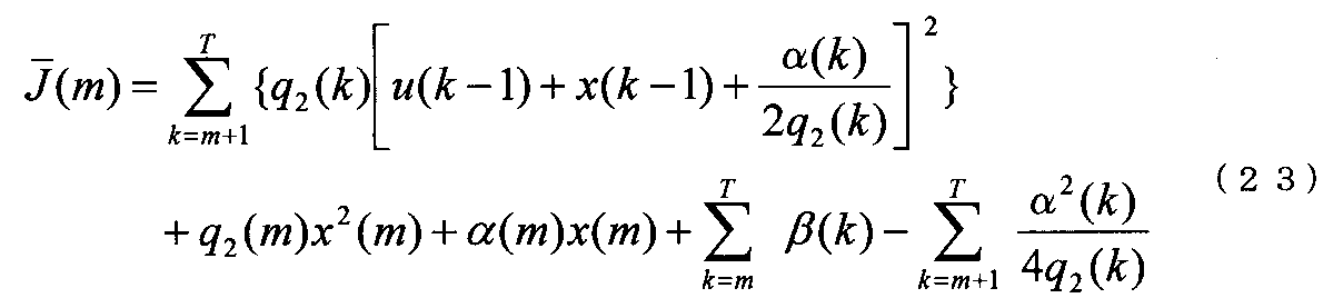


最適制御量を得るために必要な細胞数等の計測のための画像処理について説明する。
細胞2の運動モデルの同定として必要なのは、顕微鏡装置1の光軸方向への細胞2の移動速度の集団平均v0と、細胞位置の分散の時間変化Wiである。なお、被観察物2が静止物の場合は、移動速度の集団平均v0及び位置の分散の時間変化Wiを0として扱うことができる。 (Image processing)
Image processing for measuring the number of cells necessary to obtain the optimum control amount will be described.
What it is needed as the identification of the motion model of the
(i)細胞2が浮遊する試験溶液の観察領域を顕微鏡装置1の光軸方向とし、被観察物2を画像情報検出部16に設けたカメラ等で撮像する。これを観察画像1とする。この場合、観察画像1は、明視野像である。
(ii)(i)の観察画像よりも所定の距離だけ対物レンズ6に近い画像を撮影する。これを観察画像2とする。所定の距離としては、例えば1μmのオーダーとすることができる。
(iii)観察画像1と観察画像2との差分をとる。
(iv)画像上の差分値の二値化を行う。
(v)半径が所定の大きさの画素の円形マスクを使用して膨張処理を行い、同一細胞2の離れた画素を一つに纏める。
(vi) (v)の上記画素の円形マスクを使用して縮小処理し、細胞2以外の小さなノイズを除去する。
(vii)八近傍の画素を連結させてラベリングし、細胞数を計測する。
(viii)ラベリングした細胞2の大きさによってフィルタリングする。つまり、大きすぎれば二つと数え、手順(v)で別の細胞2を同一細胞2とみなした場合に対処する。小さすぎればノイズとみなし、細胞2以外の存在を消しきれなかった場合に対処する。 The transmitted light image processing can be performed by the following procedure.
(i) The observation region of the test solution in which the
(ii) An image closer to the
(iii) The difference between the
(iv) Binarize the difference values on the image.
(v) Dilation processing is performed using a circular mask of pixels having a predetermined radius, and the separated pixels of the
(vi) Reduction processing is performed using the circular mask of the pixel in (v), and small noise other than the
(vii) Connect and label the pixels in the vicinity of eight, and count the number of cells.
(viii) Filter by the size of the labeled
(1)パーソナルコンピュータ21が指示を出す。
(2)パーソナルコンピュータ21から指示を受けたD/Aボードが電圧を出力する。
(3)ステージ3を動かす。
(4)対物レンズ6を通して得た画像をカメラで取り込む。
(5)取り込んだ画像を、パーソナルコンピュータ21に送る。
(6)パーソナルコンピュータ21が画像処理を行う。
(7)画像処理した情報をもとに、パーソナルコンピュータ21がD/Aボードに指示を出す。 The above image processing can be performed by the following procedures (1) to (7) using the image
(1) The
(2) The D / A board receiving the instruction from the
(3) Move
(4) Capture an image obtained through the
(5) The captured image is sent to the
(6) The
(7) Based on the image processed information, the
図3は、本発明の第1実施形態に係る顕微鏡装置1を用いた蛍光観察手順を示すフロー図の一例である。
先ず、ステップST1において、被観察物2を所定の間隔として、d(μm)間隔(以下、d間隔と呼ぶ)で、観察領域内の明視野観察を行う。被観察物2は、例えば免疫細胞の一種であるT細胞である。 Next, fluorescence observation combining the above-described optimal control rule for fluorescence observation and image processing for measuring the number of cells necessary for application of the optimal control rule will be described.
FIG. 3 is an example of a flowchart showing a fluorescence observation procedure using the
First, in step ST1, bright field observation in an observation region is performed at d (μm) intervals (hereinafter referred to as d intervals) with the
ステップST3では、観察したい被観察物2の数に応じて蛍光観察領域を決定する。
ステップST4において、d間隔で、蛍光観察領域の蛍光観察と明視野観察を行う。透過光画像の情報を増すために、蛍光観察と比べて明視野観察の観察間隔を狭めたり、観察領域を広げてもよい。
ステップST5において、所定の時間、例えば20秒待機する。制御に用いる透過光の画像情報を増すために、蛍光観察と比べて明視野観察の待機時間を短くしてもよい。 In step ST2, the average position and variance of the
In step ST3, the fluorescence observation region is determined according to the number of
In step ST4, fluorescence observation and bright field observation of the fluorescence observation region are performed at intervals d. In order to increase the information of the transmitted light image, the observation interval of the bright field observation may be narrowed or the observation area may be expanded as compared with the fluorescence observation.
In step ST5, it waits for a predetermined time, for example, 20 seconds. In order to increase image information of transmitted light used for control, the standby time for bright field observation may be shortened as compared with fluorescence observation.
ステップST7において、被観察物2の運動モデルに従って蛍光観察領域の中心位置を決定する。 Next, in step ST6, the fluorescence observation region is determined according to the optimal control law.
In step ST7, the center position of the fluorescence observation region is determined according to the motion model of the object to be observed 2.
なお、本発明において、透過光画像を用いた蛍光観察の制御は、スケジューリング問題と一般化できる。このため、蛍光観察の制御には、上記した最適制御則以外に、PID制御等の古典制御、準最適制御等の現代制御、H∞制御,サンプル値制御,有限時間整定制御,適応制御等のポスト現代制御論、ニューラルネットワーク制御,ファジィ制御,遺伝的アルゴリズム制御等の知的制御を用いてもよい。 In the
In the present invention, control of fluorescence observation using a transmitted light image can be generalized as a scheduling problem. For this reason, in addition to the optimal control law described above, the control of fluorescence observation includes classical control such as PID control, modern control such as semi-optimal control, H∞ control, sample value control, finite time settling control, adaptive control, etc. Intelligent control such as post-modern control theory, neural network control, fuzzy control, genetic algorithm control may be used.
(第2実施形態)
図4は、第2実施形態に係る顕微鏡装置30の構成を示す模式図である。
顕微鏡装置30が顕微鏡装置1と異なるのは、対物レンズ6を透過光Tの光軸、即ちZ軸方向に移動できるように対物レンズ用駆動部32を設けた点にある。対物レンズ用駆動部32は、制御部20によって制御される。対物レンズ用駆動部32は、ピエゾ素子のような駆動部品を用いて構成することができる。他の構成は顕微鏡装置1と同じであるので説明は省略する。 Furthermore, the configuration of the
(Second Embodiment)
FIG. 4 is a schematic diagram illustrating a configuration of the
The
図5は、第3実施形態に係る顕微鏡装置35の構成を示す模式図である。
顕微鏡装置35が顕微鏡装置1と異なるのは、画像情報検出部16側の結像レンズ7を光軸方向(Z軸方向)に移動できるように透過光用の結像レンズ用駆動部37を設けた点にある。結像レンズ用駆動部37は、制御部20によって制御される。結像レンズ用駆動部37は、ピエゾ素子のような駆動部品を用いて構成することができる。他の構成は顕微鏡装置1と同じであるので、説明は省略する。 (Third embodiment)
FIG. 5 is a schematic diagram showing the configuration of the
The
図6は、第4実施形態に係る顕微鏡装置40の構成を示す模式図である。
顕微鏡装置40が顕微鏡装置1と異なるのは、蛍光画像情報検出部17の前に蛍光波長選択部25を設けた点にある。蛍光波長選択部25は、二つの蛍光波長を分離して検出できる構成を有している。蛍光波長選択部25は、第3のダイクロイックミラー26とミラー27とプリズム28とから構成されている。さらに、レンズ29を設けてもよい。この構成によれば、2波長の蛍光Fを分離して検知することができる。蛍光波長選択部25は、鏡筒部15又は蛍光画像情報検出部17に配置することができる。他の構成は顕微鏡装置1と同じであるので、説明は省略する。 (Fourth embodiment)
FIG. 6 is a schematic diagram illustrating a configuration of the
The
なお、複数種類の細胞2を同時に観察するときには、透過光画像に含まれる各細胞2の面積,形状,色の濃度,色調,テクスチャ等を用いて、細胞2の種類を画像処理で自動判別することも可能である(非特許文献7参照)。 According to the
When observing a plurality of types of
図7は、第5実施形態の顕微鏡装置45の構成を示す模式図である。
顕微鏡装置45が顕微鏡装置1と異なるのは、蛍光画像情報検出部17の光軸方向に挿入された結像レンズ12を光軸方向に移動させるように、結像レンズ用駆動部52を設けている点にある。他の構成は顕微鏡装置1と同じであるので、説明は省略する。 (Fifth embodiment)
FIG. 7 is a schematic diagram showing the configuration of the
The
図8は、第6実施形態に係る顕微鏡装置50の構成を示す模式図である。
顕微鏡装置50が顕微鏡装置1と異なるのは、光学系10中に、さらに2つのピンホール53,54を設けて共焦点光学系で構成した点にある。第1のピンホール53は、励起用光源5とフィルタ9との間の光軸上に配設されている。第1のピンホール53とフィルタ9の配置順は入れ替えてもよい。第2のピンホール54は、蛍光画像情報検出部17と結像レンズ12との間の蛍光Fの光軸上に配設されている。第2のピンホール54と結像レンズ12との配置順は入れ替えてもよい。他の構成は顕微鏡装置1と同じであるので、説明は省略する。 (Sixth embodiment)
FIG. 8 is a schematic diagram showing a configuration of a
The
図9は、第7実施形態の顕微鏡装置55の構成を示す模式図である。
顕微鏡装置55が顕微鏡装置50と異なるのは、さらに、第2のピンホール54を蛍光Fの光軸方向に移動及び/又は回転させるように第2のピンホール54を駆動するピンホール駆動部56を設けている点にある。他の構成は顕微鏡装置1と同じであるので、説明は省略する。 (Seventh embodiment)
FIG. 9 is a schematic diagram showing the configuration of the
The
図10は、第8実施形態の顕微鏡装置60の構成を示す模式図である。
顕微鏡装置60が顕微鏡装置1と異なるのは、被観察物2の下方から透過光Tが照射される、所謂正立型の光学系10Aを有している点である。他の構成は顕微鏡装置1と同じであるので、説明は省略する。 (Eighth embodiment)
FIG. 10 is a schematic diagram showing the configuration of the
The
被観察物2の環境制御について説明する。
図11は環境制御部60の構成を示す模式図であり、(A)は平面図、(B)は(A)のX-Xに沿った断面図である。環境制御部60は、本体部62と、本体部62の下部に配置され被観察物2を収容する収容部64と、を含んで構成されている。
本体部62は、樹脂板65をスライドガラス66と樹脂フィルム67とで挟んで構成され、具体的には本体部62は、収容部64に対応する開口65Aを有している長方形の樹脂板65と、樹脂板65の上部に配設され樹脂板65を覆うスライドガラス66と、樹脂板65の下部に配設され樹脂板65を覆う樹脂フィルム67と、を備えている。さらに、本体部62には、樹脂板65の開口部65Aに接続するガス配管68が設けられている。ガス配管68の入力側68Aから出力側68Bへ雰囲気ガス69が導入される。雰囲気ガス69としては、窒素ガス(単体)、酸素ガス(単体)、二酸化炭素(単体)、空気(混合物)、およびそれらの混合ガス等を挙げることができる。
ここで、樹脂板65は、例えばアクリル樹脂を用いることができる。樹脂フィルム67は、マイラーからなるフィルム等を用いることができる。さらに、樹脂板65、スライドガラス66及び樹脂フィルム67を保持する保持手段70を備えていてもよい。 (Environmental control of the object to be observed)
The environmental control of the
11A and 11B are schematic views showing the configuration of the
The
Here, for the
図12は複数の被観察物2の観察ができる環境制御部80の構成を示す模式図で、(A)は平面図、(B)は(A)のX-Xに沿った断面図である。
環境制御部80が環境制御部60と異なるのは、収容部84が一体ではなく、格子状の障壁部85によって複数の収容区画86を有するように仕切られている点である。各収容区画86には培養液74が注入されている。他の構成は環境制御部60と同じであるので、説明は省略する。 The
12A and 12B are schematic views showing the configuration of the
The
図4に示す顕微鏡装置30を製作した。光学系10としては、倒立顕微鏡(オリンパス株式会社製、IX71)を使用し、照明用光源4及び励起用光源5は、それぞれ、ハロゲンランプとキセノンランプを使用した。対物レンズ6(オリンパス株式会社製、UApo40×3/340)は40倍のものを用いた。 Hereinafter, the present invention will be described in detail with reference to examples.
A
図14から明らかなように、制御量U(k)を用いた実施例の評価関数Jと蛍光観察領域幅を固定した比較例の評価関数Jを比較すると、本発明の制御量を用いた実施例の評価関数Jの値が常に比較例のJの値よりも小さくなっており、最適化されていることが分かる。 FIG. 14 is a schematic diagram illustrating an evaluation function J of the example using the control amount U (k) and a evaluation function J of the comparative example when the fluorescence observation region width is fixed in the fluorescence observation procedure. The horizontal axis of FIG. 14 indicates the width (μm) of the fluorescence observation region when fixed, and the vertical axis indicates the value of the evaluation function J. The width of the fluorescent region when the control amount of the present invention is used is dynamically changed.
As is apparent from FIG. 14, when the evaluation function J of the example using the control amount U (k) is compared with the evaluation function J of the comparative example in which the fluorescence observation region width is fixed, the implementation using the control amount of the present invention. It can be seen that the value of the evaluation function J of the example is always smaller than the value of J of the comparative example and is optimized.
蛍光観察領域の中心は、被観察物の画像情報に基づいて、或いは当該画像情報と被観察物の運動モデルとに基づいて、求めても良い。
ステージの制御は、被観察物の運動モデルに基づいて、或いは当該運動モデルと上記画像情報とに基づいて行っても良い。
結像レンズ7、結像レンズ12の倍率を調整すること、例えば倍率を低くすることで、画像情報検出部16,蛍光画像情報検出部17で取得する画像中の被観察物の大きさを小さくすることができる。これによって、観察視野が広がり、より多数の被観察物を同時に観察することができる。そして、被観察物を表す画素数が少なくなり、制御部20における細胞一つ当たりの画像処理時間を一層短縮することもできる。 The present invention is not limited to the above-described embodiments, and various modifications are possible within the scope of the invention described in the claims, and it goes without saying that these are also included in the scope of the present invention. . In the above-described embodiment, the image pickup device is used for the first and second detection
The center of the fluorescence observation region may be obtained based on the image information of the object to be observed, or based on the image information and the motion model of the object to be observed.
The stage may be controlled based on the motion model of the object to be observed, or based on the motion model and the image information.
By adjusting the magnification of the
2:被観察物
3:XYまたはXYZステージ
4:照明用光源
5:励起用光源
5A:励起光
6:対物レンズ
7:結像レンズ(透過光用)
8,9:フィルタ
10,10A:光学系
12:結像レンズ(蛍光用)
15:鏡筒部
16:画像情報検出部
17:蛍光画像情報検出部
18,19:第1及び第2のビームスプリッター(ダイクロイックミラー)
20:制御部
21:パーソナルコンピュータ
22:ディスプレー装置
23:励起用光源制御部
25:蛍光波長選択部
26:第3のダイクロイックミラー
27:ミラー
28:プリズム
29:レンズ
32:対物レンズ用駆動部
37:透過光の結像レンズ用駆動部
52:蛍光用結像レンズ用駆動部
53:第1のピンホール
54:第2のピンホール
56,58:ピンホール駆動部
60,80:環境制御部
62:本体部
64,84:収容部
65:樹脂板
66:スライドガラス
67:樹脂フィルム
67A:細孔
68:ガス配管
68A:入力側
68B:出力側
69:雰囲気ガス
70:保持手段
71,72:カバーガラス
74:培養液
85:障壁部
86:収容区画
F:蛍光
T:透過光 1, 30, 35, 40, 45, 50, 55, 60: Microscope device 2: Object to be observed 3: XY or XYZ stage 4: Light source for illumination 5: Light source for
8, 9:
15: Lens unit 16: Image information detection unit 17: Fluorescence image
20: control unit 21: personal computer 22: display device 23: excitation light source control unit 25: fluorescence wavelength selection unit 26: third dichroic mirror 27: mirror 28: prism 29: lens 32: objective lens drive unit 37: Transmitted light imaging lens driving unit 52: Fluorescent imaging lens driving unit 53: First pinhole 54:
Claims (22)
- 被観察物を載置するステージと、
上記被観察物に照明光を照射する第1の光源と、
上記被観察物に蛍光を励起するための励起光を照射する第2の光源と、
上記被観察物で生じた光による画像情報を検出する画像情報検出部と、
上記被観察物で生じた蛍光による蛍光画像情報を検出する蛍光画像情報検出部と、
上記被観察物の運動モデルと上記画像情報検出部から入力された上記被観察物の画像情報とに基づいて上記被観察物の蛍光観察領域を決定し、上記画像情報検出部から入力された上記被観察物の画像情報と上記蛍光画像情報検出部から入力された蛍光画像情報とを上記蛍光観察領域内の所定の間隔毎に取得する制御部と、
を備えたことを特徴とする、顕微鏡装置。 A stage for placing an object to be observed;
A first light source for illuminating the observation object with illumination light;
A second light source for irradiating the observation object with excitation light for exciting fluorescence;
An image information detection unit for detecting image information by light generated in the observation object;
A fluorescence image information detection unit for detecting fluorescence image information due to fluorescence generated in the observation object;
A fluorescence observation area of the observation object is determined based on the motion model of the observation object and the image information of the observation object input from the image information detection unit, and the input of the image information detection unit A control unit that obtains image information of an object to be observed and fluorescence image information input from the fluorescence image information detection unit at predetermined intervals in the fluorescence observation region;
A microscope apparatus comprising: - 前記制御部は、前記蛍光観察領域を最適制御,準最適制御等の現代制御に従って決定することを特徴とする、請求項1に記載の顕微鏡装置。 2. The microscope apparatus according to claim 1, wherein the control unit determines the fluorescence observation region according to modern control such as optimal control and semi-optimal control.
- 前記制御部は、前記被観察物の運動モデルと前記被観察物の画像情報との少なくも一方に従って前記蛍光観察領域の中心位置を決定することを特徴とする、請求項1に記載の顕微鏡装置。 The microscope apparatus according to claim 1, wherein the control unit determines a center position of the fluorescence observation region according to at least one of a motion model of the object to be observed and image information of the object to be observed. .
- 前記制御部は、前記被観察物の運動モデルと前記被観察物の画像情報との少なくとも一方により、上記被観察物を追尾するように上記ステージを制御すると共に、上記被観察物の蛍光情報の取得を制御することを特徴とする、請求項1に記載の顕微鏡装置。 The control unit controls the stage so as to track the object to be observed by at least one of the motion model of the object to be observed and the image information of the object to be observed, and the fluorescence information of the object to be observed. The microscope apparatus according to claim 1, wherein acquisition is controlled.
- 前記制御部は、前記画像情報による比例,積分,微分の何れか又はこれらの組み合わせによるPID制御等の古典制御により前記ステージを制御することを特徴とする、請求項1に記載の顕微鏡装置。 The microscope apparatus according to claim 1, wherein the control unit controls the stage by classical control such as PID control based on any one of proportionality, integration, and differentiation based on the image information, or a combination thereof.
- 前記制御部は、前記第1の光源を制御するための第1光源制御部と前記第2の光源を制御するための第2光源制御部とを備えていることを特徴とする、請求項1に記載の顕微鏡装置。 The said control part is provided with the 1st light source control part for controlling a said 1st light source, and the 2nd light source control part for controlling a said 2nd light source, It is characterized by the above-mentioned. The microscope apparatus described in 1.
- 前記ステージは、前記被観察物の位置を移動させる二次元または三次元ステージであることを特徴とする、請求項1に記載の顕微鏡装置。 2. The microscope apparatus according to claim 1, wherein the stage is a two-dimensional or three-dimensional stage that moves a position of the object to be observed.
- 前記蛍光画像情報検出部は、少なくとも1波長以上の蛍光を分離する波長選択手段を備えたことを特徴とする、請求項1に記載の顕微鏡装置。 2. The microscope apparatus according to claim 1, wherein the fluorescence image information detection unit includes wavelength selection means for separating fluorescence of at least one wavelength.
- 前記第2の光源と前記被観察物との間に配設される第1のピンホールと、前記蛍光と前記蛍光画像情報検出部との間に配設される第2のピンホールと、を備えたことを特徴とする、請求項1に記載の顕微鏡装置。 A first pinhole disposed between the second light source and the object to be observed, and a second pinhole disposed between the fluorescence and the fluorescence image information detection unit. The microscope apparatus according to claim 1, further comprising a microscope apparatus.
- 前記第1のピンホール又は前記第2のピンホールを移動及び/又は回転させるピンホール駆動部を、備えたことを特徴とする、請求項9に記載の顕微鏡装置。 10. The microscope apparatus according to claim 9, further comprising a pinhole driving unit that moves and / or rotates the first pinhole or the second pinhole.
- 前記第1の光源と前記被観察物との間に配設される対物レンズと、該対物レンズの駆動部と、を備えたことを特徴とする、請求項1に記載の顕微鏡装置。 2. The microscope apparatus according to claim 1, further comprising: an objective lens disposed between the first light source and the object to be observed; and a drive unit for the objective lens.
- 前記被観察物で生じた光と前記画像情報検出部との間に配設される結像レンズと、該結像レンズの駆動部と、を備えたことを特徴とする、請求項1に記載の顕微鏡装置。 The image forming lens disposed between the light generated by the object to be observed and the image information detecting unit, and a driving unit for the image forming lens, and the image forming lens driving unit according to claim 1. Microscope equipment.
- 前記蛍光と前記蛍光画像情報検出部との間に配設される結像レンズと、該結像レンズの駆動部と、を備えたことを特徴とする、請求項1に記載の顕微鏡装置。 2. The microscope apparatus according to claim 1, further comprising: an imaging lens disposed between the fluorescence and the fluorescence image information detection unit, and a driving unit for the imaging lens.
- 前記被観察物を収容しかつ雰囲気ガスで満たされる環境制御部を備えたことを特徴とする、請求項13記載の顕微鏡装置。 14. The microscope apparatus according to claim 13, further comprising an environment control unit that accommodates the object to be observed and is filled with an atmospheric gas.
- 前記環境制御部は、複数の前記被観察物を収容することができる収容部を備えたことを特徴とする、請求項14に記載の顕微鏡装置。 The microscope apparatus according to claim 14, wherein the environment control unit includes a storage unit that can store a plurality of objects to be observed.
- 前記被観察物を刺激する被観察物刺激手段を備えたことを特徴とする、請求項1に記載の顕微鏡装置。 2. The microscope apparatus according to claim 1, further comprising an object stimulating means for stimulating the object to be observed.
- 被観察物の画像情報と該被観察物の蛍光画像情報とを取得する蛍光観察方法であって、
上記被観察物の画像情報と上記被観察物の運動モデルとに基づいて上記被観察物の蛍光観察領域を決定する第1段階と、
上記蛍光観察領域内の所定の位置で上記蛍光画像情報を取得する第2段階と、
を備えたことを特徴とする、被観察物の蛍光観察方法。 A fluorescence observation method for acquiring image information of an observation object and fluorescence image information of the observation object,
A first step of determining a fluorescence observation region of the observation object based on the image information of the observation object and a motion model of the observation object;
A second stage of acquiring the fluorescence image information at a predetermined position in the fluorescence observation region;
A method for observing fluorescence of an object to be observed. - 前記第1段階の蛍光観察領域を、PID制御等の古典制御、最適制御,準最適制御等の現代制御に従って決定することを特徴とする、請求項17に記載の被観察物の蛍光観察方法。 18. The fluorescence observation method for an observation object according to claim 17, wherein the first-stage fluorescence observation region is determined according to modern control such as classical control such as PID control, optimal control, and suboptimal control.
- 前記第1段階と前記第2段階との間において、前記被観察物の画像情報と前記被観察物の運動モデルとの少なくとも一方に基づいて前記蛍光観察領域の中心位置を決定することを特徴とする、請求項17に記載の被観察物の蛍光観察方法。 A center position of the fluorescence observation region is determined between the first stage and the second stage based on at least one of image information of the object to be observed and a motion model of the object to be observed. The fluorescence observation method for an observation object according to claim 17.
- 前記第2段階において、前記被観察物の画像情報を取得することを特徴とする、請求項17に記載の被観察物の蛍光観察方法。 18. The method for observing fluorescence of an observation object according to claim 17, wherein image information of the observation object is acquired in the second stage.
- 前記各段階を、前記蛍光観察領域内の所定の位置毎に所定の回数繰り返し行うことを特徴とする、請求項17~20の何れかに記載の被観察物の蛍光観察方法。 21. The fluorescence observation method for an object to be observed according to any one of claims 17 to 20, wherein each of the steps is repeatedly performed a predetermined number of times for each predetermined position in the fluorescence observation region.
- 前記運動モデルのパラメータは、前記被観察物の位置,速度,分布,種類,形状,イオン濃度,分子濃度の何れか又はこれらの組み合わせからなることを特徴とする、請求項17に記載の被観察物の蛍光観察方法。 18. The observed object according to claim 17, wherein the parameters of the motion model are any one of a position, a speed, a distribution, a type, a shape, an ion concentration, a molecular concentration of the object to be observed, or a combination thereof. Fluorescence observation method for objects.
Priority Applications (3)
| Application Number | Priority Date | Filing Date | Title |
|---|---|---|---|
| US13/061,705 US9019360B2 (en) | 2008-09-13 | 2009-06-10 | Microscope and a fluorescent observation method using the same |
| CN200980145455.XA CN102216827B (en) | 2008-09-13 | 2009-06-10 | Microscope device and fluorescent observing method using same |
| EP09812947.1A EP2325684B1 (en) | 2008-09-13 | 2009-06-10 | Microscope device and fluorescent observing method using same |
Applications Claiming Priority (2)
| Application Number | Priority Date | Filing Date | Title |
|---|---|---|---|
| JP2008235795A JP4288323B1 (en) | 2008-09-13 | 2008-09-13 | Microscope device and fluorescence observation method using the same |
| JP2008-235795 | 2008-09-13 |
Publications (1)
| Publication Number | Publication Date |
|---|---|
| WO2010029799A1 true WO2010029799A1 (en) | 2010-03-18 |
Family
ID=40921821
Family Applications (1)
| Application Number | Title | Priority Date | Filing Date |
|---|---|---|---|
| PCT/JP2009/060644 WO2010029799A1 (en) | 2008-09-13 | 2009-06-10 | Microscope device and fluorescent observing method using same |
Country Status (6)
| Country | Link |
|---|---|
| US (1) | US9019360B2 (en) |
| EP (1) | EP2325684B1 (en) |
| JP (1) | JP4288323B1 (en) |
| CN (1) | CN102216827B (en) |
| TW (1) | TWI401472B (en) |
| WO (1) | WO2010029799A1 (en) |
Cited By (3)
| Publication number | Priority date | Publication date | Assignee | Title |
|---|---|---|---|---|
| CN103210337A (en) * | 2010-09-14 | 2013-07-17 | Qbc诊断股份有限公司 | Adaptor for microscopes |
| WO2015022851A1 (en) * | 2013-08-15 | 2015-02-19 | 富士通株式会社 | Measurement device using optical interferometry and measurement method using optical interferometry |
| EP2607889A4 (en) * | 2010-08-18 | 2016-11-09 | Nanoentek Inc | Fluorescence microscope for multi-fluorescence image observation, fluorescence image observation method using the same, and multi-fluorescence image observation system |
Families Citing this family (35)
| Publication number | Priority date | Publication date | Assignee | Title |
|---|---|---|---|---|
| JP4773348B2 (en) | 2003-07-12 | 2011-09-14 | アクセラー8 テクノロジー コーポレイション | Sensitive and rapid biodetection method |
| JP4614149B2 (en) * | 2009-06-26 | 2011-01-19 | 独立行政法人科学技術振興機構 | Microscope device and contour identification program |
| CN102667473B (en) * | 2009-10-12 | 2016-06-08 | 文塔纳医疗系统公司 | The pathology of the enhancing for organizing measure and multi-modal contrast and the light field background of multiple analyte detection reproduce |
| JP5638793B2 (en) | 2009-12-03 | 2014-12-10 | オリンパス株式会社 | Microscope equipment |
| US9946058B2 (en) * | 2010-06-11 | 2018-04-17 | Nikon Corporation | Microscope apparatus and observation method |
| WO2012039636A2 (en) | 2010-09-24 | 2012-03-29 | Christian Soeller | "3d localisation microscopy and 4d localisation microscopy and tracking methods and systems" |
| ES2551922T3 (en) | 2011-03-07 | 2015-11-24 | Accelerate Diagnostics, Inc. | Rapid cell purification systems |
| US10254204B2 (en) | 2011-03-07 | 2019-04-09 | Accelerate Diagnostics, Inc. | Membrane-assisted purification |
| US8409052B2 (en) * | 2011-04-29 | 2013-04-02 | Delta Electronics, Inc. | Starting method for hybrid electric vehicle and system architecture of hybrid electric vehicle |
| JP5220172B2 (en) * | 2011-08-22 | 2013-06-26 | キヤノン株式会社 | Image acquisition device, image acquisition system, and objective optical system |
| US9599805B2 (en) | 2011-10-19 | 2017-03-21 | National Synchrotron Radiation Research Center | Optical imaging system using structured illumination |
| JP2014056078A (en) * | 2012-09-12 | 2014-03-27 | Canon Inc | Image acquisition device, image acquisition system, and microscope device |
| CN103048300A (en) * | 2012-12-17 | 2013-04-17 | 江苏大学 | Confocal laser scanning microscope |
| EP2972550A1 (en) * | 2013-03-11 | 2016-01-20 | Roche Diagnostics Hematology, Inc. | Imaging blood cells |
| US9677109B2 (en) | 2013-03-15 | 2017-06-13 | Accelerate Diagnostics, Inc. | Rapid determination of microbial growth and antimicrobial susceptibility |
| JP2014219623A (en) * | 2013-05-10 | 2014-11-20 | ソニー株式会社 | Observation system, observation program, and observation method |
| CN104083146B (en) * | 2013-06-25 | 2016-03-16 | 北京大学 | A kind of biological neural loop living imaging system |
| US10481075B2 (en) * | 2013-12-16 | 2019-11-19 | Sobru Solutions, Inc. | Microorganism evaluation system |
| CN103941752B (en) * | 2014-03-27 | 2016-10-12 | 北京大学 | A kind of nematicide real-time automatic tracing imaging system |
| KR102387134B1 (en) * | 2014-05-07 | 2022-04-15 | 일렉트로 사이언티픽 인더스트리즈, 아이엔씨 | Five axis optical inspection system |
| CN104375268A (en) * | 2014-11-26 | 2015-02-25 | 宁波江丰生物信息技术有限公司 | Optical structure capable of carrying out bright field scanning and fluorescence scanning |
| KR101734094B1 (en) * | 2015-03-09 | 2017-05-11 | 국립암센터 | Augmented Reality Image Projection System |
| KR20170132856A (en) * | 2015-03-30 | 2017-12-04 | 액셀러레이트 다이어그노스틱스, 아이엔씨. | Instruments and systems for rapid microbiological identification and antimicrobial susceptibility testing |
| US10253355B2 (en) | 2015-03-30 | 2019-04-09 | Accelerate Diagnostics, Inc. | Instrument and system for rapid microorganism identification and antimicrobial agent susceptibility testing |
| JP6613063B2 (en) * | 2015-07-07 | 2019-11-27 | 大塚電子株式会社 | Optical property measurement system |
| CN105301794B (en) * | 2015-10-30 | 2017-08-25 | 中国科学院遗传与发育生物学研究所 | Super-resolution imaging device for fast moving objects |
| JP6293186B2 (en) * | 2016-03-10 | 2018-03-14 | 株式会社Screenホールディングス | Imaging arrangement determining method in imaging apparatus and imaging apparatus |
| JPWO2018008136A1 (en) * | 2016-07-07 | 2019-04-18 | オリンパス株式会社 | Image processing apparatus and operation method of image processing apparatus |
| KR101838818B1 (en) * | 2016-11-30 | 2018-03-14 | 세메스 주식회사 | Fluorescent microscope and substrate inspection apparatus including the same |
| DE102017122413A1 (en) * | 2017-09-27 | 2019-03-28 | Abberior Instruments Gmbh | Device for selectively shaping phase fronts of a light beam and their use |
| TWI699816B (en) * | 2017-12-26 | 2020-07-21 | 雲象科技股份有限公司 | Method for controlling autonomous microscope system, microscope system, and computer readable storage medium |
| US10928617B1 (en) * | 2018-05-18 | 2021-02-23 | GDH Enterprises, LLC | Portable three-dimensional virtual imaging device |
| KR102166495B1 (en) * | 2019-05-27 | 2020-10-15 | 김은근 | Delta-bot type motorized microscope stage |
| CN110609026B (en) * | 2019-10-31 | 2020-11-03 | 福州大学 | Laser power feedback control up-conversion fluorescence imaging device and imaging method thereof |
| US11947099B1 (en) * | 2023-07-25 | 2024-04-02 | Pramana Inc. | Apparatus and methods for real-time image generation |
Citations (12)
| Publication number | Priority date | Publication date | Assignee | Title |
|---|---|---|---|---|
| JPH0580255A (en) | 1991-09-19 | 1993-04-02 | Nippon Telegr & Teleph Corp <Ntt> | Optical microscopic system device |
| JPH07253548A (en) | 1994-03-15 | 1995-10-03 | Nikon Corp | Device and method for automatically pursuing sample image |
| JPH07261097A (en) | 1994-03-17 | 1995-10-13 | Fujitsu Ltd | Microscope and image acquiring method |
| JP2000292422A (en) * | 1999-04-02 | 2000-10-20 | Olympus Optical Co Ltd | Scanning site meter |
| WO2003102636A1 (en) | 2002-06-04 | 2003-12-11 | Masatoshi Ishikawa | Variable-focus lens and lens controller |
| JP2005128086A (en) * | 2003-10-21 | 2005-05-19 | Olympus Corp | Scanning type microscope system |
| JP2005128493A (en) * | 2003-09-29 | 2005-05-19 | Olympus Corp | Microscope system |
| JP2005214924A (en) | 2004-02-02 | 2005-08-11 | Olympus Corp | Measuring method and measuring instrument |
| JP2006209698A (en) * | 2005-01-31 | 2006-08-10 | Olympus Corp | Target tracking device, microscope system and target tracking program |
| JP2006292420A (en) | 2005-04-06 | 2006-10-26 | Olympus Corp | Microscopic observation device having automatic tracking function of specimen sample |
| JP2007114130A (en) * | 2005-10-24 | 2007-05-10 | Tohoku Univ | Position analyzing method and position analyzer |
| JP2008242014A (en) * | 2007-03-27 | 2008-10-09 | V Technology Co Ltd | Fluorescence microscope |
Family Cites Families (14)
| Publication number | Priority date | Publication date | Assignee | Title |
|---|---|---|---|---|
| US6548796B1 (en) | 1999-06-23 | 2003-04-15 | Regents Of The University Of Minnesota | Confocal macroscope |
| JP4519987B2 (en) * | 2000-04-13 | 2010-08-04 | オリンパス株式会社 | Focus detection device |
| KR100530818B1 (en) * | 2001-05-11 | 2005-11-25 | 미쓰비시덴키 가부시키가이샤 | Method and device for laser beam machining of laminated material |
| EP1461592B1 (en) | 2001-12-05 | 2019-04-10 | The J. David Gladstone Institutes | Robotic microscopy systems |
| US20040241832A1 (en) * | 2003-06-02 | 2004-12-02 | Olympus Corporation | Cell culture detection apparatus, cell culture observation apparatus, and cell culture observation method |
| US7345814B2 (en) | 2003-09-29 | 2008-03-18 | Olympus Corporation | Microscope system and microscope focus maintaining device for the same |
| US7355692B2 (en) | 2004-03-05 | 2008-04-08 | Orbotech Ltd | System and method for inspecting electrical circuits utilizing reflective and fluorescent imagery |
| JP2005284136A (en) * | 2004-03-30 | 2005-10-13 | Olympus Corp | Observation device and focusing method for observation device |
| JP4673000B2 (en) * | 2004-05-21 | 2011-04-20 | 株式会社キーエンス | Fluorescence microscope, display method using fluorescence microscope apparatus, fluorescence microscope image display program, computer-readable recording medium, and stored device |
| US20080226126A1 (en) | 2005-01-31 | 2008-09-18 | Yoshinori Ohno | Object-Tracking Apparatus, Microscope System, and Object-Tracking Program |
| TWM290505U (en) * | 2005-11-30 | 2006-05-11 | Da-Ren Ji | Automatic device used in nucleus manipulation |
| TWI325494B (en) * | 2006-07-28 | 2010-06-01 | Ind Tech Res Inst | Optical measuring system |
| US7498551B2 (en) * | 2006-10-06 | 2009-03-03 | Los Alamos National Security, Llc | Apparatus and method for tracking a molecule or particle in three dimensions |
| US8143600B2 (en) * | 2008-02-18 | 2012-03-27 | Visiongate, Inc. | 3D imaging of live cells with ultraviolet radiation |
-
2008
- 2008-09-13 JP JP2008235795A patent/JP4288323B1/en not_active Expired - Fee Related
-
2009
- 2009-06-10 WO PCT/JP2009/060644 patent/WO2010029799A1/en active Application Filing
- 2009-06-10 CN CN200980145455.XA patent/CN102216827B/en not_active Expired - Fee Related
- 2009-06-10 US US13/061,705 patent/US9019360B2/en not_active Expired - Fee Related
- 2009-06-10 EP EP09812947.1A patent/EP2325684B1/en not_active Not-in-force
- 2009-06-11 TW TW98119500A patent/TWI401472B/en not_active IP Right Cessation
Patent Citations (12)
| Publication number | Priority date | Publication date | Assignee | Title |
|---|---|---|---|---|
| JPH0580255A (en) | 1991-09-19 | 1993-04-02 | Nippon Telegr & Teleph Corp <Ntt> | Optical microscopic system device |
| JPH07253548A (en) | 1994-03-15 | 1995-10-03 | Nikon Corp | Device and method for automatically pursuing sample image |
| JPH07261097A (en) | 1994-03-17 | 1995-10-13 | Fujitsu Ltd | Microscope and image acquiring method |
| JP2000292422A (en) * | 1999-04-02 | 2000-10-20 | Olympus Optical Co Ltd | Scanning site meter |
| WO2003102636A1 (en) | 2002-06-04 | 2003-12-11 | Masatoshi Ishikawa | Variable-focus lens and lens controller |
| JP2005128493A (en) * | 2003-09-29 | 2005-05-19 | Olympus Corp | Microscope system |
| JP2005128086A (en) * | 2003-10-21 | 2005-05-19 | Olympus Corp | Scanning type microscope system |
| JP2005214924A (en) | 2004-02-02 | 2005-08-11 | Olympus Corp | Measuring method and measuring instrument |
| JP2006209698A (en) * | 2005-01-31 | 2006-08-10 | Olympus Corp | Target tracking device, microscope system and target tracking program |
| JP2006292420A (en) | 2005-04-06 | 2006-10-26 | Olympus Corp | Microscopic observation device having automatic tracking function of specimen sample |
| JP2007114130A (en) * | 2005-10-24 | 2007-05-10 | Tohoku Univ | Position analyzing method and position analyzer |
| JP2008242014A (en) * | 2007-03-27 | 2008-10-09 | V Technology Co Ltd | Fluorescence microscope |
Non-Patent Citations (10)
| Title |
|---|
| D. M. DAVIS: "Intrigue at the Immune Synapse", NIKKEI SCIENCE, 2006, pages 52 - 60 |
| H. OKU ET AL.: "Two-dimensional tracking of a motile micro-oraganism allowing high-resolution observation with various imaging techniques", REVIEW OF SCIENTIFIC INSTRUMENTS, vol. 76, no. ISS.3, 2 February 2005 (2005-02-02), pages 034301-1 - 034301-9, XP012079269 * |
| H. OKU, 1. ISHII, M. ISHIKAWA: "Microscopic visual feedback system", THE TRANSACTIONS OF THE INSTITUTE OF ELECTRONICS, INFORMATION AND COMMUNICATION ENGINEERS, 2001, pages 994 - 1002 |
| H. OKU, M. ISHIKAWA, THEODORUS, K. HASHIMOTO: "High-speed autofocusing of a cell using diffraction patterns", OPTICS EXPRESS, vol. 14, no. 9, 2006, pages 3952 - 3960, XP008145357, DOI: doi:10.1364/OE.14.003952 |
| H. OKU, M. ISHIKAWA: "Structure of Dynamic Focusing Lens with kHz Order Response for High-Speed Vision System", KOGAKU, vol. 31, no. 10, 2002, pages 758 - 764 |
| H. OKU, N. OGAWA, ISHIKAWA, K. HASHIMOTO: "Two-dimensional tracking of a motile micro-organism allowing high-resolution observation with various imaging techniques", REVIEW OF SCIENTIFIC INSTRUMENTS, vol. 76, no. 3, 2005, XP012079269, DOI: doi:10.1063/1.1857632 |
| HIROMASA OKU ET AL.: "High-speed autofocusing of a cell using diffraction patterns", OPTICS EXPRESS, vol. 14, no. ISS.14, 1 May 2006 (2006-05-01), pages 3952 - 3960, XP008145357 * |
| K. HASHIMOTO: "Active sensing using microorganisms", SORST JOINT SYMPOSIUM (6), LECTURE ABSTRACTS, 2007, pages 23 - 26 |
| See also references of EP2325684A4 * |
| Y. TANIMURA: "Automated classification of white blood cells", MEDICAL IMAGIONG TECHNOLOGY, vol. 14, no. 1, 1997, pages 14 - 22 |
Cited By (4)
| Publication number | Priority date | Publication date | Assignee | Title |
|---|---|---|---|---|
| EP2607889A4 (en) * | 2010-08-18 | 2016-11-09 | Nanoentek Inc | Fluorescence microscope for multi-fluorescence image observation, fluorescence image observation method using the same, and multi-fluorescence image observation system |
| CN103210337A (en) * | 2010-09-14 | 2013-07-17 | Qbc诊断股份有限公司 | Adaptor for microscopes |
| WO2015022851A1 (en) * | 2013-08-15 | 2015-02-19 | 富士通株式会社 | Measurement device using optical interferometry and measurement method using optical interferometry |
| JPWO2015022851A1 (en) * | 2013-08-15 | 2017-03-02 | 富士通株式会社 | Measuring apparatus using optical interferometry and measuring method using optical interferometry |
Also Published As
| Publication number | Publication date |
|---|---|
| TW201011341A (en) | 2010-03-16 |
| US9019360B2 (en) | 2015-04-28 |
| CN102216827B (en) | 2015-07-22 |
| JP4288323B1 (en) | 2009-07-01 |
| EP2325684A1 (en) | 2011-05-25 |
| EP2325684B1 (en) | 2017-02-01 |
| JP2014098724A (en) | 2014-05-29 |
| EP2325684A4 (en) | 2014-04-23 |
| CN102216827A (en) | 2011-10-12 |
| US20110242308A1 (en) | 2011-10-06 |
| TWI401472B (en) | 2013-07-11 |
Similar Documents
| Publication | Publication Date | Title |
|---|---|---|
| JP4288323B1 (en) | Microscope device and fluorescence observation method using the same | |
| JP5307539B2 (en) | Biological sample imaging method and biological sample imaging apparatus | |
| JP5289768B2 (en) | Focus position determination method, focus position determination apparatus, faint light detection apparatus, and faint light detection method | |
| Ettinger et al. | Fluorescence live cell imaging | |
| Petty | Fluorescence microscopy: established and emerging methods, experimental strategies, and applications in immunology | |
| US6980293B1 (en) | Immersion medium supply apparatus, fluorescence spectrometry inspection apparatus, and culture microscope | |
| JP6513802B2 (en) | Laser light coupling for nanoparticle detection | |
| WO2009081832A1 (en) | Image processing method for time lapse image, image processing program, and image processing device | |
| WO2007074929A1 (en) | Device and method for capturing image of a sample originating from organism | |
| CA2322800A1 (en) | Color translating uv microscope | |
| US20020106715A1 (en) | System and method for collecting data from individual cells | |
| Waters | Live-cell fluorescence imaging | |
| JP4474655B2 (en) | Microscope equipment | |
| JP4106626B2 (en) | Fluorescence image measuring method and apparatus | |
| US20110121199A1 (en) | Imaging apparatus for low-light sample | |
| CN116802463A (en) | Universal multi-functional detection system for microwell plates with confocal imaging | |
| US10198659B1 (en) | Diagnostics and imaging | |
| JP4544893B2 (en) | Optical apparatus and image creation method | |
| Bharadwaj | Unedited version published online on 27/10/2023 | |
| Bharadwaj et al. | A cost-effective, modular, research-grade optical microscope | |
| WO2023161375A1 (en) | Device for measuring intrinsic autofluorescence of a biological sample and method using thereof | |
| van den Wildenberg | Check for updates Chapter 6 A Brief Introduction to Single-Molecule Fluorescence Methods Siet MJL van den Wildenberg, Bram Prevo, and Erwin JG Peterman | |
| JP3102954U (en) | Fluorescence observation system with fiber pipette as excitation light source and built-in light intensity detector | |
| Mikami | High-speed fluorescence microscopy for next-generation life science | |
| Zenisek et al. | Principles of total internal reflection microscopy (TIRFM) |
Legal Events
| Date | Code | Title | Description |
|---|---|---|---|
| WWE | Wipo information: entry into national phase |
Ref document number: 200980145455.X Country of ref document: CN |
|
| 121 | Ep: the epo has been informed by wipo that ep was designated in this application |
Ref document number: 09812947 Country of ref document: EP Kind code of ref document: A1 |
|
| REEP | Request for entry into the european phase |
Ref document number: 2009812947 Country of ref document: EP |
|
| WWE | Wipo information: entry into national phase |
Ref document number: 2009812947 Country of ref document: EP |
|
| NENP | Non-entry into the national phase |
Ref country code: DE |
|
| WWE | Wipo information: entry into national phase |
Ref document number: 13061705 Country of ref document: US |
|
| NENP | Non-entry into the national phase |
Ref country code: JP |