WO2008020815A1 - Mesenchymal stem cell conditioned medium - Google Patents
Mesenchymal stem cell conditioned medium Download PDFInfo
- Publication number
- WO2008020815A1 WO2008020815A1 PCT/SG2007/000257 SG2007000257W WO2008020815A1 WO 2008020815 A1 WO2008020815 A1 WO 2008020815A1 SG 2007000257 W SG2007000257 W SG 2007000257W WO 2008020815 A1 WO2008020815 A1 WO 2008020815A1
- Authority
- WO
- WIPO (PCT)
- Prior art keywords
- cell
- cells
- protein
- mscs
- stem cells
- Prior art date
Links
Classifications
-
- A—HUMAN NECESSITIES
- A61—MEDICAL OR VETERINARY SCIENCE; HYGIENE
- A61K—PREPARATIONS FOR MEDICAL, DENTAL OR TOILETRY PURPOSES
- A61K35/00—Medicinal preparations containing materials or reaction products thereof with undetermined constitution
- A61K35/12—Materials from mammals; Compositions comprising non-specified tissues or cells; Compositions comprising non-embryonic stem cells; Genetically modified cells
- A61K35/28—Bone marrow; Haematopoietic stem cells; Mesenchymal stem cells of any origin, e.g. adipose-derived stem cells
-
- A—HUMAN NECESSITIES
- A61—MEDICAL OR VETERINARY SCIENCE; HYGIENE
- A61P—SPECIFIC THERAPEUTIC ACTIVITY OF CHEMICAL COMPOUNDS OR MEDICINAL PREPARATIONS
- A61P11/00—Drugs for disorders of the respiratory system
- A61P11/02—Nasal agents, e.g. decongestants
-
- A—HUMAN NECESSITIES
- A61—MEDICAL OR VETERINARY SCIENCE; HYGIENE
- A61P—SPECIFIC THERAPEUTIC ACTIVITY OF CHEMICAL COMPOUNDS OR MEDICINAL PREPARATIONS
- A61P13/00—Drugs for disorders of the urinary system
- A61P13/12—Drugs for disorders of the urinary system of the kidneys
-
- A—HUMAN NECESSITIES
- A61—MEDICAL OR VETERINARY SCIENCE; HYGIENE
- A61P—SPECIFIC THERAPEUTIC ACTIVITY OF CHEMICAL COMPOUNDS OR MEDICINAL PREPARATIONS
- A61P17/00—Drugs for dermatological disorders
-
- A—HUMAN NECESSITIES
- A61—MEDICAL OR VETERINARY SCIENCE; HYGIENE
- A61P—SPECIFIC THERAPEUTIC ACTIVITY OF CHEMICAL COMPOUNDS OR MEDICINAL PREPARATIONS
- A61P17/00—Drugs for dermatological disorders
- A61P17/02—Drugs for dermatological disorders for treating wounds, ulcers, burns, scars, keloids, or the like
-
- A—HUMAN NECESSITIES
- A61—MEDICAL OR VETERINARY SCIENCE; HYGIENE
- A61P—SPECIFIC THERAPEUTIC ACTIVITY OF CHEMICAL COMPOUNDS OR MEDICINAL PREPARATIONS
- A61P17/00—Drugs for dermatological disorders
- A61P17/06—Antipsoriatics
-
- A—HUMAN NECESSITIES
- A61—MEDICAL OR VETERINARY SCIENCE; HYGIENE
- A61P—SPECIFIC THERAPEUTIC ACTIVITY OF CHEMICAL COMPOUNDS OR MEDICINAL PREPARATIONS
- A61P19/00—Drugs for skeletal disorders
- A61P19/08—Drugs for skeletal disorders for bone diseases, e.g. rachitism, Paget's disease
-
- A—HUMAN NECESSITIES
- A61—MEDICAL OR VETERINARY SCIENCE; HYGIENE
- A61P—SPECIFIC THERAPEUTIC ACTIVITY OF CHEMICAL COMPOUNDS OR MEDICINAL PREPARATIONS
- A61P25/00—Drugs for disorders of the nervous system
- A61P25/14—Drugs for disorders of the nervous system for treating abnormal movements, e.g. chorea, dyskinesia
- A61P25/16—Anti-Parkinson drugs
-
- A—HUMAN NECESSITIES
- A61—MEDICAL OR VETERINARY SCIENCE; HYGIENE
- A61P—SPECIFIC THERAPEUTIC ACTIVITY OF CHEMICAL COMPOUNDS OR MEDICINAL PREPARATIONS
- A61P25/00—Drugs for disorders of the nervous system
- A61P25/28—Drugs for disorders of the nervous system for treating neurodegenerative disorders of the central nervous system, e.g. nootropic agents, cognition enhancers, drugs for treating Alzheimer's disease or other forms of dementia
-
- A—HUMAN NECESSITIES
- A61—MEDICAL OR VETERINARY SCIENCE; HYGIENE
- A61P—SPECIFIC THERAPEUTIC ACTIVITY OF CHEMICAL COMPOUNDS OR MEDICINAL PREPARATIONS
- A61P27/00—Drugs for disorders of the senses
- A61P27/16—Otologicals
-
- A—HUMAN NECESSITIES
- A61—MEDICAL OR VETERINARY SCIENCE; HYGIENE
- A61P—SPECIFIC THERAPEUTIC ACTIVITY OF CHEMICAL COMPOUNDS OR MEDICINAL PREPARATIONS
- A61P29/00—Non-central analgesic, antipyretic or antiinflammatory agents, e.g. antirheumatic agents; Non-steroidal antiinflammatory drugs [NSAID]
-
- A—HUMAN NECESSITIES
- A61—MEDICAL OR VETERINARY SCIENCE; HYGIENE
- A61P—SPECIFIC THERAPEUTIC ACTIVITY OF CHEMICAL COMPOUNDS OR MEDICINAL PREPARATIONS
- A61P3/00—Drugs for disorders of the metabolism
- A61P3/08—Drugs for disorders of the metabolism for glucose homeostasis
- A61P3/10—Drugs for disorders of the metabolism for glucose homeostasis for hyperglycaemia, e.g. antidiabetics
-
- A—HUMAN NECESSITIES
- A61—MEDICAL OR VETERINARY SCIENCE; HYGIENE
- A61P—SPECIFIC THERAPEUTIC ACTIVITY OF CHEMICAL COMPOUNDS OR MEDICINAL PREPARATIONS
- A61P35/00—Antineoplastic agents
-
- A—HUMAN NECESSITIES
- A61—MEDICAL OR VETERINARY SCIENCE; HYGIENE
- A61P—SPECIFIC THERAPEUTIC ACTIVITY OF CHEMICAL COMPOUNDS OR MEDICINAL PREPARATIONS
- A61P37/00—Drugs for immunological or allergic disorders
- A61P37/02—Immunomodulators
-
- A—HUMAN NECESSITIES
- A61—MEDICAL OR VETERINARY SCIENCE; HYGIENE
- A61P—SPECIFIC THERAPEUTIC ACTIVITY OF CHEMICAL COMPOUNDS OR MEDICINAL PREPARATIONS
- A61P37/00—Drugs for immunological or allergic disorders
- A61P37/08—Antiallergic agents
-
- A—HUMAN NECESSITIES
- A61—MEDICAL OR VETERINARY SCIENCE; HYGIENE
- A61P—SPECIFIC THERAPEUTIC ACTIVITY OF CHEMICAL COMPOUNDS OR MEDICINAL PREPARATIONS
- A61P43/00—Drugs for specific purposes, not provided for in groups A61P1/00-A61P41/00
-
- A—HUMAN NECESSITIES
- A61—MEDICAL OR VETERINARY SCIENCE; HYGIENE
- A61P—SPECIFIC THERAPEUTIC ACTIVITY OF CHEMICAL COMPOUNDS OR MEDICINAL PREPARATIONS
- A61P7/00—Drugs for disorders of the blood or the extracellular fluid
-
- A—HUMAN NECESSITIES
- A61—MEDICAL OR VETERINARY SCIENCE; HYGIENE
- A61P—SPECIFIC THERAPEUTIC ACTIVITY OF CHEMICAL COMPOUNDS OR MEDICINAL PREPARATIONS
- A61P9/00—Drugs for disorders of the cardiovascular system
-
- A—HUMAN NECESSITIES
- A61—MEDICAL OR VETERINARY SCIENCE; HYGIENE
- A61P—SPECIFIC THERAPEUTIC ACTIVITY OF CHEMICAL COMPOUNDS OR MEDICINAL PREPARATIONS
- A61P9/00—Drugs for disorders of the cardiovascular system
- A61P9/04—Inotropic agents, i.e. stimulants of cardiac contraction; Drugs for heart failure
-
- A—HUMAN NECESSITIES
- A61—MEDICAL OR VETERINARY SCIENCE; HYGIENE
- A61P—SPECIFIC THERAPEUTIC ACTIVITY OF CHEMICAL COMPOUNDS OR MEDICINAL PREPARATIONS
- A61P9/00—Drugs for disorders of the cardiovascular system
- A61P9/10—Drugs for disorders of the cardiovascular system for treating ischaemic or atherosclerotic diseases, e.g. antianginal drugs, coronary vasodilators, drugs for myocardial infarction, retinopathy, cerebrovascula insufficiency, renal arteriosclerosis
-
- C—CHEMISTRY; METALLURGY
- C12—BIOCHEMISTRY; BEER; SPIRITS; WINE; VINEGAR; MICROBIOLOGY; ENZYMOLOGY; MUTATION OR GENETIC ENGINEERING
- C12N—MICROORGANISMS OR ENZYMES; COMPOSITIONS THEREOF; PROPAGATING, PRESERVING, OR MAINTAINING MICROORGANISMS; MUTATION OR GENETIC ENGINEERING; CULTURE MEDIA
- C12N5/00—Undifferentiated human, animal or plant cells, e.g. cell lines; Tissues; Cultivation or maintenance thereof; Culture media therefor
- C12N5/06—Animal cells or tissues; Human cells or tissues
- C12N5/0602—Vertebrate cells
- C12N5/0652—Cells of skeletal and connective tissues; Mesenchyme
- C12N5/0662—Stem cells
-
- C—CHEMISTRY; METALLURGY
- C12—BIOCHEMISTRY; BEER; SPIRITS; WINE; VINEGAR; MICROBIOLOGY; ENZYMOLOGY; MUTATION OR GENETIC ENGINEERING
- C12N—MICROORGANISMS OR ENZYMES; COMPOSITIONS THEREOF; PROPAGATING, PRESERVING, OR MAINTAINING MICROORGANISMS; MUTATION OR GENETIC ENGINEERING; CULTURE MEDIA
- C12N2500/00—Specific components of cell culture medium
- C12N2500/90—Serum-free medium, which may still contain naturally-sourced components
-
- C—CHEMISTRY; METALLURGY
- C12—BIOCHEMISTRY; BEER; SPIRITS; WINE; VINEGAR; MICROBIOLOGY; ENZYMOLOGY; MUTATION OR GENETIC ENGINEERING
- C12N—MICROORGANISMS OR ENZYMES; COMPOSITIONS THEREOF; PROPAGATING, PRESERVING, OR MAINTAINING MICROORGANISMS; MUTATION OR GENETIC ENGINEERING; CULTURE MEDIA
- C12N2500/00—Specific components of cell culture medium
- C12N2500/99—Serum-free medium
-
- C—CHEMISTRY; METALLURGY
- C12—BIOCHEMISTRY; BEER; SPIRITS; WINE; VINEGAR; MICROBIOLOGY; ENZYMOLOGY; MUTATION OR GENETIC ENGINEERING
- C12N—MICROORGANISMS OR ENZYMES; COMPOSITIONS THEREOF; PROPAGATING, PRESERVING, OR MAINTAINING MICROORGANISMS; MUTATION OR GENETIC ENGINEERING; CULTURE MEDIA
- C12N2501/00—Active agents used in cell culture processes, e.g. differentation
- C12N2501/10—Growth factors
- C12N2501/115—Basic fibroblast growth factor (bFGF, FGF-2)
-
- C—CHEMISTRY; METALLURGY
- C12—BIOCHEMISTRY; BEER; SPIRITS; WINE; VINEGAR; MICROBIOLOGY; ENZYMOLOGY; MUTATION OR GENETIC ENGINEERING
- C12N—MICROORGANISMS OR ENZYMES; COMPOSITIONS THEREOF; PROPAGATING, PRESERVING, OR MAINTAINING MICROORGANISMS; MUTATION OR GENETIC ENGINEERING; CULTURE MEDIA
- C12N2501/00—Active agents used in cell culture processes, e.g. differentation
- C12N2501/10—Growth factors
- C12N2501/135—Platelet-derived growth factor [PDGF]
-
- C—CHEMISTRY; METALLURGY
- C12—BIOCHEMISTRY; BEER; SPIRITS; WINE; VINEGAR; MICROBIOLOGY; ENZYMOLOGY; MUTATION OR GENETIC ENGINEERING
- C12N—MICROORGANISMS OR ENZYMES; COMPOSITIONS THEREOF; PROPAGATING, PRESERVING, OR MAINTAINING MICROORGANISMS; MUTATION OR GENETIC ENGINEERING; CULTURE MEDIA
- C12N2506/00—Differentiation of animal cells from one lineage to another; Differentiation of pluripotent cells
- C12N2506/02—Differentiation of animal cells from one lineage to another; Differentiation of pluripotent cells from embryonic cells
-
- C—CHEMISTRY; METALLURGY
- C12—BIOCHEMISTRY; BEER; SPIRITS; WINE; VINEGAR; MICROBIOLOGY; ENZYMOLOGY; MUTATION OR GENETIC ENGINEERING
- C12N—MICROORGANISMS OR ENZYMES; COMPOSITIONS THEREOF; PROPAGATING, PRESERVING, OR MAINTAINING MICROORGANISMS; MUTATION OR GENETIC ENGINEERING; CULTURE MEDIA
- C12N2509/00—Methods for the dissociation of cells, e.g. specific use of enzymes
-
- C—CHEMISTRY; METALLURGY
- C12—BIOCHEMISTRY; BEER; SPIRITS; WINE; VINEGAR; MICROBIOLOGY; ENZYMOLOGY; MUTATION OR GENETIC ENGINEERING
- C12N—MICROORGANISMS OR ENZYMES; COMPOSITIONS THEREOF; PROPAGATING, PRESERVING, OR MAINTAINING MICROORGANISMS; MUTATION OR GENETIC ENGINEERING; CULTURE MEDIA
- C12N5/00—Undifferentiated human, animal or plant cells, e.g. cell lines; Tissues; Cultivation or maintenance thereof; Culture media therefor
- C12N5/0018—Culture media for cell or tissue culture
-
- C—CHEMISTRY; METALLURGY
- C12—BIOCHEMISTRY; BEER; SPIRITS; WINE; VINEGAR; MICROBIOLOGY; ENZYMOLOGY; MUTATION OR GENETIC ENGINEERING
- C12N—MICROORGANISMS OR ENZYMES; COMPOSITIONS THEREOF; PROPAGATING, PRESERVING, OR MAINTAINING MICROORGANISMS; MUTATION OR GENETIC ENGINEERING; CULTURE MEDIA
- C12N5/00—Undifferentiated human, animal or plant cells, e.g. cell lines; Tissues; Cultivation or maintenance thereof; Culture media therefor
- C12N5/06—Animal cells or tissues; Human cells or tissues
- C12N5/0602—Vertebrate cells
- C12N5/0603—Embryonic cells ; Embryoid bodies
- C12N5/0606—Pluripotent embryonic cells, e.g. embryonic stem cells [ES]
-
- C—CHEMISTRY; METALLURGY
- C12—BIOCHEMISTRY; BEER; SPIRITS; WINE; VINEGAR; MICROBIOLOGY; ENZYMOLOGY; MUTATION OR GENETIC ENGINEERING
- C12N—MICROORGANISMS OR ENZYMES; COMPOSITIONS THEREOF; PROPAGATING, PRESERVING, OR MAINTAINING MICROORGANISMS; MUTATION OR GENETIC ENGINEERING; CULTURE MEDIA
- C12N5/00—Undifferentiated human, animal or plant cells, e.g. cell lines; Tissues; Cultivation or maintenance thereof; Culture media therefor
- C12N5/06—Animal cells or tissues; Human cells or tissues
- C12N5/0602—Vertebrate cells
- C12N5/0608—Germ cells
- C12N5/0611—Primordial germ cells, e.g. embryonic germ cells [EG]
-
- C—CHEMISTRY; METALLURGY
- C12—BIOCHEMISTRY; BEER; SPIRITS; WINE; VINEGAR; MICROBIOLOGY; ENZYMOLOGY; MUTATION OR GENETIC ENGINEERING
- C12N—MICROORGANISMS OR ENZYMES; COMPOSITIONS THEREOF; PROPAGATING, PRESERVING, OR MAINTAINING MICROORGANISMS; MUTATION OR GENETIC ENGINEERING; CULTURE MEDIA
- C12N5/00—Undifferentiated human, animal or plant cells, e.g. cell lines; Tissues; Cultivation or maintenance thereof; Culture media therefor
- C12N5/06—Animal cells or tissues; Human cells or tissues
- C12N5/0602—Vertebrate cells
- C12N5/0652—Cells of skeletal and connective tissues; Mesenchyme
- C12N5/0662—Stem cells
- C12N5/0663—Bone marrow mesenchymal stem cells (BM-MSC)
-
- C—CHEMISTRY; METALLURGY
- C12—BIOCHEMISTRY; BEER; SPIRITS; WINE; VINEGAR; MICROBIOLOGY; ENZYMOLOGY; MUTATION OR GENETIC ENGINEERING
- C12N—MICROORGANISMS OR ENZYMES; COMPOSITIONS THEREOF; PROPAGATING, PRESERVING, OR MAINTAINING MICROORGANISMS; MUTATION OR GENETIC ENGINEERING; CULTURE MEDIA
- C12N5/00—Undifferentiated human, animal or plant cells, e.g. cell lines; Tissues; Cultivation or maintenance thereof; Culture media therefor
- C12N5/06—Animal cells or tissues; Human cells or tissues
- C12N5/0602—Vertebrate cells
- C12N5/0652—Cells of skeletal and connective tissues; Mesenchyme
- C12N5/0662—Stem cells
- C12N5/0668—Mesenchymal stem cells from other natural sources
Definitions
- the present invention relates to the fields of development, cell biology, molecular biology and genetics. More particularly, the invention relates to a method of deriving mesenchymal stem cells from embryonic stem cells.
- Stem cells unlike differentiated cells have the capacity to divide and either self- renew or differentiate into phenotypically and functionally different daughter cells (Keller, Genes Dev. 2005;19:l 129-1155; Wobus and Boheler, Physiol Rev. 2005;85:635-678; Wiles, Methods in Enzymology. 1993;225:900-918; Choi et al, Methods MoI Med. 2005;105:359-368).
- MSCs Mesenchymal stem cells
- BM bone marrow
- adipose tissues ad
- MSCs to treat a wide spectrum of diseases in clinical and preclinical applications to treat a wide range of diseases [Al, A2] e.g. GVHD [Al] in musculoskeletal tissue bioengineering [A3,A4] and heart disease [A5,A6] has been attributed to their potential to differentiate into many different reparative cell types.
- diseases e.g. GVHD [Al] in musculoskeletal tissue bioengineering [A3,A4] and heart disease [A5,A6] has been attributed to their potential to differentiate into many different reparative cell types.
- This invention seeks to solve this and other problems with methods in the art.
- injured or lost tissues may be regenerated or repaired through enhancement of endogenous tissue repair by applying secretions from MSCs instead of, or in addition to, MSCs themselves.
- conditioned media in which the MSCs derived from human embryonic stem cells are cultured in the treatment of disease.
- conditioned media may be used to treat any disease for which ES cells, specifically MSCs, are prescribed as being suitable for treating.
- compositions comprising one or more specific biologically active compounds in the secretions of MSCs, in particular one or more of the 794 polypeptides, may be used instead of, or in addition to, the conditioned media in such treatment.
- Figures 1A-1G Characterisation of hESC-MSC cultures.
- Figure IA Cellular morphology under phase contrast.
- Figure IB Expression of pluripotency-associated genes in hESC-MSC. Transcript levels are measured by Taqman-based quantitative RT-PCR and normalized to that of hESC. The transcript level in hESC is derived from the average of HuES9 and Hl liESC lines.
- FIG. 1C Western blot analysis for pluripotency-associated genes in HuES 9 and Hl hESC lines, HuES9.El HuES9.E3 and Hl. E2 hESC-MSC cultures and E14 mouse ESC line. _
- FIG. Renal subcapsular transplantation of HuES9 and HuES9.El. Paraffin- embedded, H&E stained cross sections of kidney four months after transplantation with either HuES9.El (top) or HuES9 (bottom).
- Figure IE Alkaline phosphatase activity in human HuES9 ESC line, mouse E14 ESC line, mouse embryonic fibroblast (MEF) feeder and HuES9.El.
- Figure IF Genomic DNA analysis by PCR for the presence of human A Iu and mouse c-mos repeat sequences.
- Figure IG Chromosomal analysis of HuES9.El by G-banding (top panel) and Spectral Karyotyping (SKY) (middle panel). Inversion of chromosome 9 shown in bottom panel.
- FIGS. 2A-2B Surface antigen profiling by FACS analysis.
- FIG. 2A HuES9.El HuES9.E3 and Hl. E2 hESC-MSCs, HuES9 hESCs, and murine embryonic fibroblast feeder cells are stained and analyzed on a Cyan LX (Dako North America, Inc., Carpinteria, CA) instrument using WinMDI software. Nonspecific fluorescence is determined by incubation of similar cell aliquots with isotype-matched mouse monoclonal antibodies.
- FIG. 1B HuES9.El and HuES9.E3 hESC-MSCs are passaged twice in serum- containing BM-MSC media before being analyzed in parallel with BM-MSCs by FACS analysis.
- FIGS 3A-3C Differentiation of HuES9.El.
- HuES9.El cells are induced to undergo adipogenesis, chondrogenesis and osteogenesis using standard protocols.
- FIG. 3 Adipogenesis. i) Day 14 after inducing adipogenesis, cells are stained for oil droplets by oil red; ii) PPAR ⁇ mRNA at day 7 and dayl4 are measured by Taqman quantitative RT-PCR. All values are normalized to that of HuES9.El; iii) Relative PPAR ⁇ mRNA levels in HuES9 and HI hESCs, their derivative MSC cell cultures ( HuES9.El, HuES9.E3 and Hl. E2) and adult tissue-derived MSCs (BM-MSC and ad-MSC) as measured by Taqman quantitative RT-PCR. All values are normalized to that of HuES9.El.
- FIG. 3B Chondrogenesis. i) Day 21 after inducing chondrogenesis, cells are stained for proteoglycans by alcian blue (left) and immunoreactivity to collagen type II using a HRP -based visualization assay; ii) Aggrecan and PPARymRNA at day 7 and day 14 are measured by Taqman quantitative RT-PCR. All values are normalized to that of HuES9.El; iii) Relative PPAR ⁇ mKNA levels in HuES9 and HI hESCs, their derivative MSC cell cultures ( HuES9.El, HuES9.E3 and Hl JE2) and adult tissue-derived MSCs (BM-MSC and ad-MSC) as measured by Taqman quantitative RTPCR. AU values are normalized to that of HuES9.El.
- FIG. 3C Osteogenesis i) Day 21 after inducing chondrogenesis, cells are stained for mineralization by von Kossa stain, ii, iii) Bone-specific alkaline phosphatase (ALP) and bone sialoprotein (BSP) mRNA at day 7 and day 14 are measured by Taqman quantitative RT-PCR. All values are normalized to that of HuES9.El
- FIGS 4A-4D Gene expression analysis.
- Figure 4A Hierarchical clustering of expressed genes in three hESC-MSC cultures consisting of HuES9.El, HuES9.E3, and H1.E2, three BMMSC samples, three ad-MSC samples and three hESC lines consisting of HuES9, Hl and Hes3.
- Figure 4B Pairwise comparison of gene expression between hESC-MSCs and BMMSCs (left) and between hESC-MSCs and hESCs (right).
- Figure 4C Analysis of commonly expressed genes ( ⁇ 2 fold difference) in hESC- MSCs and BM-MSCs.
- the genes are classified into biological processes using the Panther classification system. Each biological process is determined if it is significantly over- or under-represented (p ⁇ 0.01) by comparing the observed frequency of genes to the expected frequency of genes in the NCBI: H. sapiens gene database of 23481 genes for each biological process. Significantly over- or under-represented processes are grouped and graphically presented.
- Figure 4D Analysis of differentially expressed genes (>2 fold difference) in hESC-MSCs and BM-MSCs. Biological processes that are significantly over- or under- represented (p ⁇ 0.01) by genes highly expressed in hESC-MSCs or BM-MSCs are grouped and graphically presented.
- Figures 5A-5D Positive and negative sorting for generation of hESC-MSC.
- FIG. 5A FACS analysis HuES9.El HuES9.E3 and Hl. E2 hESC-MSCs, HuES9 hESCs, and murine embryonic fibroblast feeder cells are stained and analyzed for the presence of CD24 on a Cyan LX (Dako North America, Inc., Carpinteria, CA) instrument using WinMDI software. Nonspecific fluorescence is determined by incubation of similar cell aliquots with isotype-matched mouse monoclonal antibodies.
- Figure 5B Sorting for CD105+, CD24- cells from HuES9 cells that have been trypsinized and propagated without feeder in media supplemented with PDGF and FGF2 for one week. CDl 05+, CD24- cells represented in Q4 are selected for culture.
- Figure 5C Pairwise comparison of gene expression between Q4.1 and each of the other Q4 cultures, namely Q4.2 to Q4.5.
- Figure 5D Pairwise comparison of gene expression between all Q4 cultures and hESC-MSCs consisting of HUES9.E1, HuES9.E3, and Hl. E2, and between all Q4 cultures and BM-MSCs; e) SKY analysis of Q4.3.
- FIG. 6 Protein analysis of media conditioned by HuES9.El MSC culture. 80% confluent HuES 9. El cell cultures are washed 3 times with PBS, cultured overnight in a chemically defined media consisting of DMEM media without phenol red and supplemented with ITS, 5ng/ml FGF2, 5ng/ml PDGF AB glutamine-penicillin- streptomycin and ⁇ -mercaptoethanol. The cultures are then rinsed three times with PBS and then replaced with fresh defined media. An aliquot of media are removed at 0, 24, 48 and 72 hours, centrifuged at 500xg and the supernatant is 0.2 ⁇ filtered. 10 ⁇ l of the media is separated on a 4-12% SDS-PAGE and silver-stained.
- FIG. 7 Antibody array.
- CM conditioned
- NCM non-conditoned media
- FIG. 7 The antibody map for each array is listed in the Examples. Binding of ligands to specific antibody is visualized using HRP- based chemiluminescence assay. Different exposures of each membrane is analysed. An antigen is scored as present if a signal is present on the membrane hybridized with CM but absent on that hybridized with NCM. Data from analysis of 4 independent batches of CM and NCM is summarized in the Examples.
- Figure 8 Distribution of 201 gene products into biological processes.
- the 201 genes are classified into different biological processes in the GO classification system.
- 58 biological processes that are over-represented by the frequency of genes in the secretory proteome relative to the frequency of the genes in a database collated from Unigene, Entrez and GenBank with a p-value of ⁇ 0.05 are grouped into three major groups: metabolism, defense response and tissue differentiation.
- Figure 9 Distribution of 201 gene products into pathways.
- the 201 genes are classified into different pathways in the GO classification system.
- 30 biological processes that are over-represented by the frequency of genes in the secretory proteome relative to the frequency of the genes in a database collated from Unigene, Entrez and GenBank with a p-value of ⁇ 0.05 are categorised into several major categories: receptor binding, signal transduction, cell-cell interaction, cell migration, immune response and metabolism.
- FIG. 1 IA. Graph showing mortality in a mouse model of acute myocardial infarction (AMI). CM: mice treated with ES cell conditioned media; NCM: mice treated with non-conditioned media control.
- FIG. 1 Graph showing left ventricular ejection fraction in a mouse model of acute myocardial infarction (AMI).
- CM mice treated with ES cell conditioned media
- NCM mice treated with non-conditioned media control.
- FIG. 14 Predicted molecular driven by the secreted proteins. Frequency of genes in each pathway was significantly higher in the secretome than that in the NCBI homo sapiens database (p ⁇ 0.01).
- Figure 15 shows THP-I cells that are exposed to lOO ⁇ g CM proteins or NCM proteins or PBS for 30 mins.
- Cell lysate is then prepared and analyzed by standard western blot hybridization using anti-phosphorylated Smad2 antibody and a chemiluminescence-based detection system. The signals were quantitated by densitometry and the relative intensity of the signals was estimated.
- Figure 16 shows 1 xlO5 HUVECs that are placed in the upper chamber of Trans well® plates with membrane pore size of 5 ⁇ M.
- the lower chamber is filled with RPMI media with 1Ox, 5Ox, lOOxdilution of a 25x concentrated CM or NCM stock solution.
- the positive and negative controls were RPMI with 15% and 0% fetal calf serum (FCS), respectively.
- FCS fetal calf serum
- Figure 17 shows 6 CEM cells incubated with either 50 or 500 ⁇ M H2O2 in the presence of IxCM or 1OxCM. The relative number of viable cells was determined at 12 and 24 hours by trypan blue staining.
- FIG. 18 Relative infarct size after acute ischemia-reperfusion in pigs treated with CM, NCM or saline. Acute ischemia was induced by ligation of LCx for 75 minutes followed by release of ligation for reperfusion. Five minutes before the onset of reperfusion, the pigs were treated intravenously with CM, NCM or saline. Immediately after reperfusion, CM NCM or saline was infused locally into the LCx coronary artery. Four hours after reperfusion, relative infarct size in the area at risk was assessed.
- Figures 19 and 20 Echocardiography.
- Figure 19 Systolic wall thickening.
- Figure 20 Fractional area shortening.
- Cardiac Measurements were performed before ischemia, during ischemia and 4 hours after ischemia. Four hours after ischemia, additional measurements were performed under pharmacologically induced stress by intravenous infusion of the ⁇ 1 -adrenergic receptor agonist dobutamine (2.5 and 5.0 microg/kg/min) to challenge stunned myocardium.
- Short-axis epicardial ultrasound images (Prosound SSD- 5000, 5-MHz probe UST-5280-5, Aloka Holding Europe AG, Switzerland) were obtained at the midpapillary level.
- Wall thickness (WT) of the infarct area and LV internal area (L Via) were measured at end diastole (ED) and end systole (ES).
- Systolic wall thickening (SWT) was calculated as [(WT(ES)- WT(ED))ZWT(ED)] * 100% and fractional area shortening (FAS) as [(LVia(ED)-LVia(ES))/.
- the hESC-MSCs have an expression profile which is similar to that of adult bone marrow derived MSCs (BM-MSCs).
- BM-MSCs adult bone marrow derived MSCs
- the hESC-MSCs may be used for any purpose for which BM-MSCs are suitable.
- a medium which is conditioned by culture of hESC-MSCs Such a conditioned medium comprises molecules secreted by the hESC-MSC, including unique gene products.
- Such a conditioned medium, and combinations of any of the molecules comprised therein, including in particular proteins or polypeptides, may be used in the treatment of disease. They may be used to supplement the activity of, or in place of, the hESC-MSCs, for the purpose of for example treating or preventing a disease.
- Conditioned medium may be made by culturing MSCs in a medium, such as a cell culture medium, for a predetermined length of time.
- the MSCs may in particular comprise those produced by any of the methods described in this document.
- the conditioned medium will comprise polypeptides secreted by the MSCs, as described in the Examples.
- the conditioned medium may be used in therapy as is, or after one or more treatment steps.
- the conditioned medium may be UV treated, filter sterilised, etc.
- One or more purification steps may be employed.
- the conditioned media may be concentrated, for example by dialysis or ultrafiltration.
- the medium may be concentrated using membrane ultrafiltration with a nominal molecular weight limit (NMWL) of for example 3K.
- NMWL nominal molecular weight limit
- compositions comprising one or more, preferably all, of the polypeptides described in the Examples, in lieu of, or to supplement such a conditioned medium.
- hESC-MSCs Analysis of the proteome of the hESC-MSCs shows that the proteins expressed are involved in a number of biological processes including: metabolism, defense response, and tissue differentiation including vascularization, hematopoiesis and skeletal development. We therefore provide generally for the use of hESC-MSCs or medium conditioned by hESC-MSCs in the regulation of any of these biological processeses, as detailed below.
- hESC-MSCs or medium conditioned by hESC-MSCs in the regulation of pathways including any one or more of the following: cytoskeletal regulation by Rho GTPase, cell cycle, integrin signalling pathway, Inflammation mediated by chemokine & cytokine signaling pathway, FGF signaling pathway, EGF receptor signaling pathway, angiogenesis, plasminogen activating cascade, blood coagulation, glycolysis, ubiquitin proteasome pathway, de novo purine biosynthesis, TCA cycle, phenylalanine biosynthesis, heme biosynthesis.
- hESC-MSCs or medium conditioned by hESC- MSCs in the regulation of processes including any one or more of the following: cell structure and motility, cell structure, cell communication, cell motility, cell adhesion, endocytosis, mitosis, exocytosis, cytokinesis, cell cycle, immunity and defense, cytokine/chemokine mediated immunity, macrophage-mediated immunity, granulocyte- mediated immunity, ligand-mediated signaling, cytokine and chemokine mediated signaling pathway, signal transduction, extracellular matrix protein-mediated signaling, growth factor homeostasis, receptor protein tyrosine kinase signaling pathway, cell adhesion-mediated signaling, cell surface receptor mediated signal transduction, JAK- STAT cascade, antioxidation and free radical removal, homeostasis, stress response, blood clotting, developmental processes, mesoderm development, skeletal development, angiogenesis, muscle development, muscle contraction, protein metabolism and modification,
- hESC-MSCs or medium conditioned by hESC- MSCs in the supply of functions including any one or more of the following: signaling molecule, chemokine, growth factor, cytokine, interleukin, other cytokine, extracellular matrix, extracellular matrix structural protein, other extracellular matrix, extracellular matrix glycoprotein, protease, metalloprotease, other proteases, protease inhibitor, metalloprotease inhibitor, serine protease inhibitor, oxidoreductase, dehydrogenase, peroxidase, chaperone, chaperonin, Hsp 70 family chaperone, other chaperones, synthetase, synthase and synthetase, select calcium binding protein, aminoacyl-tRNA synthetase, lyase, isomerase, other isomerase, ATP synthase, hydratase, transaminase, other lyase, other enzyme regulator, select
- signaling molecule
- the hESC-MSCs may be used to treat diseases which these functions may have a role in, or whose repair or treatment involves any one or more of these biological processes.
- the proteins expressed by the hESC-MSCs singly or in combination, preferably in the form of a conditioned medium, may be used to supplement the activity of, or in place of, the hESC-MSCs, for the purpose of for example treating or preventing such diseases.
- the gene products expressed by the hESC-MSCs are shown to activate important signalling pathways in cardiovascular biology, bone development and hematopoiesis such as Jak-STAT, MAPK, Toll-like receptor, TGF-beta signalling and niTOR signaling pathways. Accordingly, the hESC-MSCs, proteins expressed by them, etc, may be used to prevent or treat a disease in which any of these signalling pathways is involved, or whose aetiology involves one or more defects in any one or more of these signalling pathways.
- such a conditioned medium may be used to treat cardiac failure, bone marrow disease, skin disease, burns and degenerative diseases such as diabetes, Alzheimer's disease, Parkinson's disease and cancer.
- Such a conditioned medium may also be used to treat myocardial infarction, a cutaneous wound, a dermatologic disorder, a dermatological lesion, dermatitis, psoriasis, condyloma, verruca, hemangioma, keloid, skin cancer, atopic dermatitis, Behcet disease, chronic granulomatous disease, cutaneous T cell lymphoma, ulceration, a pathological condition characterised by initial injury inducing inflammation and immune dysregulation leading to chronic tissue remodeling including fibrosis and loss of function, renal ischemic injury, cystic fibrosis, sinusitis and rhinitis or an orthopaedic disease.
- the conditioned medium may be used to aid wound healing, scar reduction, bone formation, a bone graft or bone marrow transplantation in an individual.
- the conditioned medium may be used to regulate the processes involved in vascularisation, hematology (specifically immune processes) or musculoskeletal development, etc.
- any one or more proteins secreted from the MSCs described here, including in the form of conditioned media, may be used for the same purposes as the BM- MSCs described herein.
- a composition comprising one or more, preferably substantially all, the polypeptides set out in the Examples, including Example 9, Example 13, Example 14 or Example 14A.
- conditioned medium may be used for any purpose the conditioned medium may be used.
- the term "conditioned medium” should be taken to include not only cell culture medium exposed to MSCs as well as such a composition comprising one or more, preferably substantially all, the polypeptides which are present in the conditioned medium.
- the hESC-MSCs may also be used as sources for any of the proteins secreted or expressed by them, as listed in the Examples including Examples 10 to 14, particularly the tables therein.
- the hESC-MSCs may be used as a source of conditioned media, We therefore provide for a method of producing a polypeptide as shown in any of the Examples including Examples 10 to 14, the method comprising obtaining a mesenchymal stem cell as described, culturing the mesenchymal stem cell and isolating the polypeptide from the mesenchymal stem cell, preferably from a medium in which the mesenchymal stem cell is growing.
- MSCs suitable for use in the production of conditioned media may be made by any method known in the art.
- MSCs may be made by propagating a cell obtained by dispersing a embryonic stem (ES) cell colony, or a descendent thereof, in the absence of co-culture in a serum free medium comprising FGF2. This is described in detail in the sections below.
- ES embryonic stem
- MSC mesenchymal stem cells
- MSC-like cells from hESCs
- hTERT human telomerase reverse transcriptase
- our method provides for a clinically relevant and reproducible protocol for isolating similar or identical (preferably homogenous) MSC populations from differentiating hESCs.
- our method comprises dispersing a embryonic stem (ES) cell colony into cells. The cells are then plated out and propagated. The cells are propagated in the absence of co-culture in a serum free medium comprising fibroblast growth factor 2 (FGF2), in order to obtain mesenchymal stem cells (MSCs).
- FGF2 fibroblast growth factor 2
- hESC-MSCs Human ES cell derived MSCs obtained by the methods and compositions described here are remarkably similar to bone-marrow derived MSCs (BM-MSCs).
- the embryonic stem cell culture comprises a human embryonic stem cell (hESC) culture.
- hESC human embryonic stem cell
- a method of generating mesenchymal stem cells comprises trypsinizing and propagating hESCs without feeder support in media supplemented with FGF2 and optionally PDGF AB before sorting for CD105+CD24- cells.
- the method comprises sorting for CDl 05+, CD24- cells from trypsinized hESCs one week after feeder-free propagation in a media supplemented with FGF2 and optionally PDGF AB will generate to generate a hESC-MSC cell culture in which at least some, preferably substantially all, more preferably all cells are similar or identical (preferably homogenous) to each other.
- mesenchymal stem cells generated by the process include replacement of adult tissue-derived MSCs in clinical or therapeutic applications; replacement of adult tissue-derived MSCs as feeders for propagation of other cell types such as human ESCs, expansion of cord blood or bone marrow stem cell populations; and preparation of MSC-conditioned media for treatment of cardiovascular disease.
- the methods of producing mesenchymal stem cells comprise dispersing or disaggregating an embryonic stem cell colony into cells.
- the embryonic stem cell colony may comprise a huES9 colony (Cowan CA, Klimanskaya I, McMahon J, Atienza J, Witmyer J, et al. (2004) Derivation of embryonic stem-cell lines from human blastocysts. N Engl J Med 350: 1353-1356) or a Hl ESC colony (Thomson JA, Itskovitz-Eldor J, Shapiro SS, Waknitz MA, Swiergiel JJ, et al. (1998) Embryonic Stem Cell Lines Derived from Human Blastocysts. Science 282: 1145- 1147.).
- the cells in the colony are disaggregated or dispersed to a substantial extent, i.e., at least into clumps. More preferably, the colony is disaggregated or dispersed to the extent that all the cells in the colony are single, i.e., the colony is completely disaggregated.
- the disaggregation may be achieved with a dispersing agent.
- the dispersing agent may be anything that is capable of detaching at least some embryonic stem cells in a colony from each other.
- the dispersing agent may preferably comprise a reagent which disrupts the adhesion between cells in a colony, or between cells and a substrate, or both.
- the dispersing agent may comprise a protease.
- the dispersing agent comprises trypsin.
- the treatment with trypsin may last for example for 3 minutes or thereabouts at 37 degrees C.
- the cells may then be neutralised, centrifuged and resuspended in medium before plating out.
- the method comprises dispersing a confluent plate of human embryonic stem cells with trypsin and plating the cells out.
- the disaggregation may comprise at least some of the following sequence of steps: aspiration, rinsing, trypsinization, incubation, dislodging, quenching , re-seeding and aliquoting.
- aspiration rinsing
- trypsinization trypsinization
- incubation dislodging
- quenching quenching
- re-seeding aliquoting.
- the following protocol is adapted from the Hedrick Lab, UC San Diego (http://hedricldab.ucsd.edu/Protocol/COSCell.html).
- the media is aspirated or generally removed from the vessel, such as a flask.
- the cells are rinsed with a volume, for example 5-10 mis, of a buffered medium, which is preferably free from Ca 2+ and Mg 2+ .
- the cells may be rinsed with calcium and magnesium free PBS.
- an amount of dispersing agent in buffer is added to the vessel, and the vessel rolled to coat the growing surface with the dispersing agent solution. For example, 1 ml of trypsin in Hank's BSS may be added to a flask.
- the cells are left for some time at a maintained temperature.
- the cells may be left at 37°C for a few minutes (e.g., 2 to 5 minutes).
- the cells may be dislodged by mechanical action, for example by scraping or by whacking the side of the vessel with a hand.
- the cells should come off in sheets and slide down the surface.
- a volume of medium is added to the flask.
- the medium preferably contains a neutralising agent to stop the action of the dispersing agent.
- the dispersing agent is a protease such as trypsin
- the medium may contain a protein, such as a serum protein, which will mop up the activity of the protease.
- 3 ml of serum containing cell culture medium is added to the flask to make up a total of 4 mis.
- the cells may be pipetted to dislodge or disperse the cells.
- the cells are re-seeded into fresh culture vessels and fresh medium added.
- a number of re-seedings may be made at different split ratios.
- the cells may be reseeded at 1/15 dilution and 1/5 dilution.
- the cells may be re-seeded by adding 1 drop of cells into a 25cm 2 flask and 3 drops into another to re-seed the culture, and 7-8 mis media is then added to each to provide for 1/15 dilution and 1/5 dilution from for example a 75cm 2 flask.
- the cells may be aliquoted into new dishes or whatever split ratio is desired, and media added.
- the method includes the following steps: human ES cells are first grown suspended in non-adherent manner to form embryoid bodies (EBs). 5-10 day old EBs are then trypsinized before plating as adherent cells on gelatine coated tissue culture plates.
- EBs embryoid bodies
- the disaggregated cells are plated and maintained as a cell culture.
- the cells may be plated onto a culture vessel or substrate such as a gelatinized plate.
- a culture vessel or substrate such as a gelatinized plate.
- the cells are grown and propagated without the presence of co-culture, e.g., in the absence of feeder cells.
- the cells in the cell culture are grown in a serum-free medium which is supplemented by one or more growth factors such as fibroblast growth factor 2 (FGF2) and optionally platelet-derived growth factor AB (PDGF AB), at for example 5ng/ml.
- FGF2 fibroblast growth factor 2
- PDGF AB platelet-derived growth factor AB
- the cells in the cell culture are preferably split or subcultured 1 :4 when confluent, by treatment with trypsin, washing and replating.
- our methods involve culturing cells in the absence of co-culture.
- co-culture refers to a mixture of two or more different kinds of cells that are grown together, for example, stromal feeder cells.
- the inner surface of the culture dish is usually coated with a feeder layer of mouse embryonic skin cells that have been treated so they will not divide.
- the feeder layer provides an adherent surface to enable the ES cells to attach and grow.
- the feeder cells release nutrients into the culture medium which are required for ES cell growth.
- the ES and MSC cells are cultured in the absence of such co-culture.
- the cells are cultured as a monolayer or in the absence of feeder cells.
- the embryonic stem cells are cultured in the absence of feeder cells to establish mesenchymal stem cells (MSC).
- the dissociated or disaggregated embryonic stem cells are plated directly onto a culture substrate.
- the culture substrate may comprise a tissue culture vessel, such as a Petri dish.
- the vessel may be pre-treated.
- the cells are plated onto, and grow on, a gelatinised tissue culture plate.
- An example protocol for the gelatin coating of dishes follows. A solution of 0.1% gelatin in distilled water is made and autoclaved. This may be stored at room temp. The bottom of a tissue culture dish is covered with the gelatin solution and incubated for 5- 15min. Remove gelatin and plates are ready to use. Medium should be added before adding cells to prevent hypotonic lysis.
- the dissociated or disaggregated embryonic stem cells are cultured in a medium which is preferably a serum-free medium.
- serum-free media may comprise cell culture media which is free of serum proteins, e.g., fetal calf serum. Serum-free media are known in the art, and are described for example in US Patents 5,631,159 and 5,661,034. Serum-free media are commercially available from, for example, Gibco-BRL (Invitrogen).
- the serum-free media may be protein free, in that it may lack proteins, hydrolysates, and components of unknown composition.
- the serum-free media may comprise chemically-defined media in which all components have a known chemical structure. Chemically-defined serum-free media is advantageous as it provides a completely defined system which eliminates variability allows for improved reproducibility and more consistent performance, and decreases possibility of contamination by adventitious agents.
- the serum-free media comprises Knockout DMEM media (Invitrogen-Gibco, Grand Island, New York).
- the serum-free media may be supplemented with one or more components, such as serum replacement media, at a concentration of for example, 5%, 10%, 15%, etc.
- the serum-free media is preferably supplemented with 10% serum replacement media from Invitrogen- Gibco (Grand Island, New York).
- the serum-free medium in which the dissociated or disaggregated embryonic stem cells are cultured preferably comprises one or more growth factors.
- growth factors include PDGF, EGF, TGF-a, FGF, NGF, Erythropoietin, TGF-b, IGF-I and IGF-II.
- the growth factor may comprise fibroblast growth factor 2 (FGF2).
- FGF2 fibroblast growth factor 2
- the medium may also contain other growth factors such as platelet-derived growth factor AB (PDGF AB). Both of these growth factors are known in the art.
- the method comprises culturing cells in a medium comprising both FGF2 and PDGF AB.
- the medium may comprise or further comprise epidermal growth factor (EGF).
- EGF epidermal growth factor
- Use of EGF may enhance growth of MSCs.
- EGF may be used at any suitable concentration, for example 5-10 ng/ml EGF.
- EGF may be used in place of PDGF.
- EGF is a protein well known in the art, and is referred to as symbol EGF, Alt. Symbols URG, Entrez 1950, HUGO 3229, OMIM 131530, RefSeq NMJ)01963, UniProt P01133.
- FGF2 is a wide-spectrum mitogenic, angiogenic, and neurotrophic factor that is expressed at low levels in many tissues and cell types and reaches high concentrations in brain and pituitary. FGF2 has been implicated in a multitude of physiologic and pathologic processes, including limb development, angiogenesis, wound healing, and tumor growth. FGF2 may be obtained commercially, for example from Invitrogen-Gibco (Grand Island, New York).
- Platelet Derived Growth Factor is a potent mitogen for a wide range of cell types including fibroblasts, smooth muscle and connective tissue.
- PDGF which is composed of a dimer of two chains termed the A chain and B chain, can be present as AA or BB homodimers or as an AB heterodimer.
- Human PDGF-AB is a 25.5 IcDa homodimer protein consisting of 13.3 IcDa A chain and 12.2 B chain.
- PDGF AB may be obtained commercially, for example from Peprotech (Rocky Hill, New Jersey).
- the growth factor(s), preferably FGF2 and optionally PDGF AB, are preferably present in the medium at concentrations of about 100pg/ml, preferably about 500pg/ml, preferably about lng/ml, preferably about 2ng/ml, preferably about 3ng/ml, preferably about 4ng/ml, most preferably about 5ng/ml.
- the medium contains FGF2 at about 5ng/ml.
- the medium may also contain PDGF AB, preferably at about 5ng/ml.
- Cells in culture will generally continue growing until confluence, when contact inhibition causes cessation of cell division and growth. Such cells may then be dissociated from the substrate or flask, and "split", subcultured or passaged, by dilution into tissue culture medium and replating.
- the methods and compositions described here may therefore comprise passaging, or splitting during culture.
- the cells in the cell culture are split at a ratio of 1 :2 or more, preferably 1 :3, more preferably 1 :4, 1 :5 or more.
- the term "passage” designates the process consisting in taking an aliquot of a confluent culture of a cell line, in inoculating into fresh medium, and in culturing the line until confluence or saturation is obtained.
- the method further comprises a selection or sorting step, to further isolate or select for mesenchymal stem cells.
- the selection or sorting step may comprise selecting mesenchymal stem cells (MSC) from the cell culture by means of one or more surface antigen markers.
- MSC mesenchymal stem cells
- the use of a selection or sorting step further enhances the stringency of sorting and selection specificity for MSCs and furthermore potentially reduces possible contamination from embryonic stem cells such as hESCs and other hESC-derivatives from the starting material. This would then further reduce the risk of teratoma formation and further increase the clinical relevance of the protocol we describe.
- FACS fluorescence activated cell sorting
- the selection or sorting step preferably employs antigens which are differentially expressed between MSCs and ES cells.
- the selection or sorting step of our method may positively select for mesenchymal stem cells based on the expression of antigens.
- antigens may be identified by, for example, comparing the gene expression profiles of hESCs and hESCMSCs as described in the Examples.
- the selection or sorting may specifically make use of any of the antigens shown in Table ElA and ElB below.
- the selection or sorting step of our method may positively select for mesenchymal stem cells based on the expression of antigens which are identified as expressed on MSCs, but not expressed on ES cells such as hESCs.
- CD73 is highly expressed on MSCs 3 , while being not highly expressed on hESCs ( Figure 4A).
- both CD73 and CD 105 are highly expressed surface antigens in MSCs and are among the top 20 highly expressed surface antigens in hESC-MSCs relative to hESC ( Figure 3, table)
- the use of either CD73 or CD 105 (or both) as selectable marker for putative MSCs will be equally effective in sorting for putative MSCs generated by differentiating hESCs.
- the selection or sorting step may negatively select against antigens based on surface antigens that are highly expressed as surface antigen on embryonic stem cells (ES cells) such as hESCs, and not mesenchymal stem cells e.g., hESC-MSC. Selection or sorting may be based on known or previously identified hESC- specific surface antigens such as MIBP, ITGB 1BP3 and PODXL 22 , and CD24.
- ES cells embryonic stem cells
- hESC-MSC mesenchymal stem cells
- CD24 may be used as a negative selection or sorting marker either on its own, or in conjunction with CD 105 as a positive selectable marker for isolating putative MSCs from differentiating hESC cultures.
- the mesenchymal stem cells are able to maintain self-renewal without the requirement for transformation.
- known transformation treatments such as fusion with immortalised cells such as tumour cells or tumour cell lines, viral infection of a cell line with tranforming viruses such as SV40, EBV, HBV or HTLV-I, transfection with specially adapted vectors, such as the SV40 vector comprising a sequence of the large T antigen (R. D. Berry et al, Br. J. Cancer, 57, 287-289, 1988), telomerase (Bodnar-A-G. et.al., Science (1998) 279: p.
- the mesenchymal stem cells and cell lines do not display one or more characteristics of embryonic stem cells.
- Preferred such characteristics include expression of the OCT4 gene and alkaline phosphatase activity.
- the mesenchymal stem cell exhibits reduced expression of one or more characteristic markers of pluripotency.
- pluripotency markers are described in further detail below, but include Nanog, BMP4, FGF5, Oct4, Sox-2 and Utfl.
- Mesenchymal stem cells made by the methods described here are preferably non- tumorigenic.
- the mesenchymal stem cells when implanted into an immune compromised or immunodeficient host animal do not result in tumours, compared to implantation of parental embryonic stem cells which results in tumour formation.
- the immune compromised or immunodef ⁇ cient host animal is a SCID mouse or a Ragl -/- mouse.
- the mesenchymal stem cells do not form tumours after prolonged periods of implantation, preferably greater than 2 weeks, more preferably greater than 2 months, most preferably greater than 9 months. Detailed protocols for tumourigenicity testing are set out in the Examples.
- Mesenchymal stem cells made by the methods described here are also preferably display one or more of the following characteristics. They have a substantially stable karyotype as assessed by chromosome number, preferably when maintained in cell culture for at least 10 generations. They also preferably display a substantially stable gene expression pattern from generation to generation. By this we mean that the expression levels one or more, preferably substantially all, of a chosen set of genes does not vary significantly between a mesenchymal stem cells in one generation and mesenchymal stem cells in the next generation.
- the set of genes comprises one or more, a subset, or all of, the following: cerberus (GenBank Accession nos: NMJ)09887, AF031896, AF035579), FABP (GenBank Accession nos: NM_007980, M65034, AY523818, AY523819), Foxa2 (GenBank Accession nos: NM_010446, X74937, L10409), Gata-1 (GenBank Accession nos: NM_008089, Xl 5763, BC052653), Gata-4 (GenBank Accession nos: NM_008092, AF179424, U85046, M98339, AB075549), Hesxl (GenBank Accession nos: NM_010420, X80040, U40720, AK082831), HNF4a (GenBank Accession nos: NM_008261, D29015, BC039220), c-kit (GenBank Accession nos: NM_0082
- mesenchymal stem cells as well as differentiated cells, which comprise clonal descendants of mesenchymal stem cells.
- clonal descendant of a cell refers to descendants of the cells which have not undergone substantially any transforming treatment or genetic alteration. Such clonal descendants have not undergone substantial genomic changes are substantially genetically identical to the parent cell, or an ancestor, preferably, the embryonic stem cell (save with reduced potency).
- meenchymal stem cells should also preferably be taken to include cell lines derived from mesenchymal stem cells, i.e., mesenchymal stem cell lines, and vice versa.
- the methods described here may employ further steps to select or screen for mesenchymal stem cells.
- Such further steps may take place prior to the steps described above, in between such steps, or after these steps.
- the further steps may be in fact be conducted independently, and may specifically comprise deriving one or more progenitor cells or cell lines from the ES cells.
- the method comprises: (a) providing an embryonic stem (ES) cell; and (b) establishing a progenitor cell line from the embryonic stem cell; in which the progenitor cell line is selected based on its ability to self-renew.
- ES embryonic stem
- our methods may comprise deriving progenitor cells or progenitor cell lines from the embryonic stem cell prior to, during, or after the dispersal step or the propagation step.
- the method may for example comprise obtaining a mesenchymal stem cell (MSC) by providing a cell obtained by dispersing a embryonic stem (ES) cell colony, or a descendent thereof, deriving one or more progenitor cells or progenitor cell lines from the embryonic stem cell and propagating the cell in the absence of co-culture in a serum free medium comprising FGF2.
- MSC mesenchymal stem cell
- ES embryonic stem
- the progenitor cell line is selected based on its ability to self-renew, or the method may select against somatic cells based on their inability to self-renew, or both.
- the progenitor cell line is derived or established in the absence of co-culture, preferably in the absence of feeder cells.
- the absence of co-culture selects against embryonic stem cells.
- the progenitor cell line is established without transformation.
- the progenitor cell line may be established by exposing embryonic stem cells or their descendants to conditions which promote self-renewal of putative progenitor cells.
- the self-renewal-promoting conditions discourage the propagation of embryonic stem cells.
- the self-renewal-promoting conditions may comprise growth in rich media. More preferably, the self-renewal-promoting conditions comprise growing cells in the absence of LIF.
- the self-renewal-promoting conditions comprise serial passages.
- the self-renewal promoting conditions comprise at least 12 serial passages.
- the progenitor cell line has reduced potential compared to the embryonic stem cell.
- the progenitor cell line is lineage restricted, preferably non-pluripotent.
- the progenitor cell line is non-tumorigenic.
- the step of deriving the progenitor cell line comprises a step of exposing the embryonic stem cell to conditions that enhance differentiation to a specific lineage.
- the differentiation enhancing-conditions comprises generating an embryoid body from the embryonic stem cell.
- the cells are removed from differentiation enhancing-conditions after pluripotency is lost.
- the removing of the cells from lineage restriction-promoting conditions comprises disaggregating an embryoid body.
- the method comprises disaggregating embryoid bodies which have been grown from between about 3 to 6 days.
- the progenitor cell line displays reduced expression of or does not substantially express either or both of OCT4 and alkaline phosphatase activity.
- the progenitor cell line displays reduced expression of a pluripotency marker compared to an embryonic stem cell from which it is derived, the pluripotency marker preferably selected from the group consisting of: Nanog,BMP4, FGF 5, Oct4, Sox- 2 and Utfl.
- the progenitor cell lines display one or more of the following characteristics: (a) are maintainable in cell culture for greater than 40 generations; (b) have a substantially stable karyotype or chromosome number when maintained in cell culture for at least 10 generations; (c) have a substantially stable gene expression pattern from generation to generation.
- the progenitor cell line does not substantially induce formation of teratoma when transplanted to a recipient animal, preferably an immune compromised recipient animal, preferably after 3 weeks, more preferably after 2 to 9 months.
- the embryonic stem cell or progenitor cell line is a mammalian, preferably mouse or human, embryonic stem cell or progenitor cell line.
- the progenitor cell line comprises an endothelial progenitor cell line, preferably a E-RoSH cell line.
- the progenitor cell line may comprise a mesenchymal progenitor cell line, preferably a huES9.El cell line.
- the method further comprises the step of (d) deriving a differentiated cell from the progenitor cell line.
- the progenitor cell line is propagated for at least 5 generations prior to differentiation.
- ES embryonic stem
- a method comprising: (a) providing an embryonic stem (ES) cell; (b) deriving a progenitor cell from the embryonic stem cell; and (c) establishing a progenitor cell line from the progenitor cell, in which progenitor cells are selected based on their ability to self-renew.
- ES embryonic stem
- the method may specifically be used for generating a differentiated cell from an embryonic stem (ES) cell.
- the differentiated cell is an endothelial cell or a mesenchymal cell. More preferably, the differentiated cell is an adipocyte or an osteocyte.
- compositions described here may also further comprise further steps which employ other factors or characteristics of such MSCs for selection or screening or both.
- the method for generating embryonic stem cell-derived progenitor cell lines of specific lineages preferably further comprises a first step of biasing differentiation of embryonic stem cells towards a specific desired lineage or lineage of interest.
- Our methods may also comprise a second step of encouraging self-renewal of putative progenitor cells and discouraging the propagation of embryonic stem cells.
- the first step may comprise promoting the growth or propagation of a specific lineage of interest.
- Different progenitor cell lines of specific lineages of interest may be made by exposing the cells to conditions that promote the differentiation of those lineages of interest.
- the embryonic stem cells may be exposed to growth factors or small molecules such as ligands that promote or enable differentiation.
- the methods described here for establishing embryonic stem cell-derived cell lines of specific lineages preferably include a step of enhancing differentiation of embryonic stem cells towards that specific lineage.
- the differentiation- enhancing step is carried out for a predetermined period of time.
- the embryonic stem cells or their descendants are transiently exposed to differentiation- enhancing environment.
- embryoid bodies may be formed and disaggregated (see later).
- the disaggregated embryoid bodies may be exposed to growth factors or drugs or combinations thereof that induce endodermal differentiation.
- growth factors and drugs include activin A, FGF4, dexamethasone and retinoic acid.
- the disaggregated embryoid bodies may be exposed to growth factors or drugs or combinations thereof that induce hematopoietic or endothelial differentiation.
- growth factors and drugs include GM-CSF, G-CSF 5 SCF, PDGF, IL-3, erythropoietin, thrombopoeittin, TNF ⁇ and rapamycin.
- the disaggregated embryoid bodies may be exposed to growth factors or drugs or combinations thereof that induce cardiac mesoderm or skeletal myoblast differentiation.
- growth factors and drugs include dexamethasone, inhibitors of PPAR ⁇ and testosterone or its analogs.
- the second step may comprise plating the differentiating cells in a rich media. In such embodiments, continued propagation will selectively enrich for progenitor cells which can then be cloned.
- the differentiation-enhancing step comprises formation of embryoid bodies from embryonic stem cells.
- Embryoid bodies and methods for making them, are known in the art.
- the term "embryoid body” refers to spheroid colonies seen in culture produced by the growth of embryonic stem cells in suspension.
- Embryoid bodies are of mixed cell types, and the distribution and timing of the appearance of specific cell types corresponds to that observed within the embryo.
- the embryoid bodies are generated by plating out embryonic stem cells onto semi-solid media, preferably methylcellulose media as described in Lim et al, Blood. 1997;90: 1291-1299.
- the embryoid bodies are between 3 to 6 days old.
- the embryoid body is disaggregated, i.e., separating the component cells from each other, e.g., by collagenase or trypsin treatment, in order to remove the cells from lineage restriction-promoting conditions.
- the method in preferred embodiments comprises a step of choosing a putative progenitor cell for the desired specific lineage.
- the choosing may be conducted based on morphology of the cell, or by expression or markers, etc.
- Gene expression profiling or antigen profiling may also be used to choose specific progenitor cells which are of a desired lineage.
- the chosen putative progenitor cell for the desired specific lineage may then be cultured, or further choosing steps conducted thereon.
- the differentiation-enhancing step is followed by exposing differentiating cells to conditions which encourage self-renewal of putative progenitor cells and discourage the propagation of embryonic stem cells.
- conditions may preferably comprise culture in the absence of co-culture or feeder cells (see above).
- such conditions comprise plating in rich media.
- rich media as used in this document is intended to refer to media which is nutrient rich.
- such media comprises essential nutrients required for growth of the relevant cell.
- the rich media contain serum. More preferably, it comprises substantially all the nutrients required for such growth.
- the rich medium supports, promotes and encourages growth of the relevant cells, in highly preferred embodiments, the relevant cell is a progenitor cell or a putative progenitor cell of interest.
- An example of a rich medium is DMEM with 4500 mg/1 D-glucose, supplemented with 20% fetal calf serum, non essential amino acids, L-glutamine and ⁇ -mercaptoethanol.
- such rich media does not comprise additional growth regulators or hormones that allow, promote or encourage growth of embryonic stem cells, such as Leukemia Inhibitory Factor (LIF).
- LIF Leukemia Inhibitory Factor
- continued propagation will selectively enrich for progenitor cells which can then be cloned.
- the methods described here involve culturing the embryonic stem cells or their descendants for more than one generation.
- the cells are cultured for more than 5, more than 10, more than 15, more than 20, more than 25, more than 50, more than 40, more than 45, more than 50, more than 100, more than 200, more than 500 or more than 800 generations.
- the cell lines may be maintained for 1, 2, 3, 4, 5, 6, 7, 8, 9, 10, 11, 12, 13, 14, 15, 20, 25, 30, 35, 40, 45, 50, 75, 100, 200, 500 or more generations.
- Cells in culture will generally continue growing until confluence, when contact inhibition causes cessation of cell division and growth. Such cells may then be dissociated from the substrate or flask, and "split" or passaged, by dilution into tissue culture medium and replating.
- the progenitor cells may therefore be passaged, or split during culture; preferably they are split at a ratio of 1 :2 or more, preferably 1 :3, more preferably 1 :4, 1 :5 or more.
- the term "passage” designates the process consisting in taking an aliquot of a confluent culture of a cell line, in inoculating into fresh medium, and in culturing the line until confluence or saturation is obtained.
- the progenitor cells derived according to the methods described here may however be maintained for a large number of generations, based on their capacity to self-renew.
- "normal” i.e., untransformed somatic cells derived directly from an organism are not immortal.
- somatic cells have a limited life span in culture (they are mortal). They will not continue growing indefinitely, but will ultimately lose the ability to proliferate or divide after a certain number of generations.
- crisis phase such cells die after about 50 generations.
- somatic cells may only be passaged a limited number of times.
- the progenitor cells are able to maintain self-renewal without the requirement for transformation.
- known transformation treatments such as fusion with immortalised cells such as tumour cells or tumour cell lines, viral infection of a cell line with tranforming viruses such as SV40, EBV, HBV or HTLV-I, transfection with specially adapted vectors, such as the SV40 vector comprising a sequence of the large T antigen (R. D. Berry et al., Br. J. Cancer, 57, 287-289, 1988), telomerase (Bodnar-A-G. et.al, Science (1998) 279: p.
- progenitor cells may be propagated without transformation for more than 50 generations.
- the progenitor cells may be propagated indefinitely and without transformation as progenitor cell lines.
- the progenitor cells and progenitor cell lines are preferably lineage restricted compared to their parental embryonic stem cells. In particular, they are not capable of giving rise to all three germ layers.
- the progenitor cell lines are preferably non-pluripotent.
- the progenitor cells and cell lines do not display one or more characteristics of embryonic stem cells. Preferred such characteristics include expression of the OCT4 gene and alkaline phosphatase activity.
- the progenitor cell line exhibits reduced expression of one or more characteristic markers of pluripotency. Such pluripotency markers are described in further detail below, but include Nanog, BMP 4, FGF 5, Oct4, Sox-2 and Utfl.
- Progenitor cells made by the methods described here are preferably non- tumorigenic.
- the progenitor cells when implanted into an immune compromised or immunodeficient host animal do not result in tumours, compared to implantation of parental embryonic stem cells which results in tumour formation.
- the immune compromised or immunodeficient host animal is a SCID mouse or a Ragl -/- mouse.
- the progenitor cells do not form tumours after prolonged periods of implantation, preferably greater than 2 weeks, more preferably greater than 2 months, most preferably greater than 9 months. Detailed protocols for tumourigenicity testing are set out in the Examples.
- Progenitor cells made by the methods described here are also preferably display one or more of the following characteristics.
- They have a substantially stable karyotype as assessed by chromosome number, preferably when maintained in cell culture for at least 10 generations. They also preferably display a substantially stable gene expression pattern from generation to generation. By this we mean that the expression levels one or more, preferably substantially all, of a chosen set of genes does not vary significantly between a progenitor cell in one generation and a progenitor cell in the next generation.
- the set of genes comprises one or more, a subset, or all of, the following: cerberus (GenBank Accession nos: NM_009887, AF031896, AF035579), FABP (GenBank Accession nos: NM_007980, M65034, AY523818, AY523819), Foxa2 (GenBank Accession nos: NM_010446, X74937, L10409), Gata-1 (GenBank Accession nos: NM_008089, X15763, BC052653), Gata-4 (GenBank Accession nos: NM_008092, AF179424, U85046, M98339, AB075549), Hesxl (GenBank Accession nos: NM_010420, X80040, U40720, AK082831), HNF4a (GenBank Accession nos: NM_008261, D29015, BC039220), c-kit (GenBank Accession nos: NM_02
- progenitor cells and progenitor cell lines as well as differentiated cells, which comprise clonal descendants of progenitor cells.
- clonal descendant of a cell refers to descendants of the cells which have not undergone substantially any transforming treatment or genetic alteration. Such clonal descendants have not undergone substantial genomic changes are substantially genetically identical to the parent cell, or an ancestor, preferably, the embryonic stem cell (save with reduced potency).
- progenitor cell should also preferably be taken to include cell lines derived from progenitor cells, i.e., progenitor cell lines, and vice versa.
- Our methods may also be used to identify putative regulators of self-renewal or differentiation.
- the methods involve conducting the methods described for production of progenitor cell lines or differentiated cells in the presence and absence of a candidate molecule, and identifying if the presence of the molecule has any effect on the process.
- a molecule which accelerates the production of progenitor cells or differentiated cells may be used as a positive regulator of differentiation (or alternatively as an inhibitor of self-renewal).
- a molecule which retards the process can be considered an inhibitor of differentiation or a promoter of self-renewal.
- a cell preferably a progenitor, of a selected lineage, obtainable according to the method. Hitherto, preparations of progenitors were too impure for certainty as to whether any chosen cell was a progenitor cell. With culture according to the invention that can give rise to substantially 100% pure preparations of progenitors, isolation of a single progenitor is achieved.
- composition comprising a plurality of cells, wherein a majority of the cells are progenitor cells of a selected lineage.
- a majority of the cells are progenitor cells of a selected lineage.
- at least 60% of the cells are progenitor cells of the selected lineage. More preferably, at least 60% of the cells are progenitor cells.
- the invention provides an isolated progenitor cell.
- the term cell line preferably refers to cells that can be maintained and grown in culture and display an immortal or indefinite life span.
- Our methods are capable of producing of progenitor cells and cell lines of various types.
- PBPC peripheral blood progenitor cells
- neuronal progenitor cells haematopoeitic progenitor cells, myeloid progenitor cells, epithelial progenitor cells, bone marrow stromal cells, skeletal muscle progenitor cells, pancreatic islet progenitor cells, mesenchymal progenitor cells, cardiac mesodermal stem cells, lung epithelial progenitor cells, liver progenitors, and endodermal progenitor cells.
- Progenitor cells made according to the methods described here can be used for a variety of commercially important research, diagnostic, and therapeutic purposes. These uses are generally well known in the art, but will be described briefly here.
- stem cells may be used to generate progenitor cell populations for regenerative therapy.
- Progenitor cells may be made by ex vivo expansion or directly administered into a patient. They may also be used for the re-population of damaged tissue following trauma.
- hematopoietic progenitor cells may be used for bone marrow replacement, while cardiac progenitor cells may be used for cardiac failure patients.
- Skin progenitor cells may be employed for growing skin grafts for patients and endothelial progenitor cells for endothelization of artificial prosthetics such as stents or artificial hearts.
- Embryonic stem cells and their tissue stem cell derivatives may be used as sources of progenitor cells for the treatment of degenerative diseases such as diabetes, Alzheimer's disease, Parkinson's disease, etc.
- Stem cells for example may be used as sources of progenitors for NK or dendritic cells for immunotherapy for cancer, which progenitors may be made by the methods and compositions described here.
- Progenitor cells produced by the methods and compositions described here may be used for, or for the preparation of a pharmaceutical composition for, the treatment of a disease.
- a disease may comprise a disease treatable by regenerative therapy, including cardiac failure, bone marrow disease, skin disease, burns, degenerative disease such as diabetes, Alzheimer's disease, Parkinson's disease, etc and cancer.
- ES embryonic stem
- a progenitor cell line from the embryonic stem cell in which the progenitor cell line is selected based on its ability to self-renew
- a differentiated cell from the progenitor cell line
- administering the progenitor cell line or the differentiated cell into a patient.
- the MSCs obtained by the methods and compositions described here preferably satisfy the morphologic, plieiiotypic and functional criteria commonly used to identify MSCs 9 , as known in the art.
- the mesenchymal stem cells obtained by the methods and compositions described here, particularly as derived from human embroynic stem cells, may be referred to as "hESC-MSC"s.
- the MSCs obtained by the methods and compositions described here may preferably exhibit one or more morphological characteristics of mesenchymal stem cells.
- the MSCs obtained may form an adherent monolayer with a fibroblastic phenotype.
- the MSCs obtained may preferably display a surface antigen profile which is similar or identical to mesenchymal stem cells.
- the surface antigen profile of the MSCs obtained may include one or more, preferably all, of CD29+, CD44+, CD49a and e+, CDl 05+, CD 166+ and CD34-, CD45- 9 ⁇ n .
- the MSCs obtained may be differentiated into any mesenchymal lineage, using methods known in the art and described below.
- the MSCs obtained by the methods and compositions described here may display a differentiation potential that include adipogenesis, chondrogenesis and osteogenesis 9 .
- the mesenchymal stem cells obtained as described, e.g., hESC-MSCs, can have a substantial proliferative capacity in vitro.
- the mesenchymal stem cells obtained may undergo at least 10 population doublings while maintaining a normal diploid karyotype.
- the MSCs are capable of undergoing at least 20- 30 population doublings while maintaining a normal diploid karyotype.
- the MSCs display a stable gene expression and surface antigen profile throughout this time.
- the MSCs obtained do not display any defects, such as chromosomal aberrations and/or alterations in gene expression.
- defects are not evident until after 10 passages, preferably after 13 passages, more preferably after 15 passages.
- the mesenchymal stem cells produced by the method described here are similar or identical (preferably homogenous) in nature. That is to say, mesenchymal stem cell (MSC) clones isolated by the protocol show a high degree of similarity or identity with each other, whether plienotypically or otherwise..
- MSC mesenchymal stem cell
- the clones are similar or identical in gene expression.
- the method is such that any two or more mesenchymal stem cells selected by the method exhibit substantially identical or similar gene expression profiles, that is to say, a combination of the identity of genes expressed and the level to which they are expressed.
- substantially all of the mesenchymal stem cells isolated exhibit substantially identical or similar gene expression profiles.
- Homogeneity of gene expression may be measured by a number of methods.
- genome-wide gene profiling is conducted using, for example, array hybridisation of extracted RNA as described in the Examples.
- Total RNA may be extracted and converted into cDNA, which is hybridised to an array chip comprising a plurality of gene sequences from a relevant genome.
- the array comprises NCBI Reference Sequence (RefSeq) genes, which are well characterised genes validated, annotated and curated on an ongoing basis by National Center for Biotechnology Information (NCBI) staff and collaborators.
- NCBI NCBI Reference Sequence
- Gene expression between samples is then compared using analysis software.
- the similarity or identity of gene expression expressed as a "correlation coefficient".
- a high correlation coefficient between two samples indicates a high degree of similarity between the pattern of gene expression in the two samples.
- a low correlation coefficient between two samples indicates a low degree of similarity between the pattern of gene expression in the two samples. Normalisation may be conducted to remove systematic variations or bias (including intensity bias, spatial bias, plate bias and background bias) prior to data analysis.
- a Pearson's test is conducted to generate a Pearson's correlation coefficient.
- a correlation coefficient of 1.0 indicates an identical gene expression pattern.
- the cDNA is hybridised to a Sentrix HumanRef-8 Expression BeadChip and scanned using a Illumina BeadStation 50Ox.
- the data is extracted, normalised and analysed using Illumina BeadStudio (Illumina, Inc, San Diego, CA, USA). It will be clear to the reader however that any suitable chip and scanning hardware and software (which outputs a correlation measurement) may be used to assay similarity of gene expression profile.
- the gene expression correlation coefficient between any two isolates as preferably measured by the above means is greater than 0.65, preferably greater than 0.70, more preferably greater than 0.80, more preferably greater than 0.85, more preferably greater than 0.90, most preferably more than 0.95..
- the method described here generates mesenchymal stem cells whose gene expression correlation coefficient between any two or more isolates of mesenchymal stem cells so obtained is in the same order as, or slightly less than, the correlation coefficient between technical replicates of the same RNA sample, performed a period of time apart such as 1 month apart.
- the gene expression correlation coefficient between any two or more isolates of mesenchymal stem cells is greater than 0.90, preferably greater than 0.95.
- the gene expression correlation coefficients are in such ranges for cells which have undergone the selection or sorting procedure described above.
- the gene expression correlation coefficient between the majority of isolates, preferably all isolates, is in such ranges.
- the correlation coefficient shows a high degree of similarity between five mesenchymal stem cell cultures obtained, with a correlation coefficient between four of the lines of 0.96 and the one with 0.90; in contrast, the correlation coefficient between technical replicates of a RNA sample analysed one month apart is between 0.97 and 0.98.
- mesenchymal stem cells which are substantially similar or identical (preferably homogenous) with each other.
- the isolates preferably display a near identical gene expression profile.
- homogeneity may also be assessed between such isolates and other cells or cell types.
- comparisons may be made with mesenchymal stem cells derived by other methods, such as bone-marrow derived mesenchymal stem cells (BM-MSCs).
- BM-MSCs bone-marrow derived mesenchymal stem cells
- the MSCs obtained by the methods and compositions described here display a gene expression profile which is similar to, homogenous with, or identical with a BM-MSC.
- the MSCs obtained may show a correlation coefficient of gene expression of greater than 0.5, preferably greater than 0.6, preferably greater than 0.7, with BM-MSCs.
- Our methods may also be used to identify putative regulators of mesenchymal stem cell formation from embryonic stem cells.
- the methods involve conducting the methods described for production of mesenchymal stem cells in the presence and absence of a candidate molecule, and identifying if the presence of the molecule has any effect on the process.
- a molecule which accelerates the production of mesenchymal stem cells may be used as a positive regulator of mesenchymal stem cell formation.
- a molecule which retards the process can be considered an inhibitor of mesenchymal stem cell formation.
- a cell preferably a mesenchymal stem cell, obtainable according to the method.
- preparations of mesenchymal stem cells were either too impure, or not substantially phenotypically similar or identical (e.g., with respect to gene expression), or were not suitable for clinical purposes as they are produced by methods involving co-culture or presence of serum. With culture according to the invention, this can give rise to substantially 100% pure preparations of mesenchymal stem cells which are similar or identical (preferably homogenous) to each other.
- differentiated cells the method comprising obtaining a mesenchymal stem cell by a method as described herein, and differentiating the mesenchymal stem cell.
- a mesenchymal stem cell by a method as described herein, and differentiating the mesenchymal stem cell.
- differentiated cells obtainable by such methods.
- Cell lines made from such mesenchymal stem cells and differentiated cells are also provided.
- the term cell line preferably refers to cells that can be maintained and grown in culture and display an immortal or indefinite life span.
- Mesenchymal stem cells and differentiated cells produced by the methods and compositions described here may be used for, or for the preparation of a pharmaceutical composition for, the treatment of a disease.
- a disease may comprise a disease treatable by regenerative therapy, including cardiac failure, bone marrow disease, skin disease, burns, degenerative disease such as diabetes, Alzheimer's disease, Parkinson's disease, etc and cancer.
- conditioned media from MSCs may be used to treat such diseases.
- Conditioned media from mesenchymal stem cells such as those made according to the methods and compositions described here may be used for a variety of commercially important research, diagnostic, and therapeutic purposes.
- the conditioned media from mesenchymal stem cells may in particular be used for the preparation of a pharmaceutical composition for the treatment of disease.
- disease may comprise a disease treatable by regenerative therapy, including cardiac failure, bone marrow disease, skin disease, burns, degenerative disease such as diabetes, Alzheimer's disease, Parkinson's disease, etc and cancer.
- the mesenchymal stem cells made by the methods and compositions described here have similar or identical properties to bone marrow derived mesenchymal stem cells (BM-MSCs). Therefore, the mesenchymal stem cells, and any differentiated cells made from these, as well as conditioned media derived therefrom, may be used in any of the applications for which BM-MSCs are known to be used, or in which it is possible for them to be used.
- BM-MSCs bone marrow derived mesenchymal stem cells
- the conditioned medium as described in this document may be delivered to the human or animal body by any suitable means.
- the delivery system may comprise a source of conditioned medium, such as a container containing the conditioned medium.
- the delivery system may comprise a dispenser for dispensing the conditioned medium to a target.
- adelivery system for delivering a conditioned medium comprising a source of conditioned medium as described in this document together with a dispenser operable to deliver the conditioned medium to a target.
- Delivery systems for delivering fluid into the body include injection, surgical drips, cathethers (including perfusion cathethers) such as those described in United States Patent 6,139,524, for example, drug delivery catheters such as those described in United States Patent 7,122,019.
- Delivery to the lungs or nasal passages may be achieved using for example a nasal spray, puffer, inhaler, etc as known in the art (for example as shown in United States Design Patent D544,957.
- Delivery to the kidneys may be achieved using an intra-aortic renal delivery catheter, such as that described in United States Patent 7,241,273.
- the particular delivery should be configurable to deliver the required amount of conditioned medium at the appropriate interval, in order to achieve optimal treatment.
- the conditioned medium may for example be used for the treatment or prevention of atherosclerosis.
- perfusion of conditioned media may be done intravenously to stabilize atherosclerotic plaques or reduce inflammation in the plaques.
- the conditioned medium may be used for the treatment or prevention of septic shock by intravenous perfusion.
- the conditioned medium may be used for the treatment or prevention of heart failure. This may be achieved by chronic intracoronary or intramyocardially perfusion of conditioned media to retard remodeling or retard heart failure.
- the conditioned medium may be used for the treatment or prevention of lung inflammation by intranasal delivery.
- the conditioned medium may be used for the treatment or prevention of dermatological conditions e.g. psoriasis. Long term delivery of conditioned media may be employed using transdermal microinjection needles until the condition is resolved.
- the delivery method will depend on the particular organ to which the conditioned medium is to be delivered, and the skilled person will be able to determine which means to employ accordingly.
- the conditioned medium may be delivered for example to the cardiac tissue (i.e., myocardium, pericardium, or endocardium) by direct intracoronary injection through the chest wall or using standard percutaneous catheter based methods under fluoroscopic guidance for direct injection into tissue such as the myocardium or infusion of an inhibitor from a stent or catheter which is inserted into a bodily lumen.
- the cardiac tissue i.e., myocardium, pericardium, or endocardium
- the cardiac tissue i.e., myocardium, pericardium, or endocardium
- a coronary catheter or a perfusion catheter, may be used to administer the compound.
- the conditioned medium may be coated or impregnated on a stent that is placed in a coronary vessel.
- Mesenchymal stem cells and differentiated cells made according to the methods and compositions described here, and conditioned media derived therefrom, may also be used for tissue reconstitution or regeneration in a human patient in need thereof.
- the cells are administered in a manner that permits them to graft to the intended tissue site and reconstitute or regenerate the functionally deficient area.
- the methods and compositions described here may be used to modulate the differentiation of stem cells.
- Mesenchymal stem cells and differentiated cells and conditioned media derived therefrom may be used for tissue engineering, such as for the growing of skin grafts.
- Modulation of stem cell differentiation may be used for the bioengineering of artificial organs or tissues, or for prosthetics, such as stents.
- Mesenchymal stem cells and differentiated cells made by the methods and compositions described here and conditioned media derived therefrom may be used for the treatment of cancer.
- cancer and “cancerous” refer to or describe the physiological condition in mammals that is typically characterized by unregulated cell growth. Examples of cancer include but are not limited to, carcinoma, lymphoma, blastoma, sarcoma, and leukemia.
- cancers include squamous cell cancer, small-cell lung cancer, non-small cell lung cancer, gastric cancer, pancreatic cancer, glial cell tumors such as glioblastoma and neurofibromatosis, cervical cancer, ovarian cancer, liver cancer, bladder cancer, hepatoma, breast cancer, colon cancer, colorectal cancer, endometrial carcinoma, salivary gland carcinoma, kidney cancer, renal cancer, prostate cancer, vulval cancer, thyroid cancer, hepatic carcinoma and various types of head and neck cancer.
- glial cell tumors such as glioblastoma and neurofibromatosis
- cervical cancer ovarian cancer
- liver cancer bladder cancer
- hepatoma hepatoma
- breast cancer colon cancer
- colorectal cancer endometrial carcinoma
- salivary gland carcinoma salivary gland carcinoma
- kidney cancer renal cancer
- prostate cancer prostate cancer
- vulval cancer thyroid cancer
- hepatic carcinoma various types of head and neck cancer.
- solid tumor cancer including colon cancer, breast cancer, lung cancer and prostrate cancer
- hematopoietic malignancies including leukemias and lymphomas
- Hodgkin's disease aplastic anemia
- skin cancer and familiar adenomatous polyposis.
- Further examples include brain neoplasms, colorectal neoplasms, breast neoplasms, cervix neoplasms, eye neoplasms, liver neoplasms, lung neoplasms, pancreatic neoplasms, ovarian neoplasms, prostatic neoplasms, skin neoplasms, testicular neoplasms, neoplasms, bone neoplasms, trophoblastic neoplasms, fallopian tube neoplasms, rectal neoplasms, colonic neoplasms, kidney neoplasms, stomach neoplasms, and parathyroid neoplasms.
- Breast cancer, prostate cancer, pancreatic cancer, colorectal cancer, lung cancer, malignant melanoma, leukaemia, lympyhoma, ovarian cancer, cervical cancer and biliary tract carcinoma are also included
- the mesenchymal stem cells and differentiated cells made according to the methods and compositions described here may also be used in combination with anticancer agents such as endostatin and angiostatin or cytotoxic agents or chemotherapeutic agent.
- anticancer agents such as endostatin and angiostatin or cytotoxic agents or chemotherapeutic agent.
- drugs such as such as adriamycin, daunomycin, cis- platinum, etoposide, taxol, taxotere and alkaloids, such as vincristine, and antimetabolites such as methotrexate.
- cytotoxic agent refers to a substance that inhibits or prevents the function of cells and/or causes destruction of cells.
- the term is intended to include radioactive isotopes (e.g. I, Y, Pr), chemotherapeutic agents, and toxins such as enzymatically active toxins of bacterial, fungal, plant or animal origin, or fragments thereof.
- the term includes oncogene product/tyrosine kinase inhibitors, such as the bicyclic ansamycins disclosed in WO 94/22867; l,2-bis(arylamino) benzoic acid derivatives disclosed in EP 600832; 6,7-diamino-phthalazin-l-one derivatives disclosed in EP 600831; 4,5-bis(arylamino)-phthalimide derivatives as disclosed in EP 516598; or peptides which inhibit binding of a tyrosine kinase to a SH2-containing substrate protein (see WO 94/07913, for example).
- a "chemotherapeutic agent” is a chemical compound useful in the treatment of cancer.
- chemotherapeutic agents include Adriamycin, Doxorubicin, 5-Fluorouracil (5-FU), Cytosine arabinoside (Ara-C), Cyclophosphamide, Thiotepa, Busulfan, Cytoxin, Taxol, Methotrexate, Cisplatin, Melphalan, Vinblastine, Bleomycin, Etoposide, Ifosfamide, Mitomycin C, Mitoxantrone, Vincristine, VP-16, Vinorelbine, Carboplatin, Teniposide, Daunomycin, Carminomycin, Aminopterin, Dactinomycin, Mitomycins, Nicotinamide, Esperamicins (see U.S. Pat.
- stem cell refers to a cell that on division faces two developmental options: the daughter cells can be identical to the original cell (self- renewal) or they may be the progenitors of more specialised cell types (differentiation). The stem cell is therefore capable of adopting one or other pathway (a further pathway exists in which one of each cell type can be formed). Stem cells are therefore cells which are not terminally differentiated and are able to produce cells of other types.
- Stem cells as referred to in this document may include totipotent stem cells, pluripotent stem cells, and multipotent stem cells.
- totipotent cell refers to a cell which has the potential to become any cell type in the adult body, or any cell of the extraembryonic membranes (e.g., placenta).
- the only totipotent cells are the fertilized egg and the first 4 or so cells produced by its cleavage.
- Pluripotent stem cells are true stem cells, with the potential to make any differentiated cell in the body. However, they cannot contribute to making the extraembryonic membranes which are derived from the trophoblast. Several types of pluripotent stem cells have been found. Embryonic Stem Cells
- Embryonic Stem (ES) cells may be isolated from the inner cell mass (ICM) of the blastocyst, which is the stage of embryonic development when implantation occurs.
- ICM inner cell mass
- Embryonic Germ (EG) cells may be isolated from the precursor to the gonads in aborted fetuses.
- Embryonic Carcinoma (EC) cells may be isolated from teratocarcinomas, a tumor that occasionally occurs in a gonad of a fetus. Unlike the first two, they are usually aneuploid. All three of these types of pluripotent stem cells can only be isolated from embryonic or fetal tissue and can be grown in culture. Methods are known in the art which prevent these pluripotent cells from differentiating.
- Adult stem cells comprise a wide variety of types including neuronal, skin and the blood forming stem cells which are the active component in bone marrow transplantation. These latter stem cell types are also the principal feature of umbilical cord-derived stem cells. Adult stem cells can mature both in the laboratory and in the body into functional, more specialised cell types although the exact number of cell types is limited by the type of stem cell chosen.
- Multipotent stem cells are true stem cells but can only differentiate into a limited number of types.
- the bone marrow contains multipotent stem cells that give rise to all the cells of the blood but not to other types of cells.
- Multipotent stem cells are found in adult animals. It is thought that every organ in the body (brain, liver) contains them where they can replace dead or damaged cells.
- Methods of characterising stem cells include the use of standard assay methods such as clonal assay, flow cytometry, long-term culture and molecular biological techniques e.g. PCR, RT-PCR and Southern blotting.
- standard assay methods such as clonal assay, flow cytometry, long-term culture and molecular biological techniques e.g. PCR, RT-PCR and Southern blotting.
- human and murine pluripotent stem cells differ in their expression of a number of cell surface antigens (stem cell markers).
- Antibodies for the identification of stem cell markers including the Stage-Specific Embryonic Antigens 1 and 4 (SSEA-I and SSEA-4) and Tumor Rejection Antigen 1-60 and 1-81 (TRA-1-60, TRA-1-81) may be obtained commercially, for example from Chemicon International, Inc (Temecula, CA, USA).
- SSEA-I and SSEA-4 Stage-Specific Embryonic Antigens 1 and 4
- TRA-1-60, TRA-1-81 Tumor Rejection Antigen 1-60 and 1-81
- the immunological detection of these antigens using monoclonal antibodies has been widely used to characterize pluripotent stem cells (Shamblott MJ. et. al. (1998) PNAS 95: 13726-13731; Schuldiner M. et. al. (2000).
- Stem cells of various types may be used in the methods and compositions described here for producing mesenchymal stem cells and differentiated cells.
- U.S. Pat. No. 5,851,832 reports multipotent neural stem cells obtained from brain tissue.
- U.S. Pat. No. 5,766,948 reports producing neuroblasts from newborn cerebral hemispheres.
- U.S. Pat. Nos. 5,654,183 and 5,849,553 report the use of mammalian neural crest stem cells.
- U.S. Pat. No. 6,040,180 reports in vitro generation of differentiated neurons from cultures of mammalian multipotential CNS stem cells.
- WO 98/50526 and WO 99/01159 report generation and isolation of neuroepithelial stem cells, oligodendrocyte-astrocyte precursors, and lineage-restricted neuronal precursors.
- U.S. Pat. No. 5,968,829 reports neural stem cells obtained from embryonic forebrain and cultured with a medium comprising glucose, transferrin, insulin, selenium, progesterone, and several other growth factors.
- Primary liver cell cultures can be obtained from human biopsy or surgically excised tissue by perfusion with an appropriate combination of collagenase and hyaluronidase.
- EP 0 953 633 Al reports isolating liver cells by preparing minced human liver tissue, resuspending concentrated tissue cells in a growth medium and expanding the cells in culture.
- the growth medium comprises glucose, insulin, transferrin, T 3 , FCS, and various tissue extracts that allow the hepatocytes to grow without malignant transformation.
- liver parenchymal cells Kupffer cells, sinusoidal endothelium, and bile duct epithelium, and also precursor cells (referred to as "hepatoblasts” or “oval cells") that have the capacity to differentiate into both mature hepatocytes or biliary epithelial cells (L. E. Rogler, Am. J. Pathol. 150:591, 1997; M. Alison, Current Opin. Cell Biol. 10:710, 1998; Lazaro et al., Cancer Res. 58:514, 1998).
- U.S. Pat. No. 5,192,553 reports methods for isolating human neonatal or fetal hematopoietic stem or progenitor cells.
- U.S. Pat. No. 5,716,827 reports human hematopoietic cells that are Thy-1 positive progenitors, and appropriate growth media to regenerate them in vitro.
- U.S. Pat. No. 5,635,387 reports a method and device for culturing human hematopoietic cells and their precursors.
- U.S. Pat. No. 6,015,554 describes a method of reconstituting human lymphoid and dendritic cells.
- U.S. Pat. No. 5,486,359 reports homogeneous populations of human mesenchymal stem cells that can differentiate into cells of more than one connective tissue type, such as bone, cartilage, tendon, ligament, and dermis. They are obtained from bone marrow or periosteum. Also reported are culture conditions used to expand mesenchymal stem cells.
- WO 99/01145 reports human mesenchymal stem cells isolated from peripheral blood of individuals treated with growth factors such as G-CSF or GM-CSF.
- WO 00/53795 reports adipose-derived stem cells and lattices, substantially free of adipocytes and red cells. These cells reportedly can be expanded and cultured to produce hormones and conditioned culture media.
- Stem cells of any vertebrate species can be used. Included are stem cells from humans; as well as non-human primates, domestic animals, livestock, and other non-human mammals such as rodents, mice, rats, etc.
- the stem cells suitable for use in this invention are primate pluripotent stem (pPS) cells derived from tissue formed after gestation, such as a blastocyst, or fetal or embryonic tissue taken any time during gestation.
- pPS pluripotent stem
- Non-limiting examples are primary cultures or established lines of embryonic stem cells.
- Media for isolating and propagating pluripotent stem cells can have any of several different formulas, as long as the cells obtained have the desired characteristics, and can be propagated further.
- Suitable sources are as follows: Dulbecco's modified Eagles medium (DMEM), Gibco#l 1965-092; Knockout Dulbecco's modified Eagles medium (KO DMEM), Gibco# 10829-018; 200 mM L-glutamine, Gibco# 15039-027; non-essential amino acid solution, Gibco 11140-050; beta-mercaptoethanol, Sigma#M7522; human recombinant basic fibroblast growth factor (bFGF), Gibco#13256-029.
- DMEM Dulbecco's modified Eagles medium
- KO DMEM Knockout Dulbecco's modified Eagles medium
- Exemplary serum-containing embryonic stem (ES) medium is made with 80% DMEM (typically KO DMEM), 20% defined fetal bovine serum (FBS) not heat inactivated, 0.1 mM non-essential amino acids, 1 mM L-glutamine, and 0.1 niM beta-mercaptoethanol. The medium is filtered and stored at 4 degrees C for no longer than 2 weeks.
- Serum-free embryonic stem (ES) medium is made with 80% KO DMEM, 20% serum replacement, 0.1 mM non-essential amino acids, 1 mM L-glutamine, and 0.1 mM beta-mercaptoethanol.
- An effective serum replacement is Gibco#10828-028.
- the medium is filtered and stored at 4 degrees C for no longer than 2 weeks.
- human bFGF is added to a final concentration of 4 ng/niL (Bodnar et al., Geron Corp, International Patent Publication WO 99/20741).
- the media comprises Knockout DMEM media (Invitrogen-Gibco, Grand Island, New York), supplemented with 10% serum replacement media (Invitrogen- Gibco, Grand Island, New York), 5ng/ml FGF2 (Invitrogen-Gibco, Grand Island, New York) and 5ng/ml PDGF AB (Peprotech, Rocky Hill, New Jersey).
- Feeder cells are propagated in mEF medium, containing 90% DMEM (Gibco#l 1965-092), 10% FBS (Hyclone#30071-03), and 2 mM glutamine.
- mEFs are propagated in Tl 50 flasks (Coming#430825), splitting the cells 1 :2 every other day with trypsin, keeping the cells subconfluent.
- Tl 50 flasks Coming#430825), splitting the cells 1 :2 every other day with trypsin, keeping the cells subconfluent.
- To prepare the feeder cell layer cells are irradiated at a dose to inhibit proliferation but permit synthesis of important factors that support human embryonic stem cells (.about.4000 rads gamma irradiation).
- Six-well culture plates (such as Falcon#304) are coated by incubation at 37 degrees C.
- Feeder cell layers are typically used 5 h to 4 days after plating.
- the medium is replaced with fresh human embryonic stem (hES) medium just before seeding pPS cells.
- hES human embryonic stem
- Embryonic stem cells can be isolated from blastocysts of members of the primate species (Thomson et al., Proc. Natl. Acad. Sci. USA 92:7844, 1995).
- Human embryonic stem (hES) cells can be prepared from human blastocyst cells using the techniques described by Thomson et al. (U.S. Pat. No. 5,843,780; Science 282:1145, 1998; Curr. Top. Dev. Biol. 38:133 ff, 1998) and Reubinoff et al, Nature Biotech. 18:399,2000.
- human blastocysts are obtained from human in vivo preimplantation embryos.
- in vitro fertilized (IVF) embryos can be used, or one cell human embryos can be expanded to the blastocyst stage (Bongso et al., Hum Reprod 4: 706, 1989).
- Human embryos are cultured to the blastocyst stage in Gl.2 and G2.2 medium (Gardner et al., Fertil. Sterol. 69:84, 1998).
- Blastocysts that develop are selected for embryonic stem cell isolation. The zona pellucida is removed from blastocysts by brief exposure to pronase (Sigma).
- the inner cell masses are isolated by immunosurgery, in which blastocysts are exposed to a 1:50 dilution of rabbit anti-human spleen cell antiserum for 30 minutes, then washed for 5 minutes three times in DMEM, and exposed to a 1 :5 dilution of Guinea pig complement (Gibco) for 3 minutes (see Solter et al., Proc. Natl. Acad. Sci. USA 72:5099, 1975). After two further washes in DMEM, lysed trophectoderm cells are removed from the intact inner cell mass (ICM) by gentle pipetting, and the ICM plated on mEF feeder layers.
- ICM inner cell mass
- inner cell mass-derived outgrowths are dissociated into clumps either by exposure to calcium and magnesium-free phosphate-buffered saline (PBS) with 1 mM EDTA, by exposure to dispase or trypsin, or by mechanical dissociation with a micropipette; and then replated on mEF in fresh medium.
- Dissociated cells are replated on mEF feeder layers in fresh embryonic stem (ES) medium, and observed for colony formation. Colonies demonstrating undifferentiated morphology are individually selected by micropipette, mechanically dissociated into clumps, and replated.
- embryonic stem cell- like morphology is characterized as compact colonies with apparently high nucleus to cytoplasm ratio and prominent nucleoli. Resulting embryonic stem cells are then routinely split every 1-2 weeks by brief trypsinization, exposure to Dulbecco's PBS (without calcium or magnesium and with 2 mM EDTA), exposure to type IV collagenase (.about.200 U/mL; Gibco) or by selection of individual colonies by micropipette. Clump sizes of about 50 to 100 cells are optimal.
- Human Embryonic Germ (hEG) cells can be prepared from primordial germ cells present in human fetal material taken about 8-11 weeks after the last menstrual period. Suitable preparation methods are described in Shamblott et al., Proc. Natl. Acad. Sci. USA 95:13726, 1998 and U.S. Pat. No. 6,090,622.
- genital ridges are rinsed with isotonic buffer, then placed into 0.1 mL 0.05% trypsin/0.53 mM sodium EDTA solution (BRL) and cut into ⁇ 1 mm 3 chunks.
- the tissue is then pipetted through a 100/ ⁇ L tip to further disaggregate the cells. It is incubated at 37 degrees C. for about 5 min, then about 3.5 mL EG growth medium is added.
- EG growth medium is DMEM, 4500 mg/L D-glucose, 2200 mg/L mM sodium bicarbonate; 15% embryonic stem (ES) qualified fetal calf serum (BRL); 2 mM glutamine (BRL); 1 mM sodium pyruvate (BRL); 1000-2000 U/mL human recombinant leukemia inhibitory factor (LIF, Genzyme); 1-2 ng/ml human recombinant basic fibroblast growth factor (bFGF, Genzyme); and 10 ⁇ M forskolin (in 10% DMSO).
- EG cells are isolated using hyaluronidase/collagenase/DNAse.
- Gonadal anlagen or genital ridges with mesenteries are dissected from fetal material, the genital ridges are rinsed in PBS, then placed in 0.1 ml HCD digestion solution (0.01% hyaluronidase type V, 0.002% DNAse I, 0.1% collagenase type IV, all from Sigma prepared in EG growth medium). Tissue is minced and incubated 1 h or overnight at 37 degrees C, resuspended in 1-3 mL of EG growth medium, and plated onto a feeder layer.
- HCD digestion solution 0.01% hyaluronidase type V, 0.002% DNAse I, 0.1% collagenase type IV, all from Sigma prepared in EG growth medium.
- Stem cells which are self-renewing may be identified by various means known in the art, for example, morphology, immunohistochemistry, molecular biology, etc.
- Such stem cells preferably display increased expression of Oct4 and/or SSEA-I.
- expression of any one or more of FIk-I, Tie-2 and c-kit is decreased.
- Stem cells which are self-renewing preferably display a shortened cell cycle compared to stem cells which are not self-renewing.
- human embryonic stem cells display high nuclear/cytoplasmic ratios in the plane of the image, prominent nucleoli, and compact colony formation with poorly discemable cell junctions.
- Cell lines can be karyotyped using a standard G-banding technique (available at many clinical diagnostics labs that provides routine karyotyping services, such as the Cytogenetics Lab at Oakland Calif.) and compared to published human karyotypes.
- Human embryonic stem and human embryonic germ cells may also be characterized by expressed cell markers.
- tissue-specific markers discussed in this disclosure can be detected using a suitable immunological technique—such as flow cytometry for membrane-bound markers, immunohistochemistry for intracellular markers, and enzyme-linked immunoassay, for markers secreted into the medium.
- the expression of protein markers can also be detected at the mRNA level by reverse transcriptase-PCR using marker-specific primers. See U.S. Pat. No. 5,843,780 for further details.
- SSEA Stage-specific embryonic antigens
- Antibodies for SSEA markers are available from the Developmental Studies Hybridoma Bank (Bethesda Md.). Other useful markers are detectable using antibodies designated Tra-1-60 and Tra-1-81 (Andrews et al., Cell Linesfrom Human Gern Cell Tumors, in E. J. Robertson, 1987, supra).
- Human embryonic stem cells are typically SSEA-I negative and SSEA-4 positive.
- hEG cells are typically SSEA-I positive. Differentiation of pPS cells in vitro results in the loss of SSEA-4, Tra-1-60, and Tra-1-81 expression and increased expression of SSEA-I.
- pPS cells can also be characterized by the presence of alkaline phosphatase activity, which can be detected by fixing the cells with 4% paraformaldehyde, and then developing with Vector Red as a substrate, as described by the manufacturer (Vector Laboratories, Burlingame Calif.).
- Embryonic stem cells are also typically telomerase positive and OCT-4 positive.
- Telomerase activity can be determined using TRAP activity assay (Kim et al., Science 266:2011, 1997), using a commercially available kit (TRAPeze.RTM. XK Telomerase Detection Kit, Cat. s7707; Intergen Co., Purchase N. Y.; or TeloTAGGG.TM. Telomerase PCR ELISA plus, Cat. 2,013,89; Roche Diagnostics, Indianapolis).
- hTERT expression can also be evaluated at the mRNA level by RT-PCR.
- the LightCycler TeloTAGGG.TM. hTERT quantification kit (Cat. 3,012,344; Roche Diagnostics) is available commercially for research purposes.
- Differentiating cells including mesenchymal stem cells and differentiated cells derived from these, preferably display enhanced dephosphorylation of 4E-BP1 and / or S6K1. They preferably display decreased expression of Oct4 and/or SSEA-I.
- expression of any one or more of FIk-I, Tie-2 and c-kit is increased.
- expression of any one or more of Brachyury, AFP, nestin and nurrl expression increased.
- Stem cells which are self-renewing preferably display a lenghtened cell cycle compared to stem cells which are not self-renewing.
- Differentiating stem cells i.e., cells which have started to, or are committed to a pathway of differentiation can be characterized according to a number of phenotypic criteria, including in particular transcript changes.
- the criteria include but are not limited to characterization of morphological features, detection or quantitation of expressed cell markers and enzymatic activity, gene expression and determination of the functional properties of the cells in vivo.
- differentiating stem cells will have one or more features of the cell type which is the final product of the differentiation process, i.e., the differentiated cell. For example, if the target cell type is a muscle cell, a stem cell which is in the process of differentiating to such a cell will have for example a feature of myosin expression.
- the criteria will depend on the fate of the differentiating stem cell, and a general description of various cell types is provided below.
- Markers of interest for differentiated or differentiating neural cells include beta- tubulin EIII or neurofilament, characteristic of neurons; glial fibrillary acidic protein (GFAP), present in astrocytes; galactocerebroside (GaIC) or myelin basic protein (MBP); characteristic of oligodendrocytes; OCT-4, characteristic of undifferentiated human embryonic stem cells; nestin, characteristic of neural precursors and other cells.
- A2B5 and NCAM are characteristic of glial progenitors and neural progenitors, respectively.
- Cells can also be tested for secretion of characteristic biologically active substances.
- GABA-secreting neurons can be identified by production of glutamic acid decarboxylase or GABA.
- Dopaminergic neurons can be identified by production of dopa decarboxylase, dopamine, or tyrosine hydroxylase.
- Markers of interest for differentiated or differentiating liver cells include alpha- fetoprotein (liver progenitors); albumin, Ot 1 -antitrypsin, glucose-6-phosphatase, cytochrome p450 activity, transferrin, asialoglycoprotein receptor, and glycogen storage (hepatocytes); CK7, CK19, and gamma-glutamyl transferase (bile epithelium). It has been reported that hepatocyte differentiation requires the transcription factor BNF-4 alpha (Li et al., Genes Dev. 14:464, 2000).
- Markers independent of HNF-4 alpha expression include alphai -antitrypsin, alpha-fetoprotein, apoE, glucokinase, insulin growth factors 1 and 2, IGF-I receptor, insulin receptor, and leptin. Markers dependent on HNF-4 alpha expression include albumin, apoAI, apoAII, apoB, apoCIII, apoCII, aldolase B, phenylalanine hydroxylase, L-type fatty acid binding protein, transferrin, retinol binding protein, and erythropoietin (EPO).
- Cell types in mixed cell populations derived from pPS cells can be recognized by characteristic morphology and the markers they express.
- skeletal muscle myoD, myogenin, and myf-5.
- endothelial cells PECAM (platelet endothelial cell adhesion molecule), FIk-I, tie-i, tie-2, vascular endothelial (VE) cadherin, MECA-32, and MEC- 14.7.
- smooth muscle cells specific myosin heavy chain.
- cardiomyocytes GATA- 4, Nkx2.5, cardiac troponin I, alpha-myosin heavy chain, and ANF.
- pancreatic cells pdx and insulin secretion.
- hematopoietic cells and their progenitors GATA-I, CD34, AC 133, ⁇ -major globulin, and ⁇ -major globulin like gene PHl.
- tissue-specific markers listed in this disclosure or known in the art can be detected by immunological techniques-such as flow immunocytochemistry for cell- surface markers, immunohistochemistry (for example, of fixed cells or tissue sections) for intracellular or cell-surface markers, Western blot analysis of cellular extracts, and enzyme-linked immunoassay, for cellular extracts or products secreted into the medium.
- the expression of tissue-specific gene products can also be detected at the mRNA level by Northern blot analysis, dot-blot hybridization analysis, or by reverse transcriptase initiated polymerase chain reaction (RT-PCR) using sequence-specific primers in standard amplification methods.
- RT-PCR reverse transcriptase initiated polymerase chain reaction
- Hues9 and Hl hESCs are grown as previously described 6 ' 7 .
- hESCs a confluent 6 cm plate of hESCs is trypsinized for 3 mins, 37°C, neutralized, centrifuged and resuspended in Knockout DMEM media (Invitrogen- Gibco, Grand Island, New York), supplemented with 10% serum replacement media (Invitrogen- Gibco, Grand Island, New York), 5ng/ml FGF2 (Invitrogen-Gibco, Grand Island, New York), and 5ng/ml PDGF AB (Peprotech, Rocky Hill, New Jersey) on a gelatinized 10 cm plate. The cells are trypsinized when confluent and split 1:4.
- Sorting for CD 105+ and CD24- is performed one week after hESCs have been trypsinized.
- the differentiating hESCs are trypsinized for 3 mins, neutralized, centrifuged, resuspended in the culture media and then plated on bacterial culture dish.
- the cells are harvested, washed with PBS and incubated with CD24-PE and CD105-FITC (PharMingen, San Diego, California) for 90 mins at room temperature.
- the cells are then washed with PBS and sorted on a FACS Aria using FACS Diva software (BD Biosciences Pharmingen, San Diego, CA).
- BM MSCs are prepared as previously described 26 .
- the cells are cultured in DMEM supplemented with penicillin- streptomycin-glutamine, non-essential amino acids and 10% fetal calf serum (Invitrogen- Gibco, Grand Island, New York)
- chondrocytes and osteocytes are performed as previously described 3 . Oil red, alcian blue and von Kossa staining is performed using standard techniques. Immunoreactivity for collagen type II is performed on paraformaldehyde fixed, paraffin-embedded sections using a goat anti-collagen al Type II and donkey anti-goat IgG antibody conjugated with HRP (Santa Cruz, Santa Cruz, California).
- 2x10 6 cells are resuspended in 30 ⁇ l of saline and transferred into the renal subcapsular space as previously described 27 After four months, the mice are sacrificed and the kidneys are removed, fixed in 4% paraformaldehyde, paraffin-embedded, sectioned at 4 ⁇ M and stained with H&E.
- Standard procedures are used 28 . Briefly, cells are lysed in RIPA buffer and centrifuged at 14,000 rpm for 15 minutes at 4 0 C. 20 ⁇ g supernatant is denatured, separated on 10 SDS-polyacrylamide gel, electro-blotted onto a nitrocellulose membrane and membrane is incubated sequentially with a primary antibody, then either a HRP conjugated-secondary antibody or a biotinylated secondary antibody followed by neutroavidin-HRP, and finally, a HRP enhanced chemiluminescent substrate, ECS (Pierce, Rockford, IL).
- ECS HRP enhanced chemiluminescent substrate
- Primary antibodies used are 1:200 dilution of anti-OCT4, anti-SOX-2 and ⁇ -actin (Santa Cruz Biotechnology, CA), Secondary antibodies are HRP-conjugated goat anti- rabbit, rabbit anti-goat and rabbit antimouse.
- Genomic PCR for mouse- and human-specific repeat sequences are performed as previously described 8 .
- Genomic PCR for mouse- and human-specific repeat sequences are performed as previously described 8 .
- Real time RT-PCR is performed by reverse transcribing 1 ⁇ g of total RNA using a High Capacity cDNA Archive Kit (Applied Biosystems, Foster City, CA). The cDNA is diluted 1Ox in water and amplified by Taqman primers (Applied Biosystems, Foster City, CA) for 40 cycles.
- hESC-MSCs Cell surface antigens on hESC-MSCs, and hESCs are analyzed using FACS. The cells are tryspinized for 5 minutes, centrifuged, resuspended in culture media and incubated in a bacterial culture dish for 2-3 hours in a 37 0 C, 5% CO 2 incubator. Cell surface antigens on hESC-MSCs and hESCs are analyzed by FACS.
- the cells are trypsinized for 1 minute, centrifuged, washed with PBS, fixed in 4% paraformaldehyde for 0.5 hour at room temperature, washed and blocked in 2%FCS for 0.5 hour at room temperature with agitation. 1.5 x 10 5 cells are then incubated with each of the following conjugated monoclonal antibodies: CD24-PE, CD29-PE, CD44-FITC, CD49a-PE, CD49e-PE, CD105-FITC, CDl 66-PE, CD34-FITC, CD45-FITC (PharMingen, San Diego, CA) for 90 mins at room temperature.
- Nonspecific fluorescence is determined by incubation of similar cell aliquots with isotype-matched mouse monoclonal antibodies (PharMingen, San Diego, CA). Data are analyzed by collecting 20,000 events on a Cyan LX (Dako North America, Inc., Carpinteria, CA) instrument using WinMDI software. Nonspecific fluorescence is determined by incubation of similar cell aliquots with isotype-matched mouse monoclonal antibodies(PharMingen, San Diego, CA), or with secondary antibody alone.
- Total RNA (2 ⁇ g) from 3 samples each of primary BM and adipose-derived MSCs, from two biological replicates of HuES9.El, HuES9.E3, HuES9.El and three undifferentiated liESC lines, Hl, Hes3 and HuES9 are cnverted to biotinylated cRNA using the Illumina RNA Amplification Kit (Ambion, Inc., Austin, TX) according to the manufacturer's directions.
- Samples are purified using the RNeasy kit (Qiagen, Valencia, CA). Hybridization to the Sentrix HumanRef-8 Expression BeadChip (Illumina, Inc., San Diego, CA), washing and scanning are performed according to the Illumina BeadStation 50Ox manual. The data are extracted, normalized and analyzed using Illumina BeadStudio provided by the manufacturer. Transcript signals that are below the limit of detection (LOD) at 99% confidence are eliminated as genes not expressed.
- LOD limit of detection
- hESC colonies are dispersed by trypsin and then passaged on gelatinized tissue culture plates in the absence of feeder, and in serum-free media that is supplemented with serum replacement media, FGF2 and optionally PDGF AB, a culture of fibroblast- like cells which is similar or identical (preferably homogenous) to each other is generated within two weeks.
- the cultures have a fibroblastic cellular morphology that resembled BM-MSCs ( Figure IA). Dispersing hESC colonies by collagenase is not efficient in generating these fibroblast-like cells.
- Two polyclonal cultures, huES9.El and huES9.E3, are independently generated from huES9 ESC line, while the third, Hl. E2 is generated from Hl ESC line.
- Expression of several pluripotency-associated genes is generally reduced. For example, transcript levels of HESXl, POUFL5, SOX-2, UTF-I and ZFP42 are >10 1"5 fold below that in the hESCs ( Figure IB).
- Protein levels of OCT4, NANOG and SOX2 are also reduced ( Figure 1C). As typified by huES9.El, these cells did not have detectable alkaline phosphatase activity ( Figure ID).
- HuES9.El 5 HuES9.E3 and Hl. E2 are 72, 72 and 120 hours respectively. Population doubling time is highly dependent on cell density and is most optimal at between 30-80% confluency. After about 80 population doublings, HuES9.El have a 46 XX inv(9)(pl3ql2 karyotype. The chromosome 9 inversion originated from the parental huES9 hESC line 6 . HuES9.E3 also have 46 XX inv(9)(pl3ql2 karyotype while Hl. El has a 46, XY karyotype after 36 and 32 population doubling time. We observed that the cells began to manifest abnormal, nonclonal chromosomal aberrations in -10-30% of cells after about 35 population doublings (data not shown) and cells are not used after 35 population doublings.
- HuES9.El, HuES9.E3 and Hl. El are grown in the same BM-MSC culture media supplemented with 10% fetal calf serum for two passages.
- HuES9.El, HuES9.E3 and H1.E1 continued to be CD29+, CD44+, CD49a+, CD105+, CD166+ and CD34-, CD45- ( Figure 2B; data not shown for H1.E1 ) and are largely similar to that of BM-MSCs.
- An exception is CD49a which had a much lower expression in BM-MSCs.
- Example 4 Differentiation Potential of hESC-MSC: Adipogenesis, Chondrogenesis and Osteogenesis
- HuES9.El cells Differentiation potential of HuES9.El cells is therefore tested using standard differentiation conditions for adipogenesis, chondrogenesis and osteogenesis using published protocols 3 .
- Gene expression profiling of the hESC-MSCs are performed to 1) assess the relatedness of hESC-MSC cultures with adult tissue-derived MSCs using BM-MSCs and adipose derived (ad)-MSCs from three different individuals, and three human ESC lines; 2) assess the relatedness between each of the three hESC-MSC cultures; 3) compare the similarity and differences between MSCs derived from hESC and those derived from BM.
- HuES9.El, H1.E2 and HuES9.E3 are each compared to the same reference consisting of HuES9.El, H1.E2 and HuES9.E3.
- the correlation coefficients of HuES9.El, H1.E2 and HuES9.E3 to the same reference are virtually identical i.e. 0.93, 0.95 and 0.93, respectively suggesting that HuES9.El, H1.E2 and HuES9.E3 are highly similar (Figure 4C).
- 6376 genes are expressed in both hESC-MSCs and BM-MSCs at ⁇ 2.0 fold difference. As these genes are likely to provide insights into the fundamental biology of MSCs, we examine the biological processes that are driven by these genes. Of the 6376 commonly expressed genes, 4,064 are found in the Panther classified gene list (March 2006; http://www.pantherdb.org/).
- MAPKKK signalling is prominent in both BM- MSC and hESC-MSCs.
- MAPKKK signalling which consists of at least three subfamilies, namely the classical MAPK (also known as ERK), stress-activated protein kinase/c-Jun N- terminal kinase (JNK) and p38 kinase, are associated with proliferation, differentiation, development, regulation of responses to cellular stresses, cell cycle, death and survival
- BM-MSCs Genes that are preferentially expressed in BM-MSCs are clustered in biological processes that are involved in metabolic processes, cell structure, differentiation and signalling while preferentially expressed hESC-MSC genes are clustered in those processes involved in proliferation, differentiation, immunity and signal transduction (Figure 3C).
- the over-representation of genes in biological processes associated with proliferation is consistent with the higher proliferative capacity of hESC-MSC over BM- MSC.
- hESC-MSC Although highly expressed genes in either hESC-MSC or BM-MSC are over- represented in the general categories of differentiation and signalling, the specific biological processes within each category are differently represented in hESC-MSC and BM-MSC. For example, differentiation processes that are associated with early embryonic development such as embryogenesis and segmentation are over-represented in hESC-MSC while those associated with late embiyonic development e.g. skeletal development and muscle development are overrepresented in BM-MSC. Similarity, extracellular matrix protein-mediated signalling and MAPKKK cascade are overrepresented in BM-MSC. Together, these observations suggest that differentiation potential and signalling pathway utilisation in hESC-MSC and BM-MSC may not be identical.
- Example 5 Distinguishing Surface Markers for hESCs and hESC-Derived MSCs for Isolating of Single Cell-Derived MSC Population
- the genome-wide gene expression is queried for highly expressed genes in either ITESC-MSC or hESC that encode for membrane proteins to facilitate the isolation of MSCs from differentiating hESCs.
- candidate genes are selected for which antibodies against their gene product is commercially available (Table ElA and ElB below). Among those candidate genes that are highly expressed in hESC derived
- Tables ElA and ElB above show highly expressed surface antigen encoding genes in liESC-derived MSCs (Table ElA) or their parental hESCs (Table ElB). Based on gene expression analysis by microarray hybridization, the top twenty genes that were highly expressed in hESC-derived MSCs (HuES9.El, HuES9.E3 and Hl. E2) vs hESCs (HuES9, Hl and Hes3 hESCs) (Table ElA) and vice versa (Table ElB).
- MSCs are ENG (CD 105), ITGA4 (CD49d), PDGFRA, NT5E (CD73) that are characteristic surface markers of MSCs derived from adult tissues 9" ⁇ and among those candidate genes that are highly expressed in hESC are previously identified as highly expressed hESC-specific genes, ITGB 1BP3 and PODXL 22 and CD 24 whose expression has not been associated with hESCs.
- CD24 is highly expressed in hESC vs ITESC-MSC ( Figure 5A).
- CD 105+ and CD24- cells constituted ⁇ 5 % of the culture ( Figure 5B). Sorted cells are plated onto 10x96 well plates at 1 cell/well, 1x24 well plate at 100 cells/well and 3x6 well plates at 1000 cells/well. Of these, only five of the eighteen 1000 cells/wells generated MSC-like cultures suggesting that these cultures are likely to be generated from a single cell.
- Examples 7 to 15 describe experiments to analyse the proteome of human ESC- derived MSCs (hESC-MSCs).
- hESC-MSCs a chemically defined serum-free culture media is conditioned by human ESC-derived MSCs (hESC-MSCs) is analysed using a clinically compliant protocol.
- the conditioned media is analyzed by multidimensional protein identification technology (MuDPIT) and cytokine antibody array analysis, and reveals the presence of 201 unique gene products.
- MoDPIT multidimensional protein identification technology
- cytokine antibody array analysis reveals the presence of 201 unique gene products.
- HuES9.El cells are cultured as described above. 80% confluent HuES9.El cell cultures are washed 3 times with PBS, cultured overnight in a chemically defined media consisting of DMEM media without phenol red (Catalog 31053; Invitrogen-Gibco, Grand Island, New York) and supplemented with ITS (Invitrogen-Gibco, Grand Island, New York), 5ng/ml FGF2 (Invitrogen-Gibco, Grand Island, New York), 5ng/ml PDGF AB (Peprotech, Rocky Hill, New Jersey) glutamine-penicillin-streptomycin and ⁇ - mercaptoethanol. The cultures are then rinsed three times with PBS and then replaced with fresh defined media.
- DMEM media without phenol red Catalog 31053; Invitrogen-Gibco, Grand Island, New York
- ITS Invitrogen-Gibco, Grand Island, New York
- FGF2 Invitrogen-Gibco
- the media are collected, centrifuged at 500xg and the supernatant is 0.2 ⁇ filtered.
- the conditioned media is placed in dialysis cassettes with MW cutoff of 3500 (Pierce Biotechnology, Rockford, IL), dialyzed against 3 changes of 10 vol. 0.9% NaCl, then concentrated 20 times using Slide- A-Lyzer Concentrating Solution, then dialysing against 10 changes of 100 vol. 0.9% NaCl before filtering with a 0.2 ⁇ filter. A same volume of non-conditioned media is dialyzed and concentrated in parallel with the conditioned media.
- the medium may be concentrated using membrane ultrafiltration with a nominal molecular weight limit (NMWL) of 3K.
- NMWL nominal molecular weight limit
- CM dialyzed conditioned
- NCM non-conditioned media
- the samples are then desalted by passing the digested mixture through a conditioned Sep-Pak C-18 SPE cartridge (Waters, Milford, MA, USA), washed twice with a 3% acetonitrile (ACN) (JT Baker, Phillipsburg, NJ) and 0.1% formic acid (FA) buffer, and eluted with a 70% ACN and 0.1% FA buffer.
- ACN acetonitrile
- FA formic acid
- the eluted samples are then dehydrated by speedvac to about 10% of their initial volumes and adjusted to 50 ⁇ l with 0.1% formic acid.
- the samples are kept at 4 0 C prior to LC-MS/MS analysis.
- the desalted peptide mixture is analyzed by MudPIT with a LC-MS/MS system (LTQ, ThermoFinnign, San Jose, CA, USA).
- the sample is loaded into a strong cation exchange (SCX) column (Biobasic SCX, 5um, Thermo Electron, San Jose, USA) and fractioned by 6 salt steps with 50 ul of buffers (0, 2, 5, 10, 100, and 1000 mM of ammonium chloride in a 5% ACN and 0.1% FA) in first dimension.
- SCX strong cation exchange
- the peptides eluted from SCX column are concentrated and desalted in a Zorbax peptide trap (Agilent, PoIa Alto, CA, USA).
- the second dimensional chromatographic separation is carried out with a home-packed nanobored Cl 8 column (75um i.d x 10cm, 5 ⁇ m particles) directly into a pico-frit nanospray tip (New Objective, Wubrun, MA, USA), operating at a flow rate of 200 nL/min with a 120 min gradient.
- the LTQ is operated in a data-dependent mode by performing MS/MS scans for the 3 of the most intense peaks from each MS scan.
- ms/ms (dta) spectra of the 6 salt steps are combined into a single mascot generic file by a home-written program.
- Protein identification is achieved by searching the combined data against the IPI human protein database via an in-house Mascot server.
- the search parameters are: a maximum of 2 missed cleavages using trypsin; fixed modification is carbaminomethylation and variable modifications are oxidation of methionine and protein N-terminal acetylation.
- the mass tolerances are set to 2.0 and 0.8 Da for peptide precursor and fragment ions respectively.
- Protein identifications are accepted as true positive if two different peptides are found with score 50 or above. Since many growth factors and cytockines are small proteins/peptides and secreted in small amount, the corresponding MS/MS spectra will be weak and only one peptide per protein will be identified. For those peptides with Mascot score of between 20 and 50, manual validation of the MS/MS spectra is performed.
- the validated proteins are collated by removing the background proteins identified in the non-conditioned medium.
- the IPI identifier of each protein is then converted to gene symbol by using the protein cross-reference table.
- Gene products are classified into the different biological processes or pathways of the GO classification system Gene Ontology (GO) classification on GeneSpring GX7.3 Expression Analysis (Agilent Technologies, Palo Alto, CA). and then comparing the frequency of genes in each process or pathway to that in the Genbank human genome database, those processes of pathways with significantly higher gene frequency (p ⁇ 0.05) are assumed to be significantly modulated by the secretion of MSC.
- Total RNA is extracted from HuES9.El cells with Trizol Reagent (Gibco-BRL) and purified over a spin column (Nucleospin RNA II System, Macherey-Nagel GmbH &Co., D ⁇ ren, Germany) according to the manufacturers' protocol. 1 ⁇ g total RNA is converted to cDNA with random primers in a 50 ⁇ l reaction volume using a High Capacity cDNA Archive Kit (Applied Biosystems, Foster City, CA, USA). The cDNA is diluted with distilled water to a volume of 100 ⁇ l. 1 ⁇ l is used for each primer set in a pathway-specific RT 2 Profiler PCR Arrays (SuperArray, Frederick, USA) according to the manufacturer's protocol.
- the plates used for the analysis are: Chemokines & Receptors PCR Array (cat. no. APH-022), NFkB Signaling Pathway PCR Array (cat. no. APH-025), Inflammatory Cytokines & Receptors PCR Array (cat. no. APH-011), Common Cytokine PCR Array (cat. no. APH-021), JAK / STAT Signaling Pathway PCR Array (cat. no. APH-039).
- Example 8 Analysis of Proteome of Human ESC-derived MSCs (hESC-MSCs): Preparation of Conditioned Media (CM) and Non-Conditioned Media (NCM)
- HuES9.El MSCs are grown to about 80% confluency, washed three times with PBS, incubated overnight in a chemically defined media consisting of DMEM supplemented with ITS (insulin, transferrin and selenoprotein), 5ng/ml FGF2, 5ng/ml PDGF AB, glutamine-penicillin-streptomycin and ⁇ -mercaptoethanol.
- HuES9.El MSCs can be propagated in this minimal media for at least a week. The next day, the cell culture is again washed three times with PBS, and incubated with the fresh defined media.
- the media is collected after three days of conditioning.
- the conditioned media (CM) is always analyzed or processed in paralled with an equivalent volume of non- condtioned media (NCM).
- NCM non- condtioned media
- the media is concentrated ⁇ 10x before extensive dialysis against 0.9% saline as described in the materials and methods.
- the conditioning of media by MSCs is monitored by rumiing aliquots of the media on protein gels. Protein composition of the media increased in complexity with time ( Figure 6).
- the CM had a more complex protein composition than NCM.
- Example 9 Analysis of Proteome of Human ESC-derived MSCs (hESC-MSCs): Analysis of MSC Conditioned Media by LC MS/MS and Antibody Array
- LC MS/MS analysis identified 250 proteins that are present in two independently prepared batches of CM, but not in a similarly processed NCM. These are:
- IPI00021428 Actin, alpha skeletal muscle; 2. IPI00414057 Actin alpha 1 skeletal muscle protein; 3. IPI00008603 Actin, aortic smooth muscle; 4. IPI00021439 Actin, cytoplasmic 1; 5. IPI00023006 Actin, alpha cardiac; 6. IPI00021440 Actin, cytoplasmic 2; 7. IPI00025416 Actin, gamma-enteric smooth muscle; 8. IPI00479925 agrin; 9. IPI00015102 CD166 antigen precursor; 10. IPI00007423 Acidic leucine-rich nuclear phosphoprotein 32 family member B; 11. IPI00413331 36 IcDa protein; 12. IPI00412577 34 kDa protein; 13.
- IPI00413506 33 kDa protein; 14. IPI00418169 Hypothetical protein DKFZp686P03159; 15. IPI00003815 Rho GDP-dissociation inhibitor 1; 16. IPI00004656 Beta-2-microglobulin precursor; 17. IPI00218042 Splice Isoform BMP1-5 of Bone morphogenetic protein 1 precursor; 18. IPI00009054 Splice Isoform BMP1-3 of Bone morphogenetic protein 1 precursor; 19. IPI00014021 Splice Isofo ⁇ n BMPl-I of Bone morphogenetic protein 1 precursor; 20. IPI00218040 Splice Isoform BMP 1-4 of Bone morphogenetic protein 1 precursor; 21.
- IPI00290085 Neural-cadherin precursor 31. IPI00029739 Splice Isoform 1 of Complement factor H precursor; 32. IPI00012011 Cofilin, non-muscle isoform; 33. IPI00007257 calsyntenin 1 isoform 2; 34.
- IPI00221384 Splice Isoform Short of Collagen alpha- 1 (XII) chain precursor; 38. IPI00400935 Collagen alpha-l(XVI) chain precursor; 39. IPI00297646 Collagen alpha- 1(1) chain precursor; 40. IPI00164755 Prepro-alpha2(I) collagen precursor; 41. IPI00304962 Collagen alpha-2(I) chain precursor; 42. IPI00021033 Collagen alpha-l(III) chain precursor; 43. IPI00167087 COL3A1 protein; 44. IPI00021034 Collagen alpha- l(IV) chain precursor; 45. IPI00479324 alpha 2 type IV collagen preproprotein; 46.
- IPI00072918 alpha 3 type VI collagen isoform 4 precursor 56. IPI00220701 Splice Isoform 2 of Collagen alpha-3(VI) chain precursor; 57. IPI00072917 alpha 3 type VI collagen isoform 3 precursor; 58. IPI00021828 Cystatin B; 59. IPI00007778 Di-N- acetylchitobiase precursor; 60. IPI00295741 Cathepsin B precursor;
- IPI00296537 Splice Isoform C of Fibulin-1 precursor; 71. IPI00328113 Fibrillin-1 precursor; 72. IPI00019439 fibrillin 2 precursor; 73. IPI00385645 Splice Isoform 2 of Fibroblast growth factor 17 precursor ; 74. IPI00216602 Splice Isoform 5 of Fibroblast growth factor receptor 2 precursor ; 75. IPI00216604 Splice Isoform 8 of Fibroblast growth factor receptor 2 precursor ; 76. IPI00034099 Hypothetical protein FLJ21918; 77. IPI00333541 Filamin-A; 78. IPI00302592 Filamin A, alpha; 79.
- IPI00339227 Hypothetical protein DKFZp68601166; 80. IPI00414283 Fibronectin precursor (FN) (Cold-insoluble globulin) (CIG). Splice isoform 3; 81. IPI00339225 Splice Isoform 5 of Fibronectin precursor; 82. IPI00339319 Splice Isoform 11 of Fibronectin precursor; 83. IPI00556632 Splice Isoform 12 of Fibronectin precursor; 84. IPI00411462 Hypothetical protein DKFZp686B 18150; 85. IPI00029723 Follistatin-related protein 1 precursor; 86.
- IPI00005401 Polypeptide N-acetylgalactosaminyltransferase 5; 87. IPI00219025 Glutaredoxin-1; 88. IPI00171411 Golgi phosphoprotein 2; 89. IPI00026314 Gelsolin precursor;
- IPI00024284 Basement membrane-specific heparan sulfate proteoglycan core protein precursor; 98. IPI00297284 Insulin-like growth factor binding protein 2 precursor; 99. IPI00297284 Insulin-like growth factor binding protein 2 precursor ; 100. IPI00029236 Insulin-like growth factor binding protein 5 precursor; 101. IPI00029236 Insulin-like growth factor binding protein 5 precursor ; 102. IPI00029235 Insulin-like growth factor binding protein 6 precursor; 103. IPI00029235 Insulin-like growth factor binding protein 6 precursor ; 104. IPIOOO 16915 Insulin-like growth factor binding protein 7 precursor; 105. IPIOOO 16915 Insulin-like growth factor binding protein 7 precursor ; 106.
- IPI00328163 K- ALPHA-I protein 107. IPI00021396 Vascular endothelial growth factor receptor 2 precursor ; 108. IPI00298281 Laminin gamma- 1 chain precursor; 109. IPI00219219 Galectin-1; 110. IPI00023673 Galectin-3 binding protein precursor; 111. IPI00021405 Splice Isoform A of Lamin-A/C; 112. IPI00216953 Splice Isoform ADeltalO of Lamin- A/C; 113. IPI00180173 PREDICTED: similar to tropomyosin 4; 114. IPI00401614 PREDICTED: similar to FKSG30; 115.
- IPI00374397 PREDICTED similar to tropomyosin 4; 116. IPI00374732 PREDICTED: similar to PPIA protein; 117. IPI00402104 PREDICTED: similar to peptidylprolyl isomerase A isoform 1; cyclophilin A; peptidyl-pro; 118. IPI00455415 PREDICTED: similar to Heterogeneous nuclear ribonucleoprotein C-like dJ845O24.4; 119. IPI00454722 PREDICTED: similar to Phosphatidylethanolamme-binding protein; 120. IPI00454852 PREDICTED: similar to Teratocarcinoma-derived growth factor 1 ; 121.
- IPI00002802 Protein-lysine 6-oxidase precursor; 122. IPI00410152 latent transforming growth factor beta binding protein 1 isoform LTBP-IL; 123. IPI00220249 Latent transforming growth factor beta-binding protein, isoform IL precursor; 124. IPI00220249 Latent transforming growth factor beta-binding protein, isoform IL precursor "; 125. IPI00410152 latent transforming growth factor beta binding protein 1 isoform LTBP-IL ; 126. IPI00020986 Lumican precursor; 127. IPI00291006 Malate dehydrogenase, mitochondrial precursor; 128. IPI00005707 Macrophage mannose receptor 2 precursor; 129.
- IPI00472718 peptidylprolyl isomerase A isoform 2; 152. IPI00000874 Peroxiredoxin-1; 153. IPI00024915 Peroxiredoxin-5, mitochondrial precursor; 154. IPI00375306 peroxiredoxin 5 precursor, isoform b; 155. IPI00012503 Splice Isoform Sap- mu-0 of Proactivator polypeptide precursor; 156. IPI00374179 proteasome activator subunit 1 isoform 2; 157. IPI00030154 Proteasome activator complex subunit 1; 158. IPI00168812 PTK7 protein tyrosine kinase 7 isoform d precursor; 159.
- IPI00419941 PTK7 protein tyrosine kinase 7 isoform a precursor; 160. IPI00003590 Quiescin Q6, isoform a; 161. IPIOOO 15916 Bone-derived growth factor (Fragment); 162. IPIOOO 15916 Bone-derived growth factor ; 163. IPI00298289 Splice Isoform 2 of Reticulon-4; 164. IPI00021766 Splice Isoform 1 of Reticulon-4; 165. IPI00013895 Calgizzarin; 166. IPI00010402 Hypothetical protein; 167. IPI00218733 Superoxide dismutase; 168. IPI00014572 SPARC precursor; 169.
- IPI00005614 Splice Isoform Long of Spectrin beta chain, brain 1; 170. IPI00008780 Stanniocalcin-2 precursor; 171. IPI00301288 SEL-OB protein; 172. IPI00216138 Transgelin; 173. IPIOOO 18219 Transforming growth factor- beta-induced protein ig-h3 precursor ; 174. IPI00304865 transforming growth factor, beta receptor III "; 175. IPI00296099 Thrombospondin-1 precursor; 176. IPI00032292 Metalloproteinase inhibitor 1 precursor; 177. IPI00027166 Metalloproteinase inhibitor 2 precursor; 178. IPI00220828 Thymosin beta-4; 179. IPI00180240 thymosin-like 3;
- IPI00552815 Collagen, type V, alpha 1; 207. IPI00552981 CDNA PSEC0266 fis, clone NT2RP3003649, highly similar to Homo sapiens fibulin-lD mRNA; 208. IPI00180776 29 kDa protein; 209. IPI00552416 Filamin A, alpha;
- IPI00640698 Actin, gamma-enteric smooth muscle; 211. IPI00514530 Actin, alpha 1, skeletal muscle; 212. IPI00556442 Insulin-like growth factor-binding protein 2 variant (Fragment); 213. IPI00513782 Gelsolin; 214. IPI00478731 29 IcDa protein; 215. IPI00396479 24 kDa protein; 216. IPI00334627 39 kDa protein; 217. IPI00555762 PTK7 protein tyrosine kinase 7 isoform a variant (Fragment); 218. IPI00658202 97 kDa protein; 219.
- IPI00412473 Protein 230. IPI00414489 Protein; 231. IPI00411463 Protein; 232. IPI00556415 Transgelin variant (Fragment); 233. IPI00718825 Calmodulin; 234. IPI00478156 17 IdDa protein; 235. IPI00386621 CALM3 protein; 236. IPI00647001 Acidic; 237. IPI00642650 Similar to Stanniocalcin 2 precursor; 238. IPI00641471 Collagen-like protein; 239. IPI00514669 SH3 domain binding glutamic acid-rich protein like 3; 240. IPI00719422 Triosephosphate isomerase (Fragment); 241.
- IPI00003734 Putative SlOO calcium-binding protein H_NHO456N16.1; 242. IPI00029574 11 kDa protein; 243. IPI00641047 Gelsolin; 244. EPI00647556 Gelsolin; 245. IPI00654821 hypothetical protein LOC54845 isoform 1; 246. IPI00647572 Dickkopf related protein-3 precursor; 247. IPI00639879 Similar to Cytokinesis protein sepA ; 248. IPI00657746 Similar to Dedicator of cytokinesis protein 8 ; 249. IPI00555993 Vascular endothelial growth factor receptor 3 variant ; 250. IPI00552545 Dedicator of cytokinesis protein 8.
- ACTAl ACTAl
- COL5A2 HSPA8; PSAP
- ACTA2 COL5A3
- HSPEl PSMEl
- ACTB COL6A1; HSPG2; PTK7; ACTC; COL6A2; IGFBP2; QSCN6; ACTGl; COL6A3; IGFBP5; RTN4; ACTG2; CSTB; IGFBP6; SlOOAI l; AGRN; CTBS; IGFBP7; SH3BGRL3; ALCAM; CTSB; K-ALPHA-I; SODl; ANP32B; CYR61; KDR; SPARC; ANXA2; DOCKlO; LAMCl; SPTBNl; ARHGDIA; DOCK8; LGALSl; STC2; B2M; ECMl; LGALS3BP; SVEPl; BMPl; EEFlG; LMNA; TAGL
- IPI00642991 Hypothetical protein DKFZp686F10164; IPI00470919 Hypothetical protein DKFZp686K08164; IPI00654685 Similar to SPARC precursor; IPI00719778 Similar to Annexin A2; IPI00552981 CDNA PSEC0266 fis, clone NT2RP3003649, highly similar to Homo sapiens fibulin-lD mRNA; IPIOO 180776 29 kDa protein; IPI00478731 29 kDa protein; IPI00396479 24 IcDa protein; IPI00334627 39 IcDa protein; IPI00658202 97 kDa protein; IPI00376163 5 kDa protein; IPI00514817 Similar to Lamin A/C; IPI00644087 Progerin; IPI00655812 Rhabdomyosarcoma antigen MU-RMS-40.12; IPI00604517 Similar
- the MSCs described in this document can therefore be used as sources of any or all of these proteins, or any proteins or other molecules which are secreted or expressed by them.
- CM and NCM are also analyzed by hybridization to 5 different antibody arrays that together carried antibodies against 101 cytokines/growth factors (See also Figure 7).
- 72 of the cytokines/growth factors are found to be reproducibly secreted by HuES9.El MSCs in at least three of four independently prepared CM and not in NCM.
- 72 proteins identified by antibody array Name GeneSymbol GenelD: 1. MDC ADAMl 1 GIJ 1496997-A; 2. Angiogenin ANG GI_42716312-S; 3. BDNF BDNF GI_34106709-A; 4. 1-309 CCLl GI_4506832-S; 5. Eotaxin CCLI l GI_22538399; 6. MlP-l ⁇ CCL15 GI_34335180-A; 7. HCC-4 CCL16 GI_22538800; 8.
- PIGF PIGF GI_27894289-A 62. TGF- ⁇ l TGFBl GI_10863872; 63. TGF- ⁇ 2 TGFB2 GI_4507462-S; 64. Thrombopoietin THPO GI_40805871-S; 65. TIMP-I TIMPl GI_4507508-S; 66. TIMP-2 TIMP2 GI_9257247-S; 67. TIMP-3 TIMP3 GI_21536431 -S; 68. TIMP-4 TIMP4 GI_4507514-S; 69. TNF- ⁇ TNF GI_25952110; 70. Osteoprotegerin TNFRSFl IB GI_22547122-S; 71. VEGF VEGF GI_30172563-S; 72. Lymphotactin XCLl GI_4506852-S.
- 29 proteins not detected by antibody array Name GeneSymbol GenelD: b FGF CCL23 GI_22538807-A; MCP-4 CCL13 GI_22538799-S; TARC CCL17 GI_22538801- S; MIP-3b CCL19 GI_22165424-S; MIP-3a CCL20 GI_4759075-S; MPIF-I CCL23 GI_22538807-A; TECK CCL25 GI_22538797-A; CTACK CCL27 GI_22165428-S; CCL28 CCL28 GI_22538810-A; MIP-Ib CCL4 GI_4506844-S; SDF-Ia CXCL12 GI_40316923-1; SDF-Ib CXCL12 GI_40316923-1; CXCL16 CXCL16 GI_11545764-S; MIG CXCL9 GI_4505186-S; MIP-Ia CXCR
- Table E2 Alphabetical list of 201 unique gene products identified by LC-MS/MS and antibody array. The proteins identified by LC-MS/MS and antibody array are combined and represented by their gene symbol. Transcript level for each gene is assessed using a high throughput Illumina BeadArray.
- Example 10 Analysis of Proteome of Human ESC-derived MSCs (hESC-MSCs): Verification by Genome Wide-Gene Expression Analysis
- Comparison of the 201 gene products to a genome-wide gene expression profile of the hESC-MSCs generated by hybridizing total RNA to a Illumina BeadArray revealed that 134 or 67% of the gene products had gene transcript levels that are present at above the limit of detection (LOD) with a 99% confidence (Table E2). While 115 or 88% of the 132 gene products identified by LC MS/MS had detectable transcript levels (Table E2), 27 or 38% of the 72 gene products identified by antibody array had detectable transcript levels and 45 or 62% had no detectable transcript level (Table E2). Probes for two of the gene products, ENOlB and SVEPl are not present on the Illumina BeadArray.
- transcript levels for most of the 72 gene may be too low in abundance for detection by Illumina BeadArray as mRNAs encoding for cytokines/chemokines are known to contain AU-rich elements that caused rapid degradation of the niRNA during translation (22,23).
- More sensitive qRT-PCR assays are therefore performed. 42 of the 72 gene products are randomly selected and tested. 36 or 86% of the 42 gene products have detectable transcript levels as defined as having a normalized C t value of ⁇ 35 (Table E3 below). This frequency is similar to 88% frequency observed for gene products identified by LC MS/MS and whose gene transcripts are detectable by Illumina BeadArray. In addition, all 15 that have detectable transcript levels by Illumina BeadArray, also have detectable transcript levels by qRT-PCR (Table E3). 21 of 27 (78%) gene products that did not have detectable transcript levels by Illumina BeadArray have transcript level detectable by qRT-PCR (Table E3).
- Example 11 Analysis of Proteome of Human ESC-derived MSCs (hESC-MSCs): Biological Processes that are Modulated by the Secreted Proteins
- gene products are first classified according to their biological processes and pathways according to the Gene Ontology (GO).
- GO Gene Ontology
- the frequency of unique genes in the secreted MSC proteome associated with each process or pathway is then compared to the gene-frequency for the respective pathway or process in a database collated from Unigene, Entrez and GenBank.
- Significantly higher frequencies of genes are associated with 58 biological processes and 30 pathways.
- the 58 biological processes could be approximated into three major groups: metabolism, defense response and tissue differentiation while the 30 pathways could be broadly categorized into: receptor binding, signal transduction, cell-cell interaction, cell migration, immune response and metabolism (Figure 8, Figure 9).
- the postulated biological processes and pathways both suggest that the secreted proteins have a major impact on the cellular metabolism that will modulate energy production, breakdown, biosynthesis and secretion of macromolecules, processes essential for the removal of damaged tissues and regeneration of new tissues (Figure 8, Figure 9).
- the analysis also predicted that the secreted factors could elicit many cellular responses that are dependent on external stimuli .e.g. chemotaxis, taxis and many immune responses (Figure 8).
- the conditioned media could also induce biological processes that are important in tissue differentiation particularly processes that promote vascularization, hematopoiesis and bone development ( Figure 8).
- the main signal transduction pathways that could be activated by the secreted proteome include Jak-STAT signaling pathway, MAPK signaling pathway, Toll-like receptor signaling pathway, TGF- beta signaling pathway, mTOR signaling pathway, Fc epsilon RI signaling pathway and Epithelial cell signaling in Helicobacter pylori infection.
- the computational analysis of the secreted proteome also suggested that MSC secretion could enhance cell-cell interaction, migration and immune responses.
- MSCs have been used in pre-clinical and clinical trials to treat a myriad of diseases (3-5,24-27).
- the underlying mechanism has remained imprecisely understood.
- MSCs have to potential to differentiate into numerous cell type e.g. endothelial cells, cardiomyctes, chondrocytes that can potentially repair or regenerate damaged tissues
- the therapeutic effects of MSCs cannot be solely mediated by generation of MSC- derived reparative cell types as differentiation of MSCs is generally too inefficient to mediate tissue repair or restore tissue function.
- MSCs may be mediated by paracrine factors secreted by MSCs (8).
- MSCs may be mediated by paracrine factors secreted by MSCs (8).
- composition of the secreted proteome of hESC-MSCs through a combination of two techniques, LC-LC-MS and antibody arrays.
- LC-LC-MS Although shot-gun proteomic analysis by LC-LC-MS is a sensitive technique and has high throughput capability, it is difficult to detect small proteins/peptides that include most of the cytokines, chemokines and growth factors. This is partially mitigated by the use of antibody arrays.
- the qualitative proteomic profile of the MSC secretion using the two techniques is highly reproducible. Proteins identified by LC MS/MS are present in two independently prepared batches of CM while those identified by antibody array are present in at least three of four independently prepared batches of CM.
- the resulting proteomic profile of secretion by hESC-MSCs included almost all the factors that are previously reportedly secreted by adult tissue-derived MSCs (13,16,19-21) as well as many others that have not been described.
- the robustness of the proteomic profiling is further substantiated by the detection of transcripts for 86-88% of gene products in the proteomic profile using a high throughput microarray-based gene expression analysis.
- Jak-STAT signaling is associated with cardioprotection (28), hematopoiesis (29,30), and skeletal repair and remodelling (31,32); MAPK signaling plays a crucial role in many aspects of cardiovascular responses (33,34), skeletal repair and remodelling (32,35), and hematopoiesis (36); Toll-like receptor signalling has been implicated in the initiation and progression of cardiovascular pathologies (37), and modulation of innate and adaptive immunity (38); TGF-beta signalling is critical in correct heart development cardiac remodeling, progression to heart failure and vascularization (39-41) hematopoiesis (42), formation and remodelling of bone and cartilage (27,43) as well as general wound healing (44); and mTOR as an important regulator of cell growth and proliferation plays a non-specific yet critical role in both normal physiology and diseases (45-47).
- hESC-derived MSCs have several advantages over adult tissue-derived MSCs.
- the use of hESC cell lines as a tissue source of MSC constitutes an infinitely renewable and expansible tissue source, and enhances the reproducible and consistent batch to batch preparation of MSCs and therefore CM in a clinically compliant manner. It also boosts the scalability of preparing CM and the potential of developing low cost off- the-shelf therapeutics.
- the development of a serum-free chemically defined medium for the preparation of hESC-derived MSC CM reduces confounding and variable contaminants associated with complex media supplements such as serum or serum replacement media. Therefore, our elucidation of the CM is very relevant for the translation of MSC-based biologies towards clinical applications.
- Figure 12 shows the predicted pathways driven by the secreted proteins. Frequency of genes in each pathway was significantly higher in the secretome than that in the NCBI homo sapiens database (p ⁇ 0.01).
- Figure 13 shows the predicted processes driven by the secreted proteins. Frequency of genes in each pathway was significantly higher or lower in the secretome than that in the NCBI homo sapiens database (p ⁇ 0.01).
- Figure 14 shows the predicted molecular driven by the secreted proteins. Frequency of genes in each pathway was significantly higher in the secretome than that in the NCBI homo sapiens database (p ⁇ 0.01).
- CM conditioned media
- Figure 10 shows MMP gelatinase activity in ES cell conditioned medium.
- a zymography using 0.1 ⁇ g protein confirmed that MMP gelatinase activity, possibly MMP2 or MMP9, is present in the CM but not the non-conditioned media (NCM) control [A24].
- NCM non-conditioned media
- Example 16A Biological activity in Conditioned Media(CM) of ES Cells
- TGF -beta protein present in the secretome is biologically active. Exposure of THP-I cells to the secretome induced increased phosphorylation of Smad2, a downstream target of TGF -beta signalling (Euler-Taimor, G., and J. Heger. 2006. The complex pattern of SMAD signaling in the cardiovascular system. Cardiovascular Research 69:15-25.). This is shown in Figure 15.
- Figure 15 shows THP-I cells that are exposed to lOO ⁇ g CM proteins or NCM proteins or PBS for 30 mins.
- Cell lysate is then prepared and analyzed by standard western blot hybridization using anti-phosphorylated Smad2 antibody and a chemiluminescence-based detection system. The signals were quantitated by densitometry and the relative intensity of the signals was estimated.
- CM human umbilical cord endothelial cells
- Figure 16 shows 1x10⁵ HUVECs that are placed in the upper chamber of Trans well® plates with membrane pore size of 5 ⁇ M.
- the lower chamber is filled with RPMI media with 1Ox, 5Ox 3 lOOxdilution of a 25x concentrated CM or NCM stock solution.
- the positive and negative controls were RPMI with 15% and 0% fetal calf serum (FCS), respectively.
- FCS fetal calf serum
- CM significantly reduced oxidative stress- induced apoptosis. This is shown in Figure 17.
- Figure 17 shows 6 CEM cells incubated with either 50 or 500 ⁇ M H2O2 in the presence of 1 xCM or 10 ⁇ CM. The relative number of viable cells was determined at 12 and 24 hours by trypan blue staining.
- AMI is induced in mice by permanent ligation of the left anterior descending coronary artery as previously described [A57].
- mice 100 ⁇ l of 10 X concentrated CM or NCM (control) made as described above is then administered to the mice via an osmotic pump placed at the jugular vein over the next 72 hours. Heart function in these mice is assessed by MRI three weeks later. Mice treated with CM have a significantly reduced mortality rate (Figure 1 IA) and improved left ventricular ejection fraction (Figure 1 IB).
- Example 18 Cardioprotective Effect in Porcine Model of Ischemia-rep ferfusion
- the conditioned media will have a reparative effect on the heart of a clinically more relevant porcine model of ischemia-repferfusion and improve heart function.
- This model and time is comparable with the myocardial infarction patient that arrives on average at 90 minutes after infarction in the hospital where the occluded artery will be opened and a stent placed.
- the pigs are sedated with ketamine (10 mg/kg i.m.) and anesthetized with thiopental (4 mg/kg, i.v.) before they are intubated and connected to a respirator for intermittent positive pressure ventilation with a mixture of oxygen and air (1:1 v Iv).
- a venous catheter is placed in an ear vein for continuous administration of saline and anesthetic drugs.
- Anesthesia is maintained by continuous infusion of midazolam (0.3 mg/kg/h, i.v.), while analgesia is obtained by continuous infusion of sufentanylcitrate (1 ⁇ g/kg/h, i.v.) and muscle relaxation by infusion of pancuronium bromide (0.1 mg/kg/h, i.v.).
- midazolam 0.3 mg/kg/h, i.v.
- analgesia is obtained by continuous infusion of sufentanylcitrate (1 ⁇ g/kg/h, i.v.) and muscle relaxation by infusion of pancuronium bromide (0.1 mg/kg/h, i.v.).
- pancuronium bromide 0.1 mg/kg/h, i.v.
- electrocardiogram During the entire operation, electrocardiogram, arterial pressure and capnogram is continuously monitored.
- a pacing lead After median sternotomy, a pacing lead is introduced into the right atrium through a small hole in the right auricle to enable measurements at fixed heart rates.
- Left ventricular pressure (LVP) is measured using a pressure tipped Millar catheter that is inserted through the apex into the left ventricle.
- a transonic flow probe Transonic Systems Inc, Ithaca, NY, USA
- echocardiography Prior to induction of the infarct, echocardiography is performed.
- LCx left circumflex coronary artery
- the test group will get the paracrine secretion i.e. media conditioned by hESC-MSCs and the control group will get the non-conditioned control media.
- the medium is delivered directly into the three major coronary arteries. First a bolus of 1 ml is given into the LCX after removing of the sutures around the LCX after 90 minutes and a bolus of 1 ml each in the RCA and LAD.
- osmotic pumps each with 2 ml of medium is implanted in chest and a cathether is shuttled from the pump directly to each of the arteries (LCX, RCA and LAD).
- the pumps will deliver the medium (10 ul/h) for 7 days after implantation into the coronary arteries.
- Each pig will therefore receive locally 9 ml of medium. Survival is three weeks.
- each pig will receive a bolus of CM or NCM (3ml) intramyocardially in the peri-infarct area immediately after ischemia-repeferfusion followed by IV administration of 6 ml for the next 72 hours; 2) intracoronary administration of 3ml CM or NCM by catheter immediately after ischemia-repeferfusion followed by IV administration of 6 ml for the next 72 hours. Internal defibrillation with 50 Joules is used if VF occurs.
- measurement of cardiac output mean arterial pressure, left ventricle pressure and echocardiography is performed before and after myocardial infarction. At termination this is extended with PV loops and measurement of infarct size. After stabilization of hemodynamics and heart rhythm, the thorax is closed and the animals are allowed to recover in the stable.
- the electrocardiogram, arterial pressure, cardiac output and left ventricular pressure (LVP) is digitized at a minimal sampling rate of 300 Hz and stored for off-line analysis (Sonometrics Corporation, Ontario, Canada).
- Short axis epicardial ultrasound (Prosound SSD-5000, 5 MHz probe UST-5280-5, Aloka Holding Europe AG, Switzerland) is obtained at the mid-papillary level.
- Wall thickness of the infarct area (WT) and left ventricular internal areas (L Via) is measured at end-diastole (ED) and end-systole (ES).
- Systolic wall thickening (SWT) is calculated as (WT(ED) - WT(ES))/WT(ED) * 100 (%) and fractional area shortening FAS as (LVia(ED) - LVia(ES))/LVia(ED) * 100 (%).
- the conductance catheter method provides a continuous online measurement of LV volume and LV pressure and has been performed as described previously [A58, A59].
- Continuous LV pressure and volume signals derived from the conductance catheter is displayed and acquired at a 250 Hz sampling rate using a Leycom CFL-512 (CD Leycom, Zoetermeer, the Netherlands). Data is acquired during steady state and during temporal caval vein occlusion, all with the ventilator turned off at end-expiration. Acquisition is performed at a fixed atrial pacing rate of 80 beats/min. Analysis of the pressure volume loops is performed using custom software. End-diastole is defined as the onset of rapid increase in left ventricular pressure and end-systole at the maximal rate of decrease in left ventricular pressure (dP/dtmin).
- the LV is isolated and cut into 5 slices from apex to base.
- the slices are incubated in 1% triphenyltetrazolium chloride (TTC, Sigma- Aldrich Chemicals, Zwijndrecht, Netherlands) in 37 0 C Sorensen buffer (13.6 g/1 KH 2 PO 4 + 17.8 g/1 Na 2 H PO 4 -2H 2 O, pH 7.4) for 15 minutes to discriminate infarct tissue from viable myocardium. All slices were scanned from both sides and in each slide the infarct area was compared to total area using digital planimetry software. After correction for the weight of the slices, infarct size was calculated as a percentage of the LV.
- TTC triphenyltetrazolium chloride
- Example 18 An "abbreviated" version of Example 18 is performed. The experiment is essentially similar with the major difference being that the endpoint was reduced from three weeks to a shorter 4 hours. The procedure is identical apart from the following modification:
- electrocardiogram During the entire operation, electrocardiogram, arterial pressure and capnogram is continuously monitored.
- a pacing lead After median sternotomy, a pacing lead is introduced into the right atrium through a small hole in the right auricle to enable measurements at fixed heart rates.
- Left ventricular pressure (LVP) is measured using a pressure tipped Millar catheter that is inserted through the apex into the left ventricle.
- a transonic flow probe Transonic Systems Inc, Ithaca, NY, USA
- echocardiography Prior to induction of the infarct, echocardiography is performed.
- CM left circumflex coronary artery
- control groups were given either non-conditioned control media or saline.
- the pigs were treated intravenously with MSC-CM (2.0 mg protein, 1 ml), NCM or saline.
- SWT Systolic Wall Thickening
- FAS Fractional Area Shortening
- FIG. 18 Relative infarct size after acute ischemia-reperfusion in pigs treated with CM, NCM or saline. Acute ischemia was induced by ligation of LCx for 75 minutes followed by release of ligation for reperfusion. Five minutes before the onset of reperfusion, the pigs were treated intravenously with CM, NCM or saline. Immediately after reperfusion, CM NCM or saline was infused locally into the LCx coronary artery. Four hours after reperfusion, relative infarct size in the area at risk was assessed.
- FIG 19 Systolic wall thickening.
- Figure 20 Fractional area shortening.
- SWT Systolic wall thickening
- the conductance catheter method provides a continuous online measurement of LV volume and LV pressure and was performed as described previously (Timmers, L., J. P. G. Sluijt ⁇ r, C. W. J. Verlaan, P. Steendijk, M. J. Cramer, M. Emons, C. Strijder, P. F. Grundeman, S. K. Sze, L. Hua, J. J. Piek, C. Borst, G. Pasterkamp, and D. P. V. de Kleijn. 2007. Cyclooxygenase-2 Inhibition Increases Mortality, Enhances Left Ventricular Remodeling, and Impairs Systolic Function After Myocardial Infarction in the Pig. Circulation 115:326-332).
- LV pressure and volume signals derived from the conductance catheter were displayed and acquired at a 250-Hz sampling rate with a Leycom CFL-512 (CD Leycom, Zoetermeer, the Netherlands). Data were acquired during steady state and during temporal caval vein occlusion, all with the ventilator turned off at end expiration. Analysis of the pressure-volume loops was performed with custom software.
- the molecular composition of the CM by hESC-MSCs and the computational predictions of the collective functions of the CM suggest that the CM induce reparative effects on cutaneous wounds or dermatological lesions disorders such as dermatitis or psoriasis.
- Many of the molecular components and the biological processes that are driven by the CM have been either used or targeted in the treatment of cutaneous wound healing and dermatological disorders.
- secreted cyokines such as interferon-gamma and interleukins-2 and - 10 have been used in the in the treatment of condyloma and verruca, hemangiomas, keloids, skin cancers, atopic dermatitis, psoriasis, Behcet disease, chronic granulomatous disease, wound healing, and cutaneous T cell lymphoma [A60].
- Other secreted proteins such as ECM protein including the collagens, modulators of ECM e.g. MMPs and their inhibitors.
- TIMPs angiogenic factors e.g VEGF, PIGF, growth factors e..g TGF- ⁇ , EGF have also been implicated in wound healing scar reduction ulceration [A61-67].
- MAPK signaling pathway is implicated in psoriasis [A68, A69] modulation of IGF axis through IGF and IGFBPs in psoriasis [A70], Toll signaling in dermatologic disease [A71].
- CM topical application of CM on cutaneous wounds or dermatological lesions or disorders such as dermatitis or psoriasis improves healing and reduce scarring. It should also maintain homeostasis.
- the CM may be delivered in liposome-based emulsion, gel or cream formulations and part of standard wound dressing.
- the CM may also be used as a supplement in cosmetic skincare product to promote skin repair and healing.
- a suitable animal model to test the efficacy of the CM on cutaneous disorders is a mouse model of dermatitis. Epicutaneous sensitization of mice is performed as described earlier [A72, A73]. Briefly, 50 ⁇ g of BIo t 5 in lOO ⁇ l of PBS or PBS alone are applied to lcm 2 gauze and patched to the skin with a transparent dressing (Smith Nephew). This procedure is repeated twice over a period of 50 days. CM and NCM are then applied to Icm2 gauze and patched to the skin as described above for varying period of time.
- specimens are obtained from the patched skins and fixed in 10% buffered neutral formalin immediately.
- Asthma is a complex disease with an equally complex etiology caused by a poorly characterized set of genetic and environmental factors.
- the resulting pathology is immune dysregulation leading to chronic inflammation of the airways and subepithelial fibrosis characterized by increase in smooth muscle mass and increased deposition of extracellular matrix proteins and subsequently, reduced lung function.
- Current therapies include modulating several factors or signaling pathways e.g. the ECM, integrins, and mesenchymal cell function [A74], toll-like receptors [A75], growth factors such as TGF- ⁇ and EGF [A76, A77] and the IL6 pathway [A78].
- ECM ECM
- integrins integrins
- mesenchymal cell function A74
- toll-like receptors A75
- growth factors such as TGF- ⁇ and EGF [A76, A77]
- IL6 pathway A78.
- the CM by hESC-MSCs has been predicted to have biological effects on these targeted area and we predict that the CM helps restore immune regulation in asthmatic lungs and promote tissue repair and minimize scarring of lung tissues.
- mice epicutaneous sensitization of mice are performed as described above [A72, A73] After 50 days, the patched mice are anesthetized and receive intranasal challenge with 50 ⁇ g of BIo t 5 for three consecutive days. Twenty-four hours after the last dose, airway hyperresponsivensess (AHR) is measured using invasive BUXCO [A79]. The mice are anesthetized and given CM or NCM intranasally for three consecutive days. Twenty- four hours after the last dose, airway hyperresponsivensess (AHR) is measured. BAL fluid is collected after another twenty-four hours. Following bronchoalveolar lavage collection, lungs are fixed with 10% neutralized formalin.
- AHR airway hyperresponsivensess
- CM and the NCM are used to establish a mouse allergy model.
- sensitization of naive mice is done by adoptive transfer of 2.5 x 10 6 BIo t 5 specific Th2 cells intravenously in each mouse. These mice are anesthetized and intranasal (IN) challenged with 50 ⁇ g of BIo t 5 for three consecutive days. Twenty-four hours after the last IN challenge, airway hyperresponsivensess (AHR) is measured using invasive BUXCO [A79].
- mice are then anesthetized and given CM or NCM intranasally for three consecutive days.
- BAL fluid is collected at forty-eight hours after the last BIo t 5 challenge.
- lungs are fixed with 10% neutralized formalin for histopathological analysis.
- CM CM to treat lung disease may be administered effectively using standard aerosol therapy [A80-86].
- CM of hESC-MSCs is useful in restoring homeotstasis and promoting tissue repair in pathological conditions where the initial injury induced inflammation and immune dysregulation leads to chronic tissue remodeling that includes fibrosis and loss of function.
- Other diseases include renal ischemic injury, cystic fibrosis, sinusitis and rhinitis.
- New Zealand white rabbits receive a 15 -mm critical size defect on one radius [A91], which is filled with a suitable matrix such as a collagen sponge or hydrogel coated with either CM or NCM. Radiographs are obtained every 3 weeks. After 6 or 12 weeks, animals are killed. New bone is measured by microCT scans and vascularity is measured using anti-CD31 staining of endothelial cells in the implant. There should be increased vascularity at the least initially and also increased new bone formation.
- CM is coated on a suitable scaffold such as collagen or gydrogel and implanted into 3-mm osteochondral knee defects [A93].
- CM may be used by incorporating the CM into existing scaffolds or bone grafts [A94].
- MSCs have been shown to enhance bone marrow transplantation [A95] and ameliorate graft versus host disease. Transplantation of MSCs has been shown to improve the outcome of allogeneic transplantation by promoting hematopoietic engraftment [A96] and limiting GVHD [A97, A98]. It is postulated that MSCs mediate these effects through the enhancement of the hematopoieitic stem cell niche [A99] and the induction of tolerance and reduce GVHD, rejection of allogeneic tissue transplant and modulation of inflammation [A98], possiblly through the secretion of soluble factors [AlOO].
- CM by MSCs expansion of hematopoietic stem cell population in vitro by supplementing culture media with CM or in vivo by infusing CM with hematopoietic stem cells during transplantation, ameliorate GVHD by intravenous infusion of CM or induction of immune tolerance to transplanted cells or tissues by intravenous infusion of CM as part of the pre and post-transplant therapy.
- Example 24 References for Examples 16 to 22
- VEGF-A Vascular endothelial growth factor
- VEGF 165-GAM Vascular endothelial growth factor gene-activated matrix
Abstract
Description


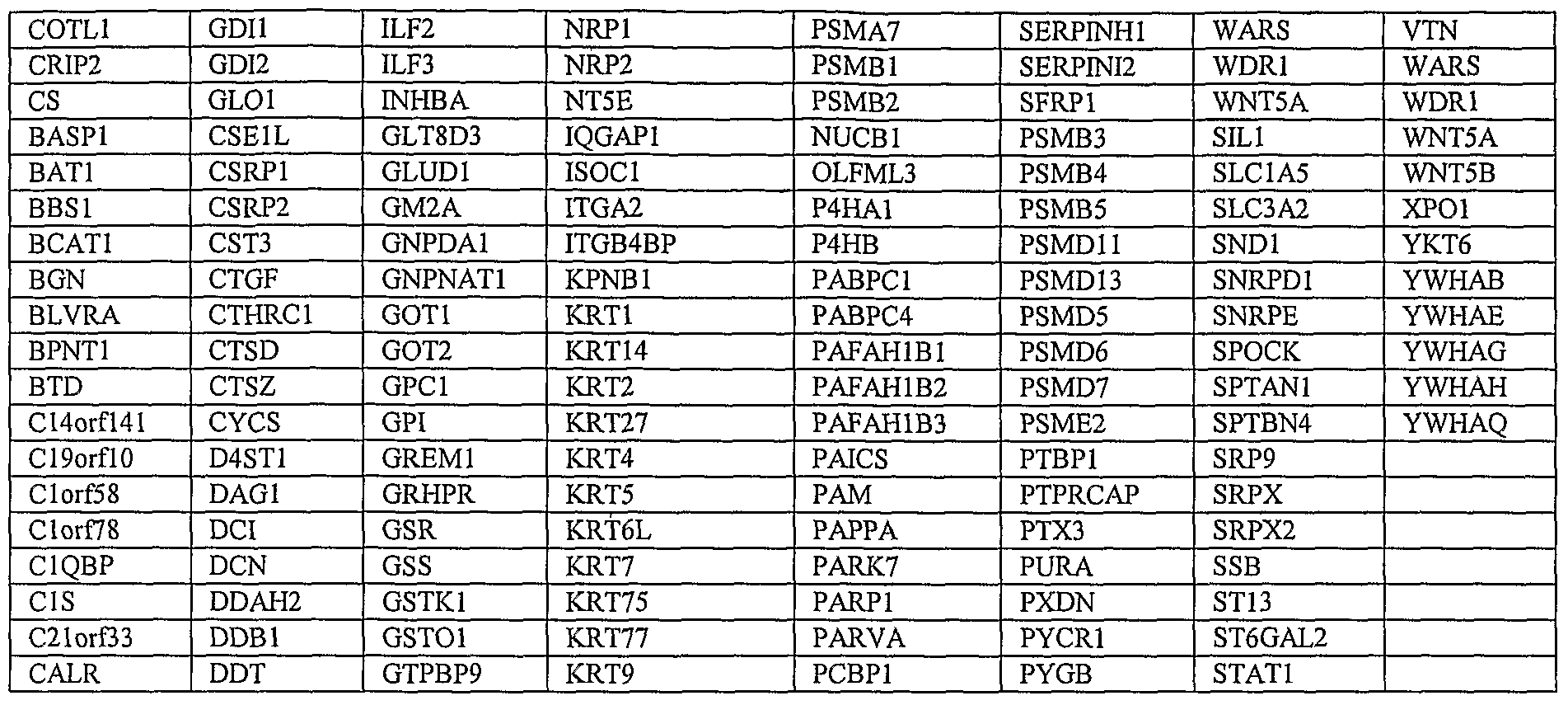
Claims
Priority Applications (4)
| Application Number | Priority Date | Filing Date | Title |
|---|---|---|---|
| EP07794267A EP2054506A1 (en) | 2006-08-15 | 2007-08-15 | Mesenchymal stem cell conditioned medium |
| AU2007285057A AU2007285057B2 (en) | 2006-08-15 | 2007-08-15 | Mesenchymal stem cell conditioned medium |
| JP2009524583A JP2010500047A (en) | 2006-08-15 | 2007-08-15 | Mesenchymal stem cell conditioned medium |
| US12/377,398 US9029146B2 (en) | 2005-09-02 | 2007-08-15 | Mesenchymal stem cell conditioned medium |
Applications Claiming Priority (4)
| Application Number | Priority Date | Filing Date | Title |
|---|---|---|---|
| SGPCT/SG2006/000232 | 2006-08-15 | ||
| PCT/SG2006/000232 WO2007027156A1 (en) | 2005-09-02 | 2006-08-15 | Method of deriving mesenchymal stem cells |
| US87822207P | 2007-01-03 | 2007-01-03 | |
| US60/878,222 | 2007-01-03 |
Publications (1)
| Publication Number | Publication Date |
|---|---|
| WO2008020815A1 true WO2008020815A1 (en) | 2008-02-21 |
Family
ID=40456592
Family Applications (1)
| Application Number | Title | Priority Date | Filing Date |
|---|---|---|---|
| PCT/SG2007/000257 WO2008020815A1 (en) | 2005-09-02 | 2007-08-15 | Mesenchymal stem cell conditioned medium |
Country Status (6)
| Country | Link |
|---|---|
| US (1) | US9029146B2 (en) |
| EP (2) | EP2617428A1 (en) |
| JP (1) | JP2010500047A (en) |
| KR (1) | KR20090043559A (en) |
| AU (1) | AU2007285057B2 (en) |
| WO (1) | WO2008020815A1 (en) |
Cited By (35)
| Publication number | Priority date | Publication date | Assignee | Title |
|---|---|---|---|---|
| WO2009105044A1 (en) * | 2008-02-22 | 2009-08-27 | Agency For Science, Technology And Research (A*Star) | Mesenchymal stem cell particles |
| WO2010137722A1 (en) * | 2009-05-26 | 2010-12-02 | 武田薬品工業株式会社 | Method for screening of regenerative medicine |
| WO2011010966A1 (en) * | 2009-07-23 | 2011-01-27 | Agency For Science, Technology And Research (A*Star) | Pre-natal mesenchymal stem cells |
| WO2011033260A1 (en) | 2009-09-18 | 2011-03-24 | Msd Biologics (Uk) Limited | Stem cell conditioned medium compositions |
| WO2011043524A1 (en) * | 2009-10-08 | 2011-04-14 | 주식회사 알앤엘바이오 | Anticancer composition containing human-derived adult stem cells |
| WO2011053257A2 (en) | 2009-11-02 | 2011-05-05 | Agency For Science, Technology And Research (A*Star) | Methods of monitoring cellular states |
| WO2011070001A1 (en) * | 2009-12-09 | 2011-06-16 | Fresenius Medical Care Deutschland G.M.B.H. | Adult stem cell derived conditioned medium and/or adult stem cells for use in the therapeutic treatment of a tumor disease |
| WO2011127090A1 (en) * | 2010-04-05 | 2011-10-13 | Medstar Health Research Institute, Inc. | Conditioned medium obtained form stem cells and its use in therapy |
| CN102228472A (en) * | 2011-06-09 | 2011-11-02 | 无锡人寰生物医药科技有限公司 | Reagent for causing targeting effect of marrow mesenchymal stem cells on myocardial infarction focus, and its preparation method |
| WO2011116117A3 (en) * | 2010-03-18 | 2011-11-10 | Wisconsin Alumni Research Foundation | Generation of clonal mesenchymal progenitors and mesenchymal stem cell lines under serum-free conditions |
| EP2606121A2 (en) * | 2010-08-19 | 2013-06-26 | The Regents of the University of California | Compositions comrpising perivascular stem cells and nell-1 protein |
| US20130195991A1 (en) * | 2010-03-26 | 2013-08-01 | National University Corporation Nagoya University | Composition for Treatment of Damaged Part |
| WO2013121427A1 (en) * | 2012-02-13 | 2013-08-22 | Gamida-Cell Ltd. | Mesenchymal stem cells conditioned medium and methods of generating and using the same |
| US8846393B2 (en) | 2005-11-29 | 2014-09-30 | Gamida-Cell Ltd. | Methods of improving stem cell homing and engraftment |
| WO2015028900A1 (en) * | 2013-08-29 | 2015-03-05 | Stempeutics Research Pvt. Ltd. | Stromal cells derived conditioned medium, method of obtaining said conditioned medium compositions, formulations and applications thereof |
| US9175266B2 (en) | 2012-07-23 | 2015-11-03 | Gamida Cell Ltd. | Enhancement of natural killer (NK) cell proliferation and activity |
| WO2016043654A1 (en) | 2014-09-15 | 2016-03-24 | Agency For Science, Technology And Research | Methods of treating graft versus host disease (gvhd) or epidermolysis bullosa (eb) with exosomes |
| US20160199413A1 (en) * | 2013-08-01 | 2016-07-14 | Isletone Ab | Mscs in the treatment of inflammatory pulmonary diseases |
| US9423402B2 (en) | 2011-11-30 | 2016-08-23 | Singapore Health Services Pte. Ltd. | GM1 ganglioside to annexin V microparticle polypeptide ratio for biological monitoring |
| WO2016139340A1 (en) * | 2015-03-04 | 2016-09-09 | Mesoblast International Sàrl | Cell culture method for mesenchymal stem cells |
| EP2318519B1 (en) | 2008-08-14 | 2016-10-05 | Mesoblast International Sàrl | Purified mesenchymal stem cell compositions |
| EP3111946A1 (en) | 2015-06-30 | 2017-01-04 | Guido Wouters | Wound healing composition |
| US9567569B2 (en) | 2012-07-23 | 2017-02-14 | Gamida Cell Ltd. | Methods of culturing and expanding mesenchymal stem cells |
| US9901600B2 (en) | 2011-03-11 | 2018-02-27 | Children's Medical Center Corporation | Methods and compositions relating to mesenchymal stem cell exosomes |
| US10179900B2 (en) | 2008-12-19 | 2019-01-15 | DePuy Synthes Products, Inc. | Conditioned media and methods of making a conditioned media |
| CN110257333A (en) * | 2019-05-27 | 2019-09-20 | 江苏大学 | A kind of neural stem cell microcapsule bubble and its application |
| WO2019238952A1 (en) * | 2018-06-15 | 2019-12-19 | Marinas Pardo Luis | Pharmaceutical composition for dermatology and uses thereof |
| US10513689B2 (en) | 2016-04-29 | 2019-12-24 | Hope Biosciences, Llc | Culture media for multipotent stem cells |
| US10624929B2 (en) | 2014-05-18 | 2020-04-21 | Children's Medical Center Corporation | Methods and compositions relating to exosomes |
| US10758571B1 (en) | 2019-04-09 | 2020-09-01 | Combangio, Inc. | Processes for making and using a mesenchymal stem cell derived secretome |
| US11186826B2 (en) | 2017-04-03 | 2021-11-30 | Kaneka Corporation | Cell population including mesenchymal stem cells and production method therefor, and pharmaceutical composition |
| WO2022009134A3 (en) * | 2020-07-10 | 2022-04-21 | Bioceltix Spolka Akcyjna | Method of encapsulation of an active protein using electrodeposition techniques, an immunomodulating composition containing the active protein and a polymer, and its use for the production of a veterinary composition for the treatment of atopic dermatitis in animals |
| WO2022009132A3 (en) * | 2020-07-10 | 2022-04-21 | Bioceltix Spolka Akcyjna | Method of encapsulation of an active protein using electrodeposition techniques, an immunomodulating composition containing the active protein and a polymer, and its use for the production of a pharmaceutical composition for the treatment of atopic dermatitis in humans |
| US11684574B2 (en) | 2016-06-30 | 2023-06-27 | University of Pittsburgh—of the Commonwealth System of Higher Education | Artificial cells and delivery devices for use in tissue engineering, and related methods |
| US11898162B2 (en) | 2007-03-23 | 2024-02-13 | Wisconsin Alumni Research Foundation | Reprogramming somatic cells into pluripotent cells using a vector encoding Oct4 and Sox2 |
Families Citing this family (41)
| Publication number | Priority date | Publication date | Assignee | Title |
|---|---|---|---|---|
| KR20140146224A (en) | 2006-04-14 | 2014-12-24 | 어드밴스드 셀 테크놀로지, 인코포레이티드 | Hemangio-colony forming cells |
| JP2013536860A (en) * | 2010-08-31 | 2013-09-26 | クック・ジェネラル・バイオテクノロジー・エルエルシー | Systemic, allogeneic stem cell therapy for the treatment of animal diseases |
| JP5710634B2 (en) * | 2010-09-30 | 2015-04-30 | 国立大学法人 東京大学 | Method for differentiating human-derived pluripotent stem cells |
| WO2012131733A2 (en) * | 2011-03-31 | 2012-10-04 | Godavari Biorefineries Limited | Media compositions, method of initiating and enriching cultures of stem cells and/or cancer stem-like cells |
| KR101405437B1 (en) * | 2011-08-16 | 2014-06-11 | 사회복지법인 삼성생명공익재단 | Composition for promoting neurogenesis comprising stemcell-derived microvesicle |
| US8940294B2 (en) | 2012-03-02 | 2015-01-27 | Tissuetech, Inc. | Methods of isolating and culturing stem cells |
| KR101914580B1 (en) * | 2012-03-07 | 2018-11-06 | 주식회사 탑셀바이오 | Composition for preventing, improving or treating cranial nerve or spinal nerve related disease comprising conditioned medium powder from stem cells |
| US9383358B2 (en) * | 2012-06-28 | 2016-07-05 | Cellprint Ip Holding, Llc | Method to assess patterns of molecular expression |
| WO2014011407A2 (en) * | 2012-07-12 | 2014-01-16 | Imstem Biotechnology, Inc. | Mesenchymal-like stem cells derived from human embryonic stem cells, methods and uses thereof |
| CN104662148A (en) * | 2012-09-07 | 2015-05-27 | 日本化学研究株式会社 | Medium, for culturing corneal endothelial cells, containing conditioned medium from mesenchymal stem cells |
| KR101486314B1 (en) | 2012-10-29 | 2015-02-13 | (주)치아줄기세포뱅크 | Composition for bone regeneration comprising conditioned medium powder of stem cell |
| DK2956543T3 (en) | 2013-02-12 | 2019-01-02 | Replicel Life Sciences Inc | COMPOSITIONS AND PROCEDURES FOR TREATMENT AND REPAIR |
| US20150368616A1 (en) * | 2013-02-14 | 2015-12-24 | The Cleveland Clinic Foundation | Methods for induction of cell fates from pluripotent cells |
| EP3013379B1 (en) * | 2013-06-26 | 2017-02-22 | Stemform ApS | Lipofilling with ex-vivo expanded adipose tissue-derived stem cells for cosmetic breast filling or for facial filling and/or rejuvenation |
| TWI486447B (en) * | 2013-09-05 | 2015-06-01 | Buddhist Tzu Chi Medical Foundation | Adjuvant for rapid proliferation of human mesenchymal stem cells in vitro, method for rapid proliferation of human mesenchymal stem cells in vitro, method for growth factor harvested from rapid proliferation of human mesenchymal stem cells in vitro and u |
| US9283172B2 (en) * | 2013-12-13 | 2016-03-15 | Growgene Biotech Inc. | Use of stem cell conditioned medium to induce ZO-1 proteins expression for skin regeneration, repair and firming |
| GB201322869D0 (en) | 2013-12-23 | 2014-02-12 | Ucl Business Plc | Therapeutic agents and delivery with microspheres |
| TW201538730A (en) | 2014-02-12 | 2015-10-16 | Replicel Life Sciences Inc | Compositions and methods for treating bone, joints and cartilage |
| US9284527B2 (en) * | 2014-04-07 | 2016-03-15 | Growgene Biotech Inc. | Use of stem cell conditioned medium to inhibit melanin formation for skin whitening |
| US9284528B2 (en) * | 2014-04-18 | 2016-03-15 | Growgene Biotech Inc. | Use of stem cell conditioned medium to inhibit oxidation for anti-aging skin |
| CN108064265A (en) * | 2014-09-29 | 2018-05-22 | 株式会社钟化 | Cell culture medium and use its cultural method |
| RU2644650C2 (en) | 2014-12-01 | 2018-02-13 | Общество с ограниченной ответственностью "Т-Хелпер Клеточные Технологии" | Stem cell material and method for its reception |
| KR101723030B1 (en) * | 2015-01-13 | 2017-04-04 | 한국생명공학연구원 | Behcet's disease human cell model and use thereof |
| CN105624104B (en) * | 2015-06-01 | 2018-10-12 | 中国医学科学院血液病医院(血液学研究所) | A kind of cellular processes improving human mesenchymal stem cell function |
| CA2993955A1 (en) * | 2015-07-29 | 2017-02-02 | Indiana University Research And Technology Corporation | Materials and methods for treating and evaluating ischemic and/or reperfusion-injured tissue and/or tissue susceptible to same |
| CA2995977A1 (en) * | 2015-08-18 | 2017-02-23 | Astellas Institute For Regenerative Medicine | Clinical formulations |
| RU2708329C2 (en) | 2016-05-31 | 2019-12-05 | Общество с ограниченной ответственностью "Т-Хелпер Клеточные Технологии" | Stem cell material, compositions and methods of use |
| JP2018017503A (en) * | 2016-07-25 | 2018-02-01 | 国立大学法人信州大学 | Evaluation method of lung cancer risk state, lung cancer risk state evaluation device, lung cancer risk state evaluation program, lung cancer risk state evaluation system, and information communication terminal device |
| JP7082372B2 (en) * | 2017-02-15 | 2022-06-08 | ロート製薬株式会社 | Pulmonary fibrosis treatment agent, PTPRR expression promoter and pulmonary fibrosis treatment kit |
| EP3591037A4 (en) * | 2017-03-03 | 2020-11-11 | Rohto Pharmaceutical Co., Ltd. | Mesenchymal stem cells and therapeutic agent for liver disease |
| US11718829B2 (en) * | 2017-07-28 | 2023-08-08 | Breakthrough Tech Llc | Methods and compositions for manufacturing extracellular matrix |
| US11285177B2 (en) | 2018-01-03 | 2022-03-29 | Globus Medical, Inc. | Allografts containing viable cells and methods thereof |
| CN108324993B (en) * | 2018-01-15 | 2020-11-03 | 朱剑虹 | Stem cell complex for inducing hair regeneration, preparation method and application thereof |
| KR102142956B1 (en) * | 2018-11-09 | 2020-08-10 | 코아스템(주) | Biomarker for prediction of proliferation and migration capacity of mesenchymal stem cell and use thereof |
| KR102175792B1 (en) * | 2018-11-20 | 2020-11-06 | 사회복지법인 삼성생명공익재단 | A marker for detecting proliferation ability of stem cells and a method for highly efficient proliferation of stem cells using the same |
| KR20200000415A (en) * | 2019-12-24 | 2020-01-02 | (주)안트로젠 | Mesenchymal stem cell culture media for preventing or treating immune disease or inflammatory disease and a method for preparing the same |
| CN112746056A (en) * | 2021-03-17 | 2021-05-04 | 辽宁盛京干细胞科技有限公司 | Culture solution for enhancing NK (natural killer) cytotoxic effect and preparation method thereof |
| CN113209279A (en) * | 2021-05-14 | 2021-08-06 | 山东大学 | Composition for protecting primordial follicles, application and mesenchymal stem cell function evaluation method |
| CN113957048A (en) * | 2021-10-29 | 2022-01-21 | 深圳市默赛尔生物医学科技发展有限公司 | Method for producing natural killer cells by using umbilical cord blood mononuclear cells |
| CN113817667A (en) * | 2021-11-25 | 2021-12-21 | 广东乾晖生物科技有限公司 | Culture solution for hepatocyte hypoxia culture, preparation method and application thereof |
| EP4285915A1 (en) * | 2022-06-03 | 2023-12-06 | Medizinische Hochschule Hannover | Human mesenchymal stem cell-conditioned medium for use in treatment of chronic heart-lung and vascular diseases, in particular, of pulmonary arterial hypertension (pah) |
Citations (2)
| Publication number | Priority date | Publication date | Assignee | Title |
|---|---|---|---|---|
| WO2001032189A1 (en) * | 1999-10-26 | 2001-05-10 | Osiris Therapeutics, Inc. | Supernatant from mesenchymal stem cells for prevention and treatment of immune responses in transplantation |
| WO2005076845A2 (en) * | 2004-02-06 | 2005-08-25 | Theradigm, Inc. | Compositions and methods relating to culturing neural stem cells with bone marrow stromal cells |
Family Cites Families (35)
| Publication number | Priority date | Publication date | Assignee | Title |
|---|---|---|---|---|
| US4675187A (en) | 1983-05-16 | 1987-06-23 | Bristol-Myers Company | BBM-1675, a new antibiotic complex |
| US5192553A (en) | 1987-11-12 | 1993-03-09 | Biocyte Corporation | Isolation and preservation of fetal and neonatal hematopoietic stem and progenitor cells of the blood and methods of therapeutic use |
| US5061620A (en) | 1990-03-30 | 1991-10-29 | Systemix, Inc. | Human hematopoietic stem cell |
| US5635387A (en) | 1990-04-23 | 1997-06-03 | Cellpro, Inc. | Methods and device for culturing human hematopoietic cells and their precursors |
| US5486359A (en) | 1990-11-16 | 1996-01-23 | Osiris Therapeutics, Inc. | Human mesenchymal stem cells |
| DE4117305C2 (en) | 1991-05-27 | 1994-07-21 | Weidmueller Interface | Pliers for gripping and / or pressing processed objects |
| US5851832A (en) | 1991-07-08 | 1998-12-22 | Neurospheres, Ltd. | In vitro growth and proliferation of multipotent neural stem cells and their progeny |
| US5661034A (en) | 1991-07-18 | 1997-08-26 | Fuji Yakuhin Kogyo Kabushiki Kaisha | Serum-free medium for tissue culture containing tissue inhibitor of metalloproteinases and method for growing cells using the medium |
| US5376542A (en) | 1992-04-27 | 1994-12-27 | Georgetown University | Method for producing immortalized cell lines using human papilluma virus genes |
| US5849553A (en) | 1992-07-27 | 1998-12-15 | California Institute Of Technology | Mammalian multipotent neural stem cells |
| US5654183A (en) | 1992-07-27 | 1997-08-05 | California Institute Of Technology | Genetically engineered mammalian neural crest stem cells |
| WO1994007913A1 (en) | 1992-09-25 | 1994-04-14 | Warner-Lambert Company | Peptide antagonists of sh2 binding and therapeutic uses thereof |
| EP0600831A1 (en) | 1992-11-27 | 1994-06-08 | Ciba-Geigy Ag | Phtalazinon derivatives |
| EP0600832A1 (en) | 1992-11-27 | 1994-06-08 | Ciba-Geigy Ag | Derivatives of diamino benzoic and diamino phthalic acid und their use as protein linase inhibitors |
| US5766948A (en) | 1993-01-06 | 1998-06-16 | The Regents Of The University Of California | Method for production of neuroblasts |
| US5387584A (en) | 1993-04-07 | 1995-02-07 | Pfizer Inc. | Bicyclic ansamycins |
| US5631159A (en) | 1993-09-22 | 1997-05-20 | Creative Biomolecules, Inc. | Lipid-modified serum free media |
| US5843780A (en) | 1995-01-20 | 1998-12-01 | Wisconsin Alumni Research Foundation | Primate embryonic stem cells |
| US6015554A (en) | 1995-06-07 | 2000-01-18 | Systemix, Inc. | Method of reconstituting human lymphoid and dendritic cells |
| US5753506A (en) | 1996-05-23 | 1998-05-19 | Cns Stem Cell Technology, Inc. | Isolation propagation and directed differentiation of stem cells from embryonic and adult central nervous system of mammals |
| US6090622A (en) | 1997-03-31 | 2000-07-18 | The Johns Hopkins School Of Medicine | Human embryonic pluripotent germ cells |
| US6361996B1 (en) | 1997-05-07 | 2002-03-26 | University Of Utah Research Foundation | Neuroepithelial stem cells and glial-restricted intermediate precursors |
| EP1028737B1 (en) | 1997-07-03 | 2007-04-04 | Osiris Therapeutics, Inc. | Human mesenchymal stem cells from peripheral blood |
| AU755657B2 (en) | 1997-07-04 | 2002-12-19 | University Of Utah Research Foundation | Lineage-restricted neuronal precursors |
| US5968829A (en) | 1997-09-05 | 1999-10-19 | Cytotherapeutics, Inc. | Human CNS neural stem cells |
| AU1197699A (en) | 1997-10-23 | 1999-05-10 | Geron Corporation | Methods and materials for the growth of primate-derived primordial stem cells |
| EP0953633A1 (en) | 1998-04-28 | 1999-11-03 | Livercell L.L.C. | Cell culturing method and medium for producing proliferated, normal, differentiated human liver cells |
| US6139524A (en) | 1998-10-16 | 2000-10-31 | Scimed Life Systems, Inc. | Stent delivery system with perfusion |
| US7122019B1 (en) | 2000-11-28 | 2006-10-17 | Flowmedica Inc. | Intra-aortic renal drug delivery catheter |
| KR100979664B1 (en) | 1999-03-10 | 2010-09-02 | 유니버시티 오브 피츠버그 오브 더 커먼웰쓰 시스템 오브 하이어 에듀케이션 | Adipose-derived stem cells and lattices |
| US20060051334A1 (en) * | 1999-03-30 | 2006-03-09 | Myocardial Therapeutics, Inc. | Injection of bone marrow-derived conditioned medium for angiogenesis |
| ES2539105T3 (en) | 2001-07-06 | 2015-06-26 | Asterias Biotherapeutics, Inc. | Mesenchymal and osteoblast cells from human embryonic stem cells |
| US7063679B2 (en) | 2002-09-20 | 2006-06-20 | Flowmedica, Inc. | Intra-aortic renal delivery catheter |
| EP1743023A1 (en) * | 2004-05-07 | 2007-01-17 | Wisconsin Alumni Research Foundation | Method of forming mesenchymal stem cells from embryonic stem cells |
| EP1880002A4 (en) * | 2005-05-10 | 2009-03-11 | Us Health | Therapy of kidney diseases and multiorgan failure with mesenchymal stem cells and mesenchymal stem cell conditioned media |
-
2007
- 2007-08-15 WO PCT/SG2007/000257 patent/WO2008020815A1/en active Application Filing
- 2007-08-15 AU AU2007285057A patent/AU2007285057B2/en not_active Ceased
- 2007-08-15 KR KR1020097005144A patent/KR20090043559A/en not_active Application Discontinuation
- 2007-08-15 JP JP2009524583A patent/JP2010500047A/en active Pending
- 2007-08-15 EP EP13164423.9A patent/EP2617428A1/en not_active Withdrawn
- 2007-08-15 EP EP07794267A patent/EP2054506A1/en not_active Ceased
- 2007-08-15 US US12/377,398 patent/US9029146B2/en active Active
Patent Citations (2)
| Publication number | Priority date | Publication date | Assignee | Title |
|---|---|---|---|---|
| WO2001032189A1 (en) * | 1999-10-26 | 2001-05-10 | Osiris Therapeutics, Inc. | Supernatant from mesenchymal stem cells for prevention and treatment of immune responses in transplantation |
| WO2005076845A2 (en) * | 2004-02-06 | 2005-08-25 | Theradigm, Inc. | Compositions and methods relating to culturing neural stem cells with bone marrow stromal cells |
Non-Patent Citations (1)
| Title |
|---|
| See also references of EP2054506A1 * |
Cited By (66)
| Publication number | Priority date | Publication date | Assignee | Title |
|---|---|---|---|---|
| US8846393B2 (en) | 2005-11-29 | 2014-09-30 | Gamida-Cell Ltd. | Methods of improving stem cell homing and engraftment |
| US11898162B2 (en) | 2007-03-23 | 2024-02-13 | Wisconsin Alumni Research Foundation | Reprogramming somatic cells into pluripotent cells using a vector encoding Oct4 and Sox2 |
| US9771561B2 (en) | 2007-09-25 | 2017-09-26 | Wisconsin Alumni Research Foundation | Method of making primate cells expressing apelin receptor that have mesangioblast potential |
| CN104127438B (en) * | 2008-02-22 | 2021-01-08 | 新加坡科技研究局 | Mesenchymal stem cell particles |
| US10780128B2 (en) | 2008-02-22 | 2020-09-22 | Agency For Science, Technology And Research (A*Star) | Mesenchymal stem cell particles |
| WO2009105044A1 (en) * | 2008-02-22 | 2009-08-27 | Agency For Science, Technology And Research (A*Star) | Mesenchymal stem cell particles |
| JP2011513217A (en) * | 2008-02-22 | 2011-04-28 | エイジェンシー・フォー・サイエンス,テクノロジー・アンド・リサーチ(エイ・スター) | Mesenchymal stem cell particles |
| KR101640381B1 (en) * | 2008-02-22 | 2016-07-18 | 에이전시 포 사이언스, 테크놀로지 앤드 리서치 | Mesenchymal stem cell particles |
| KR20100122087A (en) * | 2008-02-22 | 2010-11-19 | 에이전시 포 사이언스, 테크놀로지 앤드 리서치 | Mesenchymal stem cell particles |
| CN104127438A (en) * | 2008-02-22 | 2014-11-05 | 新加坡科技研究局 | Mesenchymal stem cell particles |
| AU2009215934C1 (en) * | 2008-02-22 | 2014-05-08 | Agency For Science, Technology And Research (A*Star) | Mesenchymal stem cell particles |
| AU2009215934B2 (en) * | 2008-02-22 | 2013-10-24 | Agency For Science, Technology And Research (A*Star) | Mesenchymal stem cell particles |
| CN102014934B (en) * | 2008-02-22 | 2014-08-20 | 新加坡科技研究局 | Mesenchymal stem cell particles |
| EP2318519B1 (en) | 2008-08-14 | 2016-10-05 | Mesoblast International Sàrl | Purified mesenchymal stem cell compositions |
| US10179900B2 (en) | 2008-12-19 | 2019-01-15 | DePuy Synthes Products, Inc. | Conditioned media and methods of making a conditioned media |
| WO2010137722A1 (en) * | 2009-05-26 | 2010-12-02 | 武田薬品工業株式会社 | Method for screening of regenerative medicine |
| JPWO2010137722A1 (en) * | 2009-05-26 | 2012-11-15 | 武田薬品工業株式会社 | Regenerative medicine screening method |
| WO2011010966A1 (en) * | 2009-07-23 | 2011-01-27 | Agency For Science, Technology And Research (A*Star) | Pre-natal mesenchymal stem cells |
| WO2011033260A1 (en) | 2009-09-18 | 2011-03-24 | Msd Biologics (Uk) Limited | Stem cell conditioned medium compositions |
| WO2011043524A1 (en) * | 2009-10-08 | 2011-04-14 | 주식회사 알앤엘바이오 | Anticancer composition containing human-derived adult stem cells |
| WO2011053257A2 (en) | 2009-11-02 | 2011-05-05 | Agency For Science, Technology And Research (A*Star) | Methods of monitoring cellular states |
| CN102686723B (en) * | 2009-12-09 | 2016-11-23 | 弗雷森纽斯医疗护理德国有限责任公司 | For treating the derivative conditioned medium of the adult stem cell processing tumor disease and/or adult stem cell |
| US9034335B2 (en) | 2009-12-09 | 2015-05-19 | Fresenius Medical Care Deutschland Gmbh | Adult stem cell derived conditioned medium and/or adult stem cells for use in the therapeutic treatment of a tumor disease |
| CN102686723A (en) * | 2009-12-09 | 2012-09-19 | 弗雷森纽斯医疗护理德国有限责任公司 | Adult stem cell derived conditioned medium and/or adult stem cells for use in the therapeutic treatment of a tumor disease |
| WO2011070001A1 (en) * | 2009-12-09 | 2011-06-16 | Fresenius Medical Care Deutschland G.M.B.H. | Adult stem cell derived conditioned medium and/or adult stem cells for use in the therapeutic treatment of a tumor disease |
| JP2013513366A (en) * | 2009-12-09 | 2013-04-22 | フレゼニウス メディカル ケア ドイッチェランド ゲゼルシャフト ミット ベシュレンクテル ハフツング | Adult stem cell-induced habituation media and / or adult stem cells for use in the treatment of tumor diseases |
| WO2011116117A3 (en) * | 2010-03-18 | 2011-11-10 | Wisconsin Alumni Research Foundation | Generation of clonal mesenchymal progenitors and mesenchymal stem cell lines under serum-free conditions |
| US20130195991A1 (en) * | 2010-03-26 | 2013-08-01 | National University Corporation Nagoya University | Composition for Treatment of Damaged Part |
| US11135243B2 (en) | 2010-03-26 | 2021-10-05 | Shed Tech Corporation | Composition for treatment of damaged part |
| WO2011127090A1 (en) * | 2010-04-05 | 2011-10-13 | Medstar Health Research Institute, Inc. | Conditioned medium obtained form stem cells and its use in therapy |
| US8911963B2 (en) | 2010-04-05 | 2014-12-16 | Medstar Health Research Institute, Inc. | Conditioned medium obtained from stem cells and its use in therapy |
| US9301976B2 (en) | 2010-08-19 | 2016-04-05 | The Regents Of The University Of California | Compositions comprising perivascular stem cells and nell-1 protein |
| US10675307B2 (en) | 2010-08-19 | 2020-06-09 | The Regents Of The University Of California | Compositions comprising perivascular stem cells and nell-1 protein |
| EP2606121A4 (en) * | 2010-08-19 | 2014-01-15 | Univ California | Compositions comrpising perivascular stem cells and nell-1 protein |
| EP2606121A2 (en) * | 2010-08-19 | 2013-06-26 | The Regents of the University of California | Compositions comrpising perivascular stem cells and nell-1 protein |
| US9901600B2 (en) | 2011-03-11 | 2018-02-27 | Children's Medical Center Corporation | Methods and compositions relating to mesenchymal stem cell exosomes |
| CN102228472A (en) * | 2011-06-09 | 2011-11-02 | 无锡人寰生物医药科技有限公司 | Reagent for causing targeting effect of marrow mesenchymal stem cells on myocardial infarction focus, and its preparation method |
| US9423402B2 (en) | 2011-11-30 | 2016-08-23 | Singapore Health Services Pte. Ltd. | GM1 ganglioside to annexin V microparticle polypeptide ratio for biological monitoring |
| US9977032B2 (en) | 2011-11-30 | 2018-05-22 | Agency For Science, Technology And Research | Microparticle fractionation |
| US10481166B2 (en) | 2011-11-30 | 2019-11-19 | Singapore Health Services Pte. Ltd. | Microparticle fractionation |
| US10047345B2 (en) | 2012-02-13 | 2018-08-14 | Gamida-Cell Ltd. | Culturing of mesenchymal stem cells with FGF4 and nicotinamide |
| WO2013121427A1 (en) * | 2012-02-13 | 2013-08-22 | Gamida-Cell Ltd. | Mesenchymal stem cells conditioned medium and methods of generating and using the same |
| US9175266B2 (en) | 2012-07-23 | 2015-11-03 | Gamida Cell Ltd. | Enhancement of natural killer (NK) cell proliferation and activity |
| US9567569B2 (en) | 2012-07-23 | 2017-02-14 | Gamida Cell Ltd. | Methods of culturing and expanding mesenchymal stem cells |
| US20160199413A1 (en) * | 2013-08-01 | 2016-07-14 | Isletone Ab | Mscs in the treatment of inflammatory pulmonary diseases |
| WO2015028900A1 (en) * | 2013-08-29 | 2015-03-05 | Stempeutics Research Pvt. Ltd. | Stromal cells derived conditioned medium, method of obtaining said conditioned medium compositions, formulations and applications thereof |
| US11759481B2 (en) | 2014-05-18 | 2023-09-19 | Children's Medical Center Corporation | Methods and compositions relating to exosomes |
| US10624929B2 (en) | 2014-05-18 | 2020-04-21 | Children's Medical Center Corporation | Methods and compositions relating to exosomes |
| WO2016043654A1 (en) | 2014-09-15 | 2016-03-24 | Agency For Science, Technology And Research | Methods of treating graft versus host disease (gvhd) or epidermolysis bullosa (eb) with exosomes |
| WO2016139340A1 (en) * | 2015-03-04 | 2016-09-09 | Mesoblast International Sàrl | Cell culture method for mesenchymal stem cells |
| US11685899B2 (en) | 2015-03-04 | 2023-06-27 | Mesoblast International Sarl | Cell culture method for mesenchymal stem cells |
| AU2016227607B2 (en) * | 2015-03-04 | 2021-09-30 | Mesoblast International Sarl | Cell culture method for mesenchymal stem cells |
| EP3111946A1 (en) | 2015-06-30 | 2017-01-04 | Guido Wouters | Wound healing composition |
| US10513689B2 (en) | 2016-04-29 | 2019-12-24 | Hope Biosciences, Llc | Culture media for multipotent stem cells |
| US11684574B2 (en) | 2016-06-30 | 2023-06-27 | University of Pittsburgh—of the Commonwealth System of Higher Education | Artificial cells and delivery devices for use in tissue engineering, and related methods |
| US11186826B2 (en) | 2017-04-03 | 2021-11-30 | Kaneka Corporation | Cell population including mesenchymal stem cells and production method therefor, and pharmaceutical composition |
| WO2019238952A1 (en) * | 2018-06-15 | 2019-12-19 | Marinas Pardo Luis | Pharmaceutical composition for dermatology and uses thereof |
| US11413314B2 (en) | 2018-06-15 | 2022-08-16 | NextPhase Therapeutics, Inc. | Pharmaceutical composition for dermatology and uses thereof |
| EP3875097A1 (en) * | 2018-06-15 | 2021-09-08 | Mariñas Pardo, Luis | Pharmaceutical composition for dermatology and uses thereof |
| US11129853B2 (en) | 2019-04-09 | 2021-09-28 | Combangio, Inc. | Processes for making and using a mesenchymal stem cell derived secretome |
| US11654160B2 (en) | 2019-04-09 | 2023-05-23 | Combangio, Inc. | Processes for making and using a mesenchymal stem cell derived secretome |
| US10881693B2 (en) | 2019-04-09 | 2021-01-05 | Combangio, Inc. | Processes for making and using a mesenchymal stem cell derived secretome |
| US10758571B1 (en) | 2019-04-09 | 2020-09-01 | Combangio, Inc. | Processes for making and using a mesenchymal stem cell derived secretome |
| CN110257333A (en) * | 2019-05-27 | 2019-09-20 | 江苏大学 | A kind of neural stem cell microcapsule bubble and its application |
| WO2022009134A3 (en) * | 2020-07-10 | 2022-04-21 | Bioceltix Spolka Akcyjna | Method of encapsulation of an active protein using electrodeposition techniques, an immunomodulating composition containing the active protein and a polymer, and its use for the production of a veterinary composition for the treatment of atopic dermatitis in animals |
| WO2022009132A3 (en) * | 2020-07-10 | 2022-04-21 | Bioceltix Spolka Akcyjna | Method of encapsulation of an active protein using electrodeposition techniques, an immunomodulating composition containing the active protein and a polymer, and its use for the production of a pharmaceutical composition for the treatment of atopic dermatitis in humans |
Also Published As
| Publication number | Publication date |
|---|---|
| US20100323027A1 (en) | 2010-12-23 |
| EP2054506A1 (en) | 2009-05-06 |
| AU2007285057A1 (en) | 2008-02-21 |
| EP2617428A1 (en) | 2013-07-24 |
| KR20090043559A (en) | 2009-05-06 |
| JP2010500047A (en) | 2010-01-07 |
| US9029146B2 (en) | 2015-05-12 |
| AU2007285057B2 (en) | 2013-08-01 |
Similar Documents
| Publication | Publication Date | Title |
|---|---|---|
| AU2007285057B2 (en) | Mesenchymal stem cell conditioned medium | |
| US8962318B2 (en) | Method of deriving mesenchymal stem cells from ES cells using FGF2 | |
| Lian et al. | Derivation of clinically compliant MSCs from CD105+, CD24− differentiated human ESCs | |
| US10584312B2 (en) | Isolating method for umbilical cord blood-derived pluripotent stem cells expressing ZNF281 | |
| EP2294183B1 (en) | Mesenchymal stem cells, compositions, and methods for treatment of cardiac tissue damage | |
| US20120107413A1 (en) | Pre-natal mesenchymal stem cells | |
| US20230093399A1 (en) | Methods for generating pluripotent stem cell-derived brown fat cells | |
| US8507275B2 (en) | Method of inducing differentiation of embryonic stem cells into hemangioblast | |
| US20200157505A1 (en) | Methods for the re-derivation of diverse pluripotent stem cell-derived brown fat cells | |
| US20080254002A1 (en) | Bone Marrow Derived Oct3/4+ Stem Cells | |
| WO2007122233A1 (en) | Preparation of mesenchymal progenitor cells, particularly osteogenic progenitor cells | |
| JP2009100679A (en) | Efficient method for preparing momc (monocyte-derived multi-potent cell) | |
| AU2022261504A1 (en) | Method for treating acute respiratory distress syndrome (ards) in specific patients using mesenchymal lineage precursor or stem cells |
Legal Events
| Date | Code | Title | Description |
|---|---|---|---|
| 121 | Ep: the epo has been informed by wipo that ep was designated in this application |
Ref document number: 07794267 Country of ref document: EP Kind code of ref document: A1 |
|
| WWE | Wipo information: entry into national phase |
Ref document number: 2009524583 Country of ref document: JP |
|
| WWE | Wipo information: entry into national phase |
Ref document number: 2007285057 Country of ref document: AU Ref document number: 861/CHENP/2009 Country of ref document: IN |
|
| NENP | Non-entry into the national phase |
Ref country code: DE |
|
| ENP | Entry into the national phase |
Ref document number: 2007285057 Country of ref document: AU Date of ref document: 20070815 Kind code of ref document: A |
|
| WWE | Wipo information: entry into national phase |
Ref document number: 2007794267 Country of ref document: EP Ref document number: 1020097005144 Country of ref document: KR |
|
| NENP | Non-entry into the national phase |
Ref country code: RU |
|
| WWE | Wipo information: entry into national phase |
Ref document number: 12377398 Country of ref document: US |