WO2006110862A2 - Micelle composition of polymer and passenger drug - Google Patents
Micelle composition of polymer and passenger drug Download PDFInfo
- Publication number
- WO2006110862A2 WO2006110862A2 PCT/US2006/013800 US2006013800W WO2006110862A2 WO 2006110862 A2 WO2006110862 A2 WO 2006110862A2 US 2006013800 W US2006013800 W US 2006013800W WO 2006110862 A2 WO2006110862 A2 WO 2006110862A2
- Authority
- WO
- WIPO (PCT)
- Prior art keywords
- peg
- rapamycin
- geldanamycin
- micelle
- tocopherol
- Prior art date
Links
Classifications
-
- A—HUMAN NECESSITIES
- A61—MEDICAL OR VETERINARY SCIENCE; HYGIENE
- A61K—PREPARATIONS FOR MEDICAL, DENTAL OR TOILETRY PURPOSES
- A61K9/00—Medicinal preparations characterised by special physical form
- A61K9/48—Preparations in capsules, e.g. of gelatin, of chocolate
-
- A—HUMAN NECESSITIES
- A61—MEDICAL OR VETERINARY SCIENCE; HYGIENE
- A61K—PREPARATIONS FOR MEDICAL, DENTAL OR TOILETRY PURPOSES
- A61K9/00—Medicinal preparations characterised by special physical form
- A61K9/0012—Galenical forms characterised by the site of application
- A61K9/0019—Injectable compositions; Intramuscular, intravenous, arterial, subcutaneous administration; Compositions to be administered through the skin in an invasive manner
-
- A—HUMAN NECESSITIES
- A61—MEDICAL OR VETERINARY SCIENCE; HYGIENE
- A61K—PREPARATIONS FOR MEDICAL, DENTAL OR TOILETRY PURPOSES
- A61K31/00—Medicinal preparations containing organic active ingredients
- A61K31/33—Heterocyclic compounds
- A61K31/335—Heterocyclic compounds having oxygen as the only ring hetero atom, e.g. fungichromin
- A61K31/337—Heterocyclic compounds having oxygen as the only ring hetero atom, e.g. fungichromin having four-membered rings, e.g. taxol
-
- A—HUMAN NECESSITIES
- A61—MEDICAL OR VETERINARY SCIENCE; HYGIENE
- A61K—PREPARATIONS FOR MEDICAL, DENTAL OR TOILETRY PURPOSES
- A61K31/00—Medicinal preparations containing organic active ingredients
- A61K31/33—Heterocyclic compounds
- A61K31/395—Heterocyclic compounds having nitrogen as a ring hetero atom, e.g. guanethidine or rifamycins
- A61K31/435—Heterocyclic compounds having nitrogen as a ring hetero atom, e.g. guanethidine or rifamycins having six-membered rings with one nitrogen as the only ring hetero atom
- A61K31/4353—Heterocyclic compounds having nitrogen as a ring hetero atom, e.g. guanethidine or rifamycins having six-membered rings with one nitrogen as the only ring hetero atom ortho- or peri-condensed with heterocyclic ring systems
- A61K31/436—Heterocyclic compounds having nitrogen as a ring hetero atom, e.g. guanethidine or rifamycins having six-membered rings with one nitrogen as the only ring hetero atom ortho- or peri-condensed with heterocyclic ring systems the heterocyclic ring system containing a six-membered ring having oxygen as a ring hetero atom, e.g. rapamycin
-
- A—HUMAN NECESSITIES
- A61—MEDICAL OR VETERINARY SCIENCE; HYGIENE
- A61K—PREPARATIONS FOR MEDICAL, DENTAL OR TOILETRY PURPOSES
- A61K31/00—Medicinal preparations containing organic active ingredients
- A61K31/33—Heterocyclic compounds
- A61K31/395—Heterocyclic compounds having nitrogen as a ring hetero atom, e.g. guanethidine or rifamycins
- A61K31/435—Heterocyclic compounds having nitrogen as a ring hetero atom, e.g. guanethidine or rifamycins having six-membered rings with one nitrogen as the only ring hetero atom
- A61K31/47—Quinolines; Isoquinolines
- A61K31/4738—Quinolines; Isoquinolines ortho- or peri-condensed with heterocyclic ring systems
- A61K31/4745—Quinolines; Isoquinolines ortho- or peri-condensed with heterocyclic ring systems condensed with ring systems having nitrogen as a ring hetero atom, e.g. phenantrolines
-
- A—HUMAN NECESSITIES
- A61—MEDICAL OR VETERINARY SCIENCE; HYGIENE
- A61K—PREPARATIONS FOR MEDICAL, DENTAL OR TOILETRY PURPOSES
- A61K9/00—Medicinal preparations characterised by special physical form
- A61K9/10—Dispersions; Emulsions
- A61K9/107—Emulsions ; Emulsion preconcentrates; Micelles
-
- A—HUMAN NECESSITIES
- A61—MEDICAL OR VETERINARY SCIENCE; HYGIENE
- A61K—PREPARATIONS FOR MEDICAL, DENTAL OR TOILETRY PURPOSES
- A61K9/00—Medicinal preparations characterised by special physical form
- A61K9/10—Dispersions; Emulsions
- A61K9/127—Liposomes
- A61K9/1271—Non-conventional liposomes, e.g. PEGylated liposomes, liposomes coated with polymers
-
- A—HUMAN NECESSITIES
- A61—MEDICAL OR VETERINARY SCIENCE; HYGIENE
- A61K—PREPARATIONS FOR MEDICAL, DENTAL OR TOILETRY PURPOSES
- A61K9/00—Medicinal preparations characterised by special physical form
- A61K9/48—Preparations in capsules, e.g. of gelatin, of chocolate
- A61K9/50—Microcapsules having a gas, liquid or semi-solid filling; Solid microparticles or pellets surrounded by a distinct coating layer, e.g. coated microspheres, coated drug crystals
-
- A—HUMAN NECESSITIES
- A61—MEDICAL OR VETERINARY SCIENCE; HYGIENE
- A61K—PREPARATIONS FOR MEDICAL, DENTAL OR TOILETRY PURPOSES
- A61K9/00—Medicinal preparations characterised by special physical form
- A61K9/10—Dispersions; Emulsions
- A61K9/107—Emulsions ; Emulsion preconcentrates; Micelles
- A61K9/1075—Microemulsions or submicron emulsions; Preconcentrates or solids thereof; Micelles, e.g. made of phospholipids or block copolymers
Definitions
- This invention is directed generally to micelle compositions, methods of making micelles, and the use of micelle compositions with drugs for treatment of disease.
- Cancer is a very deadly disease.
- Various cytoxic chemotherapy agents have been used to eradicate cancer and/or prevent the spread of the cancer.
- Alkylating agents such as cisplatin and chlorambucil, crosslink NDA to prevent cell division.
- Antitumor antibiotics such as dactinomycin and bleomycin, bind DNA and thus prevent DNA separation and mRNA synthesis.
- Antimetabolites such as purine and pyrimidine antagonists and 5-fluorouracil, may mimic cell nutrients and prevent normal DNA synthesis.
- Plant alkaloids such as paclitaxel and vinblastine, block cell division by blocking microtubule formation.
- Topoisomerase inhibitors such as camptothecins, topotecan, and irinotecan, inhibit DNA supercoiling and block transcription and replication.
- Many drugs that are potentially efficacious for treating diseases such as cancer have poor solubility that limits their usefulness.
- Rapamycin is a large, highly hydrophobic compound with applications in chemotherapy, immunosuppression, anti-restenosis, fungal infections, and neurological disorders. Rapamycin as an anti-cancer agent is generally formed as ester analogs which are quickly hydrolyzed and sequestered into the red blood cells thereby reducing the effectiveness of rapamycin at tumor iites. Rapamycin is currently used as an immunosuppressant for kidney transplant patients, ⁇ apamune (Wyeth-Ayerst), and has shown long te ⁇ n clinical safety. However, rapamycin is a )oorly water soluble drug, creating difficulties in drug administration in patients.
- Geldanamycin is also a hydrophobic compound with applications including the treatment if cancer.
- Geldanamycin is a member of the new class of compounds known as heat shock protein inhibitors, having both anti-tumor and neurological disease applications.
- Hsp 90 is a molecular chaperon responsible for the folding, stability, and function of numerous client proteins. Inhibition of Hsp 90 leads to the destabilization and eventual ubiquitination of many oncogenic client proteins.
- geldanamycin may be efficacious against a broad range of tumors and may increase the chances of overcoming drug resistance.
- the inhibition of Hsp90 leads to an up-regulation of Hsp70, which reduces the formation of abnormal tau species, the primary component of plaque deposits in Alzheimer's and Parkinson's disease.
- Paclitaxel is another hydrophobic compound with applications including the treatment of cancer.
- Paclitaxel belongs to a group of medicines called antineoplastics, which inhibit cellular growth. The inhibition is accomplished by disrupting microtubule function by binding to the beta subunit of tubulin. The disrupted microtubule looses the ability to disassemble, a necessary function, for example, in chromosomal migration during cell replication. Additionally, research has indicated that paclitaxel induces apoptosis, programmed cell death, by binding to an apoptosis stopping protein called Bcl-2 and stopping its function.
- Bcl-2 apoptosis stopping protein
- ABC's comprised of a hydrophobic, such as poly(propylene glycol), and hydrophilic block, such as polyethylene glycol (PEG), can assemble into a microphase separated, core/shell architecture in a selective solvent.
- PEG-poly( ⁇ - caprolactone) (PEG-PCL) and PEG-poly(amino acids) can form these polymeric micelles.
- phospholipids can be used, such as, PEG-distearoylphosphatidylethanolamine (PEG-DSPE) to form these polymeric micelles.
- the hydrophobic drug can be encapsulated into the hydrophobic core of the micelle and have aqueous solubility provided by a poly(ethylene glycol) (PEG) and corona (shell). Due to their nanoscopic dimensions and stealth properties imparted by a PEG corona, micelles may have long-term circulation capabilities. During the circulation period, the micelle may gradually release drug and eventually dissociate and be eliminated from circulation.
- PEG poly(ethylene glycol)
- shell corona
- Excipients and co-excipients have been used to solubilize poorly soluble compounds.
- Alpha-tocopherol commonly known as Vitamin E or simply tocopherol, has been used as an excipient because of its ring and alkyl chain structures common to many poorly-soluble drugs. Vitamin E is not toxic to living organisms. Additionally, tocopherol stabilizes biological membranes. Tocopherol, however, is not soluble in water and therefore it has had limited usefulness in intravenous solutions.
- a micelle composition may comprise an amphiphilic polymer, a hydrophobic excipient, and a hydrophobic passenger drug.
- the amphiphilic polymer is PEG-DSPE.
- the excipient is tocopherol.
- the ratio of tocopherol to PEG- DSPE is between about 0.1 and about 3.
- a micelle composition comprises an amphiphilic polymer and rapamycin.
- the micelle composition may have an amphiphilic polymer, rapamycin and tocopherol.
- the concentration of PEG-DSPE may be between about 1 and about 10 mM
- the concentration of tocopherol may be between about 2 and about 20 mM
- the concentration of rapamycin may be between about 0.1 and 1.0 mg/ml.
- a micelle composition may comprise an amphiphilic polymer and geldanamycin.
- the geldanamycin may be a geldanamycin prodrug with increased hydrophobic properties.
- a micelle composition may comprise an amphiphilic polymer and paclitaxel.
- the paclitaxel may be a paclitaxel prodrug with increased hydrophobic properties.
- a process for forming micelle compositions may include mixing amphiphilic polymer, hydrophobic excipient, and hydrophobic drug into an organic solvent to form a solution, removing substantially all of the organic solvent from the solution to leave a substantially solvent-free mixture, and resuspending the solvent-free mixture in water or buffer.
- a process may also include adding said solution to a substantially water solution before removing substantially all of said organic solvent from said solution to leave a substantially solvent-free mixture.
- a process and resulting prodrug composition made for improving micelle encapsulation efficiency of hydrophobic drugs.
- a process for making geldanamycin prodrugs for encapsulation In yet another aspect, a process for making paclitaxel prodrugs for encapsulation.
- a method of treatment for a disease or condition in a human or an animal may comprise administering an effective amount of a micelle composition comprising an amphiphilic polymer, a hydrophobic excipient and a hydrophobic passenger drug.
- FIG. 1 is a schematic showing a micelle structure for drug delivery, including a hydrophobic core and a hydrophilic corona.
- FIG. 2 is a schematic showing a depiction of micelle formation by unimers above critical micelle concentration through hydrophobic interaction.
- FIG. 3 is a graph showing polarity as a function of micelle concentration.
- FIG. 4 is a schematic showing micelles being administered intravenously, and the uptake by tumors due to their leaky vasculature.
- FIG. 5 depicts the structure of PEG-DSPE.
- FIG. 6 depicts the structure of PEG-PCL.
- FIG. 7 is a schematic showing tocopherol incorporation into PEG-DSPE.
- FIG. 8 depicts the structure of tocopherol.
- FIG. 9 depicts the structure of rapamycin.
- FIG. 10 depicts the structure of geldanamycin.
- FIG. 11 depicts the structure of paclitaxel.
- FIG. 12 shows a graph of critical micelle concentration at different PEG-DSPE to tocopherol ratios as a function of the concentration of the PEG-DSPE micelles.
- FIG. 13 is a bar graph of relative core viscosity as a function of the PEG-DSPE to tocopherol ratio.
- FIG. 14 is a bar graph showing the increasing aggregate number within the core as a function of various PEG-DSPE to tocopherol ratios.
- FIG. 15 is a graph showing the stability of PEG-DSPE micelles in phosphate buffered saline and in 4% bovine serum albumin as a function of time.
- FIG. 16 is a graph showing the stability of PEG-PCL micelles in 4% bovine serum albumin as a function of time.
- FIG. 17 is a graph showing the stability of PEG-DSPE micelles in 4% bovine serum albumin as a function of time.
- FIG. 18 is a graph showing the core polarity of PEG-DSPE micelles for various PEG-DSPE to tocopherol ratios and PEC-DSPE concentrations.
- FIG. 19 is a graph showing the rapamycin loading efficiency by diffusion-evaporation as a function of rapamycin to amphiphilic polymer ratio, for ratios of PEG-DSPE: tocopherol at 1:2,
- FIG. 20 is a schematic of a method of forming PEG-DSPE micelles.
- FIG. 21 is a schematic of a drop wise method of forming polymer micelles.
- FIG. 22 is a graph showing rapamycin loading efficiency in micelles as a function of the ratio of rapamycin to amph philic polymer.
- FIG. 23 is a graph showing rapamycin release in the presence of albumin as a function of time in different bovine serum albumin concentrations.
- FIG. 24 is a bar graph showing the interaction of serum albumin, fibrinogen, and bovine pancreatic trypsin inhibitor with PEG-DSPE micelles.
- FIG. 25 is a bar graph showing how tocopherol incorporation affects the size of resulting micelles.
- FIG. 26 is a graph showing the incorporation of rapamycin in micelles through size exclusion chromatography.
- FIG. 27 is an analysis of release kinetics based on Ficlcian diffusion from sphere for short time periods.
- FIG. 28 is a graph showing the effect of tocopherol on rapamycin release from PEG-DSPE micelles in phosphate buffered saline solution.
- FIG. 29 is a graph showing the effect of tocopherol on rapamycin release from PEG-DSPE micelles in 4% bovine serum albumin.
- FIG. 30 shows the stability of PEG-PCL micelles in the presence of tocopherol.
- FIG. 31 is a graph showing the release of rapamycin from PEG-PCL micelles with incorporated tocopherol as a function of time in phosphate buffered saline.
- FIG. 32 is a graph showing the release of rapamycin from PEG-PCL micelles with incorporated tocopherol as a function of time in 4% bovine serum albumin.
- FIG. 33 is a graph showing rapamycin control formulation disposition in whole blood following intravenous administration.
- FIG. 34 is a graph showing rapamycin PEG-PCl formulation disposition in whole blood following intravenous administration.
- FIG. 35 is a graph showing rapamycin PEG-PCl + ⁇ -tocopherol formulation disposition in whole blood following intravenous administration.
- FIG. 36 is a bar graph showing rapamycin concentration in plasma or red blood cells for rapamycin control formulation, rapamycin PEG-PCl 3 and rapamycin PEG-PCl + ⁇ -tocopherol formulation at 1 min after intravenous administration.
- FIG. 37 is a bar graph showing plasma/RBC ratios of rapamycin control formulation, rapamycin
- FIG. 38 is a bar graph showing rapamycin concentration in plasma or red blood cells for rapamycin control formulation, rapamycin PEG-PCl, and rapamycin PEG-PCl + ce-tocopherol formulation at 12 hours after intravenous administration.
- FIG. 39 is a bar graph showing plasma/RBC ratios of rapamycin control formulation, rapamycin
- FIG. 40 is a schematic showing the targets of geldanamycin (in boxes).
- FIG. 41 shows the properties of geldanamycin and geldanamycin prodrugs.
- FIG. 42 shows the loading percentage of geldanamycin into micelles.
- FIG. 43 shows the formulation of fatty acid prodrugs of geldanamycin.
- FIG. 44 shows the lipophilicity and loading percentage of different geldanmycin prodrugs.
- FIG. 45 shows a process schematic for adding a fatty acid to C17 position of geldanamycin.
- FIG. 46 shows a process schematic for forming geldanamycin-C17-amino-hexadecane.
- FIG. 47 shows a process schematic for forming geldanamycin-C17-aminoethyl-2- isopropylhexadecanoate.
- FIG. 48 shows a process schematic for forming geldanamycin-C17-aminoethylonate-Phe-Leu-
- FIG. 49 shows a process schematic for forming geldanamycin-C17-aminoethylidene- palmitohydrazide .
- FIG. 50 shows a process schematic for forming PEO- ⁇ -PEGA.
- FIG. 51 is a graph showing geldanamycin prodrug release over time.
- FIG. 52 is a chart and a graph showing geldanamycin prodrug encapsulation in micelles and release over time.
- an amphiphilic polymer, a hydrophobic excipient, and a hydrophobic passenger drug can form a micelle composition.
- Methods for making these compositions are also part of the scope of the invention.
- methods of treatment of a disease or condition utilizing these micelles are part of the scope of the invention.
- Micelles incorporated with tocopherol may increase the drug loading capability of the micelles and also increase the micellar stability during in vivo conditions.
- Rapamycin is a drug that demonstrates impressive activity in the nanomolar range against many tumor xenograft models, including various solid tumors.
- the low solubility of rapamycin may be overcome by incorporating rapamycin into micelle compositions for delivery to target tumor sites.
- Nonionic surfactants such as Cremophor EL and Tween 80
- Cremophor EL and Tween 80 may be used for intravenous administration of cancer treatments.
- circulating particles should be less than about 200 nm to avoid filtering by the interendothelial cell slits at the spleen.
- Polymeric micelles have been shown to circulate in the blood for prolonged periods and capable of targeted delivery of poorly water-soluble compounds. Upon disassociation, micelle unimers are typically ⁇ 50,000 g/mol, permitting elimination by the kidneys. Ideally, this allows prolonged circulation with no buildup of micelle components in the liver that could lead to storage diseases.
- Polymers that can encapsulate poorly- water soluble drugs include: pegylated phospholipids and pegylated poly-e-caprolactone. These polymers exhibit high biocompatibility and solubilization capacity for a broad range of compounds. Coexcipients, such as ⁇ -tocopherol, can substantially increase the drug loading capacity of micelles formed from these polymers and allow solubilization of potential drug candidates previously thought incompatible or poorly solubilized by existing polymeric carriers.
- Amphiphilic polymers are typically composed of a hydrophilic domain, e.g. polyethylene glycol (PEG), and a hydrophobic domain such as poly(propylene glycol), poly(L-amino acid), poly(ester), and phospholipids. These polymers can assemble into polymeric micelles, highly ordered supramolecular core-shell structures having a hydrophobic interior capable of encapsulating small hydrophobic compounds and a hydrophilic exterior. As shown in FIG. 3, the micelle core has low polarity and is a hydrophobic environment. There is a high core capacity for hydrophobic compounds. There can be up to about 4:1 drug:polymer loading. The micelle core can increase in solubility of up to about 30,000 times. The micelle corona is hydrophilic.
- PEG polyethylene glycol
- hydrophobic domain such as poly(propylene glycol), poly(L-amino acid), poly(ester), and phospholipids.
- These polymers can assemble into polymeric micelles, highly
- Example 1 illustrates that drugs such as doxorubicin and paclitaxel can be encapsulated in micelles and targeted to tumors.
- micelle compositions are capable of long blood circulation, low mononuclear phagocyte uptake, and low levels of renal excretion. Also, micelle compositions have enhanced permeability and retention (EPR) to increase the likelihood of the chemotherapeutics reaching tumors. As shown in FIG. 4, tumors have high vascular density as well as defective vasculature so high extravasation occurs. There may be impaired lymphatic clearance. The endocytosis and subsequent drug release increases the effect of the chemotherapeutics on the tumor.
- EPR permeability and retention
- PEG-DSPE may be a safe and effective micelle carrier for both chemotherapeutic agents.
- PEG-PCL is biodegradable and may have biocompatibility.
- the principal difference between neutral PEG-DSPE and negatively charged PEG-DSPE membranes is the electrostatic force between the two charged membranes.
- Membrane charges affect the adsorption of acidic and basic proteins on charged and neutral membranes. This may alter the interactions of various proteins with the bilayers. These differences may be responsible for the differences in opsonization and phagocytosis of neutral versus charged liposomes.
- the phosphate group at the hydrophobic head of PEG-DSPE may affect the tightness of the PEG- DSPE's at the core-water interface due to electrostatic repulsion. Also, this charged nature may influence protein interaction with the hydrophobic core should the protein penetrate the PEG corona. Tocopherol (FIG.
- PEG-b-PCL may be biocompatible and biodegradable.
- PEG-b-PCL may have a low critical micelle concentration (CMC).
- a PEG:PCL ratio of about 5:6 may have a CMC of under about 0.5 ⁇ M.
- PEG-PCL may have a rigid core structure and be stable in the presence of albumin.
- polymeric micelle compositions can be highly dependent on the structural relationship between the target drug compound and the hydrophobic core of the carrier.
- tocopherol may also modify the core properties of the micelles so as to induce higher loading of drugs which are otherwise poorly soluble in the micelle of study.
- drugs can be passenger compounds in polymer carriers.
- Such drugs include: rapamycin (FIG. 9), geldanamycin (FIG. 10), and paclitaxel (FIG. 11).
- rapamycin FOG. 9
- geldanamycin FOG. 10
- paclitaxel FOG. 11
- These drugs are potent small molecule chemotherapeutic agents with unique targets of action. Studies of these compounds and the development of clinical products have been hampered by their extremely low water solubilities, for example, rapamycin ⁇ 2.6 ⁇ g/ml and geldanamycin -1.5 ⁇ g/ml.
- polymeric micelle carrier can be highly dependent on the structural relationship between the target passenger drug compound and the hydrophobic core of the carrier. Less than 3% (w/w) paclitaxel may be loaded into PEG-PCL micelles. However, PEG- poly(D,L-lactide) micelles have a loading capacity >20% (w/w). Therefore, conditions of polymeric micelle carriers must be optimized for loading a desired passenger compound.
- rapamycin for intravenous delivery without the use of co-solvents, e.g., ethanol or polyethylene glycol, permits them for therapeutic usage.
- co-solvents e.g., ethanol or polyethylene glycol
- micelle carriers allows delivery of therapeutic dosages of this drug without chemical modification.
- micelle delivery allows targeted treatment to tumors through the EPR effect, reducing the likelihood of immunosuppression, a side-effect of free rapamycin and its water soluble derivatives.
- Rapamycin (Fig. 9) is a large, highly hydrophobic compound with applications in chemotherapy, immunosuppression, anti-restenosis, fungal infections, and neurological disorders, e.g., Alzheimer's and Huntington's disease. Rapamycin has a unique target of action, binding the immunophilin FKBP 12 and inhibiting the mammalian target of rapamycin (mTOR) pathway, which prevents cell cycle Gi to S phase transition. Rapamycin has demonstrated impressive activity against a broad range of human tumor xenograph models including lymphocytic leukemia, melanocarcinoma, ependymoblastoma, and various solid tumors with a typical IC 50 of 10 '8 M.
- a novel mechanism may have rapamycin binding to FK506-12, in which rapamycin inhibits mTOR growth regulators, prevents Gl to S phase transition, and inhibits NF-kB and enhances apoptosis.
- rapamycin is practically insoluble in water ( ⁇ 2.6 ⁇ g/ml) and has no ionizable groups.
- the targeted delivery and retention of rapamycin to tumor sites, using the EPR effect, may substantially increase its potency.
- targeted delivery may attenuate the side effects of rapamycin treatment including immunosuppression.
- the retention of rapamycin's native hydrophobic nature may be important in neurological applications where modification (to increase water solubility) may hinder crossing of the blood brain barrier.
- rapamycin can be solubilized in large quantities- well within the range required for clinical feasibility. Rapamycin has been solubilized using PEG-PCL and PEG-DSPE micelles with the addition of tocopherol. Results are summarized in Example 2.
- Geldanamycin (FIG. 10) is a member of the new class of compounds known as heat shock protein inhibitors, having both anti-tumor and neurological disease applications.
- Hsp90 inhibitors may be useful in drug resistant cancers by inducing different pathways, such as in rapamycin resistant tumors.
- Hsp90 inhibitors such as geldanamycin
- Radicicol an Hsp90 inhibitor
- Geldanamycin has extremely poor water solubility, and is hepatotoxic in vivo (MTD dog ⁇ 100 mg/rn 2 ).
- Geldanamycin prodrugs such as 17- AAG have slightly better solubility and lower hepatoxicity (MTD dog 500 mg/m 2 ), but are still difficult to formulate, requiring toxic excipients such as Cremaphor, Tween 80, and DMSO.
- Water soluble prodrugs of geldanamycin, such as 17DMAG (MTD dog 8 mg/m 2 ) may avoid these formulation problems, but the wide biodistribution and increased toxicity of these prodrugs may present additional difficulties.
- a solubility of at least about 1 mg/ml is desirable.
- Phase I results found GI toxicity to be dose limiting for 17-AGG, with a suggested Phase II dosing of 40 mg/m 2 .
- Preclinical trials found severe hepatotoxicity to be dose limiting for the parent compound, geldanamycin (4 mg/kg).
- geldanamycin By targeting multiple oncogenic proteins, geldanamycin promises efficacy against a broad range of tumors and increases the chances of overcoming drug resistance.
- the inhibition of Hsp90 leads to an up-regulation of Hsp70, which reduces the formation of abnormal tau species, the primary component of plaque deposits in Alzheimer's and Parkinson's disease.
- geldanamycin Because of the extremely low water solubility of geldanamycin, ⁇ 1.5 ⁇ g/ml, formulations have used various soluble analogs such as 17-AAG. As with rapamycin, the targeted delivery of geldanamycin to tumor sites and the EPR effect are expected to substantially increase its potency. In addition, prolonged circulation time and reduced liver retention should dramatically reduce hepatotoxicity. Finally, the possible advancement of geldanamycin as a treatment in neurological diseases will require the highly hydrophobic nature of the parent compound, which is attenuated in soluble analogues, in order to cross the blood-brain barrier.
- Paclitaxel is another hydrophobic compound with applications including the treatment of cancer.
- Paclitaxel belongs to a group of medicines called antineoplastics, which inhibit cellular growth. The inhibition is accomplished by disrupting microtubule function by binding to the beta subunit of tubulin. The disrupted microtubule looses the ability to disassemble, a necessary function, for example, in chromosomal migration during cell replication. Additionally, research has indicated that paclitaxel induces apoptosis, programmed cell death, by binding to an apoptosis stopping protein called Bcl-2 and stopping its function.
- Bcl-2 apoptosis stopping protein
- Multi-component excipients may be used in drug formulations, where a poorly water soluble component solubilizes the drug compound in addition with a second excipient or co- solvent.
- the solubilization capacity and stability of polymeric micelles may be enhanced by the inclusion of a co-excipient highly compatible with both the hydrophobic micelle core formed by the micelle unimers and the loaded drug.
- Multi-component excipients may be used in drug formulations, where a poorly water soluble component solubilizes the drug compound in addition with a second excipient or co- solvent, e.g., risperidone oral formulation containing benzoic acid, tartaric acid, and sodium hydroxide.
- a second excipient or co- solvent e.g., risperidone oral formulation containing benzoic acid, tartaric acid, and sodium hydroxide.
- the solubilization capacity and stability of polymeric micelle compositions may be enhanced by the inclusion of a co-excipient highly compatible with both the hydrophobic micelle core formed by the micelle unimers and the loaded drug.
- Excipients may have a high Po/w, preferably greater than about 3.5, and a low molecular weight, preferably less than 1000Da. Excipients may improve biocompatibility and may improve drug-carrier compatibility or increase the drug loading and release time from the carrier.
- the ring and alkyl chain structure of ⁇ -tocopherol (FIG. 7), the most common isomer tocopherol, is a feature common to many poorly-soluble drugs, hence tocopherol's long history as an excipient for many difficult to formulate drugs.
- Tocopherol may also be a modifying agent to micelle structures. Drug loading capacities of PEG-DSPE and PEG-PCL micelles are significantly enhanced by the addition of tocopherol. See Example 2.
- tocopherol may also enhance the stability of micelles.
- PEG-DSPE micelles can be formed with up to about 4 mg/ml of rapamycin, however, the micelles quickly "crash” causing the drug to come out of solution (typically ⁇ 2 hours).
- the same micelles with the incorporation of tocopherol are stable for at least several days. See Example 3 and 6.
- the critical micelle concentration increases with the incorporation of tocopherol into the micelle compositions, thereby increasing the kinetic stability of the micelle composition. See FIG. 13.
- the phytol chain of tocopherol interpolates between phospholipid acyl chains.
- a phase has a tocopherol: phospholipids ratio greater than 0.2:1 then the phase is a tocopherol-rich phase.
- FIG. 8 shows the tocopherol incorporation between PEG-DSPE chains. Tocopherol incorporation results in the formation of separate tocopherol phase. The mobility of mixed acyl and phytol chains are decreased after tocopherol incorporation. There is a kinetic contribution of polymers to micelle composition stability. The micelle unimer exchange rate is slow with a highly viscous, or rigid, core. A reduced core viscosity, or rigidity may increase diffusion rate of the passenger drug.
- FIG. 13 shows the core rigidity data.
- the core rigidity generally decreases.
- An increase in the hydrophobic core size influenced by the addition of tocopherol, may modulate the drug diffusion rate.
- the increased core size causes the drug to travel a further distance, but the less viscous core allows the drug to travel faster. If there is not optimized interaction between the tocopherol and the drug, then diffusion may be slowed.
- Tocopherol and drug incorporation into a micelle composition may affect the size of the micelle and thus affect extravasation at the tumor site. See Example 9 and FIG 14.
- PEG-DSPE micelles are stable in phosphate buffered saline solution, but are unstable in 4% bovine serum albumin which approximates in vivo conditions.
- FIG. 16 shows PEG-PCL is stable in a 4% albumin serum.
- PEG-DSPE micelle compositions with incorporated tocopherol stay about 60% solubilized in 4% bovine serum albumin for about 25 hours. See Example 6.
- Example 3 the critical micelle concentration (CMC) increases with the incorporation of tocopherol into the micelle composition.
- Micelle compositions are formed between 10 '6 and 10 "5 M PEG-DSPE.
- the PEG-DSPE:tocopherol ratio and the effect on the CMC are described in Example 3.
- the core polarity of a micelle composition with incorporated tocopherol also changes with the proportion of tocopherol.
- the core polarity decreases with the greater incorporation of tocopherol.
- Rapamycin and tocopherol are both very hydrophobic and have similar structural components. Both have ring structures and long alkyl chains. Both may increase stability of drug incorporation within micelle compositions.
- rapamycin loading efficiency increases with the incorporation of tocopherol at all rapamycin to PEG-DSPE ratios.
- the most effective tocopherol to PEG-DSPE ratio is about 2 and about 4, both ratios leading to a loading efficiency around 25%.
- PEG-DSPE 2 ooo micelles were prepared according to the solvent film method of Lukyanov et al. (as summarized in FIG. 20), wherein, phospholipids, additives, and drug were dissolved in an organic solvent, evaporated to produce a dry film, and micelles were formed by the addition of water. Micelles were then filtered and/or centrifuged to remove unincorporated drug aggregates and drug incorporation verified by Size Exclusion Chromatography (SEC).
- SEC Size Exclusion Chromatography
- PEG-DSPE 2 ooo used in this process may have a concentration between about 1 mM and about 20 niM, preferably between about 1.5 mM and about 10 mM, and most preferably about 5 mM.
- Tocopherol used in this process may have a concentration between about 1 mM and about 20 mM, preferably between about 2 mM and about 15 mM, and more preferably about 10 mM.
- the phospholipids, additives, and drug dissolved in an organic solvent may be spun at between about 50 rpm and about 200 rpm, preferably between about 70 rpm and about 150 rpm, and most preferably about 100 rpm.
- Solvent may be removed by vacuum at between about 1 and about 500 ⁇ bar, preferably between about 5 and about 200 ⁇ bar, and most preferably between about 10 and about 100 ⁇ bar.
- PEG-PCL micelles were also prepared by the drip-wise addition of drug and PEG-PCL dissolved in a miscible solvent, acetone, to vigorously stirred water, followed by removal of the solvent by N 2 purge, and 0.2- ⁇ m filtration and/or centrifugation.
- the final solvent to water ratio is between about 0.1 and about 5, preferably between about 0.5 and about 4, and more preferably about 2.
- the micelle solution should be delivered at a rate of between about 2 s/drop and about 60 s/drop, preferably between about 5 s/drop and about 30 s/drop, and more preferably between about 10 s/drop and about 20 s/drop.
- rapamycin loading by the solvent film method had a loading efficiency of between about 30% and about 50%, preferably between about 32% and about 47% and more preferably about 40% at a rapamycin to PEG-DSPE ratio of about 2:1.
- the weight % of rapamycin at the ratio of 2: 1 is between about 10% and about 40%, preferably between about 15% and about 30%, and more preferably about 20%.
- Rapamycin as shown in FIG. 23, stays solubilized for a longer period of time when loaded into a micelle composition compared to a free drug under in vivo conditions.
- PEG-DSPE is unstable in the presence of human serum albumin.
- Tocopherol alters the core structure of PEG-DSPE as expected based on studies with unpeglylated DSPE micelles. As shown in Example 3, the addition of up to a 2:1 molar ratio of tocopherol to PEG-DSPE 2O oo micelles increased the critical micelle concentration (CMC) from 2.1 ⁇ M to 28 ⁇ M, but this CMC range is still indicative of a very stable micelle. Likewise, PEG- PCL micelles retained very low CMC's at 10 and 20:1 ratios of tocopherols to PEG-PCL unimers. As shown in FIG.18, tocopherol incorporation decreases core polarity and may increase the loading of lipophilic molecules.
- CMC critical micelle concentration
- tocopherol did not increase the size of micelles formed with PEG-DSPE. This may be due to the incorporation of tocopherol into the alkyl chains and minimal swelling of the hydrophobic core (Example 6).
- the PEG-PCL micelles increased in size with the addition of tocopherol.
- tocopherol incorporation does not affect the size of the micelle composition significantly.
- the increasing aggregate number of incorporation also reflects an increasing size of the core.
- the change in aggregate number became statistically significant. This may in part be due to the greater loading of tocopherol into the PEG-PCL micelles.
- Rapamycin or geldanamycin may be loaded into PEG-DPSE and PEG-PCL micelles with varying amounts of tocopherol. See Example 1. As shown in FIG. 26, rapamycin may be loaded into PEG-DSPE micelles. The loading of rapamycin may be increased by between about 2 and about 7 fold, preferably between about 4 and about 6 fold, and more preferably over 3 -fold by the addition of tocopherol to PEG-DSPE and PEG-PCL micelles. In addition, in the absence of tocopherol, precipitation may be observed after 1-4 hours; this indicated that tocopherol may increase the stability of drug loaded PEG-DSPE micelles. See Example 10.
- Tocopherol increased the loading of geldanamycin into PEG-DSPE micelles by between about 1 and about 4 fold, preferably between about 1 and about 3 fold, and more preferably about 2 fold and the loading into PEG-PCL micelles by between about 7 and about 15 fold, preferably between about 8 and about 12 fold, and more preferably about 10 fold.
- the human body is like a perfect sink. As shown in FIG. 27, Crank's solution for Fickian diffusion informs the diffusion of the drug from the micelle composition.
- tocopherol increases the time over which rapamycin is released in a phosphate buffered solution, but not significantly so. In FIG. 29, tocopherol is shown as having a significant effect on the increased time over which rapamycin is released in a 4% bovine albumin solution.
- PEG-PCL micelle compositions are capable of loading more rapamycin when incorporated with tocopherol. See FIG. 30. Furthermore, as shown in FIGS. 31 and 32, PEG- PCL keeps rapamycin solubilized longer in both phosphate buffered saline solution and 4% bovine serum solution.
- the dose of rapamycin through micelle a micelle delivery system can be similar to doses used in clinical trials for rapamycin analogues: CCI-779, RAD-OOl, and AP-23573.
- the doses for CCI-779 is about 7.5 to 220 mg/m2/week i.v., about 0.75 to 20 mg/m2/day i.v. for about 5 days every 2 to 3 weeks, about 25 to 100 mg/day p.o. for about 5 days every 2 weeks.
- AP-23573 about 6.0 to 100 mg/week i.v., about 3 to 30 mg/day i.v. for about 5 days every 2 weeks.
- PEG-b- PCL micelles given solubilization of rapamycin at about 1 to 4 mg/ml.
- the content of rapamycin in PEG-b-PCL micelles is about 10 to 20% wgt drug/wgt polymer.
- PEG-b-PCL micelles can reach at least about 40 mg/ml.
- the dose of geldanamycin prodrugs can be about 100 to 1000 mg/m 2 at about 1 to 7 mg/ml, preferably about 200 to 700 at about 2 to 6 mg/ml, even more preferably at about 100 ml at about 4.0 mg/ml.
- geldanamycin loads poorly into PEG-b-PCL micelles and into PEG-DSPE micelles due to not being lipophilic enough.
- fatty acid (ester) prodrugs of geldanamycin may increase lipophilicity.
- increasing the log Po/w increases the loading percentage by weight of a geldanamycin prodrug. See Example 18.
- a major concern must be drug-carrier interaction.
- Encapsulation of Hsp90 inhibitors may be dependent on hydrophobicity of the drug molecule.
- the octanol-water partition coefficient of geldanamycin was determined by microemulsion electrokinetic chromatography.
- rapamycin which was loaded to high levels (>10% w/w) in PEG-PCL micelles, has a log Po/w of 3.77, as determined by MEEKC.
- prodrugs were synthesized by DMAP/DCC chemistry, as shown in FIG. 44. As shown in FIGS., 45 and 46, extending the fatty acid chain length increases the hydrophobicity of the resulting molecule, resulting in a higher value log Po/w.
- the addition of a bromine adjacent to the carbonyl of the ester acts as an electron withdrawing group, destabilizing the ester bond.
- bromine (Br) is extremely hydrophobic and increases the molecule's overall log Po/w coefficient.
- the addition of the Br may also increase loading into the nanocarrier, but may reduce the accessibility of hydronium and hydroxide ions to the ester bond, decreasing the hydrolysis rate of the encapsulated esters.
- slow hydrolysis may prolong the drug release rate if the prodrug partitions into the micelle core significantly better than the parent drug.
- a highly partitioned drug, with a stable ester bond, may be realized if the Br is replaced with a hydrophobic group which is not electron withdrawing, such as an isopropyl group, shown in FIG. 47.
- geldanamycin prodrugs are highly hydrophobic, as evidenced by the high log Po/w values.
- Unmodified geldanamycin has a log Po/w value of about 2.77, which is not hydrophobic enough to be encapsulated by PEG-b-PCL.
- Effective encapsulation by PEG- b-PCL may occur when the carrier has a hydrophobicity of about 3.5 or higher.
- the compound 17-aminoethyl-hexonate-l 7-demethoxygeldanamycin has a log Po/w of about 3.87, which is enough to allow the molecule to be substantially encapsulated into a micelle, such as PEG-b- PCL.
- the compound 17-aminoethyl-bromohexonate-l 7-demethoxygeldanamycin is a very hydrophobic molecule with a log Po/w at about 4.49 and should encapsulate into a micelle, such as PEG-b-PCL.
- FIG. 45 shows the process for formulating 17-aminoethyl-hexonate-17- demethoxygeldanamycin, 17-aminoethyl-dodeconate- 17-demethoxygeldanamycin, 17- aminoethyl-bromopalmitate- 17-demethoxygeldanamycin, 17-aminoehtyl-bromohexonate- 17- demethoxygeldanamycin, as shown in Table 1.
- FIG. 45 shows an extension of a fatty acid chain.
- the addition of ethanol amine to geldanamycin may be accomplished by dissolving geldanamycin in chlorofo ⁇ n with about 10 equivalents of ethanol amine for between about 1 and about 4 hours.
- the reaction is monitored by thin layer chromatography (TLC) until complete.
- the organic layer is washed with sodium bicarbonate (NaHCO 3 ) and then brine.
- the organic layer is then dried over sodium sulfate (NaSO 4 ) and then the solvent is removed by rotary evaporation.
- a fatty acid chain is added to the geldanamycin prodrug structure shown as 2, by a DMAP/DCC reaction.
- a fatty acid is added with a hydrophobic entity (such as Br or H) adjacent to the carbonyl of the ester.
- the geldanamycin prodrug from 2 is suspended in about 10 ml of dichloromethane having about 1.5 equivalents of the fatty acid, about 3 equivalents of DCC and about 1 equivalent of DMAP.
- the reaction is monitored by TLC for between about 2 and about 6 hours until completion.
- the solution is chilled and filtered.
- the solution is then purified by flash chromatography on silica loaded with about 1 :9 methanolxhloroform. The solution is then rotovapped to obtain the product.
- FIG. 46 shows the process for formulating 17-amino-hexyldecyl-17- demethoxygeldanamycin.
- FIG. 46 shows a different first step from FIG. 45, but the same second step.
- the addition of NH 2 (CH 2 ) I5 CH 3 amine to geldanamycin may be accomplished by dissolving geldanamycin in chloroform with about 5 equivalents of NH 2 (CH 2 ) ! 5 CH 3 for between about 1 and about 4 hours.
- the reaction is monitored by thin layer chromatography (TLC) until complete.
- the organic layer is washed with sodium bicarbonate (NaHCO 3 ) and then brine.
- the organic layer is then dried over sodium sulfate (NaSO 4 ).
- the solution is then purified by flash chromatography on silica and eluted with about 1:9 methanol:chloroform. The solution is then rotovapped to obtain the product.
- FIG. 47 shows the process for formulating 17-hydroxyethylamino-(l-isopropyl- palmitate)-17-demethoxygeldanamycin. This is made by suspending diethyl malonate in about 1 equivalent OfNaOCH 2 CH 3 in ethanol and refluxing for about 1 hour. Then about 0.95 equivalents of 2-bromo-isopropane is added dropwise and refluxed for about 4 hours. Twice the volume of cold water is added to the solution. The product is extracted three times by ether and then vacuum distilled. The isopropylmalonate diester is mixed with about 1 equivalent of NaOCH 2 CH 3 in ethanol and refluxed for about 1 hour.
- 2-isopropyl-2-tetradecdane-malonatediester may be dissolved in about 1 : 1 KOH:water and refluxed for about 8 hours. Then water is added until the solids are gone. The aqueous layer is extracted. Concentrated hydrochloric acid is added until there are no more solids. The solution is extracted with ether three times, and reduced in a vacuum. The product is then heated to about 180 degrees C for about 3 hours and then vacuum distilled. This results in the fatty acid with isopropyl shown as 3 in FIG. 2. Then the geldanamycin prodrug in 2 in FIG. Ia is mixed with 3 in FIG. 2.
- the geldanamycin prodrug is mixed with about 1.5 equivalents of the fatty acid containing isopropyl with about 3 equivalents of DCC and about 1 equivalent of DMAP in about 10 ml of dichloromethane for between about 2 and about 6 hours.
- the solution is chilled and filtered.
- the solution is then purified by flash chromatography on silica loaded with about 1:9 methanol:chloroform.
- the solution is then rotovapped to obtain geldanamycin- C 17-aminoethyl-2-isopropylhexadecanoate.
- FIG. 48 shows the process for formulating geldanamycin-C17-aminoethylonate-Phe-Leu- Phe-amine.
- the hydrophobic peptide is added to the geldanamycin prodrug shown as 2 in FIG. 45.
- Three equivalents of DCC and 1 equivalent of DMAP are added along with about 10 ml of dichloromethane.
- the reaction time may be between about 2 and about 6 hours.
- the solution is chilled and filtered.
- the solution is then purified by flash chromatography on silica loaded with about 1:9 methanolxhloroform and then rotovapped.
- the resulting product is mixed with about 2:8 piperidine:DMF and reacted for between about 1 and about 2 hours.
- FIG. 49 shows the process for formulating geldanamycin-C17-aminoethylidene- palmitohydrazide.
- Fmoc-ethanolamine may be converted to the aldehyde using about 1 equivalent of Dess-Martin in DCM. After about 20 minutes, the reactions may be diluted with about 1 volume of saturated sodium bicarbonate and about 7 equivalents of saturated sodium thiosulfate.
- the reaction may be stirred for about 20 minutes and extracted about 3 times with substantially equal volumes of diethyl ether.
- the organic then may be washed with about IM HCl and H 2 O, dried over sodium sulfate, and the solvent removed by rotary evaporation.
- the product was purified by flash chromatography on silica and eluted with about 99: 1 EtOac:TEA.
- the Fmoc-ethylaldehyde may be mixed with about 1 equivalent of palmitic acid hydrazide and refluxed overnight in EtOH.
- the Fmoc-hydrazide product may be purified by flash chromatography on silica and eluted with about 89: 10: 1 chloroform:MeOH:TEA.
- the Fmoc-hydrazide may be deprotected in about 2:2:98 DBU:piperidine:DMF overnight at room temperature.
- the product (E)-N'-(2- aminoethylidene)palmitohydrazide may be filtered and purified by flash chromatography with about 89:10:1 chloroform:MeOH:TEA.
- the hydrazide was then conjugated to geldanamycin in DMF by nucleophilic attack at the C17-methoxy.
- the product, 17-(2- aminoethylidene)palmitohydrazide-17-geldanamycin was purified by flash chromatograpy on silica eluted with 1:9 MeOHxhloroform.
- FIG. 50 shows the process for formulating PEO-b-PEGA.
- PEO-b-PBLA is aminolysed with HOOC(CH 2 ) 5 NH 2 in DMF and 2-hydroxypyridines, thus incorporating a hydroxyl moiety.
- the product is then conjugated to 17-hydroxyethyl-amino-17-geldanamycin using DCC/DMAP chemistry in DCM.
- the product may be purified by cold filtering and ether precipitation.
- Increasing the hydrophobicity of geldanamycin may increase the nanoencapsulation of the compound.
- Prodrugs of geldanamycin at the 17 carbon have been shown to have less impact on bioactivity of geldanamycin than other positions; however, derivatization often leads to a decrease in activity, especially large groups (Sasaki et al, US Pat 4261989 (1981)).
- geldanamycin is not limited to those listed above.
- hydrophobic peptide sequences could be used, and, for example, attached via the terminal C-group using an ester bond.
- a sequence of phenylalanines and leucines may be used. The sequence may alternate between amino acids to prevent the formation of extensive secondary structures.
- a representative prodrug, C17-amino-ester-Phe-Leu-Phe is shown in Figure 48.
- Amino acids may be assembled using standard solid phase peptide chemistry, e.g. Fmoc protected amino acids, with HATU/HOAt activated coupling.
- the resulting N-protected peptide may be conjugated using by DMAP/DCC chemistry as in FIG. 47. After conjugation, the terminal amino acid Fmoc protecting group may be removed.
- hydrazone linkers may be used that have the advantage of stability at neutral pH and enhanced hydrolysis at acidic conditions. Tumors may present an acidic environment that may enhance release of the drug, while the drug may be stable in the nanocarrier JM plasma, reducing non-specific release and resulting toxicity.
- An example of one linker is shown in FIG. 44.
- the Hsp90 drug may also be linked using other bonds such as acetyl and disulfide bonds, cleavable peptide bonds (eg. Ala- VaI), or a combination of these linkers.
- a tumor selectively-cleaved linker e.g. Ala- VaI peptide
- the N-terminus may be linked directly to the Hsp90 inhibitor (e.g. via the Cl 7 carbon of geldanamycin) or via a spacer linker such as an aminoethanol or aminohexanol.
- the N-terminus may also be linked via another cleavable linker.
- the resulting compound may show reduced non-specific toxicity after nanocarrier release due to the bulky Ala-Val-(drug linker) groups reducing drug affinity to Hsp90. After tumor specific cleavage of the Ala- VaI, the resulting compound may show sufficient Hsp90 binding for inhibition.
- the Hsp90 inhibitor may also be linked to the nanocarrier. If linked reversibly, the drug may release from the nanocarrier and become bioactive. If linked irreversibly or reversibly, the presence of the bound drug may increase the partitioning of free drug into the micelle. An example is shown in FIG. 45 using PEO- ⁇ -PEGA as the carrier.
- modified Hsp90 inhibitors may show sustained release from the carrier.
- the release kinetics of several of these carriers are shown in Table 2.
- Drugs were loaded into 0.5 niM PEG-b-PCL (5000:10000Da) micelles to achieve a 25% wt loading (or 1.9mg/ml solution). These data were obtained by measuring release from IOOOOMWCO dialysis cassettes into pH 7.4 phosphate buffer under perfect sink conditions at 37 0 C. Drug diffusion was calculated as described in Forrest and Kwon, 2005 (Journal of Controlled Release).
- PEG-PCL micelles are prepared by the drop-wise addition of geldanamycin prodrug and PEG-PCL dissolved in a miscible solvent, acetone, to vigorously stirred water, followed by removal of the solvent by N 2 purge, and 0.2- ⁇ m filtration. Alternatively, the solution may be centrifuged to remove unincorporated and aggregated drug.
- the final solvent to water ratio is between about 0.1 and about 5, preferably between about 0.5 and about 4, and more preferably about 2.
- the micelle solution should be delivered at a rate of between about 2 s/drop and about 60 s/drop, preferably between about 5 s/drop and about 30 s/drop, and more preferably between about 10 s/drop and about 20 s/drop. Table 2.
- FIG. 51 is a graph showing the loading of timed release of geldanamycin prodrugs, with dodeconate, bromododeconate, and aminohexyldecyl, C16-amino-geldanamycin, and C 16- bromo-ester-geldanamycin.
- PEG-PCL micelles including C16-ester-geldanamycin may carry about 1.1 mg/ml of the drug and may be an about 13 wt% carrier.
- PEG-PCL micelles including C16-amino-geldanamycin may carry about 1.1 mg/ml of the drug and be an about 14 wt% carrier.
- PEG-PCL micelles including C 16-bromo-ester-geldanamycin may carry about 1.1 mg/ml of the drug and be an about 14 wt% carrier.
- Cytotoxicities of the drugs to the MDA-MB-468 breast cancer cell line were determined. Cells are plated at a density of 3000cells/well into 96 well plates (lOO ⁇ l/well DMEM medium). After 24 hours, drugs were added dissolved in 1% DMSO. Cells were incubated with drugs for 4 days and toxicity determined using the MTS cytotoxicity assay according to manufacturer's directions (Promega, Madison, WI).
- a Cremephor® and solvent free formulation of paclitaxel was prepared using amphiphilic block co-polymer micelles of poly(ethylene glycol)-b-poly(e-caprolactone) (PEG- PCL).
- PEG- PCL poly(ethylene glycol)-b-poly(e-caprolactone)
- the poor loading of paclitaxel in micelles of PEG-PCL was overcome by forming hydrolysable fatty acid prodrugs of paclitaxel.
- Paclitaxel prodrugs had solubilities in excess of 5 mg/ml in PEG-PCL micelles.
- Drug loaded PEG-PCL micelles were prepared by a co-solvent extraction technique. Resulting PEG-PCL micelles contained 17-22% w/w prodrug and were less than 50 nm in diameter.
- PEG-PCL micelles released paclitaxel prodrugs over several days, ti /2 > 3 d.
- a micelle composition may comprise an amphiphilic polymer, a hydrophobic excipient, and a hydrophobic passenger drug.
- the amphiphilic polymer may be a pegylated phospholipids, such as PEG-DSPE, or a block copolymer, such as PEG-b-PCL and PEG-b-amino acids.
- the hydrophobic excipient may have a log Po/w greater than about 3.5 and a molecular weight less than about 1000Da.
- the hydrophobic excipient may be Vitiamin E, which has many isomers, including: alpha-tocopherol, beta-tocopherol, gamma-tocopherol, delta-tocopherol, alpha-tocotrienol, beta-tocotrienol, gamma-tocotrienol, delta-tocotrinol.
- the hydrophobic passenger drug may be geldanamycin, geldanamycin prodrug, rapamycin, paclitaxel, or a paclitaxel prodrug.
- a micelle composition may be an amphiphilic polymer and a hydrophobic passenger drug may be utilized for a micelle.
- the hydrophobic passenger drug may be geldanamycin, geldanamycin prodrug, rapamycin, paclitaxel, or a paclitaxel prodrug.
- the amphiphilic polymer may be PEG-DSPE, PEG-PCL, or PEG-polyamino acid.
- a hydrophobic excipient may be included, preferably, Vitamin E.
- a micelle composition may have a concentration of between about 1 and about 50 mM, Vitamin E may have a concentration of between about 2 and about 100 mM, and a rapamycin concentration of between about 0.1 and about 10.0 mg/mL.
- a micelle composition may also have the amphiphilic polymer concentration of between about 3 and about 7 mM, the Vitamin E a concentration of between about 8 and about 12 mM, and the rapamycin a concentration of between about 0.3 and about 0.7 mg/ml.
- the ratio of Vitamin E to amphiphilic polymer may be between about 0.2 and about 50 and the micelle may have a diameter of less than about 200 nm.
- the ratio of rapamycin to polymer may be about 0.1 and about 4.
- a process for forming micelle compositions may comprise: mixing amphiphilic polymer, hydrophobic excipient, and hydrophobic drug into an organic solvent to form a solution and removing substantially all of the solvent from the solution to leave a substantially solvent-free mixture.
- the process may further include resuspending the substantially solvent-free mixture in water or buffer.
- the process may also include adding the solution to a substantially water solution before removing substantially all of the solvent from the solution to leave a substantially solvent-free mixture.
- the process for forming micelle compositions may further include removing the drug that has not incorporated into said micelle compositions.
- the process may be have the mixing step be spinning the solution at between about 50 and about 1000 rpm.
- the amphiphilic polymers may have a concentration of between about 0.1 mM and about 60 mM, and the hydrophobic excipients may have a concentration of between about 0.1 mM and about 600 mM, and the drugs may have a concentration of between about 0.1 mg/ml and about 10.0 mg/ml.
- any organic solvent may work in the process that all the components are soluble, for example, but not exclusively, MeOH, acetone, THF, ACN.
- the solvent may be about a 50:50 chlorofornrmethane solution.
- the spinning step and the removing step of the process may occur simultaneously and the resuspending step may be combined with ultrasonif ⁇ cation for between about 3 and about 20 minutes.
- the hydrophobic passenger drug may be rapamycin, paclitaxel, paclitaxel prodrugs, geldanamycin, and geldanamycin prodrugs.
- a process for solubilizing rapamycin may comprise: dissolving amphiphilic polymer, a hydrophobic excipient, and rapamycin into an organic solvent to form a solution; mixing said solution; removing solvent from said solution to form a substantially solvent-free composition; and resuspending said substantially solvent-free mixture in water or buffer.
- the resuspending step may form micelle compositions.
- the polymers may be PEG-DSPE.
- a ratio of hydrophobic excipient to PEG-DSPE may be between about 0.1 and about 3.
- the hydrophobic excipient may be Vitamin E.
- a micelle composition may comprise amphiphilic polymers and geldanamycin.
- the micelle composition may also include a hydrophobic excipient.
- the hydrophobic excipient may be Vitamin E.
- the geldanamycin may be between about 200 and about 800 ⁇ g/ml.
- a prodrug composition may have a log P o/w of at least about 3.5.
- the prodrug may be of geldanamycin or paclitaxel.
- a geldanamycin prodrug may have an amino spacer group at the Cl 7 position, and an R group adjacent said spacer group.
- the R group may be a carbon chain between about 4 and about 24 carbons, more preferably between about 6 and about 16 carbons. The chain may be saturated or partially unsaturated.
- the R group may be an ester, bromoester, aminoethyl-hexonate, aminoethyl-dodeonate, aminoethyl-palmitate, aminoethyl-bromopalmitate, or amino-hexadecyl.
- a micelle composition may comprise an amphiphilic polymer and one of these geldanamycin prodrugs.
- the geldanamycin prodrug may have a log Po/w of at least about 3.5.
- a paclitaxel prodrug may have an amino linker group and an R group adjacent said linker group.
- the amino linker group may be at the C7 or C2 position.
- the paclitaxel prodrug may have a log Po/w of at least about 3.5.
- the R group may be a carbon chain between about 4 and about 24 carbons, more preferably between about 6 and about 16 carbons. The chain may be saturated or partially unsaturated.
- the R group may be an ester, bromoester, aminoethyl- hexonate, aminoethyl-dodeonate, aminoethyl-palmitate, aminoethyl-bromopalmitate, or amino- hexadecyl.
- a micelle composition may comprise an amphiphilic polymer and one of these paclitaxel prodrugs.
- the paclitaxel prodrug may have a log Po/w of at least about 3.5.
- a micelle composition may include a paclitaxel prodrug comprising one of: 7-palmitate- paclitaxel, 7-palmitate-paclitaxel, 2-TBS-paclitaxel, 2-palmitate-paclitaxel, 2-TBS-7-palmitate- paclitaxel.
- a process for forming the micelle compositions may comprise: formulating a paclitaxel prodrug having a log Po/w of at least about 3.5; mixing amphiphilic polymer and said paclitaxel prodrug into an organic solvent to form a solution; removing solvent from said solution to leave a substantially solvent-free mixture; and resuspending said solvent-free mixture in water or buffer.
- a process for forming micelle compositions may also comprise: formulating a paclitaxel prodrug having a log Po/w of at least about 3.5; mixing amphiphilic polymer and said paclitaxel prodrug into an organic solvent to form a solution; removing solvent from said solution to leave a substantially solvent-free mixture; and resuspending said solvent-free mixture in water or buffer.
- a process for forming micelle compositions with a geldanamycin prodrug may comprise or produce: 17-hydroxy-ethylaniino-17-demethoxy geldanamycin, 17-amionoethyl-hexonate-17- demethoxygeldanamycin, 17-amionoethyl-bromohexonate- 17-demethoxygeldanamycin, 17- aminoethyl-dodeconate- 17-demethoxygeldanamycin, 17-aminoethyl-bromododeconate- 17- demethoxygeldanamycin, 17-amionoethyl-palmitate- 17-demethoxygeldanamycin, 17- aminoethyl-bromopalmitate- 17-demethoxygeldanamycin, 17-amiono-hexyldecyl- 17- demethoxygeldanamycin.
- a process for forming micelle compositions with a paclitaxel prodrug may comprise or produce: 7-palmitate-paclitaxel, 7-palmitate-paclitaxel, 2-TBS-paclitaxel, 2-palmitate-paclitaxel, 2-TBS-7-palmitate-paclitaxel.
- a method of treatment for a disease or a condition in a human or an animal comprising administering a micelle composition comprising an amphiphilic polymer, a hydrophobic excipient and a hydrophobic passenger drug.
- the hydrophobic passenger drug may be geldanamycin, geldanamycin prodrugs, rapamycin, paclitaxel, or paclitaxel prodrugs.
- the amphiphilic polymer may be PEG-DSPE, PEG-PCL, or PEG-polyamino acid.
- the hydrophobic excipient may be Vitamin E.
- Human or animal diseases or conditions may: cancer, neurological disorder, Alzheimer's disease, Huntington's disease, restenosis, fungal infection, immunosuppression.
- the fungal infection may be Candida albicans.
- Doxorubicin and paclitaxel can be incorporated into micelle compositions to be delivered to targeted tumors.
- PEG-poly(aspartic acid), PEG-poly(aspartate), PEG-poly(lactide), PEG- DSPE are a few of the micelle carriers that can encapsulate passenger drug compounds. See Table 1. Table 4. Passenger Drugs
- Critical Micelle Concentration increases with the incorporation of tocopherol into the micelle compositions, thereby increasing the stability of the micelle composition. See FIG. 12.
- amphiphilic polymers and the desired passenger drug are dissolved in a highly water miscible solvent for which they have excellent solubility.
- a highly water miscible solvent for which they have excellent solubility. Examples include: MeOH, acetone, EtOH, acetonitrile, THF, dioxane, and IPA.
- the volume of water should be sufficient so that the final solvent to water ratio is 2: 1 or less. Typically at least 1 ml of water should be used.
- the water (or other aqueous buffer [e.g. PBS]) is placed in a small beaker with a stirbar, covered in parafilm, and placed on a stirplate with vigorous stirring. Delivery is started and should finish in 15-45 minutes based upon the delivery rate.
- PBS aqueous buffer
- a slower flowrate (20 s/drop) may be used and for easily formed systems (e.g. PEG-DSPE) the rate may be increased to 10 s/drop.
- PEG-DSPE easily formed systems
- the vial is placed under a stream of nitrogen or other dry non- reactive gas (e.g. purified dry air, argon, helium) and the solvent is evaporated. If necessary the solution can be concentrated by the continuing the evaporation past the point that the water is all gone.
- a benefit of using acetone verses azetrope forming solvents is that all of the solvent can be removed under these conditions. Also a solvent such as DMSO or DMF would not evaporate before the water.
- the vial can be allowed to sit overnight or longer (maybe without a purge gas) to allow the solvent to slowly evaporate. This may be important for long hydrophobic chain polymers such as the PEG-PCL that may swell in the presence of the acetone and would require slow removal of the acetone to allow micelle stability.
- the solution can be sterile filtered (e.g. through a 0.2 ⁇ m or 0.45 ⁇ m syringe filter) to remove an aggregates of unincorporated drug or other non-micelle, > 200 nm sized particles.
- the solution can be centrifuged to get rid of aggregates of drugs, (e.g. 16000 xg for 5 minutes).
- Thin film evaporation method for forming micelle compositions example is as follows:
- the loading efficiency of the drug increased until the drug to amphiphilic unimer ratio reached 2: 1.
- the loading efficiency was about 40% of the desired rapamycin that was dissolved into the volative solution.
- the loading efficiency of the desired rapamycin then decreased after the drug:unimer ratio increased beyond 2: 1 to a drug loading efficiency of less than 20% at drug:unimer ratios of 3:1 and 4:1.
- the PEG-DSPE micelle- tocopherol size may have been about 14 + 2 nm and the micelle-tocopherol-rapamycin composition may have a size of about 16 + 2 nm. Thus, the rapamycin does not increase the micelle composition to be beyond EPR standards.
- rapamycin The incorporation of rapamycin into the micelle compositions can be detected by SEC.
- the micelles and rapamycin both come off the column at the same time, thus showing that they are incorporated into one compound. Unincorporated amphyphylic unimers do not form micelle compounds and come off the column at a later time.
- This example was conducted in a Shodex 804 SEC column, at 0.75 ml/min, and 37 degrees C, and RI and 277 nm UV detection.
- PEG-DSPE micelles are very stable.
- PEG-DSPE micelle compositions are mixed in a phosphate buffered solution with 4% bovine serum albumin (BSA), the micelle compositions are much less stable and the passenger compound crashes out of the drug within 1 hour.
- BSA bovine serum albumin
- the micelle compositions were released into 37 degrees Celsius deionized water from a 7500 molecular weight cutoff dialysis.
- the core viscosity, or rigidity, of a micelle composition decreases slightly when tocopherol is incorporated.
- PEG-DSPE without any tocopherol has a relative core viscosity of a little less than about 3 I m /I e .
- the core viscosity decreases when tocopherol is added to the micelle composition.
- the core viscosity does not decrease linearly, but holds steady at about 1 I m /I e when the PEG-DSPE:tocopherol ratio increases past 1:1.
- the decrease in micelle composition core rigidity may decrease micelle stability and increase drug diffusion.
- the core polarity of micelle compositions with incorporated tocopherol molecules is lower than micelles without tocopherol molecules.
- the core polarity of PEG-DSPE alone is about 1.1.
- the core polarity of a PEG-DSPE and tocopherol micelle composition having a PEG-DSPE:tocopherol ratio of 1:2 is about 0.8.
- the incorporation of tocopherol may decrease core polarity and thereby increase the loading of hydrophobic molecules. This will affect the release kinetics due to enhanced partitioning.
- the size of the micelle compositions is important because of the extravasation into tumor site.
- the micelles should ideally be less than about 400 run in diameter in order to reach tumor sites. As shown in FIG. 24, the incorporation of tocopherol into micelle compositions does not increase the size of the resulting micelle compositions beyond 400 nm in diameter.
- the aggregate number of polymers increases with the incorporation of tocopherol into micelle compositions.
- the increased aggregate number may indicate an enlarged core.
- the core increased in size from 5 to 6 nm radius for the PEG-PCL 1:0 tocopherol to the 1 :20 tocopherol.
- the core increased from 1.5 nm to 3 nm radius for the PEG-DSPE 1:0 tocopherol to the 1:2 tocopherol.
- a PEG-DPSE:tocopherol ratio of 1 :0.5 then the difference in aggregate numbers within the micelle composition becomes statistically significant.
- the weight percent of rapamycin in the micelle compositions when there is tocopherol incorporated showing the benefit of tocopherol incorporation.
- FIG. 18 when there is no tocopherol incorporated, at a rapamycin:micelle unimer ratio of 2:1, there is about 20 weight % rapamycin in the micelle composition.
- the weight % of rapamycin increases past 25%.
- tocopherol increases the time over which rapamycin is released in a polar buffer solution, but not significantly so.
- the difference in drug retention between PEG- DSPE micelle without tocopherol and PEG-DSPE with incorporated tocopherol is not statistically significant.
- tocopherol increases the amount of rapamycin and geldanamycin capable of being loaded into a PEG-PCL micelle.
- a PEG-PCL:tocopherol ratio of 1:10 leads to a rapamycin load of 0.34 mg/ml. That is at 90% loading efficiency.
- a 1:20 ratio of PEG-PCL to tocopherol leads to a 54% loading efficiency of geldanamycin.
- tocopherol incorporation into PEG-PCL micelles also help the resulting micelle composition retain rapamycin in 4% BSA solution. This shows the stabilizing effect of tocopherol incorporation into PEG-PCL micelles in in vivo conditions.
- Table 20 PEG-PCL without Tocopherol not in BSA
- PEG-DSPE2000 1:2 tocopherol was released into 0.23mg/ml BSA.
- Rats Male Sprague-Dawley rats (200 - 240 g) were obtained from Simonsen Labs (Gilroy, CA, USA) and given food (Purina Rat Chow 5001) and water ad libitum in our animal facility for at least 3 days before use. Rats were housed in temperature-controlled rooms with a 12 h light/dark cycle. The day before the pharmacokinetic experiment the right jugular veins of the rats were catherized with sterile silastic cannula (Dow Corning, Midland, MI, USA) under halothane anesthesia. This involved exposure of the vessel prior to cannula insertion.
- the Intramedic PE-50 polyethylene tubing (Becton, Dickinson and Company, Franklin Lakes, NJ, USA) connected to the cannula was exteriorized through the dorsal skin.
- the cannula was flushed with 0.9% saline.
- the animals were transferred to metabolic cages and were fasted overnight. Animal ethics approval was obtained from The Institutional Animal Care and Use Committee at Washington State University.
- Serial blood samples (0.25 ml) were collected at 0, 1 min, 0.5, 1, 2, 4, 6, 12, 24, and 48 h. Each blood sample was divided into two 0.1 ml fractions, the first one was collected into regular polypropylene microcentrifuge tube and labeled as whole blood sample and stored at -70 0 C until analyzed. The second fraction was collected in heparanized tubes (Monoject, Mansfield MA) and following centrifugation, the plasma and red blood cell (RBC) fractions were collected and stored at -70 0 C until analyzed.
- the column was conditioned with 1 ml methanol followed by 1 ml of water.
- the prepared supernatant was passed slowly through the column (1-2 ml/min), then the column was washed with 1 ml of water and air-dried for about 30 seconds.
- the LC/MS analyses were carried on a Agilent 1100 system. In the positive-ion mode the monitored multiple-reaction monitoring transition (m/z) was: rapamycin 931.6- ⁇ 864.5. Separation was performed with a Waters Xtterra MSj 8 2.1 x 100 mm maintained at 4O 0 C. The injection volume was 25 ul with a flow rate of 0.4 ml/min.
- the mobile phases were (A) 10 mM ammonium acetate and 0.1% formic acid in water and (B) 10 mM ammonium acetate and 0.1% formic acid in methanol.
- the gradient program was 50% A and 50% B for the whole run (15 minutes).
- Pharmacokinetic analysis was performed using WinNONLIN ® software (Ver. 1). Summary data were expressed as mean ⁇ standard error of the mean (S.E.M.).
- the elimination rate constant ( ⁇ n ) was estimated by linear regression of the plasma concentrations in the log- linear terminal phase.
- the AUCo- ⁇ was calculated using the combined log-linear trapezoidal rule for data from time of dosing to the last measured concentration, plus the quotient of the last measured concentration divided by ⁇ n.
- Non-compartmental pharmacokinetic methods were used to calculate clearance (CL) and volume of distribution (V d ) after iv dosing.
- the blood distribution of rapamycin was calculated by dividing the rapamycin concentration detected in plasma by the concentration detected in RBC at different time points after intravenous dosing with the different rapamycin formulations.
- the plasma/RBC ratios were calculated at 1 min ( Figure 4) and 12 hours ( Figure 5) after intravenous dosing of the different rapamycin formulations.
- the plasma/RBC ratios after 1 min and 12 hr i.v. dosing of rapamycin control formulation are 2.21 and 0.41 respectively.
- the ratios after i.v. dosing of rapamycin PEG-PCl formulation are 3.44 and 0.48 respectively, and the rations after i.v. dosing of rapamycin PEG-PCl + ce-tocopherol are 4.80 and 0.76 respectively.
- Rapamycin pharmacokinetics has been studied extensively in different species including rat, monkey, rabbit, and human. These studies have characterized rapamycin to be a drug with a relatively long half-life of more than 5 hours, with volume of distribution values that indicates a substantial proportion of the drug residing extravascularly, and rapidly absorbed in the body [2- 5]. Rapamycin is a lipophilic compound with a partition coefficient (XLogP) of 5.773 and is highly distributed into the tissue as evidenced by the high volume of distribution value. In addition, rapamycin is highly extracted as suggested by its clearance values.
- XLogP partition coefficient
- the different formulations studied show a change in the pharmacokinetic parameters of rapamycin.
- Vd volume of distribution
- the two formulations offer an increase in the half-life from 11.52 h (control) to 15.55 and 14.63 h for PEG-PCl and PEG-PCl + tocopherol respectively.
- AUC values and a decrease in clearance values are also studied.
- rapamycin The blood distribution of rapamycin was also studied in vivo, and the plasma/RBC ratios were calculated at two time points (1 min and 12 h) after intravenous dosing of the different rapamycin formulations. These results show a higher distribution of rapamycin in plasma than red blood cells at 1 minute in all the formulations. However, after 12 hours rapamycin has a higher distribution in red blood cells than plasma. This change in blood distribution among time could be explained by the fact that rapamycin binds to FKBP [FK506 binding protein] in red blood cells [6]. This protein binding could make the clearance of rapamycin out of the red blood cells slower than the clearance out of the plasma giving this biodistribution change.
- FKBP FK506 binding protein
- geldanamycin prodrugs loaded into micelles are pretty stable.
- Micelles loaded with 17-aminoethyl-palmitate-17-demethoxygeldanamycin or 17-aminoethyl- dodeconate-17-demethoxygeldnamycin release almost all the drug after about 8 days.
- Micelles loaded with 17-aminoethyl-bromododeconate-17-demethoxygeldanamycin or 17-amino- hexyldecyl-17-demethoxygeldandamycin release substantially of all the drug after about 12 days.
- 2-TBS-paclitaxel 2.
- TBDMSCl 158.84 mg, 1.053 mmol
- imidazole 59.80 mg, 0.8783 mmol
- the reaction mixture was stirred at room temperature for 12 h.
- the resulting solution was reduced to dryness in vaccuo, redissolved in 2 ml CH 2 Cl 2 , washed with saturated NH 4 CI (5 ml x 1) followed by water (5 ml x 1), and the organic layer dried over Na 2 SO 4 .
- Paclitaxel prodrug loaded PEG-b-PCL micelles were prepared by dissolving PEG-b-PCL (5000: 10500, M w /M n 1.11, JCS Biopolytech Inc., Toronto, Ontario Canada) and prodrug in a minimum volume of acetone and adding drop-wise to vigorously stirred ddH 2 O using a syringe pump. The organic solvent was then removed by stirring under an air purge. Where stated, samples were further concentrated by prolonged evaporation under an air purge. After removing the organic solvent, PEG-b-PCL micelles were passed through a 0.22- ⁇ m polyestersulfone filter to remove insoluble material and unincorporated drug [I].
- PEG-b-PCL micelles were injected on an OHpak SB-806M GPC column (20- ⁇ L injections, 0.5-mM PEG-b-PCL, 0.75 ml/min of ddH 2 O, 1O 0 C) (Shodex, Kawasaki, Japan) and detected by refractive index (RI) and UV absorbance (232 nm).
- Prodrug loading into PEG-b-PCL micelles was quantitatively determined by reverse-phase HPLC (Alltech Econosphere 3- ⁇ m 4.6x50mm) using a 0.01% (v/v) trifluoroacetic acid - ACN gradient (40 - 100% ACN, 5O 0 C, 232-nm detection).
- Hydrodynamic diameters of PEG-b-PCL micelles were determined by dynamic light scattering (DLS) (NICOMP 380 ZLS, Particle Sizing Systems, Santa Barbara, CA). Data were analyzed by intensity-weighted Gaussian distribution fitting (NICOMP version 1.76). Measurements were made for a minimum of 10 min or at least 100x10 5 counts in channel 1.
- mice were prepared at 0.5 mM (PEG-b-PCL basis) with 20% w/w prodrug as above, and 0.5 ml of each solution was diluted to 2.5 ml with ddH 2 O and injected into 10000 MWCO dialysis cassettes (Pierce, Rockford, IL) (n 4). Dialysis cassettes were placed in a well-mixed temperature controlled water bath at 37°C, overflowed with ddH 2 O so that the bath volume was refreshed every 15 to 20 min.
- Peristaltic pumps under computer control separately injected 50-g/L solutions of tribasic and monobasic phosphate to maintain pH at 7.4 ⁇ 0.05 (apparatus built in-house).
- dialysis cassette volumes were made up to 2.5 ml with (IdH 2 0, 100- ⁇ L aliquots withdrawn, and prodrug concentrations determined by reverse-phase HPLC (see supra).
- Diffusion constants and release half-lives were determined as described previously by modeling release as Fickian diffusion from an impenetrable sphere using the Crank solution for short time periods [I]. Linear regression of release data was performed in Sigma Plot 9.0 (Sysstat Software, Inc.). Diffusion constants were determined for independent samples (n >3) and reported as the average ⁇ standard deviation. Release half-lives were determined using the calculated diffusion constant in the Crank solution for 50% drug release.
- Octanol-water partition coefficients Octanol-water partition coefficients.
- Octanol- water partition coefficients (log P 0 / w ) of paclitaxel prodrugs were determined indirectly by microemulsion electrokinetic chromatography (MEEKC) based on the technique of Klotz et al. (22).
- Running buffer was prepared by titration of 25-mM sodium phosphate monobasic with 50-mM sodium tetraborate to pH 7.00, and 1.44 g of sodium dodecyl sulfate, 6.49 g of 1-butanol, and 0.82 g of heptane were made up to 100 ml with phosphate-borate buffer.
- the running buffer was ultrasonicated for 30 min in a closed 250- ml flask in ice water (Gl 12SP1 Special Ultrasonic Cleaner, Laboratory Supplies Company Inc., Hicksville, NY). Longer times may be required to obtain a stable emulsion with lower power ultrasonicators.
- a BioFocus 3000 capillary electrophoresis system (Bio-Rad, Hercules, CA) equipped with a 50- ⁇ m ID x 37-cm uncoated fused-silica column (Polymicron Technologies LLC, Phoenix, AZ) was used for MEEKC experiments.
- the column was prewashed with 1-M NaOH for 5 min and before runs with 0.1 -M NaOH for 1 min, ddH 2 O for 1 min, and running buffer for 1 min at 100 psi (690 kPa).
- Running conditions were 10 kV (ca. 30-35 ⁇ A, 30 min/run) at 20 0 C with 1-psi-s injections (6.9 kPa-s) and detection at 210 and 232 ran.
- Log P 0/w and retention factors, k' were calculated using the equations:
- t r , to, and t me are retention times of the prodrug, nitromethane, and 1- phenyldodecane, respectively.
- Table 29 Sizing of PEG-PCL micelles loaded with paclitaxel prodrugs.
- MCF-7 and MDA-MB-231 human breast cancer cells were plated in 96-well plates at an initial density of 5000 cells per well in 90 ⁇ L of RMPI 1640 (MCF-7) or DMEM (MDA-MB-231) supplemented with 10% fetal bovine serum, 100 IU penicillin, and 100 ⁇ g/ml streptomycin, 2 mM L -glutamine, and maintained at 37 0 C in a 5% CO 2 atmosphere.
- MCF-7 and MDA-MB-231 human breast cancer cells (American Tissue Type Collection) were plated in 96-well plates at an initial density of 5000 cells per well in 90 ⁇ L of RMPI 1640 (MCF-7) or DMEM (MDA-MB-231) supplemented with 10% fetal bovine serum, 100 IU penicillin, and 100 ⁇ g/ml streptomycin, 2 mM L -glutamine, and maintained at 37 0 C in a 5% CO 2 atmosphere.
- Table 29 Sizing of PEG-PCL micelles loaded with paclitaxel prodrugs.
Abstract
Description


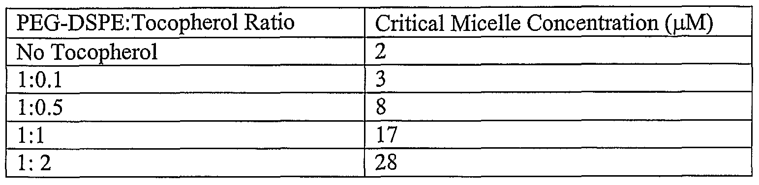
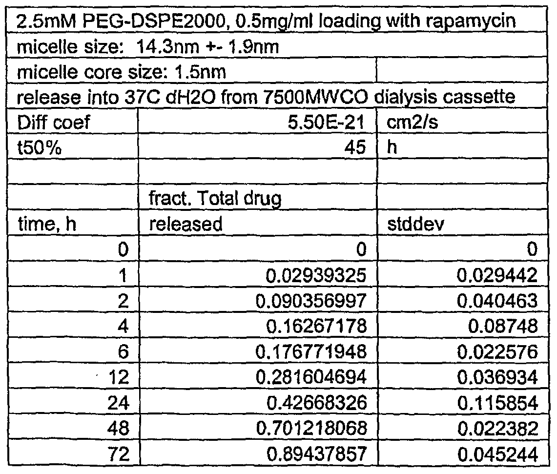


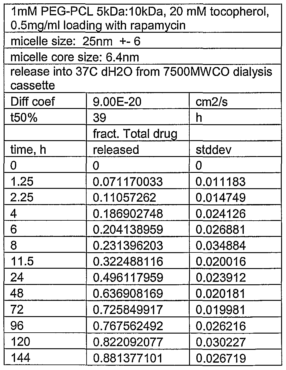


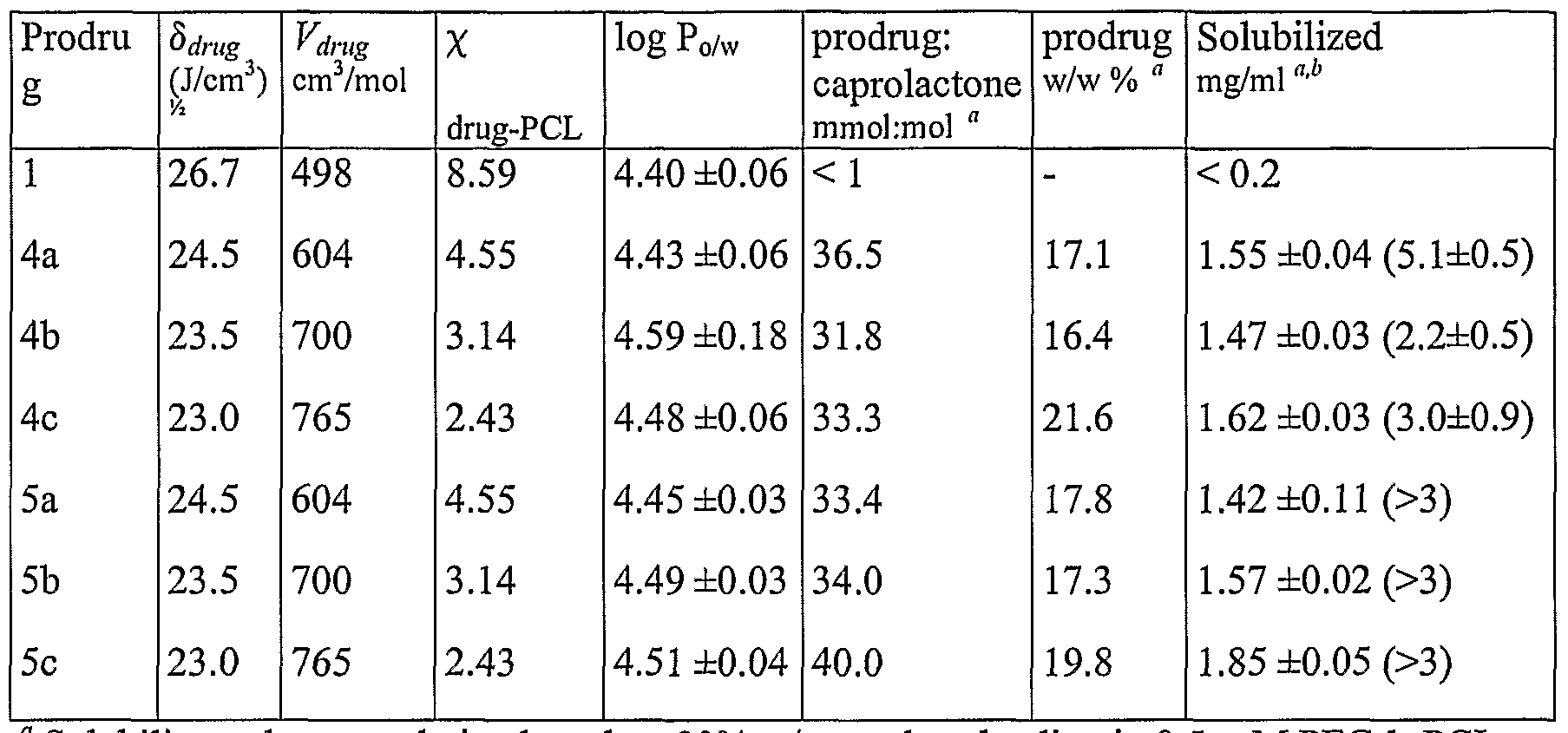
Claims
Priority Applications (4)
| Application Number | Priority Date | Filing Date | Title |
|---|---|---|---|
| EP06758321A EP1871343A2 (en) | 2005-04-12 | 2006-04-12 | Micelle composition of polymer and passenger drug |
| JP2008506677A JP2008535927A (en) | 2005-04-12 | 2006-04-12 | Polymer and passenger drug micelle composition |
| CA002603987A CA2603987A1 (en) | 2005-04-12 | 2006-04-12 | Micelle composition of polymer and passenger drug |
| AU2006235538A AU2006235538A1 (en) | 2005-04-12 | 2006-04-12 | Micelle composition of polymer and passenger drug |
Applications Claiming Priority (4)
| Application Number | Priority Date | Filing Date | Title |
|---|---|---|---|
| US67046005P | 2005-04-12 | 2005-04-12 | |
| US60/670,460 | 2005-04-12 | ||
| US71600005P | 2005-09-09 | 2005-09-09 | |
| US60/716,000 | 2005-09-09 |
Publications (3)
| Publication Number | Publication Date |
|---|---|
| WO2006110862A2 true WO2006110862A2 (en) | 2006-10-19 |
| WO2006110862A3 WO2006110862A3 (en) | 2007-07-05 |
| WO2006110862A9 WO2006110862A9 (en) | 2008-08-07 |
Family
ID=37011968
Family Applications (1)
| Application Number | Title | Priority Date | Filing Date |
|---|---|---|---|
| PCT/US2006/013800 WO2006110862A2 (en) | 2005-04-12 | 2006-04-12 | Micelle composition of polymer and passenger drug |
Country Status (7)
| Country | Link |
|---|---|
| US (2) | US8173167B2 (en) |
| EP (1) | EP1871343A2 (en) |
| JP (1) | JP2008535927A (en) |
| KR (1) | KR20080009196A (en) |
| AU (1) | AU2006235538A1 (en) |
| CA (1) | CA2603987A1 (en) |
| WO (1) | WO2006110862A2 (en) |
Cited By (11)
| Publication number | Priority date | Publication date | Assignee | Title |
|---|---|---|---|---|
| WO2009009067A2 (en) * | 2007-07-09 | 2009-01-15 | Kwon Glen S | Micelle encapsulation of theropeutic agents |
| US8383136B2 (en) | 2009-09-25 | 2013-02-26 | Wisconsin Alumni Research Foundation | Micelle encapsulation of therapeutic agents |
| CN103142479A (en) * | 2013-03-29 | 2013-06-12 | 中国药科大学 | Application of phospholipid-Vitamin E tocopherol acid polyethylene glycol succinate micelle |
| WO2015103447A1 (en) | 2013-12-31 | 2015-07-09 | Rapamycin Holdings, Llc | Oral rapamycin nanoparticle preparations and use |
| WO2015161139A1 (en) | 2014-04-16 | 2015-10-22 | Rapamycin Holdings, Llc | Oral rapamycin preparation and use for stomatitis |
| US9173841B2 (en) | 2009-12-30 | 2015-11-03 | Samyang Biopharmaceuticals Corporation | Polymer nanoparticle injection formulation composition containing rapamycin with improved water solubility, preparation method thereof, and anticancer composition for combined use with radiotherapy |
| CN105232459A (en) * | 2015-09-24 | 2016-01-13 | 沈阳药科大学 | Redissolving self-assembly type poorly-water-soluble drug-polymer micelle composition and preparation method thereof |
| AU2017225072B2 (en) * | 2009-12-30 | 2019-01-31 | Caliber Therapeutics, Llc | Balloon catheter systems for delivery of dry drug delivery vesicles to a vessel in the body |
| US10207084B2 (en) | 2009-12-30 | 2019-02-19 | Caliber Therapeutics, Inc. | Balloon catheter systems for delivery of dry drug delivery vesicles to a vessel in the body |
| EP3322404A4 (en) * | 2015-07-15 | 2019-03-20 | Celator Pharmaceuticals, Inc. | Improved nanoparticle delivery systems |
| IT202000007228A1 (en) * | 2020-04-06 | 2021-10-06 | Diego Dolcetta | ADMINISTRATION OF mTOR INHIBITORS IN THE CENTRAL NERVOUS SYSTEM |
Families Citing this family (36)
| Publication number | Priority date | Publication date | Assignee | Title |
|---|---|---|---|---|
| WO2003082303A1 (en) * | 2002-03-29 | 2003-10-09 | Wisconsin Alumni Research Foundation | Polymeric micelle formulations of hydrophobic compounds and methods |
| US20080082036A1 (en) * | 2006-04-25 | 2008-04-03 | Medtronic, Inc. | Cerebrospinal fluid shunt having long term anti-occlusion agent delivery |
| WO2007134245A2 (en) * | 2006-05-12 | 2007-11-22 | Wisconsin Alumni Research Foundation | Elastin-like polymer delivery vehicles |
| DE102006038233A1 (en) * | 2006-08-07 | 2008-02-14 | Biotronik Vi Patent Ag | Marker composite for medical implants |
| US20080102127A1 (en) * | 2006-10-26 | 2008-05-01 | Gao Hai Y | Hybrid lipid-polymer nanoparticulate delivery composition |
| JP2010519305A (en) * | 2007-02-26 | 2010-06-03 | ウィスコンシン・アルムニ・リサーチ・ファウンデーション | Polymeric micelles for combined drug delivery |
| US20100210575A1 (en) * | 2007-06-29 | 2010-08-19 | Wisconsin Alumni Research Foundation | Structuring effect of cholesterol in peg-phospholipid micelles, drug delivery of amphotericin b, and combination antifungals |
| WO2009070302A1 (en) * | 2007-11-30 | 2009-06-04 | The Johns Hopkins University | Prostate specific membrane antigen (psma) targeted nanoparticles for therapy of prostate cancer |
| WO2009089076A2 (en) * | 2008-01-10 | 2009-07-16 | Teva Pharmaceutical Industries Ltd. | Processes for the preparation and purification of paliperidone palmitate |
| US20090232762A1 (en) * | 2008-03-11 | 2009-09-17 | May Pang Xiong | Compositions for delivery of therapeutic agents |
| WO2010068432A1 (en) * | 2008-11-25 | 2010-06-17 | Ecole Polytechnique Federale De Lausanne (Epfl) | Block copolymers and uses thereof |
| CN102481361B (en) * | 2009-04-16 | 2014-10-22 | 默沙东公司 | Compositions and methods for treating cancer |
| US20120082717A1 (en) * | 2009-06-05 | 2012-04-05 | Snu R&Db Foundation | Complex, multilayer using the same, and device coated with the multilayer |
| US9283211B1 (en) | 2009-11-11 | 2016-03-15 | Rapamycin Holdings, Llc | Oral rapamycin preparation and use for stomatitis |
| EP2382966A1 (en) * | 2010-03-12 | 2011-11-02 | DSM IP Assets B.V. | Micelle compositions and process for the preparation thereof |
| US9289476B2 (en) | 2011-04-29 | 2016-03-22 | Selecta Biosciences, Inc. | Tolerogenic synthetic nanocarriers for allergy therapy |
| WO2012151544A1 (en) | 2011-05-05 | 2012-11-08 | Wisconsin Alumni Research Foundation | Micelles for the solubilization of gossypol |
| CN102293736B (en) * | 2011-08-31 | 2013-06-05 | 兰州大学 | Preparation process of taxol-polymer medicine-carrying micelles |
| JP2013203680A (en) * | 2012-03-28 | 2013-10-07 | Kobe Gakuin | Vitamin e derivative-containing drug carrier particle with retentivity in blood |
| CN103845290A (en) | 2012-12-03 | 2014-06-11 | 曼丽国际有限公司 | Use of UMIROLIMUS and its derivatives for treating cancer |
| CA2894871A1 (en) * | 2012-12-12 | 2014-06-19 | University Of Pittsburgh - Of The Commonwealth System Of Higher Education | Formulations and carrier systems including compound interactive domains |
| WO2014179772A2 (en) | 2013-05-03 | 2014-11-06 | Selecta Biosciences, Inc. | Tolerogenic synthetic nanocarriers to reduce or prevent anaphylaxis in response to a non-allergenic antigen |
| US10682415B2 (en) | 2013-07-22 | 2020-06-16 | Wisconsin Alumni Research Foundation | Thermogel formulation for combination drug delivery |
| US10711106B2 (en) | 2013-07-25 | 2020-07-14 | The University Of Chicago | High aspect ratio nanofibril materials |
| US9700544B2 (en) * | 2013-12-31 | 2017-07-11 | Neal K Vail | Oral rapamycin nanoparticle preparations |
| HUP1400075A2 (en) | 2014-02-14 | 2015-08-28 | Druggability Technologies Ip Holdco Jersey Ltd | Complexes of sirolimus and its derivatives, process for the preparation thereof and pharmaceutical composition containing them |
| US9855341B2 (en) | 2014-02-19 | 2018-01-02 | University of Pittsburgh—of the Commonwealth System of Higher Education | Formulations and carrier systems including farnesylthiosalycylic moities |
| CA2954064C (en) | 2014-07-02 | 2018-10-30 | The Research Foundation For The State University Of New York | Surfactant-stripped micelle compositions with high cargo to surfactant ratio |
| WO2016037165A1 (en) | 2014-09-07 | 2016-03-10 | Selecta Biosciences, Inc. | Methods and compositions for attenuating exon skipping anti-viral transfer vector immune responses |
| US10232050B1 (en) | 2014-12-12 | 2019-03-19 | Clemson University | Multi-functional particles and methods of using the same |
| US11633355B2 (en) | 2014-12-12 | 2023-04-25 | Clemson University Research Foundation | Multi-functional particles and methods of using the same |
| KR20190124295A (en) | 2017-03-11 | 2019-11-04 | 셀렉타 바이오사이언시즈, 인크. | Methods and compositions related to combination treatment with synthetic nanocarriers comprising anti-inflammatory agents and immunosuppressants |
| US20190029970A1 (en) * | 2017-07-31 | 2019-01-31 | The Chinese University Of Hong Kong | Fatty acid conjugated nanoparticles and uses thereof |
| WO2019204799A1 (en) | 2018-04-20 | 2019-10-24 | University Of Pittsburgh -Of The Commonwealth System Of Higher Education | Cationic amphiphilic polymers for codelivery of hydrophobic agents and nucleic acids |
| KR102051593B1 (en) | 2018-11-15 | 2020-01-08 | 재단법인대구경북과학기술원 | Dual-responsive amphiphiles, Manufacturing method thereof and Drug delivery using the same |
| CN111358754A (en) * | 2020-03-13 | 2020-07-03 | 广州白云山汉方现代药业有限公司 | Long-circulating emulsion of macrolide antibiotics and preparation method thereof |
Citations (7)
| Publication number | Priority date | Publication date | Assignee | Title |
|---|---|---|---|---|
| US5580899A (en) * | 1995-01-09 | 1996-12-03 | The Liposome Company, Inc. | Hydrophobic taxane derivatives |
| WO1997010234A1 (en) * | 1995-09-12 | 1997-03-20 | The Liposome Company, Inc. | Hydrolysis-promoting taxane hydrophobic derivatives |
| WO2000009071A2 (en) * | 1998-08-11 | 2000-02-24 | All India Institute Of Medical Sciences | A novel liposomal formulation useful in treatment of cancer and other proliferation diseases |
| US6284267B1 (en) * | 1996-08-14 | 2001-09-04 | Nutrimed Biotech | Amphiphilic materials and liposome formulations thereof |
| US20030114450A1 (en) * | 2001-08-06 | 2003-06-19 | Daniel Santi | Benzoquinone ansamycins |
| WO2003105765A2 (en) * | 2002-06-12 | 2003-12-24 | The Board Of Trustees Of The University Of Illinois | Phospholipid micelles in liposomes as solubilizers for water-insoluble compounds |
| WO2005046671A1 (en) * | 2003-11-07 | 2005-05-26 | Baxter International Inc. | Method for preparing submicron particles of paclitaxel |
Family Cites Families (15)
| Publication number | Priority date | Publication date | Assignee | Title |
|---|---|---|---|---|
| US6090414A (en) * | 1970-05-20 | 2000-07-18 | Life Science Labs, Inc. | Method and composition to reduce cancer incidence |
| US4261989A (en) | 1979-02-19 | 1981-04-14 | Kaken Chemical Co. Ltd. | Geldanamycin derivatives and antitumor drug |
| US5536729A (en) * | 1993-09-30 | 1996-07-16 | American Home Products Corporation | Rapamycin formulations for oral administration |
| US6107332A (en) * | 1995-09-12 | 2000-08-22 | The Liposome Company, Inc. | Hydrolysis-promoting hydrophobic taxane derivatives |
| US6458373B1 (en) * | 1997-01-07 | 2002-10-01 | Sonus Pharmaceuticals, Inc. | Emulsion vehicle for poorly soluble drugs |
| US6322810B1 (en) | 1997-07-14 | 2001-11-27 | Hayat Alkan-Onyuksel | Materials and methods for making improved micelle compositions |
| US6217886B1 (en) * | 1997-07-14 | 2001-04-17 | The Board Of Trustees Of The University Of Illinois | Materials and methods for making improved micelle compositions |
| US6469132B1 (en) | 1999-05-05 | 2002-10-22 | Mcgill University | Diblock copolymer and use thereof in a micellar drug delivery system |
| MXPA02010999A (en) * | 2000-05-12 | 2003-03-10 | Samyang Corp | Method for the preparation of polymeric micelle via phase separation of block copolymer. |
| US6764507B2 (en) * | 2000-10-16 | 2004-07-20 | Conor Medsystems, Inc. | Expandable medical device with improved spatial distribution |
| TWI246524B (en) * | 2001-01-19 | 2006-01-01 | Shearwater Corp | Multi-arm block copolymers as drug delivery vehicles |
| US6939561B2 (en) | 2001-06-28 | 2005-09-06 | Wisconsin Alumni Research Foundation | Methods and compositions for polyene antibiotics with reduced toxicity |
| WO2004034992A2 (en) | 2002-10-15 | 2004-04-29 | Wisconsin Alumni Research Foundation | Encapsulation and deaggregation of polyene antibiotics using poly(ethylene glycol)-phospholipid micelles |
| US20050026893A1 (en) | 2003-05-30 | 2005-02-03 | Kosan Biosciences, Inc. | Method for treating diseases using HSP90-inhibiting agents in combination with immunosuppressants |
| CA2574767C (en) | 2004-07-19 | 2015-02-17 | Celator Pharmaceuticals, Inc. | Particulate constructs for release of active agents |
-
2006
- 2006-04-12 WO PCT/US2006/013800 patent/WO2006110862A2/en active Application Filing
- 2006-04-12 EP EP06758321A patent/EP1871343A2/en not_active Withdrawn
- 2006-04-12 KR KR1020077025059A patent/KR20080009196A/en not_active Application Discontinuation
- 2006-04-12 AU AU2006235538A patent/AU2006235538A1/en not_active Abandoned
- 2006-04-12 US US11/402,639 patent/US8173167B2/en not_active Expired - Fee Related
- 2006-04-12 JP JP2008506677A patent/JP2008535927A/en active Pending
- 2006-04-12 CA CA002603987A patent/CA2603987A1/en not_active Abandoned
-
2012
- 2012-04-30 US US13/460,366 patent/US9107814B2/en not_active Expired - Fee Related
Patent Citations (7)
| Publication number | Priority date | Publication date | Assignee | Title |
|---|---|---|---|---|
| US5580899A (en) * | 1995-01-09 | 1996-12-03 | The Liposome Company, Inc. | Hydrophobic taxane derivatives |
| WO1997010234A1 (en) * | 1995-09-12 | 1997-03-20 | The Liposome Company, Inc. | Hydrolysis-promoting taxane hydrophobic derivatives |
| US6284267B1 (en) * | 1996-08-14 | 2001-09-04 | Nutrimed Biotech | Amphiphilic materials and liposome formulations thereof |
| WO2000009071A2 (en) * | 1998-08-11 | 2000-02-24 | All India Institute Of Medical Sciences | A novel liposomal formulation useful in treatment of cancer and other proliferation diseases |
| US20030114450A1 (en) * | 2001-08-06 | 2003-06-19 | Daniel Santi | Benzoquinone ansamycins |
| WO2003105765A2 (en) * | 2002-06-12 | 2003-12-24 | The Board Of Trustees Of The University Of Illinois | Phospholipid micelles in liposomes as solubilizers for water-insoluble compounds |
| WO2005046671A1 (en) * | 2003-11-07 | 2005-05-26 | Baxter International Inc. | Method for preparing submicron particles of paclitaxel |
Non-Patent Citations (1)
| Title |
|---|
| LUNDBERG B B ET AL: "A lipophilic paclitaxel derivative incorporated in a lipid emulsion for parenteral administration." JOURNAL OF CONTROLLED RELEASE : OFFICIAL JOURNAL OF THE CONTROLLED RELEASE SOCIETY 9 JAN 2003, vol. 86, no. 1, 9 January 2003 (2003-01-09), pages 93-100, XP002419233 ISSN: 0168-3659 * |
Cited By (21)
| Publication number | Priority date | Publication date | Assignee | Title |
|---|---|---|---|---|
| WO2009009067A3 (en) * | 2007-07-09 | 2009-04-30 | Glen S Kwon | Micelle encapsulation of theropeutic agents |
| WO2009009067A2 (en) * | 2007-07-09 | 2009-01-15 | Kwon Glen S | Micelle encapsulation of theropeutic agents |
| US8383136B2 (en) | 2009-09-25 | 2013-02-26 | Wisconsin Alumni Research Foundation | Micelle encapsulation of therapeutic agents |
| US8529917B2 (en) | 2009-09-25 | 2013-09-10 | Wisconsin Alumni Research Foundation | Micelle encapsulation of a combination of therapeutic agents |
| US8858965B2 (en) | 2009-09-25 | 2014-10-14 | Wisconsin Alumni Research Foundation | Micelle encapsulation of a combination of therapeutic agents |
| AU2017225072B2 (en) * | 2009-12-30 | 2019-01-31 | Caliber Therapeutics, Llc | Balloon catheter systems for delivery of dry drug delivery vesicles to a vessel in the body |
| AU2020281081B2 (en) * | 2009-12-30 | 2022-06-23 | Caliber Therapeutics, Llc | Balloon catheter systems for delivery of dry drug delivery vesicles to a vessel in the body |
| US10806909B2 (en) | 2009-12-30 | 2020-10-20 | Caliber Therapeutics, Llc | Balloon catheter systems for delivery of dry drug delivery vesicles to a vessel in the body |
| US9173841B2 (en) | 2009-12-30 | 2015-11-03 | Samyang Biopharmaceuticals Corporation | Polymer nanoparticle injection formulation composition containing rapamycin with improved water solubility, preparation method thereof, and anticancer composition for combined use with radiotherapy |
| AU2019202994B2 (en) * | 2009-12-30 | 2020-09-03 | Caliber Therapeutics, Llc | Balloon catheter systems for delivery of dry drug delivery vesicles to a vessel in the body |
| US10207084B2 (en) | 2009-12-30 | 2019-02-19 | Caliber Therapeutics, Inc. | Balloon catheter systems for delivery of dry drug delivery vesicles to a vessel in the body |
| CN103142479A (en) * | 2013-03-29 | 2013-06-12 | 中国药科大学 | Application of phospholipid-Vitamin E tocopherol acid polyethylene glycol succinate micelle |
| EP3089737A4 (en) * | 2013-12-31 | 2017-09-06 | Rapamycin Holdings, LLC | Oral rapamycin nanoparticle preparations and use |
| AU2014373683B2 (en) * | 2013-12-31 | 2020-05-07 | Rapamycin Holdings, Llc | Oral rapamycin nanoparticle preparations and use |
| WO2015103447A1 (en) | 2013-12-31 | 2015-07-09 | Rapamycin Holdings, Llc | Oral rapamycin nanoparticle preparations and use |
| EP3131546A4 (en) * | 2014-04-16 | 2017-12-27 | Rapamycin Holdings, Inc. | Oral rapamycin preparation and use for stomatitis |
| WO2015161139A1 (en) | 2014-04-16 | 2015-10-22 | Rapamycin Holdings, Llc | Oral rapamycin preparation and use for stomatitis |
| EP3322404A4 (en) * | 2015-07-15 | 2019-03-20 | Celator Pharmaceuticals, Inc. | Improved nanoparticle delivery systems |
| CN105232459A (en) * | 2015-09-24 | 2016-01-13 | 沈阳药科大学 | Redissolving self-assembly type poorly-water-soluble drug-polymer micelle composition and preparation method thereof |
| IT202000007228A1 (en) * | 2020-04-06 | 2021-10-06 | Diego Dolcetta | ADMINISTRATION OF mTOR INHIBITORS IN THE CENTRAL NERVOUS SYSTEM |
| WO2021205297A1 (en) * | 2020-04-06 | 2021-10-14 | Diego Dolcetta | ADMINISTRATION OF mTOR INHIBITORS INTO THE CENTRAL NERVOUS SYSTEM |
Also Published As
| Publication number | Publication date |
|---|---|
| WO2006110862A3 (en) | 2007-07-05 |
| WO2006110862A9 (en) | 2008-08-07 |
| JP2008535927A (en) | 2008-09-04 |
| US9107814B2 (en) | 2015-08-18 |
| US20120309780A1 (en) | 2012-12-06 |
| AU2006235538A1 (en) | 2006-10-19 |
| US8173167B2 (en) | 2012-05-08 |
| EP1871343A2 (en) | 2008-01-02 |
| KR20080009196A (en) | 2008-01-25 |
| US20060251710A1 (en) | 2006-11-09 |
| CA2603987A1 (en) | 2006-10-19 |
Similar Documents
| Publication | Publication Date | Title |
|---|---|---|
| US9107814B2 (en) | Micelle composition of polymer and passenger drug | |
| Straubinger | Biopharmaceutics of paclitaxel (Taxol): formulation, activity, and pharmacokinetics | |
| EP2480207B1 (en) | Micelle encapsulation of therapeutic agents | |
| AU730599B2 (en) | A method of administering liposomal encapsulated taxane | |
| AU2017350499B2 (en) | Liposomal formulation for use in the treatment of cancer | |
| US20100203114A1 (en) | Micelle encapsulation of therapeutic agents | |
| WO2006102768A1 (en) | Colloidal solid lipid vehicle for pharmaceutical use | |
| JP2002535242A (en) | New particulate dosage form | |
| KR101132626B1 (en) | Method for preparing HDL nanoparticles for the delivery of hydrophobic drugs | |
| CN108289833B (en) | Stabilized assembled nanostructures for delivery of encapsulants | |
| WO2008151433A1 (en) | Injectable polymer-lipid blend for localized drug delivery | |
| BR112020006191A2 (en) | copolymers in amphiphilic blocks, micelles and methods for treating or preventing heart failure | |
| WO2012151544A1 (en) | Micelles for the solubilization of gossypol | |
| WO2005123078A1 (en) | Liposome preparation containing slightly water-soluble camptothecin | |
| WO2014167435A2 (en) | Liposomal pharmaceutical formulations for enhanced drug delivery | |
| US8859001B2 (en) | Fenoldopam formulations and pro-drug derivatives | |
| US7314637B1 (en) | Method of administering liposomal encapsulated taxane | |
| JP2006124351A (en) | Sustained release antitumor agent composition | |
| WO2001070220A1 (en) | A method of administering liposomal encapsulated taxane | |
| Lu | TARGETED DELIVERY OF ANTICANCER AGENTS TO CANCER |
Legal Events
| Date | Code | Title | Description |
|---|---|---|---|
| WWE | Wipo information: entry into national phase |
Ref document number: 2006235538 Country of ref document: AU |
|
| ENP | Entry into the national phase |
Ref document number: 2603987 Country of ref document: CA |
|
| WWE | Wipo information: entry into national phase |
Ref document number: MX/a/2007/012632 Country of ref document: MX |
|
| ENP | Entry into the national phase |
Ref document number: 2008506677 Country of ref document: JP Kind code of ref document: A |
|
| NENP | Non-entry into the national phase |
Ref country code: DE |
|
| WWE | Wipo information: entry into national phase |
Ref document number: 2006758321 Country of ref document: EP |
|
| ENP | Entry into the national phase |
Ref document number: 2006235538 Country of ref document: AU Date of ref document: 20060412 Kind code of ref document: A |
|
| WWE | Wipo information: entry into national phase |
Ref document number: 1020077025059 Country of ref document: KR |
|
| NENP | Non-entry into the national phase |
Ref country code: RU |