INTEGRIN CD18 IS A NOVEL STROMAL STEM CELL MARKER AND FUNCTIONS TO PROMOTE OSTEOGENESIS
CROSS-REFERENCE TO RELATED APPLICATIONS
[0001] This application claims the benefit of Provisional Application No. 60/606,982, filed September 3, 2004, and Provisional Application No. 60/690,767, filed June 15, 2005, the entire contents of which are hereby incorporated by reference as if fully set forth herein, under 35 U.S.C. §119(e).
STATEMENT OF GOVERNMENTAL INTEREST
[0002] This invention was made with Government support under Grant # HL61589. The Government has certai n rights i n the i nvention. This i nvention was also ki ndly supported with funds from the American Heart Association, Grant no. 0240208N.
BACKGROUND OF THE INVENTION
1. ReId of the I nvention
[0003] The present invention relates to cell surface proteins on bone marrow stromal stem cells (BMSSCs), in particular CD18, which can be used as a marker for making stem cell enriched extracts from unfractionated bone marrow aspirates and from other sources such as adipose tissue Theinvention also relates to methods of using the BM SSCs for the treatment of diseases, especially bone marrow diseases.
2. Description of the Related Art
[0004] Bone marrow contains stem-like precursors for non-hematopoietic cells, such as osteoblasts, chondrocytes, adipocytes and myoblasts (Owen etal., 1988, in CeI I and Molecular Biology of Vertebrate Hard Tissues, Ciba Foundation Symposium 136, Chichester, UK, pp. 42-60; Caplan, 1991, J. Orthop. Res. 9:641-650; Prockop, 1997, Science 276:71-74). Non-hematopoietic precursors of the bone marrow have been variously referred to ascolony-forming-unit-fibroblasts, mesenchymal stem cells, and
bone marrow stromal stem cells (BMSSCs). BM SSCs are believed to participate in the creation of the microenvironment within the bone marrow in vivo. When isolated, BMSSCs are initially quiescent but eventually begin dividing so that they can be cultured in vitro. Expanded numbers of stromal cells can be established and maintained. Stromal eel Is have been used to generate colonies of fibroblastic adipocytic and osteogenic cells when cultured under appropriate conditions. They can also be made to differentiate into cartilage cell s and myoblasts.
[0005] There are several examples of the use of BMSSCs for treatment of disease. Stromal cells have been used to produce fibrous tissue, bone or cartilage when implanted into selective tissues in vivo (Ohgushi et al., 1989, Acta Orthop. Scand. 60:334-339; Nakaharaet al., 1992, J. Orthop. Res. 9:465-476; Niedzwiedski et al., 1993, Biomaterials 14:115-121; and Wakitani et al., 1994, J. Bone& Surg. 76A:579-592). In some reports, stromal eel Is have been used to generate bone or carti I age in vivo when i mpl anted subcutaneously with a porous ceramic (Ohgushi, et al., 1989, Acta Orthop. Scand. 60:334-339), intraperitoneal Iy in adiffuaon chamber (Nakaharaet al., 1991, J. Orthop. Res. 9:465-476), percutaneously into a surgical Iy induced bone defect (Niedzwiedski, et al., 1993, Biomaterials 14: 115-121), or transplanted within a collagen gel to repair a surgical defect in ajoint cartilage (Wakitani et al., 1994, J. BoneSurg. 76A: 579-592). Rersmaet al. (1983, Brit. J. Hematol. 94:285-290) disclose that after intravenous bone marrow transplantation, the fibroblast colony-forming cells which make up the hemopoietic stroma lodge and remain inthehost bone marrow. Stewart et al. (1993, Blood 81:2566-2571) recently observed that unusually large and repeated administrations of whole marrow cells produced long-term engraftment of hematopoietic precursors into mice that had not undergone marrow ablation. Also, Bienzleet al. (1994, Rroc. Natl. Acad. Sci. USA, 91:350-354) successfully used long-term bone marrow cultures as donor cellsto permanently populate hematopoietic cells in dogs without marrow ablation. In some reports, stromal cells were used either as eel Is that established a microenvironment for the culture of hematopoietic precursors (Anklesaria, 1987, PNAS USA 84:7681-7685) or as a sourceof an enriched population of hematopoietic stem cells (Kiefer, 1991, Blood 78(10):2577-2582).
[0006] This new research hasshown that adult stem cellsin fact possess much wider potential for differentiation than previously thought. The identification and isolation
of the stem cellsenablesthe ranfusion of these long-term repopulating stem cells in various clinical therapies. For example, purging bone marrow stem cells of contaminating tumor cells would requirereintroduction of new uncontaminated stem cells, In gene therapy, stem cells either from a patient or adonor can betransfected to contain new genes of therapeutic use and then reintroduced into the patient. Identification of surface markers on stem cells isextremely useful in hematopoietic research and related therapies because such markers al I ow the i sol ati on of rel ati vdy pure populati ons of i mmature stem eel I s. However, thelack of appropriate surface markers has been a major obstaclefor incorporating bone marrow stromal stem cells (BMSSCs) in clinical applicationa(4) At present the only useful marker for this purpose is STRO-1. Thus, there is a continued need for the identification other antigens on BM SCCs to simplify the identification and separation of these eel Is from bone marrow aspirates (or from peripheral blood or adipose tissue which also have BMSSCs).
DEFINITIONS
[0007] The term " CD18" refers to the β2 i ntegri ns, i ncl udi ng aφ2, αM β2, αx β2 and αDβ2- CD18 isacell adhesion molecule.
[0008] The term "BMSSCs" means bone marrow stem cells, BMSSCsinclude those bone marrow stem cellsthat express the CD18 antigen, among other surface antigens, and include pluripotent stem cells A cell is operationally defined asCD18- positiveif it expresses sufficient CD18 antigen to be detected by a given method of assay including flow microfluorimetry using a fluorescence-activated cell sorter (FACS), immunofluorescence or immunoperoxidase staining using a fluorescence or light microscope, fluorescence-activated cell sorting, immunoblotting, panning (Wysocki and Sato, Proc. Natl. Acad. SeL (USA) 75: 2844, 1978), radioimmunoassay, immunoaffinity chromatography (Basch et al., J. Immunol. Methods 56: 269, 1983), magnetic-activated cell sorting (Miltenyi et al., Cytometry 11:231, 1990), and cytolysis, among numerous other methods which will be readily apparent to one skilled in the art (see, for example, Lansdorp and Thomas in Bone Marrow Rrocessing and Purging, A. P. Gee (ed.), Boca Raton: CRC Press (1991) pg. 351), the entire contents of which are hereby incorporated by reference as if fully set forth herein, under 35 U.S.C. §119(e). [0009] The term "osteogenic differentiation of BMSSCs" refers to a process by which BMSSCs become osteoblastic cells and form bone tissue under defined in vitro culture conditions or in vivo microenvironment. Part of normal osteogenic differentiation of BM SSCs requi res osteogeni c master protei n Cbf a1.
[0010] The term " osteoclastic activity" refers to the bone resorption activity associated with osteoclasts.
[0011] The term " Hematopoiesis-supportive" refers to the function of BMSSCs in supporting hematopoietic cellsgrowth and survival in vitro and in vivo.
BRIEF DESCRIPTION OF THE DRAWINGS
[0012] The present invention is illustrated by way of example, and not by way of limitation, in the figures of the accompanying drawings and in which like reference numerals refer to similar elements and in which:
[0013] FIG. 1 FIG. 1A shows expression of CD18 on mouse BMSSCs by fluorescence assisted cell sorting analysis (FACS) using monoclonal antibodies (mAbs) against CD18 (on the Y axis) with one mAb from the group Sca-1, CD14, CD34 and FITC-IgGI (on theX axis). FIG. 1B shows immunoblot analyses of murineand human BMSSCa Equal protein loading was confirmed by re-probing with mAbs specific for β- actin or α-actinin. The data shown are representative of two independent experiments. [0014] FIG. 2 shows FACS analysis of Human STRaI0119711ZCDIe+ BMSSCs.
FIG. 2A showstwo distinct human cell populations collected by FACS (R2: STRO- 1Dn9ra/CD18+ and R3: STRO-T^ZCDIβ"). FIG. 2B shows CFU-F assays of thecellsin FIG. 2A. FIG. 2C shows the phenoty pic characterization of human R2 STRaI00901ZCDIS+ BMSSCs by FACS analysis using different lineage-specific mAbs. Their corresponding isotype-matched non-immune IgGs were used as controls (bold line). FIG. 2D shows the differentiation potential of the STRaI0119111ZCDIe+ BMSSCs cultured in vitro under adipogenic induction condition for 2 weeks (a), osteogenic induction condition for 3 weeks (b), and chondrogenic induction condition for 3 weeks (c). Oil Red O staining demonstrates the generation of lipid-laden adipocytes (arrows) (a); Alizarin Red Sstaining shows mineral deposits (arrows) made by osteogenic cells (b); and Aldan blue staining of the cartilage matrix deposition (arrows) demonstrates the chondrogenic differentiation in aggregate cultures (c). The data shown are representative of two independent experiments, Magnification: a-b, x400; c, x64.
[0015] Fl G. 3 shows phenotypi c comparisons between CD18"'" mi ce and thei r WT sex-matched littermates. FIG. 1A isFaxitron analysis demonstrating a decreased bone density i n the femurs of 5-week-old CD18"'" mi ce (KO, right) as compared to thei r WT counterparts (left). FIG. 3B is DXA analysis of the femurs from 15-week-old mice showing a statistical Iy significant differencein BMD between WT (left) and CD18"7' (right) mice (p=0.025, n=4). FIG. X shows hematoxylin and eosin staining on the metaphysisareaof thefemura Magnification 400X. FIG. D is representative images of the
distal femur metaphysison micro-computed tomography analysis showing decreased bone volume, trabecular bone number, trabecular bone thickness, and an increased trabecular bone space. FIG. 3E is alizarin red and alcian blue double skeletal staining for bone and cartilage of 1-week-old mice. WT, wild type; KO, CD18"'".
[0016] FIG. 4 shows CD18"'' in vivo osteoclastic activity determined by the serum concentration of C-terminaJ telopeptidesof type 1 collagen in WT and CD18 deficient mice(p=0.68, n=3 mice). FIG. B shows the number of mature osteoclasts in the femurs of the WT and CD18"'' mice determined by TRAPstaining. (p=0.13, n=4 mice). Magnification x200.
[0017] FIG. 5 FIG. 5A shows in vitro mineralization induction of CD18"'"
BM SSCs determined by Alizarin red Sstaining (magnification x10) compared to WT cells (p=0.00003, n=3 mice). The total mineralized area by WT cells was assigned to 100%. Fl G. 5B shows eel I adhesi on of CD18~'~ BM SSCs compared to WT. Fl G. 5C shows proliferation of BMSSCs evaluated by BrdU incorporation with the percentage of BrdlT cellsdetermined manually by counting 10 representative fields. (p=0.01, n= 3 mice). FIG. 5D shows the number of BMSSC colonies obtained from 106 bone marrow cellsfor CD18" " mice vs. WT (p=0.0064, n=6 mice). Magnification x10.
[0018] FIG. 6 FIG. 6A showsCbfal expression and Smad2 phosphorylation by immunoblot. Equal protθ'n loading was verified by re-probing with an α-actinin-specific mAb. The data shown are representative of three i ndependent experi menta Fl G. 6B shows immunoblotsof retroviral -mediated expression of recombinant CD18 using an anti-CD18 cytoplasmic tail antibody, which does not react with CD18 (CT-) well. CD18"'' BMSSCs were infected with retroviral supernatants encoding either full length CD18 or the cytoplasmic tail-truncated CD18 (CT-) for 6 days. Non-infected WT and CD18"'' BMSSCs were i ncl uded as control & Protei n I oadi ng was shown by re-probi ng for α-acti ni n. R G.6C shows bone formation in vivo. WT, CD18"'", and retroviral infected CD18"'" BMSSCs with either full length CD18 or truncated CD18 (CT-) were mixed with HA/TCP and then implanted in nude micesubcutaneously. BMSSC-mediated bone formation was analyzed 7 weeks post-implantation by hematoxylin and eosin staining. Staining was quantified using the software NIH I mage based on five representative areas, and was expressed as a percentage of bone formation by WT BMSSCs, (p=0.008, n=4 mice). B=newly formed bone; HA=hydroxyapatite/tricalcium phosphate; and CT=connective tissues.
Magnification x200. FIG. 6D shows restoration of the osteogenic capability of theCDIβr'" BMSSCa (p=0.0001, n= 4 mice) but not CD18 (CT-) (p=0.39, n= 4 mice).
SUMMARY OF THE INVENTION
[0019] I n one aspect of the invention, a population of animal cellsenriched in bone marrow stromal stem cellsthat express the CD18 antigen isproduced by: obtaining a sampleof bone marrow from the animal, selecting bone marrow mononuclear cellsfrom the sample, contacting the bone marrow mononuclear cells of step (b) with a reagent that binds to theCD18 antigen under conditions that permit the reagent and theCD18 antigen to bind; and selecting the bone marrow mononuclear cellsthat are bound to the reagent, thereby obtaining the population of animal cellsenriched in bone marrow stromal stem cellsthat express the CD18 antigen. In another preferred embodiment a population of animal cellsenriched in bone marrow stromal stem eel Is are selected by: obtaining a sample of bone marrow from the animal, (b) selecting bone marrow mononuclear cells from the sample, (c) contacting the bone marrow mononuclear cells of step (b) with a first reagent that binds to theSTRO-1 antigen under conditions that permit the first reagent and theSTRO-1 antigen to bind; (d) selecting the bone marrow mononuclear cellsthat are bound to the first reagent, thereby obtaining bone marrow stromal stem cellsthat express theSTRO-1 antigen, (e) contacting cells of step (d) with a second reagent that binds to the CD18 antigen under conditions that permit the second reagent and theCD18 antigen to bind, and (f) selecting cdlsof step (e) to obtain bone marrow stromal stem cellsthat express both theCD18 and STRO-1 antigens, thereby obtaining the population of animal cellsenriched in bone marrow stromal stem cells. The reagents are preferably antibodies directed against the BMSSC markers, but any agent that selectively binds to the BMSSC marker can be used. I n an aspect, the reagents are labeled for easy detection, for example with fluorescent markers.
[0020] In an aspect of theinvention, BMSSCs are derived from peripheral blood or adipocytes. In an aspect of theinvention, the BMSSC-enriched population is obtained using fluorescent assisted cell sorting, an immunomagnetic method, flow micrσfluorimetry, immunofluorescence, immunoperoxidasestaining, radioimmunoassay or immunoaffinity chromatography. I n another aspect the invention is directed to an isolated population of animal cellsenriched in bone marrow stromal stem cellsthat express the CD18 antigen or both CD18 and the STRO-1 antigen produced according to the methods descri bed herei n.
[0021] An aspect of the invention isfurther directed to chondrocytes, adipocytes, neurons and osteoblasts derived from BMSSCs that were isolated using CD18 as a selection marker. Another aspect isdirected to CD18 as a selection marker for isolating enriched populations of BMSSCa Other aspects are directed to methods for treating bone marrow diseases, leukocyte adhesion deficiency I, and bone degenerative diseases by administering to a patient having the disease or at risk for developing the disease, BMSSCs that have been transformed with an expression vector having a normal gene for CD18. A further aspect includes bone marrow stem cellsfrom an animal, which cells have been transformed with an expression vector having a normal gene for CD18
DETAILED DESCRIPTION
[0022] It has been discovered that CD18 antigen is expressed on the surface of
BM SSCs and can be used as a selective marker to obtain a population of animal cells enriched in bone marrow stromal stem cells. CD18 is also referred to in the literature as p2 i ntegrins of which there are four (αLβ2, αwiβa αχβ2, and αofc). One embodiment of the present invention is directed to the use of CD18 as a BMSSC marker to isolate a population of animal cellsenriched in bone marrow stromal stem cells using techniques known in the art such as fluorescent assisted cell sorting, an immunomagnetic method, flow microfluorimetry, immunofluorescence, immunoperoxidase staining, radioimmunoassay and immunoaffinity chromatography. An embodiment is directed to the BMSSC-enriched population of cellsthat isselected from asampleof the animal's bone bone marrow mononuclear cells (BM NCs) based on the selective expression of CD18 on the surface of BMSSCs and its binding to a reagent that specifically binds to CD18, such as an anti-CD18 antibody. Another embodiment isdirected to apopulation of animal cellsenriched in BMSSCs that isselected from asampleof the animal's bone marrow mononuclear cells (BMNCs) based on the selective expression of both CD18 and STRO-1 on the surface of BMSSCs, which population isselected based on the ability of BMSSCs to bind to both afirst reagent that specifically binds to CD18 antigen, and to an second reagent that specifically binds to STRO-1 antigen. Theinvention isfurther directed to methods for obtaining these populations of BMSSC-enriched animal ceils. [0023] BMSSCs that express CD18 or CD18 and STRO-1 are pleuripotent, and are capable of differentiating into chondrocytes, osteoblasts or adipocytes. Some have reported that BMSSCs can also differentiate into neurons and muscle Sanchez-Ramos, et al. have shown that bone marrow stromal cells (BMSC) differentiate into neuron-like phenotypes in vitro and in vivo, when engrafted into normal or denervated rat striatum. U.S. Ratent No. 6,528,245, the entire contents of which is incorporated by reference as if set forth fully herein. The BMSC did not remain localized to the site of the graft, but migrated throughout the brain and integrated into specific brain regions in various architectonic patterns. The most orderly integration of BMSCwasin thelaminar distribution of cerebellar Purkinjecells, where the BMSC-derived cellstcok on the Purkinje phenotype. The BMSC exhibited site-dependent differentiation and expressed
several neuronal markers including neuron-specific nuclear protein, tyrosine hydroxylase and calbindin. BMSC can be used to target specific brain nuclei in strategies of neural repair and gene therapy.
[0024] Certai n embodi ments are therefore di rected to chondrocytes, neurons, osteoblasts or adipocytes that differentiate from the population of cellsenriched in BMSSCs that areisolated based on their expression of CD18 or CD18 and STRO-1. [0025] Leukocyte Adhesion Deficiency typel (LADI) is a disease where one or more mutations occur in theCD18 subunit such that expression of all four CD18 integrins (αL.β2, otMp2, ocχβ2, and αDβ2> is abolished. Methods are provided for preventing or treating LADI in an animal by administering a population of cellsenriched in BMSSCs that express normal CD18 antigen, which BMSSCs were isolated from the patient using the methods of the present i nventi on from a heal thy ani ma! of the same sped es usi ng CD18 as a selective marker. In another embodiment, the ani ma! 'sown BMSSCsare isolated, transformed with an expression vector having gene for normal CD18, and rei ntroduced i nto the ani mal . I n a preferred embodi ment the ani mal i s a human. [0026] In the foil owing description, for thepurposesof explanation, numerous specif i c detai I s are set forth i n order to provi de a thorough understandi ng of the present invention. It will be apparent, however, to one skilled in the art that the present invention may be practiced without these specific details, ln other instances, well-known structures and devices are shown in block diagram form in order to avoid unnecessarily obscuring the present i nventi on.
[0027] Bone marrow stromal stem cells are potentially involved in the replenishment of awide range of cell types through an adult life, including osteoblasts, chondrocytes, adipocytes, and neural cells. U.S. Patent No. 6,528,245. Thus, BMSSCs represent an easily accessibleand renewable source of stem cells for tissue engineering to repair damaged tissues (18). I n addition, osteoblasts derived from BMSSCs are critical to the maintenance of the niche microenvironment for hematopoiesis (1,2). However, duein part to the I ack of specifi c surface markers for BM SSCs1 thei r therapeuti c use has been limited. It has now been discovered that CD18 is selectively expressed on bone marrow stromal stem cells a new and can be used as unique surface marker for separating BMSSCs from a heterogeneous population of cells, typically bone marrow mononuclear cells (BMNCs) in asampleof bone marrow. However, CD18 can also be used asa
selective marker to separate BMSSCs from a population of adipose cells or peripheral blood ceils. CD18 includes the four different heterodimeric receptors (αLβ2, αwiβa αχβ2, and αoβ2), which are primarily expressed on cells of hematopoietic origin (3). [0028] It has also been discovered that CD18 can be used together with STR0 1 as markers to separate BMSSCs from a heterogeneous population of cells, including BMNC in a bone marrow sample. Using CD18 in addition to STRO-1 enriched the population of i sol ated BM SSCs by a factor of 15 compared to the use of STR0 1 al one [0029] It was further discovered that genetic inactivation of CD18 in mice caused impaired osteogenic differentiation of BMSSCs through inhibition of osteogenic master protein Cbfai which leads to defective bone formation in vivo. CD18-deficient animals also showed decreased bone mineral density (BMD). By contrast, inactivation of CD18 had no significant effect on in vivo osteoclastic activity. The defective osteogenesis of the CD18-deficient BMSSCs was rescued by expression of full length but not a cytoplasmic domain-truncated CD18. This shows that CD18 is critical to the function of the BMSSCs; its deficiency causes a predisposition to bone defects Mice lacking CD18 exhibit certain features of osteoporosis, including decreased bone marrow density (BMD), reduced trabecular bone number, decreased trabecular bone thickness, and increased trabecular bone space.
[0030] Osteoporosis is characterized by excessive loss of bone and deterioration of bone tissue, due to an overall imbalance between osteoblast-mediated bone formation and osteoclast-mediated bone resorption. Osteoblasts are derived from the BMSSCs (18), and play critical rolesin the maintenance of bone mineral density (BMD) and in the formation of a bone marrow niche mi croenvironment that isessential for hematopoiesis(1, 2). When implanted subcutaneously, the BMSSCs are capable of forming ectopic bone and bone marrow (19). Given tha'r osteogenic capability, the in vitro expanded BMSSCs have been successfully used clinically to repair fractured bones (20).
CD18 is selectively expressed on BMSSCs
[0031] Theantigen CD18 isaknown cell surface antigen on hematopoietic cells, however the sel ecti ve presence of CD18 on the surface of BM SSCs (and progenitor eel I s in bone marrow was not known until thiswork was undertaken. BMSSCs can be obtained from fetal and adult bone marrow and can be separated usi ng reagents and methods of the
invention. BM SSCs can also be obtained from peripheral blood and adipose tissue Efficient hematopoiesis occurs mainly in the bone marrow (BM) mi cro-environment and depends on cooperation between the bone marrow stromal stem cells (BMSSCs) and the hematopoietic stem cells (HSCs).
[0032] To examine whether CD18 isalso expressed on BMSSCs, BMSSCs were isolated from wild type mice (mBMSSCs) under established culture conditions (19, 22) and from CD18-deficient mica The isolated BMSSCs were grown in vitro for 23 days. Expression of CD18 on mouse BMSSCs (m BMSSCs ) was demonstrated by fluorescence assisted cell sorting analysis (FACS) using monoclonal antibodies (rnAbs) against CD18, plus one additional mAb from the group Sca-1 (astromal stem cell marker (29), CD14 (a commonly used macrophage marker), CD34 (a hematopoietic stem cell marker) and FITC-IgGl BMSSCs that were derived directly from bone marrow without cell passage (i.e. passage 0), showed that a major cell population (about 65%) were double positive for CD18 and Sca-1 (astromal stem cell marker, (29)). FIG. 1A, pane! a Expression of CD18 was not due to contaminating macrophages as the CD18+ cellswere negative for CD14, a commonly used macrophage marker (FIG. 1A, panel b). CD18+ cells also did not express CD34. FIG. 1A, panel c. lmmunoblot analyses showed that both murine and human BMSSCs expressed CD18. In both cases, theCD18 expression level decreased upon consecutive cell passages(FIG. 1B). CD18 is thus a marker for early stageBMSSCs, Equal protein loading was confirmed by re-probing with rnAbs specific for β-actin or α- acti ni n. The data shown are representative of two i ndependent expert ments. [0033] Given thepresenceof CD18 on the BMSSCs1 wetested the possibility of using CD18 as aselection marker for these mouse and human stem cella hBMSSCs were sorted from human bone marrow aspirates based on dual expression of CD18 (mAb 6.7) and the stromal stem cell marker, STRO-1 (5, 30, 32). (FIG. 2A). The number of BMSSCs among the sorted cells was determined by CFU-F assays (FIG. 2B). The results using dual-color FACS isolation showed that human bone marrow mononuclear cells (hBM) co- expressing CD18 and STRO-1 antigens(FIG. 2A, R2, CDIδ^STRO-i1*9111) resulted in a 15-fold enhancement in the total number of BMSSC colonies compared to selecting hBMSSCs based on the expression of STRO-1 alona (FIG. 2B) shows the CD18-negative population (R3, CDIδ'STRO-i1*19'11). Phenoty pic characterization of theCD18+STRO- 1brighl cells showed that they expressed CD44, CD166, CD106, CD90, CD105, and
CD146, but not CD45 (a leukocyte marker), CD14 (a macrophage marker), or CD34 (a hematopoieticstem cell marker) (FIG. 2C). These results are consistent with the dual- color FACS results for murine BMSSCs (FIG. 1A).
[0034] The number of stromal stem cells obtained in the CDIe+STRO-I"*9*11 population was 15-fold higher than that of the CD18"STRO-I Mm population, suggesting that only theCD18 expressing cellsin the STRO-1 ^91* population arecapableof self- renewal. Most of the other surface markers for stromal stem cells, including STRO-1, CD106/VCAM-1, CD146/MUC-18, HOP-26, CD49A/integrin P1 and SB-10/CD166 (30, 32, 28) are expressed both on progenitor and on differentiated mesenchymal cella [0035] I n a preferred embodi ment, a popul ati on of ani mal eel I s enri ched i n bone marrow stromal stem cellsthat express the CD18 antigen isproduced by: obtaining a sampleof bone marrow from the ani mal, selecting bone marrow mononuclear cellsfrom the sample, contacting the bone marrow mononuclear cells of step (b) with a reagent that binds to the CD18 antigen under conditions that permit the reagent and theCD18 antigen to bind; and selecting the bone marrow mononuclear cells that are bound to the reagent, thereby obtaining the population of animal cellsenriched in bone marrow stromal stem cellsthat express the CD 18 antigen. In another preferred embodiment a popul ati on of animal cellsenriched in bone marrow stromal stem eel Is are selected by: obtaining a sampleof bone marrow from the ani mal, (b) selecting bone marrow mononuclear cells from the sample, (c) contacting the bone marrow mononuclear cellsof step (b) with afirst reagent that binds to the STRO-1 antigen under conditions that permit the first reagent and theSTRO-1 antigen to bind; (d) selecting the bone marrow mononuclear cellsthat are bound to the first reagent, thereby obtaining bone marrow stromal stem cellsthat express the STRO-1 antigen, (e) contacting cellsof step (d) with asecond reagent that binds to the CD18 antigen under conditions that permit the second reagent and theCD18 antigen to bind, and (f) selecting cellsof step (e) to obtain bone marrow stromal stem cellsthat express both theCD18 and STRO-1 antigens, thereby obtaining the population of animal cellsenriched in bone marrow stromal stem cells. [0036] Other stromal stem cell markers, including CD106/VCAM-1,
CD146/MUC-18, HOP-26, CD49A/integrin P1 and SB-10/CD166 (30, 32) can be used in combination with theCD18 selection marker to obtain enriched populations of BMSSCs. In these other multi-marker embodiments, BMSSCs expressing the one or more other
stromal stem cell marker(s) can be selected first, and then these cells can be contacted with a reagent that binds to CD18 to facilitate separation of BM SSCs that express CD18 and one or more other markers. Alternatively, thecellscan be contacted with all of the reagents that bind to selective BMSSC markers, and then selected.
BMSSCs isolated based on CD18 expression are pleuripotent
[0037] When cultured in specific differentiation-inducing conditions, the
CDIe+STRO-I**19111 cells were capable of differentiating into adipocytes (FIG. 2D, panel a), osteoblasts (FIG. 2D, panel b), and chondrocytes (Fl G. 2D, panel c). These results show that the BMSSCs isolated using the methods of the present invention based on selective expression of CD18 are pleuripotent and can cultured under conditions that permit them to mature, proliferate and differentiate to generate osteoblasts, chondrocytes and adipocytes. An embodiment of the invention is directed to osteoblasts, chondrocytes and adi pocytes that diff erenti ate from BM SSCs in vitro and thei r therapeuti c use to treat diseases where normal osteoblasts, chondrocytes and adipocytes are needed. Cytokines likelL-3, IL-6, IL-7, and sol ubl e proteins Ii kef 11-3, erythropoietin, and stem cell factor, all have been shown to act in concert to achieve differentiation down a specific pathway. It is thought precise combinations of growth factors, cytokines, and tissue localization could give rise to different differentiated stem cells populations.
Mice lacking CD18 have normal skeletal development but exhibit features of osteoporosis.
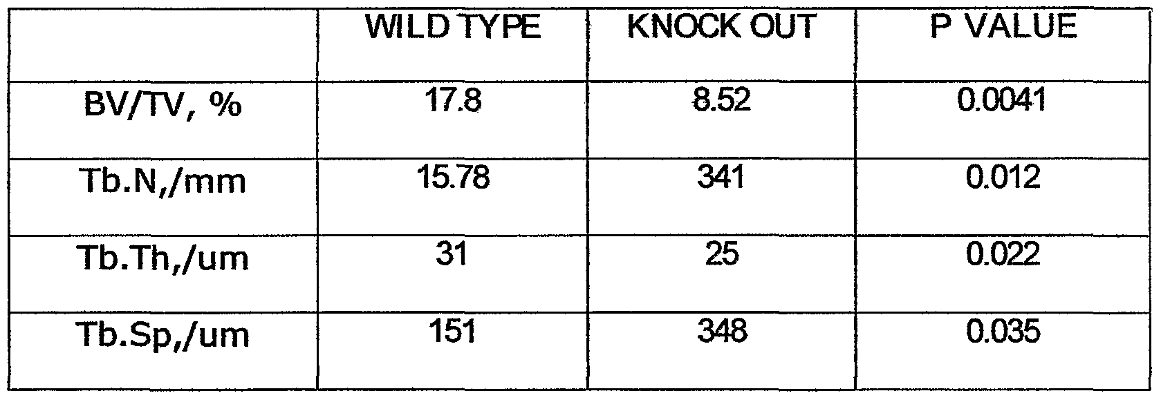
[0038] To determine whether CD18 played a rolein the function of BMSSCs, we examined bone phenotypes of the CD18"'" mice (Knockout or KO mice). Compared to their wild type (WT) sex-matched littermates, inactivation of the CD18 gene led to a significant (p<0.05) decrease in bone marrow density (BMD) of the femurs taken from both 5-week-old (n=3) and 15-week-old (n=4) mice, as assessed by Faxitron (FIG. 3A) and DXA [dual-energy X-ray absorptimetry] (FIG. 3B). The bone defects did not exacerbate with age between 5 and 15 weeks (data not shown), showing that the reduced BMD in CD18-deficient animals was probably not caused by chronic inflammation. Histological analysisof the femurs demonstrated that CD18"'" mice had decreased trabecular bones in the distal metaphysis(FIG. 3C), and micro-computed tomography
analysis of the distal femur metaphysis indicated that bone volume, trabecular bone number and trabecular bone thickness were diminished, and trabecular bone space was increased in CD187' mice (FlG. 3D and Table 1). Table 1 shows mi cro-computed tomography analysisof distal femoral metaphysesfrom five week old mice; scanning regi ons were conf i ned to secondary spongi osa and were about 0.30mm i n thi ckness. Guided by the two-dimensional images, a region of interest was manually drawn near the endocortical surface Trabecular bone morphometric indices, including bone volume relative to tissue volume (BV/TV, %), trabecular number (Tb.N), trabecular thickness (Tb.Th), and trabecular separation (Tb.§p), were assessed based on the reconstructed three-dimensional images, No significant defect in skeletal development was observed in CD18"'" mice as compared to WT control mice(i-week-old) (FIG. 3E).
TABLE l
MICRO-COMPUTED TOMOGRAPHY ANALYSIS OF DISTAL FEMORAL METAPHYSES FROM FIVE WEEK OLD MICE
TABLE l
MICRO-COMPUTED TOMOGRAPHY ANALYSIS OF DISTAL FEMORAL METAPHYSES FROM FIVE WEEK OLD MICE
CD18 deficiency does not affect overall osteoclastic activity. [0039] Proper bone integrity is maintained through a balance between osteoclast- mediated bone resorption and osteoblast-mediated bone formation (7), both of which require engagement of theintegrin receptors. For example, chondrocyte-specific
inactivation of integrin βi leads to diminished chondrocyte motility, defective cell proliferation, and compromised chondrocyte-dependent endochondral bone formation (8). M ice deficient in integrin P3 have normal osteoclastogenesis, but their mature osteoclasts are defective in ruffled membrane formation and bone resorption, resulting in osteopetrosis (9). Finally, deficiency of αι.β2, one of the four CD18 (β2) integrins, affects osteoclast progenitor cell adhesion to the stromal cells, resulting in diminished osteoclastogenesis (10). Deficiency of all CD18 integrins could result in two opposite phenotypes: increased number of osteoclastsin theCD18-deficient mice due to severe leukocytosis (11), and defective osteoclastogenesis due to lack of one or more than one of the CD18 integrins, To test this hypothesis, we carried out quantitative measurement of in vivo osteoclastic activity, based on the serum concentration of the C-termina! telopeptides of type 1 collagen, a commonly used marker for bone resorption (24). No significant difference between CD18-deficient and WT mice was observed (FIG. 4A). To further evaluate the effect of CD18 deficiency on osteoclast formation, the total number of mature osteoclastsin thelong bones of CD18"'" mice was quantified by TRAPstaining (FIG.4B). No difference was found between CD18-deficient and WT mice The above data suggests that osteoclast-mediated bone resorption is probably not responsi ble for the decreased bone formation in theCDIδ"7' mice.
CD18 is important for BMSSC differentiation but not for proliferation. [0040] We hypothesized that genetic inactivation of CD18 impairs osteogenic activity of the BMSSCs, leading to the observed decreased bone formation. To test this hypothesis, in vitro mineralization induction assays were performed on both WT and CD18"'" BMSSCs. It was discovered that mineralization by theCDIδ"'" BM SSCs was significantly lower (p<0.01) than that by WT cells. FIG. 5A shows in vitro that mineralization induction of CD18"'" BM SSCs determined by Alizarin red Sstaining (magnification x10), was significantly lower than that of WT cells (p=0.00003, n=3 mice). The total mineralized area by WT cells was assigned to 100%. In addition, CD18"'~ BMSSCs adhered poorly to tissue culture dishes compared to their WT counterparts as is shown in FIG. 5B. CeI Is were all owed to adhere for 3 hours at 370C. After washing, the adherent eel Is were counted manually based on 10 randomly picked view fields and
©(pressed as a percentage of WT eel I adhesion. (p=0.03, n=4). Interestingly, CD18- deficient BMSSCs proliferated better than the WT cells (p=0.01) based on BrdU incorporation assays determined manually by counting 10 representative fields (Fl G. 5C). Moreover, the bone marrow from CD 18"'" mice contained higher numbers of single colony-derived BMSSCs (CFU-F) (p<0.01) than that from WT mice (FIG. 5D: (p=0.0064, n=6 mice). Magnification x10.
[0041] I n addition to CD18, another known HSC marker Sca-1 is also expressed on stroma! stem cells (29). Deficiency of Sca-1 leads to defects in HSC renewal (34) and mesenchymal stem cell renewal (35). Asa result, Sca-1-deficient mice exhibit signs of osteoporosis, caused by both reduced bone formation and decreased bone resorption (35). Unlike Sca-1, CD18 deficiency affected the differentiation but not proliferation of BMSSCs(FIG. 5). Whether CD 18 deficiency also affects the differentiation and renewal of HSC iscurrently unclear and needs further investigation. It has been reported that CD18 deficiency causes severe leukocytosis (11), which may potentially lead to increased number of osteoclast precursor cells in CD18 deficient mice. However, the defective expression of αLβ2 (one of the four CD18 integrins) in CD18"/- mice also decreases osteoclastogenesis(IO). Asaresult, theoverall osteoclastic activity in theCD18"?" mice remains unchanged (FIG. 4).
[0042] Cbf a1 (Runx2/AM L3/PEBF2αC) i s a master regul atory gene i n osteogenesis (13), and its biological effects are regulated by transformi ng growth factor-β (TGF-β) through the Smad pathway (14, 15). TGF-β negatively regulates bone formation by inhibiting Cbfai expression and therefore the differentiation of osteoprogenitors through the Smad pathway. It was discovered that Cbfai expression in CD18"'" BMSSCs (Knock Out Mutants, KO) was decreased compared with that of WT BMSSCs(FIG. 6A, top panel). In addition, Smad2 phosphorylation was increased in response to TGF-β treatment in CD18"'~ KO BMSSCs(FIG. 6A, lower panel). These results indicate that defective osteogenesis seen in CD18-deficient BMSSCs is associated with compromised Cbfai expression.
[0043] To further confirm the critical role of CD18 in BMSSCs-mediated osteogenesis, and to exclude the possibility that CD18 contributes indirectly to osteogenesis through its function in haematopoetic cells, we conducted function-rescue
experimenta In these experiments CD18-defiάent BM SSC& were transformed with vectors having either full length or cytoplasmic domain-truncated (CT-) CD18. Similar to WT BIvISSCs, no macrophage contamination was detected in the PO CD18T*" BMSSCs by FACS analysis (data not shown). To express recombinant CD18, theCD18~'~ BMSSCs were infected with retroviruses having either full-length CD18 or mutant truncatedCD18 (CT-). Expression of recombinant CD18 on BM SSCs was confirmed 6 days after infection by immunoblot, using a polyclonal antibody against the cytoplasmic tail of CD18 (FIG. 6B). The osteogenic capability of WT BMSSCs, CD18"7' BM SSCs and the two different CD18-expressing CD18"Λ" BM SSCs was evaluated using an in vivo model of ectopic bone formation (23). Consistent with the decreased osteogenic activity of theCD18~'~ BMSSCs in vitro (FIG. 5A), we found that theCD18''" BM SSCs failed to support bone formation in vivo (FIG. 6C and 6D; WT vsKO, p= 0.016). Bone formation was restored by the expression of full length CD18 in theCD18"'' BMSSCs(KO vsCD18, p=0.0001). [0044] Surprisingly, transformed BM SSCs expressing CD18 constitutively using the retroviral promoter exhibited significantly higher in vivo osteogenic activity (~3-fold over WT BMSSCs; WT vsCD18, p=0.0047). This indicates that prolonged expression of CD18 in BM SSCs enhances osteogenesis. In contrast, eel Is transformed with the cytoplasmic domain-truncated CD18 failed to rescue the defective phenotypeof bone formation (KO vs CD18 (CT-), p=0.39), showing that the cytoplasmic domain of CD 18, which is required for integrin "outside-in" signaling, isalso required for the osteogenic differentiation of the BMSSCs. Therefore, an embodiment of the invention isdirected to a method for treating bone degenerative diseases in an animal by administering BMSSCs from the animal that have been transformed with an expression vector to express CD18. The BM SSCs can be isol ated from the ani mal or they can come from another ani mal of the same species, ln ail preferred embodi ments, the ani mal is human. [0045] Given the osteoporotic phenotype of the CD18"?" mice, the results show that patients with the severe form of LAD I , including those with restored HSC functions by gene therapy, could be predisposed to bone defects and the development of osteoporosis. Thus, it would be necessary to include reconstitution of CD18 expression on both HSCs and BMSSCs for gene therapy to treat LAD I patients In an embodiment of the invention methods are provided for preventing or treating LADI in an animal by administering a population of cells enriched in BMSSCs that have been transformed with a vector having a
normal gene for CD18. In apreferred embodiment, the animal 'sown BMSSCs are isolated, transformed with an expression vector having gene for normal CD18, and rei ntroduced i nto the ani mal .
[0046] Table 1 shows micro CT analysis for distal femoral metaphyses from 5-wk- old mice by the use of μCT-20 (Scanco Medical, Bassersdorf, Switzerland). Scanning regi ons were conf i ned to secondary spongi osa and were about 0.30mm i n thi ckness. Usi ng 2-dimensional images, a region of interest was manually drawn near theendocortical surface. Trabecular bone morphometric indices, assessed using 3-dimensional image reconstructions, included bone volume relative to tissue volume (BWTV, %), trabecular thickness (Tb.Th), trabecular number (Tb.N) and trabecular spacing (Tb.Sp). It was observed that prolonged expression of CD18 on BM SSCs significantly enhances osteogenesis and bone formation (FIG. 6D). Therefore increasing CD18 expression in BMSSCs by transforming them with retroviruses having theCD18 gene is beneficial to tissue engineering of bone and bone marrow. An embodiment of the invention isthus di rected to the use of BM SSCs transformed with a gene f or CD18 i n ti ssue engi neeri ng of bone and bone marrow.
[0047] To summarize, theseresultsdemonstratethat CD18 isanewly identified specific marker for both human and mouse BMSSCs that can be used to select a population of cellsenriched in BMSSCs from bone marrow mononuclear cellsfrom bone marrow aspirate. The data shows that combining the CD18marker with other stromal stem cell markers (eg. STRO-1 and VCAM-1)4 in cell sorting significantly enriches the BMSSC population from unfractionated bone marrow aspirates. [0048] At the functional level, CD18-deficient miceshowed bonedefects, including decreased BMD and compromised osteogenic differentiation of BMSSCs, These defects are associated with a decrease in expression of Cbfai, due to increased Smad signaling in absenceof CD18-mediated "outside-in" signaling. The osteogenic differentiation deficiency of theCD18-deficient BMSSCs was rescued by expressing full- length CD18 in the absence of cells of the hematopoietic origin. In humans, the lethal bacterial and fungal infections in patients with complete CD18 deficiency, the most severe form of Leukocyte Adhesion Deficiency I (LAD I), could mask potential bonedefects. Ratientswith thesevereform of LAD I, including those with restored HSC functions by gene therapy, are still predisposed to bonedefects Thus, it would be necessary to include
reconstitution of CD18 expression on both HSCs and BM SSCs for therapeutic treatments of the LAD I pati ents i n the future. Therefore an embodi ment of the present i nventi on i s directed to treating patients with LAD I with gene therapy to introduce a gene for CD18.
Obtaining An Enriched Population of BMSCCs Based on CD18 Expression
[0049] Generally, separation of a heterogeneous population of cells, such as in a bone marrow aspirate into target (such as, CD18-positive) and non-target (such as, CD18- negative) fractions is rarely complete. For the purposes of the present invention, separation is considered to have been accomplished if the target fraction is comprised of at least about 20% precursor cells, more often about 50% precursor cells, and preferably about 70% precursor eel Ia
[0050] Precursor cells may be positively selected or negatively selected. By positive selection is meant the capture of cells by some means, usually immunological, on the basis of their expression of a specific characteristic or set of characteristics (usually an antigen(s) expressed at the cell surface). For example, CD18-positivecellscan be positively selected by any of the above methods (except cytolysis, which would result in destruction of the desired cells) on thebasisof their expression of theCD18 antigen utilizing an anti-CD18 antibody, such as the monoclonal antibodies 6.7 (commercially availablefrom Pharmingen/BD Bioscience.
[0051] Negative selection means the exclusion or depletion of cellsby some means, usually immunological, on the basis of their lack of expression of aspecific characteristic or set of characteristics (again, usually a surface antigen). For example, CD18-positive cells can be negatively selected by any of the above methods on the basis of their lack of expression of lineage-defining antigens utilizing antibodies to the lineage- defining non-CD18 antigens. By using a cocktail or mixture of monoclonal antibodies directed to red cell, platelet, granulocyte, lymphocyte, adipocyte, and/or tumor cell antigens it is possibleto leave behind a population of cells which is highly enriched for CD18-positive cells. Numerous monoclonal and polyclonal antibodies suitablefor this purpose are known intheart and are commercial Iy availablefrom a wide variety of sources (for example, Becton Dickinson Co., Mountain View, Calif.; Coulter Immunology, Hialeah, FIa; Ortho Diagnostics, Raritan, N.J., etc.).
[0052] Alternatively, BMSSCs can be separated from mature eel Is by a combination of negative and positive selection techniques. For example, there can bea first selection for CD18-positivecells utilizing an anti-CD18 antibody, followed by a second selection for lineage-negative/CD18-positive cells, using an anti-lineage antibody kit from BD Bioscienceand deletion using magnetic beada The advantage of this or other dual selection strategies is that the volume of cells which is placed into culture is smaller and thus more manageable.
[0053] Although selection of CD18-positivecellsusually involves the use of one or more antibodies or fragments thereof, in some cases selection may involve the use of lectins or other types of receptors or ligands expressed on the cell surface. [0054] Some methods and devices for the selection of BMSSCsfrom a mixture of non-target and target cells, involve label ing the target cells, directly, or indirectly, with a biotinylated antibody to one or more target cell surface antigens (CD-18 and STR0 1). Labeled cells are separated from unlabeled cells by flowing them through a bed of immobilized avidin, the labeled cells binding to theavidin by virtue of the biotinylated antibody bound to their surface, whilethe unlabeled cellspass through the bed. After washing the bed material, thelabeled (bound) cellscan beeluted from the bed, for example, by mechanical agitation.
[0055] The conventional MACS procedure is described by Miltenyi et al., "High
Gradient Magnetic Cell Separation with MACS," Cytometry 11:231-238 (1990). To sort cd I s by MACS, one label see) Is with magnetic beads and passes the cellsthrough a paramagnetic separation column. The separation column is placed in a strong permanent magnet, thereby creating a magneticfidd within the column. Cd Is that are magnetically labeled are trapped in the column; cd Is that are not pass through. One then dutesthe trapped eel Is from the col umn.
[0056] Aocordi ng to the methods of the present i nventi on, BM SSCs can be separated from bone marrow by bringing the aspirate into contact with one or more antibodies (polyclonal and/or monoclonal) against CD18 (integrin β2>. Cd Is that have been bound by the monoclonal antibody are then separated from unbound eel Is by any means known to those ski 11 ed i n the art.
[0057] Vari ous methods of separati ng anti body-bound eel I s from unbound eel I s are known. For example, the antibody bound to the eel I (or an anti -isotype anti body) can be
labeled aid then the eel Is separated by amechanical cell sorter that detects the presence of the label . Fluorescence-activated cell sorters (FACS) are well known in the art. I n one preferred embodiment, the anti-stem cell antibody is attached to a solid support. Various solid supports are known to those of skill in the art, including, but not limited to, agarose beads, polystyrene beads, hollow fiber membranes, polymers, and plastic petri dishes. CeI Is that are bound by the anti body can be removed from the eel I suspension by si mply physically separating the sol id support from the eel I suspension. [0058] Selective cytophoresis can be used to produce a cell suspension from mammalian bone marrow ceils. For example, marrow can be harvested from a donor (the patient in the case of an autologous transplant; adonor in the case of an allogeneic transplant) by any appropriate means. The marrow can be processed as desired, depending mainly upon the use intended for the recovered cells. The suspension of marrow cells is allowed to physically contact, for example, a solid phase-linked monoclonal antibody that recognizes an antigen on the desired cells, such as CD18. The solid phase-linking can comprise, for instance, adsorbing the antibodies to aplastic, nitrocellulose, or other surface. The anti bodies can also be adsorbed on to the walls of the large pores (sufficiently large to permit flow-through of cells) of a hollow fiber membrane. Alternatively, the antibodies can be covalently linked to asurfaceor bead, such as PharmaciaSepharose 6MB macrobeads. The exact conditions and duration of incubation for the solid phase- linked antibodies with the marrow cell suspension will depend upon several factors specificto the system employed. The selection of appropriate conditions, however, iswell within the skill of the art.
[0059] The unbound eel I s are then el uted or washed away with physi ol ogi c buffer after allowing sufficient time for the stem cells to be bound. The unbound marrow cells can be recovered and used for other purposes or discarded after appropriate testing has been done to ensure that the desired separation had been achieved. The bound cellsare then separated from the sol id phase by any appropriate method, depending mainly upon the nature of the sol id phase and the anti body. For example, bound cd Is can be el uted from a plastic petri dish by vigorous agitation. Alternatively, bound eel I scan be el uted by enzymatically "nicking" or digesting a enzyme-sensitive "spacer" sequence between the solid phase and the antibody. Spacers bound to agarose beads are commercially available from, for example, Pharmacia
[0060] The el irted, enri ched fracti on of eel I s may then be washed with a buffer by centrifugation and either cryopreserved in a viable state for later use according to conventional technology or immediately infused intravenously into the transplant recipient.
[0061] Procedures for separation include magnetic separation using antibody- coated magnetic beads and affinity chromatography or "panning" using antibody attached to a solid matrix (e.g. plate). Techniques providing accurate separation include fluorescence-activated cell sorters, which can have varying degrees of sophistication, such as having multiple color channels, low angle and obtuselight scattering detecting channels, or impedance channels. Dead eel I scan be eliminated by selection with dyes associated with dead cells e.g., (propidium iodide, LDS). Red blood cells can be removed by (for example) elutriation, hemolysis, or Ficoll-Paque gradients. Any technique can be employed that is not unduly detrimental to the viability of the selected cells. [0062] Conveniently, antibodies can be conjugated with labels for a number of different purposes: e.g., magnetic beads to allow for ease of separation of a particular cell type; biotin, which binds with high affinity to avidin or streptavidin; fluorochromes, which can be used with a fluorescence activated cell sorter; haptens; andthelike. Multi-color analyses can be empl oyed with a FACS or i n a combi nati on of i mmunomagneti c separation and flow cytometry. Multi-color analysis is of interest for the separation of cells based on multiple surface antigens such as STRO- 1 and/or CD18. Fluorochromes which find usein a multi-color anaJysisincludephycobiliproteins, eg. phycoerythrin and allophycocyanins; fluorescein, and Texas red.
[0063] In one embodiment of theinvention, an antibody against CD18 isdirectly or indirectly conjugated to a magnetic reagent, such as a superparamagnetic mi croparticle (microparticle). Direct conjugation to a magnetic particle is achieved by use of various chemical I i nki ng groups as known i n the art. For example, anti body can be coupled to the microparticles through side chain amino or sulfhydryl groups and heterofunctional cross- linking reagents. A large number of heterofunctional compounds are availablefor linking to entities. A preferred linking group is3-(2-pyridyldithio)propionicacid N- hydroxysuccini mi de ester (SPDP) or 4-(N-maleimidomethyl)-cyclohexane-1-carboxylic acid N-hydroxysuccinimide ester (SMCC) with a reactive sulfhydryl group on the antibody and a reactive amino group on the magnetic particle.
[0064] Alternatively, an anti CD18 antibody is indirectly coupled to magnetic particles. The antibody isdirectly conjugated to ahapten, and hapten-specific, second- stage antibodies are conjugated to the particles Suitable haptens include digoxin, digoxigenin, FITC, dinitrophenyl, nitrophenyl, avidin, and biotin. Methods for conjugation of the hapten to a protei n are known i n the art, and ki ts for such conj ugati ons are commercially available.
[0065] For separation or identification of stem cells or progenitor cells, an antibody is added to a bone marrow aspirate or to a suspension of BMNCs. The amount of an antibody necessary to bind a particular cell subset is empirically determined by performing atest separation and analysis The cells and an appropriate antibody are incubated for a period of time sufficient for complexes to form, usually at least about five minutes, more usually at least about 10 minutes, and usually not more than one hour, more usually not more than about 30 minutes,
[0066] Thecellscan additionally beincubated with antibodiesor binding molecules specific for other cell-surface markers known to be present or absent on BMSCCs, such asSTRO-1. For example, STRO-1 is useful in the positive selection of stem eel I s. Vari ous markers known to be absent on stem eel I s, such as CD3, CD4, CD8, CD14, CD15, and CD19, can be used for negative selection. Thelabded cellsare separated in accordance with the specific anti body preparation. Fluorochrome-labeled anti bodies are useful for FACS separation and magnetic particles for immunomagnetic selection or particularly high gradient magnetic selection (HGMS). [Exemplary magnetic separation devices are described in WO/90/07380, FCT/US96/00953 and EP 438,520, herein incorporated by reference.
[0067] The purified cell population can be collected in any appropriate medium.
Various media are commercially avail able and can be used, including Dulbecco's Modified EagleMedium (DMEM), Hank's Basic Salt Solution (HBSS), Dulbecco's phosphate buffered saline (DPBS), RPMI, Iscove's modified Dulbecco's medium (IMDM), and phosphate buffered saline (PBS) with 5 mM EDTA, any of which can be supplemented with fetal calf serum (FCS), bovine serum albumin (BAD), or human serum albumin (HSA).
[0068] Once the desired cells have been isolated, they can be propagated by growing in conditioned medium, for examplefrom stromal cells, or in media comprising
ma ntenεnce factors supporting the proliferation of BMSSCs e.g., stem cell factor or combi nati ons of i nterl euki ns. The medi urn empl oyed for cul turi ng eel I s i s conveni entl y a defined enriched medium, such as I M DM or a mixture of IMDM and RPMI 1640, and will generally Decomposed of salts, amino acids, vitamins, beta-mercaptoethanol, streptomycin/penicillin and 10% fetal calf serum, and can be changed from time to time, generally at least once to twice per week. See Example3.
[0069] The subject BMSSCs of the present invention find usein avariety of ways.
In one embodiment BMSSCs are isolated from apatient before radiation or chemotherapy, cultured to multiply and readministered to the patient to reconstitute an irradiated host and/or a host subject to chemotherapy. In another embodiment, BM SSCs are cultured under conditions that permit their maturation, proliferation and differentiation into one or more selected lineages through specific different growth factors as descri bed in Example 3. The BM SSCs can be used as a source to make osteoblasts, chondrocytes, adi pocytes, and neural eel Is in vitro for reintroduction into the animal from which the BM SSCs were derived (autologous), or into another animal of the same species (heterologous). Our results showed that when cultured in spedfic differentiation-inducing conditions, the CD18+STRO-1bri9W BM SSCs were capable of differentiating into adipocytes (FIG. 2D, panel a), osteoblasts (FIG. 2D, panel b), and chondrocytes (Fl G. 2D, pane! c), thus confirming their pleuripotent differentiation potentials. See Example3. Such factors as erythropoietin, colony stimulating factors (e.g., GM-CSF, G-CSF or M-CSF), interleukins (e.g. IL-1, -2, -3, -4, -5, -6, -7, -8, -9, or -10), or thelike, or stromal cells can be used to influence the growth and differentiation of BMSSCs cells. [0070] The cells can also be used in the isolation and evaluation of factors associated with the differentiation and maturation of BMSSC cells, including reagents that specifically bind to theCD18 antigen. Thus, the eel Is can be used in assays to determine the activity of media, such as conditioned media; to evaluate fluids for growth factor activity or involvement with dedication of lineages; or thelike. [0071] BMSS cellscan be frozen at liquid nitrogen temperatures and stored for long periods of time, as they can be thawed and reused. Thecellswill usually be stored in 5% DMSO and 95% fetal calf serum. Once thawed, the cells can be expanded by use of growth factors or stromal cells associated with stem cell proliferation and differentiation.
[0072] Theenriched mammalian BM SSCs can be used in therapeutic methods such as stem eel I transplantation, as well as other methods that are readily apparent to those skilled in theart. For example, BM SSCs can be isolated from a patient using the methods described heran, they can be cultured to multiply and can then be administered to the same patient or to adifferent patient requiring by infusion in an amount sufficient to restore the patient's damaged or diminished BMSSCs. Since BMSSCs are pleuripotent, they can differentiate into osteoblasts, chondrocytes, adipocytes, and neural cells in situ if they areinjected into bone, cartilage, fat or the nervous system, respectively. R-ecise, effective quantities can be readily determined by those ski I led in the art and will depend, of course, upon the exact condition being treated by the therapy. In many applications, however, an amount contai ni ng approxi mately the same number of stem eel I s found i n one-half to one liter of aspirated marrow should be adequate [0073] The use of astern cell-specific antibody need not Delimited to the purification of stem cells prior to a transfection procedure With the goal of generating vectors for in vivo gene therapy, it has been proposed to engi neer i nto the gene therapy vectors themselves, mechanisms by which the vector will recognize its target cell (and preferably only its target) within the context of the entire organism. See, Kasaharaet al., Science 266: 1373-1376 (1994); Michael & Curiel, Gene Therapy 1:223-232 (1994); Chatterjeeet al., Ann. N.Y. Acad. Sei. 770:79-90 (1995); Sehwarzenberger et aJ., Blood 87:472-478 (1996). By incorporating stem cell-specificantibodiesaganst CD18 into a vector, it may be possible to generate vectors that will recognize and target stem cells in the patient's bone marrow. Specifically, the antibody could be incorporated into liposome vectors, (Hughes et al., Cancer Res. 49:6214-6220 (1989); Wang & Huang, Biochemistry 28:9508-9514 (1989); Ahmad et al., Cancer Res. 53:1484-1488 (1993)), poly-L lysine conjugate vectors (Michael & Curiel, supra; Sehwarzenberger et al., supra), or into viral vectors, including but not limited to adenoviral vectors, retroviral vectors, (Russell et al., Nucleic Acids Res. 21:1081-1085 (1993); Somiaet al., RΌC. Natl. Acad. Sei. USA 92:7570-7574 (1995)), and adeno-associated vectors (Chatterjee et al., supra), modified to express on the vector surface, the antibody itself or proteins which would bind the antibody to the vector surface (such as the Fc receptor).
[0074] I n another embodi ment for a method of in vivo gene therapy, diseases of the bone marrow may be corrected by the introduction of the normal gene for CD18 into
the patient' s abnormal human stem cells, which can then be transplanted into a patient's bone marrow. For a general review of the methodologies, see Friedmann, T., Science 244:1275-1281 (June 1989) and Lancet 1: 1271-1272 (Jun. 4, 1988), the entire contents of which are hereby incorporated by reference as if fully set forth herein, under 35 U.S.C. §119(e). Leukocyte Adhesion Deficiency is a genetic diseases associated with BMSSCs that do not express CD18 or that express mutant CD18. In one embodiment LAD istreated by i ntroduci ng the wi I d-type gene for CD18 i nto BM SSCs derived from the ani mal havi ng the disease, and the transformed BMSSCs are then be rei ntroduced i nto the ani mal to treat or prevent the disease. A therapeutic gene can be introduced into the population cells enriched for BMSSCs, isolated as above by any method known in the art including physical methods such as co-precipitation with calcium phosphate, electroporation or microinjection (eg., U.S. Pat. No.4,873,191), or using viral vectors such as adenoviral, or retroviral vectors. In the latter case, the DNA of the retrovirus is cut with a restriction enzyme and the human DNA containing the desired sequence is inserted and ligated. The retrovirus containing theinsertion isthen infected into the stem cella The stem cellscan then be assayed for production of the desired protein. See, eg., U.S. Rat. No.4,902,783. The entire contents of the above references are hereby incorporated by reference as if fully set forth heran, under 35 U.S.C. §119(e)
EXAMPLES
Example 1 Cell Culture and In Vitro Assays
[0075] Mice. The CD18 null and their sex-matched C57BL/6J littermates were obtained by breeding heterozygotesof theCD18-null mice. Scharffetter-Kochanek, K., Lu, H-, Norman, K., van Nood, N., Munoz, F., Grabbe, S., McArthur, M., Lorenzo, I., Kaplan, S., Ley, K. etal. (1998) JExpMedlS8, 119-131 and Miura, M., et a!. (JCIin Invest. 2004 Dec;114 (12):1704-13), the entire contents of which are hereby incorporated by reference as if fully set forth herein, under 35 U.S.C. §119(e). TheCD18 null mice have been backcrossed more than 10 generations into the C57BL/6J background. I mmunocompromised bg-nu/nu-xid nude mice were purchased from Harlan Sprague- Dawley (Indianapolis, IN). Micewere maintained in sterile micro-isolator cages and all animal experiments were performed with the approval of the committee of the American
Red Cross (protocols #407 and #447) and the National Institute of Dental and Craniofadal Research (protocol #04-317).
Mouse and Human BMSSCs culture
[0076] Preparati on and expansi on of the mouse BM SSCs were performed based on a previously published method. Miura, M., et al. (J Clin Invest. 2004 Dec;114 (12):17Q4- 13). Mouse bone marrow (BM) cells(1.5 x 10⁷) harvested from long bones were seeded into 10cm culture dishes (Corning, Corning NY), incubated for 3 hours at 37°C to allow attachment of adherent eel I s, and washed twi ce with PBS to remove non-adherent eel I a BM cdls(1.5 x 10⁷) from long bones of guinea pigs were then added as feeder cells. To prevent proliferation in culture, the feeder cellswereγ-irradiated (Caesium- 137) with 6,000 cGy by aGammacell-1000 Irradiator (Atomic Energy of Canada Ltd. Ontario, Canada) prior to seeding. The mouseculture medium consisted of α-MEM (Gibco BRL; Invitrogen Corp. Grand Island, NY), 20% fetal bovine serum (FBS; Equitech-Bio Inc. Kerrvile, TX), 2 mM L-glutamine, 100 U/ml penicillin and 100 μg/ml streptomycin (Biofluidslnc, Rockville, MD), and 10 nM dexarnethasone(3gma-Aldrich, St Louis, MO) and 55 μM 2-mercaptoethanol (Gibco BRL; Invitrogen Corp.). BMSSCs formed adherent colonies after 7-16 days of culture, which was designated as passage 0. Primary BMSSCs were lifted to disperse the colony-forming cells and seeded on freshly prepared culture dishes. These eel Is were passaged when they reached confluence and utilized for further expert menta
[0077] Human BMSSCs culture Human BM aspirates from healthy adult volunteers were purchased from Al I CeI I s, LLC (Berkeley, CA). BMSSCs constitute about 0.1% of bone marrow cells in aspirates. Bone marrow is typical Iy aspirated from the iliac crest, but may be obtained from other sites (such as the sternum) if necessitated by prior or concurrent disease or therapy. To identify putative BMSSCs, single-cell suspension of 1x10⁶ of bone marrow mononuclear cells (BMNCs) were seeded into 15cm culture dishes (Falcon; BD Biosάence, San Jose, CA) and non-adherent eel Is were removed after 3 hours of incubation at 37°C. The adherent cells were cultured with α-MEM supplemented with 15% FBS, 100 μM L-ascorbicacid 2-phosphate (Wako Pure Chemical Industries Ltd, Osaka, Japan), 2 mM L-glutamine, and a combination of 100 U/ml penicillin and 100 μg/ml streptomycin. The human culture medium was changed on day 7 and 14, if the cells
were not passaged by day 14. Subsequent passages were performed when the eel Is were approaching confluent. BMSSCs of the second to the fifth passages were utilized for further experiments unless specifically mentioned.
Colony-forming unit fibroblast (CFU-F) assay.
[0078] The CFU-F assay was performed as previ ously descri bed. Kuznetsov, S. &
Gehron, R. P. (1996) Caiάf. Tissue Int. 59, 265-270. BMSSCs can also be detected by other colony-forming assays, such as CFU-GM and CFU-S assays (see, for example, Sutherland et al., in Bone Marrow Frocessing and Purging, A. P. Gee (ed), Boca Raton: CRC Fress(1991), pg. 155). The entire contents of all of the above references are hereby incorporated by reference as if fully set forth herein, under 35 U.S.C. §119(e). [0079] Mouse BM cells (1-5 * 106) harvested from long bones were seeded into T-
25 culture flasks (Nalge Nunc, Rochester, NY), incubated for 3 hours at 37°C to allow attachment of adherent cells, and then rinsed twice with PBS to remove nonadherent cells. BM cells (1.5 * 107) from long bones of guinea pigs were then added as feeder cells. To prevent proliferation in culture, the feeder cellswereγ-irradiated (Caesium-137) with 6,000 cGy by aGammacell-1000 Irradiator (Atomic Energy of Canada Ltd. Ontario, Canada) prior to seeding. Culture medium consisted of α-MEM, 20% FBS, 2 mM L- glutamine, 100 U/ml penicillin and 100 μg/ml streptomycin and 55 μM 2- mercaptoethanol. Adherent colonies were fixed with methanol between day 7 and day 16 and stained with an aqueous solution of saturated methyl violet (Sgma-Adlrich). The sorted human BMSSCs, either STRO-1br1gtTt/CD18+ or STRO- 1bri9hl/CD 18", were plated on the culture dishes. Colony forming efficiency assays were performed at day 14 of culture following staining of the cultures with 0.1% (w/v) toluidinebluein 1% paraformaldehyde Colonies containing equal to or greater than 50 cells were counted as colonies under a dissecting microscope.
Cell proliferation assays.
[0080] The proliferation of BMSSCs was assessed by bromodeoxyuridine (BrdU) incorporation of the cells. Mouse BM SSCs were seeded at 5x1f/ cellson 2 well chamber slides (Nalge Nunc, Rochester, NY), incubated with BrdU solution (Zymed, San Francisco, CA) for 20 hrs. The number of BrdU positive eel Is were detected using BrdU
staining kit (Zymed, San Francisco, CA) according to the manufacturer's instruction with hematoxylin counter staining. For quantification of BrdU positive cells, ten representative images captured at 20Ox magnification were used to calculate BrdU positivecell number. Cell proliferationwasshown asapercentageof BrdU-positivecellsover total nucleated cdls.
Cell adhesion assays.
[0081] Samenumbersof WT and CD18~Λ BM SSCs were seeded on treated plastic chamber slides (NalgeNunc, Rochester, NY) in the culture medium. After 3 hours of i ncubati on, the chamber si ides were gently washed with PBS twi ce to remove non¬ adherent cd I s and the number of adherent eel Is was determined by manual counting of five representative fields at 100x magnifications,
Western blot analysis.
[0082] Cellswerelysed in M-FER extraction reagent (Pierce Chemical Co.,
Rockford, I L), and protein concentrations were measured using Bio-Rad Protein Assay (Bio-Rad Laboratories Inc. Hercules, CA). To examine CD18 expression in mouse BMSSCs, total cell I ysates from the same number of WT, CD18T7', and CD18- reconstituted (with either full-length or cytoplasmic tail-truncated CD18) BM SSCs were applied onto NuPAGE gel (Invitrogen Corp. Carlsbad, CA) at ~3x10⁴ cells per lana The separated proteins were transferred onto PVDF membranes (Mil I ipore, Bedford, MA) and blocked with asolution containing 1OmM Tris-HCI [pH7.5], 154mM NaCI, 4% BSA, 1% milk, and 0.05% Tween-20 for 60 mi n at room temperature (RT). After washing, the membrane was incubated with rabbit anti-CD18 cytoplasmic tail antibodies (1:1000 dilution) in incubation buffer (1OmM Tris-HCI [pH7.5], 154mM NaQ, 0.5% BSA, 0.05% Tween 20) for 60 mi n at RT. The membranes were then washed and i ncubated with a horseradish peroxidase (HRP)-conjugated goat anti-rabbit IgG (Santa Cruz Biotechnology Inc., Santa Cruz, CA) at 1:5,000 dilutions in incubation buffer for 30 min at RT. After washing with T-TBS, the membranes were reacted with HRP substrate (Pierce Chemical Co.) to visualize positive bands on x-ray films (Eastman Kodak Co., Rochester, NY). To examine CD18 expression in human BMSSCs, total cell lysatesfrom human BMSSCs at different passages were prepared. In each lane, twelve micrograms of protein were loaded
and analyzed as described above. To examine Cbfai expression, 2.2 μg of total protein lysatefrom WT or CD18"Λ BMSSCs were applied to NuPAGE and detected by immunoblot using rabbit anti-Cbfa1 (Oncogene Research Products, Cambridge MA) antibody at 1:500 dilution. To examine the effect of TGF- treatments, mouse BMSSCs were serum-starved with α-M EM supplemented with 2% FBS for 16 hours and then treated with 2 ng/ml TGF-β (R&D Systems Inc. Minneapolis, MN) for theindicated periods. Phosphorylation of Smad2 was examined by loading equal amount of cell lysate (12 μg) in each lane and detected by immunoblot using rabbit anti-phospho-Smad2 antibody (Cell Signaling Technology Inc., Beverly, MA) at 1:500 dilution. Each membrane was also stripped using the stripping buffer (Pierce Chemical Co.) and re- probed with mouse anti-α-actinin mAb (Upstate, Lake Placid, NY) or anti-β-actin (Sgma- Aldrich) to quantify the amount of proteins loaded.
Example 2
Enrichment of hBMSSCs by fluorescence-activated cell sorting
[0083] Detailed procedures for enrichment of the BMSSC population by cell sorting have been published (5), Gronthos, S. et al., Blood 84, 4164-4173 (1994) the entire contents of which are hereby incorporated by reference as if fully set forth herein, under
35 U.S.C. §119(e). and (3, 4). Bone marrow (BM) aspirates were provided by AIICdI. Inc.
To obtain the, 25ml bone marrow was aspirated by board-certified physicians from a singlesiteof the posterior iliac crest and withdrawn i nto a 60 cc syringe containing 15 ml of PBS plus 125 units heparin per ml of BM. Bone marrow mononuclear eel Is (BM NCs) were isolated from BM aspirates by density centrifugation over a Ficoll solution
(Amersham).
[0084] Approximately 1-3x10⁸ BMNCs were sequential Iy incubated with STRO-1 supernatant (IgM), anti-lgM-biotin, streptavidin microbeadsand finally streptavidin-FITC
(Caltag Laboratories, Burlingame, CA) before being separated on aMini MACS magnetic column (Miltenyi Biotec lnc, Auburn, CA). The FITC-labeled STRO-1 positive BMNCs isolated by MACS were co-labeled with a mouse anti-human CD18 mAb (clone6.7,
Pharmingen/BD Bioscience) for 30 minutes on ice, washed and incubated with PE- conjugated goat anti-mouse IgG (Pharmingen/BD Bioscience) for an additional 20 min on
ice. Whilethiscan bedonein onestep, better cell quality can be obtained using two steps. After washing, eel Is were sorted for both CD18 and STRO-1 using a FACStarPLUSflow cytometer (Becton Dickinson). The sorted human BMSSCs, either STRO-1 bright /CD18+ or STRO-I15"9111/ CD18", were used for further expert menta
Flow cytometric analysis of BMSSCs.
[0085] Thesorted human STRO-1bri9ht /CD18+ BMSSCs were incubated with either of the primary antibodies or tha'r corresponding isotype-matched control antibodies at a concentration of 10 μg /ml for one hour on ice Primary antibodies: mouse IgG1 anti- human CD14, CD34, CD45 (DAKO cytomation, Carpinteria, CA); mouse IgGi anti- human CD44 (H9H11) and IgG23 anti -human CD146 (CC9); mouse IgGi anti-human CD90, CD105, CD166 (Pharmingen; BD Bioscience, San Jose, CA); mouse lgd anti- human CD106 (6G10) (kindly provided by Dr. B. Masinovsky, ICOS Corporation, Bothell, WA). Isotype-matched control mouse monoclonal antibodies: 1B5 (IgGi) and 1A6.11 (IgGa) (kindly provided by Prof. L.K. Ashman, Medical Science Building, University of Newcastle, New South Wales, Australia). After washing, the cells were incubated with the secondary detection reagents, goat anti -mouse IgGr or IgG^-FITC conjugated antibodies (1/50) (Southern Biotechnology Associates Inc., Birmingham, AL) for 46 minutes on ice. Following washing, the samples were analyzed using an Epics®- XL-MCL flow cytometer (Beckman Coulter, Hialeah, FL). For dual-color FACS analysis of mouse BMSSCs, single cell suspension of PO BMSSCs (1X10⁶) were incubated with a pair of FITC- and PE-conjugated antibodies or their corresponding isotype-matched controls (each antibody at 10 μg /ml) for 45 min on ice Following washing, the samples were analyzed using a FACScan flow cytometer (Becton Dickinson, San Jose, CA). The percentage of cell population in each quadrant was calculated using the FACScan program. All antibody conjugates were purchased from Pharmingen/BD Bioscience unless specifically mentioned, including PE-coηjugated rat anti-mouse CD18 (C71/16, lgG2a), FITC- and PE-coηjugated Sca-1 (E13-161.7, lgG2a), FITC-coηjugated rat anti-mouse CD14 (rmC5-3, IgGI), FITC-conjugated anti-CD34 (49E81, lgG2a), and their corresponding FITC- and PE-conjugated IgG controls.
Example 3
In vivo Osteoclast activity.
[0086] In vivo osteocl asti c activity was determi ned by measuri ng the serum concentrations of c-terminal telopeptidesof type 1 collagen, obtained from the peripheral blood of 8-week-old mice, using Ratlap ELISA kit (Osteometer BioTech A/S, Herlev, Denmark) accordi ng to the manufacture' s i nstructi ona The number of mature osteoclasts was determi ned by the tartrate-resi start acid phosphate (TRAP) staining, as previously described. Chiang, C. Y., Kyritsis, G., Graves, D. T. & Amar, S (1999) Infect Immun 67, 4231-4236. Briefly, the left femurs were harvested from mice, fixed with 2% paraformaldehyde, decalcified with 10% EDTA (pH 8.0) and embedded in paraffin. Sections were deparaffinized, hydrated and stained for TRAP. TheTRAPsolution wasa mixture of the following two solutions: 9.6 mg of naphthol AS-BI phosphate substrate (Sgma-Aldrich) dissolved in 0.6 ml of N^V-dimethylformamide and 84 mg of fast red- violet LB diazonium salt (Sigma-Aldrich), 58.2 mg of tartaric acid (Sigma-Aldrich), 240 μl of 10% MgCI2, 4ml of 3 M sodium acetate buffer (pH 5.0) dissolved in 56ml of distilled water. The mixture was passed through aθ.22 μm filter before use. Thesections were incubated for 30 min in the TRAP solution at 37°C in the dark and then washed with distilled water for 10 min, followed by acounterstaining with hematoxylin. For quantification of TRAP positive cells in the bones, 4 representative images were captured under microscope with a 20Ox magnification and then analyzed using the program NI H Image The results were expressed as the number of TRAP positive eel Is per total bone area
In vitro differentiation potentials of the sorted human BMSSCs.
[0087] Osteogenic differentiation of the sorted human STRO-I^CDIβ*
BMSSCs was induced in the presence of 100 μM L-ascorbate-2-phosphate, 3 mM inorganic phosphate and 10 nM dexamethasone. Calcium deposits were identified by Alizarin Red S staining after 3 weeks of cultivation. Gronthos, S., Graves, S. E., Ohta, S. & Simmons, P. J. (1994) Blood 84, 4164-4173. Adi oogenesis was induced in α-MEM supplemented with 15% FBS, 100 μM L-ascorbate-2-phosphate, 0.5 mM isobutyl- methylxanthine, 0.5 μM hydrocortisone, 60 μM indomethacin and 10 μg/ml recombinant human insulin. Oil Red O staining was used to identify lipid-laden fat cells after 2 weeks
of cultivation, as previously described. Gimble, J. M., Morgan, C, Kelly, K., Wu, X., Dεndapani, V., Wang, C. S. & Rosen, V. (1995) J. CellBiochem. 58, 393-402. Chondrogenic differentiation was assessed by histochemical staining with Alcian blue(pH 1) in aggregate cultures treated with 100 μM L-ascorbate-2-phosphate, 2mM sodium pyruvate, 1% insulin/transferring/selenousacid mixture (ITS; BD Biosciences), 100 nM dexamethasone and 10 ng/ml transforming growth factor β as described previously. Rttenger, M. F., Mackay, A. M., Beck, S. C, Jaiswal, R. K., Douglas, R., Mosca, J. D., Moorman, M. A., Smonetti, D. W., Craig, S & Marshak, D. R. (1999) Science 284, 143- 147.
Example 4 In vitro mineralization induction of mouse BMSSCs.
[0088] For the mineralization induction of mouse BMSSCs in vitro, 2 mM μ- glycerophosphate (Sgma-Aldrich) and 100 μM L-ascorbic acid 2-phosphate (Wako Pure Chemical Industries Ltd, Osaka, Japan) were added to the mouse culture medium. After 6 weeks of cultivation, the cultures were stained with 1% alizarin red to examine calcium accumulation in the cells. The images were captured with ascanner (Epson, Tokyo, Japan) and mineralized areas were quantified using TotaLab software (Nonlinear Dynamics). Mineral deposits were expressed as a ratio relative to WT mouse BMSSC-mediated mineralization.
Example 5 Mouse BMSSC-mediated bone formation in vivo.
[0089] Approximately 4.0x10⁶ mouse BMSSCs were mixed with 40mg hydroxyapatite/tri calcium phosphate (HA/TCP) ceramic powder (Zimmer Inc, Warsaw, IN), and the mixture was implanted subcutaneously into the dorsal surface of 8- to 10- week-old immunocompromised bg-nu/nu-xid nude mice as previously described. Krebsbach, P. H., Kuznetsov, S. A., Satomura, K., Emmons, R. V., Rowe, D. W. & Robey, P. G. (1997) Transplantation 63, 1059-1069. The transplants were recovered 7 weeks after implantation and fixed with 2% paraformaldehyde, decalcified with 10% EDTA (pH 8.0) and embedded in paraffin. For quantification of bone formation in the transplants, sections were deparaffini zed, hydrated and stained with hematoxylin and eosin. Five to seven representative fields at 5Ox magnification were selected for each
BMSSC transplant. The total bone area within each field was calculated using the program NIH I mage as previously described, and expressed as a percentage of bone formation by WT BMSSCs. Histological analysis and quantification o bone formation in the harvested implants was done as previously described Shi, S. et al. (23).
Analysis of bone phenotypes.
[0090] TheCD18-deficient mice and their sex-matched C57BL/6J littermates at the age of 5- to 15-week-old were used to analyze bone phenotypes. Radiographs of left femurs were taken by Faxitron (Wheeling, IL). Quantitative analysisfor bone mineral density (BMD) on left femurs was based on dual energy x-ray absorptiometry (DXA) by the use of aGE Lunar Rximus(GE Lunar, Madison, Ml). Distal femoral metaphyseswere analyzed by micro-computed tomography (μCT-20; Scanco Medical, Bassersdorf, Switzerland) as previously described. Miura, M., Chen, X. D., Allen, M. R., Bi, Y., Gronthos, S., Seo, B. M., Lakhani, S., Flavell, R. A., Feng, X. H., Robey, P. G. etal (2004) J. CUn. Invest 114, 1704-1713. Thescanning regions were confined to secondary spongiosaand were approximately 0.30 mm in thickness. Using 2-dimensional images, a region of interest was manually drawn near theendocortical surface. Cancellous bone morphometric indices were assessed using 3-dimensional image reconstructions, included bone volume/total volume (BV/TV) (%), trabecular thickness (Tb.Th), trabecular number (Tb.N), and trabecular separation (Tb.Sp). Peripheral quantitative computed tomography (pQCT) analysis of the distal femora was performed using an XCT Research M scanner (Stratec; Norland Co.) as previously described. Miura, M., et al. Briefly, scans were obtaned at 2.25 mm and 2.75 mm from the distal condyles and cancellous BMD. Machine cancel I ous BMD preci si on (based on the manufacturer1 s data) was ±3 mg/cm3, whi I e the coefficient of variation in our laboratory based on repeat scans was 2.26%. For histological analysis, left femurs were harvested from mice, fixed with 2% paraformaldehyde, decalcified with 10% EDTA (pH 8.0) and embedded in paraffin. Sections were deparaffinized, hydrated and stained with hematoxylin and eosin. To analyze the wholeskeleton, one-week-old mice were dissected to remove skin, muscleand fat and kept in acetone to remove further fat for 3 days. They were then staned with 0.09% alizarin red Sand 0.05% alάan blue in a solution containing ethanol, glacial acetic acid and water (67:5:28) for 48 hrsat 370C. After staining, the mice were transferred to
1% potassium hydroxide until the skeleton was clearly visible The mice were preserved in 100% glycerol with gradual increase in concentration.
Osteoclast activity.
[0091] Thetartrate-resistant acid phosphate (TRAP) staining was performed as previously described (25). Serum concentration of C-terminal telopeptides of type 1 collagen in mice was measured using Ratlap ELISA kit (Osteometer BioTech A/S, Herlev,
Denmark).
Retroviral meditated CD18 expression in mouse BMSSCs.
[0092] M uri ne CD18 cDNA was subcl oned i nto the retrovi ra! expressi on vector
MGIN, using theEcoRI and Notl restriction sites. Cheng, L., Du, C1 Murray, D., Tong, X., Zhang, Y. A., Chen, B. P. & Hawley, R. G. (1997) Gene Ther 4, 1013-1022. To achieve better expression, a Kozak sequence (ACCATGG) was inserted before the initiation codon of theCD18 protein. After confirming the correctness of theinserted CD18 sequence by DNA sequencing, the retroviral expression vector MGI N-CD18 was transfected into the packaging cell lineGP+E-δβ, using Lipofectamine(lnvitrogen). Markowitz, D., Goff, S. & Bank, A. (1988) J Virol 62, 1120-1124. Following G418 (600 μg/ml) selection of the transfected cells, individual clones were established by picking up single colonies and the titers of theirviral supernatants were determined by infecting NIH3T3 cellsand counting the colonies formed upon G418 selection. The clones that produce the highest retroviral titer (~1x10⁶ colony-forming units/ml or CFU/ml) were used to prepare retroviral supernatants, which were subsequently concentrated to 6X10⁶ CFU/ml and used to infect the CD18-deficient mouse BMSSCs in the presence of 8 μg/ml polybrene Three days later, the medium was replaced with freshly prepared viral supernatants and the i nf ected BM SSCs were used one day I ater for in vivo bone f ormati on experiments. To evaluate the infection efficiency, aportion of theinfected cellswerekept for additional 3 days, and then analyzed by FACS analysis using a PE-coηjugateof rat anti-mouse CD18 (mAb C71/16) and by immunoblot using arabbit anti-CD18 cytoplasmic domain antibody. Xiong, Y. M., Chen, J. & Zhang, L. (2003) J Immunol 171, 1042-1050.
[0093] Infection of theCD18-defiάent mBM SSCs was performed by incubating theviral parti cleswith subconf I uent mBM SSCs in the presence of 8μg/ml polybrene. EΞxpression of the recombinant CD18 was assessed by FACS analysis and by RT-PCR.
Analytical methods.
[0094] FACS analysis, immunoblot, cell adhesion assay, CFU-F assay, and BrdU labeling were conducted based on our published procedures (23, 27), and are described above.
Statistical analysis.
[0095] Student1 s t test was used to analyze signif i cance between 2 groups. A P value of less than 0.05 was considered significant.
[0096] Al I publ i cati ons menti oned herei n are i ncorporated herei n by reference for the purpose of describing and disclosing, for example, the compounds and methodologies that are described in the publications which might be used in connection with the presently described invention. The publications discussed above and throughout the text are provided solely for their disclosure prior to the filing date of the present application.
Nothing herein is to be construed as an admission that the inventors are not entitled to antedate such disclosure by virtue of prior invention.
[0097] In the foregoing specification, theinvention has been descri bed with reference to specific embodiments thereof. It will, however, be evident that various modifications and changes may be made thereto without departing from the broader spirit and scope of theinvention. The specification and drawings are, accordingly, to be regarded in an illustrative rather than a restrictive sense.
Reference List
1. Zhang, J. et al. Identification of the haematopoietic stem cell niche and control of the niche size. Nature 425, 836-841 (2003).
2. Calvi,L.M. et al. Osteoblastic cells regulate the haematopoietic stem cell niche. Nature 425, 841-846 (2003).
3. Springer,T.A. Adhesion receptors of the immune system. Nature 346, 425-434 (1990).
4. Gronthos,S. et al. Molecular and cellular characterisation of highly purified stromal stem cells derived from human bone marrow. JCe// Sci 116, 1827-1835 (2003).
5. Gronthos,S., Graves,S.E., Ohta,S. & Simmons,P.J. The STRO-1+ fraction of adult human bone marrow contains the osteogenic precursors. Blood 84, 4164-4173 (1994).
6. Simmons,P.J. & Torok-Storb,B. Identification of stromal cell precursors in human bone marrow by a novel monoclonal antibody, STRO-I. Blood 78, 55-62 (1991).
7. Bianco,P. & Gehron,R.P. Marrow stromal stem cells. J CHn. Invest 105, 1663-1668 (2000).
8. Aszodi,A., Hunziker,E.B., Brakebusch,C. & Fassler,R. Betal integrins regulate chondrocyte rotation, Gl progression, and cytokinesis. Genes Dev. 17, 2465-2479 (2003).
9. McHugh,K.P. et al. Mice lacking beta3 integrins are osteosclerotic because of dysfunctional osteoclasts. J CHn. Invest 105, 433-440 (2000).
10. Tani-Ishii,N., Penninger,J.M., Matsumoto,G., Teranaka,T. & Umemoto.T. The role of LFA-I in osteoclast development induced by co-cultures of mouse bone marrow cells and MC3T3-G2/PA6 cells. J. Periodontal Res. 37, 184-191 (2002).
11. Scharffetter-Kochanek,K. et at Spontaneous skin ulceration and defective T cell function in CD18 null mice. J Exp Med 188, 119-131 (1998).
12. Bonde,M., Qvist,P., Fledelius.C, Riis,B.J. & Christiansen,C. Applications of an enzyme immunoassay for a new marker of bone resorption (CrossLaps): follow-up on hormone replacement therapy and osteoporosis risk assessment. J. Clin. Endocrinol. Metab 80, 864-868 (1995).
13. Komori,T. et άl. Targeted disruption of Cbfal results in a complete lack of bone formation owing to maturational arrest of osteoblasts. Cell 89, 755-764 (1997).
14. Miyazono,K., Maeda,S. & Imamura,T. Coordinate regulation of cell growth and differentiation by TGF-beta superfamily and Runx proteins. Oncogene 23, 4232-4237 (2004).
15. Alliston,T., Choy,L., DucyJP., Karsenty,G. & Derynck,R. TGF-beta-induced repression of CBFAl by Smad3 decreases cbfal and osteocalcin expression and inhibits osteoblast differentiation. EMBO J20, 2254-2272 (2001).
16. SchmalstiegJF.C. Leukocyte adherence defect. Pediatr Infect Dis Jl, 867-872 (1988).
17. Cheng,L. et άl. A GFP reporter system to assess gene transfer and expression in human hematopoietic progenitor cells. Gene Ther 4, 1013-1022 (1997).
18. Bianco, P. & Robey, P. G. (2001) Nature 414, 118-121.
19. Krebsbach, P. H., Kuznetsov, S. A., Satomura, K., Emmons, R. V., Rowe, D. W. & Robey, P. G. (1997) Transplantation 63. 1059-1069.
20. Quarto, R., Mastrogiacomo, M., Cancedda, R., Kutepov, S. M., Mukhachev, V., Lavroukov, A., Kon, E. & Marcacci, M. (2001) N. Engl. J. Med. 344, 385-386.
21. Aszodi, A., Hunziker, E. B., Brakebusch, C. & Fassler, R. (2003) Genes Dev. 17, 2465-2479.
22. Miura. M., Chen, X. D., Allen, M. R., Bi5 Y., Gronthos, S., Seo, B. M., Lakhani, S., Flavell, R. A., Feng, X. H., Robey, P. G. et al. (2004) J. Clin. Invest 114, 1704-1713.
23. Shi, S., Gronthos, S., Chen, S., Reddi, A., Counter, C. M., Robey, P. G. & Wang, C. Y. (2002) Nat. Biotechnol. 20, 587-591.
24. Kuznetsov, S. & Gehron, R. P. (1996) Calcif. Tissue Int. 59, 265-270.
25. Baron, R., Neff, L., Tran, V. P., Nefossi, J. R. & Vignery, A. (1986) Am J Pathol 122, 363-378.
26. Markowitz, D., Goff, S. & Bank, A. (1988) J Virol 62, 1120-1124.
27. Xiong, Y. M., Chen, J. & Zhang, L. (2003) J Immunol 171, 1042-1050.
28. Stewart, K., Monk, P., Walsh, S., Jefferiss, C. M., Letchford, J. & Beresford, J. N. (2003) Cell Tissue Res. 313, 281-290.
29. Van Vlasselaer, P., Falla, N., Snoeck, H. & Mathieu, E. (1994) Blood 84, 753-763.
30. Gronthos, S., Zannettino, A. C, Hay, S. J., Shi, S., Graves, S. E., Kortesidis, A. & Simmons, P. J. (2003) J Cell Sci 116, 1827-1835.
31. Simmons, P. J. & Torok-Storb, B. (1991) Blood 78, 55-62.
32. Shi, S. & Gronthos, S. (2003) J. Bone Miner. Res. 18, 696-704.
33. Bianco, P. & Gehron, R. P. (2000) J. CUn. Invest 105, 1663-1668.
34. Ito, C. Y., Li, C. Y., Bernstein, A., Dick, J. E. & Stanford, W. L. (2003) Blood 101, 517-523.
35. Bonyadi, M., Waldman, S. D., Liu, D., Aubin, J. E., Grynpas, M. D. & Stanford, W. L. (2003) Proc. Natl. Acad. Sci. U. S. A 100, 5840-5845.
36. Papayannopoulou, T., Priestley, G. V., Nakamoto, B., Zafiropoulos, V., Scott, L. M. & Harlan, J. M. (2001) Blood 97, 1282-1288.