WO2006029094A2 - Erythropoietin derivatives with altered immunogenicity - Google Patents

Erythropoietin derivatives with altered immunogenicity Download PDF

Info

Publication number
WO2006029094A2
WO2006029094A2 PCT/US2005/031566 US2005031566W WO2006029094A2 WO 2006029094 A2 WO2006029094 A2 WO 2006029094A2 US 2005031566 W US2005031566 W US 2005031566W WO 2006029094 A2 WO2006029094 A2 WO 2006029094A2
Authority
WO
WIPO (PCT)
Prior art keywords
group
agretope
residues
protein
amino acid
Prior art date
Legal status (The legal status is an assumption and is not a legal conclusion. Google has not performed a legal analysis and makes no representation as to the accuracy of the status listed.)
Ceased
Application number
PCT/US2005/031566
Other languages
French (fr)
Other versions
WO2006029094A3 (en
Inventor
Shannon Alicia Marshall
Current Assignee (The listed assignees may be inaccurate. Google has not performed a legal analysis and makes no representation or warranty as to the accuracy of the list.)
Xencor Inc
Original Assignee
Xencor Inc
Priority date (The priority date is an assumption and is not a legal conclusion. Google has not performed a legal analysis and makes no representation as to the accuracy of the date listed.)
Filing date
Publication date
Application filed by Xencor Inc filed Critical Xencor Inc
Publication of WO2006029094A2 publication Critical patent/WO2006029094A2/en
Publication of WO2006029094A3 publication Critical patent/WO2006029094A3/en
Anticipated expiration legal-status Critical
Ceased legal-status Critical Current

Links

Classifications

    • CCHEMISTRY; METALLURGY
    • C07ORGANIC CHEMISTRY
    • C07KPEPTIDES
    • C07K14/00Peptides having more than 20 amino acids; Gastrins; Somatostatins; Melanotropins; Derivatives thereof
    • C07K14/435Peptides having more than 20 amino acids; Gastrins; Somatostatins; Melanotropins; Derivatives thereof from animals; from humans
    • C07K14/475Growth factors; Growth regulators
    • C07K14/505Erythropoietin [EPO]
    • AHUMAN NECESSITIES
    • A61MEDICAL OR VETERINARY SCIENCE; HYGIENE
    • A61KPREPARATIONS FOR MEDICAL, DENTAL OR TOILETRY PURPOSES
    • A61K48/00Medicinal preparations containing genetic material which is inserted into cells of the living body to treat genetic diseases; Gene therapy

Definitions

  • the present invention relates to variant erythropoietin (EPO) proteins with reduced immunogenicity.
  • EPO erythropoietin
  • variants of EPO with reduced ability to bind one or more human class Il MHC molecules are described.
  • Erythropoietin or EPO is well known and characterized in the art for possessing erythropoiesis stimulating activity. See, for example, US Pat. Nos. 4,667,195 and 4,703,008, both incorporated entirely by reference.
  • Erythropoietin EPO is an acidic glycoprotein hormone of about 34 kD molecular weight. It is comprised of 165 amino acids and has four carbohydrate chains (three N-linked and one O-linked), which have been shown to affect the protein's stability, solubility, and in vivo bioactivity but are not required for receptor binding.
  • EPO induces proliferation and differentiation of erythroid progenitor cells into erythrocytes, stimulates hemoglobin C synthesis, and increases hematocrit levels. Stimulation of erythropoiesis involves the binding of EPO to the extracellular domain of the EPO receptor (EPOR) on the surface of erythroid progenitor cells; this association triggers intracellular signaling events including phosphorylation of the receptor and activation of the JAK-STAT, RAS and P13 kinase pathways. These signaling pathways trigger cells to undergo proliferation and differentiation and to prevent apoptosis.
  • EPO induces proliferation and differentiation of erythroid progenitor cells into erythrocytes, stimulates hemoglobin C synthesis, and increases hematocrit levels. Stimulation of erythropoiesis involves the binding of EPO to the extracellular domain of the EPO receptor (EPOR) on the surface of erythroid progenitor cells; this association triggers intracellular signaling events including phosphorylation of
  • EPO is produced by the liver in the fetus and by the kidney in adults; it circulates in the blood to stimulate production of red blood cells in bone marrow.
  • Anemia is almost invariably a consequence of renal failure due to decreased production of EPO from the kidney.
  • EPO receptors are also found in numerous nonerythroid cells including myeloid cells, lymphocytes, megakaryocytes, endothelial cells, mesangial, myocardial, and smooth muscle cells, as well as neural, prostate, and renal cells. Many of these cell types have active EPO signaling pathways and biologic responses. EPO has been shown to be important in the development of new blood vessels and has biologic effects in many tissues especially in the brain, ovary, oviduct, uterus, and testis, as well as in selected tumors. See Weiss Oncologist 8 Suppl 3: 18-29 (2003), incorporated entirely by reference.
  • rHuEPO Recombinant human EPO
  • rHuEPO Recombinant human EPO
  • rHuEPO Recombinant human EPO
  • rHuEPO is in clinical use worldwide for the treatment of anemias deriving from renal failure, chemotherapy, and AIDS. It is also used to treat anemia in premature infants, to reduce the need for blood transfusions due to trauma or surgery, and to treat renal disease and patients undergoing dialysis. Other indications include use in autologous blood transfusions, refractory anemia, and anemia associated with hematological malignancies and other blood disorders such as hemophilia and sickle cell disease. See, for example, US 4,667,195 and 4,703,008.
  • a rHuEPO dimer has also been investigated as a potential therapy for immune disorders and for hematopoietic restoration following radio- or chemotherapy; the dimer was found to be more active than the monomeric form in vitro and in vivo (see Sytkowski et al. Proc Natl Acad Sci U S A 95: 1184- 1188 (1998), incorporated entirely by reference).
  • EPO also has important neuroprotective effects.
  • EPO and EpoR are abundant in the brain and spinal cord and are significantly upregulated by metabolic stress.
  • rHuEPO was found to cross the blood brain barrier and attenuate ischemia-induced inflammation by reducing neuronal death, indicating its potential utility in the treatment of cerebral ischemia and brain trauma (Kalialis and Olsen Ugeskr Laeger 165: 2477-2481 (2003), Villa et al. J Exp Med 198: 971-975 (2003), both incorporated entirely by reference).
  • EAE experimental autoimmune encephalomyelitis
  • lmmunogenicity is a major barrier to the development and utilization of protein therapeutics. Although immune responses are typically most severe for non-human proteins, even therapeutics based on human proteins may be immunogenic, lmmunogenicity is a complex series of responses to a substance that is perceived as foreign and may include production of neutralizing and non- neutralizing antibodies, formation of immune complexes, complement activation, mast cell activation, inflammation, and anaphylaxis.
  • Immunogenicity may limit the efficacy and safety of a protein therapeutic in multiple ways. Efficacy can be reduced directly by the formation of neutralizing antibodies. Efficacy may also be reduced indirectly, as binding to either neutralizing or non-neutralizing antibodies typically leads to rapid clearance from serum. Severe side effects and even death may occur when an immune reaction is raised. One special class of side effects results when neutralizing antibodies cross-react with an endogenous protein and block its function.
  • a more general approach to immunogenicity reduction involves mutagenesis targeted at the agretopes in the protein sequence and structure that are most responsible for stimulating the immune system. Some success has been achieved by randomly replacing solvent-exposed residues to lower binding affinity to panels of known neutralizing antibodies (see for example Laroche et al. Blood 96: 1425-1432 (2000), incorporated entirely by reference). Due to the enormous diversity of the antibody repertoire, mutations that lower affinity to known antibodies will typically lead to production of an another set of antibodies rather than abrogation of immunogenicity. However, in some cases it may be possible to decrease surface antigenicity by replacing hydrophobic and charged residues on the protein surface with polar neutral residues (see Meyer et al. Protein Sci. 10: 491-503 (2001), incorporated entirely by reference).
  • the present invention provides novel erythropoietin proteins having reduced immunogenicity as compared to naturally occurring EPO proteins.
  • the present invention is directed to methods for engineering or designing less immunogenic proteins with EPO activity for therapeutic use.
  • An aspect of the present invention are EPO variants that show decreased binding affinity for one or more class Il MHC alleles relative to a parent EPO and which significantly maintain the activity of native naturally occurring Epo.
  • the invention provides recombinant nucleic acids encoding the variant EPO proteins, expression vectors, and host cells.
  • the invention provides methods of producing a variant EPO protein comprising culturing the host cells of the invention under conditions suitable for expression of the variant EPO protein.
  • the invention provides pharmaceutical compositions comprising a variant EPO protein or nucleic acid of the invention and a pharmaceutical carrier.
  • the invention provides methods for preventing or treating EPO responsive disorders comprising administering a variant EPO protein or nucleic acid of the invention to a patient.
  • the invention provides methods for screening the class Il MHC haplotypes of potential patients in order to identify individuals who are particularly likely to raise an immune response to a wild type or variant EPO therapeutic.
  • the present invention provides EPO variant proteins comprising amino acid sequences with at least one amino acid insertion, deletion, or substitution compared to the wild type EPO proteins.
  • Figure 1 shows a method for engineering less immunogenic erythropoietin derivatives.
  • Figure 2 shows a schematic representation of a method for in vitro testing of the immunogenicity of erythropoietin peptides or proteins with IW technology.
  • no-mer peptide frame and grammatical equivalents herein is meant a linear sequence of nine amino acids that is located in a protein of interest, nine-mer frames may be analyzed for their propensity to bind one or more class Il MHC alleles.
  • allele and grammatical equivalents herein is meant an alternative form of a gene. Specifically, in the context of class Il MHC molecules, alleles comprise all naturally occurring sequence variants of DRA, DRB1 , DRB3/4/5, DQA-I, DQB1 , DPA-I, and DPB1 molecules.
  • Epo-responsive disorders and grammatical equivalents herein is meant diseases, disorders, and conditions that can benefit from treatment with Epo.
  • cardiovascular diseases particularly atherosclerosis, coronary artery disease, heart attack, stroke, and restenosis
  • diseases of lipid and cholesterol homeostasis including conditions associated with EPO or HDL defects or deficiencies
  • analphalipoproteinemia hypoalphalipoproteinemia
  • hyperlipidemia amyloidosis
  • viral and bacterial infections viral and bacterial infections.
  • a hit is defined to be a peptide with binding affinity among the top 5%, or 3%, or 1 % of binding scores of random peptide sequences.
  • a hit is defined to be a peptide with a binding affinity that exceeds some threshold, for instance a peptide that is predicted to bind an MHC allele with at least 100 ⁇ M or 10 ⁇ M or 1 ⁇ M affinity.
  • immunoogenicity and grammatical equivalents herein is meant the ability of a protein to elicit an immune response, including but not limited to production of neutralizing and non-neutralizing antibodies, formation of immune complexes, complement activation, mast cell activation, inflammation, and anaphylaxis.
  • a variant protein can be said to have “reduced immunogenicity” if it elicits neutralizing or non-neutralizing antibodies in lower titer or in fewer patients than the wild type protein.
  • the probability of raising neutralizing antibodies is decreased by at least 5% , with at least 50% or 90% decreases being especially preferred. So, if a wild type produces an immune response in 10% of patients, a variant with reduced immunogenicity would produce an immune response in not more than 9.5% of patients, with less than 5% or less than 1 % being especially preferred.
  • a variant protein also can be said to have "reduced immunogenicity” if it shows decreased binding to one or more MHC alleles or if it induces T-cell activation in a decreased fraction of patients relative to the parent protein.
  • the probability of T-cell activation is decreased by at least 5% , with at least 50% or 90% decreases being especially preferred.
  • matrix method and grammatical equivalents thereof herein is meant a method for calculating peptide - MHC affinity in which a matrix is used that contains a score for each possible residue at each position in the peptide, interacting with a given MHC allele.
  • MHC-binding agretopes and grammatical equivalents herein is meant peptides that are capable of binding to one or more class Il MHC alleles with appropriate affinity to enable the formation of MHC - peptide - T- cell receptor complexes and subsequent T-cell activation.
  • MHC-binding agretopes are linear peptide sequences that comprise at least approximately 9 residues.
  • parent protein as used herein is meant a protein that is subsequently modified to generate a variant protein.
  • Said parent protein may be a wild-type or naturally occurring protein, or a variant or engineered version of a naturally occurring protein.
  • Parent protein may refer to the protein itself, compositions that comprise the parent protein, or any amino acid sequence that encodes it. Accordingly, by “parent erythropoietin protein” as used herein is meant an erythropoietin protein that is modified to generate a variant erythropoietin protein.
  • patient herein is meant both humans and other animals, particularly mammals, and organisms. Thus the methods are applicable to both human therapy and veterinary applications. In the preferred embodiment the patient is a mammal, and in the most preferred embodiment the patient is human.
  • protein herein is meant at least two covalently attached amino acids, which includes proteins, polypeptides, oligopeptides and peptides.
  • the protein may be made up of naturally occurring amino acids and peptide bonds, or synthetic peptidomimetic structures, i.e., "analogs” such as peptoids [see Simon et al., Proc. Natl. Acad. Sci. U.S.A. 89(20:9367-71 (1992), incorporated entirely by reference), generally depending on the method of synthesis. For example, homo-phenylalanine, citrulline, and noreleucine are considered amino acids for the purposes of the invention.
  • Amino acid also includes amino acid residues such as proline and hydroxyproline.
  • Both D- and L- amino acids may be utilized.
  • treatment herein is meant to include therapeutic treatment, as well as prophylactic, or suppressive measures for the disease or disorder.
  • successful administration of a variant EPO protein prior to onset of the disease may result in treatment of the disease.
  • successful administration of a variant EPO protein after clinical manifestation of the disease to combat the symptoms of the disease comprises “treatment” of the disease.
  • Treatment also encompasses administration of a variant EPO protein after the appearance of the disease in order to eradicate the disease.
  • Successful administration of an agent after onset and after clinical symptoms have developed, with possible abatement of clinical symptoms and perhaps amelioration of the disease, further comprises “treatment” of the disease.
  • variant erythropoietin nucleic acids and grammatical equivalents herein is meant nucleic acids that encode variant erythropoietin proteins. Due to the degeneracy of the genetic code, an extremely large number of nucleic acids may be made, all of which encode the variant erythropoietin proteins of the present invention, by simply modifying the sequence of one or more codons in a way which does not change the amino acid sequence of the variant erythropoietin.
  • variant erythropoietin proteins and grammatical equivalents thereof herein is meant non-naturally occurring proteins which differ from the wild type or parent erythropoietin protein by at least 1 amino acid insertion, deletion, or substitution.
  • Erythropoietin variants are characterized by the predetermined nature of the variation, a feature that sets them apart from naturally occurring allelic or interspecies variation of the erythropoietin protein sequence.
  • the erythropoietin variants typically either exhibit biological activity that is comparable to naturally occurring erythropoietin or have been specifically engineered to have alternate biological properties.
  • variant erythropoietin proteins may contain insertions, deletions, and/or substitutions at the N- terminus, C-terminus, or internally.
  • variant erythropoietin proteins have at least 1 residue that differs from the naturally occurring erythropoietin sequence, with at least 2, 3, 4, or 5 different residues being more preferred.
  • variant erythropoietin proteins may contain further modifications, for instance mutations that alter stability or solubility or which enable or prevent posttranslational modifications such as PEGylation or glycosylation.
  • variant erythropoietin proteins may be subjected to co- or post-translational modifications, including but not limited to synthetic derivatization of one or more side chains or termini, glycosylation, PEGylation, circular permutation, cyclization, fusion to proteins or protein domains, and addition of peptide tags or labels.
  • wild type or wt and grammatical equivalents thereof herein is meant an amino acid sequence or a nucleotide sequence that is found in nature and includes allelic variations; that is, an amino acid sequence or a nucleotide sequence that has not been intentionally modified.
  • the wild type sequence is SEQJD NO:1.
  • MHC-binding peptides are obtained from proteins by a process called antigen processing. First, the protein is transported into an antigen presenting cell (APC) by endocytosis or phagocytosis. A variety of proteolytic enzymes then cleave the protein into a number of peptides. These peptides can then be loaded onto class Il MHC molecules, and the resulting peptide-MHC complexes are transported to the cell surface. Relatively stable peptide-MHC complexes can be recognized by T-cell receptors that are present on the surface of naive T cells. This recognition event is required for the initiation of an immune response. Accordingly, blocking the formation of stable peptide-MHC complexes is an effective approach for preventing unwanted immune responses.
  • APC antigen presenting cell
  • MHC-binding propensity scores are calculated for each 9-residue frame along the erythropoietin sequence using a matrix method (see Sturniolo et al., supra; Marshall et al., J. Immunol. 154: 5927-5933 (1995), and Hammer et al., J. Exp. Med. 180: 2353-2358 (1994), both incorporated entirely by reference). It is also possible to consider scores for only a subset of these residues, or to consider also the identities of the peptide residues before and after the 9-residue frame of interest.
  • the matrix comprises binding scores for specific amino acids interacting with the peptide binding pockets in different human class Il MHC molecule.
  • the scores in the matrix are obtained from experimental peptide binding studies.
  • scores for a given amino acid binding to a given pocket are extrapolated from experimentally characterized alleles to additional alleles with identical or similar residues lining that pocket Matrices that are produced by extrapolation are referred to as "virtual matrices”.
  • the matrix method is used to calculate scores for each peptide of interest binding to each allele of interest. Several methods can then be used to determine whether a given peptide will bind with significant affinity to a given MHC allele.
  • the binding score for the peptide of interest is compared with the binding propensity scores of a large set of reference peptides. Peptides whose binding propensity scores are large compared to the reference peptides are likely to bind MHC and may be classified as "hits". For example, if the binding propensity score is among the highest 1% of possible binding scores for that allele, it may be scored as a "hit" at the 1% threshold.
  • the total number of hits at one or more threshold values is calculated for each peptide.
  • the binding score may directly correspond with a predicted binding affinity.
  • a hit may be defined as a peptide predicted to bind with at least 100 ⁇ M or 10 ⁇ M or 1 ⁇ M affinity.
  • the number of hits for each nine-mer frame in the protein is calculated using one or more threshold values ranging from 0.5% to 10%. In an especially preferred embodiment, the number of hits is calculated using 1%, 3%, and 5% thresholds.
  • MHC-binding agretopes are identified as the nine-mer frames that bind to several class Il MHC alleles.
  • MHC-binding agretopes are predicted to bind at least 10 alleles at 5% threshold and/or at least 5 alleles at 1% threshold. Such nine-mer frames may be especially likely to elicit an immune response in many members of the human population.
  • MHC-binding agretopes are predicted to bind MHC alleles that are present in at least 0.01 - 10% of the human population.
  • MHC-binding agretopes are predicted to bind MHC alleles that are present in at least 0.01 - 10% of the relevant patient population.
  • MHC binding agretopes are predicted for MHC heterodimers comprising highly prevalent MHC alleles.
  • Class Il MHC alleles that are present in at least 10% of the US population include but are not limited to: DPA-l*0103, DPA-l*0201 , DPB1*0201 , DPB1*0401 , DPB1*0402, DQA-l*0101 , DQA-l*0102, DQA-l*0201 , DQA-P0501 , DQBT0201 , DQB1*0202, DQB1*0301 , DQB1*0302, DQB1*0501 , DQB1*0602, DRA*0101 , DRB1*0701 , DRB1*1501 , DRB1*0301 , DRB1*0101 , DRB1*1101 , DRB1*1301 , DRB3*0101 , DRB3*0202,
  • MHC binding agretopes are also predicted for MHC heterodimers comprising moderately prevalent MHC alleles.
  • Class Il MHC alleles that are present in 1% to 10% of the US population include but are not limited to: DPA-l * 0104, DPA-l*0302, DPA-l*0301 , DPB1*0101 , DPB1 * 0202, DPB1*0301 , DPB1* 0501, DPB1*0601, DPB1*0901 , DPB1*1001, DPB1*1101, DPB1*1301 , DPB1*1401 , DPB1*1501 , DPB1*1701 , DPB1*1901 , DPB1 *2001 , DQA-l*0103, DQA- 1*0104, DQA-l*0301 , DQA-l*0302, DQA-l*0401 , DQB1*0303, DQB1*0402, DQ
  • MHC binding agretopes may also be predicted for MHC heterodimers comprising less prevalent alleles.
  • Information about MHC alleles in humans and other species can be obtained, for example, from the IMGT/HLA sequence database, part of the international ImMunoGeneTics project (IMGT).
  • an immunogenicity score is determined for each peptide, wherein said score depends on the fraction of the population with one or more MHC alleles that are hit at multiple thresholds.
  • agretopes with lscore greater than or equal to 10 are preferred and agretopes with lscore greater than or equal to 25 are especially preferred.
  • MHC-binding agretopes are identified as the nine-mer frames that are located among "nested" agretopes, or overlapping 9-residue frames that are each predicted to bind a significant number of alleles. Such sequences may be especially likely to elicit an immune response.
  • Preferred MHC-binding agretopes are those agretopes that are predicted to bind, at a 3% threshold, to MHC alleles that are present in at least 5% of the population.
  • Preferred MHC-binding agretopes in erythropoietin include, but are not limited to, agretope 1 : residues 5-13; agretope 3: residues 51-59; agretope 5: residues 64-72; agretope 8: residues 75-83; agretope 10: residues 93- 101 ; agretope 11 : residues 102-110; agretope 13: residues 138-146; agretope 14: residues 141-149; agretope 15: residues 142-150; agretope 18: residues 149-157; agretope 19: residues 153-161.
  • Especially preferred MHC-binding agretopes are those agretopes that are predicted to bind, at a 1% threshold, to MHC alleles that are present in at least 10% of the population.
  • Especially preferred MHC-binding agretopes in erythropoietin include, but are not limited to, agretope 3: residues 51-59; agretope 11 : residues 102-110; agretope 13: residues 138-146; agretope 14: residues 141-149; agretope 15: residues 142-150; agretope 18: residues 149-157.
  • Alternate preferred MHC-binding agretopes are those agretopes that have lscore greater than or equal to 10.
  • Preferred MHC-binding agretopes in erythropoietin include, but are not limited to, agretope 1 : residues 5-13; agretope 3: residues 51-59; agretope 5: residues 64-72; agretope 10: residues 93-101 ; agretope 11 : residues 102-110; agretope 13: residues 138-146; agretope 14: residues 141-149; agretope 15: residues 142-150; agretope 18: residues 149-157; agretope 19: residues 153-161.
  • MHC-binding agretopes are those agretopes that have lscore greater than or equal to 25.
  • Preferred MHC-binding agretopes in erythropoietin include, but are not limited to, agretope 3: residues 51-59; agretope 11 : residues 102-110; agretope 13: residues 138-146; agretope 14: residues 141-149; agretope 15: residues 142-150; agretope 18: residues 149-157.
  • Additional especially preferred MHC-binding agretopes are those agretopes whose sequences partially overlap with additional MHC-binding agretopes.
  • Sets of overlapping MHC-binding agretopes in erythropoietin include, but are not limited to, agretope 13: residues 138-146; agretope 14: residues 141-149; agretope 15: residues 142-150; agretope 18: residues 149-157.
  • the immunogenicity of the above-predicted MHC-binding agretopes is experimentally confirmed by measuring the extent to which peptides comprising each predicted agretope can elicit an immune response.
  • T cells and antigen presenting cells from matched donors can be stimulated with a peptide containing an agretope of interest, and T-cell activation can be monitored. It is also possible to first stimulate T cells with the whole protein of interest, and then re- stimulate with peptides derived from the whole protein. If sera are available from patients who have raised an immune response to Epo, it is possible to detect mature T cells that respond to specific epitopes.
  • interferon gamma or IL-5 production by activated T-cells is monitored using Elispot assays, although it is also possible to use other indicators of T-cell activation or proliferation such as tritiated thymidine incorporation or production of other cytokines.
  • HLA genotype is a major determinant of susceptibility to specific autoimmune diseases (see for example Nepom Clin. Immunol. Immunopathol. 67: S50-S55 (1993)) and infections (see for example Singh et al. Emerg. Infect. Dis. 3: 41-49 (1997)), both incorporated entirely by reference. Furthermore, the set of MHC alleles present in an individual can affect the efficacy of some vaccines (see for example Cailat-Zucman et al. Kidney Int. 53: 1626-1630 (1998) and Tru et al. Vaccine 20: 430-438 (2001), both incorporated entirely by reference). HLA genotype may also confer susceptibility for an individual to elicit an unwanted immune response to an erythropoietin therapeutic.
  • class Il MHC alleles that are associated with increased or decreased susceptibility to elicit an immune response to EPO proteins are identified.
  • patients treated with EPO therapeutics may be tested for the presence of anti-EPO antibodies and genotyped for class Il MHC.
  • T-cell activation assays such as those described above may be conducted using cells derived from a number of genotyped donors.
  • Alleles that confer susceptibility to EPO immunogenicity may be defined as those alleles that are significantly more common in those who elicit an immune response versus those who do not.
  • alleles that confer resistance to EPO immunogenicity may be defined as those that are significantly less common in those who do not elicit an immune response versus those that do. It is also possible to use purely computational techniques to identify which alleles are likely to recognize EPO therapeutics.
  • the genotype association data is used to identify patients who are especially likely or especially unlikely to raise an immune response to an EPO therapeutic.
  • the above-determined MHC-binding agretopes are replaced with alternate amino acid sequences to generate active variant EPO proteins with reduced or eliminated immunogenicity.
  • the MHC-binding agretopes are modified to introduce one or more sites that are susceptible to cleavage during protein processing. If the agretope is cleaved before it binds to a MHC molecule, it will be unable to promote an immune response.
  • one or more possible alternate nine-mer sequences are analyzed for immunogenicity as well as structural and functional compatibility.
  • the preferred alternate nine-mer sequences are then defined as those sequences that have low predicted immunogenicity and a high probability of being structured and active. It is possible to consider only the subset of nine-mer sequences that are most likely to comprise structured, active, less immunogenic variants. For example, it may be unnecessary to consider sequences that comprise highly non-conservative mutations or mutations that increase predicted immunogenicity.
  • less immunogenic variants of each agretope are predicted to bind MHC alleles in a smaller fraction of the population than the wild type agretope.
  • the less immunogenic variant of each agretope is predicted to bind to MHC alleles that are present in not more than 5% of the population, with not more than 1% or 0.1% being most preferred.
  • substitution matrices or other knowledge-based scoring methods are used to identify alternate sequences that are likely to retain the structure and function of the wild type protein. Such scoring methods can be used to quantify how conservative a given substitution or set of substitutions is. In most cases, conservative mutations do not significantly disrupt the structure and function of proteins (see for example, Bowie et al. Science 247: 1306-1310 (1990), Bowie and Sauer Proc. Nat. Acad. Sci. USA 86: 2152-2156 (1989), and Reidhaar-Olson and Sauer Proteins 7: 306-316 (1990), all incorporated entirely by reference). However, non-conservative mutations can destabilize protein structure and reduce activity (see for example, Lim et al. Biochem.
  • Substitution matrices including but not limited to BLOSUM62 provide a quantitative measure of the compatibility between a sequence and a target structure, which can be used to predict non-disruptive substitution mutations (see Topham et al. Prot. Eng. 10: 7-21 (1997), incorporated entirely by reference). The use of substitution matrices to design peptides with improved properties has been disclosed; see Adenot et al. J. MoI. Graph. Model. 17: 292-309 (1999), incorporated entirely by reference. [62] Substitution matrices include, but are not limited to, the BLOSUM matrices (Henikoff and Henikoff, Proc. Nat. Acad. Sci.
  • each of the substitution mutations that are considered has a BLOSUM62 score of zero or higher.
  • preferred substitutions include, but are not limited to conservative mutations, shown in Table 1 :
  • the total BLOSUM62 score of an alternate sequence for a nine residue MHC-binding agretope is decreased only modestly when compared to the BLOSUM62 score of the wild type nine residue agretope.
  • the score of the variant nine-mer is at least 50% of the wild type score, with at least 67%, 75%, 80%, or 90% being especially preferred.
  • alternate sequences can be selected that minimize the absolute reduction in BLOSUM score; for example it is preferred that the score decrease for each nine-mer is less than 20, with score decreases of less than about 10 or about 5 being especially preferred.
  • the exact value may be chosen to produce a library of alternate sequences that is experimentally tractable and also sufficiently diverse to encompass a number of active, stable, less immunogenic variants.
  • substitution mutations are preferentially introduced at positions that are substantially solvent exposed.
  • solvent exposed positions are typically more tolerant of mutation than positions that are located in the core of the protein.
  • substitution mutations are preferentially introduced at positions that are not highly conserved.
  • positions that are highly conserved among members of a protein family are often important for protein function, stability, or structure, while positions that are not highly conserved often may be modified without significantly impacting the structural or functional properties of the protein.
  • alanine substitutions may be made, regardless of whether an alanine substitution is conservative or non-conservative. As is known in the art, incorporation of sufficient alanine substitutions may be used to disrupt intermolecular interactions.
  • variant nine-mers are selected such that residues that have been or can be identified as especially critical for maintaining the structure or function of erythropoietin retain their wild type identity.
  • site 1 includes helix D (Asn147, Arg150, Gly151 and Leu155) and the A/B connecting loop (residues 42-51);
  • site 2 includes helix A (VaIH , Arg14, and Tyr15) and helix C (SeMOO, Arg103, Ser104 and Leu108) (see Elliott et al. Blood 89: 493-502 (1997), Grodberg et al. Eur J Biochem 218: 597-601 (1993), Wen et al.
  • Arg103 appears to be particularly important for activity and may also play a stabilizing structural role (Matthews et al. Proc Natl Acad Sci U S A 93: 9471-9476 (1996), Qiu et al. J Biol Chem 273: 11173-11176 (1998), both incorporated entirely by reference.
  • Protein design methods and MHC agretope identification methods may be used together to identify stable, active, and minimally immunogenic protein sequences (see WO03/006154, incorporated entirely by reference).
  • the combination of approaches provides significant advantages over the prior art for immunogenicity reduction, as most of the reduced immunogenicity sequences identified using other techniques fail to retain sufficient activity and stability to serve as therapeutics.
  • Protein design methods may identify non-conservative or unexpected mutations that nonetheless confer desired functional properties and reduced immunogenicity, as well as identifying conservative mutations.
  • Nonconservative mutations are defined herein to be all substitutions not included in Table 1 above; nonconservative mutations also include mutations that are unexpected in a given structural context, such as mutations to hydrophobic residues at the protein surface and mutations to polar residues in the protein core.
  • protein design methods may identify compensatory mutations. For example, if a given first mutation that is introduced to reduce immunogenicity also decreases stability or activity, protein design methods may be used to find one or more additional mutations that serve to recover stability and activity while retaining reduced immunogenicity. Similarly, protein design methods may identify sets of two or more mutations that together confer reduced immunogenicity and retained activity and stability, even in cases where one or more of the mutations, in isolation, fails to confer desired properties.
  • PDA® technology couples computational design algorithms that generate quality sequence diversity with experimental high-throughput screening to discover proteins with improved properties.
  • the computational component uses atomic level scoring functions, side chain rotamer sampling, and advanced optimization methods to accurately capture the relationships between protein sequence, structure, and function. Calculations begin with the three-dimensional structure of the protein and a strategy to optimize one or more properties of the protein. PDA® technology then explores the sequence space comprising all pertinent amino acids (including unnatural amino acids, if desired) at the positions targeted for design. This is accomplished by sampling conformational states of allowed amino acids and scoring them using a parameterized and experimentally validated function that describes the physical and chemical forces governing protein structure.
  • PDA® Powerful combinatorial search algorithms are then used to search through the initial sequence space, which may constitute 10 50 sequences or more, and quickly return a tractable number of sequences that are predicted to satisfy the design criteria.
  • Useful modes of the technology span from combinatorial sequence design to prioritized selection of optimal single site substitutions.
  • PDA® technology has been applied to numerous systems including, important pharmaceutical and industrial proteins and has a demonstrated record of success in protein optimization. [81] PDA® utilizes three-dimensional structural information. In a most preferred embodiment, the structure of EPO is determined using X-ray crystallography or NMR methods, which are well known in the art. The solution structure of free human EPO has been determined by NMR (Cheetham eif a/.
  • the results of matrix method calculations are used to identify which of the 9 amino acid positions within the agretope(s) contribute most to the overall binding propensities for each particular allele "hit". This analysis considers which positions (P1-P9) are occupied by amino acids which consistently make a significant contribution to MHC binding affinity for the alleles scoring above the threshold values. Matrix method calculations are then used to identify amino acid substitutions at said positions that would decrease or eliminate predicted immunogenicity and PDA® technology is used to determine which of the alternate sequences with reduced or eliminated immunogenicity are compatible with maintaining the structure and function of the protein.
  • the residues in each agretope are first analyzed by one skilled in the art to identify alternate residues that are potentially compatible with maintaining the structure and function of the protein. Then, the set of resulting sequences are computationally screened to identify the least immunogenic variants. Finally, each of the less immunogenic sequences are analyzed more thoroughly in PDA® technology protein design calculations to identify protein sequences that maintain the protein structure and function and decrease immunogenicity.
  • each residue that contributes significantly to the MHC binding affinity of an agretope is analyzed to identify a subset of amino acid substitutions that are potentially compatible with maintaining the structure and function of the protein.
  • This step may be performed in several ways, including PDA® calculations or visual inspection by one skilled in the art. Sequences may be generated that contain all possible combinations of amino acids that were selected for consideration at each position. Matrix method calculations can be used to determine the immunogenicity of each sequence. The results can be analyzed to identify sequences that have significantly decreased immunogenicity. Additional PDA® calculations may be performed to determine which of the minimally immunogenic sequences are compatible with maintaining the structure and function of the protein.
  • pseudo-energy terms derived from the peptide binding propensity matrices are incorporated directly into the PDA® technology calculations. In this way, it is possible to select sequences that are active and less immunogenic in a single computational step.
  • more than one method is used to generate variant proteins with desired functional and immunological properties.
  • substitution matrices may be used in combination with PDA® technology calculations.
  • Strategies for immunogenicity reduction include, but are not limited to, those described in USSN 09/903,378; WO 01/21823; USSN 10/039,170; WO 02/00165; USSN 10/339,788; 10/754,296; and USSN 10/638,995, all incorporated entirely by reference.
  • a variant protein with reduced binding affinity for one or more class Il MHC alleles is further engineered to confer improved solubility.
  • protein aggregation may contribute to unwanted immune responses, increasing protein solubility may reduce immunogenicity. See USSN 10/820,467, filed March 30, 2004, entitled, "Interferon Variants With Improved Properties", incorporated entirely by reference.
  • a variant protein with reduced binding affinity for one or more class Il MHC alleles is further modified by derivitization with PEG or another molecule.
  • PEG may sterically interfere with antibody binding or improve protein solubility, thereby reducing immunogenicity.
  • rational PEGylation methods are used. See, PCT/US2004/008425; and USSN 10/ 10/956,352, filed September 30, 2004, entitled, "Rational Chemical Modification,” both incorporated entirely by reference.
  • a variant protein with reduced binding affinity for one or more class Il MHC alleles is further modified by circular permutation or cyclization.
  • PDA® technology and matrix method calculations are used to remove more than one MHC-binding agretope from a protein of interest.
  • Variant interferon nucleic acids and proteins of the invention may be produced using a number of methods known in the art.
  • nucleic acids encoding EPO variants are prepared by total gene synthesis, or by site-directed mutagenesis of a nucleic acid encoding wild type or variant EPO protein. Methods including template-directed ligation, recursive PCR, cassette mutagenesis, site-directed mutagenesis or other techniques that are well known in the art may be utilized (see for example Strizhov et a/. PNAS 93:15012-15017 (1996), Prodromou and Perl, Prot. Eng. 5: 827-829 (1992), Jayaraman and Puccini, Biotechniques 12: 392-398 (1992), and Chalmers et at. Biotechniques 30: 249-252 (2001), all incorporated entirely by reference).
  • variant EPO proteins and nucleic acids can be made that include substitutions in "fixed” or "non-agretope” positions as well.
  • any particular variant can include additional variants within non-agretope positions.
  • any combination of recited variants can be made, as well as combinations of recited variants and non-agretope variants can be made.
  • an expression vector that comprises the components described below and a gene encoding a variant EPO protein is prepared.
  • Numerous types of appropriate expression vectors and suitable regulatory sequences for a variety of host cells are known in the art.
  • the expression vectors may contain transcriptional and translational regulatory sequences including but not limited to promoter sequences, ribosomal binding sites, transcriptional start and stop sequences, translational start and stop sequences, transcription terminator signals, polyadenylation signals, and enhancer or activator sequences.
  • the regulatory sequences include a promoter and transcriptional start and stop sequences.
  • the expression vector may comprise additional elements.
  • the expression vector may have two replication systems, thus allowing it to be maintained in two organisms, for example in mammalian or insect cells for expression and in a prokaryotic host for cloning and amplification.
  • the expression vector contains at least one sequence homologous to the host cell genome, and preferably two homologous sequences, which flank the expression construct.
  • the integrating vector may be directed to a specific locus in the host cell by selecting the appropriate homologous sequence for inclusion in the vector. Constructs for integrating vectors are well known in the art.
  • the expression vector contains a selectable marker gene to allow the selection of transformed host cells. Selection genes are well known in the art and will vary with the host cell used.
  • the expression vectors may be either self-replicating extrachromosomal vectors or vectors which integrate into a host genome.
  • the expression vector may include a secretory leader sequence or signal peptide sequence that provides for secretion of the variant EPO protein from the host cell. Suitable secretory leader sequences that lead to the secretion of a protein are known in the art.
  • the signal sequence typically encodes a signal peptide comprised of hydrophobic amino acids, which direct the secretion of the protein from the cell.
  • the protein is either secreted into the growth media or, for prokaryotes, into the periplasmic space, located between the inner and outer membrane of the cell.
  • bacterial secretory leader sequences operably linked to a variant EPO encoding nucleic acid, are usually preferred.
  • the variant EPO nucleic acids are introduced into the cells either alone or in combination with an expression vector in a manner suitable for subsequent expression of the nucleic acid.
  • the method of introduction is largely dictated by the targeted cell type. Exemplary methods include CaPO 4 precipitation, liposome fusion, Lipofectin®, electroporation, viral infection, dextran-mediated transfection, polybrene mediated transfection, protoplast fusion, direct microinjection, etc.
  • the variant EPO nucleic acids may stably integrate into the genome of the host cell or may exist either transiently or stably in the cytoplasm. As outlined herein, a particularly preferred method utilizes retroviral infection, as outlined in PCT/US97/01019, incorporated entirely by reference.
  • EPO variants include yeast, bacteria, archaebacteria, fungi, and insect and animal cells, including mammalian cells.
  • bacteria such as E. co// and Bacillus subtilis, fungi such as Saccharomyces cerevisiae, Pichia pastoris, and Neurospora, insects such as Drosophila melangaster and insect cell lines such as SF9, mammalian cell lines including 293, CHO, COS, Jurkat, NIH3T3, etc (see the ATCC cell line catalog, hereby expressly incorporated entirely by reference), as well as primary cell lines.
  • EPO variants can also be produced in more complex organisms, including but not limited to plants (such as corn, tobacco, and algae) and animals (such as chickens, goats, cows); see for example Dove, Nature Biotechnol. 20: 777-779 (2002), incorporated entirely by reference.
  • the cells may be additionally genetically engineered, that is, contain exogenous nucleic acid other than the expression vector comprising the variant EPO nucleic acid.
  • Variant EPO proteins of the invention and nucleic acids encoding them may be produced using a number of methods known in the art.
  • nucleic acids encoding the EPO variants are prepared by total gene synthesis, or by site-directed mutagenesis of a nucleic acid encoding a parent EPO protein. Methods including template-directed ligation, recursive PCR, cassette mutagenesis, site-directed mutagenesis or other techniques that are well known in the art may be utilized (see for example Strizhov et al. PNAS 93:15012-15017 (1996), Prodromou and Perl, Prot. Eng. 5: 827-829 (1992), Jayaraman and Puccini, Biotechniques 12: 392-398 (1992), and Chalmers et al. Biotechniques 30: 249-252 (2001), all incorporated entirely by reference).
  • EPO variants are cloned into an appropriate expression vector and expressed in E. coli (see McDonald, J. R., Ko, C, Mismer, D., Smith, D.J. and Collins, F. Biochim. Biophys. Acta 1090: 70-80 (1991), both incorporated entirely by reference).
  • EPO variants are expressed in mammalian cells, yeast, baculovirus, or in vitro expression systems. A number of expression systems and methods for their use are well known in the art (see Current Protocols in Molecular Biology, Wiley & Sons, and Molecular Cloning - A Laboratory Manual - 3 rd Ed., Cold Spring Harbor Laboratory Press, New York (2001), incorporated entirely by reference). The choice of codons, suitable expression vectors and suitable host cells will vary depending on a number of factors, and may be easily optimized as needed.
  • the EPO variants are purified or isolated after expression.
  • Standard purification methods include electrophoretic, molecular, immunological and chromatographic techniques, including ion exchange, hydrophobic, affinity, and reverse-phase HPLC chromatography, and chromatofocusing.
  • a EPO variant may be purified using a standard anti- recombinant protein antibody column. Ultrafiltration and diafiltration techniques, in conjunction with protein concentration, are also useful.
  • suitable purification techniques see Scopes, R., Protein Purification, Springer-Verlag, NY, 3rd ed. (1994), incorporated entirely by reference. The degree of purification necessary will vary depending on the desired use, and in some instances no purification will be necessary.
  • EpoR binding can be determined by measuring displacement of 125 !-rHuEPO in human erythroleukemia cells (Elliott et al. Blood 89: 493-502 (1997), incorporated entirely by reference.
  • In vitro bioactivity assays include cell proliferation (stimulation of thymidine uptake) in Epo- dependent or Epo-responsive cells including 32D cells with EpoR, primary murine erythroid spleen cells, murine erythroleukemia cells, and human leukemia cells, and Janus kinase 2 phosphorylation assays (Elliott et al. Blood 89: 493-502 (1997), Qiu et al. J Biol Chem 273: 11173-11176 (1998), Wen et al. J Biol Chem 269: 22839-22846 (1994), all incorporated entirely by reference.
  • In vitro assays indicative of neuroprotective or anti ⁇ inflammatory effects include inhibition of TNF production by glial cells after neuronal death and measurement of the response of human PBMCs, rat glial cells or brain to lipopolysaccharide (LPS) (Villa et al. J Exp Med 198: 971-975 (2003), incorporated entirely by reference).
  • In vivo assays include measurement of change in hematocrit in rodents and humans (Imai et al.
  • a rat model of cerebral ischemia (middle cerebral artery occlusion) has been used: brains are histologically evaluated for reduction of inflammatory responses (astrocyte activation and recruitment of leukocytes and microglia) and inhibition of the release of inflammatory cytokines is determined; effects on LPS-induced TNF production can also be measured (Villa et al.
  • Preclinical models are also available for other CNS disorders including EAE, multiple sclerosis, spinal cord trauma, and light- or ischemia-induced retinal damage.
  • a murine model can be used to measure Epo-induced angiogenesis in wound healing. See Weiss Oncologist 8 Suppl 3: 18-29 (2003), incorporated entirely by reference.
  • the immunogenicity of the erythropoietin variants is determined experimentally to confirm that the variants do have reduced or eliminated immunogenicity relative to the parent protein.
  • ex vivo T-cell activation assays are used to experimentally quantitate immunogenicity.
  • antigen presenting cells and na ⁇ ve T cells from matched donors are challenged with a peptide or whole protein of interest one or more times.
  • T cell activation can be detected using a number of methods, for example by monitoring production of cytokines or measuring uptake of tritiated thymidine.
  • interferon gamma production is monitored using Elispot assays (see Schstoff et al. J. Immunol. Meth., 24: 17-24 (2000), incorporated entirely by reference).
  • T-cell assays include those disclosed in Meidenbauer, et al. Prostate 43, 88-100 (2000); Schultes, B.C and Whiteside, T.L., J. Immunol. Methods 279, 1-15 (2003); and Stickler, et al., J. Immunotherapy, 23, 654-660 (2000), all incorporated entirely by reference.
  • the PBMC donors used for the above-described T-cell activation assays will comprise class Il MHC alleles that are common in patients requiring treatment for erythropoietin responsive disorders. For example, for most diseases and disorders, it is desirable to test donors comprising all of the alleles that are prevalent in the population. However, for diseases or disorders that are linked with specific MHC alleles, it may be more appropriate to focus screening on alleles that confer susceptibility to erythropoietin responsive disorders.
  • the MHC haplotype of PBMC donors or patients that raise an immune response to the wild type or variant erythropoietin are compared with the MHC haplotype of patients who do not raise a response. This data may be used to guide preclinical and clinical studies as well as aiding in identification of patients who will be especially likely to respond favorably or unfavorably to the erythropoietin therapeutic.
  • immunogenicity is measured in transgenic mouse systems.
  • mice expressing fully or partially human class Il MHC molecules may be used.
  • immunogenicity is tested by administering the erythropoietin variants to one or more animals, including rodents and primates, and monitoring for antibody formation.
  • Non- human primates with defined MHC haplotypes may be especially useful, as the sequences and hence peptide binding specificities of the MHC molecules in non-human primates may be very similar to the sequences and peptide binding specificities of humans.
  • genetically engineered mouse models expressing human MHC peptide-binding domains may be used (see for example Sonderstrup et al. Immunol. Rev. 172: 335-343 (1999) and Forsthuber et al. J. Immunol. 167: 119-125 (2001), both incorporated entirely by reference).
  • variant EPO proteins and nucleic acids of the invention find use in a number of applications.
  • a variant EPO protein or nucleic acid is administered to a patient to treat an EPO related disorder.
  • compositions of the present invention comprise a variant EPO protein in a form suitable for administration to a patient.
  • the pharmaceutical compositions are in a water soluble form, such as being present as pharmaceutically acceptable salts, which is meant to include both acid and base addition salts.
  • “Pharmaceutically acceptable acid addition salt” refers to those salts that retain the biological effectiveness of the free bases and that are not biologically or otherwise undesirable, formed with inorganic acids such as hydrochloric acid, hydrobromic acid, sulfuric acid, nitric acid, phosphoric acid and the like, and organic acids such as acetic acid, propionic acid, glycolic acid, pyruvic acid, oxalic acid, maleic acid, malonic acid, succinic acid, fumaric acid, tartaric acid, citric acid, benzoic acid, cinnamic acid, mandelic acid, methanesulfonic acid, ethanesulfonic acid, p-toluenesulfonic acid, salicylic acid and the like.
  • inorganic acids such as hydrochloric acid, hydrobromic acid, sulfuric acid, nitric acid, phosphoric acid and the like
  • organic acids such as acetic acid, propionic acid, glycolic acid, pyruvic acid,
  • “Pharmaceutically acceptable base addition salts” include those derived from inorganic bases such as sodium, potassium, lithium, ammonium, calcium, magnesium, iron, zinc, copper, manganese, aluminum salts and the like. Particularly preferred are the ammonium, potassium, sodium, calcium, and magnesium salts.
  • Salts derived from pharmaceutically acceptable organic non-toxic bases include salts of primary, secondary, and tertiary amines, substituted amines including naturally occurring substituted amines, cyclic amines and basic ion exchange resins, such as isopropylamine, trimethylamine, diethylamine, t ⁇ ethylamine, tripropylamine, and ethanolamine.
  • compositions may also include one or more of the following: carrier proteins such as serum albumin; buffers such as NaOAc; fillers such as microcrystalline cellulose, lactose, corn and other starches; binding agents; sweeteners and other flavoring agents; coloring agents; and polyethylene glycol.
  • carrier proteins such as serum albumin
  • buffers such as NaOAc
  • Adjuvant materials suitable for use in compositions of the invention include compounds independently noted for erythropoietic stimulatory effects, such as testosterones, progenitor cell stimulators, insulin-like growth factor, prostaglandins, serotonin, cyclic AMP, prolactin and triiodothyzonine, as well as agents generally employed in treatment of aplastic anemia, such as methenolene, stanozolol and nandrolone [see, e.g., Resegotti, et al., Panminerva Medica, 23, 243- 248 (1981); McGonigle, ef al., Kidney Int., 25(2), 437-444 (1984); Pavlovic-Kantera, ef al., t ⁇ xpt.
  • erythropoietic stimulatory effects such as testosterones, progenitor cell stimulators, insulin-like growth factor, prostaglandins, serotonin, cyclic AMP, prolactin and trii
  • erythropoietin or asialo-EPO also contemplated as adjuvants are substances reported to enhance the effects of, or synergize, erythropoietin or asialo-EPO, such as the adrenergic agonists, thyroid hormones, androgens and BPA [see, Dunn, "Current Concepts in Erythropoiesis", John Wiley and Sons (Chichester, England, 1983); Weiland, et al., Blut, 44(3), 173-175 (1982); Kalmanti, Kidney Int., 22, 383-391 (1982); Shahidi, New.
  • compositions may be administered. Moreover, the compositions may be administered in combination with other therapeutics.
  • the administration of the variant EPO proteins of the present invention may be done in a variety of ways, including, but not limited to, orally, subcutaneously, intravenously, intranasally, transdermally, intraperitoneally, intramuscularly, parenterally, intrapulmonary, vaginally, rectally, or intraocularly.
  • the variant EPO protein may be directly applied as a solution or spray.
  • the pharmaceutical composition may be formulated in a variety of ways.
  • a therapeutically effective dose of a variant EPO protein is administered to a patient in need of treatment.
  • the concentration of the therapeutically active variant EPO protein in the formulation may vary from about 0.1 to about 100 weight %. In another preferred embodiment, the concentration of the variant EPO protein is in the range of 0.003 to 1.0 molar, with dosages from 0.03, 0.05, 0.1 , 0.2, and 0.3 millimoles per kilogram of body weight being preferred.
  • rHuEPO is often given 2-3 times/week; however, preferred routes of administration, dosing, etc. depend on the indication (see Weiss Oncologist 8 Suppl 3: 18-29 (2003), incorporated entirely by reference).
  • Aranesp® the novel rHuEPO engineered to have two additional N-glycans, has a markedly prolonged half-life and has the advantage that it can be administered less frequently (see Elliott et al. Nat Biotechnol 21 : 414-421 (2003), incorporated entirely by reference).
  • variant EPO nucleic acids may be administered; i.e., "gene therapy” approaches may be used.
  • variant EPO nucleic acids are introduced into cells in a patient in order to achieve in vivo synthesis of a therapeutically effective amount of variant EPO protein.
  • Variant EPO nucleic acids may be introduced using a number of techniques, including but not limited to transfection with liposomes, viral (typically retroviral) vectors, and viral coat protein-liposome mediated transfection (Dzau et al., Trends in Biotechnology 11 :205-210 (1993), incorporated entirely by reference).
  • the nucleic acid source with an agent that targets the target cells, such as an antibody specific for a cell surface membrane protein or the target cell, a ligand for a receptor on the target cell, etc.
  • an agent that targets the target cells such as an antibody specific for a cell surface membrane protein or the target cell, a ligand for a receptor on the target cell, etc.
  • proteins which bind to a cell surface membrane protein associated with endocytosis may be used for targeting and/or to facilitate uptake, e.g. capsid proteins or fragments thereof tropic for a particular cell type, antibodies for proteins which undergo internalization in cycling, proteins that target intracellular localization and enhance intracellular half-life.
  • the technique of receptor-mediated endocytosis is described, for example, by Wu et al., J. Biol. Chem.
  • Agretopes were predicted for the following alleles, each of which is present in at least 1% of the US population: DRB1*0101 , DRB1*0102, DRB1*0301 , DRB1*0401 , DRB1*0402, DRB1*0404, DRB1*0405, DRB1*0408, DRB1*0701 , DRB1*0801 , DRB1*1101 , DRB1*1102, DRB1 * 1104, DRB1*1301 , DRB1*1302, DRB1*1501 , and DRB1*1502.
  • Example 2 Identification of suitable less immunogenic sequences for MHC-bindin ⁇ a ⁇ retopes in erythropoietin as determined by BLOSUM method
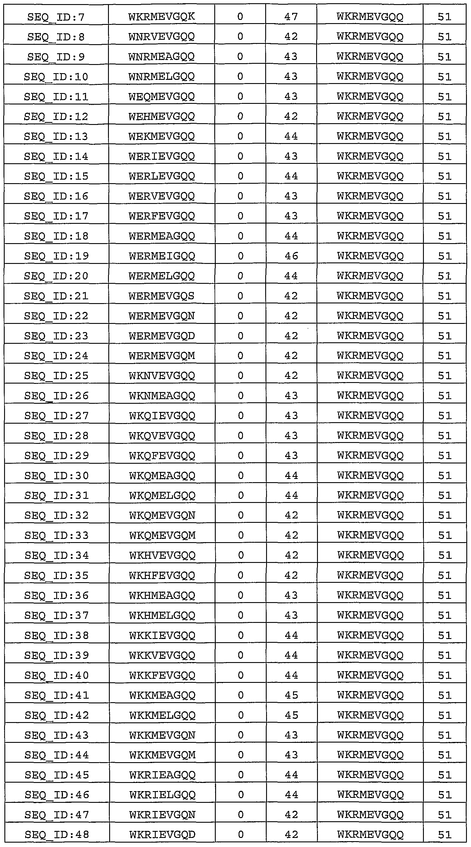
  • each substitution has a score of 0 or greater in the BLOSUM62 substitution matrix, (2) each substitution is capable of conferring reduced binding to at least one of the MHC alleles considered, and (3) once sufficient substitutions are incorporated to prevent any allele hits at a 1 % threshold, no additional substitutions are added to that sequence.
  • Alternate sequences were scored for immunogenicity and structural compatibility. Preferred alternate sequences were defined to be those sequences that are not predicted to bind to any of the 17 MHC alleles tested above using a 1% threshold, and that have a total BLOSUM62 score that is at least 85% of the wild type score.
  • B(wt) is the BLOSUM62 score of the wild type nine- mer
  • l(alt) is the percent of the US population containing one or more MHC alleles that are predicted to bind the alternate nine-mer at a 1 % threshold
  • B(alt) is the BLOSUM62 score of the alternate nine-mer.
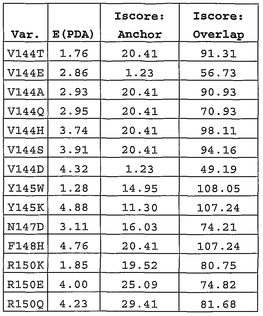
  • Example 3 Identification of suitable less immunogenic sequences for MHC-bindinq aqretopes in erythropoietin as determined bv PDA® technology
  • E(PDA) is the energy determined using PDA® technology calculations compared against the wild-type
  • lscore Anchor is the lscore for the agretope
  • lscore Overlap is the sum of the lscores for all of the overlapping agretopes.
  • EPO as used herein, also refers to wild-type human erythropoietin protein including the following sequence:
  • the wild-type human erythropoietin protein also refers to sequences corresponding to SEQ ID NO: 1

Landscapes

  • Chemical & Material Sciences (AREA)
  • Health & Medical Sciences (AREA)
  • Life Sciences & Earth Sciences (AREA)
  • Organic Chemistry (AREA)
  • General Health & Medical Sciences (AREA)
  • Gastroenterology & Hepatology (AREA)
  • Biochemistry (AREA)
  • Biophysics (AREA)
  • Zoology (AREA)
  • Genetics & Genomics (AREA)
  • Medicinal Chemistry (AREA)
  • Molecular Biology (AREA)
  • Proteomics, Peptides & Aminoacids (AREA)
  • Toxicology (AREA)
  • Medicines That Contain Protein Lipid Enzymes And Other Medicines (AREA)
  • Peptides Or Proteins (AREA)

Abstract

The present invention relates to novel erythropoietin protein variants with altered immunogenicity.

Description

Erythropoietin Derivatives with Altered lmmunogenicity
[01] This application claims benefit under 35 USC 119(e) to U.S. Provisional Application No. 60/607,461 filed September 2, 2004, which is incorporated by reference in its entirety.
Field of the Invention
[02] The present invention relates to variant erythropoietin (EPO) proteins with reduced immunogenicity. In particular, variants of EPO with reduced ability to bind one or more human class Il MHC molecules are described.
Background of the Invention
[03] Erythropoietin or EPO is well known and characterized in the art for possessing erythropoiesis stimulating activity. See, for example, US Pat. Nos. 4,667,195 and 4,703,008, both incorporated entirely by reference. Erythropoietin EPO is an acidic glycoprotein hormone of about 34 kD molecular weight. It is comprised of 165 amino acids and has four carbohydrate chains (three N-linked and one O-linked), which have been shown to affect the protein's stability, solubility, and in vivo bioactivity but are not required for receptor binding. It is a member of the cytokine family that includes interleukins 2- 7, G-CSF, GM-CSF, TPO, growth hormone and leptin. EPO induces proliferation and differentiation of erythroid progenitor cells into erythrocytes, stimulates hemoglobin C synthesis, and increases hematocrit levels. Stimulation of erythropoiesis involves the binding of EPO to the extracellular domain of the EPO receptor (EPOR) on the surface of erythroid progenitor cells; this association triggers intracellular signaling events including phosphorylation of the receptor and activation of the JAK-STAT, RAS and P13 kinase pathways. These signaling pathways trigger cells to undergo proliferation and differentiation and to prevent apoptosis. EPO is produced by the liver in the fetus and by the kidney in adults; it circulates in the blood to stimulate production of red blood cells in bone marrow. Anemia is almost invariably a consequence of renal failure due to decreased production of EPO from the kidney.
[04] EPO receptors are also found in numerous nonerythroid cells including myeloid cells, lymphocytes, megakaryocytes, endothelial cells, mesangial, myocardial, and smooth muscle cells, as well as neural, prostate, and renal cells. Many of these cell types have active EPO signaling pathways and biologic responses. EPO has been shown to be important in the development of new blood vessels and has biologic effects in many tissues especially in the brain, ovary, oviduct, uterus, and testis, as well as in selected tumors. See Weiss Oncologist 8 Suppl 3: 18-29 (2003), incorporated entirely by reference.
[05] Recombinant human EPO (rHuEPO) is in clinical use worldwide for the treatment of anemias deriving from renal failure, chemotherapy, and AIDS. It is also used to treat anemia in premature infants, to reduce the need for blood transfusions due to trauma or surgery, and to treat renal disease and patients undergoing dialysis. Other indications include use in autologous blood transfusions, refractory anemia, and anemia associated with hematological malignancies and other blood disorders such as hemophilia and sickle cell disease. See, for example, US 4,667,195 and 4,703,008. A rHuEPO dimer has also been investigated as a potential therapy for immune disorders and for hematopoietic restoration following radio- or chemotherapy; the dimer was found to be more active than the monomeric form in vitro and in vivo (see Sytkowski et al. Proc Natl Acad Sci U S A 95: 1184- 1188 (1998), incorporated entirely by reference).
[06] EPO also has important neuroprotective effects. EPO and EpoR are abundant in the brain and spinal cord and are significantly upregulated by metabolic stress. In rodent models, rHuEPO was found to cross the blood brain barrier and attenuate ischemia-induced inflammation by reducing neuronal death, indicating its potential utility in the treatment of cerebral ischemia and brain trauma (Kalialis and Olsen Ugeskr Laeger 165: 2477-2481 (2003), Villa et al. J Exp Med 198: 971-975 (2003), both incorporated entirely by reference). Studies in a rat model of experimental autoimmune encephalomyelitis (EAE) indicate that EPO may act as a protective cytokine in inflammatory pathologies of the CNS (Agnello et al. Brain Res 952: 128-134 (2002), incorporated entirely by reference). Epo's neuroprotective effects have also been exhibited in other preclinical models of CNS disorders including multiple sclerosis, spinal cord trauma, and light- or ischemia-induced retinal damage. In initial clinical studies, rHuEPO appeared to reduce the damage from stroke (Ehrenreich et al. MoI Med 8: 495-505 (2002), incorporated entirely by reference). Possible mechanisms of EPO's neuroprotective effects include prevention of glutamate-induced toxicity, inhibition of apoptosis, anti¬ inflammatory effects, antioxidant effects, and stimulation of angiogenesis.
[07] Immunogenicitv of EPO
[08] lmmunogenicity is a major barrier to the development and utilization of protein therapeutics. Although immune responses are typically most severe for non-human proteins, even therapeutics based on human proteins may be immunogenic, lmmunogenicity is a complex series of responses to a substance that is perceived as foreign and may include production of neutralizing and non- neutralizing antibodies, formation of immune complexes, complement activation, mast cell activation, inflammation, and anaphylaxis.
[09] Several factors can contribute to protein immunogenicity, including but not limited to the protein sequence, the route and frequency of administration, and the patient population.
[10] Immunogenicity may limit the efficacy and safety of a protein therapeutic in multiple ways. Efficacy can be reduced directly by the formation of neutralizing antibodies. Efficacy may also be reduced indirectly, as binding to either neutralizing or non-neutralizing antibodies typically leads to rapid clearance from serum. Severe side effects and even death may occur when an immune reaction is raised. One special class of side effects results when neutralizing antibodies cross-react with an endogenous protein and block its function.
[11] Incidence of immunogenic response to rHuEPO is rare (<1 :10,000 patient-years). However, since 1998, more than 200 patients with chronic kidney disease treated with rHuEPO developed cross- reactive neutralizing antibodies (to endogenous Epo), causing pure red cell aplasia (PRCA). All of these patients were receiving EPO subcutaneously, and the product most typically prescribed was epoetin alfa (Eprex®, Ortho Biotech). It has been suggested that since the rates of PRCA remained low for other EPO products (Epogen® and NeoRecorman®), the increase in PRCA was likely product-specific. Possible mechanisms that have been identified include modification of drug formulation and down stream processing. See Casadevall et al. N Engl J Med 346: 469-475 (2002), Macdougall Curr Med Res Opin 20: 83-86 (2004), Verhelst et al. Lancet 363: 1768-1771 (2004), Locatelli and Del Vecchio J Nephrol 16: 461-466 (2003), all incorporated entirely by reference.
[12] In an attempt to decrease aggregation of E coli-derived EPO, investigators replaced N- glycosylation site residues N24, N38, and N83 with basic residues (lysines) and improved the stability of the protein (Narhi et al. Protein Eng 14: 135-140 (2001 ), incorporated entirely by reference). Although these studies were done to facilitate high-resolution structure analysis, decreasing the formation of aggregates is also likely to reduce immunogenicity.
[13] Several methods have been developed to modulate the immunogenicity of proteins. In some cases, PEGylation has been observed to reduce the fraction of patients who raise neutralizing antibodies by sterically blocking access to antibody agretopes (see for example, Hershfield ef al. PNAS 1991 88:7185-7189 (1991); Bailon. et al. Bioconjug. Chem. 12: 195-202(2001); He et al. Life Sci. 65: 355-368 (1999), all incorporated entirely by reference). Methods that improve the solution properties of a protein therapeutic may also reduce immunogenicity, as aggregates have been observed to be more immunogenic than soluble proteins.
[14] A more general approach to immunogenicity reduction involves mutagenesis targeted at the agretopes in the protein sequence and structure that are most responsible for stimulating the immune system. Some success has been achieved by randomly replacing solvent-exposed residues to lower binding affinity to panels of known neutralizing antibodies (see for example Laroche et al. Blood 96: 1425-1432 (2000), incorporated entirely by reference). Due to the incredible diversity of the antibody repertoire, mutations that lower affinity to known antibodies will typically lead to production of an another set of antibodies rather than abrogation of immunogenicity. However, in some cases it may be possible to decrease surface antigenicity by replacing hydrophobic and charged residues on the protein surface with polar neutral residues (see Meyer et al. Protein Sci. 10: 491-503 (2001), incorporated entirely by reference).
[15] An alternate approach is to disrupt T-cell activation. Removal of MHC-binding agretopes offers a much more tractable approach to immunogenicity reduction, as the diversity of MHC molecules comprises only ~103 alleles, while the antibody repertoire is estimated to be approximately 108 and the T-cell receptor repertoire is larger still. By identifying and removing or modifying class Il MHC- binding peptides within a protein sequence, the molecular basis of immunogenicity can be evaded. The elimination of such agretopes for the purpose of generating less immunogenic proteins has been disclosed previously; see for example WO 98/52976, WO 02/079232, and WO 00/3317, all incorporated entirely by reference. Examples of replacing MHC binding agretopes in EPO have also been disclosed; see WO 02/062843, incorporated entirely by reference.
[16] While mutations in MHC-binding agretopes can be identified that are predicted to confer reduced immunogenicity, most amino acid substitutions are energetically unfavorable. As a result, the vast majority of the reduced immunogenicity sequences identified using the methods described above will be incompatible with the structure and/or function of the protein. In order for MHC agretope removal to be a viable approach for reducing immunogenicity, it is crucial that simultaneous efforts are made to maintain a protein's structure, stability, and biological activity. [17] There remains a need for novel EPO proteins having reduced immunogenicity. Variants of EPO with reduced immunogenicity could find use in the treatment of a number of EPO responsive conditions. Summary of the Invention
[18] In accordance with the objects outlined above, the present invention provides novel erythropoietin proteins having reduced immunogenicity as compared to naturally occurring EPO proteins. In an additional aspect, the present invention is directed to methods for engineering or designing less immunogenic proteins with EPO activity for therapeutic use.
[19] An aspect of the present invention are EPO variants that show decreased binding affinity for one or more class Il MHC alleles relative to a parent EPO and which significantly maintain the activity of native naturally occurring Epo.
[20] In a further aspect, the invention provides recombinant nucleic acids encoding the variant EPO proteins, expression vectors, and host cells.
[21] In an additional aspect, the invention provides methods of producing a variant EPO protein comprising culturing the host cells of the invention under conditions suitable for expression of the variant EPO protein.
[22] In a further aspect, the invention provides pharmaceutical compositions comprising a variant EPO protein or nucleic acid of the invention and a pharmaceutical carrier.
[23] In a further aspect, the invention provides methods for preventing or treating EPO responsive disorders comprising administering a variant EPO protein or nucleic acid of the invention to a patient.
[24] In an additional aspect, the invention provides methods for screening the class Il MHC haplotypes of potential patients in order to identify individuals who are particularly likely to raise an immune response to a wild type or variant EPO therapeutic.
[25] In accordance with the objects outlined above, the present invention provides EPO variant proteins comprising amino acid sequences with at least one amino acid insertion, deletion, or substitution compared to the wild type EPO proteins.
Brief Description of the Figures
[26] Figure 1 shows a method for engineering less immunogenic erythropoietin derivatives.
[27] Figure 2 shows a schematic representation of a method for in vitro testing of the immunogenicity of erythropoietin peptides or proteins with IW technology.
Detailed Description of the Invention
[28] By "nine-mer peptide frame" and grammatical equivalents herein is meant a linear sequence of nine amino acids that is located in a protein of interest, nine-mer frames may be analyzed for their propensity to bind one or more class Il MHC alleles. By "allele" and grammatical equivalents herein is meant an alternative form of a gene. Specifically, in the context of class Il MHC molecules, alleles comprise all naturally occurring sequence variants of DRA, DRB1 , DRB3/4/5, DQA-I, DQB1 , DPA-I, and DPB1 molecules. By "Epo-responsive disorders" and grammatical equivalents herein is meant diseases, disorders, and conditions that can benefit from treatment with Epo. Examples of disorders that may benefit from treatment with EPO include, but are not limited to, cardiovascular diseases, particularly atherosclerosis, coronary artery disease, heart attack, stroke, and restenosis, diseases of lipid and cholesterol homeostasis, including conditions associated with EPO or HDL defects or deficiencies, analphalipoproteinemia, hypoalphalipoproteinemia, hyperlipidemia, amyloidosis, and viral and bacterial infections. By "hit" and grammatical equivalents herein is meant, in the context of the matrix method, that a given peptide is predicted to bind to a given class Il MHC allele. In a preferred embodiment, a hit is defined to be a peptide with binding affinity among the top 5%, or 3%, or 1 % of binding scores of random peptide sequences. In an alternate embodiment, a hit is defined to be a peptide with a binding affinity that exceeds some threshold, for instance a peptide that is predicted to bind an MHC allele with at least 100 μM or 10 μM or 1 μM affinity. By "immunogenicity" and grammatical equivalents herein is meant the ability of a protein to elicit an immune response, including but not limited to production of neutralizing and non-neutralizing antibodies, formation of immune complexes, complement activation, mast cell activation, inflammation, and anaphylaxis. By "reduced immunogenicity" and grammatical equivalents herein is meant a decreased ability to activate the immune system, when compared to the wild type protein. For example, a variant protein can be said to have "reduced immunogenicity" if it elicits neutralizing or non-neutralizing antibodies in lower titer or in fewer patients than the wild type protein. In a preferred embodiment, the probability of raising neutralizing antibodies is decreased by at least 5% , with at least 50% or 90% decreases being especially preferred. So, if a wild type produces an immune response in 10% of patients, a variant with reduced immunogenicity would produce an immune response in not more than 9.5% of patients, with less than 5% or less than 1 % being especially preferred. A variant protein also can be said to have "reduced immunogenicity" if it shows decreased binding to one or more MHC alleles or if it induces T-cell activation in a decreased fraction of patients relative to the parent protein. In a preferred embodiment, the probability of T-cell activation is decreased by at least 5% , with at least 50% or 90% decreases being especially preferred. By "matrix method" and grammatical equivalents thereof herein is meant a method for calculating peptide - MHC affinity in which a matrix is used that contains a score for each possible residue at each position in the peptide, interacting with a given MHC allele. The binding score for a given peptide - MHC interaction is obtained by summing the matrix values for the amino acids observed at each position in the peptide. By "MHC-binding agretopes" and grammatical equivalents herein is meant peptides that are capable of binding to one or more class Il MHC alleles with appropriate affinity to enable the formation of MHC - peptide - T- cell receptor complexes and subsequent T-cell activation. MHC-binding agretopes are linear peptide sequences that comprise at least approximately 9 residues. By "parent protein" as used herein is meant a protein that is subsequently modified to generate a variant protein. Said parent protein may be a wild-type or naturally occurring protein, or a variant or engineered version of a naturally occurring protein. "Parent protein" may refer to the protein itself, compositions that comprise the parent protein, or any amino acid sequence that encodes it. Accordingly, by "parent erythropoietin protein" as used herein is meant an erythropoietin protein that is modified to generate a variant erythropoietin protein. By "patient" herein is meant both humans and other animals, particularly mammals, and organisms. Thus the methods are applicable to both human therapy and veterinary applications. In the preferred embodiment the patient is a mammal, and in the most preferred embodiment the patient is human. By "protein" herein is meant at least two covalently attached amino acids, which includes proteins, polypeptides, oligopeptides and peptides. The protein may be made up of naturally occurring amino acids and peptide bonds, or synthetic peptidomimetic structures, i.e., "analogs" such as peptoids [see Simon et al., Proc. Natl. Acad. Sci. U.S.A. 89(20:9367-71 (1992), incorporated entirely by reference), generally depending on the method of synthesis. For example, homo-phenylalanine, citrulline, and noreleucine are considered amino acids for the purposes of the invention. "Amino acid" also includes amino acid residues such as proline and hydroxyproline. Both D- and L- amino acids may be utilized. By "treatment" herein is meant to include therapeutic treatment, as well as prophylactic, or suppressive measures for the disease or disorder. Thus, for example, successful administration of a variant EPO protein prior to onset of the disease may result in treatment of the disease. As another example, successful administration of a variant EPO protein after clinical manifestation of the disease to combat the symptoms of the disease comprises "treatment" of the disease. "Treatment" also encompasses administration of a variant EPO protein after the appearance of the disease in order to eradicate the disease. Successful administration of an agent after onset and after clinical symptoms have developed, with possible abatement of clinical symptoms and perhaps amelioration of the disease, further comprises "treatment" of the disease. By "variant erythropoietin nucleic acids" and grammatical equivalents herein is meant nucleic acids that encode variant erythropoietin proteins. Due to the degeneracy of the genetic code, an extremely large number of nucleic acids may be made, all of which encode the variant erythropoietin proteins of the present invention, by simply modifying the sequence of one or more codons in a way which does not change the amino acid sequence of the variant erythropoietin. By "variant erythropoietin proteins" and grammatical equivalents thereof herein is meant non-naturally occurring proteins which differ from the wild type or parent erythropoietin protein by at least 1 amino acid insertion, deletion, or substitution. Erythropoietin variants are characterized by the predetermined nature of the variation, a feature that sets them apart from naturally occurring allelic or interspecies variation of the erythropoietin protein sequence. The erythropoietin variants typically either exhibit biological activity that is comparable to naturally occurring erythropoietin or have been specifically engineered to have alternate biological properties. The variant erythropoietin proteins may contain insertions, deletions, and/or substitutions at the N- terminus, C-terminus, or internally. In a preferred embodiment, variant erythropoietin proteins have at least 1 residue that differs from the naturally occurring erythropoietin sequence, with at least 2, 3, 4, or 5 different residues being more preferred. Variant erythropoietin proteins may contain further modifications, for instance mutations that alter stability or solubility or which enable or prevent posttranslational modifications such as PEGylation or glycosylation. Variant erythropoietin proteins may be subjected to co- or post-translational modifications, including but not limited to synthetic derivatization of one or more side chains or termini, glycosylation, PEGylation, circular permutation, cyclization, fusion to proteins or protein domains, and addition of peptide tags or labels. By "wild type or wt" and grammatical equivalents thereof herein is meant an amino acid sequence or a nucleotide sequence that is found in nature and includes allelic variations; that is, an amino acid sequence or a nucleotide sequence that has not been intentionally modified. In a preferred embodiment, the wild type sequence is SEQJD NO:1. [29] Identification of MHC-bindinα aqretopes in erythropoietin [30] MHC-binding peptides are obtained from proteins by a process called antigen processing. First, the protein is transported into an antigen presenting cell (APC) by endocytosis or phagocytosis. A variety of proteolytic enzymes then cleave the protein into a number of peptides. These peptides can then be loaded onto class Il MHC molecules, and the resulting peptide-MHC complexes are transported to the cell surface. Relatively stable peptide-MHC complexes can be recognized by T-cell receptors that are present on the surface of naive T cells. This recognition event is required for the initiation of an immune response. Accordingly, blocking the formation of stable peptide-MHC complexes is an effective approach for preventing unwanted immune responses.
[31] The factors that determine the affinity of peptide-MHC interactions have been characterized using biochemical and structural methods. Peptides bind in an extended conformation bind along a groove in the class Il MHC molecule. While peptides that bind class Il MHC molecules are typically approximately 13-18 residues long, a nine-residue region is responsible for most of the binding affinity and specificity. The peptide binding groove can be subdivided into "pockets", commonly named P1 through P9, where each pocket is comprises the set of MHC residues that interacts with a specific residue in the peptide. A number of polymorphic residues face into the peptide-binding groove of the MHC molecule. The identity of the residues lining each of the peptide-binding pockets of each MHC molecule determines its peptide binding specificity. Conversely, the sequence of a peptide determines its affinity for each MHC allele.
[32] Several methods of identifying MHC-binding agretopes in protein sequences are known in the art and may be used to identify agretopes in Epo. Sequence-based information can be used to determine a binding score for a given peptide - MHC interaction (see for example Mallios, Bioinformatics 15: 432-439 (1999); Mallios, Bioinformatics 17: p942-948 (2001); Sturniolo et al. Nature Biotech. 17: 555- 561 (1999), all incorporated entirely by reference). It is possible to use structure-based methods in which a given peptide is computationally placed in the peptide-binding groove of a given MHC molecule and the interaction energy is determined (for example, see WO 98/59244 and WO 02/069232, both incorporated entirely by reference). Such methods may be referred to as "threading" methods. Alternatively, purely experimental methods can be used; for example a set of overlapping peptides derived from the protein of interest can be experimentally tested for the ability to induce T- cell activation and/or other aspects of an immune response, (see for example WO 02/77187, incorporated entirely by reference).
[33] In a preferred embodiment, MHC-binding propensity scores are calculated for each 9-residue frame along the erythropoietin sequence using a matrix method (see Sturniolo et al., supra; Marshall et al., J. Immunol. 154: 5927-5933 (1995), and Hammer et al., J. Exp. Med. 180: 2353-2358 (1994), both incorporated entirely by reference). It is also possible to consider scores for only a subset of these residues, or to consider also the identities of the peptide residues before and after the 9-residue frame of interest. The matrix comprises binding scores for specific amino acids interacting with the peptide binding pockets in different human class Il MHC molecule. In the most preferred embodiment, the scores in the matrix are obtained from experimental peptide binding studies. In an alternate preferred embodiment, scores for a given amino acid binding to a given pocket are extrapolated from experimentally characterized alleles to additional alleles with identical or similar residues lining that pocket Matrices that are produced by extrapolation are referred to as "virtual matrices".
[34] In a preferred embodiment, the matrix method is used to calculate scores for each peptide of interest binding to each allele of interest. Several methods can then be used to determine whether a given peptide will bind with significant affinity to a given MHC allele. In one embodiment, the binding score for the peptide of interest is compared with the binding propensity scores of a large set of reference peptides. Peptides whose binding propensity scores are large compared to the reference peptides are likely to bind MHC and may be classified as "hits". For example, if the binding propensity score is among the highest 1% of possible binding scores for that allele, it may be scored as a "hit" at the 1% threshold. The total number of hits at one or more threshold values is calculated for each peptide. In some cases, the binding score may directly correspond with a predicted binding affinity. Then, a hit may be defined as a peptide predicted to bind with at least 100 μM or 10 μM or 1 μM affinity.
[35] In a preferred embodiment, the number of hits for each nine-mer frame in the protein is calculated using one or more threshold values ranging from 0.5% to 10%. In an especially preferred embodiment, the number of hits is calculated using 1%, 3%, and 5% thresholds.
[36] In a preferred embodiment, MHC-binding agretopes are identified as the nine-mer frames that bind to several class Il MHC alleles. In an especially preferred embodiment, MHC-binding agretopes are predicted to bind at least 10 alleles at 5% threshold and/or at least 5 alleles at 1% threshold. Such nine-mer frames may be especially likely to elicit an immune response in many members of the human population.
[37] In a preferred embodiment, MHC-binding agretopes are predicted to bind MHC alleles that are present in at least 0.01 - 10% of the human population. Alternatively, to treat conditions that are linked to specific class Il MHC alleles, MHC-binding agretopes are predicted to bind MHC alleles that are present in at least 0.01 - 10% of the relevant patient population.
[38] Data about the prevalence of different MHC alleles in different ethnic and racial groups has been acquired by groups such as the National Marrow Donor Program (NMDP); for example see Mignot et al. Am. J. Hum. Genet 68: 686-699 (2001), Southwood et al. J. Immunol. 160: 3363-3373 (1998), Hurley et al. Bone Marrow Transplantation 25: 136-137 (2000), Sintasath Hum. Immunol. 60: 1001 (1999), Collins et al. Tissue Antigens 55: 48 (2000), Tang et al. Hum. Immunol. 63: 221 (2002), Chen et al. Hum. Immunol. 63: 665 (2002), Tang et al. Hum. Immunol. 61 : 820 (2000), Gans et al. Tissue Antigens 59: 364-369, and Baldassarre et al. Tissue Antigens 61 : 249-252 (2003), all incorporated entirely by reference.
[39] In a preferred embodiment, MHC binding agretopes are predicted for MHC heterodimers comprising highly prevalent MHC alleles. Class Il MHC alleles that are present in at least 10% of the US population include but are not limited to: DPA-l*0103, DPA-l*0201 , DPB1*0201 , DPB1*0401 , DPB1*0402, DQA-l*0101 , DQA-l*0102, DQA-l*0201 , DQA-P0501 , DQBT0201 , DQB1*0202, DQB1*0301 , DQB1*0302, DQB1*0501 , DQB1*0602, DRA*0101 , DRB1*0701 , DRB1*1501 , DRB1*0301 , DRB1*0101 , DRB1*1101 , DRB1*1301 , DRB3*0101 , DRB3*0202, DRB4*0101 , DRB4*0103, and DRB5*0101. [40] In a preferred embodiment, MHC binding agretopes are also predicted for MHC heterodimers comprising moderately prevalent MHC alleles. Class Il MHC alleles that are present in 1% to 10% of the US population include but are not limited to: DPA-l*0104, DPA-l*0302, DPA-l*0301 , DPB1*0101 , DPB1*0202, DPB1*0301 , DPB1* 0501, DPB1*0601, DPB1*0901 , DPB1*1001, DPB1*1101, DPB1*1301 , DPB1*1401 , DPB1*1501 , DPB1*1701 , DPB1*1901 , DPB1 *2001 , DQA-l*0103, DQA- 1*0104, DQA-l*0301 , DQA-l*0302, DQA-l*0401 , DQB1*0303, DQB1*0402, DQB1*0502, DQB1*0503, DQB1*0601, DQB1*0603, DRB1*1302, DRB1*0404, DRB1*0801, DRB1*0102, DRB1*1401, DRB1*1104, DRB1*1201 , DRB1*1503, DRB1*0901 , DRB1*1601 , DRB1*0407, DRB1*1001 , DRB1*1303, DRB1*0103, DRB1*1502, DRB1*0302, DRB1*0405, DRB1*0402, DRB1*1102, DRB1*0803, DRB1*0408, DRB1*1602, DRB1*0403, DRB3*0301, DRB5*0102, and DRB5*0202.
[41] MHC binding agretopes may also be predicted for MHC heterodimers comprising less prevalent alleles. Information about MHC alleles in humans and other species can be obtained, for example, from the IMGT/HLA sequence database, part of the international ImMunoGeneTics project (IMGT).
[42] In an especially preferred embodiment, an immunogenicity score is determined for each peptide, wherein said score depends on the fraction of the population with one or more MHC alleles that are hit at multiple thresholds. For example, the equation lscore = N( W1P1 + W3P3 + W5P5 ) may be used, where P1 is the percent of the population hit at 1%, P3 is the percent of the population hit at 3%, P5 is the percent of the population hit at 5%, each W is a weighting factor, and N is a normalization factor. In a preferred embodiment, W1 = 10, W3 = 5, W5 = 2, and N is selected so that possible scores range from 0 to 100. In this embodiment, agretopes with lscore greater than or equal to 10 are preferred and agretopes with lscore greater than or equal to 25 are especially preferred.
[43] In an additional preferred embodiment, MHC-binding agretopes are identified as the nine-mer frames that are located among "nested" agretopes, or overlapping 9-residue frames that are each predicted to bind a significant number of alleles. Such sequences may be especially likely to elicit an immune response.
[44] Preferred MHC-binding agretopes are those agretopes that are predicted to bind, at a 3% threshold, to MHC alleles that are present in at least 5% of the population. Preferred MHC-binding agretopes in erythropoietin include, but are not limited to, agretope 1 : residues 5-13; agretope 3: residues 51-59; agretope 5: residues 64-72; agretope 8: residues 75-83; agretope 10: residues 93- 101 ; agretope 11 : residues 102-110; agretope 13: residues 138-146; agretope 14: residues 141-149; agretope 15: residues 142-150; agretope 18: residues 149-157; agretope 19: residues 153-161.
[45] Especially preferred MHC-binding agretopes are those agretopes that are predicted to bind, at a 1% threshold, to MHC alleles that are present in at least 10% of the population. Especially preferred MHC-binding agretopes in erythropoietin include, but are not limited to, agretope 3: residues 51-59; agretope 11 : residues 102-110; agretope 13: residues 138-146; agretope 14: residues 141-149; agretope 15: residues 142-150; agretope 18: residues 149-157.
[46] Alternate preferred MHC-binding agretopes are those agretopes that have lscore greater than or equal to 10. Preferred MHC-binding agretopes in erythropoietin include, but are not limited to, agretope 1 : residues 5-13; agretope 3: residues 51-59; agretope 5: residues 64-72; agretope 10: residues 93-101 ; agretope 11 : residues 102-110; agretope 13: residues 138-146; agretope 14: residues 141-149; agretope 15: residues 142-150; agretope 18: residues 149-157; agretope 19: residues 153-161.
[47] Alternate especially preferred MHC-binding agretopes are those agretopes that have lscore greater than or equal to 25. Preferred MHC-binding agretopes in erythropoietin include, but are not limited to, agretope 3: residues 51-59; agretope 11 : residues 102-110; agretope 13: residues 138-146; agretope 14: residues 141-149; agretope 15: residues 142-150; agretope 18: residues 149-157.
[48] Additional especially preferred MHC-binding agretopes are those agretopes whose sequences partially overlap with additional MHC-binding agretopes. Sets of overlapping MHC-binding agretopes in erythropoietin include, but are not limited to, agretope 13: residues 138-146; agretope 14: residues 141-149; agretope 15: residues 142-150; agretope 18: residues 149-157.
[49] Confirmation of MHC-bindinα agretopes
[50] In a preferred embodiment, the immunogenicity of the above-predicted MHC-binding agretopes is experimentally confirmed by measuring the extent to which peptides comprising each predicted agretope can elicit an immune response. However, it is possible to proceed from agretope prediction to agretope removal without the intermediate step of agretope confirmation.
[51] Several methods, discussed in more detail below, can be used for experimental confirmation of agretopes. For example, sets of naϊve T cells and antigen presenting cells from matched donors can be stimulated with a peptide containing an agretope of interest, and T-cell activation can be monitored. It is also possible to first stimulate T cells with the whole protein of interest, and then re- stimulate with peptides derived from the whole protein. If sera are available from patients who have raised an immune response to Epo, it is possible to detect mature T cells that respond to specific epitopes. In a preferred embodiment, interferon gamma or IL-5 production by activated T-cells is monitored using Elispot assays, although it is also possible to use other indicators of T-cell activation or proliferation such as tritiated thymidine incorporation or production of other cytokines.
[52] Patient genotype analysis and screening
[53] HLA genotype is a major determinant of susceptibility to specific autoimmune diseases (see for example Nepom Clin. Immunol. Immunopathol. 67: S50-S55 (1993)) and infections (see for example Singh et al. Emerg. Infect. Dis. 3: 41-49 (1997)), both incorporated entirely by reference. Furthermore, the set of MHC alleles present in an individual can affect the efficacy of some vaccines (see for example Cailat-Zucman et al. Kidney Int. 53: 1626-1630 (1998) and Poland et al. Vaccine 20: 430-438 (2001), both incorporated entirely by reference). HLA genotype may also confer susceptibility for an individual to elicit an unwanted immune response to an erythropoietin therapeutic.
[54] In a preferred embodiment, class Il MHC alleles that are associated with increased or decreased susceptibility to elicit an immune response to EPO proteins are identified. For example, patients treated with EPO therapeutics may be tested for the presence of anti-EPO antibodies and genotyped for class Il MHC. Alternatively, T-cell activation assays such as those described above may be conducted using cells derived from a number of genotyped donors. Alleles that confer susceptibility to EPO immunogenicity may be defined as those alleles that are significantly more common in those who elicit an immune response versus those who do not. Similarly, alleles that confer resistance to EPO immunogenicity may be defined as those that are significantly less common in those who do not elicit an immune response versus those that do. It is also possible to use purely computational techniques to identify which alleles are likely to recognize EPO therapeutics.
[55] In one embodiment, the genotype association data is used to identify patients who are especially likely or especially unlikely to raise an immune response to an EPO therapeutic.
[56] Design of active, less-immunogenic variants
[57] In a preferred embodiment, the above-determined MHC-binding agretopes are replaced with alternate amino acid sequences to generate active variant EPO proteins with reduced or eliminated immunogenicity. Alternatively, the MHC-binding agretopes are modified to introduce one or more sites that are susceptible to cleavage during protein processing. If the agretope is cleaved before it binds to a MHC molecule, it will be unable to promote an immune response. There are several possible strategies for integrating methods for identifying less immunogenic sequences with methods for identifying structured and active sequences, including but not limited to those presented below.
[58] In one embodiment, for one or more nine-mer agretope identified above, one or more possible alternate nine-mer sequences are analyzed for immunogenicity as well as structural and functional compatibility. The preferred alternate nine-mer sequences are then defined as those sequences that have low predicted immunogenicity and a high probability of being structured and active. It is possible to consider only the subset of nine-mer sequences that are most likely to comprise structured, active, less immunogenic variants. For example, it may be unnecessary to consider sequences that comprise highly non-conservative mutations or mutations that increase predicted immunogenicity.
[59] In a preferred embodiment, less immunogenic variants of each agretope are predicted to bind MHC alleles in a smaller fraction of the population than the wild type agretope. In an especially preferred embodiment, the less immunogenic variant of each agretope is predicted to bind to MHC alleles that are present in not more than 5% of the population, with not more than 1% or 0.1% being most preferred.
[60] Substitution matrices
[61] In another especially preferred embodiment, substitution matrices or other knowledge-based scoring methods are used to identify alternate sequences that are likely to retain the structure and function of the wild type protein. Such scoring methods can be used to quantify how conservative a given substitution or set of substitutions is. In most cases, conservative mutations do not significantly disrupt the structure and function of proteins (see for example, Bowie et al. Science 247: 1306-1310 (1990), Bowie and Sauer Proc. Nat. Acad. Sci. USA 86: 2152-2156 (1989), and Reidhaar-Olson and Sauer Proteins 7: 306-316 (1990), all incorporated entirely by reference). However, non-conservative mutations can destabilize protein structure and reduce activity (see for example, Lim et al. Biochem. 31 : 4324-4333 (1992), incorporated entirely by reference). Substitution matrices including but not limited to BLOSUM62 provide a quantitative measure of the compatibility between a sequence and a target structure, which can be used to predict non-disruptive substitution mutations (see Topham et al. Prot. Eng. 10: 7-21 (1997), incorporated entirely by reference). The use of substitution matrices to design peptides with improved properties has been disclosed; see Adenot et al. J. MoI. Graph. Model. 17: 292-309 (1999), incorporated entirely by reference. [62] Substitution matrices include, but are not limited to, the BLOSUM matrices (Henikoff and Henikoff, Proc. Nat. Acad. Sci. USA 89: 10917 (1992), incorporated entirely by reference, the PAM matrices, the Dayhoff matrix, and the like. For a review of substitution matrices, see for example Henikoff Curr. Opin. Struct. Biol. 6: 353-360 (1996), incorporated entirely by reference. It is also possible to construct a substitution matrix based on an alignment of a given protein of interest and its homologs; see for example Henikoff and Henikoff Comput. Appl. Biosci. 12: 135-143 (1996), incorporated entirely by reference.
[63] In a preferred embodiment, each of the substitution mutations that are considered has a BLOSUM62 score of zero or higher. According to this metric, preferred substitutions include, but are not limited to conservative mutations, shown in Table 1 :
Figure imgf000013_0002
Figure imgf000013_0001
[64] In addition, it is preferred that the total BLOSUM62 score of an alternate sequence for a nine residue MHC-binding agretope is decreased only modestly when compared to the BLOSUM62 score of the wild type nine residue agretope. In a preferred embodiment, the score of the variant nine-mer is at least 50% of the wild type score, with at least 67%, 75%, 80%, or 90% being especially preferred.
[65] Alternatively, alternate sequences can be selected that minimize the absolute reduction in BLOSUM score; for example it is preferred that the score decrease for each nine-mer is less than 20, with score decreases of less than about 10 or about 5 being especially preferred. The exact value may be chosen to produce a library of alternate sequences that is experimentally tractable and also sufficiently diverse to encompass a number of active, stable, less immunogenic variants.
[66] In a preferred embodiment, substitution mutations are preferentially introduced at positions that are substantially solvent exposed. As is known in the art, solvent exposed positions are typically more tolerant of mutation than positions that are located in the core of the protein.
[67] In a preferred embodiment, substitution mutations are preferentially introduced at positions that are not highly conserved. As is known in the art, positions that are highly conserved among members of a protein family are often important for protein function, stability, or structure, while positions that are not highly conserved often may be modified without significantly impacting the structural or functional properties of the protein.
[68] Alanine substitutions [69] In an alternate embodiment, one or more alanine substitutions may be made, regardless of whether an alanine substitution is conservative or non-conservative. As is known in the art, incorporation of sufficient alanine substitutions may be used to disrupt intermolecular interactions.
[70] In a preferred embodiment, variant nine-mers are selected such that residues that have been or can be identified as especially critical for maintaining the structure or function of erythropoietin retain their wild type identity. In alternate embodiments, it may be desirable to produce variant EPO proteins that do not retain wild type activity. In such cases, residues that have been identified as critical for function may be specifically targeted for modification.
[71] Mutagenesis studies indicate four regions on EPO that are important for receptor binding and bioactivity: amino acids 11 to 15, 44 to 51, 100 to 108, and 147 to 151. These have been mapped to two sites: site 1 includes helix D (Asn147, Arg150, Gly151 and Leu155) and the A/B connecting loop (residues 42-51); site 2 includes helix A (VaIH , Arg14, and Tyr15) and helix C (SeMOO, Arg103, Ser104 and Leu108) (see Elliott et al. Blood 89: 493-502 (1997), Grodberg et al. Eur J Biochem 218: 597-601 (1993), Wen et al. J Biol Chem 269: 22839-22846 (1994), Kung et al. Arch Biochem Biophys 379: 85-89 (2000), all incorporated entirely by reference. C-terminal residues 154-159 have also been implicated (Bittorf et al. FEBS Lett 336: 133-136 (1993), incorporated entirely by reference). Others have similarly identified two distinct receptor binding sites: site 1 (Arg150 and Lys152), which binds initially to one EpoR, and site 2 (Arg103, Ser104 and possibly Arg14) which binds to a second EpoR, and have postulated that both sites are required to form a homodimeric receptor complex required for EpoR activation. Arg103 appears to be particularly important for activity and may also play a stabilizing structural role (Matthews et al. Proc Natl Acad Sci U S A 93: 9471-9476 (1996), Qiu et al. J Biol Chem 273: 11173-11176 (1998), both incorporated entirely by reference.
[72] Removal of N-glycosylation sites (Asn24, Asn38, Asn83) has no effect on in vitro activity, but severely decreases in vivo activity, indicating that N-linked sugars are important for (1) proper biosynthesis and/or secretion and (2) expression of the in vivo activity probably by enhancing survival in the circulation. Aranesp®, the novel rHuEPO engineered to have two additional N-glycans, has a markedly prolonged half-life and therefore can be administered less frequently (see Yamaguchi et al. J Biol Chem 266: 20434-20966 (1991), Delorme et al. Biochemistry 31 : 9871-9876 (1992), Bunn Blood 99: 1503 (2002), Jelkmann Eur J Haematol 69: 265-274 (2002), all incorporated entirely by reference.
[73] Protein Design Methods
[74] Protein design methods and MHC agretope identification methods may be used together to identify stable, active, and minimally immunogenic protein sequences (see WO03/006154, incorporated entirely by reference). The combination of approaches provides significant advantages over the prior art for immunogenicity reduction, as most of the reduced immunogenicity sequences identified using other techniques fail to retain sufficient activity and stability to serve as therapeutics.
[75] Protein design methods may identify non-conservative or unexpected mutations that nonetheless confer desired functional properties and reduced immunogenicity, as well as identifying conservative mutations. Nonconservative mutations are defined herein to be all substitutions not included in Table 1 above; nonconservative mutations also include mutations that are unexpected in a given structural context, such as mutations to hydrophobic residues at the protein surface and mutations to polar residues in the protein core.
[76] Furthermore, protein design methods may identify compensatory mutations. For example, if a given first mutation that is introduced to reduce immunogenicity also decreases stability or activity, protein design methods may be used to find one or more additional mutations that serve to recover stability and activity while retaining reduced immunogenicity. Similarly, protein design methods may identify sets of two or more mutations that together confer reduced immunogenicity and retained activity and stability, even in cases where one or more of the mutations, in isolation, fails to confer desired properties.
[77] A wide variety of methods are known for generating and evaluating sequences. These include, but are not limited to, sequence profiling (Bowie and Eisenberg, Science 253(5016): 164-70, (1991)), residue pair potentials (Jones, Protein Science 3: 567-574, (1994)), and rotamer library selections (Dahiyat and Mayo, Protein Sci 5(5): 895-903 (1996); Dahiyat and Mayo, Science 278(5335): 82-7 (1997); Desjarlais and Handel, Protein Science 4: 2006-2018 (1995); Harbury et al, PNAS USA 92(18): 8408-8412 (1995); Kono et al., Proteins: Structure, Function and Genetics 19: 244-255 (1994); Hellinga and Richards, PNAS USA 91 : 5803-5807 (1994)), ail incorporated entirely by reference.
[78] Protein Design Automation® (PDA®) technology
[79] In an especially preferred embodiment, rational design of improved erythropoietin variants is achieved by using Protein Design Automation® (PDA®) technology. (See U.S. Patent Nos. 6,188,965; 6,269,312; 6,403,312; WO98/47089 and USSNs 09/058,459, 09/127,926, 60/104,612, 60/158,700, 09/419,351, 60/181 ,630, 60/186,904, 09/419,351 , 09/782,004 and 09/927,790, 60/347,772, and 10/218,102; and PCT/US01/218.102 and U.S.S.N. 10/218,102, U.S.S.N. 60/345,805; U.S.S.N. 60/373,453 and U.S.S.N. 60/374,035, all incorporated entirely by reference.)
[80] PDA® technology couples computational design algorithms that generate quality sequence diversity with experimental high-throughput screening to discover proteins with improved properties. The computational component uses atomic level scoring functions, side chain rotamer sampling, and advanced optimization methods to accurately capture the relationships between protein sequence, structure, and function. Calculations begin with the three-dimensional structure of the protein and a strategy to optimize one or more properties of the protein. PDA® technology then explores the sequence space comprising all pertinent amino acids (including unnatural amino acids, if desired) at the positions targeted for design. This is accomplished by sampling conformational states of allowed amino acids and scoring them using a parameterized and experimentally validated function that describes the physical and chemical forces governing protein structure. Powerful combinatorial search algorithms are then used to search through the initial sequence space, which may constitute 1050 sequences or more, and quickly return a tractable number of sequences that are predicted to satisfy the design criteria. Useful modes of the technology span from combinatorial sequence design to prioritized selection of optimal single site substitutions. PDA® technology has been applied to numerous systems including, important pharmaceutical and industrial proteins and has a demonstrated record of success in protein optimization. [81] PDA® utilizes three-dimensional structural information. In a most preferred embodiment, the structure of EPO is determined using X-ray crystallography or NMR methods, which are well known in the art. The solution structure of free human EPO has been determined by NMR (Cheetham eif a/. Nat Struct Biol 5: 861-866 (1998), incorporated entirely by reference). The crystal structure of human EPO complexed to the extracellular ligand-binding domains of EpoR has been resolved at 1.9 A and the crystal structure of the extracellular domain of EpoR in its unliganded form has been resolved at 2.4 A. See Syed e* a/. Nature 395: 511-516 (1998), Livnah ef a/. Science 283: 987-990 (1999), incorporated entirely by reference.
[82] In a preferred embodiment, the results of matrix method calculations are used to identify which of the 9 amino acid positions within the agretope(s) contribute most to the overall binding propensities for each particular allele "hit". This analysis considers which positions (P1-P9) are occupied by amino acids which consistently make a significant contribution to MHC binding affinity for the alleles scoring above the threshold values. Matrix method calculations are then used to identify amino acid substitutions at said positions that would decrease or eliminate predicted immunogenicity and PDA® technology is used to determine which of the alternate sequences with reduced or eliminated immunogenicity are compatible with maintaining the structure and function of the protein.
[83] In an alternate preferred embodiment, the residues in each agretope are first analyzed by one skilled in the art to identify alternate residues that are potentially compatible with maintaining the structure and function of the protein. Then, the set of resulting sequences are computationally screened to identify the least immunogenic variants. Finally, each of the less immunogenic sequences are analyzed more thoroughly in PDA® technology protein design calculations to identify protein sequences that maintain the protein structure and function and decrease immunogenicity.
[84] In an alternate preferred embodiment, each residue that contributes significantly to the MHC binding affinity of an agretope is analyzed to identify a subset of amino acid substitutions that are potentially compatible with maintaining the structure and function of the protein. This step may be performed in several ways, including PDA® calculations or visual inspection by one skilled in the art. Sequences may be generated that contain all possible combinations of amino acids that were selected for consideration at each position. Matrix method calculations can be used to determine the immunogenicity of each sequence. The results can be analyzed to identify sequences that have significantly decreased immunogenicity. Additional PDA® calculations may be performed to determine which of the minimally immunogenic sequences are compatible with maintaining the structure and function of the protein.
[85] In an alternate preferred embodiment, pseudo-energy terms derived from the peptide binding propensity matrices are incorporated directly into the PDA® technology calculations. In this way, it is possible to select sequences that are active and less immunogenic in a single computational step.
[86] Combining immunoqenicitv reduction strategies
[87] In a preferred embodiment, more than one method is used to generate variant proteins with desired functional and immunological properties. For example, substitution matrices may be used in combination with PDA® technology calculations. Strategies for immunogenicity reduction include, but are not limited to, those described in USSN 09/903,378; WO 01/21823; USSN 10/039,170; WO 02/00165; USSN 10/339,788; 10/754,296; and USSN 10/638,995, all incorporated entirely by reference.
[88] In a preferred embodiment, a variant protein with reduced binding affinity for one or more class Il MHC alleles is further engineered to confer improved solubility. As protein aggregation may contribute to unwanted immune responses, increasing protein solubility may reduce immunogenicity. See USSN 10/820,467, filed March 30, 2004, entitled, "Interferon Variants With Improved Properties", incorporated entirely by reference.
[89] In an additional preferred embodiment, a variant protein with reduced binding affinity for one or more class Il MHC alleles is further modified by derivitization with PEG or another molecule. As is known in the art, PEG may sterically interfere with antibody binding or improve protein solubility, thereby reducing immunogenicity. In an especially preferred embodiment, rational PEGylation methods are used. See, PCT/US2004/008425; and USSN 10/ 10/956,352, filed September 30, 2004, entitled, "Rational Chemical Modification," both incorporated entirely by reference.
[90] In a further preferred embodiment, a variant protein with reduced binding affinity for one or more class Il MHC alleles is further modified by circular permutation or cyclization.
[91] In a preferred embodiment, PDA® technology and matrix method calculations are used to remove more than one MHC-binding agretope from a protein of interest.
[92] Generating the variants
[93] Variant interferon nucleic acids and proteins of the invention may be produced using a number of methods known in the art.
[94] Preparing nucleic acids encoding the EPO variants
[95] In a preferred embodiment, nucleic acids encoding EPO variants are prepared by total gene synthesis, or by site-directed mutagenesis of a nucleic acid encoding wild type or variant EPO protein. Methods including template-directed ligation, recursive PCR, cassette mutagenesis, site-directed mutagenesis or other techniques that are well known in the art may be utilized (see for example Strizhov et a/. PNAS 93:15012-15017 (1996), Prodromou and Perl, Prot. Eng. 5: 827-829 (1992), Jayaraman and Puccini, Biotechniques 12: 392-398 (1992), and Chalmers et at. Biotechniques 30: 249-252 (2001), all incorporated entirely by reference).
[96] In addition, it should be noted that variant EPO proteins and nucleic acids can be made that include substitutions in "fixed" or "non-agretope" positions as well. For example, while variants within an agretope are recited, any particular variant can include additional variants within non-agretope positions. In addition, any combination of recited variants can be made, as well as combinations of recited variants and non-agretope variants can be made.
[97] Expression vectors
[98] In a preferred embodiment, an expression vector that comprises the components described below and a gene encoding a variant EPO protein is prepared. Numerous types of appropriate expression vectors and suitable regulatory sequences for a variety of host cells are known in the art. The expression vectors may contain transcriptional and translational regulatory sequences including but not limited to promoter sequences, ribosomal binding sites, transcriptional start and stop sequences, translational start and stop sequences, transcription terminator signals, polyadenylation signals, and enhancer or activator sequences. In a preferred embodiment, the regulatory sequences include a promoter and transcriptional start and stop sequences. In addition, the expression vector may comprise additional elements. For example, the expression vector may have two replication systems, thus allowing it to be maintained in two organisms, for example in mammalian or insect cells for expression and in a prokaryotic host for cloning and amplification. Furthermore, for integrating expression vectors, the expression vector contains at least one sequence homologous to the host cell genome, and preferably two homologous sequences, which flank the expression construct. The integrating vector may be directed to a specific locus in the host cell by selecting the appropriate homologous sequence for inclusion in the vector. Constructs for integrating vectors are well known in the art. In addition, in a preferred embodiment, the expression vector contains a selectable marker gene to allow the selection of transformed host cells. Selection genes are well known in the art and will vary with the host cell used. The expression vectors may be either self-replicating extrachromosomal vectors or vectors which integrate into a host genome.
[99] The expression vector may include a secretory leader sequence or signal peptide sequence that provides for secretion of the variant EPO protein from the host cell. Suitable secretory leader sequences that lead to the secretion of a protein are known in the art. The signal sequence typically encodes a signal peptide comprised of hydrophobic amino acids, which direct the secretion of the protein from the cell. The protein is either secreted into the growth media or, for prokaryotes, into the periplasmic space, located between the inner and outer membrane of the cell. For expression in bacteria, bacterial secretory leader sequences, operably linked to a variant EPO encoding nucleic acid, are usually preferred.
[100] Transfection/Transformation
[101] The variant EPO nucleic acids are introduced into the cells either alone or in combination with an expression vector in a manner suitable for subsequent expression of the nucleic acid. The method of introduction is largely dictated by the targeted cell type. Exemplary methods include CaPO4 precipitation, liposome fusion, Lipofectin®, electroporation, viral infection, dextran-mediated transfection, polybrene mediated transfection, protoplast fusion, direct microinjection, etc. The variant EPO nucleic acids may stably integrate into the genome of the host cell or may exist either transiently or stably in the cytoplasm. As outlined herein, a particularly preferred method utilizes retroviral infection, as outlined in PCT/US97/01019, incorporated entirely by reference.
[102] Hosts for the expression of EPO variants
[103] Appropriate host cells for the expression of EPO variants include yeast, bacteria, archaebacteria, fungi, and insect and animal cells, including mammalian cells. Of particular interest are bacteria such as E. co// and Bacillus subtilis, fungi such as Saccharomyces cerevisiae, Pichia pastoris, and Neurospora, insects such as Drosophila melangaster and insect cell lines such as SF9, mammalian cell lines including 293, CHO, COS, Jurkat, NIH3T3, etc (see the ATCC cell line catalog, hereby expressly incorporated entirely by reference), as well as primary cell lines.
[104] EPO variants can also be produced in more complex organisms, including but not limited to plants (such as corn, tobacco, and algae) and animals (such as chickens, goats, cows); see for example Dove, Nature Biotechnol. 20: 777-779 (2002), incorporated entirely by reference. [105] In one embodiment, the cells may be additionally genetically engineered, that is, contain exogenous nucleic acid other than the expression vector comprising the variant EPO nucleic acid.
[106] Expression and purification methods
[107] Variant EPO proteins of the invention and nucleic acids encoding them may be produced using a number of methods known in the art.
[108] In a preferred embodiment, nucleic acids encoding the EPO variants are prepared by total gene synthesis, or by site-directed mutagenesis of a nucleic acid encoding a parent EPO protein. Methods including template-directed ligation, recursive PCR, cassette mutagenesis, site-directed mutagenesis or other techniques that are well known in the art may be utilized (see for example Strizhov et al. PNAS 93:15012-15017 (1996), Prodromou and Perl, Prot. Eng. 5: 827-829 (1992), Jayaraman and Puccini, Biotechniques 12: 392-398 (1992), and Chalmers et al. Biotechniques 30: 249-252 (2001), all incorporated entirely by reference).
[109] In a preferred embodiment, EPO variants are cloned into an appropriate expression vector and expressed in E. coli (see McDonald, J. R., Ko, C, Mismer, D., Smith, D.J. and Collins, F. Biochim. Biophys. Acta 1090: 70-80 (1991), both incorporated entirely by reference). In an alternate preferred embodiment, EPO variants are expressed in mammalian cells, yeast, baculovirus, or in vitro expression systems. A number of expression systems and methods for their use are well known in the art (see Current Protocols in Molecular Biology, Wiley & Sons, and Molecular Cloning - A Laboratory Manual - 3rd Ed., Cold Spring Harbor Laboratory Press, New York (2001), incorporated entirely by reference). The choice of codons, suitable expression vectors and suitable host cells will vary depending on a number of factors, and may be easily optimized as needed.
[110] In a preferred embodiment, the EPO variants are purified or isolated after expression. Standard purification methods include electrophoretic, molecular, immunological and chromatographic techniques, including ion exchange, hydrophobic, affinity, and reverse-phase HPLC chromatography, and chromatofocusing. For example, a EPO variant may be purified using a standard anti- recombinant protein antibody column. Ultrafiltration and diafiltration techniques, in conjunction with protein concentration, are also useful. For general guidance in suitable purification techniques, see Scopes, R., Protein Purification, Springer-Verlag, NY, 3rd ed. (1994), incorporated entirely by reference. The degree of purification necessary will vary depending on the desired use, and in some instances no purification will be necessary.
[111] Protocols for the expression and purification of EPO have been disclosed for bacteria such as E coli, yeast, baculovirus, and mammalian expression systems, including CHO cells. See Lin et al. Proc Natl Acad Sci U S A 82: 7580-7584 (1985), Han et al. Chin J Biotechnol 12: 227-233 (1996), Zhang et al. Zhongguo Yi Xue Ke Xue Yuan Xue Bao 19: 389-394 (1997), Lee-Huang Proc Natl Acad Sci U S A 81 : 2708-2712 (1984), Bill et al. Biochim Biophys Acta 1340: 13-20 (1997), Quelle et al. Protein Expr Purif 3: 461-469 (1992), and US 4,703,008, all incorporated entirely by reference.
[112] Assaying the activity of the variants
[113] The variant EPO proteins of the invention may be tested for activity using any of a number of methods, including but not limited to those described below. [114] EpoR binding can be determined by measuring displacement of 125!-rHuEPO in human erythroleukemia cells (Elliott et al. Blood 89: 493-502 (1997), incorporated entirely by reference.
[115] In vitro bioactivity assays include cell proliferation (stimulation of thymidine uptake) in Epo- dependent or Epo-responsive cells including 32D cells with EpoR, primary murine erythroid spleen cells, murine erythroleukemia cells, and human leukemia cells, and Janus kinase 2 phosphorylation assays (Elliott et al. Blood 89: 493-502 (1997), Qiu et al. J Biol Chem 273: 11173-11176 (1998), Wen et al. J Biol Chem 269: 22839-22846 (1994), all incorporated entirely by reference. Epo's effects on numerous nonerythroid cells and tissues can also be measured (see Weiss Oncologist 8 Suppl 3: 18- 29 (2003), incorporated entirely by reference). In vitro assays indicative of neuroprotective or anti¬ inflammatory effects include inhibition of TNF production by glial cells after neuronal death and measurement of the response of human PBMCs, rat glial cells or brain to lipopolysaccharide (LPS) (Villa et al. J Exp Med 198: 971-975 (2003), incorporated entirely by reference). In vivo assays include measurement of change in hematocrit in rodents and humans (Imai et al. Clin Exp Hypertens 17: 485-506 (1995), Sytkowski et al. Proc Natl Acad Sci U S A 95: 1184-1188 (1998), Elliott et al. Nat Biotechnol 21 : 414-421 (2003), all incorporated entirely by reference, and quality of life and survival in anemic cancer patients. A rat model of cerebral ischemia (middle cerebral artery occlusion) has been used: brains are histologically evaluated for reduction of inflammatory responses (astrocyte activation and recruitment of leukocytes and microglia) and inhibition of the release of inflammatory cytokines is determined; effects on LPS-induced TNF production can also be measured (Villa et al. J Exp Med 198: 971-975 (2003), incorporated entirely by reference). Preclinical models are also available for other CNS disorders including EAE, multiple sclerosis, spinal cord trauma, and light- or ischemia-induced retinal damage. A murine model can be used to measure Epo-induced angiogenesis in wound healing. See Weiss Oncologist 8 Suppl 3: 18-29 (2003), incorporated entirely by reference.
[116] Determining the immunogenicitv of the variants
[117] In a preferred embodiment, the immunogenicity of the erythropoietin variants is determined experimentally to confirm that the variants do have reduced or eliminated immunogenicity relative to the parent protein.
[118] In a preferred embodiment, ex vivo T-cell activation assays are used to experimentally quantitate immunogenicity. In this method, antigen presenting cells and naϊve T cells from matched donors are challenged with a peptide or whole protein of interest one or more times. Then, T cell activation can be detected using a number of methods, for example by monitoring production of cytokines or measuring uptake of tritiated thymidine. In the most preferred embodiment, interferon gamma production is monitored using Elispot assays (see Schmittel et al. J. Immunol. Meth., 24: 17-24 (2000), incorporated entirely by reference).
[119] Other suitable T-cell assays include those disclosed in Meidenbauer, et al. Prostate 43, 88-100 (2000); Schultes, B.C and Whiteside, T.L., J. Immunol. Methods 279, 1-15 (2003); and Stickler, et al., J. Immunotherapy, 23, 654-660 (2000), all incorporated entirely by reference.
[120] In a preferred embodiment, the PBMC donors used for the above-described T-cell activation assays will comprise class Il MHC alleles that are common in patients requiring treatment for erythropoietin responsive disorders. For example, for most diseases and disorders, it is desirable to test donors comprising all of the alleles that are prevalent in the population. However, for diseases or disorders that are linked with specific MHC alleles, it may be more appropriate to focus screening on alleles that confer susceptibility to erythropoietin responsive disorders.
[121] In a preferred embodiment, the MHC haplotype of PBMC donors or patients that raise an immune response to the wild type or variant erythropoietin are compared with the MHC haplotype of patients who do not raise a response. This data may be used to guide preclinical and clinical studies as well as aiding in identification of patients who will be especially likely to respond favorably or unfavorably to the erythropoietin therapeutic.
[122] In an alternate preferred embodiment, immunogenicity is measured in transgenic mouse systems. For example, mice expressing fully or partially human class Il MHC molecules may be used.
[123] In an alternate embodiment, immunogenicity is tested by administering the erythropoietin variants to one or more animals, including rodents and primates, and monitoring for antibody formation. Non- human primates with defined MHC haplotypes may be especially useful, as the sequences and hence peptide binding specificities of the MHC molecules in non-human primates may be very similar to the sequences and peptide binding specificities of humans. Similarly, genetically engineered mouse models expressing human MHC peptide-binding domains may be used (see for example Sonderstrup et al. Immunol. Rev. 172: 335-343 (1999) and Forsthuber et al. J. Immunol. 167: 119-125 (2001), both incorporated entirely by reference).
[124] Formulation and administration to patients
[125] Once made, the variant EPO proteins and nucleic acids of the invention find use in a number of applications. In a preferred embodiment, a variant EPO protein or nucleic acid is administered to a patient to treat an EPO related disorder.
[126] The pharmaceutical compositions of the present invention comprise a variant EPO protein in a form suitable for administration to a patient. In a preferred embodiment, the pharmaceutical compositions are in a water soluble form, such as being present as pharmaceutically acceptable salts, which is meant to include both acid and base addition salts. "Pharmaceutically acceptable acid addition salt" refers to those salts that retain the biological effectiveness of the free bases and that are not biologically or otherwise undesirable, formed with inorganic acids such as hydrochloric acid, hydrobromic acid, sulfuric acid, nitric acid, phosphoric acid and the like, and organic acids such as acetic acid, propionic acid, glycolic acid, pyruvic acid, oxalic acid, maleic acid, malonic acid, succinic acid, fumaric acid, tartaric acid, citric acid, benzoic acid, cinnamic acid, mandelic acid, methanesulfonic acid, ethanesulfonic acid, p-toluenesulfonic acid, salicylic acid and the like. "Pharmaceutically acceptable base addition salts" include those derived from inorganic bases such as sodium, potassium, lithium, ammonium, calcium, magnesium, iron, zinc, copper, manganese, aluminum salts and the like. Particularly preferred are the ammonium, potassium, sodium, calcium, and magnesium salts. Salts derived from pharmaceutically acceptable organic non-toxic bases include salts of primary, secondary, and tertiary amines, substituted amines including naturally occurring substituted amines, cyclic amines and basic ion exchange resins, such as isopropylamine, trimethylamine, diethylamine, tήethylamine, tripropylamine, and ethanolamine. [127] The pharmaceutical compositions may also include one or more of the following: carrier proteins such as serum albumin; buffers such as NaOAc; fillers such as microcrystalline cellulose, lactose, corn and other starches; binding agents; sweeteners and other flavoring agents; coloring agents; and polyethylene glycol. Additives are well known in the art, and are used in a variety of formulations.
[128] Adjuvant materials suitable for use in compositions of the invention include compounds independently noted for erythropoietic stimulatory effects, such as testosterones, progenitor cell stimulators, insulin-like growth factor, prostaglandins, serotonin, cyclic AMP, prolactin and triiodothyzonine, as well as agents generally employed in treatment of aplastic anemia, such as methenolene, stanozolol and nandrolone [see, e.g., Resegotti, et al., Panminerva Medica, 23, 243- 248 (1981); McGonigle, ef al., Kidney Int., 25(2), 437-444 (1984); Pavlovic-Kantera, ef al., t≡xpt. Hematol., 8(Supp. 8), 283-291 (1980); and Kurtz, FEBS Letters, 14a(1 ), 105-108 (1982), all incorporated entirely by reference). Also contemplated as adjuvants are substances reported to enhance the effects of, or synergize, erythropoietin or asialo-EPO, such as the adrenergic agonists, thyroid hormones, androgens and BPA [see, Dunn, "Current Concepts in Erythropoiesis", John Wiley and Sons (Chichester, England, 1983); Weiland, et al., Blut, 44(3), 173-175 (1982); Kalmanti, Kidney Int., 22, 383-391 (1982); Shahidi, New. Eng.J.Med., 289, 72-80 (1973);l Fisher, et al., Steroids, 30(6), 833-845 ( 1977); Urabe, et al., J. Exp. Med., 149, 1314-1325 (1979); and Billat, ef al., Expt.Hematol., 10(1), 133-140 (1982), all incorporated entirely by reference) as well as the classes of compounds designated "hepatic erythropoietic factors" [see, Naughton, et al., Acta.Haemat., 69, 171-179 (1983), incorporated entirely by reference) and "erythrotropins" (as described by Congote, ef al. in Abstract 364, Proceedings 7th International Congress of Endocrinology (Quebec City, Quebec, July 1-7, 1984); Cingote, Biochem.Biophys.Res.Comm., 115(2), 447-483 (1983) and Congote, Anal.Biochem., 140, 428-433 (1984), all incorporated entirely by reference) and "erythrogenins" (as described in Rothman, et al., J.Surg.Oncol., 20, 105-108 (1982), incorporated entirely by reference). Preliminary screenings designed to measure erythropoietic responses of ex-hypoxic polycythemic mice pre-treated with either 5-.alpha.-dihydrotestosterone or nandrolone and then given erythropoietin of the present invention have generated equivocal results.
[129] Combinations of pharmaceutical compositions may be administered. Moreover, the compositions may be administered in combination with other therapeutics.
[130] The administration of the variant EPO proteins of the present invention, preferably in the form of a sterile aqueous solution, may be done in a variety of ways, including, but not limited to, orally, subcutaneously, intravenously, intranasally, transdermally, intraperitoneally, intramuscularly, parenterally, intrapulmonary, vaginally, rectally, or intraocularly. In some instances, for example, the variant EPO protein may be directly applied as a solution or spray. Depending upon the manner of introduction, the pharmaceutical composition may be formulated in a variety of ways. In a preferred embodiment, a therapeutically effective dose of a variant EPO protein is administered to a patient in need of treatment. By "therapeutically effective dose" herein is meant a dose that produces the effects for which it is administered. The exact dose will depend on the purpose of the treatment, and will be ascertainable by one skilled in the art using known techniques. In a preferred embodiment, the concentration of the therapeutically active variant EPO protein in the formulation may vary from about 0.1 to about 100 weight %. In another preferred embodiment, the concentration of the variant EPO protein is in the range of 0.003 to 1.0 molar, with dosages from 0.03, 0.05, 0.1 , 0.2, and 0.3 millimoles per kilogram of body weight being preferred. As is known in the art, adjustments for variant EPO protein degradation, systemic versus localized delivery, and rate of new protease synthesis, as well as the age, body weight, general health, sex, diet, time of administration, drug interaction and the severity of the condition may be necessary, and will be ascertainable with routine experimentation by those skilled in the art.
[131] rHuEPO is often given 2-3 times/week; however, preferred routes of administration, dosing, etc. depend on the indication (see Weiss Oncologist 8 Suppl 3: 18-29 (2003), incorporated entirely by reference). Aranesp®, the novel rHuEPO engineered to have two additional N-glycans, has a markedly prolonged half-life and has the advantage that it can be administered less frequently (see Elliott et al. Nat Biotechnol 21 : 414-421 (2003), incorporated entirely by reference).
[132] In an alternate embodiment, variant EPO nucleic acids may be administered; i.e., "gene therapy" approaches may be used. In this embodiment, variant EPO nucleic acids are introduced into cells in a patient in order to achieve in vivo synthesis of a therapeutically effective amount of variant EPO protein. Variant EPO nucleic acids may be introduced using a number of techniques, including but not limited to transfection with liposomes, viral (typically retroviral) vectors, and viral coat protein-liposome mediated transfection (Dzau et al., Trends in Biotechnology 11 :205-210 (1993), incorporated entirely by reference). In some situations it is desirable to provide the nucleic acid source with an agent that targets the target cells, such as an antibody specific for a cell surface membrane protein or the target cell, a ligand for a receptor on the target cell, etc. Where liposomes are employed, proteins which bind to a cell surface membrane protein associated with endocytosis may be used for targeting and/or to facilitate uptake, e.g. capsid proteins or fragments thereof tropic for a particular cell type, antibodies for proteins which undergo internalization in cycling, proteins that target intracellular localization and enhance intracellular half-life. The technique of receptor-mediated endocytosis is described, for example, by Wu et al., J. Biol. Chem. 262:4429-4432 (1987); and Wagner et al., Proc. Natl. Acad. Sci. U.S.A. 87:3410-3414 (1990), both incorporated entirely by reference. For review of gene marking and gene therapy protocols see Anderson et al., Science 256:808-813 (1992), incorporated entirely by reference.
[133] While the foregoing invention has been described above, it will be clear to one skilled in the art that various changes and additional embodiments made be made without departing from the scope of the invention. All publications, patents, patent applications (provisional, utility and PCT) or other documents cited herein are incorporated entirely by references in their entirety.
Examples
[134] Example 1. Identification of MHC-bindinq agretopes in erythropoietin
[135] Matrix method calculations (Sturniolo, supra) were conducted using the parent erythropoietin sequence shown in SEQ_ID:1.
[136] Agretopes were predicted for the following alleles, each of which is present in at least 1% of the US population: DRB1*0101 , DRB1*0102, DRB1*0301 , DRB1*0401 , DRB1*0402, DRB1*0404, DRB1*0405, DRB1*0408, DRB1*0701 , DRB1*0801 , DRB1*1101 , DRB1*1102, DRB1*1104, DRB1*1301 , DRB1*1302, DRB1*1501 , and DRB1*1502.
[137] For each nine-mer that is predicted to bind to at least one allele at a 5% threshold, the number of alleles that are hit at 1 %, 3%, and 5% thresholds were given, as well as the percent of the US population that are predicted to react to the nine-mer. The worst nine-mers are shown in bold. They are predicted to be immunogenic in at least 10% of the US population, using a 1% threshold.
Figure imgf000024_0001
[138] Alleles that are predicted as "hits" for each of the agretopes above are shown in the table below. "1" indicates a hit using a 1 % threshold, "3" indicates a hit using a 3% threshold, and "5" indicates a hit using a 5% threshold.
Figure imgf000024_0002
Figure imgf000025_0001
[139] Example 2. Identification of suitable less immunogenic sequences for MHC-bindinα aαretopes in erythropoietin as determined by BLOSUM method
[140] MHC-binding agretopes that were predicted to bind alleles present in at least 10% of the US population, using a 1% threshold, were analyzed to identify suitable less immunogenic variants.
[141] At each agretope, all possible combinations of amino acid substitutions were considered, with the following requirements: (1 ) each substitution has a score of 0 or greater in the BLOSUM62 substitution matrix, (2) each substitution is capable of conferring reduced binding to at least one of the MHC alleles considered, and (3) once sufficient substitutions are incorporated to prevent any allele hits at a 1 % threshold, no additional substitutions are added to that sequence.
[142] Alternate sequences were scored for immunogenicity and structural compatibility. Preferred alternate sequences were defined to be those sequences that are not predicted to bind to any of the 17 MHC alleles tested above using a 1% threshold, and that have a total BLOSUM62 score that is at least 85% of the wild type score. In Tables 4-9, B(wt) is the BLOSUM62 score of the wild type nine- mer, l(alt) is the percent of the US population containing one or more MHC alleles that are predicted to bind the alternate nine-mer at a 1 % threshold, and B(alt) is the BLOSUM62 score of the alternate nine-mer.
Figure imgf000025_0002
Figure imgf000026_0001
Figure imgf000027_0001
Figure imgf000028_0001
Figure imgf000029_0001
ible7.Suitableless immunogenicvariantsofagretope 14 (residues 141 - 149).
Variant
Sequence ID sequence I(alt) B(alt) WT sequence B(wt)
SEQ_ID:154 LWRVYSNFL 0 41 LFRVYSNFL 46
SEQ_ID:155 LFEVYSNFL 0 41 LFRVYSNFL 46
SEQ ID:156 LFQVYSNFL 0 42 LFRVYSNFL 46
SEQ_ID:157 LFHVYSNFL 0 41 LFRVYSNFL 46
SEQ_ID:158 LFKVYSNFL 0 43 LFRVYSNFL 46
SEQ_ID:159 LFRAYSNFL 0 43 LFRVYSNFL 46
SEQ_ID:160 LFRLYSNFL 0 43 LFRVYSNFL 46
SEQ_ID:161 LFRVYTNFL 0 43 LFRVYSNFL 46
SEQ_ID:162 LFRVYANFL 0 43 LFRVYSNFL 46
SEQ ID:163 LFRVYGNFL 0 42 LFRVYSNFL 46 SEQ__ID:164 LFRVYNNFL 0 43 LFRVYSNFL 46
SEQ_ JED:165 LFRVYDNFL 0 42 LFRVYSNFL 46
SEQ_ _ID:166 LFRVYENFL 0 42 LFRVYSNFL 46
SEQ_ _ID:167 LFRVYQNFL 0 42 LFRVYSNFL 46
SEQ_ ID:168 LFRVYKNFL 0 42 LFRVYSNFL 46
SEQ_ _ID:169 LFRVYSSFL 0 41 LFRVYSNFL 46
SEQ_ _ID:170 LFRVYSTFL 0 40 LFRVYSNFL 46
SEQ_ ID:171 LFRVYSGFL 0 40 LFRVYSNFL 46
SEQ_ _ID:172 LFRVYSDFL 0 41 LFRVYSNFL 46
SEQ_ _ID:173 LFRVYSEFL 0 40 LFRVYSNFL 46
SEQ_ ID:174 LFRVYSQFL 0 40 LFRVYSNFL 46
SEQ_ _ID:175 LFRVYSHFL 0 41 LFRVYSNFL 46
SEQ_ _ID:176 LFRVYSRFL 0 40 LFRVYSNFL 46
SEQ_ _ID:177 LFRVYSKFL 0 40 LFRVYSNFL 46
SEQ_ _ID:178 LFRVYSNFM 0 44 LFRVYSNFL 46
SEQ_ _ID:179 LFRVYSNFV 0 43 LFRVYSNFL 46
SEQ ID:180 LFRVYSNFF 0 42 LFRVYSNFL 46
Figure imgf000030_0001
Figure imgf000031_0001
Figure imgf000032_0001
[143] Example 3: Identification of suitable less immunogenic sequences for MHC-bindinq aqretopes in erythropoietin as determined bv PDA® technology
[144] Each position in the agretopes of interest was analyzed to identify a subset of amino acid substitutions that are potentially compatible with maintaining the structure and function of the protein. PDA® technology calculations were run for each position of each nine-mer agretope and compatible amino acids for each position were saved. In these calculations, side-chains within 5 Angstroms of the position of interest were permitted to change conformation but not amino acid identity. The variant agretopes were then analyzed for immunogenicity. The PDA® energies and lscore values for the wild- type nine-mer agretope were compared to the variants and the subset of variant sequences with lower predicted immunogenicity and PDA® energies within 5.0 kcal/mol of the wild-type (wt) were noted. In Tables 10-15, E(PDA) is the energy determined using PDA® technology calculations compared against the wild-type, lscore: Anchor is the lscore for the agretope, and lscore: Overlap is the sum of the lscores for all of the overlapping agretopes.
[145] Table 10. Suitable less immunogenic variants of agretope 3 (residues 51 - 59).
Figure imgf000032_0002
Figure imgf000032_0003
Figure imgf000033_0002
Figure imgf000033_0001
[146] Table 11. Suitable less immunogenic variants of agretope 11 (residues 102 - 110).
Figure imgf000033_0003
Figure imgf000033_0004
[147] Table 12. Suitable less immunogenic variants of agretope 13 (residues 138 - 146).
Figure imgf000033_0005
Figure imgf000033_0006
Figure imgf000034_0002
Figure imgf000034_0001
[148] Table 13. Suitable less immunogenic variants of agretope 14 (residues 141 - 149).
Figure imgf000034_0004
Figure imgf000034_0003
[149] Table 14. Suitable less immunogenic variants of agretope 15 (residues 142 - 150).
Figure imgf000035_0001
Figure imgf000035_0002
[150] Table 15. Suitable less immunogenic variants of agretope 18 (residues 149 - 157).
Figure imgf000035_0004
Figure imgf000035_0003
[151] The amino acid sequence of a wild type human erythropoietin protein (SEQ_ID:1), Protein Data
Bank 1 EER, is shown below. [152] >EPO (SEQ_ID:1) [153] APPRLICDSRVLERYLLEAKEAEKITTGCAEHCSLNEKITVPDTKVNFYAWKRMEVGQQAVEVWQ GLALLSEAVLRGQALLVKSSQPWEPLQLHVDKAVSGLRSLTTLLRALGAQKEAISNSDAASAAPLRTIT ADTFRKLFRVYSNFLRGKLKLYTGEACRTGDR
[154] EPO, as used herein, also refers to wild-type human erythropoietin protein including the following sequence:
[155] EPO (SEQ ID NO : 249)
[156] APPRLICDSRVLERYLLEAKEAENITTGCAEHCSLNENITVPDTKVNFYAWKRMEVGQQAVEVW
QGLALLSEAVLRGQALLVNSSQPWEPLQLHVDKAVSGLRSLTTLLRALGAQKEAISPPDAASAAPLRT
ITADTFRKLFRVYSNFLRGKLKLYTGEACRTGDR. [157] The wild-type human erythropoietin protein also refers to sequences corresponding to SEQ ID
NO:1 or 249 that lack the C terminal arginine, sequences including an N-terminal methionine at the -1 position, and sequences substituting methionine for the N-terminal Alanine.

Claims

CLAIMSWe claim:
1. A non-naturally occurring variant EPO protein having reduced immunogenicity as compared with a naturally occurring EPO protein comprising SEQ. ID. NO: 1 or SEQ ID NO:249, wherein said variant protein comprises at least two amino acid modifications as compared to said naturally occurring EPO protein.
2. A variant EPO protein of claim 1 , wherein at least one said modification is made to an amino acid in an agretope selected from the group consisting of: agretope 1 : residues 5-13; agretope 2: residues 48-56; agretope 3: residues 51-59; agretope 4: residues 61-69; agretope 5: residues 64-72; agretope 6: residues 69-77; agretope 7: residues 74-82; agretope 8: residues 75-83; agretope 9: residues 80-88; agretope 10: residues 93-101 ; agretope 11 : residues 102-110; agretope 12: residues 109-117; agretope 13: residues 138-146; agretope 14: residues 141-149; agretope 15: residues 142-150; agretope 16: residues 144-152; agretope 17: residues 145-153; agretope 18: residues 149-157; agretope 19: residues 153- 161 ; and agretope 20: residues 156-164.
3. A variant EPO protein of claim 1 , wherein at least one amino acid modification is made to an amino acid in agretope 1 (residues 5-13).
4. A variant EPO protein of claim 1 , wherein at least one amino acid modification is made to an amino acid in agretope 3 (residues 51-59).
5. A variant EPO protein of claim 1 , wherein at least one amino acid modification is made to an amino acid in agretope 5 (residues 64-72).
6. A variant EPO protein of claim 1 , wherein at least one amino acid modification is made to an amino acid in agretope 10 (residues 93-101).
7. A variant EPO protein of claim 1 , wherein at least one amino acid modification is made to an amino acid in agretope 11 (residues 102-110).
8. A variant EPO protein of claim 1 , wherein at least one amino acid modification is made to an amino acid in agretope 13 (residues 138-146).
9. A variant EPO protein of claim 1 , wherein at least one amino acid modification is made to an amino acid in agretope 14 (residues 141-149).
10. A variant EPO protein of claim 1 , wherein at least one amino acid modification is made to an amino acid in agretope 15 (residues 142-150).
11. A variant EPO protein of claim 1, wherein at least one amino acid modification is made to agretope 18 (residues 149-157).
12. A variant EPO protein of claim 1 , wherein at least one amino acid modification is made to an amino acid in agretope 19 (residues 153-161).
13. A variant EPO protein of claim 1 , wherein at least one modification is to an amino acid at a position selected from an the group consisting of positions 52, 53, 54, 56, 57, 59, 103, 104, 105, 107, 108, 110, 139, 140, 141 , 142, 143, 144, 145, 146, 147, 148, 149, 150, 151 , 152, 154, 155, and 157; and wherein: modifications to position 52 are selected from the group consisting of D and E; modifications to position 53 are selected from the group consisting of A, D, E, G, H, N, Q, and S; modifications to position 54 are selected from the group consisting of A, D, E, G, H, K, Q, S, T, V, W, and Y; modifications to position 56 are selected from the group consisting of D, E, L, N, P, W, and Y; modifications to position 57 are selected from the group consisting of D and E; modifications to position 59 are selected from the group consisting of A, E, F, H, I1 K, L, M, N, R, W, and Y; modifications to position 103 are selected from the group consisting of H, I, K, and M; modifications to position 104 are selected from the group consisting of A and T; modifications to position 105 are selected from the group consisting of I and V; modifications to position 107 are selected from the group consisting of
D, E, G, K, N, and R; modifications to position 108 are selected from the group consisting of E and Q; modifications to position 110 are selected from the group consisting of D, E, G, H, K, N, Q, T, and Y; modifications to position 139 are selected from the group consisting of A, D,
E, G, H, K, N, P, Q, S, and T; modifications to position 140 are selected from the group consisting of D and E; modifications to position 141 are selected from the group consisting of I, K, Q, and V; modifications to position 142 is H; modifications to position 143 are selected from the group consisting of A, D, E, G, H, K, L, M, N, Q, S, T, and W; modifications to position 144 are selected from the group consisting of A, D, E, H, K, N, Q, S, and T; modifications to position 145 are selected from the group consisting of K and W; modifications to position 146 are selected from the group consisting of A, D, E, F, G, H, K, L, M, N, Q, T, W, and Y; the modification to position 147 is D; modification to position 148 is H; modifications to position 149 are selected from the group consisting of D, N, and T; modifications to position 150 are selected from the group consisting of E, K, and Q; the modification to position 151 is A; the modification to position 152 is P; modifications to position 154 are selected from the group consisting of A, D, E, F, G, H, L, M, N, Q, S, T, W, and Y; modifications to position 155 are selected from the group consisting of D, E, N, Q, and W; and modifications to position 157 are selected from the group consisting of D, E, and N.
14. A composition comprising a variant human EPO monomer comprising the formula:
Fx(1-51 )-Vb(52)- Vb(53)- Vb(54)-Fx(55)-Vb(56)-Vb(57)-Fx(58)-Vb(59)-Fx(60-102)-
Vb(103)-Vb(104)-Vb(105)-Fx(106)-Vb(107)-Vb(108)-Fx(109)-Vb(110)-Fx(111-138)-
Vb(139)-Vb(140)-Vb(141 )-Vb(142)-Vb(143)-Vb(144)-Vb(145)-Vb(146)-Vb(147)-
Vb(148)-Vb(149)-Vb(15O)-Vb(151 )-Vb(152)-Fx(153)-Vb(154)-Vb(155)-Fx(156)-
Vb(157)-Fx(158-165) wherein:
Fx(1-51) comprises the amino acid sequence of SEQ. ID. NO: 1 or SEQ ID NO: 249 at positions 1-51 ;
Vb(52) is selected from the group consisting of D, E, and K;
Vb(53) is selected from the group consisting of A, D, E, G, H, N, Q, R, and S; Vb(54) is selected from the group consisting of A, D, E, G, H, K, M, Q, S, T1 V, W, and Y;
Fx(55) comprises the amino acid sequence of SEQ. ID. NO: 1 or SEQ ID NO: 249 at position
55;
Vb(56) is selected from the group consisting of D, E, L, N, P, V, W, and Y;
Vb(57 ) is selected from the group consisting of D, E, and G;
Fx(58) comprises the amino acid sequence of SEQ. ID. NO: 1 or SEQ ID NO: 249 at position
58;
Vb(59) is selected from the group consisting of A, E, F, H, I, K, L, M, N, Q, R, W, and Y;
Fx(60-102) comprises the amino acid sequence of SEQ. ID. NO: 1 or SEQ ID NO: 249 at positions 60-102;
Vb(103) is selected from the group consisting of H, I, K, M, and R;
Vb(104) is selected from the group consisting of A, S, and T;
Vb(105) is selected from the group consisting of I, L, and V;
Fx(106) comprises the amino acid sequence of SEQ. ID. NO: 1 or SEQ ID NO: 249 at position 106;
Vb(107) is selected from the group consisting of D, E, G, K, N, R, and T;
Vb(108) is selected from the group consisting of E, L, and Q;
Fx(109) comprises the amino acid sequence of SEQ. ID. NO: 1 or SEQ ID NO: 249 at position 109;
Vb(HO) is selected from the group consisting of D, E, G, H, K, N, Q, R, T, and Y;
Fx(111-138) comprises the amino acid sequence of SEQ. ID. NO: 1 or SEQ ID NO: 249 at positions 111-138;
Vb(139) is selected from the group consisting of A, D, E, G, H, K, N, P, Q, R, S, and T;
Vb(140) is selected from the group consisting of D1 E, and K;
Vb(141 ) is selected from the group consisting of I, K, L, Q, and V;
Vb(142) is selected from the group consisting of F and H;
Vb(143) is selected from the group consisting of A, D, E, G, H, K, L, M, N, Q, R, S, T, and W;
Vb(144) is selected from the group consisting of A, D, E, H, K, N, Q, S, T, and V;
Vb(145) is selected from the group consisting of K, W, and Y; Vb(146) are selected from the group consisting of A, D, E, F, G, H, K, L, M, N, Q, S, T, W, and Y;
Vb(147) is selected from the group consisting of D and N;
Vb(148) is selected from the group consisting of F and H;
Vb(149) is selected from the group consisting of D, L, N, and T; .
Vb(150) is selected from the group consisting of E, K, Q, and R;
Vb(151) is selected from the group consisting of A and G;
Vb(152) is selected from the group consisting of K and P;
Fx(153) comprises the amino acid sequence of SEQ. ID. NO: 1 or SEQ ID NO: 249 at position 153;
Vb(154) is selected from the group consisting of A, D, E, F, G, H, K, L, M, N, Q, S, T, W, and
Y;
Vb(155) is selected from the group consisting of D, E, L, N, Q, and W;
Fx(156) comprises the amino acid sequence of SEQ. ID. NO: 1 or SEQ ID NO: 249 ) at position 156;
Vb(157) is selected from the group consisting of D, E, N, and T;
Fx(158-165) comprises the amino acid sequence of SEQ. ID. NO: 1 or SEQ ID NO: 249 at positions 158-165; and wherein said variant has at least two amino acid substitutions as compared to SEQ. ID. NO: 1 or SEQ ID NO: 249.
PCT/US2005/031566 2004-09-02 2005-09-02 Erythropoietin derivatives with altered immunogenicity Ceased WO2006029094A2 (en)

Applications Claiming Priority (2)

Application Number Priority Date Filing Date Title
US60746104P 2004-09-02 2004-09-02
US60/607,461 2004-09-02

Publications (2)

Publication Number Publication Date
WO2006029094A2 true WO2006029094A2 (en) 2006-03-16
WO2006029094A3 WO2006029094A3 (en) 2007-02-01

Family

ID=36036932

Family Applications (1)

Application Number Title Priority Date Filing Date
PCT/US2005/031566 Ceased WO2006029094A2 (en) 2004-09-02 2005-09-02 Erythropoietin derivatives with altered immunogenicity

Country Status (2)

Country Link
US (1) US20060073563A1 (en)
WO (1) WO2006029094A2 (en)

Cited By (90)

* Cited by examiner, † Cited by third party
Publication number Priority date Publication date Assignee Title
WO2008065372A3 (en) * 2006-11-28 2008-10-23 Nautilus Biotech S A Modified erythropoietin polypeptides and uses thereof for treatment
WO2009094551A1 (en) 2008-01-25 2009-07-30 Amgen Inc. Ferroportin antibodies and methods of use
WO2009152944A1 (en) * 2008-05-29 2009-12-23 Hanall Pharmaceutical Co., Ltd Modified erythropoietin (epo)polypeptides that exhibit increased protease resistance and pharmaceutical compositions thereof
WO2010056981A2 (en) 2008-11-13 2010-05-20 Massachusetts General Hospital Methods and compositions for regulating iron homeostasis by modulation bmp-6
WO2011050333A1 (en) 2009-10-23 2011-04-28 Amgen Inc. Vial adapter and system
WO2011156373A1 (en) 2010-06-07 2011-12-15 Amgen Inc. Drug delivery device
WO2012135315A1 (en) 2011-03-31 2012-10-04 Amgen Inc. Vial adapter and system
WO2013055873A1 (en) 2011-10-14 2013-04-18 Amgen Inc. Injector and method of assembly
EP2620448A1 (en) 2008-05-01 2013-07-31 Amgen Inc. Anti-hepcidin antibodies and methods of use
WO2014081780A1 (en) 2012-11-21 2014-05-30 Amgen Inc. Drug delivery device
WO2014143770A1 (en) 2013-03-15 2014-09-18 Amgen Inc. Body contour adaptable autoinjector device
WO2014144096A1 (en) 2013-03-15 2014-09-18 Amgen Inc. Drug cassette, autoinjector, and autoinjector system
WO2014149357A1 (en) 2013-03-22 2014-09-25 Amgen Inc. Injector and method of assembly
WO2015061386A1 (en) 2013-10-24 2015-04-30 Amgen Inc. Injector and method of assembly
WO2015061389A1 (en) 2013-10-24 2015-04-30 Amgen Inc. Drug delivery system with temperature-sensitive control
WO2015119906A1 (en) 2014-02-05 2015-08-13 Amgen Inc. Drug delivery system with electromagnetic field generator
WO2015171777A1 (en) 2014-05-07 2015-11-12 Amgen Inc. Autoinjector with shock reducing elements
WO2015187793A1 (en) 2014-06-03 2015-12-10 Amgen Inc. Drug delivery system and method of use
WO2016049036A1 (en) 2014-09-22 2016-03-31 Intrinsic Lifesciences Llc Humanized anti-hepcidin antibodies and uses thereof
WO2016061220A2 (en) 2014-10-14 2016-04-21 Amgen Inc. Drug injection device with visual and audio indicators
US9320797B2 (en) 2007-09-27 2016-04-26 Amgen Inc. Pharmaceutical formulations
WO2016100781A1 (en) 2014-12-19 2016-06-23 Amgen Inc. Drug delivery device with proximity sensor
WO2016100055A1 (en) 2014-12-19 2016-06-23 Amgen Inc. Drug delivery device with live button or user interface field
WO2017039786A1 (en) 2015-09-02 2017-03-09 Amgen Inc. Syringe assembly adapter for a syringe
US9657098B2 (en) 2013-03-15 2017-05-23 Intrinsic Lifesciences, Llc Anti-hepcidin antibodies and uses thereof
WO2017100501A1 (en) 2015-12-09 2017-06-15 Amgen Inc. Auto-injector with signaling cap
WO2017120178A1 (en) 2016-01-06 2017-07-13 Amgen Inc. Auto-injector with signaling electronics
WO2017160799A1 (en) 2016-03-15 2017-09-21 Amgen Inc. Reducing probability of glass breakage in drug delivery devices
WO2017189089A1 (en) 2016-04-29 2017-11-02 Amgen Inc. Drug delivery device with messaging label
WO2017192287A1 (en) 2016-05-02 2017-11-09 Amgen Inc. Syringe adapter and guide for filling an on-body injector
WO2017197222A1 (en) 2016-05-13 2017-11-16 Amgen Inc. Vial sleeve assembly
WO2017200989A1 (en) 2016-05-16 2017-11-23 Amgen Inc. Data encryption in medical devices with limited computational capability
WO2017209899A1 (en) 2016-06-03 2017-12-07 Amgen Inc. Impact testing apparatuses and methods for drug delivery devices
WO2018004842A1 (en) 2016-07-01 2018-01-04 Amgen Inc. Drug delivery device having minimized risk of component fracture upon impact events
WO2018034784A1 (en) 2016-08-17 2018-02-22 Amgen Inc. Drug delivery device with placement detection
WO2018081234A1 (en) 2016-10-25 2018-05-03 Amgen Inc. On-body injector
WO2018136398A1 (en) 2017-01-17 2018-07-26 Amgen Inc. Injection devices and related methods of use and assembly
WO2018152073A1 (en) 2017-02-17 2018-08-23 Amgen Inc. Insertion mechanism for drug delivery device
WO2018151890A1 (en) 2017-02-17 2018-08-23 Amgen Inc. Drug delivery device with sterile fluid flowpath and related method of assembly
WO2018164829A1 (en) 2017-03-07 2018-09-13 Amgen Inc. Needle insertion by overpressure
WO2018165499A1 (en) 2017-03-09 2018-09-13 Amgen Inc. Insertion mechanism for drug delivery device
WO2018165143A1 (en) 2017-03-06 2018-09-13 Amgen Inc. Drug delivery device with activation prevention feature
WO2018172219A1 (en) 2017-03-20 2018-09-27 F. Hoffmann-La Roche Ag Method for in vitro glycoengineering of an erythropoiesis stimulating protein
EP3381445A2 (en) 2007-11-15 2018-10-03 Amgen Inc. Aqueous formulation of antibody stablised by antioxidants for parenteral administration
WO2018183039A1 (en) 2017-03-28 2018-10-04 Amgen Inc. Plunger rod and syringe assembly system and method
WO2018226515A1 (en) 2017-06-08 2018-12-13 Amgen Inc. Syringe assembly for a drug delivery device and method of assembly
WO2018226565A1 (en) 2017-06-08 2018-12-13 Amgen Inc. Torque driven drug delivery device
WO2018236619A1 (en) 2017-06-22 2018-12-27 Amgen Inc. Device activation impact/shock reduction
WO2018237225A1 (en) 2017-06-23 2018-12-27 Amgen Inc. ELECTRONIC DRUG DELIVERY DEVICE COMPRISING A CAP ACTIVATED BY A SWITCH ASSEMBLY
WO2019014014A1 (en) 2017-07-14 2019-01-17 Amgen Inc. Needle insertion-retraction system having dual torsion spring system
WO2019018169A1 (en) 2017-07-21 2019-01-24 Amgen Inc. Gas permeable sealing member for drug container and methods of assembly
WO2019022951A1 (en) 2017-07-25 2019-01-31 Amgen Inc. Drug delivery device with gear module and related method of assembly
WO2019022950A1 (en) 2017-07-25 2019-01-31 Amgen Inc. Drug delivery device with container access system and related method of assembly
WO2019032482A2 (en) 2017-08-09 2019-02-14 Amgen Inc. Hydraulic-pneumatic pressurized chamber drug delivery system
WO2019036181A1 (en) 2017-08-18 2019-02-21 Amgen Inc. Wearable injector with sterile adhesive patch
WO2019040548A1 (en) 2017-08-22 2019-02-28 Amgen Inc. Needle insertion mechanism for drug delivery device
WO2019070472A1 (en) 2017-10-04 2019-04-11 Amgen Inc. Flow adapter for drug delivery device
WO2019070552A1 (en) 2017-10-06 2019-04-11 Amgen Inc. Drug delivery device with interlock assembly and related method of assembly
WO2019074579A1 (en) 2017-10-09 2019-04-18 Amgen Inc. Drug delivery device with drive assembly and related method of assembly
WO2019090086A1 (en) 2017-11-03 2019-05-09 Amgen Inc. Systems and approaches for sterilizing a drug delivery device
WO2019090303A1 (en) 2017-11-06 2019-05-09 Amgen Inc. Fill-finish assemblies and related methods
WO2019089178A1 (en) 2017-11-06 2019-05-09 Amgen Inc. Drug delivery device with placement and flow sensing
WO2019094138A1 (en) 2017-11-10 2019-05-16 Amgen Inc. Plungers for drug delivery devices
WO2019099324A1 (en) 2017-11-16 2019-05-23 Amgen Inc. Door latch mechanism for drug delivery device
WO2019099322A1 (en) 2017-11-16 2019-05-23 Amgen Inc. Autoinjector with stall and end point detection
EP3498323A2 (en) 2011-04-20 2019-06-19 Amgen Inc. Autoinjector apparatus
EP3556411A1 (en) 2015-02-17 2019-10-23 Amgen Inc. Drug delivery device with vacuum assisted securement and/or feedback
WO2019231618A1 (en) 2018-06-01 2019-12-05 Amgen Inc. Modular fluid path assemblies for drug delivery devices
WO2019231582A1 (en) 2018-05-30 2019-12-05 Amgen Inc. Thermal spring release mechanism for a drug delivery device
EP3593839A1 (en) 2013-03-15 2020-01-15 Amgen Inc. Drug cassette
WO2020023336A1 (en) 2018-07-24 2020-01-30 Amgen Inc. Hybrid drug delivery devices with grip portion
WO2020023444A1 (en) 2018-07-24 2020-01-30 Amgen Inc. Delivery devices for administering drugs
WO2020023220A1 (en) 2018-07-24 2020-01-30 Amgen Inc. Hybrid drug delivery devices with tacky skin attachment portion and related method of preparation
WO2020023451A1 (en) 2018-07-24 2020-01-30 Amgen Inc. Delivery devices for administering drugs
WO2020028009A1 (en) 2018-07-31 2020-02-06 Amgen Inc. Fluid path assembly for a drug delivery device
WO2020068476A1 (en) 2018-09-28 2020-04-02 Amgen Inc. Muscle wire escapement activation assembly for a drug delivery device
WO2020068623A1 (en) 2018-09-24 2020-04-02 Amgen Inc. Interventional dosing systems and methods
WO2020072846A1 (en) 2018-10-05 2020-04-09 Amgen Inc. Drug delivery device having dose indicator
WO2020072577A1 (en) 2018-10-02 2020-04-09 Amgen Inc. Injection systems for drug delivery with internal force transmission
WO2020081480A1 (en) 2018-10-15 2020-04-23 Amgen Inc. Platform assembly process for drug delivery device
WO2020081479A1 (en) 2018-10-15 2020-04-23 Amgen Inc. Drug delivery device having damping mechanism
WO2020092056A1 (en) 2018-11-01 2020-05-07 Amgen Inc. Drug delivery devices with partial needle retraction
WO2020091981A1 (en) 2018-11-01 2020-05-07 Amgen Inc. Drug delivery devices with partial drug delivery member retraction
WO2020091956A1 (en) 2018-11-01 2020-05-07 Amgen Inc. Drug delivery devices with partial drug delivery member retraction
WO2020219482A1 (en) 2019-04-24 2020-10-29 Amgen Inc. Syringe sterilization verification assemblies and methods
WO2021041067A2 (en) 2019-08-23 2021-03-04 Amgen Inc. Drug delivery device with configurable needle shield engagement components and related methods
EP3981450A1 (en) 2015-02-27 2022-04-13 Amgen, Inc Drug delivery device having a needle guard mechanism with a tunable threshold of resistance to needle guard movement
WO2022246055A1 (en) 2021-05-21 2022-11-24 Amgen Inc. Method of optimizing a filling recipe for a drug container
WO2024094457A1 (en) 2022-11-02 2024-05-10 F. Hoffmann-La Roche Ag Method for producing glycoprotein compositions
WO2026030152A1 (en) 2024-07-29 2026-02-05 Amgen Inc. System and method for assessing transferability of a fill recipe

Families Citing this family (6)

* Cited by examiner, † Cited by third party
Publication number Priority date Publication date Assignee Title
CA2498319A1 (en) 2002-09-09 2004-03-18 Nautilus Biotech Rational evolution of cytokines for higher stability, the cytokines and encoding nucleic acid molecules
JP5197571B2 (en) * 2006-03-30 2013-05-15 シリコン イメージ,インコーポレイテッド Interport communication in multiport memory devices
WO2008045380A2 (en) * 2006-10-04 2008-04-17 Codon Devices, Inc. Nucleic acid libraries and their design and assembly
ITMI20071119A1 (en) * 2007-06-01 2008-12-02 Tecnogen Spa NEW SYNTHETIC LIGANDS FOR IMMUNOGLOBULINES AND PHARMACEUTICAL COMPOSITIONS THAT INCLUDE THEM
WO2009149218A2 (en) * 2008-06-03 2009-12-10 Codon Devices, Inc. Novel proteins and methods of designing and using same
KR101148191B1 (en) * 2011-09-27 2012-05-23 김후정 Erythropoietin-derived peprides and uses thereof

Family Cites Families (7)

* Cited by examiner, † Cited by third party
Publication number Priority date Publication date Assignee Title
EP1051432B1 (en) * 1998-12-08 2007-01-24 Biovation Limited Method for reducing immunogenicity of proteins
BR0207015A (en) * 2001-02-06 2004-07-06 Merck Patent Ges Mit Beschroen Modified erythropoientin (epo) with reduced immunogenicity
AU2002256624B2 (en) * 2001-02-19 2007-12-06 Merck Patent Gmbh Method for identification of T-cell epitopes and use for preparing molecules with reduced immunogenicity
US20040230380A1 (en) * 2002-01-04 2004-11-18 Xencor Novel proteins with altered immunogenicity
ATE401344T1 (en) * 2002-08-09 2008-08-15 Merck Patent Gmbh T-CELL EPITOPES IN ERYTHROPOIETIN
CA2498319A1 (en) * 2002-09-09 2004-03-18 Nautilus Biotech Rational evolution of cytokines for higher stability, the cytokines and encoding nucleic acid molecules
US20040091961A1 (en) * 2002-11-08 2004-05-13 Evans Glen A. Enhanced variants of erythropoietin and methods of use

Cited By (150)

* Cited by examiner, † Cited by third party
Publication number Priority date Publication date Assignee Title
US8252743B2 (en) 2006-11-28 2012-08-28 Hanall Biopharma Co., Ltd. Modified erythropoietin polypeptides and uses thereof for treatment
WO2008065372A3 (en) * 2006-11-28 2008-10-23 Nautilus Biotech S A Modified erythropoietin polypeptides and uses thereof for treatment
US10653781B2 (en) 2007-09-27 2020-05-19 Amgen Inc. Pharmaceutical formulations
US9320797B2 (en) 2007-09-27 2016-04-26 Amgen Inc. Pharmaceutical formulations
EP3381445A2 (en) 2007-11-15 2018-10-03 Amgen Inc. Aqueous formulation of antibody stablised by antioxidants for parenteral administration
EP2574628A1 (en) 2008-01-25 2013-04-03 Amgen Inc. Ferroportin antibodies and methods of use
WO2009094551A1 (en) 2008-01-25 2009-07-30 Amgen Inc. Ferroportin antibodies and methods of use
US9688759B2 (en) 2008-01-25 2017-06-27 Amgen, Inc. Ferroportin antibodies and methods of use
US9175078B2 (en) 2008-01-25 2015-11-03 Amgen Inc. Ferroportin antibodies and methods of use
EP2803675A2 (en) 2008-01-25 2014-11-19 Amgen, Inc Ferroportin antibodies and methods of use
EP2620448A1 (en) 2008-05-01 2013-07-31 Amgen Inc. Anti-hepcidin antibodies and methods of use
EP2816059A1 (en) 2008-05-01 2014-12-24 Amgen, Inc Anti-hepcidin antibodies and methods of use
WO2009152944A1 (en) * 2008-05-29 2009-12-23 Hanall Pharmaceutical Co., Ltd Modified erythropoietin (epo)polypeptides that exhibit increased protease resistance and pharmaceutical compositions thereof
WO2010056981A2 (en) 2008-11-13 2010-05-20 Massachusetts General Hospital Methods and compositions for regulating iron homeostasis by modulation bmp-6
EP3693014A1 (en) 2008-11-13 2020-08-12 The General Hospital Corporation Methods and compositions for regulating iron homeostasis by modulation bmp-6
WO2011050333A1 (en) 2009-10-23 2011-04-28 Amgen Inc. Vial adapter and system
WO2011156373A1 (en) 2010-06-07 2011-12-15 Amgen Inc. Drug delivery device
WO2012135315A1 (en) 2011-03-31 2012-10-04 Amgen Inc. Vial adapter and system
EP3498323A2 (en) 2011-04-20 2019-06-19 Amgen Inc. Autoinjector apparatus
EP4074355A1 (en) 2011-04-20 2022-10-19 Amgen Inc. Autoinjector apparatus
EP3045189A1 (en) 2011-10-14 2016-07-20 Amgen, Inc Injector and method of assembly
EP3335747A1 (en) 2011-10-14 2018-06-20 Amgen Inc. Injector and method of assembly
EP3269413A1 (en) 2011-10-14 2018-01-17 Amgen, Inc Injector and method of assembly
EP3045190A1 (en) 2011-10-14 2016-07-20 Amgen, Inc Injector and method of assembly
EP3045187A1 (en) 2011-10-14 2016-07-20 Amgen, Inc Injector and method of assembly
EP3744371A1 (en) 2011-10-14 2020-12-02 Amgen, Inc Injector and method of assembly
EP3045188A1 (en) 2011-10-14 2016-07-20 Amgen, Inc Injector and method of assembly
WO2013055873A1 (en) 2011-10-14 2013-04-18 Amgen Inc. Injector and method of assembly
WO2014081780A1 (en) 2012-11-21 2014-05-30 Amgen Inc. Drug delivery device
US12370304B2 (en) 2012-11-21 2025-07-29 Amgen Inc. Drug delivery device
EP4234694A2 (en) 2012-11-21 2023-08-30 Amgen Inc. Drug delivery device
US10682474B2 (en) 2012-11-21 2020-06-16 Amgen Inc. Drug delivery device
EP3656426A1 (en) 2012-11-21 2020-05-27 Amgen, Inc Drug delivery device
US11344681B2 (en) 2012-11-21 2022-05-31 Amgen Inc. Drug delivery device
EP3072548A1 (en) 2012-11-21 2016-09-28 Amgen, Inc Drug delivery device
EP3081249A1 (en) 2012-11-21 2016-10-19 Amgen, Inc Drug delivery device
US11458247B2 (en) 2012-11-21 2022-10-04 Amgen Inc. Drug delivery device
US11439745B2 (en) 2012-11-21 2022-09-13 Amgen Inc. Drug delivery device
US12115341B2 (en) 2012-11-21 2024-10-15 Amgen Inc. Drug delivery device
US10239941B2 (en) 2013-03-15 2019-03-26 Intrinsic Lifesciences Llc Anti-hepcidin antibodies and uses thereof
WO2014144096A1 (en) 2013-03-15 2014-09-18 Amgen Inc. Drug cassette, autoinjector, and autoinjector system
US9803011B2 (en) 2013-03-15 2017-10-31 Intrinsic Lifesciences Llc Anti-hepcidin antibodies and uses thereof
US9657098B2 (en) 2013-03-15 2017-05-23 Intrinsic Lifesciences, Llc Anti-hepcidin antibodies and uses thereof
EP3593839A1 (en) 2013-03-15 2020-01-15 Amgen Inc. Drug cassette
WO2014143770A1 (en) 2013-03-15 2014-09-18 Amgen Inc. Body contour adaptable autoinjector device
WO2014149357A1 (en) 2013-03-22 2014-09-25 Amgen Inc. Injector and method of assembly
EP3831427A1 (en) 2013-03-22 2021-06-09 Amgen Inc. Injector and method of assembly
WO2015061389A1 (en) 2013-10-24 2015-04-30 Amgen Inc. Drug delivery system with temperature-sensitive control
WO2015061386A1 (en) 2013-10-24 2015-04-30 Amgen Inc. Injector and method of assembly
EP3789064A1 (en) 2013-10-24 2021-03-10 Amgen, Inc Injector and method of assembly
EP3957345A1 (en) 2013-10-24 2022-02-23 Amgen, Inc Drug delivery system with temperature-sensitive control
EP3501575A1 (en) 2013-10-24 2019-06-26 Amgen, Inc Drug delivery system with temperature-sensitive-control
EP3421066A1 (en) 2013-10-24 2019-01-02 Amgen, Inc Injector and method of assembly
WO2015119906A1 (en) 2014-02-05 2015-08-13 Amgen Inc. Drug delivery system with electromagnetic field generator
WO2015171777A1 (en) 2014-05-07 2015-11-12 Amgen Inc. Autoinjector with shock reducing elements
EP3785749A1 (en) 2014-05-07 2021-03-03 Amgen Inc. Autoinjector with shock reducing elements
EP4036924A1 (en) 2014-06-03 2022-08-03 Amgen, Inc Devices and methods for assisting a user of a drug delivery device
US11992659B2 (en) 2014-06-03 2024-05-28 Amgen Inc. Controllable drug delivery system and method of use
WO2015187793A1 (en) 2014-06-03 2015-12-10 Amgen Inc. Drug delivery system and method of use
US11213624B2 (en) 2014-06-03 2022-01-04 Amgen Inc. Controllable drug delivery system and method of use
WO2015187797A1 (en) 2014-06-03 2015-12-10 Amgen Inc. Controllable drug delivery system and method of use
WO2015187799A1 (en) 2014-06-03 2015-12-10 Amgen Inc. Systems and methods for remotely processing data collected by a drug delivery device
US11738146B2 (en) 2014-06-03 2023-08-29 Amgen Inc. Drug delivery system and method of use
EP4362039A2 (en) 2014-06-03 2024-05-01 Amgen Inc. Controllable drug delivery system and method of use
US10323088B2 (en) 2014-09-22 2019-06-18 Intrinsic Lifesciences Llc Humanized anti-hepcidin antibodies and uses thereof
WO2016049036A1 (en) 2014-09-22 2016-03-31 Intrinsic Lifesciences Llc Humanized anti-hepcidin antibodies and uses thereof
WO2016061220A2 (en) 2014-10-14 2016-04-21 Amgen Inc. Drug injection device with visual and audio indicators
EP3943135A2 (en) 2014-10-14 2022-01-26 Amgen Inc. Drug injection device with visual and audible indicators
US10799630B2 (en) 2014-12-19 2020-10-13 Amgen Inc. Drug delivery device with proximity sensor
US10765801B2 (en) 2014-12-19 2020-09-08 Amgen Inc. Drug delivery device with proximity sensor
EP3848072A1 (en) 2014-12-19 2021-07-14 Amgen Inc. Drug delivery device with proximity sensor
EP3689394A1 (en) 2014-12-19 2020-08-05 Amgen Inc. Drug delivery device with live button or user interface field
WO2016100781A1 (en) 2014-12-19 2016-06-23 Amgen Inc. Drug delivery device with proximity sensor
WO2016100055A1 (en) 2014-12-19 2016-06-23 Amgen Inc. Drug delivery device with live button or user interface field
US11357916B2 (en) 2014-12-19 2022-06-14 Amgen Inc. Drug delivery device with live button or user interface field
US12415039B2 (en) 2014-12-19 2025-09-16 Amgen Inc. Drug delivery device with proximity sensor
US11944794B2 (en) 2014-12-19 2024-04-02 Amgen Inc. Drug delivery device with proximity sensor
EP3556411A1 (en) 2015-02-17 2019-10-23 Amgen Inc. Drug delivery device with vacuum assisted securement and/or feedback
EP3981450A1 (en) 2015-02-27 2022-04-13 Amgen, Inc Drug delivery device having a needle guard mechanism with a tunable threshold of resistance to needle guard movement
WO2017039786A1 (en) 2015-09-02 2017-03-09 Amgen Inc. Syringe assembly adapter for a syringe
WO2017100501A1 (en) 2015-12-09 2017-06-15 Amgen Inc. Auto-injector with signaling cap
WO2017120178A1 (en) 2016-01-06 2017-07-13 Amgen Inc. Auto-injector with signaling electronics
EP4035711A1 (en) 2016-03-15 2022-08-03 Amgen Inc. Reducing probability of glass breakage in drug delivery devices
EP3721922A1 (en) 2016-03-15 2020-10-14 Amgen Inc. Reducing probability of glass breakage in drug delivery devices
WO2017160799A1 (en) 2016-03-15 2017-09-21 Amgen Inc. Reducing probability of glass breakage in drug delivery devices
WO2017189089A1 (en) 2016-04-29 2017-11-02 Amgen Inc. Drug delivery device with messaging label
WO2017192287A1 (en) 2016-05-02 2017-11-09 Amgen Inc. Syringe adapter and guide for filling an on-body injector
WO2017197222A1 (en) 2016-05-13 2017-11-16 Amgen Inc. Vial sleeve assembly
WO2017200989A1 (en) 2016-05-16 2017-11-23 Amgen Inc. Data encryption in medical devices with limited computational capability
WO2017209899A1 (en) 2016-06-03 2017-12-07 Amgen Inc. Impact testing apparatuses and methods for drug delivery devices
WO2018004842A1 (en) 2016-07-01 2018-01-04 Amgen Inc. Drug delivery device having minimized risk of component fracture upon impact events
WO2018034784A1 (en) 2016-08-17 2018-02-22 Amgen Inc. Drug delivery device with placement detection
WO2018081234A1 (en) 2016-10-25 2018-05-03 Amgen Inc. On-body injector
WO2018136398A1 (en) 2017-01-17 2018-07-26 Amgen Inc. Injection devices and related methods of use and assembly
WO2018152073A1 (en) 2017-02-17 2018-08-23 Amgen Inc. Insertion mechanism for drug delivery device
WO2018151890A1 (en) 2017-02-17 2018-08-23 Amgen Inc. Drug delivery device with sterile fluid flowpath and related method of assembly
WO2018165143A1 (en) 2017-03-06 2018-09-13 Amgen Inc. Drug delivery device with activation prevention feature
WO2018164829A1 (en) 2017-03-07 2018-09-13 Amgen Inc. Needle insertion by overpressure
WO2018165499A1 (en) 2017-03-09 2018-09-13 Amgen Inc. Insertion mechanism for drug delivery device
WO2018172219A1 (en) 2017-03-20 2018-09-27 F. Hoffmann-La Roche Ag Method for in vitro glycoengineering of an erythropoiesis stimulating protein
EP4725523A2 (en) 2017-03-28 2026-04-15 Amgen Inc. Plunger rod and syringe assembly system
EP4241807A2 (en) 2017-03-28 2023-09-13 Amgen Inc. Plunger rod and syringe assembly system and method
EP4512445A2 (en) 2017-03-28 2025-02-26 Amgen Inc. Plunger rod and syringe assembly system
WO2018183039A1 (en) 2017-03-28 2018-10-04 Amgen Inc. Plunger rod and syringe assembly system and method
WO2018226515A1 (en) 2017-06-08 2018-12-13 Amgen Inc. Syringe assembly for a drug delivery device and method of assembly
WO2018226565A1 (en) 2017-06-08 2018-12-13 Amgen Inc. Torque driven drug delivery device
WO2018236619A1 (en) 2017-06-22 2018-12-27 Amgen Inc. Device activation impact/shock reduction
WO2018237225A1 (en) 2017-06-23 2018-12-27 Amgen Inc. ELECTRONIC DRUG DELIVERY DEVICE COMPRISING A CAP ACTIVATED BY A SWITCH ASSEMBLY
WO2019014014A1 (en) 2017-07-14 2019-01-17 Amgen Inc. Needle insertion-retraction system having dual torsion spring system
WO2019018169A1 (en) 2017-07-21 2019-01-24 Amgen Inc. Gas permeable sealing member for drug container and methods of assembly
EP4292576A2 (en) 2017-07-21 2023-12-20 Amgen Inc. Gas permeable sealing member for drug container and methods of assembly
EP4085942A1 (en) 2017-07-25 2022-11-09 Amgen Inc. Drug delivery device with gear module and related method of assembly
WO2019022950A1 (en) 2017-07-25 2019-01-31 Amgen Inc. Drug delivery device with container access system and related method of assembly
WO2019022951A1 (en) 2017-07-25 2019-01-31 Amgen Inc. Drug delivery device with gear module and related method of assembly
WO2019032482A2 (en) 2017-08-09 2019-02-14 Amgen Inc. Hydraulic-pneumatic pressurized chamber drug delivery system
WO2019036181A1 (en) 2017-08-18 2019-02-21 Amgen Inc. Wearable injector with sterile adhesive patch
WO2019040548A1 (en) 2017-08-22 2019-02-28 Amgen Inc. Needle insertion mechanism for drug delivery device
WO2019070472A1 (en) 2017-10-04 2019-04-11 Amgen Inc. Flow adapter for drug delivery device
EP4257164A2 (en) 2017-10-06 2023-10-11 Amgen Inc. Drug delivery device with interlock assembly and related method of assembly
WO2019070552A1 (en) 2017-10-06 2019-04-11 Amgen Inc. Drug delivery device with interlock assembly and related method of assembly
WO2019074579A1 (en) 2017-10-09 2019-04-18 Amgen Inc. Drug delivery device with drive assembly and related method of assembly
WO2019090079A1 (en) 2017-11-03 2019-05-09 Amgen Inc. System and approaches for sterilizing a drug delivery device
WO2019090086A1 (en) 2017-11-03 2019-05-09 Amgen Inc. Systems and approaches for sterilizing a drug delivery device
WO2019090303A1 (en) 2017-11-06 2019-05-09 Amgen Inc. Fill-finish assemblies and related methods
WO2019089178A1 (en) 2017-11-06 2019-05-09 Amgen Inc. Drug delivery device with placement and flow sensing
WO2019094138A1 (en) 2017-11-10 2019-05-16 Amgen Inc. Plungers for drug delivery devices
WO2019099322A1 (en) 2017-11-16 2019-05-23 Amgen Inc. Autoinjector with stall and end point detection
WO2019099324A1 (en) 2017-11-16 2019-05-23 Amgen Inc. Door latch mechanism for drug delivery device
WO2019231582A1 (en) 2018-05-30 2019-12-05 Amgen Inc. Thermal spring release mechanism for a drug delivery device
EP4721782A2 (en) 2018-06-01 2026-04-08 Amgen Inc. Modular fluid path assemblies for drug delivery devices
WO2019231618A1 (en) 2018-06-01 2019-12-05 Amgen Inc. Modular fluid path assemblies for drug delivery devices
WO2020023451A1 (en) 2018-07-24 2020-01-30 Amgen Inc. Delivery devices for administering drugs
WO2020023336A1 (en) 2018-07-24 2020-01-30 Amgen Inc. Hybrid drug delivery devices with grip portion
WO2020023444A1 (en) 2018-07-24 2020-01-30 Amgen Inc. Delivery devices for administering drugs
WO2020023220A1 (en) 2018-07-24 2020-01-30 Amgen Inc. Hybrid drug delivery devices with tacky skin attachment portion and related method of preparation
WO2020028009A1 (en) 2018-07-31 2020-02-06 Amgen Inc. Fluid path assembly for a drug delivery device
WO2020068623A1 (en) 2018-09-24 2020-04-02 Amgen Inc. Interventional dosing systems and methods
WO2020068476A1 (en) 2018-09-28 2020-04-02 Amgen Inc. Muscle wire escapement activation assembly for a drug delivery device
WO2020072577A1 (en) 2018-10-02 2020-04-09 Amgen Inc. Injection systems for drug delivery with internal force transmission
WO2020072846A1 (en) 2018-10-05 2020-04-09 Amgen Inc. Drug delivery device having dose indicator
WO2020081480A1 (en) 2018-10-15 2020-04-23 Amgen Inc. Platform assembly process for drug delivery device
WO2020081479A1 (en) 2018-10-15 2020-04-23 Amgen Inc. Drug delivery device having damping mechanism
WO2020092056A1 (en) 2018-11-01 2020-05-07 Amgen Inc. Drug delivery devices with partial needle retraction
WO2020091981A1 (en) 2018-11-01 2020-05-07 Amgen Inc. Drug delivery devices with partial drug delivery member retraction
WO2020091956A1 (en) 2018-11-01 2020-05-07 Amgen Inc. Drug delivery devices with partial drug delivery member retraction
WO2020219482A1 (en) 2019-04-24 2020-10-29 Amgen Inc. Syringe sterilization verification assemblies and methods
WO2021041067A2 (en) 2019-08-23 2021-03-04 Amgen Inc. Drug delivery device with configurable needle shield engagement components and related methods
WO2022246055A1 (en) 2021-05-21 2022-11-24 Amgen Inc. Method of optimizing a filling recipe for a drug container
WO2024094457A1 (en) 2022-11-02 2024-05-10 F. Hoffmann-La Roche Ag Method for producing glycoprotein compositions
WO2026030152A1 (en) 2024-07-29 2026-02-05 Amgen Inc. System and method for assessing transferability of a fill recipe

Also Published As

Publication number Publication date
US20060073563A1 (en) 2006-04-06
WO2006029094A3 (en) 2007-02-01

Similar Documents

Publication Publication Date Title
US20060073563A1 (en) Erythropoietin derivatives with altered immunogenicity
US11697678B2 (en) Soluble fibroblast growth factor receptor 3 (SFGFR3) polypeptides and uses thereof
JP6060167B2 (en) Fibroblast growth factor 21 mutant
JP6608799B2 (en) Highly soluble leptin
JP6181752B2 (en) Fibroblast growth factor 21 mutant
TWI436776B (en) FGF21 mutant and its use
JP2021052780A (en) Fgf21 mutants and uses thereof
AU2016346870A1 (en) Dual function proteins and pharmaceutical composition comprising same
US20170296653A1 (en) Methods of Lowering Serum Cholesterol
WO2013188181A1 (en) Fibroblast growth factor 21 proteins
EP3038642A1 (en) Methods of using interleukin-10 for treating diseases and disorders
CN107108753A (en) C1 esterase inhibitor fusion protein and use thereof
EP1756159A1 (en) C1q family member proteins with altered immunogenicity
NZ790135A (en) Soluble fibroblast growth factor receptor 3 (SFGFR3) polypeptides and uses thereof

Legal Events

Date Code Title Description
AK Designated states

Kind code of ref document: A2

Designated state(s): AE AG AL AM AT AU AZ BA BB BG BR BW BY BZ CA CH CN CO CR CU CZ DE DK DM DZ EC EE EG ES FI GB GD GE GH GM HR HU ID IL IN IS JP KE KG KM KP KR KZ LC LK LR LS LT LU LV MA MD MG MK MN MW MX MZ NA NG NI NO NZ OM PG PH PL PT RO RU SC SD SE SG SK SL SM SY TJ TM TN TR TT TZ UA UG US UZ VC VN YU ZA ZM ZW

AL Designated countries for regional patents

Kind code of ref document: A2

Designated state(s): BW GH GM KE LS MW MZ NA SD SL SZ TZ UG ZM ZW AM AZ BY KG KZ MD RU TJ TM AT BE BG CH CY CZ DE DK EE ES FI FR GB GR HU IE IS IT LT LU LV MC NL PL PT RO SE SI SK TR BF BJ CF CG CI CM GA GN GQ GW ML MR NE SN TD TG

NENP Non-entry into the national phase

Ref country code: DE

121 Ep: the epo has been informed by wipo that ep was designated in this application
122 Ep: pct application non-entry in european phase