DIAGNOSIS OF HYPERINSULINEMIA AND TYPE II DIABETES AND PROTECTION AGAINST SAME BASED ON GENES DIFFERENTIALLY EXPRESSED IN WHITE ADIPOSE TISSUE (13)
This application claims the benefit, under 35 USC 119 (e), of U.S. Provisional Appl . No. 60/591,077, filed July 27, 2004, incorporated by reference in its entirety.
Cross-Reference to Related Applications
Anti-Aging Applications. Mice with a disrupted growth hormone receptor/binding protein gene enjoy an increased lifespan. In U.S. Prov. Appl. 60/485,222, filed July 8, 2003 (Kopchickδ) mouse genes differentially expressed in comparisons of gene expression in growth hormone receptor/binding protein gene-disrupted mouse livers and normal mouse livers were identified, as were corresponding human genes and proteins. It was suggested that the human molecules, or antagonists thereof, could be used for protection against faster-than-normal biological aging, or to achieve slower-than-normal biological aging. It was also taught that the human molecules may also be used as markers of biological aging.
In provisional application Ser. No. 60/474,606, filed June 2, 2003 (our docket Kopchick7-USA) , our research group used a gene chip to study the genetic changes in the liver of C57B1/6J mice that occur at frequent intervals of the aging process. Differential hybridization techniques were used to identify mouse genes that are differentially expressed in mice, depending upon their age. The level of gene expression of approximately 10,000 mouse genes (from the Amersham Codelink UniSet Mouse I Bioarray, product code: 300013)in the liver of mice with average ages of 35, 49, 56, 77, 118, 133, 207, 403, 558 and 725 days was determined. In essence, complementary RNA derived from mice of, different ages was screened for hybridization with oligonucleotide probes each specific to a particular mouse gene, each gene in turn representative of a particular mouse gene cluster (Unigene) . Mouse genes which were differentially expressed (younger vs. older), as measured by different levels of hybridization of the respective cRNA
samples with the particular probe corresponding to that mouse gene, were identified. Related human genes and proteins were identified by sequence comparisons to the mouse gene or protein. In the international appl. Kopchick7A-PCT, filed June 2, 2004, PCT/US04/17322, we added some additional studies of CIDE-A (see below) .
In a like manner, the effect of aging on the expression of genes in mouse skeletal muscle was studied, see provisional applications Ser. No. 60/566,068, filed April 29, 2004 (our docket Kopchickl4-USA) , and 60/577,930, filed June 9, 2004 (Kopchickl4.1] ) .
Anti-Diabetes Applications. In U.S. Provisional Appl. Ser. No. 60/458,398 (our docket Kelderl-USA) , filed March 31, 2003, members of our research group describe the identification of genes differentially expressed in normal vs. hyperinsulinemic, hyperinsulinemic vs. type II diabetic, or normal vs. type II diabetic mouse liver. Forward- and reverse-substracted cDNA libraries were prepared, clones were isolated, and differentially expressed cDNA inserts were sequenced and compared with sequences in publicly available sequence databases. The corresponding mouse and human genes and proteins were identified.
The purpose of our research group's provisional application Ser. No. 60/460,415 (our docket: Kopchick6- USA), filed April 7, 2003, was similar, but complementary
RNA, derived from RNA of mouse liver, was screened against a mouse gene chip. See also 60/506,716, filed Sept. 30, 2003 (Kopchick6.1) .
Gene chip analyses have also been used to identify genes differentially expressed in normal vs. hyperinsulinemic, hyperinsulinemic vs. type II diabetic, or normal vs. type II diabetic mouse pancreas, see U.S. Provisional Appl. 60/517,376, filed Nov. 6, 2003 (Kopchickl2) and 60/579,232 filed June 15, 2004 (Kopchickl2.1) and muscle, see U.S Provisional Appl.
60/547,512, filed Feb. 26, 2004 (Kopchickl5) and 60/579,342, filed June 15, 2004 (Kopchickl5.1) .
Other differential hybridization applications. The use of differential hybridization to identify genes and proteins
is also described in our research group's Ser. No.
PCT/USOO/12145 (Kopchick 3A-PCT) , Ser. No. PCT/USOO/12366 (Kopchick4A-PCT) , and Ser. No. 60/400,052 (Kopchick5) .
All of the foregoing applications are hereby incorporated by reference in their entirety.
BACKGROUND OF THE INVENTION
Field of the Invention The invention relates to various nucleic acid molecules and proteins, and their use in (1) diagnosing hyperinsulinemia and type II diabetes, or conditions associated with their development, and (2) protecting mammals (including humans) against them.
Description of the Background Art
Anatomy and Physiology of Adipose Tissue
Adipose tissue is a specialized connective tissue that primarily acts as the main storage site for triglycerides (fat) .' In mammals, it exists in two forms: white adipose tissue and brown adipose tissue.
Fat is an efficient way to store energy; more energy can be derived from one gram of fat (9 kcal) than from one gram of carbohydrate (4 kcal) or protein (4 kcal) . Besides storing energy, white adipose tissue acts as a shock absorber and as a heat insulator. A person with a 2-mm layer of subcutaneous fat will feel as comfortable at 15 °C. as a person with a 1-mm layer at 16 °C. The bulk of adult adipose tissue is a loose association of lipid-filled cells called adipocytes, held in a framework of collagen fibers. The adipocytes in white adipose tissues are mostly unilocular, that is, the storage lipid content is concentrated into a single large lipid droplet. These cells are typically 25-200 microns in size. Adipocytes originate from fibroblast-like precursor cells.
The adipose tissue also contains fibroblastic connective tissue cells, leukocytes, macrophages, and pre- adipocytes. Each adipocyte is in contact with at least one
capillary.
About 60-85% of the weight of white adipose tissue is lipid, with 90-99% of the latter being triglyceride. The remaining weight of white adipose tissue is mostly water (5- 30%) and protein (2-3%) . The principal lipid fatty acids are myristic, palmitic, palmitoleic, stearic, oleic and linoleic.
The process of deposition of fat is called lipogenesis, and the metabolism of the fat is called lipolysis. Lipolysis leads to the release of glycerol and free fatty acids. Lipolysis is catalyzed by hormone-sensitive lipase and stimulated by catecholamines, norepinethrine, and epinethrine.
Adipose tissue mass can increase through hyperplastic growth (an increase in the number of adipocytes) or hypertrophic growth (an increase in adipocyte size, mostly by lipid accumulation) . In humans, hyperplastic growth occurs during the third trimester of pregnancy, just prior to and during puberty, and in adulthood. Adult hyperplastic growth occurs when adipocytes reach a critical size. The precursor cells are stimulated to differentiate, and an increase in adipocyte number results. Once new adipocytes are formed, they remain throughout life and only a reduction in the size of the cell is possible. Thus, hyperplastic growth increases the risk of obesity.
White adipose tissue occurs both subcutaneously (directly under the skin) and viscerally (around the organs) . The subcutaneous fat 'is about 80% of the total , and is functionally divided into abdominal and gluteofemoral. Visceral fat is drained by the portal vein and is anatomically divided into omental and mesenteric fat. Only the visceral fat has direct access to the liver (via the portal vein and the hepatic artery) .
The traditional view of WAT emphasizes its relatively passive role in energy storage. However, WAT produces several hormones, and consequently may be considered an endocrine gland. One of the secretory products of WAT, leptin, is believed to play a role in the regulation of body weight and the total amount of adipose tissue in the body.
WAT also secretes pro-inflammatory cytokines, regulators of lipoprotein metabolism and growth factors.
It has been reported that there is an association between leptin concentrations and insulin resistance. However, Cnop et al. explains this as being coincidental: there is a concurrent increase in intra-abdominal fat (which correlates with insulin resistance) and subcutaneous fat (which correlates with circulating leptin levels) .
The overall metabolic activity is lowest in the subcutaneous gluteofemoral area, middling in the abdominal subcutaneous area, and highest in the visceral region. The lipolytic catecholamines are most active in visceral fat, while the anti-lipolytic hormones and parahormones (insulin, prostaglandin, adenosine) are most active in the subcutaneous fat.
Excess visceral fat is associated with an increased risk of cardiovascular disease and non-insulin dependent (type 2) diabetes. Klein, S., "The case of visceral fat: argument for the defense," J. Clin. Investig. 113: 1530 (June 2004) suggest that it is not so much the visceral fat per se, as the rate of delivery of free fatty acids to the liver, which is the crux. These free fatty acids can, for example, increase hepatic insulin resistance.
White adipose tissue is unevenly distributed. In males, white adipose tissue tends to accumulate in the upper body, leading to an "apple" body shape (also known as upper body, central, or android obesity) . In females, it accumulates predominantly in the lower body, leading to a "pear" body shape (also known as lower body, peripheral or gynoid obesity) . The central obesity distribution exacerbates the health risk implicated by visceral fat. In particular, the association of obesity with insulin resistance is stronger when the body fat has a central distribution.
The distribution of fat may be judged on the basis of the waist-to-hip ratio. Women are at risk if the waist circumference: hip circumference is greater than 0.85, men if it is greater than 0.95.
See generally Albright, Ann L., and Stern, Judith S., "Adipose Tissue," in Fahey, T.D. (ed.) , Encyclopedia of
Sports Medicine and Science, Internet Society for Sport
Science, http; //sportsci.orct (30 May 1998); Arner, P., "Regional adipocity in man", J. Endocrinol. 155: 191-2 (1997); Cnop, M., et al. , "The concurrent accumulation of intra-abdominal and subcutaneous fat explains the association between insulin reistance and plasma leptin concentrations," Diabetes 51: 1005-15 (2002) .
Diseases Affecting or Affected by White Adipose Tissue Diabetes. A deficiency of insulin in the body results in diabetes mellitus, which affects about 13 million individuals in the United States. It is characterized by a high blood glucose (sugar) level and glucose spilling into the urine due to a deficiency of insulin. As more glucose concentrates in the urine, more water is excreted, resulting in extreme thirst, rapid weight loss, drowsiness, fatigue, and possibly dehydration. Because the cells of the diabetic cannot use glucose for fuel, the body uses stored protein and fat for energy, which leads to a buildup of acid (acidosis) in the blood. If this condition is prolonged, the person can fall into a diabetic coma, characterized by deep labored breathing and fruity-odored breath.
There are two types of diabetes mellitus, Type I and Type II. Type II diabetes is the predominant form found in the Western world; fewer than 8% of diabetic Americans have the type I disease.
Type I diabetes. In Type I diabetes, formerly called juvenile-onset or insulin-dependent diabetes mellitus, the pancreas cannot produce insulin. People with Type I diabetes must have daily insulin injections. But they need to avoid taking too much insulin because that can lead to insulin shock, which begins with a mild hunger. This is quickly followed by sweating, shallow breathing, dizziness, palpitations, trembling, and mental confusion. As the blood sugar falls, the body tries to compensate by breaking down fat and protein to make more sugar. Eventually, low blood sugar leads to a decrease in the sugar supply to the brain, resulting in a loss of consciousness. Eating a sugary food
can prevent insulin shock until appropriate medical measures can be taken.
Type I diabetics are often characterized by their low or absent levels of circulating endogenous insulin, i.e., hypoinsulinemia (1) . Islet cell antibodies causing damage to the pancreas are frequently present at diagnosis. Injection of exogenous insulin is required to prevent ketosis and sustain life.
Type II diabetes. Type II diabetes, formerly called adult-onset or non-insulin-dependent diabetes mellitus (NIDDM) , can occur at any age. The pancreas can produce insulin, but the cells do not respond to it.
Type II diabetes is a metabolic disorder that affects approximately 17 million Americans. It is estimated that another 10 million individuals are "prone" to becoming diabetic. These vulnerable individuals can become resistant to insulin, a pancreatic hormone that signals glucose (blood sugar) uptake by fat and muscle. In order to maintain normal glucose levels, the islet cells of the pancreas produce more insulin, resulting in a condition called hyperinsulinemia. When the pancreas can no longer produce enough insulin to compensate for the insulin resistance, and thereby maintain normal glucose levels, hyperglycemia (elevated blood glucose) results, and type II diabetes is diagnosed.
Early Type II diabetics are often characterized by hyperinsulinemia and resistance to insulin. Late Type II diabetics may be normoinsulinemic or hypoinsulinemic. Type II diabetics are usually not insulin dependent or prone to ketosis under normal circumstances.
Little is known about the disease progression from the normoinsulinemic state to the hyperinsulinemic state, and from the hyperinsulinemic state to the Type II diabetic state.
As stated above, type II diabetes is a metabolic disorder that is characterized by insulin resistance and impaired glucose-stimulated insulin secretion (2,3,4) . However, Type II diabetes and atherosclerotic disease are
viewed as consequences of having the insulin resistance syndrome (IRS) for many years (5) . The current theory of the pathogenesis of Type II diabetes is often referred to as the "insulin resistance/islet cell exhaustion" theory. According to this theory, a condition causing insulin resistance compels the pancreatic islet cells to hypersecrete insulin in order to maintain glucose homeostasis. However, after many years of hypersecretion, the islet cells eventually fail and the symptoms of clinical diabetes are manifested. Therefore, this theory implies that, at some point, peripheral hyperinsulinemia will be an antecedent of Type II diabetes. Peripheral hyperinsulinemia can be viewed as the difference between what is produced by the β cell minus that which is taken up by the liver. Therefore, peripheral hyperinsulinemia can be caused by increased β cell production, decreased hepatic uptake or some combination of both. It is also important to note that it is not possible to determine the »origin of insulin resistance once it is established since the onset of peripheral hyperinsulinemia leads to a condition of global insulin resistance.
Multiple environmental and genetic factors are-, involved in the development of insulin resistance, hyperinsulinemia and type II diabetes. An important risk factor for the development of insulin resistance, hyperinsulinemia and type II diabetes is obesity, particularly visceral obesity (6,7,8) . Type II diabetes exists world-wide, but in developed societies, the prevalence has risen as the average age of the population increases and the average individual becomes more obese.
Diabetes and White Adipose Tissue. If insulin resistance in adipose tissue is increased, e.g., by obesity- induced macrophage infiltration, obesity-induced TNF-alpha production in adipose tissue (which interferes with proper insulin signal transduction) , or inhibition of the insulin receptor in the adipose tissue, then this dysfunction results in a subsequent lipogenic burden on other tissues,
notably liver. See Rajala et al. , http: //www.endoiournals.org/cgi/content/full/144/9/3765 and Nadler and Attie, "Please Pass the Chips: Genomic Insights into Obesity and Diabetes" Journal of Nutrition. 2001;131:2078-2081, available online at http: //www.nutrition.org/cqi/content/full/131/8/2078
For obesity induced inflammatory changes in adopose tissue, see Wellen and Hotamisligil,"Obesity-induced inflammatory changes in adipose tissue", J. Clin. Invest. 112:1785-1788 (2003) . doi:10.1172/JCI200320514, available online at http: //www.ici,org/cgi/content/full/ll2/l2/l785
Obesity and Diabetes. Obesity is a serious and growing problem in the United States. Obesity-related health risks include high blood pressure, hardening of the arteries, cardiovascular disease, and Type II diabetes (also known as non-insulin-dependent diabetes mellitus, Type II diabetes) (9,10,11) . Recent studies show that 85% of the individuals with Type II diabetes are obese (12) .
Treatment of Diabetes. For many years, treatment was insulin therapy for Type I and oral sulfonylureas and/or insulin therapy for Type II. Metformin (glucophage) was the first antidiabetic drug approved by FDA (May 1995) for the treatment of Type II diabetes since the oral sulfonylureas were introduced in 1984. Metformin promotes the use of insulin already in the blood. This May 1995 approval was followed by the September 1995 approval of another antidiabetic drug, Acarbose (precose) . It slows down the digestion and absorption of complex sugars, which reduces blood sugar levels after meals.
Before 1982, insulin was purified from beef or pork pancreas. This was a problem for those diabetics allergic to animal insulin. Researchers produced a synthetic insulin called humulin. Approved by FDA in 1982, it was the first genetically engineered consumer health product manufactured
for diabetics. Synthetic insulins can be produced in unlimited quantities.
Another possible treatment for diabetes includes surgically replacing the pancreas1 endocrine tissues (islets of Langerhans) with healthy islet of Langerhans tissue grafts. Since 1988, 45 patients worldwide have undergone successful transplantation.
Complications. Complications of diabetes (end organ damage) include retinopathy, neuropathy, and nephropathy
(traditionally designated as microvascular complications) as well as atherosclerosis (a macrovascular complication) . Early stages of hyperglycemia can usually be controlled by an alteration in diet and increasing the amount of exercise, but drug treatment, including insulin, may be required. It has been shown that meticulous blood glucose control can often slow down or halt the progression of diabetic complications if caught early enough (1) . However, tight metabolic control is extremely difficult to achieve.
Animal Models
Transgenic Mouse Models of Diabetes or Diabetes Resistance. McGrane, et al . , J. Biol. Chem. 263:11443-51
(1988) and Chen, et al. , J. Biol. Chem., 269:15892-7 (1994) describe the genetic engineering of mice-to express bovine growth hormone (bGH) or human growth hormone (hGH) , respectively. These mice exhibited an enhanced growth phenotype. They also developed kidney lesions similar to those seen in diabetic glomerulosclerosis, see Yang, et al. , Lab. Invest., 68:62-70 (1993) . Ogueta, et al. , J. Endocrinol., 165: 321-8 (2000) reported that transgenic mice expressing bovine GH develop arthritic disorder and self- antibodies.
Growth hormone has many roles, ranging from regulation of protein, fat and carbohydrate metabolism to growth promotion. GH is produced in the somatrophic cells of the anterior pituitary and exerts its effects either through the
GH-induced action of IGF-I, in the case of growth promotion, or by direct interaction with the GHR on target cells including liver, muscle, adipose, and kidney cells. Hyposecretion of GH during development leads to dwarfism, and hypersecretion before puberty leads to gigantism. In adults, hypersecretion of GH results in acromegaly, a clinical condition characterized by enlarged facial bones, hands, feet, fatigue and an increase in weight. Of those individuals with acromegaly, 25% develop type II diabetes. This may be due to insulin resistance caused by the high circulating levels of GH leading to high circulating levels of insulin (Kopchick et al. , Annual Rev. Nutrition 1999. 19:437-61) .
A further mode of GH action may be through the transcriptional regulation of a number of genes contributing to the physiological effects of GH.
Growth hormone genes and the proteins encoded by them can be converted into growth hormone antagonists by mutation, see Kopchick USP 5, 350; 836. Transgenic mice have been made that express the GH antagonists bGH-G119R or hGH G120R, and which exhibit a dwarf phenotype. Chen, et al . , J. Biol. Chem., 263:15892-7 (1994) ; Chen, et al. , MoI. Endocrinol, 5:1845-52 (1991); Chen, et al . , Proc. Nat. Acad. Sci. USA 87:5061-5 (1990) . These mice did not develop kidney lesions. See Yang (1993) , supra.
Chen, et al. , Endocrinol, 136:660-7 (1995) compared the effect of streptozotocin treatment in normal nontransgenic mice, and in mice transgenic for (1) a GH receptor antagonist, the G119R mutant of bovine growth hormone or (2) the E117L-mutant of bGH. (According to Chen's ref. 24, these large GH transgenic streptozotocin-treated mice constitute an animal model for diabetes.) Glomerulosclerosis was seen in diabetic (STZ-treated) nontransgenic mice and in diabetic bGH-E117L mice, but not in diabetic bGH-G119R (GH antagonist) mice.
Two of the proteins which mediate growth hormone activity are the growth hormone receptor and the growth hormone binding protein, encoded by the same gene in
mice (GHR/BP) . It is possible to genetically engineer mice so that the gene encoding these proteins is disrupted ("knocked-out" ; inactivated) , see Zhou, et al . , Proc. Nat. Acad. Sci. (USA) , 94:13215-20 (1997) . Zhou, et al. inactivated the GHR/BP gene by replacing the 3 ' portion of exon 4 (which encodes a portion of the GH binding domains) and the 5 ' region of intron 4 with a neomycin gene cassette. The modified gene was introduced into the target mice by homologous recombination. Like mice expressing a GH antagonist, homozygous GHR/BP-KO mice exhibit a dwarf phenotype. GHR/BP-KO mice, made diabetic by streptozotocin treatment, are protected from the development of diabetes- associated nephropathy. Bellush, et al. , Endocrinol., 141:163-8 (2000) .
High-Fat Diets. High-fat diets have been shown to induce both obesity and Type II diabetes in laboratory animals (13) . Surwit and colleagues demonstrated that male C57BL/6J mice are extremely sensitive to the diabetogenic effects of a high-fat diet when initiated at weaning. At six months of age, high-fat fed animals had significantly elevated fasting blood-glucose and insulin levels and also demonstrated a decrease in insulin sensitivity (14) . Ahren and colleagues (15) reported evidence of insulin resistance as well as diminished glucose-stimulated insulin release, after feeding with a high-fat diet for 12 weeks. These mice also showed elevated levels of total cholesterol, triglycerides, and free fatty acids, another hallmark of Type II diabetes.
Studies of Gene Expression in Adipose Tissue
See the following references:
Urs S, Smith C, Campbell B, Saxton AM, Taylor J, Zhang B,
Snoddy J, Jones Voy B, Moustaid-Moussa N. , "Gene expression profiling in human preadipocytes and adipocytes by microarray analysis," J Nutr.134 (4) :762-70 (2004 Apr)
Unami A, Shinohara Y, Kajimoto K, Baba Y., "Comparison of gene expression profiles between white and brown adipose tissues of rat by microarray analysis", Biochera Pharmacol. 67(3) :555-64 (2004 Feb 1) .
Moraes RC. et al.
Study of the alteration of gene expression in adipose tissue of diet-induced obese mice by microarray and reverse transcription-polymerase chain reaction analyses Endocrinology. 2003 Nov;144 (11) :4773-82. Epub 2003 Aug 13.
Nadler ST et al .
The expression of adipogenic genes is decreased in obesity and diabetes mellitus. Proc Natl Acad Sci U S A. 2000 Oct 10;97 (21) :11371-6. (used genetically obese mice, ob/ob)
Lan H et al . Gene expression profiles of nondiabetic and diabetic obese mice suggest a role of hepatic lipogenic capacity in diabetes susceptibility. Diabetes. 2003 Mar;52 (3) : 688-700. (used genetically obese mice, ob/ob)
Weisberg SP et al.
Obesity is associated with macrophage accumulation in adipose tissue. J Clin Invest. 2003 Dec;112 (12) :1796-808.
(focuses on obesity with no insulin or glucose parameters reported)
Xu H. et al.
Chronic inflammation in fat plays a crucial role in the development of obesity-related insulin resistance. J Clin
Invest. 2003 Dec;112 (12) : 1821-30. (Performed rt-PCR not chip analysis)
Soukas A. et al
Distinct transcriptional profiles of adipogenesis in vivo and in vitro. J Biol Chem. 2001 Sep 7;276 (36) :34167-74. Epub 2001 JuI 09.
(used genetically obese mice, ob/ob)
Soukas A. et al
Leptin-specific patterns of gene expression in white adipose tissue.
Genes Dev. 2000 Apr 15;14 (8) :963-80. (used genetically obese mice, ob/ob)
Lopez IP et al.
DNA microarray analysis of genes differentially expressed in diet-induced (cafeteria) obese rats. Obes Res. 2003 Feb;ll(2) :188-94.
Castro-Chavez F. et al.
Coordinated upregulation of oxidative pathways and downregulation of lipid biosynthesis underlie obesity resistance in perilipin knockout mice: a microarray gene expression profile. Diabetes. 2003 Nov;52 (11) :2666-74.
Sartipy P. et al.
Expression profiling identifies genes that continue to respond to insulin in adipocytes made insulin-resistant by treatment with tumor necrosis factor-alpha J Biol Chem. 2003
Dec 26;278 (52) :52298-306. Epub 2003 Oct 05
(Experiments performed using cultured cells: preadipocytes and adipocytes)
Walder K. et al.
Obesity and diabetes gene discovery approaches. Curr Pharm
Des. 2003;9(17) :1357-72.
G. Atzmon et al.
Differential Gene Expression Between Visceral and Subcutaneous Fat DepotsHorm Metab Res. 2002 Nov-Dec;34 (11- 12) :622-8. (performed in RAT)
Yang X, et al. , Evidence of impaired adipogenesis in insulin resistance," Biochem Biophys Res Commun. 317 (4) :1045-51 (2004 May 14) (isolated adipose from human subjects)
Kast-Woelbern HR, Dana SL, Cesario RM, Sun L, de Grandpre LY, Brooks ME, Osburn DL, Reifel-Miller A, Klausing K, Leibowitz MD, "Rosiglitazone induction of Insig-1 in white adipose tissue reveals a novel interplay of peroxisome proliferator-activated receptor gamma and sterol regulatory element-binding protein in the regulation of adipogenesis," J Biol Chem. 279 (23) ':23908-15. (2004 Jun 4; Epub 2004 Apr 08) .
Identification of genes involved in hyperinsulinemia and type II diabetes, generally
In some of the previous studies aimed at identifying genes involved in diabetes-induced glomerulosclerosis, differential display and traditional subtractive hybridization techniques were used (16-20) . While effective for the identification of a few genes (e.g. hmuncl3,
PED/PEA-15, lactate dehydrogenase, amiloride sensitive sodium channel, ubiguitin-like protein, mdr 1, and a-amyloid protein precursor as well as a few novel genes) , these techniques can be quite labor intensive. The PCR-based method of subtractive hybridization requires less starting material, and allows the simultaneous isolation of all differentially expressed cDNAs into two groups (up-regulated and down-regulated) .
However, the PCR-based method of subtractive hybridization is also quite labor-intensive, produced large numbers of false positive candidates and ultimately resulted in the identification of a relatively limited number of differentially expressed genes, (see Kelderl-USA application) . In order to expand the number of genes that can be analyzed simultaneously, several groups have begun to utilize DNA microarray analysis to measure differences in gene expression between normal and diseased states. However, these experiments have been limited in regards to
the number of experimental conditions analyzed. DNA microarray analysis has been performed on normal, obese and diabetic mice (21) . Also, the obesity and diabetes in the mouse models examined were caused by a specific endogenous genetic mutation (22) . The differentially expressed genes in the above models may be very different from genes differentially expressed due to diet-induced obesity and Type-II diabetes.
The use of differential expression and related techniques to identify genes useful in the treatment of diabetes has been reviewed by Perfetti, et al. , Diabetes Technol . & Therapeut . , 5(3) : 421-3 (2003) . Bernal-Mizrachi, et al. , Diabetes Metab. Res. Rev. 19: 32-42 (2003) .
Other papers of interest include:
Wada, et al., "Gene expression profile in streptozotocin-induced diabetic mice kidneys undergoing glomerulosclerosis", Kidney Int, 59:1363-73 (2001);
Song, et al. , "Cloning of a novel gene in the human kidney homologous to rat muncl3S: its potential role in diabetic nephropathy", Kidney Int., 53:1689-95 (1998);
Page, et al. , "Isolation of diabetes-associated kidney genes using differential display", Biochem. Biophys. Res. Comm. , 232:49-53 (1997) .
Peradi, "Subtractive hybridization claims: An efficient technique to detect overexpressed mRNAs in diabetic nephropathy," Kidney Int. 53:926-31 (1998) . Condorelli, EMBO J., 17:3858-66 (1998) .
Lim, et al. , Biochem. Biophys. Res. Comm., 299: 806-12 (2002) studied gene expression in the pancreas of rats subjected to a 90% partial pancreatectomy (Px) (said to be a hyperglycemia-linked type II diabetes model) . A total of 180 putative differentially expressed cDNAs were found. Tables list cDNAs over-expressed in normal rat pancreas (table 2) or in the diabetes model (table 3) . The highest expression
ratio was 3.5 for the normal-favored genes and 3.0 for the
Px-favored genes.
This was similar to an earlier study, Laybutt, et al. , J. Biol. Chem., 277: 10912-10921 (2002) .
See also Shalev, et al. , "Oligonucleotide microarray analysis of intact human pancreatic islets: identification of glucose-responsive genes and a highly-regulated TGFbeta signaling pathway, Endocrinology, 143(9) : 3695-8 (Sept. 2002) ; Mulder, et al. , Differential changes in islet amyloid polypeptide (anylin) and insulin mRNA expression after high fat diet-induced insulin resistance in C57BL/6J mice," Metabolism, 49(12) : 1518-22 (Dec. 2000) .
Apoptosis and CIDE-A
Apoptosis is a form of programmed cell death that occurs in an active and controlled manner to eliminate unwanted cells. The process of apoptosis is highly conserved and involves the activation of the caspase cascade. Caspases are a family of serine proteases that are synthesized as inactive proenzymes. Their activation by apoptotic signals such as CD95 (Fas) death receptor activation or tumor necrosis factor results in the cleavage of specific target proteins and execution of the apoptotic program. Apoptosis may occur by either an extrinsic pathway involving the activation of cell surface death receptors (DR) or by an intrinsic mitochondrial pathway. These pathways are not mutually exclusive, and some cell types require the activation of both pathways for maximal apoptotic signaling. In one pathway, the pro-apoptotic protein Bid is truncated by activated caspases-8/10 and translocates to the mitochondria. Cleavage of BID by caspase 8 mediates the mitochondrial damage in the Fas pathway of apoptosis. Cell 94:491-501. This translocation leads to mitochondrial cytochrome c release and eventual activation of caspases-3 and 7 via cleavage by activated caspase-9.
One of the substrates for activated caspase-3 is the DNA fragmentation factor (DFF) . DFF is composed of a 45 kDa regulatory subunit (DFF45) and a 40 kDA catalytic subunit
(DFF40) . \ DFF45 cleavage by activated caspase-3 results in its dissociation from DFF40 and allows the caspase- activated DNAse (CAD) activity of DFF40 to cleave chromosomal DNA into oligonucleosomal size fragments. The 40-kDa subunit of DNA fragmentation factor induces DNA fragmentation and chromatin condensation during apoptosis.
Recently, a novel family of cell-death-indueing DFF45- like effectors (CIDEs) have been identified that includes CIDE-A, CIDE-B and CIDE-3/FSP2. Inohara, N., Koseki, T., Chen, S., Wu, X., Nunez, G. (1998) CIDE, a novel family of cell death activators with homology to the 45 kDa subunit of the DNA fragmentation factor. EMBO J. 17:2526-2533; Danesch, U., Hoeck, W., Ringold, G.M. (1992) Cloning and transcriptional regulation of a novel adipocyte-specific gene, FSP27. CAAT-enhancer-binding protein (C/EBP) and
C/EBP-like proteins interact with sequences required for differentiation-dependent expression. J. Biol. Chem. 267:7185-7193; Liang, L., Zhao, M., Xu, Z., Yokoyama, K.K., Li, T. (2003) Molecular cloning and characterization of CIDE-3, a novel member of the cell-death-inducing DNA- fragmentation-factor (DFF45) -like effector family. Biochem. J. 370:195-203.
The CIDEs contain an N-terminal domain that shares homology with the N-terminal region of DFF45 and may represent a regulatory region via protein interaction. See Inohara, supra; Lugovskoy, A.A. , Zhou, P., Chou, J.J., McCarty, J.S., Li, P., Wagner, G. (1999) Solution structure of the CIDE-N domain of CIDE-B and a model for CIDE-N/CIDE-N interactions in the DNA fragmentation pathway of apoptosis. Cell 9:747-755. The family members also share a C-terminal domain that is necessary and sufficient for inducing cell death and DNA fragmentation; see Inohara supra. The overexpression of CIDE-A induces cell death that can be inhibited by DFF45. However, CIDE-A-induced apoptosis is not inhibited by caspase-8 inhibitors thereby suggesting the presence of additional, caspase-independent, pathway(s) for the induction of apoptosis, see Inohara supra. Previous reports have indicated that human and mouse CIDE-A are expressed in several tissues such as brown
adipose tissue (BAT) and heart and are localized to the mitochondria, Zhou, Z., Yon Toh, S., Chen, Z., Guo, K., Ng, CP. , Ponniah, S., Lin, S.C, Hong, W., Li, P. (2003) Cidea-deficient mice have lean phenotype and are resistant to obesity. Nat. Genet. 35:49-56. . In addition to the ability to induce apoptosis, CIDE-A can interact and inhibit UCPl in BAT and may therefore play a role in regulating energy balance, see Zhou supra.
Previous reports have indicated that CIDE-A is not expressed in either adult human or mouse liver tissue, see Inohara supra, Zhou supra.
The human protein cell death activator CIDE-A is of particular interest because of its highly dramatic change in liver expression with age, first demonstrated in our Kopchick7 application, supra. CIDE-A expression is elevated in older normal mice. CIDE-A expression was studied for normal C57BI/6J mouse ages 35, 49, 77, 133, 207, 403 and 558 days. Expression is low at the first five data points, then rises sharply at 403 days, and again at 558 days. CIDE-A was therefore classified as an "unfavorable protein", i.e., it was taught that an antagonist to CIDE-A could retard biological aging.
In Kopchick7A-PCT we reported that CIDE-A is also prematurely expressed in hyperinsulinemic and type-II diabetic mouse liver tissue. CIDE-A expression also correlates with liver steatosis in diet-induced obesity, hyperinsulinemia and type-II diabetes. These observations suggest an additional pathway of apoptotic cell death in Non-Alcoholic Fatty Liver Disease (NAFLD) and that CIDE-A may play a role in this serious disease and potentially in liver dysfunction associated with type-II diabetes.
SUMMARY OF THE INVENTION Our attention recently has focused on the generation of white adipose tissue mRNA expression profiles and the identification of genes involved in the genesis of the obesity-induced hyperinsulinemia and type-II diabetes.
Differential hybridization techniques have been used to identify mouse genes that are differentially expressed in the white adipose tissue of mice, depending upon their development of hyperinsulinemia or type II diabetes. In essence, complementary RNA derived from normal mice, or mouse models of hyperinsulinemia or type II diabetes, was screened for hybridization with oligonucleotide probes each specific to a particular mouse database DNA, the latter being identified, by database accession number, by the gene manufacturer. Each database DNA in turn was also identified by the gene chip manufacturer as representative of a particular mouse gene cluster (Uhigene) .
In most cases, this database DNA sequence is a full length genomic DNA or cDNA sequence, and is therefore either identical to, or otherwise encodes the same protein as does, a natural full-length genomic DNA protein coding sequence. Those which don't present at least a partial sequence of a natural gene or its cDNA equivalent.
For the sake of simplicity, all of these mouse database DNA sequences, whether full-length or partial, and whether cDNA or genomic DNA, are referred to herein as "mouse genes". When only the genomic sequence is intended, we will refer specifically to "genomic DNA" or "gDNA" .
The sequences in the protein databases are determined either by directly sequencing the protein or, more commonly, by sequencing a DNA, and then determining the translated amino acid sequence in accordance with the Genetic Code. All of the mouse sequences in the mouse polypeptide database are referred to herein as "mouse proteins" regardless of whether they are in fact full length sequences.
Mouse genes which were differentially expressed (normal vs. hyperinsulinemic, hyperinsulinemic vs. diabetic, or normal vs. diabetic), as measured by different levels of
hybridization of the respective cRNA samples with the particular probe corresponding to that mouse gene) were identified.
Since the progression is from normal to hyperinsulinemic, and thence from hyperinsulinemic to type
II diabetic, one may define mammalian subjects as being more favored or less favored, with normal subjects being more favored than hyperinsulinemic subjects, and hyperinsulinemic subjects being more favored than type II diabetic subjects. The subjects' state may then be correlated with their gene expression activity.
The terms "normal" and "control" are used interchangeably in this specification, unless expressly stated otherwise. The control or normal subject is a mouse which is normal vis-a-vis fasting insulin and fasting glucose levels. The term "normal", as used herein, means normal relative to those parameters, and does not necessitate that the mouse be normal in every respect. A mouse gene is said to have exhibited a favorable behavior if, for a particular mouse age of observation, its average level of expression in mice which are in a more favored state is higher than that in mice which are in a less favored state. A mouse gene is said to have exhibited an unfavorable behavior if, for a particular mouse age of observation, its average level of expression in mice which are in a more favored state is lower than that in mice which are in a less favored state.
When we observe the mice at several different ages, it is possible for their expression behavior to vary from time point to time point. For a given comparison of subjects, e.g., normal vs. hyperinsulinemic, we classify the mouse gene as favorable or unfavorable on the basis of the direction of the largest expression change, and it is the magnitude of this largest expression change, expressed as a ratio of greater to lesser, which is set forth in the Master Table 1 data for that mouse gene. Thus, if at 2 weeks, there was a 3-fold favorable behavior, and at 8 weeks, there was a 4-fold unfavorable behavior, and at all other time points, the behavior was weaker than 3-fold, the mouse gene would be
classified as an unfavorable gene with respect to the subject comparison in question.
It will be appreciated that it may be that if the mouse gene were observed at an age other than one of the ages noted in the Examples, we would have observed a still stronger differential expression behavior. Nonetheless, we must classify the mouse genes on the basis of the behavior which we actually observed, not the behavior which might have been observed at some other age.
We are particularly interested in mouse genes which exhibit strongly favorable or unfavorable differential expression behaviors. A behavior is considered strong if the ratio of the higher level to the lower level is at least two-fold.
However, a mouse gene may still be identified as favorable or unfavorable even if none of its observed behaviors are substantial as defined above. In general, we consider the consistency of its behaviors (that is, are all or most of the differential expression behaviors at different ages in the same direction, e.g., hyperinsulinemic higher than control) , the magnitude of the behaviors (higher the better) , and the expression behavior of structurally or functionally related mouse genes (a mouse gene is more likely to be identified as favorable on the basis of a weakly favorable behavior if it is related to other mouse genes which exhibited favorable, especially strongly favorable, behavior) . If we considered a mouse gene with only weak differential expression behavior to be worthy of consideration on the basis of these criteria, then we listed it in Master Table 1 in the appropriate subtable.
Preferably, the differential behavior observed is both ■ strong and consistent. Preferably, if related'mouse genes were tested, they exhibit the same direction of differential expression behavior.
A mouse gene which was more strongly expressed in hyperinsulinemic tissue than in either normal or type II diabetic tissue (i.e., C<HI, HI>D) will be" deemed both
"unfavorable", by virtue of the control:hyperinsulinemic comparison, and "favorable", by virtue of the hyperinsulinemic:diabetic comparison. This is one of several possible "mixed" expression patterns. Thus, we can subdivide the "favorables" into wholly and partially favorables. Likewise, we can subdivide the unfavorables into wholly and partially unfavorables. The genes/proteins with "mixed" expression patterns are, by definition, both partially favorable and partially unfavorable. In general, use of the wholly favorable or wholly unfavorable genes/proteins is preferred to use of the partially favorable or partially unfavorable ones.
It is evident from the foregoing that mixed genes/proteins are those exhibiting a combination of favorable and unfavorable behavior. A mixed gene/protein can be used as would a favorable gene/protein if its favorable behavior outweighs the unfavorable. It can be used as would an unfavorable gene/protein if its unfavorable behavior outweighs the favorable. Preferably, they are used in conjunction with other agents that affect their balance of favorable and unfavorable behavior. Use of mixed genes/proteins is, in general, less desirable than use of purely favorable or purely unfavorable genes/proteins, but it is not excluded. It should be noted that a mouse gene is classified on the basis of the strongest C-HI behavior among the ages tested, the strongest HI-D behavior among the ages tested, and the strongest C-D behavior among the ages tested. If at least one of these three behaviors is significantly favorable, and none of the others of these three behaviors is significantly unfavorable, the mouse gene will be classified as wholly favorable and listed in subtable IA of Master Table 1. However, that does not mean that it may not have exhibited a weaker but unfavorable expression behavior at some tested age.
The "favorable", "unfavorable" and "mixed" mouse proteins of the present invention include the mouse database proteins listed in the Master Table in the same row as a particular "favorable", "unfavorable" or "mixed" mouse gene,
respectively. These proteins may be the exact translation product of the identified mouse gene (database DNA) . However, if they were sequenced directly, they could be shorter or longer than that translation product. They could also differ in sequence from the exact translation product as a result of post-translational modifications.
The mouse proteins of interest also include mouse proteins which, while not listed in the table, correspond to (i.e., homologous to, i.e., which could be aligned in a statistically significant manner to) such mouse proteins or genes, and mouse proteins which are at least substantially identical or conservatively identical to the listed mouse proteins.
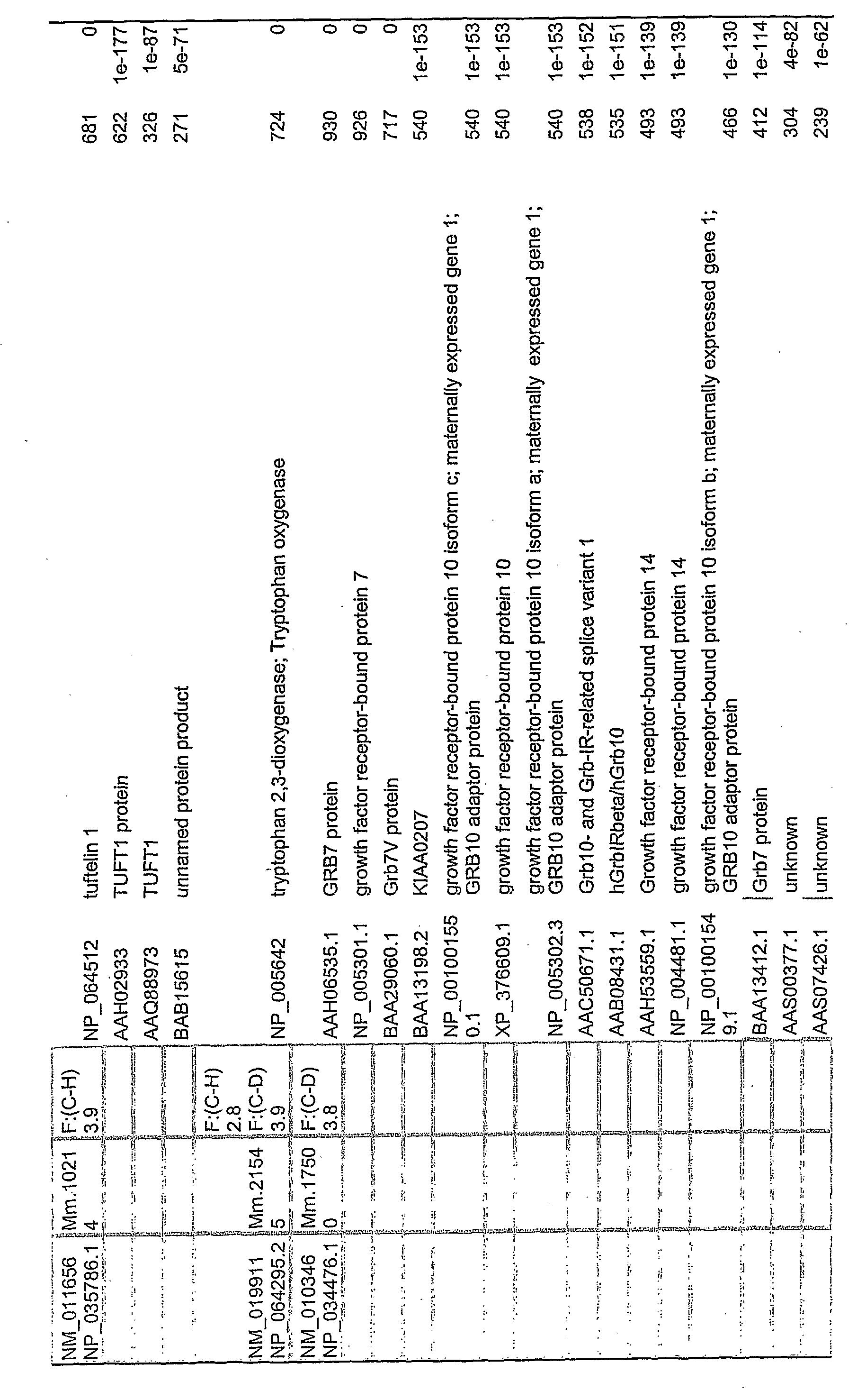
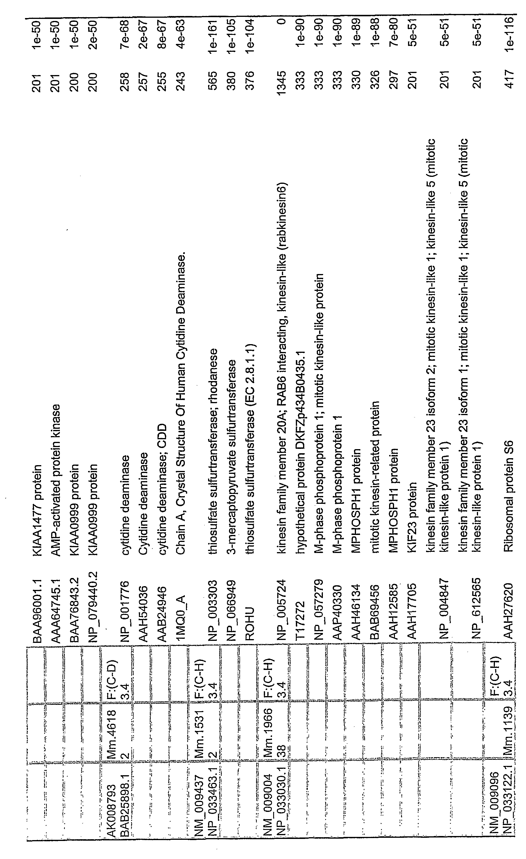
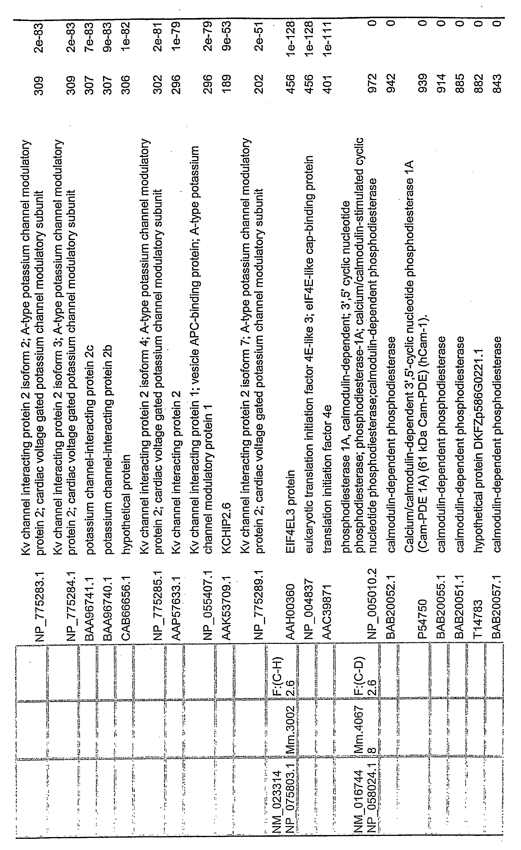
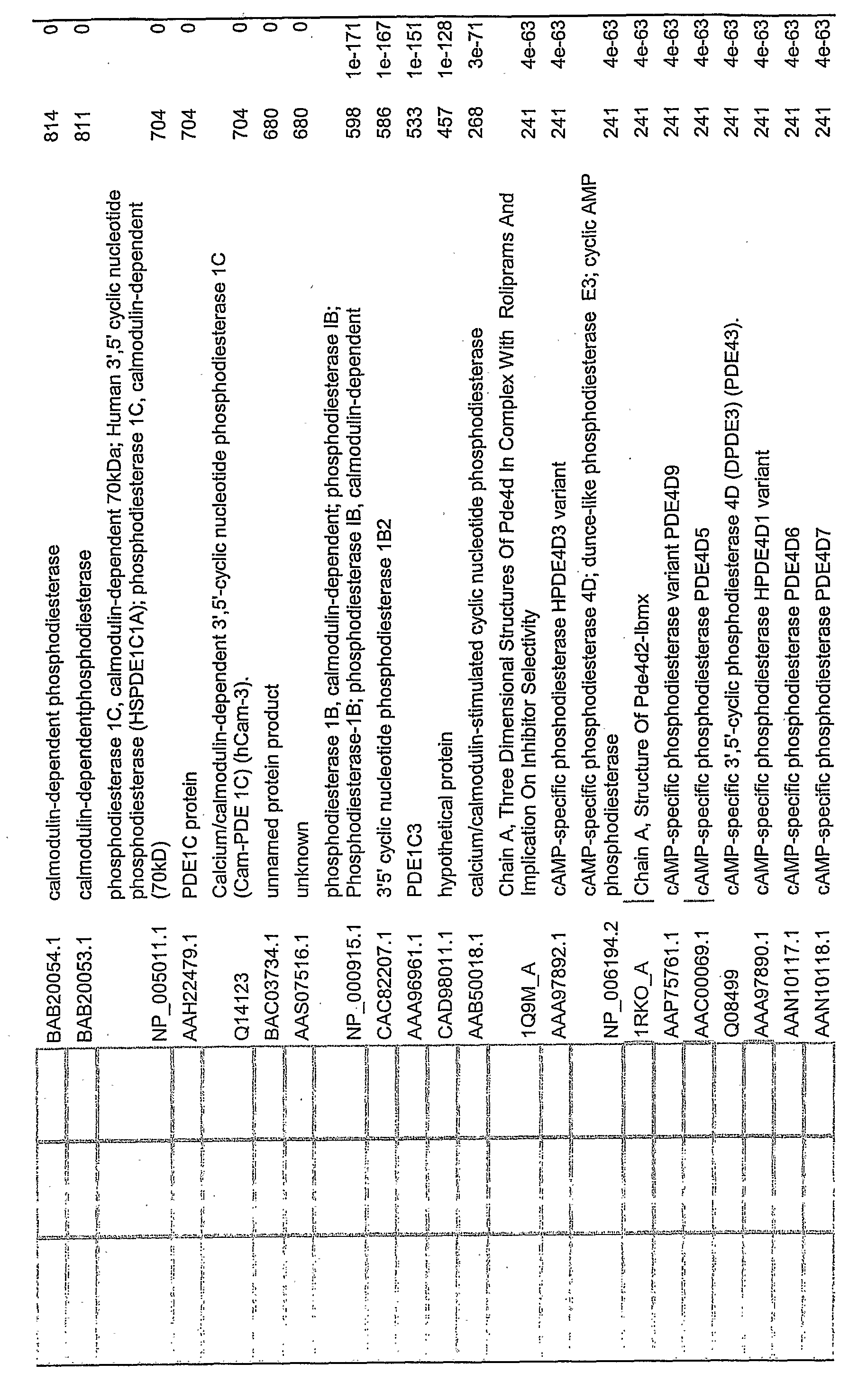
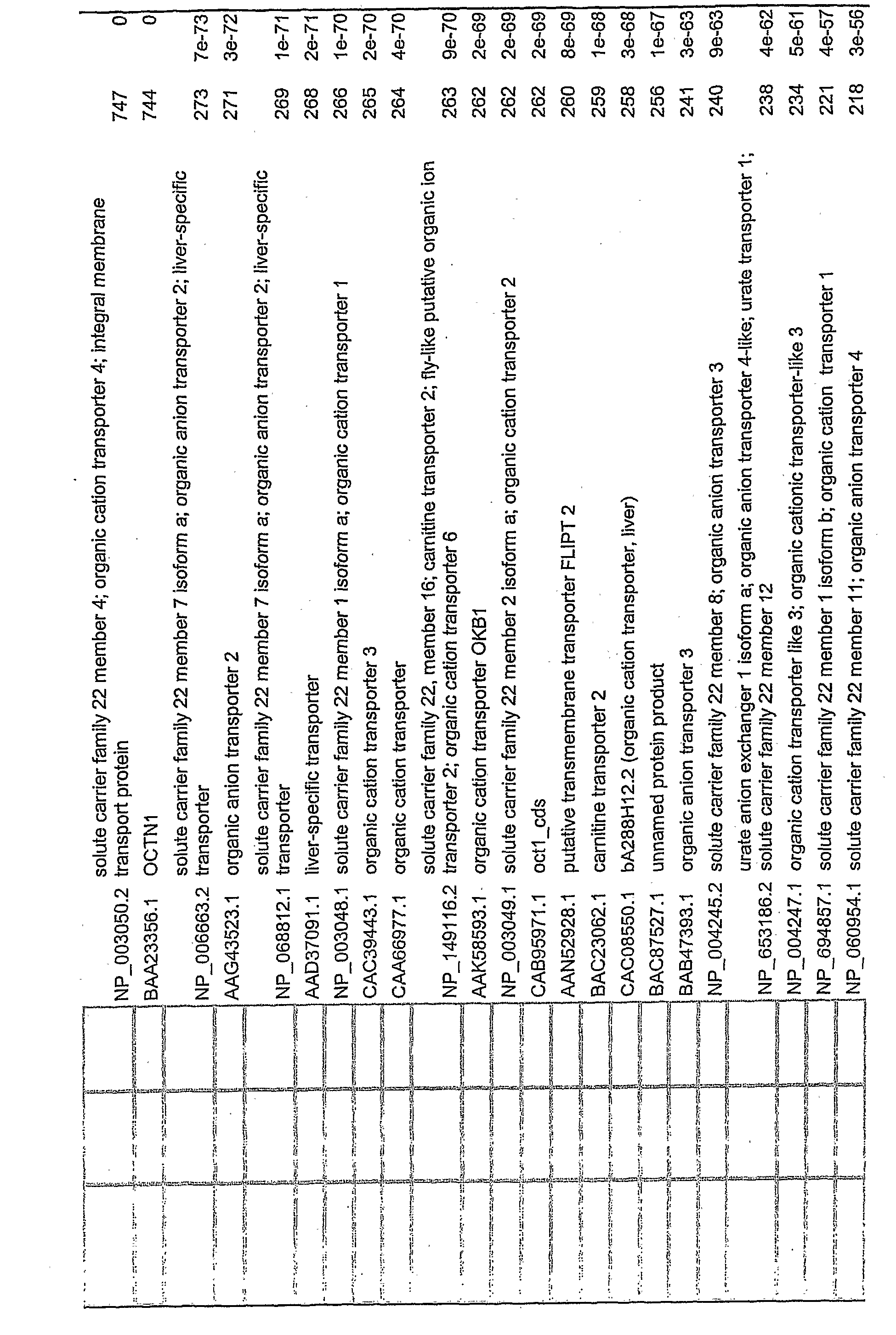
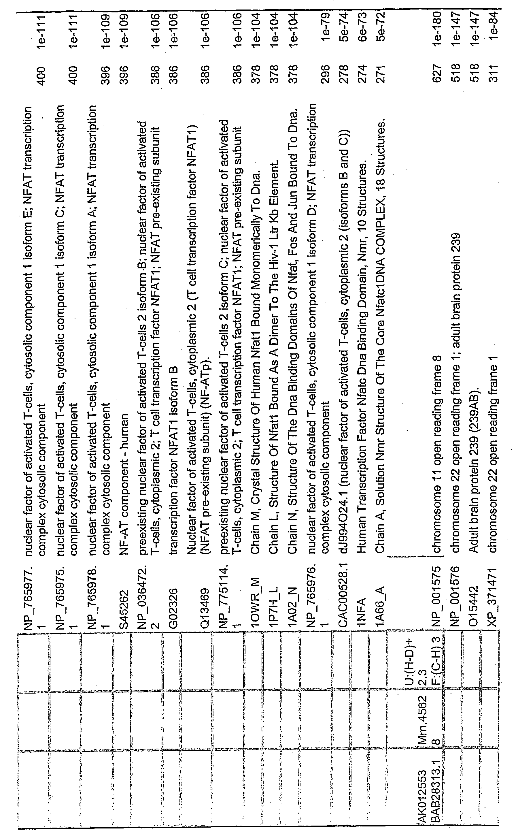
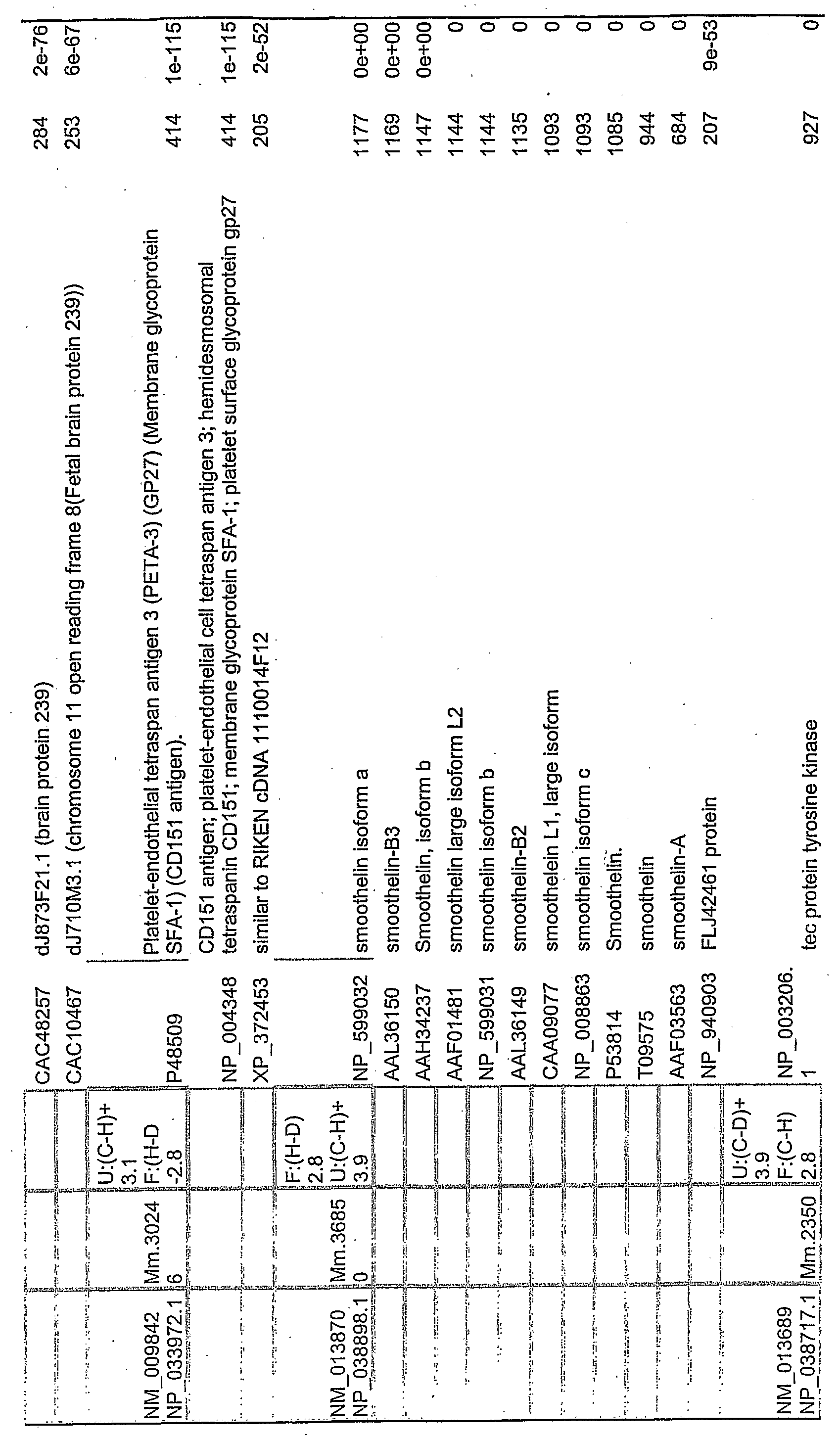
Related human genes (database DNAs) and proteins were identified by searching a database comprising human DNAs or proteins for sequences corresponding to (i.e., homologous to, i.e., which could be aligned in a statistically significant manner to) the mouse gene or protein. More than one human protein may be identified as corresponding to a particular mouse chip probe and to a particular mouse gene.
Note that the terms "human genes" and "human proteins" are used in a manner analogous to that already discussed in the case of "mouse genes" and "mouse proteins". As used herein, the term "corresponding" does not mean identical, but rather implies the existence of a statistically significant sequence similarity, such as one sufficient to qualify the human protein or gene as a homologus protein or DNA as defined below. The greater the degree of relationship as thus defined (i.e., by the statistical significance of each alignment used to connect the mouse cDNA to the human protein or gene, measured by an E value), the more close the correspondence. The connection may be direct (mouse gene to human protein) or indirect (e.g., mouse gene to human gene, human gene to human protein) .By "mouse gene", we mean the mouse gene from which the gene chip DNA in question was derived.
In general, the human genes/proteins which most closely correspond, directly or indirectly, to the mouse genes are preferred, such as the one(s) with the highest, top two highest, top three highest, top four highest, top five highest, and top ten highest E values for the final alignment in the connection process. The human genes/proteins deemed to correspond to our mouse genes are identified in the Master Tables.
Note that it is possible to identify homologous full- length human genes and proteins, if they are present in the database, even if the query mouse DNA or protein sequence is not a full-length sequence.
If there is no homologous full-length human gene or protein in the database, but there is a partial one, the latter may nonetheless be useful. For example, a partial protein may still have biological activity, and a molecule which binds the partial protein may also bind the full- length protein so as to antagonize a biological activity of the full-length protein. Likewise, a partial human gene may encode a partial protein which has biological activity, or the gene may be useful in the design of a hybridization probe or in the design of a therapeutic antisense DNA.
The partial genes and protein sequences may of course also be used in the design of probes intended to identify the full length gene or protein sequence.
For the sake of convenience, we refer to a human protein as favorable if (1) it is listed in Master Table 1 as corresponding to a favorable mouse gene, or (2) it is at least substantially identical or conservatively identical to a listed protein per (1) , or (3) it is a member of a human protein class listed in Master Table 2 (if provided) as corresponding to a favorable mouse gene. We define a human protein as unfavorable in an analogous manner. We may further identify a human protein as being wholly favorable (see mouse genes of subtable IA, wholly unfavorable (see mouse genes of subtable IB), or mixed, i.e., both partially favorable and partially unfavorable (see mouse genes of subtable 1C) .
Likewise, a human gene which encodes a particular human protein may be classified in the same way as the human protein which it encodes.
However, it should be noted that this classification is not based on the direct study of the expression of the human gene/protein. of course, the human genes/proteins of ultimate interest will be the ones whose change in level of expression is, in fact, correlated, directly or inversely, with the change of state (normal, hyperinsulinemic, diabetic) of the subject.
After identifying related human genes and proteins, one may formulate agents useful in screening humans at risk for progression toward hyperinsulinemia or toward type II diabetes, or protecting humans at risk thereof from progression from a normoinsulinemic state to a hyperinsulinemic state, or from either to a type II diabetic state.
Agents which bind the "favorable" and "unfavorable" nucleic acids (e.g., the agent is a substantially complementary nucleic acid hybridization probe) , or the corresponding proteins (e.g., an antibody vs. the protein) may be used to evaluate whether a human subject is at increased or decreased risk for progression toward type II diabetes. A subject with one or more elevated "unfavorable" and/or one or more depressed "favorable" genes/proteins is at increased risk, and one with one or more elevated "favorable" and/όr one or more depressed "unfavorable" genes/proteins is at decreased risk. ' One may further take into account whether the subject is normoinsulinemic or hyperinsulinemic at the time of the assay. If the subject is non-diabetic and normoinsulinemic, we are especially interested in the "favorable" and "unfavorable" genes/proteins corresponding to mouse genes differentially expressed in hyperinsulinemic vs. normal white adipose tissue. If the subject is already hyperinsulinemic, yet non-diabetic, we are especially interested in the "favorable" and "unfavorable" genes/proteins corresponding
to mouse genes differentially expressed in type II diabetic vs. hyperinsulinemic white adipose tissue.
The assay may be used as a preliminary screening assay to select subjects for further analysis, or as a formal diagnostic assay.
The identification of the related genes and proteins may also be useful in protecting humans against these disorders. Thus, Applicants contemplate:
(1) use of the "favorable" mouse DMAs (or fragments thereof) of the Master Tables (below) to isolate or identify related human DNAs;
(2) use of human DNAs, related to favorable mouse DNAs, to express the corresponding human proteins;
(3) use of the corresponding human proteins (and mouse proteins, if biologically active in humans) , to protect against the disorder(s);
(4) use of the corresponding mouse or human proteins, or nucleic acid probes derived from the mouse or human genes, in diagnostic agents, in assays to measure progression toward hyperinsulinemia or type II diabetes, or protection against the disorder(s), or to estimate related end organ damage such as kidney damage; and (5) use of the corresponding human or mouse genes therapeutically in gene therapy, to protect against the disorder(s) .
Moreover Applicants contemplate:
(1) use of the "unfavorable" mouse DNAs (or fragments thereof) of the Master Tables to isolate or identify related human DNAs;
(2) use of the complement to the "unfavorable" mouse DNAs or related human DNAs, as antisense molecules to inhibit expression of the related human DNAs; (3) use of the mouse or human DNAs to express the corresponding mouse or human proteins;
(4) use of the corresponding mouse or human proteins, in diagnostic agents, to measure progression toward hyperinsulinemia or type II diabetes, or protection against
the disorder(s) , or to estimate related end organ damage such as kidney damage;
(5) use of the corresponding mouse or human proteins in assays to determine whether a substance binds to (and hence may neutralize) the protein; and
(6) use of the neutralizing substance to protect against the disorder(s) .
Thus, DNAs of interest include those which specifically hybridize to the aforementioned mouse or human genes, and are thus of interest as hybridization assay reagents or for antisense therapy. They also include synthetic DNA sequences which encode the same polypeptide as is encoded by the database DNA, and thus are useful for producing the polypeptide in cell culture or in situ (i.e., gene therapy) . Moreover, they include DNA sequences which encode polypeptides which are substantially structurally identical or conservatively identical in amino acid sequence to the mouse and human proteins identified in the Master Table 1, subtables IA or 1C. Finally, they include DNA sequences which encode peptide (including antibody) antagonists of the proteins of Master Table 1, subtables IB or 1C.
The related human DNAs may be identified by comparing the mouse sequence (or its AA translation product) to known human DNAs (and their AA translation products) . Related human DNAs also may be identified by screening human cDNA or genomic DNA libraries using the mouse gene of the Master Table, or a fragment thereof, as a probe. If the mouse gene of Master Table 1 is not full-length, and there is no closely corresponding full-length mouse gene in the sequence databank, then the mouse DNA may first be used as a hybridization probe to screen a mouse cDNA library to isolate the corresponding full-length sequence. Alternatively, the mouse DNA may be used as a probe to ' screen a mouse genomic DNA library.
Our animal models of hyperinsulinemia and diabetes are also obese. It is possible that the genes found to be
favorable act indirectly by inhibiting obesity. Likewise, it is possible that the genes found to be unfavorable act indirectly by accentuating obesity. Consequently, it is within the compass of the present invention to use the favorable genes and proteins, or to use antagonists of the unfavorable genes and proteins, to protect against obesity, as well as against sequelae of obesity such as hyperinsulinemia and diabetes.
Since type II diabetes is an age-related disease, the agents of the present invention may be used in conunction with known anti-aging or anti-age-related disease agents. It is of particular interest to use the agents of the present invention in conjunction with an agent disclosed in one of the related applications cited above, in particular, an antagonist to CIDE-A, the latter having been taught in Kopchick7 and. Kopchick7A-PCT.
DETAILED DESCRIPTION OF THE PREFERRED EMBODIMENTS OF THE
INVENTION
Full-Length vs. Partial Length Genes/Proteins
A "full length" gene is here defined as (1) a naturally occurring DNA sequence which begins with an initiation codon (almost always the Met codon, ATG) , and ends with a stop codon in phase with said initiation codon (when introns, if any, are ignored) , and thereby encodes a naturally occurring polypeptide with biological activity, or a naturally occurring precursor thereof, or (2) a synthetic DNA sequence which encodes the same polypeptide as that which is encoded by (1) . The gene may, but need not, include introns.
A "full-length" protein is here defined as a naturally occurring protein encoded by a full-length gene, or a protein derived naturally by post-translational modification of such a protein. Thus, it includes mature proteins, proproteins, preproteins and preproproteins. It also includes substitution and extension mutants of such naturally occurring proteins.
Subjects A mouse is considered to be a diabetic subject if, regardless of its fasting plasma insulin level, it has a fasting plasma glucose level of at least 190 mg/dL. A mouse is considered to be a hyperinsulinemic subject if its fasting plasma insulin level is at least 0.67 ng/mL and it . does not qualify as a diabetic subject. A mouse is considered to be "normal" if it is neither diabetic nor hyperinsulinemic. Thus, normality is defined in a very- limited manner.
A mouse is considered "obese" if its weight is at least 15% in excess of the mean weight for mice of its age and sex. A mouse which does not satisfy this standard may be characterized as "non-obese", the term "normal" being reserved for use in reference to glucose and insulin levels as previously described.
A human is considered a diabetic subject if, regardless of his or her fasting plasma insulin level, the fasting plasma glucose level is at least 126 mg/dL. A human is considered a hyperinsulinemic subject if the fasting plasma insulin level is more than 26 micro International Units/mL (it is believed that this is equivalent to 1.08 ng/mL) , and does not qualify as a diabetic subject. A human is considered to be "normal" if it is neither diabetic nor hyperinsulinemic. Thus, normality is defined in a very limited manner.
A human is considered "obese" if the body mass index (BMI) (weight divided by height squared) is at least 30 kg/m2. A human who does not satisfy this standard may be characterized as "non-obese", the term "normal" being reserved for use in reference to glucose and insulin levels as previously described.
A human is considered overweight if the BMI is at least 25 kg/m2. Thus, we define overweight to include obese individuals, consistent with the recommendations of the National Institute of Diabetes and Digestive and Kidney
Diseases (NIDDK) . A human who does not satisfy this standard may be characterized as "non-overweight."
According to the Report of the Expert Committe on the Diagnosis and Classification of Diabetes Mellitus, Diabetes Care 20: 1183-97 (1997), the following are risk factors for diabetes type II:
older (e.g., at least 45; see below)
excessive weight (see below)
first-degree relative with diabetes mellitus
member of high risk ethnic group (black, Hispanic, Native American, Asian)
history of gestational diabetes mellitus or delivering a baby weighing more than 9 pounds (4.032 kg)
hypertensive (>140/90 mm Hg)
HDL cholesterol level >35 mg/dL (0.90 mmol/L)
triglyceride level >=250 mg/dL (2.83 mmol/L)
Hence, in a preferred embodiment, the diagnostic and protective methods of the present invention are applied to human subjects exhibiting one or more of the aforementioned risk factors. Likewise, in a preferred embodiment, they are applied to human subjects who, while not diabetic, exhibit impaired glucose homeostasis (110 tp <126 mg/dL) .
The risk of diabetes increases with age. Hence, in successive preferred embodiments, the age of the subjects is at least 45, at least 50, at least 55, at least 60, at least 65, at least 70, and at least 75.
With regard to excessive weight, NIDDK says that "The relative risk of diabetes increases by approximately 25 percent for each additional unit of BMI over 22." Hence, in successive preferred embodiments, the BMIs of the human subjects is at least 23, at least 24, at least 25 (i.e., overweight by our criterion), at least 26, at least 27, at least 28, at least 29, at least 30 (i.e., obese), at least 31, at least 32, at least 33, at least 34, at least 35, at least 36, at least 37, at least 38, at least 39, at least 40, or over 40.
Age-Related Diseases
Age-related (senescent) diseases include certain cancers, atherosclerosis, diabetes (type 2) , osteoporosis, hypertension, depression, Alzheimer's, Parkinson's, glaucoma, certain immune system defects, kidney failure, and liver steatosis. In general, they are diseases for which the relative risk (comparing a subpopulation over age 55 to a suitably matched population under age 55) is at least 1.1.
Preferably, the agents of the present invention protect against one or more age-related diseases for at least a subpopulation of mature (post-puberty) adult subjects.
Direct and Indirect Utility of Identified Nucleic Acid Sequences and Related Molecules
The mouse or human genes (or fragments thereof) may be used directly. For diagnostic or screening purposes, they (or specific binding fragments thereof) may be labeled and used as hybridization probes. For therapeutic purposes, they (or specific binding fragments thereof) may be used as antisense reagents to inhibit the expression of the corresponding gene, or of a sufficiently homologous gene of another species.
If the database DNA appears to be a full-length cDNA or gDNA, that is, it encodes an entire, functional, naturally occurring protein, then it may be used in the expression of that protein. Likewise, if the corresponding human gene is known in full-length, it may be used to express the human protein. Such expression may be in cell culture, with the protein subsequently isolated and administered exogenously to subjects who would benefit therefrom, or in vivo, i.e., administration by gene therapy. Naturally, any DNA encoding the same protein may be used for the same purpose, and a DNA encoding a protein which a fragment or a mutant of that naturally occurring protein which retains the desired activity, may be used for the purpose of producing the active fragment or mutant. The encoded protein of course has utility therapeutically and, in labeled or immobilized form, diagnostically.
The genes may also be used indirectly, that is, to identify other useful DNAs, proteins, or other molecules. We have attempted to determine whether the mouse genes disclosed herein have significant similarity to any known human DNA, and whether, in any of the six possible combinations of reference frame and strand, they encode a protein similar to a known 'human protein. If so, then it follows that the known human protein, and DNAs encoding that
protein, may be used in a similar manner. In addition, if the known human protein is known to have additional homologues, then those homologous proteins, and DNAs encoding them, may be used in a similar manner.
There thus are several ways that a human protein homologue of interest can be identified by database searching, including but not limited to:
1) a DNA->DNA (BlastN) search for human database DNAs closely related to the mouse gene identifies a known human gene, and the sequence of the human protein is deduced by the Genetic Code;
2) a DNA->Protein (BlastX) search for human database proteins closely related to the translated DNA of the mouse gene identifies a known human protein; and
3) the sequence of the mouse protein is known or is deduced by the Genetic Code, and a Protein->Protein (BlastP) search for closely related database proteins identifies a known human protein.
Once a known human gene is identified, it may be used in further BlastN or BlastX searches to identify other human genes or proteins. Once a known human protein is identified, it may be used in further BlastP searches to identify other human proteins.
Searches may also take cognizance, intermediately, of known genes and proteins other than mouse or human ones, e.g., use the mouse sequence to identify a known rat sequence and then the rat sequence to identify a human one.
If we have identified a mouse gene, and it encodes a mouse protein which appears similar to a human protein, then that human protein may be used (especially in humans) for purposes analogous to the proposed use of the mouse protein
in mice. Moreover, a specific binding fragment of an appropriate strand of the corresponding human gene (gDNA or cDNA) could be labeled and used as a hybridization probe (especially against samples of human mRNA or cDNA) . In determining whether the disclosed genes (gDNA or cDNA) have significant similarities to known DNAs (and their translated AA sequences to known proteins) , one would generally use the disclosed gene as a query sequence in a search of a sequence database. The results of several such searches are set forth in the Examples. Such results are dependent, to some degree, on the search parameters. Preferred parameters are set forth in Example 1. The results are also dependent on the content of the database. While the raw similarity score of a particular target (database) sequence will not vary with content (as long as it remains in the database) , its informational value (in bits) , expected value, and relative ranking can change. Generally speaking, the changes are small.
It will be appreciated that the nucleic acid and protein databases keep growing. Hence a later search may identify high scoring target sequences which were not uncovered by an earlier search because the target sequences were not previously part of a database.
Hence, in a preferred embodiment, the cognate DNAs and proteins include not only those set forth in the examples, but those which would have been highly ranked (top ten, more preferably top three, even more preferably top two, most preferably the top one) in a search run with the same parameters on the date of filing of this application.
If the known mouse or human database DNA appears to be a partial sequence (that is, partial relative to a cDNA or gDNA encoding the whole naturally occurring protein) , it may be used as a hybridization probe to isolate the full-length DNA. If the partial DNA encodes a biologically functional fragment of the cognate protein, it may be used in a manner
similar to the full length DNA, i.e., to produce the functional fragment.
If we have indicated that an antagonist of a protein or other molecule is useful, then such an antagonist may be obtained by preparing a combinatorial library, as described below, of potential antagonists, and screening the library members for binding to the protein or other molecule in question. The binding members may then be further screened for the ability to antagonize the biological activity of the target. The antagonists may be used therapeutically, or, in suitably labeled or immobilized form, diagnostically. If the identified mouse or human database DNA is related to a known protein, then substances known to interact with that protein (e.g., agonists, antagonists, substrates, receptors, second messengers, regulators, and so forth) , and binding molecules which bind them, are also of utility. Such binding molecules can likewise be identified by screening a combinatorial library.
Isolation of Full Length DNAs Using Partial DNAs as probes
If it is determined that a DNA of the present invention is a partial DNA, and the cognate full length DNA is not listed in a sequence database, the available DNA may be used as a hybridization probe to isolate the full-length DNA from a suitable DNA library.
Stringent hybridization conditions are appropriate, that is, conditions in which the hybridization temperature is 5-10 deg. C. below the Tm of the DNA as a perfect duplex.
Identification and Isolation of Homologous Genes Using a DNA Probe
It may be that the sequence databases available do not include the sequence of any homologous gene (cDNA or gDNA) , or at least of the homologous gene for a species of interest. However, given the cDNAs set forth above, one may readily obtain the homologous gene.
The possession of one DNA (the "starting DNA") greatly facilitates the isolation of homologous DNAs. If only a
partial DNA is known, this partial DNA may first be used as a probe to isolate the corresponding full length DNA for the same species, and that the latter may be used as the starting DNA in the search for homologous genes. The starting DNA, or a fragment thereof, is used as a hybridization probe to screen a cDNA or genomic DNA library for clones containing inserts which encode either the entire homologous protein, or a recognizable fragment thereof. The minimum length of the hybridization probe is dictated by the need for specificity. If the size of the library in bases is L, and the GC content is 50%, then the probe should have a length of at least 1, where L = 41. This will yield, on average, a single perfect match in random DNA of L bases. The human cDNA library is about 108 bases and the human genomic DNA library is about 1010 bases.
The library is preferably derived from an organism which is known, on biochemical evidence, to produce a homologous protein, and more preferably from the genomic DNA or mRNA of cells of that organism which are likely to be relatively high producers of that protein. A cDNA library (which is derived from an mRNA library) is especially preferred.
If the organism in question is known to have substantially different codon preferences from that of the organism whose relevant cDNA or genomic DNA is known, a synthetic hybridization probe may be used which encodes the same amino acid sequence but whose codon utilization is more similar to that of the DNA of the target organism. Alternatively, the synthetic probe may employ inosine as a substitute for those bases which are most likely to be divergent, or the probe may be a mixed probe which mixes the codons for the source DNA with the preferred codons (encoding the same amino acid) for the target organism. By routine methods, the Tm of a perfect duplex of starting DNA is determined. One may then select a hybridization temperature which is sufficiently lower than the perfect duplex Tm to allow hybridization of the starting DNA (or other probe) to a target DNA which is divergent from the starting DNA. A 1% sequence divergence typically lowers
the Tm of a duplex by 1-2°C, and the DNAs encoding homologous proteins of different species typically have sequence identities of around 50-80%. Preferably, the library is screened under conditions where the temperature is at least 20°C, more preferably at least 500C, below the perfect duplex Tm. Since salt reduces the Tm, one ordinarily would carry out the search for DNAs encoding highly homologous proteins under relatively low salt hybridization conditions, e.g., <1M NaCl. The higher the salt concentration, and/or the lower the temperature, the greater the sequence divergence which is tolerated.
For the use of probes to identify homologous genes in other species, see, e.g., Schwinn, et al. , J. Biol. Chem. , 265:8183-89 (1990) (hamster 67-bp cDNA probe vs. human leukocyte genomic library; human 0.32kb DNA probe vs. bovine brain cDNA library, both with hybridization at 42°C in 6xSSC) ; Jenkins et al. , J. Biol. Chem., 265:19624-31 (1990) (Chicken 770-bp cDNA probe vs. human genomic libraries; hybridization at 40°C in 50% formamide and 5xSSC) ; Murata et al., J. Exp. Med., 175:341-51 (1992) (1.2-kb mouse cDNA probe v. human eosinophil cDNA library; hybridization at 65°C in 6xSSC) ; Guyer et al . , J. Biol. Chem., 265:17307-17 (1990) (2.95-kb human genomic DNA probe vs. porcine genomic DNA library; hybridization at 42°C in 5xSSC) . The conditions set forth in these articles may each be considered suitable for the purpose of isolating homologous genes.
Corresponding (Homologous) Proteins and DNAs In the case of a gene chip, the manufacturer of the gene chip determines which DNA to place at each position on the chip. This DNA may correspond in sequence to a genomic DNA, a cDNA, or a fragment of genomic or cDNA, and may be natural, synthetic or partially natural and partially synthetic in origin. The manufacturer of the gene chip will normally identify the DNA for a mouse gene chip as corresponding to a particular mouse gene, in which case it will be assumed that the alignments of chip DNA to mouse gene satisfies the homology criteria of the invention.
Usually, the gene chip manufacturer will provide a sequence database accession number for the mouse DNA. If so, to identify the corresponding mouse protein, we will first inspect the database record for that mouse DNA. Often, the mouse protein accession number will appear in that record or in a linked record. If it doesn't, the corresponding mouse protein can be identified by performing a BlastX search on a mouse protein database with the mouse database DNA sequence as the query sequence. Even if the protein sequence is not in the database, if the DNA sequence comprises a full-length coding sequence, the corresponding protein can be identified by translating the coding sequence in accordance with the Genetic Code.
A human protein can be said to be identifiable as corresponding (homologous) to a gene chip DNA if it is identified as corresponding (homologous) to the mouse gene (gDNA or cDNA, whole or partial) identified by the gene chip manufacturer as corresponding to that gene chip DNA.
In turn, it is identifiable as corresponding (homologous) to said identified mouse gene, if
(1) it can be aligned by BlastX directly to that mouse gene, and/or
(2) it is encoded by a human gene, or can be aligned to a human gene by BlastX, which in turn can be aligned by BlastN to said mouse gene and/or
(3) it can be aligned by BlastP to a mouse protein, the latter being encoded by said mouse gene, or aligned to said mouse gene BlastX,
where any alignment by BlastN, BlastP or BlastX is in accordance with the default parameters set forth below, and the expected value (E) of each alignment (the probability that such an alignment would have occurred by chance alone)
is less than e-10. (Note that because this is a negative exponent, a value such as e-50 is less than e-10.)
Desirably, two or all three of these conditions (I)- (3) are satisfied for the corresponding (homologous) human genes and proteins.
A human gene is corresponding (homologous) to a mouse gene chip DNA, and hence to said identified mouse gene (or cDNA) and protein, if it encodes a corresponding
(homologous) human protein as defined above, or it can be aligned by BlastN to said mouse gene.
Preferably, for at least one of conditions (I)- (3), the E value is less than e-50, more preferably less than e-60, still more preferably less than e-70, even more preferably less than e-80, considerably more preferably less than e-90, and most preferably less than e-100. Desirably, it is true for two or even all three of these conditions.
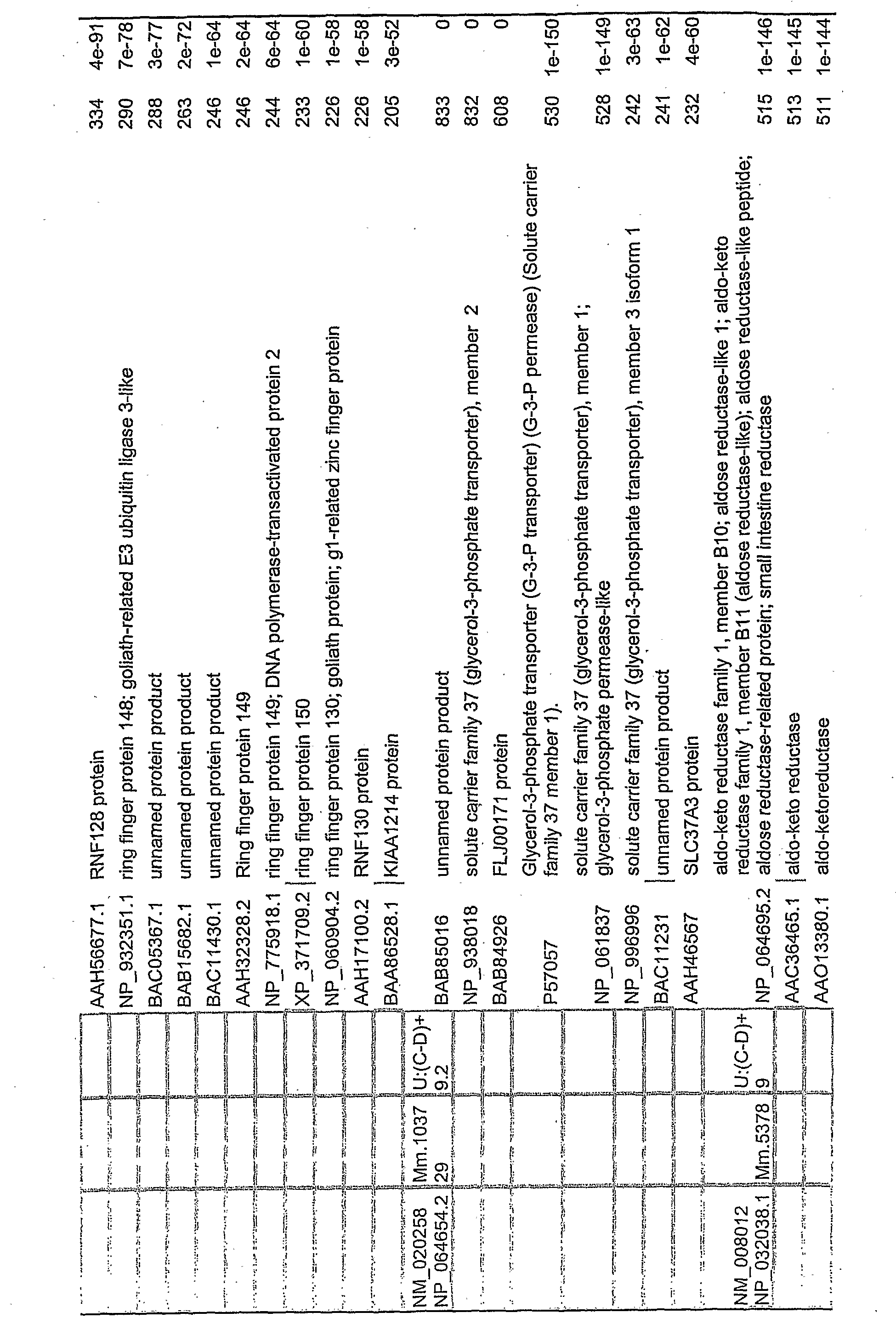
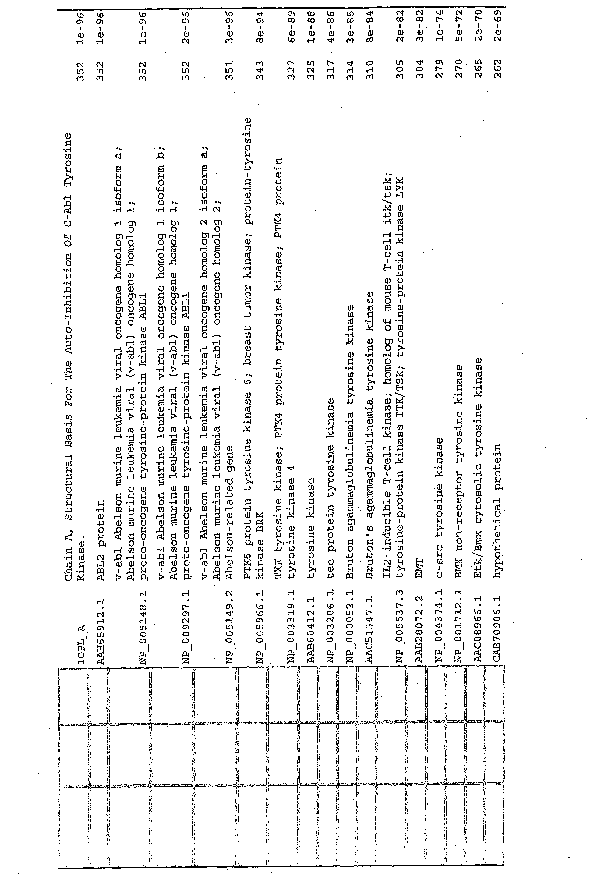
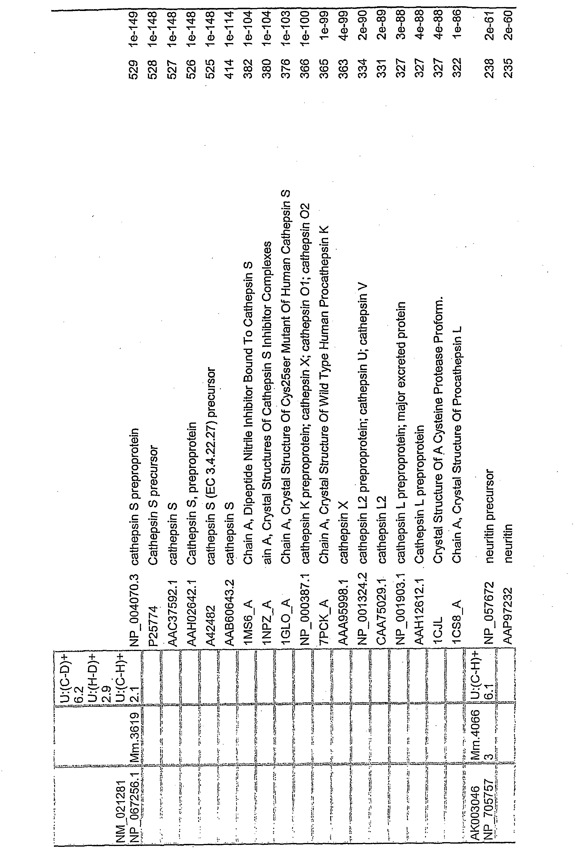
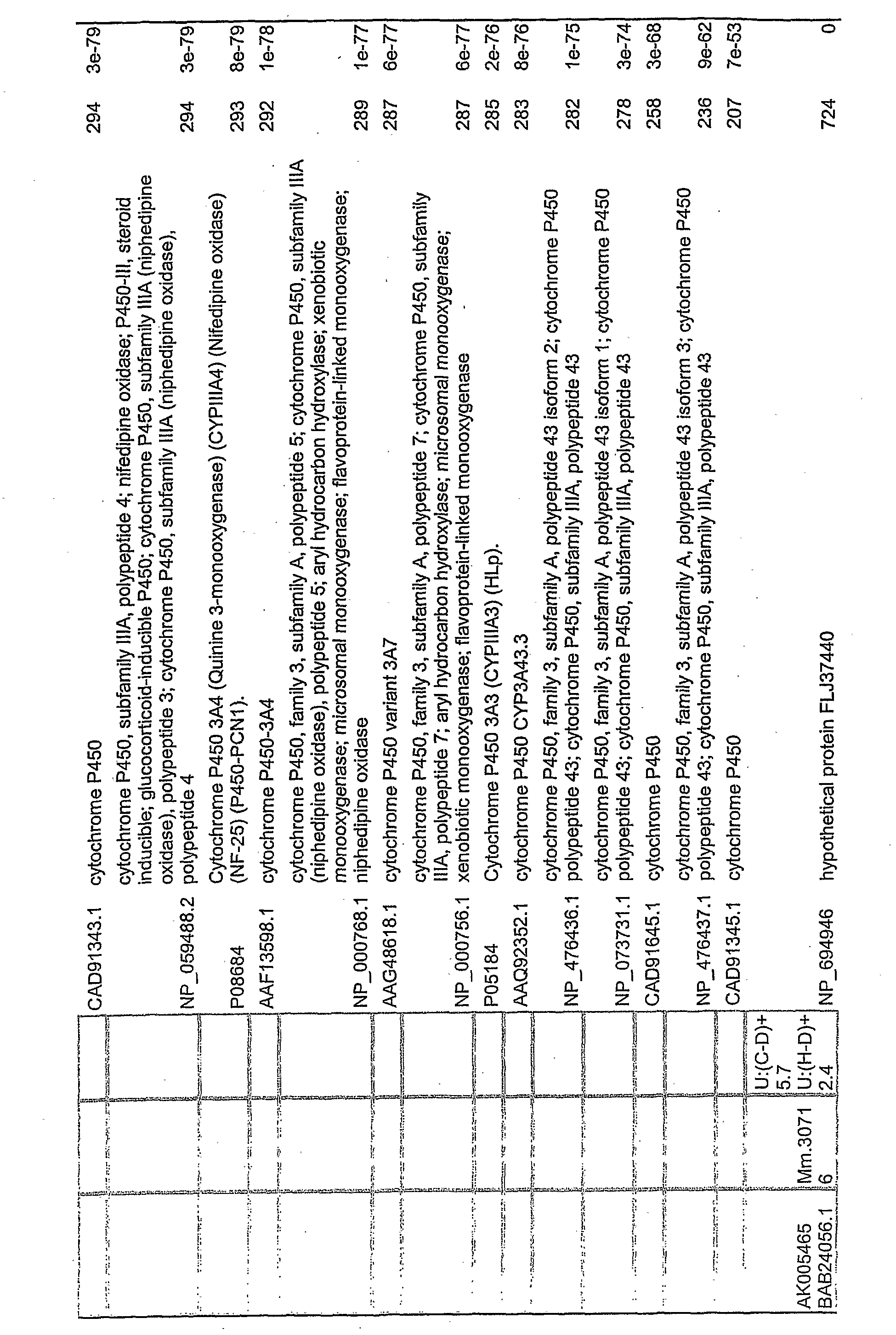
In constructing Master table 1, we generally used a BlastX (mouse gene vs. human protein) alignment E value cutoff of e-50. However, if there were no human proteins with that good an alignment to the mouse DNA in question, or if there were other reasons for including a particular human protein (e.g., a known functionality supportive of the observed differential cognate mouse protein expression) , then a human protein with a score worse (i.e., higher) than e-50 may appear in Master Table 1.
If the manufacturer of the gene chip identifies the gene chip DNA as corresponding to an EST, or other DNA which is not a full-length mouse gene or cDNA, a longer (possibly full length) mouse gene or cDNA may be identified by a BlastN search of the mouse DNA database. Alternatively, the identified DNA may be used to conduct a BlastN search of a human DNA database, or a BlastX search of a mouse or human protein database.
Thus, more generally, a human protein can be said to be identifiable as corresponding (homologous) to a gene chip
DNA, or to a DNA identified by the manufacturer as corresponding to that gene chip DNA, if
(1') it can be aligned directly to the gene chip or corresponding manufacturer identified DNA by BlastX. and/or
(2') it can be aligned to a human gene/cDNA by BlastX, whose genomic DNA (gDNA) or cDNA (DNA complementary to messenger RNA) in turn can be aligned to the gene chip or corresponding manufacturer identified DNA by BlastN, and/or
(3') it can be aligned to a mouse gene/cDNA by BlastX, whose gDNA or cDNA in turn can be aligned to the gene chip or corresponding manufacturer identified DNA by BlastN, and/or
(4') it can be aligned to a mouse protein by BlastP, which in turn can be aligned to the gene chip or corresponding manufacturer identified DNA by BlastX, and/or
(5') it can be aligned to a mouse protein by BlastP, which in turn can be aligned to a mouse gene/cDNA by BlastX, whose gDNA or cDNA can in turn be aligned to the gene chip or corresponding manufacturer identified DNA by BlastN;
where any alignment by BlastN, BlastP, or BlastX is in accordance with the default parameters set forth below, and the expected value (E) of each alignment (the probability that such an alignment would have occurred by chance alone) is less than e-10. (Note that because this is a negative exponent, a value such as e-50 is less than e-10.)
Preferably, two, three, four or all five of conditions (1')-(5') are satisfied.
Preferably, for at least one of conditions (l')-(5r), for at least the final alignment (i.e., vs. the human protein), the E value is less than e-50, more preferably less than e-60, , still more preferably less than e-70, even more preferably less than e-80, considerably more preferably less than e-90, and most preferably less than e-100.
Desirably, one or more of these standards of preference are met for two, three, four or all five of conditions (I1)- (51) . In particular, for those conditions in which the gene chip or corresponding manufacturer identified DNA is indirectly connected to the human protein by virtue of two or more successive alignments, the E value is preferably, so limited for all of said alignments in the connecting chain.
A human gene corresponds (is homologous) to a gene chip DMA or manufacturer identified corresponding DNA if it encodes a homologous human protein as defined above, or if it can be aligned either directly to that DNA, or indirectly through a mouse gene which can be aligned to said DNA, according to the conditions set forth above.
Master table 1 assembles a list of human protein corresponding to each of the mouse DNAs/proteins identified as related to the chip DNA. These human proteins form a set and can be given a percentile rank, with respect to E value, within that set. The human proteins of the present invention preferably are those scorers with a percentile rank of at least 50%, more preferably at least 60%, still more preferably at least 70%, even more preferably at least 80%, and most preferably at least 90%.
For each mouse gene (gDNA or cDNA) in Master Table 1, there is a particular human protein which provides the best alignment match as measured by BlastX, i.e., the human protein with the best score (lowest e-value) . These human proteins form a subset of the set above and can be given a percentile rank within that subset, e.g., the human proteins with scores in the top 10% of that subset have a percentile rank of 90% or higher.
The human proteins of the present invention preferably are those best scorer subset proteins with a percentile rank within the subset of at least 50%, more preferably at least 60%, still more preferably at least 70%, even more preferably at least 80%, and most preferably at least 90%.
BlastN and BlastX report very low expected values as
"0.0". This does not truly mean that the expected value is exactly zero (since any alignment could occur by chance) , but merely that it is so infinitesimal that it is not reported. The documentation does not state the cutoff value, but alignments with explicit E values as low as e-178 (624 bits) have been reported as nonzero values, while a score of 636 bits was reported as "0.0".
Functionally homologous human proteins are also of interest. A human protein may be said to be functionally homologous to the mouse gene if the human protein has at least one biological activity in common with the mouse protein encoded by said mouse gene. The human proteins of interest also include those that are substantially and/or conservatively identical (as defined below) to the homologous and/or functionally homologous human proteins defined above.
Degree of Differential Expression
The degree of differential expression may be expressed as the ratio of the higher expression level to the lower expression level. Preferably, this is at least 2-fold, and more preferably, it is higher, such as at least 3-fold, at least 4-fold, at least 5-fold, at least 6-fold,- at least 7- fold, at least 8-fold, at least 9-fold, or at least 10-fold.
Most preferably, the human protein of interest corresponds to a mouse gene for which the degree of differential expression places it among the top 10% of the mouse genes in the appropriate subtable.
Relevance of Favorable and Unfavorable Genes
If a gene is down-regulated in more favored mammals, or up-regulated in less favored mammals, (i.e., an "unfavorable gene") then several utilities are apparent.
First, the complementary strand of the gene, or a portion thereof, may be used in labeled form as a hybridization probe to detect messenger RNA and thereby monitor the level of expression of the gene in a subject. Elevated levels are indicative of progression, or propensity to progression, to a less favored state, and clinicians may take appropriate preventative, curative or ameliorative action.
Secondly, the messenger RNA product (or equivalent cDNA) , the protein product, or a binding molecule specific for that product (e.g., an antibody which binds the product) , or a downstream product which mediates the activity (e.g., a signaling intermediate) or a binding molecule (e.g., an antibody) therefor, may be used, preferably in labeled or immobilized form, as an assay reagent in an assay for said nucleic acid product, protein product, or downstream product (e.g., a signaling intermediate) . Again, elevated levels are indicative of a present or future problem.
Thirdly, an agent which down-regulates expression of the gene may be used to reduce levels of the corresponding protein and thereby inhibit further damage. This agent could inhibit transcription of the gene in the subject, or translation of the corresponding messenger RNA. Possible inhibitors of transcription and translation include antisense molecules and repressor molecules. The agent could also inhibit a post-translational modification (e.g., glycosylation, phosphorylation, cleavage, GPI attachment) required for activity, or post-translationally modify the protein so as to inactivate it. Or it could be an agent which down- or up-regulated a positive or negative regulatory gene, respectively.
Fourthly, an agent which is an antagonist of the messenger RNA product or protein product of the gene, or of a downstream product through which its activity is
manifested (e.g., a signaling intermediate), may be used to inhibit its activity.
This antagonist could be an antibody, a peptide, a peptoid, a nucleic acid, a peptide nucleic acid (PNA) oligomer, a small organic molecule of a kind for which a combinatorial library exists (e.g., a benzodiazepine), etc. An antagonist is simply a binding molecule which, by binding, reduces or abolishes the undesired activity of its target. The antagonist, if not an oligomeric molecule, is preferably less than 1000 daltons, more preferably less than 500 daltons.
Fifthly, an agent which degrades, or abets the degradation of, that messenger RWA, its protein product or a downstream product which mediates its activity (e.g., a signaling intermediate) , may be used to curb the effective period of activity of the protein.
If a gene is uχ>-regulated in more favored mammals, or down-regulated in less favored animals then the utilities are converse to those stated above. First, the complementary strand of the gene, or a portion thereof, may be used in labeled form as a hybridization probe to detect messenger RNA and thereby monitor the level of expression of the gene in a subject. Depressed levels are indicative of damage, or possibly of a propensity to damage, and clinicians may take appropriate preventative, curative or ameliorative action.
Secondly, the messenger RNA product, the equivalent cDNA, protein product, or a binding molecule specific for those products, or a downstream product, or a signaling intermediate, or a binding molecule therefor, may be used, preferably in labeled or immobilized form, as an assay reagent in an assay for said protein product or downstream product. Again, depressed levels are indicative of a present or future problem. Thirdly, an agent which up-regulates expression of the gene may be used to increase levels of the corresponding protein and thereby inhibit further progression to a less favored state. By way of example, it could be a vector which carries a copy of the gene, but which expresses the
gene at higher levels than does the endogenous expression system. Or it could be an agent which up- or down-regulates a positive or negative regulatory gene.
Fourthly, an agent which is an agonist of the protein product of the gene, or of a downstream product through which its activity . (of inhibition of progression to a less favored state) is manifested, or of a signaling intermediate may be used to foster its activity.
Fifthly, an agent which inhibits the degradation of that protein product or of a downstream product or of a signaling intermediate may be used to increase the effective period of activity of the protein.
Mutant Proteins
The present invention also contemplates mutant proteins (peptides) which are substantially identical (as defined below) to the parental protein (peptide) . In general, the fewer the mutations, the more likely the mutant protein is to retain the activity of the parental protein. The effect of mutations is usually (but not always) additive. Certain individual mutations are more likely to be tolerated than others.
A protein is more likely to tolerate a mutation which (a) is a substitution rather than an insertion or deletion;
(b) is an insertion or deletion at the terminus, rather than internally, or, if internal, is at a domain boundary, or a loop or turn, rather than in an alpha helix or beta strand;
(c) affects a surface residue rather than an interior residue;
(d) affects a part of the molecule distal to the binding site; (e) is a substitution of one amino acid for another of similar size, charge, and/or hydrophobicity, and does not destroy a disulfide bond or other crosslink; and
(f) is at a site which is subject to substantial variation among a family of homologous proteins to which the protein of interest belongs.
These considerations can be used to design functional mutants.
Surface vs. Interior Residues
Charged amino acid residues almost always lie on the surface of the protein. For uncharged residues, there is less certainty, but in general, hydrophilic residues are partitioned to the surface and hydrophobic residues to the interior. Of course, for a membrane protein, the membrane- spanning segments are likely to be rich in hydrophobic residues. Surface residues may be identified experimentally by various labeling techniques, or by 3-D structure mapping techniques like X-ray diffraction and NMR. A 3-D model of a homologous protein can be helpful.
Binding Site Residues
Residues forming the binding site may be identified by (1) comparing the effects of labeling the surface residues before and after complexing the protein to its target, (2) labeling the binding site directly with affinity ligands, (3) fragmenting the protein and testing the fragments for binding activity, and (4) systematic mutagenesis (e.g., alanine-scanning mutagenesis) to determine which mutants destroy binding. If the binding site of a homologous protein is known, the binding site may be postulated by analogy.
Protein libraries may be constructed and screened that a large family (e.g., 108) of related mutants may be evaluated simultaneously.
Hence, the mutations are preferably conservative modifications as defined below.
"Substantially Identical"
A mutant protein (peptide) is substantially identical to a reference protein (peptide) if (a) it has at least 10%
of a specific binding activity or a non-nutritional biological activity of the reference protein, and (b) is at least 50% identical in amino acid sequence to the reference protein (peptide) . It is "substantially structurally identical" if condition (b) applies, regardless of (a) .
Percentage amino acid identity is determined by aligning the mutant and reference sequences according to a rigorous dynamic programming algorithm which globally aligns their sequences to maximize their similarity, the similarity being scored as the sum of scores for each aligned pair according to an unbiased PAM250 matrix, and a penalty for each internal gap of -12 for the first null of the gap and - 4 for each additional null of the same gap. The percentage identity is the number of matches expressed as a percentage of the adjusted (i.e., counting inserted nulls) length of the reference sequence.
A mutant DNA sequence is substantially identical to a reference DNA sequence if they are structural sequences, and encoding mutant and reference proteins which are substantially identical as described above.
If instead they are regulatory sequences, they are substantially identical if the mutant sequence has at least 10% of the regulatory activity of the reference sequence, and is at least 50% identical in nucleotide sequence to the reference sequence. Percentage identity is determined as for proteins except that matches are scored +5, mismatches - 4, the gap open penalty is -12, and the gap extension penalty (per additional null) is -4.
More preferably, the sequence is not merely substantially identical but rather is at least 51%, at least
66%, at least 75%, at least 80%, at least 85%, at least 90%, at least 95%, at least 96%, at least 97%, at least 98% or at least 99% identical in sequence to the reference sequence. DNA sequences may also be considered "substantially identical" if they hybridize to each other under stringent conditions, i.e., conditions at which the Tm of the heteroduplex of the one strand of the mutant DNA and the more complementary strand of the reference DNA is not in
excess of 10°C. less than the Tm of the reference DNA homoduplex. Typically this will correspond to a percentage identity of 85-90%.
"Conservative Modifications"
"Conservative modifications" are defined as
(a) conservative substitutions of amino acids as hereafter defined; or
(b) single or multiple insertions (extension) or deletions (truncation) of amino acids at the termini.
Conservative modifications are preferred to other modifications. Conservative substitutions are preferred to other conservative modifications. "Semi-Conservative Modifications" are modifications which are not conservative, but which are (a) semi- conservative substitutions as hereafter defined; or (b) single or multiple insertions or deletions internally, but at interdomain boundaries, in loops or in other segments of relatively high mobility. Semi-conservative modifications are preferred to nonconservative modifications. Semi- conservative substitutions are preferred to other semi- conservative modifications.
Non-conservative substitutions are preferred to other non-conservative modifications.
The term "conservative" is used here in an a priori sense, i.e., modifications which would be expected to preserve 3D structure and activity, based on. analysis of the naturally occurring families of homologous proteins and of past experience with the effects of deliberate mutagenesis, rather than post facto, a modification already known to conserve activity. Of course, a modification which is conservative a priori may, and usually is, also conservative post facto. Preferably, except at the termini, no more than about five amino acids are inserted or deleted at a particular locus, and the modifications are outside regions known to contain binding sites important to activity.
Preferably, insertions or deletions are limited to the termini.
A conservative substitution is a substitution of one amino acid for another of the same exchange group, the exchange groups being defined as follows
I GIy, Pro, Ser, Ala (Cys) (and any nonbiogenic, neutral amino acid with a hydrophobicity not exceeding that of the aforementioned a.a. 's)
II Arg, Lys, His (and any nonbiogenic, positively- charged amino acids)
III Asp, GIu, Asn, GIn (and any nonbiogenic negatively-charged amino acids)
IV Leu, lie, Met, VaI (Cys) (and any nonbiogenic, aliphatic, neutral amino acid with a hydrophobicity too high for I above)
V Phe, Trp, Tyr (and any nonbiogenic, aromatic neutral amino acid with a hydrophobicity too high for I above) .
Note that Cys belongs to both I and IV. Residues Pro, GIy and Cys have special conformational roles. Cys participates in formation of disulfide bonds. GIy imparts flexibility to the chain. Pro imparts rigidity to the chain and disrupts α helices. These residues may be essential in certain regions of the polypeptide, but substitutable elsewhere.
One, two or three conservative substitutions are more likely to be tolerated than a larger number.
"Semi-conservative substitutions" are defined herein as being substitutions within supergroup I/II/III or within supergroup IV/V, but not within a single one of groups I-V. They also include replacement of any other amino acid with alanine. If a substitution is not conservative, it preferably is semi-conservative.
"Non-conservative substitutions" are substitutions which are not "conservative" or "semi-conservative" .
"Highly conservative substitutions" are a subset of conservative substitutions, and are exchanges of amino acids within the groups Phe/Tyr/Trp, Met/Leu/Ile/Val, His/Arg/Lys, Asp/Glu and Ser/Thr/Ala. They are more likely to be
tolerated than other conservative substitutions. Again, the smaller the number of substitutions, the more likely they are to be tolerated.
"Conservatively Identical"
A protein (peptide) is conservatively identical to a reference protein (peptide) it differs from the latter, if at all, solely by conservative modifications, the protein (peptide remaining at least seven amino acids long if the reference protein (peptide) was at least seven amino acids long.
A protein is at least semi-conservatively identical to a reference protein (peptide) if it differs from the latter, if at all, solely by semi-conservative or conservative modifications.
A protein (peptide) is nearly conservatively identical to a reference protein (peptide) if it differs from the latter, if at all, solely by one or more conservative modifications and/or a single nonconservative substitution. It is highly conservatively identical if it differs, if at all, solely by highly conservative substitutions. Highly conservatively identical proteins are preferred to those merely conservatively identical. An absolutely identical protein is even more preferred.
The core sequence of a reference protein (peptide) is the largest single fragment which retains at least 10% of a particular specific binding activity, if one is specified, or otherwise of at least one specific binding activity of the referent. If the referent has more than one specific binding activity, it may have more than one core sequence, and these may overlap or not.
If it is taught that a peptide of the present invention may have a particular similarity relationship (e.g., markedly identical) to a reference protein (peptide) , preferred peptides are those which comprise a sequence having that relationship to a core sequence of the reference protein (peptide) , but with internal insertions or deletions
in either sequence excluded. Even more preferred peptides are those whose entire sequence has that relationship, with the same exclusion, to a core sequence of that reference protein (peptide) .
Gene Ontology
The Gene Ontology Consortium has developed controlled vocabularies which describe gene products in terms of their associated biological processes, cellular components and molecular functions in a species-independent manner.
Formally speaking, the controlled vocabularies are specified in the form of three structured networks of controlled terms to describe gene product attributes. The three networks are molecular function, biological process, and cellular component.
Each network is composed of terms of differing breadth. If term A is a subset of term B, then term A is the child of B and B is the parent of A.
In a given network, the terms are connected into a directed acyclic graph (DAG) structure, rather than a hierarchial structure. In a DAG, a child term can have more than one parent term. For example, the biological process term
"hexose biosynthesis" has two parents, "hexose metabolism" and "monosaccharide biosynthesis" . This is because biosynthesis is a subtype of metabolism, and a hexose is a type of monosaccharide. If a child term describes the gene product, then all of its parents, must describe the gene product. And likewise all fo the grandparents, great- grandparents, etc.
Molecular function describes the specific tasks performed by the gene product, i.e., its activities, such as catalytic or binding activities, at the molecular level. GO molecular function terms represent activities rather than the entities (molecules or complexes) that perform the actions, and do not specify where or when, or in what context, the action
takes place. Molecular functions generally correspond to activities that can be performed by individual gene products, but some activities are performed by assembled complexes of gene products. Examples of broad functional terms are catalytic activity, transporter activity, or binding; examples of narrower functional terms are adenylate cyclase activity or Toll receptor binding.
Note that a single gene product might have several molecular functions, and many gene products can share a single molecular function. Hence, while gene products are often given names which set forth their molecular function, the use of a molecular function ontology term is meant to characterize the function of any gene product with that molecular function, not to refer to a particular gene product even if only one gene product is presently known to have that function.
Biological process describes the role of the gene product in achieving broad biological goals, such as mitosis or purine metabolism. A biological process is accomplished by one or more ordered assemblies of molecular functions. Examples of broad biological process terms are cell- growth and maintenance or signal transduction. Examples of more specific terms are pyrimidine metabolism or alpha-glucoside transport. It can be difficult to distinguish between a biological process and a molecular function, but the general rule is that a process must have two or more distinct steps. Nonetheless, a biological process is not equivalent to a pathway, as the biological process ontologies do not attempt to capture any of the dynamics or dependencies that would be required to describe a pathway.
A cellular component is just that, a component of a cell but with the proviso that it is part of some larger object, which may be an anatomical structure (e.g. rough endoplasmic reticulum or nucleus) or a gene product group (e.g. ribosome, proteasome ■or a protein dimer) .
GO does not contain the following:
* Gene products: e.g. cytochrome c is not in the ontologies, but attributes of cytochrome c, such as electron transporter, are.
* Processes, functions or components that are unique to mutants or diseases: e.g. oncogenesis is not a valid GO term because causing cancer is not the normal function of any gene. * Attributes of sequence such as intron/exon parameters: these are not attributes of gene products and will be described in a separate sequence ontology (see the OBO web page for more information) .
* Protein domains or structural features. * Protein-protein interactions.
The General Ontology data structures defines these ontology terms and their relationships. The data structures may be downloaded from the General Ontology Consortium website. A sample GO entry would be:
id: GO: 0045174 name: glutathione dehydrogenase (ascorbate) activity xref_analog: EC:1.8.5.1 "" def: "Catalysis of the reaction: 2 glutathione + dehydroascorbate = \ glutathione disulfide + ascorbate." [EC:1.8.5.1] synonym: dehydroascorbate reductase [] is_a: GO:0009055 is_a: GO:0015038 is_a: GO.:0016672
Thus, it includes a GOid (the number has no significance other than that it is unique to that term) , the name of the term, arid, unless it is the root term of the network, identification of one or more immediate parents. These are identified by "is_a" if the parent need not comprise that child, and by "part_of" if the parent necessarily comprises that child. Cross-references and synonyms are optional.
To identify the gene ontology terms applicable to a particular gene product, one may search a collaborating database whose gene or gene product records have been annotated with one or more GOids. The annotation may include evidence codes to indicate the basis for assigning particular GOids to that gene or gene product.
In Master Table 3, each of the differentially expressed mouse genes (identified by accession # in Col. 1) is characterized by its differential expression behavior (Col. 2) one or more molecular function ontologies (Col. 3), one or more biological process ontologies (COl. 4), and one or more cellular component ontologies (Col.. 5) . This information is obtainable by searching a collaborating database.
For example, a search on the mouse gene NM_008490 (for lecithin cholesterol acyltransferase [Mus musculus] ) in the NCBI Protein database (accessible, e.g., at http://www.ncbi.nlm.nih.gov/entrez/query.fcgi?db=Gene ) generates a view which includes the following gene ontology entries (reformatted for clarity) :
Function acyltransferase' activity catalytic activity phosphatidylcholine-sterol 0-acyltransferase activity transferase activity
Process cholesterol metabolism lipid metabolism
Component extracellular space
In a like manner, the ontologies associated with each of the human genes and proteins listed in Master Table 1 as corresponding to a particular mouse gene can be determined.
For example, a search on NP_000220 (lecithin-cholesterol acyltransferase precursor [Homo sapiens] , (a human protein homologous to mouse gene NM_008490 according to BlastX) in the NCBI Protein database (accessible, e.g., at http; //www.ncbi.nlm.nih.gov/entrez/query.fcgi?db=Protein )
generates an NCBI Sequence Viewer view which includes the following gene ontology entries (reformatted for clarity) :
/note=
"go_component: extracellular [goid 0005576] [evidence NR];
go_function: acyltransferase activity [goid 0008415] [evidence IEA] ;
go_function: phosphatidylcholine-sterol O-acyltransferase activity [goid 0004607] [evidence IEA] ;
go_function: transferase activity [goid 0016740] [evidence IEA] ;
go_process: lipid metabolism [goid 0006629] [evidence IEA];
go_process: cholesterol metabolism [goid 0008203] [evidence IEA] "
Note that this listing is essentially identical to the one for mouse gene NM__008490.
In searching for ontologies for mouse or human genes' or proteins, one need not use the NCBI database. The Mouse
Genome Informatics database can be searched directly for GO annotations of these genes. As the MGI website (http://www.informatics.iax.org/) is presently constructed, the accession number of a differentially expressed mouse
gene of Master Table 1 is entered in the Search for field, and the section searched is accession IDs. Click on MGI Marker Detail, and this brings up a webpage with a link to "All GO classifications" . Follow this link, and all GO classifications, and the supporting evidence, is listed. You may also click on a classification term to find its definition and its place in the network.
For example, entering NM_010810 into MGI, one is directed to Mmp7 (matrix metalloproteinase 7) for which 12 GO terms are listed:
Biological Process collagen metabolism
Biological Process proteolysis and peptidolysis Cellular Component extracellular matrix
Cellular Component extracellular space
Cellular Component nucleus
Molecular Function calcium ion binding
Molecular Function DNA binding Molecular Function hydrolase activity
Molecular Function matrilysin activity
Molecular Function metalloendopeptidase activity
Molecular Function metallopeptidase activity
Molecular Function zinc ion binding
(The boldfaced terms are those which are not parents of other terms listed against NM_010810.)
It will be appreciated that even if a particular mouse gene product or human gene product has not been annotated in a collaborating database, it is possible to determine its ontologies by considering the available evidence concerning its associated molecular functions, biological processes, and cellular components and classifying it according to the GO definitions in the same manner as was done by the collaborating database curators for the annotated genes.
The collaborating databases do not necessarily exhaustively annotate a qene. For example, if ontoloσv A is child of B.
and B is child of C, and C is child of D, and D is child of
E, they may list the lower order ontologies A, B and C, but not the higher order ones D and E. It would, of course, be possible for a technician to examine all the terms in tables 3 and 4, determine which higher order ontologies have been omitted by comparing the terms with a complete directory of the gene ontology network, and add the missing higher order terms. We have not done this because, in general, the higher order ontologies, being less specific, are less likely to be of interest, at least taken by themselves.
The favorable genes and proteins of the present invention include not only the mouse and human genes and proteins enumerated in Master Table 1, but also other mouse and human genes and proteins which satisfy certain criteria based on the gene ontology data for the Master Table 1 entries.
One set of criteria is based on Master Table 4, which inverts and summarizes the data of Master Table 3, that is, for each ontology (Col. 1) appearing somewhere in Master
Table 3, it lists the number of favorable mouse genes (col. 2), unfavorable mouse genes (col. 3) and mixed mouse genes (Col. 4) .
A mouse or human gene or protein, even if not itself listed in Master Table 1, may be considered to be a favorable gene or protein if is characterized by at least one favorable ontology as defined below. In a like manner, a mouse or human gene or protein, even if not itself listed in Master Table 1, may be considered to be a unfavorable gene or protein if is characterized by at least one unfavorable ontology.
Preferably, the unlisted mouse or human gene or protein, if characterized by any favorable ontology, is characterized by a favorable component ontology, a favorable function ontology, and by a favorable process ontology. The reverse applies to one characterized by any unfavorable ontology.
We define now an ontology bias index as the number of favorable genes having the ontology in question (as shown in Master Table 4) less the number of unfavorable genes shown as having that ontology, divided by the total number of differentially expressed genes (favorable + unfavorable + mixed) shown in the Master Table as having that ontology. Thus, if there were 6 favorable, 2 unfavorable and 2 mixed, the value is +0.4 but if there were 7 favorable, 1 unfavorable and 2 mixed, the value would be +0.6. And if there were more unfavorables than favorables, the value would be negative.
A favorable ontology is one for which the ontology bias index is more than 0.5. An unfavorable ontology is one for which the ontology bias index is more than 0.5. Preferably, if the ontology is favorable, it has an ontology bias index of at least 0.6, more preferably at least 0.7, still more preferably at least 0.8, and most preferably at least 0.9. Preferably, if the ontology is unfavorable, it has an ontology bias index of not more than -0.6, more preferably not more than -0.7, still more preferably not more than - 0.8, and most preferably not more than -0.9.
The data of Table 3 may, if desired, be further analyzed to identify the distribution of favorable, unfavorable and mixed genes with respect to combinations of two or more ontologies.
There are three possible types of combinations to be considered: a) combinations of ontologies in which each ontology is from a different network (i.e., molecular function, biological process, biological component) ; b) combinations of ontologies in which each ontology is from the same network, but in which no ontology is a child or a parent of any other ontology in the same combination; and c) combinations of ontologies which include ontologies from more than one network, as well as more than one ontology from the same network, but where no ontology is a child or a parent of any other ontoloqy in the same combination.
These combinations may then be judged against the same criteria, mutatis mutandis, as those for individual ontologies, to determine if they are favorable or unfavorable.
A mouse or human gene or protein, even if not itself listed in Master Table 1, may be considered to be a favorable gene or protein if is characterized by at least one favorable combination of ontologies as defined above. Likewise, a mouse or human gene or protein, even if not itself listed in Master Table 1, may be considered to be an unfavorable gene or protein if is characterized by at least one unfavorable combination of ontologies as defined above. The same bias index preferences set forth for individual ontologies apply, mutatis mutandis, to combinations of ontologies.
Combinations of at least one component ontology, at least one function ontology, and at least one process ontology are of particular interest. More preferably, each ontology in the combination is one which, for the gene in question, is not a parent of any other ontology listed against the gene in question.
For example, in the case of NM_010810, i.e., Mmp7 (matrix metalloproteinase 7) , if you eliminate from consideration those GO terms which are parents of other listed GO terms, ■ then you are left with one biological process, three molecular functions, and three cellular components, and thus 1X3+3=9 possible combinations which might be examined.
The ontologies vary from those which cover hundreds of the genes on the gene chip, to those which are unique to a single gene. Those which encompass more than 500 genes (-5% of the gene chip) are "very common", those which encompass more than 100 genes (-1% of the gene chip) are "common" (which thus includes "very common" as a subset) , and all others are "uncommon" . The "uncommon" ontologies include "rare" ontologies, which encompass less than 10 genes, and
the latter include unique ontologies, represented by only a single gene.
Preferably, the favorable or unfavorable ontology is not a very common ontology, more preferably, it is not a common ontology, still more preferably, it is a rare ontology. If a rare ontology, it may be unique or non-unique.
In a similar manner, one may define a combination of ontologies as being very common, common, uncommon, rare, unique or non-unique relative to the gene chip in question. Preferably, the favorable or unfavorable combination of ontologies is not very common, more preferably is not common, and still more preferably is rare; and if rare may be unique or non-unique.
Preferably, any favorable or unfavorable ontology, or combination of ontologies, as defined above, is one which is represented by at least 2 mouse genes, more preferably at least 3 mouse genes, with the corresponding behavior in the Master Table.
An activity index may be calculated for any mouse gene ontology; it is the number of mouse genes which were differentially expressed (hence listed in table 3) , divided by the total number of genes with that ontology which were on the screened mouse gene chip. The activity index for an ontology is preferably at least 0.1, more preferably at least 0.2, most preferably at least 0.5.
Preferably, the unlisted mouse or human genes or proteins which are considered favorable or unfavorable on the basis of gene ontology are characterized only by (1) at least one favorable ontology and no unfavorable ontologies, or (2) at least one unfavorable ontology and no favorable ontologies. If the unlisted mouse or human gene or protein is characterized by both at least one favorable ontology and at least one unfavorable ontology, then its preferred usage is on the basis of (1) the favorable or unfavorable ontology
with the highest absolute bias index score, and/or (2) the favorable or unfavorable ontology with the highest activity- index. Preferably, these are either both favorable or both unfavorable.
Library
The term "library" generally refers to a collection of chemical or biological entities which are related in origin, structure, and/or function, and which can be screened simultaneously for a property of interest.
Libraries may be classified by how they are constructed (natural vs. artificial diversity; combinatorial vs. noncombinatorial) , how they are screened (hybridization, expression, display) , or by the nature of the screened library members (peptides, nucleic acids, etc.) .
In a "natural diversity" library, essentially all of the diversity arose without human intervention. This would be true, for example, of messenger RNA extracted from a non- engineered cell. In a "synthetic diversity" library, essentially all of the diversity arose deliberately as a result of human intervention. This would be true for example of a combinatorial library; note that a small level of natural diversity could still arise as a result of spontaneous mutation. It would also be true of a noncombinatorial library of compounds collected from diverse sources, even if they were all natural products.
In a "non-natural diversity" library, at least some of the diversity arose deliberately through human intervention. In a "controlled origin" library, the source of the diversity is limited in some way. A limitation might be to cells of a particular individual, to a particular species, or to a particular genus, or, more complexly, to individuals of a particular species who are of a particular age, sex, physical condition, geographical location, occupation and/or familial relationship. Alternatively or additionally, it might be to cells of a particular tissue or organ. Or it could be cells exposed to particular pharmacological, environmental, or pathogenic conditions. Or the library
could be of chemicals, or a particular class of chemicals, produced by such cells.
In a "controlled structure" library, the library members are deliberately limited by the production conditions to particular chemical structures. For example, if they are oligomers, they may be limited in length and monomer composition, e.g. hexapeptides composed of the twenty genetically encoded amino acids.
Hybridization Library
In a hybridization library, the library members are nucleic acids, and are screened using a nucleic acid hybridization probe. Bound nucleic acids may then be amplified, cloned, and/or sequenced.
Expression Library
In an expression library, the screened library members are gene expression products, but one may also speak of an underlying library of genes encoding those products. The library is made by subcloning DNA encoding the library members (or portions thereof) into expression vectors (or into cloning vectors which subsequently are used to construct expression vectors) , each vector comprising an expressible gene encoding a particular library member, introducing the expression vectors into suitable cells, and expressing the genes so the expression products are produced.
In one embodiment, the expression products are secreted, so the library can be screened using an affinity reagent, such as an antibody or receptor. The bound expression products may be sequenced directly, or their sequences inferred by, e.g., sequencing at least the variable portion of the encoding DNA.
In a second embodiment, the cells are lysed, thereby exposing the expression products, and the latter are screened with the affinity reagent.
In a third embodiment, the cells express the library members in such a manner that they are displayed on the
surface of the cells, or on the surface of viral particles produced by the cells. (See display libraries, below) .
In a fourth embodiment, the screening is not for the ability of the expression product to bind to an affinity reagent, but rather for its ability to alter the phenotype of the host cell in a particular detectable manner. Here, the screened library members are transformed cells, but there is a first underlying library of expression products which mediate the behavior of the cells, and a second underlying library of genes which encode those products.
Display Library
In a display library, the library members are each conjugated to, and displayed upon, a support of some kind. The support may be living (a cell or virus) , or nonliving (e.g. , a bead or plate) .
If the support is a cell or virus, display will normally be effectuated by expressing a fusion protein which comprises the library member, a carrier moiety allowing integration of the fusion protein into the surface of the cell or virus, and optionally a lining moiety. In a variation on this theme, the cell coexpresses a first fusion comprising the library member and a linking moiety Ll, and a second fusion comprising a linking moiety L2 and the carrier moiety. Ll and L2 interact to associate the first fusion with the second fusion and hence, indirectly, the library member with the surface of the cell or virus.
Soluble Library In a soluble library, the library members are free in solution. A soluble library may be produced directly, or one may first make a display library and then release the library members from their supports.
Encapsulated Library
In an encapsulated library, the library members are inside cells or liposomes. Generally speaking, encapsulated libraries are used to store the library members for future use; the members are extracted in some way for screeninσ
purposes. However, if they differentially affect the phenotype of the cells, they may be screened indirectly by screening the cells.
cDNA Library
A cDNA library is usually prepared by extracting RNA from cells of particular origin, fractionating the RNA to isolate the messenger RNA (mRNA has a poly(A) tail, so this is usually done by oligo-dT affinity chromatography) , synthesizing complementary DNA (cDNA) using reverse transcriptase, DNA polymerase, and other enzymes, subcloning the cDNA into vectors, and introducing the vectors into cells. Often, only mRNAs or cDNAs of particular sizes will be used, to make it more likely that the cDNA encodes a functional polypeptide.
A cDNA library explores the natural diversity of the transcribed DNAs of cells from a particular source. It is not a combinatorial library.
A cDNA library may be used to make a hybridization library, or it may be used as an (or to make) expression library.
Genomic DNA Library
A genomic DNA library is made by extracting DNA from a particular source, fragmenting the DNA, isolating fragments of a particular size range, subcloning the DNA fragments into vectors, and introducing the vectors into cells.
Like a cDNA library, a genomic DNA library is a natural diversity library, and not a combinatorial library. A genomic DNA library may be used the same way as a cDNA library.
Synthetic DNA library
A synthetic DNA library may be screened directly (as a hybridization library) , or used in the creation of an expression or display library of peptides/proteins.
Combinatorial Libraries
The term "combinatorial library" refers to a library in which the individual members are either systematic or random combinations of a limited set of basic elements, the properties of each member being dependent on the choice and location of the elements incorporated into it. Typically, the members of the library are at least capable of being screened simultaneously. Randomization may be complete or partial; some positions may be randomized and others predetermined, and at random positions, the choices may be limited in a predetermined manner. The members of a combinatorial library may be oligomers or polymers of some kind, in which the variation occurs through the choice of monomeric building block at one or more positions of the oligomer or polymer, and possibly in terms of the connecting linkage, or the length of the oligomer or polymer, too. Or the members may be nonoligomeric molecules with a standard core structure, like the 1,4-benzodiazepine structure, with the variation being introduced by the choice of substituents at particular variable sites on the core structure. Or the members may be nonoligomeric molecules assembled like a jigsaw puzzle, but wherein each piece has both one or more variable moieties (contributing to library diversity) and one or more constant moieties (providing the functionalities for coupling the piece in question to other pieces) . Thus, in a typical combinatorial library, chemical building blocks are at least partially randomly combined into a large number (as high as 1015) of different compounds, which are then simultaneously screened for binding (or other) activity against one or more targets. In a "simple combinatorial library", all of the members belong to the same class of compounds (e.g., peptides) and can be synthesized simultaneously. A "composite combinatorial library" is a mixture of two or more simple libraries, e.g., DNAs and peptides, or peptides, peptoids, and PNAs, or benzodiazepines and carbamates. The number of component simple libraries in a composite library will, of course, normally be smaller than the average number of members in each simple library, as otherwise the advantage of a library over individual synthesis is small.
Libraries of thousands, even millions, of random oligopeptides have been prepared by chemical synthesis (Houghten et al. , Nature, 354:84-6(1991)), or gene expression (Marks et al., J MoI Biol, 222:581-97(1991)), displayed on chromatographic supports (Lam et al. , Nature, 354:82-4(1991)) , inside bacterial cells (Colas et al. , Nature, 380:548-550(1996)), on bacterial pili (Lu, Bio/Technology, 13.-366-372 (1990) ), or phage (Smith, Science, 228:1315-7(1985)), and screened for binding to a variety of targets including antibodies (Valadon et al., J MoI Biol, 261:11-22(1996)), cellular proteins (Schmitz et al. , J MoI Biol, 260:664-677(1996)), viral proteins (Hong and Boulanger, Embo J, 14:4714-4727(1995)) , bacterial proteins (Jacobsson and Frykberg, Biotechniques, 18:878-885(1995)), nucleic acids (Cheng et al. , Gene, 171:1-8(1996)), and plastic (Siani et al. , J Chem Inf Comput Sci, 34:588- 593 (1994) ) .
Libraries of proteins (Ladner, USP 4,664,989), peptoids (Simon et al., Proc Natl Acad Sci U S A, 89:9367-71(1992)), nucleic acids (Ellington and Szostak, Nature,
246:818(1990)), carbohydrates, and small organic molecules (Eichler et al., Med Res Rev, 15:481-96(1995)) have also been prepared or suggested for drug screening purposes. The first combinatorial libraries were composed of peptides or proteins, in which all or selected amino acid positions were randomized. Peptides and proteins can exhibit high and specific binding activity, and can act as catalysts. In consequence, they are of great importance in biological systems. Nucleic acids have also been used in combinatorial libraries. Their great advantage is the ease with which a nucleic acid with appropriate binding activity can be amplified. As a result, combinatorial libraries composed of nucleic acids can be of low redundancy and hence, of high diversity.
There has also been much interest in combinatorial libraries based on small molecules, which are more suited to pharmaceutical use, especially those which, like benzodiazepines, belong to a chemical class which has
already yielded useful pharmacological agents. The techniques of combinatorial chemistry have been recognized as the most efficient means for finding small molecules that act on these targets. At present, small molecule combinatorial chemistry involves the synthesis of either pooled or discrete molecules that present varying arrays of functionality on a common scaffold. These compounds are grouped in libraries that are then screened against the target of interest either for binding or for inhibition of biological activity.
The size of a library is the number of molecules in it. The simple diversity of a library is the number of unique structures in it. There is no formal minimum or maximum diversity. If the library has a very low diversity, the library has little advantage over just synthesizing and screening the members individually. If the library is of very high diversity, it may be inconvenient to handle, at least without automatizing the process. The simple diversity of a library is preferably at least 10, 10E2, 10E3, 10E4, 10E6, 10E7, 10E8 or 10E9, the higher the better under most circumstances. The simple diversity is usually not more than 10E15, and more usually not more than 10E10. The average sampling level is the size divided by the simple diversity. The expected average sampling level must be high enough to provide a reasonable assurance that, if a given structure were expected, as a consequence of the library design, to be present, that the actual average sampling level will be high enough so that the structure, if satisfying the screening criteria, will yield a positive result when the library is screened. Thus, the preferred average sampling level is a function of the detection limit, which in turn is a function of the strength of the signal to be screened.
There are more complex measures of diversity than simple diversity. These attempt to take into account the degree of structural, difference between the various unique sequences. These more complex measures are usually used in the context of small organic compound libraries, see below.
The library members may be presented as solutes in solution, or immobilized on some form of support. In the latter case, the support may be living (cell, virus) or nonliving (bead, plate, etc.) . The supports may be separable (cells, virus particles, beads) so that binding and nonbinding members can be separated, or nonseparable (plate) . In the latter case, the members will normally be placed on addressable positions on the support. The advantage of a soluble library is that there is no carrier moiety that could interfere with the binding of the members to the support. The advantage of an immobilized library is that it is easier to identify the structure of the members which were positive.
When screening a soluble library, or one with a separable support, the target is usually immobilized. When screening a library on a nonseparable support, the target will usually be labeled.
Oligonucleotide Libraries An oligonucleotide library is a combinatorial library, at least some of whose .members are single-stranded oligonucleotides having three or more nucleotides connected by phosphodiester or analogous bonds. The oligonucleotides may be linear, cyclic or branched, and may include non- nucleic acid moieties. The nucleotides are not limited to the nucleotides normally found in DNA or RNA. For examples of nucleotides modified to increase nuclease resistance and chemical stability of aptamers, see Chart 1 in Osborne and Ellington, Chem. Rev., 97: 349-70 (1997) . For screening of RNA, see Ellington and Szostak, Nature, 346: 818-22 (1990.) .
There is no formal minimum or maximum size for these oligonucleotides. However, the number of conformations which an oligonucleotide can assume increases exponentially with its length in bases. Hence, a longer oligonucleotide is more likely to be able to fold to adapt itself to a protein surface. On the other hand, while very long molecules can be synthesized and screened, unless they provide a much superior affinity to that of shorter molecules, they are not likely to be found in the selected population, for the
reasons explained by Osborne and Ellington (1997) . Hence, the libraries of the present invention are preferably- composed of oligonucleotides having a length of 3 to 100 bases, more preferably 15 to 35 bases. The oligonucleotides in a given library may be of the same or of different lengths.
Oligonucleotide libraries have the advantage that libraries of very high diversity (e.g., 10ls) are feasible, and binding molecules are readily amplified in vitro by polymerase chain reaction (PCR) . Moreover, nucleic acid molecules can have very high specificity and affinity to targets.
In a preferred embodiment, this invention prepares and screens oligonucleotide libraries by the SELEX method, as described in King and Famulok, Molec. Biol. Repts., 20: 97- 107 (1994) ; L. Gold, C. Tuerk. Methods of producing nucleic acid ligands, US#5595877; Oliphant et al. Gene 44:177 (1986) .
The term "aptamer" is conferred on those oligonucleotides which bind the target protein. Such aptamers may be used to characterize the target protein, both directly (through identification of the aptamer and the points of contact between the aptamer and the protein) and indirectly (by use of the aptamer as a ligand to modify the chemical reactivity of the protein) .
In a classic oligonuclotide, each nucleotide (monomeric unit) is composed of a phosphate group, a sugar moiety, and either a purine or a pyrimidine base. In DNA, the sugar is deoxyribose and in RNA it is ribose. The nucleotides are linked by 5 '-3' phosphodiester bonds.
The deoxyribose phosphate backbone of DNA can be modified to increase resistance to nuclease and to increase penetration of cell membranes. Derivatives such as mono- or dithiophosphates, methyl phosphonates, boranophosphates, formacetals, carbamates, siloxanes, and dimethylenethio- - sulfoxideo- and-sulfono- linked species are known in the art.
Peptide Library
A peptide is composed of a plurality of amino acid residues joined together by peptidyl (-NHCO-) bonds. A biogenic peptide is a peptide in which the residues are all genetically encoded amino acid residues; it is not necessary that the biogenic peptide actually be produced by gene expression.
Amino acids are the basic building blocks with which peptides and proteins are constructed. Amino acids possess both an amino group (-NH2) and a carboxylic acid group (- COOH) . Many amino acids, but not all, have the alpha amino acid structure NH2-CHR-COOH, where R is hydrogen, or any of a variety of functional groups.
Twenty amino acids are genetically encoded: Alanine, Arginine, Asparagine, Aspartic Acid, Cysteine, Glutamic Acid, Glutamine, Glycine, Histidine, Isoleucine, Leucine, Lysine, Methionine, Phenylalanine, Proline, Serine, Threonine, Tryptophan, Tyrosine, and Valine. Of these, all save Glycine are optically isomeric, however, only the L- form is found in humans. Nevertheless, the D-forms of these amino acids do have biological significance; D-Phe, for example, is a known analgesic.
Many other amino acids are also known, including: 2- Aminoadipic acid; 3-Aminoadipic acid; beta-Aminopropionic acid; 2-Aminobutyric acid; 4-Aminobutyric acid (Piperidinic acid) ;6-Aminocaproic acid; 2-Aminoheptanoic acid; 2-
Aminoisobutyric acid, 3-Aminoisobutyric acid; 2-Aminopimelic acid; 2,4-Diaminobutyric acid; Desmosine; 2,2'- Diaminopimelic acid; 2,3-Diaminopropionic acid; N- Ethylglycine; N-Ethylasparagine; Hydroxylysine; allo- Hydroxylysine; 3-Hydroxyproline; 4-Hydroxyproline;
Isodesmosine; allo-Isoleucine; N-Methylglycine (Sarcosine) ; N-Methylisoleucine; N-Methylvaline; Norvaline; Norleucine; and Ornithine.
Peptides are constructed by condensation of amino acids and/or smaller peptides. The amino group of one amino acid (or peptide) reacts with the carboxylic acid group of a second amino acid (or peptide) to form a peptide (-NHCO-) bond, releasing one molecule of water. Therefore, when an amino acid is incorporated into a peptide, it should,
technically speaking, be referred to as an amino acid residue. The core of that residue is the moiety which excludes the -NH and -CO linking functionalities which connect it to other residues. This moiety consists of one or more main chain atoms (see below) and the attached side chains.
The main chain moiety of each amino acid consists of the -NH and -CO linking functionalities and a core main chain moiety. Usually the latter is a single carbon atom. However, the core main chain moiety may include additional carbon atoms, and may also include nitrogen, oxygen or sulfur atoms, which together form a single chain. In a preferred embodiment, the core main chain atoms consist solely of carbon atoms. The side chains are attached to the core main chain atoms. For alpha amino acids, in which the side chain is attached to the alpha carbon, the C-I, C-2 and N-2 of each residue form the repeating unit of the main chain, and the word "side chain" refers to the C-3 and higher numbered carbon atoms and their substituents. It also, includes H atoms attached to the main chain atoms.
Amino acids may be classified according to the number of carbon atoms which appear in the main chain between the carbonyl carbon and amino nitrogen atoms which participate in the peptide bonds. Among the 150 or so amino acids which occur in nature, alpha, beta, gamma and delta amino acids are known. These have 1-4 intermediary carbons. Only alpha' amino acids occur in proteins. Proline is a special case of an alpha amino acid; its side chain also binds to the peptide bond nitrogen.
For beta and higher order amino acids, there is a choice as to which main chain core carbon a side chain other than H is attached to. The preferred attachment site is the C-2 (alpha) carbon, i.e., the one adjacent to the carboxyl carbon of the -CO linking functionality. It is also possible for more than one main chain atom to carry a side chain other than H. However, in a preferred embodiment, only one main chain core atom carries a side chain other than H.
A main chain carbon atom may carry either one or two side chains; one is more common. A side chain may be attached to a main chain carbon atom by a single or a double bond; the former is more common. A simple combinatorial peptide library is one whose members are peptides having three or more amino acids connected via peptide bonds.
The peptides may be linear, branched, or cyclic, and may covalently or noncovalently include nonpeptidyl moieties. The amino acids are not limited to the naturally occurring or to the genetically encoded amino acids.
A biased peptide library is one in which one or more (but not all) residues of the peptides are constant residues.
Cyclic Peptides
Many naturally occurring peptides are cyclic. Cyclization is a common mechanism for stabilization of peptide conformation thereby achieving improved association of the peptide with its ligand and hence improved biological activity. Cyclization is usually achieved by intra-chain cystine formation, by formation of peptide bond between side chains or between N- and C- terminals. Cyclization was usually achieved by peptides in solution, but several publications have appeared that describe cyclization of peptides on beads.
A peptide library may be an oligopeptide library or a protein library.
Oligopeptides
Preferably, the oligopeptides are at least five, six, seven or eight amino acids in length. Preferably, they are composed of less than 50, more preferably less than 20 amino acids. In the case of an oligopeptide library, all or just some of the residues may be variable. The oligopeptide may be unconstrained, or constrained to a particular conformation by, e.g., the participation of constant
cysteine residues in the formation of a constraining disulfide bond.
Proteins Proteins, like oligopeptides, are composed of a plurality of amino acids, but the term protein is usually reserved for longer peptides, which are able to fold into a stable conformation. A protein may be composed of two or more polypeptide chains, held together by covalent or noncovalent crosslinks. These may occur in a homooligomeric or a heterooligomeric state.
A peptide is considered a protein if it (1) is at least 50 amino acids long, or (2) has at least two stabilizing covalent crosslinks (e.g., disulfide bonds) . Thus, conotoxins are considered proteins.
Usually, the proteins of a protein library will be characterizable as having both constant residues (the same for all proteins in the library) and variable residues (which vary from member to member) . This is simply because, for a given range of variation at each position, the sequence space (simple diversity) grows exponentially with the number of residue positions, so at some point it becomes inconvenient for all residues of a peptide to be variable positions. Since proteins are usually larger than oligopeptides, it is more common for protein libraries than oligopeptide libraries to feature variable positions.
In the case of a protein library, it is desirable to focus the mutations at those sites which are -tolerant of mutation. These may be determined by alanine scanning mutagenesis or by comparison of the protein sequence to that of homologous proteins of similar activity. It is also more likely that mutation of surface residues will directly affect binding. Surface residues may be determined by inspecting a 3D structure of the protein, or by labeling the surface and then ascertaining which residues have received labels. They may also be inferred by identifying regions of high hydrophilicity within the protein.
Because proteins are often altered at some sites but not others, protein libraries can be considered a special case of the biased peptide library.
There are several reasons that one might screen a protein library instead of an oligopeptide library, including (1) a particular protein, mutated in the library, has the desired activity to some degree already, and (2) the oligopeptides are not expected to have a sufficiently high affinity or specificity since they do not have a stable conformation.
When the protein library is based on a parental protein which does not have the desired activity, the parental protein will usually be one which is of high stability (melting point >= 50 deg. C.) and/or possessed of hypervariable regions.
The variable domains of an antibody possess hypervariable regions and hence, in some embodiments, the protein library comprises members which comprise *a mutant of VH or VL chain, or a mutant of an antigen-specific binding fragment of such a chain. VH and VL chains are usually each about 110 amino acid residues, and are held in proximity by a disulfide bond between the adjoing CL and CHl regions to form a variable domain. Together, the VH, VL, CL and CHl form an Fab fragment. In human heavy chains, the hypervariable regions are at
31-35, 49-65, 98-111 and 84-88, but only the first three are involved in antigen binding. There is variation among VH and VL chains at residues outside the hypervariable regions, but to a much lesser degree. A sequence is considered a mutant of a VH or VL chain if it is at least 80% identical to a naturally occurring VH or VL chain at all residues outside the hypervariable region.
In a preferred embodiment, such antibody library members comprise both at least one VH chain and at least one VL chain, at least one of which is a mutant chain, and which chains may be derived from the same or different antibodies. The VH and VL chains may be covalently joined by a suitable linker moiety, as in a "single chain antibody", or they may
be noncovalently joined, as in a naturally occurring variable domain.
If the joining is noncovalent, and the library is displayed on cells or virus, then either the VH or the VL chain may be fused to the carrier surface/coat protein. The complementary chain may be co-expressed, or added exogenously to the library.
The members may further comprise some or all of an antibody constant heavy and/or constant light chain, or a mutant thereof;
Peptoid Library
A peptoid is an analogue of a peptide in which one or more of the peptide bonds (-NH-CO-) are replaced by pseudopeptide bonds, which may be the same or different. It is not necessary that all of the peptide bonds be replaced, i.e., a peptoid may include one or more conventional amino acid residues, e.g., proline.
A peptide bond has two small divalent linker elements, -NH- and -CO-. Thus, a preferred class of psuedopeptide bonds are those which consist of two small divalent linker elements. Each may be chosen independently from the group consisting of amine (-NH-), substituted amine (-NR-), carbonyl (-C0-) , thiocarbonyl (-CS-) ,methylene (-CH2-) , monosubstituted methylene (-CHR-) , disubstituted methylene (-CR1R2-) , ether (-0-) and thioether (-S-) . The more preferred pseudopeptide bonds include: N-modified -NRCO- Carba Ψ -CH2-CH2- Depsi Ψ -CO-O-
Hydroxyethylene Ψ -CHOH-CH2- Ketomethylene Ψ -CO-CH2- Methylene-Oxy -CH2-O- Reduced -CH2-NH- Thiomethylene -CH2-S-
Thiopeptide -CS-NH- Retro-Inverso -CO-NH-
A single peptoid molecule may include more than one kind of pseudopeptide bond.
For the purposes of introducing diversity into a peptoid library, one may vary (1) the side chains attached to the core main chain atoms of the monomers linked by the pseudopeptide bonds, and/or (2) the side chains (e.g., the - R of an -NRCO-) of the pseudopeptide bonds. Thus, in one embodiment, the monomeric units which are not amino acid residues are of the structure -NR1-CR2-CO-, where at least one of Rl and R2 are not hydrogen. If there is variability in the pseudopeptide bond, this is most conveniently done by using an -NRCO- or other pseudopeptide bond with an R group, and varying the R group. In this event, the R group will usually be any of the side chains characterizing the amino acids of peptides, as previously discussed.
If the R group of the pseudopeptide bond is not variable, it will usually be small, e.g., not more than 10 atoms (e.g., hydroxyl, amino, carboxyl, methyl, ethyl, propyl) . If the conjugation chemistries are compatible, a simple combinatorial library may include both peptides and peptoids.
Peptide Nucleic Acid Library A PNA oligomer is here defined as one comprising a plurality of units, at least one of which is a PNA monomer which comprises a side chain comprising a nucleobase. For nucleobases, see USP 6,077,835.
The classic PNA oligomer is composed of (2- aminoethyl)glycine units, with nucleobases attached by methylene carbonyl linkers. That is, it has the structure
H- (-HN-CH2-CH2-N(-CO-CH2-B) -CH2-CO-)n -OH
where the outer parenthesized substructure is the PNA monomer.
In this structure, the nucleobase B is separated from the backbone N by three bonds, and the points of attachment
of the side chains are separated by six bonds. The nucleobase may be any of the bases included in the nucleotides discussed in connection with oligonucleotide libraries. The bases of nucleotides A, G, T, C and U are preferred.
A PNA oligomer may further comprise one or more amino acid residues, especially glycine and proline.
One can readily envision related molecules in which (1) the -COCH2- linker is replaced by another linker, especially one composed of two small divalent linkers as defined previously, (2) a side chain is attached to one of the three main chain carbons not participating in the peptide bond (either instead or in addition to the side chain attached to the N of the classic PNA) ; and/or (3) the peptide bonds are replaced by pseudopeptide bonds as disclosed previously in the context of peptoids.
PNA oligomer libraries have been made; see e.g. Cook, 6,204,326.
Small Organic Compound Library
The small organic compound library ("compound library", for short) is a combinatorial library whose members are suitable for use as drugs if, indeed, they have the ability to mediate a biological activity of the target protein. Peptides have certain disadvantages as drugs. These include susceptibility to degradation by serum proteases, and difficulty in penetrating cell membranes. Preferably, all or most of the compounds of the compound library avoid, or at least do not suffer to the same degree, one or more of the pharmaceutical disadvantages of peptides.
In designing a compound library, it is helpful to bear in mind the methods of molecular modification typically used to obtain new drugs. Three basic kinds of modification may be identified: disjunction, in which a lead drug is simplified to identify its component pharmacophore moieties; conjunction, in which two or more known pharmacophoric moieties, which may be the same or different, are associated, covalently or noncovalently, to form a new drug; and alteration, in which one moiety is replaced by
another which may be similar or different, but which is not in effect a disjunction or conjunction. The use of the terms "disjunction", "conjunction" and "alteration" is intended only to connote the structural relationship of the end product to the original leads, and not how the new drugs are actually synthesized, although it is possible that the two are the same.
The process of disjunction is illustrated by the evolution of neostigmine (1931) and edrophonium (1952) from physostigmine (1925) . Subsequent conjunction is illustrated by demecarium (1956) and ambenonium (1956) .
Alterations may modify the size, polarity, or electron distribution of an original moiety. Alterations include ring closing or opening, formation of lower or higher homologues, introduction or saturation of double bonds, introduction of optically active centers, introduction, removal or replacement of bulky groups, isosteric or bioisosteric substitution, changes in the position or orientation of a group, introduction of alkylating groups, and introduction, removal or replacement of groups with a view toward inhibiting or promoting inductive (electrostatic) or conjugative (resonance) effects.
Thus, the substituents may include electron acceptors and/or electron donors. Typical electron donors (+1) include -CH3, -CH2R, -CHR2, -CR3 and -COO". Typical electron acceptors (-1) include -NH3+, -NR3+, -NO2, -CN, -COOH,. -COOR, -CHO, -COR, -COR, -F, -Cl, -Br, -OH, -OR, -SH, -SR, -CH=CH2, -CR=CR2, and -C=CH.
The substituents may also include those which increase or decrease electronic density in conjugated systems. The former (+R) groups include -CH3, -CR3, -F, -Cl, -Br, -I, -OH, -OR, -OCOR, -SH, -SR, -NH2, -NR2, and -NHCOR. The later (-R) groups include -NO2, -CN, -CHC, -COR, -COOH, -COOR, -CONH2, -SO2R and -CF3. Synthetically speaking, the modifications may be achieved by a variety of unit processes, including nucleophilic and electrophilic substitution, reduction and oxidation, addition elimination, double bond cleavage, and cyclization.
For the purpose of constructing a library, a compound, or a family of compounds, having one or more pharmacological activities (which need not be related to the known or suspected activities of the target protein) , may be disjoined into two or more known or potential pharmacophoric moieties. Analogues of each of these moieties may be identified, and mixtures of these analogues reacted so as to reassemble compounds which have some similarity to the original lead compound. It is not necessary that all members of the library possess moieties analogous to all of the moieties of the lead compound.
The design of a library may be illustrated by the example of the benzodiazepines. Several benzodiazepine drugs, including chlordiazepoxide, diazepam and oxazepam, have been used as anti-anxiety drugs. Derivatives of benzodiazepines have widespread biological activities; derivatives have been reported to act not only as anxiolytics, but also as anticonvulsants; cholecystokinin (CCK) receptor subtype A or B, kappa opioid receptor, platelet activating factor, and HIV transactivator Tat antagonists, and GPIIbIIa, reverse transcriptase and ras farnesyltransferase inhibitors.
The benzodiazepine structure has been disjoined into a 2-aminobenzophenone, an amino acid, and an alkylating agent. See Bunin, et al . , Proc. Nat. Acad. Sci. USA, 91:4708
(1994) . Since only a few 2-aminobenzophenone derivatives are commercially available, it was later disjoined into 2- aminoarylstannane, an acid chloride, an amino acid, and an alkylating agent. Bunin, et al . , Meth. Enzymol. , 267:448 (1996) . The arylstannane may be considered the core structure upon which the other moieties are substituted, or ' all four may be considered equals which are conjoined to make each library member.
A basic library synthesis plan and member structure is shown in Figure 1 of Fowlkes, et al . , U.S. Serial No.
08/740,671, incorporated by reference in its entirety. The acid chloride building block introduces variability at the R1 site. The R2 site is introduced by the amino acid, and the R3 site bv the alkvlatinσ aσent. The R4 site is inherent in
the arylstannane. Bunin, et al. generated a 1, 4- benzodiazepine library of 11,200 different derivatives prepared from 20 acid chlorides, 35 amino acids, and 16 alkylating agents. (No diversity was introduced at R4; this group was used to couple the molecule to a solid phase.) According to the Available Chemicals Directory (HDL Information Systems, San Leandro CA) ,' over 300 acid chlorides, 80 Fmoc-protected amino acids and 800 alkylating agents were available for purchase (and more, of course, could be synthesized) . The particular moieties used were chosen to maximize structural dispersion, while limiting the numbers to those conveniently synthesized in the wells of a microtiter plate. In choosing between structurally similar compounds, preference was given to the least substituted compound.
The variable elements included both aliphatic and aromatic groups. Among the aliphatic groups, both acyclic and cyclic (mono- or poly-) structures, substituted or not, were tested. (While all of the acyclic groups were linear, it would have been feasible to introduce a branched aliphatic) . The aromatic groups featured either single and multiple rings, fused or not, substituted or not, and with heteroatoms or not. The secondary substitutents included - NH2, -OH, -OMe, -CN, -Cl, -F, and -COOH. While not used, spacer moieties, such as -0-, -S-, -00-, -CS-, -NH-, and - NR-, could have been incorporated.
Bunin et al. suggest that instead of using a 1, 4- benzodiazepine as a core structure, one may instead use a 1, 4-benzodiazepine-2, 5-dione structure. As noted by Bunin et al . , it is advantageous, although not necessary, to use a.linkage strategy which leaves no trace of the linking functionality, as this permits construction of a more diverse- library.
Other combinatorial nonoligomeric compound libraries known or suggested in the art have been based on carbamates, mercaptoacylated pyrrolidines, phenolic agents, aminimides, N-acylamino ethers (made from amino alcohols, aromatic hydroxy acids, and carboxylic acids) , N-alkylamino ethers
(made from aromatic hydroxy acids, amino alcohols and aldehydes) 1, 4-piperazines, and 1, 4-piperazine-6-ones.
DeWitt, et al. , Proc. Nat. Acad. Sci. (USA), 90:6909-13 (1993) describe the simultaneous but separate, synthesis of 40 discrete hydantoins and 40 discrete benzodiazepines.
They carry out their synthesis on a solid support (inside a gas dispersion tube), in an array format, as opposed to other conventional simultaneous synthesis techniques (e.g., in a well, or on a pin) . The hydantoins were synthesized by first simultaneously deprotecting and then treating each of five amino acid resins with each of eight isocyanates. The benzodiazepines were synthesized by treating each of five deprotected amino acid resins with each of eight 2-amino benzophenone imines. Chen, et al. , J. Am. Chem. Soc. , 116:2661-62 (1994) described the preparation of a pilot (9 member) combinatorial library of formate esters. A polymer bead- bound aldehyde preparation was "split" into three aliquots, each reacted with one of three different ylide reagents. The reaction products were combined, and then divided into three new aliquots, each of which was reacted with a different Michael donor. Compound identity was found to be determinable on a single bead basis by gas chromatography/mass spectroscopy analysis. Holmes, USP 5,549,974 (1996) sets forth methodologies for the combinatorial synthesis of libraries of thiazolidinones and metathiazanones. These libraries are made by combination of amines, carbonyl compounds, and thiols under cyclization conditions. Ellman, USP 5,545,568 (1996) describes combinatorial synthesis of benzodiazepines, prostaglandins, beta-turn mimetics, and glycerol-based compounds. See also Ellman, USP 5,288,514.
Summerton, USP 5,506,337 (1996) discloses methods of preparing a combinatorial library formed predominantly of morpholino subunit structures.
Heterocylic combinatorial libraries are reviewed generally in Nefzi, et al . , Chem. Rev., 97:449-472 (1997) .
For pharmacological classes, see, e.g., Goth, Medical
Pharmacology: Principles and Concepts (CV. Mosby Co. : 8th ed. 1976) ; Korolkovas and Burckhalter, Essentials of Medicinal Chemistry (John Wiley & Sons, Inc. : 1976) . For synthetic methods, see, e.g., Warren, Organic Synthesis: The Disconnection Approach (John Wiley & Sons, Ltd.: 1982); Fuson, Reactions of Organic Compounds (John Wiley & Sons: 1966) ; Payne and Payne, How to do an Organic Synthesis (Allyn and Bacon, Inc. : 1969); Greene, Protective Groups in Organic Synthesis (Wiley-Interscience) . For selection of substituents, see e.g., Hansch and Leo, Substituent Constants for Correlation Analysis in Chemistry and Biology (John Wiley & Sons: 1979) .
The library is preferably synthesized so that the individual members remain identifiable so that, if a member is shown to be active, it is not necessary to analyze it. Several methods of identification have been proposed, including:
(1) encoding, i.e., the attachment to each member of an identifier moiety which is more readily identified than the member proper. This has the disadvantage that the tag may itself influence the activity of the conjugate.
(2) spatial addressing, e.g., each member is synthesized only at a particular coordinate on or in a matrix, or in a particular chamber. This might be, for example, the location of a particular pin, or a particular well on a microtiter plate, or inside a "tea bag". The present invention is not limited to any particular form of identification.
However, it is possible to simply, characterize those members' of the library which are found to be active, based on the characteristic spectroscopic indicia of the various building blocks.
Solid phase synthesis permits greater control over which derivatives are formed. However, the solid phase could interfere with activity. To overcome this problem,
some or all of the molecules of each member could be liberated, after synthesis but before screening.
Examples of candidate simple libraries which might be evaluated include derivatives of the following: Cyclic Compounds Containing One Hetero Atom Heteronitrogen pyrroles pentasubstituted pyrroles pyrrolidines pyrrolines prolines indoles beta-carbolines pyridines dihydropyridines
1,4-dihydropyridines pyrido[2,3-d]pyritnidines tetrahydro-3H-imidazo [4, 5-c] pyridines Isoquinolines tetrahydroisoquinolines quinolones beta-lactams azabicyclo [4.3.0]nonen-8-one amino acid Heterooxygen furans tetrahydrofurans
2, 5-disubstituted tetrahydrofurans pyrans hydroxypyranones tetrahydroxypyranones gamma-butyrolactones Heterosulfur sulfolenes
Cyclic Compounds with Two or More Hetero atoms Multiple heteronitrogens imidazoles pyrazoles piperazines diketopiperazines
arylpiperazines benzylpiperazines benzodiazepines 1,4-benzodiazepine-2,5-diones hydantoins
5-alkoxyhydantoins dihydropyrimidines
1,3-disubstituted-5,6-dihydopyrimidine-2,4- diones cyclic ureas cyclic thioureas quinazolines chiral 3-substituted-quinazoline-2,4- diones triazoles
1,2,3-triazoles purines Heteronitrogen and Heterooxygen dikelomorpholines isoxazoles isoxazolines Heteronitrogen and Heterosulfur thiazolidines N-axylthiazolidines dihydrothiazoles
2-methylene-2,3-dihydrothiazates 2-aminothiazoles thiophenes 3-amino thiophenes
4-thiazolidinones 4-melathiazanones benzisothiazolones
For details on synthesis of libraries, see Nefzi, et al., Chem. Rev., 97:449-72 (1997) , and references cited therein.
Pharmaceutical Methods and Preparations
The preferred animal subject of the present invention is a mammal. By the term "mammal" is meant an individual belonging to the class Mammalia. The invention is particularly useful in the treatment of human subjects, although it is intended for veterinary and nutritional uses as well. Preferred nonhuman subjects are of the orders Primata (e.g., apes and monkeys) , Artiodactyla or Perissodactyla (e.g., cows, pigs, sheep, horses, goats), Carnivora (e.g., cats, dogs), Rodenta (e.g., rats, mice, guinea pigs, hamsters), Lagomorpha (e.g., rabbits) or other pet, farm or laboratory mammals.
The term "protection", as used herein, is intended to include "prevention," "suppression" and "treatment." "Prevention", strictly speaking, involves administration of the pharmaceutical prior to the induction of the disease (or other adverse clinical condition) . "Suppression" involves administration of the composition prior to the clinical appearance of the disease. "Treatment" involves administration of the protective composition after the appearance of the disease.
It will be understood that in human and veterinary medicine, it is not always possible to distinguish between "preventing" and "suppressing" since the ultimate inductive event or events may be unknown, latent, or the patient is not ascertained until well after the occurrence of the event or events. Therefore, unless qualified, the term "prevention" will be understood to refer to both prevention in the strict sense, and to suppression.
The preventative or prophylactic use of a pharmaceutical usually involves identifying subjects who are at higher risk than the general population of contracting the disease, and administering the pharmaceutical to them in advance of the clinical appearance of the disease. The effectiveness of such use is measured by comparing the subsequent incidence or severity of the disease, or of particular symptoms of the disease, in the treated subjects against that in untreated subjects of the same high risk group.
While high risk factors vary from disease to disease, in general, these include (1) prior occurrence of the disease in one or more members of the same family, or, in the case of a contagious disease, in individuals with whom the subject has come into potentially contagious contact at a time when the earlier victim was likely to be contagious, (2) a prior occurrence of the disease in the subject, (3) prior occurrence of a related disease, or a condition known to increase the likelihood of the disease, in the subject; (4) appearance of a suspicious level of a marker of the disease, or a related disease or condition; (5) a subject who is immunologically compromised, e.g., by radiation treatment, HIV infection, drug use,, etc., or (6) membership in a particular group (e.g., a particular age, sex, race, ethnic group, etc.) which has been epidemiologically associated with that disease.
In some cases, it may be desirable to provide prophylaxis for the general population, and not just a high risk group. This is most likely to be the case when essentially all are at risk of contracting the disease, the effects of the disease are serious, the therapeutic index of the prophylactic agent is high, and the cost of the agent is low.
A prophylaxis or treatment may be curative, that is, directed at the underlying cause of a disease, or ameliorative, that is, directed at the symptoms of the disease, especially those which reduce the quality of life.
It should also be understood that to be useful, the protection provided need not be absolute, provided that it is sufficient to carry clinical value. An agent which provides protection to a lesser degree than do competitive agents may still be of value if the other agents are ineffective for a particular individual, if it can be used in combination with other agents to enhance the level of protection, or if it is safer than competitive agents. It is desirable that there be a statistically significant (p=0.05 or less) improvement in the treated subject relative to an appropriate untreated control, and it is desirable that this improvement be at least 10%, more preferably at least 25%,
still more preferably at least 50%, even more preferably at least 100%, in some indicia of the incidence or severity of the disease or of at least one symptom of the disease.
At least one of the drugs of the present invention may be administered, by any means that achieve their intended purpose, to protect a subject against a disease or other adverse condition. The form of administration may be systemic or topical. For example, administration of such a composition may be by various parenteral routes such as subcutaneous, intravenous, intradermal, intramuscular, intraperitoneal, intranasal, transdermal, or buccal routes. Alternatively, or concurrently, administration may be by the oral route. Parenteral administration can be by bolus injection or by gradual perfusion over time. A typical regimen comprises administration of an effective amount of the drug, administered over a period ranging from a single dose, to dosing over a period of hours, days, weeks, months, or years.
It is understood that the suitable dosage of a drug of the present invention will be dependent upon the age, sex, health, and weight of the recipient, kind of concurrent treatment, if any, frequency of treatment, and the nature of the effect desired. However, the most preferred dosage can be tailored to the individual subject, as is understood and determinable by one of skill in the art, without undue experimentation. This will typically involve adjustment of a standard dose, e.g., reduction of the dose if the patient has a low body weight.
Prior to use in humans, a drug will first be evaluated for safety and efficacy in laboratory animals. In human clinical studies, one would begin with a dose expected to be safe in humans, based on the preclinical data for the drug in question, and on customary doses for analogous drugs (if any) . If this dose is effective, the dosage may be decreased, to determine the minimum effective dose, if desired. If this dose is ineffective, it will be cautiously increased, with the patients monitored for signs of side effects. See, e.g., Berkow et al, eds. , The Merck Manual, 15th edition, Merck and Co., Rahway, N.J., 1987; Goodman et
al. , eds. , Goodman and Gilman's The Pharmacological Basis of
Therapeutics, 8th edition, Pergamon Press, Inc., Elmsford, N.Y., (1990) ; Avery's Drug Treatment: Principles and Practice of Clinical Pharmacology and Therapeutics, 3rd edition, ADIS Press, LTD., Williams and Wilkins, Baltimore, MD. (1987), Ebadi, Pharmacology, Little, Brown and Co., Boston, (1985) , which references and references cited therein, are entirely incorporated herein by reference.
The total dose required for each treatment may be administered by multiple doses or in a single dose. The protein may be administered alone or in conjunction with other therapeutics directed to the disease or directed to other symptoms thereof.
Typical pharmaceutical doses, for adult humans, are in the range of 1 ng to 1Og per day, more often 1 mg to Ig per day.
The appropriate dosage form will depend on the disease, the pharmaceutical, and the mode of administration; possibilities include tablets, capsules, lozenges, dental pastes, suppositories, inhalants, solutions, ointments and parenteral depots. See, e.g., Berker, supra, Goodman, supra, Avery, supra and Ebadi, supra, which are entirely incorporated herein by reference, including all references cited therein. . In the case of peptide drugs, the drug may be administered in the form of an expression vector comprising a nucleic acid encoding the peptide; such a vector, after incorporation into the genetic complement of a cell of the patient, directs synthesis of the peptide. Suitable vectors include genetically engineered poxviruses (vaccinia) , adenoviruses, adeno-associated viruses, herpesviruses and lentiviruses which are or have been rendered nonpathogenic.
In addition to at least one drug as described herein, a pharmaceutical composition may contain suitable pharmaceutically acceptable carriers, such as excipients, carriers and/or auxiliaries which facilitate processing of the active compounds into preparations which can be used pharmaceutically. See, e.g., Berker, supra, Goodman, supra, Avery, supra and Ebadi, supra, which are entirelv
incorporated, herein by reference, included all references cited therein.
Assay Compositions and Methods Target Organism
The invention contemplates that it may be appropriate to ascertain or to mediate the biological activity of a substance of this invention in a target organism.
The target organism may be a plant, animal, or microorganism.
In the case of a plant, it may be an economic plant, in which case the drug may be intended to increase the disease, weather or pest resistance, alter the growth characteristics, or otherwise improve the useful characteristics or mute undesirable characteristics of the plant. Or it may be a weed, in which case the drug may be intended to kill or otherwise inhibit the growth of the plant, or to alter its characteristics to convert it from a weed to an economic plant. The plant may be a tree, shrub, crop, grass, etc. The plant may be an algae (which are in some cases also microorganisms), or a vascular plant, especially gymnosperms (particularly conifers) and angiosperms. Angiosperms may be monocots or dicots. The plants of greatest interest are rice, wheat, corn, alfalfa, soybeans, potatoes, peanuts, tomatoes, melons, apples, pears, plums, pineapples, fir, spruce, pine, cedar, and oak.
If the target organism is a microorganism, it may be algae, bacteria, fungi, or a virus (although the biological activity of a virus must be determined in a virus-infected cell) . The microorganism may be human or other animal or plant pathogen, or it may be- nonpathogenic. It may be a soil or water organism, or one which normally lives inside other living things.
If the target organism is an animal, it may be a vertebrate or a nonvertebrate animal. Nonvertebrate animals are chiefly of interest when they act as pathogens or parasites, and the drugs are intended to act as biocidic or biostatic agents. Nonvertebrate animals of interest include worms, mollusks, and arthropods.
The target organism may also be a vertebrate animal, i.e., a mammal, bird, reptile, fish or amphibian. Among mammals, the target animal preferably belongs to the order Primata (humans, apes and monkeys), Artiodactyla (e.g., cows, pigs, sheep, goats, horses), Rodenta (e.g., mice,. rats) Lagomorpha (e.g., rabbits, hares), or Carnivora (e.g., cats, dogs) . Among birds, the target animals are preferably of the orders Anseriformes (e.g., ducks, geese, swans) or Galliformes (e.g., quails, grouse, pheasants, turkeys and chickens) . Among fish, the target animal is preferably of the order Clupeiformes (e.g., sardines, shad, anchovies, whitefish, salmon) .
Target Tissues The term "target tissue" refers to any whole animal, physiological system, whole organ, part of organ, miscellaneous tissue, cell, or cell component (e.g., the cell membrane) of a target animal in which biological activity may be measured. Routinely in mammals one would choose to compare and contrast the biological impact on virtually any and all tissues which express the subject receptor protein. The main tissues to use are: brain, heart, lung, kidney, liver, pancreas, skin, intestines, adipose, stomach, skeletal muscle, adrenal glands, breast, prostate, vasculature, retina, cornea, thyroid gland, parathyroid glands, thymus, bone marrow, bone, etc.
Another classification would be by cell type: B cells, T cells, macrophages, neutrophils, eosinophils, mast cells, platelets, megakaryocytes, erythrocytes, bone marrow stomal cells, fibroblasts, neurons, astrocytes, neuroglia, microglia, epithelial cells (from any organ, e.g. skin, breast, prostate, lung, intestines etc) , cardiac muscle cells, smooth muscle cells, striated muscle cells, osteoblasts, osteocytes, chondroblasts, chondrocytes, keratinocytes, melanocytes, etc.
Of course, in the case of a unicellular organism, there is no distinction between the "target organism" and the "target tissue" .
Screening Assays
Assays intended to determine the binding or the biological activity of a substance are called preliminary screening assays.
Screening assays will typically be either in vitro (cell-free) assays (for binding to an immobilized receptor) or cell-based assays (for alterations in the phenotype of the cell) . They will not involve screening of whole multicellular organisms, or isolated organs. The comments on diagnostic biological assays apply mutatis mutandis to screening cell-based assays.
In Vitro vs. In Vivo Assays The term in vivo is descriptive of an event, such as binding or enzymatic action, which occurs within a living organism. The organism in question may, however, be genetically modified. The term in vitro refers to an event which occurs outside a living organism. Parts of an organism (e.g., a membrane, or an isolated biochemical) are used, together with artificial substrates and/or conditions. For the purpose of the present invention, the term in vitro excludes events occurring inside or on an intact cell, whether of a unicellular or multicellular organism. In vivo assays include both cell-based assays, and organismic assays. The cell-based assays include both assays on unicellular organisms, and assays on isolated cells or cell cultures derived from multicellular organisms . The cell cultures may be mixed, provided that they are not organized into tissues or organs. The term organismic assay refers to assays on whole multicellular organisms, and assays on isolated organs or tissues of such organisms.
In vitro Diagnostic Methods and Reagents
The in vitro assays of the present invention may be applied to any suitable analyte-containing sample, and may be qualitative or quantitative in nature.
Sample
The sample will normally be a biological fluid, such as blood, urine, lymph, semen, milk, or cerebrospinal fluid, or a fraction or derivative thereof, or a biological tissue, in the form of, e.g., a tissue section or homogenate. However, the sample conceivably could be (or derived from) a food or beverage, a pharmaceutical or diagnostic composition, soil, or surface or ground water. If a biological fluid or tissue, it may be taken from a human or other mammal, vertebrate or animal, or from a plant. The preferred sample is blood, or a fraction or derivative thereof.
Binding and Reaction Assays
The assay may be a binding assay, in which one step involves the binding of a diagnostic reagent to the analyte, or a reaction assay, which involves the reaction of a reagent with the analyte. The reagents used in a binding assay may be classified as to the nature of their interaction with analyte: (1) analyte analogues, or (2) analyte binding molecules (ABM) . They may be labeled or insolubilized.
In a reaction assay, the assay may look for a direct reaction between. the analyte and a reagent which is reactive with the analyte, or if the analyte is- an enzyme or enzyme inhibitor, for a reaction catalyzed or inhibited by the analyte. The reagent may be a reactant, a catalyst, or an inhibitor for the reaction.
An assay may involve a cascade of steps in which the product of one step acts as the target for the next step. These steps may be binding steps, reaction steps, or a combination thereof.
Signal Producing System (SPS)
In order to detect the presence, or measure the amount, of an analyte, the assay must provide for a signal producing system (SPS) in which there is a detectable difference in the signal produced, depending on whether the analyte.is present or absent (or, in a quantitative assav, on the
amount of the analyte) . The detectable signal may be one which is visually detectable, or one detectable only with instruments. Possible signals include production of colored or luminescent products, alteration of the characteristics (including amplitude or polarization) of absorption or emission of radiation by an assay component or product, and precipitation or agglutination of a component or product. The term "signal" is intended to include the discontinuance of an existing signal, or a change in the rate of change of an observable parameter, rather than a change in its absolute value. The signal may be monitored manually or automatically.
In a reaction assay, the signal is often a product of the reaction. In a binding assay, it is normally provided by a label borne by a labeled reagent.
Labels
The component of the signal producing system which is most intimately associated with the diagnostic reagent is called the "label". A label may be, e.g., a radioisotope, a fluorophore, an enzyme, a co-enzyme, an enzyme substrate, an electron-dense compound, an agglutinable particle.
The radioactive isotope can be detected by such means as the use of a gamma counter or a scintillation counter or by autoradiography. Isotopes which are particularly useful for the purpose of the present invention include 3H, 125I, 131I, 35S, 14C, 32P and 33P. 125I is preferred for antibody labeling.
The label may also be a fluorophore. When the fluorescently labeled reagent is exposed to light of the proper wave length, its presence can then be detected due to fluorescence. Among the most commonly used fluorescent labeling compounds are fluorescein isothiocyanate, rhodamine, phycoerythrin, phycocyanin, allophycocyanin, o- phthaldehyde and fluorescamine.
Alternatively, fluorescence-emitting metals such as 125Eu, or others of the lanthanide series, may be incorporated into a diagnostic reagent using such metal
chelating groups as diethylenetriaminepentaacetic acid
(DTPA) of ethylenediamine-tetraacetic acid (EDTA) .
The label may also be a chemiluminescent compound. The presence of the chemiluminescentIy labeled reagent is then determined by detecting the presence of luminescence that arises during the course of a chemical reaction. Examples of particularly useful chemiluminescent labeling compounds are luminol, isolumino, theromatic acridinium ester, imidazole, acridinium salt and oxalate ester. Likewise, a bioluminescent compound may be used for labeling. Bioluminescence is a type of chemiluminescence found in biological systems in which a catalytic protein increases the efficiency of the chemiluminescent reaction. The presence of a bioluminescent protein is determined by detecting the presence of luminescence. Important bioluminescent compounds for purposes of labeling are luciferin, luciferase and aequorin.
Enzyme labels, such as horseradish peroxidase and alkaline phosphatase, are preferred. When an enzyme label is used, the signal producing system must also include a substrate for the enzyme. If the enzymatic reaction product is not itself detectable, the SPS will include one or more additional reactants so that a detectable product appears.
An enzyme analyte may act as its own label if an enzyme inhibitor is used as a diagnostic reagent.
Binding Assay Formats
Binding assays may be divided into two basic types, heterogeneous and homogeneous. In heterogeneous assays, the interaction between the affinity molecule and the analyte does not affect the label, hence, to determine the amount or presence of analyte, bound label must be separated from free label. In homogeneous assays, the interaction does affect the activity of the label, and therefore analyte levels can be deduced without the need for a separation step.
In one embodiment, the ABM is insolubilized by coupling it to a macromolecular support, and analyte in the sample is allowed to compete with a known quantity of a labeled or SOecificallv labelable analvf.R sηsinσnp. Thp "analvfp
analogue" is a molecule capable of competing with analyte for binding to the ABM, and the term is intended to include analyte itself. It may be labeled already, or it may be labeled subsequently by specifically binding the label to a moiety differentiating the analyte analogue from analyte. The solid and liquid phases are separated, and the labeled analyte analogue in one phase is quantified. The higher the level of analyte analogue in the solid phase, i.e., sticking to the ABM, the lower the level of analyte in the sample.
In a "sandwich assay", both an insolubilized ABM, and a labeled ABM are employed. The analyte is captured by the insolubilized ABM and is tagged by the labeled ABM, forming a ternary complex. The reagents may be added to the sample in either order, or simultaneously. The ABMs may be the same or different. The amount of labeled ABM in the ternary complex is directly proportional to the amount of analyte in the sample.
The two embodiments described above are both heterogeneous assays. However, homogeneous assays are conceivable. The key is that the label be affected by whether or not the complex is formed. Conjugation Methods
A label may be conjugated, directly or indirectly (e.g., through a labeled anti-ABM antibody), covalently
(e.g., with SPDP) or noncovalently, to the ABM, to produce a diagnostic reagent. Similarly, the ABM may be conjugated to a solid phase support to form a solid phase ("capture") diagnostic reagent. Suitable supports include glass, polystyrene, polypropylene, polyethylene, dextran, nylon, amylases, natural and modified celluloses, polyacrylamides, agaroses, and magnetite. The nature of the carrier can be either soluble to some extent or insoluble for the purposes of the present invention.
The support material may have virtually any possible structural configuration so long as the coupled molecule is capable of binding to its target. Thus the support configuration may be spherical, as in a bead, or
cylindrical, as in the inside surface of a test tube, or the external surface of a rod. Alternatively, the surface may be flat such as a sheet, test strip, etc.
Biological Assays
A biological assay measures or detects a biological response of a biological entity to a substance.
The biological entity may be a whole organism, an isolated organ or tissue, freshly isolated cells, an immortalized cell line, or a subcellular component (such as a membrane; this term should not be construed as including an isolated receptor) . The entity may be, or may be derived from, an organism which occurs in nature, or which is modified in some way. Modifications may be genetic (including radiation and chemical mutants, and genetic engineering) or somatic (e.g., surgical, chemical, etc.) . In the case of a multicellular entity, the modifications may affect some or all cells. The entity need not be the target organism, or a derivative thereof, if there is a reasonable correlation between bioassay activity in the assay entity and biological activity in the target organism.
The entity is placed in a particular environment, which may be more or less natural. For example, a culture medium may, but need not, contain serum or serum substitutes, and it may, but need not, include a support matrix of some kind, it may be still, or agitated. It may contain particular biological or chemical agents, or have particular physical parameters (e.g., temperature), that are intended to nourish or challenge the biological entity. There must also be a detectable biological marker for the response. At the cellular level, the most common markers are cell survival and proliferation, cell behavior (clustering, motility) , cell morphology (shape, color) , and biochemical activity (overall DNA synthesis, overall protein synthesis, and specific metabolic activities, such as utilization of particular nutrients, e.g., consumption of oxygen, production of CO2, production of organic acids, uptake or discharge of ions) .
The direct signal produced by the biological marker may¬ be transformed by a signal producing system into a different signal which is more observable, for example, a fluorescent or colorimetric signal. The entity, environment, marker and signal producing system are chosen to achieve a clinically acceptable level of sensitivity, specificity and accuracy.
In some cases, the goal will be to identify substances which mediate the biological activity of a natural biological entity, and the assay is carried out directly with that entity. In other cases, the biological entity is used simply as a model of some more complex (or otherwise inconvenient to work with) biological entity. In that event, the model biological entity is used because activity in the model system is considered more predictive of activity in the ultimate natural biological entity than is simple binding activity in an in vitro system. The model entity is used instead of the ultimate entity because the former is more expensive or slower to work with, or because ethical considerations forbid working with the ultimate entity yet.
The model entity may be naturally occurring, if the model entity usefully models the ultimate entity under some conditions. Or it may be non-naturally occurring, with modifications that increase its resemblance to the ultimate entity.
Transgenic animals, such as transgenic mice, rats, and rabbits, have been found useful as model systems.
In cell-based model assays, where the biological activity is mediated by binding to a receptor (target protein) , the receptor may be functionally connected to a signal (biological marker) producing system, which may be endogenous or exogenous to the cell . There are a number of techniques of doing this.
11Zero-Hybrid" Systems
In these systems, the binding of a peptide to the target protein results in a screenable or selectable phenotypic change, without resort to fusing, the target
protein (or a ligand binding moiety thereof) to an endogenous protein. It may be that the target protein is endogenous to the host cell, or is substantially identical to an endogenous receptor so that it can take advantage of the latter's native signal transduction pathway. Or sufficient elements of the signal transduction pathway normally associated with the target protein may be engineered into the cell so that the cell signals binding to the target protein.
"One-Hybrid" Systems
In these systems, a chimera receptor, a hybrid of the target protein and an endogenous receptor, is used. The chimeric receptor has the ligand binding characteristics of the target protein and the signal transduction characteristics of the endogenous receptor. Thus, the normal signal transduction pathway of the endogenous receptor is subverted.
Preferably, the endogenous receptor is inactivated, or the conditions of the assay avoid activation of the endogenous receptor, to improve the signal-to-noise ratio.
See Fowlkes USP 5,789,184 for a yeast system.
Another type of "one-hybrid" system combines a peptide: DNA-binding domain fusion with an unfused target receptor that possesses an activation domain.
"Two-Hybrid" System
In a preferred embodiment, the cell-based assay is a two hybrid system. This term implies that the ligand is incorporated into a first hybrid protein, and the receptor into a second hybrid protein. The first hybrid also comprises component A of a signal generating system, and the second hybrid comprises component B of that system. Components A and B, by themselves, are insufficient to generate a signal. However, if the ligand binds the receptor, components A and B are brought into sufficiently close proximity so that they can cooperate to generate a signal.
Components A and B may naturally occur, or be substantially identical to moieties which naturally occur, as components of a single naturally occurring biomolecule, or they may naturally occur, or be substantially identical to moieties which naturally occur, as separate naturally occurring biomolecules which interact in nature.
Two-Hybrid System: Transcription Factor Type
In a preferred "two-hybrid" embodiment, one member of a peptide ligand:receptor binding pair is expressed as a fusion to a DNA-binding domain (DBD) from a transcription factor (this fusion protein is called the "bait"), and the other is expressed as a fusion to a transactivation domain (TAD) (this fusion protein is called the "fish", the "prey", or the "catch") . The transactivation domain should be complementary to the DNA-binding domain, i.e., it should interact with the latter so as to activate transcription of a specially designed reporter gene that carries a binding site for the DNA-binding domain. Naturally, the two fusion proteins must likewise be complementary.
This complementarity may be achieved by use of the complementary and separable DNA-binding and transcriptional activator domains of a single transcriptional activator protein, or one may use complementary domains derived from different proteins. The domains may be identical to the native domains, or mutants thereof. The assay members may be fused directly to the DBD or TAD, or fused through an intermediated linker.
The target DNA operator may be the native operator sequence, or a mutant operator. Mutations in the operator may be coordinated with mutations in the DBD and the TAD. An example of a suitable transcription activation system is one comprising the DNA-binding domain from the bacterial repressor LexA and the activation domain from the yeast transcription factor Gal4, with the reporter gene operably linked to the LexA operator.
It is not necessary to employ the intact target receptor; just the ligand-binding moiety is sufficient.
The two fusion proteins may be expressed from the same or different vectors. Likewise, the activatable reporter gene may be expressed from the same vector as either fusion protein (or both proteins) , or from a third vector. Potential DNA-binding domains include Gal4, LexA, and mutant domains substantially identical to the above.
Potential activation domains include E. coli B42, Gal4 activation domain II, and HSV VP16, and mutant domains substantially identical to the above. Potential operators include the native operators for the desired activation domain, and mutant domains substantially identical to the native operator.
The fusion proteins may comprise nuclear localization signals. The assay system will include a signal producing system, too. The first element of this system is a reporter gene operably linked to an operator responsive to the DBD and TAD of choice. The expression of this reporter gene will result, directly or indirectly, in a selectable or screenable phenotype (the signal) . The signal producing system may include, besides the reporter gene, additional genetic or biochemical elements which cooperate in the production of the signal. Such an element could be, for example, a selective agent in the cell growth medium. There may be more than one signal producing system, and the system may include more than one reporter gene.
The sensitivity of the system may be adjusted by, e.g., use of competitive inhibitors of any step in the activation or signal production process, increasing or decreasing the number of operators, using a stronger or weaker DBD or TAD, etc.
When the signal is the death or survival of the cell in question, or proliferation or nonproliferation of the cell in question, the assay is said to be a selection. When the signal merely results in a detectable phenotype by which the signaling cell may be differentiated from the same cell in a nonsignaling state (either way being a living cell) , the assay is a screen. However, the term "screening assay" may be used in a broader sense to include a selection. When the
narrower sense is intended, we will use the term
"nonselective screen" .
Various screening and selection systems are discussed in Ladner, USP 5,198,346. Screening and selection may be for or against the peptide: target protein or compound:target protein interaction.
Preferred assay cells are microbial (bacterial, yeast, algal, protozooal) , invertebrate, vertebrate (esp. mammalian, particularly human) . The best developed two- hybrid assays are yeast and mammalian systems.
Normally, two hybrid assays are used to determine whether a protein X and a protein Y interact, by virtue of their ability to reconstitute the interaction of the DBD and the TAD. However, augmented two-hybrid assays have been used to detect interactions that depend on a third, non¬ protein ligand.
For more guidance on two-hybrid assays, see Brent and Finley, Jr., Ann. Rev. Genet., 31:663-704 (1997); Fremont- Racine, et al . , Nature Genetics, 277-281 (16 -July 1997);
Allen, et al. , TIBS, 511-16 (Dec. 1995); LeCrenier, et al . , BioEssays, 20:1-6 (1998); Xu, et al. , Proc. Nat. Acad. sci. (USA), 94:12473-8 (Nov. 1992); Esotak, et al. , MoI. Cell. Biol., 15:5820-9 (1995); Yang, et al. , Nucleic Acids Res., 23:1152-6 (1995) ; Bendixen, et al. , Nucleic Acids Res.,
22:1778-9 (1994); Fuller, et al. , BioTechniques, 25:85-92 (July 1998); Cohen, et al. , PNAS (USA) 95:14272-7 (1998); Kolonin and Finley, Jr., PNAS (USA) 95:14266-71 (1998) . See also Vasavada, et al. , PNAS (USA), 88:10686-90 (1991) (contingent replication assay), and Rehrauer, et al. , J.
Biol. Chem. , 271:23865-73 91996) (LexA repressor cleavage assay) .
Two-Hybrid Systems: reporter Enzyme type In another embodiment, the components A and B reconstitute an enzyme which is not a transcription factor.
As in the last example, the effect of the reconstitution of the enzyme is a phenotypic change which may be a screenable change, a selectable change, or both.
In vivo Diagnostic Uses
Radio-labeled ABM may be administered to the human or animal subject. Administration is typically by injection, e.g., intravenous or arterial or other means of administration in a quantity sufficient to permit subsequent dynamic and/or static imaging using suitable radio-detecting devices. The dosage is the smallest amount capable of providing a diagnostically effective image, and may be determined by means conventional in the art, using known radio-imaging agents as a guide. Typically, the imaging is carried out on the whole body of the subject, or on that portion of the body or organ relevant to the condition or disease under study. The amount of radio-labeled ABM accumulated at a given point in time in relevant target organs can then be quantified. A particularly suitable radio-detecting device is a scintillation camera, such as a gamma camera. A scintillation camera is a stationary device that can be used to image distribution of radio-labeled ABM. The detection device in the camera senses the radioactive decay, the distribution of which can be recorded. Data produced by the imaging system can be digitized. The digitized information can be analyzed over time discontinuously or continuously. The digitized data can be processed to produce images, called frames, of the pattern of uptake of the radio-labeled ABM in the target organ at a discrete point in time. In most continuous (dynamic) studies, quantitative data is obtained by observing changes in distributions of radioactive decay in target organs over time. In other words, a time-activity analysis of the data will illustrate uptake through clearance of the radio-labeled binding protein by the target organs with time.
Various factors should be taken into consideration in selecting an appropriate radioisotope. The radioisotope must be selected with a view to obtaininq qood quality
resolution upon imaging, should be safe for diagnostic use in humans and animals, and should preferably have a short physical half-life so as to decrease the amount of radiation received by the body. The radioisotope used should preferably be pharmacologically inert, and, in the quantities administered, should not have any substantial physiological effect.
The ABM may be radio-labeled with different isotopes of iodine, for example 123I, 125I, or 131I (see for example, U.S. Patent 4,609,125) . The extent of radio-labeling must, however be monitored, since it will affect the calculations made based on the imaging results (i.e. a diiodinated ABM will result in twice the radiation count of a similar monoiodinated ABM over the same time frame) . In applications to human subjects, it may be desirable to use radioisotopes other than 125I for labeling in order to decrease the total dosimetry exposure of the human body and to optimize the detectability of the labeled molecule (though this radioisotope can be used if circumstances require) . Ready availability for clinical use is also a factor. Accordingly, for human applications, preferred radio-labels are for example, 99mTc, 67Ga, 58Ga, 90Y, 111In,
113mI n # 123 J # 186Re # 188Re Q r 211^ -
The radio-labeled ABM may be prepared by various methods. These include radio-halogenation by the chloramine - T method or the lactoperoxidase method and subsequent purification by HPLC (high pressure liquid chromatography) , for example as described by J. Gutkowska et al in "Endocrinology and Metabolism Clinics of America: (1987) IG. (1) :183. Other known methods of radio-labeling can be used, such as IODOBEADS™.
There are a number of different methods of delivering the radio-labeled ABM to the end-user. It may be administered by any means that enables the active agent to reach the agent's site of action in the body of a mammal. Because proteins are subject to being digested when administered orally, parenteral administration, i.e., intravenous, subcutaneous, intramuscular, would ordinarily
be used to optimize absorption of an ABM, such as an antibody, which is a protein.
EXAMPLES
We are utilizing a mouse model of diet-induced obesity that progresses to diabetes. The diet is high in fat and has been documented to lead to diabetes in C57BL/6J mice (Surwit at al. , 1988) . After weaning, C57BL/6J mice were fed either the high fat diet or a standard lab chow diet for 16 weeks. Body weight was monitored bi-weekly. Fasting glucose and insulin levels were measured after 2, 4, 8, and 16 weeks on the diets. At each time point, several diabetic and control mice were sacrificed and a number of tissues collected. For further analysis, RJSTA was extracted from visceral white adipose tissue at each time point and used in DNA microarray analyses.
Animal Models.
Obesity and subsequent hyperinsulinemia and hyperglycemia were induced by feeding a group of 3 week old mice (50 C57BL/6 males) a high-fat diet (Bio-Serve, Frenchtown, NJ, #F1850 High Carbohydrate-High Fat; 56% of calories from fat, 16% from protein and 27% from carbohydrates) . Another group of 3 week old mice (20 C57B1/6 males) were fed the normal control diet (PMI Nutrition International Inc., Brentwood, MO, Prolab RMH3000; 14% of calories from fat, 16% from protein and 60% from carbohydrates) . The mice were placed onto the respective diets immediately following weaning. Animal weights were determined weekly. Fasting blood-glucose and plasma insulin measurements were determined after 2, 4, 8 and 16 weeks on the respective diets.
Normal weight, normal fasting blood glucose and normal fasting plasma insulin levels are defined as the respective mean values of the animals fed the control diet.
Two of the "most typical" animals were selected for each group (Control, hyperinsulinemic and Diabetic) at each time point ( 2,4, 8, and 16 weeks after commencement of diet) for sacrifice. The selected mice were sacrificed and visceral white adipose tissue tissue obtained and i mmι=rl i 31" P>1 \r nrnpp o oprl fπ-r TPATΔ i ar*,7 af i r\τn
Fasting Blood Glucose Levels.
The day after obtaining body weight measurements at the indicated time points (top panel) , mice were fasted 8 hours and blood glucose levels was measured from a drop of blood taken from the tip of the tail of fasted (8 hr) mice using a Lifescan Genuine One Touch glucometer. All measurements occurred between 2:00 pm and 5:00 pm.
Plasma insulin measurements.
Blood was collected from the tail of fasted (8 hr) mice into a heparinized capillary tube and stored on ice. All collections occurred between 2:00 pm and 5:00 pm. Plasma was separated from red blood cells by centrifugation for 10 minutes at 8000 x g and then stored at -20 "C. Insulin concentrations were determined using the Ultra-Sensitive Rat Insulin ELISA kit (ALPCO) and rat insulin standards (ALPCO) essentially as instructed by the manufacturer. Values were adjusted by a factor of 1.23 as determined by the manufacturer to correct for the species difference in cross- reactivity with the antibody.
RNA isolation.
Total RNA was isolated from white adipose tissue using the RNA STAT-60 Total RNA/mRNA Isolation Reagent according to the manufacturer's instructions (Tel-Test, Friendswood, TX) .
Sample Quantification and Quality Assessment
Total RNA was quantified and assessed for quality on a Bioanalyzer RNA 6000 Nano chip (Agilent) . Each chip contained an interconnected set of gel-filled channels that allowed for molecular sieving of nucleic acids. Pin- electrodes in the chip were used to create electrokinetic forces capable of driving molecules through these micro- channels to perform electrophoretic separations. Ribosomal peaks were measured by fluorescence signal and displayed in
an electropherogram. A successful total RNA sample featured
2 distinct ribosomal peaks (18S and 28S rRNA) .
Biotinylated cRNA Hybridization Target. Total RNA was prepared for use as a hybridization target as described in the manufacturer's instructions for CodeLink Expression Bioarrays (TM) (Amersham Biosciences) . The CodeLink Expression Bioarrays utilize nucleic acid hybridization of a biotin-labeled complementary RNA(cRNA) target with DNA oligonucleotide probes attached to a gel matrix.
The biotin-labeled cRNA target is prepared by a linear amplification method. Poly (A) + RNA (within the total RNA population) is primed for reverse transcription by a DNA oligonucleotide containing a T7 RNA polymerase promoter 5 ' to a (dT) 24 sequence. After second-strand cDNA synthesis, the cDNA serves as the template in an in vitro transcription (IVT) reaction to produce the target cRNA. The IVT is performed in the presence of biotinylated nucleotides to label the target cRNA. This procedure results in a 50-200 fold linear amplification of the input poly (A) + RNA.
Hybridization Probes.
The oligonucleotide probes were provided by the Codelink Uniset Mouse I Bioarray (Amersham, product code 300013) . Amine-terminated oligonucleotide probes are • attached to a three-dimensional polyacrylamide gel matrix. There are 10,000 oligonucleotide probes, each specific to a well-characterized mouse gene. Each mouse gene is representative of a unique gene cluster from the fourth quarter 2001 Genbank Unigene build. There are also 500 control probes.
The sequences of the probes are proprietary to Amersham. However, for each probe, Amersham identifies the corresponding mouse gene by NCBI accession number, OGS, LocusLink, Unigene Cluster ID, and description (name) . This information should be available from Amersham. In the case of the differentially expressed probes, this information is dun"! i πat-prf -in masfpr t-ahlp i Pπr t-ho
complete list, see http: //www4.amersharnbiosciences.com/aptrix/upp01077.nsf/Cont ent/codelink_literature
Under "Gene Lists", select "Uniset Mouse I", and a gene list, in Excel format, can be downloaded.
Hybridization
Using the cRNA target, the hybridization reaction mixture is prepared and loaded into array chambers for bioarray processing as set .forth in the manufacturer's instructions for CodeLink Gene Expression BioarraysTM (Amerhsam Biosciences) . Each sample is hybridized to an individual microarray. Hybridization is at 37°C. The hybridization buffer is prepared as set forth in the
Motorola instructions. Hybridization to the microarray is detected with an avidinated fluorescent reagent, Streptavidin-Alexa Fluor ® 647 (Amersham) .
Mouse Gene Expression Analysis
Processed arrays were scanned using a GenePix 4000B Microarray Scanner (Axon Instruments, Inc.); array images were acquired using the Amersham CodeLink™ Analysis Software (Release 2.2) . The Amersham CodeLink™ Analysis Software gives an integrated optical density (IOD) value for every spot; a unique background value for that spot is subtracted, resulting in "raw" data points. Individual chips are then normalized by the Amersham Codelink™ software according to the median raw intensity for all 10,000 genes. A negative control threshold (0.2) is also calculated according to the control probes. The expression data was analyzed to identify genes whose expression levels changed significantly with respect to:
Normal mice compared to hyperinsulinemic mice at 2, 4, 8 and 16 weeks on normal vs. high-fat diet..
Normal mice compared to hyperinsulinemic/hyperglycemic mice at 2, 4, 8 and 16 weeks on normal vs. high-fat diet.
Hyperinsulinemic compared to hyperinsulinemic/hyperglycemic mice at 2, 4, 8 and 16 weeks on high-fat diets.
Database Searches Nucleotide sequences and predicted amino acid sequences were compared to public domain databases using the Blast 2.0 program (National Center for Biotechnology Information, National Institutes of Health) . Nucleotide sequences were displayed using ABI prism Edit View 1.0.1 (PE Applied Biosystems, Foster City, CA) . Nucleotide database searches were conducted with the then current version of BLASTN 2.0.12, see Altschul, et al. , "Gapped BLAST and PSI-BLAST: a new generation of protein database search programs", Nucleic Acids Res., 25:3389-3402 (1997) . Searches employed the default parameters, unless otherwise stated.
For blastN searches, the default was the blastN matrix (1,-3), with gap penalties of 5 for existence and 2 for extension.
Protein database searches were conducted with the then- current version of BLAST X, see Altschul et al. (1997) , supra. Searches employed the default parameters, unless otherwise stated. The scoring matrix was BLOSUM62, with gap costs of 11 for existence and 1 for extension. The standard low complexity filter was used. "ref" indicates that NCBI' s RefSeq is the source database. The identifier that follows is a RefSeq accession number, not a GenBank accession number. "RefSeq sequences are derived from GenBank and provide non-redundant curated data representing our current knowledge of known genes. Some records include additional sequence information that was never submitted to an archival database but is available in the literature. A small number of sequences are provided through collaboration; the underlying primary sequence data is available in GenBank, but may not be available in any one
GenBank record. RefSeq sequences are not submitted primary sequences. RefSeq records are owned by NCBI and therefore can be updated as needed to maintain current annotation or to incorporate additional sequence information." See also http: //www.ncbi.nlm.nih.Qov/LocusLink/refsea.html
It will be appreciated by those in the art that the exact results of a database search will change from day to day, as new sequences are added. Also, if you query with a longer version of the original sequence, the results will change. The results given here were obtained at one time and no guarantee is made that the exact same hits would be obtained in a search on the filing date. However, if an alignment between a particular query sequence and a particular database sequence is discussed, that alignment should not change (if the parameters and sequences remain unchanged) .
Northern Analysis. Northern analysis may be used to confirm the results. Favorable and unfavorable genes, identified as described above, or fragments thereof, will be used as probes in Northern hybridization analyses to confirm their differential expression. Total RNA isolated from subject mice will be resolved by agarose gel electrophoresis through a 1% agarose, 1 % formaldehyde denaturing gel, transferred to positively charged nylon membrane, and hybridized to a probe labeled with [32P] dCTP that was generated from the aforementioned gene or fragment using the Random Primed DNA Labeling Kit (Roche, Palo Alto, CA) , or to a probe labeled with digoxigenin (Roche Molecular Biochemicals, Indianapolis, IN), according to the manufacturer's instructions.
Real-Time RNA Analysis.
Real-time RNA analysis may also be used for confirmation. For "real-time" RNA analysis, RNA will be converted to cDNA and then probed with gene-specific primers made for each clone. "Real-time" incorporation of
fluorescent dye will be measured to determine the amount of specific transcript present in each sample. Sample differences (control vs. hyperinsulinemic, hyperinsulinemic vs. diabetic, or control vs. diabetic) will be evaluated. Confirmation using several independent animals is desirable.
In situ Hybridization
Another form of confirmation may be provided by nonisotopic in situ hybridizations (NISH) on selected human (obtained by Tissue Informatics) and mouse tissues using cRNA probes generated from mouse genes found to be up- or down-regulated during the disease progression. In situ hybridizations may also be performed on mouse tissues using cRNA probes generated from differentially expressed DNAs. These cRNA's will hybridize to their corresponding messenger RNA' s present in cells and will provide information regarding the particular cell types within a tissue that is expressing the particular gene as well as the relative level of gene expression. The cRNA probes may be generated by in vitro transcription of template cDNA by Sp6 or T7 RNA polymerase in the presence of digoxigenin-11-UTP (Roche Molecular Biochemicals, Mannheim, Germany; Pardue, M.L. 1985. In: In situ hybridization, Nucleic acid hybridization, a practical approach: IRL Press, Oxford, 179- 202) .
Transgenic Animals.
Transgenic expression may be used to confirm the results. In one embodiment, a mouse is engineered to overexpress the favorable or unfavorable mouse gene in question. In another embodiment, a mouse is engineered to express the corresponding favorable or unfavorable human gene. In a third embodiment, a nonhuman animal other than a mouse, such as a rat, rabbit, goat, sheep or pig, is engineered to express the favorable or unfavorable mouse or human gene.
Hyperquantitative Tissue Analysis
In addition to gene expression analysis the tissue sections can also be analyzed using Tissuelnformatics,
Inc.' s TissueAnalytics™ software. A single representative section may be cut from each tissue block, placed on a slide, and stained with H&E. Digital images of each slide may be acquired using an research microscope and digital camera (Olympus E600 microscope and Sony DKC-ST5) . These images may be acquired at 2Ox magnification with a resolution of 0.64 mm/pixel. A hyperquantitative analysis may be performed on the resulting images: First a digital image analysis can identify and annotate structural objects in a tissue using machine vision. These objects, that are constituents of the tissue, can be annotated because they are visually identifiable and have a biological meaning like islets and tubules. Subsequently a quantification of these structures regarding their geometric properties like area or stain intensities and their relationship to the field of view or per unit area in terms of a % coverage may be performed. Features or parameters for hyper-quantification are specific for each tissue, and may also include relations between features, measures of overall heterogeneity, including orientation, relative locations, and textures.
Correlation Analysis
Mathematical statistics provides a rich set of additional tools to analyze time resolved data sets of hyper- quantitative and gene expression profiles for similarities, including rank correlation, the calculation of regression and correlation coefficients, and clustering. Continuous functions may also be fitted through the data points of individual gene and tissue feature data. Relation between gene expression and hyper-quantitative tissue data may be linear or non-linear, in synchronous or asynchronous arrangements.
Example 1
Obesity is increasing at an alarming rate in the. United States. In parallel, the incidence of type II diabetes.is also rising. We are interested in defining alterations in
gene expression that correlate with the development of these conditions in the hopes of reversing these dangerous trends.
Insulin plays a major role in regulating blood glucose levels. It stimulates the uptake of glucose in adipose tissue and striated muscle for storage as intracellular triglycerides and glycogen. Insulin also inhibits the release of glucose from the liver. Normally, this would prevent the rise in blood sugar concentration that occurs after eating. However, in the early stages of type 2 diabetes, resistance to insulin is seen. Initially, production and secretion of insulin from the pancreas increases to compensate for the tissue resistance. Eventually, however, the pancreas cannot keep up with the insulin resistance, blood glucose levels rise, and insulin production by the pancreas is finally exhausted.
We are utilizing a mouse model of diet-induced obesity that progresses to diabetes. The diet is high in fat, an increasing component in the U.S. diet, and has been documented to lead to diabetes in C57BL/6J mice (Surwit et al., 1988) . After weaning, C57BL/6J mice were fed either the high fat diet or a standard lab chow diet for 16 weeks. Body weight was monitored bi-weekly. Fasting glucose and insulin levels were measured after 2, 4, 8, and 16 weeks on the diets. . Consumption of the HF diet resulted in significant, progressive increases in body weight and fasting insulin levels in comparison to consumption of the Std diet. Fasting glucose levels of mice on the HF diet were dramatically increased at the first time point assayed (2 weeks) and remained high through the duration of the experiment (16 weeks) .
At each time point, several diabetic and control mice were sacrificed and a number of tissues collected. RNA was extracted from the visceral white adipose tissue at each time point .
In order to identify white adipose tissue genes involved in the development of type 2 diabetes, we used microarray analysis to compare RNA expression levels of 10,000 genes in white adipose tissue of high fat diet fed and control diet
fed mice at various time points in the progression of type 2 diabetes. Microarray analysis provides a more global picture of gene regulation, allowing the identification of families or groups of genes showing similar expression patterns that potentially imply similar or coordinated roles in disease progression.
Of the 10,000 genes analyzed, 289 showed favorable behavior, 395 showed unfavorable behavior, and 25 showed mixed behavior. Thus, a total of 709 genes were differentially expressed.
Introduction to Master Tables
The master tables reflect applicants' analysis of the gene chip data.
For each probe corresponding to a differentially expressed mouse gene, Master Table 1 identifies
Col. 1: The mouse gene (upper) and mouse protein (lower) database accession #s.
Col. 2: The corresponding mouse Unigene Cluster, as of the 4th Quarter 2001 build.
Col. 3: The behavior (differential expression) observed for the mouse gene. This column identifies the gene as favorable (F) or unfavorable (U) on the basis of its differential behavior. There are three possible comparisons, HI-D, C-HI, and C-D, where C=control (normal) , HI=hyperinsulinemic, and D=diabetic. If HI>D, C>HI, or C>D, the behavior for that subject comparison is considered unfavorable. If the inequality is reversed, the behavior for that subject comparison is considered favorable.
In the Master Table, the numerical value is the ratio of the greater value to the lesser value. If this ratio is at least two fold, the degree of differential expression is considered strong. Usually only mouse genes exhibiting at least one strong differential expression behavior are listed in the Master Table; exceptions are noted in the Examples. In some of the related applications cited above, and perhaps occasionally in this application, a ratio may be given as a negative number. This does not have its usual mathematical meaning; it is merely a flag that in the comparison, the former value was less than the latter one, i.e., the gene was favorable. For the purpose of applying the teachings of the specification concerning desired ratios, any negative value should be converted to a positive one by taking its absolute value.
Col. 4: A related human protein, identified by its database accession number. Usually, several such proteins are identified relative to each mouse gene. These proteins have been identified by BLAST searches, as explained in cols. S- 8.
Col. 5: The name of the related human protein.
Col. 6: The score (in bits) for the alignment performed by the BLAST program.
Col. 7: The E-value for the alignment performed by the BLAST program. It is worth noting that Unigene considers a Blastx E Value of less than Ie-6 to be a "match" to the reference sequence of a cluster.
Unless otherwise indicated, the bit score and E-value for the alignment is with respect to the alignment of the mouse DNA of col. 1 to the human protein of col. 4 by BlastX, according to the default parameters.
Master Table- 1 is divided into three subtables on the basis of the behavior in col. 3. If a gene has at least one significantly favorable behavior, and no significantly unfavorable ones, it is put into Subtable IA. In the opposite case, it is put into Subtable IB. If its behavior is mixed, i.e., at least one significantly favorable and at least one significantly unfavorable, it is put into Subtable 1C. Note that this classification is based on the strongest observed differential expression behaviors for each of the three subject comparisons, C-HI, HI-D and C-D.
The corresponding human gene clusters are also of interest. These may be obtained in a number of ways. First, one may search on Unigene
(http://www.ncbi.nlm.nih.gov/entrez/guery.fcgi?db=unigene) for the identified human protein. Review the "hits" (each
of which is a Unigene record) for those prefixed by "Hs."
Secondly, one may access the Unigene record for the mouse gene cluster (which is given in Master Table 1) , and then click on "Homologene" . This will bring up a new page which includes the section "Possible Homologous Genes" . One of the entries should be a Homo sapiens gene (considered by Unigene to be the most related human gene) ; click on its Unigene record link.
Additional information of interest may be accessed by searching with the mouse gene accession # in the Mouse Gene Informatics database, at http://www.informatics.jax.org/.
There is no Master Table 2 in this application. For the sake of consistency with several related applications, this table title is reserved for future use.
Master Tables 3 and 4 are gene ontology analyses of the differentially expressed mouse genes. Master Table 3, gives, for each mouse gene, the component, function and process gene ontologies by which it is characterized in the collaborating databases which were consulted. Master Table 4 tabulates, for each ontology, the number of favorable, unfavorable, and mixed behavior mouse genes in Master Table 1 (and 3) , the total number of differentially expressed (active) genes, and the total number of genes on the chip. It should be appreciated that many genes are assigned to a plurality of ontologies on each gene ontology network.
.
Subtable 1B Unfavorable Genes and Proteins
I
!
!
I
i
Subtable 1C: Mixed Genes and Proteins
0κ0>
MASTER TABLE 3 : Gene Ontology of Differentially Expressed Mouse Genes Subtable 3A: Ontology of Favorable Mouse Genes
Subtable 3B: Gene Ontologies of Unfavorable Mouse Genes
0.14 -0.143
1.00 -1.000
0.50 -0.500
0.14 0.143
0.50 0.500
0.14 0.029
0.06 0.000
0.12 -0.125
0.33 -0.333
0.20 -0.200
1.00 0.000
0.50 0.500
0.50 0.000
0.06 0.000
0.06 0.062
0.10 0.000
0.02 -0.025
0.04 -0.040
0.25 -0.250
0.07 0.034
0.10 0.100
1.00 1.000
0.14 0.012
0.03 0.000
0.50 -0.500
1.00 0.077
0.09 0.000
0.12 -0.125
0.04 -0.037
0.25 0.000
0.50 0.500
0.11 -0.111
0.08 0.026
0.07 -0.067
0.20 -0.200
0.33 0.333
0.17 -0.167
0.08 -0.050
0.12 -0.125
0.14 0.000
0.12 -0.125
0.24 -0.120
1.00 -1.000
0.33 0.333
0.25 0.250
0.15 0.000
0.12 -0.023
0.20 -0.200
0.08 0.077
0.14 1.000
0.50 -0.500
0.06 0.028
0.03 0.033
0.00 -0.200
0.17 0.167
1.00 1.000
1.00 1.000
0.06 0.062
0.12 0.040
0.33 0.333
0.07 -0.045
0.06 -0.065
0.07 -0.016
0.12 0.109
0.15 -0.032
0.11 0.105
0.06 0.011
0.12 -0.062
0.13 -0.133
0.26 -0.148
0.17 0.056
0.14 -0.052
0.05 0.045
0.07 0.071
0.11 0.107
0.05 0.000
0.10 -0.012
0.13 -0.130
0.20 -0.200
References
1. Unger, R.H., Foster, D.W. (1998) Diabetes mellitus. In Williams Textbook of Endocrinology, J.D. Wilson, D.W. Foster, H.M. Kronenberg, and P.R. Larsen, eds. (Philadelphia, W.B. Saunders Company) , pp. 973-1059.
2. Polonsky, K.S. (1995) The beta-cell in diabetes: from molecular genetics to clinical research. Diabetes 44:705-717
3. Velho, G., Froguel, P. (1997) Genetic determinants of non-insulin-dependent diabetes mellitus: strategies and recent results. Diabete et Metabolisme 23:7-17
4. Groop, L.C, Tuomi, T. (1997) Non-insulin-dependent diabetes mellitus-a collision between thrifty genes and an affluent society. Ann. Med. 29:37-53. 5. Reaven, G.M. (1988) Role of insulin resistance in human disease. Diabetes 37:1595-1607. 6. Clark, M.G., Rattigan, S., Clark, D.G. (1983) Obesity with insulin resistance: experimental insights. Lancet (ii) 1236-1240. 7. Kissebah, A.H. , Vydelingum, N., Murray, R., Evans, D.J., Hartz, A.J., Kakloff, R.K., Adams, P.W. (1982) Relation of body fat distribution to metabolic complications of obesity. J Clin. Endo and Metab 54 (2) :254-260.
8. Kissebah, A.H. (1996) Intra-abdominal fat: is it a major factor in developing diabetes and coronary artery disease? Diabetes Res Clin Pract 30 (Suppl) :25-30.
9. Friedman, J.M., Leibel, R. (1992) Tackling a weighty problem. Cell 69:217-220
10. Bjorntorp, P. (1991) Metabolic implications of body fat distribution. Diabetes Care 14:1132-1143.
11. Emery, E.M., Schmid, T.L., Kahn, H.S., Filozof, P.P. (1993) A review of the association between abdominal fat distribution, health outcome measures, and modifiable risk factors. Am J Health Promot 7:342-353. 12. Wickelgren, I. (1998) Obesity: how big a problem? Science 280:1365.
13. Surwit, R.S., Kuhn, CM., Cochrane, C1 McCubbin, J.A., Feinglos, M.N. (1988) Diet-induced type-II diabetes in C57BL/6J mice. Diabetes 37:1163-1167.
14. Surwit, R.S., Feinglos, M.N. , Rodin, J., Sutherland,
A., Petro, A.E., Opara, E.C, Kuhn, CM., Rebuffe-Scrive, M. (1995) Differential effects of fat and sucrose on the development of obesity and diabetes in C57BL/6J and A/J mice. Metabolism 44 (5) :645-651.
15. Ahren, B.E., Simonson, E., Scheurink, A.J.W., Mulder, H., Myerson, U., Sundler, F. (1997) Dissociated insulinotropic sensitivity to glucose and carbachol in high- fat diet-induced insulin resistance in C57BL/6J mice. Metabolism 46(1) :97-106.
16. Page, R., Morris, C, Williams, J., von Ruhland, C, Malik, A.N. (1997) Isolation of diabetes-associated kidney genes using differential display. Biochem Biophys Res Commun 232(1) :49-53 17. Condorelli, G., Vigliotta, G., Iavarone, C, Caruso, M., Tocchetti, CG. , Andreozzi, F., Cafieri, A., Tecce, M.F., Formisano, P., Beguinot, L., Beguinot, F. (1998) PΞD/PEA-15 gene controls glucose transport and is overexpressed in type 2 diabetes mellitus. Embo J 17(14) :3858-66
18. Peraldi, M.N., Berrou, J., Hagege, J., Rondeau, E., Sraer, J.D. (1998) Subtractive hybridization cloning: an efficient technique to detect overexpressed mRNAs in diabetic nephropathy. Kidney Int 53 (4) :926-31 19. Song, Y., Ailenberg, M., Silverman, M. (1998) Cloning of a novel gene in the human kidney homologous to rat muncl3s: its potential role in diabetic nephropathy. Kidney Int 53 (6) :1689-95
20. Imagawa, M., Tsughiya, T., and Nishihara, T. (1999) Identification of inducible genes at the early stage of adipocyte differentiation of 3T3-L1 cells. Biochem. Biophys. Res. Comm. 254:299-305.
21. Nadler, S.T., Stoehr, J.P., Schueler, K.L., Tanimoto, G., Yandell, B.S., Attie, A.D. (2000) The expression of adipogenic genes is decreased in obesity and diabetes mellitus. Proc Natl Acad Sci U S A 97:11371-11376
22. Lan H, Rabaglia ME, Stoehr JP, Nadler ST, Schueler KL, Zou F, Yandell BS, Attie AD. (2003) Gene expression profiles of nondiabetic and diabetic obese mice suggest a
role of hepatic lipogenic capacity in diabetes susceptibility. Diabetes 52:688-700.
Citation of documents herein is not intended as an admission that any of the documents cited herein is pertinent prior art, or an admission that the cited documents is considered material to the patentability of any of the claims of the present application. All statements as to the date or representation as to the contents of these documents is based on the information available to the applicant and does not constitute any admission as to the correctness of the dates or contents of these documents. The appended claims are to be treated as a non-limiting recitation of preferred embodiments.
In addition to those set forth elsewhere, the following references are hereby incorporated by reference, in their most recent editions as of the time of filing of this application: Kay, Phage Display of Peptides and Proteins: A Laboratory Manual; the John Wiley and Sons Current Protocols series, including Ausubel, Current Protocols in Molecular Biology; Coligan, Current Protocols in Protein Science; Coligan, Current Protocols in Immunology; Current Protocols in Human Genetics; Current Protocols in Cytometry; Current Protocols in Pharmacology; Current Protocols in Neuroscience; Current Protocols in Cell Biology; Current Protocols in Toxicology; Current Protocols in Field Analytical Chemistry; Current Protocols in Nucleic Acid Chemistry; and Current Protocols in Human Genetics; and the following Cold Spring Harbor Laboratory publications: Sambrook, Molecular Cloning: A Laboratory Manual; Harlow, Antibodies: A Laboratory Manual; Manipulating the Mouse Embryo: A Laboratory Manual; Methods in Yeast Genetics: A Cold Spring Harbor Laboratory Course Manual; Drosophila
Protocols; Imaging Neurons: A Laboratory Manual; Early Development of Xenopus laevis: A Laboratory Manual; Using Antibodies: A Laboratory Manual; At the Bench: A Laboratory Navigator; Cells: A Laboratory Manual; Methods in Yeast Genetics: A Laboratory Course Manual; Discovering Neurons: The Experimental Basis of Neuroscience; Genome Analysis: A Laboratory Manual Series ; Laboratory DNA Science; Strategies for Protein Purification and Characterization: A Laboratory Course Manual; Genetic Analysis of Pathogenic
Bacteria: A Laboratory Manual; PCR Primer: A Laboratory- Manual/ Methods in Plant Molecular Biology: A Laboratory Course Manual ; Manipulating the Mouse Embryo: A Laboratory Manual; Molecular Probes of the Nervous System; Experiments with Fission Yeast: A Laboratory Course Manual; A Short Course in Bacterial Genetics: A Laboratory Manual and Handbook for Escherichia coli and Related Bacteria; DNA Science: A First Course in Recombinant DNA Technology; Methods in Yeast Genetics: A Laboratory Course Manual; Molecular Biology of Plants: A Laboratory Course Manual.
All references cited herein, including journal articles or abstracts, published, corresponding, prior or otherwise related U.S. or foreign patent applications, issued U.S. or foreign patents, or any other references, are entirely incorporated by reference herein, including all data, tables, figures, and text presented in the cited references. Additionally, the entire contents of the references cited within the references cited herein are also entirely incorporated by reference. Reference to known method steps, conventional methods steps, known methods or conventional methods is not in any way an admission that any aspect, description or embodiment of the present invention is disclosed, taught or suggested in the relevant art. The foregoing description of the specific embodiments will so fully reveal the general nature of the invention that others can, by applying knowledge within the skill of the art (including the contents of the references cited herein), readily modify and/or adapt for various applications such specific embodiments, without undue experimentation, without departing from the general concept of the present invention. Therefore, such adaptations and modifications are intended to be within the meaning and range of equivalents of the disclosed embodiments, based on the teaching and guidance presented herein. Jt is to be understood that the phraseology or terminology herein is for the purpose of description and not of limitation, such that the terminology or phraseology of the present specification is to be interpreted by the skilled artisan in light of the
teachings and guidance presented herein, in combination with the knowledge of one of ordinary skill in the art.
Any description of a class or range as being useful or preferred in the practice of the invention shall be deemed a description of any subclass (e.g., a disclosed class with one or more disclosed members omitted) or subrange contained therein, as well as a separate description of each individual member or value in said class or range.
The description of preferred embodiments individually shall be deemed a description of any possible combination of such preferred embodiments, except for combinations which are impossible (e.g, mutually exclusive choices for an element of the invention) or which are expressly excluded by this specification. If an embodiment of this invention is disclosed in the prior art, the description of the invention shall be deemed to include the invention as herein disclosed with such embodiment excised.