PHOSPHORAMIDE AND USES THEREOF
FIELD OF THE INVENTION The invention relates to cell surface structures useful in the identification and targeting of C. jejuni.
BACKGROUND Campylobacter jejuni is the major bacterial cause of gastrointestinal disease in developed countries and infection can lead to the development of the neuropathy known as Guillain-Barre syndrome. Carbohydrates are implicated in a variety of functions in all domains of life. There continues to be a growing demand for methodologies that can analyze carbohydrate structures with increasing levels of sensitivity and simplicity. Genome sequencing of C. jejuni NCTC11168 demonstrated that the strain contained four gene clusters necessary for carbohydrate biosynthesis. The flagellar modification locus, adjacent to the flagellin structural genes flaA and flaB, encodes enzymes involved in the biosynthesis of 0-linked pseudaminic acid and its derivatives. The LOS and adjacent protein glycosylation loci encode enzymes involved in the formation of outer core ganglioside mimics and bacillosamine- containing N-linked heptasaccharide, respectively. While the capsular biosynthesis locus, containing a Kps transport system similar to that found in other encapsulated organisms, transfers a branched tetrasaccharide repeat to the outer membrane surface. The CPS product of this locus has been demonstrated to be the major serodeterminant in the heat stabile typing scheme first described by Penner and Hennessy. However, an inconsistency in the literature developed when only a limited number of C. jejuni serotypes were believed to produce capsules based on detection by immunoblotting yet, all strains examined contained ϊφs genes necessary for capsule transport. It was then shown in C. jejuni 81-176 that the high molecular weight CPS was antigenically variable but it remained to be determined whether the loss in CPS reactivity was due to the lack of CPS production or changes in its structure. Since capsular polysaccharides are the outermost structure on the bacterial cell they play an important role in the interaction between the pathogen, host, and environment. In C. jejuni 81-176 the capsule is involved in IΝT407 cell invasion,
virulence in ferrets, serum resistance and maintenance of bacterial cell surface hydrophilicity. The C. jejuni pgl locus encodes enzymes necessary for the glycosylation of multiple proteins and disruption of this pathway by mutagenesis results in multiple pleiotrophic effects. The structure of the N-linked glycan is GalΝAc-αl,4-GalΝAc- αl,4-[Glc-βl,3-]GalNAc-αl,4-GalNAc-αl,4-GalNAc-αl,3-Bac. However, it was unknown in the literature whether the same N-linked glycan was present in multiple campylobacter isolates or whether slight structural variations exist as is observed for the campylobacter O-linked flagellin glycan. C. jejuni LOS have received much attention due to their unique mimicry of human ganglioside structures and their potential involvement in the induction of the autoimmune polyneuropathies, Guillain-Barre (GBS) and Miller Fisher syndromes. C. jejuni LOS have also recently been shown to be phase variable and important in virulence. However, in the elucidation of C. jejuni LOS structures there are two major problems, the need for a large amount of biomass and the time consuming effort to isolate and purify LOS. Capsular polysaccharides (CPSs) are found on the surface of a large number of bacterial species. CPSs are known to play an important role in bacterial survival and persistence in the environment and often contribute to pathogenesis. In addition, through structural variation, the potential to mimic host cell antigens, and the ability to resist innate mechanisms such as phagocytosis and complement-mediated killing, bacterial CPSs play a role in evasion of host immune responses. Assembly of these surface polysaccharides is remarkably conserved in bacteria. Nucleotide diphosphate sugars are synthesized in the cytoplasm and sequentially added by glycosyltransferases to an undecaprenyl pyrophosphate carrier anchored in the membrane. Many Gram-negative bacteria flip the assembled polysaccharide across the membrane using an ABC transporter consisting of the transmembrane channel, KpsM, and the ATPase, KpsT. These transporters form a complex with 4-5 additional Kps proteins to ensure proper translocation of the polysaccharide to the bacterial surface. The genetic organization of the capsule gene clusters is also conserved in bacteria with kps transporter genes flanking polysaccharide biosynthesis genes, an organization conducive to genetic recombination and reorganization.
Identification of kps genes potentially involved in capsule biosynthesis during sample sequencing of the shot-gun library of NCTC11168 prompted a systematic genetic analysis of the corresponding locus and resulted in identification of CPSs in a number of strains of C. jejuni. These molecules were found to be the major antigens in the Penner serotyping scheme. Similar experiments performed on C. jejuni 81-176 confirmed these findings and demonstrated a role for the capsule in serum resistance, epithelial cell invasion and diarrhoeal disease. Subsequent characterisation of the CPSs by Alcian blue staining led to the visualisation of capsule by electron microscopy. These experiments suggested that the previously described high molecular weight "lipopolysaccharides" (HMW LPSs) of C. jejuni are in fact CPSs. Recently, the CPS structure of NCTC11168 was determined to contain 6-O- methyl-D- fycero-α-L-g/wco-heptose, β-D-glucuronic acid modified with 2-amino~2- deoxyglycerol, β-D-Gal NAc and β-D-ribose. There are several notable features encoded by the cps locus of NCTC11168 that correlate well with the published structure: homologues of the GDP-D-gfycero-D-mannoheptose pathway (GrnhA2, HddA and HddC); hornologue of the UDP-glucose dehydrogenase, Udg, involved in the formation of UDP-glucuronic acid; and a hornologue of the UDP-pyranose mutase, Glf, predicted to catalyse the reversible conversion of pyranoses to furanoses and shown to cause loss of CPS when mutated in NCTC11168 {St Michael, 2002 #212}. Early studies of the structural analysis of HMW LPSs (now realised as CPSs) of C. jejuni, showed that these molecules are highly heterogeneous. Microarray hybridisation analysis also demonstrated some differences in the CPS-related genes between the strains of various serotypes. However, hybridisation analysis does not allow detailed investigation of gene content. Sequencing of the C. jejuni NCTC 11168 genome revealed that the GC content of the cps locus (cjl415-cjl442) is lower (26.5%) in comparison to that for the entire genome (30.6%) suggesting that this locus was acquired through horizontal gene transfer. In addition, the biosynthetic region of the cps locus is also prone to phase variation due to the presence of six genes with homopolymeric tracts. It was subsequently shown that CPS from 81-176 undergoes antigenic variation at high frequency {Bacon, 2001 #189} and that CPS from NCTC11168 can vary in structure. However, the genetic mechanisms underlying the structural heterogeneity and antigenic variation remain unknown.
SUMMARY OF THE INVENTION There is disclosed herein phosphoramide structures found on the surface of a majority of Campylobacter jejuni isolates and uses thereof. C. jejuni produces a capsular polysaccharide (CPS) that is the major antigenic component of the classical Penner serotyping system. High resolution magic angle spinning (HR-MAS) NMR was used to examine capsular polysaccharides directly from campylobacter cells and showed profiles similar to those observed for purified polysaccharides analysed by solution NMR. This method also exhibited the potential for campylobacter serotyping, mutant verification, and preliminary sugar analysis. HR-MAS NMR examination of growth from individual colonies of C. jejuni NCTC11168 indicated that the capsular glycan modifications are also phase variable. These variants show different staining patterns on deoxycholate-PAGE and reactivity with immune sera. One of the identified modifications, that showed both reduced reactivity with silver staining and rabbit sera, was a novel -OP=O(NH )OMe phosphoramide not observed previously in nature. This modification was attached to the 3-position of the CPS Gal NAc. Biosynthetic cps regions were sequenced, ranging in size from 15 to 34 kb, from C. jejuni strains of HS:1, HS:19, HS:23, HS:36, HS:23/36 and HS:41 serotypes and compared with the sequenced strain, NCTC11168 (HS:2). Extensive structural studies, including HR-MAS NMR, demonstrated polysaccharide heterogeneity in campylobacter CPS and demonstrated the presence of additional CPS modifications and the commonality of the recently described phosphoramide. Development of a novel HRMAS filtering method has allowed investigation of multiple isolates of C. jejuni from various clinical presentations and geographical locations and revealed that the phosphoramide is common to approximately 70% of all strains examined. This modification appears specific to C. jejuni and was not observed in the closely related Campylobacter coli. Structural analysis of the HS:1 and HS:19 strains demonstrated that the phosphoramide can be attached to different sugars in different linkages. Multiple phosphoramide signals are observed during HRMAS analysis suggesting that the modification is attached to varying capsule
backbones, attached to alternate structures and/or being detected as biosynthetic intermediates. Sequential inactivation of the cps biosynthetic genes in C. jejuni NCTC11168 followed by phosphoramide filter analysis has allowed identification of multiple genes encoding enzymes involved in the biosynthesis of phosphoramide: cjl416c, cj 1417c, cj 1418c and cj 1421c (and potentially the duplicated gene, cj 1422c). All or most of these genes are missing in other Campylobacter species and genome sequenced strains belonging to the epsilon proteobacteria confirming their inability to synthesize phosphoramide. Preliminary examination of the C. jejuni mutants in human cell culture assays has demonstrated that the phosphoramide is required for efficient adherence but is not necessary for cell invasion. Furthermore, the expression of this modifications renders the bacteria more sensitive to human sera. In an embodiment of the invention there is provided use of the phosporamide OP=O(NH2)OMe or an immunologically active derivative thereof in the identification of Campylobacter jejuni. In some instances the phosphoramide is used as a target for a binder. In some instances, the phosphoramide further includes an alkyl group attached to the O which is attached to the P of the methyl amidophosphate group, wherein the alkyl group is a sugar producable in Campylobacter jejuni. The sugzar may be a naturally occurring suger, an enantiomer, or other variant, or a non-natuarally- occurring sugar. In an embodiment of the invention there is provided use of alkyl methyl amidophosphate or an immunologically active derivative thereof in the identification of Campylobacter jejuni, wherein the alkyl group is a sugar producable in Campylobacter jejuni. The phosphoramides discussed above may be used as vaccines in mammal to compate or reduce the severity of C. jejuni infection. In an embodiment of the invention there is provided a method of modulating the adhesion of C. jejuni cells to a surface, the method comprising modulating the concentration of binders in surrounding fluid. In an embodiment of the invention there is provided a substantially pure pharmaceutical composition comprising one or more the phosphoramide described herein and a physiologically acceptable carrier. Pharmaceutical compositions of interest include those containing immunogenic conjugates and/or immunostimulants
capable of enhancing immune response in a mammal (by way of non-limiting example, E. coli labile toxin). In an embodiment of the invention there is provided at least one of the phosphoramides described above which is linked through the O which is attached to the P of the methyl amidophosphate group, to an amino acid or alkyl group wherein the alkyl group is a sugar producable in Campylobacter jejuni and the amino acid is an amino acid producablein Campylobacter jejuni. In an embodiment of the invention there is provided a kit comprising: a) the binder as described herein; and b) instructions for carrying out the method of claim 1 or 13. In an embodiment of the invention there is provided use of an isolated nucleic acid encoding Campylobacter jejuni Cj 1421 c or Cj 1422c or a portion or variant thereof in producing a polypeptide sequence having wild-type transferase activity in producing an amino acid sequence useful in producing non-natually occurring antigenic compounds comprising the phosphoramide of claim 16 or 17. In some cases the nucleic acid sequence encodes an amino acid sequence at least 90% identical to the wild type Cj 1421c or Cj 1422c sequence. In an embodiment of the invention there is provided use of an amino acid sequence encoding Campylobacter jejuni Cj 1421c or Cj 1422c or a portion or variant thereof having wild-type transferase activity in producing an amino acid sequence useful in producing non-natually occurring antigenic compounds comprising the phosphoramide described herein. In some instances the amino acid sequence encodes a variant at least 90% identical to the wild type Cj 1421c or Cj 1422c sequence. The wild type sequence of Cj 1421c or Cj 1422c can be readily determined by reference to published sequences. It will be understood that some variability between wildtype sequences of different straisn may occur and such variant wild tyope sequences are all "wild type" so long as they exhibit transferase activity sufficnet for the production of the phosphoramide described herein. By way of non-limiting example, example, previously disclosed amino acid sequences of potential interest are listed in Table XV. A transferase will be considered to have "wild type" transferase activity if it is capable of producing in vitro the phosphoramide described herein at at least 60% of the level of a wild-type transferase under the same conditions.
BRIEF DESCRIPTION OF THE FIGURES
FIGURE 1 Is a depiction of Proton NMR spectra of NCTC11168 and HS:2 serostrain. (a) 1H spectrum of NCTC11168 purified CPS with the structure of the major component shown above. The anomeric, OMe and NAc resonances are labeled. HR-MAS proton NMR spectra with 10 ms CPMG filter of NCTC11168 (b) whole cells, (c) 1/100 dilution of whole cells, and (d) HS:2 serostrain. The Asp (aspartic acid) resonances are labeled in (d). The HOD resonance at 4.8 ppm was saturated and digitally filtered affecting the intensity of the anomeric resonance C in b) and c).
FIGURE 2 Is a depiction of comparison of individual colonies of NCTC11168.
(a) Silver-stained deoxycholate-PAGE: lane 1-NCTCl 1168 wild type population; lane 2-NCTC11168 variant #1; lane 3-NCTC11168 variant #2; lane 4-NCTC11168 variant #3. (b) Western blot of same samples loaded in same order and immunodetected with HS:2 typing sera. HR-MAS NMR spectrum of (c) the wild type population, (d) variant #1 with arrow indicating presence of an ethanolamine resonance, (e) variant #2 with arrow indicating presence of the novel modification, and (f) variant #3 with arrow indicating loss of OMe resonance. The anomeric resonances are labeled A, B, C and D. Also note the movement of the anomeric peak for residue C in all variant spectra. The HOD resonance at 4.8 ppm was saturated and digitally filtered sometimes affecting the intensity of the anomeric resonance C.
FIGURE 3 Is a depiction of NMR experiments for the C jejuni NCTC11168 variant 2 CPS. The structure of the CPS is shown above the spectra, (a) 1H spectrum of the purified CPS. (b) Selective TOCSY (H-3C, 50 Hz, 80 ms) for assignments of the proton resonances of residue C. (c) Selective NOESY(H-4C, 50 Hz, 200 ms) to detect inter-residue NOEs between residue C and D. (d) Trace from the 1H-31P HMQC for the 31P signal at 13.6 ppm. (e) 1H-13C HMQC spectrum showing assignments for residue C, the anomeric resonances and the POMe resonance.
FIGURE 4 Is a depiction of MS analysis for the C jejuni NCTC11168 variant
2 CPS. (a) CE-MS (m/z 100-1600) with orifice voltage of 200 V. (b) MS/MS spectrum of m/z 884 prompted by front-end collision induced dissociation.
FIGURE 5 Is a depiction of strategy for amplification of the cps regions. The primers corresponding to the conserved cps genes (shown in open arrows) were used in combination with primers derived from conserved regions of kps genes (thick solid arrows) for long-range PCR as described in the section Experimental Procedures.
FIGURE 6 Is a depiction of graphical representation of the sequenced CPS biosynthethic regions. In cases with high level of similarity between putative gene products (usually with e- values below le-30), the genes were given names of counterparts found in other bacteria. When no such similarity was found, the genes were assigned the names of respective genes from strain NCTC11168. The genes with no similarity to either NCTC11168 or other bacteria are given strain-specific systematic names. The cps clusters of serostrains HS:23 and HS:36 are almost identical to that of strain 81-176 and are not shown (see text).
FIGURE 7 Is a depiction of summary of the C. jejuni capsular polysaccharide structures described in this study. The CPS structures of the heat-stable (HS) Penner type strains HS:1, HS:19, HS:23 and HS:36 have been reviewed by Moran et al. {Moran, 2000 #217}. The structures of NCTC11168 CPS {St Michael, 2002 #212} and HS:41 CPS {Hanniffy, 1999 #175} have recently been described. Sugars are shown in pyranose configurations unless otherwise noted. P, phosphate; Gal, galactose; Gro, glycerol; Me, methyl; Hep, heptose; Rib, ribose; GalNAc, N- acetylgalactosamine; GlcA6, glucuronic acid; ΝGro, aminoglycerol; GlcΝAc, N- acetylglucosamine; Ara, arabinose; Alt, altrose; Fuc, fucose.
FIGURE 8 Is a depiction of proton ΝMR spectra of C. jejuni strains ΝCTC 12500 (HS: 1 serostrain) and Gl (HS: 1). a) HR-MAS spectrum of ΝCTC12500 (HS: 1) whole cells at 21°C. b) HR-MAS spectrum of Gl whole cells at 21°C. c) NMR spectrum of partially purified Gl CPS at 40°C with acetone as the internal reference. The CPS
anomeric resonance corresponding to Gal and the methyl resonance from the common phosphoramide are labeled.
FIGURE 9 Is a depiction of proton NMR spectra of C. jejuni strains CCUG 10954 (HS:23 serostrain), ATCC 43456 (HS:36 serostrain) and 81-176 (HS:23/HS:36). a) NMR spectrum of partially purified CCUG 10954 CPS. b) NMR spectrum of ATCC 43456. c) NMR spectrum of 81-176. HR-MAS spectra were acquired at 21°C. NMR spectra were acquired at 40°C. For the CPS, acetone was used as the internal reference. Is also a depiction of the HMQC and TOCSY spectra of the C. jejuni strains CCUG 10954 (a), ATCC 43456 (b), and 81-176 (c). For the HMQC spectra, the anomeric region is shown. For the TOCSY, the mixing time was 90ms. Crosspeaks between signals in the anomeric region (4.7 to 5.5 ppm) and the sugar ring region (3.4 to 4.4 ppm) are shown.
FIGURE 10 Is a depiction of comparisons of the cps clusters of 81-176 (top) with NCTC11168 (middle) and NCTC 12517 (bottom). Each of the genes are shown as boxes.
FIGURE 11 Is a depiction of regions of significant homology between the capsule locus (Cjl413c to Cjl448c) in C. jejuni NCTC11168 and C. jejuni RM1221, Helicobacter hepaticus ATCC51449, Helicobacter mustelae ATCC43772, and Wolinella succinogenes DMSZ 1740. No significant homology was seen with Helicobacter pylori strains 26695 or J99. Note that the genome sequences of C. jejuni RM1221 and H mustelae ATCC43772 are incomplete.
FIGURE 12 3 IP ΗMQC NMR analysis of purified capsule isolated from C. jejuni ΗS:1. The signal at 3.81 ppm (arrow) is characteristic of a phosphoramide modification found on the capsule and originates from the CH3 methyl group of the phosphoramide. The signal at 4.84 ppm (arrow) is indicative of the capsular sugar to which the phosphoramide is attached. This sugar was not described in previous structural studies of HS:1 and is currently under investigation.
FIGURE 13 3 IP HMQC NMR analysis of purified capsule isolated from C. jejuni HS:19. The signal at 3.76 ppm (arrow) is characteristic of a phosphoramide modification found on the capsule and originates from the CH3 methyl group of the phosphoramide. The signal at 4.26 ppm (arrow) is indicative of the capsular sugar to which the phosphoramide is attached (position 4 of β-GlcNAc).
FIGURE 14 Whole cell 31P-1H filtered 1H HR-MAS NMR spectra of several strains of C. jejuni. The number of peaks represent the number of phosphoramide residues in different chemical( structural) environments. The amplitude of the peaks reflects the relative amounts of each residue.
FIGURE 15 Adherence and invasion results for phosphoramide mutants C. jejuni 1416-1, 1417-1, 1418-3 and their parent strain, NCTC11168H (UK-H) as well as the phosphoramide mutant C. jejuni 1421-3 and its parent strain, Variant 4 (V4). Efficiency is defined as the number of bacteria that either adhered or invaded CaCo-2 cells divided by the total number of bacteria added and expressed as a percentage. Results are presented as the means of at least three experiments ± the standard error of the mean.
FIGURE 16 Results of motility assays for phosphoramide mutants C. jejuni 1416-1, 1417-1, 1418-3 and their parent strain, NCTC11168H (UK-H) as well as the phosphoramide mutant C. jejuni 1421-3 and its parent strain, Variant 4 (V4). Results are presented as the mean of at least two experiments ± the standard deviations.
FIGURE 17 Results of two serum sensitivity assays for the phosphoramide mutant C. jejuni 1416-1 and the parent strain, NCTC11168 (UK-H). Bars in grey represent colony counts without serum while bars in white represent counts in the presence of 100 μl of serum.
DETAILED DESCRIPTION OF THE PREFERRED EMBODIMENTS While the invention is discussed with respect to particular examples and embodiments, it will be readily understood that it is not so limited, but in fact includes all variants and alternative embodiments thereof. While possible mechanisms and/or
modes of action may be discussed, it will be understood that the invention is not so limited.
The inclusion of a reference is not an admission or suggestion that it is relevant to the patentability of anything disclosed herein. Bacterial strains and growth conditions - Campylobacter jejuni NCTC11168 (HS:2) was isolated from a case of human enteritis and later sequenced by Parkhill et al. C. jejuni serosrrains: HS:1 (ATCC 43429), HS:2 (ATCC 43430), HS:3 (ATCC 43431), HS:4 (ATCC 43432), HS:10 (ATCC 43438), HS:19 (ATCC 43446), HS:36 (ATCC 43456) and HS:41 (ATCC 43460) were obtained from ATCC; C. jejuni HS:23 was obtained from Dr. Peggy Godschalk, Erasmus University Medical Center, Rotterdam; C. jejuni OH4382 and OH4384 were obtained from Health Canada; and C. coli HS:30 (NCTC 12532) was obtained from NCTC. All campylobacter strains were routinely grown on Mueller Hinton agar (Difco) under microaerophilic conditions at 37°C. C. jejuni NCTC11168 mutants were grown on Mueller Hinton agar with 30 μg/mL kanamycin. Spectroscopy - All CE-ESI-MS and CE-ESI-MS/MS experiments and structural analysis of the purified CPS by NMR were performed substantially as described (in St. Michael et al. Eur.J. Biochem 269:5119, (2002)). 31P NMR experiments were acquired using a Varian Inova 500 MHz spectrometer equipped with a Z-gradient 3 mm triple resonance (!H, 13C, 31P) probe substantially as described in Kneidinger et al. JBC 278, 3615 (2003). External 85% phosphoric acid was used at the chemical shift reference.
Preparation of cells for HR-MAS NMR - C. jejuni overnight growth from one agar plate (~1010 cells) was harvested and suspended in 1 mL of 10 mM potassium buffered saline (pH 7) made in D2O containing 10% sodium azide (w/v). The suspension was incubated for 1 h at room temperature to kill the bacteria. The cells were pelleted by centrifugation (7 500 X g for 2 min) and washed once with 10 mM potassium buffered saline in D2O. The pellet was resuspended by adding 20 μL of D2O and then 40 μL of the suspension was inserted into the rotor for analysis. HR-MAS NMR spectroscopy - HR-MAS experiments were performed using a Varian Inova 600 MHz spectrometer equipped with a Varian nano-NMR probe
substantially as described in St. Michael (2002), above, and Young et.al. JBC 277:42530 (2002). Spectra from 40 μL samples were spun at.3 KHz and recorded at ambient temperature (21°C). The experiments were performed with suppression of the HOD signal at 4.8 ppm. Proton spectra of bacterial cells were acquired with the Carr-Purcell-Meiboom-Gill (CPMG) pulse sequence [90-(τ-180-τ)n-acquisition] to remove broad lines arising from lipids and solid-like material. The total duration of the CPMG pulse (n*2τ) was 10 ms with τ set to (1/MAS spin rate). One-dimensional selective TOCSY experiments with various spin-lock times from 30-150 ms and selective NOESY with mixing times from 100-400 ms were performed substantially as described in Uhrin and Brisson (2000) in NMR in Microbiology, p.165-210 Horizon Science Press, UK, and in Brisson et.al. (2002) in NMR spectroscopy of glycoconjugates ρ.59-93, Wiley-BCH, Weinheim. For use under MAS conditions, the TOCSY sequences were modified so that the DIPSI-2 mixing sequence was replaced with the adiabatic WURST-2 pulses. Selective experiments were described as EXP [selected spins, selective excitation bandwidth, mixing time] where EXP is TOCSY or NOESY. Typically, proton spectra of bacterial cells could be obtained using 256 to 1024 transients (15 min to 1 hour). For the selective experiments on the N-linked glycan resonances present as a minor component in the bacterial cells, the time for each TOCSY and ΝOESY varied from 1 to 8 hours. Deoxycholate-PAGE, silver-staining and immunoblotting of polysaccharides -
Proteinase K treated whole cells of C. jejuni wild type and phase variants were prepared and analyzed by deoxycholate-PAGE substantially as described in St Michael (2002) above. One portion of the gel was silver-stained while the other portion of the gel was transferred to a PVDF membrane (Roche Molecular Biochemicals) and immunodetected with HS:2 antiserum (1:500 dilution). The immunoblot was then incubated with goat-anti-rabbit secondary antibody conjugated to alkaline phosphatase (1:2500 dilution, Sigma) and then developed with the nitro blue tetrazolium chloride / 5-bromo-4-chloro-3-indolyl phosphate detection system (Roche Molecular Biochemicals). Bacterial strains and growth conditions - Sequences of cps regions from a wide range of C. jejuni strains have been investigated: ΝCTC11168 (HS:2, genome sequenced strain, enteritis isolate), 176.83 (HS:41 serostrain, enteritis isolate,
NCTC12517 (HS:19 serostrain, enteritis isolate), Gl (HS:1, GBS isolate), 81-176
(HS:23/36, enteritis isolate used in human challenge studies), CCUG 10954 (HS:23 serostrain, enteritis isolate) and ATCC 43456 (HS:36 serostrain, enteritis isolate). Three of the strains examined in this study (NCTC11168, Gl and serostrain HS:19) and the additional strains used for comparative analysis of homopolymeric tracts in the cps region are listed in Table IV. C. jejuni strains were grown in microaerophilic conditions at 37°C on 7% blood agar plates for 2 days. The E. coli XL2 Blue MRF' strain (Stratagene), used in cloning experiments, was grown overnight at 37°C on LB agar plates supplemented with 100 ug/ml ampicillin when necessary. Sequencing of homopolymeric tracts in contingency genes - Genes cjl420 and cjl421 were amplified with primers akl49 (GAGTGCCACTGCTTACACGAGC SΕQ. ID. NO.l) and akl50 (GCTCAACCCAAATTCAGCCATAGAAAG SΕQ. ID. NO.2) and sequenced with primers akl52
(CACCTCCTTTATACCAATTCTGATAAGCC SΕQ. ID. NO.3) and akl51 (GGCATAAAGGGAGTGGCGAAGAAACCTGC SΕQ. ID. NO.4), respectively. Gene cjl422 was amplified using primers akl53
(GATACGGCACAGTAAATGTTGATG SΕQ. ID. NO.5) and akl47 (GCTACTATATCTGGACGATGTTGCTTG SΕQ. ID. NO.6) and sequenced with primers akl50 and akl52. Genes cjl426 and cjl429 were amplified using primers akl44 (CTCATTCGACCTTTGGAATTGCCTTTG SΕQ. ID. NO.7) and akl45 (CTGTTTCATAATTTCTGTCCGATACTGC SΕQ. ID. NO.8) and sequenced using1 the same primers. Gene cjl437 was amplified with primers akl54 (CTCCTTATTTATCTATTCCACAC SΕQ. ID. NO.9) and akl55 (CTTGTATTTTTTCAGCAACATAACTC SΕQ. ID. NO.10) and sequenced with primer akl56 (GCATTGGCGAGTTTTAGGATAGG SΕQ. ID. NO.l l). The single base run polymorphisms are detected directly from the sequencing chromatograms, from which the variable number Gs for a particular region can be estimated via comparison with the published consensus sequence. The strategy of amplification and sequencing ofC. jejuni CPS regions - In the preliminary experiments using PCR analyses, high sequence conservation of the CPS transport and assembly genes (kps genes,) was found flanking the internal biosynthetic region of the CPS locus (cps cluster). In some cases such conservation extended into the adjacent internal cps genes. As all CPS-related genes in strain NCTC11168 are transcribed in the same direction, the strategy of PCR amplification
was based on the assumption that this is the case in all other C. jejuni strains. In order to design primers suitable for long-range PCR, short sequences of kpsC and kpsF genes using the SP-PCR procedure were derived. Sequence comparison allowed detection of highly conserved regions suitable for the design of universal PCR primers. Primers akl76 (CGGTTACCGCTTAACACATCAGGATGGGG . SEQ. ID. NO. 12) and akl77 (GTTAAACCCCAGCCCGCATAAAAAGGC SEQ. ID. NO.13) were used in SP-PCR sequencing of kpsC genes, and primers akl73 (GGGCGTTGCATAGTTAGTGGTATGGGTAAATCAGG SEQ. ID. NO.14) and akl74 (GGCGCTAAGATAGCAGCTACTTTAGCAAGCACAGG SEQ. ID. NO. 15) were used for SP-PCR sequencing of fragments of kpsF genes in various strains of C. jejuni. Alignment of the derived sequences allowed the design of universal primers suitable for long PCR: akl86 for kpsF gene and akl88 for kpsC (see below). For serostrain HS:19 a more optimal kpsC primer akl87 was designed. Long-range PCR with kpsF and kpsC primers alone failed to produce any product with the reference strain NCTC11168. This could be due to a relatively large size of the amplicon (over 36 kb). However, it was possible to amplify the entire cps region as two long-range PCR products, when kpsC and kpsF primers were combined with primers derived from internal biosynthetic genes. The same strategy was used for amplification of cps regions from other strains. A possibility of extension of the strategy to other strains was based on an assumption that various strains would share some genes in the internal cps clusters. This was supported by preliminary hybridisation data. It was assumed that the conserved genes, when present, would be located in the same orientation. The sequencing of the cps regions of serostrains HS:23 and HS:36 was performed after the sequencing of strain 81-176 was complete. The identical gene content and high sequence identity between these three strains allowed complete sequencing of the cps regions of serostrains HS:23 and HS:36 using custom-made oligonucleotides. The biosynthetic cps region of strain NCTC11168 contains 28 genes. The conserved genes present in the biosynthetic regions of other strains were identified using PCR amplification with primers derived from the sequence of NCTC11168 genome. When a product of expected size was present, the corresponding primers could be used in combination with the IφsC and kpsF primers for long-range PCR.
Primer pairs specific to the biosynthetic cps genes of strain NCTC11168 are indicated in the supplementary material. The primers designed for the genes found in internal cps regions of various strains were used in combination with the primers corresponding to the conserved flanking kpsC and kpsF genes to generate long PCR products (Fig. 5). The long-range PCR resulted in overlapping products suitable for generation of complete sequences of the internal biosynthetic regions. Long-range PCR was performed using the Expand 20 kbP US PCR System (Roche) using conditions described by the manufacturer. KpsC specific primers akl88 (CCCCTAAAATCATCGAAGCATCATCTTCAACTTGAGC SEQ. ID. NO.16) and akl87 (CATGCTTTAAACCATTATACTTTGAAAAGCGGTTCTCAAG SEQ. ID. NO.17), for serostrain HS:19) and kpsF specific primer akl86 (GAAAAGGAAGCTTGTCCTTTGCAGCTTGC SEQ. ID. NO.18) were used in long-range PCR experiments. The long-range PCR products were treated with polynucleotide kinase, sonicated, and blunt-ended with T4 DNA polymerase. Then, 1-2 kb fragments were gel extracted and cloned into alkaline phosphatase-treated pUC18 (Promega) prior to sequencing. For closing gaps, primers corresponding to the ends of the contigs were designed and the regions were amplified and sequenced either directly or after cloning into pGEM-T-Easy vector (Promega) using the automatic sequencer. DNA sequencing was perfoπned on ABI 377 or ABI 3700 automatic sequencers using an ABI PRISM BigDye Terminator Cycle Sequencing Kit (Perkin-Elmer). The sequences generated via shot-gun sequencing were assembled and edited using GAP4 or GeneTool software (DoubleTwist.com), and were deposited at EMBL database with the following IDs and accession numbers:
Designation Strain ID Ace. Numb
HS19 NCTC12517 CJ12517CPS BX545860
HS1 Gl CJG1CPS BX545859
HS23/36 81-176 CJ81176CPS BX545858
HS41 176.83 CJ17683CPS BX545857
HS:36 ATCC 43456 CPS O36 AY332624
HS:23 CCUG 10954 CPS 023 AY332625
Multiple sequence alignment was performed using the ClustalW program (http://www2.ebi.ac.uk/clustalw/). The cps sequences were analysed using Artemis software and the extracted amino acid sequences were analysed by similarity searches with the BLASTp program against NCTC11168 at http://www.sanger.ac.uk/Projects/Cjejuni/ and a non-redundant protein database at http://www.blast.genome.ad.jp/. The entire cps regions were compared with the cps region of NCTC11168 using BLASTn and tBLASTx programs (http://www.hgmp.mrc.ac.uk/) followed by the analysis using MSPcrunch (http ://bioweb.pasteur. fr/seq anal/interfaces/mspcrunch.htmD and ACT programs (http://www.sanger.ac.uk). Isolation and purification of CPS - The CPS was isolated from dried cell mass (approx. 1 g) by the hot water/phenol method {Westphal, 1965}. The aqueous phase was dialyzed against water and lyophilized. The dried sample was then dissolved in water to a 1 % solution (w/v) and subjected to ultracentrifugation to yield a gel-like pellet containing LOS and supernatant containing the CPS. Analytical methods - Sugars were determined by examining their alditol acetate derivatives by GLC-MS. Samples were hydrolyzed for 4 h using 4 M trifluoroacetic acid at 100°C. The sample was reduced in NaBD4 overnight in H O and acetylated with acetic anhydride at 100°C for 2 h using residual sodium acetate as the catalyst. The GLC-MS was equipped with a 30 M DB-17 capillary column (180°C to 260°C at 3.5°C/min) and MS was performed in the electron impact mode on a Varian Saturn II mass spectrometer. HR-MAS NMR allows the screening of small amounts of bacterial cells directly without having to purify surface carbohydrates HR-MAS experiments were performed on a Varian Inova 600 MHz spectrometer using a gradient 4 mm indirect detection high-resolution magic angle spinning nano-NMR probe (Varian) with a broadband decoupling coil as previously described {St Michael, 2002; Young, 2002}. Proton spectra of cells were acquired with the Carr-Purcell-Meiboom-Gill (CPMG) pulse sequence {90-(τ-180-τ)n-acquisition} to remove broad lines arising from lipids and solid-like material. The total duration of the CPMG pulse (n2τ) was 10 ms with τ set to (1/spin rate). High-resolution NMR experiments on the partially purified CPS were acquired using a Varian Inova 500 MHz spectrometer equipped with a Z-gradient 3
mm triple resonance (1H, 13C, 31P) probe. The experiments were performed at 40°C with suppression of the water resonance. The methyl resonance of acetone was used as an internal reference at δπ 2.225 ppm and δc 31.07 ppm. Standard sequences from Varian, COSY, TOCY, NOESY, HMQC, and 31P HMQC were used. Adherence and invasion assays - The procedure was perfonned as described previously by Bacon et al. (2001) with the following modifications: there were approximately 2X10⁵ CaCo-2 epithelial cells per well infected with approximately 3X10⁷ bacteria (multiplicity of infection = 150 bacteria per epithelial cell). Motility assays - The OD600 of a suspension of bacterial cells in Mueller- Hinton broth was adjusted to 1.0 and 5μL of the culture was inoculated into the centre of duplicate 0.4% agar plates. The plates were incubated at 37°C under microaerophilic conditions and the diameter was measured after 52 hours. Serum sensitivity assays - Five μL of a bacterial suspension, adjusted to an OD6oo = 0.1, is added to duplicate wells containing 900μL of Mueller-Hinton (MH) broth with either lOOμL of active serum or additional MH broth. After a 1-hour incubation under microaerophilic conditions at 37°C with shaking at lOOrpm, strains from each well are diluted to 10"2 and 10"3 and plated on dry MH agar plates. The plates are incubated under microaerophilic conditions for 2 days and colony counts are performed.
RESULTS Examination of CPS from whole cells by HR-MAS NMR - The capsular polysaccharide structure of the genome sequenced strain, NCTCl 1168 (HS:2) was described. The proton spectrum obtained from HR-MAS of suspended NCTCl 1168 bacterial cells closely resembled the spectrum of the purified capsular polysaccharide and clearly demonstrated the N-acetyl, O-methyl, and anomeric resonances (Fig. la, b). The HR-MAS ΝMR spectrum was obtained in a few minutes directly from 40 ul of whole cells. Hence, this method permitted quick screening of campylobacter CPS directly from one plate of growth (~10 cells), but was sensitive enough to detect a 1/100 dilution of the suspension containing 8xl07 cells (Fig. lc). This method also permitted serotype comparisons between strains. The capsular polysaccharide structure from the HS:2 serostrain had not been determined.
In fact, it was previously believed that this strain did not produce high molecular weight glycans. Whole cell NMR spectra of the HS:2 serostrain and NCTCl 1168 are comparable (Fig. la,d). These results provide further evidence that capsular polysaccharides are the main serodeterminant in the heat-stabile typing scheme and demonstrate that HR-MAS NMR can be used to confirm serotype. While simple HR-MAS spectra of bacterial cells can allow one to monitor glycan resonances, assignment of resonances to specific residues may require further information. In the present study, selective TOCSY and NOESY experiments were employed to identify sugar residues or assign unknown resonances. For example, in the spectra of several campylobacter strains, sharp multiplets were often observed between 2.6 and 2.9 ppm (Fig. Id). A series of selective TOCSY experiments identified all the spins for this compound and they were determined to correspond to those of free aspartic acid. Addition of aspartic acid to the cells resulted in increased peak intensity between 2.6 and 2.9 ppm. Also, selective TOCSY or NOESY could be performed on various C. jejuni serostrains to identify other sugar resonances in accordance with those reported in the literature. In the case of C. jejuni HS:41, previous studies have reported that the purified CPS was composed of a mixture of polysaccharides with the major and minor components differentiated by the presence of either 6-deoxy-altrofuranosyl or D- fucofuranosyl residues. The HR-MAS spectrum of HS:41 cells exhibited extensive spectral overlap so that signals for the signature 6-deoxy-sugars could not be assigned unambiguously. However, selective TOCSY experiments on HS:41 cells starting with selective irradiation of lH resonances near 1.2 ppm established scalar coupling connectivities between the dominant CH3 resonance at 1.27 ppm with other sugar ring protons whose chemical shifts (5.23 ppm, 4.25 ppm, 3.9 ppm, 3.7 ppm) agreed with those of the 6-deoxy-D-altrofuranosyl moiety reported for the purified CPS (5.185 ppm, 4.19 ppm, 4.34 ppm, 3.71 ppm, 3.89 ppm, and CH3 1.27 ppm). These results suggests that the dominant form of the HS:41 CPS contains the 6-deoxy- altrofuranosyl moiety. HR-MAS NMR analysis of C. jejuni NCTCl 1168 CPS phase variants -10 single colonies of NCTCl 1168 were selected and restreaked to one plate each. The growth from a single plate was examined directly by HR-MAS NMR and also digested with proteinase K followed by deoxycholate-PAGE silver-staining or
immunoblotting. Three different phenotypes were observed by these methods. Growth from the first colony (variant 1) showed similar silver-staining patterns relative to the wild type population from which it was isolated from but showed increased levels of reactivity with HS:2 sera (Fig. 2a, b). Comparison of the HR- MAS NMR spectra (Fig. 2) with those of the wild type NMR spectra of purified CPS demonstrated that variant 1 predominantly exhibited a resonance at 3.2 ppm consistent with an N-ethanolamine modification on the glucuronic acid (GlcA) (Fig. 2d) in contrast to the major wild type form which exhibited GlcA modified with aminoglycerol (Fig. 2c). Variant 2 showed extremely reduced levels of silver-staining and immunoblotting although the HR-MAS spectra clearly indicated that similar amounts of polysaccharides were present in both variant and wild type samples (Fig. 2e). The HR-MAS spectrum of variant 2 revealed new resonances at 3.75 ppm (Fig. 2e) indicative of a novel modification, which had not been previously observed. In addition, the anomeric chemical shift for residue C moved downfield closer to the one for residue B. Variant 3 showed increased silver-staining but similar levels of immunoreactivity. This variant lacks the 6-O-Me group on the heptose confirmed by the loss of the resonance at 3.55 ppm (Fig. 2f). The structural determination of the purified polysaccharide from C. jejuni ΝCTC11168 variant 2 was done substantially as described in St. Michael (2002), above. Its backbone CPS structure was found to be the same as determined previously (Fig. 1), but with the addition of a modified phosphate group at C-3 of the Gal NAc residue C (Fig. 3). The proton spectrum of the purified CPS from variant 2 is shown in Fig.3a. The sample also contained about 30% of the major wild-type CPS whose structure is shown in Fig. 1. Comparison of the HMQC spectra of variant 2 with the one from the wild-type sample, showed similarity in chemical shifts for residues A, B and D (Fig. 3e and Table I). Proton chemical shifts for residue C for variant 2 were identified using a selective TOCSY experiment (Fig. 3b). The 5D-4C and 3D-4C NOEs were also observed (Fig. 3c), as before for the wild-type CPS. The proton spectrum for variant 2 (Fig. 3 a) contained a signal for the methyl group linked to phosphate via an ester bond: δjj 3.75, δc 54.8 ppm, which had a Jp,H of 11.5 Hz. In the 31P-1H HMQC spectrum (Fig. 3d) this methyl group showed correlation to the 31P signal at 13.6 ppm, which also gave a correlation to H-3 of the Gal NAc residue C. A Jp,H-3c value of 8 Hz was obtained from simulation of the
undecoupled 31P spectrum. In the 31P-1H HMQC-TOCSY spectrum, correlations from the phosphorus signal at 13.6 ppm to H-2, H-3, H-4, and H-5 of the Gal NAc residue were observed. These data indicated that the methylphosphate group was linked at O- 3 of the Gal NAc residue C. However, the low field chemical shift of 31P resonating at 13.6 ppm was inconsistent with the presence of a phosphodiester group. Different conditions of mild acid hydrolysis of the polysaccharide were tested, in an effort to cleave furanoside bonds without complete destruction of the phosphor- containing substituent. Hydrolysis with 1% trifluoroacetic acid for 20 min. at 100° C completely depolymerized the polysaccharide. At the same time the P signal at 13.6 ppm disappeared and a group of 31P signals with one major component arose at ~2 ppm. All of them correlated with methyl group signals, with the major methyl signal observed at 3.88 (1H)/56.1 (13C) ppm. Milder hydrolysis conditions (1% TFA at 60° or 2% AcOH at 100°, lh) led to incomplete conversion of the phosphate group, but did not depolymerize the polysaccharide completely. However, the chemical behavior and 31P chemical shift of the methylphosphate group were consistent with the presence of the amide of methylated phosphoric acid R-OP=O(NH2)OMe. Phosphoramides can be hydrolyzed in dilute acids with the replacement of the NH group with the OH group, in the conditions where alkyl esters of phosphoric acid are stable. Phosphoramides usually have 31P signals between 10 and 20 ppm (41-47), which agrees with the position of the 31P signal within the analyzed structure. CE-MS analysis of C. jejuni NCTCl 1168 CPS phase variant 2 - In order to confirm the structure of the capsular glycan derived from NMR studies, the purified CPS sample was also analyzed by using CE-MS and CE-MS/MS techniques. All the CE-MS and CE-MS/MS experiments were acquired using high orifice voltage. With this experimental setup, the polysaccharide breaks up into shorter oligosaccharide units due to the front-end collision induced dissociation. In this study, a orifice voltage of 200 V was applied and the extracted mass spectrum is shown in Fig. 4a. Compared to the spectrum obtained with a low orifice voltage (60 V), the typical polymer peak disappeared and strong peaks that correspond to oligosaccharide or monosaccharide units appear. The ion [M + 1H]1+ = 884 corresponded to the mass of the one repeat unit minus H O. The ion m/z 791 arose from the repeat unit of the wild-type polysaccharide lacking unit E (30% of the sample) or the loss of unit E.
The ions m/z 1181 and 1472 were assigned to one repeat unit plus CE, and to one repeat unit plus BD and A, respectively. To further investigate the composition of the CPS repeat unit, the MS/MS experiments were conducted with the precursor ions at m/z 297, 382, 678, 884, 1181 and 1472. The MS/MS of ion m/z 297 clearly indicated the composition of C and E, whereas the MS/MS of ion m/z 382 displayed the composition of A and B. Although the presence of the sugar residues D and A were not directly detected (Fig. 4a), the existence of these two residues in the oligosaccharide repeat unit from the tandem mass spectrum of ion m/z 884 could easily be determined. As shown Fig. 9b, there was a lost of 206 Da, which corresponded to the mass of residue D, resulting in the fragment ion m/z 678. Similarly, the fragment ions m/z 588, 456 and 429 were generated from the losses of CE (296 Da), AC (428 Da), and BD (455 Da), respectively. In Fig. 4b, the fragment ion at m/z 186 was assigned to the anhydrate C (203 Da) and the ions at m/z 168 and 126 were generated from the consecutive neutral losses of H2O (18 Da) and acetyl group (42 Da), respectively. The reason for the coexistence of fragment ions that correspond to losses of D, CE, ACE, and BD could be explained by the nature of generation of ions m/z 884 which is due to different breakage points along the polymer chain (A[CE][BD], [CE][BD]A, [BD]A[CE]). Hence, all the MS and MS/MS data was consistent with the structure for the CPS of variant 2 shown in Fig.3. Correlation of cps genes from strains ofserotypέs HS:1, HS:2, HS:19, HS:23, HS:36 and HS:41 with respective CPS structures - The strategy used for sequencing the variable cps loci from the different strains is described elsewhere herein and shown in Fig. 5. The overall summary of the cps sequencing results is presented in Table II. A schematic of all the cps loci compared in this study is shown in Fig. 6 with the genes involved in phosphoramide biosynthesis shown in bold (see below). Some of the gene products are involved in the biosynthesis of activated sugars. Such activated sugars contain energy-rich nucleotide-phosphate bonds and serve as substrates for glycosyltransferases involved in the biosynthesis of polysaccharides. In addition, nucleotide sugars may be modified by enzymes such as epimerases, dehydratases and reductases before transfer of the final product. Additional modifying enzymes can add groups such as O-methyl, phosphate, ethanolamine- and aminoglycerol- to further increase the complexity of the structures. Indeed, genes
encoding these enzymes can be found in various C. jejuni cps regions. The predicted function of cps genes from strain NCTCl 1168 (HS:2) based on the published genome sequence {Parkhill, 2000} and the recently published CPS structure {St Michael, 2002} are presented in Table III. The CPS structures of NCTCl 1168 and the other strains used in this study are shown in Fig.7. There are three notable features encoded by the cps locus of NCTCl 1168: homologues of the GDP-D-g/ycero-D-mαwnoheptose pathway (HddC, GmhA2 and HddA), the presence of a UDP-glucose dehydrogenase homologue, Udg, responsible for the formation of UDP-glucuronic acid, and a UDP-pyranose mutase homologue, Glf, catalysing the reversible conversion of pyranoses to furanoses. NMR analysis of the HS:19 serostrain used in this study confirmed that the CPS structure was consistent with the published disaccharide repeat (Fig.7, results not shown). The cps region of the HS:19 serostrain did not contain homologues of the heptose pathway but did have the udg homologue (Table IV) correlating well with the presence of β-D-glucuronic acid which is also amidated with 2-amino-2- deoxyglycerol. NMR analysis also detected two acid-labile functional groups that were not reported previously. Both the phosphoramide modification recently described for NCTCl 1168 and an unknown labile group were observed during the analysis. In contrast to NCTCl 1168 (HS:2) and the HS:19 serostrain, the CPS locus of
Gl (HS:1) does not encode a homologue of UDP-glucose 6-dehydrogenase (Table V) and thus the strain should not have the ability to synthesise glucuronic acid. However, this strain contains a potential tagD homologue encoding a glycerol-3-phosphate cytidylyltransferase necessary for the formation of CDP-glycerol (Table V, Gl.l l). Gl also encodes a TagF homologue, which transfers glycerol-phosphate residues from CDP-glycerol. Therefore, the repeating unit of this CPS may contain glycerophosphate residues. Indeed, the HS:1 serostrain was reported to contain glycerol- 1 -phosphate residues alternating with galactose in the repeating unit (Fig. 7). The NMR spectra of Gl (Fig. 8) revealed that the structure of this CPS is consistent with the HS:1 structure. An additional anomeric resonance in the HR-MAS spectrum of Gl was not present in the partially purified CPS sample suggesting that this resonance probably came from the medium used. Extensive NMR analysis by COSY, TOCSY, NOESY and HMQC indicated the presence of only one anomeric resonance
consistent with the presence of one sugar in the repeating unit. 31P NMR experiments indicated the presence of a phosphate diester linkage, also consistent with the reported structure. The common anomeric resonances corresponding to the N-linked Pgl glycan were also observed during HR-MAS analysis for both the HS:1 serostrain and Gl, although they are prominent only for the HS:1 serostrain in Fig. 8. The phosphoramide modification was also observed during analysis of both strains (Fig. 8). In the 31P HMQC spectra, a strong correlation was observed between the POMe resonance at 3.8 ppm and the phosphoramide resonance at 14 ppm, indicative of a - OP=O(ΝH2)OMe modification. The CPS loci of the HS:23 and HS:36 serostrains and of strain 81-176 (which reacts with both HS:23 and HS:36 antisera) all have exactly the same gene content (Figure 6 and Table II). The CPSs of HS:23 and HS:36 were found to contain repeating units of a-D-galactose, β-D-GlcNAc- and D-gfycero-D-α/tro-heptose or deoxy variants with and without methyl groups (Fig. 9). However, it was reported that the D-g ycero-D-α/tro-heptose variant was not detected in the HS:23 serostrain. Analysis of the gene products, encoded by the cps regions of serostrains HS:23 and HS:36 and of strain 81-176 (Table VI), demonstrate a potential for deoxyheptose biosynthesis due to the presence of genes hddC, gmhA2, hddA and dm A (Fig. 6). The latter gene homologue is suggested to be involved in conversion of heptose to deoxyheptose in Yersinia pseudotuberculosis. HR-MAS spectra of cells and NMR spectra of the partially purified CPS from strain 81-176 demonstrated similar sugar resonances with the HS:23 and HS:36 serostrains (Fig. 9). In all the spectra, the characteristic OMe signal at 3.5 ppm and NAc resonance at 2.05 ppm were observed. The anomeric region of the HS:23 serostrain was the simplest with two anomeric resonances in the HR-MAS spectrum (Fig. 9a). In the 1H NMR spectra of the CPS, a third anomeric resonance was observed (Fig. 9b) which was obscured in the HR-MAS spectra by the large saturated HOD peak. The anomeric region (4.7 to 5.5 ppm) for the other samples was more complex with the spectrum of strain 81-176 being the most complex. 2D-NMR experiments were done to further characterize the sugar resonances
(Fig. 9). The HMQC and TOCSY spectra for the HS:23 serostrain (Fig. 9a) were the simplest with proton anomeric resonances at 5.06 ppm, 4.97 ppm and 4.77 ppm, corresponding to the Gal, Hep and GlcNAc anomeric resonances, respectively. In the
HMQC spectrum, the C-6 crosspeaks of the 6-deoxy-heptose were observed at 34.8 ppm (13C) and 2.06 and 1.71 ppm (1H). For the HS:36 serostrain, three anomeric resonances were also observed as detected by TOCSY and HMQC experiments on the CPS (Fig. 9b). The 1H resonance at 4.92 ppm was confirmed to be a non-anomeric resonance using HMQC. While the anomeric carbon resonances had similar chemical shifts, the proton anomeric resonance of the heptose residue was different, probably due to different structural motifs on the heptose residue. In the TOCSY spectrum, the anomeric resonances at 4.76 ppm and 5.06 ppm exhibited connectivities that were similar to those observed for the HS:23 serostrain, indicating the presence of similar sugars in both serostrains. In the HMQC spectrum, resonances characteristic of a 6-deoxy-heptose could not be observed, indicating that for this serostrain this modification was not predominant. The HMQC and TOCSY spectra for strain 81-176 (Fig. 9c) showed correlation patterns similar to those observed for the HS:23 serostrain for the Gal, Hep and GlcNAc anomeric resonances, again indicating similar sugar structures to those of HS:23 and HS:36. This observation is in agreement with predictions derived from the 81-176 gene analysis. However, structural analysis of strain 81-176 also demonstrated the presence of additional resonances indicating the presence of a more complex repeating unit or the presence of another polysaccharide structure. Comparison of the NOESY spectra established that serostrains HS:23 and HS:36 and strain 81-176 exhibited similar NOE patterns for the Gal, Hep and GlcNAc residues, a result that is consistent with the conclusions arrived at from the analysis of the TOCSY experiments. The phosphoramide modification observed for NCTCl 1168 was also observed for serostrain HS:36 and strain 81-176 but not for serostrain HS:23. The major and minor components of CPS isolated from the HS:41 serostrain were described to contain β-L-arabinose , 6-deoxy-β-D-α/trøheptose , 6-deoxy-β-L- altrose and β-D-fucose all in the furanose form (Fig. 7). NMR analysis demonstrated that the CPS of the sequenced strain used in this study is consistent with the published structure. Interestingly, sequencing results from this strain (Table VII) show three UDP-pyranose mutase glf gene homologues which are involved in pyranose to furanose ring conversions which is consistent with having three of the CPS sugars in the furanose form (note that arabinose is a pentose and therefore is naturally in the furanose configuration). The presence of genes: hddC, gmhA2, hddA and dmhA
(Fig.6) is consistent with the presence of deoxyheptose in the CPS (Fig.7). Additional sugar dehydratases will be required for the biosynthesis of fucose and deoxyaltrose and putative homologues are observed in Table VII. Two tandem copies of the fcl gene were also found in this strain. According to the CPS structure it appears that one copy is involved in heptose biosynthesis and the other in fucose production or alternatively, they are duplicate copies that are capable of converting both substrates. Comparative analyses of the cps regions - The derived nucleotide sequences of various biosynthetic cps regions were compared with the complete genome sequence of strain NCTCl 1168 using both BLASTn and tBLASTx programs. Some features are outlined below. The cps region of serostrain HS:19 contains genes which are almost identical to genes cjl415-cjl420 of strain NCTCl 1168. This region (Fig.6) is followed by gene HS19.07 with similarity in the 5' region to both gene cjl421 and cjl422. However, no similarity between the 3' region of this gene and either the cjl421 or cjl422 genes of NCTCl 1168 could be found. Genes cjl423-cjl433 are not present in serostrain HS: 19 and there is limited similarity to genes cjl434-cjl435, cjl437-cjl438 and cjl440- cjl442 (Fig. 6 and Table IV). The biosynthetic region of strain Gl is the smallest (15 kb) and contains only 11 genes. Organisation of the genes from cjl415 to cjl421 in this strain is similar to that of serostrain HS:19 and strain NCTCl 1168. However, the remaining genes have no counterparts in the corresponding regions of these strains (Fig.6, Table V). Genes cjl415-cjl420 are also conserved in the HS:23 serostrain, the HS:36 serostrain and strain 81-176 (Fig. 6, Table VI). However, in this case there are also a number of other conserved genes outside this region. Genes cjl423 (l}ddC), cjl424 (gmhA2), cj'1425 (JiddA) and cjl427 are conserved and present in the same place as in NCTCl 1168, but there is almost a precise deletion of gene cjl426 (Fig. 6). Also, a new gene (dmhA) is present between genes cjl427 and fcl. Genes cjl429 and cjl430 appear to be present (Fig. 6), but genes cjl431 and cjl440 appear replaced with two genes (81176.16 and 81176.17 as well as the corresponding ORFs in the HS:23 and HS:36 serostrains) encoding glycosyltransferases. Overall, despite gene reshuffling, the cps regions of the HS:23 and HS:36 serostrains and strain 81-176 are more similar to that of NCTCl 1168 than to serostrain HS:19 and strain Gl.
The cps region of serostrain HS:41 is interesting in that it lacks the cjl415- cjl420 genes conserved in the other strains. However, three heptose-related genes in the middle of the cps locus of serostrain HS:41 (hddC, gmhA2 and hddA) are almost identical to those in NCTCl 1168, although gmhA2 and hddA are separated via insertion of gene HS41.09 encoding a putative sugar transferase with low similarity to cj'1300 (Fig. 6, Table VII). The mosaic patterns of similarity and divergence indicate that these cps regions have a diverse recent ancestry, suggesting that recombination between different cps clusters has occurred. The serostrains HS:23 and HS:36 and strain 81-176 (HS:23/36) all appeared to have the same gene content. Pair-wise alignments of the CPS biosynthetic regions (24.6 kb) of these strains showed that the HS:23 and HS:36 serostrains share 97.6% DNA sequence identity between them while strain 81-176 shares 97.6% and 98.9% identity with the serostrains HS:23 and HS:36, respectively. As expected from the high DNA sequence identity in this region, there is also high protein sequence identity when the individual ORFs are compared (see Table VI). All pair- wise comparisons showed above 93% protein sequence identity except for ORF HS23.08 (hddC) which shared 87.9% and 86.6% identity with the corresponding ORFs in the HS:36 serostrain and strain 81-176, respectively. Variation in the contingency genes - The potentially phase variable cps genes of C. jejuni strains of various serotypes were investigated. The biosynthetic cps locus of C. jejuni NCTCl 1168 was found to contain six genes with homopolymeric G tracts potentially prone to phase variation. It was examined whether the "ON" and "OFF" states of these genes, if present in other strains, can also be detected. The results of this analysis, shown in Table VIII, indicate that most of the genes tested are predominantly in the "ON" state although many are demonstrated to vary. This suggests that closer examination of these genes variable modifications (for example, methyl, ethanolamine, aminoglycerol and phosphoramide) will reveal that CPS structures can be further modulated. Such modulation may explain the presence of variant structures in the HS:41 serostrain and in serostrains HS:23 and HS:36 compared to strain 81-176 (HS:23/36). The latter three strains were examined in more detail because they share the same gene content, yet produce capsules with slight differences in CPS structure (Fig. 7). Since these three strains share >95% gene identity in their cps biosynthetic regions,
phase-variable genes could be responsible for the differential expression of deoxyheptose and phosphoramide observed in this study, ie HS:23 (Gal, GlcNAc, Hep, deoxyhep), HS:36 (Gal, GlcNAc, Hep, phosphoramide) and 81-176 (Gal, GlcNAc, Hep, deoxyhep, phosphoramide). There are six contingency genes in the cps cluster of the HS:23 and HS:36 serostrains but only five in 81-176 since the d ihA homologue (ORF#12) is not phase-variable (Table IX). DmhA has been shown to be involved in deoxyheptose synthesis in Yersinia and interestingly, in this study, the dinhA homologue is functional in 81-176 and HS:23, but variable in HS:36. This may correspond to the detection of deoxyheptose in 81-176 and HS:23 and the difficulty in detecting this heptose variant in HS:36. In the HS:23 serostrain, two "OFF" genes (HS:23.07 and HS:23.20) show high sequence similarity with the putative glycosyltransferase (cj 1422c) from NCTCl 1168 and may play a role in adding the missing phosphoramide. However, function of these contingency genes must be proven experimentally. In this study, CE-MS/MS and HR-MAS NMR have been used successfully to examine glycan structures from 108-1010 bacterial cells. These methods can now be applied to investigate expression of glycans under different laboratory growth conditions and directly from the natural environments in which the pathogen is found. Examination of mutants will allow the assignment of genes involved in the biosynthetic pathways of these glycans and their modifications and help to determine the importance of structural phase variability in survival and pathogenesis. Due to the sensitivity and mildness of these methods, minor glycan structures that were not previously identified in the literature can also been detected. Recently, HR-MAS NMR has been used to detect the polysaccharides of LPS and CPS on intact bacteria. The method has also been used to detect nanomole amounts of purified LPS, purified < -linked and N-linked glycopeptides, and LOS ganglioside mimics. In this study, HR-MAS ΝMR was used to further examine CPS directly from campylobacter cells. The CPS resonances could be readily identified and were in agreement with published spectra from purified CPS. In ΝCTC11168, the spectra clearly demonstrate the CPS anomeric protons from individual sugars and modifications, allowing simple screening of potential NCTCl 1168 capsular mutants to determine what residues are affected. Since capsular polysaccharides are the major
serodeterminant of the heat-stabile typing scheme, HR-MAS NMR also provides a quick method of determining whether strains belong to the same Penner serogroup. HR-MAS NMR also allowed us to examine structural capsule variants from the diverse NCTCl 1168 population. Phase variability of campylobacter capsule structures was noted as early as 1991 by Mills et al. when several strains showed serotyping differences after in vitro laboratory passage. The authors then observed differences in antibody response with typing sera after multiple in vivo samplings of the same strain. There are an abundance of variable bacterial sugar modifications mentioned in the literature, some of which have been recently summarized. A novel modification for C. jejuni NCTCl 1168 variant 2 was observed with -OP=O(NH2)OMe on the 3 -position of Gal/NAc. This structure could not be resolved in the previous study due to its low abundance in the wild type population. The phosphoramide has not been described previously in nature and, it shows structural similarity to synthetic organophosphate insecticides. Future studies will determine the relevance of these phase variable modifications, identify the genes necessary for their biosynthesis, and examine the commonality of the phosphoramide addition. It is generally accepted that a single microorganism can give rise to a diverse population with very different virulence properties. However, sensitive methods for the structural analysis of bacterial populations have been limiting. As disclosed herein, CE-MS/MS can be used to examine the structure and variability in C. jejuni LOS. HR-MAS NMR has been used to investigate CPS structure, confirm serotype, demonstrate population variability, study the effect of mutagenesis, and detect N- linked glycoprotein sugars. Campylobacter has a large repertoire of variable surface glycans in addition to a conserved N-linked glycan. These studies have implications in vaccine development, provide possibilities for the induction of GBS following campylobacter enteritis, describe methods that can be adapted for the analysis of glycans from other important bacterial pathogens, expand the new field of metabolomics, and can provide more insight into the importance of bacterial LOS, capsules, and protein glycosylation allowing scientists to expand the discipline of glycomics beyond the gene complement and glycan structure. The sequences of capsule biosynthetic loci from six C. jejuni strains were compared using a PCR amplification procedure based on the presence of highly conserved genes in this region. CPS structure prediction based on the analysis of these
sequences showed a good correlation with NMR and sugar analysis and with published data. The presence of additional genes in the cps regions suggests a potential for the biosynthesis of CPSs with modified structures. There is extensive duplication of glycosyltransferase genes in these loci resulting in approximately double the number of transferases predicted by the structure. Striking similarity between the cps regions of both the HS:23 and HS:36 serostrains and that of strain 81-176, which is of mixed HS:23/HS:36 serotype (Fig.6), suggests a common origin. Some variation in the respective CPS structures may be attributed to the presence of phase variable genes. Phase-variable expression of methyl, ethanolamine, aminoglycerol and phosphoramide groups on the CPS of strain NCTCl 1168 has been observed. Sequence data was used in further comparison of the cps regions with that of the C. jejuni NCTCl 1168 strain. Both highly conserved and variable genes were found. The biosynthetic genes that are proximal to the transport- and assembly-related kps genes were usually more conserved. The most conserved genes were the five to six genes near the kpsC gene. Interestingly, the study of Streptococcus pneumoniae cps loci also revealed a non-random variation of CPS-related genes, with the highest difference for those closest to the central region. The current data suggest that recombination events leading to variant forms of CPS of C. jejuni usually occur in the middle of this region with the exception of the heptose biosynthetic genes (Fig. 10). In addition, most biosynthetic cps genes of serostrains HS:41 and strain NCTCl 1168, except for three genes in the middle, were found to have very low levels of similarity. This finding suggests that the cps regions of these strains are the most distantly related. In addition to the mechanism of variation attributed to horizontal gene transfer, extensive intragenomic variation in the cps regions has been observed. Some genes, e.g. cjl421 and cjl422 in NCTCl 1168 share long regions of identity, which may have resulted from gene duplication. In other strains only one copy of these genes is present. Other genes may have also arisen from deletions resulting in formation of hybrid genes. Such deletions/duplications may also play an important role in structural variation of CPSs. One interesting feature related to the mechanism of genetic variation was the finding of mosaic structure of some genes and their respective products. The N-terminal region of the CJ1440 homologue from serostrain
HS:19 (H19.l l) revealed similarity to many C. jejuni glycosyltransferases, with the first 169 aa residues almost identical to the N-terminal residues of CJ1440 protein of NCTCl 1168. However, the C-terminus of HS19.11 revealed no similarity to the corresponding region of CJ1440, and resembled instead that of the CJ1438 glycosyltransferase. The finding supports the possibility of intra-cistron recombinations between the genes performing a similar function (e.g. encoding glycosyltransferases), which may result in altered substrate specificity and may contribute to antigenic variation of the CPS. This is similar to the observation in the LOS biosynthesis locus of the HS:10 serostrain which contains a β-l,4-N- acetylgalactosaminyltransferase (CgtA) and a β-l,3-galactosyltransferase (CgtB) that have diverged mostly in their C-termini when compared with the corresponding glycosyltransferases in the HS:19 serostrain {Gilbert, 2002 #181}. Furthermore, a number of genes in the cps region have homopolymeric tracts that may also contribute to variation. The question remains, however, why so many different capsular structures are required for C. jejuni! Structural variation of CPSs is not restricted to C. jejuni. Other bacteria also developed various ways of changing cell surface properties through variation of CPSs. One remarkable example is S. pneumoniae where more than 90 different capsular serotypes have been described, the large majority of which are encoded by different cassettes at the same genomic locus. Extensive variation in the cps regions of C. jejuni adds to its arsenal of antigenic variation mechanisms involving cell surface structures. In addition, the requirement for CPS and variability of its structure may be dictated by changing host or environmental conditions. For example, colanic acid (exopolysaccharide) contributes to acid and heat tolerance in E. coli. Similarly, resistance of C. jejuni to heat treatment during food preparation may be attributed to certain cell surface located structures, including CPS. Variation in C. jejuni CPS structure may be a consequence of selective pressure in various environmental and in vivo conditions. The discovery of the high conservation of some genes in the biosynthetic cps region along with the variation of others, serves as a basis for a PCR based typing procedure, which can provide a number of advantages over a classical Penner typing scheme. The limited number of antisera available for serotyping (usually a panel of 66 antisera) used in the standard Penner typing protocol results in up to 20% of strains
being untypeable. PCR amplification of the cps loci can allow for differentiation of these strains based on their potential of CPS production. For example, PCR analysis allowed detection of CPS-related genes in the untypeable strain X, known to produce a CPS. An advantage of a PCR-based typing scheme based on the sequences derived from the cps regions is that it is based on the presence of the genes, rather than on their expression, which may be affected by a number of factors, including growth conditions. In addition, slight variation in the method of antigen preparation and conditions of passive hemagglutination may affect the results of typing using the classical Penner typing protocol. For example, the results of passive hemagglutination depend on the origin of erythrocytes. Therefore, a PCR-based approach based on genetic difference in the cps regions can produce a more reliable and comprehensive typing scheme. Multi-strain comparison of C. jejuni CPS loci has revealed a high conservation in genes involved in heptose biosynthesis and those flanking the kps regions, particularly near kpsC. The findings suggest that CPS clusters are exchanged between C. jejuni and other bacteria and may in part be responsible for the structural variation observed. Other putative mechanisms of structural variation revealed here include gene duplication, deletion, recombination and contingency gene variation. Furthermore, there still remain a remarkably large number of genes with as yet unknown function that may be involved in the biosynthesis of CPSs with modified structures. Analysis . of the polysaccharides using NMR has provided novel CPS structural information, including the demonstration that the recently identified phosphoramide modification is common to many C. jejuni CPSs, and where known, the predicted CPS structure showed good correlation with published structural data. This "belt-and-braces" sequencing approach has provided an opportunity to test hypotheses formed regarding the structure of the capsular polysaccharides synthesized by the respective loci. In addition, these comparative studies form the basis for further work in the elucidation of heptose biosynthesis in C. jejuni. Future studies will confirm these correlations by mutagenesis, enzyme assays and analysis of general carbohydrate metabolism. This study demonstrates the extensive variability of the CPS structural determinant in C. jejuni and underpins the genetic basis for Penner serotyping. The commonality of CPS-related heptose biosynthetic pathways among
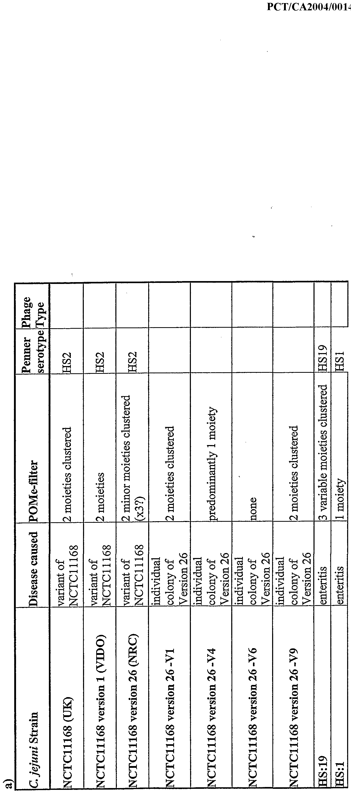
bacteria and the presence of a mobile genetic element responsible for heptose biosynthesis in various strains of C. jejuni has also been described herein. C. jejuni strains from different disease presentations and geographical locations were surveyed for the phosphoramide (Table Xa). Examination of the closely related Campylobacter coli demonstrated that this modification is absent from this species (Table Xb) and also absent in other species sampled with the exception of one C. fetus isolate from a human with bacterial septicemia. Multiple colonies from selected mutants were analysed to demonstrate their potential role in phosphoramide biosynthesis in NCTCl 1168. Mutagenesis of cj'1416c, cjl417c, cjl418c and cjl421c resulted in loss of phosphoramide while inactivation of the second copy of cj 1421 (ie. cj 1422c) had no effect in NCTCl 1168 (Table Xc). However, based on the comparative sequencing results described above, cj!422c homologues are found in some phosphoramide producing strains without cj'1421c and appear to be responsible for the addition of phosphoramide in these strains (for example the variable phosphoramide observed in the HS:23, HS:36 and HS:23/36 group). Interestingly, mutagenesis of cjl435c and cj 1437c resulted in loss of capsule and yet the phosphoramide signal was still detected. These results suggest that this modification can either be added to additional structures and/or that we are detecting biosynthetic intermediates in the phosphoramide pathway. A summary of the phosphoramide survey results is shown in Table XI demonstrating that the phosphoramide is common to 10% of C. jejuni isolates and 0% of C. coli isolates examined. Many strains were tested for the presence of genes cjl416c, cjl421c and cjl422c by PCR. The results summarizing the PCR probing and sequencing (which also includes cj 1417c and cj 1418c) are summarized in Table XIII and the primers used for probing are listed in Table XIV. Phosphoramide analysis demonstrated that C. jejuni, RM1221 is unable to produce phosphoramide and this is consistent with this strain lacking the genes involved in its biosynthesis (Fig. 11). Comparison of the C. jejuni NCTCl 1168 capsule locus with other sequenced epsilon proteobacteria demonstrates that these strains also lack these genes and are likely unable to synthesize the phosphoramide (Fig. 11). Thus, this structure appears to be unique to C. jejuni with rare exceptions in other Campylobacter species.
It is interesting to note that the phosphoramide is added to capsules of strains with different serotypes and thus to different structures. A more thorough analysis of the capsule structures of two serostrains known to produce phosphoramide, HS:1 and HS:19 was performed. As mentioned above, the HS:1 CPS structure consists of galactose and glycerol-phosphate. However, an unusual sugar that has not previously been described (see Table II) was detected. It is to this unusual sugar that the phosphoramide is being attached (Fig. 12). More convincing results were obtained with HS:19 (Fig. 13). The phosphoramide is attached at the 4-position of GlcNAc in contrast to NCTCl 1168 where the phosphoramide is attached at the 3-position of Gal/NAc. As mentioned above, the phosphoramide is detected in select mutants that lack the CPS. This suggests that the phosphoramide is added to alternate structures or that intermediate forms can be detected. Occasionally additional phosphoramide signals are observed during phosphate scans of capsulated C. jejuni isolates (Fig. 14). Thus, in addition to the possibilities mentioned, phosphoramides may be attached to a varying CPS backbone which could lead to additional signals. In order to determine whether the expression of phosphoramide on C. jejuni CPS has any biological relevance, tissue culture and serum sensitivity assays were performed. Preliminary adherence and invasion assays comparing wildtype to the phosphoramide mutants demonstrated that loss of phosphoramide caused decreased adherence to CaCo-2 cells while invasion appeared unaffected (Fig. 15). To ensure that the differences in the tissue culture assays were not due to differences in motility
' of the mutants, motility assays were done comparing the wildtype strains with their respective mutants and demonstrated that all mutants had similar levels of motility compared to the parent (Fig. 16). Preliminary serum sensitivity assays comparing wildtype to the 1416-1 mutant indicated that expression of the phosphoramide increases C. jejuni sensitivity to pooled human serum (Fig. 17). These results suggest that it would be beneficial for C. jejuni to suppress phosphoramide expression while traveling to its optimal location and then expressing the phosphoramide in order to efficiently bind to host cells.
1 The abbreviations used are: Bac, bacillosamine, 2,4-diacetamido-2,4,6-trideoxy-D- glucopyranose; CE, capillary electrophoresis; CPMG, Carr-Purcell-Meiboom-Gill;
CPS, capsular polysaccharide; DIPSI-2, decoupling in the presence of scalar interactions; ESI-MS, electrospray ionization mass spectrometry; GBS, Guillain- Barre Syndrome; HR-MAS, high resolution magic angle spinning; LOS, lipooligosaccharides; LPS, lipopolysaccharide; MAS, magic angle spinning; NOESY, nuclear Overhauser effect spectroscopy; PVDF, polyvinylidene difluoride; TOCSY, total correlation spectroscopy; WURST-2, wideband, uniform rate, and smooth truncation; HMQC, heteronuclear multiple quantum correlation; HMBC, heteronuclear multiple bond correlation; NMR, nuclear magnetic resonance. The presence of the identified phosphoramide across most serotypes of C. jejuni makes it a useful target for attack on these cells, as well as for the identification of these cells. Molecules having a good binding affinity for this phosphoramide ("binders") can be employed, either alone or as conjugates or on the surface of liposomes or other suitable cargo carriers or matricies to bind to C. jejuni cells. Where such molecules are functionally associated with a toxin or similar substance, they may be used to reduce C. jejuni viability or proliferation on a surface or in a solution, fluid, or semifluid of concern. Similarly, when such molecules are associated with a marker, such as an enzyme able to catalyze a colourometric reaction, or a fluorescent or radioactive marker, they can be used to identify and/or localize C. jejuni. One example of suitable binders for this purpose are antibodies having specificity for the phosphoramide. Such antibodies may be single domain antibodies. In some cases only fragments of antibodies having the desired specificity will be employed. Such fragments may be expressed as part of a fusion protein with a "cargo" polypeptide of interest. Another example of suitable binders are bacteriophages or portions thereof having a good affinity for the phosphoramide. In some instances the phage particles or portions will also be capable of lysing the C. jejuni cells. Parts of interest can include any functional part. In some instances phagetail sheaths and/or tail spike proteins may be employed. Other binder molecules can be identified by screening of materials for specific binding to the phosphoramide. Thus, in one embodiment the invention provides a use
of the phosphoramide in identifying compounds, or materials useful in identifying or reducing the viability of C. jejuni. In some instances binders which recognize the phosphoramide regardless of its sugar of attachment will be desired. In some instances binders which recognize the phosphoramide in association with one or more particular sugars of attachment will be desired. Binders which specifically recognize the phosphoramide structure unique to
Campylobacter and no other phosphate compounds produced in nature are considered to be useful binders. In an embodiment of the invention there is provided a method of modulating the adhesion of C. jejuni cells to a surface, comprising modulating the concentration of binders in the surrounding fluid. It will be understood that the surface may include a non-living material, cells, and/or cell-derived materials.
Table I Chemical shifts3 (ppm) for the C. jejuni NCTCl 1168 variant 2 CPS
Unit
A δc 106.2 81.2 70.7 84.0 63.0 δH 5.35 4.21 4.30 4.13 3.89, 3.70
B δc 99.0 73.1 73.8 76.4 72.7 171.3C 53.9 61.3* δH 5.14 3.94 4.10 3.93 4.38 8.32c 4.03 3.72, 3.67*
C δc 106.4 62.5 79.6 82.2 78.3 61.9 174.9C 22.9 δH 5.13 4.27 4.88 4.48 3.97 3.82, 3.77 8.31c 2.05
D δc 98.1 72.2 73.8 70.2 72.3 79.5 63.1 60.4 δH 5.61 3.53 3.72 3.56 4.08 3.80 3.86 3.55
E δc 54.8 δH 3.75
a Measured at 600 MHz (XH) in D2O at 35υC (± 0.2 ppm error for dc and ± 0.02 ppm for dH). Internal acetone CH3 resonance set at dH 2.225 ppm and dc 31.07 ppm. b (CH2)2 of glycerol (8B and 9B). c C=ONH. dc from HMBC at 25°C. dH from 1H spectrum in 90% H2O 10%D2O at 25°C.
Table. II. Biosynthetic cps regions of various strains of C. jejuni.
"Additional uncharacterised labile substituent is present
Table m. Strain NCTCl 1168. In this and the following tables: nrdb - non-redundant database; contingency genes are shown in bold; the last three letters in the names of homologues, which indicate a gene product with the highest similarity score, are preceded by two letter codes for bacteria: Aa - Aquifex aeolicus, Af '- Archaeoglobus fulgidus, At - Aneurinibacillus tliermoaerophilus, Au - Agrobacterium tumefaciens, Bb - Borrelia burgdorferi, Bf - B acter oides fragilis, Bh - Bacillus halodurans, Bj - Bradyrhizobiumjaponicum, Bs - Bacillus subtϊlis, Ca - Clostridium acetobutylicum, Cd - Corynebacterium diphtheriae, Bc -E. coli, Hi - Haemophilus influenzae, Kp - Klebsiella pneumoniae, Li - Leptospira interrogans, LI - Lactococcus lactis, Mj - Methanococcus jannaschii, Mp - Mycoplasma pneumoniae, Mh - Methanobacterium thermoautotrophicum, Mt - Mycobacterium tuberculosis, No - Nostoc sp., Pa - Pyrococcus abyssi, Pm - Pasteurella multocida, Sm - Salmonella muenchen, - Sp - Streptococcus pneumoniae, Sy - Synechocystis sp., Em - Rhizobium meliloti, Tn - Thermotoga neapolitana, Ye - Yersinia enterocolitica, Yp - Yersinia pseudotuberculosis. ' Function prediction of glycosyltransferases is based on either annotation by the Sanger Institute (for Cj 1421, Cj 1422, Cj 1432, Cj 1434, Cj 1438 and CJ1440) or similarity to other glycosyltransferases of C. jejuni (for Cj 1431 and CJ1442). These glycosyltransferases are labelled as conserved hypothetical without indicating E values. 2According to {St Michael, 2002 #212}.
1-phosphate sedoheptulose 7-phosphate D-a-D-heptose-7-phosphate GmhA, sedoheptulose 7-phosphate isomer D-a-D-heptose-7-phosρhate D-a-D-heptose-l,7-diphosphate HddA, heptose 7-phosphate kinase - unknown UDP-glucose UDP-galactose nucleotidyl-sugar epimerase GDP-4- keto-6-deoxy-D-mannose GDP-fucose GDP-heptose biosynthesis
2 - unknown dTDP-6-deoxy-D-xylo-4-hexulose dTDP-6-deoxyVL-lyxo-4-hexulose nucleotidyl-sugar epimerase - sugar transferase - sugar transferase - unknown - sugar transferase phosphoserine serine unknown imidazole acetol phosphate histidinol phosphate unknown imidazole acetol phosphate histidinol phosphate unknown - sugar transferase UDP-D-galactopyranose UDP-D-galactofuranose UDP-GalNAc pyranose mutase
2 - sugar transferase UDP-glucose UDP-glucuronate Udg, UDP-glucose 6-dehydrogenase - sugar transferase
/
Table IV. Strain NCTC 12517 (HS:19).
4-
O
Table V. Strain Gl (HS:1).
Table VI. Strains 81-176 (HS:23 HS:36), CCUG 10954 (HS:23) and ATCC 43456 (HS:36).
'Similarity is calculated using the sequence from strain 81-176.
2ORF #12 is phase-variable in serostrains HS:23 and HS:36 but not in strain 81-176.
Table VH. Strain 176.83 (HS:41).
biosynthesis - by similarity to CJ1428 ) dTDP-4-dehydro-6-deoxy-L- nucleotidyl-sugar epimerase mannose. UDP-D-galacto- 1 ,4-furanose nucletidyl-sugar pyranose mutase sugar transferase UDP-D-galacto-1 ,4-furanose nucletidyl-sugar pyranose mutase CDP-abequose nucleotidyl-sugar dehydratase dTDP-4-dehydro-6-deoxy-L- nucleotidyl-sugar epimerase mannose. CDP-4-dehydro-6-deoxy-D- nucleotidyl-sugar dehydratase glucose CDP-glucose sugar-1-phosphatenucleotidyltransferase sugar transferase sugar transferase UDP-D-galacto-l,4-furanose nucletidyl-sugar pyranose mutase UDP-galactose sugar epimerase UDP-glucuronate Udg, UDP-glucose 6-dehydrogenase UDP-galactose nucletidyl-sugar pyranose mutase sugar transferase sugar transferase sugar transferase
Table VIII. Analysis of contingency genes in the biosynthetic cps region
2{Parkhill, 2000 #24}
3Single colony hypermotile isolate of NCTCl 1168 "Ratio of "ON'V'OFF" genes in the sequenced genome of NCTCl 1168 {Parkhill, 2000 #24}.
5No detectable PCR product
Table LX. Comparison of the contingency genes in the biosynthetic cps region of serostrains HS:23, HS:36 and strain 81-176:
4- -4
Homologue in strain NCTCl 1168
2 The number in bold indicates the most frequent variant,
3 ORF No.12 is not phase-variable in strain 81-176.
Table X The commonality of the phosphoramide moiety on C. jejuni capsular polysaccharides as determined by the P-filter method usin
HR-MAS NMR. Structural work has indicated that the phosphoramide is found on position-3 of Gal NAc in NCTCl 1168, on position-4 o GlcNAc of HS:19 and on a unique deoxy sugar in HS:1. Empty boxes indicate experiments that have not been performed. The Penner serotyp refers to the heat-stable (HS) type. Phage types (PT) indicated as RDNC refer to 'react with phages but do not conform to designated type'. Table a) includes C. jejuni wildtype strains, b) includes Campylobacter species other than C. jejuni and c) includes results for mutants in gene affecting phosphoramide biosynthesis or accumulation. Note that V4 represents variant 2 described above while V9 represents variant 3 above.
-O
b)
c)
Table XI A summary of the distribution of the phosphoramide on the capsular polysaccharide of Campylobacter strains isolated from a variety of animal sources and geographical locations.
Campylobacter jejuni Asymptomatic (UK) 5/7
Enteritis (Various locations) 8/10
Enteritis (phage propagating) (NCTC) 5/6
GBS (Rotterdam / Japan) 4/5
MF' (Rotterdam) 4/4 Septicemia (UK) 5/8
Poultry and retail meat (Canada) 2/10
Chicken and goose droppings (US) 4/6
Pigeon (phage propagating) (NCTC) 1/1
Animal (UK) 5/5 Total 43/62 = 70%
Other Species
Campylobacter coli (Canada, US) 0/18 = o% Campylobacter fetus (UK) 1/1* Campylobacter fetus fetus (NCTC) 0/1
Campylobacter fetus venerealis (ATCC) 0/1 Campylobacter hominus (UK) 0/1
Total strains examined 84
*only non C. jejuni examined with phosphoramide - this strain was isolated from a human with septicemia
Table XLTI: The commonality of genes involved in phosphoramide biosynthesis. The presence of Cjl416c, Cj 1417c, Cj 1418c, Cj 1421c and Cj 1422c homologues wa determined using sequencing and gene-specific polymerase chain reactions using primers found in Table XTV. The on off status of Cjl421c and Cjl422c, if known, i indicated as these genes are phase-variable, and was determined by sequencing. Empty boxes indicate that the experiment was not done.
-4
Table XTV The sequences of primers used for the identification and characterization of Cj 1416c, Cj 1421c and Cj 1422c. The chromosomal location represents the location of the rimer on the forward strand in the genome of NCTC 11168
Table XV
Part A cj 1421c: MLNPNSAIERVKNHLAYKLGQWIEHRHNGGGYIALFKKLYKIKKQHKKEQ KIYQQIIQV FPQLKYPSLETCSDYNEALRCKFHLSYMIGEVLIKAYQNWYKGGGFKIJKNN IKKANKEFQ IFREILKEFKELNGETL AIQDNKQ F KEFPRIKNILKTHQDYQPILDNI FHNFNYFIK NFDLIEEWLLSDDFKEKYKKENHPYPSLLDPKKLNDENEKINYHNIPAELA WKMNLPLPP NYEFM FFSHGAGAFTLGQFFYH FKINILDYFCGGDGDIRYYKFYNKL E LKDKRNIIT INDIDPSWYGNQHKRDKLFSSFQKITPILFQIRDPIELIKHAYGRKWGNNL AKTKEFD S YQFNDIITEVEVYNYNLPNTLEGQRPQSFLWKSLIECFDKFNDCFYLDISK IRGΞETIHT LNYLSNKFNLKQIKINDKE TKSYFKGNLYFLLPLTLYLNKEDLNTNIPN KKINKNNSL IININFFQNDNNLFNLYSELSILDMDSSVGFYIDKQDYNKLKNDSIFYKQV IDY RNFAY ELKNRIQIEEDLMLKVEDVLRHLYNNKNARVSAKNILDEELVYIKQHRPDI VAS KYYQE FEKMCKELDGDI
Part B cj 1422c: MLNPNSAIERVKNH AYKLGQTVIEHRH GGGYIALFKK Y I KQHKKEQ KIYQQIIQV FPQLKYPSLETCSDYNEALRCKFHLSY IGEVLIKAYQN YKGGGFKLKNN IKKANKEFQ IFREILKEFKE NGETLKAIQDNKQLF KEFPRIKNI KTHQDYQPILDNI FHNFNYFIK NFDLIEE LLSDDFKEKYKKENHPYPSLLDPKKLNDENEKINYHNIPAELA WKMNLPLPP NYEFVGFFLHTSGEKAMERFLKEVGWLIGAFGYEDGKRYISIFNFLISEA CACNDLKFA IGILDΛ7NCQKYDKFCFLLQNKPVLILLRDPIDSLKSFINVRHQKNGFNEIL KIDINNTDF DKINDRIVYVHESNGCFNPDTNQKFPSLESIKALSDTNHWMLMYNIRRNKT IEFFRFNKI IYIDMMDIVGDKTLFTLEKLSKILNFSSPDKNNKIFYQQLYSPLTVLLPCI IKVNNKVKI FVSNRFSVKNIQIMENCIDITDKFKEIFHENLIIFCSKDHFDSLINNQTLY NWLEYINK FLISLKKRINVEKNKEVKVDDVLDYFKKNISVAKSYKDILDEELVYIKQHR PDIVASWKY YQEFERMCKELDENNQNPSLSFSNQ