COMBINATIONS OF PKC INHIBITORS AND THERAPEUTIC AGENTS FOR TREATING CANCERS
This application is a continuation-in-part application of U.S. Serial No. 08/619,304, filed March 21, 1996 which is a continuation-in-part application U.S. Serial No. 08/603,814, filed February 20, 1996, the contents of which are hereby incorporated by reference into the present application.
Throughout this application, various references are referred to within parentheses. Disclosures of these publications in their entireties are hereby incorporated by reference into this application to more fully describe the state of the art to which this invention pertains. Full bibliographic citation for these references may be found at the end of each series of experiments in this application, preceding the claims.
Background of the Invention
Protein kinase C (PKC) functions in processes relevant to carcinogenesis, tumor cell metastasis, and apoptosis. Safingol, an optical isomer (the L- threo enantiomer) of dihydrosphingosine, is a specific inhibitor of PKC and may represent a novel target for anti-cancer therapy. Preclinical animal studies show that safingol alone has minimal effects on tumor cell growth, but combination of this compound with conventional chemotherapy agents dramatically potentiates their anti-tumor effects. It has been suggested that many chemotherapeutic agents exert their anti-tumor effects by inducing apoptosis.
A large body of evidence indicates a fundamental role for the involvement of protein kinase C (PKC) , family members
of serine/threonine protein kinases, in processes relevant to neoplastic transformation, carcinogenesis, and tumor cell invasion of surrounding tissues (1-3) . Consequently, PKC may represent a novel target for anti- cancer therapy. Safingol, the L-tήreo enantiomer of dihydrosphingosine, is a specific inhibitor of PKC (4) . Preclinical animal studies show that safingol is non- toxic at doses that achieve serum levels sufficient to inhibit PKC enzyme activity (5) . While safingol has negligible impact on tumor cell growth in vivo, the combination of safingol with conventional chemotherapeutic agents such as doxorubicin and cisplatin significantly potentiates the anti-tumor effects of these drugs (6) .
Based on these observations, safingol, used in combination with doxorubicin, has become the first PKC specific inhibitor to enter clinical trials. The mechanism by which safingol potentiates the activity of chemotherapeutic agents is unclear, although inhibition of P-glycoprotein phosphorylation and reversal of the multidrug resistant (mdr) phenotype have been suggested
(7,8) . While this hypothesis can explain the synergism achieved with combinations of safingol and doxorubicin, it does not explain the synergism reported for combinations of safingol with drugs that are not believed to produce resistance by the mdr mechanism (e.g., cisplatin) (6) , nor does it explain safingol-induced effects that occur in tumor cell lines that dc not express the P-glycoprotein (8) . Therefore, pathways other than P-glycoprotein inhibition are likely to be involved in the safingol-mediated enhancement of chemotherapy.
It has been suggested that the anti-tumor activity of
many chemotherapeutic agents (e.g., cisplatin and etoposide) is a consequence of their induction of apoptosis (9) . In this context it has been proposed that activation of PKC acts as an antagonist to apoptosis, whereas inhibition of PKC promotes apoptosis (10-12) . Thus, safingol-mediated potentiation of chemotherapy might be attributed to its PKC inhibitory effect, subsequently leading to increased apoptosis after drug- induced damage.
The present studies sought to determine the extent to which whether safingol by itself, or in combination with a specific chemotherapeutic drug (e.g. mitomycin-C, MMC) , would promote apoptosis in gastric cancer cells. Furthermore, applicants investigated whether the p53 status of these cells influences the development of apoptosis after treatment with safingol and MMC.
Summary of the Invention
This invention provides a method for screening protein kinase C inhibitors capable of potentiating apoptosis in tumor cells comprising steps of (a) contacting an amount of a protein kinase C inhibitors with tumor cells effective to potentiate apoptosis of tumor cells, (b) contacting the potentiated tumor cells of step (a) with an antitumor therapeutic agent; (c) determining the apoptosis of tumor cells; and (d) comparing the apoptosis determined in step (c) with apoptosis of same tumor cells which are only treated with the antitumor therapeutic agent, an increase in apoptosis indicating that the protein kinase C inhibitor is capable of potentiating apoptosis in tumor cells. This invention also provide the above method, wherein step (a) is carried out in the presence of the antitumor therapeutic agent.
This invention also provides the protein kinase C inhibitor capable of potentiating apoptosis in tumor cells as determined by the above-described methods.
This invention further provides a pharmaceutical composition comprising the protein kinase C inhibitor capable of potentiating apoptosis in tumor cells as determined by the above-described methods and a pharmaceutically acceptable carrier.
This invention provides a method for screening antitumor therapeutic agents suitable for combination therapy with a protein kinase C inhibitors capable of potentiating apoptosis in tumor cells comprising steps of: (a) contacting an amount of a protein kinase C inhibitor with tumor cells effective to potentiate apoptosis of tumor cells; (b) contacting the potentiated tumor cells of step
(a) with an antitumor therapeutic agent; (c) determining the apoptosis of tumor cells; and (d) comparing the apoptosis determined in step (c) with apoptosis of same tumor cells which are only treated with the protein kinase C inhibitor, an increase in apoptosis indicating that the antitumor therapeutic agent is suitable for combination therapy with a protein kinase C inhibitor capable of potentiating apoptosis in tumor cells. In an embodiment, step (a) is carried out in the presence of the protein kinase C inhibitor. In another embodiment, the antitumor therapeutic agent is not previously known.
This invention also provides an antitumor therapeutic agent suitable for combination therapy with a protein kinase C inhibitors capable of potentiating apoptosis in tumor cells determined by the above-described methods.
This invention also provides a pharmaceutical composition comprising an effective amount of the antitumor therapeutic agent determined by the above-described methods, a protein kinase C inhibitor capable of potentiating apoptosis in tumor cells and a pharmaceutically acceptable carrier.
This invention provides a method for enhancing therapy in a tumor bearing subject comprising administering to the subject an effective amount of a specific protein kinase C inhibitor capable of potentiating apoptosis in tumor cells during or prior to the treatment of an antitumor therapeutic agent. This invention provides the above method, wherein the specific protein kinase C inhibitor is Safingol (L-threo-dihydrosphingosine) , RO32-0432 (Bisindolylmaleimide tertiary amine) , UCN-01 (7-OH- staurosporine) , Flavopiridol ( 86-8275) , Bryostatin 1 (Macrocyclic lactone) or antisense nucleotides capable of
inhibiting the expression of the protein kinase C.
In an embodiment, the antitumor therapeutic agent is a chemotherapeutic agent. In a further embodiment, the chemotherapeutic agent is selected from a group consisting of Mitomycin C, Carboplatin, Taxol and Doxorubicin. In a still further embodiment, the antitumor therapeutic agent is a radiotherapeutic agent
In an embodiment of the above-described methods, the tumor is a gastrointestinal cancer. In a further embodiment, the gastrointestinal cancer is gastric cancer, small bowel cancer, colon cancer or rectal cancer.
In a separate embodiment of the above-described methods, the tumor is a breast cancer. In another embodiment, the tumor is a ovarian cancer. In a still another embodiment, the tumor is of prostate cancer, lung cancer, melanoma, cervical carcinoma, pancreatic cancer, , sarcoma, hepatoma, gallbladder cancer, leukemia or lymphoma.
Finally, this invention provides a method for potentiating apoptosis in tumor cells comprising contacting the cancerous cells with an effective amount of a specific protein kinase C inhibitor capable of potentiating apoptosis during or prior to the treatment of an antitumor therapeutic agent.
Brief Description of the Figures
Figure 1. Photomicrographs of representative fields of SK-GT-5 cells (mutant for p53) stained with bibenzimide trihydrochloride
(Hoescht-33258) to evaluate nuclear chromatin condensation (i.e., apopotosis) after treatment for 24 hours with no drug (panel A) , safingol alone (50 μM, panel B) , MMC alone (5 μg/ml, panel C) , or the combination of safingol (50 μM) and MMC (5 μg/ml) (panel D) .
Figure 2. Induction of apoptosis in MKN-74 cells (a wild-type for p53) with safingol alone (50 μM) , MMC alone (5 μg/ml) , or safingol (50 μM) in combination with MMC (5 μg/ml) . Cells were counted and scored for apoptotic chromatin condensation by the QFM method described in the "Material and
Methods" section. Bars represent mean number of cells counted that undergo apoptosis as a percentage of 500 total cells randomly counted in duplicate samples, ±SD.
Figure 3. Contour plots are shown for the frequency of MKN-74 cells exhibiting increased green fluorescence emitted from digoxigenin- dUTP: fluorescein isothyiocyanate-labeled antibody complexes [dUTP Incorp. (FITC fluorescence) ] , which identifies subpopulations of cells undergoing apoptosis by the TdT assay, versus cellular DNA content (PI fluorescence) , as
described in the "Material and Methods" section. MKN-74 cells were incubated for 24 hours with no drug (panel A) , safingol alone (50 μM, panel B) , MMC alone (5 μg/ml, panel C) , or the combination of safingol (50 μM) and MMC (5 μg/ml) (panel D) . Apoptotic cells are identified as those sorting above the Rx region of the contour plots. The G0/G y S , and G /M regions of the cell cycle are indicated in
Panel A.
Figure 4. Effect of PKC activation with PMA on the induction of apoptosis in the presence of the PKC inhibitor safingol. SK-GT-5 cells were treated, as described in "Materials and Methods, " according to the following protocols: safingol alone (50 μM) for 1 hour followed by no drug for 24 hours; MMC alone (5 μg/ml) for 24 hours; safingol alone (50 μM) for 1 hour followed MMC (5 μg/ml) alone for 24 hours; PMA alone (lng/ml) for 1 hour, followed by no drug for 24 hours; and the combination of safingol (50 μM) and PMA (1 ng/ml) for 1 hour, followed by MMC alone (5 μg/ml) for 24 hours. Induction of apoptosis was then determined with the QFM staining of condensed nuclear chromatin. Bars represent mean number of cells counted that undergo apoptosis as a percentage of 500 total cells randomly counted in duplicate samples, ±SD.
Figure 5. Treatment with safingol alone did not induce significant levels of apoptosis (2%+l) when compared with untreated controls (<1%) .
Figure 6. UCN-01 enhances MMC-induced apoptosis: SK-
GT-2 cells, which have a mutation in p53, were treated with increasing concentrations of UCN-01 in the presence or absence of a fixed dose of MMC for 24 hours. The results for the QFM analysis are shown in Figure 6.
Figure 7. Apoptosis was again measured by QFM staining. The results applicants' shown in shown in Figure 7.
Applicants' studies indicate that flavopiridol increased the induction of apoptosis from 7%±l% with MMC alone to 73%±1% with MMC and flavopiridol in combination. Flavopiridol alone induced a slight degree of apoptosis (17%±2%) , but not nearly to the degree observed with the combination therapy.
Figure 8. Each lane represents the following: lane A, the IgG control; lane B, no drug therapy for 24 hours; lane C, MMC alone (5.0 μg/ml) for 24 hours; lane D, safingol alone (50 μM) for 24 hours; lane E, the combination of safingol (50 μM) and MMC (5.0 μg/ml) for 24 hours. The results indicate that untreated gastric cancer cells (lane B) have increased cdk2 activity and that both MMC (lane C) and safingol (lane D) have decreased cdk2.
However, the combination of MMC and safingol increased cdk2 activity back to basal levels.
Figure 9. MMC alone at the concentrations of 2.5 μg/ml
and 5.0 μg/ml induced apoptosis in 4%±2 and 6%+l of the MDA-MB-468 cells, respectively. However, the combination of safingol (SPC) and MMC significantly increased the percentage of cells undergoing apoptosis from 11%±1 with safingol alone to 23%±2 with safingol and 2.5 μg/ml MMC and to 33%±10 with safingol and 5.0 μg/ml MMC (p <0.001) .
Figure 10. The combination of UCN-01 and MMC together increased the induction of apoptosis of the MDA-MB-468 cells from 20%±4 with UCN-01 alone to 41%±3 with UCN-01 and 2.5 μg/ml MMC and to 58%±1 with UCN-01 and 5.0 μg/ml MMC.
Figure 11. Effect on MMC-induced apoptosis by Ro32-0432 and 2241.
Figure 12. Effect of Ro32-2241 on proliferation of MMC- treated MKN-74 cells.
Detailed Description of the Invention
This invention provides a method for screening for a modulating agent which when combined with an antitumor therapeutic agent increases apoptosis in tumor cells comprising the steps of: (a) contacting tumor cells with an amount of the modulating agent and with an antitumor therapeutic agent known to cause apoptosis when used in combination with an appropriate modulating agent in an amount effective to increases apoptosis in tumor cells; (b) determining the degree of apoptosis of tumor cells from step (a) ; and (c) comparing the degree of apoptosis determined in step (b) with the degree of apoptosis of tumor cells which are treated with only the antitumor therapeutic agent or only the modulating agent, an increase in apoptosis indicating that the modulating agent in combination with the antitumor therapeutic agent is capable of increasing apoptosis in tumor cells.
This invention also provides a method for screening for a modulating agent which when combined with an antitumor therapeutic agent increases apoptosis in tumor cells comprising the steps of: (a) contacting tumor cells with an amount of the modulating agent for a period of time; (b) contacting the tumor cells from step (a) with an antitumor therapeutic agent known to increase apoptosis when used in combination with a modulating agent in an amount effective to increase apoptosis; (c) determining the degree of apoptosis of tumor cells from step (b) ; and (d) comparing the degree of apoptosis determined in step (b) with the degree of apoptosis of tumor cells which are treated with only the modulating agent, or only the antitumor therapeutic agent, an increase in apoptosis indicating that the modulating agent in combination with the antitumor
therapeutic agent is capable of increasing apoptosis in tumor cells.
This invention provides a method for screening for a modulating agent which when combined with an antitumor therapeutic agent increases apoptosis in tumor cells comprising the steps of: (a) contacting tumor cells with an antitumor therapeutic agent known to increases apoptosis in tumor cells when used in combination with an appropriate modulating agent in an amount effective to increase apoptosis for a period of time; (b) contacting the tumor cells from step (a) with an amount of the modulating agent; (c) determining the degree of apoptosis of tumor cells from step (b) ; and (d) comparing the degree of apoptosis determined in step (b) with the degree of apoptosis of tumor cells which are treated with only the antitumor therapeutic agent or only the modulating agent, an increase in apoptosis indicating that the modulating agent in combination with the antitumor therapeutic agent is capable of increasing apoptosis in tumor cells.
In an embodiment of the above described methods, the modulating agent is a chemical compound that inhibits cellular apoptotic signalling.
In another embodiment, the modulating agent is a protein kinase C inhibitor.
In a still another embodiment, the modulating agent is not previously known.
This invention also provides the modulating agent capable of increasing apoptosis in tumor cells as determined by the above-described methods.
This invention ctlso provides a pharmaceutical composition comprising the modulating agent capable of causing apoptosis in tumor cells as determined by the above methods and a pharmaceutically acceptable carrier.
This invention provides a method for screening for an antitumor therapeutic agent which when combined with a modulating agent increases apoptosis in tumor cells comprising the steps of: (a) contacting tumor cells with a modulating agent known to increase apoptosis in tumor cells when used in combination with an appropriate antitumor therapeutic agent in an amount effective to increases apoptosis in tumor cells and with an amount of the antitumor therapeutic agent; (b) determining the degree of apoptosis of tumor cells from step (a) ; and (c) determining the degree of apoptosis of ι_umor cells from step (b) ; and (d) comparing the degree of apoptosis determined in step (c) with the degree of apoptosis of tumor cells which are treated with only the modulating agent or only the antitumor therapeutic agent, an increase in apoptosis indicating that the antitumor therapeutic agent in combination with the modulating agent is capable of increasing apoptosis in tumor cells .
This invention provides a method for screening for an antitumor therapeutic agent which when combined with a modulating agent increases apoptosis in tumor cells comprising the steps of: (a) contacting tumor cells with a modulating agent known to increase apoptosis in tumor cells when used in combination with an appropriate antitumor therapeutic agent in an amount effective to increases apoptosis in tumor cells for a period of time; (b) contacting the tumor cells from step (a) with an amount of the antitumor therapeutic agent; (c) determining the degree of apoptosis of tumor cells from step (b) ; and (d) comparing
the degree of apoptosis determined in step (c) with the degree of apoptosis of tumor cells which are treated with only the modulating agent or only the antitumor therapeutic agent, an increase in apoptosis indicating that the antitumor therapeutic agent in combination with the modulating agent is capable of increasing apoptosis in tumor cells.
This invention provides a method for screening for an antitumor therapeutic agent which when combined with a modulating agent increases apoptosis in tumor cells comprising the steps of: (a) contacting tumor cells with an amount of the antitumor therapeutic agent for a period time; (b) contacting the tumor cells from step (a) with a modulating agent known to increase apoptosis in tumor cells when used in combination with an appropriate antitumor therapeutic agent in an amount effective to increases apoptosis in tumor cells; (c) determining the degree of apoptosis of tumor cells from step (b) ; and (d) comparing the degree of apoptosis determined in step (c) with the degree of apoptosis of tumor cells which are treated with only the modulating agent or only the antitumor therapeutic agent, an increase in apoptosis indicating that the antitumor therapeutic agent in combination with the modulating agent is capable of increasing apoptosis in tumor cells. In an embodiment, the antitumor therapeutic agent is not previously known.
This invention also provides the antitumor therapeutic agent suitable for combination therapy with a modulating agent capable of increasing apoptosis in tumor cells by the above methods.
This invention also provides a pharmaceutical composition comprising an effective amount of at least one antitumor
therapeutic agent determined by one of the above methods, at least one modulating agent capable of increasing apoptosis in tumor cells, and a pharmaceutically acceptable carrier.
This invention provides a method for treating cancer in a subject comprising administering to the subject an effective amount of at least one antitumor therapeutic agent, and at least one modulating agent, sequentially or concomitantly. In an embodiment of the method for treating cancer in a subject, the tumor bearing subject is administered at least one antitumor therapeutic agent and subsequently administered at least one modulating agent.
In another embodiment, the antitumor therapeutic agent is paclitaxel and the modulating agent is flavopiridol. In a separate embodiment, the antitumor therapeutic agent is mitomycin C and the modulating agent is Ro32-0432.
In an embodiment the tumor bearing subject is administered at least one modulating agent and subsequently administered at least one antitumor therapeutic agent. In a further embodiment, the modulating agent is safingol and the antitumor therapeutic agent is mitomycin C or doxorubicin.
In another embodiment, the tumor bearing subject is administered at least one modulating agent concomitantly with at least one antitumor agent. In a further embodiment, the modulating agent is flavopiridol and the antitumor therapeutic agent is mitomycin C.
In an embodiment of the above methods, the modulating agents are safingol and UCN-01, and antitumor therapeutic agent is mitomycin C. In another embodiment, the modulating agent is UCN-01 and the antitumor therapeutic agent is mitomycin C. In a still another embodiment, the modulating agent is Ro-1
and the antitumor therapeutic agent is mitomycin C. In a separate embodiment, the modulating agent is safingol and the antitumor therapeutics agents are paclitaxel and mitomycin C.
The modulating agent includes, but is not limited to Safingol (L-threo-dihydrosphingosine ) , Ro-1 (Bisindolylmaleimide) , Ro32-0432 (Bisindolylmaleimide tertiary amine) , UCN-01 (7-OH-staurosporine) , Flavopiridol (L86-8275) , Bryostatin 1 (macrocyclic lactone) , and antisense nucleotides capable of inhibiting the expression of protein kinase C. In a preferred embodiment, the antisense nucleotide capable of inhibiting the expression of protein kinase C is protein kinase C a antisense.
In another preferred embodiment, the antitumor therapeutic agents is a chemotherapeutic agent. The chemotherapeutic agent includes but is not limited to mitomycin C, carboplatin, cisplatin, paclitaxel, etoposide, and doxorubicin.
In a separate embodiment, the antitumor therapeutic agents is a radiotherapeutic agent .
In an embodiment of the above method, the cancer is a gastrointestinal cancer, breast cancer or an ovarian cancer. In a further embodiment, the gastrointestinal cancer includes gastric cancer, small bowel cancer, colon cancer, and rectal cancer. The cancer can further includes lymphoma, adenocarcinoma, glioblastoma, leukemia, esophageal carcinoma, head and neck cancer, prostate cancer, lung cancer, melanoma, cervical carcinoma, pancreatic cancer, sarcoma, hepatoma, and gallbladder cancer.
This invention provides a method for increasing apoptosis in
tumor cells comprising contacting tumor cells with an effective amount of a modulating agent during, prior to, or after treatment with one or more antitumor therapeutic agents .
This invention provides a method for screening protein kinase C inhibitors capable of potentiating apoptosis in tumor cells comprising steps of (a) contacting an amount of a protein kinase C inhibitor with tumor cells effective to potentiate apoptosis of tumor cells; (b) contacting the potentiated tumor cells of step (a) with an antitumor therapeutic agent; (c) determining the apoptosis of tumor cells; and (d) comparing the apoptosis determined in step (c) with apoptosis of same tumor cells which are only treated with the antitumor therapeutic agent, an increase in apoptosis indicating that the protein kinase C inhibitor is capable of potentiating apoptosis in tumor cells.
This invention also provide the above method, wherein step (a) is carried out in the presence of the antitumor therapeutic agent. In an embodiment, the protein kinase C inhibitor is not previously known.
This invention also provides the protein kinase C inhibitor capable of potentiating apoptosis in tumor cells as determined by the above-described methods.
This invention further provides a pharmaceutical composition comprising the protein kinase C inhibitor capable of potentiating apoptosis in tumor cells as determined by the above-described methods and a pharmaceutically acceptable carrier.
This invention provides a method for screening antitumor therapeutic agents suitable for combination therapy with a
protein kinase C inhibitors capable of potentiating apoptosis in tumor cells comprising steps of: (a) contacting an amount of a protein kinase C inhibitor with tumor cells effective to potentiate apoptosis of tumor cells; (b) contacting the potentiated tumor cells of step (a) with an antitumor therapeutic agent; (c) determining the apoptosis of tumor cells; and (d) comparing the apoptosis determined in step (c) with apoptosis of same tumor cells which are only treated with the protein kinase C inhibitor, an increase in apoptosis indicating that the antitumor therapeutic agent is suitable for combination therapy with a protein kinase C inhibitor capable of potentiating apoptosis in tumor cells. In an embodiment, step (a) is carried out in the presence of the protein kinase C inhibitor. In another embodiment, the antitumor therapeutic agent is not previously known.
This invention also provides an antitumor therapeutic agent suitable for combination therapy with a protein kinase C inhibitors capable of potentiating apoptosis in tumor cells determined by the above-described methods.
This invention also provides a pharmaceutical composition comprising an effective amount of the antitumor therapeutic agent determined by the above-described methods, a protein kinase C inhibitor capable of potentiating apoptosis in tumor cells and a pharmaceutically acceptable carrier.
This invention provides a method for enhancing therapy in a tumor bearing subject comprising administering to the subject an effective amount of a specific protein kinase C inhibitor capable of potentiating apoptosis in tumor cells during or prior to the treatment of an antitumor therapeutic agent. This invention provides the above method, wherein the specific protein kinase C inhibitor is selected from a
group consisting of Safingol (L-threo-dihydrosphingosine) ,
Ro-1, Ro32-0432 (Bisindolylmaleimide tertiary amine) , UCN-01
(7-OH-staurosporine) , Flavopiridol (L86-8275) , Bryostatin 1
(Macrocyclic lactone) and antisense nucleotides capable of inhibiting the expression of the protein kinase C. The above specific protein kinase C inhibitors are simply described for illustrative purposes of this invention. As it will be appreciated by a person of ordinary skill in the art that other specific protein kinase inhibitors may be used in this invention. Applicants have provided as methodology to determine whether a protein kinase C inhibitor may be useful for the claimed combination therapy.
As it can be easily appreciated by an ordinary skilled artisan that other modification of the Bisindolylmaleimide tertiary amine may be possible to produce useful specific protein kinase C inhibitor for the combination therapy. As provided below, the method to synthesize such compounds are known.
In an embodiment, the antitumor therapeutic agent is a chemotherapeutic agent. In a further embodiment, the chemotherapeutic agent is Mitomycin C, Carboplatin, Taxol or Doxorubicin. In a still further embodiment, the antitumor therapeutic agent is a radiotherapeutic agent
In an embodiment of the above-described methods, the tumor is a gastrointestinal cancer. In a further embodiment, the gastrointestinal cancer is gastric cancer, small bowel cancer, colon cancer or rectal cancer, leukemia or lymphoma.
In a separate embodiment of the above-described methods, the tumor is a breast cancer. In another embodiment, the tumor is a ovarian cancer. In a still another embodiment, the
tumor is selected from a group consisting of prostate cancer, lung cancer, melanoma, cervical carcinoma, pancreatic cancer, , sarcoma, hepatoma and gallbladder cancer.
Finally, this invention provides a method for potentiating apoptosis in tumor cells comprising contacting the cancerous cells with an effective amount of a specific protein kinase C inhibitor capable of potentiating apoptosis during or prior to the treatment of an antitumor therapeutic agent.
Thiε invention will be better understood from the Experimental Details which follow. However, one skilled in the art will readily appreciate that the specific methods and results discussed are merely illustrative of the invention as described more fully in the claims which follow thereafter.
Experimental Details
First Series of the Experiments Materials and Methods
Cell culture
Early passage human gastric cancer cell lines SK-GT-5 and MKN-74 were established and characterized as described previously (13,14) . All cultures were maintained at 37°C in Eagles minimum essential media (MEM) supplemented with 20% fetal calf serum in an humidified 5% C02 atmosphere; the cultures tested negative for Mycoplasma species.
Determination of Apoptosis Apoptosis was measured by two different methods: (i) Quantitative fluorescent microscopy (QFM) of nuclear changes induced by apoptosis, as determined by bisbenzimide trihydrochloride (Hoescht-33258) staining of nuclear chromatin (15) , and (ii) a terminal deoxynucleotidyl transferase (TdT) assay that labels the 3 ' -OH ends of DNA fragments produced in apoptotic cells (16) . Cells were treated according to one of several protocols: (i) no drug (control) for 24 hours; (ii) safingol alone for 24 hours at 50 μM, a concentration representing the highest non-toxic dose for SK-GT cells (as determined by cell proliferation studies with [3H] -thymidine (4)) and slightly exceeding the concentration (i.e., 30 μM) which inhibits PKC enzyme activity by 50% in vitro; (iii) Mitomycin-C alone at 5 μg/ml for 24 hours; (iv) a combination of safingol (50 μM) and Mitomycin-C (5 μg/ml) for 24 hours; (v) safingol (50 μM) alone for 1 hour, followed by an immediate wash out of the drug with fresh MEM, and then an additional 24 hours of MMC
(5 μg/ml) exposure; (vi) safingol (50 μM) with 1 ng/ml 3- phorbol 12-myristate 13-acetate (PMA) for 1 hour, followed by an immediate wash out of the drug with MEM and 24 hours
of MMC (5 μg/ml) exposure; and (vii) either safingol (50 μM) alone or PMA (1 ng/ml) alone for 1 hour, followed by drug washout with MEM and 24 hours of exposure to media without drug. The dose of MMC used was based on proliferating studies with [3H] -thymidine (14, and unpublished data) indicating 20% inhibition of cell proliferation. MMC was diluted in watei . A stock safingol solution was constituted in DMSO and was subsequently diluted at least 1:100 in media for all experiments. This dilute concentration of DMSO does not induce apoptosis or inhibit cell proliferation in these cell lines (unpublished data) . For QFM determinations, the cells were fixed in 3% paraformaldehyde and incubated at room temperature for 10 minutes. The fixative was removed and the cells were washed with IX PBS, resuspended in 20 μl IX PBS containing only 8 μg/ml of bisbenzimide trihydrochloride (Hoechst #33258) , and incubated at room temperature for 15 minutes. Aliquots of the cells (lOμl) were placed on glass slides coated with 3-amino-propyl- triethoxysilane, and duplicate samples of 500 cells each were counted and scored for the incidence of apoptotic chromatin condensation using an Olympus BH-2 fluorescence microscope equipped with a BH2-DM2U2UV Dich. Mirror Cube filter (Olympus, Lake Success, NY) . For the TdT assays, the ApopTag Kit (Oncor, Gaithersburg, MD) was used. This method employs a fluoresceinated antidigoxigenin antibody directed against nucleotides of digoxigenin-11-dUTP (d-dUTP) which are catalytically added to the 3-ends fragmented DNA by TdT. Briefly, 1-2 x 10⁶ cells were washed and fixed with 1% paraformaldehyde. The fixed cells were incubated in a reaction mixture containing TdT and d-dUTP for 30 minutes at 37°C. Stop/wash buffer was added, and the cells were resuspended in 100 μl of fluorescinated anti-digoxigenin antibody for 30 minutes at room temperature. The cells were then washed with 0.1% Triton X-100 and counterstained with propidium iodide (PI) solution. Green (d-dUTP labeled DNA
strand breaks) and red (PI staining for total DNA content) fluorescence of individual cells were measured on a FACScan flow cytometer (Becton Dickinson, San Jose, CA) . The resulting bivariate plots enabled the detection of apoptotic events within the cell cycle. The Rx cursor was set using the control specimen to define normal levels of green fluorescence (i.e., basal levels of apoptosis) . Cells with fluorescence above the Rx cursor were considered apoptotic. The data from 10,000 cells were collected and analyzed using CellFit and LYSYS software (Becton Dickinson) .
Statistical analysis
All experiments were done in duplicate and were repeated at least three times unless otherwise indicated. The statistical significance of the experimental results was determined by the two-sided Student's t test.
Experimental Results
Effect of safingol and MMC on inducing apoptosis in SK-GT-5 and MKN-74 cells.
SK-GT-5 cells, which have a mutated p53 gene (14) and have deficient p53 function (unpublished data) , were treated with safingol in the presence or absence of MMC for 24 hours. Cells were counted and scored for condensed nuclear chromatin, and representative photomicrographs are shown in Figure 1. Treatment with safingol alone (Panel B) did not induce significant levels of apoptosis (2%±1) when compared with untreated controls (<1%, Panel A) . However, the combination of safingol and MMC (Panel D) significantly increased the percentage of cells undergoing apoptosis from 18%±1 with MMC alone (Panel C) to 39%±1 with the drug combination (p <0.001) . Treatment of SK-GT-5 cells with safingol for only 1 hour prior to 24 hours of treatment with
MMC resulted in an increase in apoptosis (44%±4% of exposed cells) that was essentially equivalent to that which was observed with concomitant exposure of the cells to the two drugs for the entire 24 hour interval (data not shown) . MKN-74 cells, which are a wild- type for p53, were subjected to the same treatments. As shown in Figure 2, cell counts of MKN-74 cells following treatment with safingol alone indicated no significant induction of apoptosis (8%±3) when compared to untreated controls (<1%) . However, the combination of safingol and MMC significantly increased the percentage of cells undergoing apoptosis from 40%±4 with MMC alone to 83%±4 with safingol and MMC together (p < 0.005) .
Identifying gastric cancer cells undergoing apoptosis by the TdT assay.
MKN-74 cells were treated in the same manner and analyzed by combining the TdT assay and flow cytometry. With this approach, apoptotic cells were identified as those shifting above the Rλ region of the contour plot . Figure 3 shows that MKN-74 cells incubated with safingol alone for 24 hours (Panel B) had the same levels of green fluorescence as the control cells (Panel A) , i.e., essentially no apoptosis. Treatment with MMC alone for 24 hours (Panel C) resulted in the cells exhibiting increased green fluorescence (sorted above the R region) , indicating development of apoptosis. The results in Panel C indicate that the apoptotic cells, after MMC treatment, alone are derived from those that were in the G0/G1 and S phases of the cell cycle. However, exposure of the MKN-74 cells to the combination of MMC and safingol for 24 hours (Panel D) resulted in an even greater increase in cells exhibiting elevated green fluorescence. In the presence of safingol and MMC, apoptotic cells appear to arise out of all phases of the cell cycle. Similar
results were also observed for SK-GT-5 cells (data not shown) .
Effect of PMA on apoptosis induced by safingol and MMC. PMA activates PKC by binding to its regulatory site (17) .
Safingol, as a sphingosine, inhibits PKC activity by interfering with the function of PKC's regulatory domain
(18) . Therefore, applicants hypothesized that if the potentiation of MMC-induced apoptosis by safingol is mediated by inhibition of PKC, then PMA should abrogate this effect. As shown in Figure 4, treatment of SK-GT-5 cells with either safingol or PMA alone had essentially no effect on inducing apoptosis. Preexposure of the cells to safingol alone for one hour followed by MMC for an additional 24 hours induced a significant increase in apoptosis from 36%±4 with a 24 hour exposure of MMC alone to 55%±1 with the combination (p < 0.01) . However, under the conditions in which cells were preexposed to PMA and safingol together for one hour before MMC, the induction of apoptosis was inhibited to the level observed for MMC alone.
Experimental Conclusion
The present studies show that safingol potentiates the cytotoxic effect of MMC in two human gastric cancer cell lines which differ in their baseline sensitivity to MMC and in their p53 status. Neither cell line expresses P- glycoprotein (unpublished data) . As shown by bisbenzimide trihydrochloride staining and the TdT assay, safingol alone did not dramatically induce apoptosis in either the MMC- sensitive MKN-74 cells, which have a wild-type p53 function, or the MMC-resistant SK-GT-5 cells, which are deficient for p53. In addition, the typical oligonucleosomal base pair fragments (DNA "ladders") were not induced by safingol under the conditions tested (data not shown) . When exposed to MMC
alone, apoptosis was induced in both cell lines although to different degrees. Addition of safingol potentiated this apoptotic response approximately 2-fold in both cell lines. Applicants have observed a similar potentiation of apoptosis by safingol with other chemotherapeutic agents, including doxorubicin.
Recent studies have suggested a link between the induction of apoptosis and a p53-dependent pathway (19) . Cells which express wild-type p53 are capable of undergoing apoptosis after exposure to common chemotherapeutic agents or ionizing radiation; whereas cells with mutated or deleted p53 are resistant, avoiding apoptosis and continuing to replicate. In view of the prevalence of p53 mutations in solid tumors, overcoming this form of resistance should greatly enhance the efficacy of cancer chemotherapy. Applicants' studies demonstrate that safingol sensitizes gastric cancer cells, with either wild-type or mutated p53, to the induction of apopotosis by MMC. These results suggest that the effect of safingol on enhancing MMC-induced apoptosis may be independent of the p53 status of the cells. The existence of a p53- independent pathway for growth arrest has been reported (20) . Further studies to define the effects of safingol, especially as it pertains to PKC and steps independent of p53 in the cell cycle, are a subject of ongoing investigation.
The mechanism of action of safingol may be associated with its anti-PKC effect. Safingol is a highly specific inhibitor of PKC (4) . Safingol inhibits PKC activity by binding to its amino-terminal regulatory domain (18) , which contains PKC's phorbol ester binding site (17) . PKC inhibitors, such as staurosporine, inhibit PKC activity presumably by binding to the catalytic domain at the C- terminus of PKC (21) . The use of staurosporine has been
associated with a high level of nonspecific toxicity due to the fact that the catalytic domain of PKC is highly homologous to similar domains found in other protein kinases (e.g., pp60v"src, cAMP-dependent protein kinase A, and casein kinase) , each of which is critical for normal cellular functions (21,22) . In contrast, safingol shows no toxicity in vivo at concentrations that effectively inhibit PKC (5) . The fact that PMA, which competes with safingol for the regulatory binding site of PKC, effectively abrogated the safingol effect of potentiating MMC-induced apoptosis, supports the hypothesis that this process is a PKC-dependent event.
Sphingosines, including di ethylsphingosine, have been reported to induce DNA fragmentation as single agents (23) . I n applicants' studies, safingol (L-ϋ reo- dihydrosphingosine) alone did not induce apoptosis when administered to gastric cancer cells. This observation was confirmed by histochemical staining, the TdT assay, and the failure to induce DNA "ladders" under identical experimental conditions. The reason for the different effects of sphingoid bases on inducing apoptosis remains unexplained. The different results may depend on which enantiomer of sphingosine is used, since it appears that different sphingosine enantiomers induce varying physiological responses. For example, D- ery thro-sphingosine induces dephosphorylation of the retinoblastoma gene product in human T-cells; whereas the racemic mixture of DL-t reo- sphingosine does not (24) . Thus, the actual sphingosine used may be critical in evaluating its ability to induce apoptosis. This hypothesis is currently being tested. Nevertheless, the inability of safingol alone to induce apoptosis supports the in vivo observation of the lack of single agent activity against tumor xenografts. The anti¬ cancer effect of safingol in vivo depends on concomitant exposure with a standard chemotherapeutic agent.
Recent investigations into the elements that regulate apoptosis have provided evidence for the existence of a balance between pro- and anti-apoptotic signaling that determines the final choice. This balance appears to be reciprocally regulated through the sphingomyelin signal transduction pathway that mediates the pro-apoptotic signals (15,25) and the activation of the phosphoinositide-PKC pathway that mediates the anti- apoptotic signals (26,27) . Thus, inhibition of the phosphoinositide-PKC pathway by the PKC-specific inhibitor safingol in combination with MMC may be sufficient to tip the balance in favor of pro-apoptotic signals.
The demonstration that safingol potentiates chemotherapy- induced apoptosis, presumably via inhibition of PKC, may have important clinical implications in view of its recent introduction into clinical trials as a specific PKC inhibitor, in combination with chemotherapy. This agent is well-tolerated, and serum levels of safingol can be achieved in patients approximating those that potentiate the effects of chemotherapy in tumor-bearing animals (28) .
While alternative mechanisms for safingol action may exist (i.e., inhibition of DNA repair) , applicants' studies indicate that safingol may represent a new class of therapeutic agents that potentiate the cytotoxic effects of anti-cancer chemotherapy through the inhibition of PKC and the enhancement of apoptosis. Of particular importance is the finding that safingol is effective even in tumor cells that have a negative p53 and exhibit relative resistance to chemotherapy. Hence, the use of safingol may provide a new approach to overcoming drug resistance in tumors that are resistant to chemotherapy by virtue of lacking p53 function.
References of the First Series of Experiments
1. Nishizuka Y. The role of PKC in cell surface signal transduction and tumor promotion. Nature, 308: 693-698, 1984.
2. Housey GM, Johnson MD, Hsiao WLW, O'Brian CA, Murphy JP, Kirschmeirer P, and Weinstein IB. Overproduction of protein kinase C causes disordered growth control in rat fibroblasts. Cell 52: 343-354, 1988.
3. Schwartz, GK, Redwood SM, Ohnuma T, Holland JF, Droller MJ, and Liu BCS. Inhibition of invasion of invasive human bladder carcinoma cells by protein kinase C inhibitor staurosporine. J. Natl. Cancer Inst . 82: 1753-1756, 1990.
4. Schwartz GK, Jiang J, Kelsen D, and Albino AP. Protein kinase C: a novel target for inhibiting gastric cancer cell invasion. J. Natl. Cancer Inst. 85: 402-407, 1993.
5. Susick RL, Bozigian HP, Kurtzberg J, Adams LM, Weiler MS, Harrison SD, and Kedderis LB. Combination toxicology studies with the chemopotentiating agent SPC-100270 (a PKC inhibitor) and chemotherapeutic agent. Proc. Amer. Assoc. Cancer Res. 34: 410, 1993.
6. Adams LM, Dykes D, Harrison SD, Saleh J, and Saah L. Combined effect of the chemopotentiator SPC-100270, a protein kinase C inhibitor, and doxorubicin or cisplatin (Cis) on murine isografts and human tumor xenografts. Proc. Amer. Assoc. Cancer Res. 34:410, 1993.
7. Sachs CW, Safa A, and Fine RL. Inhibition of protein kinase C by Safingol is associated with chemosensitization of multidrug resistant MCF-7 cells. Proc. Amer. Assoc. Cancer Res. 35:447, 1994.
8. Adams LM, Cofield DJ, Seldin JC, Kitchen PA, et al . Effect of the protein kinase C (PKC) inhibitor SPC- 100270 on drug accumulation and cytotoxicity in drug resistant and sensitive tumor cell in vitro. Proc. AACR 34 :410, 1993.
9. Evans DL and Dive C. Effects of cisplatin on the induction of apoptosis in proliferating hepatoma cells and nonproliferating immature thymocytes . Cancer Res. 53: 2133-2139, 1993.
10. Jarvis WD, Turner AJ, Povirk LF, Traylor RS, and Grant S. Induction of apoptotic DNA fragmentation and cell death in HL-60 human promyelocytic leukemia cells has been reported for pharmacological inhibitors of protein kinase C. Cancer Res. 54: 1707-1714, 1994.
11. McConkey DJ, Hartzell P, Jondal M, and Arrenius S. Inhibition of DNA fragmentation in thymocytes and isolated thymocyte nuclei by agents that stimulate protein kinase. J. Biol. Chem. 264: 13399-13402, 1989.
12. Haimovitz-Friedman A, Balaban NA, McLoughlin M, Ehleiter D, Michaeli J, Vlodavsky I, and Fuks Z. PKC mediates basic fibroblast growth factor of endothelial cells against radiation-induced apoptosis. Cancer Res.
54: 2591-2597,1994.
13. Altorki N, Schwartz GK, Blundell M . , Davis D, Kelsen D,
and Albino A. Characterization of cell lines established from human gastric-esophageal adenocarcinomas : Biologic phenotype and invasion potential. Cancer 72: 649-657, 1993
14. Nabeya Y, Loganzo F, Maslak P, Lai L, et al . The mutational status of p53 protein in gastric and esophageal adenocarcinoma cell lines predicts sensitivity to chemotherapeutic agents. Int. J. Cancer 64: 1-10, 1994.
15. Haimovitz-Friedman A, Chu-Cheng K, Ehleiter D, Persaud RS et al. Ionizing radiation acts on cellular membranes to generate ceramide and initiate apoptosis. J. Exp. Med. 180: 525-535, 1994.
16. Schmitz G, Walter T, Serbel R, and Kessle C. Nonradioactive labeling of oligonucleotides in vitro with the hapten digoxigenin by tailing with terminal transferase. Anal. Biochem. 192: 222-231, 1991.
17. Blumberg PM. Protein kinase C as the receptor for the phorbol ester tumor promoter: Sixth Rhoads memorial award lecture. Cancer Res. 48: 1-8, 1988.
18. Hannun YA, Loomis CR, Merrill AH, and Bell RM. Sphingosine inhibition of protein kinase C activity and of phorbol dibutyrate binding in vitro and in human platelets. J. Biol. Chem. 261: 12602-12609, 1986.
19. Lowe SW, Ruley HE, Jack T, Housman DE. p53-Dependent apoptosis modulates the cytotoxicity of anticancer agents. Cell 74: 957-967, 1993.
20. Johnson M, Dimitrov D, Vojta PJ, Barrett JC, Noda A,
Pereira-Smith O, and Smith J. Evidence for a p53- independent pathway for upregulation of
SDIl/CIPl/WAFl/p21 RNA in human cells. Mol Carcinogenesis 11: 59-64, 1994.
21. Tamaoki T and Nakano H. Potent and specific inhibitors of PKC or microbial origin. Biotech. 8: 732-735, 1990.
22. Nakano H., Kobayashi, E., Takahashi, I., Tamaoki, T., Kuzuu, Y., and Iba, H. Staurosporine inhibits tyrosine- specific protein kinase activity of Rous sarcoma virus transforming protein. J. Antibiot (Tokyo) 40:706-708, 1987.
23. Ohta H, Elizabeth AS, Masamune A, Yatomi Y, Hakomori S, and Igarashi Y. Induction of apoptosis by sphingosine in human leukemic HL-60 cells: a possible endogenous modulator of apoptotic DNA fragmentation occurring during phorbol ester-induced differentiation. Cancer Res. 55: 6S1-697, 1995.
24. Choa R, Khan W, and Hannun YA. Retinoblastoma protein dephosphorylation induced by D-erythro-sphingosine. J. Bio. Chem. 267: 23459-23462, 1992.
25. Jarvis WD, Kolesnick RN, Fornari F, Traylor RS, Gewirtz DA and Grant S. Induction of apoptotic DNA damage and cell death by activation of the sphingomyelin pathway. Proc. Natl. Acad. Sci. USA 91:73-77, 1994.
26. Obeid LM, Linardic CM, Karolak LA, and Hannun YA. Programmed cell death induced by ceramide. Science 259:1769-1771, 1993.
27. Jarvis WD, Fornari FA, Browning JL, Gewirtz DA,
Kolesnick RN, and Grant S. Attenuation of cera ide- induced apoptosis by diglyceride and pharmacological activators of protein kinase C in human myeloid leukemia cells. J. Biol. Chem. 268:31685-31692, 1994.
28. Schwartz GK, Ward D, Saltz L. Casper E, et al . A phase I study of the protein kinase C specific inhibitor safingol alone and in combination with doxorubicin. Proc. Amer. Soc. Clin. One. 14:1557,1995.
Second Series of Experiments
Applicants' long term objective is to improve clinical cancer therapy by developing a new therapeutic strategy in the treatment of human malignancies which utilizes protein kinase C (PKC) as a target for enhancing chemotherapy induced apoptosis in tumor cells. Study designs will be based on applicants' pre-clinical data indicating that PKC represents a major new target in the enhancement of chemotherapy induced apoptosis of human gastric cancer cells that are resistant to chemotherapy by virtue of a mutation in p53. Applicants' hypothesis is that the activation of PKC inhibits the induction of chemotherapy induced apoptosis and that this can be overcome by utilizing PKC inhibitors in conjunction with chemotherapy. This process may be associated with other mechanisms including alterations in cell cycle progression and modulation of cyclin dependent kinase 2 (cdk2) activity. Applicants' specific immediate and long term aims are:
1. To develop an integrated clinical program with PKC inhibitors in combination with chemotherapy that uses laboratory correlates (PKC activity, cdk2 activity, and terminal deoxynucleotidyl transferase (TdT) activity in tumor tissues and leukocytes) as putative surrogate end- points of activity. The immediate goal will be to complete two current phase I clinical trials with the PKC specific inhibitor L-threo-dihydrosphingosine (safingol) in combination with (i) doxorubicin (DOX) and (ii) cisplatin with the plan to take this to a phase II study in gastric cancer; and to initiate additional clinical trials with combinations of chemotherapy and other PKC inhibitors including UCN-01, flavopiridol, and bryostatin.
2. To perform laboratory studies with human gastric, breast,
and ovary cancer cells to: i) study a variety of new PKC inhibitors currently under pre-clinical development as inducers of apoptosis in combination with chemotherapy; ii) define the optimal conditions in which to combine safingol, UCN-01, flavopiridol, and bryostatin with chemotherapy for future clinical development; and iii) further examine cdk2 in vitro and to identify other related cell cycle dependent proteins associated with induction of apoptosis and the inhibition of PKC.
3. To determine whether a specific PKC isoform may be a critical target for drug development in induction of apoptosis with chemotherapy by: (i) testing whether an antisense for PKCα in combination with MMC has superior anti-tumor activity against human gastric cancer cells when compared to either agent alone or to a PKC issense with MMC; (ii) testing PKC antisense against other PKC specific isoforms in combination with chemotherapy; (iii) studying in gastric cancer cells whether any of the PKC isoforms are being preferentially inhibited during the induction of apoptosis.
Background
Recent studies have indicated a link between the induction of apoptosis (programmed cell death) and p53 expression (1) . Cells which express wild-type p53 are capable of undergoing apoptosis after exposure to common chemotherapeutic agents or ionizing radiation; whereas cells with mutated or deleted p53 are resistant, avoiding apoptosis and continuing to replicate. A major hurdle for cancer chemotherapy is how to overcome this form of drug resistance. It has been suggested that the anti-tumor activity of many chemotherapeutic agents (e.g., cisplatin and etoposide) is a consequence of their induction of apoptosis (1,2) . Recent
investigations into the elements that regulate apoptosis have provided evidence for the existence of a balance between pro- and anti-apoptotic signaling that determines the final choice. Applicants' hypothesis is that this balance appears to be reciprocally regulated through the sphingomyelin signal transduction pathway that mediates the pro-apoptotic signals (3,4) and the activation of the phosphoinositide-Protein Kinase C (PKC) pathway that mediates the anti-apoptotic signals (5-7) . Thus, inhibition of the phosphoinositide-PKC pathway by the PKC specific inhibitors may be sufficient to tip the balance in favor of pro-apoptotic signals. Consequently, PKC may represent a novel target for anti-cancer therapy in gastric cancer.
In the United States adenocarcinoma of the stomach is the 8th leading cause of death from cancers. It is estimated that over 24,000 new cases will be diagnosed in 1995 and this will be associated with approximately 14,000 deaths (8) . Even though the number of newly diagnosed patients with gastric cancer in the United States has remained essentially constant over the past 10-15 years, the incidence of proximal gastric cancers, which is a much more aggressive form of this disease, appears to be rising rapidly in this country (9) . This disease appears to affect young males and its incidence is independent of socio- economic status. There have been no identifiable factors to account for the dramatic rise in tumors of the gastric cardia or gastro-esophageal junction, but the rate of rise exceeds that of lung cancer or melanoma for this age group. Adjuvant chemotherapy for this disease has not had a meaningful impact on survival (10) and in the metastatic setting objective response rates are low and generally brief.
The tumors of many of the patients with gastric cancer are resistant to the most active chemotherapeutic agents. The basis of drug resistance in these disease has not been well defined. The expression of the multi-drug resistant gene (mdrl) , which encodes the P-glycoprotein, is low (11) , suggesting alternate mechanisms of drug resistance. For gastric cancer applicants have been successful in establishing five long-term cell lines, SK-GT-1, SK-GT-2, SK-GT-4, SK-GT-5, and SK-GT-6 for Sloan Kettering Gastric Tumor (12,13) none of which express P-glycoprotein (14) . Yet, all these tumors are resistant to chemotherapy and to the induction of apoptosis. Applicants have reported that the relative sensitivity of these gastric cancer cells to chemotherapy is influenced by their p53 status (14) . These results are summarized below with the relative resistance (% cell survival by clonogenic assay/% cell survival of MKN-74 cells, wild-type for p53, by clonogenic assay) to 10 μg/ml Cisplatin (CDDP) , 5 μg/ml Mitomycin-C (MMC) , and 50 μM 5- Fluorouracil (5-FU) noted:
Fold-Increase in Resistance Relative to MKN-74 (p53wt)
Cell line/ p53 CDDP MMC 5-FU site mutational status/ codon
SK-GT-1/ 17 base pair 21X 166X 11X
GE deletion/57- junction 62
SK-GT-2/ CGC to CAC/ 11X 106X 12X fundus 175
SK-GT-4/ CGC to CAC/ N.A.* N.A. N.A.
GE 175 junction
SK-GT-5/ GAC to GAG/ 12X 55X 6X
GE 281 junction
MKN-74/ No mutation IX IX IX lymph node
NU-GC-4/ No mutation 2X 10X 0.6X lymph node
Not assayed
Applicants' results indicate that the that the MKN-74 and the NU-GC-4 cells, which are wild-type for p53, are exhibit sensitivity to chemotherapy (e.g. CDDP, 5-FU, and MMC) and to the induction of apoptosis; whereas the SK-GT cell lines which have a mutation in p53 exhibit resistance to chemotherapy and to the induction of apoptosis (14) .
Since the activity of PKC has been reported to be increased in gastric cancer cells (15) and since p53 mutation is prevalent in gastric tumors (16) , applicants believed
applicants could use these gastric cancer cell lines to test applicants' hypothesis that the activation of PKC inhibits the induction of chemotherapy induced apoptosis and that inhibiton of PKC can enhance chemotherapy induced apoptosis even in tumor cells with a p53 mutation. The selection of a PKC inhibitor for these studies ultimately depended on its specificity for PKC. The "classic" PKC inhibitor, staurosporine (STSN) , inhibits PKC activity in nanomalar concentrations presumably by binding to the enzyme's catalytic site (17) . Members of the PKC family are characterized by a unique amino-terminal regulatory domain containing co-factor binding sites and a carboxyl-terminal catalytic domain which is homologous to that of these other protein kinases (18,19) . However, STSN is also exceptionally toxic (18) . The concentrations of STSN that inhibit enzyme activities of PKC, pp60v"src tyrosine kinase and cAMP-dependent protein kinase A by 50% (IC50) , as well as other enzymes that are critical for normal cellular functions, are ail within a ten-fold range of concentration (17,20) . The toxicity of STSN then appears to be related to its non-selectivity in view of the considerable homology PKC shares with these other protein kinases (20) . Due to this lack of specificity, the development of a PKC inhibitor for clinical trials will ultimately depend on an agent that is either highly specific for the catalytic domain of PKC or inhibits PKC by mechanisms that are independent of the catalytic site. A series of PKC inhibitors are now in pre¬ clinical development. These agents and the site by which they inhibit PKC are summarized in the table below:
Drug Chemical Site of PKC derivative inhibition
Safingol (5) L-threo- Regulatory dihydrosphingosine
UCN-01 (21) 7-OH-staurosporine Catalytic
RO32-0432 (22) Bisindolylmaleimid Catalytic e tertiary amine
Bryostatin 1 (23) Macrocyclic Regulatory
(prolonged lactone exposure only)
Flavopiridol (24) Flavone Catalytic (?)
(in μM L86-8275 concentrations)
Safingol, as a sphingosine (25), is a highly specific PKC inhibitory by virtue of its interfering with the function of the enzyme's regulatory domain (5) . The concentration of safingol that inhibits PKC enzyme activity by 50% (I50) is 33μM but exceeds 218μM for the inhibition of cyclic- adenosine monophosphate-dependent protein kinase A, creatinine phosphokinase and casein kinase. As a single agent safingol has been shown to have a negligible impact on tumor growth in vivo. However, the combination of safingol with doxorubicin or cisplatin substantially potentiates the anti-tumor effects of these drugs (26) . Based on these observations, safingol, used in combination with doxorubicin, has become the first PKC specific inhibitor to enter clinical trials, MSKCC IRB protocols #93-44 (27), and a second trial combining safingol with cisplatin has been approved protocol, MSKCC protocol #94-115. Non-toxic doses of safingol have been given to patients that approach concentrations necessary to inhibit PKC in vivo and to induce chemosensitization in animals in (see Preliminary Results below) .
It has been suggested that the mechanism by which safingol potentiates chemotherapeutic agents is by inhibition of P- glycoprotein phosphorylation and reversal of the multidrug
resistant (mdr) phenotype (28) . While this hypothesis can explain the synergism achieved with combinations of safingol and doxorubicin, it does not explain the synergism reported for combinations of safingol with drugs that are not believed to produce resistance by the mdr mechanism (e.g., cisplatin) (28) , nor does it explain safingol-induced effects that occur in tumor cell lines that do not express the P-glycoprotein (29) . Therefore, pathways other than P- glycoprotein inhibition are likely to be involved in the safingol-mediated enhancement of chemotherapy. Applicants proposed that safingol-mediated potentiation of chemotherapy might be attributed to its PKC inhibitory effect, subsequently leading to increased apoptosis after drug- induced damage. In order to test this hypothesis applicants initially sought to determine the extent to which safingol by itself, or in combination with MMC would promote apoptosis in gastric cancer cells. Furthermore, applicants investigated whether the p53 status of these cells influences the development of apoptosis after treatment with safingol and MMC (5) .
For these initial studies applicants used the gastric cancer cell lines SK-GT-5 cells and MKN-74. Neither cell line expresses P-glycoprotein. These studies (see PRELIMINARY RESULTS) show that safingol potentiates the cytotoxic effect of MMC in two human gastric cancer cell lines which differ in their baseline sensitivity to MMC and in their p53 status. Neither cell line expresses P-glycoprotein (unpublished data) . Safingol alone did not dramatically induce apoptosis in either the MMC-sensitive MKN-74 cells, which have a wild-type p53 function, or the MMC-resistant SK-GT-5 cells, which are deficient for p53. In addition, the typical oligonucleosomal base pair fragments (DNA "ladders") were not induced by safingol under the conditions tested (data not shown) . When exposed to MMC alone,
apoptosis was induced in both cell lines although to different degrees. Addition of safingol potentiated this apoptotic response approximately 2-fold in both cell lines. Applicants have observed a similar potentiation of apoptosis by safingol with other chemotherapeutic agents, including doxorubicin.
In order to test whether the effect of safingol in inducing apoptosis was due to its anti-PKC effect, applicants performed comparable studies with safingol in the presence of the phorbol ester, 3-phorbol 12-myristate 13-acetate (PMA) , which activates PKC by binding to its amino-terminal regulatory domain (19) . Safingol inhibits PKC activity by interfering with the function of PKC's regulatory domain (5) . Therefore, applicants hypothesized that if the potentiation of MMC-induced apoptosis by safingol is mediated by inhibition of PKC, then PMA should abrogate this effect. In applicants' studies with the gastric cancer cells, PMA effectively abrogated the safingol effect of potentiating MMC-induced apoptosis in this cells, supporting the hypothesis that this process is a PKC-dependent event (5) .
Applicants' studies demonstrate that safingol sensitizes gastric cancer cells, with either wild-type or mutated p53, to the induction of apoptosis by MMC. This suggests that the effect of safingol on enhancing MMC-induced apoptosis is independent of the p53 status of the cells. The existence of a p53 independent pathway for apoptosis has been reported (30) . In order to identify apoptotic cells in the cell cycle applicants have treated MKN-74 and SK-GT-5 cells with
MMC and safingol and analyzed the cells by combining the TdT assay and flow cytometry. By this approach applicants have reported that in the presence of safingol and MMC apoptotic cells arise out of all phases of the cell cycle (GQ/GJ, S,
and G2/M) (5) . This is in contrast to treatment with safingol alone, in which there are no apoptotic cells detected by this method, and to treatment with MMC alone in which apoptotic cells are derived from Gn Gi and S (but to a significantly less degree than the combination therapy) . Thus, it appears that safingol may effect more than one point regulating progression through the cell cycle.
The PKC inhibitor UCN-01 has been reported to enhance anti- tumor activity of MMC against xenografted human colon carcinoma Co-3 cells in nude mice (31) . Similar to safingol, UCN-01 induces apoptosis and has effects on the cell cycle (32) . Both agents induce apoptosis in all phases of the cell cycle. However, in contrast to safingol, this effect is observed with UCN-01 alone and does not require the presence of chemotherapy (though the effect on apoptosis of combining UCN-01 with chemotherapy has not been tested) . This difference between UCN-01 and safingol may reflect the differences in cell lines tested (Jurkat T lymphoblastic leukemia cells for UCN-01 and SK-GT gastric cells for safingol) or it may be a direct result of the specificity of each PKC inhibitor. For example, UCN-01 is a more potent inhibitor of PKC than safingol; whereas safingol, by virtue of interfering with PKC's regulatory domain, is more specific. Alternatively, these PKC inhibitors may have different effects, either alone or in combination with chemothrapy, on cell-cycle specific events.
Progression through the cell cycle is regulated by cyclin- dependent kinases (cdks) , the activation of which involves both the binding of a cyclin partner and phosphorylation and dephosphorylation of specific threonine/tyrosine residues. In particular cdk2 is activated by the phosphorylation of threonine-160 and dephosphorylation of tyrosine-15 and threonine-14 (33) . The activation of cdk2 complexes is
necessary for the G1/S progression of the cell cycle, and the inappropriate activation of cdks has been associated with apoptosis (34) . The effect of UCN-01, as a PKC inhibitor, on cdk2 activity has been studied in Jurkat T leukemia cells (32) . These results indicate that UCN-01 increase the activity of cdk2 in association with a decrease in tyrosine-15 phosphorylation. Similar studies has been performed with the PKC activator, bryostatin 1, which has undergone phase I evaluation as a single agent (35) . Short term exposure of tumor cells to bryostatin induces PKC activation with translocation of PKC to the membrane, whereas as prolonged exposure of tumor cells to bryostatin induces PKC's inhibition by causing its depletion from the cell. It has been reported that treatment of U937 human monoblastoid leukemic cells with bryostatin for less than 48 hours resulted in the inhibition of cdk2 activity with the dephosphorylation of threonine-160; whereas treatment of these cells with bryostatin for 72 hours or longer induced an increase in cdk2 activity with increased phosphorylation (36) . Thus, these two studies support a hypothesis that, in terms of cell cycle mediated events, apoptosis induced by PKC inhibition is associated with an increase in cdk2 activity. It remains unknown whether this is secondary to decreased expression of cyclin inhibitors (i.e p21) that ordinarily bind to and inhibit cdk2 or whether it is a direct result of modification of the sites of cdk2 phosphorylation. The end results of these events would be to increase cdk2 activity.
PRELIMINARY STUDIES.
In vitro studies indicate that safingol, UCN-01, flavopiridol, and RO 32-0432 enhance MMC-induced apoptosis in gastric cancer cells:
(i) Safingol enhances MMC-induced apoptosis: SK-GT-5 cells, which have a mutated p53 gene (9) , were treated with safingol in the presence or absence of MMC for 24 hours . For determination of apoptosis SK-GT-5 cells were treated according to one of several conditions: (i) no drug (control) for 24 hours, (ii) Safingol alone (50 μM) for 24 hours. (This concentration represents the highest non-toxic dose of safingol for the SK-GT-5 cells, as determined by cell proliferation studies, and slightly exceeds the safingol concentration (30 μM) which inhibits PKC enzyme activity by 50% in vitro.), (iii) Mitomycin-C alone (2.5 μg/ml) for 24 hours, (iv) Mitomycin-C alone (5 μg/ml) for 24 hours, (v) the combination of safingol (50 μM) and Mitomycin-C (2.5 μg/ml) for 24 hours, (vi) the combination of safingol (50 μM) and Mitomycin-C (5 μg/ml) for 24 hours. Apoptosis was measured by quantitative fluorescent microscopy (QFM) of nuclear changes induced by apoptosis, as determined by bisbenzimide trihydrochloride (Hoescht-33258) staining of condensed nuclear chromatin (3,5) . For the QFM duplicate samples of 500 cells each were counted and scored for the incidence of apoptotic chromatin condensation using an Olympus BH-2 fluorescence microscope. As shown in figure 5, treatment with safingol alone did not induce significant levels of apoptosis (2%±1) when compared with untreated controls (<1%) :
However, the combination of safingol and MMC significantly increased the percentage of cells undergoing apoptosis from 18%±1 with MMC alone to 39%±1% with the drug combination (p <0.001) . MKN-74 cells, which are wild-type for p53, were subjected to the same treatments. Cell counts of MKN-74 cells following treatment with safingol alone indicated no significant induction of apoptosis (8%±3%) when compared to untreated controls (<1%) . However, the combination of safingol and MMC significantly increased the percentage of
cells undergoing apoptosis from 40%±4% with MMC alone to 83%±4% with safingol and MMC together (p < 0.005) .
(ii) UCN-01 enhances MMC-induced apoptosis: SK-GT-2 cells, which have a mutation in p53, were treated with increasing concentrations of UCN-01 in the presence or absence of a fixed dose of MMC for 24 hours. The results for the QFM analysis are shown in Figure 6.
MMC alone at the concentration 5.0 μg/ml induced apoptosis in 7%+3% of the SK-GT-2 cells. However, the combination of UCN-01 and MMC significantly increased the percentage of cells undergoing apoptosis in a dose-dependent . With 0.1 μM UCN-01 the percentage of gastric cancer cells undergoing apoptosis increased from 1%±0% with UCN-01 alone to 11%±2% with MMC and UCN-01 in combination. With 1 μM UCN-01, the induction of apoptosis increased from 1%±0 with UCN-01 alone to 18%±4% with the combination (p <0.01) , and with 10 μM UCN-01 the induction of apoptosis increased from 21%±1% with UCN-01 alone to 53%±1% with the combination therapy (p <0.001) . Applicants have observed comparable effects with the combination of UCN-01 and MMC against the breast cancer cell line MDA-MB-468 which has a mutation in the p53 gene (data not shown) .
(iii) Flavopiridol inhibits PKC in nanomolar concentrations and enhances MMC-induced apoptosis: Flavopiridol is currently in phase I clinical trial at the NCI. In micromolar concentrations flavopiridol has been reported to inhibit PKC activity, but in nanomolar concentrations it has been reported to inhibit tyrosine kinases (24) . Before testing flavopiridol's effect on enhancing MMC-induced apoptosis in applicants' cell system, applicants first elected to determine the extent to which flavopiridol inhibited PKC activity with PKC obtained from MKN-74 gastric
cancer cells. For these experiments MKN-74 cells were homogenized and PKC was purified on a DEAE cellulose column as described (37) . The PKC activity was determined by determining the extent to which the purified PKC phosphorylated myelin basic protein in the presence or absence of flavopiridol at 34 and 380 nM. A control sample was also run with the PKC pseudosubstrate, which functions as an established PKC inhibitor. The results, as shown in the table below, are expressed as the absolute counts of [32P]ATP incorporated into myelin basic protein/minute of assay time:
Condition PKC activity Percent inhibition
No drug 25,000 -
Flavopiridol (34 nM) 4,000 84%
Flavopiridol (340 nM) 3,300 87%
Pseudosubstrate 6,000 76% (positive control)
These results indicate that, in terms of the PKC obtained from MKN-74 cells, flavoperidol is an excellent PKC inhibitor. For this reason applicants next elected to test the effect of flavopiridol on enhancing MMC-induced apoptosis. For these studies the conditions were essentially the same as described above for safingol and MMC except that flavopiridol was used at a concentration of 300 nM and MMC remained fixed at a concentration of 50 μM. Apoptosis was again measured by QFM staining. The results applicants' shown in shown in Figure 7.
Applicants' studies indicate that flavopiridol increased the induction of apoptosis from 7%±1% with MMC alone to 73%+l% with MMC and flavopiridol in combination. Flavopiridol alone induced a slight degree of apoptosis (17%±2%) , but not
nearly to the degree observed with the combination therapy.
(iv) RO32-0432 enhances MMC-induced apoptosis: Roche Pharmaceuticals has supplied us with several new PKC inhibitors to test for the enhancement of chemotherapy induced apoptosis. RO 32-0432 is one of series of bisindolylmaleimide inhibitors of PKC which are more selective than staurosporine (22) . By introduction of a cationic sidechain and with conformational restriction of the amine sidechain, they have developed agents that are both highly PKC specific as well as orally bioavailable. For determination of apoptosis MKN-74 cells were treated according to the same conditions described for safingol and MMC except that RO32-0432 (1 μM) was substituted for safingol. Apoptosis was again measured by QFM (24) . The combination of MMC with RO 32-0432 increased the percentage of cells undergoing apoptosis from 16%±2% with MMC alone to 25%±3% with 1 nM RO 32-0432 and to 41%±1% with 1 μM RO 32- 0432. Similar to what applicants observed with the other PKC inhibitors, RO 32-0432 as a single agent had essentially no effect on inducing apoptosis of these cells.
(v) The combination of safingol and UCN-01 enhances MMC- induced apoptosis to a greater degree than either agent alone with MMC: Since both UCN-01 and safingol inhibit PKC by binding to two different sites of the enzyme, applicants tested the effect that these agents had together on enhancing MMC induced apoptosis of gastric cancer cells. For determination of apoptosis MKN-74 cells were treated according to one of several conditions: (i) no drug
(control) for 24 hours, (ii) Safingol alone (50 μM) for 24 hours, (iii) UCN-01 alone (10 μM) for 24 hours, (iv)
Mitomycin-C (MMC) alone (5.0 μg/ml) for 24 hours, (v) the combination of safingol (50 μM) and UCN-01 (10 μM) for 24 hours, (vi) the combination of safingol (50 μM) and
Mitomycin-C (5 μg/ml) for 24 hours, (vii) the combination of
UCN-01 (10 μM) and Mitomycin-C (5 μg/ml) for 24 hours,
(viii) the combination of safingol (50 μM) , UCN-01 (10 μM) and Mitomycin-C (5 μg/ml) for 24 hours. Apoptosis was again measured by QFM staining. The induction of apoptosis in the
MKN-74 cells increased from 26%±5% with MMC alone, to 36%±1% with safingol and MMC, to 60%±2% with safingol, UCN-01, and
MMC in combination. The induction of apoptosis by UCN-01 and
MMC together for these cells was essentially no different than MMC alone. Thus the combination of UCN-01, safingol, and MMC induced a greater degree of apoptosis than any other condition tested in these sets of experiments.
Taken together (i-v) these results indicate that even though the PKC inhibitors safingol, UCN-01, and RO32-0432 can by themselves induce a slight degree of apoptosis in the breast and gastric cancer cells the induction of apoptosis is greatly enhanced when the PKC inhibitors are given in combination with MMC. The demonstration that these agents potentiate chemotherapy induced apoptosis may have important clinical implications in view of the recent introduction of at least two ol these two agents into clinical trials. Furthermore indications that the effect of combination of the two inhibitors on enhancing MMC induced apoptosis is greater than either PKC inhibitor alone with MMC would suggest another new direction for clinical development . Of particular importance is the finding that these agents are effective even with tumor cells that have a mutation in p53. Hence, the use of PKC inhibitors may provide a new approach to overcoming drug resistance in tumor cells that are resistant to chemotherapy because they lack p53 function. The purpose of these studies is then to determine how to optimize this effect in breast and gastric cancer cells by examining the best combinations of PKC inhibitors with chemotherapy as well as to examine the cellular basis of
this phenomenon.
Safingol increases cells in S and in combination with chemotherapy increases cdk2 activity.
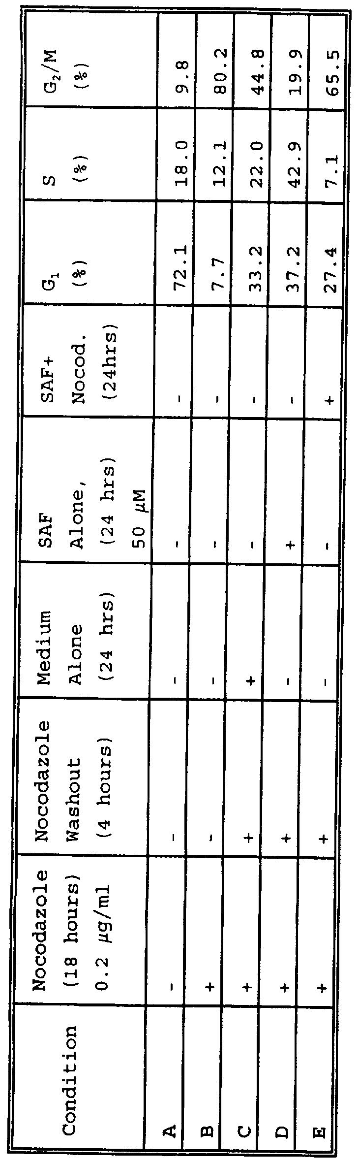
(i) Safingol effects the cell cycle by increasing the percent distribution of cells in S phase: applicants' studies indicate that safingol enhances chemotherapy induced apoptosis of gastric cancer cells in all phases of the cell cycle. However, since other PKC inhibitors, including UCN- 01, have been shown to have cell cycle specific activity, despite the induction of apoptosis throughout the cell cycle, applicants elected to determine whether safingol also effected cell cycle specific events. In order to do this applicants first performed flow cytometry studies on MKN-74 cells synchronized in G2/M with nocodazole and then treated with safingol. Applicants believed that if these studies showed a perturbation in one particular phase of the cell cycle, then applicants could focus on a cell cycle event (e.g. cdk2) associated with a particular cell cycle phase (e.g. G1/S ) . As shown in the table below, the cells were treated according to a series of different steps (noted as a "+" in the table) resulting in a series of different conditions (listed as A-E) . The cells were analyzed for their percent distribution within the cell cycle as determined by flow cytometry:
LD
As shown above, exposure of the cells to nocodazole (0.2 μg/ml) for 18 hours increased the cells remaining in G2/M from 9.8% in untreated controls (A) to 80% with the nocodazole therapy (B) , consistent with a cell cycle block. When cells were washed free of nocodazole for 4 hours and then examined at that time point by flow cytometry, the percentage of cells in G2/M had decreased from 80% to 44.8% (indicating removal of the block) , the percentage of cells in Gx had increased from 7.7% to 33.2% and the percentage of cells in S phase had increased from 12.1% to 22.0% (C) . However, if after washing out the nocodazole for four hours the cells were treated with 50 μM safingol (SAF) for an additional 24 hours, there percentage of cells increased in S increased to 42.9% (D) . This is in contrast to the cells treated with no drug during this additional 24 period. Under this condition the percentage of cells in S remained at 20.4%, suggesting that safingol induced an increase in the S phase fraction. In order to determine whether safingol was inducing an arrest of cells in G1/S the cells were treated according to these same conditions except that for the additional 24 hour period following the initial nocodazole washout, the cells were exposed to both safingol and nocodazole. Applicants anticipated that if the percent increase in S phase induced by safingol was due to cell cycle arrest then co-exposure with nocodazole would induce the cells to remain in S and not proceed into a G2/M block. As the results show (E) , reexposure of the cells to nocodazole in the presence of safingol for the additional 24 hour interval resulted in an increase in the percentage of cells in G2/M from 19.9% with safingol alone to 65.5% with safingol and nocodazole and a decrease in the percentage of cells in S phase from 42.9% with safingol alone to 7.1% with safingol and nocodazole.
These results suggest that safingol does not induce a cell
cycle arrest at Gx or S but may be affecting a regulatory event within the cell cycle that promotes the induction of apoptosis once they pass through Gα and enter S. Applicants proposed that a potential candidate for this regulatory event was cdk2.
(ii) Safingol increases cdk2 activity only when co- administered with chemotherapy: For determination of cdk2 activity MKN-74 cells were treated according to the conditions shown to enhance chemotherapy induced apoptosis:
(i) no drug (control) for 24 hours, (ii) Mitomycin-C alone
(5.0 μg/ml) for 24 hours, (iii) Safingol alone (50 μM) for
24 hours, (iv) the combination of safingol (50 μM) and
Mitomycin-C (MMC, 5.0 μg/ml) for 24 hours. Cdk2 activity was determined by histone type-I phosphorylation, as described (38) . For these studies cdk2 was first immunoprecipitated from the cell lysates following treatment under these four conditions. The kinase reaction was allowed to proceed at 30° for 30 minutes in the presence of histone type-I and [32P]ATP. For each sample a control sample was run under the same conditions, but IgG was used in place of histone Type-I. This allowed for detection on non¬ specific kinase activity which was then subtracted out from the cdk2 activity to determine the absolute radioactive incorporation attributable to cdk2. The samples were transferred to a 10% SDS PAGE gel for analysis. The autoradiographs were analyzed on a phosphoimager for determinations of relative cdk2 activity. The results are shown in figure 8
Each lane represents the following: lane A, the IgG control; lane B, no drug therapy for 24 hours; lane C, MMC alone (5.0 μg/ml) for 24 hours; lane D, safingol alone (50 μM) for 24 hours; lane E, the combination of safingol (50 μM) and MMC (5.0 μg/ml) for 24 hours. The results indicate that
untreated gastric cancer cells (lane B) have increased cdk2 activity and that both MMC (lane C) and safingol (lane D) have decreased cdk2. However, the combination of MMC and safingol increased cdk2 activity back to basal levels.
These results may explain why safingol, as a single agent, does not induce apoptosis, but for apoptosis to proceed in the presence of safingol there is an obligate requirement for concomitant exposure of the tumor cells to a chemotherapeutic agent (e.g.MMC) . Applicants propose that apoptosis takes place when at least three conditions are met : DNA damage by chemotherapy, an increase in cdk2 activity, and inhibition of PKC activity. Thus, in control cells there is an increase in cdk2 activity but no DNA damage or P C inhibition; in MMC treated cells there is DNA damage, a decrease in cdk2 activity, and no PKC inhibition (data not shown) ; and in safingol treated cells there is PKC inhibition but a decrease in cdk2 activity and no known DNA damage. However, with the combination therapy of safingol and MMC, all three conditions are met: increased cdk2 activity, DNA damage, and inhibition of PKC. Thus, apoptosis proceeds . The basis for the decrease in cdk2 activity by safingol (as well as by MMC) remains unknown and stands in contrast to the effects of UCN-01 and bryostatin as single agents. However, flavoperidol alone has been reported to inhibit the cdks (39) , including cdk2 (Edward Sausville, NCI, Bethesda, MD) , at concentrations in which applicants have observed flavopiridol to inhibit PKC activity. All these results strongly suggest that cdk2 activity may be a major marker for the apoptosis associated with PKC inhibition. The increase in cdk2 activity with safingol and MMC, indicates a potential biomarker of apoptosis that will be tested in human tissues on applicants' clinical trials (see METHODS and PLANS below) .
Safingol enhances the anti-tumor effect of cisplatin and doxorubicin in tumor-bearing animals .-
Although safingol showed no direct anti-tumor activity in vivo, when tested as a single agent in a variety of murine tumor models and human tumor xenografts, its synergism with cisplatin and doxorubicin in vitro suggested it might potentiate the antitumor efficacy of these chemotherapeutic agents in vivo. For these experiments safingol was administered alone or in combination with cisplatin or DOX to female C3H/HeJ mice bearing 16C mammary carcinoma implanted intramuscularly. Antitumor response to the agents alone or in combination was assessed by tumor growth delay. Safingol by itself at doses of 5 to 20 mg/kg had no effect on inhibiting tumor cell growth. However, when administered intraperitoneally (i.p.) at a dose of 20 mg/kg and combined with intravenous (i.v.) cisplatin (10 mg/kg) , safingol produced a modest enhancement in growth delay when compared to cisplatin alone. The administration of cisplatin under these conditions resulted in a the development of a 16/C mammary carcinoma that took 10 days to reach 5 times its starting size, whereas the combination of the same dose of cisplatin and a single dose resulted in a tumor mass that took 26 days to reach 5 times its starting size. Comparable effects against 16/C tumors were observed with safingol and DOX (10 mg/kg, i.v.) (74) .
Safingol does not induce increased toxicity of chemotherapy in animals :
From the preclinical data it appears that doses of safingol ranging from 10 to 20 mg/kg are necessary in order to achieve any level of chemopotentiation. Because safingol is being developed as a chemopotentiation agent, studies were conducted in rats and dogs to evaluate the ability of safingol to potentiate the toxicity of established cancer chemotherapeutic agents (40) . Single-dose studies with rats
in which safingol was administered prior to cisplatin, cyclophosphamide, or doxorubicin showed that safingol did not potentiate the myelotoxicity of these agents or affect reversal of the toxicity. No overt potentiation of cisplatin- ediated nephrotoxicity was observed. Cisplatin had no apparent effect on the pharmacokinetic profile of safingol. A possible effect of safingol on cisplatin pharmacokinetics was demonstrated by a statistically significant higher cisplatin Cmax value following safingol treatment, compared with the cisplatin Cmax value following vehicle administration. Other than the Cmax time point, the concentration-time profile of cisplatin was unaffected by safingol. A single-dose combination study in dogs indicated that safingol did not potentiate doxorubicin toxicity and vice versa, and that plasma concentrations of safingol or doxorubicin were also unaffected by administration of the other (Investigators brochure) .
Intravenous administration of safingol in emulsion caused clinical pathology and histopathology findings consistent with venous irritation and transient intravascular hemolysis in mice, rats, and dogs (40) . It seemed to occur as a function of safingol concentration and depended on the caliber of the vein used for delivery. Although venous irritation and hemolysis were significant and, in some instances, dose limiting toxicities in the animal studies, it was demonstrated that these effects may be mitigated by the use of low safingol concentrations and large-sized, high flow veins.
Results from the phase I clinical trial combining safingol with doxorubicin:
(i) Study design: This Phase I trial was designed as an open-label, non-randomized, dose escalation study, in which
groups of three to six patients received on day 1 of each cycle received sequentially increased dosages of intravenous safingol emulsion and then (provided there was no acute toxicity) 14 days later received the same dose of safingol one hour prior to a fixed intravenous dose of doxorubicin. Individual patients could receive up to six cycles of treatment with the same dose of safingol and doxorubicin (DOX) every 21 days, until signs of tumor progression or unacceptable toxicity occurs, or until the patient's cumulative life-time dose of doxorubicin exceeds 400 mg/m2 with an additional cycle of treatment. Prior DOX therapy was allowed up to a dose of 280 mg/m2 as long as the left ventricular ejection fraction was >50%. The starting dose of safingol was 15 mg/m2. This represented 1/10 the LD10 in mice, 4% the highest non-toxic dose (HNTD) in dogs, and 12% the HNTD in rats.
(ii) Patient characteristics: To date applicants have entered 17 patients onto the study, all of whom are evaluable. The patient characteristic are as follows: 1) median age (range) : 59 (29-77), 2) median KPS (range) : 80
(70-90) 3) Male:Female : 10:7, 4) Primary sites of disease: pancreas:6, gastric:2, colon:3, unknown primary -. 2 , sarcoma:3 , nasopharyngeal:1;
(iii) No dose limiting hematologic Toxicity: As shown in the table below with a fixed doxorubicin dose of 45 mg/m2 and escalating doses of safingol, there has been no-dose limiting hematologic toxicity. In fact the white blood count nadirs have increased with increasing safingol dose. Dose escalation above 120 mg/m2 is planned.
Hematologic Toxicity with Safingol and DOX at 45 mg/m2
Safingol # of Mean White Mean Mean
(mg/m2) patients Blood Absolute Platelet
Count Neutrophil Count
(range) Count (range) (range)
15 3 3.2 (1.4- 1.2 (0.8- 150 (121-
5.0) 1.8) 189)
30 4 4.6 (1.1- 2.3 (0.4- 187 (37-
9.0) 4.0) 349)
60 3 4.0 (2.2- 2.3 (0.7- 154 (70-
6.1) 4.7) 233)
120 3 5.4 (4.9- 5.2 (2.3- 283 (225-
7.5) 5.1) 279)
b) No dose-limiting hemolysis: Thus far only one patient has experienced a grade I hemolysis as evidenced by a >50% decrease in serum haptoglobin. This was associated with no changes in reticulocyte count, no evidence of hemolysis on the peripheral smear, no "pink" discoloration to the plasma, and no evidence or urine hemosiderin. This was believed to be related to relatively poor venous access which only became apparent on the first day of therapy. The patient was retreated according to applicants' protocol criteria for grade I hemolysis which included infusion of safingol through a central vein and at one half the concentration
(twice the total volume but the same total dose) . Under these conditions the patient has experienced no further evidence of hemolysis.
(iii) Safingol pharmacokinetics appear linear with increasing dose: The pharmacokinetics of patients treated with safingol and DOX at 45 mg/m2 is shown below:
Mean Plasma Safingol Pharmacokinetics with 45 mg/m2 DOX
Dose Cmax, AUC. (ng x AUC. (ng x Level, ng/ml hr/mL) hr/mL) mg/m2 without DOX without DOX
15 (n=3) 199 168 161
30 (n=4) 336 501 428
60 (n=3) 422 531 501
120 (n=3) 988 1227 1226
It appears that with the safingol dose escalation the increases in plasma safingol levels is generally linear with increasing safingol dose. in addition, DOX did not change the pharmacokinetics of safingol.
Based on the mice studies applicants have predicted that in order to achieve meaningful chemopotentiation with either DOX or cisplatin applicants need to deliver the equivalent mouse dose of 5 mg/kg to 20 mg/kg safingol. From the preclinical pharmacology in the mice this translates into a Cmax and AUC of 1,797 ng/ml and 668 ng x hr/ml respectively for 5 mg/kg safingol and a Cmax and AUC of 7,021 ng/ml and 2,702 ng x hr/ml respectively for 20 mg/kg safingol. From the pharmacology data obtained thus far from the patients treated with escalating doses of safingol and 45 mg/m2 of DOX, it appears that applicants are approaching these target levels. In view of this and the fact that applicants are still significantly below the predicted pharmacological toxicity levels, applicants continue to accrue patients to the study.
METHODS and PLAN.
To develop an integrated clinical program with PKC inhibitors in combination with chemotherapy that uses laboratory correlates (PKC activity, cdk2 activity, and terminal deoxynucleotidyl transferase (TdT) activity in tumor tissues and leukocytes) as putative surrogate end- points of activity. The immediate goal will be to complete two current phase I clinical trials with the PKC specific inhibitor L-threo-dihydrosphingosine (safingol) in combination with (i) doxorubicin (DOX) and (ii) cisplatin with the plan to take this to a phase II study in gastric cancer; and to (iii) initiate additional clinical trials with combinations of chemotherapy and other PKC inhibitors including UCN-01, flavopiridol, and bryostatin.
Applicants now have two open MSKCC IRB-approved phase I studies with safingol. The first combination study with doxorubicin (see PRELIMINARY RESULTS) will close once applicants reach the MTD. Based on the pre-clinical data in gastric cancer cells a phase II study of safingol and doxorubicin will then be planned. In the interim the second study with safingol and cisplatin is soon to open. This study has been approved for funding as an administrative supplement by the NCI to the MSKCC P30 CA 08748-30. The study design and plan are as follows:
(i) To complete the phase I trial of safingol and doxorubicin.
(ii) To initiate a phase I study of safingol and cisplatin:
Study design: This Phase I trial is designed as an open- label, non-randomized, dose escalation study, in which groups of three to six patients will receive sequentially
increased dosages of intravenous safingol emulsion, one hour prior to a fixed, 75 mg/m2 intravenous dose of cisplatin, until dose limiting toxicity is demonstrated in at least three of six patients. Based on the current phase I study of safingol and doxorubicin, the starting dose for safingol in this new combination trial with cisplatin will be 60 mg/m2. The dose of safingol will be fixed within each co-hort. Individual patients will receive additional cycles of treatment with the same dose of safingol and cisplatin every 21 days, until signs of tumor progression or unacceptable toxicity occurs.
iii) To initiate additional clinical trials with other PKC inhibitors including UCN-01. flavopiridol, and bryostatin.
a) A combination study of UCN-01 and Carboplatin: Based on applicants' pre-clinical data with UCN-01 as a PKC inhibitor that enhances the induction of chemotherapy induced apoptosis. The proposal for this study is that UCN-01 will be given on day 1 as a 24 hour infusion and carboplatin be given as 300 mg/m2 bolus six hours after initiating UCN-01. UCN-01 will be escalated in subsequent co-horts but the dose of carboplatin will remain fixed. This treatment will be repeated every 28 days.
b) Future combination trials of PKC inhibitors (i.e bryostatin) and chemotherapy: For the future studies utilizing PKC inhibitors in combination with chemotherapy, applicants will use a general approach, as outlined below, to their development and conduct. These points, including biostatistical considerations, are also applicable to all the PKC inhibitory studies now in progress or proposed.
Patient eligibility and verification of eligibility: Patients will be selected from applicants' Department of
- 62 -
Medicine populations. For general Phase I studies, all types of cancer will be included including gastric cancer, other gastrointestinal cancer, non-small cell lung cancer, refractory breast cancer, renal cell cancer, melanoma, gynecologic neoplasms, and sarcomas. This type of design allows us to do Phase I testing of highly specific new agents and utilizes the great depth of the MSKCC patient population. Memorial Sloan-Kettering Cancer Center has filed form HHS 441 (Re: Civil Rights) , form HHS 641 (Re: Handicapped individuals) and form 639A (re: sex discrimination) . In selecting patients for study in the proposed contract work, due notice is taken of the NIH/ ADAMHA Policy concerning women and minorities in clinical research populations. The study population will be fully representative of the whole range of patients seen at Memorial Hospital. Unless a disease is gender specific, no eligibility limitations will be employed regarding age, gender, childbearing potential, race or ethnic origin.
Applicants' past record indicates a commitment to entering patients relatively early in the course of basically incurable advanced neoplasms (e.g., colorectal, renal cells, non small cell lung cancer) . As no systemic therapy offers substantial benefit for such patients, applicants feel that Phase I therapy - especially studies to evaluate combinations seeking to enhance the activity of active drugs
(e.g., MMC and Carboplatin) is the best therapy for many patients with these diseases. Measurable or evaluable tumor masses or parameters, while desirable, will not be required for eligibility.
Protocol Design: All Phase I PKC studies will be performed within the context of a specific written protocol. Each individual protocol will be initially designed after receipt of complete preclinical efficacy, toxicologic and
pharmacologic information and discussions with NCI staff. A biostatistician (Ying Huang, Ph.D) is a member of the PKC Phase I Group and provides statistical methodology when it is necessary to deviate from the standard dose escalations. Particular attention will be given to blending applicants' Center's expertise with the needs of the NCI Phase I program. Applicants envision continued efforts toward developing combination therapy programs designed to modulate cytotoxicity and overcome resistance, using PKC as a novel drug target.
Initial Drug Dosage: In combined Phase I trials initial dose schedules will utilize known pharmacologic data on each drug, prior clinical experience using each drug individually (if available) and the biochemical rational for combining the drugs. Whenever possible, basic laboratory data will be used to determine sequence, schedule, modes of administration, target plasma levels, etc. However, applicants remain cognizant that modulating agents may not only alter the duration and intensity of the toxic effects of the anticancer agent they are paired with but the modulating agents themselves may have toxicities which must be taken into account in the phase I design.
Dose Escalation: The scheme of dose escalation is a key feature of Phase I trials. Historically, a traditionally empiric dose escalation has been employed with 3 patients in each dose cohort. For such empiric designs at MSKCC, the following scheme will be utilized: N, 2N, 4N, 8N; then escalation by increments of 30%. If any toxicity > grade 2
(except nausea and vomiting > grade 3) is seen early in the escalation, 30% increments of drug escalation will be begun from that level causing > grade 2 toxicity. Studies which combine modulators with chemotherapy must be designed differently than traditional Phase I studies. The design of
these trials depends heavily on the support of laboratory investigators to establish target of modulation and to monitor the effects. For modulating agents, the goal is to administer the minimum dose of modulator which consistently results in the desired modulatory effects. In general, for the PKC inhibitor being escalated, applicants will choose a starting dose which is known to be without toxicity in previous human experience and will escalate from this dose in a conservative manner while maintaining the dose of the other agents constant.
Pharmacokinetics: Whenever possible, pharmacokinetics will be performed on both the modulator and the chemotherapeutic agent to establish the appropriate schedules for optimal combination. The biological endpoint must be ascertained for each patient to establish the appropriate modulator dose. Then, the chemotherapy dose must be escalated to identify the alterations in toxicity. In parallel, pharmacokinetics of both agents must be monitored to insure that drug-drug interactions do not materially alter the pharmacokinetic behavior of the two agents. Careful attention will be paid to the biostatistical design and analysis of the trial in order to discriminate the effects of each of the individual agents from those related to drug- drug interaction. Applicants believe that it is important to incorporate pharmacokinetic studies into applicants' Phase I trials . Applicants will perform pharmacokinetic analysis on 2-3 patients at the first level of a trial and at each subsequent level (assuming that the drug is detectable at the early levels) . If the mouse pharmacokinetics are known (i.e., mouse LD10 AUC) and if the pharmacokinetics of the drug can be considered linear, then a prediction of the "target" AUC at the human MTD can be made based on the mouse AUC, and the escalations necessary to reach the projected MTD can be assessed. Continued
pharmacokinetic analysis during the course of the trial will allow us to monitor progress toward the MTD and allow us to carefully monitor the levels near the projected MTD. If applicants find that the MTD estimated by the AUC analysis will occur before level 4, applicants can amend applicants' protocol to approach the projected MTD more slowly, escalating by increments of 30%.
Biostatistical considerations: At least 3 study drug-naive patients at each level shall be necessary to fully evaluate that dose level. When a dose level with any patient experiencing Grade 3 or greater drug-related toxicity has been identified. , at least 6 patients at that dose level will be required to more fully evaluate the nature of that toxicity. The MTD will be defined as that dose which produces reversible >_ grade 3 toxicity, exclusive of nausea and vomiting, in 2/6 patients treated.
The probability of dose escalation based on the true incidence of dose limiting toxicity at a specific dose level is shown in the table below.
True Toxicity risk 0.10 0.20 0.30 0.40 0.50 0.60 Probability of Escalation 0.91 0.71 0.49 0.31 0.17 0.08
Thus, the risk of escalation in high toxicity risk dose levels is less tnan 50% for true toxicity rates below 0.30.
One of the principal objectives of Phase I studies will be determination of the maximum tolerated dose schedule. As outlined above, the treatment dose will be increased after three patients have been treated at a given dose with no toxicity and further testing will take place at doses with toxicity seen in the first three patients. Because of the iterative nature of the process to determine MTD, the
confidence limit around the true toxicity rate will be based on additional patients tested at the MTD. The following table gives the 90% confidence limits for the true toxicity rate as a function of the number of additional patients treated at the MTD and the number that experience toxicity.
#Additional # with
Patients Toxici'ty, at the dose
0 1 2 3 4 5 6
6 .30 .58 .73 .85 .93 .99
0 .009 .06 .15 .27 .42
8 .31 .47 .60 .71 .81 .89 .95
0 .006 .05 .11 .19 .29 .40
10 .26 .39 .51 .61 .70 .78 .85
0 .005 .04 .09 .15 .22 .30
12 .22 .34 .44 .53 .61 .68 .75
0 .004 .03 .07 .12 .18 .24
Thus, if 10 patients are treated at the MTD, and if for instance 2 patients have toxicity, there is a 90% chance that the true toxicity rate is between 4 and 51%. It is anticipated that most studies will require the accumulation of data on approximately 20-30 adult patients.
(iii) To perform correlative laboratory studies: Studies from applicants' laboratory with safingol, as well as studies utilizing other PKC inhibitors, indicate that the critical event for enhancement of chemotherapy induced apoptosis by PKC inhibition takes place at the G1/S interphase and that this is associated with increased cdk2
activity. Therefore, applicants' plan is to measure cdk2 activity, PKC activity, and apoptosis by TdT cn tumor tissues and leukocytes obtained from patients on the clinical trials. For these studies applicants will obtain serial tumor biopsies and peripheral lymphocytes from all patients on the studies at three time points: a baseline before therapy, following treatment with the PKC inhibitor alone, and following treatment with the PKC inhibitor and chemotherapy. Lymphocytes will be obtained from peripheral blood by density gradient centrifugation over Ficoll, as described (41) . Tumor cells will be carefully dissected from the biopsies so as to avoid contamination with normal tissue. When available tumor cells will be obtained from ascitic fluid with passage through a 30 μm nylon mesh, as described (42) .
Cdk2 assays : For cdk2 assays tissues and cells will be lysed in Lysis Buffer containing 0.1% Tween-20, PMSF (0.2 mM) , aprotinin (10 μg/ml) , and leupeptin (10 μg/ml) . Immunoprecipitation will be performed with 200 μg of protein lysate for each kinase reaction (38) . Following incubation for 1 hour on ice with the cdk2 antibody the mixture will be equilibrated with protein A-beads (20-25 μl per immunoprecipitate) . This was then rocked for 45-60 minutes in the cold room. The beads will be washed with Lysis Buffer and Hepes-KOH Kinase Buffer containing 50 mM beta- glycerophosphate. The kinase reaction mixture will contain 0.2 μg of histone type-I, 300 μM ATP, the kinase buffer, and [32P]ATP. For each sample a control sample will be run with the same reaction mixture, but IgG will be used in place of histone Type-I. This allows for detection on non-specific kinase activity which is then subtracted out from the cdk2 activity to determine the absolute radioactive incorporation attributable to cdk2 activity. Each reaction will be allowed to proceed at 30° for 30 minute and then the samples
will be transferred to a 10% SDS PAGE gel for analysis.
PKC assays: For these studies tissues and cells will be homogenized with gentle ultrasonication. PKC assays will be performed as described (37) , but myelin basic protein will be used as a substrate. Briefly, PKC will be extracted from the gastric cancer cells as described (21,61) using a 20 mM Tris-HCL (pH 7.5) buffer containing 2 mM EDTA, 2 mM EGTA, 5 mM β-mercaptoethanol, 0.5 mM phenylmethylsulphonyl fluoride, 10 ug/ml leupeptin, and 0.25 M sucrose. Cells or tissues will be homogenized with brief sonication. The cell homogenates will then centrifuged at 100,000 xg for 1 hour at 4° C. The supernatant represents the cytosolic fraction and the pellet the membrane fractions. The pellet is resolubilized in the original Tris-HCl buffer. Both fractions are then assayed for PKC activity for 15 minutes at 25°C by determining the transfer of γ-phosphate group of adenosine-5' -triphosphate to myelin basic protein (Gibco Laboratory) in a 50 mM Tris-HCl (pH 7.5) buffer containing 8 mM % L-α-phosphatidylserine, 24 ug/ml PMA, and 12 mM calcium acetate. The reaction mixture is then transferred to Whatman P-81 paper for liquid scintillation counting.
TdT assays: Fresh tissue will be fixed in 10% neutral buffered formalin in a coplin jar for 10 minutes at room temperature and then fixed with ethanol acetatic acid. For paraffin-embedded tissues, samples will first be deparaffinized with ethanol and then protein digested with Proteinase K. For the TdT assays, the ApopTag Kit (Oncor, Gaithersburg, MD) is used (5,43) . This method employs a fluoresceinated antidigoxigenin antibody directed against nucleotides of digoxigenin-11-dUTP (d-dUTP) which are catalytically added to the 3-OH ends of fragmented DNA by TdT. Briefly, fixed sections are washed and fixed with 1%
paraformaldehyde. The fixed cells are incubated in a reaction mixture containing TdT and d-dUTP for 30 minutes at 37°C. Stop/wash buffer is added, and the cells are resuspended in 100 μl of fluorescinated anti-digoxigenin antibody for 30 minutes at room temperature. The slides are then washed with 0.1% Triton X-100 before they examined with a Olympus BH-2 fluorescence microscope equipped with a BH2- DM2U2UV Dich. Mirror Cube filter (Olympus, Lake Success, NY) and scored for fluorescence. For positive controls a cytospsin preparation on a silanized slide of human peripheral lymphocytes stimulated with lμM dexamethasone will be used. For negative controls sham staining will be performed substituting distilled water for TdT.
In order to exclude an effect from chemotherapy alone, this assays will be repeated in the final co-hort of patients one level below the MTD. Three patients will first be treated with chemothrapy (i,e cisplatin) alone for cycle 1, followed 28 days later by the combination of safingol and cisplatin for cycle 2. Three other patients will first be treated with the combination of safingol plus cisplatin for cycle 1, followed 28 days later by the cisplatin alone for cycle The cellular responses will be determined at each of these dosing visits. After completion of the proposed phase I study applicants will examine the results of these assays to see whether an association exists between a change in these biological assays and clinical response and/or toxicity in the patients treated. The Wilcoxon rank-sum test will be employed to compare responders to non-responders (or toxicity vs. no toxicity) in order to test the validity of any possible associations observed in this patient population.
To perform laboratory studies with human gastric, breast, and ovary cancer cells to: i) study a variety of new PKC
inhibitors currently under pre-clinical development as inducers of apoptosis in combination with chemotherapy; ii) define the optimal conditions in which to combine safingol, UCN-01, flavoperidol, or bryostatin with chemotherapy in order to achieve the maximum induction of apoptosis for the purpose of clinical trial development; and iii) further examine in vitro cdk2 and to identify other related cell cycle dependent proteins associated with induction of apoptosis and the inhibition of PKC;
(i) To evaluate a variety of new PKC inhibitors in combination with chemotherapy to determine their effect on induction of apoptosis: Selective PKC inhibitors other than safingol and UCN-01 may induce even greater degrees of apoptosis when combined with chemotherapy against breast (MDA-MB-468) , gastric (SK-GT and MKN-74) and ovarian (OVCAR) cancer cells. The other PKC inhibitors to be tested for the induction of apoptosis against the cell lines including a series of Roche inhibitors (to be supplied by Roche Pharmaceuticals, Nutley, NJ) , and bryostatin 1 (36) , an agent that activates PKC with short exposure but inhibits
PKC following prolonged drug exposure by inducing PKC degradation. Apoptosis will be quantitated by two methods:
(1) QFM staining: This method involves staining with bisbenzimide trihydrochloride (Hoescht-33258) of condensed chromatin which characterizes the cells undergoing apoptosis (3,5) and (ii) terminal deoxynucleotidyl transferase (TdT) assay which labels the 3' -OH ends of DNA fragments in apoptotic cells (5,43) .
For QFM determinations, the cells are fixed in 3% paraformaldehyde and incubated at room temperature for 10 minutes. The fixative is removed and the cells are washed with IX PBS, resuspended in 20 μl IX PBS containing only 8 μg/ml of bisbenzimide trihydrochloride (Hoechst #33258) , and
incubated at room temperature for 15 minutes. Aliquots of the cells (lOμl) are placed on glass slides coated with 3- amino-propyl-triethoxysilane, and duplicate samples of 500 cells each were counted and scored for the incidence of apoptotic chromatin condensation using an Olympus BH-2 fluorescence microscope equipped with a BH2-DM2U2UV Dich. Mirror Cube filter (Olympus, Lake Success, NY) .
For the TdT assays, the ApopTag Kit (Oncor, Gaithersburg, MD) will be used. This method will essentially be identical to the one described above for tissue and lymphocyte samples from patients except that after washing with 0.1% Triton X- 100 the cells will be counterstained with propidium iodide (PI) solution. Green (d-dUTP labeled DNA strand breaks) and red (PI staining for total DNA content) fluorescence of individual cells are measured on a FACScan flow cytometer
(Becton Dickinson, San Jose, CA) . The resulting bivariate plots enables the detection of apoptotic events within the cell cycle. The Rλ cursor is set using the control specimen to define normal levels of green fluorescence (i.e., basal levels of apoptosis) . Cells with fluorescence above the Rλ cursor are considered apoptotic. The data from 10,000 cells are collected and analyzed using CellFit and LYSYS software (Becton Dickinson) .
(ii) To define the optimal conditions in which to combine safingol, UCN-01, flavoperidol, and bryostatin with chemotherapy for future clinical development: Safingol, UCN-01, and flavoperidol are now in phase I clinical trials. Combining these agents with chemotherapeutic agents appears to be the best way to maximize the beneficial effects of these drugs as anti-cancer agents against breast cancer cells. However, the optimal way in which to achieve this end remains to be defined. Applicants plan to pursue this issue with a series of studies:
The optimal chemotherapeutic agent (s) : The plan is to perform experiments with safingol and UCN-01 in combination with other conventional chemotherapeutic agents such as cis¬ platin, Taxol, 5-fluorouracil and doxorubicin in order to determine the extent to which these agents induce apoptosis with these other agents in the breast (MDA-MB-468) , gastric (SK-GT and MKN-74) and ovarian (OVCAR) cells. The methods to quantitate apoptosis will be by QFM and TdT assays, as described above.
Combinations of PKC inhibitors: Since many of these inhibitors act at different sites of the enzyme (regulatory vs. catalytic), it may be possible to combine multiple PKC inhibitors together with chemotherapy in order to further enhance the induction of apoptosis. For example, as applicants have shown, the combination of UCN-01 and safingol with MMC at the conditions described above induce a greater degree of apoptosis than treatment either PKC inhibitor alone with MMC. Further studies combining flavoperidol with safingol or with UCN-01 are planned and apoptosis will be quantitated by the QFM and the TdT technique.
The optimal sequence and timing of the PKC inhibitors with the chemotherapeutic agent: The exact sequence and timing of the PKC inhibitor relative to chemotherapy exposure in breast cancer cell remains unknown. For these experiments applicants propose exposing the breast and gastric cancer cells to safingol (50 μM) for different time intervals (30 minutes, 1 hour, 3 hours, 6 hours, 18 hours, and 24 hours) , washing the cells free of drug, and then exposing to MMC 5 μg/ml) for 24 hours. The relative induction of apoptosis will then be quantitated as above. Conversely, cells will first be exposed to safingol for 24 hours, but the MMC exposure will now be limited to different time intervals
before being assayed for apoptosis.
(iii) To examine in vitro cdk2 and to identify other related cell cycle dependent proteins associated with the induction of apoptosis and the inhibition of PKC: The basis for the increase in cdk2 activity by safingol and MMC in combination remains unknown. It may be related to a decrease in the expression of a cdk2 inhibitor (i.e. p21) or it may be related to a modification of a phosphorylated site on cdk2 which results in its activation. A further understanding of this process may lead to new surrogate markers of activity. In order to examine this further applicants plan to examine: 1) protein expression of p2l and p27 with immunoblotting using an enhanced chemiluminescence system using specific antibodies (Santa Cruz Biotechnology, Inc. Santa Cruz, CA) ; 2) differences in protein phosphorylation of cdk2 with [32P] orthophosphate cell labelling, as previously described, using a cdk2 specific antibody suitable for immunoprecipitating (36) ; and 3) correlative studies of cdk2 activity as measured by the histone HI kinase assays (38) . Control cells will be treated in the same way as described above except that standard media without drug will be used for all incubations.
To determine whether a specific PKC isoform may be a critical target for drug development in induction of apoptosis with chemotherapy by: (i) testing whether antisense for PKCcv in combination with MMC has superior anti-tumor activity against human gastric cancer cells when compared to either agent alone or to a PKC missense with MMC; (ii) testing other PKC antisense against other PKC specific isoforms in combination with chemotherapy; (iii) studying in gastric cancer cells whether any of the PKC isoforms are being preferentially inhibited during the induction of apoptosis.
(i) Testing whether antisense for PKCcc in combination with MMC has superior anti-tumor activity against human gastric cancer cells when compared to either agent alone or to a PKC missense with MMC: applicants' results indicate that PKC represents a novel target for enhancing the induction of apoptosis by chemotherapy. However, PKC is ubiquitous in both normal and malignant tissue. Even though safingol's specificity represents a considerable advance in the development of PKC inhibitors especially as they pertain to eventual clinical development, even further specificity would still provide theoretical advantages in drug development .
PKC comprises a multigene family, consisting of at least 13 distinct genes which have 4 large constant regions separated by 5 variable regions (18,44) . A current concept is that the variable regions of these 13 PKC isoforms encode isozymes-specific properties (45) . These 13 PKC isozymes can be grouped into three categories based on Ca2+ requirements for activation and phorbol ester binding activity. PKC is also the major cellular receptor for tumor promoting phorbol esters which, as analogues of DAG, directly activate PKC (19) . These isoforms are designated , βl , β2, γ, δ, e, η , θ , μ , ζ , λ, and T. The biological significance of PKC heterogeneity is unknown, but it is speculated that, since they show differential tissue expression, specific PKC isoforms will phosphorylate different substrates and mediate distinct and different biological responses in the cells in which they are expressed. The role of these different isoforms is essentially unknown. However, identifying the isoform(s) involved in apoptosis would provide a more specific target for drug therapy.
Recent data have implicated PKCα in apoptosis. Studies have
shown that if PKCα is inactivated then apoptosis can proceed. Most attempts to develop isoforms specific PKC inhibitors have been unsuccessful. However, some inroads have been made with PKCα by testing of antisense oligonucleotides. A 20 mer has been developed by Isis
Pharmaceuticals (Carlsbad, CA 92008) which specificly inhibits the expression of human PKCα without affecting the expression of other PKC isoforms. It has been shown to inhibit the growth of subcutaneously implanted human tumor xenografts (46) . This inhibition was dose-dependent when administered intravenously (i.v.) or intraperitoneally
(i.p.) between 0.06 and 6 mg/ml/.
Applicants' results indicate that the best way to maximize the therapeutic benefits of PKC inhibitors is to combine them with chemotherapy. Applicants have previously reported that all the SK-GT cell lines express PKCa mRNA and protein (15,47) . Therefore, these cell lines appear to be appropriate for the evaluation of a PKCαr anti-sense. The goal of this study is to test the combination of the PKCα anti-sense in combination with MMC for the first time in an in vivo system. The PKC inhibitor for these studies will be a 20 mer PKCcx antisense. The dose and schedule (10 mg/kg, daily, i.p.) of the anti-sense in the vivo study will be based on preclinical toxicology, which has been generated by ISIS pharmaceutical. The dose of appears to be a safe and well-tolerated dose when administered both i.p. or i.v. to tumor bearing nude mice. The anti-sense easily dissolves in saline. Doses of 100 mg/kg have been given to mice on a daily schedule with no major toxicity and no deaths. At the highest non-toxic dose (100 mg/kg) mild lymphocytic infiltration has been noted in the liver, spleen and kidney. In order to determine the specificity of this response, a missense 20 mer will also be administered to two of the control groups. This will ensure that the observed response
- 76 - is secondary to the effect of the PKCα anti-sense and not due to a non-specific effect of the anti-sense therapy. The missense selected has also been safely administered at 10 m9/kg i.p to mice on a daily schedule without significant toxicity. The dose of MMC selected for these studies will be 14.5 mg/kg in saline. This dose has been safely administered to nude mice and has also been safely administered to nude mice when combined with the PKC inhibitor UCN-01 (31) .
The animal protocol (MSKCC IACUC #95-07-027) design will be as follows: All animals with be injected subcutaneously into the hind quarter on day 1 with 5 x 10⁶ SK-GT-2 cells. Applicants' preclinical; data indicates that within 10-12 days these animals develop a palpable 0.25 gr. tumor mass. Once the animals have a palpable tumor, they will be randomly assigned to one of 4 treatment' arms (17 animals in each arm) :
(1) PKCo; anti-sense: by itself at a dose of 10 mg/kg/day i.p. daily for four weeks,
(2) PKCα anti-sense + MMC: PKC anti-sense will be given at a dose of 10 mg/kg i.p daily for two weeks. On the first day of week #3 MMC will be given as a single i.v. dose (14.5 mg/kg) . PKC anti-sense will be repeated i.p daily for an additional two weeks (i.e. weeks #3 and 4) . (3) MMC alone: Daily i.p injections of a saline control for two weeks. On the first day of week #3 a single dose of Mitomycin-C well be administered (14.5 mg/kg) i.v.. This will be followed by two additional weeks of i.p. saline control.
(4) PKCα missense: by itself at a dose of 10 mg/kg/day i.p. daily for four weeks.
Assuming a "non-treatment" response rate of 5% (i.e. antisense alone or Mitomycin-C alone) relative to the
control animals (missense alone) , then in order to detect a 50% decrease in size of the palpable tumor mass for the treated group (i.e antisense + Mitomycin-C) using a one sided alternative normal approximation z-test with a significance of 0.05 and a power of 80%, the total number of animals for each group needs to be 17. The total number of animals will be 64.
(ii) Testing other PKC antisense against other PKC specific isoforms in combination with chemotherapy: If anti-sense for PKCα does not show an enhancement of MMC-induced apoptosis, then anti-sense against the other PKC isoforms will be tested according to the same study design. The materials for these PKC specific anti-sense studies will be supplied by Isis Pharmaceuticals (Carlsbad, CA) , as become availabl .
(iii) Studying in gastric cancer cells whether any of the PKC isoforms are being preferentially inhibited during the induction of apoptosis: Even though safingol is not PKC isoform specific, in regard to the induction of apoptosis safingol may still be inhibiting one isoform preferentially over another. One way to study this is to examine the relative distribution of the PKC isoforms between the membrane and the cytosolic fractions of the cells. PKC isoforms become activated when they are translocated from the cytosol to the membrane of the cell (48) . Conversely, inactivation of these isoforms is associated with depletion of the isoform(s) from the membrane component of the cells. In order to test the influence of isoform distribution on chemotherapy induced apoptosis, applicants propose treating the cell lines with the four conditions that applicants have used in applicants' prior studies: (i) no drug (control) for 24 hours, (ii) Safingol alone (50 μM) for 24 hours, (iii) Mitomycin-C alone (5 μg/ml) for 24 hours, (iv) the
combination of safingol (50 μM) and Mitomycin-C (5.0 μg/ml) for 24 hours. Since the distribution of the PKC isoforms between the cytosolic and membrane fraction of the cells appears to be the critical event for their relative activation, applicants will obtain cytosolic and membrane fractions, as described (13) , to determine whether there are differences in isoform expression and localization with these different drug treatments that applicants have shown induce apoptosis. Briefly, PKC will be extracted from the gastric cancer cells, using a 20 mM Tris-HCL (pH 7.5) buffer containing 2 mM EDTA, 2 mM EGTA, 5 mM /S-mercaptoethanol, 0.5 mM phenylmethylsulphonyl fluoride, 10 ug/ml leupeptin, and 0.25 M sucrose (Buffer A) . Cells will be homogenized and th homogenates will then be centrifuged at 100,000 xg for 1 hour at 4° C. The supernatant represents the cytosolic fraction and the pellet which is resolubilized in buffer A containing 0.1% Triton-X represents the membrane fraction. These fractions are then passed through a DEAE-52 column and the PKC protein is eluted off the column with Buffer A containing 0.15 M NaCl. Briefly, cells are lysed following drug treatment with lysis buffers containing protease and phosphatase inhibitors. Proteins are resolved by SDS-PAGE electrophoresis. Immunoblots are prepared with Immobilon P membranes by electrophoretic transfer and blocked in TN- Tween buffer (50 mM Tris, pH 7.5, 200 mM NaCl, 0.1% Tween- 20) containing 5% (w/v) nonfat dry milk. After washing the blots are incubated with horseradish peroxidase-conjugated secondary antibodies (dilution 1/2000. Immunologically reactive protein will be visualized with the enhanced chemi- luminescence system (Amersham, Arlington Heights, IL) . using PKC isoform specific antibodies (49) (Santa Cruz Biotechnology, Inc. Santa Cruz, CA) .
Reference of the Second Series Experiment
1. Lowe SW, Ruley HE, Jack T, Housman DE. p53- Dependent apoptosis modulates the cytotoxicity of anticancer agents. Cell 74: 957-967, 1993.
2. Evans DL and Dive C. Effects of cisplatin on the induction of apoptosis in proliferating hepatoma cells and nonproliferating immature thymocytes. Cancer Res . 53: 2133-2139, 1993.
3. Haimovitz-Friedman A, Chu-Cheng K, Ehleiter D, Persaud RS et al . Ionizing radiation acts on cellular membranes to generate ceramide and initiate apoptosis. J. Exp. Med. 180: 525-535,
1994.
4. Jarvis WD, Kolesnick RN, Fornari F, Traylor RS, Gewirtz DA and Grant S. Induction of apoptotic DNA damage and cell death by activation of the sphingomyelin pathway. Proc. Natl. Acad. Sci. USA 91:73-77, 1994.
5. Schwartz GK, Haimovitz-Friedman A, Dhupar SK, Ehleiter D, Maslak P, Loganzo F, Kelsen DP, Fuks Z,
Albino AP. Potentiation of apoptosis by treatment with the protein kinase C specific inhibitor safingol in mitomycin-c treated gastric cancer cells. J. Natl. Cancer Institute 87(18) : 1394-1399, 1995.
6. Jarvis WD, Fornari FA, Browning JL, Gewirtz DA, Kolesnick RN, and Grant S. Attenuation of ceramide-induced apoptosis by diglyceride and pharmacological activators of protein kinase C in
human myeloid leukemia cells. J. Biol. Chem. 268:31685-31692, 1994.
7. Jarvis WD, Turner AJ, Povirk LF, Traylor RS, and Grant S. Induction of apoptotic DNA fragmentation and cell death in HL-60 human promyelocytic leukemia cells has been reported for pharmacological inhibitors of protein kinase C. Cancer Res . 54: 1707-1714, 1994.
8. Wingo PA, Tong T, and Bolden S. Cancer Statistics, 1995. CA 45:19-36,1995.
9. Blot WJ, Devesa SS, Kneller RW, Fraumeni JF, Jr. Rising incidence of adenocarcinoma of the esophagus and gastric cardia. JAMA265 :1287-91, 1991.
10. Kelsen D, Atiq O.Therapy of upper gastrointestinal tract cancers. Curr. Probl. Cancer:239-294. 1991.
11. Goldstein LJ, Galski H, Fojo A, Willingham M, Lai S-L, Gazdar. Expression of a multidrug resistance in human cancers. J. Natl. Cancer Inst. 81:116-24, 1989.
12. Altorki N, Schwartz GK, Blundell M., Davis D, Kelsen D, and Albino A. Characterization of cell lines established from human gastric-esophageal adenocarcinomas: Biologic phenotype and invasion potential. Cancer 72: 649 -657, 1993
13. Schwartz GK, Wang H, Lampin N, Altorki N, Kelsen D, Albino A. Defining the invasive phenotype of proximal gastric cancer cells. Cancer, 73: 22- 27,1994.
14. Nabeya Y, Loganzo F, Maslak P, Lai L, de Oliveira A, Schwartz GK. et al. The mutational status of p53 protein in gastric and esophageal adenocarcinoma cell lines predicts sensitivity to chemotherapeutic agents. Int .
15. Schwartz GK, Jiang J, Kelsen D, and Albino AP. Protein kinase C: a novel target for inhibiting gastric cancer cell invasion. J. Natl. Cancer Inst. 85: 402-407, 1993.
16. Matozaki T, Sakamoto C, Matsuda K, Suzuki T, Konda Y, Nakono O et al . Mis-sense mutations and a deletion of the p53 gene in human gastric cancer. Bioch. Biophys.. Res. Comm 182: 215-223, 1992.
17. Tamaoki, T. and Nakano, H. Potent and specific inhibitors of protein kinase C or microbial origin. Biotech.. 8: 732-735, 1990.
18 . Kikkawa , U . , Kishimoto, A . , and Nishizuka , Y . The protein kinase C family : heterogeneity and its implications . Annu . Rev . Biochem . , 58 : 31 -44 , 1989 .
19. Bell, R.M., and Burns, D.J. Lipid activation of protein kinase C. J.Biol . Chem. , 266: 4661-4664, 1991.
20. Nakano, H. , Kobayashi, E., Takahashi, I., Tamaoki, T., Kuzuu, Y, and Iba, H. Staurosporine inhibits tyrosine-specifid protein kinase activity of Rous sarcoma visus transforming protein. J.Antibiot . (Tokyo) . 40: 706-708, 1987.
21. Takahashi I, Saitoh Y, Yoshida M, Sano H, et al . UCN-01 and UCN-02, new selective inhibitors of protein kinase C. J. Antibiot. 42: 571-576, 1989.
22. Birchall AM, Bishop J, Bradshaw D, Cline A, Coffey J. , Elliott LH, et al . RO 32-0432, a selective and orally active inhibitor of protein kinase C prevents T-cell activation. J. Pharmacol . Exp. Therapeutics 26:922-929, 1994.
23. Kraft AS, Smith JB, and Berkow RL. Bryostatin, an activator of calcium phospholipid-dependent protein kinase, blocks phorbol ester-induced differentiation of human promyelocytic HL-60 cells. Proc. Natl. Acad. Sci. USA 83:1334-1338,1986.
24. Kaur G, Stelter-Stevenson M, Sebers S, Worland P, Sedlack H, Myers C, Czech J, Naik R, Sausville E. Growth inhibition with reversible cell cycle arrest of carcinoma cells by flavone L86-8275. J. Natl.
Cancer Inst. 84: 1736-1740, 1992.
25. Hannun, Y.A., Loomis, C.R., Merrill, A.H. , Jr., and Bell, R.M. Sphingosine inhibition of protein kinase C activity and of phorbol dibutyrate binding in vitro and in human platelets. J.Biol.Chem. , 261: 12602-12609, 1986.
26. Adams LM, Dykes D, Harrison SD, Saleh J, and Saah L. Combined effect of the che opotentiator SPC-
100270, a protein kinase C inhibitor, and doxorubicin or cisplatin (Cis) on murine isografts and human tumor xenografts . Proc . Amer. Assoc. Cancer Res . 34:410, 1993.
27. Schwartz GK, Ward D, Saltz L. Casper E, et al . A phase I study of the protein kinase C specific inhibitor safingol alone and in combination with doxorubicin. Proc. Amer. Soc. Clin. One. 14:1557,1995.
28. Sachs CW, Safa A, and Fine RL. Inhibition of protein kinase C by Safingol is associated with chemosensitization of multidrug resistant MCF-7 cells. Proc. Amer. Assoc. Cancer Res. 35:447,
1994.
29. Adams LM, Cofield DJ, Seldin JC, Kitchen PA, et al. Effect of the protein kinase C (PKC) inhibitor SPC- 100270 on drug accumulation and cytotoxicity in drug resistant and sensitive tumor cell in vitro. Proc. AACR 34:410, 1993.
30. Johnson M, Dimitrov D, Vojta PJ, Barrett JC, Noda A, Pereira-Smith 0, and Smith J. Evidence for a p53-independent pathway for upregulation of SDIl/CIPl/WAFl/p21 RNA in human cells. Mol. Carcinogenesis 11: 59-64, 1994.
31. Akinaga S, Nomura K, Gomi K, Okabe M. Enhancement of anti-turnor activity of mitomycin C in vitro and in vivo by UCN-01, a selective inhibitor of protein kinase C. Cancer Chem Pharm 32: 183-189, 1993) .
32. Wang Q, Worland PJ, Clark JL, Carlson BA, Sausville EA. Apoptosis in 7-hydroxystaurosproine-treated T lymphoblasts correlates with activation of cyclin- dependent kinases 1 and 2. Cell Growth and Diff. 6:927-936, 1995.
33. Gu Y, RosenblattJ, and Morgan D. Cell cycle regulation of cdk2 activity by phosphoryaltion of Thr 160 and tyrl5. EMBO J. 11: 3995-4005, 1992.
34. Shi L, Nishioka WK, Th'ng J, Bradbury M, Litchfield, Greenburg AH. Premature p34cdc2 activation required for apoptosis. Science 263: 11431145, 1994.
35. Philip PA, Rea D, Thavasu P, Carmichael J, Nicholas SA, Rockett H, et al . Phase I study of bryostatin 1: Assessment of IL-6 and Tumor necross factor induction in vivo. J. Natl. Cancer Instit. 85: 1812-1818, 1993.
36. Asiedu C. Biggs J. Lilly M, Kraft AS. Inhibition of leukemic cell growth by the protein kinase C activator bryostatin 1 correlates with the dephosphorylation of eye1in-dependent kinase 2. Cancer Res. 55: 3716-3720, 1995.
37. Schwartz GK, Arkin H, Holland JF and Ohnuma T. Decreased protein kinase C activity and multidrug resistance in MOLT-3 human lymphoblastic leukemia cells resistant to trimetrexate. Cancer Res.. 51:
55-61, 1991.
38. Koff A, Cross F, Fisher A, Schumacher J, Leguelle K, Philippe M, and Roberts J. Human eye1in E, a new cyclin that interacts with two members of the cdc2 gene family. Cell 66: 1217-1228, 1991.
39. Losiewicz MD, Carlson BA, Kaur G, Sausville EA, Worland P. Potent inhbition of cdc2 kinase activity by the flavonoid L86-8275. Bichem Biophys . Res .
Comm . 201 : 589 - 595 , 1994 .
40. Kedderis LB, Weiler MS, Christian BJ, Thomford PJ, Hall RL, Salamon CM, Harrison SD,Jr., and Susick RL. Preclinical safety assessment of SPC-100270
Emulsion, a novel protein kinase C inhibitor under development for use as a chemopotentiation agent. Proc. Amer. Assoc. Cancer Res.. 34:410, 1993.
41. Wakamiya N, Stone N, Takeyama H, Spriggs D, and Kufe D. Detection of tumor necrosis factor gene expression as a cellular level in human acute myeloid leukemia. Leukemia 3: 51-56, 1989.
42. Hirte HW, Clark DA, Mazurka J, O'Connell G, and Rusthoven J. A rapid and simple method for the purification of tumor cells from ascitic fluid of ovarian carcinoma. Gynecologic One. 44: 223-226, 1992.
43. Schmitz G, Walter T, Serbel R, and Kessle C. Nonradioactive labeling of oligonucleotides in vitro with the hapten digoxigenin by tailing with terminal transferase. Anal. Biochem. 192: 222-231, 1991.
44. Coussens, L. , Parker, P.J., Rhee, L., Yang-Feng, T.L., Chen, E., Waterfield, M.D., Franke, U. , and Ullrich, A. Multiple, distinct forms of bovine and human protein kinase C suggests diversity in cellular signaling pathways. Science. 233: 859- 866, 1986.
45. Nishizuka, Y. Studies and perspectives of protien kinase C. Science, 233: 305-312, 1986.
46. Dean NM, , McKay R, Miraglia L, Marcel M, Fabbro D.
Inhibition of growth of xenografted human tumor cell lines in the nude mice by an antisense oligonucleotide targeting human PKCα. Proc. Amer. Assoc . Cancer Res . 36: 413, 1995.
47. Schwartz GK, Dhupar SK, Nabeya Y, Kelsen D, Yokozaki H, Tahara E, and Albino AP. Evidence that increased protein kinase C activity in metastatic gastric cancer cells is attributable to PKC isoforms other than PKC . Proc. Amer. Assoc. Cancer Res.. 35: 61, 1994.
48. Kraft AS and Anderswon WB. Phorbol esters increase the amount of Ca2+-phospholipid-dependent protein kinase associated with the plasma membrane . Nature 351: 621-623, 1983.
49. Blobe GC, Sachs CW, Khan WA Fabbro D, Selective regulation of expression of proteinkinase C (PKC) isoforms in multidrug resistant MCF-7 cells. C . Biol. Chem. 268: 658-664, 1993.
Third Series of Experiments
Protein Kinase C as a Novel Target for Inducing Apoptosis in Breast Cancer This invention develops new therapeutic approaches in the treatment of breast cancer by utilizing protein kinase C (PKC) as a target for inducing apoptosis in breast cancer cells. The specific aims are:
1. To determine the optimal chemotherapeutic agents to combine with the PKC specific inhibitors safingol and UCN-01 in the induction of apoptosis;
2. To evaluate other novel PKC inhibitors in combination with chemotherapy to determine their effects on induction of apoptosis;
3. To test the optimal combination and sequence of PKC inhibitors with chemotherapy;
4. To determine whether a specific PKC isoform may be a critical target for drug development in inducing apoptosis in breast cancer cells;
5. To assess the relationship between PKC and cell cycle regulation in the induction of apoptosis.
BACKGROUND and SIGNIFICANCE. In the United States adenocarcinoma of the breast is the most common malignant neoplasm in women. It is estimated that over 182,000 new cases of breast cancer will be diagnosed in 1995 and this will be associated with approximately 46,000 deaths, making breast cancer the second leading cause of cancer related- death in women in the United States (1) . The incidence of breast cancer has been rising steadily in this country at a rate of one to two percent a year. Even though multi-agent
chemotherapy in the adjuvant setting has had a significant impact on improving overall survival, the five year survival in node-positive breast cancer following adjuvant chemotherapy still remains approximately 65% (2) . Furthermore, for metastatic breast cancer, despite innumerable clinical trials using various combinations of chemotherapy, the survival of patients has not significantly improved (3) . Because of the high incidence of breast cancer worldwide, even small improvements in the efficacy of treatment may represent improved survival for tens of thousands of patients.
The breast tumors of many of these patients are resistant to the most active chemotherapeutic agents. The basis of drug resistance in breast cancer has not been well defined. The expression of the multi-drug resistant gene (mdrl) in breast cancer is low (4) , suggesting alternate mechanisms of drug resistance. Recent studies have indicated a link between the induction of apoptosis (programmed cell death) and p53 expression (5) . Cells which express wild-type p53 are capable of undergoing apoptosis after exposure to common chemotherapeutic agents or ionizing radiation; whereas cells with mutated or deleted p53 are resistant, avoiding apoptosis and continuing to replicate. Similarly, in breast cancer mutation in p53 has been associated with increased chemotherapy resistance (6) . The incidence of p53 mutation in breast cancer has been reported to be 22% in primary lesions of patients with sporadic carcinomas (7) and may be as high as 50% in lymph nodes containing metastatic disease (8) . For familial syndromes the incidence ranges from 34% for patients with familial breast cancer to 52% in patients with the familial breast and ovary cancer syndrome (7) . In view of the prevalence of p53 mutations in breast cancer overcoming this form of resistance may greatly enhance the efficacy of cancer chemotherapy.
A large body of evidence indicates a fundamental role for protein kinase C (PKC) , a multigene family of serine/threonine protein kinases, in processes relevant to neoplastic transformation, carcinogenesis, and tumor cell metastases (9-12) . The activity of PKC has been reported to be increased in human breast cancer cell lines as (13) , well as human breast cancer tissue as compared to normal breast tissue from the same patient (14) . Consequently, PKC may represent a novel target for anti-cancer therapy in breast cancer. In order to test this hypothesis the PKC specific inhibitor safingol (e.g. the L-threo enantiomer of dihydrosphingosine) has been tested both by itself and in combination with conventional chemotherapeutic agents. While safingol as a single agent had negligible impact on tumor growth of mice-bearing mouse mammary 16/C and MT#7 carcinomas, the combination of safingol with doxorubicin or cisplatin substantially potentiated the anti-tumor effects of these drugs (15) . Based on these observations, safingol, used in combination with doxorubicin, has become the first PKC specific inhibitor to enter clinical trials (16) .
The mechanism by which safingol potentiates the activity of chemotherapeutic agents is unclear, although inhibition of P-glycoprotein phosphorylation and reversal of the multidrug resistant (mdr) phenotype in breast cancer cell lines has been suggested (17-18) . While this hypothesis can explain the synergism achieved with combinations of safingol and doxorubicin, it does not explain the synergism reported for combinations of safingol with drugs that are not believed to produce resistance by the mdr mechanism (e.g., cisplatin)
(15) , nor does it explain safingol-induced effects that occur in tumor cell lines that do not express the P- glycoprotein (18) . Therefore, pathways other than P- glycoprotein inhibition are likely to be involved in the safingol-mediated enhancement of chemotherapy.
It has been suggested that the anti-tumor activity of many chemotherapeutic agents (e.g., cisplatin and etoposide) is a consequence of their induction of apoptosis (5,19) . In this context it has been proposed that activation of PKC acts as an antagonist to apoptosis, whereas inhibition of PKC promotes apoptosis (20-22) . Thus, safingol-mediated potentiation of chemotherapy might be attributed to its PKC inhibitory effect, subsequently leading to increased apoptosis after drug-induced damage. In order to test this hypothesis applicants initially sought to determine the extent to which safingol by itself, or in combination with a specific chemotherapeutic drug (e.g. mitomycin-C, MMC) , would promote apoptosis in gastric cancer cells. Furthermore, applicants investigated whether the p53 status of these cells influences the development of apoptosis after treatment with safingol and MMC (23) .
For these initial studies applicants used the gastric cancer cell lines SK-GT-5 cells, which have a mutated p53 gene and are resistant to MMC, and MKN-74, which are wild-type for p53 and sensitive to MMC (23) . Neither cell line expresses P-glycoprotein. Applicants' results show that safingol alone did not induce apoptosis in either the MMC-sensitive MKN-74 cells, or the MMC-resistant SK-GT-5 cells, as quantified by both bisbenzimide trihydrochloride staining and the TdT assay (24) . In addition, the typical oligonucleosomal base pair fragments (DNA "ladders") were not induced by safingol under the conditions tested. When exposed to MMC alone, apoptosis was induced in both cell lines although to different degrees. However, addition of safingol significantly potentiated this apoptotic response. For MKN-74 cells MMC induced apoptosis in 40% of the cells, whereas the combination of MMC and safingol induced apoptosis in 83% of the exposed cells. With the SK-GT-5 cells, MMC alone induced apoptosis in 18% of the cells,
whereas the combination of safingol and MMC induced apoptosis in 39% of the cells (24) . These studies showed that safingol potentiated the cytotoxic effect of MMC in two human gastric cancer cell lines which differed in their baseline sensitivity to MMC and in their p53 status. Applicants have observed a similar potentiation of apoptosis by safingol in combination with other chemotherapeutic agents, including doxorubicin, against the gastric cancer cells .
In order to test whether the effect of safingol in inducing apoptosis was due to its anti-PKC effect, applicants performed comparable studies with safingol in the presence of the phorbol ester, 3-phorbol 12-myristate 13-acetate (PMA) , which activates PKC by binding to its amino-terminal regulatory domain (25) . Safingol, as a sphingosine, inhibits PKC activity by interfering with the function of PKC's regulatory domain (26) . Therefore, applicants hypothesized that if the potentiation of MMC-induced apoptosis by safingol is mediated by inhibition of PKC, then PMA should abrogate this effect. In applicants' studies with the gastric cancer cells, PMA effectively abrogated the safingol effect of potentiating MMC-induced apoptosis in this cells, supporting the hypothesis that this process is a PKC-dependent event (22) .
Applicants' studies demonstrate that safingol sensitizes gastric cancer cells, with either wild-type or mutated p53, to the induction of apoptosis by MMC. Applicants therefore sought to determine whether this effect by safingol on MMC induced apoptosis could be detected in breast cancer cells.
PRELIMINARY STUDIES. For these studies applicants used MDA- MB-468 breast cancer cells which have a mutation in p53 (7)
and have PKC enzyme activity, as determined by enzyme extraction on a DEAE-52 column and assayed for PKC activity by incorporation of [γ32P]ATP into myelin basic protein. For determination of apoptosis MDA-MB-468 cells were treated according to one of several conditions: (i) no drug (control) for 24 hours, (ii) Safingol alone (50 μM) for 24 hours. (This concentration represents the highest non-toxic dose of safingol for the MDA-MB-468 cells, as determined by cell proliferation studies, and slightly exceeds the safingol concentration (30 μM) which inhibits PKC enzyme activity by 50% in vitro.) , (iii) Mitomycin-C alone (2.5 μg/ml) for 24 hours, (iv) Mitomycin-C alone (5 μg/ml) for 24 hours, (v) the combination of safingol (50 μM) and Mitomycin-C (2.5 μg/ml) for 24 hours, (vi) the combination of safingol (50 μM) and Mitomycin-C (5 μg/ml) for 24 hours. Apoptosis was measured by quantitative fluorescent microscopy (QFM) of nuclear changes induced by apoptosis, as determined by bisbenzimide trihydrochloride (Hoescht-33258) staining of condensed nuclear chromatin (24) . For the QFM duplicate samples of 500 cells each were counted and scored for the incidence of apoptotic chromatin condensation using an Olympus BH-2 fluorescence microscope. The results are shown in Figure 9. MMC alone at the concentrations of 2.5 μg/ml and 5.0 μg/ml induced apoptosis in 4%±2 and 6%±1 of the MDA-MB-468 cells, respectively. However, the combination of safingol (SPC) and MMC significantly increased the percentage of cells undergoing apoptosis from 11%±1 with safingol alone to 23%±2 with safingol and 2.5 μg/ml MMC and to 33%±10 with safingol and 5.0 μg/ml MMC (p <0.001) .
Applicants also tested the PKC inhibitor UCN-01 (27) which is currently in phase I clinical trial at the NCI. In contrast to safingol, which inhibits PKC by binding to its regulatory domain, UCN-01 inhibits PKC at its catalytic
domain. Therefore, assessment of the effect of UCN-01 on inducing apoptosis in combination with MMC on MDA-MB-468 was also performed. The QFM method to quantitate apoptosis was again used. The conditions tested were identical to those described above for safingol except UCN-01 (1 μM) was substituted for safingol in all the conditions. The results are shown in Figure 10. The combination of UCN-01 and MMC together increased the induction of apoptosis of the MDA-MB- 468 cells from 20%±4 with UCN-01 alone to 41%±3 with UCN-01 and 2.5 μg/ml MMC and to 58%±1 with UCN-01 and 5.0 μg/ml MMC.
These results indicate that even though the PKC inhibitors safingol and UCN-01 can by themselves induce a slight degree of apoptosis in the breast cancer cells the induction of apoptosis is greatly enhanced when the PKC inhibitors are given in combination with MMC. The demonstration that these agents potentiate chemotherapy induced apoptosis may have important clinical implications in view of the recent introduction of these two agents into clinical trials. Of particular importance is the finding that these agents are effective even with tumor cells that have a mutation in p53. Hence the use of PKC inhibitors may provide a new approach to overcoming drug resistance in tumor cells that are resistant to chemotherapy because they lack p53 function. The purpose of these studies is then to determine how to optimize this effect in breast cancer cells by examining the best combinations of PKC inhibitors with chemotherapy as well as to examine the cellular basis of this phenomenon.
METHODS and PLAN.
To determine the optimal chemotherapeutic agent (s) to combine with the PKC specific inhibitor safingol and UCN-01 in the induction of apoptosis. Both Safingol and UCN-01 are
now in phase I clinical trials. Combining these agents with chemotherapeutic agents appears to be the best way to maximize the beneficial effects of these drugs as anti¬ cancer agents against breast cancer cells. However, the optimal chemotherapeutic agent to combine with safingol and UCN-01 against breast cancer cells remains to be defined. The plan is to perform studies with safingol and UCN-01 in combination with other conventional chemotherapeutic agents such as cis-platin, Taxol, 5-fluorouracil and doxorubicin in order to determine the extent to which safingol and UCN-01 induce apoptosis with these other agents. For these studies applicants will use MDA-MB-468 cells. Applicants will also test other breast cancer cell lines, including MCF-7 and MCF-7 sublines resistant to doxorubicin and cisplatin, in order to determine whether this phenomenon of enhancing the induction of apoptosis with PKC inhibitors is more generalized. Apoptosis will be quantitated by two methods: (1) QFM staining: This method involves staining with bisbenzimide trihydrochloride (Hoescht-33258) of condensed chromatin which characterizes the cells undergoing apoptosis
(24) , and (ii) terminal deoxynucleotidyl transferase (TdT) assay which labels the 3' -OH ends of DNA fragments in apoptotic cells (24,28) .
For QFM determinations, the cells are fixed in 3% paraformaldehyde and incubated at room temperature for 10 minutes. The fixative is removed and the cells are washed with IX PBS, resuspended in 20 μl IX PBS containing only 8 μg/ml of bisbenzimide trihydrochloride (Hoechst #33258) , and incubated at room temperature for 15 minutes. Aliquots of the cells (lOμl) are placed on glass slides coated with 3- amino-propyl-triethoxysilane, and duplicate samples of 500 cells each were counted and scored for the incidence of apoptotic chromatin condensation using an Olympus BH-2 fluorescence microscope equipped with a BH2-DM2U2UV Dich.
Mirror Cube filter (Olympus, Lake Success, NY) .
For the TdT assays, the ApopTag Kit (Oncor, Gaithersburg, MD) is used. This method employs a fluoresceinated antidigoxigenin antibody directed against nucleotides of digoxigenin-11-dUTP (d-dUTP) which are catalytically added to the 3-OH ends of fragmented DNA by TdT. Briefly, 1-2 x 10⁶ cells were washed and fixed with 1% paraformaldehyde. The fixed cells are incubated in a reaction mixture containing TdT and d-dUTP for 30 minutes at 37°C. Stop/wash buffer is added, and the cells are resuspended in 100 μl of fluorescinated anti-digoxigenin antibody for 30 minutes at room temperature. The cells are then washed with 0.1% Triton X-100 and counterstained with propidium iodide (PI) solution. Green (d-dUTP labeled DNA strand breaks) and red
(PI staining for total DNA content) fluorescence of individual cells are measured on a FACScan flow cytometer
(Becton Dickinson, San Jose, CA) . The resulting bivariate plots enables the detection of apoptotic events within the cell cycle. The Rx cursor is set using the control specimen to define normal levels of green fluorescence (i.e., basal levels of apoptosis) . Cells with fluorescence above the Rx cursor are considered apoptotic. The data from 10,000 cells are collected and analyzed using CellFit and LYSYS software (Becton Dickinson) .
To evaluate other novel PKC inhibitors in combination with chemotherapy to determine their effect on induction of apoptosis. Selective PKC inhibitors other than safingol and UCN-01 may induce even greater degrees of apoptosis when combined with chemotherapy against breast cancer cells. The other PKC inhibitors to be tested for the induction of apoptosis against the breast cancer cells will include RO 32-0432 (29), a specific inhibitor of PKC's catalytic domain (supplied by Roche Pharmaceuticals, Nutley, NJ) , and
bryostatin 1 (30) , an agent that activates PKC with short exposure but inhibits PKC following prolonged drug exposure by inducing PKC degradation. The methods to quantitate apoptosis will be similar to those described above.
To test the optimal combination and sequence of PKC inhibitors with chemotherapy. Since many of these inhibitors act at different sites of the enzyme (regulatory vs. catalytic) , it may be possible to combine multiple PKC inhibitors together with chemotherapy in order to further enhance the induction of apoptosis. For example, applicants plan to determine whether the combination of UCN-01 and safingol with MMC at the conditions described above induce a greater degree of apoptosis than treatment either PKC inhibitor alone and MMC. In addition, the exact sequence and timing of the PKC inhibitor relative to chemotherapy exposure in breast cancer cell remains unknown. For these experiments applicants propose exposing the MDA-MB-468 cells to safingol (50 μM) for different time intervals (30 minutes, 1 hour, 3 hours, 6 hours, 18 hours, and 24 hours) , washing the cells free of drug, and then exposing to MMC 5 μg/ml) for 24 hours. The relative induction of apoptosis will then be quantitated as above. Conversely, cells will first be exposed to safingol for 24 hours, but the MMC exposure will now be limited to different time intervals before being assayed for apoptosis.
To determine whether a specific PKC isoform may be a critical target for drug development in inducing apoptosis in breast cancer cells; PKC comprises a multigene family, consisting of at least 13 distinct genes designated a , β , y , δ , e , ζ and L (7) . The β gene actually yields two distinct transcripts designated βx and β . These isoforms show differential tissue expression, exhibit differences in enzymatic properties, and may carry out different
specialized functions. Their role in apoptosis remains unknown. Since PKC is expressed to some degree in even normal breast tissue (14) , it would be ideal to identify PKC isoforms that are unique to human breast tumors. Identifying such an isoform and directly relating the expression of this isoform to the induction of apoptosis in breast cancer cells would provide a strong rationale for drug development of isoform specific inhibitors in this disease. Already some progress has been made in this area with the development of antisese oligonucleotides. A 20 mer has been developed that specificly inhibits the expression of human PKCα without affecting the expression of other PKC isoforms (31) . It has also been shown, as a single agent, to inhibit growth of subcutaneously implanted human tumor xenografts. Recent data has, in fact, implicated PKCα in apoptosis of bovine aortic endothelial cells. These studies indicate that only if PKCα is inhibited (e.g defined in these studies as inability to translocate from the cytosol to the active membrane fraction) can apoptosis proceed (22) .
In order to pursue this further in breast cancer cells, applicants first propose identifying those isoforms that are expressed in the MDA-MB-468 cells and determining whether the PKC inhibitors (i.e safingol and UCN-01) effect their protein expression in the presence or absence of MMC. Since the distribution of the PKC isoforms between the cytosolic and membrane fraction of the cells appears to be the critical event for their relative activation, applicants will obtain cytosolic and membrane fractions, as described (13) , to determine whether there are differences in isoform expression and localization with the different drug treatments that applicants have shown induce apoptosis. Briefly, PKC will be extracted from the breast cancer cells, using a 20 mM Tris-HCL (pH 7.5) buffer containing 2 mM EDTA, 2 mM EGTA, 5 mM /S-mercaptoethanol, 0.5 mM
phenylmethylsulphonyl fluoride, 10 ug/ml leupeptin, and 0.25 M sucrose (Buffer A) . Briefly cells will be homogenized with a 80 strokes (the minimum number required to break the breast cancer cells) of a hand-held Dounce homogenizer. The cell homogenates will then be centrifuged at 100,000 xg for 1 hour at 4° C. The supernatant represents the cytosolic fraction and the pellet which is reεolubilized in buffer A containing 0.1% Triton-X represents the membrane fraction. These fractions are then passed through a DEAE-52 column and the PKC protein is eluted off the column with Buffer A containing 0.15 M NaCl. Briefly, cells are lysed following drug treatment with lysis buffers containing protease and phosphatase inhibitors. Proteins are resolved by SDS-PAGE electrophoresis. Immunoblots are prepared with Immobilon P membranes by electrophoretic transfer and blocked in TN- Tween buffer (50 mM Tris, pH 7.5, 200 mM NaCl, 0.1% Tween- 20) containing 5% (w/v) nonfat dry milk. After washing the blots are incubated with horseradish peroxidase-conjugated secondary antibodies (dilution 1/2000. Immunologically reactive protein will be visualized with the enhanced chemi- luminescence system (Amersham, Arlington Heights, IL) . using PKC isoform specific antibodies (Santa Cruz Biotechnology, Inc. Santa Cruz, CA) .
To assess the relationship between PKC and the cell cycle: The results with gastric cancer cells indicate that p53 is not critical to the induction of apoptosis with PKC inhibition since this phenomenon is observed in the presence of tumor cells with wild type and mutant p53. The preliminary studies with the breast cancer cell line MDA-MB-
468, which is known to contain a mutation in p53 , would support this hypothesis and would indicate that the effect of PKC inhibitors on enhancing MMC-induced apoptosis is independent of the p53 status of the cells. The existence of a p53-independent pathway for growth arrest has been
reported (32) . Further studies to define the effects of safingol, especially as it pertains to PKC and steps independent of p53 in the cell cycle of breast cancer cells are proposed. Preliminary data from applicants' lab would indicate that the critical event for induction of apoptosis by PKC inhibition takes place at the Gl/S interphase . Proteins implicated in regulating this phase of the cell cycle are the cyclin kinases, including cyclin dependent kinase 2, and the cyclin kinase inhibitors, including p21 and p27. In order to magnify the effect of treatment on these events in the cell cycle, the MDA-MB-468 cells will be synchronized with nocadozole (0.2 μg/ml) for 18 hours, washed thoroughly for 4 hours, and then exposed to 50μM safingol for 24 hours in the presence or absence of MMC (5μg/ml) . Control cells will be treated in the same way as described above except that standard media without drug will be used for all incubations.
The plan is to examine the effect of safingol and chemotherapy (i.e MMC) on the expression and activation of these proteins by: 1) protein expression with immunoblotting using an enhanced chemiluminescence system; 2) differences in protein phosphorylation with [3 P] orthophosphate cell labelling, as previously described (33) ; 3) and cyclin- dependent kinase activity as measured by the histone HI kinase assays (34) .
The protein kinase C(PKC) inhibitors UCN-01 and flavopiridol (Flavo) significantly enhance the cytotoxic effect of chemotherapy by promoting apoptosis in gastric and breast cancer cells.
We have reported that the PKC inhibitor safingol significantly enhances the cytotoxic effects of Mitomycin-C (MMC) by promoting MMC-induced apoptosis in gastric cancer
cells (JNCI 87: 1394, 1995) . Both UCN-01 and Flavo inhibit PKC. These drugs are cytotoxic to tumor cells and have been reported to induce apoptosis. Our studies with safingol indicate that the effectiveness of UCN-01 and Flavo on inducing apoptosis would be greatly enhanced by combining them with chemotherapy. In order to test this hypothesis we elected to treat gastric cancer cells, MKN-74 and SK-GT-2, as well as breast cancer cells, MDA-MB-468, with UCN-01 (7.5 to 10 μM) or Flavo (300 nM) in the presence or absence of MMC (5 μg/ml) or Taxol (50 μM) for 24 hours. Induction of apoptosis was estimated by counting the frequency of condensed nuclear chromatin with Hoechst-33258 stain in duplicate samples of 400 cells. The results are summarized as follows:
Cell Line No drug MMC Flavo Flavo + MMC
MKN-74 1%±1 7%±1 17%±2 73%±1, p<.001
Cell Line No drug Taxol UCN-01 UCN-01 + Taxol
MKN-74 1%±1 2%±1 10%±2 28%±9, p< .05
Cell Line No drug MMC UCN-01 UCN-01 + MMC
SK-GT-2 1%±1 11%±2 18%±4 53%±1, p<.001
MDA-MB-468 1%±1 6%±1 20%±1 58%±1, p<.001
These results indicate that even though both UCN-01 and Flavo can induce apoptosis as single agents their greatest effect is observed when they are combined with chemotherapy. These studies suggest that combinations of these agents with classical chemotherapeutic drugs may improve the efficacy of chemotherapy in both gastric and breast cancer.
References of the Third Series of Experiments
1. Wingo PA, Tong T, and Bolden S. Cancer Statistics, 1995. CA 45:19-36,1995.
2. Hortobagyi GN, and Buzdar AU. Current status of adjuvant systemic therapy for primary breast cancer: progress and controversy. CA 45: 199-226.
3. Schwartz GK, and Bhardwaj S. Chemotherapy of breast cancer In: Gunter Deppe (ed.) : Chemotherapy of gynecologic cancer, second edition: 303-362, 1990.
4. Goldstein LJ, Galski H, Fojo A, Willingham M, Lai S-L, Gazdar A, et al . Expression of a multidrug resistance in human cancers . J. Natl. Cancer Inst. 81:116-24, 1989.
5. Lowe SW, Ruley HE, Jack T, Hous an DE. p53- Dependent apoptosis modulates the cytotoxicity of anticancer agents. Cell 74: 957-967, 1993.
6. Koechii O, Schaer GN, Seifert B, Hornung R, Haller U, Eppenberger U, Mueller H. Mutant p53 protein associated with chemosensitivity in breast cancer specimens. Lancet 344: 1647-1648, 1994.
7. Thor AD, Moore DH, Edgerton SM, Kawasaki ES, Reihsaus E, Lynch HT et al. Accumulation of p53 tumor suppressor gene protein: an independent marker of prognosis in breast cancers . J. Natl. Cancer Institute 84: 845-55, 1992.
8. Davidoff AM, Kerns BJ, Pence JC, Marks JR, and
Iglehart JD. p53 alterations in all stages of breast cancer. J. Surg. Oncol. 48: 260-267, 1991.
9. Nishizuka Y. The role of PKC in cell surface signal > transduction and tumor promotion Nature. 308: 693-
698, 1984.
10. Housey GM, Johnson MD, Hsiao WLW, O'Brian CA, Murphy JP, Kirschmeirer P, and Weinstein IB. Overproduction of protein kinase C causes disordered growth control in rat fibroblasts. Cell 52: 343-354, 1988.
11. Schwartz, GK, Redwood SM, Ohnuma T, Holland JF, Droller MJ, and Liu BCS. Inhibition of invasion of invasive human bladder carcinoma cells by protein kinase C inhibitor staurosporine. J. Natl. Cancer Inst. 82: 1753-1756, 1990.
12. Schwartz GK, Jiang J, Kelsen DP and Albino AP. Protein kinase C: a novel target for inhibiting gastric cancer cell invasion. J. Natl. Cancer Inst. , 85: 402-407, 1993.
13. Schwartz GK, Arkin H, Holland JF and Ohnuma T. Decreased protein kinase C activity and multidrug resistance in MOLT-3 human lymphoblastic leukemia cells resistant to trimetrexate. Cancer Res .. 51: 55-61, 1991.
14. O' Brian C, Vogel VG, Singletary SE, and Ward NE. Elevated protein kinase C expression in human breast tumor biopsies relative to normal breast tissue. Cancer Res. 49: 3215-3217, 1989.
15. Adams LM, Dykes D, Harrison SD, Saleh J, and Saah L. Combined effect of the chemopotentiator SPC- 100270, a protein kinase C inhibitor, and doxorubicin or cisplatin (Cis) on murine isografts and human tumor xenografts. Proc. Amer. Assoc.
Cancer Res. 34:410, 1993.
16. Schwartz GK, Ward D, Saltz L. Casper E, et al . A phase I study of the protein kinase C specific inhibitor safingol alone and in combination with doxorubicin. Proc. Amer. Soc. Clin. One. 14 :1557, 1995.
17. Sachs CW, Safa A, and Fine RL. Inhibition of protein kinase C by Safingol is associated with chemosensitization of multidrug resistant MCF-7 cells. Proc. Amer. Assoc. Cancer Res. 35:447, 1994.
18. Adams LM, Cofield DJ, Seldin JC, Kitchen PA, et al. Effect of the protein kinase C (PKC) inhibitor SPC- 100270 on drug accumulation and cytotoxicity in drug resistant and sensitive tumor cell in vitro. Proc. AACR 34:410, 1993. 19. Evans DL and Dive C. Effects of cisplatin on the induction of apoptosis in proliferating hepatoma cells and nonproliferating immature thymocytes. Cancer Res. 53: 2133-2139, 1993.
20. Jarvis WD, Turner AJ, Povirk LF, Traylor RS, and Grant S. Induction of apoptotic DNA fragmentation and cell death in HL-60 human promyelocytic leukemia cells has been reported for pharmacological inhibitors of protein kinase C. Cancer Res. 54: 1707-1714, 1994.
21. McConkey DJ, Hartzell P, Jondal M, and Arrenius S. Inhibition of DNA fragmentation in thymocytes and isolated thymocyte nuclei by agents that stimulate protein kinase. J. Biol. Chem. 264: 13399-13402, ~> 1989.
22. Haimovitz-Friedman A, Balaban NA, McLoughlin M, Ehleiter D, Michaeli J, Vlodavsky I, and Fuks Z. PKC mediates basic fibroblast growth factor of endothelial cells against radiation-induced apoptosis. Cancer Res . 54: 2591-2597,1994.
23. Nabeya Y, Loganzo F, Maslak P, Lai L, de Oliveira A, Schwartz GK. et al. The mutational status of p53 protein in gastric and esophageal adenocarcinoma cell lines predicts sensitivity to chemotherapeutic agents. Int. J. Cancer 64: 1-10, 1995.
24. Schwartz GK, Haimovitz-Friedman A, Dhupar SK, Ehleiter D, Maslak P, Loganzo F, Kelsen DP, Fuks Z,
Albino AP. Potentiation of apoptosis by treatment with the protein kinase C specific inhibitor safingol in mitomycin-c treated gastric cancer cells. J. Natl. Cancer Institute 87(18) : in press, 1995.
25. Hannun YA, Loomis CR, Merrill AH, and Bell RM. Sphingosine inhibition of protein kinase C activity and of phorbol dibutyrate binding in vitro and in human platelets. J. Biol. Chem. 261: 12602-12609,
1986.
26. Blumberg PM. Protein kinase C as the receptor for the phorbol ester tumor promoter: Sixth Rhoads memorial award lecture. Cancer Res. 48: 1-8, 1988.
27. Takahashi I, Saitoh Y, Yoshida M, Sano H, et al . UCN-01 and UCN-02, new selective inhibitors of protein kinase C. J. Antibiot. 42: 571-576, 1989.
28. Schmitz G, Walter T, Serbel R, and Kessle C. Nonradioactive labeling of oligonucleotides in vitro with the hapten digoxigenin by tailing with terminal transferase. Anal. Biochem. 192: 222-231, 1991.
29. Birchall AM, Bishop J, Bradshaw D, Cline A, Coffey J., Elliott LH, et al. RO 32-0432, a selective and orally active inhibitor of protein kinase C prevents T-cell activation. J. Pharmacol. Exp. Therapeutics, in press, 1995.
30. Kraft AS, Smith JB, and Berkow RL. Bryostatin, an activator of calcium phospholipid-dependent protein kinase, blocks phorbol ester-induced differentiation of human promyelocytic HL-60 cells.
Proc. Natl. Acad. Sci. USA 83:1334-1338,1986.
31. Dean NM, , McKay R, Miraglia L, Marcel M, Fabbro D. Inhibition of growth of xenografted human tumor cell lines in the nude mice by an antisense oligonucleotide targeting human PKCY. Proc. Amer. Assoc. Cancer Res. 36: 413, 1995.
32. Johnson M, Dimitrov D, Vojta PJ, Barrett JC, Noda A, Pereira-Smith 0, and Smith J. Evidence for a p53-independent pathway for upregulation of SDIl/CIPl/WAFl/p21 RNA in human cells. Mol . Carcinogenesis 11: 59-64, 1994.
32. Xiong Y, Zhang H, and Beach D type cyclins
associate with multiple protein kinases and DNA replication and repair factor. Cell 71: 505-514, 1992.
34. Koff A, Cross F, Fisher A, Schumacher J, Leguelle K, Philippe M, and Roberts J. Human cyclin E, a new cyclin that interacts with two members of the cdc2 gene family. Cell 66: 1217-1228, 1991.
Fourth Series of Experiments
To evaluate the ability of the PKC inhibitor RO 32-2241 to increase the sensitivity of gastric cancer cells to the cytotoxic effects of Mitomycin-C.
Gastric cancer is one of the leading causes of cancer death throughout the world. The reported 5 years survival of surgically resected patients approaches 15-20% while the overall survival of patients with gastric carcinoma is approximately 10%. Currently there are no effective regimens for inhibiting the growth gastric cancer. Therefore, the identification of an agent which inhibits the growth of gastric cancer may have a significant clinical impact on prolonging the survival of patients with this disease.
A large body of evidence indicates a fundamental role for the involvement of protein kinase C (PKC) , a family of serine/threonine protein kinases, in processes related to neoplastic transformation, carcinogenesis, and tumor cell invasion. Consequently, PKC may present a novel target for anti-cancer therapy. RO 32-2241, is a bisindolymaleimide that inhibits PKC by binding to its catalytic site. Introduction of a cationic side chain and conformational restriction of the amine side chain has resulted in an agent that is highly selective for PKC and which can penetrate cells. Preclinical animal studies show that RO 32-2241 is non-toxic at doses that achieve serum levels sufficient to inhibit PKC-driven responses. For example, this agent has been shown to be an inhibitor of T cell activation and phorbol ester induced paw edema in female AHH/R rats. However, despite extensive evaluation of this agent as an anti inflammatory drug, there has been no formal testing of this agent in cancer therapy.
It has been shown that PKC activity can act as an antagonist
to apoptosis, whereas PKC inhibition can promote apoptosis. These observations imply that one function of PKC stimulated processes is to inhibit the induction of programmed cell death. Thus, anti-tumor effects of chemotherapeutic agents may be potentiated by PKC inhibitors that contribute to the induction of apoptosis after drug-induced damage. In order to test this hypothesis we have used the human gastric cancer cell MKN-74 to determine whether RO 32-2241 would enhance the cytotoxic effect of the chemotherapeutic agent Mitomycin-C (MMC) . Cytotoxicity was measured and quantified using the Alamar Blue Assay. Cells were treated according to one of four conditions: (i) no drug (control) for 24 hours, (ii) RO 32-2241 alone at 10-6 M, (iii) Mitomycin-C alone at 5μg/ml for 24 hours, (iv)the combination of RO 32- 2241 (10-6 M) and Mitomycin-C (5μg/ml) for 24 hours. The results are summarized below for percent survival:
Drug Treatment Percent survival
MMC 87%
RO 32-2241 alone 100%
Combination of MMC+RO 32-2241 42%
Thus, treatment of these cells with either RO 32-2241 or MMC alone had essentially minimal or no effect on inhibiting cellular proliferation;whereas the combination of these two agent at the same concentrations was exceptionally toxic to the MKN-74 cells.
In contrast to safingol, the PKC inhibitor currently in clinical at MSKCC, RO 32-2241 is a more protent and selective inhibitor of PKC. Whereas safingol inhibits PKC in micromolar ranges, RO 32-2241 inhibits PKC activity in nanomolar ranges. Therefore, in view of its exceptionally
potency and specificity there are theoretical advantages for the development of RO 32-2241 for clinical trial. The purpose of the present study therefore is to determine whether effect that we are observing with RO 32-2241 in combination with mitomycin-C in vitro can also be achieved in tumor bearing animals in vivo.
The PKC inhibitor for these studies will be RO 32-2241. The dose and schedule of the PKC inhibitor for this study (30 mg/kg), i.p./day, x 4 weeks) is based on preclinical toxicology, which has been generated by ROCHE Research center (Welwyn Garden City, UK) . The dose appears to be a safe and well-tolerated dose when administered i.p. to MF1 mice. It has been shown to effectively inhibit PKC-mediated responses (e.g paw edema) in animals. This effect has been shown to persist for up to 6 hours when Ro 32-2241 is administered either 1 or 24 hours before challenge with an inflammatory stimulus. The inhibitor will be formulated by sonicating in 10% succinylated gelatin to from a solution appropriate for i.p. injection. This will be freshly prepared every 5 days. (The 10% succinylated gelatin is prepared by slowly dissolving in distilled water, stirring continuously, and heated below 60°C. One prepared it is autoclaved and stored below 5°C.) Doses of 200 mg/kg/day have been given to mice on this schedule with no major animal toxicity or deaths.
The decision to combine RO 32-2241 with MMC is based on our pre-clinical data which indicated that RO 32-2241 enhanced the cytotoxicity of MMC. The dose of MMC selected for these studies will be 4 mg/kg in saline. The dose has been safely administered to nude mice and has also been safely administered to nude mice when combined with the PKC inhibitor UCN-01 (Akinaga et al. , Enhancement of anti-tumor activity of mitomycin C in vitro and in vivo by UCN-01, a
selective inhibitor of protein kinase C. Cancer Chem Pharm 32: 183-189, 1993) .
The protocol design will be as follows: All animals with be injected subcutaneously into the hind quarter on day 1 with 5xl06 MKN-74 cells. Our preclinical data indicates that within 10-12 days these animals develop a palpable 0.25 gr. tumor mass. Once the animals have a palpable tumor, they will be randomly assigned to one of 4 treatment arms (17 animals in each arm) on day #1:
1) Ro 32-2241: by itself at a dose of 30 mg/kg/day i.p. starting day #1 for 4 weeks.
2) MMC alone: administered alone (4.0 mg/kg) i.v., xl, day #1.
3) RO 32-2241 + MMC: Ro32-2241 will be given at a dose of 30 mg/kg/day i.p. starting day #1 and repeated daily for 4 weeks. One hour following the day #1 i.p. injection of RO 32-2241, MMC will be given as a single i.v. dose (4.0 mg/kg)
Group RO 32-2241 Mitomycin-C Saline controls
(17 (30mg/kg/day,i.p. , (4.0mg/kg,xl, animals/ x 4 weeks) day #1) group)
I + _
II _ + _
III + + IV _ _ +
(control)
Following the final dose of RO 32-2241 each treatment arm will be divided into 3 groups (5 animals/subgroup) and each subgroup will then be used for 0 hr, 1 hr, and 3 (or 6) hour time points for blood collection following the final dose of RO 32-2241. Blood samples will be collected into EDTA tubes. Plasma will be separated by centrifugation (2000 g; 10 min,
at 4°C, if possible) within 2 hours of collection and stored at -20°C.
Fifth Series of Experiments
Evaluation of the Protein Kinase C a Anti-sense, Both by Itself and in Combination with Mitomycin-C, as a New Anti- Cancer Treatment for Gastric Cancer
To evaluate the anti-tumor activity of the PKC anti-sense both by itself and in combination with mitomycin-C against gastric cancer.
A large body of evidence indicates a fundamental role for the involvement of protein kinase C (PKC) , a family of serine/threonine protein kinases, in processes related to neoplastic transformation, carcinogenesis, and tumor cell invasion. Consequently, PKC may present a novel target for anti-cancer therapy. It has been shown that PKC activity can act as an antagonist to apoptosis, whereas PKC inhibition can promote apoptosis. These observations imply that one function of PKC stimulated processes is to inhibit the induction of programmed cell death. Thus, anti-tumor effects of chemotherapeutic agents may be potentiated by PKC inhibitors that contribute to the induction of apoptosis after drug-induced damage.
In order to test this hypothesis applicants have used gastric cancer cells (MKN-74 and SK-GT-5) to determine the induction of apoptosis with the PKC inhibitor safingol alone and in combination with the chemotherapeutic agent Mitomycin-C. Safingol is ideal for these experiments since it specificly inhibits PKC activity by binding to the enzymes' s regulatory domain, a site which is unique to PKC. This is in contrast to other PKC inhibitors such as staurosporine which inhibit PKC enzyme activity by binding to the enzyme's catalytic domain, a site that is highly homologous to the catalytic domain of other protein kinases.
For these studies apoptosis was measured by quantitative
fluorescent microscopy (QFM) determination with bisbenzimide trihydrochloride stain (Hoescht-33258) for condensed nuclear chromatin characteristic of apoptotic cells. Gastric cancer cells (MKN-74) were treated according to one of several conditions: (i) no drug (control) for 24 hours, (ii) safingol alone at 50 μM, the highest non-toxic dose as determined by cell proliferation studies with [3H] -thymidine for 24 hours; (iii) Mitomycin-C alone (5 μg/ml) for 24 hours,- (iv) the combination of safingol (50 μM) and Mitomycin-C (5 μg/ml) for 24 hours. The doses of MMC used was based on cell proliferation studies with [3H] -thymidine with gastric cancer cells that indicate <20% inhibition of proliferation.
For QFM determinations 500 cells were counted and scored for the incidence of apoptotic chromatin changes under an Olympus BH-2 fluorescence microscope in duplicate samples, ±SD, using a BH2-DM2U2UV Dich. Mirror Cube filter. The results were summarized in Figure 2.
As the results indicate treatment of MKN-74 cells with safingol alone did not induce apoptosis in these cells. However, the combination of safingol with Mitomycin-C induced an increase in apoptosis in a dose-dependent fashion when compared to treatment with Mitomycin-C alone. The percentage of cells undergoing apoptosis increased from 40% with Mitomycin-C alone to 80% with the combination therapy. This effect was observed in cells with wild type (MKN-74) or mutated (SK-GT-5) p53 indicating that this effect was independent of the p53 status of the cells (Schwartz GK, et al, Potentiation of apoptosis by treatment with the protein kinase C specific inhibitor safingol in Mitomycin-C treated gastric cancer cells, JNCI. In press) . Applicants have obtained similar results with other human gastric cancer cell line that including SK-GT-1, SK-GT-4, SK-GT-5. Each of
these cell lines has a mutation in p53 and exhibits drug resistance to chemotherapy (including MMC) . Thus, PKC inhibitors appear to represent a novel way to enhance chemotherapy sensitivity even in tumor cells that are resistant to chemotherapy by virtue of their p53 mutational status.
These results indicate that PKC represents a novel target for enhancing the induction of apoptosis when combined with chemotherapy. However, PKC is ubiquitous in both normal and malignant tissue. Even though safingol represents a considerable advance in the development of PKC inhibitors especially as they pertain to eventual clinical development, further specificity would still provide theoretical advantages in drug development. PKC exists as a multi-gene family with multiple isoforms that are both tumor and site specific. The role of these different isoforms is essentially unknown. However, identifying the isoform(s) involved in apoptosis would provide a more specific target for drug therapy. Recent data have implicated PKCa in apoptosis. Studies have shown that if PKCα is inactivated then apoptosis can proceed (Haimovitz-Friedman et al, Protein kinase C mediates basic fibroblast growth factor protection of endothelial cells against radiation-induced apoptosis, Cancer Res. 54: 2591-2597, 1994) .
Most attempts to develop PKC isoform specific inhibitors have been unsuccessful. However, some inroads have been made with PKCot by testing of antisense oligonucleotides. A 20 mer has been developed by Isis Pharmaceuticals (Carlsbad,
CA 92008) which specificly inhibits the expression of human PKCα without affecting the expression of other PKC isoforms. It has been shown to inhibit the growth of subcutaneously implanted human tumor xenografts. This inhibition was dose- dependent when administered intravenously (i.v.) or
intraperitoneally (i.p.) between 0.06 and 6 mg/ml (Dean, N. et al . Inhibition of growth of xenografted human tumor cell lines in nude mice by an antisense oligonucleotide targeting human PKCα. Proc. AACR 36: 2460, 1995.)
Applicants' results would indicate that the best way to maximize the therapeutic benefits of PKC inhibitors is to combine them with chemotherapy. Applicants have previously reported that all the SK-GT cell lines express PKCα mRNA and protein (Schwartz GK, et al. Defining the invasive phenotype of proximal gastric cancer cells. Cancer 73: 22-27, 1994) . Therefore, these cell lines appear to be appropriate for the evaluation of PKCα anti-sense. Applicants' published results on the effect of combining Mitomycin-C with PKC inhibitors is based on in vitro systems. Ultimately, applicants' observation needs to be verified in an in vivo model. Therefore, the goal of this study is to test the combination of the PKCα anti-sense in combination with MMC for the first time in an in vivo system.
Gastric cancer is one of the leading causes of cancer death throughout the world. The reported 5 years survival of surgically resected patients approaches 15-20% while the overall survival of patients with gastric carcinoma is approximately 10%. Currently there are no effective regimens for inhibiting the growth of gastric cancer. Therefore, the identification of an agent which inhibits the growth of gastric cancer may have a significant clinical impact on prolonging the survival of patients with this disease.
The PKC inhibitor for these studies will be a 20 mer PKCα antisense. The dose and schedule of the anti-sense for this study (10 mg/kg, daily, i.p.) will be based on preclinical toxicology, which has been generated by ISIS pharmaceutical.
The dose of appears to be a safe and well-tolerated dose when administered both i.p. or i.v. to tumor bearing nude mice. The anti-sense easily dissolves in saline. Doses of 100 mg/kg have been given to mice on a daily schedule with no major toxicity and no deaths. At the highest non-toxic dose (100 mg/kg) mild lymphocytic infiltration has been noted in the liver, spleen and kidney.
In order to determine the specificity of this response, a missense 20 mer will also be administered to two of the control groups. This will ensure that the observed response is secondary to the effect of the PKCα anti-sense and not due to a non-specific effect of the anti-sense therapy. The missense selected has also been safely administered at 10 mg/kg i.p to mice on a daily schedule without significant toxicity.
The decision to combine PKCCY antisense with MMC is based on applicants' pre-clinical data which indicated that PKC inhibitors enhanced the induction of MMC-induced apoptosis. The dose of MMC selected for these studies will be 14.5 mg/kg in saline. This dose has been safely administered to nude mice and has also been safely administered to nude mice when combined with the PKC inhibitor UCN-01 (Akinaga et al. , Enhancement of anti-tumor activity of mitomycin C in vitro and in vivo by UCN-01, a selective inhibitor of protein kinase C, Cancer Chem Pharm 32: 183-189, 1993) .
The protocol design will be as follows: All animals with be injected subcutaneously into the hind quarter on day 1 with 5 x 10⁶ SK-GT-2 cells. Applicants' preclinical; data indicates that within 10-12 days these animals develop a palpable 0.25 gr. tumor mass. Once the animals have a palpable tumor, they will be randomly assigned to one of 4 treatment arms (17 animals in each arm) :
1) PKCCY anti-sense: by itself at a dose of 10 mg/kg/day i.p. daily for four weeks,
2) PKCα anti-sense + MMC: PKC anti-sense will be given at a dose of 10 mg/kg i.p daily for two weeks. On the first day of week #3 MMC will be given as a single i.v. dose (14.5 mg/kg) . PKC anti-sense will be repeated i.p daily for an additional two weeks (i.e. weeks #3 and 4) . 3) MMC alone: Daily i.p injections of a saline control for two weeks. On the first day of week #3 a single dose of Mitomycin-C well be administered (14.5 mg/kg) i.v.. This will be followed by two additional weeks of i.p. saline control.
4) PKCα missense: by itself at a dose of 10 mg/kg/day i.p. daily for four weeks.
Because of the nature of anti-sense therapy, in vitro studies are not predictive of in vivo results. for this reason applicants plan to go directly to these animal studies .
Athymic nu/nu mice are ideal recipients of human tumor cells because they will not usually show host versus graft reactions and thus immunologically attack the injected tumor cells.
Group PKCα Mitomyci PKC
(17 antisense n-C (14.5 missense animals/ (lOmg/kg mg/kg, (lOmg/kg group) i.p, day #14) i.p, daily, x daily, x
28 days) 28 days)
I + - -
II - + -
III + +
IV - - +
(control)
Assuming a "non-treatment" resposnse rate of 5% (i.e. antisense alone or Mitomycin-C alone) relative to the control animals (missense alone) , then in order to detect a 50% decrease in size of the palpable tumor mass for the treated group (i.e antisense + Mitomycin-C) using a one sided alternative normal approximation z-test with a significance of 0.05 and a power of 80%, the total number of animals for each group needs to be 17. The total number of animals will be 64.
Sixth Series of Experiments
Structural modification of protein kinase C specific inhibitors predicts for enhancement of mitomycin-C induced apoptosis.
Applicants have reported that the protein kinase C (PKC) specific inhibitor safingol enhances mitomycin-C (MMC) - induced apoptosis in gastric cancer cells (JNCI 87:1394,
1995) . To study this further applicants elected to examine five bis-indolylmaleimides with various potencies against PKC. Unlike safingol, which inhibits PKC at its regulatory domain, these compounds inhibit PKC by competing with ATP at its catalytic domain. MKN-74 gastric cancer cells were treated for 24 hours according to one of four conditions: 1) No drug, 2) PKC inhibitor (1 to 4 μM) , 3) MMC alone (5 μg/ml, 4) MMC + PKC inhibitor. The induction of apoptosis was estimated by counting the frequency of condensed nuclear chromatin with Hoechst-33258 stain in duplicate samples of 400 cells. None of the PKC inhibitors induced a significant degree of apoptosis when given alone. However, only two compounds (Ro-1 and Ro 32-0432) enhanced MMC-induced apoptosis from 10% ± 2% with MMC alone to 41% ± 1% with the combination of Ro 32-0432 and MMC (p<0.001) and to 34% ± 2% with the combination of Ro-1 and MMC (p<0.001) . Interestingly, other compounds, one with similar potency against PKC (Ro-2, Ro-4 and Ro-2241) , did not potentiate MMC-induced apoptosis. Therefore, potent activity against PKC appears to be necessary, but not sufficient, to cause apoptosis in combination with MMC. Examination of the structure of the two active PKC inhibitors indicates a common modification of the indole ring which the other PKC inhibitors lack. These results suggest that this specific modification of bis-indolylmaleimides, which confers PKC specificity, has the potential to enhance chemotherapy- induced apoptosis.
Applicants have discovered that PKC specific inhibitor, Ro-1 and Ro 32-0432 significantly enhances Mitomycin-C (MMC) induced apoptosis of MKN-74 gastric cancer cells. The structures of Ro-1 and Ro32-0432 are:
N
The method to prepare the above compounds was described in European Patent EP 384,349, filed February 19, 1990 and Australian Patent No., 50,033/90, filed February 21, 1990. The content of these two documents are hereby incorporated by their entirety.
Both agents have a common modification to the indole ring, suggesting that this modification may be critical in developing agents that enhance chemotherapy induced apoptosis and inhibit PKC.
In vitro studies with Ro32-0432, Ro-1, as well as other PKC inhibitors that share this common structural modification of the indole ring are conducted. Applicants' current studies on drug-induced apoptosis have essentially focused on MMC and MKN-74 cells with 24 hours of concomitant exposure to one of these drugs. The purposes of the below studies are as follows :
1) to determine the in optimal in vitro conditions under which this effect can be maximized (e.g. continuous vs. short term exposure) ;
2) to determine the optimal chemotherapy agent or agents to combine with these drugs (e.g. cis-platin vs, MMC vs. Doxorubicin vs. Taxol) ;
3) to evaluate other tumor types (e.g. ovary and breast cancer) with these agents and chemotherapy; and
4) to test other agents that have common structural similarities to Ro32-0432 and Ro-1 for enhancement of chemotherapy induced apoptosis.
Methods: To define the optional conditions in which to combine specific PKC inhibitors with chemotherapy for future clinical development: Combining these agents with chemotherapeutic agents to maximize the beneficial effects of these drugs as anti-cancer agents against cancer cells. However, the optimal way in which to achieve this end
remains to be defined. This issue are addressed with a serial of studies:
1. The optimal chemotherapeutic agents (s) : the plan is to perform experiments with these drugs in combination with other convention chemotherapeutic agents such as cis-platin, Taxol, 5-fluorouracil and doxorubicin in order to determine the extent to which these agents induce apoptosis with these other agents in the breast (MDA-MB-468) , gastric (SK-GT and MKN-74) and ovarian (SKOV) cells. The methods to candidate apoptosis will be by QFM and Tdt assays as described above.
2. The optimal sequence and timing of the PKC inhibitors with the chemotherapeutic agents: The exact sequence and timing of the PKC inhibitor relative to chemotherapy exposure in breast cancer cell remains unknown. For these experiments applicants will expose the tumor cell lines to combinations of different specific PKC inhibits and chemotherapeutic agents, including MMC, for intervals that are longer than 24 hours (e.g. the current conditions of our studies) . This would include an evaluation of apoptosis with continuous drug exposure for 24, 48, and 72 hours. In addition, in order to evaluate the optimal sequencing of these drugs with chemotherapy, propose exposing tumor cells to Ro32-0432 (1 μM) for different time intervals (30 minutes, 1 hour, 3 hours, 6 hours, 18 hours, and 24 hours) , washing the cells free of drug, and then exposing to MMC
(5μg/ml) for 24 hours, The relative induction of apoptosis will then be quantitated as described above. Conversely, cells will first be exposed to Ro32-0432 for 24 hours, but the MMC exposure will now be limited to different interval before being assayed for apoptosis.
Apoptosis will be quantitated by two methods: (1) QFM staining: This methods involves staining with bisbenzimide
trihydrochloride (Hoescht-33258) of condensed chromatin which characterized the cells undergoing apoptosis and (ii) terminal deoxynucleotidyl transferase (TdT) assay which label the 3"-OH ends of DNA fragments in apoptotic cells (5,43) .
For QFM determinations the cell are fixed in 3% paraformaldehyde and incubated at room temperature for 10 minutes. The fixative is removed and the cells are washed with IX PBS, resuspended in 20 μl IX PBS containing only 8μg/ml of bisbenzimide trihydrochloride (Hoechse #33258) , and incubated at room temperature for 15 minutes . Aliquots of the cells (lOμl) are placed on glass slides coated with 3-amino-propyl-triethoxysilane, and duplicate samples of 50 cells each were counted and scored for the incidence of apoptotic chromatin condensation using an Olympus BH-2 fluorescence microscope equipped with a BH2-DM2U2UV Dich. Mirror Cube Filter (Olympus, Lake Success, NY) .
For the TdT assays, the ApopTag Kit (Oncor, Gaithersburg, MD) will be used. This method will essentially be identical to the one described above for tissue and lymphocyte samples from patients except that after washing with 0.1% Triton X- 100 the cells will be counterstained with propidium iodide (PI) solution. Green (d-dUTP labeled DNA strand breasts) and red (PI staining for total DNA content) fluorescence of individual cells are measured on a FACS can flow cytometer
(Becton Dickinson; San Jose, Ca) The resulting bivariate plot enables the detection of apoptotic evens within the cell cycle. The R1 cursor is set using the control specimen to define normal levels of green fluorescence (i.e. basal levels of apoptosis) . Cells with fluorescence above the Rt cursor are considered apoptotic. The data from 10,000 cells are collected and analyzed using CellFit and LYSYS software (Becton Dickinson)
II. To conduct in vivo studies with Ro32-0432 and chemotherapy against tumor bearing animals . With the in vitro observation that Ro32-0432 enhances MMC-induced apoptosis, an animal model will be established to test the combination of MMC and Ro32-0432 against MKN-74 human gastric cells in nude mice. Applicants will use the Ro32- 2241 animal study with MMC as a prototype. However, the scheduling of Ro32-0432 relative to MMC would depend on the established pharmacokinetics of this drug. In addition, in view of the very encouraging animal studies with the PKC inhibitor safingol and Doxorubicin in the mouse mammary model (and with common mechanisms of action) applicants would also perform parallel studies with Ro32-0432 and Doxoroicin in the spontaneous mouse mammary CD8F1 system.
Methods : In vivo studies in tumor bearing animals : The purpose of this experiments is to move the in vitro observation from the test tube and to evaluate it in animal systems. MSKCC have a fully accredited nude mouse colony that allows applicants to test the activity of new cancer drug against human cancer cell lines. In addition, applicants also have facilities to accommodate studies using a spontaneous mammary mouse carcinoma model. Applicants have outlined below an experimental design that applicants can utilize in testing the combination of a Ro32-0432 and MMC against MKN-74 human gastric cancer cells in the nude mouse. Comparable studies will be done with the CD8F1 mouse mammary tumor model in order to evaluate Ro32-2241 and Doxorubicin.
Group RO32-0432 Mitomycin-C S a l i n e
(17 animals/ (appropriate (4.0 mg/kg,xl, controls group) dose and days) schedule)
I
II +
III +
IV(control)
Assuming a "non-treatment" response rate of 5% (i.e. Ro32- 0432 or Mitomycin-C alone) relative to the control animals (no drug) , then in order to detect a 50% decrease in size of the palpable tumor mass for the treated group i.e. Ro32-0432 + Mitomycin_c) using a one sided alternative normal approximation z-test with a significance of 0.05 and a power of 80%, the total number of animals for each group needs to be 17. The total number of animals will be 68.
Seventh Series of Experiments
An Open-Label, Non-Randomized Phase I Study of the Protein Kinase C Inhibitor Flavopiridol Administered in Combination with Paclitaxel in Patients with Advanced Solid Tumors
OBJECTIVES
The objectives of this Phase I study of intravenous flavopiridol in patients with a solid tumor were:
1. to determine the maximum tolerated dose (MTD) of flavopiridol when administered in combination with a fixed dose of paclitaxel once every 28 days.
2. to investigate the clinical pharmacokinetics of intravenous flavopiridol when administered in combination with Paclitaxel.
3. to obtain preliminary data on the therapeutic activity of flavopiridol in combination with Paclitaxel in patients with advanced solid tumors.
4. to evaluate surrogate markers of activity (i.e., inhibition of PKC activity, inhibition of CDK1 activity, induction of apoptosis in tumor tissues and peripheral mononuclear cells) .
BACKGROUND AND RATIONALE
The anti-tumor activity of many chemotherapeutic agents (e.g., cisplatin and etoposide) is a consequence of their induction of apoptosis [1] . Recent investigations into the elements that regulate apoptosis have provided evidence for the existence of a balance between pro- and anti-apoptotic signaling that determines the final choice. This balance appears to be reciprocally regulated through the
sphingomyelin signal transduction pathway that mediates the pro-apoptotic signals, and the activation of the phosphoinositide-Protein Kinase C (PKC) pathway that mediates the anti-apoptotic signals [2-5] . Thus, inhibition of the phosphoinositide-PKC pathway by the PKC specific inhibitors may be sufficient to tip the balance in favor of pro-apoptotic signals. Consequently, PKC may represent a novel target for anti-cancer therapy in solid tumors.
This hypothesis has been supported by several studies. Most of these have used PKC inhibitors to test the model. Safingol (L-threo-dihydrosphingosine) is a PKC specific inhibitor currently in clinical trials [6] . In vivo safingol has been shown to be essentially inactive in tumor models. However, in combination with conventional chemotherapeutic agents such as doxorubicin and cisplatin, safingol substantially potentiates the antitumor effect of these drugs [7] . We have reported that this effect can be attributed to the induction of apoptosis [8] . In these studies safingol alone did not induce a significant degree of apoptosis. However the combination of safingol and paclitaxel significantly enhanced the cytotoxic effect of the chemotherapeutic agent mitomycin C (MMC) by promoting drug-induced apoptosis. The effect occurred regardless of the p53 status of the cells [8] . The PKC activator, 3- phorbol 12-myristate 13-acetate (PMA) , which competes with safingol for the regulatory binding site of PKC, effectively abrogated the safingol effect of potentiating paclitaxel- induced apoptosis.
The enhancement of chemotherapy induced apoptosis by PKC inhibitors has been reported by other investigators. For example, UCN-01 (7-OH-staurosporine) , which inhibits PKC in nanomolar concentrations, has been shown to significantly enhance the cytotoxic effects of cisplatin against Chinese
hamster ovary cells [9] . We have observed similar effects with UCN-Ol and cisplatin against the human ovarian cancer cell lines SKOV-4 and OVCAR. UCN-01, at nontoxic concentrations, has also been shown to significantly enhance apoptosis induced by paclitaxel and carboplatin in MKN-74 gastric cancer cells and MDA-MB-468 breast cancer cells
[10] . The macrocyclic lactone bryostatin, under conditions in which it inhibits PKC by depleting it from the cells, has also been shown to enhance Ara-C induced apoptosis in HL-60 cells [11] .
Flavopiridol (L86-8275) : Background Information
Flavopiridol (L86-8275) is a synthetic flavone with a novel structure compared with that of polyhydroxylated flavones including quercetin or genistein. Its chemical name is 2-
(2-chlorophenyl) -5, 7-dihydroxy-8- (cis-3-hydroxy-1-methyl-4- piperidinyl) -4H-l-benzopyran-4-one, hydrochloride. Its molecular name is C12H20CINO5HCI . It has a molecular weight of 438. It is prepared by total synthesis and is identical to a compound obtained by derivation form a natural product obtained from Drysoxylu bineactariferi , a plant indigenous to India (12) . Single agent anti-tumor activity has been observed in a variety of preclinical models (13) . It has been reported to induce apoptosis in low nanomolar concentrations (i.e. 100 nM) in SUDHL-4 lymphoma cells, but at least 1000 nM was required to produce a similar effect in PC-3 prostate cancer cells (14) . In the 250 to 300 nM range, flavopiridol also inhibits the colony formation of a series of human cancer cell lines including A549 non-small cell lung cancer, HCT 8 iliocecal adenocarcinoma, T986 glioblastoma, MCF-7 breast adenocarcinoma and HL-60 promyeolocytic leukemia (15) . Exposure of MDA-MB-468 cells to flavopiridol for 24 hours shows essentially no cytotoxicity against MDA-MB-468 cells (13) . However,
prolonged exposure of MDA-MB-468 cells to flavopiridol for 72 hours inhibits cell proliferation. In vitro activity has also been documented against the COLO 205 colon cancer cell line and the LNCAP prostate cell line with prolonged exposures .
In vivo activity has been documented against COLO 205 following 14 days of treatment as well as against the LNCAP prostate cancer cell line when exposed days 1-4 and 7-11. Flavopiridol has demonstrated in vivo growth inhibitory activity in a number of other subcutaneously implanted human tumor xenograft models (including lung, colon, stomach, mammary, brain, and prostate tumor cells) following daily oral administration.
Even though its exact mechanism of action remains unknown, it has been reported to inhibit PKC enzyme activity by 50%
(ICS0) at a concentration of 6 μM (16) . This inhibition is more selective than its inhibition of cyclic AMP-dependent protein kinase (IC50 = 145 μM) or epidermal growth factor (EGF) -receptor kinase (IC50 = 25 μM) . Inhibition of cyclin dependent kinases (CDKs) including CDK2 and CDK4 have been reported with an IC50 in the 100 nanomolar range (17) . It directly inhibits CDK1 enzymatic activity with an IC50 of 400 nM and also inhibits CDK1 phosphorylation (16,18) . Flavopiridol has been found to inhibit tumor cell growth in vitro through blockade of cell cycle progression at the G1/S or G2/M interfaces. This may occur via antagonism of cyclin-dependent kinase 1 (CDK1) activity either by direct inhibition of CDK1 enzymatic activity or by inhibition of CDK1 phosphorylation (18) . This appears to be independent of the p53 status of the cells (17) .
Effect of Flavopiridol on Enhancing Apoptosis Induced by Mitomycin-C and Carboplatin
In view of the fact that PKC inhibitors appear to enhance the cytotoxicity of chemotherapy by inducing apoptosis and that flavopiridol inhibits PKC, as well as that of other kinases, we elected to examine the effect of flavopiridol on enhancing chemotherapy induced apoptosis. In our laboratory we have observed that 24-hour exposure of MDA-MB-468 cells and gastric cancer MKN-74 cells to flavopiridol results in either minimal or relatively modest degrees of apoptosis (10, manuscript submitted) . However, when flavopiridol is combined with chemotherapy, the percent induction of apoptosis with the combination treatment is increased significantly when compared to either flavopiridol or chemotherapy alone (10) . For these studies apoptosis was measured using bisbenzamide (Hoescht) staining of condensed nuclear chromatin. Data are summarized in Table 1 below. The percent induction of apoptosis was measured by quantitative fluorescent microscopy following 24 hours of continuous and concomitant exposure of MKN-74 cells to flavopiridol (300 nM) and MMC (1 μg/ml) . These studies indicate that flavopiridol increased the induction of apoptosis from 7%±1% with MMC alone to 73%±1% with MMC and flavopiridol in combination. This effect was abrogated by short exposure of the MKN-74 cells to pre-treatment with the PKC activator, 3-phorbol 12-myristate 13-acetate (PMA) . Flavopiridol alone induced a slight degree of apoptosis, but not nearly to the degree observed with the combination therapy.
The sequence of therapy appears to be a critical determinant in achieving this effect. Treatment of MKN-74 cells with flavopiridol (300 nM) for 24 hours followed by MMC for 24 hours (1 μg/ml) induced apoptosis in 27%±2% of the cells.
However, treatment with MMC for the first 24 hours followed by flavopiridol for the subsequent 24 hours induced apoptosis in 69%+l% of the cells. Concurrent and continuous exposure of the MKN-74 cells to both agents for 24 hours still achieved a degree of apoptosis ( 73%+l%) that was greater than any condition tested.
Flavopiridol has also been evaluated in combination with carboplatin and paclitaxel (10) . For these studies, treatment of MKN-74 cells with carboplatin alone did not induce significant levels of apoptosis (1%±1%) when compared to untreated controls (<1%) . However, the combination of flavopiridol and carboplatin for 24 hours significantly increased the percentage of cells undergoing apoptosis from 12%±1% with flavopiridol alone to 27%±1% with flavopiridol and carboplatin together.
The following table shows the degree of apoptosis in mkn-74 cells treated in vitro with flavopiridol, mitomycin c (mme) , and carboplatin: effect of sequential and concomitant therapy:
Exposure Drug #1 Drug #2 Percent Apoptosis
Untreated <1% controls
Continuous Flavopiridol 12% ± 1% x 24h
Continuous MMC x 24 h 7% ± 1%
Continuous Carboplatin x 1% ± 1% 24 h
Concomitant Flavopiridol MMC x 24 h 73% ± 1% x 24 h
Sequential Flavopiridol MMC x 24 h 27% ± 2% x 24 h
Sequential MMC x 24 h Flavopiridol 69% ± 1% x 24 h
Concomitant Flavopiridol Carboplatin 27% ± 1% x 24 h x 24 h
The effect of flavopiridol on enhancing chemotherapy- induced apoptosis is not limited to gastric cancer cells. For example, treatment of the breast cancer cell line MDA- MB-468 with flavopiridol alone induced a modest degree of apoptosis (29%±2%) when compared with MMC alone (10%±2%) . However, the combination of flavopiridol and MMC induced a significant increase in the percentage of cells undergoing apoptosis from 29%±2% with flavopiridol alone to 56%2:3% with MMC and flavopiridol together.
Flavopiridol Enhances Apoptosis Induced by Paclitaxel in MKN-74 Cells
Treatment with paclitaxel alone (50 μM) alone for 24 hours did not induce significant levels of apoptosis (5.5%±1%) when compared with untreated controls (<1%) or with cells treated with flavopiridol (300 nM) alone (11.6%±1%) . In addition, the combination of concomitant flavopiridol and paclitaxel for 24 hours did not change the percentage of cells undergoing apoptosis (5% ± 1%) . These results are summarized in Table 2.
However, the schedule of drug therapy appears to be a critical determinant in the induction of apoptosis with the combination of paclitaxel and flavopiridol. Pretreatment of MKN-74 cells with paclitaxel (50 μM) for 24 hours followed by flavopiridol (300 nM) for 24 hours induced apoptosis in 46% ± 1% of the cells, whereas sequential therapy of MKN-74 cells to both drugs in reverse order (e.g. flavopiridol followed by paclitaxel) induced apoptosis in 23%+ 1% of the cells (Table 2) . Concomitant exposure of the cells to both drugs for 24 hours induced apoptosis in only 5% ± 1% of the cells .
The following table shows the degree of apoptosis in mkn-74 cells treated in vitro with flavopiridol and paclitaxel: effect of sequential and concomitant therapy
Exposure Drug #1 Drug #2 Percent Apoptosis
Untreated <1% controls
Continuous Flavopiridol 12% + 1% x 24h
Continuous Faclitaxel x 6% + 1% 24 h
Concomitant Flavopiridol Paclitaxel x 5% ± 1% x 24 h 24 h
Sequential Flavopiridol Paclitaxel 23% +. 1% x 24 h x 24 h
Sequential Paclitaxel x Flavopiridol 46% + 1% 24 h x 24 h
In addition, we have also tested the effect of the duration of paclitaxel and flavopiridol treatment on the induction of apoptosis. Pre-treatment with paclitaxel (50 μM) for 3 hours followed by flavopiridol (300 nM) for 24 hours induced apoptosis in only 6% ± 1% of the MKN-74 cells (see Table 3 below) . Furthermore, when the 3 hours of pre-exposure to paclitaxel (50 μM) was followed by 48 hours of prolonged flavopiridol (300 nM) exposure, there was only a slight increase in the induction of apoptosis (11%+ 1%) . However, when MKN-74 cells were pre-exposed to paclitaxel (50 μM) for 24 hours and then treated with flavopiridol (300 nM) for a prolonged 48 hour period, 57%± 10% of the cells underwent apoptosis. This was not statistically different than the same sequence of therapy with only 24 hours of flavopiridol
exposure (46%± 1%, p>.01) . These results indicate that when paclitaxel is combined with flavopiridol the optimal conditions are obtained when the tumor cells are pre-exposed to paclitaxel for 24 hours followed by flavopiridol for 24 hours .
The following table shows the degree of apoptosis in mkn-74 cells treated in vitro with flavopiridol and paclitaxel : effect of duration of sequential therapy:
Exposure Drug #1 Drug #2 Percent Apoptosis
Untreated <1% controls
Continuous Flavopiridol 12% + 1% x 24h
Continuous Paclitaxel x 6% + 1% 24 h
Sequential Paclitaxel x Flavopiridol 46% + 1% 24 h x 24 h
Sequential Paclitaxel x Flavopiridol 57% + 10% 24 h x 48 h
Sequential Paclitaxel x Flavopiridol 6% ± 1% 3 h x 24 h
Sequential Paclitaxel x Flavopiridol 11% + 1% 3 h x 48 h
More recently we have examined the effect of reducing the paclitaxel and flavopiridol concentration on inducing apoptosis in the MKN-74 cells. This data is summarized in Table 4. For these studies we first reduced the paclitaxel dose from 50 μM to 1 and 5 μM, but kept the flavopiridol
dose fixed at 300 nM. For the second series of studies we kept the paclitaxel dose fixed at 50 μM, but reduced the flavopiridol dose from 300 nM to 3 and 30 nM (Table 4) . The data shows that with the reduction of paclitaxel to concentrations as low as 1 μM there is no statistically significant change in the percent induction of apoptosis (46%+ 1% with 50 μM, 52%± 1% with 5 μM, and 45%± 5% with 1 μM of paclitaxel) . On the other hand, when the flavopiridol dose is reduced and paclitaxel remains fixed at 50 μM, there is a statistically significant decrease in the percent induction of apoptosis from 46%± 1% with 300 nM of flavopiridol to 10%± 1% with 30 nM flavopiridol (p < .001) and to 7%± 1% with 3 nM flavopiridol. Therefore, the induction of apoptosis by the sequential therapy may not require high doses of paclitaxel in order to achieve a cytotoxic effect. The dose of flavopiridol though must be at a minimum threshold in order to induce apoptosis with this combination.
The following table shows the degree of apoptosis in mkn-74 cells treated in vitro with flavopiridol and paclitaxel: effect of drug concentration on sequential therapy.
Drug #1 Drug #2 Percent Apoptosis
Untreated <l% controls
Flavopiridol 12% ± 1% (300 nM)x 24h
Paclitaxel 6% ± 1% (50 μM)x24h
Paclitaxel Flavopiridol 46% ± 1% (1 μM)x 24h (300 nM)x24h
Paclitaxel Flavopiridol 52% + 1% (5 μM)x 24h (300 nM)x24h
Paclitaxel Flavopiridol 46% ± 1% (50 μM)x24 h (300 nM)x24h
Paclitaxel Flavopiridol 7% ± 1% (50 μM)x24 h (3 nM)x24h
Paclitaxel Flavopiridol 10% ± 1% (50 μM)x24 h (30 nM)x 24h
Paclitaxel Flavopiridol 46% ± 1% (50 μM)x24 h (300 nM)x24h
These results regarding the sequence, duration, and concentration of therapy probably relate to the fact that both paclitaxel and flavopiridol are drugs that directly effect the cell cycle. A solid tumor represents a heterogeneous population of cells. These cells proceed through the cell cycle at different rates and, at the time
of initial drug exposure, are at different points within the cell cycle. Therefore, the 24 hour paclitaxel pre-exposure should allow more tumor cells to complete at least one cell cycle before they become arrested at a point (e.g. G2/M) at which they are then more susceptible to the effect of flavopiridol. Therefore, for paclitaxel, duration of drug exposure (24 hours) may be more important than peak dose (3 hours) , whereas, for flavopiridol, peak dose (24 hrs) may be more critical than duration of drug exposure (48 or 72 hours) . This hypothesis continues to be evaluated in the laboratory but recent data with A549 non-small cell lung cancer cells indicates that the maximum cytotoxic effect was achieved with 24 hours of flavopiridol exposure and that further duration of exposure did not increase the cytotoxic effect (15) .
Flavopiridol Significantly Enhances the Cytotoxic Effect of Chemotherapy by Promoting Apoptosis in Gastric Cancer Cells
FLAVO is a synthetic flavone that will soon enter clinical trials as a single agent in gastric cancer. It inhibits protein kinases including PKC. We have previously reported that the PKC inhibitor safingol significantly enhances the cytotoxic effects of Mitomycin-C (MMC) by promoting MMC- induced apoptosis in gastric cancer cells (JNCI 87:1394,1995) . We elected to test whether FLAVO had similar effects when combined with chemotherapy. For these studies MKN-74 gastric cancer cells were treated according to the following conditions for 24 hours: 1. FLAVO (300 nM) alone, 2. Taxol (50 μM) alone, 3. MMC (lμg/ml) alone, 4. FLAVO (300 nM) in combination with Taxol (50 μM) , 5. FLAVO (300 nM) in combination MMC (lμg/ml) , 6. No drug. In additon, cells were also treated sequentially with Taxol (50 μM) or MMC
(lμg/ml) for 24 hours followed by FLAVO (300 nM) for 24 hours (Sequence A) , or the same drug combinations given in
reverse order (Sequence B) . The percent induction of apoptosis was estimated by counting the frequency of condensed nuclear chromatin with Hoechst-33258 stain in duplicate samples of 400 cells. The results are summarized as follows:
DRUG Concomitant Sequence A Sequence B
None 1%±1% - -
Taxol 9%±1% - -
MMC 7%+l% - -
FLAVO 17%+2% - -
Taxol + 20%±3% 67±2% 9%±1%
FLAVO
MMC + 84%+l% 69±1% 21%+2%
FLAVO
These results indicate that under certain conditions FLAVO can significantly enhance the cytotoxicity of Taxol or MMC. For Taxol, this effect appears to be best achieved when Taxol is followed by FLAVO (Sequence A) ; whereas for MMC concomitant exposure gives the highest percent induction of apoptosis. However, the least favorable condition appears to occur when FLAVO treatment precedes that of either Taxol or MMC (Sequence B) . As combination trials of FLAVO with chemotherapy are planned for gastric cancer, these results will have a significant impact on the design of clinical trials.
Enhancement of Chemotherapy-induced Apoptosis by Flavopiridol (Flavo) in Gastric Cancer Cells Is Sequence Dependent
We elected to test whether FLAVO had similar effects when combined with chemotherapy. For these studies MKN-74
gastric cancer cells were treated according to the following conditions for 24 hours: FLAVO (300 nM) , Taxol (50 μM) , MMC
(lμg/ml) , FLAVO (300 nM) in combination with Taxol (50 μM)
(F+T) , FLAVO (300 nM) in combination with MMC (lμg/ml)
(F+M) , or no drug (control) . In addition, cells were also treated sequentially with Taxol (1 to 50 μM) or MMC (lμg/ml) for 24 hours followed by FLAVO (300 nM) for 24 hours (T or
M=>F) , or the same drug combinations given in reverse order
(F=>T or M) . The percent induction of apoptosis was estimated by counting the frequency of condensed nuclear chromatin with Hoechst-33258 stain in duplicate samples of
400 cells. The results are summarized as follows:
Drug None FLAVO Taxol F+T T =F F =T
% 1% 17% 9% 20% 67% 23% APOPTOSIS
Drug None FLAVO MMC F+M M =>F F =>M
% 1% 17% 7% 84% 69% 21% APOPTOSIS
These results indicate that under certain conditions FLAVO can significantly enhance the cytotoxicity of Taxol or MMC. For Taxol, this effect appears greatest when Taxol is followed by FLAVO at Taxol doses as low as 1 μM; whereas for MMC concomitant exposure gives the highest percent induction of apoptosis. The least favorable condition appears to occur when FLAVO treatment precedes that of either drug. As combination trials of FLAVO with chemotherapy are planned for gastric cancer, these results will have a significant impact on the clinical design.
Flavopiridol Pharmacokinetics
Pharmacokinetics in dogs reveal that for flavopiridol,
plasma concentration-time profiles exhibited bi-exponential behavior with harmonic mean half-lives of 23.4 and 273.9 minutes for the a and β phases, respectively. The mean total body plasma clearance was 10.4 mL/min/kg. Administration of 2.6 mg/kg by 24-hour continuous intravenous infusion was tolerated without evidence of significant toxicity as indicated by clinical signs, routine hematology and clinical chemistry. There was a mild elevation of the serum alkaline phosphatase levels . Steady state plasma concentrations of approximately 0.3 :M (0.13 :g/mL) were achieved using this approach.
The pharmacokinetics of flavopiridol were also studied in Fischer 344 rats. The plasma flavopiridol disposition was best described as a two-compartment model. Maximal plasma drug concentration (Cmax) was 9.5 μg/mL (21.7 μM) and the elimination half-life (T1/2) was 112 minutes. Plasma clearance was calculated at 58 mL/min/kg after a rapid state was obtained suggesting extensive distribution of flavopiridol. A phase I trial using flavopiridol as a 72- hour infusion showed linear excretion in 75% of patients and anomalous excretion in 25%, such that the peak serum level increased after stopping the infusion and then decreased in a linear fashion. Serum levels at the MTD of 50 mg/m2/day were 40-50 nM. A second phase I trial showed t M of 14 hours.
The following is the analytical method for the analysis of flavopiridol in human plasma:
Sample Preparation:
1. Dispense 100 μl of human plasma into 15 ml, glass, conical-bottom, screw top centrifuge tube (cleaned and silanized) . Add 100 μl of 0.0125 M sodium borate
buffer (pH=8.0) containing 100 nM (20 ng/ml) 2- amino-3-benzyloxy pyridine (internal standard) . Mix by vortexing for 15 seconds.
2. To the plasma-buffer mixture add 7.5 ml of t- butylmethyl ether. Extract by shaking vigorously for 5 minutes at room temperature .
3. Transfer the organic (upper) phase to a similar centrifuge tube and evaporate to dryness in a centrifugal vacuum concentrator.
4. To the evaporated extract add 250 μl of mobile phase. Dissolve the residue by vortexing for 1 minute, taking care to maximally wet the inside surface of the tube.
5. Transfer the reconstituted sample to a 300 μl, conical-bottom, silanized autosa pler vial, and cap using a Teflon-lined septum.
Chromatography:
Mobile phase: Methanol-0.01 M. pH 11.0 sodium phosphate buffer (53:47, v/v) . Column: YMC-Pack Polymer C18, 4.6 x 150 mm. Flow rate : 1 ml/minute. Temperature : Ambient .
Analytical system: Hewlett Packard 1050 series autosampler and quaternary pump with associated HP Chemstation software for module control, peak detection and data analysis .
Detector: Hewlett Packard 1049A series electrochemical detector equipped with a glassy-carbon electrode, operated in oxidation mode
(potential=0.75 V, full scale=0.5 μA, response time=4 seconds, peak width=0.4 minutes) . 0 Injection volume: 200 μl
Retention times: Flavopiridol=7 minutes, internal standard=17 minutes.
Flavopiridol Animal Toxicity
The toxicity of flavopiridol was determined in dogs given 24 or 72-hour continuous intravenous infusions and in rats given intravenous bolus injections three times a day (every 8 hours) for 3 days. Doses of 2.8 mg/kg/day or more of flavopiridol given as a continuous infusion for 72 hours and doses of 6 mg/kg/day given as 3 bolus doses per day for 3 days were lethal in dogs and rats, respectively. The toxic dose low (TDL) was 1.3 mg/kg/day (26 mg/m2/day) in dogs and the maximally tolerated dose (MTD) was 3 mg/kg/day (18 mg/m2/day) in rats. Gastrointestinal and bone marrow toxicity was dose-limiting in both dogs and rats. Clinical signs of gastrointestinal toxicity (diarrhea) , leukopenia and thrombocytopenia were present in rats treated with doses of 6 mg/kg/day of flavopiridol or more.
Results of Phase I Studies with Flavopiridol
Currently, there are 2 Phase I trials administering flavopiridol as a 72 hour infusion every 2 weeks. The MTD for flavopiridol in one study is 50 mg/m2/day (150 mg/m2/72 h) with diarrhea as the dose limiting toxicity. The trial is continuing using loperamide (Imodium) to control the diarrhea. They have not seen diarrhea at a level of 78 mg/m2/day (234 mgYm /72 h) when loperamide is used
preventively.
The diarrhea peaks approximately 60 hours into the infusion, resolves within 4-48 hours of stopping the infusion and is associated with no mucosal abnormalities. Other toxicities seen include asthenia, low grade fever, phlebitis, pain at the tumor site and a mild reversible transaminitis. The other trial found a MTD of 56 mg/m2/day with the DLT also found to be diarrhea. Some of their patients had decreases in their blood pressure that responded to intravenous fluids.
Results of Phase II Studies with Paclitaxel
Paclitaxel is a plant product isolated from the western yew, Taxus brevifolia, which promotes formation and stabilization of microtubules (19) . Significant antitumor activity of paclitaxel has been reported in patients with ovarian and breast cancer, non small cell and small cell lung cancer, and head and neck cancer. The dose-limiting toxicity in clinical trials of paclitaxel has been yelosuppression, with neutropenia occurring 7 to 10 days after paclitaxel administration but usually not lasting more than 7 days. Anemia, thrombocytopenia, and lymphopenia have all been noted.
The broad spectrum of antitumor activity of paclitaxel prompted a phase II trial in esophageal carcinoma. Ajani et al reported the results of a joint Memorial Sloan- Kettering Cancer Center--MD Anderson Cancer Center trial of paclitaxel administered at a dose of 250 mg/m2 given by 24 hour infusion, recycled every 21 days (20) and administered together with G-CSF. Paclitaxel had significant antitumor activity, with 16 major responses (32%) including one complete response (2%) seen in 51 patients with unresectable
or metastatic disease. Comparable activity was seen for adenocarcinoma and squamous cell carcinoma, and the median survival of patients on this study was and 13 months. Toxicity was mild on this trial, with only 1 patient developing a severe, persistent peripheral neuropathy.
The optimal schedule of infusion of paclitaxel and the appropriate dose of paclitaxel to use in routine clinical practice have yet to be firmly established, and indeed may vary as a function of the tumor type treated. Phase II trials in breast cancer using paclitaxel as first chemotherapy for metastatic disease have reported response rates of 56-62%, giving paclitaxel as a 24 hour infusion with or without G-CSF support (21,22) . Phase II studies using a shorter 3 hour infusion schedule have indicated lower response proportions, with one phase II trial of paclitaxel as first line chemotherapy for metastatic breast cancer, using paclitaxel at a dose of 250 mg/ms given over a 3 hour infusion, reporting a response rate of 32% (23) . The potential dependence of response on the duration of paclitaxel infusion is underscored by the preliminary report of a study of prolonged 96 hour infusion of paclitaxel in a population of patients which had shown prior resistance to 3 hour infusion paclitaxel or 1 hour infusion docetaxel (24) . A 28% response rate was observed in this taxane resistant population, indicating that schedule dependent mechanisms of resistance to paclitaxel may exist and may be overcome by increasing the duration of paclitaxel infusion.
Ongoing phase III clinical trials will hopefully address the issue of response as a function of duration of paclitaxel infusion, including the Canadian European study comparing 3 versus 24 hour infusion at a dose of 175 mg/m2, the NSABP trial comparing paclitaxel at 3 and 24 hour
infusion schedules at a dose of 250 mg/m2, and a multicenter trial sponsored by MD Anderson Cancer Center comparing 3 hour and 96 hour infusion schedules of paclitaxel. However, the results of our pre-clinical data indicate that a prolonged 24 hour exposure results in the greatest degree of apoptosis when combined with flavopiridol (see Table 3) . For this reason we will use a 24 hour schedule for this study.
Other Toxicities of Paclitaxel
Allergic Reaction
Three patients treated in the 3-hour infusion schedule developed hypersensitivity reactions and one of these patients died (25) . Paclitaxel is formulated in Cre ophor- EL, an agent which is known to be associated with allergic reactions consisting of shortness of breath, hypotension and skin rash. Of the 101 patients entered on the seven trials to date, these reactions have occurred in 12 (12%) . The overall incidence is higher than what has been described for VM-26 and echinomycin, which are also formulated with Cremophor EL (26,27) . However, the concentration of Cremophor EL is less in these preparations. Ten of the 12 reactions were observed in 48 courses given on the daily x 1 bolus IV schedule, while two of the reactions occurred in the 30 courses given on the daily x 5 bolus IV schedule. No reactions occurred during the 22 courses given on the continuous IV schedule.
Premedication with decadron, diphenhydramine, and cimetidine has virtually eliminated all adverse hypersensitivity reactions (19) . No patient in the ongoing phase II study in esophageal carcinoma at MSKCC or MDACC experienced a hypersensitivity reaction.
Neurotoxicity
Two phase I studies (28,29) and one phase II study (30) have revealed significant neurotoxicity which was dose- limiting in two (28,29) . The neurotoxicity is caused by aggregation of microtubules in neurons, axons, or Schwann cells and manifests as a painful sensory neuropathy. Electrophysiologic data are consistent with both axonal degeneration and demyelination (31) . Lipton suggests that this effect is peak dose and concentration dependent and was not present in any patient treated with less than 200 mg/m2 of paclitaxel. Sixteen of 29 patients (55%) in his phase I study who received doses ,>200 mg/m2 developed neuropathy. Despite a small sample size (29 patients) it appeared that more patients treated at higher doses (250 mg/m2, 275 mg/m2) developed neurotoxicity.
The neuropathy generally begins two to three days after the infusion of paclitaxel. It includes numbness and tingling of the hands and feet which may progress to painful paresthesias. It may be accompanied by diffuse myalgias, bone and joint pain. Motor function abnormalities were unusual or mild except in two diabetic patients (52) who developed severe, generalized weakness which made them bedridden transiently. A transient paralytic ileus also occurred in these two patients. Subsequent neurological exam revealed distal sensory loss of vibration and proprioception, pin-prick and temperature sensation. The former two reflect large fiber injury; the latter two small neuronal fiber injury. Lipton (31) suggested this toxicity was cumulative.
Cardiac toxicity
Asymptomatic bradycardia was observed in patients without
predisposing cardiac risk factors (19) . This was transient in most, but two patients developed more severe bradyarrhythmias and one patient required a pacemaker. Although it is impossible to determine whether paclitaxel or its vehicle, Cremophor EL, is responsible, similar bradyarrhythmias have been noted with other agents suspended in Cremophor EL. Ventricular tachycardia has also been noted.
Others
Other less frequent toxicities reported with paclitaxel are gastrointestinal (nausea, vomiting, mucositis, and pharyngitis) , hemodynamic (hypotension and hypertension) , pulmonary (pneumonitis) , dermatologic (erythema, induration, nail changes) , hepatic (increases in transaminases, bilirubin, and alkaline phosphatase, and rarely hepatic failure) , ocular (flashing lights and blurred vision) , alopecia, fatigue, arthralgia, myalgia, light headedness, and myopathy.
STUDY DESIGN
This Phase I trial is designed as an open-label, non- randomized, dose escalation study in which groups of three to six patients will receive sequentially increased dosages of intravenous flavopiridol over 24 hours in combination with a fixed does of paclitaxel until dose limiting toxicity is demonstrated in at least three of six patients. The paclitaxel will be administered on day #1 as a 24 hour infusion. The flavopiridol will be administered on day #2 as a 24 hour infusion. The dose of paclitaxel will initially be fixed at 135 mg/m2 for all patients. Individual patients will receive additional cycles of treatment with the same dose of flavopiridol and paclitaxel
every 21 days until signs of tumor progression or unacceptable toxicity develops.
The treatment will be given in the adult day hospital. Patients will be given Baxter™ pumps for each 24 hour infusion. They will therefore need to come to have their pump changed on day #2 and disconnected on day #3. Pharmacokinetic studies will require a visit for phlebotomy on day #4 of the first cycle as well.
Initial Flavopiridol Dose and Dose Escalation
The initial dose of flavopiridol will be 10 mg/m2/day which represents 20% of the MTD from the phase I single agent trial (50 mg/m2/day) . When a minimum of 3 patients have completed at least one cycle (or 3 weeks in the absence of a dose delay) at that dose level and if the MTD has not been exceeded, subsequent patients can be entered into the trial at the next higher flavopiridol dose level. Flavopiridol dose escalation will not be permitted within individual patients. Patients will be evaluated for response after two cycles (or 6 weeks in the absence of a dose delay) . Patients with responding or stable disease will continue on therapy using the same dose of flavopiridol .
Flavopiridol dose escalations between cohorts will be 100% until the MTD of the phase I trials (50 mg/m2/day) is reached or until any one patient develops Grade 3 or greater toxicity of any type attributable to the combination of flavopiridol and paclitaxel. Data from the phase I trial indicates that doses above 50 mg/m2/day of flavopiridol appear to be feasible with antidiarrheal prophylaxis (i.e. loperamide) . Therefore, if the MTD of flavopiridol in combination with paclitaxel is reached before 50 mg/m2/day and the DLT is diarrhea, further dose escalations with
flavopiridol will be performed in combination with loperamide. Subsequent increases in the dose of flavopiridol with loperamide will be by 25%. Dose escalations are summarized in Table 5.
The following table shows flavopiridol dose escalations:
COHORT FLAVOPIRIDOL DOSE
1 10 g/ m2/ day
2 20 mg/ m2 /day
3 40 mg/ m2/ day
4 50 mg/ m2/ day
5 62.5 mg/ m2/day
6 78 mg/ m2/ day
7 97.5 mg/ m2/ day
8 122 mg/ m2/ day
Antidiarrheal prophylaxis will consist of administration of loperamide which will be initiated with the first loose stool. Patients will take 2 tablets of loperamide with the first loose stool and then 1 tablet every 2 hours thereafter for the next 12 hours. Cholestyramine has recently been shown to decrease the diarrhea induced by flavopiridol and may also be added.
Initial Paclitaxel Dose and Dose Escalation
The initial dose of paclitaxel will remain fixed for all patients at 135 mg/m2 and infused continuously through a central access device (i.e. Broviac or Mediport) for 24
hours before flavopiridol. From the pre-clinical data it would appear that the lower dose of paclitaxel may be as effective as higher doses in inducing apoptosis with flavopiridol. However, in phase II studies with paclitaxel in gastric and esophageal cancers, paclitaxel doses >135 mg/m2 were used (20) . Therefore, since most of our current data has been generated using cell lines of gastric and gastro-esophageal origin, we would propose further paclitaxel dose escalations so as to approach the established therapeutic dose of paclitaxel in this disease. For these patients, flavopiridol will be fixed at a dose that is one level below the MTD established from the first phase of this study for flavopiridol and 135 mg/m2 of paclitaxel. The first cohort of patients will then be treated with 150 mg/m2 of paclitaxel. If there is no DLT, the second cohort will receive 175 mg/m2 of paclitaxel. Dose escalations above 175 mg/m2 may not be necessary and further dose escalations with G-CSF are not anticipated.
Definition of DLT and MTD
Dose Limiting Toxicity (DLT) is defined as the occurrence of Grade 4 hematologic toxicity, Grade 3 or 4 nonhematologic toxicity including diarrhea, or failure of thrombocytopenia or any of the nonhematologic toxicities not to recover fully within 21 days after causing a dose delay, or failure of the absolute neutrophil count to return to >. 1500/mm3 within 21 days after causing a dose delay.
A minimum of three patients must be followed for at least 1 complete cycles (or 3 weeks in the absence of a dose delay) of therapy before the trial can escalate to the next dose. If none of the three patients experience DLT as described above then new patients will be entered at the next higher flavopiridol dose level. The dose level is
escalated in successive cohorts of patients so long as no DLT is observed. If one instance of DLT is observed among the initial three patients treated at a dose level, an additional three patients must be treated at that dose level with no further DLT in order that dose escalation may proceed. If two instances of DLT are observed at a dose level, the MTD has been surpassed, and a total of six patients must be treated at the previous level to assure its tolerability. The following table provides the NCI Common Toxicity Criteria:
GRADES
TOXICITY
HEMATOLOGIC
WBC >4.0 3.0-3.9 2.0-2.9 1.0-1.9 <1.0
PLT WNL 75.0-normal 50.0-74.9 25.0-49.9 <25.0
Hgb WNL 10.0-normal 8.0-10.0 6.5-7.9 <6.5
Granulocytes ≥2.0 1.5-1.9 1.0-1.4 0.5-0.9 <0.5
Lymphocytes ≥2.0 1.5-1.9 1.0-1.4 0.5-0.9 <0.5
Hemorrhage None Mild, no Gross. 1-2 Gross. 3-4 Massive. >4 (clinical) transfusion units tr unites units ansfusion per tranfusion transfusion episode per episode per episode
Infection None Mild Moderate Severe Life- threatening
kblNIAtttooneaaee iiiiibliflfttsgcayaesgncannnreasno idikdbktttsnecreeunaaeae taneac i i idid d 6105 10 2 1onseeepsopssoseepe >-- i ii Ld4H242 2H4Hsnnepsoencr ii 2F4reqnurg ltparernea tsuppor ff f I I f I Isnsnsnececreaeocreaonsreaocreaeo //d /d ll /ld 63 ld79 42t Ot t Itooosoossssossoos≥--- ll tRrrveeronocurorpxonaorgrossy- ii l bld t tcoosnosornnenoocey d dihtoeormraesereaverreaor iidfracmpngcrampgennero ltpreanera tpsupor i i l l ilff P Pi P Raasasnnuneeuqeurs l hhttltryemacersryeueemaparenraero,,, dhdtltermeoyaemmareeoearoreenra illdbldtcosteumsrenscerurseusanppsuor tttanncecanoaea
Φ Φ Φ Φ β β β β o o 0 o
2
CQ
■rl
Cn (TS 4-1 β φ •H rd -rH -β 4-1 0) 4J rd Q •H ε β rd 0 rd -H 4-) ss > Q CΛ
Bilirubin WNL - <1.5xN 1.5-3. O N >3. OxN
Transaminase WNL ≤2.5xN 2.6-5.0XN 5.1-20. OxN >20.0xN
AlkPhos or WNL ≤2.5xN 2.6-5. OxN 5.1-20. OxN >20.OxN 5' Nucleoside
Liver No change — — Precoma Hepatic coma clinical from base-
RENAL
Creatinine WNL <1.5xN 1.5-3.OxN 3.1-6. OxN >6.0xN
Proteinuria No change 1 + or 2-3 + or 4+ or >1.0g% Nephrotic
<0.3g% or 0.3-1.0g% or or >10g/l syndrome
<3g/l 3-10g/l
Hematuria Neg Micro only Gross no Gross + Requires clots clots transfusion
Alopecia No loss Mild hair Pronounced loss or total hair loss
Pulmonary None or no Asymptomatic Dyspnea on Dyspnea at Dyspnea at change with significant normal level rest abnormality exertion of activity in PFTs
Cardiac None Non-specific Asymptomatic Angina Acute ischemia T-wave ST and T wave without myocardial flattening changes evidence for infarction suggesting infarction schema
Cardiac None Asymptomatic Pericarditis Symptomatic Tamponade pericardial effusion; no (rub, chest effusion; drainage intervention pain, ECG drainage urgently required changes) required required
BLOOD PRESSURE
Hypertension None or no Asymptomatic Recurrent or Requires Hypertensive change Transient increase by persistent therapy increase by >20 mm Hg(D) in- crisis
>20 mm Hg(D) or to or to >150/100 if
>150/100 if previously previously WNL. No
WNL. No treatment treatment required. required
Hypotension None or no Changes Requires Requires Requires change requiring no fluid therapy and therapy and therapy replacement hospitalizati hospitalizati
(including or other on; resolves on for >48H transient therapy but within 48H of after orthostatic not stopping the stopping the hypotension hospitalizati agent agent on
NEUROLOGIC
Neuro- None or no Mild Mild or Severe -- sensory change parathesias; moderate objective loss of deep objective sensory loss tendon sensory loss; parathesias reflexes moderate that
• paresthesias interfere with function
Dose Attenuation in the Setting of a DLT If clinical setting of stable or responding disease, patients may continue treatment after experiencing a DLT with the flavopiridol dose reduced to one lower level, once any unacceptable toxicity has completely resolved.
Hematologic Toxicities
Subjects may proceed with treatment if, on the day of scheduled treatment, the absolute neutrophil count (ANC) is ≥ 1500/mm3, and platelet count ≥ 100,000/mm . If counts are below these levels, then therapy will be delayed. If the counts remain low after 21 days, then the subject has reached DLT. Further treatments may continue at the next lowest dose level, if clinically appropriate, once the ANC is ≥ 1500/mm3, and platelet count is ≥ 100,000/mm3.
Nonhematoloqic Toxicities
If diarrhea and/or stomatitis are not fully resolved (grade 0) by the day of scheduled treatment, then treatment will be delayed. If these toxicities are not fully resolved within 21 days, then the subject has reached DLT. Treatment may continue, once toxicity is resolved, at the next lowest dose level, if clinically appropriate.
Treatment End Points
Subjects may cease treatment for any of the following reasons:
(a) unacceptable toxicity not responsive to dose attenuation
(b) investigator considers it would be in the subject's best interest not to continue
(c) objective disease progression
(d) the subject is unwilling or unable to continue (dropouts)
(e) death of the subject
(f) lost to follow-up
BACKGROUND DRUG INFORMATION
Flavopiridol
Product Description The chemical name is 2- (2-chlorophenyl) -5, 7-dihydroxy-8- (cis-3-hydroxy-1-methyl-4-piperidinyl) -4H-l-benzopyran-4- one, hydrochloride. The molecular formula is C21H20CINO5HCI with a molecular weight of 438 gm.
Flavopiridol is supplied by DCTDC, NCI in both 10 mg and 50 mg sterile vials. The 10 mg vial contains 10.9 mg lyophilized flavopiridol equivalent to 10 mg of free base with 19 mg citric acid, 300 mg hydroxypropy1- β - cyclodextrin, and sodium hydroxide to adjust pH to 3.5- 5.5. The 50 mg vial contains 54.5 mg lyophilized flavopiridol equivalent to 50 mg of free base with 96 mg citric acid, 1500 mg hydroxypropyl- -cyclodextrin, and sodium hydroxide to adjust pH to 3.5-5.5.
Solution Preparation
The 10 mg vial should be reconstituted with 2 mL of Sterile Water for Injection, USP, 5% Dextrose for
Injection, USP, or 0.9% Sodium Chloride for Injection,
USP to give 4.5 mg flavopiridol, 8.6 mg citric acid, and
136 mg hydroxypropyl-b-cyclodextrin per mL.
The 50 mg vial should be reconstituted with 10 mL of Sterile Water for Injection, USP, 5% Dextrose for Injection, USP or 0.9% Sodium Chloride for Injection, USP
to give 4.5 flavopiridol, 8.6 mg citric acid, and 134 mg hydroxypropyl-b-cyclodextrin per mL.
Storage and Stability Store the vials in their original carton in the refrigerator (2-8°C) .Shelf-life surveillance of the intact vials in on-going. Solutions of flavopiridol reconstituted as described above have been ascertained to be stable for at least 8 hours stored at room temperature (22-28°C) . Reconstituted solution further diluted in 0.9% Sodium Chloride Injection, USP, or 5% Dextrose for Injection, USP to a concentration of 0.05, 0.1, 0.5, or 1.0 mg/ml in PVC bags was stable at ambient temperature and under room lighting for 28 hours, with no indication of impurities due to leaching. is single use lyophilized vial contains no antibacterial preservative. Reconstituted product should thus be discarded within 8 hours of vial entry.
Route of Administration and Expected Toxicities
Flavopiridol is to be administered intravenously over 24 hours into a Mediport or Broviac catheter by means of a Baxter pump. The expected toxicities include nausea, diarrhea, fever, granulocytic leukopenia, and thrombocytopenia
Paclitaxel
Product Description Paclitaxel' s chemical name is Tax-ll-en-9-one, 5 beta, 20-epoxy-l, 2 alpha, 4, 7 beta, 10 beta, 13 alpha- hexahydroxy-1, 4, 10-diacetate, 2 benzoate 13- (alpha- phenylhippurate) (NSC 125343) . It is a plant product isolated from the stem bark of Taxus brevifolia, the western yew, a small evergreen native to the Pacific Northwest .
Paclitaxel is commercially available as a fully reconstituted sterile solution in a 30 mg vial at a concentration of 6 mg/ml in 5 ml ampules in polyethoxylated castor oil (Cremophor EL) 50% and dehydrated alcohol, USP, 50%.
Solution preparation The appropriate dose of paclitaxel should be withdrawn from the ampule and further diluted with either 0.9% sodium chloride or 5% dextrose injection.
Stability and storage requirements The intact vials will be stored under refrigeration. Doses will be prepared prior to use because of the concentration dependent stability of paclitaxel. This is a physical stability problem and not a chemical one; precipitation may occur if the stability guidelines are exceeded. After further dilution in polyolefin containers paclitaxel is stable for 24 hours in concentrations up to 1.2 mg/ml (32,33) . However, the National Cancer Institute recommends that a concentration of 0.6 mg/ml not be exceeded so that the cremaphor diluent will not be administered too rapidly. All of these solutions will exhibit a slight haze. A small number of particles have been observed after dilution, therefore, in-line filtration is necessary with all paclitaxel infusions. Analysis of solutions filtered through IVEX-2 (Abbot) 0.2 micron filters showed no appreciable loss of potency. Only glass or polyolefin containers and polyethylene-lined nitroglycerin tubing should be used to prevent the leaching of paclitaxel from plastic tubing or solution bags composed of polyvinyl chloride (34) .
Administration
Paclitaxel exhibits concentration dependent precipitation
and must be administered within 24 hours after addition to infusion fluids. The drug will be supplied by the hospital pharmacy and administered over 24 hours as an intravenous infusion. The total dose must be administered through a standard 0.22 micron filter.
SUBJECT SELECTION
Source of Patients
This is a single institution trial, and all patients will be registered through Memorial Sloan-Kettering Cancer Center.
Number of Patients
It is anticipated that 3 to 60 patients will be enrolled in the trial, and that the study will last approximately 20 months with 3 patients enrolled each month.
Inclusion Criteria
Patients will be selected on the basis of meeting the following criteria:
1. the patient must have a solid tumor, with pathological confirmation of malignancy.
2. the patient must have disease that is refractory to standard therapy (or for which there is no standard therapy) .
3. the patient has been off all previous chemotherapy, immunotherapy, or radiotherapy for four weeks prior to study entry (six weeks for nitrosureas and for mitomycin C) .
4. the patient must be at least 18 years of age.
5. if female and of child bearing potential, the patient must have a negative serum pregnancy test, and, if male or female and of child bearing potential, must currently use (and agree to continue to use throughout the study) an acceptable method of birth control (I.U.D., oral contraceptive, or barrier device) . They should agree to continue to use these for two months after the study is completed.
6. the patient has a performance status of >_ 60% on the Karnofsky scale.
7. the patient has a life expectancy of >. 12 weeks, to allow for adequate follow-up of toxicity.
8. the patient has adequate hematopoietic function, defined as having a total WBC count >. 3500/mm3, a total neutrophil count >_ 1500/mm3, a platelet count > 100,000/mm3.
9. the patient has adequate renal function, defined as having a serum creatinine <. 1.5 mg/dl.
10. the patient has adequate hepatic function, defined as having a total serum bilirubin <. 1.5 mg/dl and serum AST (SGOT) levels < 2 .5 times the upper limit of normal.
11. Mediport or Broviac access. A Mediport or Broviac catheter will need to be placed (unless the patient already has one in place. ) These procedures can be performed on an outpatient basis in either the interventional radiology suite or in same day surgery. Local anesthesia and possibly mild
sedation will be required during this procedure. A separate informed consent will need to be obtained for this procedure.
12. the patient has the mental capacity to understand the explanation of the study and gives his/her informed consent to participate (see Section 11.1 -- Informed Consen t) .
Exclusion Criteria
Patients will be excluded from participation in the study for any of the following reasons :
1. presence of any ongoing toxic effect from a prior treatment.
2. presence of any serious or uncontrolled infection.
3. known CNS metastasis or a CNS primary.
4. pre-existing neurotoxicity that is graded 3+ or greater.
5. history of cardiac arrhythmias or myocardial infarction in the preceding 6 months.
6. patients with history of HIV disease.
7. any other medical condition or reason that, in the investigator's opinion, makes the patient unsuitable to participate in a clinical trial.
Pre-Study Evaluations
Pretreatment clinical assessment must be performed within 0 days of initiation of treatment:
1. Medical and medication history, including medications taken in the month prior to study entry.
2. Complete physical examination with neurologic exam. Assessments of body weight, height, calculated body surface area, and sitting vital signs (blood pressure, heart rate, respiratory rate) .
3. Documentation of Karnofsky performance status (see Appendix A -- Karnofsky Performance Status Scale) and all significant symptoms of the patient's disease (including toxicity from any prior treatment) .
4. Routine laboratory studies, including CBC with differential, review of smear, and platelet, PT, PTT, biochemical screening profile, BUN, creatinine, electrolytes and urinalysis.
5. Serum pregnancy test for females.
6. 12-lead EKG.
7. Chest radiograph.
8. Measurement of the tumor indicator lesion (s) with physical examinations, chest radiography, CT scan, or other diagnostic tests, as appropriate.
9. Histological confirmation of malignancy by a MSKCC pathologist.
10. Patients eligible for participation in the study must sign the IRB-approved informed consent form prior to the first administration of study medication.
Evaluations During the Study
The following evaluations are to be performed before, during and after completing the infusions of paclitaxel and flavopiridol. In addition, patients will then return for weekly evaluations prior to subsequent flavopiridol administratio :
1. Physical examination, including assessments of body weight and vital signs (blood pressure, heart rate, respiratory rate) . In addition to pre-dose assessments at the start of each cycle of treatment, vital signs will be measured every 15 minutes for the first hour of the flavopiridol infusion and hourly thereafter for the following 3 hour period, at the completion of the flavopiridol infusion and 24 hours later.
2. Documentation of Karnofsky performance status, based on the following Karnofsky performance status scales:
100% Normal; no complaints, no evidence of disease
90% Able to carry on normal activity; minor signs or symptoms of disease
80% Normal activity with effort, some signs or symptoms of disease
70% Cares for self; unable to carry on normal activity or to do active work
60% Requires occasional assistance but is able to care for most of own needs
50% Requires considerable assistance and frequent medical care
40% Disabled; requires special medical assistance
30% Severely disabled; hospitalization is indicated although death not imminent
20% Very sick; hospitalization necessary; active supportive treatment necessary
10% Moribund; fatal processes progressing rapidly
Dead
3. Routine laboratory studies, including CBC (with differential) and platelet, biochemical screening profile, BUN, creatinine, electrolytes weekly.
4. Evaluation of toxicity and other adverse events (see Section 7.0 -- Adverse Events, and Appendix B -- NCI Common Toxici ty Cri teria) .
5. Evaluation of compliance with ongoing birth control in female and male patients of child-bearing potential, and a serum pregnancy test, if indicated
(in the opinion of the investigator) , to be performed prior to administration of study medication at each dosing visit, and at the final study evaluation visit.
6. Measurement of the tumor indicator lesion(s) with physical examinations, chest radiography, CT scan, or other diagnostic tests, as appropriate, to be performed prior to administration of study
medication and after every two cycles of combination therapy .
7. Assessment of plasma flavopiridol and paclitaxel concentrations for pharmacokinetic (PK) studies. These tests will be performed at the first dosing visits, with a pre-treatment urine sample (10 cc) and plasma sampling at the following time points:
1. just prior to the start of flavopiridol (time = 0:00)
2. 5 minutes post-initiation of flavopiridol infusion (time = 0:05) .
3. 15 minutes post-initiation of flavopiridol infusion (time = 0:15) .
4. 30 minutes post-initiation of flavopiridol infusion (time = 0:30) .
5. 60 minutes post-initiation of flavopiridol infusion (time = 1:00) .
6. 120 minutes post-initiation of flavopiridol infusion (time = 2:00) .
7. 180 minutes post-initiation of flavopiridol infusion (time = 3:00) .
8. 240 minutes post-initiation of flavopiridol infusion (time = 4:00) .
9. 24 hours post-initiation of flavopiridol infusion (time = 24:00) .
10. 48 hours post-initiation of flavopiridol infusion (time = 48:00) .
The catheter used for PK sampling will be different from that used for administration of study medication. However, in the exceptional case of poor peripheral access, it may be necessary to use the same central venous catheter. If this is the case, then the catheter used for drug administration, will be sufficiently flushed before PK samples are obtained.
8. Ex Vivo Pharmacology Studies. Ex vivo studies of the effects of PKC inhibition on peripheral mononuclear cells will be performed at the first flavopiridol/paclitaxel cycle. Whole blood samples will be collected in VACUTAINER CPT™ tubes with sodium citrate (one 6-8 ml sample) at three time points: just prior to paclitaxel, then 24 and 48 hours after paclitaxel injection. Within two hours of collection, these tubes are inverted then centrifuged at room temperature in a horizontal rotor for 20 minutes. The mononuclear layer is collected and the cells will be collected for ex vivo studies including PKC activity, determination of CDK activity, and for apoptosis measurements.
10. Tumor biopsies. Patients with cancers accessible for biopsy and who are willing to undergo the procedure will undergo biopsy of up to three 100 mg specimens before and after completion of the 24 hour flavopiridol infusion. These samples will be snap frozen in liquid nitrogen and stored at -70°C for purposes of evaluation of PKC activity, CDK activity, and for apoptosis measurements. Alternatively, if the patient has malignant ascites, then fluid will be obtained and the malignant cells collected for comparable measurements.
Alternatives
A disclosure of appropriate alternative courses of treatment will be give to the patient. This will include available standard and investigational therapies. In addition, patients will be offered an option of supportive care.
DEFINITIONS OF TREATMENT RESPONSE
Treatment responses will be evaluated after 2 cycles (6 weeks in the absence of toxicity) of combination therapy.
Five categories of Treatment Response for patients with measurable disease (i.e., with lesions that can be measured in two dimensions) will be defined as follows:
A Complete Response is defined as complete disappearance of all measurable and evaluable clinical evidence of cancer, demonstrated at two consecutive evaluations not less than 6 weeks apart. The patient also must have no increase in cancer-associated symptomatology or decrease in performance status. Pathological proof of complete response should be sought for all accessible sites.
A Partial Response is defined as a decrease by 50% or more in the sum of the products of the two largest perpendicular diameters of all measurable lesions, as determined by two observations not less than 6 weeks apart. No simultaneous increase in the size of any lesion or the appearance of any new lesion may occur.
A Minor Response is defined as a decrease by 25% or more (but < 50%) in the sum of the products of the two largest perpendicular diameters of all measurable lesions, as determined by two observations not less than 6 weeks apart . No simultaneous increase in the size of any lesion or the appearance of any new lesion may occur.
No Change (stable disease) is a failure to satisfy the criteria for complete, partial or minor response (defined above) , in the absence of any finding of progressive disease (defined below) , as determined by two observations not less than 6 weeks apart.
Progressive Disease is defined as a 25% or more increase in the sum of the products of the two largest perpendicular diameters of all measurable lesions, the appearance of any new lesion or the new occurrence of significant malignant pleural effusion or ascites.
The duration of response is to be measured from the time of the first observation of response.
PRE-TRIAL AND CONCOMITANT MEDICATIONS
Documentation of Medications
All medications taken within 30 days of study entry or any time during the course of the study must be recorded in the case report form with indication, dosage information and dates of administration.
Guidelines for the Use of Premedication Antiemetics
All patients will be premedicated with 20 mg of oral dexamethasone the night before and the morning of paclitaxel therapy. The morning of paclitaxel therapy, the will receive 300 mg of intravenous Cimetidine and 50 mg of intravenous benadryl. If patients develop nausea or vomiting, they may receive ondansetron or benzodiazepines.
Guidelines for the use of Granulocyte-Colony Stimulating Factor (G-CSF)
If patients develop neutropenic fever, they may receive G-CSF at the time of hospitalization.
ADVERSE EVENTS
An adverse event is any noxious, pathologic, or unintended change in anatomical, physiologic, or metabolic functions, as indicated by physical signs, symptoms, and/or laboratory changes occurring in any phase of the clinical trial, whether associated with drug or placebo and whether or not considered drug related. All of the following are to be considered adverse events:
1. an exacerbation of a pre-existing condition.
2. an intercurrent illness.
3. any drug interaction.
4. any event related to a concomitant medication.
5. development of an abnormal laboratory value or a significant change from baseline in a laboratory value within the range of normal, considered by the investigator to be clinically important.
6. an unexpected significant worsening of the cancer under treatment. Anticipated day-to-day fluctuations in the activity of the cancer or the anticipated progression of the cancer (other than death) should not be considered an adverse event .
Serious Adverse Event : A serious adverse event is one that is fatal or life-threatening (see below) , is temporarily or permanently disabling, requires inpatient hospitalization (initial or prolonged) , or is associated with a congenital anomaly, a new cancer or a drug
overdose (either accidental or intentional) . In addition, any event suggesting a significant hazard, contraindication, side effect or precaution should also be considered serious.
Life-threatening: means an immediate risk of death from the reaction as it occurred. Life-threatening does not include a reaction that, had it occurred in a more serious form, might have caused death. For example, drug-induced hepatitis that resolved without evidence of hepatic failure would not be considered life-threatening even though drug-induced hepatitis can be fatal.
Assessment of Severity
All adverse events during the treatment phase of the study that are included among the toxicities for which NCI Common Toxicity Criteria are specified (see Appendix B) will be graded (Grade 1 through 4) accordingly.
Assessment of Relationship to Study Medication
The following definitions of relationship to study medication should be used in assessing the suspected causality of an adverse event :
1. Related: There is a direct cause and effect relationship between the adverse event and the study medication.
2. Possibly Related: A direct cause and effect relationship between the study medication and the adverse event has not been demonstrated, but is possible or likely.
3. Probably Unrelated: A direct cause and effect relationship between the study drug and the adverse event has not been demonstrated, is improbable but
not impossible.
4. Unrelated: The adverse event is definitely not related to the study drug.
Documentation of Adverse Events
All adverse events, whether observed by the investigator, elicited from the patient, or volunteered by the patient, will be recorded. Data will include start and end dates, investigator-specified severity and relationship to the study medication (causality) , and action taken with respect to the study medication. Whether the event resulted in death, hospitalization, or temporary or permanent disability, required prescription treatment will also be recorded.
Reporting of Serious Adverse Events
The following must be reported by telephone or FAX within 24 hours (if physically possible) to the Investigational Drug Branch of the National Cancer Institute: 301-230- 2330 or FAX 301-230-0159:
1. All life-threatening events (Grade 4) which may be due to drug administration.
2. All fatal events.
3. The first occurrence of any previously unknown clinical event (regardless of Grade) .
Written report to follow within 10 working days to:
Investigational Drug Branch
P.O Box 30012 Bethesda, Maryland 20824.
Patients who become pregnant during the study should discontinue the study immediately. Patients should also be instructed to notify the investigator if it is determined after the completion of the study that they became pregnant during or within 30 days after the treatment phase of the study.
DATA EVALUATION and MONITORING
This study is to be monitored by the CTMS . Therefore, data will be submitted to CTMS at least once every two weeks. The NCI/DCTDC Case Report or ACES will be used to report to CTMS. The analysis of safety will include all patients who received at least one dose of study medication.
Adverse events, including all toxic effects of treatment, will be tabulated individually, and summarized by body system, according to dosage of study medication (single dose as well as cumulative dose of flavopiridol and paclitaxel), and according to severity or toxicity grade. Laboratory data will be tabulated and summarized by descriptive statistics, as well as on the basis of MSKCC specified normal ranges.
The analysis of treatment response will include all patients with measurable disease who received at least one dose of combination treatment with flavopiridol and paclitaxel, and who had at least one evaluation after receiving treatment .
ETHICAL AND ADMINISTRATIVE ISSUES
The investigator will agree to personally conduct and supervise the proposed investigations according to recognized principles of good clinical practice (GCP) .
Informed Consent
Before protocol-specified procedures are carried out, investigators or their staff will explain full details of the protocol and study procedures as well as the risks involved to patients prior to their inclusion in the trial. Patients will also be informed that they are free to withdraw from the study at any time. All patients must sign an IRB-approved consent form indicating their consent to participate. This consent form will conform to the applicable requirements of 21 CFR 50.25 elements of informed consent. The original signed consent forms will become part of the patient's medical record. Each patient will receive a copy of the signed consent form.
Institutional Review Board Approval
This protocol and the informed consent form will be reviewed and approved by the IRB before the study is initiated. The Investigator is then responsible for informing the IRB of the completion of the study and will provide the IRB a final study status report . The Investigator/Study Coordinator will inform the IRB of all serious adverse events.
Adherence to the Protocol
The study will be conducted as described in the approved protocol, except for an emergency situation in which proper care for the safety of the patient requires alternative treatment. Any deviation from the protocol will be reported, explained and documented in the patient's medical record.
Protocol Amendment
Any amendment to the protocol will be reviewed and
approved by the investigators and the Investigational Drug Branch of the National Cancer Institute, and subsequently submitted to the Institutional Review Board for approval. If che protocol amendment substantially alters the study design or potential risk to the subject, written consent for continued participation in the study will be obtained from each patient.
Study Medication Acquisition and Accountability
Flavopiridol may be requested by completing a Clinical
Drug Request (NIH-986) and mailing it to the Drug
Management and Authorization Section, DCTDC, NCI, EPN,
Room 707, Bethesda, MD 20892 or faxing it to 301-480- 4612. For questions call 301-496-5725.
Upon initial receipt of study medication supplies, the Investigator (s) and/or Coordinator (s) will acknowledge receipt of the material, indicating shipment content and condition. The Investigator will maintain a dated inventory record of drug received from the Investigational Drug Branch of the NCI and drug administered in the clinic.
Experimental supplies will be kept in a secure area inaccessible to unauthorized individuals. At the end of the study, all unused study medication supplies will be returned to the NCI. A drug inventory log will be included in the shipment.
Retention of Patient Records
The Investigator will maintain primary records of each patient's data at all times. These include hospital records, office visit records, examining physician's notes/findings at clinic visits, printouts from equipment used to evaluate or test a patient, consultant's
183 opinions/findings, laboratory reports, and drug accountability records.
The Investigator will also retain a copy of each patient's informed consent (original document) , protocol amendments, IRB submissions and approvals and all correspondence from the IRB and from the NCI .
Inclusion of Women and Minorities
Memorial Sloan-Kettering Cancer Center has filed forms HHS 441 (civil rights) , HHS (handicapped individual) , 639-A (sex discrimination) , and 680 (age discrimination) ; we also take due notice of the NIH/ADAMHA policy concerning inclusion of women and minorities in clinical research populations. Patients of all races, both male and female, will be accepted into the protocol. In the NY metropolitan area there is a high proportion of minority patients (African-American, Hispanic) . In general, about 15% of patients at Memorial Sloan- Kettering Cancer Center are minorities. We would expect at least this percentage of minority participation in this study, and will actively try to include minority patients in this protocol.
BIOSTATISTICAL CONSIDERATIONS
This is an open-label, non-randomized phase I study of intravenous flavopiridol in patients with advanced solid tumors. The primary objective is to determine the maximum tolerated dose (MTD) of flavopiridol when administered in combination with paclitaxel. The secondary objectives include: (a) to study the clinical pharmacokinetics of the regimen under investigation, (b) to obtain preliminary data on the therapeutic activity of flavopiridol in combination with paclitaxel in patients with advanced solid tumors.
Eight dose levels will be tested for flavopiridol (10, 20, 40, 50, 62.5, 78, 97.5, 122 mg/m2) and paclitaxel (135, 175 mg/m2) . Initially, the dose of paclitaxel will remain fixed at the starting level of 135 mg/m2. Dose escalation of paclitaxel will take place after the MTD or the planned maximum dose level of flavopiridol has been reached. A cohort of three to six patients will be entered on each dose level. The probability of dose escalation depends on the Dose Limiting Toxicity (DLT) . Definition of DLT and MTD, and dose escalation scheme are provided in Section 3.1. The following table provides the probability of dose escalation for some specific values of the true risk of DLT:
True Risk of DLT 10% 20% 30% 40% 50% 60%
Probability of 91% 71% 47% 31% 17% 8%
Escalation
A minimum of three patients must be followed for at least two complete cycles (each complete cycle takes three weeks in the absence of a dose delay) of therapy before the dose escalation proceeds to the next level. With 10 possible dose levels to be tested (eight dose levels of flavopiridol with paclitaxel fixed at its initial dose level, and three dose levels of paclitaxel with flavopiridol fixed at its planned maximum dose level) , the trial will need a minimum of three to a maximum of 60 patients. With an expected accrual rate of 3 patients per month, this trial should be completed in 1 to 20 months.
Upon completion of the trial, the following analyses will be performed for clinical pharmacokinetics studies. The measurements of flavopiridol and paclitaxel levels will be taken at various time points within 48 hours following initiation of flavopiridol infusion as described in Section 5.6. The plasma flavopiridol and paclitaxel
disposition will be assessed using a two-compartment model. Concentrations of plasma flavopiridol and paclitaxel will be summarized using descriptive statistics for variables including maximal plasma drug concentration (C ^) , the elimination half-life (T1/2) , and plasma clearance.
References of the Seventh Series of Experiments
1. Evans DL, Dive C: Effects of cisplatin on the induction of apoptosis in proliferating hepatoma cells and nonproliferating immature thymocytes.
Cancer Res 53:2133-2139, 1993.
2. Jarvis WD, Kolesnick RN, Fornari F, et al : Induction of apoptotic DNA damage and cell death by activation of the sphingomyelin pathway. Proc Natl Acad Sci
91:73-77, 1994
3. Obeid LM, Linardic CM, Karolak LA, et al : Programmed cell death induced by ceramide. Science 259:1769-1771, 1993.
4. Jarvis WD, Fornari FA, Browning JL, et al : Attenuation of ceramide-induced apoptosis by diglyceride and pharmacological activators of protein kinase C in human myeloid leukemia cells. J
Biol Chem 268:31685-31692, 1994.
5. Lucas M. , Sanchez-Margalet V. Protein kinase C involvement in apoptosis. Gen. Pharmac. 26:881-887, 1995.
6. Schwartz GK, Ward D, Saltz L, et al: A phase I study of the protein kinase C specific inhibitor safingol alone and in combination with doxorubicin. Proc Amer Soc Clin One 14:1557,1995.
7. Adams LM, Dykes D, Harrison SD, et al : Combined effect of the chemopotentiator SPC-100270, a protein kinase C inhibitor, and doxorubicin or cisplatin (Cis) on murine isografts and human tumor xenografts. Proc Amer Assoc of Cancer Res 34:410,1993.
8. Schwartz GK, Haimovitz-Friedman A, Dhupar SK, et al : Potentiation of apoptosis by treatment with the protein kinase C specific inhibitor safingol in mitomycin-C treated gastric cancer cells. J Natl Cancer Inst 87:1394-1399, 1995.
9. Bunch RT, Eastman A: Enhancement of c i sp 1 at in - induced cytotoxicity by 7-hydroxystaurosporine (UNC-01) , a new G2-checkpoint inhibitor. Clin Cancer Res 2:791-797, 1996.
10. Schwartz GK, Farsi K, Danso D, et al : The protein kinase C (PKC) inhibitors UCN-01 and flavopiridol
(FLAVO) significantly enhance the cytotoxic effect of chemotherapy by promoting apoptosis in gastric and breast cancer cells. Proc Amer Soc Clin One 15:501, 1996.
11. Jarvis WD, Povirk LF, Turner AJ, et al : Effects of bryostatin 1 and other pharmacological activators of p r o t e i n k i n a s e C o n 1- tbeta-D-arabinofuranosyl] cytosine -induced apoptosis in HL-60 human promyelocytic leukemia cells. Biochem Pharma 47:839-852, 1994.
12. Maik, R.G., Kattice S.L., Bhat S.V., Alreja B., De Souza N.J., Rupp R.H. An anti-inflammatory cum immunomodulatory piperidinylbenzopyranone from dysoxylum binectariferum: isolation, structure and total synthesis. Tetrahedron 44:2081-2086, 1988.
13. Kaur G, Stelter-Stevenson M, Sebers S, Worland P, Sedlacek H, Myers C, et al . Growth inhibition with reversible cell cycle arrest of carcinoma cells by flavone L86-8275. J Natl Cancer Inst 1992;
84:1736-40.
14. Parker B.W., Senderowicz, A.M., Nieves-Neira, W. ,
Pommier Y. , Sausville E.A. DNA fragmentation and apoptosis of lymphoma and prostate cancer cell lines after flavopiridol treatment. Proc Amer Assoc Cancer Res 37:398, 1996.
15. Bible K.C., Kaufmann S.H. Flavopiridol: A cytotoxic flavone that induces cell death in noncycling A549 human lung carcinoma cells. Cancer Res. 56: 4856- 4861, 1996.
16. Losiewicz MD, Carlson BA, Kaur G, Sausville EA, Worland PJ. Potent inhibition of cdc2 kinase activity by the flavonoid L86-8275. Biochem. Biophys. Res. Comm. 201:589-95, 1994.
17. Carlson BA, Dubay MM, Sausville EA, Brizuela L, Worland PJ. Flavopiridol induces GI arrest with inhibition of cyclin-dependent kinase (CDK) 2 and CDK4 in human breast carcinoma cells. Cancer Res.
56:2973-78 1996.
18. Worland P.J., Kaur G., Stetler-Stevenson M. Alteration of the phosphorylation state or p34cdc2 kinase by the flavone L86-8275 in breast carcinoma cells. Biochem. Pharmacol. 46:1831-1840, 1993.
19. Rowinsky EK, Cazenave LA, Donehower RC. Paclitaxel: a novel investigational antimicrotubule agent . JNCI 82:1247-1259, 1990.
20. Ajani J, Ilson D, Daugherty K et al . Activity of paclitaxel in patients with squamous cell carcinoma and adenocarcinoma of the esophagus. J Natl Cancer Inst 86:1086-1091,1994.
21. Holmes FA, Walters RS, Theriault RL et al . Phase II
trial of paclitaxel, an active drug in the treatment of metastatic breast cancer. J Natl Cancer Inst 83: 1797-1805, 1991.
22. Reichman BS, Seidman AD, Crown J et al . Paclitaxel and recombinant human granulocyte colony stimulating factor as initial chemotherapy for metastatic breast cancer. J Clin Oncol 11:1943-1951,1993.
23. Seidman AD, Barret S, Hudis C et al. Three hour paclitaxel infusion as initial and as salvage chemotherapy for metastatic breast cancer. Proc Amer Soc Clin Oncol 13:198,1994.
24. Seidman AD, Hochhauser D, Yao TJ et al . 96 hour paclitaxel after prior short taxane infusion: phase II phamacokinetic and phamarcodynamic study in metastatic breast cancer. Proc Amer Soc Clin Oncol 14: 151, 1995.
25. Kris MG, O'Connell JP, Gralla RJ, et al . Phase I trial of paclitaxel given as a 3-hour infusion every 21 days. Cancer Treat Rep 70:605-607,1986.
26. O'Dwyer PJ, King SA, Fortner CL, et al . Hypersensitivity reactions to teniposide (VM-26) : an analysis. J Clin Oncol 4:1262-1269, 1986.
27. Lassarus M, Scott D, Leyland-Jones B. Allergic reactions (AR) associated with Cremophor ® containing antineoplastics (ANP) (Abstr C-1042) . Proc Amer Soc Clin Oncol 4:268, 1985.
28. Wiernik PH, Schwartz EL, Strauman JJ, et al . Phase I clinical and pharmacokinetic study of paclitaxel.
Cancer Res 47:2486-2493, 1987.
29. Ohnuma T, Zimet AS, Coffey VA, et al. Phase I study of paclitaxel in a 24-hour infusion schedule. Proc Am Assoc Cancer Res 26:662, 1985.
30. Legha SS, Ring S, Papadopoulos N, Raber M, Benjamin R. Paclitaxel: A phase II study in patients with metastatic melanoma (Abstr 1149) . Proc Amer Soc Clin Oncol 1989; 30:289.
31. Lipton RB, Apfel SC, Dutcher JP, et al . Paclitaxel produces a predominantly sensory neuropathy. Neurology 1989; 39:368-373.
32. Wilson JW, Pharmaceutical Resources Branch, National Cancer Institute, personal communication.
33. Waugh WN, Trissel LA, and Stella V. Stability, compatibility, and plasticizer extraction of paclitaxel (NSC-125973) injection diluted in infusion solutions and stored in various containers.
Am J Hosp Prac 1991; 48:1520-1524.
34. National Cancer Institute. Notice to recipients of paclitaxel injection, NSC 125973. Bethesda, Md: NCI, July 17,1989.
Eighth Series of Experiments
Sensitivity of tumor cells to the cyclin dependent kinase (CDK) inhibitor flavopiridol (FLAVO) correlates to loss of bcl-2 expression
Flavopiridol (FLAVO) is a synthetic flavone currently undergoing phase I clinical development as an anti- neoplastic agent. It has been shown to be a potent CDK inhibitor and inducer of apoptosis, especially when combined with chemotherapy (Schwartz GK, Proc.ASCO:501, 1996) . Over-expression of bcl-2 has been associated with resistance to chemotherapy and inhibition of apoptosis. In order to examine the relationship between bcl-2 and the sensitivity of tumor cells to FLAVO, we treated ovarian cancer (OV432) , CLL (183CLL) , and gastric cancer (MKN-74) cells with FLAVO for 24 hours and examined their sensitivity to FLAVO relative to their expression of bcl- 2 by Western immunoblotting. Using trypan blue exclusion and alamar blue vital dye indicator assays, we determined that the IC50's of FLAVO were 100 nM for OV432, 400 nM for 183CLL, and >800 nM for MKN-74 cells. In OV432 cells FLAVO concentrations as low as 50 nM induced complete loss of bcl-2. In 183CLL cells 250 nM of FLAVO induced an approximate 20% decrease in bcl-2 expression relative to untreated controls. With MKN-74 cells 300 nM of FLAVO failed to induce any decrease in bcl-2. In addition, co- treatment of MKN-74 cells with Mitomycin-C (MMC, 50 μM) , under conditions which have been shown to significantly enhance the induction of apoptosis by FLAVO, failed to induce any decrease in bcl-2 expression. These results indicate that the sensitivity of tumor cells to FLAVO appears to correlate to the relative decrease in bcl-2 expression, but it does not explain the mechanism by which FLAVO enhances the induction of apoptosis by chemotherapy.
The protein kinase C (PKC) inhibitor Ro32-0432 (Ro) significantly enhances Mitomycin-C (MMC) induced apoptosis .
The bis-indolylmaleimide Ro32-0432 (Ro) is a potent and specific PKC inhibitor. It inhibits PKC enzyme activity by 50% (IC50) at 50 nM. We have previously reported that the PKC specific inhibitor safingol significantly enhances the induction of apoptosis by MMC (JNCI 87:1394,1995) . In order to determine whether Ro also enhances the induction of apoptosis by MMC, we treated the gastric cancer cell line MKN-74 for 6, 12, 24, 48, and 72 hours according to the following conditions: 1. Ro alone (2 μM) , 2 . MMC alone (1 μg/ml) , 3. Ro (2 μM) and MMC (1 μg/ml) together, 4. No drug. We also evaluated the effect of the sequence of therapy, in which cells were first treated with MMC (1 μg/ml) for 24 hours followed by Ro (2 μM) for 24 hours, or the treatments given in reverse. The percent induction of apoptosis was estimated by counting the frequency of condensed nuclear chromatin with Hoechst-33258 stain in duplicate samples. These results can be summarized as follows:
Therapy 6hrs 12hrs 24hrs 48hrs 72hrs
No drug 0.5% 1.0% 1.5% 1.5% 8%
MMC 1.0% 0.8% 5.8% 27% 30%
Ro 0.3% 1.5% 1.2% 5.0% 13%
MMC+Ro 1.5% 5.0% 18% 40% 75%
In addition, when cells were treated in sequence with MMC followed by Ro, apoptosis was induced in 25% of the cells, whereas the reverse treatment induced apoptosis in only 10% of the cells. These results indicate that Ro significantly enhances the induction of apoptosis by MMC in a time-dependent fashion. Pre-treatment with MMC appears to be superior to pre-treatment with Ro, suggesting that DNA damage must preceed PKC inhibition before apoptosis can occur. These results support future
clinical development of this drug as an agent that enhances chemotherapy induced apoptosis.
Ninth Series of Experiments
Treatment Plan Day #1: A fixed dose of cisplatin: 30mg/m2 or paclitaxel: 80mg/m2: Day #2: bryostatin-1 to be administered 24 hours later, as a 24 hour infusion: Retreatment with the combination weekly x3 , followed by a one week break. Dose escalations of bryostatin in subsequent co-horts if there is no DLT after week 4. The chemotherapy will stay fixed for all patients.
Rationale/hypothesis: Combinations of inhibitors of protein kinases, especially protein kinase C. appear to be synergistic in the potentiation of cytotoxicity from chemotherapy. For example, the PKC inhibitor safingol (L- threo-dihydrosphingosine) enhances the cytotoxicity of adria ycin, cisplatin, and mitomycin-C in human gastric cancer cellε. This appears to be related to the enhancement of chemotherapy induced apoptosis by PKC inhibition. This effect is even observed in tumor cells that are resistant to chemotherapy by virtue of a p53 mutation (see attached reprint: Schwartz GK, et al . Potentiation of apoptosis with the protein kinase C- specific inhibitor safingol in mitomycin-C treated gastric cells. JNCI 87:1394-1399, 1995) . We have now observed comparable results with the PKC inhibitor UCN-01 at concentrations of l-10μM as well as with flavopiridol at concentrations of 300μM. In all these studies both the kinase inhibitors and the chemotherapeutic agent under study induce minimal degrees of apoptosis as single agents. However, when the agents are combined, there is a significant increase in the induction of apoptosis. For example, as single agents flavopiridol and mitomycin- c induce apoptosis in 10% to 20% of the cells treated: whereas the combination of the same two drugs under identical conditions induces apoptosis in >80% of the treated cells. (Schwartz GK, Farsi K. Danso D. et al : The protein kinase C (PKC) inhibitors UCN-Ol and
flavopiridol (FLAVO) significantly enhance the cytotoxic effect of chemotherapy by promoting apoptosis in gastric and breast cancer cells. Proc Amer Soc Clin One 15:501 (1996) . Our preclinical data indicating that flavopiridol significantly enhances the cytotoxicity of paclitaxel by promoting apoptosis has formed the basis of the NCI protocol #T96-0091 entitled "An Open-Label, Non- Randomized Phase I Study of the Protein Kinase C Inhibitor Flavopiridol Administered in Combination with Paclitaxel in Patients with Advanced Solid Tumors."
Other investigators have reported similar enhancement of apoptosis with bryostatin-1 and chemotherapy in human promyelocytic leukemia cells (Jarvis W.D. Povirk, L.F. Turner A.J., et al . : Effects of bryostatin 1 and other pharmacological activators of protein kinase C on 1-
[beta-D-arabinofuranosyllcytosine-induced apoptosis in
HL-60 human promyelocytic leukemia cells. Biochem Pharma
47:839-852, 1994.] The basis of this effect appears to be related to the depletion of PKC from the cells.
Recently our laboratory has studied the effects of combining bryostatin with chemotherapy both in vitro and in vivo against solid tumors. For our in vitro studies, we have been using the human gastric cancer cell line MKN-74. Cytotoxicity has been examined both by cell viability assays using trypan blue dye exclusion assays and by the induction of apoptosis using quantitative fluorescent microscopy. For these studies MKN-74 cells were treated with bryostatin (lμM) concomitantly with one of three chemotherapy drugs (Mitomycin-C (lμg/ml) , Taxol (50μM) , or Cisplatin (50μM) together for 24 hours or the same concentrations of drugs but with treatment in a sequential fashion (i.e. bryostatin for the first 24 hours (Drug #1) followed by drug wash out and then exposure to one of three chemotherapeutic agents (Drug #2) for an additional 24 hours, or the same drug
combinations but in the reverse order) These results can be summarized as follows:
Drug #1 (24 hours) Drug #2 (24 % of hours) Cells Alive
None None 94%
Bryostatin None 85%
Taxol None 93%
Taxol + Bryostatin None 63%
Taxol Bryostatin 71%
Bryostatin Taxol 81%
Mitomycin-C None 76%
Mitomycin-C + Bryostatin None 73%
Mitomycin-C Bryostatin 59%
Bryostatin Mitomycin-C 90%
Cisplatin None 60%
Cisplatin + Bryostatin None 32%
Cisplatin Bryostatin 13%
Bryostatin Cisplatin 70%
These results indicate that for all three drugs Bryostatin significantly enhances the cytotoxic effect. Except for the case of Taxol in which concomitant therapy appears to be superior to the sequential conditions, the increased cytotoxicity appears to be sequence dependent with the greatest enhancement of cytotoxicity apparent when the cells were first treated with Mitomycin-C or Cisplatin and then followed by Bryostatin. In fact, retreatment with bryostatin and these two drugs may be cytoprotective.
We are also now conducting a series of in vivo studies with bryostatin in combination with chemotherapy. We have reported that treatment of CH. mice bearing mammary
carcinomas with bryostatin results in dose-dependent decrease in tumor energy and pH as measured by an external magnet and mass spectroscopy readings (JA Kouthcher, C Matie, G.K. Schwartz, D. Ballon P31 NMR and tumor growth delay studies with bryostatin, a novel chemotherapeutic agent. Proc. Int. Soc. Magnetic Resonance in Medicine, in press copy enclosed.) These effects were first noted 12 to 24 hours after bryostatin and was associated with hemorrhagic necrosis of the tumor. There is now a large body of literature indicating that decreases in intra tumoral energy of pH creates an intracellular environment that promotes the induction of apoptosis. However, in order to take advantage of this fact and maximize the induction of apoptosis, it is unclear when to administer concomitant chemotherapy (e.g. at the time of highest energy levels or at the lowest . )
This hypothesis is now being testing with sequential combinations of bryostatin, paclitaxel and mitomycin-C in this tumor bearing model. In our preliminary studies tumor bearing animals have been treated sequentially with bryostatin (60 μg/kg) followed in 12 hours by either Mitomycin-C (3 μg/kg) or Taxol (50 μg/kg) . Five to ten animals have been treated in each cohort and so far we have day #6 measurements on all treated groups. With bryostatin alone there was an 80% increase in tumor size, which approximated untreated controls. With Taxol alone there was a 22% increase in tumor size (p< 01 by paired Student t-test. When compared to tumor size on day #0.) However, with the combination of bryostatin and Taxol there was no apparent growth of the tumors by day #6. In contrast, the effect of the combination of bryostatin and Mitomycin-C with this particular sequential schedule was no better than Mitomycin-C alone which was predicted from the in vitro studies. Further animal studies utilizing different sequential schedules and multiple weekly dosing
so as to approximate the proposed clinical trials are planned.
Laboratory correlates 1) Patients with accessible tumors will have post-Rx biopsies for cyclin dependent kinase 2 activity (CDK2) , PKC activity and for apoptosis measurements;
2) Classical pharmacology studies for bryostatin elimination in combination with chemotherapy;
3) Leukocytes for platinum adducts; 4) Measurements of tumor energy levels and pH by NMR (see above and enclosed abstract) .
Endpoints/Statistical considerations. Standard Phase I criteria will be used. The endpoints will be the identification of the maximum tolerable dose and the recommended phase II dose, as well as characterization of the dose-limiting and non-dose-limiting toxicities of bryostatin when given with the proposed fixed dose of paclitaxel or cisplatin on the proposed schedule.
Endpoints/Statistical considerations Standard Phase I criteria will be used. The endpoints will be the identification of the maximum tolerable dose and the recommended phase II dose, as well as characterization of the dose limiting and non-dose-limiting toxicities of bryostatin when given with the proposed fixed dose of paclitaxel .
Proposed sample size: Will depend on incidence of toxicity. Most likely 30-35 patients.
Estimated annual accrual: Accrual is expected to take 12- 16 months/study.
Projected accrual datesbeginning 4/1/97 (month/year)
Ending 4/1/98 (month/year)
Accrual documented by prior trials (similar tumor type/PS/prior Rx patients) : See UOl documentation for accrual capacity. We have completed more than 200 NCI phase I trials at Memorial Sloan Kettering.
Listing competing studies for which this patient population will be eligible: This will be the priority phase I study for a large number of patients with solid tumors . The only competing study may be flavopiridol + paclitaxel (T96-0091m approval pending) . However, due to the extremely large patient population at Memorial who are eligible for phase I studies, we anticipate rapid accrual to this study without effect on the accrual of other ongoing phase I investigations.