US9901625B2 - Methods of regulating uptake and transcellular transport of leukocytes and therapeutics - Google Patents
Methods of regulating uptake and transcellular transport of leukocytes and therapeutics Download PDFInfo
- Publication number
- US9901625B2 US9901625B2 US13/652,165 US201213652165A US9901625B2 US 9901625 B2 US9901625 B2 US 9901625B2 US 201213652165 A US201213652165 A US 201213652165A US 9901625 B2 US9901625 B2 US 9901625B2
- Authority
- US
- United States
- Prior art keywords
- icam
- agent
- administration
- transcellular
- uptake
- Prior art date
- Legal status (The legal status is an assumption and is not a legal conclusion. Google has not performed a legal analysis and makes no representation as to the accuracy of the status listed.)
- Active
Links
Images


Classifications
-
- A—HUMAN NECESSITIES
- A61—MEDICAL OR VETERINARY SCIENCE; HYGIENE
- A61K—PREPARATIONS FOR MEDICAL, DENTAL OR TOILETRY PURPOSES
- A61K38/00—Medicinal preparations containing peptides
- A61K38/16—Peptides having more than 20 amino acids; Gastrins; Somatostatins; Melanotropins; Derivatives thereof
- A61K38/43—Enzymes; Proenzymes; Derivatives thereof
- A61K38/46—Hydrolases (3)
- A61K38/465—Hydrolases (3) acting on ester bonds (3.1), e.g. lipases, ribonucleases
-
- A—HUMAN NECESSITIES
- A61—MEDICAL OR VETERINARY SCIENCE; HYGIENE
- A61K—PREPARATIONS FOR MEDICAL, DENTAL OR TOILETRY PURPOSES
- A61K31/00—Medicinal preparations containing organic active ingredients
- A61K31/16—Amides, e.g. hydroxamic acids
- A61K31/164—Amides, e.g. hydroxamic acids of a carboxylic acid with an aminoalcohol, e.g. ceramides
-
- A—HUMAN NECESSITIES
- A61—MEDICAL OR VETERINARY SCIENCE; HYGIENE
- A61K—PREPARATIONS FOR MEDICAL, DENTAL OR TOILETRY PURPOSES
- A61K31/00—Medicinal preparations containing organic active ingredients
- A61K31/21—Esters, e.g. nitroglycerine, selenocyanates
- A61K31/215—Esters, e.g. nitroglycerine, selenocyanates of carboxylic acids
- A61K31/22—Esters, e.g. nitroglycerine, selenocyanates of carboxylic acids of acyclic acids, e.g. pravastatin
- A61K31/23—Esters, e.g. nitroglycerine, selenocyanates of carboxylic acids of acyclic acids, e.g. pravastatin of acids having a carboxyl group bound to a chain of seven or more carbon atoms
- A61K31/231—Esters, e.g. nitroglycerine, selenocyanates of carboxylic acids of acyclic acids, e.g. pravastatin of acids having a carboxyl group bound to a chain of seven or more carbon atoms having one or two double bonds
-
- A—HUMAN NECESSITIES
- A61—MEDICAL OR VETERINARY SCIENCE; HYGIENE
- A61K—PREPARATIONS FOR MEDICAL, DENTAL OR TOILETRY PURPOSES
- A61K31/00—Medicinal preparations containing organic active ingredients
- A61K31/33—Heterocyclic compounds
- A61K31/395—Heterocyclic compounds having nitrogen as a ring hetero atom, e.g. guanethidine or rifamycins
- A61K31/495—Heterocyclic compounds having nitrogen as a ring hetero atom, e.g. guanethidine or rifamycins having six-membered rings with two or more nitrogen atoms as the only ring heteroatoms, e.g. piperazine or tetrazines
- A61K31/4965—Non-condensed pyrazines
-
- A—HUMAN NECESSITIES
- A61—MEDICAL OR VETERINARY SCIENCE; HYGIENE
- A61K—PREPARATIONS FOR MEDICAL, DENTAL OR TOILETRY PURPOSES
- A61K31/00—Medicinal preparations containing organic active ingredients
- A61K31/33—Heterocyclic compounds
- A61K31/395—Heterocyclic compounds having nitrogen as a ring hetero atom, e.g. guanethidine or rifamycins
- A61K31/55—Heterocyclic compounds having nitrogen as a ring hetero atom, e.g. guanethidine or rifamycins having seven-membered rings, e.g. azelastine, pentylenetetrazole
-
- A—HUMAN NECESSITIES
- A61—MEDICAL OR VETERINARY SCIENCE; HYGIENE
- A61K—PREPARATIONS FOR MEDICAL, DENTAL OR TOILETRY PURPOSES
- A61K31/00—Medicinal preparations containing organic active ingredients
- A61K31/66—Phosphorus compounds
- A61K31/683—Diesters of a phosphorus acid with two hydroxy compounds, e.g. phosphatidylinositols
- A61K31/688—Diesters of a phosphorus acid with two hydroxy compounds, e.g. phosphatidylinositols both hydroxy compounds having nitrogen atoms, e.g. sphingomyelins
-
- A—HUMAN NECESSITIES
- A61—MEDICAL OR VETERINARY SCIENCE; HYGIENE
- A61K—PREPARATIONS FOR MEDICAL, DENTAL OR TOILETRY PURPOSES
- A61K38/00—Medicinal preparations containing peptides
- A61K38/16—Peptides having more than 20 amino acids; Gastrins; Somatostatins; Melanotropins; Derivatives thereof
Definitions
- the present invention relates to methods of regulating engulfment or docking structures, vesicular uptake, and transcellular migration of leukocytes and other agents and relates to methods of utilizing regulated engulfment, uptake, and transcellular endothelial migration.
- WBC white blood cell
- ECs endothelial cells
- TEM transendothelial migration
- WBC TEM The most studied route of TEM is the paracellular pathway, which involves dissociation of endothelial junctions and leukocyte extravasation between adjacent ECs (Dejana, E (2004) Nat Rev Mol Cell Bio 15:261-270; Ley et al., 2007). WBC TEM also occurs via the transcellular pathway, by crossing the EC body independently of junction opening (Dvorak, A M et al. (2001) J Histochem Cytochem 49:419-432; Carman C V, et al. (2004) J. of Cell Biol. 167:377-388; Millan J, et al. (2006) Nat Cell Biol 8:113-123), which has been observed in cell culture (Carman et al. 2004; Yang L, et al.
- ICAM-1 is over-expressed on ECs during inflammation and is an anchor for leukocyte 132 integrins leukocyte function-associated antigen 1 (LFA-1) and macrophage differentiation antigen 1 (Mac-1) (Muro 2007). Through these interactions, ICAM-1 is involved in leukocyte firm adhesion to ECs, lateral crawling, and TEM (Phillipson et al.).
- ICAM-1 engagement by WBCs mediates signaling involving Ca 2+ , Src kinases and protein kinase C (PKC), Rho/ROCK-mediated formation of actin stress fibers, and cytoskeleton anchorage to the EC surface via binding of actin crosslinkers to the cytosolic domain of ICAM-1 (Hubbard A K et al. (2000) Free Radic Biol Med 28:1379-1386; Barreiro O., et al. (2002) J. Cell Biol. 157:1233-1245; Muro 2007). This is key in the formation of endothelial structures contributing to WBC TEM (Hubbard et al.; Barreiro et al. 2002; Yang L, et al.
- ECs extend ICAM-1-rich microvilli projections that form a “cup” engulfing WBCs (endothelial docking structure), which depends on the cytosolic domain of ICAM-1 (Barreriro et al. 2002; Carman et al. 2004; Yang et al., 2005; Oh H-M, et al. (2007) Mol Biol Cell 18:2322-2335; van Buul et al. 2007).
- leukocytes Prior to this, leukocytes extend podosomes into shallow invaginations in ECs in search for sites suitable for transcellular TEM (Carman C V, et al. (2007) Immunity 26:784-797). At these sites, ICAM-1-rich invaginations and vesicles from 200 nm to 1 ⁇ m in diameter coalesce, forming transcellular pores of up to 6 ⁇ m in diameter, through which leukocytes migrate transcellularly (Carman et al. 2007). Although there is consensus on the key role of ICAM-1 in WBC transcellular TEM, the nature of the transcellular pore and the vesicular pathway regulating its dynamic formation have not been established.
- transmigration via the transcellular route may lead to more controlled transport between the bloodstream and the tissues, whereas the paracellular route involving opening between adjacent ECs may result in more profound leakage of blood components into tissues and edema.
- the transcellular route serves as a surveillance mechanism and/or during physiological (controlled) inflammation, versus the paracellular route which may operate during inflammatory transmigration and/or pathological (uncontrolled) inflammation.
- both routes operate as a surveillance mechanism and/or during the inflammatory response, yet they may be distinctly used by different types of WBCs (e.g., T or B lymphocytes, neutrophils, monocytes/macrophages, dendritic cells, natural killer cells, etc) and/or in different vascular beds of the body (pulmonary, brain, spleen, liver, skin, joints, the gastrointestinal tract, etc).
- WBCs e.g., T or B lymphocytes, neutrophils, monocytes/macrophages, dendritic cells, natural killer cells, etc
- vascular beds of the body pulmonary, brain, spleen, liver, skin, joints, the gastrointestinal tract, etc.
- the present invention relates to a method of regulating formation of engulfment or docking structures, uptake or transcellular transport of an agent, the method comprising administration of a regulator of CAM-mediated endocytosis or the sphingomyelin/ceramide pathway, wherein such administration is effective to regulate engulfment, uptake or transcellular transport of the agent.
- the invention relates to a method of recovery of an inhibited pathway, the method comprising administration of a regulator of CAM-mediated endocytosis or the sphingomyelin/ceramide pathway, wherein such administration is effective to regulate engulfment, uptake or transcellular transport of the agent and wherein the inhibition is inhibition of engulfment, uptake or transcellular transport within a pathway selected from the group consisting of CAM-mediated endocytosis, sphingomyelin/ceramide pathway, phagocytosis, macropinocytosis, clathrin-mediated endocytosis and caveolar-mediated endocytosis.
- the invention in another aspect, relates to a method of potentiating engulfment, uptake or transcellular migration of an agent, wherein the agent is complexed to a carrier targeted to a non-ICAM cell surface molecule or receptor, the method comprising administration of a regulator of CAM-mediated or the sphingomyelin/ceramide pathway, wherein such administration is effective to induce uptake of the agent and potentiation of transcellular transport of the agent.
- the invention relates to a method of modulating inflammation, comprising administration of a regulator of CAM-mediated endocytosis or the sphingomyelin/ceramide pathway, which is effective to regulate engulfment, uptake or transcellular transport of an agent of said inflammation.
- FIG. 1 is a graph of the model of Example 1, showing (A) transport of white blood cells (WBCs), lymphocytes (pre-blocked or not) and K562 cells across activated HUVEC monolayers and (B) transmigration of antibody-blocked WBCs in the presence (black bars) or absence (white bars) of HUVEC monolayer.
- WBCs white blood cells
- lymphocytes pre-blocked or not
- K562 cells K562 cells across activated HUVEC monolayers
- B transmigration of antibody-blocked WBCs in the presence (black bars) or absence (white bars) of HUVEC monolayer.
- FIG. 2 is a graph of the endothelial transmigration (A) and binding (B) of WBCs in the presence or absence of filipin, methyl- ⁇ -cyclodextrin (Cdx) or amiloride.
- FIG. 3 provides graphs of migrating lymphocytes or K562 cells (lacking ICAM-1 and VCAM-1 binding integrins) as described in Example 2, showing (A) transmigration as measured by projecting podosomes into/under HUVECs; (B) location of transmigrated WBCs; and (C) comparison of transmigrating and arrested WBCs.
- FIG. 4 provides graphs of activated white blood cells as described in Example 2, showing (A) transmigration as measured by projecting podosomes into/under HUVECs; (B) location of transmigrated WBCs; (C) comparison of transmigrating and arrested WBCs; and (D) electron micrographs showing effect of amiloride on distribution of WBC transmigration events.
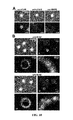
- FIG. 5 provides fluorescence micrographs of anti-ICAM beads of Example 3, demonstrating (A) engulfment by ICAM-1 rich structure and (B) engulfment by the plasma membrane of the beads and (C) tetraspanin CD9 in ECs, at regions of binding of the anti-ICAM beads.
- FIG. 6 provides fluorescence micrographs of Example 4, showing molecular recruitment to sites of endothelial ICAM-1 engagement of anti-ICAM beads, and modulation of this process.
- FIG. 7 provides graphs demonstrating quantification of the molecules recruited to the sites of endothelial ICAM-1 engagement of anti-ICAM beads of Example 4, and modulation of this process.
- FIG. 8 provides fluorescence micrographs showing the effects of amiloride on the formation of ICAM-1-rich endothelial docking-like structures by anti-ICAM beads, as described in Example 4.
- FIG. 9 provides fluorescence micrographs showing distribution of endothelial ASM upon ICAM-1 engagement by anti-ICAM beads, as described in Example 5.
- FIG. 10 provides fluorescence micrographs showing redistribution of endothelial ASM and NHE1 upon ICAM-1 engagement, as described in Example 5.
- FIG. 11 provides fluorescence micrographs showing recruitment of molecules at sites of endothelial engagement of a classical clathrin-associated receptor mannose-6-phosphate receptor (M6PR) by anti-M6PR beads.
- M6PR mannose-6-phosphate receptor
- FIG. 12 provides electron microscopy images from the in vivo testing of Example 6, demonstrating presence or absence of CAM-mediated endocytosis in wild type (i-iii) caveolin-1 ⁇ / ⁇ (iv-vi) and ASM ⁇ / ⁇ (vii-ix) mice.
- FIG. 13 provides images of the experiments performed in Example 6; (A) fluorescence micrographs showing formation of ICAM-1-rich vesicles coalescing into a pore; (B) fluorescence microscopy images showing said pore surrounded by actin filaments; (C) images showing formation of pores in the EC underneath a bound particle; and (D) formation of transmigration pores in binding of WBCs to HUVECs.
- FIG. 14 provides the results of the experiments performed in Example 7 (A) images showing immunostaining of ASM at the WBC-HUVEC interface; (B) graph of transmigration as measured by projecting podosomes into/under HUVECs; (C) graph of determined location of transmigrated WBCs; and (D) comparison of transmigrating and arrested WBCs.
- FIG. 15 provides fluorescence micrographs visualizing the effect of coupling acid sphingomyelinase to ICAM-1-targeted particles as described in Example 8, showing (A) total amount of particles and (B) surface located particles, which enhances uptake of particles by cells deficient in this enzyme.
- FIG. 16 provides graphs showing the effects of coupling sphingomyelinases to particles targeted to cell surface markers other than ICAM-1, e.g., mannose-6-phosphate receptor (M6PR), where (A) provides percent internalization of said particles prior to coupling to M6PR and in comparison to anti-ICAM particles, and (B) provides data regarding the uptake of M6PR targeted carriers after coupling to neutral sphingomyelinase to the surface of the carriers.
- M6PR mannose-6-phosphate receptor
- the present invention relates to methods of regulating transcellular transport of an agent and the involved engulfment or docking structures, and cellular uptake of such agent by manipulation of CAM-mediated pathways and/or the sphingomyelinase/ceramide pathway and the mechanisms involved in such pathways.
- ICAM-1 is an immunoglobulin-family transmembrane glycoprotein that serves as an adhesive surface for leukocytes during inflammation (Yang et al., 2005; Muro et al. 2007; Rothlein R, et al. J Immunol (1986); 137:1270-4). It is constitutively expressed on diverse cell types, including, but not restricted to endothelial cells (EC) (Muro et al., 2007; Hopkins, A. M. et al.
- EC endothelial cells
- ICAM-1 Intra-inflammatory benefits
- ICAM-1 represents an attractive target for drug delivery to different sites in the body.
- antibodies to ICAM-1 are being explored as therapeutics and affinity carriers in cell cultures, animal models, and early clinical studies, where they have shown good safety (Muro et al., 2006; Garnacho, C. et al.
- ICAM-1 plays a key role in transcellular TEM (Barreiro et al., 2002; Yang et al., 2005; Millan et al., 2006; Ley et al., 2007; Oh P, et al. (2007) Nat Biotechnol 25:327-337). Endothelial endocytic vesicles that form in this process coalesce, generating a transcellular pore through which WBCs transmigrate transcellularly (Carman et al. 2004; Carman et al.
- ICAM-1 IL-1-binding to ICAM-1
- ICAM-1 engagement would contribute to inducing formation of engulfment or docking structures, endocytic vesicles, and transcellular transport pores in ECs.
- Specific engagement of endothelial ICAM-1 by other “ligands” protein conjugates and polymer particles coated with antibodies to ICAM-1 (anti-ICAM) used for drug delivery) is sufficient to elicit formation of endocytic vesicles in cytokine activated ECs (Muro et al., 2003; Muro et al., 2006; Muro et al., 2008).
- CAM Cell adhesion molecule
- ICAM-1 interacts with the Na + /H + exchanger NHE1, a molecule which acts as a crosslinker of actin filaments to the cytosolic domain of ICAM-1 (Denker, S P, et al. (2000) Mol Cell 6:1425-1436; Muro et al., 2006).
- endocytic vesicles upon engagement of ICAM-1 is mediated by CAM-mediated endocytosis (Muro et al., 2003; Muro et al., 2006; Muro et al., 2008), which is unrelated to said caveolar-mediated endocytosis or the related vesiculo-vacuolar organelle (Muro et al., 2003; Muro et al., 2006; Muro et al., 2008).
- Formation of endocytic vesicles upon ICAM-1 engagement by anti-ICAM particles is not affected by filipin, a drug that sequesters cholesterol in the plasma membrane of cells and inhibits caveolar-mediated pathways (Muro et al., 2006).
- anti-ICAM beads and related vesicles do not co-localize with cholera toxin B, a molecule known to bind to ganglioside GM1 in lipid raft-related regions of the plasmalemma, followed by internalization within cells via caveolar- and (alternatively) clathrin-mediated pathways.
- CAM-mediated endocytosis has only been associated with engagement of endothelial ICAM-1 by “artificial” and “inert” objects, such as protein conjugates and polymer particles targeted to ICAM-1 via anti-ICAM antibodies or peptides (Muro et al., 2003; Muro et al., 2006; Muro et al., 2008), but it has not been observed in any of the multiple previous works looking at the interaction of WBCs with endothelial cells.
- CAM-mediated endocytosis has been also shown to be induced in the case of engagement of another endothelial molecule, platelet-endothelial cell adhesion molecule 1 (PECAM-1), by anti-PECAM conjugates and polymer particles (Muro et al., 2006). Yet, only ICAM-1, but not PECAM-1, has been shown to be necessary for transcellular TEM of leukocytes.
- PECAM-1 platelet-endothelial cell adhesion molecule 1
- endocytosis induced by “artificial” and “inert” anti-ICAM conjugates or polymer particles or beads has not been shown to result in endocytic vesicles that coalesce to form transendothelial pores but rather to result in transport of individual vesicles to either plasma membrane recycling pathways (in rare instances) or (in most instances) to transport to endosomes and lysosomes (Muro et al., 2003, Muro et al., 2005; Muro et al., 2006).
- CAM-mediated endocytosis On WBC TEM, particularly via the transcellular route, was hardly predictable and somewhat unlikely. Yet, signaling associated to CAM-endocytosis is somewhat similar to leukocytes: Ca 2+ signaling, activation of Src, PKC, and Rho/ROCK, and formation of actin stress fibers (Muro et al., 2003; Muro et al., 2006; Muro et al., 2008).
- ECs endocytose anti-ICAM beads of various shapes and sizes, whose dimensions range from a hundred nanometers to several micrometers, both in cell culture and in vivo (Muro et al., 2003; Muro et al., 2005; Muro et al., 2006; Muro et al., 2008) and this occurs without opening of the endothelial junctions (Muro et al., 2005; Muro et al., 2008). Therefore, the present inventors explored the association among these phenomena in order to more fully understand transcellular transport.
- the invention relates to methods of regulating transcellular TEM of agents, involving control of such CAM-mediated endocytosis and sphingomyelin/ceramide pathway.
- Such regulation can be utilized in myriad ways to control cellular uptake, internalization, transport, delivery and/or arrest of agents. Further uses of the regulation include control of pathogens that bind to ICAM-1, which may invade within cells or be transported across cells.
- the invention relates to methods of regulating transcellular transport of agents, which further provides the ability to control the inflammatory interaction of leukocytes and the endothelium and to regulate delivery of therapeutics in the body via ICAM-1 targeting strategies, as well as via binding to other cell surface markers while providing elements of the CAM- or sphingomyelin/ceramide pathways.
- a method of regulating formation of engulfment or docking structures, uptake and/or transcellular transport of an agent comprising administration of a regulator of CAM-mediated endocytosis or the sphingomyelin/ceramide pathway, wherein such administration is effective to regulate engulfment, uptake or transcellular transport of the agent.
- Example 1 A model for examination of transmigration of white blood cells across endothelial monolayers was developed as described in Example 1. As described in detail in Example 1 and shown in the results presented in FIG. 2 , it was demonstrated that both lipid domains and CAM-mediated endocytosis are involved in WBC transmigration, but caveolae-mediated pathways are not.
- Example 2 provides a model of paracellular versus transcellular transmigration of white blood cells, which was used to examine the role of lipid domains and CAM-mediated endocytosis in paracellular versus transcellular diapedesis of leukocytes.
- the results of Example 2 suggest a main role for lipid domains and CAM-mediated endocytosis, and not caveolae-related pathways, in transcellular TEM versus the paracellular route.
- Example 3 documents the evaluation of the association of lipid domains and the sphingomyelin/ceramide pathway to endothelial docking structures induced by ICAM-1 engagement.
- Endothelial engulfment of WBCs at lipid raft-like, ICAM-1-rich docking structures occurs in association with WBC adhesion to the endothelium and TEM via transcellular pores (Barreiro et al., 2002; Carman and Springer 2004; Oh et al., 2007; van Buul et al., 2007).
- experiments using WBCs involve engagement of multiple adhesion molecules on the endothelial plasmalemma.
- Example 3 demonstrates correlation of ICAM-1 specifically with lipid domains at the EC surface and formation of engulfing or docking structures.
- Example 4 demonstrates that the typical lipid raft-domain components cholesterol, sphingomyelin and ganglioside GM1 were enriched in areas of engulfment of anti-ICAM beads in Example 3, mimicking endothelial docking structures (ring-shaped fluorescent regions in FIG. 6A ).
- the results of Example 4 correlate well with the findings presented in FIGS. 2 and 4 , showing that disruption of lipid domains and CAM-mediated pathway affect transcellular TEM and indicating that ICAM-1 engagement may be sufficient to induce endothelial docking structures at these lipid domains.
- ceramide also increased (3.5 ⁇ 0.01-fold) at these regions over adjacent areas, which was impaired by EIPA (35.6% decrease; FIG. 6 Cii,iv), an amiloride derivative that more specifically inhibits NHE1 involved in CAM-mediated endocytosis (Muro et al., 2003; Muro S, et al. (2006) Am J Physiol Lung Cell MolPhysiol 290:L809-817).
- NHE1 was identified as having a critical function in particle internalization and as a possible connector of the CAM-mediated pathway to sphingomyelin/ceramide signaling.
- ASM acid sphingomyelinase
- Example 5 support the assertion that ASM secretion is associated with the CAM-mediated pathway. Accordingly, Example 6 was performed to confirm this by evaluating the effect of impairing acid sphingomyelinase on CAM-mediated endocytosis.
- Ceramide production by ASM at sites of ICAM-1 engagement where NHE1 acidifies the milieu may provide plasmalemma plasticity (and likely cytoskeleton signaling; Holopainen et al., 1998; Zha et al., 1998; Holpainen et al., 2000; Zeidan et al., 2008) required by endothelial docking-like structures involved in engulfment of micron-sized objects and WBCs, which the present inventors attribute to the CAM-mediated pathway, rather than caveolae-mediated pathways.
- Example 6 Furthermore, an in vivo mouse model detailed in Example 6 confirmed that engulfment leading to endocytosis of anti-ICAM beads by ECs was inhibited in ASM ⁇ / ⁇ mice.
- Example 7 provides an examination of the role of acid sphingomyelinase in leukocyte transcellular transmigration.
- the inventors had hypothesized that ASM was involved not only in the formation of CAM-mediated vesicles but also fusion of said vesicles into larger structures, similar to formation of ICAM-1-rich invaginations and vesicles that coalesce into transmigration pores.
- the example looked for ASM to appear at areas of WBC migration across ECs and its inhibition to affect transcellular TEM.
- Example 7 support a concerted role for sphingomyelin/ceramide signaling and NHE1-dependent CAM-mediated endocytosis induced upon ICAM-1 engagement at the EC surface, in transcellular TEM. This model is consistent with findings obtained using WBCs and specific ICAM-1 engagement by anti-ICAM beads, pharmacological inhibitors and genetically modified models, cell cultures and in vivo systems.
- the results reported herein support a model for transcellular TEM that includes, but is not limited to, engagement of ICAM-1 in lipid domains enriched in sphingomyelin, which induces secretion of acid sphingomyelinase (ASM) from intracellular compartments to these areas of the endothelial plasma membrane.
- ASM acid sphingomyelinase
- engaged ICAM-1 forms a complex with NHE1, which results in local acidification, sphingomyelin hydrolysis by secreted ASM, and local production of ceramide.
- This signal leads to actin polymerization and cytoskeleton remodeling, stabilizes the engagement platform by restricting molecular diffusion and providing cytoskeletal anchorage, regulates membrane deformability, and favors dynamic formation of CAM-mediated endocytic vesicles, which occurs at sites of leukocyte-podosome sampling in search from sites suitable for transcellular TEM. Finally, vesicular fusion mediated through sphingomyelin/ceramide signaling at this interface results in transmigration pores.
- the model of transcellular TEM presented herein explains particular molecular and cellular features required for such events to take place.
- the ion exchange activity of NHE1 regulates the elasticity of the endothelial apical surface (Hillebrand U, et al. (2006) Cardiovasc Res 69:916-924), in agreement with high permissibility of CAM-mediated endocytosis for engulfment and uptake of large micron-sized objects in vitro and in vivo (Muro et al., 2008).
- This is in contrast to the caveolar and clathrin pathways, shown to be rather restricted regarding the size of “ligands” that can accommodate to typical caveolar vesicles (Oh et al., 2007).
- CAM endocytosis Such deformability properties of CAM endocytosis, which can be exploited for transport of drug carriers into and across cells, would suitably adapt for formation of large endothelial docking structures, the wide range of sizes exhibited by the invasive podosomes that leukocytes extend into ECs during TEM, and formation of the transcellular pore (Carman et al., 2004; Carman et al., 2007). Also in the context of CAM-mediated endocytosis and WBC TEM, diffusion of molecules in the plasmalemma must be temporarily reduced in areas of binding to ICAM-1, to permit formation of engagement and signaling platforms, and to anchor the cytoskeleton.
- the level of deformability required to engulf large objects and cells by endothelial docking structures progressing into transmigration pores must also relate to a particular lipid composition of the plasmalemma. As shown here, such domains seem to be related also to CAM-mediated pathway, and are associated to induction of sphingomyelin/ceramide signaling upon ICAM-1 engagement at the EC plasmalemma. As observed in other systems, ceramide confers particular properties to the membrane environment depending on the ratio of raft components (Rotolo J A, et al. (2009) Blood 11 4:3693-3706; Silva L C, et al.
- Biophys J 96:3210-3222 it can promote the formation of large lipid domains (Holopainen et al., 1998; Holopainen et al., 2000) or displace lipid domain constituents to affect membrane function (Zeidan et al., 2008).
- Ceramide production by ASM at the outer leaflet of the plasma membrane modifies its curvature and results in vesiculization (Zha et al., 1998; Holopainen et al., 2000; Tam C, et al. (2010) J. Cell Biol. 189:1027-1038), as well as cytoskeletal rearrangements (Zeidan et al., 2008).
- These events downstream of the sphingomyelin/ceramide pathway could contribute to formation of large micron-sized vesicles in the absence of clathrin or caveolin coats, as observed in CAM-mediated endocytosis (Muro et al., 2003; Muro et al., 2008) (Table 1).
- ceramide production by ASM is associated with vesicular fusion (Utermohlen O. et al. Immunobiology 21 3:307-314), which could contribute to dynamic formation of transcellular pores from vesicles forming via CAM-mediated pathway.
- vesicular fusion Utermohlen O. et al. Immunobiology 21 3:307-314
- NHE1 ion transport activity of NHE1 will locally acidify ICAM-1 engagement regions, creating a confined acidic microenvironment.
- a similar function of NHE1 has been shown in the context of other pH-sensitive enzymes (Bourguignon et al., 2004).
- Such methods are useful to impact a plethora of diseases in which inflammation plays a role, including but not restricted to inflammatory and autoimmune conditions (rheumatoid arthritis, psoriasis, ulcerative colitis, Crohn disease, etc.), infections and septic shock, ischemia-reperfusion injury, atherosclerosis and thrombosis, metabolic and genetic disorders, asthma and acute lung injury, cancer and tumor metastasis, and many others.
- inflammatory and autoimmune conditions rheumatoid arthritis, psoriasis, ulcerative colitis, Crohn disease, etc.
- infections and septic shock ischemia-reperfusion injury
- atherosclerosis and thrombosis ischemia-reperfusion injury
- metabolic and genetic disorders asthma and acute lung injury
- cancer and tumor metastasis and many others.
- Methods of the invention include intracellular delivery or delivery across cellular barriers, such as the blood-brain barrier in the central nervous system, the blood-air barrier in the lungs, the epithelial barrier in the gastrointestinal tract, penetrability into tissues and organ, and the like.
- the invention relates to a method of regulating transcellular transendothelial migration of an agent, the method comprising administration of a regulator of CAM-mediated endocytosis or the sphingomyelin/ceramide pathway, wherein such administration is effective to regulate transcellular TEM of the agent.
- a “regulator” can be administered to regulate the CAM-mediated endocytosis or the sphingomyelin/ceramide pathway.
- regulation or “regulating” of CAM-mediated endocytosis or the sphingomyelin/ceramide pathway includes any of controlling, managing, adjusting, directing, manipulating or modulating CAM-mediated endocytosis or the sphingomyelin/ceramide pathway.
- Regulators of CAM-mediated endocytosis or the sphingomyelin/ceramide pathway according to methods of the invention are effective after ICAM-1 engagement of the agent. Regulators of the invention target steps of engulfment or formation of docking structures, uptake by cells and formation of vesicles, as well as pore opening and transcellular TEM or transcytosis.
- Administration of a regulator may include actual administration of the regulator in vitro or in vivo to a system or patient in need of such administration. Administration may be by any suitable administration mechanism that provides effective levels of the regulator to the endothelium. Any suitable administrative routes that are compatible with the selected regulator may be employed. Administration methods of regulators described herein include, but are not limited to, parenteral administration, intraperitoneal (i.p.) administration, intravenous (i.v.) administration, intraarterial (i.a.) administration, intradermal (i.d.) administration, intramuscular (i.m.) administration, and subcutaneous (sc) administration.
- parenteral administration intraperitoneal (i.p.) administration, intravenous (i.v.) administration, intraarterial (i.a.) administration, intradermal (i.d.) administration, intramuscular (i.m.) administration, and subcutaneous (sc) administration.
- Administration of a regulator may also include indirect administration such as induction or inhibition of a regulator within the subject system or patient.
- regulators may include, but are not limited to, NHE1, sphingomyelinases, acid sphingomyelinase and ceramide.
- Further regulators useful in methods of the invention may include, but are not limited to, proteins affecting the CAM-mediated pathway, ICAM-1, lipids affecting the CAM-mediated pathway, sphingomyelin, and ceramidases.
- Acid sphingomyelinase (ASM), other sphingomyelinases, and NHE1 are proteins that may be directly administered to regulate CAM-mediated endocytosis or the sphingomyelin/ceramide pathway. Such proteins may be obtained by any means known, from any known source, such as from organisms, humans or recombinantly produced. ASM, other sphingomyelinases, and NHE1 may also be administered by indirect means, such as by inducement of expression or over-expression of these proteins within the subject system or patient. In one embodiment, methods of the invention may include gene therapy to induce expression of a regulator of the CAM-mediated endocytosis or the sphingomyelin/ceramide pathway.
- methods of the invention may include administration of an activator that activates production of the regulator.
- Insulin is an activator of NHE1, and other activators include molecules that activate PKC (e.g., PMA, bryostatin) and Rho, among others.
- Activators of sphingomyelinases are saposins, DC-SIGN, OxPAPC, neutral sphingomyelinase (N-SMase) activation associated factor or NSMAF, molecules that activate PKC, and the like.
- Regulators such as ASM, other sphingomyelinases, and NHE1 may also be inhibited by known methods, such as use of siRNA to knock-down expression of these proteins or by using blocking antibodies. Induction may also be achieved by administration of inhibiting compounds.
- Inhibitors of NHE1 may include, but are not limited to amiloride and derivatives like 5′-(N-ethyl-N-isopropyl)amiloride (EIPA), and benzoylguanidine (Hoechst type inhibitor (HOE))-type compounds.
- Inhibitors of ASM may include, but are not limited to imipramine, its derivatives like desipramine, SR33557, D609, and others.
- Inhibitors of sphingmyelinases may include, but are not limited to scyphostatin, 3,4-Dichloroisocoumarin Chlorpromazine, Hydrochloride Fumonisin B 1 , Fusarium moniliforme Gentamycin Sulfate Manumycin A, Streptomyces parvulus N-SMase Inhibitor, GW4869 and N ⁇ -Tosyl-Phe Chloromethyl Ketone. Such inhibition is contemplated within administration of a regulator.
- Additional regulators may include, but are not limited to lipids such as sphingomyelin and ceramide.
- lipid regulators may be administered directly, such as by exogenous application to a system or subject in need of a method of the invention or may be administered indirectly, such as by modulation of enzymes involved in the mechanisms of synthesis or degradation routes of these lipids.
- activation or inhibition of ceramidases would degrade ceramide or inhibit its degradation, respectively, and thereby impact the presence of ceramide in the pathway.
- Regulators of the invention are useful for regulation of CAM-mediated endocytosis or the sphingomyelin/ceramide pathway. By such regulation, the engulfment, uptake within vesicles, and/or transcellular transport of an agent can be affected.
- the regulator inhibits CAM-mediated endocytosis and inhibits engulfment, uptake within cells, and/or transcellular TEM or transport of the agent.
- the regulator induces CAM-mediated endocytosis and induces engulfment, uptake within cells, and/or transcellular TEM or transport of the agent.
- the regulator inhibits the sphingomyelin/ceramide pathway and inhibits engulfment, uptake within cells, and/or transcellular TEM or transport of the agent.
- the regulator induces the sphingomyelin/ceramide pathway and induces engulfment, uptake within cells, and transcellular TEM or transport of the agent.
- the invention further contemplates combined effects on CAM-mediated endocytosis and the sphingomyelin/ceramide pathway.
- Agents useful in methods of the invention may include, but are not limited to, autologous or foreign white blood cells, leukocytes, pathogens, drugs, natural and/or artificial molecules and/or objects including, but not limited to, research, analytical or molecular probes, diagnostic agents, therapeutic agents, biologically active agents, research agents, analytical agents, imaging agents, monitoring agents, enzymes proteins, hormones, lipids, sugars, nucleic acids, lipoproteins, and chemicals.
- Agents may be present alone or may be complexed to an additional moiety.
- complexed refers to the association between the agent and the moiety, including binding, fusing, linking, coupling, connecting or otherwise associating the agent and the additional moiety.
- the resulting complexes may be a single entity, such as a fusion protein or may result from coupling via absorption mechanisms, by chemical modification, through a crosslinker molecule, or via adaptor molecules. Any such complexing is contemplated in methods of the invention.
- Additional moieties for complexing to the agent may include, but are not limited to, targeting moieties, cargo, carriers, delivery vehicles, and combinations thereof.
- a targeting moiety such may include, but is not limited to, a polypeptide such as an antibody, antibody fragment, single chain Fv derivative, humanized antibody, natural protein, peptide, or any other natural, recombinant or synthetic affinity moiety recognizing CAMs.
- the targeting moiety targets a cell surface marker other than ICAM-1, including, but not limited to, receptors associated to other mechanisms of endocytosis and transport across cells, including but not restricted to phagocytosis, macropinocytosis, clathrin-mediated transport and caveolar-mediated transport.
- the non-ICAM receptor is M6PR.
- the cargo may include, but is not limited to a cell or modified cell, reporter probe, biosensor, marker, antibody, peptide or protein, enzyme, ligand, genetic material (DNA- and RNA-based), drug or chemical, imaging or therapeutic agent, or any combination of the above.
- Cargo included in methods of the invention may be directly delivered by the targeting moiety or may be additionally assisted by a delivery vehicle or carrier.
- the invention provides a new strategy to regulate transcellular TEM and to thereby regulate interaction of leukocytes with the endothelium and transport thereof and also to regulate transport of therapeutics and their carriers in the body, and, still further, to generally regulate cellular uptake of agents within cells and across cells via the transcellular pathway, supporting multiple basic, research, and translational applications.
- the regulatory methods of the invention are broadly applicable to methods such as, but not limited to, modulation of inflammation, pathogen invasion and drug delivery.
- the identification of pathways subject to regulation permits control of transcellular TEM.
- the transcellular TEM pathway can selectively be upregulated to promote transmigration or downregulated to inhibit or avoid transmigration.
- the methods of the invention can be used to shift between transcellular and paracellular transmigration pathways.
- paracellular TEM the “leaky vasculature” of the open junctions between cells can permit entrance of undesired substance, such as red blood cells, proteins and the like. Accordingly, in some situations it is desirable to favor transcellular TEM. However, in other situations it is desirable to disfavor transcellular TEM.
- Agents may be targeted to a surface marker (e.g. ICAM-1 or a receptor associated to classical vesicular transport, including but not restricted to M6PR) and engulfment, uptake by cells, or transcellular transport downregulated, such that the agent remains immobilized on the surface of the EC. As such the agent is anchored on the EC. Additionally, agents may be targeted to a surface receptor and engulfment, uptake by cells, or transcellular transport upregulated, such that the transport of the agent is controlled.
- a surface marker e.g. ICAM-1 or a receptor associated to classical vesicular transport, including but not restricted to M6PR
- agents may be targeted to a surface receptor and engulfment, uptake by cells, or transcellular transport upregulated, such that the transport of the agent is controlled.
- Such steps of immobilization and transport may also be combined through use of multiple regulators and/or timing of regulators, such that an agent may be initially immobilized, then transported via transcellular TEM at an appropriate time to a desired locale.
- the methods of the invention are therefore applicable to promote or avoid entrance of an agent into endothelial cells and/or across the endothelial lining.
- the invention relates to applicability of methods of the invention to control of pathogenic invasion via ICAM-1.
- an agent is a pathogen
- the engulfment, uptake within cells, and/or transcellular transport pathway can selectively be upregulated to promote uptake and transmigration or downregulated to inhibit or avoid uptake or transmigration.
- Promotion of transmigration can be utilized to promote or otherwise include transport of the pathogens to lysosomes and/or vacuoles for subsequent degradation and protection of the cells in the body against infection.
- Inhibition of transmigration can be used to prevent pathogenic cellular invasion.
- agent is a drug and such agent is complexed to an imaging agent. Promotion of uptake within cells and/or transcellular transport can be utilized for targeted delivery of the drug to the cells and/or tissues.
- the invention relates to recovery of the action of an inhibited CAM-mediated uptake and transcellular transport pathway.
- the invention relates to enhancement of uptake and/or transcellular transport when using targeting to other cell surface markers and pathways while providing exogenously elements of the CAM- or sphingomyelin/ceramide pathways.
- the method relates to a method of recovery of an inhibited pathway, the method comprising administration of a regulator of CAM-mediated endocytosis or the sphingomyelin/ceramide pathway, wherein such administration is effective to regulate engulfment, uptake or transcellular transport of the agent and wherein the inhibition is inhibition of engulfment, uptake or transcellular transport within a pathway selected from the group consisting of CAM-mediated endocytosis, sphingomyelin/ceramide pathway, phagocytosis, macropinocytosis, clathrin-mediated endocytosis and caveolar-mediated endocytosis.
- Example 8 demonstrates that even in ASM ⁇ / ⁇ ECs, endocytosis of very large objects (anti-ICAM particles about 5 micrometers in diameter) can be achieved by coupling ASM, as a regulator, to said objects as agents (which in this case improved endocytosis from 7% to 25%).
- ASM as a regulator
- agents which in this case improved endocytosis from 7% to 25%.
- the invention relates to a method of potentiating cellular engulfment, uptake and/or transcellular transport of an agent, where the agent is complexed to a carrier targeted to a non-ICAM cell surface molecule or receptor, the method comprising administration of a regulator of CAM-mediated endocytosis or the sphingomyelin/ceramide pathway, wherein such administration is effective to induce uptake of the agent and potentiation of transcellular transport of the complex.
- FIG. 16 demonstrates enhancement of cell uptake of drug delivery carriers by sphingomyelinases.
- FIG. 16A illustrates the observed percent internalization into human vascular endothelial cells (HUVECs) at 30 minutes or 3 hours, of model 1 micrometer diameter polymer (polystyrene) drug carriers, targeted to either ICAM-1 or mannose-6-phosphate receptor (M6PR).
- HAVECs human vascular endothelial cells
- M6PR mannose-6-phosphate receptor
- ICAM-1-mediated pathway results in more effective uptake of carriers by cells, since ICAM-1 mediates uptake via cell adhesion molecule (CAM) endocytosis, which associates to the sphingomyelin/ceramide pathway, where acid sphingomyelinase regulates formation of plasma membrane engulfment structures and remodeling of the cytoskeleton, conducive to uptake of objects, even those that are micrometers in size.
- CAM cell adhesion molecule
- M6PR is known to mediate uptake via the clathrin pathway, which lacks the ability to associate with the sphingomyelin/ceramide pathway.
- FIG. 16B shows improvement of the uptake of nanocarriers targeted to M6PR by coupling neutral sphingomyelinase to the surface of these carriers, which modulates uptake by providing this effector exogenously.
- the method of modulating the transcellular endothelial transmigration includes enhancement of uptake of cells generally regulated by non CAM-mediated pathways.
- the invention in another embodiment relates to a method of modulating inflammation, comprising administration of a regulator of CAM-mediated endocytosis or the sphingomyelin/ceramide pathway, which is effective to regulate engulfment, uptake or transcellular transport of an agent of said inflammation.
- a confluent EC monolayer from human umbilical vein ECs was grown on a porous membrane through which peripheral lymphocytes isolated from healthy individuals (wild-type WBCs) can transmigrate, driven by the presence of the chemoattractant SDF1- ⁇ in the chamber underneath the ECs.
- wild-type WBCs peripheral lymphocytes isolated from healthy individuals
- SDF1- ⁇ chemoattractant- ⁇ in the chamber underneath the ECs.
- 77.7 ⁇ 2.3% wild-type WBCs underwent transmigration by 30 min, which was abrogated when K562 cells lacking the ICAM-1- and VCAM-1-binding integrins LFA-1 and VLA-4, respectively, were used ( FIG. 1A ).
- FIG. 1B shows the results of transmigration of antibody-blocked WBCs in the presence (black bars) or absence (white bars) of HUVEC monolayer, as a control. Data are normalized to control values, and represent mean and standard errors of the mean (n ⁇ 3 experiments). *, P ⁇ 0.001 by Student's t test.
- WBCs peripheral blood lymphocytes
- FIG. 3 shows the results of the migration of activated peripheral blood lymphocytes (control) or K562 cells lacking ICAM-1- and VCAM-1-binding integrins (pre-stained with green fluorescent calcein), determined after co-incubation for 30 min at 37° C. with activated HUVECs and analyzed by fluorescence microscopy after fixation.
- FIG. 3A shows WBCs projecting podosomes into/under HUVECs, scored as transmigrating (black bars) versus non-transmigrating WBCs, scored as arrested cells (round-like WBCs; white bars).
- FIG. 3B shows spatial distribution of transmigrating WBCs, scored as occurring at either the endothelial cell (EC) border (white bars) or center (black bars), measured at ⁇ 3 ⁇ m or ⁇ 3 ⁇ m distance from the cell border, respectively.
- FIG. 3C shows transmigration activity of WBCs at the EC center (black bars), scored as in FIG. 3A , compared to non-transmigrating activity (arrested; white bars) at these areas. Data are mean and standard errors of the mean (n ⁇ 30 WBCs).
- FIG. 4 shows the results of the migration of activated WBCs (pre-stained with green fluorescent calcein) incubated over activated HUVECs growing on glass coverslips, determined after co-incubation for 30 min at 37° C. in control media or media containing filipin, methyl- ⁇ -cyclodextrin (Cdx) or amiloride, and analyzed by fluorescence microscopy after fixation.
- FIG. 4A shows WBCs projecting podosomes into/under HUVECs, scored as transmigrating (black bars).
- FIG. 4B shows spatial distribution of transmigrating WBCs scored as occurring at either the endothelial cell (EC) border (white bars) related to paracellular transmigration or center (black bars) related to transcellular transmigration, measured at ⁇ 3 ⁇ m or ⁇ 3 ⁇ m distance from the cell border, respectively.
- FIG. 4C shows transmigration activity of WBCs at the EC center (black bars), scored as in FIG. 4A , compared to non-transmigrating activity (arrested, round-like WBCs; white bars) at these areas. Data are normalized to control values (horizontal dashed lines) and represent mean and standard errors of the mean (n ⁇ 30 WBCs). *, P ⁇ 0.05; **, P ⁇ 0.01; ***, P ⁇ 0.001 by Student's t test.
- FIG. 4D Scanning electron micrographs showing the effect of amiloride on distribution of WBC transmigration events.
- White arrows indicate WBCs transmigrating at the EC border.
- Arrowheads mark WBCs transmigrating (white arrowheads) or arrested (black arrowheads) at EC center regions.
- Nu Nucleus.
- Scale bar 10 ⁇ m.
- Cdx and amiloride decreased diapedesis (71.5 ⁇ 14.6% and 56.0 ⁇ 27.5% of control) and increased the amount of arrested WBCs (170.5 ⁇ 36.2% and 209.1 ⁇ 68.1% of control; FIGS. 4C-D ).
- Anti-ICAM beads Polymer beads coated with multiple copies of an antibody against ICAM-1 (anti-ICAM beads) were used. These have been previously used for studying aspects of leukocyte transmigration (Allingham et al., 2007; van Buul et al., 2007; van Buul et al., 2010) and CAM-mediated pathway (Muro et al., 2003; Muro et al., 2005; Muro et al., 2006; Muro et al., 2008).
- micrographs showed that, within 15 min incubation, anti-ICAM beads (immunostained in green FITC) bound to ECs and were engulfed by ICAM-1-enriched membrane protrusions (immunostained in Texas red).
- FIG. 5C provides fluorescence immunostaining of tetraspanin CD9 in ECs, at regions of binding of anti-ICAM beads.
- (i) Micrograph showing CD9 enrichment as ring-like structures (arrows). Scale bar 10 ⁇ m.
- FIGS. 6A and 7A show the results of cholesterol (i), sphingomyelin (ii) or ganglioside GM-1 (iii), stained using fluorescent blue filipin, green BODIPY-sphingomyelin, or Texas-red cholera toxin B, respectively.
- FIGS. 6A and 7A show the results of cholesterol (i), sphingomyelin (ii) or ganglioside GM-1 (iii), stained using fluorescent blue filipin, green BODIPY-sphingomyelin, or Texas-red cholera toxin B, respectively.
- FIG. 6B and 7B show the effect of methyl- ⁇ -cyclodextrin (Cdx) on enrichment of cholesterol labeled with blue filipin (i) or ICAM-1 immunostained with a Texas-red-labeled antibody (ii-iii) in regions of anti-ICAM-bead binding.
- FIG. 6C shows immunostaining of ceramide using a Texas-red labeled antibody in regions of anti-ICAM-bead binding in control (i), EIPA-treated (ii), and imipramine-treated (iii) cells. Left panels show fluorescence micrographs and phase-contrast insets of bound beads.
- Fluorescence intensity of cholesterol, sphingomyelin and ganglioside GM1 was increased by 1.6 ⁇ 0.04-fold, 3.1 ⁇ 0.1-fold, and 1.4 ⁇ 0.1-fold, respectively, in regions of bead engulfment by ICAM-1 engagement over adjacent areas ( FIG. 7A ).
- Cdx treatment to chelate cholesterol confirmeded in FIG. 6Bi and FIG. 7Bi ) decreased ICAM-1 enrichment in areas of bead engulfment (43% decrease; FIG. 6 Bii-iii; FIG. 7 Bii-iii).
- ceramide was tested for at regions of ICAM-1 engagement by anti-ICAM beads.
- imipramine a drug that inhibits acid sphingomyelinase (ASM), impaired ceramide enrichment in areas of bead engulfment associated to ICAM-1 engagement (23.6% decrease; FIG. 6 Ciii-iv), implicating for the first time ASM in ICAM-1-driven formation of endothelial docking-like structures.
- Immunofluorescence of ASM in ECs showed that, in absence of ICAM-1 engagement by anti-ICAM beads, most ASM located to vesicular compartments (likely lysosomes) in the perinuclear region of cells (41.8 ⁇ 4.7 vesicles/cell), while only a few ASM-positive vesicles were found outside the perinuclear area (23.7 k3.3 vesicles/cell; FIG. 9 ).
- FIG. 10A Fluorescence immunostaining of ASM ( FIG. 10A ; bottom panels) in regions of respective bead binding ( FIG. 10A ; phase-contrast, top panels).
- FIG. 10B provides fluorescence microscopy showing immunostaining of ASM (green), and ICAM-1 (red, i-iv) or NHE1 (red, v-viii). Boxes indicate the respective beads and bead regions selected for enlargement in iii and vii, and in iv and viii.
- Activated HUVECs were incubated with anti-mannose-6-phosphate receptor (M6PR) beads for 15 min at 37° C. to engage M6PR on endothelial cells (ECs), followed by washing and fixation.
- Phase contrast FIG. 11 ; left panels
- fluorescence micrographs FIG. 11 ; right panels
- ICAM-1 top
- NHE1 molecular epitop
- clathrin heavy chain bottom
- Arrowheads indicate lack of enrichment of the corresponding marker around beads.
- Arrows indicate enrichment of the corresponding marker around beads.
- Scale bar 10 ⁇ m.
- Anti-M6PR beads induced recruitment neither of ICAM-1 nor NHE1, but recruited clathrin heavy chain ( FIG. 11 ), validating the specificity of this model.
- FIG. 10B areas of bead engulfment mediated by ICAM-1 engagement ( FIG. 10B ) revealed that ASM co-localized well with ICAM-1 and NHE1 (85.1 ⁇ 2.9% and 85.3 ⁇ 3.4% co-localization).
- ASM appeared to distribute within ICAM-1- and NHE1-lined vesicular structures ( FIG. 10 Biv,viii), supporting secretion of ASM associated to CAM-mediated pathway.
- the number of beads internalized within ECs was quantified from the micrographs. Data are mean and standard errors of the mean (n ⁇ 13 micrographs). *, P ⁇ 0.05 by Student's t test. As in cell cultures, engulfment leading to endocytosis of anti-ICAM beads by ECs was inhibited in ASM ⁇ / ⁇ mice but not in caveolin-1 ⁇ / ⁇ mice (12.2% and 92.8% of wild-type mice).
- ICAM-1 in the surface of the plasma membrane was then stained using anti-ICAM and a secondary antibody labeled in Texas red (yellowish in the picture). Cells were then washed and permeabilized to access internal compartments. ICAM-1 in internal structures was then stained with anti-ICAM followed by a secondary antibody labeled in green FITC. Imaging by fluorescence microscopy permitted to observe multiple small vesicles enriched in ICAM-1 just underneath the cell surface. These vesicles appeared to clealesce or merge in large structures preliminary to pore formation. A similar experiment with similar result is shown by scanning electron microscopy in FIG. 13B . Activated HUVECs were incubated with 4.5 pm diameter anti-ICAM particles and imaged by dynamic phase-contrast microscopy. A pore forms in the EC underneath a bound particle, as shown in FIG. 13C .
- FIG. 14A Fluorescence microscopy revealed enrichment of ASM (immunostained in Texas red) in the interface between ECs and transmigrating peripheral lymphocytes (stained with green calcein; FIG. 14A ).
- FIG. 14A shows phase contrast (left panel) and Texas-red immunostaining of ASM (right and bottom panels) at the WBC-HUVEC interface in control conditions.
- Nu nucleus.
- Scale bars 10 ⁇ m or 2 ⁇ m, as indicated. This is similar to endothelial docking-like structures formed upon sole engagement of ICAM-1 by anti-ICAM beads, as shown in FIG. 10 .
- FIG. 14C spatial distribution of transmigrating WBCs was scored as occurring at either the endothelial cell (EC) border (white bars) or center (black bars), measured at ⁇ 3 ⁇ m or ⁇ 3 ⁇ m distance from the cell border, respectively.
- transmigration activity of WBCs at the EC center was scored as in (B), compared to non-transmigrating activity (arrested, round-like WBCs; white bars) at these areas.
- Data are normalized to control values (horizontal dashed lines), and represent mean and standard errors of the mean (n ⁇ 100 WBCs). * P ⁇ 0.01 by Student's t test.
- polystyrene particles (4.5 ⁇ m diameter), a model for a carrier of diagnostic and/or therapeutic agents, were coated with anti-ICAM to target ECs and ASM, to recover uptake and intracellular transport of carriers by facilitating endocytosis in ASM ⁇ / ⁇ ECs, which are otherwise voided of ASM and hence do not support CAM-mediated pathway (as shown in Table 1).
- Particles only coated with anti-ICAM but not ASM were used as negative controls for lack of internalization.
- the cells were incubated with the particles for 30 min at 37° C. to first allow binding of particles to ICAM-1 on the cell surface.
- non-bound particles were washed off and cells were incubated in control media for 1 h at 37° C. to permit potential endocytosis of bound particles. Cells were finally washed and fixed.
- Surface-bound non-internalized particles were immunostained using a secondary antibody labeled with Texas red. This antibody can only bind to anti-ICAM on surface-located beads, while it can not access particles internalized within the cells.
- Samples were analyzed by fluorescence microscopy to visualize total beads associated to cells by phase contrast (upper panels) and fluorescence microscopy to visualize non-internalized particles (lower panels, arrowheads), from which the percent of internalization of particles was calculated.
- polystyrene particles (1 ⁇ m diameter), a model for a carrier of diagnostic and/or therapeutic agents, were coated with anti-ICAM or anti-M6PR, a cell surface marker related to classical endocytic transport pathway (clathrin-mediated uptake or transcytosis, in particular).
- Particles targeted to M6PR were not efficient in being transported by cells, in contrast to particles targeted to ICAM-1, when tested either at 30 min or 3 h incubation at 37° C.
- 16B shows that incorporation of a sphingomyelinase (neutral sphigomyelinase, in particular) but not a control protein (IgG) to anti-M6PR particles enhanced transport by cells by providing this element of the CAM- and sphingomyelin/ceramide pathway.
- Data represent mean and standard errors of the mean (n ⁇ 15 ECs). * P ⁇ 0.05 by Student's t test.
Landscapes
- Health & Medical Sciences (AREA)
- Life Sciences & Earth Sciences (AREA)
- Public Health (AREA)
- Veterinary Medicine (AREA)
- Epidemiology (AREA)
- Medicinal Chemistry (AREA)
- Animal Behavior & Ethology (AREA)
- General Health & Medical Sciences (AREA)
- Chemical & Material Sciences (AREA)
- Pharmacology & Pharmacy (AREA)
- Gastroenterology & Hepatology (AREA)
- Engineering & Computer Science (AREA)
- Bioinformatics & Cheminformatics (AREA)
- Immunology (AREA)
- Proteomics, Peptides & Aminoacids (AREA)
- Emergency Medicine (AREA)
- Pharmaceuticals Containing Other Organic And Inorganic Compounds (AREA)
- Medicinal Preparation (AREA)
Abstract
Description
| TABLE 1 | |||
| Internalization (%) | |||
| HUVECs | |||
| Control | 100.0 ± 7.6 | ||
| Amiloride | 13.8 ± 3.3** | ||
| Imipramine | 61.9 ± 6.7** | ||
| Na+ depletion | 7.4 ± 2.5** | ||
| Filipin | 107.2 ± 1.5 | ||
| MLECs | |||
| Control | 100.0 ± 25.4 | ||
| ASM−/− | 25.4 ± 10.5* | ||
| Values are normalized to controls | |||
| * and ** indicate p ≦ 0.05 and p ≦ 0.001, respectively. | |||
| n ≧ 10 micrographs from 2 replicates. | |||
Claims (14)
Priority Applications (2)
| Application Number | Priority Date | Filing Date | Title |
|---|---|---|---|
| US13/652,165 US9901625B2 (en) | 2011-10-15 | 2012-10-15 | Methods of regulating uptake and transcellular transport of leukocytes and therapeutics |
| US15/679,250 US20180021414A1 (en) | 2011-10-15 | 2017-08-17 | Methods of regulating uptake and transcellular transport of leukocytes and therapeutics |
Applications Claiming Priority (2)
| Application Number | Priority Date | Filing Date | Title |
|---|---|---|---|
| US201161547687P | 2011-10-15 | 2011-10-15 | |
| US13/652,165 US9901625B2 (en) | 2011-10-15 | 2012-10-15 | Methods of regulating uptake and transcellular transport of leukocytes and therapeutics |
Related Child Applications (1)
| Application Number | Title | Priority Date | Filing Date |
|---|---|---|---|
| US15/679,250 Continuation US20180021414A1 (en) | 2011-10-15 | 2017-08-17 | Methods of regulating uptake and transcellular transport of leukocytes and therapeutics |
Publications (2)
| Publication Number | Publication Date |
|---|---|
| US20130095091A1 US20130095091A1 (en) | 2013-04-18 |
| US9901625B2 true US9901625B2 (en) | 2018-02-27 |
Family
ID=48086135
Family Applications (2)
| Application Number | Title | Priority Date | Filing Date |
|---|---|---|---|
| US13/652,165 Active US9901625B2 (en) | 2011-10-15 | 2012-10-15 | Methods of regulating uptake and transcellular transport of leukocytes and therapeutics |
| US15/679,250 Abandoned US20180021414A1 (en) | 2011-10-15 | 2017-08-17 | Methods of regulating uptake and transcellular transport of leukocytes and therapeutics |
Family Applications After (1)
| Application Number | Title | Priority Date | Filing Date |
|---|---|---|---|
| US15/679,250 Abandoned US20180021414A1 (en) | 2011-10-15 | 2017-08-17 | Methods of regulating uptake and transcellular transport of leukocytes and therapeutics |
Country Status (1)
| Country | Link |
|---|---|
| US (2) | US9901625B2 (en) |
Citations (15)
| Publication number | Priority date | Publication date | Assignee | Title |
|---|---|---|---|---|
| WO1993025218A1 (en) | 1992-06-11 | 1993-12-23 | The Scripps Research Institute | Methods and compositions for inhibiting endothelial cell and fibrinogen mediated inflammation |
| WO1994015641A1 (en) | 1993-01-12 | 1994-07-21 | The Rockefeller University | Method for modulating transendothelial migration of cells promoting inflammation, and related methods of measurement thereof |
| WO1995028946A1 (en) | 1994-04-25 | 1995-11-02 | The Scripps Research Institute | Methods and compositions for inhibiting endothelial cell and fibrinogen mediated inflammation |
| US20040029779A1 (en) * | 2002-04-05 | 2004-02-12 | Genzyme Corporation | Methods of enhancing lysosomal storage disease therapy by modulation of cell surface receptor density |
| US7223395B2 (en) | 2000-03-13 | 2007-05-29 | Cornell Research Foundation, Inc. | Blocking leukocyte emigration and inflammation by interfering with CD99/HEC2 |
| US20100168219A1 (en) | 2008-12-31 | 2010-07-01 | Jonathan Steven Alexander | Chronic inflammation and transplantation |
| WO2011011500A1 (en) | 2009-07-21 | 2011-01-27 | Abt Holding Company | Use of stem cells to reduce leukocyte extravasation |
| WO2011044329A2 (en) | 2009-10-07 | 2011-04-14 | University Of Pittsburgh - Of The Commonwealth System Of Higher Education | Devices, systems and methods for cell modification |
| US20110177155A1 (en) | 2007-08-21 | 2011-07-21 | Immune Disease Institute, Inc. | Methods of delivery of agents to leukocytes and endothelial cells |
| WO2011112732A2 (en) | 2010-03-12 | 2011-09-15 | The Brigham And Women's Hospital, Inc. | Methods of treating vascular inflammatory disorders |
| EP2377542A2 (en) | 2009-01-15 | 2011-10-19 | Corestem Co., Ltd. | Pharmaceutical composition for bone-disease treatment or countering inflammation, comprising cartilage stem cells as an active principle |
| WO2011134060A1 (en) | 2010-04-27 | 2011-11-03 | National Research Council Of Canada | Anti-icam-1 single domain antibody and uses thereof |
| US8088382B2 (en) | 2005-07-05 | 2012-01-03 | Cornell Research Foundation, Inc. | Methods of inhibiting transendothelial migration of neutrophils and monocytes with anti-CD99L2 antibodies |
| US20120082732A1 (en) | 2007-09-03 | 2012-04-05 | Jens Fehre | Medicine for treatment of a carcinoma |
| WO2012046001A1 (en) | 2010-10-06 | 2012-04-12 | Aston University | Method to inhibit recruitment of monocytes and macrophages by an icam-3 inhibitor |
-
2012
- 2012-10-15 US US13/652,165 patent/US9901625B2/en active Active
-
2017
- 2017-08-17 US US15/679,250 patent/US20180021414A1/en not_active Abandoned
Patent Citations (21)
| Publication number | Priority date | Publication date | Assignee | Title |
|---|---|---|---|---|
| US6737058B2 (en) | 1992-06-11 | 2004-05-18 | The Scripps Research Institute | Methods for inhibiting endothelial cell and fibrinogen mediated inflammation using fibrinogen specific antibodies |
| US5919754A (en) | 1992-06-11 | 1999-07-06 | The Scripps Research Institute | Method of inhibiting fibrinogen binding to endothelial cells with fibrinogen gamma chain peptides |
| DE69331193T2 (en) | 1992-06-11 | 2002-04-18 | The Scripps Research Institute, La Jolla | METHODS AND COMPOSITIONS FOR PREVENTING INFLAMMATION CAUSED BY ENDOTHEL CELLS AND FIBRINOGENS |
| US20020131970A1 (en) | 1992-06-11 | 2002-09-19 | The Scripps Research Institute | Methods and compositions for inhibiting endothelial cell and fibrinogen mediated inflammation |
| US20020169280A1 (en) | 1992-06-11 | 2002-11-14 | The Scripps Research Institute | Methods and compositions for inhibiting endothelial cell and fibrinogen mediated inflammation |
| US6676940B2 (en) | 1992-06-11 | 2004-01-13 | The Scripps Research Institute | Methods and compositions for inhibiting endothelial cell and fibrinogen mediated inflammation |
| WO1993025218A1 (en) | 1992-06-11 | 1993-12-23 | The Scripps Research Institute | Methods and compositions for inhibiting endothelial cell and fibrinogen mediated inflammation |
| WO1994015641A1 (en) | 1993-01-12 | 1994-07-21 | The Rockefeller University | Method for modulating transendothelial migration of cells promoting inflammation, and related methods of measurement thereof |
| WO1995028946A1 (en) | 1994-04-25 | 1995-11-02 | The Scripps Research Institute | Methods and compositions for inhibiting endothelial cell and fibrinogen mediated inflammation |
| US7223395B2 (en) | 2000-03-13 | 2007-05-29 | Cornell Research Foundation, Inc. | Blocking leukocyte emigration and inflammation by interfering with CD99/HEC2 |
| US20040029779A1 (en) * | 2002-04-05 | 2004-02-12 | Genzyme Corporation | Methods of enhancing lysosomal storage disease therapy by modulation of cell surface receptor density |
| US8088382B2 (en) | 2005-07-05 | 2012-01-03 | Cornell Research Foundation, Inc. | Methods of inhibiting transendothelial migration of neutrophils and monocytes with anti-CD99L2 antibodies |
| US20110177155A1 (en) | 2007-08-21 | 2011-07-21 | Immune Disease Institute, Inc. | Methods of delivery of agents to leukocytes and endothelial cells |
| US20120082732A1 (en) | 2007-09-03 | 2012-04-05 | Jens Fehre | Medicine for treatment of a carcinoma |
| US20100168219A1 (en) | 2008-12-31 | 2010-07-01 | Jonathan Steven Alexander | Chronic inflammation and transplantation |
| EP2377542A2 (en) | 2009-01-15 | 2011-10-19 | Corestem Co., Ltd. | Pharmaceutical composition for bone-disease treatment or countering inflammation, comprising cartilage stem cells as an active principle |
| WO2011011500A1 (en) | 2009-07-21 | 2011-01-27 | Abt Holding Company | Use of stem cells to reduce leukocyte extravasation |
| WO2011044329A2 (en) | 2009-10-07 | 2011-04-14 | University Of Pittsburgh - Of The Commonwealth System Of Higher Education | Devices, systems and methods for cell modification |
| WO2011112732A2 (en) | 2010-03-12 | 2011-09-15 | The Brigham And Women's Hospital, Inc. | Methods of treating vascular inflammatory disorders |
| WO2011134060A1 (en) | 2010-04-27 | 2011-11-03 | National Research Council Of Canada | Anti-icam-1 single domain antibody and uses thereof |
| WO2012046001A1 (en) | 2010-10-06 | 2012-04-12 | Aston University | Method to inhibit recruitment of monocytes and macrophages by an icam-3 inhibitor |
Non-Patent Citations (73)
Also Published As
| Publication number | Publication date |
|---|---|
| US20180021414A1 (en) | 2018-01-25 |
| US20130095091A1 (en) | 2013-04-18 |
Similar Documents
| Publication | Publication Date | Title |
|---|---|---|
| Ayer et al. | T cell‐mediated transport of polymer nanoparticles across the blood–brain barrier | |
| Liu et al. | Transcytosis of nanomedicine for tumor penetration | |
| Wu et al. | How nanoparticles open the paracellular route of biological barriers: mechanisms, applications, and prospects | |
| Hatakeyama et al. | Tumor targeting of doxorubicin by anti-MT1-MMP antibody-modified PEG liposomes | |
| Georgieva et al. | Surface characteristics of nanoparticles determine their intracellular fate in and processing by human blood–brain barrier endothelial cells in vitro | |
| Kim et al. | Low-density lipoprotein receptor-mediated endocytosis of PEGylated nanoparticles in rat brain endothelial cells | |
| Corbo et al. | The impact of nanoparticle protein corona on cytotoxicity, immunotoxicity and target drug delivery | |
| Georgieva et al. | Smuggling drugs into the brain: an overview of ligands targeting transcytosis for drug delivery across the blood–brain barrier | |
| Huang et al. | A monoclonal antibody that binds anionic phospholipids on tumor blood vessels enhances the antitumor effect of docetaxel on human breast tumors in mice | |
| Jansen et al. | Lipidoid-polymer hybrid nanoparticles loaded with TNF siRNA suppress inflammation after intra-articular administration in a murine experimental arthritis model | |
| Kreuter et al. | Apolipoprotein-mediated transport of nanoparticle-bound drugs across the blood-brain barrier | |
| Fekri et al. | Ultrasound microbubble treatment enhances clathrin-mediated endocytosis and fluid-phase uptake through distinct mechanisms | |
| Serrano et al. | Intercellular adhesion molecule 1 engagement modulates sphingomyelinase and ceramide, supporting uptake of drug carriers by the vascular endothelium | |
| Hsu et al. | Specific Binding, Uptake, and Transport of ICAM-1-Targeted Nanocarriers Across Endothelial and Subendothelial Cell Components of the Blood–Brain Barrier: Hsu, Rappaport and Muro | |
| Hymel et al. | Synthetic cell surface receptors for delivery of therapeutics and probes | |
| Rappaport et al. | A comparative study on the alterations of endocytic pathways in multiple lysosomal storage disorders | |
| Lin et al. | Imaging the cytosolic drug delivery mechanism of HDL-like nanoparticles | |
| Tosi et al. | Exploiting bacterial pathways for BBB crossing with PLGA nanoparticles modified with a mutated form of diphtheria toxin (CRM197): In vivo experiments | |
| Mane et al. | Biodistribution and endocytosis of ICAM-1-targeting antibodies versus nanocarriers in the gastrointestinal tract in mice | |
| CN104244965A (en) | Nanostructures for treating cancers and other conditions | |
| Parodi et al. | Biomimetic functionalization with leukocyte membranes imparts cell like functions to synthetic particles | |
| US8926946B2 (en) | Peptides for transport of therapeutics and their carriers in mouse models and humans | |
| Serrano et al. | How carrier size and valency modulate receptor-mediated signaling: understanding the link between binding and endocytosis of ICAM-1-targeted carriers | |
| Gregori et al. | Novel antitransferrin receptor antibodies improve the blood–brain barrier crossing efficacy of immunoliposomes | |
| Hsu et al. | Targeting, Endocytosis, and Lysosomal Delivery of Active Enzymes to Model Human Neurons by ICAM-1-Targeted Nanocarriers: Hsu, Hoenicka and Muro |
Legal Events
| Date | Code | Title | Description |
|---|---|---|---|
| AS | Assignment |
Owner name: UNIVERSITY OF MARYLAND, COLLEGE PARK, MARYLAND Free format text: ASSIGNMENT OF ASSIGNORS INTEREST;ASSIGNORS:MURO-GALINDO, SILVIA;SERRANO, DANIEL;REEL/FRAME:029842/0398 Effective date: 20130206 |
|
| AS | Assignment |
Owner name: NATIONAL INSTITUTES OF HEALTH (NIH), U.S. DEPT. OF Free format text: CONFIRMATORY LICENSE;ASSIGNOR:UNIVERSITY OF MARYLAND COLLEGE PK CAMPUS;REEL/FRAME:029874/0349 Effective date: 20130225 |
|
| STCF | Information on status: patent grant |
Free format text: PATENTED CASE |
|
| CC | Certificate of correction | ||
| MAFP | Maintenance fee payment |
Free format text: PAYMENT OF MAINTENANCE FEE, 4TH YR, SMALL ENTITY (ORIGINAL EVENT CODE: M2551); ENTITY STATUS OF PATENT OWNER: SMALL ENTITY Year of fee payment: 4 |
|
| FEPP | Fee payment procedure |
Free format text: MAINTENANCE FEE REMINDER MAILED (ORIGINAL EVENT CODE: REM.); ENTITY STATUS OF PATENT OWNER: SMALL ENTITY |
|
| FEPP | Fee payment procedure |
Free format text: 7.5 YR SURCHARGE - LATE PMT W/IN 6 MO, SMALL ENTITY (ORIGINAL EVENT CODE: M2555); ENTITY STATUS OF PATENT OWNER: SMALL ENTITY |
|
| MAFP | Maintenance fee payment |
Free format text: PAYMENT OF MAINTENANCE FEE, 8TH YR, SMALL ENTITY (ORIGINAL EVENT CODE: M2552); ENTITY STATUS OF PATENT OWNER: SMALL ENTITY Year of fee payment: 8 |