US9879252B2 - Protein discovery using intracellular ribosome display - Google Patents
Protein discovery using intracellular ribosome display Download PDFInfo
- Publication number
- US9879252B2 US9879252B2 US12/671,446 US67144608A US9879252B2 US 9879252 B2 US9879252 B2 US 9879252B2 US 67144608 A US67144608 A US 67144608A US 9879252 B2 US9879252 B2 US 9879252B2
- Authority
- US
- United States
- Prior art keywords
- protein
- sequence
- complex
- scfv13
- construct
- Prior art date
- Legal status (The legal status is an assumption and is not a legal conclusion. Google has not performed a legal analysis and makes no representation as to the accuracy of the status listed.)
- Active, expires
Links
Images


Classifications
-
- C—CHEMISTRY; METALLURGY
- C12—BIOCHEMISTRY; BEER; SPIRITS; WINE; VINEGAR; MICROBIOLOGY; ENZYMOLOGY; MUTATION OR GENETIC ENGINEERING
- C12N—MICROORGANISMS OR ENZYMES; COMPOSITIONS THEREOF; PROPAGATING, PRESERVING, OR MAINTAINING MICROORGANISMS; MUTATION OR GENETIC ENGINEERING; CULTURE MEDIA
- C12N15/00—Mutation or genetic engineering; DNA or RNA concerning genetic engineering, vectors, e.g. plasmids, or their isolation, preparation or purification; Use of hosts therefor
- C12N15/09—Recombinant DNA-technology
- C12N15/10—Processes for the isolation, preparation or purification of DNA or RNA
- C12N15/1034—Isolating an individual clone by screening libraries
- C12N15/1041—Ribosome/Polysome display, e.g. SPERT, ARM
Definitions
- the present invention relates to protein discovery using intracellular ribosome display.
- Ribosome display is a powerful approach for affinity and stability maturation of recombinant antibodies.
- ribosome display is performed entirely in vitro, there are several limitations to this approach including technical challenges associated with: (i) efficiently expressing and stalling antibodies on ribosomes using cell-free translation mixtures; and (ii) folding of antibodies in buffers where the concentration and composition of factors varies from that found in the intracellular milieu.
- scFv single-chain variable fragment
- V H and V L covalently linked variable domains
- the scFv also shows great promise for binding and inactivating target antigens in an intracellular compartment such as the cytoplasm (Biocca et al., “Expression and Targeting of Intracellular Antibodies in Mammalian Cells,” EMBO J 9:101-8 (1990) and Biocca et al., “Intracellular Immunization with Cytosolic Recombinant Antibodies,” Biotechnology ( N Y ) 1:396-9 (1994)).
- the binding properties exhibited by monoclonal antibodies in the extracellular environment should be transferable to the inside of a living cell using intracellularly expressed scFvs, commonly referred to as intrabodies.
- cytoplasmic expression of scFvs is generally confronted with difficulties concerning stability, solubility, and aggregation.
- the primary reason for these difficulties is that the two conserved intradomain disulfide bonds found in scFvs cannot form under the reducing conditions of the cytoplasm.
- in vitro ribosome display can be limited in usefulness, because: (i) efficient in vitro translation and stalling can be technically challenging; (ii) concentrations of cellular factors that may be required for efficient scFv folding differ from concentrations found in vivo; and (iii) in vivo verification is ultimately needed to ensure that any functional improvements discovered in vitro are reproducible inside host cells, where the scFv will be expressed for either in vivo applications or for manufacturing.
- the present invention is directed to overcoming these and other deficiencies in the art.
- One aspect of the present invention relates to a method of identifying a protein that binds to a target molecule and has intracellular functionality.
- This method includes providing a construct comprising a deoxyribonucleic acid molecule encoding the protein which binds to the target molecule, with the deoxyribonucleic acid molecule being coupled to a stall sequence.
- a host cell is transformed with the construct and then cultured under conditions effective to form, within the host cell, a complex of the protein whose translation has been stalled, the mRNA encoding the protein, and ribosomes.
- the protein in the complex is in a properly folded, active form and the complex is recovered from the cell.
- Another aspect of the present invention relates to a construct which includes a deoxyribonucleic acid molecule encoding a protein that binds to a target molecule and an SecM stalling sequence coupled to the deoxyribonucleic acid molecule.
- the deoxyribonucleic acid molecule and the SecM stalling sequence are coupled with sufficient distance between them to permit expression of their encoded protein, within the cell, in a properly folded, active form.
- Another aspect of the present invention relates to a method of identifying a protein that binds to a target molecule and has intracellular functionality.
- This method includes providing a construct comprising a deoxyribonucleic acid molecule encoding the protein which binds to the target molecule, said deoxyribonucleic acid molecule being coupled to a stall sequence.
- a cell-free extract preparation containing ribosomes is also provided.
- the method further involves contacting the construct with the cell-free extract preparation containing ribosomes under conditions effective for ribosome translation and the formation of a complex of the protein whose translation has been stalled, the mRNA encoding the protein, and the ribosomes.
- the protein in the complex is in a properly folded, active form and the complex is recovered.
- Applicants have developed a novel method for intracellular ribosome display that takes advantage of the recently discovered Escherichia coli SecM translation arrest mechanism. This is the first evidence that the encoding mRNA of SecM-stalled heterologous proteins remains stably attached to ribosomes, thereby enabling creation of stalled antibody-ribosome-mRNA (ARM) complexes entirely inside of living cells. Since ARM complexes faithfully maintain a genotype-phenotype link between the arrested antibody and its encoding mRNA, this method is ideally suited for isolating stability-enhanced single-chain variable fragment (scFv) antibodies that are efficiently folded and functional in the bacterial cytoplasm.
- scFv stability-enhanced single-chain variable fragment
- SecM-mediated ribosome display is ideally suited for engineering recombinant antibodies. Moreover, since scFv synthesis and stalling on ribosomes is performed in the cytoplasm of intact cells, intracellular ribosome display naturally selects for proteins that are correctly folded and soluble under reducing conditions, in the face of macromolecular crowding and in the presence of all cellular factors (e.g., chaperones, isomerases, proteases, etc.) that impact protein solubility.
- cellular factors e.g., chaperones, isomerases, proteases, etc.
- cytoplasmic stability, and thus intracellular function, of an scFv can be enhanced 2-3 fold after only a single round of mutagenesis and selection using intracellular ribosome display.
- the novelty of this approach lies in the fact that intact, highly stable ARM complexes can be created inside cells and that these complexes can be isolated selectively for stability engineering of antibodies in the cytoplasm.
- SecM translation arrest mechanism By capitalizing on the recently discovered SecM translation arrest mechanism in this manner, ribosome display for in vivo applications such as the development of intrabodies can be achieved.
- FIG. 1A-B illustrate the principle of intracellular ribosome display for affinity and stability maturation of protein (scFv) libraries in accordance with the present invention.
- a plasmid-encoded scFv library is first amplified by PCR, whereby a flexible linker sequence and SecM17 fusion partner are introduced.
- scFv-SecM17 fusions is induced; mRNA is transcribed and translated entirely in vivo.
- RNA is isolated from the dissociated ARM complexes and reverse transcribed to cDNA.
- FIG. 1B shows a schematic drawing of unfused and SecM17-fused scFv constructs which include: a FLAG epitope tag (F); the anti- ⁇ -gal scFv13 sequence; a c-Myc epitope tag (M); a 6 ⁇ -his tag (H); a thrombin cleavage site (T); a flexible Gly-Ser linker (GS); a SecM stall sequence, FSTPVWISQAQGIRAGP (SEQ ID NO: 1); and a stop codon (star).
- F FLAG epitope tag
- M c-Myc epitope tag
- H 6 ⁇ -his tag
- T a thrombin cleavage site
- GS flexible Gly-Ser linker
- SEQ ID NO: 1 a stop codon
- FIGS. 2A-B show the in vivo expression of scFv-SecM17 fusion proteins, in accordance with the present invention.
- FIG. 2A is a Western blot analysis of soluble fractions isolated from BL21(DE3) E. coli cells, expressing unfused (upper panel) or SecM17-fused (lower panel) wt scFv13 (wt) or solubility-enhanced scFv13-R4 (R4) using anti-FLAG IgG. Induced (+) and uninduced ( ⁇ ) samples are shown for scFv13-SecM17 fusions. An equivalent amount of total protein was loaded in each lane.
- FIG. 2A is a Western blot analysis of soluble fractions isolated from BL21(DE3) E. coli cells, expressing unfused (upper panel) or SecM17-fused (lower panel) wt scFv13 (wt) or solubility-enhance
- 2B shows a normalized ELISA signal from soluble fractions prepared from cells expressing the unfused or SecM17-fused scFv constructs as indicated on ⁇ -gal-coated plates. An equivalent amount of total protein was assayed in each well. Absorbance values for each sample were normalized to the absorbance measured for the value for scFv13-R4-SecM17. Data is the average of three replicate experiments and error bars represent the standard error of the mean.
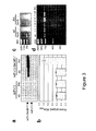
- FIGS. 3A-D show that antibody fragments and mRNA are ribosome-associated.
- Western blot analysis FIG. 3A
- ELISA FIG. 3B
- 70S ribosome fractions were prepared from cells expressing the unfused or SecM17-fused scFv constructs (wt and R4) as indicated.
- An equivalent amount of total protein was loaded in each lane or analyzed in each ELISA plate well.
- absorbance values for each sample were normalized to the absorbance measured for the ribosome fraction isolated from scFv13-R4-SecM17-expressing cells. Data is the average of three replicate experiments, and the error bars represent the standard error of the mean.
- FIGS. 4A-C show the solubility and binding activity of selected scFv13 fragments.
- Western blot analysis was conducted for soluble fractions recovered from BL21(DE3) cells expressing unfused versions of scFv13 and related variants from plasmid pET28a ( FIG. 4A ) and pTrc99A ( FIG. 4B ).
- Clones scFv13-R1, scFv13-R2, and scFv13-R4 were isolated previously after 1, 2, and 4 rounds of evolution, respectively (Martineau, et. al., “Expression of an Antibody Fragment at High Levels in the Bacterial Cytoplasm,” J. Mol. Biol.
- Clones S20 and S23 represent the two best clones isolated in this study after a single round of mutagenesis. Note that expression of all scFvs from pTrc99A was lower relative to expression from pET28a, however sample volumes and development time for pET28a blots were 4-fold and 10-fold reduced, respectively, relative to pTrc99A blots. An equivalent amount of total protein was loaded in each lane of FIGS. 4A and 4B . Blots were first probed with anti-FLAG IgG and, following stripping, reprobed with anti-GroEL IgG.
- FIG. 4C show the results of whole cell ⁇ -gal assays of intact X-gal treated AMEF 959 cells expressing wt scFv13 or related variants from plasmid pTrc99A. An equivalent number of cells were analyzed in each well.
- AMEF ⁇ -gal activation is reported as the change in X-gal hydrolysis for 959 cells expressing an scFv13 variant normalized to the change in X-gal hydrolysis for 959 cells expressing scFv13-R4.
- Data is the average of six replicate experiments and the error bars represent the standard error of the mean.
- Absorbance values for each sample were normalized to the absorbance measured for scFv13-R4-expressing cells.
- FIGS. 5A-B show the mutations in scFv13 fragments selected by intracellular ribosome display.
- FIG. 5A shows the amino acid sequence alignment of wt scFv13 (SEQ ID NO: 2) and related variants, S20 (SEQ ID NO: 3) and S23 (SEQ ID NO: 4). The sequence of wt scFv13 is written in single letter amino acid code. Numbering of amino acid residues in V H and V L , and the labeling of CDRs is according to Kabat numbering scheme. Immunodetection epitopes are italicized.
- FIG. 5B shows the location of mutations for clones S20 and S23 in scFv13 structure.
- the structure was previously modeled by homology (Martineau, et. al., “Expression of an Antibody Fragment at High Levels in the Bacterial Cytoplasm,” J. Mol. Biol. 280: 117-27 (1998), which is hereby incorporated by reference in its entirety).
- the drawing was generated with MacPyMOL.
- the V H is shown in olive, the V L in green, the disulfide bonds in black, and the mutations for clones S20 and S23 in red and purple, respectively.
- Asterisks indicate mutations that are shared between selected variants and those isolated previously by Martineau, et. al., “Expression of an Antibody Fragment at High Levels in the Bacterial Cytoplasm,” J. Mol. Biol. 280: 117-27 (1998), which is hereby incorporated by reference in its entirety).
- FIGS. 6A-D show the isolation of 70S ribosome fractions by sucrose density gradient centrifugation.
- the absorbance (254 nm) profile of gradient fractions shows accumulation of 70S ribosomes in fractions 23-28 (peak 70S-containing fraction indicated by asterisk).
- These figures show fractions generated from cells expressing: scFv13 ( FIG. 6A ); scFv13-R4 ( FIG. 6B ); scFv13-SecM17 ( FIG. 6C ); and scFv13-R4-SecM17 ( FIG. 6D ).
- One aspect of the present invention relates to a method of identifying a protein that binds to a target molecule and has intracellular functionality.
- This method includes providing a construct comprising a deoxyribonucleic acid molecule encoding the protein which binds to the target molecule, with the deoxyribonucleic acid molecule being coupled to a stall sequence.
- a host cell is transformed with the construct and then cultured under conditions effective to form, within the host cell, a complex of the protein whose translation has been stalled, the mRNA encoding the protein, and ribosomes.
- the protein in the complex is in a properly folded, active form and the complex is recovered from the cell.
- the protein that binds to the target molecule having intracellular functionality of the present invention can include any ligand binding protein. Suitable ligand binding proteins, include high-affinity antibody fragments (e.g., Fab, Fab′ and F(ab′) 2 ), single-chain Fv antibody fragments, nanobodies or nanobody fragments, fluorobodies, or aptamers.
- the protein is a single-chain variable fragment antibody, an antibody in which the heavy chain and the light chain of a traditional two chain antibody have been joined to form one chain.
- a linker peptide is inserted between the two chains to allow for proper folding and creation of an active binding site.
- the methods of the present invention can be used to generate libraries of single-chain antibodies that are cytoplasmically stable and intracellularly functional. The libraries of single-chain antibodies are useful for screening and selection of antibodies having the desired high-affinity binding properties.
- ligand binding proteins suitable for use in the methods of the present invention include biotin-binding proteins, lipid-binding proteins, periplasmic binding proteins, lectins, serum albumins, enzymes, phosphate and sulfate binding proteins, immunophilins, metallothionein, or various other receptor proteins.
- the method of the present invention can be used to generate peptide libraries that are useful for screening high-affinity ligand binding partners.
- a deoxyribonucleic acid molecule encoding the protein of interest is coupled to a stall sequence.
- a stall sequence is any sequence that interacts with residues in the ribosomal exit tunnel to stall translation, resulting in the display of the protein of interest on the ribosome surface.
- the SecM stall sequence is encoded by a nucleic acid sequence set forth in SEQ ID NO: 28: ttc agc acg ccc gtc tgg ata agc cag gcg caa ggc atc cgt get ggc cct.
- This corresponds to DNA bases 448-498 of the secM gene.
- Schmidt, et al. “Nucleotide Sequence of the secA Gene and secA (Ts) Mutations Preventing Protein Export in Escherichia Coli,” J. Bacteriol. 170:3404-14 (1988), which is hereby incorporated by reference in its entirety.
- a consensus for the SecM protein is FXXXXWIXXXXGIRAGP; where X can be any amino acid (SEQ ID NO: 26).
- Suitable stall sequences includes: (1) cat leader 5-mer peptide from Gram-positive bacteria; and (2) cmlA leader 8-mer peptide from Gram-negative bacteria (see Lovett, et al., “Nascent Peptide Regulation of Translation,” J Bacteriol 176(21):6415-7 (1994), which is hereby incorporated by reference in its entirety).
- the fusion protein i.e. the protein of interested coupled to a stall sequence
- the fusion protein can be prepared by translation of an in-frame fusion of the deoxyribonucleic acid molecule encoding the protein of interest and the polynucleotide stall sequences, i.e., a hybrid gene.
- the hybrid gene encoding the fusion polypeptide is inserted into an expression vector which is used to transform or transfect a host cell.
- the deoxyribonucleic acid molecule encoding the protein or polypeptide of interest is inserted into an expression vector in which the polynucleotide encoding the stall polypeptide is already present.
- the stall polypeptide or protein of the fusion protein is preferably fused to the C-terminal, end of the protein or polypeptide of interest.
- Fusions between the deoxyribonucleic acid molecule encoding the protein or polypeptide of interest and a stall polynucleotide sequence may be such that the nucleic acid sequence encoding the protein or polypeptide of interest is directly contiguous with the nucleic acid sequence encoding the stall polypeptide or protein of the present invention.
- the deoxyribonucleic acid molecule encoding the protein of interest may be coupled to the stall polynucleotide sequence by way of a linker sequence such as the flexible 8-residue Gly-Ser linker described herein having the sequence, AGSAAGSG (SEQ ID NO:27).
- the Gly-Ser linker may comprise between 10-50 Gly/Ser units depending on the optimal separation needed between the ribosome and the target protein.
- other suitable linkers include a Gly linker or the flexible linkers from an immunoglobulin disclosed in U.S. Pat. No. 5,516,637 to Huang et al, which is hereby incorporated by reference in its entirety.
- the linker may also contain a protease-specific cleavage site so that the protein of interest may be controllably released from the stall polypeptide or protein.
- protease sites include those specific to cleavage by factor Xa, enterokinase, collagenase, Igase (from Neisseria gonorrhoeae ), thrombin, and TEV (Tobacco Etch Virus) protease.
- one or more polynucleotide encoding marker proteins can also be positioned between the deoxyribonucleic acid molecule encoding the protein of interest and the stall polynucleotide sequence.
- Marker proteins are well known in the art and include affinity protein markers, such as chitin binding protein, maltose binding protein, glutathione-s-transferase, and the poly(His) tag; epitope markers, such as the V5-tag, c-myc-tag, HA-tag, or FLAG-tag.
- a c-Myc epitope tag, a 6 ⁇ -His tag, a thrombin cleavage site, and a linker are all positioned within the construct between the deoxyribonucleic acid molecule encoding the protein of interest and stalling sequence.
- the nucleic acid construct containing the deoxyribonucleic acid molecule encoding the protein of interest and the stall polynucleotide with the optional c-Myc epitope tag, 6 ⁇ -His tag, thrombin cleavage site, and linker positioned in between preferably also contains a polynucleotide sequence encoding a marker sequence upstream (5′) of the deoxyribonucleic acid molecule encoding the protein of interest.
- Any of the marker proteins mentioned above i.e.
- chitin binding protein maltose binding protein, glutathione-s-transferase, and the poly(His) tag; epitope markers, such as the VS-tag, c-myc-tag or the HA-tag) are suitable.
- a polynucleotide encoding a FLAG-tag is inserted upstream of the deoxyribonucleic acid molecule encoding the protein of interest.
- a stop codon is inserted at the 3′ end of the polynucleotide encoding the stall sequence.
- the nucleic acid construct encoding the fusion protein is inserted into an expression system to which the molecule is heterologous.
- the heterologous nucleic acid molecule is inserted into the expression system or vector in proper sense (5′ ⁇ 3′) orientation relative to the promoter and any other 5′ regulatory molecules, and correct reading frame.
- the preparation of the nucleic acid constructs can be carried out using standard cloning methods well known in the art as described by Sambrook et al., Molecular Cloning: A Laboratory Manual , Cold Springs Laboratory Press, Cold Springs Harbor, N.Y. (1989), which is hereby incorporated by reference in its entirety.
- U.S. Pat. No. 4,237,224 to Cohen and Boyer which is hereby incorporated by reference in its entirety, also describes the production of expression systems in the form of recombinant plasmids using restriction enzyme cleavage and ligation with DNA ligase.
- Suitable expression vectors include those which contain replicon and control sequences that are derived from species compatible with the host cell. For example, if E. coli is used as a host cell, plasmids such as pUC19, pUC18 or pBR322 may be used. Other suitable expression vectors are described in Molecular Cloning: a Laboratory Manual: 3rd edition, Sambrook and Russell, 2001, Cold Spring Harbor Laboratory Press, which is hereby incorporated by reference in its entirety. Many known techniques and protocols for manipulation of nucleic acid, for example in preparation of nucleic acid constructs, mutagenesis, sequencing, introduction of DNA into cells and gene expression, and analysis of proteins, are described in detail in Current Protocols in Molecular Biology , Ausubel et al. eds., (1992), which is hereby incorporated by reference in its entirety.
- RNA transcription and messenger RNA (“mRNA”) translation control many levels of gene expression (e.g., DNA transcription and messenger RNA (“mRNA”) translation) and subsequently the amount of fusion protein that is displayed on the ribosome surface.
- Transcription of DNA is dependent upon the presence of a promoter, which is a DNA sequence that directs the binding of RNA polymerase, and thereby promotes mRNA synthesis. Promoters vary in their “strength” (i.e., their ability to promote transcription). For the purposes of expressing a cloned gene, it is desirable to use strong promoters to obtain a high level of transcription and, hence, expression and surface display.
- any one of a number of suitable promoters may also be incorporated into the expression vector carrying the deoxyribonucleic acid molecule encoding the protein of interest coupled to a stall sequence.
- promoters such as the T7 phage promoter, lac promoter, trp promoter, recA promoter, ribosomal RNA promoter, the P R and P L promoters of coliphage lambda and others, including but not limited, to lacUV5, ompF, bla, lpp, and the like, may be used to direct high levels of transcription of adjacent DNA segments.
- a hybrid trp-lacUV5 (tac) promoter or other E. coli promoters produced by recombinant DNA or other synthetic DNA techniques may be used to provide for transcription of the inserted gene.
- SD Shine-Dalgarno
- Host cells suitable for expressing and displaying the fusion protein on the ribosome surface include any one of the more commonly available gram negative bacteria.
- Suitable microorganisms include Pseudomonas aeruginosa, Escherichia coil, Salmonella gastroenteritis ( typhimirium ), S. typhi, S. enteriditis, Shigella flexneri, S. sonnie, S dysenteriae, Neisseria gonorrhoeae, N. meningitides, Haemophilus influenzae H. pleuropneumoniae, Pasteurella haemolytica, P. multilocida, Legionella pneumophila, Treponema pallidum, T.
- fetus Helicobacter pylori, Francisella tularenisis, Vibrio cholerae, Vibrio parahaemolyticus, Bordetella pertussis, Burkholderie pseudomallei, Brucella abortus, B. susi, B. melitensis, B. canis, Spirillum minus, Pseudomonas mallei, Aeromonas hydrophila, A. salmonicida , and Yersinia pestis.
- the host cell is E. coli .
- An additional preferred embodiment includes the utilization of an E. coli host strain carrying mutations in the both thioredoxin reductase (trxB) and glutathione reductase (gar) genes (e.g., OrigamiTM) wherein disulfide bond formation in the cytoplasm is significantly enhanced.
- trxB gor mutant strain can be used to affinity- and/or stability-mature scFvs that are stalled and folded under oxidizing conditions.
- eukaryotic cells such as mammalian and yeast, and baculovirus systems are also suitable host cells that can be used in accordance with the methods of the present invention.
- Mammalian cell lines available in the art for expression of a heterologous polypeptide include Chinese hamster ovary cells, HeLa cells, baby hamster kidney cells, COS cells and many others. Methods for transforming/transfecting host cells with expression vectors are well-known in the art and depend on the host system selected, as described in Sambrook et al., Molecular Cloning: A Laboratory Manual , Cold Springs Laboratory Press, Cold Springs Harbor, N.Y. (1989), which is hereby incorporated by reference in its entirety.
- suitable techniques may include calcium phosphate transfection, DEAE-Dextran, electroporation, liposome-mediated transfection and transduction using retrovirus or other virus, e.g. vaccinia or, for insect cells, baculovirus.
- suitable techniques may include calcium chloride transformation, electroporation, and transfection using bacteriophage.
- a complex of the stabilized protein whose translation has been stalled, the mRNA encoding the protein, and ribosomes form.
- this complex is referred to as an “ARM” complex, i.e. antibody-ribosome and mRNA complex.
- Recovering the complex from the host cell can be carried out by affinity selection with an agent specific for the protein.
- cellular ribosome fractions are prepared and incubated with an immobilized antigen, protein binding partner, or marker protein binding partner that specifically recognizes and selectively binds to the protein of interest or marker protein that is displayed on the surface of the ribosome.
- the antigen, protein binding partner, or marker binding protein can be immobilized on any solid surface or support, such as a polystyrene microtiter plate, column, or a magnetic bead (e.g. Dynabeads®)
- the antigen or protein binding partner can be co-expressed in vivo, in the cell, along with the protein of interest displayed on the ribosome.
- the marker binding protein can be an antibody recognizing the epitope tag immobilized on the solid support.
- marker proteins having a polyhistidine-tag His-tag
- the complex can be recovered using affinity purification media such as, NTA-agarose, HisPur resin or Talon resin.
- Dissociating the bound protein-mRNA-ribosome complex from the solid support can be carried out using any appropriate chelating or elution buffer readily used in the art.
- the protein-mRNA-ribosome complex is dissociated using EDTA.
- the method of the present invention additionally includes isolating the mRNA from the recovered complex.
- the isolated mRNA is reverse transcribed to form a cDNA encoding the protein, and a construct, comprising the cDNA coupled to the stall sequence, is formed.
- the steps of transforming, culturing, and recovering, as described above, are repeated to enrich the protein recovered.
- RNA preparation that produces enzymatically manipulatable mRNA or analyzable RNA can be used in accordance with the present invention.
- the RNA can be isolated by using the guanidinium isothiocyanate-ultracentrifugation method, the guanidinium and phenol-chloroform method, the lithium chloride-SDS-urea method or poly A+/mRNA from tissue lysates using oligo(dT) cellulose method. It is important when isolating the RNA that enough high quality RNA is isolated.
- the mRNA can be reverse transcribed to form cDNA using any commercially available kit following manufacturer's instructions. Typically, the reaction is carried out using either oligo-dT or random decamer primers and a the reverse transcriptase enzyme.
- the steps of transforming, culturing, and recovering, as described above, are repeated to enrich the protein recovered.
- the enriched protein can be characterized by direct amino acid sequencing. Generally, protein sequencing is carried out using mass spectrometry or Edman degradation reaction.
- the protein can further be characterized based on affinity screening (i.e. is ability to bind to a ligand binding partner) using an panning, chromatography, or an ELISA based assay.
- affinity screening i.e. is ability to bind to a ligand binding partner
- the protein can also be characterized by its activity in an enzyme based assay.
- the stability of the identified protein of the present invention can be enhanced by altering or optimizing cellular conditions.
- Stability can generally be defined as the propensity of a molecule to exist in its folded and active state. Since stalled proteins, i.e. proteins that are displayed on the surface of the ribosome due to stalled translation, undergo folding in the cytoplasm, potent molecular chaperones and/or isomerases can be co-expressed in the host cell to enhance the stability, solubility and/or native folding capacity.
- Preferred molecular chaperones include DnaK, DnaH, GrpE, GroEL, or GroES and a preferred isomerase is the protein disulfide isomerase.
- the stability of the identified protein can also be enhanced via the addition of oxidized and reduced glutathione.
- the stability of the identified protein can further be enhanced by mutating the deoxyribonucleic acid molecule encoding the protein to produce variant nucleic acid sequences encoding variants with amino acid sequences. Methods of site directed or random mutagenesis are well known in the art and are suitable for use in the method of the present invention (See Current Protocols in Molecular Biology , Ausubel et al. eds., (1992), which is hereby incorporated by reference in its entirety).
- Another aspect of the present invention relates to a construct which includes a deoxyribonucleic acid molecule encoding a protein that binds to a target molecule and an SecM stalling sequence coupled to the deoxyribonucleic acid molecule.
- the deoxyribonucleic acid molecule and the SecM stalling sequence are coupled with sufficient distance between them to permit expression of their encoded protein, within the cell, in a properly folded, active form.
- This construct can be incorporated into an expression vector or a host cell as described above.
- Another aspect of the present invention relates to a method of identifying a protein that binds to a target molecule and has intracellular functionality.
- This method includes providing a construct comprising a deoxyribonucleic acid molecule encoding the protein which binds to the target molecule, said deoxyribonucleic acid molecule being coupled to a stall sequence.
- a cell-free extract preparation containing ribosomes is also provided.
- the method further involves contacting the construct with the cell-free extract preparation containing ribosomes under conditions effective for ribosome translation and the formation of a complex of the protein whose translation has been stalled, the mRNA encoding the protein, and the ribosomes.
- the protein in the complex is in a properly folded, active form and the complex is recovered.
- the protein to be identified can be any ligand binding protein and the stall sequence is any sequence that interacts with residues in the ribosomal exit tunnel to stall translation.
- the ligand binding protein is a single chain antibody.
- a primary advantage of using a cell-free translation system to achieve ribosome display is the ability to easily manipulate selection biases to enhance stability of the identified protein.
- stability can generally be defined as the propensity of a molecule to exist in its folded and active state.
- a stability selection pressure may disrupt or prevent a polypeptide folding correctly such that it does not attain an active or fully active state.
- a stability selection pressure may affect the ability of a polypeptide to remain in its folded and active state.
- a stability selection pressure may differentiate in some way between polypeptides that are in a folded and active state and those that are not.
- a stability selection pressure may be a chemical denaturant, such as urea, guanidine HCl (GuHCl) or thiocyanate, for example, sodium thiocyanate.
- a stability selection pressure may be a reducing agent, such as dithiothreitol (DTT), Tris[2-carboxyethyl]phosphine hydrochloride (TCEP), mercaptoethanol or glutathione.
- a stability selection pressure may be a physical denaturant, such as pH or temperature, in particular increased temperature.
- a selection pressure may be a protease or enzyme capable of degrading protein.
- a selection pressure may be depletion of chaperons or small molecule protein folding inhibitors.
- a stability selection pressure may be the use of hydrophobic interaction chromatography (HIC).
- Hydrophobic interaction chromatography is a technique for the separation of biomolecules based on differences in their surface hydrophobicity.
- HIC techniques have been used as a part of protein purification strategies as well as an analytical tool for the detection of protein conformational changes (reviewed in Queiroz et al., “Hydrophobic Interaction Chromatography of Proteins,” J. Biotech. 87: 143-159 (2001), which is hereby incorporated by reference in its entirety.
- HIC is based on hydrophobic attraction between the HIC matrix and the protein molecules.
- the HIC matrix consists of small non-polar groups (butyl, octyl or phenyl) attached to a hydrophilic polymer backbone (e.g.
- HIC cross-linked dextran or agarose
- Many proteins generally considered to be hydrophilic, also have sufficient numbers of hydrophobic groups allowing interaction with the HIC matrix.
- HIC is sensitive enough to interact with non-polar groups normally buried within the tertiary structure of the protein but exposed due to incorrect folding. The strength of the interaction is dependent upon the type of matrix, type and concentration of salt, pH, additives, and temperature.
- the present invention is suitable for a number of uses.
- scFv stability-enhanced single-chain variable fragment
- intracellular ribosome display method of the present invention the cytoplasmic stability, and thus intracellular function, of an scFc can be enhanced 2-3 fold after only a single round of mutagenesis and selection.
- Another use of the present invention involves isolation of functionally enhanced disulfide-bond containing antibody fragments.
- a trxB gor host mutant strain in which the redox potential of the cytoplasm favors the formation of disulfide bonds in proteins is used to affinity- and/or stability-mature scFvs that are stalled and folded under oxidizing conditions.
- the present invention can also be employed in the screening of na ⁇ ve libraries for cytoplasmically functional proteins with specific binding affinity to a given target molecule:
- the technology of the present invention is suited for the selection of intracellularly functional antibodies that bind a specific antigen target from na ⁇ ve (i.e., not stemming from preimmunized cells) libraries.
- Stability-enhancement/evolution of proteins under optimized cellular conditions can also be carried out with the present invention. Since stalled proteins undergo folding in the cytoplasm, it is relatively straightforward and inexpensive to optimize in vivo folding conditions by co-expressing potent molecular chaperones and/or isomerases.
- the present invention can be used for protein engineering experiments (i.e. by random mutagenesis) under conditions where the cellular environment is tuned to better suit the specific folding requirements of a particular target protein.
- the present invention can also be combined with in vitro/in vivo selection strategies for protein engineering via ribosome display. Since SecM17-mediated stalling was previously shown to operate in vitro, it is foreseeable that the SecM17-mediated antibody display strategy of the present invention could be performed akin to traditional in vitro ribosome display, in which all steps including transcription and translation are performed using a cell-free system. As a result, the need for transformation was eliminated, yielding extremely large (>10) antibody libraries.
- E. coli strain BL21(DE3) was used throughout except for in vivo ⁇ -gal activation experiments where the E. coli 959 strain was used which carries the AMEF ⁇ -gal gene (Martineau et al., “Expression of an Antibody Fragment at High Levels in the Bacterial Cytoplasm,” J Mol Biol 280:117-27 (1998), which is hereby incorporated by reference in its entirety). Plasmids encoding the SecM stall sequence fusions were constructed as follows.
- a 285-nucleotide segment of the secM gene containing the 17-amino acid stall sequence (FSTPVWISQAQGIRAGP (SEQ ID NO: 1)), plus additional downstream regions, was amplified from plasmid pNH21 by PCR using primers (5′-CTCATGGTCGACTTCAGCACGCCCGTCTGG-3′ (SEQ ID NO: 5)) and (5′-CTCATGCTCGAGTTAAAGCTTCTGCGCAACTGTTGGGAAGC-3′ (SEQ ID NO: 6)) to introduce a SalI restriction site at the 5′ end and an XhoI-HindIII restriction site at the 3′ end.
- This PCR product was SalI-XhoI digested and ligated into the same sites of pET28a (Novagen).
- Second, removal of the additional SecM downstream regions performed by introducing a HindIII restriction site immediately after the 17-residue stall sequence using site-directed mutagenesis (Stratagene QuikChange® Kit) and primers (5′-GGCATCCGTGCTGGCCCTAAGCTTCAACGCCTCACCTAACAA-3′ (SEQ ID NO: 7) and 5′-GTTGTTAGGTGAGGCGTTGAAGCTTAGGGCCAGCACGGATGCC-3′ (SEQ ID NO: 8)), followed by digestion with HindIII and self-ligation to yield pET-SecM.
- scFv13 and scFv13-R4 were amplified by PCR from plasmids pPM163 and pPM163-R4, (Martineau et al., “Expression of an Antibody Fragment at High Levels in the Bacterial Cytoplasm,” J Mol Biol 280:117-27 (1998), which is hereby incorporated by reference in its entirety) respectively, using primers (5′-GCGATGCCATGGCCGACTACAAGGACGATGACGACAAGGGAGCCGAGGT GCAGCTG-3′ (SEQ ID NO: 9) and 5′-GCGATGGTCGACTGCGGCCCATTCAG-3′ (SEQ ID NO: 10)) that introduce a FLAG epitope tag and an NcoI restriction site at the 5′ end and a SalI restriction site at the 3′ end of each scFv sequence.
- Each PCR product was digested with NcoI and SalI and ligated into NcoI-SalI-digested pET-SecM to yield the intermediate constructs pET-scFv13-SecM17′ and pET-scFv13-R4-SecM17′.
- a SacI restriction site was inserted immediately before the SalI restriction site using QuikChange® and primers (5′-GGATCTGAATGGGGCCGCAGAGCTCGTCGACTT CAGCACGCC-3′ (SEQ ID NO: 11) and 5′-GGCGTGCTGAAGTCGACGAGCTCTGCGGCCCCATTCAGA TCC-3′ (SEQ ID NO: 12)).
- a 6 ⁇ his tag, thrombin recognition sequence, and an AGSAAGSG (SEQ ID NO: 13) linker was introduced by amplifying a 670-nucleotide fragment from plasmid pET28-NDPK-GFP that contained these sequence elements plus the downstream NDPK sequence. SacI and SalI restriction sites were introduced at the 5′ and 3′ ends, respectively, of this 670-nucleotide fragment during amplification using primers (5′-CTCATGGAGCTCCATCATCATCATCATCATCACAGCAGCGGCCTGGTGC-3′ (SEQ ID NO: 14) and 5′-CTCATGGTCGACGCC AGAACCAGCAGCGG-3′ (SEQ ID NO: 15)).
- This PCR product was SacI-SalI-digested and ligated into similarly digested pET-scFv13-SecM17′ or pET-scFv13-R4-SecM17′.
- the additional NDPK sequence was excised by first inserting an EcoRI restriction site before the NDPK sequence using QuikChange® and primers (5′-GTGCCGCGCGGCAGCCATGAATTCATGCATGCTATAAATATTGC-3′ (SEQ ID NO: 16) and 5′-GCAATATTTATAGCATGCATGAATTCATGGCTGCCGCGCGGCAC-3′ (SEQ ID NO: 17)).
- a second EcoRI restriction site was inserted immediately after the NDPK sequence using the QuikChange® Kit and primers (5′-GAGGAGGTTTAGAGGAATTCGGATCCGCTGGCTCCG-3′ (SEQ ID NO: 18) and 5′-CGGAGCCAGCGGATCCGAATTCCTCTAAAACCTCCTC-3′ (SEQ ID NO: 19)).
- excision of NDPK with EcoRI and self-ligation yield the final pET-scFv13-SecM17 and pET-scFv13-R4-SecM17 vectors (illustrated in FIG. 1 ).
- Plasmids encoding the unfused scFv sequences were constructed by amplifying the scFv13 (or the scFv13 variants R1, R2 and R4) sequence by PCR from plasmids pPM163, pPM163-R1, pPM163-R2, and pPM163-R4 (Martineau et al., “Expression of an Antibody Fragment at High Levels in the Bacterial Cytoplasm,” J Mol Biol 280:117-27 (1998), which is hereby incorporated by reference in its entirety) with primers (5′-GCGATGCCATGGCCGACTACAAGGACGATGACGACAAGGGAGCCGAGGT GCAGCTG-3′ (SEQ ID NO: 20) and 5′-GCGATGGAGCTCTTATGCGGCCCCATTCAG-3′ (SEQ ID NO: 21)) that introduce a FLAG epitope tag and an NcoI restriction site at the 5′ end and a SacI restriction site at the 3′
- a library of random mutants was constructed by error-prone PCR of the scFv13 gene sequence using pET-scFv13-SecM17 as template and skewing the nucleotide and magnesium concentrations as described (DeLisa et al., “Genetic Analysis of the Twin Arginine Translocator Secretion Pathway in Bacteria,” J Biol Chem 277: 29825-31 (2002) and Fromant et al., “Direct Random Mutagenesis of Gene-Sized DNA Fragments Using Polymerase Chain Reaction,” Anal Biochem 224:347-53 (1995), which are hereby incorporated by reference in their entirety) to generate a 1.5% error-rate library.
- Error-prone PCR products were amplified with using primers (5′GCGATGCCATGGCCGACTACAAGGACGATGACGACAAGGGAGCCGAG GTGCAGCTG-3′ (SEQ ID NO: 22) and 5′-GCGATGGTCGACTGCGGCCCCATTCAG-3′ (SEQ ID NO: 23)), restriction digested with NcoI-SacI and ligated into the pET-scFv13-SecM17 that had previously been digested with NcoI-SacI to excise the wt scFv13 gene. Reaction mixtures were electroporated into E.
- cloni ExpressTM BL21(DE3) cells (Lucigen) and serial dilutions of these cells were plated on kanamycin (50 ⁇ g/ml) to determine the number of independent transformants. Transformed cells were selected on LB plates containing kanamycin (50 ⁇ g/ml). Library cells were pooled, cultured, and induced for scFv expression prior to isolation of ribosomes for panning and selection experiments.
- Cells transformed with the pET28a-derived scFv constructs were grown in 10-ml cultures at 37° C. in Luria-Bertani (LB) supplemented with kanamycin (50 ⁇ g/ml). Protein synthesis was induced by adding 1 mM isopropyl- ⁇ -D-thiogalactopyranoside (IPTG) when cells reached to mid log phase (OD 600 ⁇ 0.5). Cells were harvested after 1 hour of induction and pelleted by centrifugation for 15 min at 4° C. and 3,500 rpm. The soluble fraction was prepared by resuspending the pellet in 300 ml of phosphate buffered saline (PBS) solution followed by sonication (Branson Sonifier). The sonicant was spun for 15 min at 4° C. and 1,300 rpm and the resulting supernatant was collected as the soluble fraction.
- PBS phosphate buffered saline
- Ribosomes were isolated according to a procedure modified from Evans et al., “Homogeneous Stalled Ribosome Nascent Chain Complexes Produced in vivo or in vitro,” Nat Methods 2:757-62 (2005), which is hereby incorporated by reference in its entirety. Specifically, 100-ml cultures grown as above were induced with 1 mM IPTG at an OD600 ⁇ 0.5 and grown at 30° C. for an additional 30 min.
- ribosomes were isolated by ultracentrifugation for 35 h at 24,000 rpm and 4° C. using a Beckman LS 8 ultracentrifuge with an SW28 rotor.
- the crude ribosome pellet was resuspended in 200 ⁇ l Buffer C and ultracentrifuged in a 10 to 40% (w/v) sucrose gradient in Buffer A (20 nM Tris-HCl pH 7.5, 100 mM NH 4 Cl, 25 mM MgCl 2 ) for 17 h at 22,000 rpm and 4° C. in a SW41 rotor. Gradient fractionation was performed manually by pipetting 250 ⁇ l at a time from the top part of the gradient. All collected samples were stored at 4° C. for further analysis.
- a 96-well BD FALCON plate was coated with 65 ⁇ l of 1 mg/ml ⁇ -gal (Sigma) in PBS and left at 4° C. overnight.
- the plate was washed 4 times with 200 ⁇ l of PBS at room temperature and blocked with 200 ⁇ l of blocking solution (1% non-fat milk, 5 mM MgCl 2 , 2.5 mg/ml heparin, 0.05 mg/ml E. coli tRNA in PBS) for 2 h at room temperature. After 4 washes with washing solution (0.1% Tween 20, 5 mM MgCl 2 in PBS), the plate was incubated at 4° C. for 15-20 min.
- Isolated 70S ribosome samples were mixed gently with equal volume of cold blocking solution, and 100 ⁇ l this mixture was added to each well. The plate was incubated for 1 h at 4° C. and washed 5-6 times with 200 ⁇ l of cold washing solution at 4° C. to remove any unbound complexes.
- mRNA was dissociated from ribosome complexes by adding 100 ⁇ l of cold eluting solution (20 mM EDTA, 20 units/ml RNAsin in PBS) to each well and shaking gently for 30 min at 4° C. Samples were collected into cold microcentrifuge tubes after scraping the plate surface with a tip to ensure complete sample removal. mRNA was purified using the RNEasy Purification Kit (Qiagen).
- Reverse transcription PCR on the recovered mRNA was performed using the Sensiscript RT Kit (Qiagen) and reverse primer 5′-GCGATGGAGCTCTTATGCGGCCCCATTCAG-3′ (SEQ ID NO: 24), which binds the 3′ end of scFv13 and introduces a SacI restriction site and a stop codon.
- PCR amplification was performed in a second step with the same reverse primer and forward primer 5′-GCGGCGATGCCATGGCCGACTACAAGGACGATGACGACAAGGGAGGATC CGCCGAGGTGCAGCTG-3′ (SEQ ID NO: 25), which re-introduces a 5′ FLAG tag and NcoI restriction site.
- the PCR product was NcoI-SacI digested and ligated into similarly digested pET28a or pTrc99A.
- the pellet was dried of all remaining TCA and directly resuspended in 45 ⁇ l SDS-PAGE loading buffer.
- the following primary antibodies were used with the corresponding dilution in parenthesis: mouse anti-GroEL (1:10,000; Sigma); mouse anti-FLAG (1:1,500; Stratagene).
- the secondary antibodies were goat anti-mouse and goat anti-rabbit horseradish peroxidase conjugates (Promega) each diluted 1:10,000.
- a Bradford protein assay was performed on all samples to verify that an equal amount of total protein was loaded to each lane.
- fractions were normalized by rRNA content as measured by OD 260 (see FIG. 6 ).
- membranes were first probed with primary antibodies and, following development, stripped in Tris-buffered saline supplemented with 2% SDS and 0.7 M ⁇ -mercaptoethanol. Stripped membranes were reblocked and probed with anti-GroEL antibody.
- ⁇ -gal activity was measured using a whole cell assay modified from Arnold et al., “Influences of Transporter Associated with Antigen Processing (TAP) on the Repertoire of Peptides Associated With the Endoplasmic Reticulum-Resident Stress Protein gp96,” J Exp Med 186:461-6 (1997), which is hereby incorporated by reference in its entirety. Specifically, 5-bromo-4-chloro-3-indolyl- ⁇ -D-galactopyranoside (X-gal) was added in each well to a final concentration of 1 mM and absorbance at 620 nm was recorded over 4-10 h at 37° C. to measure active ⁇ -gal.
- TEP Antigen Processing
- human scFv13 was chosen as a model. This was originally isolated by Winter and coworkers in a two-step procedure: first, in vitro phage display was used to isolate scFv binders to native E.
- the solubility-enhanced scFv13-R4-SecM17 construct was enriched in ribosome fractions relative to the scFv13-SecM17 construct ( FIG. 3A ), indicating that the amount of stalled molecules correlates with the solubility of the displayed scFv.
- FIG. 3B only stalled ribosome complexes displaying the more soluble scFv13-R4-SecM17 construct were observed to strongly bind ⁇ -gal over background; scFv13-SecM17 constructs yielded only a low level of binding activity above background.
- Ribosome fractions prepared from cells expressing either scFv13 or scFv13-R4 in an unfused or SecM17-fused format were incubated with immobilized ⁇ -gal and bound ribosome complexes were dissociated with EDTA.
- Ribosome-associated RNA, including stalled mRNA, was isolated from dissociated complexes and the mRNA encoding the scFv13 sequence was amplified using RT-PCR using general primers that annealed to both scFv13 and scFv13-R4.
- ribosome fractions corresponding to unfused scFvs yielded no distinct PCR bands whereas both the scFv13-SecM17 and the solubility-enhanced scFv13-R4-SecM17 constructs gave rise to a substantial PCR product corresponding in size to the full-length scFv13 sequence ( FIG. 3C ); sequencing of each PCR product identified these as the wt scFv13 and scFv13-R4 mRNA sequences, respectively.
- the resulting error-prone PCR product was ligated in-frame with the SecM stall sequence and, following transformation, a cell library containing ⁇ 5 ⁇ 10 6 transformants was obtained.
- Members of this cell library were induced and ribosome fractions were prepared and screened by panning on immobilized ⁇ -gal to isolate clones exhibiting enhanced solubility relative to wt scFv13.
- a total of twelve unique clones were obtained after only a single round of selection and each was evaluated in an unfused format (i.e., lacking the SecM stall sequence in pET28a) for soluble expression and antigen binding.
- the clones of the present invention compare quite favorably with scFv13 clones R1 and R2 that were isolated by Martineau et al., “Expression of an Antibody Fragment at High Levels in the Bacterial Cytoplasm,” J Mol Biol 280:117-27 (1998), which is hereby incorporated by reference in its entirety, following one and two rounds of mutagenesis and selection, respectively.
- the present intracellular display strategy is suitable to isolate any stability-enhanced antibody with binding affinity for any antigen (not limited to ⁇ -gal).
- the only requirements, which are the same for traditional in vitro ribosome display, are that the antibody fragment must be amenable to display in the context of ARM complexes and that the binding target is known and available in a purified form to allow for selection.
- the human antibody fragment represented an attractive model protein owing to its ability to function as an intrabody; hence, solubility improvement accomplished using the present assay could be conveniently verified by monitoring activation of a ⁇ -gal mutant (AMEF) in vivo.
- AMEF ⁇ -gal mutant
- the present selection procedure to interrogate scFv13 error-prone library members was entirely independent of this cytoplasmic activity assay.
- the simplest measures of improved performance of an antibody evolved using this system are: (i) solubility as determined by Western blotting of the soluble fraction; and (ii) function as determined by ELISA.
- the engineering of functional intrabodies is only one potential application of the present system.
- intracellular ribosome display offers a number of advantages.
- coli strains such as trxB gor mutants in which the redox potential of the cytoplasm favors the formation of disulfide bonds in proteins (Bessette et al., “Efficient Folding of Proteins With Multiple Disulfide Bonds in the Escherichia coli Cytoplasm,” Proc Natl Acad Sci USA 96:13703-8 (1999), which is hereby incorporated by reference in its entirety). While wt E. coli were used in this study to isolate scFvs that were stable in the reducing cytoplasmic environment, one could employ a trxB gor host strain to affinity- and/or stability-mature scFvs that are stalled and folded under oxidizing conditions.
- scFv proteins enriched by intracellular ribosome display are naturally predisposed for in vivo expression and function.
- those enriched by in vitro ribosome display often do not express well in vivo and thus require refolding from inclusion bodies (Hanes et al., “Ribosome Display Efficiently Selects and Evolves High-Affinity Antibodies in vitro from Immune Libraries,” Proc Natl Acad Sci USA 95:14130-5 (1998), which is hereby incorporated by reference in its entirety) which can be laborious and time-consuming.
- intracellular ribosome display is a powerful complementary method to in vitro ribosome display for the directed evolution of proteins and should find use in the engineering of potent binding proteins that are soluble inside host cells for applications in functional genomics and proteomics as well as molecular medicine.
Landscapes
- Life Sciences & Earth Sciences (AREA)
- Health & Medical Sciences (AREA)
- Genetics & Genomics (AREA)
- Chemical & Material Sciences (AREA)
- Engineering & Computer Science (AREA)
- General Engineering & Computer Science (AREA)
- Bioinformatics & Cheminformatics (AREA)
- Biomedical Technology (AREA)
- Organic Chemistry (AREA)
- Biotechnology (AREA)
- Zoology (AREA)
- Wood Science & Technology (AREA)
- Crystallography & Structural Chemistry (AREA)
- Plant Pathology (AREA)
- Molecular Biology (AREA)
- Microbiology (AREA)
- Bioinformatics & Computational Biology (AREA)
- Physics & Mathematics (AREA)
- Biochemistry (AREA)
- General Health & Medical Sciences (AREA)
- Biophysics (AREA)
- Preparation Of Compounds By Using Micro-Organisms (AREA)
- Peptides Or Proteins (AREA)
Abstract
Description
Claims (34)
Priority Applications (1)
| Application Number | Priority Date | Filing Date | Title |
|---|---|---|---|
| US12/671,446 US9879252B2 (en) | 2007-06-13 | 2008-07-31 | Protein discovery using intracellular ribosome display |
Applications Claiming Priority (4)
| Application Number | Priority Date | Filing Date | Title |
|---|---|---|---|
| US93445407P | 2007-06-13 | 2007-06-13 | |
| US95305007P | 2007-07-31 | 2007-07-31 | |
| US12/671,446 US9879252B2 (en) | 2007-06-13 | 2008-07-31 | Protein discovery using intracellular ribosome display |
| PCT/US2008/071747 WO2009018438A1 (en) | 2007-07-31 | 2008-07-31 | Protein discovery using intracellular ribosome display |
Publications (2)
| Publication Number | Publication Date |
|---|---|
| US20110008774A1 US20110008774A1 (en) | 2011-01-13 |
| US9879252B2 true US9879252B2 (en) | 2018-01-30 |
Family
ID=43427759
Family Applications (1)
| Application Number | Title | Priority Date | Filing Date |
|---|---|---|---|
| US12/671,446 Active 2028-09-21 US9879252B2 (en) | 2007-06-13 | 2008-07-31 | Protein discovery using intracellular ribosome display |
Country Status (1)
| Country | Link |
|---|---|
| US (1) | US9879252B2 (en) |
Families Citing this family (6)
| Publication number | Priority date | Publication date | Assignee | Title |
|---|---|---|---|---|
| US10011830B2 (en) | 2013-05-19 | 2018-07-03 | The Board of Trustee of theLeland Stanford junior University | Devices and methods for display of encoded peptides, polypeptides, and proteins on DNA |
| CA2937524A1 (en) | 2014-02-05 | 2015-08-13 | Molecular Templates, Inc. | Methods of screening, selecting, and identifying cytotoxic recombinant polypeptides based on an interim diminution of ribotoxicity |
| US9840424B2 (en) * | 2015-07-09 | 2017-12-12 | Haier Us Appliance Solutions, Inc. | Water filter assembly for a beverage dispenser |
| US11174479B2 (en) | 2018-08-13 | 2021-11-16 | The Board Of Trustees Of The Leland Stanford Junior University | Devices and methods for display of encoded peptides, PolyPeptides, and proteins on DNA |
| WO2020215044A1 (en) * | 2019-04-17 | 2020-10-22 | The Regents Of The University Of California | Small molecule-based control of immune cell receptor expression |
| JP2023517008A (en) * | 2020-03-03 | 2023-04-21 | ネクスモス カンパニー リミテッド | DNA aptamer that specifically binds to glutathione and use thereof |
Citations (5)
| Publication number | Priority date | Publication date | Assignee | Title |
|---|---|---|---|---|
| US5922545A (en) * | 1993-10-29 | 1999-07-13 | Affymax Technologies N.V. | In vitro peptide and antibody display libraries |
| US6194550B1 (en) * | 1990-08-02 | 2001-02-27 | Larry Gold | Systematic polypeptide evolution by reverse translation |
| US20030165945A1 (en) | 2000-04-28 | 2003-09-04 | Bird Timothy A. | Human Pellino polypeptides |
| US20040265984A1 (en) | 2001-09-24 | 2004-12-30 | Ada Yonath | Methods of growing crystals of free, antibiotic-complexed, and substrate-complexed large ribosomal subunits, and methods of rationally designing or identifying antibiotics using structure coordinate data derived from such crystals |
| US20060177862A1 (en) | 2000-03-31 | 2006-08-10 | Cambridge Antibody Technology Limited | Ribosome display |
-
2008
- 2008-07-31 US US12/671,446 patent/US9879252B2/en active Active
Patent Citations (5)
| Publication number | Priority date | Publication date | Assignee | Title |
|---|---|---|---|---|
| US6194550B1 (en) * | 1990-08-02 | 2001-02-27 | Larry Gold | Systematic polypeptide evolution by reverse translation |
| US5922545A (en) * | 1993-10-29 | 1999-07-13 | Affymax Technologies N.V. | In vitro peptide and antibody display libraries |
| US20060177862A1 (en) | 2000-03-31 | 2006-08-10 | Cambridge Antibody Technology Limited | Ribosome display |
| US20030165945A1 (en) | 2000-04-28 | 2003-09-04 | Bird Timothy A. | Human Pellino polypeptides |
| US20040265984A1 (en) | 2001-09-24 | 2004-12-30 | Ada Yonath | Methods of growing crystals of free, antibiotic-complexed, and substrate-complexed large ribosomal subunits, and methods of rationally designing or identifying antibiotics using structure coordinate data derived from such crystals |
Non-Patent Citations (8)
| Title |
|---|
| Evans et al. "Homogeneous stalled ribosome nascent chain complexes produced in vivo or in vitro", Nature Methods , vol. 2 No. 10 , Oct. 2005. * |
| Evans et al., "Homogeneous Stalled Ribosome Nascent Chain Complexes Produced in Vivo or in Vitro," and Supplementary Methods, Nat. Methods 2(10):757-62 (2005). |
| Fitzgerald et al., Protein complex expression by using multigene baculoviral vectors, Nature Methods, vol. 3 No. 12, Dec. 2006, 1021-1032. * |
| Hanes et al., "In vitro selection and evolution of functional proteins by using ribosome display," Proc. Natl. Acad. Sci. USA vol. 94, pp. 4937-4942, May 1997. * |
| International Preliminary Report on Patentability for corresponding PCT/US2008/071747 (dated Feb. 2, 2010). |
| PCT International Search Report and Written Opinion for PCT/US2008/071747 dated Dec. 12, 2009. |
| Schraml et al., "In vitro protein engineering approaches for the development of biochemical, diagnistic and therapeutic tools" Dissertation, Nov. 16, 2005, catalogue No. urn:nbn:de:gbv:3-000009684. * |
| Sunohara et al. Ribosome Stalling during Translation Elongation Induces Cleavage of mRNA Being Translated in Escherichia coli, The Journal of Biological Chemistry, vol. 279, No. 15, Issue of Apr. 9, pp. 15368-15375, 2004. * |
Also Published As
| Publication number | Publication date |
|---|---|
| US20110008774A1 (en) | 2011-01-13 |
Similar Documents
| Publication | Publication Date | Title |
|---|---|---|
| AU691817B2 (en) | Sbp members with a chemical moiety covalently bound within the binding site; production and selection thereof | |
| US20110251106A1 (en) | Pvii phage display | |
| US20040058403A1 (en) | Combinatorial protein library screening by periplasmic expression | |
| US9879252B2 (en) | Protein discovery using intracellular ribosome display | |
| US8969253B2 (en) | Method for screening phage display libraries against each other | |
| KR100961392B1 (en) | Method for producing antibody phage surface presentation library, antibody phage surface presentation library prepared by the method, phagemid vector comprising the antibody phage surface presentation library gene | |
| Levy et al. | Enhancement of antibody fragment secretion into the Escherichia coli periplasm by co-expression with the peptidyl prolyl isomerase, FkpA, in the cytoplasm | |
| EP1904634B1 (en) | Novel phage display technologies | |
| Contreras-Martínez et al. | Intracellular ribosome display via SecM translation arrest as a selection for antibodies with enhanced cytosolic stability | |
| WO2009018438A1 (en) | Protein discovery using intracellular ribosome display | |
| EP3440208B1 (en) | Vectors for cloning and expression of proteins, methods and applications thereof | |
| Dreier et al. | Rapid selection of high-affinity antibody scFv fragments using ribosome display | |
| JP2011178691A (en) | Catenin-binding probe and use thereof | |
| JP2012503983A (en) | Compatible display vector system | |
| Jostock et al. | Screening of molecular repertoires by microbial surface display | |
| Pavoni et al. | New display vector reduces biological bias for expression of antibodies in E. coli | |
| JP5809687B2 (en) | RNF8-FHA domain modified protein and method for producing the same | |
| EP1773994A2 (en) | Polypeptide | |
| JP2019510507A (en) | Method for producing a synthetic antibody library, said library and its application | |
| US11001833B2 (en) | Method and kit for generating high affinity binding agents | |
| Matsumura et al. | Recent progress and future prospects in protein display technologies as tools for proteomics | |
| EP1025246A1 (en) | Methods for identifying nucleic acid sequences and (poly)peptides which increase the expression yields of functional periplasmic proteins | |
| JP2023544990A (en) | Bacterial pili protein complex FimGt-DsF stabilizing protein complex for producing filamentous phages | |
| Iverson et al. | Isolation of binding proteins with high affinity to ligands | |
| HK1121781A (en) | Novel phage display technologies |
Legal Events
| Date | Code | Title | Description |
|---|---|---|---|
| AS | Assignment |
Owner name: NATIONAL SCIENCE FOUNDATION, VIRGINIA Free format text: CONFIRMATORY LICENSE;ASSIGNOR:CORNELL UNIVERSITY;REEL/FRAME:024541/0946 Effective date: 20100524 |
|
| AS | Assignment |
Owner name: CORNELL RESEARCH FOUNDATION, INC., NEW YORK Free format text: ASSIGNMENT OF ASSIGNORS INTEREST;ASSIGNORS:DELISA, MATTHEW P.;CONTRERAS-MARTINEZ, LYDIA;SIGNING DATES FROM 20100408 TO 20100514;REEL/FRAME:024708/0292 |
|
| STCF | Information on status: patent grant |
Free format text: PATENTED CASE |
|
| AS | Assignment |
Owner name: NATIONAL SCIENCE FOUNDATION, VIRGINIA Free format text: CONFIRMATORY LICENSE;ASSIGNOR:CORNELL UNIVERSITY;REEL/FRAME:045541/0190 Effective date: 20180321 |
|
| MAFP | Maintenance fee payment |
Free format text: PAYMENT OF MAINTENANCE FEE, 4TH YR, SMALL ENTITY (ORIGINAL EVENT CODE: M2551); ENTITY STATUS OF PATENT OWNER: SMALL ENTITY Year of fee payment: 4 |
|
| MAFP | Maintenance fee payment |
Free format text: PAYMENT OF MAINTENANCE FEE, 8TH YR, SMALL ENTITY (ORIGINAL EVENT CODE: M2552); ENTITY STATUS OF PATENT OWNER: SMALL ENTITY Year of fee payment: 8 |