US9763975B1 - Rheological treatment of brain ischemia by drag reducing polymers - Google Patents
Rheological treatment of brain ischemia by drag reducing polymers Download PDFInfo
- Publication number
- US9763975B1 US9763975B1 US14/789,669 US201514789669A US9763975B1 US 9763975 B1 US9763975 B1 US 9763975B1 US 201514789669 A US201514789669 A US 201514789669A US 9763975 B1 US9763975 B1 US 9763975B1
- Authority
- US
- United States
- Prior art keywords
- drp
- blood
- subject
- brain
- flow
- Prior art date
- Legal status (The legal status is an assumption and is not a legal conclusion. Google has not performed a legal analysis and makes no representation as to the accuracy of the status listed.)
- Active
Links
Images


Classifications
-
- A—HUMAN NECESSITIES
- A61—MEDICAL OR VETERINARY SCIENCE; HYGIENE
- A61K—PREPARATIONS FOR MEDICAL, DENTAL OR TOILETRY PURPOSES
- A61K31/00—Medicinal preparations containing organic active ingredients
- A61K31/74—Synthetic polymeric materials
- A61K31/765—Polymers containing oxygen
Definitions
- Compromised cerebral perfusion also known as brain ischemia after various cerebral insults such as cerebrovascular accidents (CVA) including ischemic or hemorrhagic stroke, subarachnoid hemorrhage, subdural hemorrhage, intracerebral hemorrhage or chronic conditions, and traumatic brain injury (TBI) invariably leads to neuronal death in a restricted area or “core” due to lack of oxygen and nutrient delivery.
- CVA cerebrovascular accidents
- TBI traumatic brain injury
- the brain is especially vulnerable to oxygen and nutrient deprivation because of its high rate of metabolism consuming 25% of whole body oxygen uptake while representing only approximately 3-4% of total body weight while lacking significant oxygen or glucose stores.
- Brain tissue oxygen is depleted within 6 seconds and the EEG is isoelectric with 25 seconds of complete circulatory arrest.
- Brain ischemia can be acute as in acute ischemic stroke or chronic (i.e., long-lasting) as in vascular dementia.
- Acute ischemia is a neurologic emergency because irreversible damage to tissues can occur within several minutes whereas chronic ischemia may occur in slow developing ischemic diseases as in vascular dementia, Alzheimer's Disease or in sickle cell disease.
- Stroke and traumatic brain injury are the most frequent cerebral insults complicated by acute ischemia, and are serious global health problems causing long-term disability.
- Each year, about 800,000 people suffer new or recurrent stroke, which kills 130,000 of them and causes loss to the United States economy of $71.55 billion [1, 2].
- Annually, 1.7 million people suffer TBI, 52,000 die and 275,000 are hospitalized for long periods of time due to secondary injury complications [3, 4].
- the annual economic burden for TBI in the United States is approximately $60 billion. More than 1.1 million Americans are living with permanent functional disabilities resulting from stroke and 5.3 million from TBI representing more than 2% of the US population.
- ICP intracranial pressure
- AD Alzheimer's disease
- AD Alzheimer's disease
- the quantitative neuropathologic criteria for AD diagnostics as well as the main target for treatment options are the degree of deposition of amyloid plaques and Tau protein neurofibrillary tangles [2].
- treatments aimed to prevent or remove amyloid plaques have not succeeded in preventing or reducing dementia [3, 4].
- tissue plasminogen activator tPA
- tPA tissue plasminogen activator
- the present disclosure provides methods and compositions for the treatment of acute or chronic cerebral ischemia by targeting the very basis of ischemia by improving the rheology of blood flow through arteries and capillaries and as such would be relevant in the treatment, amelioration, or prevention of acute and chronic ischemic conditions of various etiologies.
- the present disclosure provides linear, blood soluble, non-toxic macromolecules, referred to herein as drag-reducing polymers (DRPs) for the treatment, amelioration or prevention of brain (and other organs) ischemia and other conditions or injuries causing or potentially leading to ischemic injury.
- DRPs drag-reducing polymers
- the present disclosure provides a method for treating, ameliorating or preventing brain (and other organs) ischemia of varying etiologies by the administration of DRP to a subject.
- FIG. 1 is the chemical formula of polyethylene oxide.
- FIG. 2 is a 2PLSM micrograph of rat cortex microvasculature with line scans settings for arteriolar blood flow velocity profiles (superimposed stripes).
- FIG. 3 shows arteriolar blood flow velocity profiles before DRP injection.
- FIG. 4 shows arteriolar blood flow velocity profiles after DRP injection. Increased velocity is seen near the vessel walls ( ⁇ , mm/s).
- FIG. 5 shows the volume from which every individual capillary flow was recorded.
- FIG. 6 a is a line-scan from the capillary before DRP injection.
- FIG. 6 b is a line-scan from the capillary after DRP injection.
- FIG. 7 is a frequency histogram showing flow velocity increase after DRP injection.
- FIG. 8 shows cortical NADH fluorescence at baseline.
- FIG. 9 shows cortical NADH fluorescence after DRP injection.
- FIG. 10 is a time plot from ROIs above showing NADH decrease after DRP injection.
- Gray lines ROIs values
- circles mean
- dashed line base line
- arrow DRP injection.
- FIG. 11 is a graph showing the acute effects of DRPs on rat permanent middle cerebral artery occlusion (pMCAO).
- Permanent MCAO causes reduction of microvascular RBC flow velocity, capillary stasis and tissue hypoxia (as reflected by NADH increase).
- DRPs improve cerebral flow, restore perfusion in collapsed capillaries and attenuate tissue hypoxia (NADH).
- Data normalized to baseline, Mean ⁇ SEM, n 5.
- FIG. 12 is a T2 MRI showing an ischemic lesion.
- FIG. 13 is a TTC-stained brain slice showing the infarcted tissue.
- FIG. 16 shows Fluoro-Jade staining for saline treated rats at 48 hours after pMCAO. superimposed line—neurodegeneration area.
- FIG. 17 shows Fluoro-Jade staining for DRP treated rats at 48 hours after pMCAO. superimposed line—neurodegeneration area.
- FIGS. 20A-20C are scatter plots of red blood cells (RBC) flow velocity versus vessel diameter showing stagnation of capillary flow and appearance of non-nutritive microvascular shunt (MVS) flow after ICP increase ( 20 B) compared to baseline 20 (A). DRP treatment partially restored capillary flow and reduced MVS flow ( 20 C). light grey outline—capillary flow, dark grey outline—MVS flow.
- RBC red blood cells
- MVS microvascular shunt
- FIG. 20A is the baseline flow.
- FIG. 20B is the MVS flow after ICP increase.
- FIG. 20C is flow after DRP treatment.
- FIGS. 21A and 21B show a fluid percussion injury model of traumatic brain injury (TBI).
- FIG. 21A is a representative T1 Magnetic Resonance image of rat brain before TBI.
- FIG. 21B is a representative T1 Magnetic Resonance image of rat brain 2 hours after TBI showing tissue damage in the left hemisphere.
- FIG. 22 is a bar graph reflecting brain edema. Calibration samples allowed for quantifying increased water content in the injured cortex and hippocampus.
- FIG. 23 is a bar graph showing that post-traumatic increase of microvascular (MVS) flow was less in the DRP treated group than in the saline-control as reflected by MVS/capillary (CAP) ratio.
- MVS microvascular
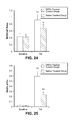
- FIG. 24 is a bar graph showing that after TBI fewer capillaries were collapsed in the DRP treated group than in the saline-control group.
- FIG. 25 is a bar graph showing that DRP treated animals showed less cortical tissue hypoxia than saline—control animals as reflected by NADH autofluorescence. Data a presented as ⁇ F/Fo, where Fo is pre-TBI baseline.
- FIG. 26 is a bar graph showing that DRP treated animals had less blood brain barrier damage than saline—control animals as reflected by less cortical tissue fluorescence due to less dye extravasation.
- Data a presented as ⁇ F/Fo, where Fo is pre-TBI baseline. All data are presented as Mean ⁇ SEM, n 5 per group, ** P ⁇ 0.01 compared to baseline ICP of 10 mmHg, P ⁇ 0.05 compared to ICP of 40 mmHg.
- the present disclosure provides novel methods and compositions for treating, ameliorating or preventing brain (and other organs) ischemia of various etiologies.
- brain ischemia caused by different conditions, accidents or injuries in a subject are treated, ameliorated or prevented by intravenous (or through any other route) delivery of one or more polymers that enhance blood flow by modulation of hemodynamics, thereby improving oxygen and nutrients delivery to tissue.
- polymers suitable for use with the presently described methods include linear, blood soluble non-toxic macromolecules with a molecular weight over 10 6 Daltons, which are commonly referred to as drag-reducing polymers (DRPs.)
- DRPs drag-reducing polymers
- Suitable DRPs exist as both naturally-occurring and synthetic polymers.
- naturally occurring DRPs include linear, blood soluble non-toxic polysaccharides and polypeptides derived from plants such as okra, aloe vera and others, algae, gums, polypeptides and polysaccharides derived from bacteria, polymers derived from fish slimes, sea-water and fresh-water biological growths, ovomucin of egg-whites, biopolymers derived from human or animal blood, blood plasma and blood cells.
- Non-natural synthetic polymers may be selected from of water soluble synthetic high-molecular weight polymers such as high molecular weight polyethylene oxides, polyacrylamides, etc.
- products with the following tradenames and available from the following companies may be useful: Polyethylene oxides (Polyox water soluble resins WSR-301, 309, N60K, N-750 and others, Union Carbide Co., USA) polyacrylamides (Praestol 2515TR, 2540TR and others, Stockhausen, Inc., Sweden), Carboxymethyl cellulose (Gum Technology Co.), gums such as Gum Guar (Sigma Chemical Co.), Tragacanth (Gum Technology Co.), Gum Karaya (Sigma Chemical Co.), Gum Xanthan (Sigma Chemical Co.).
- DRP farnesoid blood cell
- the precise mechanisms of action of DRPs are unknown, but they are thought to reduce pressure loss in small arteries and arterioles by diminishing flow separations and vortices at vessel bifurcations and thereby, increasing pre-capillary pressure, increasing the density of perfused capillaries and elimination of capillary stasis due to ischemia or other pathological conditions [9-14].
- the net effect is improved microcirculation and increased red blood cell (RBC) traffic in microvessels [12, 13, 15].
- RBC red blood cell
- DRP also reduced the thickness of the near-wall cell free layer that normally exists in vessels with ⁇ 300 ⁇ m diam. The alignment and stretching of the polymer molecules along the flow paths may diminish RBC rotation, which is an important contributor to the formation of the near-wall plasma layer [11, 12].
- a decrease of the cell-free layer also reduces plasma skimming at vessel bifurcations resulting in more RBC entering distal branches which increases the number of RBC in capillaries [11, 12] and decreases near-wall platelet concentration [14].
- Nanomolar concentrations of intravenous DRPs have been shown to improve hemodynamics and survival in animal models of the ischemic myocardium [16-18], ischemic limb [19] and hemorrhagic shock [11, 20][30, 32][26][33][31, 34].
- DRP qualitatively improved perfusion in rabbits after global ischemia [21].
- DRP increases blood flow rate in arterioles and enhances perfusion and oxygenation [22].
- DRPs restore perfusion in collapsed capillaries, reduces non-nutritive microvascular shunt flow and tissue hypoxia in hypertensive and traumatized brain [23].
- Modulation of the cerebral circulation by DRPs also overcame the effects of blood flow stasis in the ischemic penumbra after acute ischemic stroke as we reported recently [24].
- hemorheologic effects of intravenous DRPs translated into long-term improvement in clinically relevant anatomical and neurologic outcome.
- vascular wall shear stress which includes endothelium mediated pro-inflammatory processes and apoptosis.
- the vascular wall is highly sensitive to the hydrodynamic forces exerted on the endothelium by flowing blood, which affects the endothelial phenotype by regulating the activity of flow sensitive proteins [25].
- DRPs were previously shown to increase vessel wall shear stresses in microcirculation by modulation of the blood cell traffic [9].
- vascular wall shear stresses maintain levels of nitric oxide release and vasodilation by activation of eNOS at the apical membrane of endothelial cells. Endothelial wall shear stress controls apoptosis and regulates lymphocyte adhesion to the vessel wall all of which would be normalized and stabilized by DRPs, leading to improvement of compromised flow in the ischemic penumbra [26-28].
- DRPs by enhancement of systemic blood flow, increase collateral flow and, thus, can be considered as novel “collateral therapeutics” which overcome the effects of blood flow stasis in the penumbra after ischemic stroke.
- the present disclosure provides for the delivery of one or more types of DRPs to a subject before, during or after an ischemic event.
- DRPs could be administered, as a preventative measure, to a subject believed to be at risk of vascular dementia, during an ischemic event in an attempt to reduce or eliminate damage caused by the event, and as an ameliorative measure for a patient who has suffered a stroke after the time window for tPA or other known therapies has ended.
- delivery of the DRPs can be in conjunction with or in lieu of other therapies.
- the present disclosure further provides the use of DRPs in all acute and chronic brain (and other organ) ischemia caused by various etiologies in the acute as well as the recovery phase days after the initial insult, which could make a tremendous impact on the prevention of secondary brain damage as well as amelioration of the effects of the original event.
- the polymers described herein may be modified to increase their efficacy as biologicals.
- the polymers could be modified to increase water solubility and/or stability.
- the DRPs are delivered in nanomolar concentrations. According to various embodiments, the DRPs are delivered in such a way that a desired final blood concentration is achieved. According to some embodiments the desired final blood concentration is between 0.1 and 5 ( ⁇ g/ml). According to a more specific embodiment, the final blood concentration is 2 ppm.
- the DRP may be formulated as a sterile solution in an appropriate pharmaceutical buffer or carrier. Examples of suitable buffers include, but are not limited to, normal saline, phosphate buffer saline (PBS) or any other physiological saline.
- Delivery can be given intravenously as a single dose, multiple separate doses, or via a constant delivery mechanism (i.e. an infusion pump).
- the dose(s) may be delivered via a single injection, repetitive injection, or continuous dripping or infusion.
- the present disclosure provides a constant delivery mechanism designed to maintain a desired DRP concentration (e.g. between 0.1 and 5 ppm) in the subject's blood for an extended or even indefinite period of time. According to various embodiments, this extended period of time could be multiple hours, days, weeks, months, or even years.
- this constant delivery mechanism could be used in subjects suffering from chronic conditions such as Alzheimer's, Parkinson's or other type of vascular dementia.
- a device could be configured to monitor blood flow velocity (via, for example, an integrated Doppler) and/or DRP concentration and deliver an appropriate constant or variable dosage of DRP to maintain a desired blood flow velocity and/or DRP concentration.
- the “constant delivery mechanism” may not, in fact, deliver a constant dosage of DRP to the target subject, but rather would be configured to deliver an appropriate dosage when and as needed.
- other, non-intravenous delivery options may also be used oral, rectal, and transcutaneous methods.
- Examples of conditions and diseases that may receive benefit from the presently described methods include, but are not necessarily limited to: events/conditions/diseases affecting the brain including cerebrovascular accidents of various etiologies, including thrombosis, embolism, systemic hypoperfusion and venous thrombosis; traumatic brain injuries of various etiology, including injuries with secondary complications such as hemorrhagic shock and high intracranial pressure; global cerebral ischemia caused by cardiac arrest, heart failure or hemorrhagic shock; intracranial hypertension, leading to chronic ischemia, including idiopathic intracranial hypertension, hydrocephalus and pseudo tumor cerebri; mild cognitive impairment; vascular dementia; Alzheimer's disease; systemic lupus erythromatosus; multiple sclerosis; Moyamoya disease; transient ischemic attacks; hypertensive encephalopathy; to reduce aneurism formation or to prevent it's rupture by reducing turbulent flow; and other types of brain ischemia and conditions potentially leading
- Polyethylene oxide (PEO, MW ⁇ 4500 kDa) was dissolved in normal saline [0.9% of Sodium Chloride in water] to 0.1% (1000 ppm) and dialyzed against saline using a 50 kD cutoff membrane. Before injection, the PEO solution was diluted in saline to 50 ppm and slow rocked for ⁇ 2 hours. Prepared solution was sterilized using a 0.22 micron filter prior to injection. The solution was infused intravenously over 5 min to a final blood concentration of 2 ⁇ g/ml (ppm). The volume (mL) to be infused was determined by: ([rat weight (grams) ⁇ 0.07]/50 ppm).
- DRPs Enhanced the Arteriole Flow Profile and Increased RBC Flow Velocity in Capillaries Leading to Increased Tissue Oxygenation in the Healthy Rat Brain.
- the rat suture permanent middle cerebral artery occlusion (MCAO) model of acute focal brain ischemia was used. DRPs were injected i.v. 90 minutes after pMCAO. Controls were injected with an equal volume of saline.
- MCAO microvascular blood flow velocity and tissue oxygenation
- DRPs Restored Capillary Flow, Reduced Microvascular Shunt (MVS) Flow and Attenuated Hypoxia in Hypertensive Brain.
- ICP intracranial pressure
- MVS flow FIG. 20 A, B, D
- FIG. 20 C, D partially restored capillary flow and ameliorated MVS flow
- Fluid Percussion Injury Model of TBI Evaluation.
- TBI model resulting in sustained ICP increase by FPI transmitted onto the dura on the parietal cortex by a custom made gas driven device and tested FPI of different intensity [43, 44].
- T1 MRI FIGS. 21A , B
- FIGS. 21A , B revealed tissue damage in the left hemisphere after FPI ( FIG. 21 B).
- Glass tubes (below each brain) contain calibration solutions (50, 75 and 85% of water in D2O) for quantification of water content which increased in the injured cortex from 77.6 ⁇ 0.35 to 80.7 ⁇ 0.41% and in hippocampus, from 75.4 ⁇ 0.38 to 77.6 ⁇ 0.36% 2 hours after FPI ( FIG. 22 ).
- the rise in ICP was associated with an increase in the MVS/CAP ratio from 0.43 ⁇ 0.09 before injury to 1.39 ⁇ 0.23 after injury, respectively ( FIG. 6 b , P ⁇ 0.01).
- the MVS/CAP increased from 0.42 ⁇ 0.08 before injury to 0.85 ⁇ 0.25 after injury (P ⁇ 0.05), and was significantly lower than in control group ( FIG. 6 b , P ⁇ 0.05).
Landscapes
- Health & Medical Sciences (AREA)
- Chemical & Material Sciences (AREA)
- Medicinal Chemistry (AREA)
- Pharmacology & Pharmacy (AREA)
- Epidemiology (AREA)
- Life Sciences & Earth Sciences (AREA)
- Animal Behavior & Ethology (AREA)
- General Health & Medical Sciences (AREA)
- Public Health (AREA)
- Veterinary Medicine (AREA)
- Pharmaceuticals Containing Other Organic And Inorganic Compounds (AREA)
- Medicinal Preparation (AREA)
Abstract
Description
- 1. Minino, A. M., et al., Deaths: final data for 2008. Natl Vital Stat Rep, 2011. 59(10): p. 1-126.
- 2. http://www.cdc.gov/stroke/facts.htm.
- 3. Traumatic Brain Injury in the US. Center for Disease Control. 2010; Available from: http://braininjury.blogs.com/braininjury/2010/03/cdc-releases-latest-statistics-on-traumatic-brain-injury.html.
- 4. Langlois, J. A., W. Rutland-Brown, and M. M. Wald, The epidemiology and impact of traumatic brain injury: a brief overview. J Head Trauma Rehabil, 2006. 21(5): p. 375-8.
- 5. Adams, H., et al., Guidelines for the early management of patients with ischemic stroke: 2005 guidelines update a scientific statement from the Stroke Council of the American Heart Association/American Stroke Association. Stroke, 2005. 36(4): p. 916-23.
- 6. Adams, H. P., Jr., et al., Guidelines for the early management of adults with ischemic stroke: a guideline from the American Heart Association/American Stroke Association Stroke Council, Clinical Cardiology Council, Cardiovascular Radiology and Intervention Council, and the Atherosclerotic Peripheral Vascular Disease and Quality of Care Outcomes in Research Interdisciplinary Working Groups: the American Academy of Neurology affirms the value of this guideline as an educational tool for neurologists. Stroke, 2007. 38(5): p. 1655-711.
- 7. Albers, G. W., et al., Stroke Treatment Academic Industry Roundtable (STAIR) recommendations for maximizing the use of intravenous thrombolytics and expanding treatment options with intra-arterial and neuroprotective therapies. Stroke, 2011. 42(9): p. 2645-50.
- 8. Jain, K. K., Neuroprotection in cerebrovascular disease. Expert Opin Investig Drugs, 2000. 9(4): p. 695-711.
- 9. Kameneva, M. V., Microrheological effects of drag-reducing polymers in vitro and in vivo. International Journal of Engineering Science, 2012. 59: p. 168-183.
- 10. Kameneva, M. V., M. S. Poliakova, and I. A. Gvozdkova, [The nature of the effect of polymers reducing hydrodynamic resistance on blood circulation]. Dokl Akad Nauk SSSR, 1988. 298(5): p. 1253-6.
- 11. Kameneva, M. V., et al., Blood soluble drag-reducing polymers prevent lethality from hemorrhagic shock in acute animal experiments. Biorheology, 2004. 41(1): p. 53-64.
- 12. Marhefka, J. N., et al., Drag reducing polymers improve tissue perfusion via modification of the RBC traffic in microvessels. Biorheology, 2009. 46(4): p. 281-92.
- 13. Pacella, J. J., et al., Modulation of pre-capillary arteriolar pressure with drag-reducing polymers: a novel method for enhancing microvascular perfusion. Microcirculation, 2012. 19(7): p. 580-5.
- 14. Zhao, R., et al., Drag-reducing polymers diminish near-wall concentration of platelets in microchannel blood flow. Biorheology, 2010. 47(3-4): p. 193-203.
- 15. Pranevicius, M. and O. Pranevicius, Cerebral venous steal: blood flow diversion with increased tissue pressure. Neurosurgery, 2002. 51(5): p. 1267-73; discussion 1273-4.
- 16. Pacella, J. J., et al., A novel hydrodynamic approach to the treatment of coronary artery disease. Eur Heart J, 2006. 27(19): p. 2362-9.
- 17. Pacella, J. J., M. V. Kameneva, and F. S. Villanueva, Drag reducing polymers improve coronary flow reserve through modulation of capillary resistance. Biorheology, 2009. 46(5): p. 365-78.
- 18. Sakai, T., et al., I.V. infusion of a drag-reducing polymer extracted from aloe vera prolonged survival time in a rat model of acute myocardial ischaemia. Br J Anaesth, 2007. 98(1): p. 23-8.
- 19. Hu, F., et al., Improvement of the microcirculation in the acute ischemic rat limb during intravenous infusion of drag-reducing polymers. Biorheology, 2011. 48(3-4): p. 149-59.
- 20. McCloskey, C. A., et al., Tissue hypoxia activates JNK in the liver during hemorrhagic shock. Shock, 2004. 22(4): p. 380-6.
- 21. Gannushkina, I. V., et al., [Possibility of restoring the cerebral blood flow in cerebral ischemia by injecting special polymers into the blood]. Patol Fiziol Eksp Ter, 1982(3): p. 58-9.
- 22. Bragin, D. E., Thompson, S., Statom, G., Kameneva, M. V., Nemoto, E. M., Drag-reducing polymer improves microvascular flow and tissue oxygenation in the normal and traumatized rat brain. Journal of Neurotrauma, Abstracts from the 31st Annual National Neurotrauma Symposium, Nashville, Tenn., 2013. 30(15): p. C165.
- 23. Bragin, D. E., Thomson, S., Bragina, O. A., Statom, G., Kameneva, M. V., Nemoto, E. M. Drag reducing polymer enhances microvascular perfusion in the traumatized brain with intracranial hypertension. in Abstract book for the 15th International Symposium on Intracranial Pressure and Brain Monitoring 2013. Singapore.
- 24. Bragin, D. E., Peng, Z., Liu, W., Thomson, S., Statom, G., Kameneva, M. V., N, E. Memoto, Drag-Reducing Polymer Improves Microcirculation and Outcome after Permanent Middle Cerebral Artery Occlusion in Rats Stroke, Abstracts from the International Stroke Conference, San Diego, Calif., 2014. 45.
- 25. Topper, J. N. and M. A. Gimbrone, Jr., Blood flow and vascular gene expression: fluid shear stress as a modulator of endothelial phenotype. Mol Med Today, 1999. 5(1): p. 40-6.
- 26. Ji, J. Y., H. Jing, and S. L. Diamond, Hemodynamic regulation of inflammation at the endothelial-neutrophil interface. Ann Biomed Eng, 2008. 36(4): p. 586-95.
- 27. Resnick, N., et al., Fluid shear stress and the vascular endothelium: for better and for worse. Prog Biophys Mol Biol, 2003. 81(3): p. 177-99.
- 28. Yang, B. and V. Rizzo, Shear Stress Activates eNOS at the Endothelial Apical Surface Through 1 Containing Integrins and Caveolae. Cell Mol Bioeng, 2013. 6(3): p. 346-354.
- 29. Liebeskind, D. S., Collateral therapeutics for cerebral ischemia. Expert Rev Neurother, 2004. 4(2): p. 255-65.
- 30. Armitage, G. A., et al., Laser speckle contrast imaging of collateral bloodflow during acute ischemic stroke. J Cereb Blood Flow Metab, 2010. 30(8): p. 1432-6.
- 31. Wang, Z., et al., Dynamic change of collateral flow varying with distribution of regional blood flow in acute ischemic rat cortex. J Biomed Opt, 2012. 17(12): p. 125001.
- 32. Shin, H. K., et al., Mild induced hypertension improves blood flow and oxygen metabolism in transient focal cerebral ischemia. Stroke, 2008. 39(5): p. 1548-55.
- 33. Noor, R., et al., Partial intra-aortic occlusion improves perfusion deficits and infarct size following focal cerebral ischemia. J Neuroimaging, 2010. 20(3): p. 272-6.
- 34. Uflacker, R., C. Schonholz, and N. Papamitisakis, Interim report of the SENTIS trial: cerebral perfusion augmentation via partial aortic occlusion in acute ischemic stroke. J Cardiovasc Surg (Torino), 2008. 49(6): p. 715-21.
- 35. Liebeskind, D. S., Collaterals in acute stroke: beyond the clot. Neuroimaging Clin N Am, 2005. 15(3): p. 553-73, x.
- 36. Gerriets, T., et al., Noninvasive quantification of brain edema and the space-occupying effect in rat stroke models using magnetic resonance imaging. Stroke, 2004. 35(2): p. 566-71.
- 37. Lin, T. N., et al., Effect of brain edema on infarct volume in a focal cerebral ischemia model in rats. Stroke, 1993. 24(1): p. 117-21.
- 38. Butler, T. L., et al., Neurodegeneration in the rat hippocampus and striatum after middle cerebral artery occlusion. Brain Res, 2002. 929(2): p. 252-60.
- 39. Schmued, L. C. and K. J. Hopkins, Fluoro-Jade B: a high affinity fluorescent marker for the localization of neuronal degeneration. Brain Res, 2000. 874(2): p. 123-30.
- 40. Bragin, D. E., et al., Differential changes of glutathione levels in astrocytes and neurons in ischemic brains by two-photon imaging. J Cereb Blood Flow Metab, 2010. 30(4): p. 734-8.
- 41. Fujii, K., et al., Effect of antihypertensive treatment on focal cerebral infarction. Hypertension, 1992. 19(6 Pt 2): p. 713-6.
- 42. Bragin, D. E., et al., High intracranial pressure effects on cerebral cortical microvascular flow in rats. J Neurotrauma, 2011. 28(5): p. 775-85.
- 43. Bragin, D. E., Statom, G., Nemoto, E. M., Microvascular Shunt Flow after Traumatic Brain Injury with Intracranial Hypertension in Rats. Journal of Neurotrauma, Abstracts from The 31st Annual National Neurotrauma Symposium Aug. 4-7, 2013 Nashville, Tenn. 2012. 29: p. A-22.
- 44. Bragin, D. E., Statom, G., Nemoto, E. M., ICP-induced microvascular shunt flow involved in pathophysiological responses in the injured brain. Brain Injury, 2012. 26(4-5): p. 589.
- 45. Marhefka, J. N., et al., Poly(N-vinylformamide)-A drag-reducing polymer for biomedical applications. Biomacromolecules, 2006. 7(5): p. 1597-603.
-
- DRPs decreased infarct size at 24 hours after pMCAO by 29±12.4% and 24±8.1%, as evaluated by in-vivo T2-weighted Magnetic Resonance Imaging (MRI) and post-mortem triphenyltetrazolium chloride (TTC) staining, respectively, compared to saline treated rats (
FIGS. 12-15 p<0.05, n=4). Infarction volumes were calculated with DRPs correction for edema swelling by comparison with the contralateral hemisphere [36, 37]. - DRPs reduced neuronal degeneration as shown by Fluoro-Jade B stain [38, 39] at 48 hours after pMCAO. In saline-treated rats neurodegeneration involved nearly the entire cortex and striatum (
FIG. 16 ); and was smaller in DRP treated rats (FIG. 17 ). Quantification revealed that DRPs decreased the number of degenerating neurons by 28±10.3% (FIG. 18 , p<0.05, n=4). - DRPs improved neurologic outcome. At 24 hours after pMCAO, neurologic status was evaluated by a neurological scoring described by Fujii et al 1992 and in our publication [40, 41]. The average score was 2.8 for saline (n=5) and 1.9 for DRPs treated rats (n=5) showing preserved neurobehavioral function with DRP. Coordination and motor function was tested at one week after pMCAO by rotarod test. Saline animals showed a dramatic decrease in motor function reflected by a 52.7±16.1% reduction in latency compared to normal rats (
FIG. 19 , n=5). DRPs showed better recovery with a latency reduction by 24.4±15.7% compared to normal rats (FIG. 19 , n=5).
- DRPs decreased infarct size at 24 hours after pMCAO by 29±12.4% and 24±8.1%, as evaluated by in-vivo T2-weighted Magnetic Resonance Imaging (MRI) and post-mortem triphenyltetrazolium chloride (TTC) staining, respectively, compared to saline treated rats (
Claims (15)
Priority Applications (1)
| Application Number | Priority Date | Filing Date | Title |
|---|---|---|---|
| US14/789,669 US9763975B1 (en) | 2014-07-01 | 2015-07-01 | Rheological treatment of brain ischemia by drag reducing polymers |
Applications Claiming Priority (2)
| Application Number | Priority Date | Filing Date | Title |
|---|---|---|---|
| US201462019504P | 2014-07-01 | 2014-07-01 | |
| US14/789,669 US9763975B1 (en) | 2014-07-01 | 2015-07-01 | Rheological treatment of brain ischemia by drag reducing polymers |
Publications (1)
| Publication Number | Publication Date |
|---|---|
| US9763975B1 true US9763975B1 (en) | 2017-09-19 |
Family
ID=59828144
Family Applications (1)
| Application Number | Title | Priority Date | Filing Date |
|---|---|---|---|
| US14/789,669 Active US9763975B1 (en) | 2014-07-01 | 2015-07-01 | Rheological treatment of brain ischemia by drag reducing polymers |
Country Status (1)
| Country | Link |
|---|---|
| US (1) | US9763975B1 (en) |
Cited By (1)
| Publication number | Priority date | Publication date | Assignee | Title |
|---|---|---|---|---|
| CN111141639A (en) * | 2020-01-03 | 2020-05-12 | 哈尔滨工业大学 | The application of hyaluronic acid in the effect of blood rheology |
Citations (1)
| Publication number | Priority date | Publication date | Assignee | Title |
|---|---|---|---|---|
| US20100098768A1 (en) * | 2008-10-16 | 2010-04-22 | Clarkson University | Method of neuroprotection from oxidant injury using metal oxide nanoparticles |
-
2015
- 2015-07-01 US US14/789,669 patent/US9763975B1/en active Active
Patent Citations (1)
| Publication number | Priority date | Publication date | Assignee | Title |
|---|---|---|---|---|
| US20100098768A1 (en) * | 2008-10-16 | 2010-04-22 | Clarkson University | Method of neuroprotection from oxidant injury using metal oxide nanoparticles |
Non-Patent Citations (3)
| Title |
|---|
| Kameneva et al., "Blood soluble drag-reducing polymers prevent lethality from hemorrhagic shock in acute animal experiments" Biorheology 41 (2004) pp. 53-64. |
| Konorova et al., "Influence of Plasma DNA on Acid-Base Balance, Blood Gas Measurement, and Oxygen Transport in Health and Stroke" Ann. N.Y. Acad. Sci. 1137: 278-282 (2008). |
| Konorova et al., "Influence of Plasma DNA on Acid—Base Balance, Blood Gas Measurement, and Oxygen Transport in Health and Stroke" Ann. N.Y. Acad. Sci. 1137: 278-282 (2008). |
Cited By (1)
| Publication number | Priority date | Publication date | Assignee | Title |
|---|---|---|---|---|
| CN111141639A (en) * | 2020-01-03 | 2020-05-12 | 哈尔滨工业大学 | The application of hyaluronic acid in the effect of blood rheology |
Similar Documents
| Publication | Publication Date | Title |
|---|---|---|
| Neuwelt | Mechanisms of disease: the blood-brain barrier | |
| Gao et al. | Glymphatic system: an emerging therapeutic approach for neurological disorders | |
| Lutty et al. | Changes in choriocapillaris and retinal pigment epithelium in age-related macular degeneration | |
| Iliff et al. | Cerebral arterial pulsation drives paravascular CSF–interstitial fluid exchange in the murine brain | |
| Silvio Taccone et al. | Brain perfusion in sepsis | |
| JP2024504413A (en) | Complex for treatment of optic nerve diseases, method for its preparation and use thereof | |
| Liu et al. | Experimental alcoholism primes structural and functional impairment of the glymphatic pathway | |
| ES2428698T3 (en) | Use of therapeutic human albumin for the preparation of a medicine for the treatment of patients affected by cognitive disorders | |
| Wang et al. | Investigation of the mechanism of dural arteriovenous fistula formation induced by high intracranial venous pressure in a rabbit model | |
| Van Bergen et al. | Neutralization of placental growth factor as a novel treatment option in diabetic retinopathy | |
| JP2022508609A (en) | Improving glymphatic delivery by manipulating plasma osmolality | |
| US9763975B1 (en) | Rheological treatment of brain ischemia by drag reducing polymers | |
| US20190282792A1 (en) | Compounds for use in methods for treating glaucoma and retinal diseases | |
| US8288343B2 (en) | Activation of endothelial nitric oxide synthase by midkine and uses therefor in effecting vasodilation | |
| Wang et al. | Adiponectin-transfected endothelial progenitor cells have protective effects after 2-hour middle-cerebral artery occlusion in rats with type 2 diabetes mellitus | |
| US20120003180A1 (en) | Perlecan domain v protects, repairs and restores ischemic brain stroke injury and motor function | |
| Kuhn et al. | Current management and treatment of cerebral vasospasm complicating SAH | |
| Göksel et al. | Beta-thalassemia intermedia associated with moyamoya syndrome | |
| Cherniy | Topical aspects of infusion therapy | |
| Lekic et al. | Posterior circulation stroke and animal models | |
| Barić | Role of perivascular and paravascular drainage of Aß, iron ions, and waste products from the brain | |
| Barić | Increase in iron intracerebral concentration in patients suffering from Alzheimer's disease follows the rise of amyloid beta. | |
| US20180256686A1 (en) | Compositions and Methods for Treating Ischemia | |
| Gholamzadeh et al. | A review on the pretreatment effect of EPO on ischemic tolerance in different tissues with an approach to the tissue protection mechanisms | |
| Weinzierl et al. | Bromelain Protects Critically Perfused Musculocutaneous Flap Tissue from Necrosis. Biomedicines 2022, 10, 1449 |
Legal Events
| Date | Code | Title | Description |
|---|---|---|---|
| AS | Assignment |
Owner name: UNIVERSITY OF PITTSBURGH - OF THE COMMONWEALTH SYS Free format text: ASSIGNMENT OF ASSIGNORS INTEREST;ASSIGNOR:KAMENEVA, MARINA V.;REEL/FRAME:036652/0747 Effective date: 20150917 |
|
| STCF | Information on status: patent grant |
Free format text: PATENTED CASE |
|
| AS | Assignment |
Owner name: THE REGENTS OF THE UNIVERSITY OF NEW MEXICO, NEW M Free format text: ASSIGNMENT OF ASSIGNORS INTEREST;ASSIGNORS:BRAGIN, DENIS E.;NEMOTO, EDWIN M.;SIGNING DATES FROM 20180828 TO 20181127;REEL/FRAME:050512/0995 Owner name: STC.UNM, NEW MEXICO Free format text: ASSIGNMENT OF ASSIGNORS INTEREST;ASSIGNOR:THE REGENTS OF THE UNIVERSITY OF NEW MEXICO;REEL/FRAME:050513/0028 Effective date: 20181204 |
|
| MAFP | Maintenance fee payment |
Free format text: PAYMENT OF MAINTENANCE FEE, 4TH YR, SMALL ENTITY (ORIGINAL EVENT CODE: M2551); ENTITY STATUS OF PATENT OWNER: SMALL ENTITY Year of fee payment: 4 |
|
| MAFP | Maintenance fee payment |
Free format text: PAYMENT OF MAINTENANCE FEE, 8TH YR, SMALL ENTITY (ORIGINAL EVENT CODE: M2552); ENTITY STATUS OF PATENT OWNER: SMALL ENTITY Year of fee payment: 8 |