US9642922B2 - Caspase-triggered nano-aggregation probes and methods of use - Google Patents
Caspase-triggered nano-aggregation probes and methods of use Download PDFInfo
- Publication number
- US9642922B2 US9642922B2 US14/464,111 US201414464111A US9642922B2 US 9642922 B2 US9642922 B2 US 9642922B2 US 201414464111 A US201414464111 A US 201414464111A US 9642922 B2 US9642922 B2 US 9642922B2
- Authority
- US
- United States
- Prior art keywords
- probe
- moiety
- caspase
- activatable
- imaging
- Prior art date
- Legal status (The legal status is an assumption and is not a legal conclusion. Google has not performed a legal analysis and makes no representation as to the accuracy of the status listed.)
- Expired - Fee Related, expires
Links
- WOPLOTSXUMVKRX-GRFCFPJTSA-N C[C@@H]1CC(=O)[C@H]2CSC(=N2)C2=NC3=CC=C(C=C3C=C2)OCCCNC(=O)[C@H]2CSC(=N2)C2=NC3=C(C=C(C=C3)NC1=O)S2 Chemical compound C[C@@H]1CC(=O)[C@H]2CSC(=N2)C2=NC3=CC=C(C=C3C=C2)OCCCNC(=O)[C@H]2CSC(=N2)C2=NC3=C(C=C(C=C3)NC1=O)S2 WOPLOTSXUMVKRX-GRFCFPJTSA-N 0.000 description 3
- WDCCFUFXLKEPLA-VPALJEEZSA-N C.C.O=C=O.O=C=O.[2HH].[2HH].[C-]#[N+]C1=NC2=CC=C(OCCCNC(=C)[C@H]3CSC(C4=NC5=C(C=C(NC(=O)[C@@H](CCCCNC(=O)CN67CCN89CCN%10%11CCN%12(CC6)CC(=O)OC78%12%10(OC(=O)C9)OC(=O)C%11)NC(=O)[C@H](CSSCC)NC(=O)[C@H](CC(=O)O)CC(=O)[C@@H](NC(=O)[C@@H](CCC(=O)O)CC(=O)C(C)CC(=O)O)C(C)C)C=C5)S4)=N3)C=C2C=C1.[C-]#[N+]C1=NC2=CC=C(OCCCNC(=O)[C@H]3CSC(C4=NC5=C(C=C(NC(=O)[C@@H](CCCCNC)NC(=O)[C@H](CSSCC)NC)C=C5)S4)=N3)C=C2C=C1.[H]C[C@H](C)C(=O)N[C@@H](CCC(=O)O)C(=O)C[C@H](C(=O)N[C@@H](C[H])C(=O)CC(CSSCC)C(=O)NC(CC1=CN(CCOCC[18F])N=N1)C(=O)CC1=CC2=C(C=C1)N=C(C1=N[C@@H](C(=O)NCCCOC3=CC=C4N=C([N+]#[C-])C=CC4=C3)CS1)S2)C(C)C.[V] Chemical compound C.C.O=C=O.O=C=O.[2HH].[2HH].[C-]#[N+]C1=NC2=CC=C(OCCCNC(=C)[C@H]3CSC(C4=NC5=C(C=C(NC(=O)[C@@H](CCCCNC(=O)CN67CCN89CCN%10%11CCN%12(CC6)CC(=O)OC78%12%10(OC(=O)C9)OC(=O)C%11)NC(=O)[C@H](CSSCC)NC(=O)[C@H](CC(=O)O)CC(=O)[C@@H](NC(=O)[C@@H](CCC(=O)O)CC(=O)C(C)CC(=O)O)C(C)C)C=C5)S4)=N3)C=C2C=C1.[C-]#[N+]C1=NC2=CC=C(OCCCNC(=O)[C@H]3CSC(C4=NC5=C(C=C(NC(=O)[C@@H](CCCCNC)NC(=O)[C@H](CSSCC)NC)C=C5)S4)=N3)C=C2C=C1.[H]C[C@H](C)C(=O)N[C@@H](CCC(=O)O)C(=O)C[C@H](C(=O)N[C@@H](C[H])C(=O)CC(CSSCC)C(=O)NC(CC1=CN(CCOCC[18F])N=N1)C(=O)CC1=CC2=C(C=C1)N=C(C1=N[C@@H](C(=O)NCCCOC3=CC=C4N=C([N+]#[C-])C=CC4=C3)CS1)S2)C(C)C.[V] WDCCFUFXLKEPLA-VPALJEEZSA-N 0.000 description 2
- YHUREFZTTPAWQD-GZZXLJRUSA-N C.C.O=C=O.O=C=O.[2HH].[2HH].[C-]#[N+]C1=NC2=CC=C(OCCCNC(=O)[C@H]3CSC(C4=NC5=C(C=C(NC(=O)[C@@H](CCCCNC(=O)CN67CCN89CCN%10%11CCN%12(CC6)CC(=O)OC78%12%10(OC(=O)C9)OC(=O)C%11)NC(=O)[C@H](CSSCC)NC(=O)[C@H](CC(=O)O)CC(=O)[C@@H](NC(=O)[C@@H](CCC(=O)O)CC(=O)C(C)CC(=O)O)C(C)C)C=C5)S4)=N3)C=C2C=C1.[C-]#[N+]C1=NC2=CC=C(OCCCNC(=O)[C@H]3CSC(C4=NC5=C(C=C(NC(=O)[C@@H](CCCCNC)NC(=O)[C@H](CSSCC)NC)C=C5)S4)=N3)C=C2C=C1.[H]C[C@H](C)C(=O)N[C@@H](CCC(=O)O)C(=O)C[C@H](C(=O)N[C@@H](C[H])C(=O)CC(CSSCC)C(=O)NC(CC1=CN(CCOCC[18F])N=N1)C(=O)CC1=CC2=C(C=C1)N=C(C1=N[C@@H](C(=O)NCCCOC3=CC=C4N=C([N+]#[C-])C=CC4=C3)CS1)S2)C(C)C.[V] Chemical compound C.C.O=C=O.O=C=O.[2HH].[2HH].[C-]#[N+]C1=NC2=CC=C(OCCCNC(=O)[C@H]3CSC(C4=NC5=C(C=C(NC(=O)[C@@H](CCCCNC(=O)CN67CCN89CCN%10%11CCN%12(CC6)CC(=O)OC78%12%10(OC(=O)C9)OC(=O)C%11)NC(=O)[C@H](CSSCC)NC(=O)[C@H](CC(=O)O)CC(=O)[C@@H](NC(=O)[C@@H](CCC(=O)O)CC(=O)C(C)CC(=O)O)C(C)C)C=C5)S4)=N3)C=C2C=C1.[C-]#[N+]C1=NC2=CC=C(OCCCNC(=O)[C@H]3CSC(C4=NC5=C(C=C(NC(=O)[C@@H](CCCCNC)NC(=O)[C@H](CSSCC)NC)C=C5)S4)=N3)C=C2C=C1.[H]C[C@H](C)C(=O)N[C@@H](CCC(=O)O)C(=O)C[C@H](C(=O)N[C@@H](C[H])C(=O)CC(CSSCC)C(=O)NC(CC1=CN(CCOCC[18F])N=N1)C(=O)CC1=CC2=C(C=C1)N=C(C1=N[C@@H](C(=O)NCCCOC3=CC=C4N=C([N+]#[C-])C=CC4=C3)CS1)S2)C(C)C.[V] YHUREFZTTPAWQD-GZZXLJRUSA-N 0.000 description 2
- RZHYYYPYUPTBAU-QSVAGMGJSA-N C.O=C=O.O=C=O.[2HH].[2HH].[C-]#[N+]C1=NC2=CC=C(OCCCNC(=O)[C@H]3CSC(C4=NC5=C(C=C(NC(=O)[C@@H](CCCCNC(=O)CN67CCN89CCN%10%11CCN%12(CC6)CC(=O)OC78%12%10(OC(=O)C9)OC(=O)C%11)NC(=O)[C@H](CSSCC)NC(=O)[C@H](CC(=O)O)CC(=O)[C@@H](NC(=O)[C@@H](CCC(=O)O)CC(=O)C(C)CC(=O)O)C(C)C)C=C5)S4)=N3)C=C2C=C1.[C-]#[N+]C1=NC2=CC=C(OCCCNC(=O)[C@H]3CSC(C4=NC5=C(C=C(NC(=O)[C@@H](CCCCNC)NC(=O)[C@H](CSSCC)NC)C=C5)S4)=N3)C=C2C=C1.[H]C[C@H](C)C(=O)N[C@@H](CCC(=O)O)C(=O)C[C@H](C(=O)N[C@@H](C[H])C(=O)CC(CSSCC)C(=O)NC(CC1=CN(CCOCC[18F])N=N1)C(=O)CC1=CC2=C(C=C1)N=C(C1=N[C@@H](C(=O)NCCCOC3=CC=C4N=C([N+]#[C-])C=CC4=C3)CS1)S2)C(C)C.[V] Chemical compound C.O=C=O.O=C=O.[2HH].[2HH].[C-]#[N+]C1=NC2=CC=C(OCCCNC(=O)[C@H]3CSC(C4=NC5=C(C=C(NC(=O)[C@@H](CCCCNC(=O)CN67CCN89CCN%10%11CCN%12(CC6)CC(=O)OC78%12%10(OC(=O)C9)OC(=O)C%11)NC(=O)[C@H](CSSCC)NC(=O)[C@H](CC(=O)O)CC(=O)[C@@H](NC(=O)[C@@H](CCC(=O)O)CC(=O)C(C)CC(=O)O)C(C)C)C=C5)S4)=N3)C=C2C=C1.[C-]#[N+]C1=NC2=CC=C(OCCCNC(=O)[C@H]3CSC(C4=NC5=C(C=C(NC(=O)[C@@H](CCCCNC)NC(=O)[C@H](CSSCC)NC)C=C5)S4)=N3)C=C2C=C1.[H]C[C@H](C)C(=O)N[C@@H](CCC(=O)O)C(=O)C[C@H](C(=O)N[C@@H](C[H])C(=O)CC(CSSCC)C(=O)NC(CC1=CN(CCOCC[18F])N=N1)C(=O)CC1=CC2=C(C=C1)N=C(C1=N[C@@H](C(=O)NCCCOC3=CC=C4N=C([N+]#[C-])C=CC4=C3)CS1)S2)C(C)C.[V] RZHYYYPYUPTBAU-QSVAGMGJSA-N 0.000 description 1
- QMCKSLNVHHUVPZ-JIPXPUAJSA-N [C-]#[N+]C1=NC2=CC=C(OCCCNC(=C)[C@H]3CSC(C4=NC5=C(C=C(NC(=O)[C@@H](C)NC(=O)CCSSCC)C=C5)S4)=N3)C=C2C=C1 Chemical compound [C-]#[N+]C1=NC2=CC=C(OCCCNC(=C)[C@H]3CSC(C4=NC5=C(C=C(NC(=O)[C@@H](C)NC(=O)CCSSCC)C=C5)S4)=N3)C=C2C=C1 QMCKSLNVHHUVPZ-JIPXPUAJSA-N 0.000 description 1
- YPKCFSJPTFWDQU-KBMIEXCESA-N [C-]#[N+]C1=NC2=CC=C(OCCCNC(=O)[C@H]3CSC(C4=NC5=C(C=C(NC(=O)[C@@H](C)NC(=O)CCSSCC)C=C5)S4)=N3)C=C2C=C1 Chemical compound [C-]#[N+]C1=NC2=CC=C(OCCCNC(=O)[C@H]3CSC(C4=NC5=C(C=C(NC(=O)[C@@H](C)NC(=O)CCSSCC)C=C5)S4)=N3)C=C2C=C1 YPKCFSJPTFWDQU-KBMIEXCESA-N 0.000 description 1
Images


Classifications
-
- A—HUMAN NECESSITIES
- A61—MEDICAL OR VETERINARY SCIENCE; HYGIENE
- A61K—PREPARATIONS FOR MEDICAL, DENTAL OR TOILETRY PURPOSES
- A61K49/00—Preparations for testing in vivo
- A61K49/0002—General or multifunctional contrast agents, e.g. chelated agents
-
- A—HUMAN NECESSITIES
- A61—MEDICAL OR VETERINARY SCIENCE; HYGIENE
- A61K—PREPARATIONS FOR MEDICAL, DENTAL OR TOILETRY PURPOSES
- A61K49/00—Preparations for testing in vivo
- A61K49/001—Preparation for luminescence or biological staining
- A61K49/0013—Luminescence
- A61K49/0017—Fluorescence in vivo
- A61K49/0019—Fluorescence in vivo characterised by the fluorescent group, e.g. oligomeric, polymeric or dendritic molecules
- A61K49/0021—Fluorescence in vivo characterised by the fluorescent group, e.g. oligomeric, polymeric or dendritic molecules the fluorescent group being a small organic molecule
- A61K49/0032—Methine dyes, e.g. cyanine dyes
-
- A—HUMAN NECESSITIES
- A61—MEDICAL OR VETERINARY SCIENCE; HYGIENE
- A61K—PREPARATIONS FOR MEDICAL, DENTAL OR TOILETRY PURPOSES
- A61K49/00—Preparations for testing in vivo
- A61K49/001—Preparation for luminescence or biological staining
- A61K49/0013—Luminescence
- A61K49/0017—Fluorescence in vivo
- A61K49/0019—Fluorescence in vivo characterised by the fluorescent group, e.g. oligomeric, polymeric or dendritic molecules
- A61K49/0021—Fluorescence in vivo characterised by the fluorescent group, e.g. oligomeric, polymeric or dendritic molecules the fluorescent group being a small organic molecule
- A61K49/0041—Xanthene dyes, used in vivo, e.g. administered to a mice, e.g. rhodamines, rose Bengal
-
- A—HUMAN NECESSITIES
- A61—MEDICAL OR VETERINARY SCIENCE; HYGIENE
- A61K—PREPARATIONS FOR MEDICAL, DENTAL OR TOILETRY PURPOSES
- A61K49/00—Preparations for testing in vivo
- A61K49/001—Preparation for luminescence or biological staining
- A61K49/0013—Luminescence
- A61K49/0017—Fluorescence in vivo
- A61K49/005—Fluorescence in vivo characterised by the carrier molecule carrying the fluorescent agent
- A61K49/0052—Small organic molecules
-
- A—HUMAN NECESSITIES
- A61—MEDICAL OR VETERINARY SCIENCE; HYGIENE
- A61K—PREPARATIONS FOR MEDICAL, DENTAL OR TOILETRY PURPOSES
- A61K49/00—Preparations for testing in vivo
- A61K49/06—Nuclear magnetic resonance [NMR] contrast preparations; Magnetic resonance imaging [MRI] contrast preparations
- A61K49/08—Nuclear magnetic resonance [NMR] contrast preparations; Magnetic resonance imaging [MRI] contrast preparations characterised by the carrier
- A61K49/085—Nuclear magnetic resonance [NMR] contrast preparations; Magnetic resonance imaging [MRI] contrast preparations characterised by the carrier conjugated systems
-
- A—HUMAN NECESSITIES
- A61—MEDICAL OR VETERINARY SCIENCE; HYGIENE
- A61K—PREPARATIONS FOR MEDICAL, DENTAL OR TOILETRY PURPOSES
- A61K49/00—Preparations for testing in vivo
- A61K49/06—Nuclear magnetic resonance [NMR] contrast preparations; Magnetic resonance imaging [MRI] contrast preparations
- A61K49/08—Nuclear magnetic resonance [NMR] contrast preparations; Magnetic resonance imaging [MRI] contrast preparations characterised by the carrier
- A61K49/10—Organic compounds
- A61K49/101—Organic compounds the carrier being a complex-forming compound able to form MRI-active complexes with paramagnetic metals
-
- A—HUMAN NECESSITIES
- A61—MEDICAL OR VETERINARY SCIENCE; HYGIENE
- A61K—PREPARATIONS FOR MEDICAL, DENTAL OR TOILETRY PURPOSES
- A61K51/00—Preparations containing radioactive substances for use in therapy or testing in vivo
- A61K51/02—Preparations containing radioactive substances for use in therapy or testing in vivo characterised by the carrier, i.e. characterised by the agent or material covalently linked or complexing the radioactive nucleus
- A61K51/04—Organic compounds
- A61K51/041—Heterocyclic compounds
- A61K51/044—Heterocyclic compounds having nitrogen as a ring hetero atom, e.g. guanethidine, rifamycins
- A61K51/0455—Heterocyclic compounds having nitrogen as a ring hetero atom, e.g. guanethidine, rifamycins having six-membered rings with one nitrogen as the only ring hetero atom
-
- G—PHYSICS
- G01—MEASURING; TESTING
- G01N—INVESTIGATING OR ANALYSING MATERIALS BY DETERMINING THEIR CHEMICAL OR PHYSICAL PROPERTIES
- G01N33/00—Investigating or analysing materials by specific methods not covered by groups G01N1/00 - G01N31/00
- G01N33/48—Biological material, e.g. blood, urine; Haemocytometers
- G01N33/50—Chemical analysis of biological material, e.g. blood, urine; Testing involving biospecific ligand binding methods; Immunological testing
- G01N33/5005—Chemical analysis of biological material, e.g. blood, urine; Testing involving biospecific ligand binding methods; Immunological testing involving human or animal cells
-
- G—PHYSICS
- G01—MEASURING; TESTING
- G01N—INVESTIGATING OR ANALYSING MATERIALS BY DETERMINING THEIR CHEMICAL OR PHYSICAL PROPERTIES
- G01N2333/00—Assays involving biological materials from specific organisms or of a specific nature
- G01N2333/90—Enzymes; Proenzymes
- G01N2333/914—Hydrolases (3)
- G01N2333/948—Hydrolases (3) acting on peptide bonds (3.4)
- G01N2333/95—Proteinases, i.e. endopeptidases (3.4.21-3.4.99)
- G01N2333/964—Proteinases, i.e. endopeptidases (3.4.21-3.4.99) derived from animal tissue
- G01N2333/96425—Proteinases, i.e. endopeptidases (3.4.21-3.4.99) derived from animal tissue from mammals
- G01N2333/96427—Proteinases, i.e. endopeptidases (3.4.21-3.4.99) derived from animal tissue from mammals in general
- G01N2333/9643—Proteinases, i.e. endopeptidases (3.4.21-3.4.99) derived from animal tissue from mammals in general with EC number
- G01N2333/96466—Cysteine endopeptidases (3.4.22)
- G01N2333/96469—Interleukin 1-beta convertase-like enzymes
-
- G—PHYSICS
- G01—MEASURING; TESTING
- G01N—INVESTIGATING OR ANALYSING MATERIALS BY DETERMINING THEIR CHEMICAL OR PHYSICAL PROPERTIES
- G01N2510/00—Detection of programmed cell death, i.e. apoptosis
Definitions
- the present disclosure relates to caspase-triggered self-assembling nano-aggregation probes for the detection of apoptotic cells and tissues.
- the present disclosure further relates to methods of detecting and imaging cells and tissues that are apoptotic.
- membrane-permeable small molecules are shown to enter cells and undergo self-assembly after activation by intended cellular targets, such as an enzyme.
- MRI probes that can specifically report on enzyme activity have become particularly attractive due to variations in enzyme expression levels in many diseases.
- the inherently low detection sensitivity of MRI has limited the development of enzyme activatable MRI probes for in vivo application.
- novel activatable MRI probes enable can high spatial resolution imaging of specific enzyme activity in vivo and are thus in high demand.
- the disclosure provides embodiments of the synthesis, radiolabeling and biological applications of an activatable tracer that undergoes intramolecular cyclization and aggregation in apoptotic tumor cells upon caspase-3 activation.
- the aggregated nanoprobes of the disclosure may be detectable optically, by PET detection, magnetic resonance imaging, and the like depending on the detectable reporter attached to the nanoprobe.
- an activatable probe comprising a detachable capping moiety conjugated to a self-cyclizing molecule comprising a cysteine moiety, a 2-cyano-6-hydroxyquinoline moiety, an amino luciferin scaffold, and a detectable imaging moiety.
- the activatable probe can have the formula II, III, or IV:
- Another aspect of the disclosure encompasses embodiments of a pharmaceutically acceptable composition
- a pharmaceutically acceptable composition comprising an activatable probe according to the disclosure and a pharmaceutically acceptable carrier.
- nano-aggregation probe wherein said nanoprobe is an aggregate of an activated probe, wherein said activated probe comprises a self-cyclized molecule having the formula I and a detectable imaging moiety, wherein the detectable probe is a fluorophore or a radionuclide.
- Still another aspect of the disclosure encompasses embodiments of a method of forming a nano-aggregation probe comprising delivering to an apoptotic cell a pharmaceutically acceptable composition comprising an activatable probe, wherein said activatable probe can comprise a detachable capping moiety conjugated to a self-cyclizing molecule, said self-cyclizing molecule comprising a cysteine moiety, a 2-cyano-6-hydroxyquinoline moiety, an amino luciferin scaffold, and a detectable imaging moiety, wherein the detachable capping moiety has the amino acid sequence L-aspartate-glutamate-valine-aspartate and is selectively cleavable from the self-cyclizing molecule by caspase 3/7, whereupon the activatable probe can enter the apoptotic cell, an apoptopically-induced caspase 3/7 cleaves the detachable capping moiety from the activatable probe, and said probe aggregates to form the nano-aggregation probe.
- Still yet another aspect of the disclosure encompasses embodiments of a method of detecting an apoptotic cell, the method comprising the steps of: (i) delivering to the cytoplasm of an animal cell a pharmaceutically acceptable composition comprising an activatable probe, wherein said activatable probe comprises a detachable capping moiety conjugated to a self-cyclizing molecule, said self-cyclizing molecule comprising a cysteine moiety, a 2-cyano-6-hydroxyquinoline moiety, an amino luciferin scaffold, and a detectable imaging moiety, wherein the detachable capping moiety has the amino acid sequence L-aspartate-glutamate-valine-aspartate and is selectively cleavable from the self-cyclizing molecule by caspase 3/7, whereupon the activatable probe enters the apoptotic cell, an apoptopically-induced caspase 3/7 cleaves the detachable capping moiety from the activatable probe, and said probe aggregates to
- the detectable imaging moiety is a fluorophore and step (ii) can further comprise irradiating the recipient cell with an incident exciting energy, electronically detecting an emitted fluorescence, and measuring the intensity of said emission and optionally generating an image of the fluorescence.
- the detectable imaging moiety is detectable by positron electron transmission and step (ii) can further comprise detecting the nano-aggregation probe by PET imaging.
- FIGS. 1A-1C illustrate the mechanism of in vivo imaging of caspase-3/7 activity in human tumor xenograft mouse models by C-SNAF.
- FIG. 1A shows the caspase-3/7 and reduction-controlled conversion of C-SNAF to C-SNAF-cycl through the bioorthogonal intramolecular cyclization reaction, followed by self-assembly into nano-aggregates in situ.
- FIG. 1B illustrates that the fate of C-SNAF in vivo is dependent upon the tumor response to chemotherapy.
- C-SNAF extravasates into tumor tissue due to its small size.
- the pro-caspase-3 is inactive, and the DEVD capping peptide remains intact.
- C-SNAF can freely diffuse away from live tumor tissue, leading to low fluorescence.
- pro-caspase-3 is converted to active caspase-3.
- C-SNAF can readily enter dying cells due to the loss of plasma membrane integrity associated with apoptosis. After DEVD cleavage by active caspase-3 and disulfide reduction, C-SNAF undergoes macrocyclization and in situ nano-aggregation, leading to enhanced probe retention and high fluorescence.
- FIG. 1C illustrates the chemical structures of control probes L-ctrl and D-ctrl.
- FIG. 2A illustrates a scheme for the synthesis of C-SNAF. Reaction conditions: (a) TCEP, DIPEA, DCM/MeOH, Ar, room temperature 1 h; (b) (i) 20% TFA/DCM; (ii) PySSEt, MeOH, 71%; (c) Ac-Asp(O t Bu)-Glu(O t Bu)-Val-Asp(O t Bu)-COOH, HBTU, DIPEA, THF; (d) piperidine, DMF, 43% in two steps; (e) TFA/TIPSH/DCM (95%/2.5%/2.5%), 83%; (f) Cy 5.5-NHS, DMF, DIPEA, 53%.
- FIG. 2B illustrates a scheme for the synthesis of probes 15 and 16 from compound 4.
- (f) (t-BuO) 3 DOTA-COOH, HBTU, DIPEA, DMF, 3 h;
- (h) GdCl 3 .6H 2 O or EuCl 3 .6H 2 O, NaHCO 3 , pH 6-7, 75% for probe 15 and 79% for 1-Eu 3+ (16).
- FIG. 3A illustrates a scheme for the synthesis of L-ctrl. Reaction conditions: (a) TCEP, DIPEA, DCM/MeOH, Ar, room temperature 1 h; (b) (i) 20% TFA/DCM; (ii) PySSEt, MeOH; (c) Ac-Asp(O t Bu)-Glu(O t Bu)-Val-Asp(O t Bu)-COOH, HBTU, DIPEA, THF; (d) piperidine, DMF, 43% in two steps; (e) TFA/TIPSH/DCM (95%/2.5%/2.5%), 81%; (f) TCEP, NaHCO 3 , MeI, 91%; (g) Cy 5.5-NHS, DMF, DIPEA, 51%.
- FIG. 3B illustrates a scheme for the synthesis of 1-ctrl (19) from (8): Reaction conditions: (f) (t-BuO) 3 DOTA-COOH, HBTU, DIPEA, DMF, 3 h; (g) TFA/TIPSH/DCM (95%/2.5%/2.5%); 53% in two steps; (h) GdCl 3 .6H 2 O, pH 6-7, 82%.
- FIG. 4 illustrates a scheme for the synthesis of D-ctrl. Reaction conditions: (a) Ac- D -Asp(O t Bu)- D -Glu(O t Bu)- D -Val- D -Asp(O t Bu)-COOH, HBTU, DIPEA, THF; (b) piperidine, DMF, 42% in two steps; (c) TFA/TIPSH/DCM (95%/2.5%/2.5%), 81%; (d) TCEP, NaHCO 3 , MeI, 87%; (g) Cy 5.5-NHS, DMF, DIPEA, 47%.
- FIG. 5 illustrates a scheme for the synthesis of high-resolution imaging probe SIM-1 and control probe SIM-ctrl. Reaction conditions: (a) piperidine, DMF, 63%; (b) TCEP, DIPEA, DCM/MeOH, Ar, room temperature 1 h; (c) Alexa Fluor 488 5-TFP, DIPEA, DMF; (d) TFA/TIPSH/DCM (25%/72.5%/2.5%); (e) PySSEt, MeOH; (f) TCEP, NaHCO 3 , MeI.
- FIG. 6 illustrates a scheme for the synthesis of probe C-SNAF-SIM.
- FIGS. 7A-7C illustrate the in vitro characterization of caspase-3/7-sensitive nano-aggregation fluorescent probe (C-SNAF) of the disclosure.
- FIG. 7B illustrates the enzymatic reaction kinetics and specificity studies by longitudinal monitoring of the percentage conversion of C-SNAF (25 ⁇ M) to C-SNAF-cycl after incubation with equal masses (0.735 ⁇ g/ml) of recombinant human caspase-3, caspase-7, caspase-9, cathepsin B, and legumain, respectively.
- FIG. 7C is a digital TEM image of nano-aggregates after incubation C-SNAF (50 ⁇ M) with recombinant human caspase-3 (4.9 ⁇ 10 ⁇ 3 U/mL) overnight at 37° C. in caspase-3 buffer; scale bar, 1 ⁇ m.
- FIG. 8A is a scheme illustrating the molecular transformation of L-ctrl after incubation with caspase-3.
- FIG. 8B is a scheme illustrating the molecular transformation of D-ctrl after incubation with caspase-3.
- FIG. 9A illustrates overlay histograms from flow cytometry showing high Cy5.5 fluorescence in the apoptotic cells after incubation with 2 ⁇ M C-SNAF.
- FIG. 9B illustrates the quantitation of the fluorescent intensity of Cy5.5 in cells as derived from the flow cytometry analysis shown in FIG. 9A , and shows that apoptotic cells labeled with C-SNAF have approximately a 13-fold increased intensity compared to viable cells incubated with C-SNAF.
- the increased fluorescence was inhibited by the caspase-3/7 inhibitor Z-VAD-fmk (50 ⁇ M).
- FIG. 10A illustrates that overlay histograms obtained by flow cytometry analysis that show C-SNAF, but not control probes L-ctrl nor D-ctrl, can efficiently label DOX-induced apoptotic cells.
- Cells treated with caspase-3 inhibitor were not labeled by C-SNAF.
- Cells were treated with 5 ⁇ M DOX, or co-incubated with 5 ⁇ M DOX and 2 ⁇ M C-SNAF, L-ctrl, D-ctrl, or 2 ⁇ M C-SNAF together with 50 ⁇ M caspase inhibitor Z-VAD-fmk for 24 h.
- FIG. 10B illustrates the quantitation of the fluorescent intensity of Cy5.5 in cells derived from the flow cytometry analysis shown in FIG. 10A and shows that 5 ⁇ M DOX-induced apoptotic cells incubated with C-SNAF (blue) have approximately a 4-fold increased intensity compared to that without incubation or incubation with L-ctrl or D-ctrl. The labeling of apoptotic cells with C-SNAF was blocked by the caspase inhibitor.
- FIGS. 11A and 11B illustrate the imaging of caspase-3/7 activities in STS-treated cancer cells with C-SNAF.
- FIG. 11A illustrates a flow cytometry analysis of viable and STS-induced apoptotic HeLa cells after incubation with C-SNAF (2 ⁇ M), C-SNAF (2 ⁇ M) with caspase inhibitor Z-VAD-fmk (50 ⁇ M), or 2 ⁇ M of L-ctrl or D-ctrl.
- FIG. 11B is a series of digital fluorescence microscopy imaging of C-SNAF (2 ⁇ M) labeling STS-induced apoptotic HeLa cells.
- Cells were stained with nuclear binding probe Hoechst 33342 (blue). An extensive fluorescence was observed only in the apoptotic cells after incubation with C-SNAF, indicating a specifically intracellular accumulation of the probe after caspase-3/7-triggered macrocyclization and nano-aggregation.
- FIGS. 12A and 12B illustrate an analysis of caspase-3-triggered intramolecular condensation of C-SNAF in viable and STS (2 ⁇ M) treated HeLa cells.
- FIG. 12A shows HPLC traces of incubation of C-SNAF (5 ⁇ M) in viable and apoptotic HeLa cell lysates overnight at 37° C.
- HeLa cells (about 8 ⁇ 10 6 ) were either untreated or treated with 2 ⁇ M STS for 4 h and kept growing in blank culture medium for another 24 h after removal of STS. Cells were then lysed with RIPA buffer, and incubated with C-SNAF (5 ⁇ M) followed by analyzing the reaction by HPLC (678 nm UV detection).). Peaks * and ** indicate the disulfide reduction products T1 and T2; peak # indicates the cyclized product C-SNAF-cycl.
- FIG. 12B shows HPLC traces of viable and apoptotic HeLa cells incubated with C-SNAF (50 ⁇ M) for 24 h.
- HeLa cells (about 8 ⁇ 10 6 ) were untreated or treated with 2 ⁇ M STS for 4 h and then incubated with C-SNAF (50 ⁇ M) for another 24 h after removal of STS. Cells were then lysed, and analyzed by HPLC assay (675 nm UV detector).
- FIG. 13 illustrates a super-resolution imaging of caspase-3/7-triggered nano-aggregation in apoptotic cells using 3D-SIM.
- Panel a Representative 3D-SIM image of self-assembling fluorescent nanoparticles in apoptotic cells incubated with C-SNAF-SIM (2 ⁇ M). Cells were co-stained with DAPI (4′,6-diamidino-2-phenylindole);
- Panel b Enlarged 3D-SIM image of self-assembling nanoparticles in single cell from Panel a. The arrows show the probe accumulated in the apoptotic bodies.
- the insert box indicates the enlarged area; Panel c, Enlarged 3D-SIM images of self-assembling nanoparticles in 3D-slice in cells.
- Upper left panel shows XY slices, upper right and lower left panels show orthogonal YZ and XZ views of the processed Z-stack.
- the box in lower right panel indicates the enlarged area in the cell shown in Panel b.
- the arrows show the views of representative individual fluorescent dot in XY, YZ, and XZ panels, with a diameter of approximately 150 nm at X or Y dimension.
- FIGS. 14A and 14B illustrate the pharmacokinetics of the probes in mice.
- FIG. 14B shows a biodistribution study of C-SNAF in healthy nude mice after intravenous administration of 5 nmol of C-SNAF at 2, 6, and 12 hr.
- FIGS. 15A-15D illustrate non-invasive imaging of apoptosis in tumor-bearing mice treated with DOX.
- FIG. 15A illustrates an in vivo experimental design outlining HeLa tumor implantation, three rounds of applied DOX chemotherapy (3 ⁇ DOX), and fluorescence imaging.
- FIG. 15B shows longitudinal fluorescence imaging of 3 ⁇ DOX- (top) and saline-treated (bottom) tumor-bearing mice with C-SNAF (5 nmol). Anatomical locations of the tumor and kidneys are indicated by white arrows.
- FIGS. 16A and 16B illustrate the validation of a HeLa tumor xenograft mouse model response to chemotherapy.
- FIG. 16B shows the measurement of caspase-3/7 activity levels in tumor lysates, showing an approximately 2-fold increase in 3 ⁇ DOX- versus saline-treated mice that was inhibited by z-VAD-fmk.
- *p ⁇ 0.05 for saline versus 3 ⁇ DOX treatment, ⁇ p ⁇ 0.05 for 3 ⁇ DOX versus 3 ⁇ DOX+z-VAD-fmk, all groups are n 4.
- FIGS. 17A and 17B show a comparison of tumor fluorescence in saline- and DOX-treated mice with L-ctrl and D-ctrl.
- Anatomical locations of the tumor and kidneys are indicated. The results showed that only C-SNAF is able to produce higher tumor fluorescence with significant difference between DOX- and saline-treated mice.
- FIGS. 18A-18D illustrate the correlation of enhanced C-SNAF macrocyclization and tissue retention with caspase-3 activation and tumor response to therapy.
- FIG. 18A is a series of digital images illustrating an immunohistochemical analysis of tumors resected from mice treated with saline (top) or 3 ⁇ DOX (bottom) 4 h following administration of 5 nmol C-SNAF. Tissue sections were stained for nuclei and active caspase-3. Scale bars: 50 ⁇ m.
- FIG. 18B illustrates HPLC traces of C-SNAF (5 ⁇ M) following 24 h incubation with tumor lysate from mice after treatment with saline (bottom), 3 ⁇ DOX (middle), or saline with addition of recombinant human caspase-3 (top, 4.9 ⁇ 10 ⁇ 3 U/mL). Peak # indicates C-SNAF-cycl.
- FIG. 18D illustrates the same plots of maximum tumor fluorescence as shown in FIG. 18B 1 h after control probe administration versus the maximum tumor size change following 3 ⁇ DOX chemotherapy revealed no significant correlation (p>0.05) for both the L-ctrl and D-ctrl.
- the regression line (solid) and 95% confidence interval (dashed) are shown.
- FIG. 19 illustrates a scheme for the synthetic procedure of precursors 3a and 3b: a) i-butyl chloroformate, 4-methylmorpholine, THF, 0° C., 2 h and then 6-amino-2-cyanobenzothiazole, THF, 0° C.
- FIG. 20 illustrates a scheme for the radiosynthesis of 1 and 1-D: a) 18 F/K 222 /K 2 CO 3 , DMSO, 110° C., 20 min; b) 3a, Cu(CH 3 CN) 4 PF 6 , BPDS, DMSO/water, 60° C., 30 min; c) 3b, CuSO 4 , sodium ascorbate, DMSO/water, 40° C., 30 min; d) 11, 40° C., 30 min.
- FIG. 21A illustrates an analytical radio-HPLC showing the radioactive signal composition: a) 1 in saline; b) 1 incubated with caspase-3 in solution for 1 h; c) extraction from 1 cellular uptake in apoptotic cell (4 h); d) extraction from 1 cellular uptake in healthy cell (4 h); e) 1-D in saline; and f) 1-D incubated with caspase-3 for 1 h.
- the cyclized product 2 was clearly observed in apoptotic cells, but not in healthy cells.
- FIG. 22 shows representative PET images showing HeLa tumor xenografts (white dashed circles) on the right shoulder of mice 125 min after intravenous injection of tracer before (Panel A), and after (Panels B & C), doxorubicin treatment.
- Panel A Mouse #1 before treatment imaged with 1 (7.8 MBq/211 ⁇ Ci).
- Panel B Mouse #1 after treatment imaged with 1 (12 MBq/324 ⁇ Ci).
- Panel C Mouse #2 after treatment imaged with 1-D (5.4 MBq/146 ⁇ Ci). The images have been normalized to the same scale.
- FIG. 23B is a graph illustrating the effect of treatment on the uptake in tumor.
- FIG. 23C is a graph illustrating the ratio between tumor and muscle uptake in treated tumors, calculated based on the uptake (average uptake in tumor/average uptake in muscle region).
- FIG. 24A illustrates an analytical HPLC (method E) chromatograph (UV at 254 nm) for monitoring click chemistry in 1-D synthesis.
- FIG. 24B illustrates an analytical HPLC (method E) chromatograph (UV at 254 nm) for monitoring click chemistry in 1-D synthesis. Aliquot of reaction mixture 30 min after adding 11.
- FIG. 25 is a graph illustrating the stability of [ 18 F]C-SNAT, 1-D and 2 in mouse serum.
- FIG. 27A illustrates HPLC traces of probe 15 and the incubation solution of probe 15 (200 ⁇ M) with recombinant human caspase-3 (50 nM) in caspase buffer at 37° C. for 5 h.
- FIG. 27B is a graph showing an enzymatic kinetics study of probe 15 (200 ⁇ M) using HPLC assay by longitudinal monitoring of % conversion of probe 15 to 2 after incubation with caspase-3 (50 nM).
- FIG. 27C illustrates TEM images of GdNPs from the incubation solution of 1 (200 ⁇ M) with caspase-3 (50 nM) in caspase buffer.
- FIGS. 27D and 27E respectively, illustrate the T 1 values of the incubation solution of 1 (208 ⁇ M) with different proteases (50 nM) ( FIG. 27D ) or different concentration of casapase-3 ( FIG. 27E ) at 1 T.
- FIG. 28A illustrates fluorescence images of viable (STS ( ⁇ )) and apoptotic (STS (+)) HeLa cells with probe 1-FITC (22). Cells were stained with nuclear binding probe Hoechst 33342.
- FIG. 28B illustrates ICP-MS results showing the uptake of gadolinium (Gd) in HeLa cells after incubation with different Gd-based MRI probes (250 ⁇ M) for 24 h.
- Gd gadolinium
- FIGS. 28C and 28D illustrate T 1 values (1 T) ( FIG. 28C ) and T 1 -weighted images (3 T) ( FIG. 28D ) of viable or apoptotic HeLa cell pellets after incubation with 1 or Dotarem (250 ⁇ M) for 24 h.
- FIGS. 29A and 29B are representative T 1 -weighted MR images of doxorubicin pre-treated (baseline) and post-treated (treated) HeLa tumors before (pre-contrast), 40 and 120 min after intravenous injection of 0.1 mmol Kg ⁇ 1 probe 15 ( FIG. 29A ) or 1-ctrl ( FIG. 29B ).
- FIG. 29D is a graph illustrating the % difference of SI enhancement between baseline and treated tumor after injection of probe 15 or 1-ctrl (19) at indicated time point.
- FIGS. 30A and 30B illustrate the nano-characterization of cyclized products of MRI probe 15 in vitro.
- FIG. 30A illustrates dynamic light scattering (DLS) analysis of solution of probe 15 (200 ⁇ M) after incubation with caspase-3 (50 nM) in enzyme reaction buffer (pH 7.4) overnight. Error bars indicated standard deviation, coming from three repeated measurements.
- DLS dynamic light scattering
- FIG. 30B illustrates a transmission electron microscope (TEM) image of GdNPs from the solution as indicated in FIG. 30A .
- Enlarged image shows an individual GdNPs, which had a darker contrast at the edge, indicating a higher density of Gd ions at the surface (arrow indicates the surface of the nanoparticles).
- FIG. 31 illustrates treatment and imaging schemes for chemotherapy (top) and radiation therapy (bottom) of HeLa tumor-bearing mouse models.
- FL fluorescence imaging
- MRI magnetic resonance imaging
- s.c. subcutaneous
- DOX doxorubicin.
- FIGS. 32A-32C illustrate in vivo imaging of tumor responses to chemotherapy.
- FIG. 32B is a graph showing the quantitation of the entire tumor volume region of interest over 120 min of imaging. Voxel values for the 50 th (dashed lines) and 75 th (solid lines) intensity percentiles are shown. *p ⁇ 0.05 by general linear model repeated measures analysis.
- FIG. 32C is a graph showing the quantitation of the entire tumor volume region of interest over 120 min of imaging. The percent signal enhancement (SE) for the 75 th percentile is shown. *p ⁇ 0.05 by general linear model repeated measures analysis.
- FIGS. 33A-33C illustrate in vivo imaging of a tumor response to a single dose radiation therapy (7.6 Gy).
- FIG. 33B is a graph showing the quantitation of the entire tumor volume region of interest over 120 min of imaging. Voxel values for the 50 th (dashed lines) and 75 th (solid lines) intensity percentiles are shown. *p ⁇ 0.05 by general linear model repeated measures analysis.
- FIG. 33C is a graph showing the quantitation of the entire tumor volume region of interest over 120 min of imaging. The percent signal enhancement (SE) for the 75 th percentile is shown. *p ⁇ 0.05 by general linear model repeated measures analysis.
- FIGS. 34A and 34B illustrate the utility of C-SNAM for tumor therapy response monitoring by MRI molecular imaging.
- FIGS. 35A and 35B illustrate in vivo tumor radiation response monitoring with a clinical non-activatable contrast agent (PROHANCE®).
- PROHANCE® clinical non-activatable contrast agent
- FIG. 35B is a graph showing the quantitation of an entire tumor volume region of interest over 120 min of imaging.
- the percent signal enhancement (SE) for the 75 th percentile is shown for mice before (dashed line) and after (solid line) tumor irradiation.
- Embodiments of the present disclosure will employ, unless otherwise indicated, techniques of medicine, organic chemistry, biochemistry, molecular biology, pharmacology, toxicology, and the like, which are within the skill of the art. Such techniques are explained fully in the literature.
- compositions comprising, “comprising,” “containing” and “having” and the like can have the meaning ascribed to them in U.S. patent law and can mean “includes,” “including,” and the like; “consisting essentially of” or “consists essentially” or the like, when applied to methods and compositions encompassed by the present disclosure refers to compositions like those disclosed herein, but which may contain additional structural groups, composition components or method steps (or analogs or derivatives thereof as discussed above). Such additional structural groups, composition components or method steps, etc., however, do not materially affect the basic and novel characteristic(s) of the compositions or methods, compared to those of the corresponding compositions or methods disclosed herein.
- positron emission tomography refers to a nuclear medicine imaging technique that produces a three-dimensional image or map of functional processes in the body.
- the system detects pairs of gamma rays emitted indirectly by a positron-emitting radioisotope, which is introduced into the body on a metabolically active molecule. Images of metabolic activity in space are then reconstructed by computer analysis. Using statistics collected from tens-of-thousands of coincidence events, a set of simultaneous equations for the total activity of each parcel of tissue can be solved by a number of techniques, and a map of radioactivities as a function of location for parcels or bits of tissue may be constructed and plotted.
- Radioisotopes used in PET scanning are typically isotopes with short half-lives such as carbon-11 (about 20 min), nitrogen-13 (about 10 min), oxygen-15 (about 2 min), and fluorine-18 (about 110 min).
- PET technology can be used to trace the biologic pathway of any compound in living humans (and many other species as well), provided it can be radiolabeled with a PET isotope.
- the half-life of fluorine-18 is long enough such that fluorine-18 labeled radiotracers can be manufactured commercially at an offsite location.
- Magnetic Resonance Imaging is a method to obtain an image representing the chemical and physical microscopic properties of materials, by utilizing a quantum mechanical phenomenon, named Nuclear Magnetic Resonance (NMR), in which a system of spins, placed in a magnetic field resonantly absorb energy, when applied with a certain frequency.
- NMR Nuclear Magnetic Resonance
- activatable probe refers to a probe monomer of the disclosure that includes a blocking, or capping, peptide that can be cleaved from the probe. Upon cleavage, the probe may then cyclize and aggregate to generate a non-aggregation probe structure.
- activatable probe may further refer to a probe of the disclosure that includes a detectable imaging moiety that is a fluorescence emitter and a detachable quencher moiety that may be, for example, attached to the capping moiety. Upon cleavage of the capping moiety from the probe, and hence activation of said probe, the quencher is displaced from the vicinity of the fluorophore imaging moiety and a detectable signal may be generated.
- detachable capping moiety refers to a structure such as a peptide that when attached to the probe prevents self-cyclization of the probe and subsequent aggregation to form nano-aggregation probes.
- chelator refers to a molecular moiety that may form ionic bonds to an anion and in particular to metallic ions that have at least two positive charges thereon.
- Chelating agents containing paramagnetic metals for use in magnetic resonance imaging can also be employed as ancillary agents.
- a chelating agent containing a paramagnetic metal is associated with a coating on the nanoparticles.
- the chelating agent can be coupled directly to one or more of components of the coating layer, such as a polyaspartate coat.
- Suitable chelating agents include a variety of multi-dentate compounds including EDTA, DPTA, DOTA, and the like. These chelating agents can be coupled directly to functional amino groups of a polyaspartate coat of the nanoparticles.
- pharmaceutically acceptable carrier refers to a diluent, adjuvant, excipient, or vehicle with which a probe of the disclosure is administered and which is approved by a regulatory agency of the Federal or a state government or listed in the U.S. Pharmacopeia or other generally recognized pharmacopeia for use in animals, and more particularly in humans.
- Such pharmaceutical carriers can be liquids, such as water and oils, including those of petroleum, animal, vegetable or synthetic origin, such as peanut oil, soybean oil, mineral oil, sesame oil and the like.
- the pharmaceutical carriers can be saline, gum acacia, gelatin, starch paste, talc, keratin, colloidal silica, urea, and the like.
- the probe and pharmaceutically acceptable carriers can be sterile.
- Water is a useful carrier when the probe is administered intravenously.
- Saline solutions and aqueous dextrose and glycerol solutions can also be employed as liquid carriers, particularly for injectable solutions.
- Suitable pharmaceutical carriers also include excipients such as glucose, lactose, sucrose, glycerol monostearate, sodium chloride, glycerol, propylene, glycol, water, ethanol and the like.
- the present compositions if desired, can also contain minor amounts of wetting or emulsifying agents, or pH buffering agents.
- the present compositions advantageously may take the form of solutions, emulsion, sustained-release formulations, or any other form suitable for use.
- nanoparticle refers to a particle having a diameter of between about 1 and about 1000 nm. Similarly, by the term “nanoparticles” is meant a plurality of particles having an average diameter of between about 1 and about 1000 nm.
- a population of nanoparticles when referring to a population of nanoparticles as being of a particular “size”, what is meant is that the population is made up of a distribution of sizes around the stated “size”. Unless otherwise stated, the “size” used to describe a particular population of nanoparticles will be the mode of the size distribution (i.e., the peak size). By reference to the “size” of a nanoparticle is meant the length of the largest straight dimension of the nanoparticle. For example, the size of a perfectly spherical nanoparticle is its diameter.
- detecttable signal emitter for the purposes of the specification or claims, means a label molecule that is incorporated indirectly or directly into a nanoparticle, wherein the label molecule facilitates the detection of the nanoparticle in which it is incorporated.
- label molecule is used synonymously with “label molecule”.
- detectable refers to the ability to detect a signal over the background signal.
- the detectable signal is defined as an amount sufficient to yield an acceptable image using equipment that is available for pre-clinical use.
- a detectable signal maybe generated by one or more administrations of the probes of the present disclosure.
- the amount administered can vary according to factors such as the degree of susceptibility of the individual, the age, sex, and weight of the individual, idiosyncratic responses of the individual, the dosimetry, and the like.
- the amount administered can also vary according to instrument and digital processing related factors.
- in vivo imaging refers to methods or processes in which the structural, functional, or physiological state of a living being is examinable without the need for a life-ending sacrifice.
- non-invasive in vivo imaging refers to methods or processes in which the structural, functional, or physiological state of a being is examinable by remote physical probing without the need for breaching the physical integrity of the outer (skin) or inner (accessible orifices) surfaces of the body.
- optical energy refers to electromagnetic radiation between the wavelengths of about 350 nm to about 800 nm and which can be absorbed by the dyes or cellulose-based nanoparticles of the embodiments of the photoacoustic probes of the disclosure.
- optical energy may be construed to include laser light energy or non-laser energy.
- detectable imaging moiety refers to an atom, or radioactive atom detectable by such methods as ⁇ -radiation detection, positron emission transmission, and the like, or to an inorganic or organic molecule that may be detected by an optical method, for example by fluorescence detection, light absorbance and the like. It should be noted that reference to detecting a signal from a probe also includes detecting a signal from a plurality of probes. In some embodiments, a signal may only be detected that is produced by a plurality of probes. Additional details regarding detecting signals (e.g., acoustic signals) are described below.
- the “imaging moiety” may be detected either externally to a subject human or non-human animal body or via use of detectors designed for use in vivo, such as intravascular radiation or optical detectors such as endoscopes, or radiation detectors designed for intra-operative use.
- the imaging moiety is preferably chosen from, but is not limited to a positron-emitting radioactive non-metal or a reporter suitable for in vivo optical imaging. It is contemplated, however, that other detectable labels may be incorporated into the probes of the disclosure including, but not limited to a radioactive nuclide.
- the imaging moiety is a radioactive metal ion, i.e.
- radiometal a radiometal
- suitable radiometals can be either positron emitters such as 64 Cu, 48 V, 52 Fe, 55 Co, 94 mTc or 68 Ga or ⁇ -emitters such as 99mTc, 111 In, 113 In, 67 Ga.
- suitable such positron emitters can include: 11 C, 13 N, 15 O, 17 F, 75 Br, 76 Br, or 124 I.
- imaging moiety may further refer to a reporter suitable for in vivo optical imaging and the reporter is any moiety capable of detection either directly or indirectly in an optical imaging procedure.
- the reporter can be a light scatterer (e.g. a colored or uncolored particle), a light absorber or a light emitter. More preferably the reporter is a dye such as a chromophore or a fluorescent compound.
- the dye can be any dye that interacts with light in the electromagnetic spectrum with wavelengths from the ultraviolet light to the near infrared. Most advantageously, the reporter has fluorescent properties.
- Organic chromophoric and fluorophoric reporters suitable for use in the probes of the disclosure include groups having an extensive delocalized electron system, e.g. cyanines, merocyanines, indocyanines, phthalocyanines, naphthalocyanines, triphenylmethines, porphyrins, pyrilium dyes, thiapyrilium dyes, squarylium dyes, croconium dyes, azulenium dyes, indoaniline dyes, benzophenoxazinium dyes, benzothiaphenothiazinium dyes, anthraquinones, napthoquinones, indathrenes, phthaloylacridones, trisphenoquinones, azo dyes, intramolecular and intermolecular charge-transfer dyes and dye complexes, tropones, tetrazines, bis(dithiolene) complexes, bis(benzene-dithiolate) complex
- chromophores which may be used include: fluorescein, sulforhodamine 101 (Texas Red), rhodamine B, rhodamine 6G, rhodamine 19, indocyanine green, Cy2, Cy3, Cy3B, Cy3.5, Cy5, Cy5.5, Cy7, Cy7.5, Marina Blue, Pacific Blue, Oregon Green 88, Oregon Green 514, tetramethylrhodamine, and Alexa Fluor 350, Alexa Fluor 430, Alexa Fluor 532, Alexa Fluor 546, Alexa Fluor 555, Alexa Fluor 568, Alexa Fluor 594, Alexa Fluor 633, Alexa Fluor 647, Alexa Fluor 660, Alexa Fluor 680, Alexa Fluor 700, and Alexa Fluor 750.
- Optical imaging modalities and measurement techniques include, but are not limited to: luminescence imaging; endoscopy; fluorescence endoscopy; optical coherence tomography; transmittance imaging; time resolved transmittance imaging; confocal imaging; nonlinear microscopy; photoacoustic imaging; acousto-optical imaging; spectroscopy; reflectance spectroscopy; interferometry; coherence interferometry; diffuse optical tomography and fluorescence mediated diffuse optical tomography (continuous wave, time domain and frequency domain systems), and measurement of light scattering, absorption, polarization, luminescence, fluorescence lifetime, quantum yield, and quenching.
- NIR visible or near infrared
- fluorophore refers to a component of a molecule that causes a molecule to be fluorescent. It is a functional group in a molecule which will absorb energy of a specific wavelength and re-emit energy at a different (but equally specific) wavelength. The amount and wavelength of the emitted energy depend on both the fluorophore and the chemical environment of the fluorophore.
- Fluorophores for use in the compositions of the disclosure include, but are not limited to, fluorescein isothiocyanate (FITC), a reactive derivative of fluorescein, which has been one of the most common fluorophores chemically attached to other, non-fluorescent, molecules to create new fluorescent molecules for a variety of applications.
- FITC fluorescein isothiocyanate
- fluorophores are derivatives of rhodamine (TRITC), coumarin, and cyanine.
- Newer generations of fluorophores such as the ALEXA FLUORS® and the DYLIGHT FLUORS® are generally more photostable, brighter, and less pH-sensitive than other standard dyes of comparable excitation and emission.
- fluorescent acceptor molecule refers to any molecule that can accept energy emitted as a result of the activity of a bioluminescent donor protein, and re-emit it as light energy.
- fluorescence quencher refers to a molecule that interferes with the fluorescence emitted by a fluorophore or bioluminescent polypeptide.
- This quencher can be selected from non-fluorescent aromatic molecules, to avoid parasitic emissions.
- Exemplary quenchers include, but are not limited to, Dabsyl or a BLACK HOLE QUENCHER® that are non-fluorescent aromatic molecules that prevent the emission of fluorescence when they are physically near a fluorophore.
- the quencher can also be, but is not limited to, a fluorescent molecule, for example TAMRA (carboxytetramethylrhodamine).
- a particularly advantageous quencher suitable for use in the compositions of the disclosure is a modified dye such as IR-775-COOH. When the quencher is a fluorescent dye, its fluorescence wavelength is typically substantially different from that of the reporter dye.
- quench or “quenches” or “quenching” or “quenched” as used herein refer to reducing the signal produced by a molecule. It includes, but is not limited to, reducing the signal produced to zero or to below a detectable limit. Hence, a given molecule can be “quenched” by, for example, another molecule and still produce a detectable signal, albeit the size of the signal produced by the quenched molecule can be smaller when the molecule is quenched than when the molecule is not quenched.
- MRI magnetic resonance imaging
- agents may include, but are not limited to gadolinium, iron oxide, manganese and magnesium salts, and the like that may be formulated into pharmaceutically acceptable compositions for administering in vivo with limited and acceptable degrees of undesirable side effects.
- MRI contrast agent for incorporation into the liposomal nanoparticle delivery vehicles of the disclosure is gadolinium (Gd), and derivatized variants thereof.
- Gadofluorine (GdF, Bayer Schering Pharma AG), a gadolinium analogue that is an amphiphilic, macrocyclic, gadolinium-containing complex. It is a derivative of Gd-DO3A containing a perfluorooctyl side chain and a mannose moiety.
- Gd derivatives for use as an MRI contrast agent are, but not limited to, Carbocyanine-labelled GdF (cc-GdF), Gd-DTPA (MAGNEVIST®, Bayer Schering Pharma, Berlin, Germany), Gd-DO3A and the like.
- selectively cleavable refers to when a linker is not cleaved by certain reactions conditions, but selectively cleavable by different reaction conditions.
- the selectively cleavable peptide of the probes of the disclosure will include a peptide bond that can be cleaved by peptidase the activity of which is to be detected by the probe, but not cleaved by other peptidases.
- the targeted peptidase can be a caspases 3 or 7 (hereinafter caspases 3/7) that is induced by the onset of apoptosis in a cell and cleaves a peptide bond at the C-terminus of the peptide L-aspartate-glutamate-valine-aspartate, whereas same peptide bond is not cleaved by a different peptidase.
- caspases 3/7 that is induced by the onset of apoptosis in a cell and cleaves a peptide bond at the C-terminus of the peptide L-aspartate-glutamate-valine-aspartate, whereas same peptide bond is not cleaved by a different peptidase.
- caspase refers to a family of cysteine-aspartic proteases or cysteine-dependent aspartate-directed proteases that are a family of cysteine proteases that play essential roles in apoptosis (programmed cell death), necrosis, and inflammation. Failure of apoptosis is one of the main contributions to tumor development and autoimmune diseases; this, coupled with the unwanted apoptosis that occurs with ischemia or Alzheimer's disease, has stimulated interest in caspases as potential therapeutic targets. Effector caspases (e.g., CASP3, CASP6, and CASP7) cleave protein substrates within the cell to trigger the apoptotic process.
- CASP3, CASP6, and CASP7 cleave protein substrates within the cell to trigger the apoptotic process.
- caspase inhibitors The initiation of this cascade reaction is regulated by caspase inhibitors.
- Caspases are first synthesized as inactive pro-caspases that consist of a prodomain, a small subunit and a large subunit.
- Granzyme B released by cytotoxic T lymphocytes and NK cells is known to activate caspase-3 and -7.
- the disclosure provides embodiments of an activatable probe, the synthesis, and biological applications thereof, that undergoes intramolecular cyclization and subsequent aggregation in apoptotic tumor cells upon peptidase initiated, and most advantageously caspase-3, activation.
- the caspase-sensitive nano-aggregation probes (C-SNAFs) of the disclosure are generally biocompatible, possess NIR spectral properties or may serve as PET or MRI imaging agents, and have a mechanism of target-mediated nanostructure self-assembly amenable to in vivo use.
- compositions of the probes of the disclosure encompass, but are not intended to be limited to, biocompatible condensation chemistry products that may comprise D-cysteine and 2-cyano-6-hydroxyquinoline (CHQ) moieties linked to an amino-luciferin scaffold, and which can be activated by a two-step reaction requiring, most advantageously, both caspase-3/7-mediated cleavage of an aspartate-glutamate-valine-aspartate (L-DEVD) capping peptide and the free intracellular thiol-mediated reduction of the disulfide bond, as shown in FIG. 1A .
- biocompatible condensation chemistry products may comprise D-cysteine and 2-cyano-6-hydroxyquinoline (CHQ) moieties linked to an amino-luciferin scaffold, and which can be activated by a two-step reaction requiring, most advantageously, both caspase-3/7-mediated cleavage of an aspartate-glutamate-valine-aspartate (L-DEVD) capping peptide
- the activatable probes of the disclosure can further include a detectable imaging moiety such as, but not limited to, an optically detectable label such as a fluorophore or a label detectable by an imaging method such as PET or MRI.
- a detectable imaging moiety such as, but not limited to, an optically detectable label such as a fluorophore or a label detectable by an imaging method such as PET or MRI.
- the activatable probes of the disclosure are especially advantageous for the detection and imaging of cells and animal tissues that have a degree of apoptosis.
- the apoptosis may be induced as a result of being a tumorous or cancerous tissue, or therapeutically induced. Accordingly, the probes and methods of the disclosure are advantageous for the imaging of apoptotic tissues or for monitoring the effectiveness of a therapeutic regimen that can, for example, induce apoptosis in a target cell or tumor.
- Imaging tumor apoptosis could provide invaluable predictive information regarding therapeutic efficacy and anti-cancer drug selection (Brindle, K. 2008) Nat. Rev. Cancer 8: 94-107; Blankenberg, F. G. (2008) J. Nucl. Med. 49 Suppl. 2; 81S-95S).
- the effector caspases e.g. caspase-3 and -7
- the effector caspases are the most attractive as apoptosis imaging targets.
- FIG. 1B illustrates mechanisms by which a C-SNAF according to the disclosure can report chemotherapy-induced tumor cell death in vivo.
- C-SNAF extravasates and penetrates into tumor tissue.
- caspase-3 pro-caspase-3 dominates, preventing the release of the L-DEVD capping peptide of C-SNAF (Thornberry et al., (1997) J. Biol. Chem. 272: 17907-17911) and allowing for rapid diffusion away from the tumor.
- C-SNAF-cycl regardless of the nature of the attached labelling moiety, is more rigid and hydrophobic (log P: 3.06 for C-SNAF-cycl vs. ⁇ 2.44 for C-SNAF) (Tetko et al., (2005) J. Comput. Aided Mol. Des. 19; 453-463), and is thus amenable to increased intermolecular interactions (i.e. hydrophobic interactions, ⁇ - ⁇ stack) relative to C-SNAF, promoting its in situ nano-aggregation. These nano-aggregates trap the activated probe in apoptotic cells and afford high imaging contrast for treatment response detection.
- control compounds were synthesized with similar chemical structures, but with increasing degrees of inertness to activation.
- One control (designated Loth) comprised a methylated cysteine thiol and a quinolin-6-yl ring replacing the CHQ moiety, preventing intramolecular cyclization following L-DEVD peptide cleavage by caspase-3/7, as shown in FIG. 1C .
- L-ctrl can interrogate the relative contribution of peptide cleavage versus triggered nano-aggregation to the mechanism of C-SNAF molecular imaging.
- a second control probe (D-ctrl), containing a D-DEVD sequence that is resistant to caspase-3/7 cleavage, allowed monitoring of the contribution of caspase-3-independent chemotherapy-induced tumor changes (i.e. perfusion changes) that could otherwise affect C-SNAF performance in vivo.
- These probes were synthesized and unambiguously characterized as outlined in the schemes as shown in FIGS. 2-4 .
- An additional control, 1-ctrl (19) with an MRI contrast agent attached was also synthesised.
- C-SNAF Apoptotic HeLa cells labeled with C-SNAF were investigated by fluorescence microscopy.
- C-SNAF accumulated extensively in the cytosol of most of the apoptotic cells, while only negligible fluorescence was observed in either viable cells or apoptotic cells pre-treated with Z-VAD-fmk ( FIG. 11B ).
- the labeling of apoptotic cells by C-SNAF was both drug concentration- and incubation time-dependent, showing an approximately 4-fold enhancement in cell labeling efficiency (23 to 90%) with a paralleled 4-fold increase in STS dose level (0.5 to 2 ⁇ M).
- C-SNAF 5 ⁇ M was incubated with both cell lysates and whole cells, and the products were analyzed by HPLC.
- the incubation of C-SNAF with viable cell lysate resulted in the major products of glutathione (GSH)-induced disulfide reduction and some condensation products of CHQ with endogenous free cysteine.
- GSH glutathione
- CHQ endogenous free cysteine
- 3D-SIM three-dimensional structured illumination microscopy
- C-SNAF amenability of C-SNAF to in vivo molecular imaging was assessed in healthy nude mice.
- C-SNAF showed rapid clearance from the circulation with a blood half-life of 1.0 ⁇ 0.45 h, which was not significantly different from L-ctrl (0.5 ⁇ 0.28 h) (p>0.05) ( FIG. 14A ). This was in good agreement with the biodistribution data of C-SNAF ( FIG. 14B ).
- a high kidney uptake with little accumulation in other organs indicated predominantly renal clearance of the small molecule probe.
- Nearly complete elimination of C-SNAF occurred by 12 h after administration.
- An investigation of the integrity of C-SNAF, L-ctrl, and D-ctrl in mouse serum demonstrated a good stability amenable to in vivo imaging, with half-lives of more than 6 h.
- FIGS. 15A, 16A and 16B To demonstrate the feasibility of C-SNAF to monitor tumor response to chemotherapy in vivo, female nude mice bearing subcutaneous HeLa tumors received either intravenous chemotherapy consisting of three doses of 8 mg/kg DOX (3 ⁇ DOX)44 or saline ( FIGS. 15A, 16A and 16B ).
- C-SNAF or either of the control probes (5 nmol) was administered intravenously to tumor-bearing mice and whole animal fluorescence was longitudinally monitored using a Maestro fluorescence imager.
- C-SNAF was administered and fluorescence imaging was performed two days following both the first and the third treatment with DOX. Following a single dose of DOX (1 ⁇ DOX), a moderate but not statistically significant increase in tumor fluorescence intensity relative to saline-treated mice could be detected ( FIG. 15D ). There was, however, a significant enhancement in fluorescence intensity following the third round of DOX therapy in the same group of animals relative to both saline and a single DOX treatment (p ⁇ 0.05) ( FIG. 15D ).
- the present disclosure shows the in situ self-assembly of small molecules in living mice through imaging tumor response to chemotherapeutics.
- the self-assembly chemistry of the disclosure is enabled by a biocompatible cyclization reaction between free cysteine and CHQ groups, which shows a concentration-independent and kinetically fast (6 ⁇ 10 ⁇ 3 s ⁇ 1 ) intramolecular condensation (Ye et al., (2011) Angew. Chem. Int. Ed. 50: 2275-2279).
- This first-order reaction allows for macrocyclization to proceed efficiently in physiological environments independent of local probe concentrations, which is advantageous for controlled self-assembly in vivo over the intermolecular reactions, including the originally reported condensation system based on CBT and cysteine (Liang et al., (2010) Nat. Chem. 2: 54-60).
- the cysteine concentration in human blood is generally around 10-100 mM (Brigham et al., (1960) J. Clin. Invest. 39: 1633-1638; Salemi et al., (2009) Neurol. Sci. 30: 361-364), which may additionally compromise the ability of the CBT-based system to undergo intermolecular condensation in vivo (Cao et al., (2013) Sci. Rep. 3: 1024).
- the transition from inter- to intramolecular condensation between CHQ and cysteine moieties of the probes of the disclosure effectively outcompetes free cysteine and has resulted in efficient controlled self-assembly in vivo.
- D-ctrl and L-ctrl were designed and evaluated to conclusively demonstrate the occurrence and requirement of macrocyclization for self-assembly. Since D-ctrl is inert to caspase activation and remains unchanged, its lack of efficacy precludes the significant contribution of changes to tumor physiology other than caspase-3/7 activation to the mechanism of C-SNAF action. The lack of successful imaging by L-ctrl, which can undergo enzyme cleavage but neither macrocyclization nor in situ nano-aggregation, clearly indicates (1) the necessity of macrocyclization, and (2) the inability of mere cleavage of the DEVD capping peptide to account for the mechanism of C-SNAF action.
- probe activation is bioorthogonal, permitting the continued activity of target enzymes to result in the activation of orders of magnitude more probe molecules per single enzyme activation event.
- This signal amplification occurs in contrast to inhibitor-based probes that otherwise shut down the target enzyme after the activation of a single probe molecule and concomitantly interrupt regular cell function (Edgington et al., (2011) Curr. Opin. Chem. Biol. 15; 798-805).
- C-SNAF for caspase-3/7 relative to initiator caspases (i.e., caspase-9) and other off-target proteases (i.e., cathepsin B and legumain)
- FIG. 7B The high specificity of C-SNAF for caspase-3/7 relative to initiator caspases (i.e., caspase-9) and other off-target proteases (i.e., cathepsin B and legumain) is an advantage over previously designed caspase-3 activity-based probes, which show strong cross-reactivity with legumain, a non-specific protease (Edgington et al., (2009) Nat. Med. 15: 967-973; Berger et al., (2006) Mol. Cell 23: 509-521).
- C-SNAF was also capable of differentiating the degree of tumor response in the same animal throughout the course of chemotherapy, with an increase in tumor fluorescence as mice progressed from the first (1 ⁇ DOX) to the third (3 ⁇ DOX) round of treatment, as shown in FIG. 15D .
- the degree of maximum tumor fluorescence from C-SNAF strongly correlated with the maximum tumor size change at the end of chemotherapy.
- the capability to evaluate the degree of tumor response both longitudinally in a single mouse and relative to other mice receiving treatment provides valuable information regarding the response of an individual to applied therapy. This, in conjunction with the rapid, real-time, and non-invasive acquisition of this information suggests the possibility that C-SNAF may close a critical gap in the clinical delivery of personalized cancer medicine.
- an 18 F-labeled caspase-sensitive nano-aggregation PET tracer ([ 18 F]C-SNAT (1) includes a 2-cyano-6-hydroxyquinoline (CHQ) and a cysteine residue whose amino group is coupled to the peptide substrate of caspase-3, DEVD, and whose mercapto group of its side chain is converted to a disulfide bond.
- CHQ 2-cyano-6-hydroxyquinoline
- cysteine residue whose amino group is coupled to the peptide substrate of caspase-3, DEVD, and whose mercapto group of its side chain is converted to a disulfide bond.
- [ 18 F]C-SNAT (1) compares favorably to known apoptosis PET tracers (Table 1) with the highest tumor/muscle ratio in apoptotic tumors, and the highest ratio of uptake value (% ID/g) between apoptotic and non-treated tumors.
- c Tumor/blood.
- e Quantified by ex vivo bio-distribution, other data in this column are quantified by PET imaging.
- [ 18 F]-C-SNAT showed a trend of increasing uptake over times ( FIG. 23A ) in aprptotic tumors and thus increases differences between treated apoptptoc and non-treated tumors at later time points. This trend has not been observed with other apoptosis PET tracers; for example, with [ 18 F]-ICMT-11, a PET tracer that binds active caspase-3, the uptake at the apoptotic tumors decreased over the time after injection (Nguyen et al., (2009) Proc. Natl. Acad. Sci. USA 106: 16375-16380).
- the probe compositions of the disclosure are advantageous for use in detecting and imaging enzyme activities other than caspase-3 or caspase 7.
- the capping moiety of the probes may be attached to the self-cyclizing moiety by a peptide or other bond that can be selectively cleaved by another protease or peptidase, such as, but not limited to, a furin, MMPs, and the like.
- the present disclosure further encompasses activatable MRI contrast agents based on the mechanism of enzyme-activated intramolecular cyclization and subsequent self-assembly into MRI-detectable nanoparticles (GdNPs).
- the MRI-detectable labeling moiety attached to the caspase-activatable nanoparticles can be gadolinium, thulium, europium, or similar metal ion.
- metal ions may be attached to the activatable nanoparticles by a chelating agent linker covalently bonded the activatable composition.
- the MRI probe 15 consists of: a 2-cyano-6-hydroxyquinoline (CHQ) and a D-cysteine residue for efficient biocompatible cyclization, a DEVD peptide recognized by active caspase-3, a disulfide bond reduced by intracellular glutathione, and a Gd-DOTA chelate as the MRI reporter.
- CHQ 2-cyano-6-hydroxyquinoline
- D-cysteine residue for efficient biocompatible cyclization
- DEVD peptide recognized by active caspase-3 a disulfide bond reduced by intracellular glutathione
- Gd-DOTA chelate as the MRI reporter.
- the relatively small size of the probe 15 can rapidly extravasate and penetrate into tumor tissue.
- active caspase-3 can cleave the DEVD capping group, and the reductive intracellular microenvironment would reduce the disulfide, the combination of which would initiate the intramolecular cyclization reaction.
- the macrocyclic product is more rigid and hydrophobic, and can further self-assemble into GdNPs.
- the GdNPs have a prolonged retention in chemotherapy-responsive tumors due to their large size. Both of these features can enhance the resultant MRI contrast for in vivo applications.
- control probe 1-ctrl (19) was designed to have a similar structure to probe 15, but with a quinolin-6-yl ring replacing the CHQ moiety, preventing intramolecular cyclization following DEVD peptide cleavage by caspase-3 and abrogating GdNP formation. Therefore, 1-ctrl (19) can allow for the interrogation of the relative contribution of peptide cleavage versus triggered self-assembly to the mechanism of MR contrast enhancement.
- probe 15 was investigated by incubation (200 ⁇ M) with recombinant human caspase-3 (50 nM) using high-performance liquid chromatography (HPLC). Probe 15 was shown to efficiently cyclize and form two cyclized isomers and with fast reaction kinetics, as confirmed by HPLC and high resolution mass spectroscopic (HRMS) analysis, as shown in FIG. 27A . More than 90% of probe 15 and its disulfide bond reduced intermediates could be converted after 5 h ( FIG. 27B ). In contrast, when incubating 1-ctrl (19) with caspase-3, only the DEVD peptide cleaved and disulfide reduced product was observed.
- HRMS high resolution mass spectroscopic
- GdNPs formation of GdNPs in solution after caspase-3 activation was measured by dynamic light scattering (DLS), showing the aggregation of particles with a mean diameter of approximately 190 nm.
- DLS dynamic light scattering
- TEM Transmission electron microscopy
- the exposure of Gd-chelates to the surface of formed GdNPs ensures solvent accessibility and water exchange to generate a more efficient relaxation of water protons.
- the relaxivities of 15 and 1-ctrl (19) were similar and higher than Dotarem at all three magnetic fields.
- the larger relaxivities correspond to a larger molecular size of 15 and 1-ctrl (19) compared to Dotarem, resulting in a longer ⁇ R .
- enhanced relaxivity for probe 15 was observed, probably due to a further increase in ⁇ R resulting from the formation of GdNPs in solution.
- the r 1 value of probe 15 upon caspase-3 activation was approximately 29.3 mM ⁇ 1 s ⁇ 1 at 1 T, which is about 106% higher than that of probe 15 before activation (14.2 mM ⁇ 1 s ⁇ 1 ), and about 510% higher than Dotarem (4.8 mM ⁇ 1 s ⁇ 1 ).
- 1-ctrl (19) gave slightly reduced relaxivities (14% at 1 T), which is expected given the decreased size of the cleaved, but non-cyclized, products.
- caspase-3 activated self-assembly of cyclized products from probe 15 into GdNPs, but not cleaved products from 1-ctrl (19), generates higher per Gd relaxivity at lower magnetic field strengths.
- the higher relaxivity of probe 1 after caspase-3 activation was further demonstrated.
- the isolated cyclized products 2-I and 2-II gave similar relaxivities compared to probe 15 upon caspase-3 incubation, indicating a high efficacy for capase-3 activation.
- probe 15 The enzyme specificity of probe 15 was studied by measuring the T 1 value resulting from probe incubation (208 ⁇ M) with any of five relevant subcellular proteases (caspase-3, -7, -9, cathepsin B and legumain) in enzyme buffer. As shown in FIG. 27D , a similar reduced T 1 value was observed for caspases-3 and -7, which are activated during cell apoptosis and capable of cleaving the same DEVD peptide sequence. The reduced T 1 value in solution was inhibited by the caspase inhibitor Z-VAD-fmk (50 ⁇ M). No reduced T 1 value was observed for initiator caspase-9, or off-target lysosomal proteases (cathepsin B and legumain), indicating a selectivity of probe 15 for cell apoptosis imaging.
- caspase inhibitor Z-VAD-fmk 50 ⁇ M
- probe 15 A T 1 -weighted image (1 T) of probe 15 with these enzymes showed brighter signal following incubation with caspase-3 and -7. Additionally, probe 15 (200 ⁇ M) can detect as low as 5 nM of caspase-3 in solution according to T 1 value measurements ( FIG. 27E ), which is comparable to reported results with the PARACEST® MRI probe.
- cytotoxicity of 15 was first evaluated by incubation of viable HeLa cells at 250 ⁇ M for 24 h, which showed no adverse effects on cell viability.
- Apoptotic HeLa cell models were obtained by treating HeLa cells with staurosporine (STS, 2 ⁇ M) for 4 h, followed by further incubation in blank medium for another 24 h.
- Caspase-3 assays confirmed that the lysates of STS-treated cells showed approximately a 10.5-fold higher caspaes-3 activity than that of non-treated cells, which can efficiently convert probe 15 to 23.
- the enhanced caspase-3 activity was further confirmed by incubation of STS-treated and non-treated HeLa cells with a fluorescent probe 1-FITC (22), in which an FITC fluorophore was introduced to replace the Gd-DOTA chelate in probe 1.
- Caspase-3 can trigger 1-FITC (22) to form similar cyclized products and subsequent nano-aggregation, which can result in a brighter fluorescence signal retained inside apoptotic cells.
- 1-FITC 2-FITC
- the cellular uptake of Gd was studied by inductively-coupled plasma mass spectrometry (ICP-MS) analysis of the amount of Gd in viable and apoptotic cells after incubation with probe 15 (50-250 ⁇ M) for 24 h.
- ICP-MS inductively-coupled plasma mass spectrometry
- the cellular uptake of Gd in STS-treated cells was 6.6-fold that of non-treated cells with 250 ⁇ M of probe 15.
- This uptake enhancement was inhibitable by Z-VAD-fmk (50 ⁇ M), confirming the caspase-3-dependent accumulation of probe 15 in apoptotic cells.
- the incubation of STS-treated cells with either 1-ctrl (19) or Dotarem gave much lower uptake of Gd.
- the cellular uptake of probe 15 was also confirmed directly by two-photon laser confocal microscopy imaging of its europium analogue (1-Eu 3+ (16)) under the same conditions, producing a strong fluorescent signal
- activatable contrast agent probe 15 for MR imaging of caspase-3 activity in vivo was performed in nude mice bearing subcutaneous HeLa tumors. Tumors were implanted and grown for 10-15 days before receiving intratumoral injections of two doses of 0.2 mg doxorubicin separated by 2 days. Probe 15 or 1-ctrl (19) (0.1 mmol kg ⁇ 1 ) was injected intravenously the day before treatment (baseline) and four days post-treatment (treated). Spin-echo T 1 -weighted multislice MR images at 1 T were recorded before (pre-contrast) and every 4 min after contrast agent administration (post-contrast), and scanning was carried out for 4 h post-contrast for each mouse.
- the disclosure provides embodiments of a novel caspase-3-activatable Gd-based MRI probe for imaging chemotherapy-induced tumor apoptosis in mice.
- This activatable MRI probe undergoes intramolecular cyclization and subsequent self-assembly into GdNPs upon caspase-3 activation, resulting in enhanced r 1 relaxivity (106%) and longer retention in apoptotic tumors.
- the use of small-size contrast agents to achieve higher r 1 relaxivity value (29.3 mM ⁇ 1 s ⁇ 1 ) upon caspase-3 activation and significant SI enhancement in apoptotic tumor makes this an advantageous strategy for the high-resolution MR monitoring of cancer therapy response.
- an activatable probe comprising a detachable capping moiety conjugated to a self-cyclizing molecule comprising a cysteine moiety, a 2-cyano-6-hydroxyquinoline moiety, an amino luciferin scaffold, and a detectable imaging moiety.
- the detachable capping moiety can be a peptide selectively cleavable from the cysteine by at least one peptidase.
- the peptidase can be caspase-3, caspase-7, or both caspase-3 and caspase-7.
- the detachable capping moiety has the amino acid sequence L-aspartate-glutamate-valine-aspartate.
- the self-cyclizing composition has the formula I:
- the detectable probe can be an optically detectable label, a detectable by positron electron transmission, or detectable by magnetic resonance imaging (MRI).
- MRI magnetic resonance imaging
- the detectable imaging moiety is a fluorophore.
- the detectable imaging moiety detectable by positron electron transmission can be selected from the group consisting of: 11 C, 13 N, 15 O, 17 F, 18 F, 75 Br, 76 Br, and 124 I.
- the activatable probe can further comprise a chelating agent and the detectable imaging moiety detectable by magnetic resonance imaging (MRI) can be gadolinium (Gd), thulium (Tm), or europium (Eu), or a combination thereof.
- MRI magnetic resonance imaging
- the activatable probe can further comprise a quenching moiety attached to the detachable capping moiety, wherein said quenching moiety is selected to quench the fluorescence from the fluorophore when the detachable capping moiety is conjugated to the self-cyclizing molecule.
- the activatable probe can have the formula II, III or IV:
- M in formula IV, can be gadolinium (Gd), thulium (Tm), or europium (Eu), or a combination thereof.
- the activatable probe can be admixed with a pharmaceutically acceptable carrier.
- nano-aggregation probe is an aggregate of an activated probe, wherein said activated probe comprises a self-cyclized molecule having the formula V:
- the detectable imaging moiety can be an optically detectable label, detectable by positron electron transmission, or detectable by magnetic resonance imaging (MRI).
- MRI magnetic resonance imaging
- Still another aspect of the disclosure encompasses embodiments of a method of forming a nano-aggregation probe comprising delivering to an apoptotic cell a pharmaceutically acceptable composition comprising an activatable probe, wherein said activatable probe can comprise a detachable capping moiety conjugated to a self-cyclizing molecule, said self-cyclizing molecule comprising a cysteine moiety, a 2-cyano-6-hydroxyquinoline moiety, an amino luciferin scaffold, and a detectable imaging moiety, wherein the detachable capping moiety has the amino acid sequence L-aspartate-glutamate-valine-aspartate and is selectively cleavable from the self-cyclizing molecule by caspase 3/7, whereupon the activatable probe can enter the apoptotic cell, an apotopically-induced caspase 3/7 cleaves the detachable capping moiety from the activatable probe, and said probe aggregates to form the nano-aggregation probe.
- M can be, but is not limited to, gadolinium (Gd), thulium (Tm), or europium (Eu), or a combination thereof.
- the detectable imaging moiety can be a fluorophore and the activatable probe can further comprise a quenching moiety attached to the detachable capping moiety, wherein said quenching moiety can be selected to quench the fluorescence from the fluorophore when the detachable capping moiety is conjugated to the self-cyclizing molecule.
- the pharmaceutically acceptable composition further comprises a pharmaceutically acceptable carrier.
- Still yet another aspect of the disclosure encompasses embodiments of a method of detecting an apoptotic cell, the method comprising the steps of: (i) delivering to the cytoplasm of an animal cell a pharmaceutically acceptable composition comprising an activatable probe, wherein said activatable probe comprises a detachable capping moiety conjugated to a self-cyclizing molecule, said self-cyclizing molecule comprising a cysteine moiety, a 2-cyano-6-hydroxyquinoline moiety, an amino luciferin scaffold, and a detectable imaging moiety, wherein the detachable capping moiety has the amino acid sequence L-aspartate-glutamate-valine-aspartate and is selectively cleavable from the self-cyclizing molecule by caspase 3/7, whereupon the activatable probe enters the apoptotic cell, an apotopically-induced caspase 3/7 cleaves the detachable capping moiety from the activatable probe, and said probe aggregates to form
- the detectable imaging moiety is a fluorophore and step (ii) can further comprise irradiating the recipient cell with an incident excitation energy, optically detecting an emitted fluorescence, measuring the intensity of said emission, and optionally generating an image of the fluorescence.
- the detectable imaging moiety is detectable by positron electron transmission and step (ii) can further comprise detecting the nano-aggregation probe by PET imaging.
- the detectable imaging moiety is detectable by magnetic resonance imaging and step (ii) further comprises detecting the nano-aggregation probe by MRI imaging.
- MALDI-MS and High Resolution Mass spectrometric analyses were performed on a Dionex HPLC System (Dionex Corporation) equipped with a GP50 gradient pump and an inline diode array UV-Vis detector.
- a reversed-phase C18 (Phenomenax, 5 ⁇ m, 4.6 ⁇ 250 mm, 5 ⁇ m, or 21.2 ⁇ 250 mm) column was used in analysis and semi-preparation.
- UV absorbance of the probe was recorded on Agilent 8453 UV detector.
- TEM micrographs were obtained on a JEM 1230 Electron Microscope.
- Fluorescent microscopy images were acquired on an Olympus inverted fluorescence microscope (IX2-UCB) equipped with a Nuance multispectral imaging camera. Relaxivity was measured at 1 T (Bruker Icon, Bruker BioSpin Corp.), 1.5 and 3 T (Signa HDx, GE Healthcare).
- ICP-MS Inductively-coupled plasma mass spectrometry
- SIM-1 from last step was dissolved in a solution of CH 3 CN/H 2 O (1/1), to which was added TCEP and MeI.
- the pH value of the reaction solution was adjusted to 6 with NaHCO 3 , and the reaction was kept stirring at room temperature for another 2 h, following by purification with HPLC to produce SIM-ctrl (83% yield).
- Caspase (human, recombinant from E. coli ) assays were performed for caspase-3 (4.9 ⁇ 10 ⁇ 3 U; 7.35 ⁇ 10 ⁇ 4 mg, Sigma), ⁇ 7 (18.3 U; 7.35 ⁇ 10 ⁇ 4 mg, Calbiochem), and ⁇ 9 (0.3 U; 7.35 ⁇ 10 ⁇ 4 mg, Calbiochem). All caspase assays were conducted in caspase buffer with 50 mM HEPES, 100 mM NaCl, 1 mM EDTA, 10 mM TCEP, 10% glycerol, and 0.1% CHAPS at pH 7.4.
- Cathepsin B human liver; 0.27 U; 7.35 ⁇ 10 ⁇ 4 mg, Calbiochem
- Legumain human, recombinant from mouse myeloma cell line; >250 pmol/min/ ⁇ g; 7.35 ⁇ 10 ⁇ 4 mg, R&D Systems
- reaction buffer with 50 mM sodium acetate, 50 mM NaCl, 0.5 mM EDTA and 10 mM TCEP at pH 5.5.
- C-SNAF was diluted in the respective enzyme reaction buffer (25 ⁇ M, 1 mL) and remained at room temperature for 10 min, to which was then added the respective enzymes. Reactions were performed at 37° C., and monitored by HPLC hourly. The percentage conversion of the reaction was calculated based on the percentage of peak area of C-SNAF-cycl in the HPLC trace at 675 nm UV detection, which indicated the efficacy of enzyme cleavage of C-SNAF.
- HeLa (or MDA-MB-231) cells in #1.5 borosilicate 8-well chambered coverslips (Nunc) with 70% confluence were either non-treated or treated with 2 ⁇ M STS for 4 h. After removal of the medium, the cells were incubated with C-SNAF, L-ctrl, or D-ctrl at 2 ⁇ M for 24 h. Some cells were first incubated with caspase inhibitor (Z-VAD-fmk, 50 ⁇ M) for 30 min, followed by incubating with 2 ⁇ M C-SNAF for 24 h. The cells were carefully washed with PBS (1 ⁇ ) three times, and stained with Hoechst 33342 (2 ⁇ M) at 37° C. for 30 min.
- caspase inhibitor Z-VAD-fmk, 50 ⁇ M
- HeLa cells on micro cover glass slides were either untreated or treated with STS (2 ⁇ M) for 4 h, following by incubation with C-SNAF-SIM (2 ⁇ M) for 24 h.
- the protected caspase-3 substrate (Ac-DEVD-OH) was prepared by solid phase peptide synthesis.
- MALDI-TOF calcd. for C 32 H 54 N 4 NaO 12 ([M+Na] + ) 709.364. found 709.007.
- the peptide 8b (Ac-devd-OH), control peptide probe, was prepared by using D-amino acids.
- MALDI-TOF calcd. for C 32 H 54 N 4 NaO 12 ([M+Na] + ) 709.364. found 709.213.
- HeLa human cervical adenocarcinoma epithelial cells and MDA-MB-231 human breast adenocarcinoma epithelial cells from the ATCC were cultured in Dulbecco's modified eagle medium (GIBCO) supplemented with 10% fetal bovine serum (FBS, GIBCO), 100 U mL ⁇ 1 penicillin and 100 ⁇ g mL ⁇ 1 streptomycin (GIBCO).
- GEBCO Dulbecco's modified eagle medium
- FBS fetal bovine serum
- GIBCO fetal bovine serum
- HeLa and MDA-MB-231 cells were plated onto 96-well plates (Costar) and allowed to reach approximately 70% confluence.
- cells were plated onto #1.5 borosilicate 8-well chambered coverslips (Nunc) and grown to approximately 70% confluence.
- HeLa cells were plated onto 12-well clear plates and grown to approximately 70% confluence.
- HeLa cells were plated on micro cover glass (20 ⁇ 20 mm) and grown to approximately 70% confluence.
- HeLa cells (approximately 4 ⁇ 10 6 ) were seeded onto cell culture dish. After cell growth to about 70% confluence, medium was removed and 8 ml of fresh medium containing a different concentration of STS (0, or 2 ⁇ M) was added to the dishes and incubated for 4 h (STS). Then, the medium was replaced with 8 ml of fresh medium with further incubation for 24 h. The medium was removed, trypsinized, centrifuged (1200 rpm), and washed with PBS twice. The obtained cell pellets were lysed with RIPA buffer (200 ⁇ l per dish), and the protein concentration was measured using Bradford assay with BSA as standard.
- RIPA buffer 200 ⁇ l per dish
- the caspase-3/7 activity in lysates were measured using the CASPASE-GLO® 3/7 assay (Promega) according to the standard protocol.
- cell lysates were diluted in 100 ⁇ L of 2 ⁇ Caspase buffer (100 mM HEPES, 200 mM NaCl, 2 mM EDTA, 20 mM TCEP, 20% glycerol, and 0.2% CHAPS, pH 7.4), to which 100 ⁇ L of CASPASE-GLO® 3/7 reagent was added to each solution.
- the mixture was gently mixed, and incubated in dark at room temperature for 1 h, followed by measuring luminescence (RLU) with a luminometer (Turner Biosystems) as directed by the luminometer manufacturer. Data were then background subtracted, normalized by ms of protein, and the caspase-3/7 activity was expressed as normalized luminescence per mg of protein.
- RLU luminescence
- HeLa tumor xenografted mice were treated with saline or 3 ⁇ DOX (8 mg/Kg). After treatment, the mice were sacrificed, and the tumors were resected and lysed with RIPA buffer (1 g tissue/1 ml buffer). The solution was homogenized for 1 min on ice, three times. The lysed solution was kept on ice for another 30 min, and centrifuged at 14,000 ⁇ g for 30 min at 4° C. The supernatant was collected, and the protein concentration was measured using Bradford assay with BSA as standard. The caspase-3/7 activity in the lysed solution was measured by CASPASE-GLO® 3/7 assay, and normalized to the protein concentration.
- Adherent HeLa (or MDA-MB-231) cells (approximately 8 ⁇ 10 6 ) were either non-treated or treated with STS (2 ⁇ M) for 4 h (or 2 ⁇ M DOX for 24 h). After replacing the medium containing STS with blank medium, the cells were kept growing for another 24 h. The culture medium was carefully removed and the cells were washed with cold 1 ⁇ PBS. The cells were trypsinized to form cell pellets, and RIPA (RadioImmuno Precipitation Assay, sigma) buffer was added to cells using 100 ⁇ L of buffer per 4 ⁇ 10 6 cells. Then the cells were kept on ice for 0.5 h.
- RIPA RadioImmuno Precipitation Assay
- the cell lysate was gathered, centrifuged at 14,000 ⁇ g for 15 min at 4° C. The supernatant of the cell lysate was collected, and the caspase-3/7 activity in the lysate was determined by CASPASE-GLO® 3/7 assay.
- the cell lysate was diluted with caspase reaction buffer (2 ⁇ , 1:1 diluted), and incubated with C-SNAF (5 ⁇ M) at 37° C. overnight and then injected into an HPLC system (Dionex) for analysis.
- Adherent HeLa cells (about 8 ⁇ 10 6 ) were either non-treated or treated with STS (2 ⁇ M) for 4 h. After replacing the medium containing STS with blank medium, the cells were kept growing for another 24 h. The culture medium was removed and the cells were washed with cold PBS (1 ⁇ ). The cells were trypsinized to form cell pellets, and RIPA® (RadioImmuno Precipitation Assay, Sigma, Inc.) buffer was added to cells using 200 ⁇ L of buffer per 4 ⁇ 10 6 cells. The cells were kept on ice for 0.5 h. The cell lysate was gathered, centrifuged at 14,000 ⁇ g for 10 min at 4° C.
- RIPA® RadioImmuno Precipitation Assay
- the supernatant of the cell lysate was collected, and the caspase-3/7 activity in the lysate was determined by CASPASE-GLO® 3/7 assay.
- the cell lysate was diluted with caspase reaction buffer (2 ⁇ , 1:1 diluted), and incubated with probe 15 (100 ⁇ M) at 37° C. overnight and then injected into an HPLC system (Dionex) for analysis.
- HeLa cells in #1.5 borosilicate 8-well chambered coverslips were either incubated with C-SNAF (2 ⁇ M) alone or co-incubated with DOX (1, 2, and 5 ⁇ M) and 2 ⁇ M C-SNAF, 2 ⁇ M C-SNAF together with caspase inhibitor (Z-VAD-fmk, 50 ⁇ M), or control probes D-ctrl and L-ctrl at 2 ⁇ M for 24 h. Then the medium was removed, the cells were carefully washed with PBS (1 ⁇ ) three times, and stained with nuclear binding probe Hoechst 33342 (2 ⁇ M) at 37° C. for 30 min. After several rinses with PBS (1 ⁇ ), the medium was replaced and the fluorescence images were acquired with DAPI and FITC filters.
- the pellets were resuspended in 300 ⁇ L PBS (1 ⁇ ), and stained with Alexa Fluor Annexin V and PI using the ALEXA FLUOR 488 ANNEXIN V/DEAD CELL APOPTOSIS® Kit (Invitrogen: V13241) for flow cytometry.
- the FLICA staining experiment the cell pellets were resuspended in 1 ⁇ wash buffer, and stained with FLICA (FAM) and PI using the VYBRANT® FAM Caspase-3 and -7 Assay Kit (Invitrogen: V35118).
- HeLa cells in 12-well plates were treated with DOX (0, 1, and 5 ⁇ M) for 24 h, stained with Alexa Fluor Annexin V and PI or FLICA (FAM) and PI according to the aforementioned procedure, and applied for flow cytometry analysis.
- HeLa cells in 12-well plates were co-incubated with DOX (5 ⁇ M) and 2 ⁇ M C-SNAF, L-ctrl and D-ctrl, or 2 ⁇ M C-SNAF together with 50 ⁇ M Z-VAD-fmk for 24 h. After washing with PBS (1 ⁇ ) three times, the cells pellets were resuspended in 300 ⁇ L PBS, and applied for flow cytometry analysis using the Cy5.5 channels. The cell number for each flow cytometry analysis was approximately 5 ⁇ 10 3 to 10 4 , and the data was processed using FlowJo software.
- tumor tissues were excised, frozen in Optimal Cutting Temperature medium (OCT) and sectioned into 10 ⁇ m slices.
- OCT Optimal Cutting Temperature medium
- the sections were fixed with cold acetone, blocked with avidin/biotin blocking kit and DAKO blocking, and immunostained with primary cleaved caspase-3 antibody (Asp175, 1:100, Cell signalling 9661) overnight at 4° C., followed by incubation for 30 min with biotinylated anti-rabbit IgG (1:300, Vector Labs) and 30 min with Cy3-conjugated streptavidin (1:300, Jackson ImmunoResearch).
- DAPI Vectashield mounting media containing DAPI (Vector Labs).
- the images were captured using an Olympus inverted fluorescence microscope (IX2-UCB) equipped with a Nuance multispectral imaging camera after excited and acquired at respective filters for DAPI, Cy3 and Cy5.5.
- the images were processed using the standard CRi Nuance software, analyzed using Image J software package (NIH), and showed at the same intensity scale for comparison.
- Results are expressed as the mean ⁇ standard deviation unless otherwise stated.
- Statistical comparisons between two groups were determined by t-test, and between 3 or more groups by one-way ANOVA followed by a post-hoc Tukey's HSD test. Time course analysis between groups was performed by general linear model repeated-measures analysis. Correlation analyses were performed by one-tailed Spearman r (non-parametric). For all tests, p ⁇ 0.05 was considered statistically significant. All statistical calculations were performed using GraphPad Prism v. 5 (GraphPad Software Inc., CA, USA).
- 1 and 1-D were purified on a Dionex Ultimate 3000 chromatography system with a UVD 340U absorbance detector (Dionex Corporation, USA) and model 105S single-channel radiation detector (Carroll & Ramsey Associates, USA) using a Phenomenex Gemini C18 column (250 ⁇ mm, 5 ⁇ m) and gradient conditions: for 1 (method B), A: H 2 O+0.1% TFA, B: CH 3 CN+0.1% TFA; 0-2 min 10% B, 2-40 min 10-60% B, 40-50 min 95% B, 50-60 min 10% B; 5.0 mL/min; for 1-D (method C), A: H 2 O+0.1% TFA, B: CH 3 CN+0.1% TFA; 0-2 min 10% B, 2-40 min 10-40% B, 40-50 min 95% B, 50-60 min 10% B; 5.0 mL/min.
- Method B A: H 2 O+0.1% TFA, B: CH 3 CN+0.1% TFA; 0-2 min 10% B, 2
- Analytical HPLC were performed on an Agilent 1200 Series HPLC system (Agilent Technology, USA) with ChemStation software (version B.04.02) equipped with a quaternary pump, UV diode array detector and model 105S single-channel radiation detector using a Phenomenex Gemini C18 column (250 ⁇ 4.6 mm, 5 ⁇ m) and gradient conditions: for 1 (method D), A: H 2 O+0.1% TFA, B: CH 3 CN+0.1% TFA; 0-2 min 20% B, 2-25 min 20-80% B, 25-28 min 95% B, 28-32 min 20% B; 1.0 mL/min; for 1-D (method E), A: H 2 O+0.1% TFA, B: CH 3 CN+0.1% TFA; 0-2 min 10% B, 2-10 min 10-15% B, 10-25 min 15-25% B, 25-28 min 95% B, 28-32 min 10% B; 1.0 mL/min with UV chromatograms recorded at 214 and 254 nm.
- the cartridge was eluted with a solution of Kryptofix K2.2.2® (15 mg) and potassium carbonate (3 mg) in H 2 O (0.1 mL) and CH 3 CN (0.9 mL). Following azeotropic drying, compound 9 (3.0 mg in 1.0 mL dry DMSO) was added to the K[ 18 F]/K2.2.2. complex and the mixture was heated for 20 min at 110° C. to yield 10.
- reaction mixture was loaded on semi-prep HPLC (method A).
- the fraction corresponding to the peak of the desired product (retention time approximately 21 min) was collected in a round bottom flask containing sterile water (20 mL), and then transferred to an adjacent customized module for solid phase extraction (SPE) using a C-18 Sep-Pak.
- SPE solid phase extraction
- 10 trapped on C-18 cartridge was eluted with diethyl ether (2 mL) through a Na 2 SO 4 cartridge into a 5 mL V-vial with stirrer bar in the customized module.
- the diethyl ether was removed under helium stream at ambient temperature and the dried labeling agent was reconstituted with THF (50 ⁇ L) for further click chemistry.
- fraction corresponding to the peak of the desired product was collected and diluted with 20 mL of water; this solution was loaded on the C-18 light cartridge and eluted out first with 1 mL saline/water (50/50) and then another 4 mL of saline. Both eluted solutions were combined as final product 1.
- HeLa cells (cultured in DMEM with 10% FBS and 1% ampicillin) were grown in a 6 well plate culture dish for 24 h. The wells were then divided into 3 groups. Group 1 wells were cultured normally for 24 h, then the medium was changed and the cells were cultured for another 24 h. Group 2 and 3 wells were cultured with 2 ⁇ M doxorubicin (DOX) for 24 h, then the medium was changed and the cells were cultured for another 24 h, group 2 in normal culture medium without doxorubicin and group 3 in normal culture medium without doxorubicin but together with 50 ⁇ M Z-VAD-FMK, a caspase-3 inhibitor.
- DOX 2 ⁇ M doxorubicin
- HeLa cells were incubated with different concentrations of doxorubicin (0, 1, 2 and 5 ⁇ M) and the cell/Tryp-LE solution was centrifuge at 11,750 ⁇ g for 5 min, the supernatant was removed and 1 mL/g Riba buffer (Sigma) was added. The cells were left for 15 min on ice and homogenized with a sonicator (Bronson Sonifier 150) and the suspension was analyzed for Caspase-3 activity with a CASPASE-GLO® kit from Promega.
- mice were injected subcutaneously on the right shoulder with an estimated 1-2 million HeLa cells.
- the HeLa cells were allowed to establish a tumor xenograft and to grow to the size of 1 cm in diameter.
- a subset of the mice was treated with intratumoral injections of 0.2 mg (in 20 ⁇ L) doxorubicin.
- the animals were killed, the tumors removed and quickly frozen using dry ice.
- the lysed tumor solutions were centrifuged at 11,750 ⁇ g for 15 min at 4° C.
- the supernatant was analyzed for caspase-3 activity using the CASPASE-GLO® kit (Promega) and for protein concentration using the standard BSA absorption assay.
- One group of animals was scanned at baseline (before treatment) and 4 days post treatment with 0.2 mg doxorubicin injected into the tumor. On the day of scanning, the animals were moved from their housing area to the PET scanner around 1 h prior to the experiment. They were constrained shortly in a tail veiner and injected intravenously, with 440 ⁇ 97 ⁇ Ci (16 ⁇ 3 MBq) [ 18 F]C-SNAT formulation. They were anesthetized with 2% isoflurane shortly before placement in an R4 microPET scanner (Siemens Medical Solutions, USA). The anesthesia were continued with 2% isoflurane doing 5 min of acquisition.
- the reconstructed images were analyzed by making two volumes of interest (VOI), one on the tumor and one located on the left shoulder corresponding to muscle/bone mixture.
- VOI volume of interest
- the percent of injected dose per gram tissue (% ID/g) was calculated in each VOI and the ratio between tumor and muscle uptake was calculated based on this uptake (uptake in tumor/uptake in muscle region).
- the effect of treatment was calculated as the difference in uptake of [ 18 F]C-SNAT at baseline and after treatment.
- test tubes containing tissue samples were weighed and the radioactivity was measured by an automated gamma counter (Cobra Model 5002, Packard, USA) along with the standard solutions. The ratios of the radioactivity in organs to muscle are shown in Table 2.
- Comparisons were done using a one-sided unpaired t-test, except for comparing treated to baseline of [ 18 F]C-SNAT where a one-sided paired t-test was used.
- the control tracer 1-D was designed to have a similar structure and pharmacokinetics to 1 but not be cleavable by caspase-3 and undergo cyclization and aggregation. Therefore, it contained the same substrate sequence of caspase-3 but made in D-amino acids. In addition, the mercapto group of the cysteine side chain was methylated and it did not have the cyano substitution on its quinoline.
- the precursor 3a for synthesizing 1 was prepared according to the scheme shown in FIG. 19 , starting from N-Boc-propragylglycine that was coupled with 6-amino-2-cyanobenzothiazole (CBT) to afford 4.
- CBT 6-amino-2-cyanobenzothiazole
- Deprotection of N-Boc group followed by coupling with protected D-cysteine provided the dipeptide 5.
- Condensation of 5 and 6a between the CBT group and cysteine occurred under mild conditions, subsequent deprotection of N-Boc and S-Trt groups and protection of the thiol group with -SEt producing intermediate 7a.
- the amino group of 7a was coupled with the protected caspase-3 substrate, Ac-Asp(tBu)-Glu(tBu)-Val-Asp(tBu)-OH (8a), and final deprotection of the t-Butyl group gave the desired precursor 3a.
- the precursor 3b for the control tracer 1-D consisted of D-peptide amino acids, and was prepared similarly.
- the serum stabilities of 1 and 1-D were tested in human and mouse serum. Both tracers were more than 90% intact in human serum after 2 h incubation at 37° C.; in mouse serum, 1-D displayed higher stability than 1, i.e. 90% of intact 1-D versus 60% of intact 1 was observed after 1 h incubation, probably due to the replacement by the D-amino acids.
- the intramolecularly cyclized product 2 was also tested and showed greater than 90% integrity in mouse serum after 2 h incubation, as shown in FIG. 25 .
- PET tracer 1 was applied to detect caspase-3 in apoptotic cells in vitro.
- HeLa cells were incubated with doxorubicin (Dox) (2 ⁇ M) for 24 h to induce apoptosis.
- Caspase-3 assays confirmed that the lysates of Dox-treated cells showed 9.5-fold higher caspase-3 activity than that of non-treated cells ( FIG. 26A ). Both Dox-treated and non-treated cells were then incubated with 1 for 4 h.
- the retained 18 F activity in Dox-treated cells was 2.2-fold of that of non-treated cells.
- a caspase-3 inhibitor Z-VAD-fmk
- Z-VAD-fmk a caspase-3 inhibitor
- Both Dox-treated and non-treated cells were subject to extraction and analysed by Radio-HPLC.
- the major radioactive fractions from Dox-treated cells were cyclized products of 2 ( FIG. 21A ), but it was not observed in the extraction from non-Dox-treated cells.
- the biodistribution of the activatable PET tracer 1 was performed in nude mice that were euthanized at 120 minutes post injection. Organs were collected, weighed, and analyzed with a gamma counter for radioactivity uptake as shown in Table 1.
- kidney and bladder showed high levels of absolute uptake, suggesting primary clearance through the renal system.
- Low tracer uptake in muscle promises minimal background signal in PET imaging.
- Uptake in the brain tissue was low, suggesting that the anionic 1 could not cross the blood-brain barrier.
- FIG. 22 shows representative PET images of 1 before and after DOX treatment in same mouse and 1-D in a different mouse after DOX treatment.
- the solutions were placed in PCR tubes, bundled together, and MR-imaged at 1 T (Bruker Icon, Bruker BioSpin Corp.), 0.5, 1.5 and 3 T (Signa HDx, GE Healthcare) at room temperature.
- the scanning procedure began with a localizer and then consisted of a series of inversion-prepared fast spin echo scans, identical in all aspects (TR 8000 ms (6000 ms for Bruker Icon), TE minimum, field of view 6 cm, slice thickness 2 mm, matrix 128 ⁇ 128, NEX 1) except for the inversion time (TI) that was varied as: 4000, 2400, 1200, 800, 600, 400, 300, 200, 100, and 50 ms.
- TI inversion time
- the acquired MR images were transferred as DICOM images to an offline workstation, and signal intensities were extracted from each of the 5 samples at each of the 10 TI times by manual region of interest (ROI) placement and voxel averaging within the ROI.
- ROI region of interest
- Caspase (human, recombinant from E. coli ) assays were performed for caspase-3 (4.9 ⁇ 10 ⁇ 3 U; 7.35 ⁇ 10 ⁇ 4 mg, Sigma), ⁇ 7 (18.3 U; 7.35 ⁇ 10 ⁇ 4 mg, Calbiochem), and ⁇ 9 (0.3 U; 7.35 ⁇ 10 ⁇ 4 mg, Calbiochem). All caspase assays were conducted in caspase buffer with 50 mM HEPES, 100 mM NaCl, 1 mM EDTA, 10 mM TCEP, 10% glycerol, and 0.1% CHAPS at pH 7.4.
- Cathepsin B human liver; 0.27 U; 7.35 ⁇ 10 ⁇ 4 mg, Calbiochem
- Legumain human, recombinant from mouse myeloma cell line; >250 pmol/min/ ⁇ g; 7.35 ⁇ 10 ⁇ 4 mg, R&D Systems
- the C-SNAF was diluted in the respective enzyme reaction buffer (25 ⁇ M, 1 mL) and remained at room temperature for 10 min, to which was added respective enzymes.
- HeLa human cervical adenocarcinoma epithelial cells were cultured in Dulbecco's modified eagle medium (GIBCO) supplemented with 10% fetal bovine serum (FBS, GIBCO), 100 U/mL penicillin and 100 ⁇ g/mL streptomycin (GIBCO).
- GBCO Dulbecco's modified eagle medium
- FBS fetal bovine serum
- GIBCO fetal bovine serum
- HeLa cells in 1 mL DMEM medium were seeded onto 12-well plates. The following day, the medium was replaced with 1 ml of fresh medium, or medium containing probe 15 or Dotarem (200 ⁇ M). After 24 h incubation, the medium was removed, and the cells were trypsinized. The cell number was counted using trypan blue assay. The viability of cell with blank medium was set as 100%, and cells incubating with 1 or Dotarem were normalized to that in blank medium.
- In vivo MR imaging was performed on an Aspect M2 1T permanent magnet (Aspect Imaging, Shoham, Israel) using a 40 mm Transmit/Receive mouse body coil.
- the animals were anaesthetized with isofluorane and their respiration and body temperature were constantly monitored.
- Intravenous contrast injection at 0.1 mmol/kg concentration was administered in a 100 ⁇ L volume for every imaging session.
- a 1 mM Dotarem reference tube was inserted in the field of view to enable inter-session variability correction during post-processing.
- Each animal underwent two imaging session; one before DOX treatment (baseline), and one after DOX treatment (treated).
- MR data were transferred as DICOM images to an offline workstation and ImageJ (NIH) was used for post processing and quantitative image analysis. This consisted of manual segmentation of tumor ROI for each slice, slice-wise normalization of mean tumor signal intensity with the 1 mM Dotarem reference standard to account for inter-session variability, followed by combining these normalized, slice-wise values to generate mean volumetric tumor signal intensities (SI) for each time point.
- SI mean volumetric tumor signal intensities
- mice were implanted with HeLa tumor xenografts subcutaneously on the left shoulder. Once the tumors reached 0.7-0.8 mm in largest diameter, mice were treated with radiation therapy. Radiation therapy was delivered with a single 225 kV beam using the Kimtron IC 225 irradiator (Kimtron Medical, Woodbury, Conn.). The subcutaneous tumors received a dose of 7.6 Gy, which was measured in vivo with thermoluminescent dosimeters calibrated in the treatment beam. Following treatment, MR imaging was performed exactly as described for the chemotherapy mouse model, except that it occurred 24 h, 48 h, or 72 h post-treatment as shown in FIGS. 33A-35B .
Landscapes
- Health & Medical Sciences (AREA)
- Life Sciences & Earth Sciences (AREA)
- General Health & Medical Sciences (AREA)
- Veterinary Medicine (AREA)
- Public Health (AREA)
- Animal Behavior & Ethology (AREA)
- Epidemiology (AREA)
- Engineering & Computer Science (AREA)
- Biomedical Technology (AREA)
- Chemical & Material Sciences (AREA)
- Immunology (AREA)
- Medicinal Chemistry (AREA)
- Nuclear Medicine, Radiotherapy & Molecular Imaging (AREA)
- Hematology (AREA)
- Urology & Nephrology (AREA)
- Molecular Biology (AREA)
- Physics & Mathematics (AREA)
- Radiology & Medical Imaging (AREA)
- Proteomics, Peptides & Aminoacids (AREA)
- Pathology (AREA)
- General Physics & Mathematics (AREA)
- Microbiology (AREA)
- Cell Biology (AREA)
- Biotechnology (AREA)
- Biochemistry (AREA)
- Analytical Chemistry (AREA)
- Food Science & Technology (AREA)
- Optics & Photonics (AREA)
- Pharmacology & Pharmacy (AREA)
- Tropical Medicine & Parasitology (AREA)
- Medicines Containing Antibodies Or Antigens For Use As Internal Diagnostic Agents (AREA)
Abstract
Description
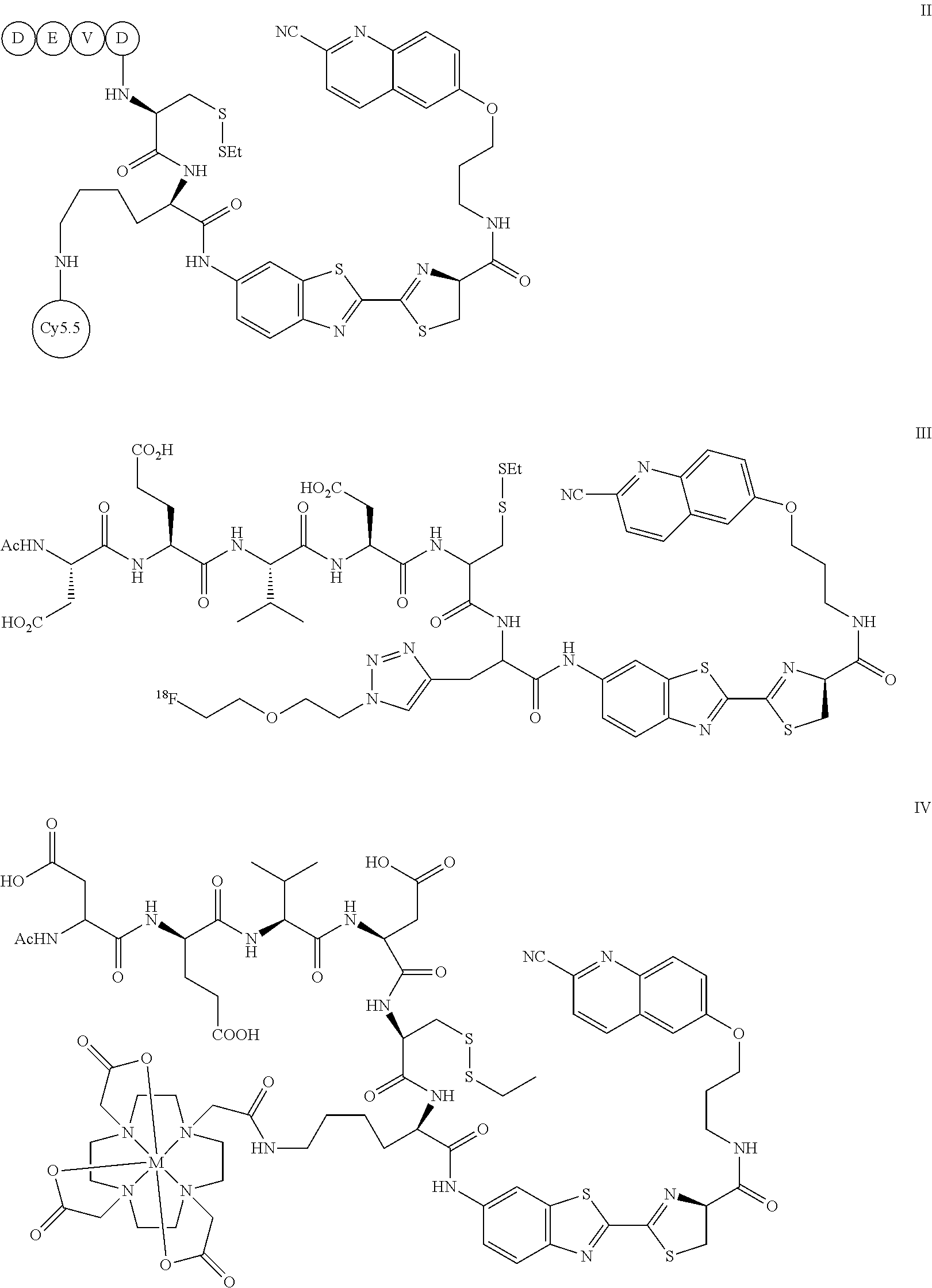
- C-SNAF: caspase-sensitive nano-aggregation fluorescent probe; C-SNAT: caspase-sensitive nano-aggregation tracer probe
| TABLE 1 | |||||||
| Probe | Origin of | % ID/g at | aTarget/Back- | Treated/Un- | |||
| Tracer | Species | Mechanism | Apoptosis | target site | ground | treated | Ref.g |
| [18F]C-SNAT | Mouse | Caspase-3 | Tumor treatment | 1.3 ± 0.1 | 7.0 | 1.9b | |
| [18F]Annexin V | Mouse | PS binding | Tumor treatment | 1.56 ± 0.2 | 3.4 | 1.6b | [2] |
| [18F]ICMT-11 | Mouse | Caspase-3 | Tumor treatment | N/A | 1.2c | 1.5d | [3] |
| Inhibitor | |||||||
| [18F]-CP18 | Mouse | Caspase-3 | Tumor | 0.17 | 10.2 | N/A | [4] |
| activated | endogenous | ||||||
| apoptosis | |||||||
| [18F]WC-IV-3 | Rat | Caspase-3 | Anti-Fas liver | 6.80 ± 3.0e | 4.3 | 3.2d | [4] |
| Inhibitor | injury | ||||||
| [18F]ML-10 | Mouse | Membrane | Cerebral | 0.3 ± 0.1 | N/A | 2.3f | [5] |
| Potential | stroke | ||||||
| aThe maximum values reported at the apoptotic tissue. | |||||||
| btarget uptake ratio between pre and post treatment in same animal. | |||||||
| cTumor/blood. | |||||||
| dtarget uptake ratio between treated and untreated group in different animal. | |||||||
| eQuantified by ex vivo bio-distribution, other data in this column are quantified by PET imaging. | |||||||
| ftarget uptake ratio between stroke and non-stroke region in same animal; PS: phosphatidylserine. | |||||||
| g[2] Hu et al., (2012) Mol. Imaging Biol.. 14: 762-770 | |||||||
| [3] Nguyen et al., (2009) Proc. Natl. Acad. Sci. USA 106: 16375-16380 | |||||||
| [4] Xia et al., (2013) Nucl. Med. Biol. DOI: 10.1007/s11307-013-0646-7 | |||||||
| [5] Chen et al., (2012) Nucl. Med. Biol. 39: 137-144 | |||||||
| [6] Reshef et al., (2008) J. Nucl. Med. 49: 1520-1528 | |||||||


and a detectable imaging moiety attached thereto.

| TABLE 2 |
| Ex vivo biodistribution data of [18F]C-SNAT (1) 2 h post injection. |
| Region (n) | Ratio of % ID/g over muscle ± SEM. | ||
| Muscle (5) | 1.0 ± 0.5 | ||
| Bone (5) | 2.4 ± 1.5 | ||
| Brain (5) | 0.5 ± 0.2 | ||
| Heart (3) | 4.5 ± 2.4 | ||
| Kidney (5) | 124.2 ± 46.6 | ||
| Liver (5) | 22.9 ± 9.7 | ||
| Lung (3) | 4.1 ± 1.8 | ||
| Plasma (2) | 4.0 ± 1.8 | ||
| Spleen (5) | 1.7 ± 0.7 | ||
| Tumor (2) | 5.6 ± 2.3 | ||
| Whole Blood (5) | 5.1 ± 2.4 | ||
SE(t)=SI(t)−SI(t=0); for each mouse, for every session
% D(t)=[SE(t,treated)−SE(t,baseline)]/SE(t,baseline); for each mouse.
Claims (19)


Priority Applications (1)
| Application Number | Priority Date | Filing Date | Title |
|---|---|---|---|
| US14/464,111 US9642922B2 (en) | 2013-08-23 | 2014-08-20 | Caspase-triggered nano-aggregation probes and methods of use |
Applications Claiming Priority (2)
| Application Number | Priority Date | Filing Date | Title |
|---|---|---|---|
| US201361869223P | 2013-08-23 | 2013-08-23 | |
| US14/464,111 US9642922B2 (en) | 2013-08-23 | 2014-08-20 | Caspase-triggered nano-aggregation probes and methods of use |
Publications (2)
| Publication Number | Publication Date |
|---|---|
| US20150056137A1 US20150056137A1 (en) | 2015-02-26 |
| US9642922B2 true US9642922B2 (en) | 2017-05-09 |
Family
ID=52480557
Family Applications (1)
| Application Number | Title | Priority Date | Filing Date |
|---|---|---|---|
| US14/464,111 Expired - Fee Related US9642922B2 (en) | 2013-08-23 | 2014-08-20 | Caspase-triggered nano-aggregation probes and methods of use |
Country Status (1)
| Country | Link |
|---|---|
| US (1) | US9642922B2 (en) |
Families Citing this family (8)
| Publication number | Priority date | Publication date | Assignee | Title |
|---|---|---|---|---|
| US10905781B2 (en) | 2016-03-02 | 2021-02-02 | The Brigham And Women's Hospital, Inc. | Reporter platform for real time monitoring of drug efficacy |
| RU2731211C1 (en) * | 2017-03-21 | 2020-08-31 | Фуджифилм Корпорэйшн | Peptide compound and method for production thereof, composition for screening use and method of selecting peptide compound |
| CN107586317B (en) * | 2017-10-16 | 2020-09-08 | 江苏省原子医学研究所 | Activatable tumor apoptosis PET imaging agent and preparation method and application thereof |
| CN109824622B (en) * | 2019-02-27 | 2022-12-30 | 中国科学技术大学 | Prodrug forming nano structure in cell to kill cancer cell and its prepn |
| CN111420053B (en) * | 2020-04-02 | 2022-09-09 | 珠海市人民医院 | A multifunctional magnetic nanoparticle complex that can be aggregated intracellularly and its preparation method |
| CN114075263B (en) * | 2020-08-18 | 2023-09-19 | 中南大学湘雅三医院 | Near infrared molecular probe and preparation and application thereof in detection of granzyme B |
| CN117801062A (en) * | 2022-09-30 | 2024-04-02 | 江苏省原子医学研究所 | Aspartic proteinase targeting recognition PET molecular probe and application thereof |
| CN117801065B (en) * | 2023-12-15 | 2025-05-06 | 中国科学院精密测量科学与技术创新研究院 | Caspase-3 enzyme response self-assembled probe and synthesis method and application thereof |
Citations (1)
| Publication number | Priority date | Publication date | Assignee | Title |
|---|---|---|---|---|
| US7951550B2 (en) * | 2005-05-31 | 2011-05-31 | Promega Corporation | Luminogenic and fluorogenic compounds and methods to detect molecules or conditions |
-
2014
- 2014-08-20 US US14/464,111 patent/US9642922B2/en not_active Expired - Fee Related
Patent Citations (1)
| Publication number | Priority date | Publication date | Assignee | Title |
|---|---|---|---|---|
| US7951550B2 (en) * | 2005-05-31 | 2011-05-31 | Promega Corporation | Luminogenic and fluorogenic compounds and methods to detect molecules or conditions |
Also Published As
| Publication number | Publication date |
|---|---|
| US20150056137A1 (en) | 2015-02-26 |
Similar Documents
| Publication | Publication Date | Title |
|---|---|---|
| US9642922B2 (en) | Caspase-triggered nano-aggregation probes and methods of use | |
| Moon et al. | Fibroblast activation protein (FAP) targeting homodimeric FAP inhibitor radiotheranostics: a step to improve tumor uptake and retention time | |
| Zeng et al. | Molecular imaging of apoptosis: from micro to macro | |
| Ogawa et al. | Dual-modality molecular imaging using antibodies labeled with activatable fluorescence and a radionuclide for specific and quantitative targeted cancer detection | |
| US8343458B2 (en) | Probes for in vivo targeting of active cysteine proteases | |
| JP5368099B2 (en) | A compound comprising a moiety for recognition of a biological target coupled to a signal moiety capable of complexing with gallium | |
| EP3510399B1 (en) | Psma-targeted nir dyes and their uses | |
| US20030129579A1 (en) | Multi-use multimodal imaging chelates | |
| CZ20032564A3 (en) | Labeled macrophage scavenger receptor antagonists for imaging atherosclerosis and vulnerable plaque | |
| US20240042068A1 (en) | Caspase-3-triggered molecular self-assembling pet probes and uses thereof | |
| JP2023145464A (en) | Dual labeled probes and their use for molecular imaging | |
| WO2019101729A1 (en) | A double-labeled probe for molecular imaging and use thereof | |
| US10196412B2 (en) | Probe for imaging PARP-1 activity | |
| Mosayebnia et al. | Radiolabeled peptides for molecular imaging of apoptosis | |
| WO2016090169A1 (en) | Intracellular caspase probes for detection of apoptosis and inflammation and kits containing such probes | |
| Zhang et al. | Synthesis and preclinical evaluation of a novel PET/fluorescence dual-modality probe targeting fibroblast activation protein | |
| Son et al. | Design and synthesis of a novel BODIPY-labeled PSMA inhibitor | |
| Kang et al. | A hybrid imaging platform (CT/PET/FMI) for evaluating tumor necrosis and apoptosis in real-time | |
| Liu et al. | Aggregation-induced emission based PET probe for liver function imaging | |
| Lin et al. | In vivo MR/optical imaging for gastrin releasing peptide receptor of prostate cancer tumor using Gd-TTDA-NP-BN-Cy5. 5 | |
| Zeng et al. | Development of small molecular probes for the molecular imaging of apoptosis | |
| Genady et al. | A bioorthogonal methylene blue derived probe for targeted photoacoustic imaging | |
| NL2033714B1 (en) | Multiplexing targeting and imaging agents | |
| Rizzo | MULTIMODAL IMAGING PROBES FOR CANCER DIAGNOSIS AND TREATMENT | |
| KR20250011148A (en) | Prostate-specific membrane antigen (PSMA) ligand |
Legal Events
| Date | Code | Title | Description |
|---|---|---|---|
| AS | Assignment |
Owner name: NATIONAL INSTITUTES OF HEALTH (NIH), U.S. DEPT. OF Free format text: CONFIRMATORY LICENSE;ASSIGNOR:THE BOARD OF TRUSTEES OF THE LELAND STANFORD JUNIOR UNIVERSITY;REEL/FRAME:033823/0673 Effective date: 20140826 |
|
| AS | Assignment |
Owner name: THE BOARD OF TRUSTEES OF THE LELAND STANFORD JUNIO Free format text: ASSIGNMENT OF ASSIGNORS INTEREST;ASSIGNORS:RAO, JIANGHONG;YE, DEJU;SHUHENDLER, ADAM;AND OTHERS;REEL/FRAME:034141/0977 Effective date: 20131004 |
|
| STCF | Information on status: patent grant |
Free format text: PATENTED CASE |
|
| MAFP | Maintenance fee payment |
Free format text: PAYMENT OF MAINTENANCE FEE, 4TH YR, SMALL ENTITY (ORIGINAL EVENT CODE: M2551); ENTITY STATUS OF PATENT OWNER: SMALL ENTITY Year of fee payment: 4 |
|
| FEPP | Fee payment procedure |
Free format text: MAINTENANCE FEE REMINDER MAILED (ORIGINAL EVENT CODE: REM.); ENTITY STATUS OF PATENT OWNER: SMALL ENTITY |
|
| LAPS | Lapse for failure to pay maintenance fees |
Free format text: PATENT EXPIRED FOR FAILURE TO PAY MAINTENANCE FEES (ORIGINAL EVENT CODE: EXP.); ENTITY STATUS OF PATENT OWNER: SMALL ENTITY |
|
| STCH | Information on status: patent discontinuation |
Free format text: PATENT EXPIRED DUE TO NONPAYMENT OF MAINTENANCE FEES UNDER 37 CFR 1.362 |
|
| FP | Lapsed due to failure to pay maintenance fee |
Effective date: 20250509 |