US9622657B2 - Automated system for measurement of zone 1 in assessment of severity of retinopathy of prematurity - Google Patents
Automated system for measurement of zone 1 in assessment of severity of retinopathy of prematurity Download PDFInfo
- Publication number
- US9622657B2 US9622657B2 US14/968,622 US201514968622A US9622657B2 US 9622657 B2 US9622657 B2 US 9622657B2 US 201514968622 A US201514968622 A US 201514968622A US 9622657 B2 US9622657 B2 US 9622657B2
- Authority
- US
- United States
- Prior art keywords
- patient
- retina
- photograph
- series
- gui
- Prior art date
- Legal status (The legal status is an assumption and is not a legal conclusion. Google has not performed a legal analysis and makes no representation as to the accuracy of the status listed.)
- Active
Links
- 206010038933 Retinopathy of prematurity Diseases 0.000 title claims abstract description 27
- 238000005259 measurement Methods 0.000 title description 2
- 210000001525 retina Anatomy 0.000 claims abstract description 38
- 238000000034 method Methods 0.000 claims abstract description 27
- 230000002792 vascular Effects 0.000 claims abstract description 12
- 238000009826 distribution Methods 0.000 claims abstract description 10
- 206010025421 Macule Diseases 0.000 claims description 8
- 201000010099 disease Diseases 0.000 claims description 6
- 208000037265 diseases, disorders, signs and symptoms Diseases 0.000 claims description 6
- 238000003745 diagnosis Methods 0.000 claims description 5
- 238000011156 evaluation Methods 0.000 claims description 5
- 238000004891 communication Methods 0.000 claims description 4
- 210000005036 nerve Anatomy 0.000 claims description 4
- 230000003287 optical effect Effects 0.000 claims description 4
- 230000002123 temporal effect Effects 0.000 claims description 4
- 238000013473 artificial intelligence Methods 0.000 claims description 2
- 238000010801 machine learning Methods 0.000 claims description 2
- 238000007796 conventional method Methods 0.000 abstract description 3
- 210000001328 optic nerve Anatomy 0.000 description 7
- 238000011282 treatment Methods 0.000 description 6
- 230000004044 response Effects 0.000 description 4
- 238000012552 review Methods 0.000 description 4
- 230000001413 cellular effect Effects 0.000 description 3
- 238000012790 confirmation Methods 0.000 description 3
- 210000001747 pupil Anatomy 0.000 description 3
- 210000001519 tissue Anatomy 0.000 description 3
- 210000005166 vasculature Anatomy 0.000 description 3
- 201000004569 Blindness Diseases 0.000 description 2
- 206010038848 Retinal detachment Diseases 0.000 description 2
- 230000002159 abnormal effect Effects 0.000 description 2
- 230000008901 benefit Effects 0.000 description 2
- 230000036770 blood supply Effects 0.000 description 2
- 210000003161 choroid Anatomy 0.000 description 2
- 210000004087 cornea Anatomy 0.000 description 2
- 238000010586 diagram Methods 0.000 description 2
- 230000010339 dilation Effects 0.000 description 2
- 230000004264 retinal detachment Effects 0.000 description 2
- 230000002207 retinal effect Effects 0.000 description 2
- 230000004233 retinal vasculature Effects 0.000 description 2
- 210000003786 sclera Anatomy 0.000 description 2
- 238000003860 storage Methods 0.000 description 2
- 208000005107 Premature Birth Diseases 0.000 description 1
- 208000017442 Retinal disease Diseases 0.000 description 1
- 108010073929 Vascular Endothelial Growth Factor A Proteins 0.000 description 1
- 102000005789 Vascular Endothelial Growth Factors Human genes 0.000 description 1
- 108010019530 Vascular Endothelial Growth Factors Proteins 0.000 description 1
- 210000002159 anterior chamber Anatomy 0.000 description 1
- 230000015572 biosynthetic process Effects 0.000 description 1
- 210000004556 brain Anatomy 0.000 description 1
- 239000000835 fiber Substances 0.000 description 1
- 238000003384 imaging method Methods 0.000 description 1
- 239000000203 mixture Substances 0.000 description 1
- 238000010295 mobile communication Methods 0.000 description 1
- 238000011369 optimal treatment Methods 0.000 description 1
- 230000002093 peripheral effect Effects 0.000 description 1
- 238000003825 pressing Methods 0.000 description 1
- 230000004256 retinal image Effects 0.000 description 1
- 210000003583 retinal pigment epithelium Anatomy 0.000 description 1
- 231100000241 scar Toxicity 0.000 description 1
- 239000000126 substance Substances 0.000 description 1
- 210000003813 thumb Anatomy 0.000 description 1
- 230000000007 visual effect Effects 0.000 description 1
- 238000005303 weighing Methods 0.000 description 1
Images


Classifications
-
- A—HUMAN NECESSITIES
- A61—MEDICAL OR VETERINARY SCIENCE; HYGIENE
- A61B—DIAGNOSIS; SURGERY; IDENTIFICATION
- A61B3/00—Apparatus for testing the eyes; Instruments for examining the eyes
- A61B3/0016—Operational features thereof
- A61B3/0025—Operational features thereof characterised by electronic signal processing, e.g. eye models
-
- A—HUMAN NECESSITIES
- A61—MEDICAL OR VETERINARY SCIENCE; HYGIENE
- A61B—DIAGNOSIS; SURGERY; IDENTIFICATION
- A61B3/00—Apparatus for testing the eyes; Instruments for examining the eyes
- A61B3/0016—Operational features thereof
- A61B3/0033—Operational features thereof characterised by user input arrangements
-
- A—HUMAN NECESSITIES
- A61—MEDICAL OR VETERINARY SCIENCE; HYGIENE
- A61B—DIAGNOSIS; SURGERY; IDENTIFICATION
- A61B3/00—Apparatus for testing the eyes; Instruments for examining the eyes
- A61B3/10—Objective types, i.e. instruments for examining the eyes independent of the patients' perceptions or reactions
- A61B3/12—Objective types, i.e. instruments for examining the eyes independent of the patients' perceptions or reactions for looking at the eye fundus, e.g. ophthalmoscopes
- A61B3/1241—Objective types, i.e. instruments for examining the eyes independent of the patients' perceptions or reactions for looking at the eye fundus, e.g. ophthalmoscopes specially adapted for observation of ocular blood flow, e.g. by fluorescein angiography
-
- A—HUMAN NECESSITIES
- A61—MEDICAL OR VETERINARY SCIENCE; HYGIENE
- A61B—DIAGNOSIS; SURGERY; IDENTIFICATION
- A61B5/00—Measuring for diagnostic purposes; Identification of persons
- A61B5/0002—Remote monitoring of patients using telemetry, e.g. transmission of vital signals via a communication network
- A61B5/0015—Remote monitoring of patients using telemetry, e.g. transmission of vital signals via a communication network characterised by features of the telemetry system
- A61B5/0022—Monitoring a patient using a global network, e.g. telephone networks, internet
Definitions
- the present invention in general relates to retinal disease diagnosis and in particular to an automated system and method for diagnosing and evaluating the extent of retinopathy of prematurity.
- the visible parts of the human eye 10 include the transparent cornea 12 , the normally white sclera 14 , the colored (blue, green, brown or a mixture of these) iris 16 , and an opening in the iris, the normally black pupil.
- a ray of light after passing through the cornea 12 , which partially focuses the image, passes through the anterior chamber, the pupil, the lens 18 , which focuses the image further, the vitreous and is then focused on the retina 20 .
- the retina 20 is a very thin layer of tissue that lines the inner part of the eye 10 .
- the retina 20 is responsible for capturing the light rays that enter the eye 10 .
- the retina 20 is supported by its retinal pigment epithelium, which is normally opaque, the choroid and the sclera.
- the blood supply of the retina 20 is primarily through the choroid and secondarily through the retinal vasculature 22 which lies on top of the retina 20 .
- the macula 24 is located roughly in the center of the retina 20 , temporal to the optic nerve 26 .
- the macula 24 is a small and highly sensitive part of the retina 20 responsible for detailed central vision.
- the light rays captured by retina are turned into impulses, and the impulses are transported to the brain via the optic nerve 24 .
- ROP Retinopathy of prematurity
- the retinal area without adequate blood supply emits a chemical trigger (vascular endothelial growth factor) to stimulate growth of the abnormal vasculature.
- vascular endothelial growth factor vascular endothelial growth factor
- the abnormal vasculature leads to a formation of a ring of scare tissue attached to both the retina and the vitreous gel that fills the center of our eyes. As the scar contracts, it may pull on the retinal creating a retinal detachment.
- Zone 1 is the posterior of the retina while zone 3 is the far peripheral retina.
- Stage 0 is the mildest form of ROP while Stage 5 is the most severe indicating total retinal detachment.
- FIG. 2 depicts the zones used in the classification and the thresholds for treatment of ROP. Based on classification of the location and extent of the ROP a treatment regime may be prescribed.
- An automated method for diagnosing and evaluating severity of retinopathy of prematurity in a retina of a patient is provided that is superior to conventional techniques.
- a graphical user interface GUI is provided for receiving biographical information for the patient creating a patient record in a database via the GUI.
- a photograph of the retina of the patient is collected and placed in the patient record via the GUI.
- the photograph is then analyzed to determine vascular distributions within the retina.
- a zone 1 boundary is assigned to the retina based on a set of threshold levels with respect to the determined vascular distributions.
- a system for performing the automated method is also provided.
- FIG. 1 is a prior art perspective cross sectioned view of a the human eye
- FIG. 2 depicts the prior art zones used in the classification and the thresholds for treatment of ROP
- FIGS. 3A-3C are screen shots of the login screen and basic patient information input according to an embodiment of the invention.
- FIGS. 4A-4H are screen shots for inputting a patient photo information for analysis and diagnostic according to an embodiment of the invention.
- FIGS. 5A-5Z are screen shots for performing analysis and diagnostics on the uploaded patient photo information according to an embodiment of the invention.
- FIG. 6 illustrates a formatted report on a patient's condition based on the analysis and diagnostics of FIGS. 5A-5Z according to embodiments of the invention
- FIGS. 7A-7G are screen shots of an analysis tool for performing automated analysis and diagnostics on the uploaded patient photo information according to an embodiment of the invention.
- FIG. 8 is a schematic diagram illustrating an overall view of communication devices, computing devices, and mediums for implementing embodiments of the invention.
- the present invention has utility as an automated system and method for accurately diagnosing and evaluating the severity of ROP.
- a graphical user interface (GUI) is provided for inputting and creating a patient database that combines patient biographical information with high resolution retinal images that are suitable for disease diagnostics of the eye via automated analysis of the images.
- GUI graphical user interface
- automated diagnostics of the extent and severity of retinopathy of prematurity is determined based on the analysis of retinal vasculature and related structures in the high resolution images, where the automated assignment of zones and quadrants indicates the location and extent of the ROP, and a treatment regime may be prescribed.
- Vascular distributions are used by the system to determine the automated assignment of zone 1 and in some embodiments, quadrants of the eye.
- machine learning and artificial intelligence may be utilized to provide diagnostic information based on a learned history of previous patient images and inputted expert analysis of the images by physicians and researchers.
- AI artificial intelligence
- the present invention affords a consistent method of identifying the boundary of zone 1, and therefore accurately diagnosing the severity of ROP.
- FIGS. 3A-3C are a series of screen shots of the login screen and for basic patient information input according to an embodiment of the invention.
- a login screen 40 is shown with input fields for username 42 , password 44 , and a sign in button 46 that provides a user with access to the system when the username 42 and password 44 are authenticated by the system.
- a patient list screen 50 is shown to the user, where the patients shown in the list is based on filter settings 52 .
- patient information is further filtered based on case status 60 , and is presented in tabular form with headers including case number 54 , date/time of last session 56 , patient name 58 , date of birth (DOB) 62 , status (information entered, uploaded, read, etc.) 64 , downloaded (yes/no) 66 , number of sessions 68 , clinic where an examination occurred 70 .
- the new patient button 72 may be selected to add patients and their corresponding information to the database via patient biography input page 80 .
- patient biographical information is inputted and uploaded to the database in step one of a three step process.
- a drop down menu 82 is provided to select the clinic where the patient was examined, and fillable input fields are also provided for a medical record number 84 , patient name 86 , gender 88 , date of birth 90 , birth weight 92 , gestational age 94 , patient ethnicity 96 , referring physician based on clinic 98 , reading physician 100 , a fillable comments section 102 .
- the photo session information screen appears 110 , as shown in FIG. 4C , for loading photographs of the retina into the patient file, as step two of three of the new patient upload, with a confirmation message 112 that the patient was successfully added to the database in FIG. 3C .
- a patient information field 114 appears at the top of the page 110 , and fillable fields are provided for the date of when the photos were taken 116 and the time the photos were taken 118 .
- the photo session information screen 110 is split into sections for the right eye (OD) 120 and the left eye (OS) 122 .
- a template with a series of boxes labeled for the series of views of the photographed eye are provided where a photograph is dragged or dropped.
- the views include right and left eye version of the external, inferior, nasal, posterior, superior, and temporal.
- an overlay window of the photographs 126 has the file of photographs which are dragged and dropped into the corresponding labeled squares for the right eye (OD) 120 and the left eye (OS) 122 .
- OD right eye
- OS left eye
- FIG. 4D the photo for the superior OO 130 is dragged and dropped into the corresponding square on the right eye (OD) 120 side.
- FIG. 4E shows the completion of the transferring of the photos to the patient file in step 2 of the new patient upload.
- the system stores the photos and the user is provided with confirmation page 140 ( FIG. 4F ) to confirm the information (step 3 of 3 ).
- the user is informed that they must confirm the photo session in order to send the file to the reading physician for review 142 .
- Selection of the “confirm and send to reading physician” button 144 completes the patient upload process.
- the Exam upload confirmation page 150 ( FIG.
- FIG. 4H notifies the user that the upload was successful 152 , and acknowledgement by the user by pressing the return to patient list 154 sends the user back to the patient list as shown in FIG. 5A .
- the patient list 50 in FIG. 5A is filtered 60 by unread cases in FIG. 5B , and the newly added patient “Smithson Baby” of FIG. 3C and FIGS. 4A-4H is circled (for illustration only) in both FIGS. 5A and 5B .
- Selection of the view link 74 initiates patient information screen 160 in FIG. 5C with the patient's biographical information and uploaded examination photographs separated in accordance to the right eye 120 and left eye 122 .
- FIG. 5D provides the user with a montage 166 R of the right eye formed with the individual examination images for the right eye.
- FIG. 5E shows the selection of the toggle montage button 162 L which provides the user with a montage 166 L of the left eye formed with the individual examination images for the left eye.
- Selection of the read presentation button 164 initiates the reading process of the uploaded patient eye examination images which begins in FIG. 5F with read photo session screen 170 .
- the reading process begins with the right eye (step 1 ) 172 , however the physician may also configure the system to begin with the left eye.
- the toggle montage button 162 R provides the user with a montage 166 R of the right eye formed with the individual examination images for the right eye. Clicking or selecting the montage 166 R, enlarges the picture 176 R. Questions 196 presented to the physician during the read photo session include: Is the pupil size adequate; Is rubeosis present; What is the vascular status; Is there virtuous haze; Highest stage; Lowest zone, etc. Selecting the enhance image button 174 initiates the pop up overlay 178 as shown in FIGS.
- Enhancement buttons 180 provide the user with the ability to pan or zoom in/out the selected image, as well as adjust image parameters such as brightness, contrast, and saturation.
- the text button provides the ability to add overlay text to the image.
- Slider control 182 may be used to adjust the image parameters.
- the selection of the draw zone 1 button 188 begins with identification of the optic nerve 26 with select optic nerve button 184 , and the select macula button 186 .
- the system automatically draws the circle 34 that represents zone 1, which is used in the diagnosis and treatment of ROP. The operator designates the position of the optic nerve; and then the system may automatically draws the boundary of zone 1.
- the save button 190 saves the changes made to the image. Selection of the next button 192 initiates step 2 of the read photo session for the left eye as shown in FIGS. 5M-5R .
- the available analysis options and screens are identical to those described for the right eye and are not repeated.
- the read session review screen 200 is displayed as indicated by step 3 : review finding indicator 202 as shown in FIGS. 5S-5X .
- Screen 200 instructs the physician to review their findings with respect to their analysis of the patients exam photos, and if the findings are believed to be correct, the physician is instructed to submit the findings by selecting the submit findings button 218 ( FIG. 5X ).
- the answers to questions 194 are summarized in right eye findings 204 R and left eye findings 204 L.
- Montage toggle buttons ( 162 R, 162 L) are provided to view the images. Comment area 208 is provided for viewer notes for the patient file.
- the physician may change the images in the report with the change button 206 , which in response to the selection of the change button 206 an image change overlay 210 with images appears for the right or left eye as shown in FIG. 5V .
- a user or physician may request an expert opinion with button 210 ( FIG. 5W ).
- an overlay 212 appears with a pull down menu 214 of available physicians and a question or comment area 216 .
- a report of the finding of the patient exam may be generated with the report request button 224 .
- An example report is shown in FIG. 6 .
- FIGS. 7A-7G are a series of screen shots of analysis tool 230 for performing automated analysis and diagnostics on the uploaded patient photo information according to an embodiment of the invention.
- the optical nerve is circled and identified by the automated system with the selection of the optical nerve button 232 .
- the zone 1 circle is automatically generated.
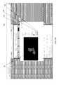
- FIG. 7D show an expanded view of zone 1 for improved visual diagnostics.
- FIG. 7E shows a highlighting tool for extracting vessels (button 236 ) with the vessels highlighted 238 .
- FIGS. 7A-7G Various parameters related to the vascular structure within zone 1 is recorded as shown in 7 F where a tortuosity index and dilation index is calculated for the four quadrants and the image itself in zone 1 in box 240 .
- FIG. 7G tortuosity and dilation for individual vessels is shown in boxes 242 and 244 .
- the measurements shown in FIGS. 7A-7G are used in the determination of the presence and severity of the ROP condition.
- FIG. 8 is a schematic diagram illustrating an overall view of communication devices, computing devices, and mediums for implementing the automated system and method for ascribing the boundary of zone 1 in a retinograph that avoids to subjective evaluation of conventional techniques. Eye treatment follows based on the accurate determination of vasculature forming the boundary of the zone 1.
- the system 300 includes multimedia devices 302 and desktop computer devices 304 configured with display capabilities 314 and processors for executing instructions and commands.
- the multimedia devices 302 are optionally mobile communication and entertainment devices, such as cellular phones, tablets, and mobile computing devices that in certain embodiments are wirelessly connected to a network 308 .
- the multimedia devices 302 typically have video displays 318 and audio outputs 316 .
- the multimedia devices 302 and desktop computer devices 304 are optionally configured with internal storage, software, and a graphical user interface (GUI) for carrying out elements of the automated diagnostic platform according to embodiments of the invention.
- GUI graphical user interface
- the network 308 is optionally any type of known network including a fixed wire line network, cable and fiber optics, over the air broadcasts, satellite 320 , local area network (LAN), wide area network (WAN), global network (e.g., Internet), intranet, etc. with data/Internet capabilities as represented by server 306 .
- Communication aspects of the network are represented by cellular base station 310 and antenna 312 .
- the network 308 is a LAN and each remote device 302 and desktop device 304 executes a user interface application (e.g., Web browser) to contact the server system 306 through the network 308 .
- the remote devices 302 and 304 may be implemented using a device programmed primarily for accessing network 308 such as a remote client.
- the software for the diagnostic platform may be resident on a USB thumb or flash drive 320 , CD or DVD 322 , or an external hard drive 324 for connection to desktop or laptop computers 304 , or stored within the server 306 or cellular base station 310 for download to an end user.
- Server 306 may implement a cloud-based service for implementing embodiments of the diagnostic platform with a multi-tenant database for storage of separate client data for each independent trial being carried out on the platform.
- Patents and references cited in the application are indicative of the skill in the art. Each of these patents and references is hereby incorporated by reference to the same extent as if each reference was individually incorporated by reference.
Landscapes
- Health & Medical Sciences (AREA)
- Life Sciences & Earth Sciences (AREA)
- Engineering & Computer Science (AREA)
- Molecular Biology (AREA)
- Animal Behavior & Ethology (AREA)
- Biophysics (AREA)
- Veterinary Medicine (AREA)
- Biomedical Technology (AREA)
- Heart & Thoracic Surgery (AREA)
- Medical Informatics (AREA)
- Public Health (AREA)
- Surgery (AREA)
- Physics & Mathematics (AREA)
- General Health & Medical Sciences (AREA)
- Ophthalmology & Optometry (AREA)
- Signal Processing (AREA)
- Hematology (AREA)
- Nuclear Medicine, Radiotherapy & Molecular Imaging (AREA)
- Radiology & Medical Imaging (AREA)
- Vascular Medicine (AREA)
- Computer Networks & Wireless Communication (AREA)
- Pathology (AREA)
- Eye Examination Apparatus (AREA)
Abstract
Description
Claims (14)
Priority Applications (1)
| Application Number | Priority Date | Filing Date | Title |
|---|---|---|---|
| US14/968,622 US9622657B2 (en) | 2014-12-12 | 2015-12-14 | Automated system for measurement of zone 1 in assessment of severity of retinopathy of prematurity |
Applications Claiming Priority (2)
| Application Number | Priority Date | Filing Date | Title |
|---|---|---|---|
| US201462091112P | 2014-12-12 | 2014-12-12 | |
| US14/968,622 US9622657B2 (en) | 2014-12-12 | 2015-12-14 | Automated system for measurement of zone 1 in assessment of severity of retinopathy of prematurity |
Publications (2)
| Publication Number | Publication Date |
|---|---|
| US20160213242A1 US20160213242A1 (en) | 2016-07-28 |
| US9622657B2 true US9622657B2 (en) | 2017-04-18 |
Family
ID=56433056
Family Applications (1)
| Application Number | Title | Priority Date | Filing Date |
|---|---|---|---|
| US14/968,622 Active US9622657B2 (en) | 2014-12-12 | 2015-12-14 | Automated system for measurement of zone 1 in assessment of severity of retinopathy of prematurity |
Country Status (1)
| Country | Link |
|---|---|
| US (1) | US9622657B2 (en) |
Families Citing this family (4)
| Publication number | Priority date | Publication date | Assignee | Title |
|---|---|---|---|---|
| RU2666268C1 (en) * | 2017-09-18 | 2018-09-06 | Федеральное государственное автономное учреждение "Межотраслевой научно-технический комплекс "Микрохирургия глаза" имени академика С.Н. Федорова" Министерства здравоохранения Российской Федерации | Method for predicting i-iii stages of active retinopathy of retinopathy of prematurity on the basis of data of fluorescence angiography |
| CN108392174B (en) * | 2018-04-19 | 2021-01-19 | 梁建宏 | Automatic examination method and system for retinopathy of prematurity |
| US12306192B2 (en) * | 2021-02-08 | 2025-05-20 | Cedars-Sinai Medical Center | Method of detecting cognitive impairment |
| EP4510922A4 (en) | 2022-04-22 | 2025-11-05 | Impel Ip Llc | DIAGNOSIS OF PATHOLOGY USING A SPECTRAL ELECTRIC IMPEDANCE DEVICE |
Citations (4)
| Publication number | Priority date | Publication date | Assignee | Title |
|---|---|---|---|---|
| US8090164B2 (en) | 2003-08-25 | 2012-01-03 | The University Of North Carolina At Chapel Hill | Systems, methods, and computer program products for analysis of vessel attributes for diagnosis, disease staging, and surgical planning |
| US8201943B2 (en) * | 2009-01-15 | 2012-06-19 | Physical Sciences, Inc. | Adaptive optics line scanning ophthalmoscope |
| US8233681B2 (en) | 2004-09-24 | 2012-07-31 | The University Of North Carolina At Chapel Hill | Methods, systems, and computer program products for hierarchical registration between a blood vessel and tissue surface model for a subject and a blood vessel and tissue surface image for the subject |
| US20130166313A1 (en) * | 2010-04-02 | 2013-06-27 | Shire Human Genetic Therapies, Inc. | Systems and methods for managing treatment of an orphan disease |
-
2015
- 2015-12-14 US US14/968,622 patent/US9622657B2/en active Active
Patent Citations (4)
| Publication number | Priority date | Publication date | Assignee | Title |
|---|---|---|---|---|
| US8090164B2 (en) | 2003-08-25 | 2012-01-03 | The University Of North Carolina At Chapel Hill | Systems, methods, and computer program products for analysis of vessel attributes for diagnosis, disease staging, and surgical planning |
| US8233681B2 (en) | 2004-09-24 | 2012-07-31 | The University Of North Carolina At Chapel Hill | Methods, systems, and computer program products for hierarchical registration between a blood vessel and tissue surface model for a subject and a blood vessel and tissue surface image for the subject |
| US8201943B2 (en) * | 2009-01-15 | 2012-06-19 | Physical Sciences, Inc. | Adaptive optics line scanning ophthalmoscope |
| US20130166313A1 (en) * | 2010-04-02 | 2013-06-27 | Shire Human Genetic Therapies, Inc. | Systems and methods for managing treatment of an orphan disease |
Non-Patent Citations (3)
| Title |
|---|
| Abramoff, Michael D. et al. "Retinal Imaging and Image Analysis", IEEE transactions on medical imaging, NIH Public Access, Author Manuscript, IEEE Trans Med Imaging. Jan. 1, 2010; 3: 169-208. |
| Chiang, Michael F. et al. "Image Analysis for Retinopathy of Prematurity Diagnosis", NIH Public Access, Author Manuscript, J AAPOS. Author manuscript; available in PMC Oct. 1, 2010. J AAPOS. Oct. 2009 ; 13(5): 438-445. doi: 10.1016/j.jaapos.2009.08.011. |
| Williams, Steven L. et al. "Telemedical Diagnosis of Retinopathy of Prematurity: Accuracy of Expert vs. Non-expert Graders", NIH Public Access, Author Manuscript, Br J Ophthalmol. Mar. 2010 ; 94(3): 351-356. doi:10.1136/bjo.2009.166348. |
Also Published As
| Publication number | Publication date |
|---|---|
| US20160213242A1 (en) | 2016-07-28 |
Similar Documents
| Publication | Publication Date | Title |
|---|---|---|
| Niemeijer et al. | Automated detection and differentiation of drusen, exudates, and cotton-wool spots in digital color fundus photographs for diabetic retinopathy diagnosis | |
| CN108553079B (en) | Pathological change recognition system based on fundus image | |
| US10722180B2 (en) | Deep learning-based diagnosis and referral of ophthalmic diseases and disorders | |
| CN111345775B (en) | Evaluation of fundus images | |
| CN113646805A (en) | Image-based detection of ophthalmic and systemic diseases | |
| JP6796413B2 (en) | Medical support method and medical support system | |
| Adam et al. | Quality and diagnostic utility of mydriatic smartphone photography: the smartphone ophthalmoscopy reliability trial | |
| US20180360305A1 (en) | Method and system for classifying optic nerve head | |
| CN110582223A (en) | Systems and methods for diagnosis, treatment and prognosis of medical conditions | |
| EP4222698A1 (en) | Machine learning for detection of diseases from external anterior eye images | |
| Lemij et al. | Characteristics of a large, labeled data set for the training of artificial intelligence for glaucoma screening with fundus photographs | |
| Lekha et al. | MII RetCam assisted smartphone based fundus imaging for retinopathy of prematurity | |
| US9622657B2 (en) | Automated system for measurement of zone 1 in assessment of severity of retinopathy of prematurity | |
| Agrawal et al. | HVDROPDB datasets for research in retinopathy of prematurity | |
| Wang et al. | Automated detection of diabetic retinopathy lesions on ultrawidefield pseudocolour images | |
| US20170100030A1 (en) | Systems and methods for retinopathy workflow, evaluation and grading using mobile devices | |
| Martins et al. | Use of artificial intelligence in ophthalmology: a narrative review | |
| Li et al. | Dual-mode imaging system for early detection and monitoring of ocular surface diseases | |
| AlShawabkeh et al. | The utilization of artificial intelligence in glaucoma: diagnosis versus screening | |
| US20260083317A1 (en) | Visual Field systems and methods for glaucoma diagnosis and monitoring by implementing adaptive map perimetry via head-mounted displays | |
| JP2020201531A (en) | Medical support system and ophthalmologic examination device | |
| CA2960501C (en) | An automated system for measurement of zone 1 in assessment of severity of retinopathy of prematurity | |
| JP6923495B2 (en) | Medical support method and medical support system | |
| US12327632B2 (en) | Automatic patient record updating | |
| CN116635889A (en) | Machine Learning for Disease Detection from External Anterior Eye Images |
Legal Events
| Date | Code | Title | Description |
|---|---|---|---|
| STCF | Information on status: patent grant |
Free format text: PATENTED CASE |
|
| AS | Assignment |
Owner name: INTERVIEW MEDICAL SYSTEMS, LLC, PENNSYLVANIA Free format text: ASSIGNMENT OF ASSIGNORS INTEREST;ASSIGNORS:TRESE, MICHAEL T;CAPONE, ANTONIO, JR.;DRENSER, KIMBERLY;AND OTHERS;REEL/FRAME:047398/0501 Effective date: 20181022 |
|
| AS | Assignment |
Owner name: PHOENIX TECHNOLOGY GROUP LLC, CALIFORNIA Free format text: ASSIGNMENT OF ASSIGNORS INTEREST;ASSIGNOR:INTERVIEW MEDICAL SYSTEMS, LLC;REEL/FRAME:047409/0022 Effective date: 20181102 |
|
| MAFP | Maintenance fee payment |
Free format text: PAYMENT OF MAINTENANCE FEE, 4TH YR, SMALL ENTITY (ORIGINAL EVENT CODE: M2551); ENTITY STATUS OF PATENT OWNER: SMALL ENTITY Year of fee payment: 4 |
|
| AS | Assignment |
Owner name: SWK FUNDING LLC, TEXAS Free format text: SECURITY INTEREST;ASSIGNOR:PHOENIX TECHNOLOGY GROUP LLC;REEL/FRAME:062752/0097 Effective date: 20230217 |
|
| AS | Assignment |
Owner name: NEOLIGHT, LLC, ARIZONA Free format text: ASSIGNMENT OF ASSIGNORS INTEREST;ASSIGNOR:PHOENIX TECHNOLOGY GROUP LLC;REEL/FRAME:063771/0706 Effective date: 20230525 |
|
| AS | Assignment |
Owner name: SWK FUNDING LLC, TEXAS Free format text: SECURITY INTEREST;ASSIGNOR:NEOLIGHT, LLC;REEL/FRAME:067053/0588 Effective date: 20230217 |
|
| MAFP | Maintenance fee payment |
Free format text: PAYMENT OF MAINTENANCE FEE, 8TH YR, SMALL ENTITY (ORIGINAL EVENT CODE: M2552); ENTITY STATUS OF PATENT OWNER: SMALL ENTITY Year of fee payment: 8 |