US9439618B2 - Method for generating a pet or spect image dataset and hybrid imaging modality for this purpose - Google Patents
Method for generating a pet or spect image dataset and hybrid imaging modality for this purpose Download PDFInfo
- Publication number
- US9439618B2 US9439618B2 US14/165,609 US201414165609A US9439618B2 US 9439618 B2 US9439618 B2 US 9439618B2 US 201414165609 A US201414165609 A US 201414165609A US 9439618 B2 US9439618 B2 US 9439618B2
- Authority
- US
- United States
- Prior art keywords
- anatomy image
- similarity
- anatomy
- pet
- datasets
- Prior art date
- Legal status (The legal status is an assumption and is not a legal conclusion. Google has not performed a legal analysis and makes no representation as to the accuracy of the status listed.)
- Active, expires
Links
Images


Classifications
-
- A—HUMAN NECESSITIES
- A61—MEDICAL OR VETERINARY SCIENCE; HYGIENE
- A61B—DIAGNOSIS; SURGERY; IDENTIFICATION
- A61B6/00—Apparatus or devices for radiation diagnosis; Apparatus or devices for radiation diagnosis combined with radiation therapy equipment
- A61B6/52—Devices using data or image processing specially adapted for radiation diagnosis
- A61B6/5258—Devices using data or image processing specially adapted for radiation diagnosis involving detection or reduction of artifacts or noise
- A61B6/5264—Devices using data or image processing specially adapted for radiation diagnosis involving detection or reduction of artifacts or noise due to motion
-
- A—HUMAN NECESSITIES
- A61—MEDICAL OR VETERINARY SCIENCE; HYGIENE
- A61B—DIAGNOSIS; SURGERY; IDENTIFICATION
- A61B6/00—Apparatus or devices for radiation diagnosis; Apparatus or devices for radiation diagnosis combined with radiation therapy equipment
- A61B6/44—Constructional features of apparatus for radiation diagnosis
- A61B6/4417—Constructional features of apparatus for radiation diagnosis related to combined acquisition of different diagnostic modalities
-
- G—PHYSICS
- G01—MEASURING; TESTING
- G01T—MEASUREMENT OF NUCLEAR OR X-RADIATION
- G01T1/00—Measuring X-radiation, gamma radiation, corpuscular radiation, or cosmic radiation
- G01T1/16—Measuring radiation intensity
- G01T1/1603—Measuring radiation intensity with a combination of at least two different types of detectors
-
- G—PHYSICS
- G01—MEASURING; TESTING
- G01T—MEASUREMENT OF NUCLEAR OR X-RADIATION
- G01T1/00—Measuring X-radiation, gamma radiation, corpuscular radiation, or cosmic radiation
- G01T1/16—Measuring radiation intensity
- G01T1/161—Applications in the field of nuclear medicine, e.g. in vivo counting
- G01T1/164—Scintigraphy
- G01T1/166—Scintigraphy involving relative movement between detector and subject
- G01T1/1663—Processing methods of scan data, e.g. involving contrast enhancement, background reduction, smoothing, motion correction, dual radio-isotope scanning, computer processing ; Ancillary equipment
-
- G—PHYSICS
- G01—MEASURING; TESTING
- G01T—MEASUREMENT OF NUCLEAR OR X-RADIATION
- G01T1/00—Measuring X-radiation, gamma radiation, corpuscular radiation, or cosmic radiation
- G01T1/29—Measurement performed on radiation beams, e.g. position or section of the beam; Measurement of spatial distribution of radiation
- G01T1/2914—Measurement of spatial distribution of radiation
- G01T1/2985—In depth localisation, e.g. using positron emitters; Tomographic imaging (longitudinal and transverse section imaging; apparatus for radiation diagnosis sequentially in different planes, steroscopic radiation diagnosis)
-
- G—PHYSICS
- G06—COMPUTING OR CALCULATING; COUNTING
- G06T—IMAGE DATA PROCESSING OR GENERATION, IN GENERAL
- G06T7/00—Image analysis
- G06T7/20—Analysis of motion
Definitions
- At least one embodiment of the present invention generally relates to a method and/or a hybrid imaging modality for generating a PET or SPECT image dataset.
- Positron emission tomography PET for short, is an imaging method for showing the spatial distribution of a radioactive substance in an examination object.
- the radioactive substance used is a positron-emitting radionuclide.
- the emitted positrons are converted to two photons moving away from one another in opposing directions. These are detected using a detector ring disposed around the examination object. If detection takes place within a predetermined time segment, this is judged to be a coincidence and therefore an annihilation event.
- the line connecting the detecting segments of the detector ring is referred to as the line of response or LOR for short.
- LOR line of response
- An individual annihilation event or a single LOR does not permit conclusions about a spatial distribution. It is only possible to calculate a positron emission tomography image dataset from the individual LORs by recording a number of annihilation events.
- the LORs can also be shown graphically in the time sequence in which they occurred in the form of a so-called sinogram. The precise calculation of a sinogram and the determination of a positron emission tomography image dataset therefrom is described for example in Fahey F. H., Data Acquisition in PET Imaging, J Nucl Med Technol 2002; 30:39-49.
- the acquisition of a positron emission tomography image dataset refers to the spatially resolved recording of annihilation events with subsequent calculation of the positron emission tomography image dataset.
- the acquisition time varies as a function of the radioactivity of the radionuclide and the desired signal intensity but it is approximately at least one minute.
- At least one embodiment of the present invention is directed to a method and a hybrid imaging modality which can be used to calculate positron emission tomography image datasets or SPECT image datasets with fewer motion artifacts with less computation outlay.
- a method for generating a PET or SPECT image dataset is disclosed.
- Advantageous developments of the invention are set out in the dependent claims.
- anatomy image datasets are acquired using a second modality parallel to the acquisition of the PET measurement signals with a positron emission tomography device. These do not have to image the identical examination region in respect of the PET measurement signals, nor do they have to have identical resolutions, slice thicknesses or the like.
- the anatomy image datasets only have to overlap with the PET measurement signals in the imaged part of the examination object in which a combination of the respective types of information is deemed necessary.
- At least one embodiment of the present invention is also directed to a hybrid imaging modality.
- This comprises a positron emission tomography device and at least one second imaging modality, in particular a magnetic resonance device and/or a computed tomography device, as well as a control facility, which may be configured to perform at least one embodiment of the method.
- control apparatus can be effected as software or as (permanently wired) hardware.
- FIG. 1 shows an inventive hybrid imaging modality
- FIG. 2 shows PET measurement signals and anatomy image datasets in time order
- FIG. 3 shows a sinogram
- FIG. 4 shows a first weighting function
- FIG. 5 shows a second weighting function
- FIG. 6 shows a third weighting function
- FIG. 7 shows a fourth weighting function
- FIG. 8 shows a flow diagram of n embodiment of the inventive method.
- spatially relative terms such as “beneath”, “below”, “lower”, “above”, “upper”, and the like, may be used herein for ease of description to describe one element or feature's relationship to another element(s) or feature(s) as illustrated in the figures. It will be understood that the spatially relative terms are intended to encompass different orientations of the device in use or operation in addition to the orientation depicted in the figures. For example, if the device in the figures is turned over, elements described as “below” or “beneath” other elements or features would then be oriented “above” the other elements or features. Thus, term such as “below” can encompass both an orientation of above and below. The device may be otherwise oriented (rotated 90 degrees or at other orientations) and the spatially relative descriptors used herein are interpreted accordingly.
- first, second, etc. may be used herein to describe various elements, components, regions, layers and/or sections, it should be understood that these elements, components, regions, layers and/or sections should not be limited by these terms. These terms are used only to distinguish one element, component, region, layer, or section from another region, layer, or section. Thus, a first element, component, region, layer, or section discussed below could be termed a second element, component, region, layer, or section without departing from the teachings of the present invention.
- anatomy image datasets are acquired using a second modality parallel to the acquisition of the PET measurement signals with a positron emission tomography device. These do not have to image the identical examination region in respect of the PET measurement signals, nor do they have to have identical resolutions, slice thicknesses or the like.
- the anatomy image datasets only have to overlap with the PET measurement signals in the imaged part of the examination object in which a combination of the respective types of information is deemed necessary.
- the anatomy image datasets of the second imaging modality are preferably acquired with identical parameters, to avoid complex postprocessing steps. It is however possible for example to reduce the resolution, without noticeably delaying the evaluation.
- the second imaging modality can be in particular a computed tomography device or a magnetic resonance device, as there are already hybrid imaging modalities including a positron emission tomography device and a computed tomography device or a magnetic resonance device and such hybrid devices do not require registration of the respective image data or the operation is highly simplified.
- an anatomy image dataset acquired at this measurement time point t′ is used. Similarity with the reference anatomy image dataset is then determined for at least some of the further anatomy image datasets acquired parallel to the PET measurement. Only the measurement signals, for which the associated anatomy image dataset is similar to the reference anatomy image dataset, are then used to produce the positron emission tomography image dataset.
- the term similarity generally defines a continuous or fluid value.
- the determination of the similarity of the anatomy image dataset to the reference anatomy image dataset can also mean that a value describing the similarity or a number of values describing the similarity is/are above or below one or more threshold values. The values above this threshold value can then be determined continuously.
- the threshold value can also be an exclusion criterion, in which case there are only anatomy image datasets that are similar and not similar to the reference anatomy image dataset and further differentiation is not required in this embodiment due to use as an exclusion criterion for PET measurement signals.
- the PET or SPECT measurement signals can be multiplied by a temporal weighting function.
- a temporal weighting function This can be a rectangular function or a Gaussian function.
- the PET or SPECT signals are filtered twice, once in respect of the similarity of the corresponding anatomy image dataset, in other words the one acquired at the measurement time point of the measurement signal, to the reference anatomy image dataset and secondly temporally in respect of the time interval in relation to the reference anatomy image dataset.
- An embodiment of the inventive method can be executed more quickly than known methods, as there is no need to calculate translation or rotation vectors.
- the method can be used particularly advantageously for periodic movements, for example due to the rhythm of the heart or breathing.
- periodic movements an embodiment of the inventive method means that only measurement signals that were acquired in a specified phase of the cycle or a region around a specified phase are taken into account. The extent of the region is determined for example by the threshold value. It is therefore possible for example to generate one PET image dataset at the time point of breathing in and one at the time point of breathing out or the systole and diastole.
- the average time between the acquisition of two PET measurement signals is a function of the radioactivity and the quantity of the radionuclide.
- one or more PET measurement signals are acquired. Then all the PET measurement signals are taken into account, regardless of whether there is one or more, during the reconstruction of the PET image dataset, if the similarity of the anatomy image dataset to the reference anatomy image dataset is above the predetermined threshold value.
- the similarity between two anatomy image datasets can be determined in a number of ways.
- the similarity between two image datasets is determined by means of pattern recognition.
- specified features of the anatomy image dataset are extracted (feature extraction) and compared with features obtained from the reference anatomy image dataset.
- Decision rules that are more complex than an individual threshold value are also possible here, in order to determine a similarity or non-similarity between an anatomy image dataset and the reference anatomy image dataset.
- the similarity between two image datasets can be determined by means of a non-rigid registration.
- landmarks can be taken into account or the image as a whole.
- the last-mentioned alternative is preferred, as the computation outlay is less, even though the registration quality may not be as optimal.
- the similarity between two image datasets can be determined by way of a subtraction image.
- an anatomy image dataset is subtracted pixel by pixel from the reference anatomy image dataset or vice versa and the sum is determined from the reference image or the absolute values of the reference image. Absolute value formation may be necessary, as otherwise negative and positive difference values cancel one another out. If the sum is below a predetermined threshold value, there is a similarity. The similarity of two anatomy image datasets is therefore inversely proportional to the calculated sum.
- the similarity can preferably only be determined in a predetermined, in particular automatically predetermined, region of the anatomy image datasets. With anatomy image datasets specifically a large region at the edge is often only filled with noise signal. This does not help to determine similarities and can therefore be ignored.
- the region used to determine a similarity can be obtained for example by means of mask formation in the reference anatomy image dataset or another anatomy image dataset. To this end a threshold value is predetermined and all the image elements or pixels, the numerical value of which is above the threshold value, are given the numerical value “1” or another value different from zero in the mask image. All the other image elements are assigned zero.
- This threshold value can be determined automatically, for example from the maximum value of the anatomy image dataset or the mean value of all image elements or the mean value of all image elements above the noise signal, or even from the noise signal itself.
- the level of the noise signal can be determined from edge regions of the anatomy image dataset.
- the mask can also be a rectangle or a circle or another geometric shape such as an ellipse in the center of the image.
- the diameter or lengths of the sides and the precise positioning can be fixed or predetermined based on the examination protocol.
- At least one periodicity of a periodic movement can preferably be determined based on further signals, in particular of an electrocardiogram (EKG), and can be taken into account when determining the parameters of the temporal weighting function.
- EKG electrocardiogram
- the signals of this EKG can then also be used to determine the periods or periodicity of the heartbeat.
- This information can be used for example to set the line width of a Gaussian weighting function or the interval and length of a number of rectangular functions as the weighting function in an optimized manner.
- At least one embodiment of the present invention is also directed to a hybrid imaging modality.
- This comprises a positron emission tomography device and at least one second imaging modality, in particular a magnetic resonance device and/or a computed tomography device, as well as a control facility, which may be configured to perform at least one embodiment of the method.
- control apparatus can be effected as software or as (permanently wired) hardware.
- FIG. 1 shows a hybrid imaging modality 1 including a magnetic resonance device 2 with a detector ring 3 disposed therein and a control unit 4 . Further components of the magnetic resonance device 2 such as gradient coils, excitation and detection coil, patient table as well as of the positron emission tomography device are not shown for the sake of clarity.
- the detector ring 3 of the positron emission tomography device is disposed in the homogeneous region of the main magnetic field of the magnetic resonance device 2 , so that simultaneous measurements with both imaging modalities are possible at the same time.
- the excitation coil also referred to as the body coil, can be embodied as a unit with the detector ring in order not to restrict space for the patient unnecessarily.
- This rigid structure registers the images acquired with the different imaging modalities simultaneously.
- the described method is realized as software in the control unit 4 .
- it can be performed at the touch of a button after the patient has been positioned and a radiopharmaceutical has been administered.
- FIG. 2 shows the temporal relationship between PET measurement signals 5 , 6 , 7 , 8 , 9 , 10 , 11 , and 12 and the magnetic resonance image datasets 13 , 14 , 15 , 16 , 17 , and 18 . These are shown over the axis 19 , which is a time axis.
- the time required to acquire a magnetic resonance image dataset is in the region of several hundred milliseconds, even when using an extremely fast acquisition method such as TrueFisp. Therefore two or three PET measurement signals 5 and 6 or 8 , 9 and 10 are detected during the acquisition of the magnetic resonance image datasets 13 and 16 .
- the magnetic resonance image dataset 15 In contrast during the acquisition of the magnetic resonance image datasets 14 , 17 and 18 only one PET measurement signal 7 , 11 or 12 is detected in each instance. During acquisition of the magnetic resonance image dataset 15 however no annihilation event takes place. The magnetic resonance image dataset can be rejected as a function of the type of weighting. If weighting takes place based on similarity, with or without additional temporal weighting, the magnetic resonance image dataset 15 is not needed. With temporal weighting it only has to be ensured that the removal of the magnetic resonance image dataset does not result in a change to the temporal classification of the remaining magnetic resonance image datasets 13 , 14 , 16 , 17 and 18 .
- the PET measurement and the acquisition of the magnetic resonance image datasets can be performed for any length of time.
- the measurements can be terminated automatically after a predetermined number of counting events of the positron emission tomography device, if a maximum measurement period is exceeded or as a function of the signal to noise ratio of a positron emission tomography image dataset calculated from weighted PET measurement signals.
- FIG. 3 shows a sinogram 20 of the PET measurement signals 5 , 6 , 7 , 8 , 9 , 10 , 11 and 12 and of further PET measurement signals.
- Each PET measurement signal represents a row in the sinogram 20 .
- the PET measurement signals acquired as time progresses are lined up from top to bottom.
- a temporal weighting can also be performed directly on a sinogram 20 or the underlying data matrix.
- FIG. 4 shows the application of a Gaussian function 21 to a sinogram 20 .
- the center 22 of the Gaussian function 21 is assigned to the time point t′, at which a PET image dataset is to be produced. It is not necessary for there to be a corresponding PET measurement signal or a row in the sinogram 20 for every point of the Gaussian function 21 . If the time point t′ is assigned to the last PET measurement signal or the last row of the sinogram 20 , there is no counterpart to the right half of the Gaussian function 21 . This is of course not a problem, as the Gaussian function can be restricted to a corresponding time region.
- the extent of the weighting is defined by means of the axis 23 .
- the parts weighted with a low weighting from the edge regions of the Gaussian function 21 are shown with broken lines. It is then possible to calculate a PET image dataset with fewer motion artifacts from this sinogram 24 . Of course the calculation does not depend on a representation of the PET measurement signals as a sinogram.
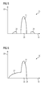
- FIG. 5 shows a weighting function 25 , which was obtained from similarities between anatomy image datasets.
- the most similar anatomy image datasets are found around the time point 26 , which represents the reference time point t′, which is why the highest function values are located in the central region 27 of the weighting function 25 .
- the axis 23 again indicates the extent of the weighting.
- Anatomy image datasets with an adequate similarity in other words a similarity above one or more threshold values, are also still found in side regions 28 and 29 somewhat removed from the central region 27 .
- the function value is correspondingly lower in each instance.
- FIG. 6 shows an alternative weighting function 30 .
- the similarities of the anatomy image datasets to a reference anatomy image dataset are greater in the central region 31 than in the side arm 32 .
- the time point 33 corresponds to the selected time point t′, for which the PET image dataset is to be calculated.
- FIG. 7 shows the application of a rectangular function for periodic movements.
- the similarities between a number of anatomy image datasets are used here to establish movement periodicities for example of the breathing and heartbeat of an examination object.
- the periodicities can then be used to specify rectangular windows 36 , 37 , 38 and 39 from a predeterminable time point 35 , from which PET measurement signals can be taken into account.
- a further temporal weighting in the form of a Gaussian function can also be multiplied onto these PET measurement signals, as described above in relation to FIG. 4 .
- FIG. 8 shows a flow diagram for producing a PET image dataset.
- step S 1 a patient is introduced into the hybrid imaging modality 1 and all the necessary steps to prepare for measurement, for example shimming the magnetic field of a magnetic resonance scanner, are performed.
- step S 2 measurement data acquisition is started simultaneously by means of the positron emission tomography device and the magnetic resonance device, with the time point of the acquisition of the PET measurement signal and also of a magnetic resonance image dataset being stored.
- step S 3 the raw data acquired using the magnetic resonance device is processed to produce image datasets, in order to be able to perform a similarity calculation thereon.
- a time point t′ is to be predetermined by a user or automatically, at which a PET image dataset is to be determined.
- the reference anatomy image dataset is established from time point t′, its similarities to the other anatomy image datasets being determined (step S 5 ).
- the following method can be applied to optimize computation power:
- the anatomy image datasets closest in time are used first. If the similarity of the anatomy image datasets to the reference anatomy image dataset decreases as the time interval increases, in particular to below a termination threshold value, the determination of similarities is terminated and the similarity values are set to zero. This termination procedure can be used for all embodiments.
- step S 6 the PET measurement signals are additionally weighted with a Gaussian function.
- a PET image dataset is determined from the twice weighted PET measurement signals.
- the weightings mean that the motion artifacts therein are minimized.
Landscapes
- Health & Medical Sciences (AREA)
- Life Sciences & Earth Sciences (AREA)
- Engineering & Computer Science (AREA)
- Physics & Mathematics (AREA)
- High Energy & Nuclear Physics (AREA)
- Molecular Biology (AREA)
- Medical Informatics (AREA)
- General Physics & Mathematics (AREA)
- Spectroscopy & Molecular Physics (AREA)
- Optics & Photonics (AREA)
- Biomedical Technology (AREA)
- Nuclear Medicine, Radiotherapy & Molecular Imaging (AREA)
- General Health & Medical Sciences (AREA)
- Animal Behavior & Ethology (AREA)
- Public Health (AREA)
- Pathology (AREA)
- Heart & Thoracic Surgery (AREA)
- Surgery (AREA)
- Biophysics (AREA)
- Computer Vision & Pattern Recognition (AREA)
- Radiology & Medical Imaging (AREA)
- Veterinary Medicine (AREA)
- Multimedia (AREA)
- Theoretical Computer Science (AREA)
- General Engineering & Computer Science (AREA)
- Nuclear Medicine (AREA)
- Magnetic Resonance Imaging Apparatus (AREA)
Abstract
Description
Claims (28)
Applications Claiming Priority (3)
| Application Number | Priority Date | Filing Date | Title |
|---|---|---|---|
| DE102013201822.3 | 2013-02-05 | ||
| DE102013201822 | 2013-02-05 | ||
| DE102013201822.3A DE102013201822B4 (en) | 2013-02-05 | 2013-02-05 | Method for generating a PET or SPECT image data set and hybrid imaging modality therefor |
Publications (2)
| Publication Number | Publication Date |
|---|---|
| US20140217293A1 US20140217293A1 (en) | 2014-08-07 |
| US9439618B2 true US9439618B2 (en) | 2016-09-13 |
Family
ID=51206070
Family Applications (1)
| Application Number | Title | Priority Date | Filing Date |
|---|---|---|---|
| US14/165,609 Active 2034-05-25 US9439618B2 (en) | 2013-02-05 | 2014-01-28 | Method for generating a pet or spect image dataset and hybrid imaging modality for this purpose |
Country Status (2)
| Country | Link |
|---|---|
| US (1) | US9439618B2 (en) |
| DE (1) | DE102013201822B4 (en) |
Cited By (1)
| Publication number | Priority date | Publication date | Assignee | Title |
|---|---|---|---|---|
| US20220147768A1 (en) * | 2020-11-12 | 2022-05-12 | The University Court Of The University Of Edinburgh | Image data processing apparatus and method, a model training apparatus and a training method |
Families Citing this family (1)
| Publication number | Priority date | Publication date | Assignee | Title |
|---|---|---|---|---|
| EP3288461B1 (en) * | 2016-03-08 | 2018-12-05 | Koninklijke Philips N.V. | Combined x-ray and nuclear imaging |
Citations (17)
| Publication number | Priority date | Publication date | Assignee | Title |
|---|---|---|---|---|
| US20030004405A1 (en) * | 1999-10-14 | 2003-01-02 | Cti Pet Systems, Inc. | Combined PET and X-Ray CT tomograph |
| US20040114708A1 (en) * | 2002-09-23 | 2004-06-17 | Herbert Bruder | Method for imaging in the computer tomography of a periodically moved object to be examined and CT device for carrying out the method |
| US20050053196A1 (en) * | 2003-09-05 | 2005-03-10 | Varian Medical Systems Technologies, Inc. | Systems and methods for processing x-ray images |
| US20050253076A1 (en) * | 2004-05-11 | 2005-11-17 | Wollenweber Scott D | Method and system for normalization of a positron emission tomography system |
| US20060002615A1 (en) * | 2004-06-30 | 2006-01-05 | Accuray, Inc. | Image enhancement method and system for fiducial-less tracking of treatment targets |
| US20060235295A1 (en) * | 2005-04-15 | 2006-10-19 | Siemens Aktiengesellschaft | Method for movement-compensation in imaging |
| US20060266947A1 (en) * | 2005-05-24 | 2006-11-30 | Robert Krieg | Method for determining positron emission measurement information in the context of positron emission tomography |
| US20080193003A1 (en) * | 2005-06-22 | 2008-08-14 | Koninklijke Philips Electronics N. V. | Bands Artifact Reduction for Cardiac Ct Imaging |
| DE102007009182A1 (en) | 2007-02-26 | 2008-08-28 | Siemens Ag | Cyclically moving object i.e. heart, image representation method, involves recording image data of object by single photon emission computed tomography method and assigning recorded image to different phases of motion cycle |
| US20090003655A1 (en) * | 2007-06-27 | 2009-01-01 | General Electric Company | Methods and systems for assessing patient movement in diagnostic imaging |
| US20090037130A1 (en) * | 2007-07-26 | 2009-02-05 | Thorsten Feiweier | Method for recording measured data from a patient by taking movements into account, and associated medical device |
| US20090041318A1 (en) * | 2007-07-26 | 2009-02-12 | Thorsten Feiweier | Method for recording measured data of a patient while taking account of movement operations, and an associated medical device |
| US20090076379A1 (en) * | 2007-09-18 | 2009-03-19 | Siemens Medical Solutions Usa, Inc. | Ultrasonic Imager for Motion Measurement in Multi-Modality Emission Imaging |
| US20100046821A1 (en) * | 2008-05-09 | 2010-02-25 | General Electric Company | Motion correction in tomographic images |
| US20110116695A1 (en) * | 2009-11-19 | 2011-05-19 | Scott David Wollenweber | Method and apparatus for reducing motion-related imaging artifacts |
| US20120281897A1 (en) * | 2011-05-03 | 2012-11-08 | General Electric Company | Method and apparatus for motion correcting medical images |
| US20130131493A1 (en) * | 2011-11-22 | 2013-05-23 | General Electric Company | Method and apparatus for performing dual-modality imaging |
-
2013
- 2013-02-05 DE DE102013201822.3A patent/DE102013201822B4/en active Active
-
2014
- 2014-01-28 US US14/165,609 patent/US9439618B2/en active Active
Patent Citations (20)
| Publication number | Priority date | Publication date | Assignee | Title |
|---|---|---|---|---|
| US20030004405A1 (en) * | 1999-10-14 | 2003-01-02 | Cti Pet Systems, Inc. | Combined PET and X-Ray CT tomograph |
| US20040114708A1 (en) * | 2002-09-23 | 2004-06-17 | Herbert Bruder | Method for imaging in the computer tomography of a periodically moved object to be examined and CT device for carrying out the method |
| US20050053196A1 (en) * | 2003-09-05 | 2005-03-10 | Varian Medical Systems Technologies, Inc. | Systems and methods for processing x-ray images |
| US20050253076A1 (en) * | 2004-05-11 | 2005-11-17 | Wollenweber Scott D | Method and system for normalization of a positron emission tomography system |
| US20060002615A1 (en) * | 2004-06-30 | 2006-01-05 | Accuray, Inc. | Image enhancement method and system for fiducial-less tracking of treatment targets |
| US20060235295A1 (en) * | 2005-04-15 | 2006-10-19 | Siemens Aktiengesellschaft | Method for movement-compensation in imaging |
| DE102005017492A1 (en) | 2005-04-15 | 2006-10-19 | Siemens Ag | Method for computationally compensating a periodic movement of an organ |
| US20060266947A1 (en) * | 2005-05-24 | 2006-11-30 | Robert Krieg | Method for determining positron emission measurement information in the context of positron emission tomography |
| DE102005023907A1 (en) | 2005-05-24 | 2006-12-07 | Siemens Ag | Method for determining positron emission measurement information in the context of positron emission tomography |
| US20080193003A1 (en) * | 2005-06-22 | 2008-08-14 | Koninklijke Philips Electronics N. V. | Bands Artifact Reduction for Cardiac Ct Imaging |
| DE102007009182A1 (en) | 2007-02-26 | 2008-08-28 | Siemens Ag | Cyclically moving object i.e. heart, image representation method, involves recording image data of object by single photon emission computed tomography method and assigning recorded image to different phases of motion cycle |
| US20080219510A1 (en) * | 2007-02-26 | 2008-09-11 | Diana Martin | Method and device for imaging cyclically moving objects |
| US20090003655A1 (en) * | 2007-06-27 | 2009-01-01 | General Electric Company | Methods and systems for assessing patient movement in diagnostic imaging |
| US20090037130A1 (en) * | 2007-07-26 | 2009-02-05 | Thorsten Feiweier | Method for recording measured data from a patient by taking movements into account, and associated medical device |
| US20090041318A1 (en) * | 2007-07-26 | 2009-02-12 | Thorsten Feiweier | Method for recording measured data of a patient while taking account of movement operations, and an associated medical device |
| US20090076379A1 (en) * | 2007-09-18 | 2009-03-19 | Siemens Medical Solutions Usa, Inc. | Ultrasonic Imager for Motion Measurement in Multi-Modality Emission Imaging |
| US20100046821A1 (en) * | 2008-05-09 | 2010-02-25 | General Electric Company | Motion correction in tomographic images |
| US20110116695A1 (en) * | 2009-11-19 | 2011-05-19 | Scott David Wollenweber | Method and apparatus for reducing motion-related imaging artifacts |
| US20120281897A1 (en) * | 2011-05-03 | 2012-11-08 | General Electric Company | Method and apparatus for motion correcting medical images |
| US20130131493A1 (en) * | 2011-11-22 | 2013-05-23 | General Electric Company | Method and apparatus for performing dual-modality imaging |
Non-Patent Citations (3)
| Title |
|---|
| Fahey,F.H., "Data Acquisition in PET Imaging", J Nucl Med Technol 2002; 30:39-49. |
| German Office Action for priority application DE 10 2013 201 822.3 dated Aug. 27, 2013. |
| German priority application DE 10 2013 201 822.3, filed Feb. 5, 2013. |
Cited By (2)
| Publication number | Priority date | Publication date | Assignee | Title |
|---|---|---|---|---|
| US20220147768A1 (en) * | 2020-11-12 | 2022-05-12 | The University Court Of The University Of Edinburgh | Image data processing apparatus and method, a model training apparatus and a training method |
| US12321416B2 (en) * | 2020-11-12 | 2025-06-03 | The University Court Of The University Of Edinburgh | Image data processing apparatus and method, a model training apparatus and a training method |
Also Published As
| Publication number | Publication date |
|---|---|
| DE102013201822A1 (en) | 2014-08-07 |
| US20140217293A1 (en) | 2014-08-07 |
| DE102013201822B4 (en) | 2022-12-29 |
Similar Documents
| Publication | Publication Date | Title |
|---|---|---|
| US12502077B2 (en) | Method for reducing noise in a medical image slice by inputting plural slice images into a trained neural network | |
| CN101765865B (en) | Motion correction in nuclear imaging | |
| JP5833637B2 (en) | Dynamic perfusion CT image data alignment | |
| EP2707853B1 (en) | List mode dynamic image reconstruction | |
| US9619869B2 (en) | Image-based motion compensation of image data | |
| US10529130B2 (en) | Methods and systems for emission computed tomography image reconstruction | |
| US20160095565A1 (en) | Method and imaging system for compensating for location assignment errors in pet data occurring due to a cyclical motion of a patient | |
| US10064593B2 (en) | Image reconstruction for a volume based on projection data sets | |
| CN109716388B (en) | Noise reduction in image data | |
| CN104126193A (en) | Image resolution enhancement | |
| Lamare et al. | PET respiratory motion correction: quo vadis? | |
| WO2017184681A1 (en) | Systems and methods for data-driven respiratory gating in positron emission tomography | |
| Marin et al. | Motion correction for PET data using subspace-based real-time MR imaging in simultaneous PET/MR | |
| CN107913078B (en) | Method for determining a perfusion data set | |
| US9439618B2 (en) | Method for generating a pet or spect image dataset and hybrid imaging modality for this purpose | |
| US8290224B2 (en) | Method and device for imaging cyclically moving objects | |
| CN115705654A (en) | Multimodal medical image processing method, device, computer equipment and storage medium | |
| CN103654829A (en) | Method for generating a pet image data record and facility therefor | |
| EP4239566B1 (en) | Computer-implemented method for determining nuclear medical image data sets, imaging device, computer program and electronically readable storage medium | |
| US20240303829A1 (en) | Object motion measurement apparatus, object motion measurement method, and imaging apparatus | |
| Buschbeck et al. | Motion Correction in Brain MR-PET | |
| JP6035167B2 (en) | Medical image processing program, medical image processing method, and medical image processing apparatus |
Legal Events
| Date | Code | Title | Description |
|---|---|---|---|
| AS | Assignment |
Owner name: SIEMENS AKTIENGESELLSCHAFT, GERMANY Free format text: ASSIGNMENT OF ASSIGNORS INTEREST;ASSIGNOR:HEISMANN, BJOERN;REEL/FRAME:032251/0799 Effective date: 20140204 |
|
| STCF | Information on status: patent grant |
Free format text: PATENTED CASE |
|
| AS | Assignment |
Owner name: SIEMENS HEALTHCARE GMBH, GERMANY Free format text: ASSIGNMENT OF ASSIGNORS INTEREST;ASSIGNOR:SIEMENS AKTIENGESELLSCHAFT;REEL/FRAME:040656/0054 Effective date: 20161104 |
|
| MAFP | Maintenance fee payment |
Free format text: PAYMENT OF MAINTENANCE FEE, 4TH YEAR, LARGE ENTITY (ORIGINAL EVENT CODE: M1551); ENTITY STATUS OF PATENT OWNER: LARGE ENTITY Year of fee payment: 4 |
|
| AS | Assignment |
Owner name: SIEMENS HEALTHINEERS AG, GERMANY Free format text: ASSIGNMENT OF ASSIGNORS INTEREST;ASSIGNOR:SIEMENS HEALTHCARE GMBH;REEL/FRAME:066088/0256 Effective date: 20231219 |
|
| AS | Assignment |
Owner name: SIEMENS HEALTHINEERS AG, GERMANY Free format text: CORRECTIVE ASSIGNMENT TO CORRECT THE ASSIGNEE PREVIOUSLY RECORDED AT REEL: 066088 FRAME: 0256. ASSIGNOR(S) HEREBY CONFIRMS THE ASSIGNMENT;ASSIGNOR:SIEMENS HEALTHCARE GMBH;REEL/FRAME:071178/0246 Effective date: 20231219 |
|
| MAFP | Maintenance fee payment |
Free format text: PAYMENT OF MAINTENANCE FEE, 8TH YEAR, LARGE ENTITY (ORIGINAL EVENT CODE: M1552); ENTITY STATUS OF PATENT OWNER: LARGE ENTITY Year of fee payment: 8 |