US9403794B2 - Imaging beta-amyloid peptides and inhibition of beta-amyloid peptide aggregation - Google Patents
Imaging beta-amyloid peptides and inhibition of beta-amyloid peptide aggregation Download PDFInfo
- Publication number
- US9403794B2 US9403794B2 US14/231,724 US201414231724A US9403794B2 US 9403794 B2 US9403794 B2 US 9403794B2 US 201414231724 A US201414231724 A US 201414231724A US 9403794 B2 US9403794 B2 US 9403794B2
- Authority
- US
- United States
- Prior art keywords
- carbazole
- alkyl
- sloh
- substituted
- peptides
- Prior art date
- Legal status (The legal status is an assumption and is not a legal conclusion. Google has not performed a legal analysis and makes no representation as to the accuracy of the status listed.)
- Active
Links
- 0 [1*]N1C2=C(C=C(C3CC([Ar+][3*])C3)C=C2)C2=C1/C=C\C([Y])=C/2.[2*].[CH3-] Chemical compound [1*]N1C2=C(C=C(C3CC([Ar+][3*])C3)C=C2)C2=C1/C=C\C([Y])=C/2.[2*].[CH3-] 0.000 description 6
- JKGDTLZVSJSJHQ-UHFFFAOYSA-O CCOC(=O)C[N+]1=C2C=CC=CC2=C(/C=C/C2=C/C3=C(\C=C/2)N(CCOCCOC)C2=C3C=CC=C2)C=C1.COCCOCCN1C2=C(C=C(F)C=C2)C2=C1/C=C\C(\C=C\C1=C3C=CC=CC3=[N+](CCO)C=C1)=C/2.COCCOCCN1C2=C(C=CC=C2)C2=C1/C=C\C(\C=C\C1=C3C=CC=CC3=[N+](CC(N)=O)C=C1)=C/2.COCCOCCN1C2=C(C=CC=C2)C2=C1/C=C\C(\C=C\C1=C3C=CC=CC3=[N+](CCOCCOC)C=C1)=C/2.[CH3-].[CH3-].[CH3-].[CH3-] Chemical compound CCOC(=O)C[N+]1=C2C=CC=CC2=C(/C=C/C2=C/C3=C(\C=C/2)N(CCOCCOC)C2=C3C=CC=C2)C=C1.COCCOCCN1C2=C(C=C(F)C=C2)C2=C1/C=C\C(\C=C\C1=C3C=CC=CC3=[N+](CCO)C=C1)=C/2.COCCOCCN1C2=C(C=CC=C2)C2=C1/C=C\C(\C=C\C1=C3C=CC=CC3=[N+](CC(N)=O)C=C1)=C/2.COCCOCCN1C2=C(C=CC=C2)C2=C1/C=C\C(\C=C\C1=C3C=CC=CC3=[N+](CCOCCOC)C=C1)=C/2.[CH3-].[CH3-].[CH3-].[CH3-] JKGDTLZVSJSJHQ-UHFFFAOYSA-O 0.000 description 2
- PTAGLPQOTGYALQ-UHFFFAOYSA-N CC[N+]1=C2C=CC=CC2=C(/C=C/C2=C/C3=C(\C=C/2)N(CCOCCOC)C2=C3C=CC=C2)C=C1.COCCOCCN1C2=C(C=CC=C2)C2=C1/C=C\C(\C=C\C1=C3C=CC=CC3=[N+](C)C=C1)=C/2.COCCOCCN1C2=C(C=CC=C2)C2=C1/C=C\C(\C=C\C1=C3C=CC=CC3=[N+](CCCO)C=C1)=C/2.COCCOCCN1C2=C(C=CC=C2)C2=C1/C=C\C(\C=C\C1=C3C=CC=CC3=[N+](CCO)C=C1)=C/2.[CH3-].[CH3-].[CH3-].[I-] Chemical compound CC[N+]1=C2C=CC=CC2=C(/C=C/C2=C/C3=C(\C=C/2)N(CCOCCOC)C2=C3C=CC=C2)C=C1.COCCOCCN1C2=C(C=CC=C2)C2=C1/C=C\C(\C=C\C1=C3C=CC=CC3=[N+](C)C=C1)=C/2.COCCOCCN1C2=C(C=CC=C2)C2=C1/C=C\C(\C=C\C1=C3C=CC=CC3=[N+](CCCO)C=C1)=C/2.COCCOCCN1C2=C(C=CC=C2)C2=C1/C=C\C(\C=C\C1=C3C=CC=CC3=[N+](CCO)C=C1)=C/2.[CH3-].[CH3-].[CH3-].[I-] PTAGLPQOTGYALQ-UHFFFAOYSA-N 0.000 description 1
- PDIMOANSXAVDSA-UHFFFAOYSA-N CN1C2=C(C=CC=C2)C2=C1/C=C\C(\C=C\C1=C3C=CC=CC3=[N+](C)C=C1)=C/2.[CH3-] Chemical compound CN1C2=C(C=CC=C2)C2=C1/C=C\C(\C=C\C1=C3C=CC=CC3=[N+](C)C=C1)=C/2.[CH3-] PDIMOANSXAVDSA-UHFFFAOYSA-N 0.000 description 1
- ZFWKUGADFIWMRO-UHFFFAOYSA-N COCCOCCN1C2=CC=CC=C2C2=C1/C=C\C(\C=C\C1=CC=[N+](C)C=C1)=C/2.COCCOCCN1C2=CC=CCC2C2=C1/C=C\C(\C=C\C1=CC=[N+](CCO)C=C1)=C/2.[Cl-].[I-] Chemical compound COCCOCCN1C2=CC=CC=C2C2=C1/C=C\C(\C=C\C1=CC=[N+](C)C=C1)=C/2.COCCOCCN1C2=CC=CCC2C2=C1/C=C\C(\C=C\C1=CC=[N+](CCO)C=C1)=C/2.[Cl-].[I-] ZFWKUGADFIWMRO-UHFFFAOYSA-N 0.000 description 1
- PUNAFYWKZHXHMZ-UHFFFAOYSA-N O=C=O.O=C=O.[HH].[H]CN1CCN(C[H])CCN(CC(=O)NC2=C(C)C=C([Y])C=C2C)CCN(CC(=O)O)CC1 Chemical compound O=C=O.O=C=O.[HH].[H]CN1CCN(C[H])CCN(CC(=O)NC2=C(C)C=C([Y])C=C2C)CCN(CC(=O)O)CC1 PUNAFYWKZHXHMZ-UHFFFAOYSA-N 0.000 description 1
Images




Classifications
-
- C—CHEMISTRY; METALLURGY
- C07—ORGANIC CHEMISTRY
- C07D—HETEROCYCLIC COMPOUNDS
- C07D401/00—Heterocyclic compounds containing two or more hetero rings, having nitrogen atoms as the only ring hetero atoms, at least one ring being a six-membered ring with only one nitrogen atom
- C07D401/02—Heterocyclic compounds containing two or more hetero rings, having nitrogen atoms as the only ring hetero atoms, at least one ring being a six-membered ring with only one nitrogen atom containing two hetero rings
- C07D401/06—Heterocyclic compounds containing two or more hetero rings, having nitrogen atoms as the only ring hetero atoms, at least one ring being a six-membered ring with only one nitrogen atom containing two hetero rings linked by a carbon chain containing only aliphatic carbon atoms
-
- A—HUMAN NECESSITIES
- A61—MEDICAL OR VETERINARY SCIENCE; HYGIENE
- A61K—PREPARATIONS FOR MEDICAL, DENTAL OR TOILETRY PURPOSES
- A61K31/00—Medicinal preparations containing organic active ingredients
- A61K31/33—Heterocyclic compounds
- A61K31/395—Heterocyclic compounds having nitrogen as a ring hetero atom, e.g. guanethidine or rifamycins
- A61K31/435—Heterocyclic compounds having nitrogen as a ring hetero atom, e.g. guanethidine or rifamycins having six-membered rings with one nitrogen as the only ring hetero atom
- A61K31/47—Quinolines; Isoquinolines
- A61K31/4709—Non-condensed quinolines and containing further heterocyclic rings
-
- A—HUMAN NECESSITIES
- A61—MEDICAL OR VETERINARY SCIENCE; HYGIENE
- A61K—PREPARATIONS FOR MEDICAL, DENTAL OR TOILETRY PURPOSES
- A61K45/00—Medicinal preparations containing active ingredients not provided for in groups A61K31/00 - A61K41/00
- A61K45/06—Mixtures of active ingredients without chemical characterisation, e.g. antiphlogistics and cardiaca
-
- A—HUMAN NECESSITIES
- A61—MEDICAL OR VETERINARY SCIENCE; HYGIENE
- A61K—PREPARATIONS FOR MEDICAL, DENTAL OR TOILETRY PURPOSES
- A61K49/00—Preparations for testing in vivo
- A61K49/0002—General or multifunctional contrast agents, e.g. chelated agents
-
- A—HUMAN NECESSITIES
- A61—MEDICAL OR VETERINARY SCIENCE; HYGIENE
- A61K—PREPARATIONS FOR MEDICAL, DENTAL OR TOILETRY PURPOSES
- A61K49/00—Preparations for testing in vivo
- A61K49/001—Preparation for luminescence or biological staining
- A61K49/0013—Luminescence
- A61K49/0017—Fluorescence in vivo
- A61K49/0019—Fluorescence in vivo characterised by the fluorescent group, e.g. oligomeric, polymeric or dendritic molecules
- A61K49/0021—Fluorescence in vivo characterised by the fluorescent group, e.g. oligomeric, polymeric or dendritic molecules the fluorescent group being a small organic molecule
-
- A—HUMAN NECESSITIES
- A61—MEDICAL OR VETERINARY SCIENCE; HYGIENE
- A61K—PREPARATIONS FOR MEDICAL, DENTAL OR TOILETRY PURPOSES
- A61K49/00—Preparations for testing in vivo
- A61K49/001—Preparation for luminescence or biological staining
- A61K49/0013—Luminescence
- A61K49/0017—Fluorescence in vivo
- A61K49/0019—Fluorescence in vivo characterised by the fluorescent group, e.g. oligomeric, polymeric or dendritic molecules
- A61K49/0021—Fluorescence in vivo characterised by the fluorescent group, e.g. oligomeric, polymeric or dendritic molecules the fluorescent group being a small organic molecule
- A61K49/0026—Acridine dyes
-
- A—HUMAN NECESSITIES
- A61—MEDICAL OR VETERINARY SCIENCE; HYGIENE
- A61K—PREPARATIONS FOR MEDICAL, DENTAL OR TOILETRY PURPOSES
- A61K49/00—Preparations for testing in vivo
- A61K49/06—Nuclear magnetic resonance [NMR] contrast preparations; Magnetic resonance imaging [MRI] contrast preparations
- A61K49/08—Nuclear magnetic resonance [NMR] contrast preparations; Magnetic resonance imaging [MRI] contrast preparations characterised by the carrier
- A61K49/085—Nuclear magnetic resonance [NMR] contrast preparations; Magnetic resonance imaging [MRI] contrast preparations characterised by the carrier conjugated systems
-
- A—HUMAN NECESSITIES
- A61—MEDICAL OR VETERINARY SCIENCE; HYGIENE
- A61K—PREPARATIONS FOR MEDICAL, DENTAL OR TOILETRY PURPOSES
- A61K49/00—Preparations for testing in vivo
- A61K49/06—Nuclear magnetic resonance [NMR] contrast preparations; Magnetic resonance imaging [MRI] contrast preparations
- A61K49/08—Nuclear magnetic resonance [NMR] contrast preparations; Magnetic resonance imaging [MRI] contrast preparations characterised by the carrier
- A61K49/10—Organic compounds
-
- A—HUMAN NECESSITIES
- A61—MEDICAL OR VETERINARY SCIENCE; HYGIENE
- A61K—PREPARATIONS FOR MEDICAL, DENTAL OR TOILETRY PURPOSES
- A61K2300/00—Mixtures or combinations of active ingredients, wherein at least one active ingredient is fully defined in groups A61K31/00 - A61K41/00
Definitions
- the present invention relates to methods of detecting and monitoring aggregation of beta-amyloid peptides which are associated with neurodegenerative diseases as well as treating and/or preventing the neurodegenerative diseases by using carbazole-based fluorophores.
- the present invention provides methods for labeling and imaging the beta-amyloid (A ⁇ ) peptides, oligomers, and fibrils in vitro and/or in vivo, as well as treating and/or preventing Alzheimer's disease by using the carbazole-based fluorophores of the present invention.
- AD Alzheimer's disease
- AD Alzheimer's disease
- the incidence rate of AD is known to increase with age. At age over 65, the incidence rate is about 5% in the general population. At age over 80, the incidence rate increases to about 20%, i.e., one in five.
- Current drug treatments can only improve symptoms and produce no profound cure.
- several approaches aimed at inhibiting disease progression have advanced to clinical trials. Among them, strategies targeting the production and clearance of the A ⁇ peptide, which is thought to be a critical protein involved in the pathogenesis of the disease, are the most advanced.
- a ⁇ peptide is the principal protein component of the A ⁇ plaques, which are found in the brains of AD patients during autopsy.
- the occurrence of the A ⁇ plaques considered a cardinal feature of AD, provides the only confirmed diagnosis of the disease.
- Extensive researches in past decades have indicated a central role for the A ⁇ peptide in the disease process where the A ⁇ peptides assemble (aggregate) into A ⁇ fibrils which exert a cytotoxic effect towards the neurons and initiate the pathogenic cascade. Recent studies showed that oligomeric, prefibrillar and diffusible assemblies of A ⁇ peptides are also deleterious.
- carbazole-based fluorophores are highly sensitive fluorescent light-up probe for double strand DNA and strongly active two-photon absorption dyes for two-photon excited bioimaging, the disclosure of which is incorporated by reference herein.
- mono-cyanine fluorophore has also been found to exhibit binding affinity towards beta amyloid (A ⁇ ) peptide concomitant with strong fluorescent enhancement.
- the first objective of the presently claimed invention relates to a method of reducing and preventing aggregation of beta-amyloid peptides for Alzheimer's disease (AD) as well as of treating and/or preventing Alzheimer's disease by using carbazole-based fluorophores.
- AD Alzheimer's disease
- said method comprising: binding said carbazole-based fluorophores to A ⁇ peptides, oligomers and/or fibrils thereof; inhibiting the growth and/or aggregation of said A ⁇ peptides, oligomers and/or fibrils upon said binding; and protecting neuronal cells against the neurotoxic activities of the A ⁇ oligomers and/or fibrils;
- Ar is a heteraromatic ring selected from the group consisting of pyridinyl, substituted pyridinyl, quinolinyl, substituted quinolinyl, acridinyl, substituted acridinyl, benzothiazolyl, substituted benzothiazolyl, benzoxazolyl, and substituted benzoxazolyl;
- R 1 is a radical selected from the group consisting of polyethylene glycol chain, alkyl, substituted alkyl, peptide chain, glycosidyl, and C(O)NHCH((CH 2 CH 2 O) 2 CH 3
- R 3 is a radical selected from the group consisting of HO-alkyl, alkyl-COOalkyl, alkyl-CONH 2 , alkyl-CONHalkyl, polyethylene glycol chain;
- X is an anion selected from the group consisting of F, Cl, Br, I, HSO 4 , H 2 PO 4 , HCO 3 , tosylate, and mesylate;
- Y is selected from the group consisting of H, F, Cl, OH, and OCH 3 .
- a method wherein Ar is selected from a quinolinyl or substituted quinolinyl; said R 1 is a 2-(2-methoxyethoxy)ethoxy; said R 2 is an ethenyl; R 3 is a 2-hydroxyethyl or acetamide or acetate or 2-(2-methoxyethoxy)ethoxy; said X is a chloride or bromide or iodide and said Y is a H or F which are represented by the formula F-SLOH, SLAD, SLAce, and SLG:
- FIG. 1 shows the fluorescence spectra of SPM, SPOH, SLM, SLOH, SLE, SLOH-Pr, Me-SLM, SAM, and SAOH (1 ⁇ M) in phosphate buffer upon addition of various concentrations of A ⁇ (1-40) fibrils prepared from A ⁇ 40 with seed incubated at 37° C. for an hour in buffer (left column).
- FIG. 1A shows the fluorescence spectra of SPM in various concentrations of A ⁇ (1-40) fibrils.
- FIG. 1C shows the fluorescence spectra of SPOH in various concentrations of A ⁇ (1-40) fibrils.
- FIG. 1E shows the fluorescence spectra of SLM in various concentrations of A ⁇ (1-40) fibrils.
- FIG. 1 shows the fluorescence spectra of SPM, SPOH, SLM, SLOH, SLE, SLOH-Pr, Me-SLM, SAM, and SAOH (1 ⁇ M) in phosphate buffer upon addition of various concentrations of A
- FIG. 1G shows the fluorescence spectra of SLOH in various concentrations of A ⁇ (1-40) fibrils.
- FIG. 1I shows the fluorescence spectra of SLE in various concentrations of A ⁇ (1-40) fibrils.
- FIG. 1K shows the fluorescence spectra of SLOH-Pr in various concentrations of A ⁇ (1-40) fibrils.
- FIG. 1M shows the fluorescence spectra of Me-SLM in various concentrations of A ⁇ (1-40) fibrils.
- FIG. 1O shows the fluorescence spectra of SAM in various concentrations of A ⁇ (1-40) fibrils.
- FIG. 1Q shows the fluorescence spectra of SAOH in various concentrations of A ⁇ (1-40) fibrils.
- FIG. 1 also shows the fluorescence spectra of SPM, SPOH, SLM, SLOH, SLE, SLOH-Pr, Me-SLM, SAM, and SAOH (1 ⁇ M) in phosphate buffer itself, in the presence of 100 equv of A ⁇ 40 in monomeric form, and in the presence of 100 equv of A ⁇ 40 in fibril state in phosphate buffer (right column).
- FIG. 1B shows the fluorescence spectra of SPM on its own and in the presence of two different forms of A ⁇ 40 .
- FIG. 1D shows the fluorescence spectra of SPOH on its own and in the presence of two different forms of A ⁇ 40 .
- FIG. 1F shows the fluorescence spectra of SLM on its own and in the presence of two different forms of A ⁇ 40 .
- FIG. 1H shows the fluorescence spectra of SLOH on its own and in the presence of two different forms of A ⁇ 40 .
- FIG. 1J shows the fluorescence spectra of SLE on its own and in the presence of two different forms of A ⁇ 40 .
- FIG. 1L shows the fluorescence spectra of SLOH-Pr on its own and in the presence of two different forms of A ⁇ 40 .
- FIG. 1N shows the fluorescence spectra of Me-SLM on its own and in the presence of two different forms of A ⁇ 40 .
- FIG. 1P shows the fluorescence spectra of SAM on its own and in the presence of two different forms of A ⁇ 40 .
- FIG. 1R shows the fluorescence spectra of SAOH on its own and in the presence of two different forms of A ⁇ 40 .
- FIG. 2 shows the fluorescence spectra of SPM, SPOH, SLM and SLOH in phosphate buffer (1 ⁇ M) upon addition of various concentrations of A ⁇ (1-40) and A ⁇ (1-42), respectively.
- FIG. 2A shows the fluorescence spectra of SPM in various concentrations of A ⁇ (1-40).
- FIG. 2B shows the fluorescence spectra of SPM in various concentrations of A ⁇ (1-42).
- FIG. 2C shows the fluorescence spectra of SPOH in various concentrations of A ⁇ (1-40).
- FIG. 2D shows the fluorescence spectra of SPOH in various concentrations of A ⁇ (1-42).
- FIG. 2E shows the fluorescence spectra of SLM in various concentrations of A ⁇ (1-40).
- FIG. 2F shows the fluorescence spectra of SLM in various concentrations of A ⁇ (1-42).
- FIG. 2G shows the fluorescence spectra of SLOH in various concentrations of A ⁇ (1-40).
- FIG. 2H shows the fluorescence spectra of SLOH in various concentrations of A ⁇ (1-42).
- FIG. 3 shows TIRFM images of A ⁇ fibrils after incubation with the carbazole-based fluorophores, SPM ( FIG. 3A ) excited at 445 nm and SLM ( FIG. 3B ) and SLOH ( FIG. 3C ) excited at 488 nm, respectively.
- FIG. 4 shows in vitro fluorescence imaging of neuronal cells by using the carbazole-based fluorophore, SLOH ( FIG. 4A )
- the lambda scans of the images match well with the fluorescence spectrum of the SLOH ( FIG. 4B ).
- FIG. 5 shows absorption and fluorescence spectra of the carbazole-based fluorophores, SAM ( FIG. 5A ) and SAOH ( FIG. 5B ) in phosphate buffer solution.
- FIG. 6 shows CD spectra of A ⁇ (1-40) peptide ( FIG. 6A ) and fibrils ( FIG. 6B ) in the absence and presence of SLOH (1:1) (20 ⁇ M).
- FIG. 7 shows TIRFM images of A ⁇ fibrils ( FIG. 7A ) and A ⁇ peptide after incubation with the carbazole-based fluorophore, SLOH ( FIG. 7B ), SAOH ( FIG. 7C ), SLE ( FIG. 7D ), SLOH-Pr ( FIG. 7E ), and Me-SLM ( FIG. 7F ).
- FIGS. 7B, 7C, 7E and 7F show an inhibition of A ⁇ fibril formation from the A ⁇ monomer by SLOH and SAOH. These images were obtained by an addition of ThT dye excited at 445 nm.
- FIG. 8 shows TEM images of A ⁇ fibril growth from A ⁇ peptide (50 ⁇ M) seeded for 1 hr at 37° C. in the absence ( FIG. 8A ) and the presence of SLOH ( FIG. 8B ).
- FIG. 9 shows ThT, SLM and SLOH fluorescence binding assays for 50 ⁇ M A ⁇ 40 fibrillation
- FIG. 9A Average length of 1 h incubated A ⁇ 40-fibril measured from TIRFM images after 1 h seed-mediated incubation of A ⁇ 40 monomer with (bottom axis, bars) and without (top axis, scatter point) SLOH (50 ⁇ M) added at different time points (0, 10, 20, 40 and 60 min) within an one hour-incubation.
- FIG. 9B shows ThT, SLM and SLOH fluorescence binding assays for 50 ⁇ M A ⁇ 40 fibrillation
- FIG. 10 shows cytotoxicities of the carbazole-based SLOH ( FIG. 10A ), SLOH-Pr ( FIG. 10B ), Me-SLM ( FIG. 10C ) and SAOH ( FIG. 10D ) towards the SH-SY5Y neuronal cell with MTT assays.
- FIG. 11 shows cytotoxicities of A ⁇ peptide monomer (DM/M), oligomers (DM S/MS) and fibrils (DF/F) towards the SH-SY5Y neuronal cell in the absence and the presence of SLOH (50 ⁇ M) ( FIG. 11A ), SLE ( FIG. 11B ), SLOH-Pr ( FIG. 11C ), Me-SLM ( FIG. 11D ) after 2 hr, 6 hr, and 24 hr incubations.
- SLOH 50 ⁇ M
- SLE SLE
- SLOH-Pr FIG. 11C
- Me-SLM FIG. 11D
- FIG. 12 shows fluorescence images of transgenic mice brain with tail vein injection of SLOH and co-stained with the A ⁇ labeling dye, ThT or A ⁇ antibody with DAB stain.
- the overlapped image revealed the colocalization of fluorescence signals of SLOH and ThT in cellular components.
- Differential Interference Contrast (DIC) image of DAB stained brain slide of transgenic mice FIG. 12D ).
- Fluorescence image of same slide corresponding to SLOH fluorescence captured at 550-630 nm under excitation at 488 nm ( FIG. 12E ); and overlapped images of previous two images ( FIG. 12F ).
- the overlapped image revealed the colocalization of fluorescence signals of SLOH and A ⁇ antibody in cellular components.
- FIG. 13 shows synthesis of carbazole-based fluorophores, SLM, SLOH, SLE, SLOH-Pr ( FIG. 13A ) and SPM, SPOH, Me-SLM, SAM, and SAOH ( FIG. 13B ).
- FIG. 14 shows the general chemical structures of carbazole-based fluorophores, including S series.
- FIG. 15 shows the formula “F-SLOH”, “SLAD”, “SLAce”, and “SLG”, respectively.
- FIG. 16 shows the fluorescence spectra of F-SLOH ( FIG. 16A-C ), SLAD ( FIG. 16D-F ), SLAce ( FIG. 16G-I ), and SLG ( FIG. 16J-L ) in phosphate buffer (1 ⁇ M) upon addition of various concentrations of A ⁇ (1-42) monomer, A ⁇ (1-40) monomer and A ⁇ (1-40) fibril, respectively.
- FIG. 17 shows the TIRFM images of A ⁇ monomer and seeds after incubation with the carbazole-based fluorophores, SLG, F-SLOH, SLAD and SLAce.
- the panels below show the images of seed only and A ⁇ fibril formation without the carbazole-based fluorophore for comparison. These images were obtained by an addition of ThT dye excited at 445 nm.
- FIG. 18 shows the cytotoxicities of the carbazole-based cyanines, F-SLOH, SLAD, SLAce and SLG towards the SH-SY5Y neuronal cell with MTT assays at 2-hour ( FIG. 18A ), 6-hour ( FIG. 18B ) and 24-hour incubation ( FIG. 18C ).
- FIG. 19 shows ( FIG. 19A ) the influence of F-SLOH, and SLAD suppression on the toxicity level against various species of A ⁇ -induced cytotoxicity towards primary cortical neural cells; ( FIG. 19B ) shows the reduction of the ROS induced by the A ⁇ species in primary cortical neural cells.
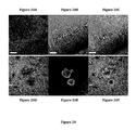
- FIG. 20 shows the fluorescence images of mice brain with tail vein injection of SLAD (upper panel) and co-stained with the A ⁇ labeling dye, and A ⁇ antibody with DAB stain.
- Fluorescence image corresponding to SLAD fluorescence captured at 550-630 nm under excitation at 488 nm FIG. 20A
- Differential Interference Contrast (DIC) image FIG. 20B
- overlapped images of previous two images FIG. 20C
- Differential Interference Contrast (DIC) image of DAB stained brain slide of transgenic mice FIG. 20D ).
- Fluorescence image of same slide corresponding to SLAD fluorescence captured at 550-630 nm under excitation at 488 nm FIG. 20E
- overlapped images of previous two images FIG. 20F ).
- the overlapped image revealed the colocalization of fluorescence signals of SLAD and A ⁇ antibody in cellular components.
- FIG. 21 shows the synthesis of carbazole-based fluorophores, F-SLOH and SLG ( FIG. 21A ), SLAD and SLAce ( FIG. 21B ).
- Reagents and Conditions a, MeCN, reflux; b, ClCH 2 CH 2 OCH 2 CH 2 OCH 3 , NaH, DMF, 75° C.; c, NBS, chloroform, 0° C. to r.t.; d, n-BuLi, NFSi, THF, ⁇ 78° C. to r.t.; e, NBS, chloroform, 0° C. to r.t.; f, n-BuLi, DMF, THF, ⁇ 78° C.
- Ar is a heteraromatic ring selected from the group consisting of pyridinyl, substituted pyridinyl, quinolinyl, substituted quinolinyl, acridinyl, substituted acridinyl, benzothiazolyl, substituted benzothiazolyl, benzoxazolyl, and substituted benzoxazolyl;
- R 1 is a radical selected from the group consisting of polyethylene glycol chain, alkyl, substituted alkyl, peptide chain, glycosidyl, and C(O)NHCH((CH 2 CH 2 O) 2 CH 3 ) 2 ;
- R 2 is selected from the group consisting of ethenyl, ethynyl, azo and azomethinyl;
- R 3 is a radical selected from the group consisting of alkyl, HO-alkyl, HS-alkyl, H 2 N-alkyl, HNalkyl-alkyl, HO
- Ar is a quinolinyl or substituted quinolinyl;
- R 1 is a 2-(2-methoxyethoxy)ethoxy;
- R 2 is an ethenyl;
- R 3 is a methyl, 2-hydroxyethyl, ethyl or 3-hydroxypropyl;
- X is a chloride, bromide or iodide, and the compounds of which are represented by the above formula “SLM”, “SLOH”, “SLE” and “SLOH-Pr”, respectively.
- Ar is a quinolinyl or substituted quinolinyl; R 1 is a methyl; R 2 is an ethenyl; R 3 is a methyl; and X is a chloride, bromide or iodide, the compounds of which are represented by the above formula Me-SLM.
- Ar is an acridinyl or substituted acridinyl;
- R 1 is a 2-(2-methoxy-ethoxy)ethoxy;
- R 2 is an ethenyl;
- R 3 is a methyl or 2-hydroxyethyl;
- X is selected from a chloride, bromide or iodide, and the fluorophores of which are represented by the above formula SAM and SAOH, respectively, where the difference between the compounds of SAM and SAOH is the substitutent at R 3 .
- Ar is selected from a pyridinyl or substituted pyridinyl
- R 1 is a 2-(2-methoxyethoxy)ethoxy
- R 2 is an ethenyl
- R 3 is selected from a methyl or 2-hydroxyethyl
- X is selected from a chloride, bromide or iodide, the compounds of which are represented by the formula SPM and SPOH, respectively.
- a novel series of water-soluble carbazole-based fluorophores has been designed and developed. These molecules were found to bind to A ⁇ (1-40) and A ⁇ (1-42) peptides and, more specifically, their oligomers, and fibrils with strong fluorescence enhancement, therefore allowing direct imaging and detection for the A ⁇ peptides, oligomers and their fibrils ( FIG. 1 ).
- the fluorescence enhancement is much stronger for fibrils than peptides.
- the carbazole-based fluorophores of the present invention provide an advantage of a wide range of excitation and emission tuning in visible to infra-red region ( FIG. 4 ). Some of these molecules, e.g., SAM and SAOH, even emit at ⁇ 760 nm ( FIG. 5 ), which can potentially be used for near infra-red fluorescence imaging.
- the inhibitory effect of the carbazole-based fluorophores of the present invention on A ⁇ fibril growth was further investigated by measuring the (average) length of the A ⁇ fibrils formed after incubation of the A ⁇ monomers for 60 min with additions of SLOH at different time points during this period ( FIG. 9 ). Parallel experiments conducted without any addition of SLOH were used as controls. FIG. 9 shows that an addition of SLOH to the A ⁇ monomer strongly arrests its fibril growth. These results clearly indicate that the inhibitory effect of these carbazole-based fluorophores on A ⁇ aggregation is instantaneous.
- FIGS. 12D-F shows the selectivity of SLOH towards A ⁇ plaques as confirmed with A ⁇ antibody which was used to identify the A ⁇ plaques in transgenic mice's brain.
- carbazole-based fluorophores of the present invention have been shown to bind to A ⁇ (1-40) and A ⁇ (1-42) as well as A ⁇ aggregates with dramatic fluorescence enhancement, thus allowing their direct imaging and labeling as well as the use of TIRFM technique to study the effects of these molecules on A ⁇ aggregation/fibrillation.
- Some embodiments of the carbazole-based fluorophores, for instance, SLOH and SAOH, have been shown to be a potent inhibitor of A ⁇ aggregation, non-toxic and exhibiting a protective effect against the neurotoxic activities of the A ⁇ oligomers and fibrils towards neuronal cells.
- compositions according to the invention were prepared and exemplified as shown in FIG. 13 .
- the Knoevenagel reaction of carbazolyl-3-aldehyde and the corresponding 4-methylpyridium or 4-methylquinolinium halide was used as the key step to synthesize various carbazole-based cyanines.
- Alkylation of carbazole with ethylene glycol chloride and methyl iodide in the presence of NaH in DMF gave alkylated carbazole 1a and 1b respectively.
- Monobromination of 1a and 1b in the presence of NBS gave alkylated 3-bromocarbazole, 2a and 2b, respectively.
- acridine-based cyanines dyes 9-methylacridine was first brominated with NBS affording brominated product 10, which gave phosphonate ester 11 by refluxing with triethyl phosphite. Condensation of phosphonate 11 and aldehyde 3a in the presence of NaH afforded 12, which was alkylated with methyl iodide and 2-iodoethanol to give SAM and SAOH, respectively. All the cyanines were fully characterized with spectroscopic techniques and found to be in good agreement with its structure.
- AD Alzheimer's disease
- the carbazole-based fluorophores of the present invention is also useful as a magnetic resonance imaging (MRI) contrast agent that bind beta amyloid peptides.
- MRI magnetic resonance imaging
- these compounds can potentially be developed into beta-amyloid peptide-specific MRI contrast agents.
- the advantage of 19 F MRI is the extraordinarily sensitivity of the 19 F shift of the reporter group to its local chemical environment, thus opening up the possibility of responsive MRI to detect changes in local pH, oxygen stress, etc.
- This ligand system is also amenable to coupling (e.g., at the —X or —Y positions indicated) to the carbazole moiety of the carbazole-based fluorophores dyes.
- FIG. 14 Another general chemical structures of carbazole-based fluorophores representation, including S series are shown in FIG. 14 .
- Ar is a heteraromatic ring selected from the group consisting of pyridinyl, substituted pyridinyl, quinolinyl, substituted quinolinyl, acridinyl, substituted acridinyl, benzothiazolyl, substituted benzothiazolyl, benzoxazolyl, and substituted benzoxazolyl;
- a novel series of water-soluble carbazole-based fluorophores has been designed and developed. These molecules were found to bind to A ⁇ (1-40) and A ⁇ (1-42) peptides and, more importantly, their oligomers, and fibrils with strong fluorescence enhancement, therefore allowing direct imaging and detection for the A ⁇ peptides, oligomers and their fibrils.
- FIG. 16 Upon binding with A ⁇ peptides, there is an increase in fluorescence intensity up to 160-fold enhancement concomitant with the substantial blue shifts in the emission spectra of the fluorophores.
- these molecules including F-SLOH, SLAD, SLAce, and SLG were found to inhibit A ⁇ peptide aggregation and prevent fibril growth.
- Ar is a quinolinyl or substituted quinolinyl;
- R 1 is a 2-(2-methoxyethoxy)ethoxy;
- R 2 is an ethenyl;
- R 3 is a 2-hydroxyethyl or acetamide or acetate or 2-(2-methoxyethoxy)ethoxy;
- X is a chloride or iodide and
- Y is a H or F which are represented by the formula “F-SLOH”, “SLAD”, “SLAce”, and “SLG”, respectively, as shown in FIG. 15 .
- TRFM Total Internal Reflection Fluorescence Microscope
- these molecules need to be able to pass through the blood-brain barrier.
- the ability of these molecules to penetrate the blood-brain barrier was demonstrated in mice ( FIG. 20 ).
- the binding of these molecules toward A ⁇ plaques in the brains of the Alzheimer's disease animal models are also demonstrated.
- F-SLOH, SLAD, SLAce, and SLG show blood-brain permeability.
- carbazole-based fluorophores bind to A ⁇ (1-40) and A ⁇ (1-42) as well as A ⁇ aggregates with strong fluorescence enhancement, thus allowing their direct imaging and labeling. TIRFM technique is used to study the effects of these molecules on A ⁇ aggregation/fibrillation.
- Some carbazole-based fluorophores, for instance, F-SLOH, and SLAD are non-toxic, potent A ⁇ aggregation inhibitors and exhibit a protective effect against the neurotoxic activities of the A ⁇ oligomers and fibrils towards neuronal cells.
- the different functions discussed herein may be performed in a different order and/or concurrently with each other. Furthermore, if desired, one or more of the above-described functions may be optional or may be combined.
- the objective of the presently claimed invention is to provide methods for labeling, imaging and detecting the beta-amyloid (A ⁇ ) peptides, oligomers, and fibrils in vitro by using carbazole-based fluorophores.
- a further aspect of the present invention relates to a method of reducing and preventing aggregation of beta-amyloid peptides for Alzheimer's disease (AD) as well as of treating and/or preventing Alzheimer's disease by using carbazole-based fluorophores.
Landscapes
- Health & Medical Sciences (AREA)
- Veterinary Medicine (AREA)
- Chemical & Material Sciences (AREA)
- Epidemiology (AREA)
- Life Sciences & Earth Sciences (AREA)
- Animal Behavior & Ethology (AREA)
- General Health & Medical Sciences (AREA)
- Public Health (AREA)
- Organic Chemistry (AREA)
- Medicinal Chemistry (AREA)
- Radiology & Medical Imaging (AREA)
- Nuclear Medicine, Radiotherapy & Molecular Imaging (AREA)
- Engineering & Computer Science (AREA)
- Biomedical Technology (AREA)
- Pharmacology & Pharmacy (AREA)
- Medicines Containing Antibodies Or Antigens For Use As Internal Diagnostic Agents (AREA)
- Indole Compounds (AREA)
Abstract
Description

wherein said method comprising:
binding said carbazole-based fluorophores to Aβ peptides, oligomers and/or fibrils thereof;
inhibiting the growth and/or aggregation of said Aβ peptides, oligomers and/or fibrils upon said binding; and
protecting neuronal cells against the neurotoxic activities of the Aβ oligomers and/or fibrils;
wherein Ar is a heteraromatic ring selected from the group consisting of pyridinyl, substituted pyridinyl, quinolinyl, substituted quinolinyl, acridinyl, substituted acridinyl, benzothiazolyl, substituted benzothiazolyl, benzoxazolyl, and substituted benzoxazolyl;
R1 is a radical selected from the group consisting of polyethylene glycol chain, alkyl, substituted alkyl, peptide chain, glycosidyl, and C(O)NHCH((CH2CH2O)2CH3)2;
R2 is selected from the group consisting of ethenyl, ethynyl, azo and azomethinyl.
R3 is a radical selected from the group consisting of HO-alkyl, alkyl-COOalkyl, alkyl-CONH2, alkyl-CONHalkyl, polyethylene glycol chain;
X is an anion selected from the group consisting of F, Cl, Br, I, HSO4, H2PO4, HCO3, tosylate, and mesylate;
Y is selected from the group consisting of H, F, Cl, OH, and OCH3.


wherein Ar is a heteraromatic ring selected from the group consisting of pyridinyl, substituted pyridinyl, quinolinyl, substituted quinolinyl, acridinyl, substituted acridinyl, benzothiazolyl, substituted benzothiazolyl, benzoxazolyl, and substituted benzoxazolyl; R1 is a radical selected from the group consisting of polyethylene glycol chain, alkyl, substituted alkyl, peptide chain, glycosidyl, and C(O)NHCH((CH2CH2O)2CH3)2; R2 is selected from the group consisting of ethenyl, ethynyl, azo and azomethinyl; R3 is a radical selected from the group consisting of alkyl, HO-alkyl, HS-alkyl, H2N-alkyl, HNalkyl-alkyl, HOOC-alkyl, (alkyl)3N+-alkyl, and (Ph)3P+-alkyl; X is an anion selected from the group consisting of F, Cl, Br, I, HSO4, H2PO4, HCO3, tosylate, and mesylate.


A slight modification of one of the hydroxypyridinone ligands of the Gd(III) complex, shown in [2], allows flexible attachment to the carbazole moiety of Aβ fibril-specific dyes via, for example, a polyethylene glycol (PEG) linkage.

-
- R1 is a radical selected from the group consisting of polyethylene glycol chain, alkyl, substituted alkyl, peptide chain, glycosidyl, and C(O)NHCH((CH2CH2O)2CH3)2;
- R2 is selected from the group consisting of ethenyl, ethynyl, azo and azomethinyl.
- R3 is a radical selected from the group consisting of HO-alkyl, alkyl-COOalkyl, alkyl-CONH2, alkyl-CONHalkyl, polyethylene glycol chain;
- X is an anion selected from the group consisting of F, Cl, Br, I, HSO4, H2PO4, HCO3, tosylate, and mesylate;
- Y is selected from the group consisting of H, F, Cl, OH, and OCH3.
Claims (7)


Priority Applications (2)
| Application Number | Priority Date | Filing Date | Title |
|---|---|---|---|
| US14/231,724 US9403794B2 (en) | 2011-04-21 | 2014-03-31 | Imaging beta-amyloid peptides and inhibition of beta-amyloid peptide aggregation |
| US15/049,126 US9795694B2 (en) | 2011-04-21 | 2016-02-21 | Imaging beta-amyloid peptides aggregation |
Applications Claiming Priority (3)
| Application Number | Priority Date | Filing Date | Title |
|---|---|---|---|
| US201161477614P | 2011-04-21 | 2011-04-21 | |
| US13/447,127 US9156814B2 (en) | 2011-04-21 | 2012-04-13 | Imaging beta-amyloid peptides and inhibition of beta-amyloid peptide aggregation |
| US14/231,724 US9403794B2 (en) | 2011-04-21 | 2014-03-31 | Imaging beta-amyloid peptides and inhibition of beta-amyloid peptide aggregation |
Related Parent Applications (1)
| Application Number | Title | Priority Date | Filing Date |
|---|---|---|---|
| US13/447,127 Continuation-In-Part US9156814B2 (en) | 2011-04-21 | 2012-04-13 | Imaging beta-amyloid peptides and inhibition of beta-amyloid peptide aggregation |
Related Child Applications (1)
| Application Number | Title | Priority Date | Filing Date |
|---|---|---|---|
| US15/049,126 Continuation-In-Part US9795694B2 (en) | 2011-04-21 | 2016-02-21 | Imaging beta-amyloid peptides aggregation |
Publications (2)
| Publication Number | Publication Date |
|---|---|
| US20140213614A1 US20140213614A1 (en) | 2014-07-31 |
| US9403794B2 true US9403794B2 (en) | 2016-08-02 |
Family
ID=51223588
Family Applications (1)
| Application Number | Title | Priority Date | Filing Date |
|---|---|---|---|
| US14/231,724 Active US9403794B2 (en) | 2011-04-21 | 2014-03-31 | Imaging beta-amyloid peptides and inhibition of beta-amyloid peptide aggregation |
Country Status (1)
| Country | Link |
|---|---|
| US (1) | US9403794B2 (en) |
Families Citing this family (3)
| Publication number | Priority date | Publication date | Assignee | Title |
|---|---|---|---|---|
| MY179462A (en) * | 2013-10-11 | 2020-11-06 | Malaysian Palm Oil Board | Protective effects of oil palm composition on alzheimer?s disease |
| KR102240400B1 (en) * | 2020-11-19 | 2021-04-15 | 한국원자력연구원 | Water soluble compound for detection of beta-amyloid |
| CN112641958A (en) * | 2020-12-29 | 2021-04-13 | 中国科学院自动化研究所 | Met targeted molecular probe and preparation method thereof |
Citations (1)
| Publication number | Priority date | Publication date | Assignee | Title |
|---|---|---|---|---|
| US7700616B2 (en) * | 2006-05-08 | 2010-04-20 | Molecular Neuroimaging, Llc. | Compounds and amyloid probes thereof for therapeutic and imaging uses |
-
2014
- 2014-03-31 US US14/231,724 patent/US9403794B2/en active Active
Patent Citations (1)
| Publication number | Priority date | Publication date | Assignee | Title |
|---|---|---|---|---|
| US7700616B2 (en) * | 2006-05-08 | 2010-04-20 | Molecular Neuroimaging, Llc. | Compounds and amyloid probes thereof for therapeutic and imaging uses |
Non-Patent Citations (1)
| Title |
|---|
| Xin Jiang Feng et. al., Cyanines as New Fluorescent Probes for DNA Detection and Two-Photon Excited Bioimaging, Organic Letters, 2010, vol. 112(10), 2194-2197. * |
Also Published As
| Publication number | Publication date |
|---|---|
| US20140213614A1 (en) | 2014-07-31 |
Similar Documents
| Publication | Publication Date | Title |
|---|---|---|
| US9156814B2 (en) | Imaging beta-amyloid peptides and inhibition of beta-amyloid peptide aggregation | |
| Gomez et al. | Nitrogen-doped carbon nanodots for bioimaging and delivery of paclitaxel | |
| Hiremathad et al. | Novel Tacrine-Hydroxyphenylbenzimidazole hybrids as potential multitarget drug candidates for Alzheimer's disease | |
| CN107849441B (en) | AIE luminophores for visualization and treatment of cancer | |
| WO2015143185A1 (en) | Amyloid targeting agents and methods of using the same | |
| US9255933B2 (en) | Imaging beta-amyloid peptides and inhibition of beta-amyloid peptide aggregation | |
| US20170137418A1 (en) | Small molecular probes, processes and use thereof | |
| Perin et al. | Biological activity and DNA binding studies of 2-substituted benzimidazo [1, 2-a] quinolines bearing different amino side chains | |
| AU2016325108B2 (en) | Novel piperazine and piperidine derivatives, their synthesis and use thereof in inhibiting VDAC oligomerization, apoptosis and mitochondria dysfunction | |
| JP6182720B2 (en) | Novel 20 (S) -sulfonylamidine derivatives of camptothecin and methods of using the derivatives as potent anticancer agents | |
| Khandar et al. | Cadmium (II) complexes of a hydrazone ligand: synthesis, characterization, DNA binding, cyto-and genotoxicity studies | |
| CN114555189B (en) | Zanthoxylum nitidum base derivatives as inhibitors of topoisomerase IB (TOP 1) and/or tyrosyl-DNA phosphodiesterase 1 (TDP 1) | |
| KR20190056300A (en) | Labeling dye and kit comprising the same | |
| Nunhart et al. | Fluorinated 3, 6, 9-trisubstituted acridine derivatives as DNA interacting agents and topoisomerase inhibitors with A549 antiproliferative activity | |
| US20140213614A1 (en) | Imaging Beta-Amyloid Peptides and Inhibition of Beta-Amyloid Peptide Aggregation | |
| US9795694B2 (en) | Imaging beta-amyloid peptides aggregation | |
| Hsin et al. | Synthesis, DNA binding, and cytotoxicity of 1, 4-bis (2-amino-ethylamino) anthraquinone–amino acid conjugates | |
| EP3774803A1 (en) | Pyclen-based macrocyclic ligands, chelates thereof and uses thereof | |
| CN110143956A (en) | Tacrine-pyridothiophene compounds and their preparation methods and uses | |
| WO2018151949A1 (en) | Fluorescent probes | |
| CN107949383B (en) | Amyloid binding agents containing nitroxide compounds for imaging | |
| EP3954697B1 (en) | Fluorescent probe for detecting calpain activity | |
| Bajad et al. | Development of multifunctional fluorescence-emitting potential theranostic agents for Alzheimer's disease | |
| CN116655531B (en) | A simplified tetrandrine compound that specifically inhibits P-gp and reverses tumor multidrug resistance, as well as its preparation method and application | |
| KR101822761B1 (en) | New compounds or pharmaceutically acceptable salt thereof selective for amyloid-β plaque and method for imaging amyloid-β plaque in vivo using the same |
Legal Events
| Date | Code | Title | Description |
|---|---|---|---|
| AS | Assignment |
Owner name: HONG KONG BAPTIST UNIVERSITY, HONG KONG Free format text: ASSIGNMENT OF ASSIGNORS INTEREST;ASSIGNORS:GUO, LEI;XU, DI;HO, SEE-LOK;AND OTHERS;REEL/FRAME:032568/0079 Effective date: 20140327 |
|
| STCF | Information on status: patent grant |
Free format text: PATENTED CASE |
|
| MAFP | Maintenance fee payment |
Free format text: PAYMENT OF MAINTENANCE FEE, 4TH YR, SMALL ENTITY (ORIGINAL EVENT CODE: M2551); ENTITY STATUS OF PATENT OWNER: SMALL ENTITY Year of fee payment: 4 |
|
| MAFP | Maintenance fee payment |
Free format text: PAYMENT OF MAINTENANCE FEE, 8TH YR, SMALL ENTITY (ORIGINAL EVENT CODE: M2552); ENTITY STATUS OF PATENT OWNER: SMALL ENTITY Year of fee payment: 8 |