US9275455B1 - Method for vessel extraction - Google Patents
Method for vessel extraction Download PDFInfo
- Publication number
- US9275455B1 US9275455B1 US14/590,595 US201514590595A US9275455B1 US 9275455 B1 US9275455 B1 US 9275455B1 US 201514590595 A US201514590595 A US 201514590595A US 9275455 B1 US9275455 B1 US 9275455B1
- Authority
- US
- United States
- Prior art keywords
- directional
- images
- directional images
- vessels
- image
- Prior art date
- Legal status (The legal status is an assumption and is not a legal conclusion. Google has not performed a legal analysis and makes no representation as to the accuracy of the status listed.)
- Expired - Fee Related
Links
Images


Classifications
-
- G—PHYSICS
- G06—COMPUTING OR CALCULATING; COUNTING
- G06T—IMAGE DATA PROCESSING OR GENERATION, IN GENERAL
- G06T5/00—Image enhancement or restoration
- G06T5/73—Deblurring; Sharpening
-
- G—PHYSICS
- G06—COMPUTING OR CALCULATING; COUNTING
- G06T—IMAGE DATA PROCESSING OR GENERATION, IN GENERAL
- G06T7/00—Image analysis
- G06T7/0002—Inspection of images, e.g. flaw detection
- G06T7/0012—Biomedical image inspection
-
- G06K9/52—
-
- G—PHYSICS
- G06—COMPUTING OR CALCULATING; COUNTING
- G06T—IMAGE DATA PROCESSING OR GENERATION, IN GENERAL
- G06T5/00—Image enhancement or restoration
-
- G—PHYSICS
- G06—COMPUTING OR CALCULATING; COUNTING
- G06T—IMAGE DATA PROCESSING OR GENERATION, IN GENERAL
- G06T5/00—Image enhancement or restoration
- G06T5/20—Image enhancement or restoration using local operators
-
- G—PHYSICS
- G06—COMPUTING OR CALCULATING; COUNTING
- G06V—IMAGE OR VIDEO RECOGNITION OR UNDERSTANDING
- G06V10/00—Arrangements for image or video recognition or understanding
- G06V10/40—Extraction of image or video features
- G06V10/44—Local feature extraction by analysis of parts of the pattern, e.g. by detecting edges, contours, loops, corners, strokes or intersections; Connectivity analysis, e.g. of connected components
- G06V10/443—Local feature extraction by analysis of parts of the pattern, e.g. by detecting edges, contours, loops, corners, strokes or intersections; Connectivity analysis, e.g. of connected components by matching or filtering
- G06V10/449—Biologically inspired filters, e.g. difference of Gaussians [DoG] or Gabor filters
- G06V10/451—Biologically inspired filters, e.g. difference of Gaussians [DoG] or Gabor filters with interaction between the filter responses, e.g. cortical complex cells
- G06V10/454—Integrating the filters into a hierarchical structure, e.g. convolutional neural networks [CNN]
-
- G—PHYSICS
- G06—COMPUTING OR CALCULATING; COUNTING
- G06T—IMAGE DATA PROCESSING OR GENERATION, IN GENERAL
- G06T2207/00—Indexing scheme for image analysis or image enhancement
- G06T2207/30—Subject of image; Context of image processing
- G06T2207/30004—Biomedical image processing
- G06T2207/30041—Eye; Retina; Ophthalmic
-
- G—PHYSICS
- G06—COMPUTING OR CALCULATING; COUNTING
- G06T—IMAGE DATA PROCESSING OR GENERATION, IN GENERAL
- G06T2207/00—Indexing scheme for image analysis or image enhancement
- G06T2207/30—Subject of image; Context of image processing
- G06T2207/30004—Biomedical image processing
- G06T2207/30101—Blood vessel; Artery; Vein; Vascular
-
- G—PHYSICS
- G06—COMPUTING OR CALCULATING; COUNTING
- G06V—IMAGE OR VIDEO RECOGNITION OR UNDERSTANDING
- G06V40/00—Recognition of biometric, human-related or animal-related patterns in image or video data
- G06V40/10—Human or animal bodies, e.g. vehicle occupants or pedestrians; Body parts, e.g. hands
- G06V40/14—Vascular patterns
-
- G—PHYSICS
- G06—COMPUTING OR CALCULATING; COUNTING
- G06V—IMAGE OR VIDEO RECOGNITION OR UNDERSTANDING
- G06V40/00—Recognition of biometric, human-related or animal-related patterns in image or video data
- G06V40/10—Human or animal bodies, e.g. vehicle occupants or pedestrians; Body parts, e.g. hands
- G06V40/18—Eye characteristics, e.g. of the iris
- G06V40/193—Preprocessing; Feature extraction
Definitions
- the present invention relates to the field of computer processing of medical images. More specifically the invention relates methods of detecting low-contrast and narrow width blood vessels in angiographic and retinal images.
- Reliable vessel extraction is a prerequisite for subsequent image analysis and processing, especially in the case of retinal and cardiac images.
- it is necessary to measure accurately the width of vessels and their abnormal branching. For example, tracing a vessel with varying width along the way may reveal the signs of stenosis, i.e., narrowing of the vessels. Grading of stenosis is important to diagnose the severity of vascular disease and subsequently determine the treatment therapy.
- NASH North American Symptomatic Carotid Endarterectomy Trial
- ECST European Carotid Surgery
- Planning and performing neurosurgical procedures require an exact insight into the complete blood vessels tree structure. This can be achieved by tracing its variability in terms of width as well as direction.
- Hessian base methods An input image is first convolved with the derivatives of a Gaussian at multiple scales and then the Hessian matrix is analyzed at each pixel in the resulting image to determine the local shape of the structures at that pixel.
- the ratio between the minimum and the maximum Hessian eigenvalues is small for line-like structures but is high for blob-like ones.
- the input image is first decomposed by a decimation-free directional filter bank (DDFB) into a set of directional images, each of which contains line-like features in a narrow directional range.
- DDFB decimation-free directional filter bank
- the directional decomposition has two advantages. One advantage is that noise in each directional image will be significantly reduced compared to that in the input image due to its random directional nature. The other advantage is that because a directional image contains vessels with similar directions, the Hessian eigenvalue calculation in it is facilitated. Then, distinct appropriate enhancement filters are applied to enhance vessels in the respective directional images. Finally, the enhanced directional images are re-combined to generate the output image with enhanced vessels and suppressed noise.
- the present invention provides an improved method of vessel extraction without eigen value involvement.
- the invention improves on prior processes of detecting low contrast, narrow width blood vessels with the explicit use of elliptical Gaussian derivative in vessel images.
- the invention also provides an improved process of characterizing blood vessels via angiography while avoiding false responses near pathologies and other non-vascular structures.
- the present invention comprises employing multi-scale matched filters instead of multi-scale Hessian-based filtering for vessel detection in each directional image.
- the method of the invention also comprises combining enhanced directional images together to construct one final enhanced image.
- the inventive method overcomes the problems associated with eigen value calculations.
- an acquired image is obtained and (B) decomposed into a number of directional images with the help of directional filter bank; (C) the directional images are first normalized by removing their mean values, and later on linear scaled; (D) a multi-scale matched filter in the form of second order elliptical derivative Gaussian filters is applied to each of the normalized directional images with a plurality of scales along x and y-direction; (E) the output of the matched filter results in enhance elongated features in each of these directional images; (F) the improved directional images are linearly combined to produce a final enhanced image.
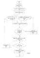
- FIG. 1 is a flow chart showing the general process of the invention.
- FIG. 2 is a flow chart showing a specific embodiment of the invention.
- FIG. 3 is an original retinal image displaying a wide variety of vessels.
- FIG. 4 is one of eight directional images displaying vessels having 45 degree orientations.
- FIG. 6 is a directional image enhanced with second-order elliptical gaussian filter.
- FIG. 7 is a final enhanced image which accumulates the maximum response from all eight directional images.
- FIG. 8 is a final enhanced image which sums over the best responses from the directional images.
- FIG. 1 is a flow chart showing as the first step acquiring an image from an angioscopy, then applying a directional filter bank to the image, followed by applying a multi-scale matched filter, with the last step being obtaining an enhanced result from the process.
- FIG. 2 is a flow chart of an embodiment of a process according to the invention wherein the process stards 11 with an image acquisition 12 followed by non-uniform background removal 13 and directional decomposition 14 .
- Eight directional images 15 a - 15 h are obtained from the directional decomposition step 14 .
- Second order elliptical gaussian derivative filtering 17 is then applied to the directional images 15 a - 15 h and then filtered using all members in sets of SigmaX 18 .
- a new SigmaX and SigmaY is chosen 19 and then pre-defined sets of SigmaX and SigmaY 16 are used for 2 nd order elliptical Gaussian Derivative Filtering 17 .
- maximum filter response for each directional image is saved 20 and then all directional images are filtered 21 .
- the directional image is changed 22 and then eight directional images 15 a - 15 h are obtained again.
- all eight maximum responses are summed/maxed 23 and the process ends 24 with an enhanced image.
- FIGS. 3-8 are photographic representations wherein FIG. 3 is an original retinal image displaying a wide variety of vessels.
- FIG. 4 is one of eight directional images displaying vessels having 45 degree orientations.
- FIG. 6 is a directional image enhanced with second-order elliptical gaussian filter.
- FIG. 7 is a final enhanced image which accumulates the maximum response from all eight directional images
- FIG. 8 is a final enhanced image which sums over the best responses from the directional images.
- the method of vessel extraction of the invention comprises (a) acquiring an angiography image; (b) decomposing the acquired angiography image into at least two directional images with a directional filter bank; (c) normalizing the directional images by removing their mean values and then scaling linearly; (d) applying second derivative Gaussian filters to each of the directional images using a combination of scales along x and y-direction such as to create an elliptical support for the resulting filter; (e) combining the resultant directional images into a final enhanced image.
- the method comprises characterizing a specific width and direction of a vessel.
- Elliptical support regions for second order Gaussian derivatives can be used.
- the method can be carried out with a specially programmed computer which in preferred embodiments provides a value to a standard deviation parameter in a horizontal direction and providing a value to a standard deviation parameter in a vertical direction, resulting in a multi-scale anisotropic second-order Gaussian derivative filter.
- Gaussian derivatives filters The derivatives along x and y direction of conventional Gaussian filters are known as Gaussian derivatives filters.
- Each of the directional images are associated with a single directional features in the method.
- the directional filter bank is decimation-free and provides pure directional decomposition of the features based on their orientations only without disturbing the frequency composition of the features.
- the method comprises applying anisotropic (elliptical) multi-scale second order Gaussian derivatives within a directional filter bank framework.
Landscapes
- Engineering & Computer Science (AREA)
- Theoretical Computer Science (AREA)
- General Physics & Mathematics (AREA)
- Physics & Mathematics (AREA)
- General Health & Medical Sciences (AREA)
- Health & Medical Sciences (AREA)
- Computer Vision & Pattern Recognition (AREA)
- Molecular Biology (AREA)
- Biomedical Technology (AREA)
- Biodiversity & Conservation Biology (AREA)
- Life Sciences & Earth Sciences (AREA)
- Evolutionary Computation (AREA)
- Multimedia (AREA)
- Artificial Intelligence (AREA)
- Medical Informatics (AREA)
- Nuclear Medicine, Radiotherapy & Molecular Imaging (AREA)
- Radiology & Medical Imaging (AREA)
- Quality & Reliability (AREA)
- Image Processing (AREA)
Abstract
Description
Claims (8)
Priority Applications (1)
| Application Number | Priority Date | Filing Date | Title |
|---|---|---|---|
| US14/590,595 US9275455B1 (en) | 2015-01-06 | 2015-01-06 | Method for vessel extraction |
Applications Claiming Priority (1)
| Application Number | Priority Date | Filing Date | Title |
|---|---|---|---|
| US14/590,595 US9275455B1 (en) | 2015-01-06 | 2015-01-06 | Method for vessel extraction |
Publications (1)
| Publication Number | Publication Date |
|---|---|
| US9275455B1 true US9275455B1 (en) | 2016-03-01 |
Family
ID=55360016
Family Applications (1)
| Application Number | Title | Priority Date | Filing Date |
|---|---|---|---|
| US14/590,595 Expired - Fee Related US9275455B1 (en) | 2015-01-06 | 2015-01-06 | Method for vessel extraction |
Country Status (1)
| Country | Link |
|---|---|
| US (1) | US9275455B1 (en) |
Citations (3)
| Publication number | Priority date | Publication date | Assignee | Title |
|---|---|---|---|---|
| US8150134B2 (en) * | 2007-08-17 | 2012-04-03 | Industry-Academic Cooperation Foundation Of Kyung Hee University | Method for enhancing blood vessels in angiography images |
| US20120207402A1 (en) * | 2009-05-27 | 2012-08-16 | Zeitera, Llc | Digital Video Content Fingerprinting Based on Scale Invariant Interest Region Detection with an Array of Anisotropic Filters |
| US8428323B2 (en) * | 2011-05-16 | 2013-04-23 | Dongguk University Industry-Academic Cooperation Foundation | Method of processing medical image of blood vessel using image fusion method |
-
2015
- 2015-01-06 US US14/590,595 patent/US9275455B1/en not_active Expired - Fee Related
Patent Citations (3)
| Publication number | Priority date | Publication date | Assignee | Title |
|---|---|---|---|---|
| US8150134B2 (en) * | 2007-08-17 | 2012-04-03 | Industry-Academic Cooperation Foundation Of Kyung Hee University | Method for enhancing blood vessels in angiography images |
| US20120207402A1 (en) * | 2009-05-27 | 2012-08-16 | Zeitera, Llc | Digital Video Content Fingerprinting Based on Scale Invariant Interest Region Detection with an Array of Anisotropic Filters |
| US8428323B2 (en) * | 2011-05-16 | 2013-04-23 | Dongguk University Industry-Academic Cooperation Foundation | Method of processing medical image of blood vessel using image fusion method |
Non-Patent Citations (20)
| Title |
|---|
| Aylward et al., "Initialization, Noise, Singularities, and Scale in Height Ridge Traversal for Tubular Object Centerline Extraction", IEEE Transactions on Medical Imaging, vol. 21, No. 2, pp. 61-75, 2002. |
| Chaudhuri et al., "Detection of Blood Vessels in Retinal Images Using Two-Dimensional Matched Filters", IEEE Transactions on Medical Imaging, vol. 8, No. 3, pp. 263-269, Sep. 1989. |
| Dehkordi et al., "A Review of Coronary Vessel Segmentation Algorithms", J. Med. Signals Sens. vol. 1, No. 1, pp. 49-54, 2011. |
| European Carotid Surgery Trialists Collaborative Group, Abstract of "Randomised Trial of Endarterectomy for Recently Symptomatic Carotid Stenosis: Final Results of the MRC European Carotid Surgery (ECST)", Lancet 351, 1379-1387, 1999. |
| Frangi et al., "Multiscale Vessel Enhancement Filtering," Medical Image Computing and Computer-Assisted Intervention-MICCAI'98, vol. 1496, pp. 130-137, Springer Verlag, Berlin, Germany, 1998. |
| Heneghan et al., "Characterization of changes in blood vessel width and tortuosity in retinopathy of prematurity using image analysis," Medical Image Analysis, vol. 6, No. 4, pp. 407-429, 2002. |
| Hoover et al., "Locating Blood Vessels in Retinal Images by Piecewise Threshold Probing of a Matched Filter Response", IEEE Transactions on Medical Imaging, vol. 19, No. 3, pp. 203-210, Mar. 2000. |
| Jiang et al., "Adaptive Local Thresholding by Verification-Based Multithreshold Probing with Application to Vessel Detection in Retinal Images," IEEE Transactions on Pattern Analysis Machine Intelligence, vol. 25, No. 1, pp. 131-137, Jan. 2003. |
| Khan et al., "Vessel Enhancement Using Directional Features", Information Technology Journal, vol. 6, No. 6, pp. 851-857, 2007. |
| Martínez-Pérez et al., "Retinal Blood Vessel Segmentation by Means of Scale-Space Analysis and Region Growing," Medical Image Computing and Computer-Assisted Intervention-MICCAI'99, vol. 1679, pp. 90-97, 1999. |
| Matei et al., "Vascular Image Processing Using Recursive Directional Filters", World Congress on Medical Physics and Biomedical Engineering, IFMBE Proceedings, vol. 39, pp. 947-950, 2013. |
| Miles et al., "Matched Filter Estimation of Serial Blood Vessel Diameters from Video Images", IEEE Transactions on Medical Imaging, vol. 12, No. 2. pp. 147-152, Jun. 1993. |
| Sofka et al., "Retinal Vessel Centerline Extraction Using Multiscale Matched Filters, Confidence and Edge Measures", IEEE Transactions on Medical Imaging, vol. 25, No. 12, pp. 1531-1546, 2006. |
| Staal et al., "A Trained Spin-Glass Model for Grouping of Image Primitives", IEEE Transactions on Pattern Analysis and Machine Intelligence, vol. 27, No. 7, pp. 1172-1182, Jul. 2005. |
| Staal et al., "Ridge-Based Vessel Segmentation in Color Images of the Retina," IEEE Transactions on Medical Imaging, vol. 23, No. 4, pp. 501-509, Apr. 2004. |
| Steering Committee, "The North American Symptomatic Carotid Endarterectomy Trial: Methods, Patient Characteristics, and Progress", North American Sympotomatic Coarotid Endarterectomy Trial (NASCET), Stroke, vol. 22, No. 6, pp. 711-720, Jun. 1991. |
| Su et al., "A New Method for Linear Feature and Junction Enhancement in 2D Images based on Morphological Operation, Oriented Anisotropic Gaussian Function and Hessian Information", Pattern Recognition , vol. 47. pp. 3193-3208, May 9, 2014. |
| Truc et al., "Vessel Enhancement Filter Using Directional Filter Bank", Computer Vision and Image Understanding, vol. 113, pp. 101-112, 2009. |
| Zana et al., "Segmentation of Vessel-Like Patterns Using Mathematical Morphology and Curvature Evaluation," IEEE Transactions on Image Processing, vol. 10, No. 7, pp. 1010-1019, Jul. 2001. |
| Zhang et al., "Detecting Optic Disc on Asians by Multiscale Gaussian Filtering", International Journal of Biomedical Imaging, vol. 2012, Article ID 727154, Feb. 10, 2012. |
Similar Documents
| Publication | Publication Date | Title |
|---|---|---|
| CN106651846B (en) | Segmentation method of retinal blood vessel images | |
| Chen et al. | 3D intracranial artery segmentation using a convolutional autoencoder | |
| Dehkordi et al. | A review of coronary vessel segmentation algorithms | |
| CN109919915B (en) | A method and device for detecting abnormal regions in retinal fundus images based on deep learning | |
| Carone et al. | Impact of automated ICA-based denoising of fMRI data in acute stroke patients | |
| EP2827298A1 (en) | Method and computer program for filtering and particularly segmenting and/or analyzing anatomic structures in a digital image | |
| Sahu et al. | Minimum time delay and more efficient image filtering brain tumour detection with the help of MATLAB | |
| Gour et al. | Blood vessel segmentation using hybrid median filtering and morphological transformation | |
| CN104077754B (en) | Based on symmetric retinal vessel filtering reinforcement method | |
| CN118898552B (en) | Multi-stage OCT image denoising method, device and storage medium based on improved clustering | |
| Mankikar | A novel hybrid approach using Kmeans clustering and threshold filter for brain tumor detection | |
| US9275455B1 (en) | Method for vessel extraction | |
| Malathi et al. | Research Article An Efficient Method to Detect Diabetic Retinopathy Using Gaussian-Bilateral and Haar Filters with Threshold Based Image Segmentation | |
| CN111712851B (en) | Image processing device, image processing method, and image processing program | |
| Rabeya et al. | Quality Assessment of Color Normalization Method by Similarity Index Metrics-A Comparative Study for Histopathology Images | |
| Zhang et al. | Retinal vessel segmentation using Gabor filter and textons | |
| Alazawee et al. | Analyzing and detecting hemorrhagic and ischemic strokebased on bit plane slicing and edge detection algorithms | |
| El Abbadi et al. | Automatic detection of vascular bifurcations and crossovers in retinal fundus image | |
| Sharma et al. | An automatic segmentation & detection of blood vessels and optic disc in retinal images | |
| Zolkifli et al. | Retina blood vessel extraction based on Kirsch’s template method | |
| Nazari et al. | Segmentation of retinal blood vessels by top-hat multi-scale detection for optic disc removal | |
| Bush et al. | Deconvolution filtering: Temporal smoothing revisited | |
| Sakim | Investigation on edge detection techniques of coronary angiography images | |
| CN115409689A (en) | Multi-mode retina fundus image registration method and device | |
| Rizzi et al. | 3-D zebrafish embryo image filtering by nonlinear partial differential equations |
Legal Events
| Date | Code | Title | Description |
|---|---|---|---|
| AS | Assignment |
Owner name: EFFAT UNIVERSITY, SAUDI ARABIA Free format text: ASSIGNMENT OF ASSIGNORS INTEREST;ASSIGNOR:KHAN, MOHAMMED ASMATULLAH;REEL/FRAME:034789/0834 Effective date: 20150106 |
|
| STCF | Information on status: patent grant |
Free format text: PATENTED CASE |
|
| MAFP | Maintenance fee payment |
Free format text: PAYMENT OF MAINTENANCE FEE, 4TH YR, SMALL ENTITY (ORIGINAL EVENT CODE: M2551); ENTITY STATUS OF PATENT OWNER: SMALL ENTITY Year of fee payment: 4 |
|
| FEPP | Fee payment procedure |
Free format text: MAINTENANCE FEE REMINDER MAILED (ORIGINAL EVENT CODE: REM.); ENTITY STATUS OF PATENT OWNER: SMALL ENTITY |
|
| LAPS | Lapse for failure to pay maintenance fees |
Free format text: PATENT EXPIRED FOR FAILURE TO PAY MAINTENANCE FEES (ORIGINAL EVENT CODE: EXP.); ENTITY STATUS OF PATENT OWNER: SMALL ENTITY |
|
| STCH | Information on status: patent discontinuation |
Free format text: PATENT EXPIRED DUE TO NONPAYMENT OF MAINTENANCE FEES UNDER 37 CFR 1.362 |
|
| FP | Lapsed due to failure to pay maintenance fee |
Effective date: 20240301 |