US9275437B2 - Method for efficient digital subtraction angiography - Google Patents
Method for efficient digital subtraction angiography Download PDFInfo
- Publication number
- US9275437B2 US9275437B2 US13/803,466 US201313803466A US9275437B2 US 9275437 B2 US9275437 B2 US 9275437B2 US 201313803466 A US201313803466 A US 201313803466A US 9275437 B2 US9275437 B2 US 9275437B2
- Authority
- US
- United States
- Prior art keywords
- images
- contrast enhanced
- tmip
- image
- subtracting
- Prior art date
- Legal status (The legal status is an assumption and is not a legal conclusion. Google has not performed a legal analysis and makes no representation as to the accuracy of the status listed.)
- Active, expires
Links
Images


Classifications
-
- G—PHYSICS
- G06—COMPUTING OR CALCULATING; COUNTING
- G06T—IMAGE DATA PROCESSING OR GENERATION, IN GENERAL
- G06T5/00—Image enhancement or restoration
- G06T5/70—Denoising; Smoothing
-
- G06T5/002—
-
- A—HUMAN NECESSITIES
- A61—MEDICAL OR VETERINARY SCIENCE; HYGIENE
- A61B—DIAGNOSIS; SURGERY; IDENTIFICATION
- A61B6/00—Apparatus or devices for radiation diagnosis; Apparatus or devices for radiation diagnosis combined with radiation therapy equipment
- A61B6/48—Diagnostic techniques
- A61B6/481—Diagnostic techniques involving the use of contrast agents
-
- A—HUMAN NECESSITIES
- A61—MEDICAL OR VETERINARY SCIENCE; HYGIENE
- A61B—DIAGNOSIS; SURGERY; IDENTIFICATION
- A61B6/00—Apparatus or devices for radiation diagnosis; Apparatus or devices for radiation diagnosis combined with radiation therapy equipment
- A61B6/48—Diagnostic techniques
- A61B6/486—Diagnostic techniques involving generating temporal series of image data
-
- A—HUMAN NECESSITIES
- A61—MEDICAL OR VETERINARY SCIENCE; HYGIENE
- A61B—DIAGNOSIS; SURGERY; IDENTIFICATION
- A61B6/00—Apparatus or devices for radiation diagnosis; Apparatus or devices for radiation diagnosis combined with radiation therapy equipment
- A61B6/50—Apparatus or devices for radiation diagnosis; Apparatus or devices for radiation diagnosis combined with radiation therapy equipment specially adapted for specific body parts; specially adapted for specific clinical applications
- A61B6/504—Apparatus or devices for radiation diagnosis; Apparatus or devices for radiation diagnosis combined with radiation therapy equipment specially adapted for specific body parts; specially adapted for specific clinical applications for diagnosis of blood vessels, e.g. by angiography
-
- G—PHYSICS
- G06—COMPUTING OR CALCULATING; COUNTING
- G06T—IMAGE DATA PROCESSING OR GENERATION, IN GENERAL
- G06T5/00—Image enhancement or restoration
- G06T5/50—Image enhancement or restoration using two or more images, e.g. averaging or subtraction
-
- G06T7/0024—
-
- G—PHYSICS
- G06—COMPUTING OR CALCULATING; COUNTING
- G06T—IMAGE DATA PROCESSING OR GENERATION, IN GENERAL
- G06T7/00—Image analysis
- G06T7/30—Determination of transform parameters for the alignment of images, i.e. image registration
-
- G—PHYSICS
- G06—COMPUTING OR CALCULATING; COUNTING
- G06T—IMAGE DATA PROCESSING OR GENERATION, IN GENERAL
- G06T2207/00—Indexing scheme for image analysis or image enhancement
- G06T2207/20—Special algorithmic details
- G06T2207/20212—Image combination
- G06T2207/20224—Image subtraction
-
- G—PHYSICS
- G06—COMPUTING OR CALCULATING; COUNTING
- G06T—IMAGE DATA PROCESSING OR GENERATION, IN GENERAL
- G06T2207/00—Indexing scheme for image analysis or image enhancement
- G06T2207/30—Subject of image; Context of image processing
- G06T2207/30004—Biomedical image processing
- G06T2207/30101—Blood vessel; Artery; Vein; Vascular
Definitions
- the present invention relates to a system and method for an alternative method of performing efficient digital subtraction angiography and particularly, but not exclusively, to automatic efficient digital subtraction angiography in image data of bodily tissue and/or object feature analysis in image data from non-biological subjects.
- Angiography refers generally to the capture and representation of blood vessels or vasculature of the human body by means of X-ray imaging, i.e., X-ray vascular imaging.
- X-ray diagnostic imaging systems may be used for angiographic imaging procedures such as digital subtraction angiography (DSA), and live fluoroscopic roadmapping.
- DSA digital subtraction angiography
- DSA is an imaging method used for visualizing blood vessels inside a patient's body that includes injecting a contrast medium bolus that is substantially opaque to X-rays into the blood vessels or vasculature under study as images are acquired by the X-ray diagnostic imaging system. Prior to acquisition of the contrast image, a mask image without contrast is acquired.
- a difference image is calculated by superimposing upon and subtracting the mask image from the contrast image. Ideally, nothing appears in the difference image other than the image of the blood vessels. Because of the time difference between acquisition of the mask image (no contrast) and acquisition of the contrast-enhanced images, global and periodic motion, fluctuations in the intensity of the X-ray source, scattering by the contrast medium, etc., unwanted artifacts may appear in the differenced or digitally subtracted angiographic image.
- DSA is useful in the diagnosis and imaging of various blood vessel disorders, such as arterial and venous occlusions, including carotid artery stenosis, pulmonary embolisms and acute limb ischemia; arterial stenosis, particularly for renal artery stenosis; and cerebral aneurysms and arterio-venous malformations.
- blood vessel disorders such as arterial and venous occlusions, including carotid artery stenosis, pulmonary embolisms and acute limb ischemia; arterial stenosis, particularly for renal artery stenosis; and cerebral aneurysms and arterio-venous malformations.
- DSA requires the acquisition of a non-contrast initial or “mask” image, followed by sequential enhanced contrast images. Subtraction of each of the enhanced images from the non-enhanced image is performed and viewed by the doctor, enabling the blood vessels and any problems therewith to be easily seen. However, if any movement occurs between obtaining the non-enhanced image and the enhanced image, these images will not be in registration and so the subtraction process will not be performed correctly.
- the present invention overcomes the above drawbacks of the background art by providing, in at least some embodiments, a system and method for efficiently performing DSA (digital subtraction angiography), which does not require a non-enhanced or “mask” image to be obtained.
- the method and system are operative without the mask image. Instead, a plurality of contrast enhanced images are obtained, such that at least two but preferably at least three images are obtained and more preferably 10 images are obtained (or even more).
- a registration is performed between these images, which may optionally be a rigid registration or a non-rigid registration, performed according to any suitable method.
- registration is performed by using a known registration method, non-limiting examples of which are described with regard to “Algorithms for radiological image registration and their clinical application” by Hawkes et al (J. Anat. (1998) 193, pp. 347-361); “Image registration: an essential tool for nuclear medicine” by Hutton et al (Eur J Nucl Med (2002) 29: pp 559-577); and US Patent Application No. 20100235352; all of which are hereby incorporated by reference as if fully set forth herein.
- other registration methods could also optionally be used in place of, or in addition to, the methods described in these papers.
- each image (or volume) is subtracted from the previous image (or volume) through subtracting the pixels.
- Volumes are subtracted for 3-dimensional images; however where reference is made to subtracting “images” it may also be understood to encompass subtracting “volumes”.
- the process of subtracting the pixels may optionally comprise taking the absolute value after subtraction, or alternatively selecting the greater of the value after subtraction or a threshold value, wherein the threshold value is greater than or equal to zero.
- R MAX(i — 2 ⁇ i — 1, thresh), where thresh is some threshold value greater than or equal to zero; optionally alternatively, this calculation may be made through a dedicated lookup table that will be generated according to a known equation. Such a lookup table may optionally be used for any function and not only subtraction, in order to apply this function to the value of pairs of pixels.
- tMIP temporary maximum intensity projection
- tMIP temporary maximum intensity projection
- a cine view of the results may be provided, assuming that the tMIP is performed on images that have been divided into a plurality of non-zero sets, such that there exists at least two images after tMIP is performed.
- one or more noise reduction algorithm and smoothing algorithms are applied to improve the results, including but not limited to passing a median or Gaussian filter, another method is called anisotropic diffusion or Perona-Malik diffusion (see scale-space and edge detection using anisotropic diffusion by Perona and Malik, Pattern Analysis and Machine Intelligence, IEEE Transactions, July 1990, Volume: 12, Issue: 7, Page(s): 629-639).
- one or more noise reduction algorithm and smoothing algorithms are applied before subtraction of the pixels as described above, additionally or alternatively.
- image data as used herein relates to two or three dimensional image data unless otherwise indicated.
- Implementation of the method and system of the present invention involves performing or completing certain selected tasks or steps manually, automatically, or a combination thereof.
- several selected steps could be implemented by hardware or by software on any operating system of any firmware or a combination thereof.
- selected steps of the invention could be implemented as a chip or a circuit.
- selected steps of the invention could be implemented as a plurality of software instructions being executed by a computer using any suitable operating system.
- selected steps of the method and system of the invention could be described as being performed by a data processor, such as a computing platform for executing a plurality of instructions.
- any device featuring a data processor and the ability to execute one or more instructions may be described as a computer, including but not limited to any type of personal computer (PC), a server, a cellular telephone, an IP telephone, a smart phone, a PDA (personal digital assistant), or a pager. Any two or more of such devices in communication with each other may optionally comprise a “computer network”.
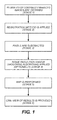
- FIG. 1 shows an exemplary, illustrative method for efficiently performing DSA (digital subtraction angiography) according to at least some embodiments of the present invention
- FIGS. 2A-2C shows some results after performing the method of FIG. 1 , plus the original image
- FIG. 3 shows an exemplary, non-limiting illustrative system according to at least some embodiments of the present invention.
- FIG. 1 shows an exemplary, illustrative method for efficiently performing DSA (digital subtraction angiography) according to at least some embodiments of the present invention.
- DSA digital subtraction angiography
- stage 1 a plurality of contrast enhanced images are obtained, such that at least two but preferably at least three images are obtained and more preferably 10 images are obtained (or even more).
- stage 2 optionally and preferably a registration is performed between these images, whether rigid or non-rigid.
- registration is performed by using a known registration method, non-limiting examples of which are described with regard to “Algorithms for radiological image registration and their clinical application” by Hawkes et al (J. Anat. (1998) 193, pp.
- each image is subtracted from the previous image through subtracting the pixels. It is to be noted that since the method is performed without obtaining a mask image, only the contrast-enhanced images are subtracted.
- the process of subtracting the pixels may optionally comprise taking the absolute value after subtraction, or alternatively selecting the greater of the value after subtraction or a threshold value, wherein the threshold value is greater than or equal to zero.
- one or more noise reduction algorithm and smoothing algorithms are applied to improve the results, including but not limited to passing a median or Gaussian filter, while another method is called anisotropic diffusion or Perona-Malik diffusion (see scale-space and edge detection using anisotropic diffusion by Perona and Malik, Pattern Analysis and Machine Intelligence, IEEE Transactions, July 1990, Volume: 12, Issue: 7, Page(s): 629-639).
- one or more noise reduction algorithm and smoothing algorithms are applied after registration and/or after application of tMIP, additionally or alternatively.
- tMIP temporary maximum intensity projection
- MIP maximum intensity projection
- a cine view of the results may be provided, assuming that the tMIP is performed on images that have been divided into a plurality of non-zero sets, such that there exists at least two images after tMIP is performed.
- the above method is characterized in that it is performed without first obtaining a mask or non-contrast-enhanced image, yet is still able to successfully perform DSA.
- the method is preferably performed with tMIP to reduce image “jitter”.
- FIG. 2A shows the resultant image after the method of FIG. 1 was performed, with tMIP of 4 images, but without the optional registration stage being performed.
- FIG. 2B shows the resultant image after the method of FIG. 1 was performed, with tMIP of 6 images, but without the optional registration stage being performed.
- FIG. 2C shows the original DSA image with residual bone effects, created according to the regular DSA, but with subtraction by using a mask image.
- FIG. 3 shows an exemplary, non-limiting illustrative system according to at least some embodiments of the present invention.
- a system 300 features a mouse 302 or other pointing device, a keyboard 304 and a display 306 .
- any of these components may be combined, for example for a touch sensitive display screen; mouse 302 and keyboard 304 may optionally be described as an “input device”.
- the user interacts with these components to perform the various methods as described herein where user interaction is indicated.
- System 300 also features a user computer 308 , which may optionally comprise any computational device and which may optionally be a local or remote computer, and may optionally be a plurality of computers.
- User computer 308 operates a DSA software 310 for performing DSA according to the method of FIG. 1 , as described above.
- System 300 additionally features a Registration software 312 for registering two sets of data, preferably through a global registration, for example optionally according to any suitable art known registration method. Registration is preferably performed before DSA, such that the image data is received by user computer 308 and is preferably first passed to registration software 312 before being processed by DSA software 310 .
- the image data undergoes tMIP and/or noise reduction and/or smoothing, by a post-DSA processing software 314 . If tMIP is performed in addition to other operations, then preferably tMIP is performed first, before one or more other operations are performed.
Landscapes
- Health & Medical Sciences (AREA)
- Engineering & Computer Science (AREA)
- Life Sciences & Earth Sciences (AREA)
- Medical Informatics (AREA)
- Physics & Mathematics (AREA)
- General Health & Medical Sciences (AREA)
- Radiology & Medical Imaging (AREA)
- Nuclear Medicine, Radiotherapy & Molecular Imaging (AREA)
- Biomedical Technology (AREA)
- Veterinary Medicine (AREA)
- Biophysics (AREA)
- Optics & Photonics (AREA)
- Pathology (AREA)
- High Energy & Nuclear Physics (AREA)
- Public Health (AREA)
- Heart & Thoracic Surgery (AREA)
- Molecular Biology (AREA)
- Surgery (AREA)
- Animal Behavior & Ethology (AREA)
- General Physics & Mathematics (AREA)
- Theoretical Computer Science (AREA)
- Computer Vision & Pattern Recognition (AREA)
- Vascular Medicine (AREA)
- Dentistry (AREA)
- Oral & Maxillofacial Surgery (AREA)
- Apparatus For Radiation Diagnosis (AREA)
- Quality & Reliability (AREA)
- Image Processing (AREA)
Abstract
Description
Claims (19)
Priority Applications (1)
| Application Number | Priority Date | Filing Date | Title |
|---|---|---|---|
| US13/803,466 US9275437B2 (en) | 2013-03-14 | 2013-03-14 | Method for efficient digital subtraction angiography |
Applications Claiming Priority (1)
| Application Number | Priority Date | Filing Date | Title |
|---|---|---|---|
| US13/803,466 US9275437B2 (en) | 2013-03-14 | 2013-03-14 | Method for efficient digital subtraction angiography |
Publications (2)
| Publication Number | Publication Date |
|---|---|
| US20140270437A1 US20140270437A1 (en) | 2014-09-18 |
| US9275437B2 true US9275437B2 (en) | 2016-03-01 |
Family
ID=51527263
Family Applications (1)
| Application Number | Title | Priority Date | Filing Date |
|---|---|---|---|
| US13/803,466 Active 2033-10-20 US9275437B2 (en) | 2013-03-14 | 2013-03-14 | Method for efficient digital subtraction angiography |
Country Status (1)
| Country | Link |
|---|---|
| US (1) | US9275437B2 (en) |
Families Citing this family (9)
| Publication number | Priority date | Publication date | Assignee | Title |
|---|---|---|---|---|
| US9204937B2 (en) | 2013-02-19 | 2015-12-08 | Stryker Trauma Gmbh | Software for use with deformity correction |
| DE102014225846B4 (en) * | 2014-12-15 | 2016-07-28 | Siemens Healthcare Gmbh | Determination of magnetic resonance angiography images with time-of-flight angiography and magnetic resonance apparatus |
| JP6843048B2 (en) | 2014-12-18 | 2021-03-17 | コーニンクレッカ フィリップス エヌ ヴェKoninklijke Philips N.V. | Automatic visualization of embolic material in X-ray interventions |
| DE102015224806B4 (en) | 2015-12-10 | 2018-01-18 | Siemens Healthcare Gmbh | A method of presenting a first structure of a body region by means of digital subtraction angiography, evaluation device and angiography system |
| US10251705B2 (en) | 2016-06-02 | 2019-04-09 | Stryker European Holdings I, Llc | Software for use with deformity correction |
| WO2018109111A1 (en) | 2016-12-15 | 2018-06-21 | Koninklijke Philips N.V. | Visualizing vascular structures |
| CN106821404A (en) * | 2017-01-20 | 2017-06-13 | 北京东软医疗设备有限公司 | Angiographic method and system |
| IT201900006616A1 (en) * | 2019-05-07 | 2020-11-07 | Angiodroid S R L | METHOD FOR IMPROVING RADIOLOGICAL IMAGES IN THE COURSE OF AN ANGIOGRAPHY |
| WO2021257906A1 (en) * | 2020-06-17 | 2021-12-23 | Northwestern University | Maskless 2d/3d artificial subtraction angiography |
Citations (29)
| Publication number | Priority date | Publication date | Assignee | Title |
|---|---|---|---|---|
| US6483891B1 (en) * | 1998-09-17 | 2002-11-19 | Quanta Vision, Inc. | Reduced-angle mammography device and variants |
| US20040189796A1 (en) * | 2003-03-28 | 2004-09-30 | Flatdis Co., Ltd. | Apparatus and method for converting two-dimensional image to three-dimensional stereoscopic image in real time using motion parallax |
| US20060067570A1 (en) * | 2004-09-29 | 2006-03-30 | Dainippon Screen Mfg. Co., Ltd. | Apparatus and method for inspecting pattern |
| US20060133660A1 (en) * | 2004-12-16 | 2006-06-22 | Dainippon Screen Mfg. Co., Ltd. | Apparatus and method for detecting defect existing in pattern on object |
| US20060209095A1 (en) * | 2005-03-02 | 2006-09-21 | Ying-Hao Hsu | Over-driving apparatus and method thereof |
| US20060239585A1 (en) * | 2005-04-04 | 2006-10-26 | Valadez Gerardo H | System and method for reducing artifacts in motion corrected dynamic image sequences |
| US20070036442A1 (en) * | 2003-04-11 | 2007-02-15 | Stoffer Jay H | Adaptive subtraction image compression |
| US20070238951A1 (en) * | 2006-03-24 | 2007-10-11 | General Electric Company | Systems, methods and apparatus for oncology workflow integration |
| US20090185730A1 (en) * | 2007-10-19 | 2009-07-23 | Siemens Medical Solutions Usa, Inc. | Automated Image Data Subtraction System Suitable for Use in Angiography |
| US20100091194A1 (en) * | 2007-03-31 | 2010-04-15 | Sony Deutschland Gmbh | Noise reduction method and unit for an image frame |
| US20100202687A1 (en) * | 2007-05-03 | 2010-08-12 | Ucl Business Plc | Image registration method |
| US20100235352A1 (en) | 2006-11-26 | 2010-09-16 | Algotec Systems Ltd. | Comparison workflow automation by registration |
| US20100246975A1 (en) * | 2007-09-28 | 2010-09-30 | Hitachi Software Engineering Co., Ltd. | Method and apparatus for determining if imaging object is still |
| US20110001884A1 (en) * | 2008-03-28 | 2011-01-06 | Nec Corporation | Image processing system, image processing method, and recording medium storing image processing program |
| US20110025857A1 (en) * | 2009-07-30 | 2011-02-03 | General Instrument Corporation | System and method for analyzing video stream |
| US20110081042A1 (en) * | 2009-10-07 | 2011-04-07 | Samsung Electronics Co., Ltd. | Apparatus and method for adjusting depth |
| US20110103671A1 (en) * | 2008-06-30 | 2011-05-05 | Koninklijke Philips Electronics N.V. | Perfusion imaging |
| US20120007895A1 (en) * | 2010-07-08 | 2012-01-12 | Kim Kiltae | Stereoscopic image display and driving method thereof |
| US20120105657A1 (en) * | 2010-10-29 | 2012-05-03 | Sanyo Electric Co., Ltd. | Image processing apparatus, image pickup apparatus, image processing method, and program |
| US20120201439A1 (en) * | 2011-02-09 | 2012-08-09 | Siemens Medical Solutions Usa, Inc. | Digital Subtraction Angiography (DSA) Motion Compensated Imaging System |
| US20120232378A1 (en) * | 2009-11-10 | 2012-09-13 | Deutsches Herzzentrum Berlin | Look-Locker IR-SSFP for Cardiac MR Imaging with Simultaneous Generation of Cardiac T1 Maps, Cine Images and IR-Prepared Images |
| US20120238870A1 (en) * | 2011-03-08 | 2012-09-20 | Hologic, Inc. | System and method for dual energy and/or contrast enhanced breast imaging for screening, diagnosis and biopsy |
| US20120269384A1 (en) * | 2011-04-19 | 2012-10-25 | Jones Michael J | Object Detection in Depth Images |
| US20130070995A1 (en) * | 2011-08-30 | 2013-03-21 | Siemens Corporation | 2d/3d image registration method |
| US20130094734A1 (en) * | 2011-10-14 | 2013-04-18 | Siemens Medical Solutions Usa, Inc. | System for Comparison of Medical Images |
| US20130188040A1 (en) * | 2011-12-21 | 2013-07-25 | Deka Products Limited Partnership | System, Method, and Apparatus for Monitoring, Regulating, or Controlling Fluid Flow |
| US20140003690A1 (en) * | 2012-07-02 | 2014-01-02 | Marco Razeto | Motion correction apparatus and method |
| US20140267837A1 (en) * | 2012-03-30 | 2014-09-18 | Fujifilm Corporation | Correction image creation device, radiographic imaging device, imaging device, computer readable medium and correction image creation method |
| US20150016728A1 (en) * | 2012-03-08 | 2015-01-15 | Koninklijke Philips N.V. | Intelligent landmark selection to improve registration accuracy in multimodal image fushion |
-
2013
- 2013-03-14 US US13/803,466 patent/US9275437B2/en active Active
Patent Citations (29)
| Publication number | Priority date | Publication date | Assignee | Title |
|---|---|---|---|---|
| US6483891B1 (en) * | 1998-09-17 | 2002-11-19 | Quanta Vision, Inc. | Reduced-angle mammography device and variants |
| US20040189796A1 (en) * | 2003-03-28 | 2004-09-30 | Flatdis Co., Ltd. | Apparatus and method for converting two-dimensional image to three-dimensional stereoscopic image in real time using motion parallax |
| US20070036442A1 (en) * | 2003-04-11 | 2007-02-15 | Stoffer Jay H | Adaptive subtraction image compression |
| US20060067570A1 (en) * | 2004-09-29 | 2006-03-30 | Dainippon Screen Mfg. Co., Ltd. | Apparatus and method for inspecting pattern |
| US20060133660A1 (en) * | 2004-12-16 | 2006-06-22 | Dainippon Screen Mfg. Co., Ltd. | Apparatus and method for detecting defect existing in pattern on object |
| US20060209095A1 (en) * | 2005-03-02 | 2006-09-21 | Ying-Hao Hsu | Over-driving apparatus and method thereof |
| US20060239585A1 (en) * | 2005-04-04 | 2006-10-26 | Valadez Gerardo H | System and method for reducing artifacts in motion corrected dynamic image sequences |
| US20070238951A1 (en) * | 2006-03-24 | 2007-10-11 | General Electric Company | Systems, methods and apparatus for oncology workflow integration |
| US20100235352A1 (en) | 2006-11-26 | 2010-09-16 | Algotec Systems Ltd. | Comparison workflow automation by registration |
| US20100091194A1 (en) * | 2007-03-31 | 2010-04-15 | Sony Deutschland Gmbh | Noise reduction method and unit for an image frame |
| US20100202687A1 (en) * | 2007-05-03 | 2010-08-12 | Ucl Business Plc | Image registration method |
| US20100246975A1 (en) * | 2007-09-28 | 2010-09-30 | Hitachi Software Engineering Co., Ltd. | Method and apparatus for determining if imaging object is still |
| US20090185730A1 (en) * | 2007-10-19 | 2009-07-23 | Siemens Medical Solutions Usa, Inc. | Automated Image Data Subtraction System Suitable for Use in Angiography |
| US20110001884A1 (en) * | 2008-03-28 | 2011-01-06 | Nec Corporation | Image processing system, image processing method, and recording medium storing image processing program |
| US20110103671A1 (en) * | 2008-06-30 | 2011-05-05 | Koninklijke Philips Electronics N.V. | Perfusion imaging |
| US20110025857A1 (en) * | 2009-07-30 | 2011-02-03 | General Instrument Corporation | System and method for analyzing video stream |
| US20110081042A1 (en) * | 2009-10-07 | 2011-04-07 | Samsung Electronics Co., Ltd. | Apparatus and method for adjusting depth |
| US20120232378A1 (en) * | 2009-11-10 | 2012-09-13 | Deutsches Herzzentrum Berlin | Look-Locker IR-SSFP for Cardiac MR Imaging with Simultaneous Generation of Cardiac T1 Maps, Cine Images and IR-Prepared Images |
| US20120007895A1 (en) * | 2010-07-08 | 2012-01-12 | Kim Kiltae | Stereoscopic image display and driving method thereof |
| US20120105657A1 (en) * | 2010-10-29 | 2012-05-03 | Sanyo Electric Co., Ltd. | Image processing apparatus, image pickup apparatus, image processing method, and program |
| US20120201439A1 (en) * | 2011-02-09 | 2012-08-09 | Siemens Medical Solutions Usa, Inc. | Digital Subtraction Angiography (DSA) Motion Compensated Imaging System |
| US20120238870A1 (en) * | 2011-03-08 | 2012-09-20 | Hologic, Inc. | System and method for dual energy and/or contrast enhanced breast imaging for screening, diagnosis and biopsy |
| US20120269384A1 (en) * | 2011-04-19 | 2012-10-25 | Jones Michael J | Object Detection in Depth Images |
| US20130070995A1 (en) * | 2011-08-30 | 2013-03-21 | Siemens Corporation | 2d/3d image registration method |
| US20130094734A1 (en) * | 2011-10-14 | 2013-04-18 | Siemens Medical Solutions Usa, Inc. | System for Comparison of Medical Images |
| US20130188040A1 (en) * | 2011-12-21 | 2013-07-25 | Deka Products Limited Partnership | System, Method, and Apparatus for Monitoring, Regulating, or Controlling Fluid Flow |
| US20150016728A1 (en) * | 2012-03-08 | 2015-01-15 | Koninklijke Philips N.V. | Intelligent landmark selection to improve registration accuracy in multimodal image fushion |
| US20140267837A1 (en) * | 2012-03-30 | 2014-09-18 | Fujifilm Corporation | Correction image creation device, radiographic imaging device, imaging device, computer readable medium and correction image creation method |
| US20140003690A1 (en) * | 2012-07-02 | 2014-01-02 | Marco Razeto | Motion correction apparatus and method |
Non-Patent Citations (4)
| Title |
|---|
| Algorithms for radiological image registration and their clinical application, D.J. Hawkes, J. Anat., 1998, 193, pp. 347-361. |
| Frank R. Korosec, Richard Frayne, Thomas M. Grist, Charles A. Mistretta, "Time-Resolved Contrast-Enhanced 3D MR Angiography", Williams & Wilkins,1996. * |
| Image registration: an essential tool for nuclear medicine, Hutton et al., European Journal of Nuclear Medicine, vol. 29, pp. 559-577, 2002. |
| Scale-Space and Edge Detection Using Anisotropic Diffusion, Pietro Perona et al., IEEE Transactions on Pattern Analysis and Machine Intelligence, vol. 12, No. 7, Jul. 1990, pp. 629-639. |
Also Published As
| Publication number | Publication date |
|---|---|
| US20140270437A1 (en) | 2014-09-18 |
Similar Documents
| Publication | Publication Date | Title |
|---|---|---|
| US9275437B2 (en) | Method for efficient digital subtraction angiography | |
| US9684980B2 (en) | Prior image based three dimensional imaging | |
| US9968324B2 (en) | Generating a 2D projection image of a vascular system | |
| US10867375B2 (en) | Forecasting images for image processing | |
| US9471987B2 (en) | Automatic planning for medical imaging | |
| US10083511B2 (en) | Angiographic roadmapping mask | |
| CN108876794B (en) | Isolation of aneurysm from parent vessel in volumetric image data | |
| EP3227857B1 (en) | Device-based motion-compensated digital subtraction angiography | |
| US10275896B2 (en) | Ciné imaging of coronary vessels using fused CT angiography and 3D rotational angiography images | |
| CN107533755A (en) | For improving the apparatus and method of medical image quality | |
| CN104252714A (en) | Reconstruction of time-varying data | |
| US11270434B2 (en) | Motion correction for medical image data | |
| CN106803241A (en) | The processing method and processing device of angiographic image | |
| US9931095B2 (en) | Method for segmenting small features in an image volume | |
| JP7167564B2 (en) | Radiographic device and method of operating the radiographic device | |
| US9786069B2 (en) | Refined reconstruction of time-varying data | |
| US10453184B2 (en) | Image processing apparatus and X-ray diagnosis apparatus | |
| US11317875B2 (en) | Reconstruction of flow data | |
| Liu et al. | A stretching transform‐based automatic nonrigid registration system for cerebrovascular digital subtraction angiography images | |
| JP2018183493A (en) | Image display system and image processing apparatus | |
| KR101638597B1 (en) | Method and Apparatus for processing of medical images | |
| JP5989498B2 (en) | Image processing apparatus and program | |
| Wang et al. | Multi-frame dynamic information fusion and vascular structure constraint for real-time enhancement of coronary angiography images | |
| CN121904113A (en) | A method, apparatus and system for determining the velocity field of blood flow in blood vessels | |
| Fieselmann et al. | Tissue Perfusion Quantification With C-arm CT |
Legal Events
| Date | Code | Title | Description |
|---|---|---|---|
| AS | Assignment |
Owner name: ALGOTEC SYSTEMS LTD., ISRAEL Free format text: ASSIGNMENT OF ASSIGNORS INTEREST;ASSIGNORS:SHREIBER, REUVEN R.;ENGELHARD, GUY E.;REEL/FRAME:030139/0478 Effective date: 20130318 |
|
| AS | Assignment |
Owner name: CREDIT SUISSE AG, CAYMAN ISLANDS BRANCH, NEW YORK Free format text: AMENDED AND RESTATED INTELLECTUAL PROPERTY SECURITY AGREEMENT (FIRST LIEN);ASSIGNORS:CARESTREAM HEALTH, INC.;CARESTREAM DENTAL LLC;QUANTUM MEDICAL IMAGING, L.L.C.;AND OTHERS;REEL/FRAME:030711/0648 Effective date: 20130607 |
|
| AS | Assignment |
Owner name: CREDIT SUISSE AG, CAYMAN ISLANDS BRANCH, NEW YORK Free format text: SECOND LIEN INTELLECTUAL PROPERTY SECURITY AGREEMENT;ASSIGNORS:CARESTREAM HEALTH, INC.;CARESTREAM DENTAL LLC;QUANTUM MEDICAL IMAGING, L.L.C.;AND OTHERS;REEL/FRAME:030724/0154 Effective date: 20130607 |
|
| FEPP | Fee payment procedure |
Free format text: PAYOR NUMBER ASSIGNED (ORIGINAL EVENT CODE: ASPN); ENTITY STATUS OF PATENT OWNER: LARGE ENTITY |
|
| STCF | Information on status: patent grant |
Free format text: PATENTED CASE |
|
| MAFP | Maintenance fee payment |
Free format text: PAYMENT OF MAINTENANCE FEE, 4TH YEAR, LARGE ENTITY (ORIGINAL EVENT CODE: M1551); ENTITY STATUS OF PATENT OWNER: LARGE ENTITY Year of fee payment: 4 |
|
| AS | Assignment |
Owner name: PHILIPS MEDICAL SYSTEMS TECHNOLOGIES LTD, ISRAEL Free format text: MERGER;ASSIGNOR:ALGOTEC SYSTEMS LTD;REEL/FRAME:059236/0780 Effective date: 20220207 |
|
| AS | Assignment |
Owner name: TROPHY DENTAL INC., NEW YORK Free format text: RELEASE OF SECURITY INTEREST IN INTELLECTUAL PROPERTY (FIRST LIEN);ASSIGNOR:CREDIT SUISSE AG, CAYMAN ISLANDS BRANCH;REEL/FRAME:061683/0441 Effective date: 20220930 Owner name: QUANTUM MEDICAL IMAGING, L.L.C., NEW YORK Free format text: RELEASE OF SECURITY INTEREST IN INTELLECTUAL PROPERTY (FIRST LIEN);ASSIGNOR:CREDIT SUISSE AG, CAYMAN ISLANDS BRANCH;REEL/FRAME:061683/0441 Effective date: 20220930 Owner name: CARESTREAM DENTAL LLC, GEORGIA Free format text: RELEASE OF SECURITY INTEREST IN INTELLECTUAL PROPERTY (FIRST LIEN);ASSIGNOR:CREDIT SUISSE AG, CAYMAN ISLANDS BRANCH;REEL/FRAME:061683/0441 Effective date: 20220930 Owner name: CARESTREAM HEALTH, INC., NEW YORK Free format text: RELEASE OF SECURITY INTEREST IN INTELLECTUAL PROPERTY (FIRST LIEN);ASSIGNOR:CREDIT SUISSE AG, CAYMAN ISLANDS BRANCH;REEL/FRAME:061683/0441 Effective date: 20220930 Owner name: TROPHY DENTAL INC., GEORGIA Free format text: RELEASE OF SECURITY INTEREST IN INTELLECTUAL PROPERTY (SECOND LIEN);ASSIGNOR:CREDIT SUISSE AG, CAYMAN ISLANDS BRANCH;REEL/FRAME:061683/0601 Effective date: 20220930 Owner name: QUANTUM MEDICAL IMAGING, L.L.C., NEW YORK Free format text: RELEASE OF SECURITY INTEREST IN INTELLECTUAL PROPERTY (SECOND LIEN);ASSIGNOR:CREDIT SUISSE AG, CAYMAN ISLANDS BRANCH;REEL/FRAME:061683/0601 Effective date: 20220930 Owner name: CARESTREAM DENTAL LLC, GEORGIA Free format text: RELEASE OF SECURITY INTEREST IN INTELLECTUAL PROPERTY (SECOND LIEN);ASSIGNOR:CREDIT SUISSE AG, CAYMAN ISLANDS BRANCH;REEL/FRAME:061683/0601 Effective date: 20220930 Owner name: CARESTREAM HEALTH, INC., NEW YORK Free format text: RELEASE OF SECURITY INTEREST IN INTELLECTUAL PROPERTY (SECOND LIEN);ASSIGNOR:CREDIT SUISSE AG, CAYMAN ISLANDS BRANCH;REEL/FRAME:061683/0601 Effective date: 20220930 |
|
| MAFP | Maintenance fee payment |
Free format text: PAYMENT OF MAINTENANCE FEE, 8TH YEAR, LARGE ENTITY (ORIGINAL EVENT CODE: M1552); ENTITY STATUS OF PATENT OWNER: LARGE ENTITY Year of fee payment: 8 |