US9234180B2 - Method for inducing pluripotency in human somatic cells with PRDM14 or NFRKB - Google Patents
Method for inducing pluripotency in human somatic cells with PRDM14 or NFRKB Download PDFInfo
- Publication number
- US9234180B2 US9234180B2 US13/701,437 US201113701437A US9234180B2 US 9234180 B2 US9234180 B2 US 9234180B2 US 201113701437 A US201113701437 A US 201113701437A US 9234180 B2 US9234180 B2 US 9234180B2
- Authority
- US
- United States
- Prior art keywords
- prdm14
- nfrkb
- oct4
- sox2
- myc
- Prior art date
- Legal status (The legal status is an assumption and is not a legal conclusion. Google has not performed a legal analysis and makes no representation as to the accuracy of the status listed.)
- Expired - Fee Related
Links
- 102100028974 PR domain zinc finger protein 14 Human genes 0.000 title claims abstract description 293
- 210000001082 somatic cell Anatomy 0.000 title claims abstract description 88
- 238000000034 method Methods 0.000 title claims abstract description 75
- 230000001939 inductive effect Effects 0.000 title claims abstract description 20
- 101001023768 Homo sapiens Nuclear factor related to kappa-B-binding protein Proteins 0.000 title claims abstract description 15
- 101001123298 Homo sapiens PR domain zinc finger protein 14 Proteins 0.000 title claims abstract description 8
- 102100035397 Nuclear factor related to kappa-B-binding protein Human genes 0.000 title claims abstract 13
- 210000004027 cell Anatomy 0.000 claims abstract description 125
- 101000687905 Homo sapiens Transcription factor SOX-2 Proteins 0.000 claims abstract description 90
- 102100024270 Transcription factor SOX-2 Human genes 0.000 claims abstract description 88
- 102100020677 Krueppel-like factor 4 Human genes 0.000 claims abstract description 73
- 101001139134 Homo sapiens Krueppel-like factor 4 Proteins 0.000 claims abstract description 72
- 238000012258 culturing Methods 0.000 claims abstract description 22
- 210000001671 embryonic stem cell Anatomy 0.000 claims abstract description 20
- 102100035423 POU domain, class 5, transcription factor 1 Human genes 0.000 claims description 150
- 101710126211 POU domain, class 5, transcription factor 1 Proteins 0.000 claims description 113
- 210000002950 fibroblast Anatomy 0.000 claims description 47
- 239000013604 expression vector Substances 0.000 claims description 24
- 230000001177 retroviral effect Effects 0.000 claims description 19
- 210000004263 induced pluripotent stem cell Anatomy 0.000 claims description 18
- 230000002463 transducing effect Effects 0.000 claims description 3
- 239000006143 cell culture medium Substances 0.000 claims 2
- 101710167114 PR domain zinc finger protein 14 Proteins 0.000 description 288
- 108090000623 proteins and genes Proteins 0.000 description 185
- 230000014509 gene expression Effects 0.000 description 65
- 108020004459 Small interfering RNA Proteins 0.000 description 46
- 102000004169 proteins and genes Human genes 0.000 description 45
- 101001094700 Homo sapiens POU domain, class 5, transcription factor 1 Proteins 0.000 description 37
- 230000001105 regulatory effect Effects 0.000 description 36
- 238000004458 analytical method Methods 0.000 description 35
- 230000008672 reprogramming Effects 0.000 description 34
- 238000002474 experimental method Methods 0.000 description 27
- 230000004069 differentiation Effects 0.000 description 25
- 102000040945 Transcription factor Human genes 0.000 description 23
- 108091023040 Transcription factor Proteins 0.000 description 23
- 230000000694 effects Effects 0.000 description 23
- 238000003197 gene knockdown Methods 0.000 description 22
- 238000010186 staining Methods 0.000 description 22
- 238000012423 maintenance Methods 0.000 description 20
- 238000001890 transfection Methods 0.000 description 19
- 108091027967 Small hairpin RNA Proteins 0.000 description 18
- 239000003623 enhancer Substances 0.000 description 18
- 101000843569 Homo sapiens Transcription factor HES-3 Proteins 0.000 description 17
- 102100030773 Transcription factor HES-3 Human genes 0.000 description 17
- 150000001413 amino acids Chemical group 0.000 description 16
- 238000003556 assay Methods 0.000 description 16
- 239000004055 small Interfering RNA Substances 0.000 description 16
- 108020004774 Alkaline Phosphatase Proteins 0.000 description 15
- 102000002260 Alkaline Phosphatase Human genes 0.000 description 15
- 101000843572 Homo sapiens Transcription factor HES-2 Proteins 0.000 description 15
- 102100030772 Transcription factor HES-2 Human genes 0.000 description 15
- 241000699666 Mus <mouse, genus> Species 0.000 description 14
- 238000009739 binding Methods 0.000 description 14
- 238000002487 chromatin immunoprecipitation Methods 0.000 description 14
- 238000010200 validation analysis Methods 0.000 description 14
- 108060001084 Luciferase Proteins 0.000 description 13
- 230000027455 binding Effects 0.000 description 13
- 210000002242 embryoid body Anatomy 0.000 description 13
- 210000002304 esc Anatomy 0.000 description 13
- 238000000338 in vitro Methods 0.000 description 13
- 102000004196 processed proteins & peptides Human genes 0.000 description 13
- 108090000765 processed proteins & peptides Proteins 0.000 description 13
- 230000008685 targeting Effects 0.000 description 13
- 230000009467 reduction Effects 0.000 description 12
- 239000013598 vector Substances 0.000 description 12
- 239000005089 Luciferase Substances 0.000 description 11
- 239000012634 fragment Substances 0.000 description 11
- 230000006698 induction Effects 0.000 description 11
- 230000001404 mediated effect Effects 0.000 description 11
- 241001430294 unidentified retrovirus Species 0.000 description 11
- 108020004414 DNA Proteins 0.000 description 10
- 239000002609 medium Substances 0.000 description 10
- 239000013642 negative control Substances 0.000 description 10
- 239000000523 sample Substances 0.000 description 10
- 210000001519 tissue Anatomy 0.000 description 10
- 238000013518 transcription Methods 0.000 description 10
- 230000035897 transcription Effects 0.000 description 10
- 108010049955 Bone Morphogenetic Protein 4 Proteins 0.000 description 9
- 102100024505 Bone morphogenetic protein 4 Human genes 0.000 description 9
- 230000004568 DNA-binding Effects 0.000 description 9
- PEDCQBHIVMGVHV-UHFFFAOYSA-N Glycerine Chemical compound OCC(O)CO PEDCQBHIVMGVHV-UHFFFAOYSA-N 0.000 description 9
- 101000882390 Homo sapiens Histone acetyltransferase p300 Proteins 0.000 description 9
- 206010043276 Teratoma Diseases 0.000 description 9
- 230000008859 change Effects 0.000 description 9
- 230000037361 pathway Effects 0.000 description 9
- 230000002441 reversible effect Effects 0.000 description 9
- 102100038885 Histone acetyltransferase p300 Human genes 0.000 description 8
- 238000011529 RT qPCR Methods 0.000 description 8
- HCHKCACWOHOZIP-UHFFFAOYSA-N Zinc Chemical compound [Zn] HCHKCACWOHOZIP-UHFFFAOYSA-N 0.000 description 8
- 230000008045 co-localization Effects 0.000 description 8
- 239000002299 complementary DNA Substances 0.000 description 8
- 230000012010 growth Effects 0.000 description 8
- 238000002493 microarray Methods 0.000 description 8
- 210000004940 nucleus Anatomy 0.000 description 8
- 230000002829 reductive effect Effects 0.000 description 8
- 238000012163 sequencing technique Methods 0.000 description 8
- 239000011701 zinc Substances 0.000 description 8
- 229910052725 zinc Inorganic materials 0.000 description 8
- 108091030071 RNAI Proteins 0.000 description 7
- FAPWRFPIFSIZLT-UHFFFAOYSA-M Sodium chloride Chemical compound [Na+].[Cl-] FAPWRFPIFSIZLT-UHFFFAOYSA-M 0.000 description 7
- 230000015572 biosynthetic process Effects 0.000 description 7
- 230000006870 function Effects 0.000 description 7
- 230000009368 gene silencing by RNA Effects 0.000 description 7
- 210000004602 germ cell Anatomy 0.000 description 7
- 210000001654 germ layer Anatomy 0.000 description 7
- 239000003102 growth factor Substances 0.000 description 7
- 108020004999 messenger RNA Proteins 0.000 description 7
- 239000013612 plasmid Substances 0.000 description 7
- 229920001184 polypeptide Polymers 0.000 description 7
- 230000002103 transcriptional effect Effects 0.000 description 7
- 102100032537 Calpain-2 catalytic subunit Human genes 0.000 description 6
- WSFSSNUMVMOOMR-UHFFFAOYSA-N Formaldehyde Chemical compound O=C WSFSSNUMVMOOMR-UHFFFAOYSA-N 0.000 description 6
- 101000867692 Homo sapiens Calpain-2 catalytic subunit Proteins 0.000 description 6
- 101100137155 Homo sapiens POU5F1 gene Proteins 0.000 description 6
- 101000785626 Homo sapiens Zinc finger E-box-binding homeobox 1 Proteins 0.000 description 6
- 101000784538 Homo sapiens Zinc finger and SCAN domain-containing protein 10 Proteins 0.000 description 6
- 108010027310 Host Cell Factor C1 Proteins 0.000 description 6
- 102100030355 Host cell factor 1 Human genes 0.000 description 6
- 102000004058 Leukemia inhibitory factor Human genes 0.000 description 6
- 108090000581 Leukemia inhibitory factor Proteins 0.000 description 6
- -1 NANOG Proteins 0.000 description 6
- 108091028043 Nucleic acid sequence Proteins 0.000 description 6
- 102100026457 Zinc finger E-box-binding homeobox 1 Human genes 0.000 description 6
- 102100020919 Zinc finger and SCAN domain-containing protein 10 Human genes 0.000 description 6
- 238000006243 chemical reaction Methods 0.000 description 6
- 230000003828 downregulation Effects 0.000 description 6
- 238000002337 electrophoretic mobility shift assay Methods 0.000 description 6
- 210000001900 endoderm Anatomy 0.000 description 6
- 239000000499 gel Substances 0.000 description 6
- 238000003125 immunofluorescent labeling Methods 0.000 description 6
- 238000001727 in vivo Methods 0.000 description 6
- 210000003205 muscle Anatomy 0.000 description 6
- 108010054624 red fluorescent protein Proteins 0.000 description 6
- 210000000130 stem cell Anatomy 0.000 description 6
- 238000011144 upstream manufacturing Methods 0.000 description 6
- 102100032303 26S proteasome non-ATPase regulatory subunit 2 Human genes 0.000 description 5
- 108010085238 Actins Proteins 0.000 description 5
- 102000007469 Actins Human genes 0.000 description 5
- 102100034612 Annexin A4 Human genes 0.000 description 5
- 102100026031 Beta-glucuronidase Human genes 0.000 description 5
- 102100028228 COUP transcription factor 1 Human genes 0.000 description 5
- 102000011068 Cdc42 Human genes 0.000 description 5
- 108010077544 Chromatin Proteins 0.000 description 5
- 102100039095 Chromatin-remodeling ATPase INO80 Human genes 0.000 description 5
- 102100035164 Coiled-coil domain-containing protein 60 Human genes 0.000 description 5
- 108010043471 Core Binding Factor Alpha 2 Subunit Proteins 0.000 description 5
- 102100030667 Eukaryotic peptide chain release factor subunit 1 Human genes 0.000 description 5
- 102100027327 Eukaryotic translation initiation factor 2 subunit 2 Human genes 0.000 description 5
- 102100024002 Heterogeneous nuclear ribonucleoprotein U Human genes 0.000 description 5
- 102100023917 Histone H1.10 Human genes 0.000 description 5
- 102100028988 Histone-lysine N-methyltransferase SUV39H2 Human genes 0.000 description 5
- 108010033040 Histones Proteins 0.000 description 5
- 102100031159 Homeobox protein prophet of Pit-1 Human genes 0.000 description 5
- 101000590272 Homo sapiens 26S proteasome non-ATPase regulatory subunit 2 Proteins 0.000 description 5
- 101000924461 Homo sapiens Annexin A4 Proteins 0.000 description 5
- 101000933465 Homo sapiens Beta-glucuronidase Proteins 0.000 description 5
- 101000860854 Homo sapiens COUP transcription factor 1 Proteins 0.000 description 5
- 101001033682 Homo sapiens Chromatin-remodeling ATPase INO80 Proteins 0.000 description 5
- 101000737071 Homo sapiens Coiled-coil domain-containing protein 60 Proteins 0.000 description 5
- 101000938790 Homo sapiens Eukaryotic peptide chain release factor subunit 1 Proteins 0.000 description 5
- 101001081893 Homo sapiens Eukaryotic translation initiation factor 2 subunit 2 Proteins 0.000 description 5
- 101001047854 Homo sapiens Heterogeneous nuclear ribonucleoprotein U Proteins 0.000 description 5
- 101000905024 Homo sapiens Histone H1.10 Proteins 0.000 description 5
- 101000696699 Homo sapiens Histone-lysine N-methyltransferase SUV39H2 Proteins 0.000 description 5
- 101000706471 Homo sapiens Homeobox protein prophet of Pit-1 Proteins 0.000 description 5
- 101000998472 Homo sapiens INO80 complex subunit E Proteins 0.000 description 5
- 101000840566 Homo sapiens Insulin-like growth factor-binding protein 5 Proteins 0.000 description 5
- 101000840582 Homo sapiens Insulin-like growth factor-binding protein 6 Proteins 0.000 description 5
- 101001045534 Homo sapiens MTRF1L release factor glutamine methyltransferase Proteins 0.000 description 5
- 101001011884 Homo sapiens Matrix metalloproteinase-15 Proteins 0.000 description 5
- 101000955310 Homo sapiens Mediator of RNA polymerase II transcription subunit 19 Proteins 0.000 description 5
- 101000597417 Homo sapiens Nuclear RNA export factor 1 Proteins 0.000 description 5
- 101001107586 Homo sapiens Nuclear pore complex protein Nup107 Proteins 0.000 description 5
- 101000693225 Homo sapiens PDZ domain-containing protein 11 Proteins 0.000 description 5
- 101001134844 Homo sapiens Pre-mRNA cleavage complex 2 protein Pcf11 Proteins 0.000 description 5
- 101000694029 Homo sapiens Probable aminopeptidase NPEPL1 Proteins 0.000 description 5
- 101001083769 Homo sapiens Probable helicase with zinc finger domain Proteins 0.000 description 5
- 101000611731 Homo sapiens Putative tRNA (cytidine(32)/guanosine(34)-2'-O)-methyltransferase Proteins 0.000 description 5
- 101000587434 Homo sapiens Serine/arginine-rich splicing factor 3 Proteins 0.000 description 5
- 101000707546 Homo sapiens Splicing factor 3A subunit 1 Proteins 0.000 description 5
- 101000707569 Homo sapiens Splicing factor 3A subunit 3 Proteins 0.000 description 5
- 101000879604 Homo sapiens Transcription factor E4F1 Proteins 0.000 description 5
- 101000979190 Homo sapiens Transcription factor MafB Proteins 0.000 description 5
- 101000825060 Homo sapiens Transcription factor SOX-14 Proteins 0.000 description 5
- 101000933296 Homo sapiens Transcription factor TFIIIB component B'' homolog Proteins 0.000 description 5
- 101000626636 Homo sapiens Transcriptional adapter 2-beta Proteins 0.000 description 5
- 101001057681 Homo sapiens Translation initiation factor eIF-2B subunit beta Proteins 0.000 description 5
- 101000925982 Homo sapiens Translation initiation factor eIF-2B subunit delta Proteins 0.000 description 5
- 101001049688 Homo sapiens Translation initiation factor eIF-2B subunit gamma Proteins 0.000 description 5
- 101001087412 Homo sapiens Tyrosine-protein phosphatase non-receptor type 18 Proteins 0.000 description 5
- 101000859416 Homo sapiens cAMP-responsive element-binding protein-like 2 Proteins 0.000 description 5
- 108010001336 Horseradish Peroxidase Proteins 0.000 description 5
- 102100033276 INO80 complex subunit E Human genes 0.000 description 5
- 102100029225 Insulin-like growth factor-binding protein 5 Human genes 0.000 description 5
- 102100029180 Insulin-like growth factor-binding protein 6 Human genes 0.000 description 5
- 102100022211 MTRF1L release factor glutamine methyltransferase Human genes 0.000 description 5
- 102100030201 Matrix metalloproteinase-15 Human genes 0.000 description 5
- 102100039000 Mediator of RNA polymerase II transcription subunit 19 Human genes 0.000 description 5
- HRNLUBSXIHFDHP-UHFFFAOYSA-N N-(2-aminophenyl)-4-[[[4-(3-pyridinyl)-2-pyrimidinyl]amino]methyl]benzamide Chemical compound NC1=CC=CC=C1NC(=O)C(C=C1)=CC=C1CNC1=NC=CC(C=2C=NC=CC=2)=N1 HRNLUBSXIHFDHP-UHFFFAOYSA-N 0.000 description 5
- 108010029782 Nuclear Cap-Binding Protein Complex Proteins 0.000 description 5
- 102100035402 Nuclear RNA export factor 1 Human genes 0.000 description 5
- 102100024372 Nuclear cap-binding protein subunit 1 Human genes 0.000 description 5
- 102100021976 Nuclear pore complex protein Nup107 Human genes 0.000 description 5
- 102100025661 PDZ domain-containing protein 11 Human genes 0.000 description 5
- 108010047613 PTB-Associated Splicing Factor Proteins 0.000 description 5
- 102100033427 Pre-mRNA cleavage complex 2 protein Pcf11 Human genes 0.000 description 5
- 102100027191 Probable aminopeptidase NPEPL1 Human genes 0.000 description 5
- 102100031018 Probable helicase with zinc finger domain Human genes 0.000 description 5
- 102100040688 Putative tRNA (cytidine(32)/guanosine(34)-2'-O)-methyltransferase Human genes 0.000 description 5
- 108700008625 Reporter Genes Proteins 0.000 description 5
- 102100025373 Runt-related transcription factor 1 Human genes 0.000 description 5
- 102100029665 Serine/arginine-rich splicing factor 3 Human genes 0.000 description 5
- 102100031713 Splicing factor 3A subunit 1 Human genes 0.000 description 5
- 102100031710 Splicing factor 3A subunit 3 Human genes 0.000 description 5
- 102100027780 Splicing factor, proline- and glutamine-rich Human genes 0.000 description 5
- 102100037331 Transcription factor E4F1 Human genes 0.000 description 5
- 102100023234 Transcription factor MafB Human genes 0.000 description 5
- 102100022431 Transcription factor SOX-14 Human genes 0.000 description 5
- 102100024858 Transcriptional adapter 2-beta Human genes 0.000 description 5
- 102100027065 Translation initiation factor eIF-2B subunit beta Human genes 0.000 description 5
- 102100034266 Translation initiation factor eIF-2B subunit delta Human genes 0.000 description 5
- 102100023225 Translation initiation factor eIF-2B subunit gamma Human genes 0.000 description 5
- 102100033018 Tyrosine-protein phosphatase non-receptor type 18 Human genes 0.000 description 5
- 239000012190 activator Substances 0.000 description 5
- 102100027985 cAMP-responsive element-binding protein-like 2 Human genes 0.000 description 5
- 108010051348 cdc42 GTP-Binding Protein Proteins 0.000 description 5
- 210000003483 chromatin Anatomy 0.000 description 5
- 238000000749 co-immunoprecipitation Methods 0.000 description 5
- 208000037265 diseases, disorders, signs and symptoms Diseases 0.000 description 5
- 210000003981 ectoderm Anatomy 0.000 description 5
- 239000003550 marker Substances 0.000 description 5
- 108010082117 matrigel Proteins 0.000 description 5
- 210000003716 mesoderm Anatomy 0.000 description 5
- 102000039446 nucleic acids Human genes 0.000 description 5
- 108020004707 nucleic acids Proteins 0.000 description 5
- 150000007523 nucleic acids Chemical class 0.000 description 5
- 239000013641 positive control Substances 0.000 description 5
- 238000011002 quantification Methods 0.000 description 5
- 238000012360 testing method Methods 0.000 description 5
- 238000001262 western blot Methods 0.000 description 5
- 239000012130 whole-cell lysate Substances 0.000 description 5
- YBJHBAHKTGYVGT-ZKWXMUAHSA-N (+)-Biotin Chemical compound N1C(=O)N[C@@H]2[C@H](CCCCC(=O)O)SC[C@@H]21 YBJHBAHKTGYVGT-ZKWXMUAHSA-N 0.000 description 4
- 108091003079 Bovine Serum Albumin Proteins 0.000 description 4
- 108060005980 Collagenase Proteins 0.000 description 4
- 102000029816 Collagenase Human genes 0.000 description 4
- 230000007067 DNA methylation Effects 0.000 description 4
- 239000006144 Dulbecco’s modified Eagle's medium Substances 0.000 description 4
- 101000652324 Homo sapiens Transcription factor SOX-17 Proteins 0.000 description 4
- 108010080991 Mediator Complex Proteins 0.000 description 4
- 102000000490 Mediator Complex Human genes 0.000 description 4
- 108010088225 Nestin Proteins 0.000 description 4
- 102000008730 Nestin Human genes 0.000 description 4
- 108010022429 Polycomb-Group Proteins Proteins 0.000 description 4
- 102000012425 Polycomb-Group Proteins Human genes 0.000 description 4
- 101100247004 Rattus norvegicus Qsox1 gene Proteins 0.000 description 4
- 102100030243 Transcription factor SOX-17 Human genes 0.000 description 4
- 108010023082 activin A Proteins 0.000 description 4
- 210000002459 blastocyst Anatomy 0.000 description 4
- 238000010367 cloning Methods 0.000 description 4
- 229960002424 collagenase Drugs 0.000 description 4
- 238000012217 deletion Methods 0.000 description 4
- 230000037430 deletion Effects 0.000 description 4
- 238000010586 diagram Methods 0.000 description 4
- 201000010099 disease Diseases 0.000 description 4
- 238000009826 distribution Methods 0.000 description 4
- 239000003814 drug Substances 0.000 description 4
- 210000000981 epithelium Anatomy 0.000 description 4
- 239000012091 fetal bovine serum Substances 0.000 description 4
- 208000015181 infectious disease Diseases 0.000 description 4
- 230000005764 inhibitory process Effects 0.000 description 4
- 230000003993 interaction Effects 0.000 description 4
- 230000007246 mechanism Effects 0.000 description 4
- 239000012528 membrane Substances 0.000 description 4
- 239000000203 mixture Substances 0.000 description 4
- 230000004048 modification Effects 0.000 description 4
- 238000012986 modification Methods 0.000 description 4
- 230000035772 mutation Effects 0.000 description 4
- 210000005055 nestin Anatomy 0.000 description 4
- 230000023895 stem cell maintenance Effects 0.000 description 4
- QKNYBSVHEMOAJP-UHFFFAOYSA-N 2-amino-2-(hydroxymethyl)propane-1,3-diol;hydron;chloride Chemical compound Cl.OCC(N)(CO)CO QKNYBSVHEMOAJP-UHFFFAOYSA-N 0.000 description 3
- QTBSBXVTEAMEQO-UHFFFAOYSA-N Acetic acid Chemical compound CC(O)=O QTBSBXVTEAMEQO-UHFFFAOYSA-N 0.000 description 3
- 102100035755 BRD4-interacting chromatin-remodeling complex-associated protein Human genes 0.000 description 3
- 102100021099 Beta-defensin 126 Human genes 0.000 description 3
- 108010083123 CDX2 Transcription Factor Proteins 0.000 description 3
- 102000006277 CDX2 Transcription Factor Human genes 0.000 description 3
- 102100031198 CGG triplet repeat-binding protein 1 Human genes 0.000 description 3
- 102100031629 COP9 signalosome complex subunit 1 Human genes 0.000 description 3
- 238000010196 ChIP-seq analysis Methods 0.000 description 3
- 102100036645 Chemokine-like protein TAFA-1 Human genes 0.000 description 3
- 108091026890 Coding region Proteins 0.000 description 3
- 102100033825 Collagen alpha-1(XI) chain Human genes 0.000 description 3
- 102100021430 Cyclic pyranopterin monophosphate synthase Human genes 0.000 description 3
- 102100025620 Cytochrome b-245 light chain Human genes 0.000 description 3
- 102100038493 Cytokine receptor-like factor 1 Human genes 0.000 description 3
- 108090000626 DNA-directed RNA polymerases Proteins 0.000 description 3
- 102000004163 DNA-directed RNA polymerases Human genes 0.000 description 3
- 102100037126 Developmental pluripotency-associated protein 4 Human genes 0.000 description 3
- KCXVZYZYPLLWCC-UHFFFAOYSA-N EDTA Chemical compound OC(=O)CN(CC(O)=O)CCN(CC(O)=O)CC(O)=O KCXVZYZYPLLWCC-UHFFFAOYSA-N 0.000 description 3
- 102100036093 Ectonucleotide pyrophosphatase/phosphodiesterase family member 7 Human genes 0.000 description 3
- 102100032450 Endothelial differentiation-related factor 1 Human genes 0.000 description 3
- 108010082945 Eukaryotic Initiation Factor-2B Proteins 0.000 description 3
- 102100036816 Eukaryotic peptide chain release factor GTP-binding subunit ERF3A Human genes 0.000 description 3
- 108700039691 Genetic Promoter Regions Proteins 0.000 description 3
- 102100029144 Histone-lysine N-methyltransferase PRDM9 Human genes 0.000 description 3
- 101000802816 Homo sapiens BRD4-interacting chromatin-remodeling complex-associated protein Proteins 0.000 description 3
- 101001041080 Homo sapiens Beta-defensin 126 Proteins 0.000 description 3
- 101000776617 Homo sapiens CGG triplet repeat-binding protein 1 Proteins 0.000 description 3
- 101000940485 Homo sapiens COP9 signalosome complex subunit 1 Proteins 0.000 description 3
- 101000916502 Homo sapiens COP9 signalosome complex subunit 8 Proteins 0.000 description 3
- 101000715175 Homo sapiens Chemokine-like protein TAFA-1 Proteins 0.000 description 3
- 101000710623 Homo sapiens Collagen alpha-1(XI) chain Proteins 0.000 description 3
- 101000969676 Homo sapiens Cyclic pyranopterin monophosphate synthase Proteins 0.000 description 3
- 101000856723 Homo sapiens Cytochrome b-245 light chain Proteins 0.000 description 3
- 101000956431 Homo sapiens Cytokine receptor-like factor 1 Proteins 0.000 description 3
- 101000881868 Homo sapiens Developmental pluripotency-associated protein 4 Proteins 0.000 description 3
- 101000876377 Homo sapiens Ectonucleotide pyrophosphatase/phosphodiesterase family member 7 Proteins 0.000 description 3
- 101001016384 Homo sapiens Endothelial differentiation-related factor 1 Proteins 0.000 description 3
- 101000851788 Homo sapiens Eukaryotic peptide chain release factor GTP-binding subunit ERF3A Proteins 0.000 description 3
- 101001124887 Homo sapiens Histone-lysine N-methyltransferase PRDM9 Proteins 0.000 description 3
- 101100510266 Homo sapiens KLF4 gene Proteins 0.000 description 3
- 101001091379 Homo sapiens Kallikrein-5 Proteins 0.000 description 3
- 101001139016 Homo sapiens Kin of IRRE-like protein 2 Proteins 0.000 description 3
- 101000899339 Homo sapiens Lymphoid-specific helicase Proteins 0.000 description 3
- 101000613629 Homo sapiens Lysine-specific demethylase 4B Proteins 0.000 description 3
- 101000980562 Homo sapiens Microspherule protein 1 Proteins 0.000 description 3
- 101001120706 Homo sapiens Outer dense fiber protein 2 Proteins 0.000 description 3
- 101001126226 Homo sapiens Polyisoprenoid diphosphate/phosphate phosphohydrolase PLPP6 Proteins 0.000 description 3
- 101001123262 Homo sapiens Proline-serine-threonine phosphatase-interacting protein 2 Proteins 0.000 description 3
- 101000852821 Homo sapiens Protein INCA1 Proteins 0.000 description 3
- 101000686246 Homo sapiens Ras-related protein R-Ras Proteins 0.000 description 3
- 101000716718 Homo sapiens Somatomedin-B and thrombospondin type-1 domain-containing protein Proteins 0.000 description 3
- 101000641019 Homo sapiens Sterile alpha motif domain-containing protein 7 Proteins 0.000 description 3
- 101000642613 Homo sapiens Sterol O-acyltransferase 2 Proteins 0.000 description 3
- 101000701302 Homo sapiens Transcription factor ATOH8 Proteins 0.000 description 3
- 101000657366 Homo sapiens Transcription initiation factor TFIID subunit 7 Proteins 0.000 description 3
- 101000775102 Homo sapiens Transcriptional coactivator YAP1 Proteins 0.000 description 3
- 101000610794 Homo sapiens Tumor protein D53 Proteins 0.000 description 3
- 101000782174 Homo sapiens WD repeat-containing protein 82 Proteins 0.000 description 3
- 101000759239 Homo sapiens Zinc finger protein 136 Proteins 0.000 description 3
- 101000964574 Homo sapiens Zinc finger protein 64 Proteins 0.000 description 3
- 102100029228 Insulin-like growth factor-binding protein 7 Human genes 0.000 description 3
- 102100034868 Kallikrein-5 Human genes 0.000 description 3
- 102100020690 Kin of IRRE-like protein 2 Human genes 0.000 description 3
- 102100027434 Luc7-like protein 3 Human genes 0.000 description 3
- 101710133040 Luc7-like protein 3 Proteins 0.000 description 3
- 102100022539 Lymphoid-specific helicase Human genes 0.000 description 3
- 102100040860 Lysine-specific demethylase 4B Human genes 0.000 description 3
- OKKJLVBELUTLKV-UHFFFAOYSA-N Methanol Chemical compound OC OKKJLVBELUTLKV-UHFFFAOYSA-N 0.000 description 3
- 102100024160 Microspherule protein 1 Human genes 0.000 description 3
- 108090000744 Mitogen-Activated Protein Kinase Kinases Proteins 0.000 description 3
- 102000004232 Mitogen-Activated Protein Kinase Kinases Human genes 0.000 description 3
- 102000007474 Multiprotein Complexes Human genes 0.000 description 3
- 108010085220 Multiprotein Complexes Proteins 0.000 description 3
- 102100026069 Outer dense fiber protein 2 Human genes 0.000 description 3
- 108700005081 Overlapping Genes Proteins 0.000 description 3
- 102100030459 Polyisoprenoid diphosphate/phosphate phosphohydrolase PLPP6 Human genes 0.000 description 3
- 102100029027 Proline-serine-threonine phosphatase-interacting protein 2 Human genes 0.000 description 3
- 102100036726 Protein INCA1 Human genes 0.000 description 3
- 102100024683 Ras-related protein R-Ras Human genes 0.000 description 3
- 108010000605 Ribosomal Proteins Proteins 0.000 description 3
- 102000002278 Ribosomal Proteins Human genes 0.000 description 3
- 238000011579 SCID mouse model Methods 0.000 description 3
- 102100020882 Somatomedin-B and thrombospondin type-1 domain-containing protein Human genes 0.000 description 3
- 102100034293 Sterile alpha motif domain-containing protein 7 Human genes 0.000 description 3
- 102100036673 Sterol O-acyltransferase 2 Human genes 0.000 description 3
- 102100030455 Transcription factor ATOH8 Human genes 0.000 description 3
- 102100034748 Transcription initiation factor TFIID subunit 7 Human genes 0.000 description 3
- 102100031873 Transcriptional coactivator YAP1 Human genes 0.000 description 3
- 102100027059 Translation initiation factor eIF-2B subunit alpha Human genes 0.000 description 3
- 102100040362 Tumor protein D53 Human genes 0.000 description 3
- 102100036550 WD repeat-containing protein 82 Human genes 0.000 description 3
- 102100023395 Zinc finger protein 136 Human genes 0.000 description 3
- 102100040798 Zinc finger protein 64 Human genes 0.000 description 3
- 101150063416 add gene Proteins 0.000 description 3
- 230000031018 biological processes and functions Effects 0.000 description 3
- 238000001369 bisulfite sequencing Methods 0.000 description 3
- 238000012512 characterization method Methods 0.000 description 3
- 239000003153 chemical reaction reagent Substances 0.000 description 3
- 229940079593 drug Drugs 0.000 description 3
- 239000000284 extract Substances 0.000 description 3
- 230000004807 localization Effects 0.000 description 3
- 230000004879 molecular function Effects 0.000 description 3
- 230000002018 overexpression Effects 0.000 description 3
- 239000011541 reaction mixture Substances 0.000 description 3
- 230000007115 recruitment Effects 0.000 description 3
- 239000011780 sodium chloride Substances 0.000 description 3
- 210000001324 spliceosome Anatomy 0.000 description 3
- 230000001225 therapeutic effect Effects 0.000 description 3
- 108091032973 (ribonucleotides)n+m Proteins 0.000 description 2
- 102100027398 A disintegrin and metalloproteinase with thrombospondin motifs 1 Human genes 0.000 description 2
- 108091005660 ADAMTS1 Proteins 0.000 description 2
- 239000012103 Alexa Fluor 488 Substances 0.000 description 2
- ZAINTDRBUHCDPZ-UHFFFAOYSA-M Alexa Fluor 546 Substances [H+].[Na+].CC1CC(C)(C)NC(C(=C2OC3=C(C4=NC(C)(C)CC(C)C4=CC3=3)S([O-])(=O)=O)S([O-])(=O)=O)=C1C=C2C=3C(C(=C(Cl)C=1Cl)C(O)=O)=C(Cl)C=1SCC(=O)NCCCCCC(=O)ON1C(=O)CCC1=O ZAINTDRBUHCDPZ-UHFFFAOYSA-M 0.000 description 2
- 102100034112 Alkyldihydroxyacetonephosphate synthase, peroxisomal Human genes 0.000 description 2
- 102100033402 Angiopoietin-4 Human genes 0.000 description 2
- 102100039181 Ankyrin repeat domain-containing protein 1 Human genes 0.000 description 2
- 102100021571 B-cell CLL/lymphoma 6 member B protein Human genes 0.000 description 2
- 108010014064 CCCTC-Binding Factor Proteins 0.000 description 2
- 102100030954 Cleavage and polyadenylation specificity factor subunit 3 Human genes 0.000 description 2
- 102100038111 Cyclin-dependent kinase 12 Human genes 0.000 description 2
- 102100021246 DDIT3 upstream open reading frame protein Human genes 0.000 description 2
- 102100039578 ETS translocation variant 4 Human genes 0.000 description 2
- 102000004190 Enzymes Human genes 0.000 description 2
- 108090000790 Enzymes Proteins 0.000 description 2
- 101000823089 Equus caballus Alpha-1-antiproteinase 1 Proteins 0.000 description 2
- 102100036118 Far upstream element-binding protein 1 Human genes 0.000 description 2
- DHMQDGOQFOQNFH-UHFFFAOYSA-N Glycine Chemical compound NCC(O)=O DHMQDGOQFOQNFH-UHFFFAOYSA-N 0.000 description 2
- 102100035364 Growth/differentiation factor 3 Human genes 0.000 description 2
- 101000799143 Homo sapiens Alkyldihydroxyacetonephosphate synthase, peroxisomal Proteins 0.000 description 2
- 101000732676 Homo sapiens Angiopoietin-4 Proteins 0.000 description 2
- 101000889396 Homo sapiens Ankyrin repeat domain-containing protein 1 Proteins 0.000 description 2
- 101000971180 Homo sapiens B-cell CLL/lymphoma 6 member B protein Proteins 0.000 description 2
- 101000727101 Homo sapiens Cleavage and polyadenylation specificity factor subunit 3 Proteins 0.000 description 2
- 101000884345 Homo sapiens Cyclin-dependent kinase 12 Proteins 0.000 description 2
- 101000813747 Homo sapiens ETS translocation variant 4 Proteins 0.000 description 2
- 101000930770 Homo sapiens Far upstream element-binding protein 1 Proteins 0.000 description 2
- 101001023986 Homo sapiens Growth/differentiation factor 3 Proteins 0.000 description 2
- 101100181422 Homo sapiens LCE1E gene Proteins 0.000 description 2
- 101000957335 Homo sapiens Lysophospholipid acyltransferase 1 Proteins 0.000 description 2
- 101000624625 Homo sapiens M-phase inducer phosphatase 1 Proteins 0.000 description 2
- 101000627858 Homo sapiens Matrix metalloproteinase-24 Proteins 0.000 description 2
- 101000614988 Homo sapiens Mediator of RNA polymerase II transcription subunit 12 Proteins 0.000 description 2
- 101001019104 Homo sapiens Mediator of RNA polymerase II transcription subunit 14 Proteins 0.000 description 2
- 101001103036 Homo sapiens Nuclear receptor ROR-alpha Proteins 0.000 description 2
- 101000582983 Homo sapiens Phospholipid phosphatase-related protein type 2 Proteins 0.000 description 2
- 101000595326 Homo sapiens Podocan-like protein 1 Proteins 0.000 description 2
- 101000793153 Homo sapiens Probable splicing factor YJU2B Proteins 0.000 description 2
- 101000984042 Homo sapiens Protein lin-28 homolog A Proteins 0.000 description 2
- 101000713965 Homo sapiens RUN and FYVE domain-containing protein 4 Proteins 0.000 description 2
- 101000620814 Homo sapiens Ras and EF-hand domain-containing protein Proteins 0.000 description 2
- 101000699771 Homo sapiens Retrotransposon Gag-like protein 8A Proteins 0.000 description 2
- 101000836079 Homo sapiens Serpin B8 Proteins 0.000 description 2
- 101000642347 Homo sapiens Splicing factor 45 Proteins 0.000 description 2
- 101000837401 Homo sapiens T-cell leukemia/lymphoma protein 1A Proteins 0.000 description 2
- 101000835606 Homo sapiens TBC1 domain family member 10A Proteins 0.000 description 2
- 101000835745 Homo sapiens Teratocarcinoma-derived growth factor 1 Proteins 0.000 description 2
- 101000658654 Homo sapiens Tetratricopeptide repeat protein 23-like Proteins 0.000 description 2
- 101001067250 Homo sapiens Transcription cofactor HES-6 Proteins 0.000 description 2
- 101000596772 Homo sapiens Transcription factor 7-like 1 Proteins 0.000 description 2
- 101000651211 Homo sapiens Transcription factor PU.1 Proteins 0.000 description 2
- 101000625358 Homo sapiens Transcription initiation factor TFIID subunit 2 Proteins 0.000 description 2
- 101001004913 Homo sapiens Transforming growth factor beta activator LRRC33 Proteins 0.000 description 2
- 101000764872 Homo sapiens Transient receptor potential cation channel subfamily A member 1 Proteins 0.000 description 2
- 101000655136 Homo sapiens Transmembrane protein 14B Proteins 0.000 description 2
- 101000801701 Homo sapiens Tropomyosin alpha-1 chain Proteins 0.000 description 2
- 101000785564 Homo sapiens Zinc finger and SCAN domain-containing protein 32 Proteins 0.000 description 2
- 101000760175 Homo sapiens Zinc finger protein 35 Proteins 0.000 description 2
- 101000760180 Homo sapiens Zinc finger protein 43 Proteins 0.000 description 2
- 101000976642 Homo sapiens Zinc finger protein ZIC 4 Proteins 0.000 description 2
- 102100024559 Late cornified envelope protein 1E Human genes 0.000 description 2
- 102100038754 Lysophospholipid acyltransferase 1 Human genes 0.000 description 2
- 102100023326 M-phase inducer phosphatase 1 Human genes 0.000 description 2
- 102100024129 Matrix metalloproteinase-24 Human genes 0.000 description 2
- 102100021070 Mediator of RNA polymerase II transcription subunit 12 Human genes 0.000 description 2
- 102100034820 Mediator of RNA polymerase II transcription subunit 14 Human genes 0.000 description 2
- 241001465754 Metazoa Species 0.000 description 2
- 241001529936 Murinae Species 0.000 description 2
- 102100038895 Myc proto-oncogene protein Human genes 0.000 description 2
- 108700026371 Nanog Homeobox Proteins 0.000 description 2
- 102000055601 Nanog Homeobox Human genes 0.000 description 2
- 102100039614 Nuclear receptor ROR-alpha Human genes 0.000 description 2
- 108091034117 Oligonucleotide Proteins 0.000 description 2
- 229930040373 Paraformaldehyde Natural products 0.000 description 2
- 108010044843 Peptide Initiation Factors Proteins 0.000 description 2
- 102000005877 Peptide Initiation Factors Human genes 0.000 description 2
- 102100030384 Phospholipid phosphatase-related protein type 2 Human genes 0.000 description 2
- 102100036038 Podocan-like protein 1 Human genes 0.000 description 2
- 102100030966 Probable splicing factor YJU2B Human genes 0.000 description 2
- 229940124158 Protease/peptidase inhibitor Drugs 0.000 description 2
- 108010029485 Protein Isoforms Proteins 0.000 description 2
- 102000001708 Protein Isoforms Human genes 0.000 description 2
- 102100025460 Protein lin-28 homolog A Human genes 0.000 description 2
- 102100036447 RUN and FYVE domain-containing protein 4 Human genes 0.000 description 2
- 102100022869 Ras and EF-hand domain-containing protein Human genes 0.000 description 2
- 102100029148 Retrotransposon Gag-like protein 8A Human genes 0.000 description 2
- 102000051614 SET domains Human genes 0.000 description 2
- 108700039010 SET domains Proteins 0.000 description 2
- 108010013721 SOX Transcription Factors Proteins 0.000 description 2
- 102000017100 SOX Transcription Factors Human genes 0.000 description 2
- 102100025520 Serpin B8 Human genes 0.000 description 2
- 101710106660 Shutoff alkaline exonuclease Proteins 0.000 description 2
- 102100036374 Splicing factor 45 Human genes 0.000 description 2
- 102100028676 T-cell leukemia/lymphoma protein 1A Human genes 0.000 description 2
- 102100026505 TBC1 domain family member 10A Human genes 0.000 description 2
- 102100026404 Teratocarcinoma-derived growth factor 1 Human genes 0.000 description 2
- 102100034910 Tetratricopeptide repeat protein 23-like Human genes 0.000 description 2
- 108010057666 Transcription Factor CHOP Proteins 0.000 description 2
- 102100034424 Transcription cofactor HES-6 Human genes 0.000 description 2
- 102100035097 Transcription factor 7-like 1 Human genes 0.000 description 2
- 102100027654 Transcription factor PU.1 Human genes 0.000 description 2
- 102100025041 Transcription initiation factor TFIID subunit 2 Human genes 0.000 description 2
- 102100027671 Transcriptional repressor CTCF Human genes 0.000 description 2
- 102100025954 Transforming growth factor beta activator LRRC33 Human genes 0.000 description 2
- 102100026186 Transient receptor potential cation channel subfamily A member 1 Human genes 0.000 description 2
- 102100033027 Transmembrane protein 14B Human genes 0.000 description 2
- 102100033632 Tropomyosin alpha-1 chain Human genes 0.000 description 2
- 241000251539 Vertebrata <Metazoa> Species 0.000 description 2
- 102100026587 Zinc finger and SCAN domain-containing protein 32 Human genes 0.000 description 2
- 102100024672 Zinc finger protein 35 Human genes 0.000 description 2
- 102100024666 Zinc finger protein 43 Human genes 0.000 description 2
- 102100035804 Zinc finger protein 823 Human genes 0.000 description 2
- 102100023493 Zinc finger protein ZIC 4 Human genes 0.000 description 2
- 230000000692 anti-sense effect Effects 0.000 description 2
- 238000013459 approach Methods 0.000 description 2
- 239000011324 bead Substances 0.000 description 2
- 229960002685 biotin Drugs 0.000 description 2
- 235000020958 biotin Nutrition 0.000 description 2
- 239000011616 biotin Substances 0.000 description 2
- 239000000872 buffer Substances 0.000 description 2
- 210000004899 c-terminal region Anatomy 0.000 description 2
- 238000004113 cell culture Methods 0.000 description 2
- 238000012832 cell culture technique Methods 0.000 description 2
- 239000013592 cell lysate Substances 0.000 description 2
- 239000008004 cell lysis buffer Substances 0.000 description 2
- 210000000349 chromosome Anatomy 0.000 description 2
- 230000002950 deficient Effects 0.000 description 2
- 238000009795 derivation Methods 0.000 description 2
- UQLDLKMNUJERMK-UHFFFAOYSA-L di(octadecanoyloxy)lead Chemical compound [Pb+2].CCCCCCCCCCCCCCCCCC([O-])=O.CCCCCCCCCCCCCCCCCC([O-])=O UQLDLKMNUJERMK-UHFFFAOYSA-L 0.000 description 2
- 230000013020 embryo development Effects 0.000 description 2
- 229940088598 enzyme Drugs 0.000 description 2
- 230000002068 genetic effect Effects 0.000 description 2
- BRZYSWJRSDMWLG-CAXSIQPQSA-N geneticin Chemical compound O1C[C@@](O)(C)[C@H](NC)[C@@H](O)[C@H]1O[C@@H]1[C@@H](O)[C@H](O[C@@H]2[C@@H]([C@@H](O)[C@H](O)[C@@H](C(C)O)O2)N)[C@@H](N)C[C@H]1N BRZYSWJRSDMWLG-CAXSIQPQSA-N 0.000 description 2
- 239000001963 growth medium Substances 0.000 description 2
- 210000005260 human cell Anatomy 0.000 description 2
- 238000010166 immunofluorescence Methods 0.000 description 2
- 238000012744 immunostaining Methods 0.000 description 2
- 238000010874 in vitro model Methods 0.000 description 2
- 239000003112 inhibitor Substances 0.000 description 2
- 238000011813 knockout mouse model Methods 0.000 description 2
- 238000005065 mining Methods 0.000 description 2
- 102000044158 nucleic acid binding protein Human genes 0.000 description 2
- 108700020942 nucleic acid binding protein Proteins 0.000 description 2
- 239000002773 nucleotide Substances 0.000 description 2
- 125000003729 nucleotide group Chemical group 0.000 description 2
- 229920002866 paraformaldehyde Polymers 0.000 description 2
- 239000000137 peptide hydrolase inhibitor Substances 0.000 description 2
- 229920002401 polyacrylamide Polymers 0.000 description 2
- 230000004850 protein–protein interaction Effects 0.000 description 2
- RXWNCPJZOCPEPQ-NVWDDTSBSA-N puromycin Chemical compound C1=CC(OC)=CC=C1C[C@H](N)C(=O)N[C@H]1[C@@H](O)[C@H](N2C3=NC=NC(=C3N=C2)N(C)C)O[C@@H]1CO RXWNCPJZOCPEPQ-NVWDDTSBSA-N 0.000 description 2
- 238000003753 real-time PCR Methods 0.000 description 2
- 230000022532 regulation of transcription, DNA-dependent Effects 0.000 description 2
- 238000011160 research Methods 0.000 description 2
- 239000011435 rock Substances 0.000 description 2
- 210000001988 somatic stem cell Anatomy 0.000 description 2
- 241000894007 species Species 0.000 description 2
- 230000037426 transcriptional repression Effects 0.000 description 2
- 239000012096 transfection reagent Substances 0.000 description 2
- 238000013519 translation Methods 0.000 description 2
- 230000003827 upregulation Effects 0.000 description 2
- 239000013603 viral vector Substances 0.000 description 2
- 238000005406 washing Methods 0.000 description 2
- 239000011592 zinc chloride Substances 0.000 description 2
- JIAARYAFYJHUJI-UHFFFAOYSA-L zinc dichloride Chemical compound [Cl-].[Cl-].[Zn+2] JIAARYAFYJHUJI-UHFFFAOYSA-L 0.000 description 2
- DGVVWUTYPXICAM-UHFFFAOYSA-N β‐Mercaptoethanol Chemical compound OCCS DGVVWUTYPXICAM-UHFFFAOYSA-N 0.000 description 2
- NNJPGOLRFBJNIW-HNNXBMFYSA-N (-)-demecolcine Chemical compound C1=C(OC)C(=O)C=C2[C@@H](NC)CCC3=CC(OC)=C(OC)C(OC)=C3C2=C1 NNJPGOLRFBJNIW-HNNXBMFYSA-N 0.000 description 1
- 102100022582 (3R)-3-hydroxyacyl-CoA dehydrogenase Human genes 0.000 description 1
- 101710120738 (3R)-3-hydroxyacyl-CoA dehydrogenase Proteins 0.000 description 1
- CDKIEBFIMCSCBB-UHFFFAOYSA-N 1-(6,7-dimethoxy-3,4-dihydro-1h-isoquinolin-2-yl)-3-(1-methyl-2-phenylpyrrolo[2,3-b]pyridin-3-yl)prop-2-en-1-one;hydrochloride Chemical compound Cl.C1C=2C=C(OC)C(OC)=CC=2CCN1C(=O)C=CC(C1=CC=CN=C1N1C)=C1C1=CC=CC=C1 CDKIEBFIMCSCBB-UHFFFAOYSA-N 0.000 description 1
- 102100026201 1-phosphatidylinositol 4,5-bisphosphate phosphodiesterase eta-1 Human genes 0.000 description 1
- 102100027831 14-3-3 protein theta Human genes 0.000 description 1
- 102100040685 14-3-3 protein zeta/delta Human genes 0.000 description 1
- JKMHFZQWWAIEOD-UHFFFAOYSA-N 2-[4-(2-hydroxyethyl)piperazin-1-yl]ethanesulfonic acid Chemical compound OCC[NH+]1CCN(CCS([O-])(=O)=O)CC1 JKMHFZQWWAIEOD-UHFFFAOYSA-N 0.000 description 1
- 102100024385 28S ribosomal protein S35, mitochondrial Human genes 0.000 description 1
- 102100040298 39S ribosomal protein L40, mitochondrial Human genes 0.000 description 1
- FWBHETKCLVMNFS-UHFFFAOYSA-N 4',6-Diamino-2-phenylindol Chemical compound C1=CC(C(=N)N)=CC=C1C1=CC2=CC=C(C(N)=N)C=C2N1 FWBHETKCLVMNFS-UHFFFAOYSA-N 0.000 description 1
- FWMNVWWHGCHHJJ-SKKKGAJSSA-N 4-amino-1-[(2r)-6-amino-2-[[(2r)-2-[[(2r)-2-[[(2r)-2-amino-3-phenylpropanoyl]amino]-3-phenylpropanoyl]amino]-4-methylpentanoyl]amino]hexanoyl]piperidine-4-carboxylic acid Chemical compound C([C@H](C(=O)N[C@H](CC(C)C)C(=O)N[C@H](CCCCN)C(=O)N1CCC(N)(CC1)C(O)=O)NC(=O)[C@H](N)CC=1C=CC=CC=1)C1=CC=CC=C1 FWMNVWWHGCHHJJ-SKKKGAJSSA-N 0.000 description 1
- MXCVHSXCXPHOLP-UHFFFAOYSA-N 4-oxo-6-propylchromene-2-carboxylic acid Chemical compound O1C(C(O)=O)=CC(=O)C2=CC(CCC)=CC=C21 MXCVHSXCXPHOLP-UHFFFAOYSA-N 0.000 description 1
- 102100024088 40S ribosomal protein S7 Human genes 0.000 description 1
- 102100026802 72 kDa type IV collagenase Human genes 0.000 description 1
- 102100032650 A disintegrin and metalloproteinase with thrombospondin motifs 9 Human genes 0.000 description 1
- 102100024049 A-kinase anchor protein 13 Human genes 0.000 description 1
- 102100039720 A-kinase-interacting protein 1 Human genes 0.000 description 1
- 108091007505 ADAM17 Proteins 0.000 description 1
- 102100029769 ADAMTS-like protein 1 Human genes 0.000 description 1
- 108091005663 ADAMTS5 Proteins 0.000 description 1
- 102000051389 ADAMTS5 Human genes 0.000 description 1
- 108091005669 ADAMTS9 Proteins 0.000 description 1
- 102100023833 ADP-ribosylation factor 5 Human genes 0.000 description 1
- 102100024387 AF4/FMR2 family member 3 Human genes 0.000 description 1
- 102100034215 AFG3-like protein 2 Human genes 0.000 description 1
- 102100025676 AMMECR1-like protein Human genes 0.000 description 1
- 102100038511 AT-rich interactive domain-containing protein 3A Human genes 0.000 description 1
- 102100030834 AT-rich interactive domain-containing protein 5A Human genes 0.000 description 1
- 102100030835 AT-rich interactive domain-containing protein 5B Human genes 0.000 description 1
- 102100022413 ATP-dependent RNA helicase DHX30 Human genes 0.000 description 1
- 102100035709 Acetyl-coenzyme A synthetase, cytoplasmic Human genes 0.000 description 1
- 102100022094 Acid-sensing ion channel 2 Human genes 0.000 description 1
- 102100033408 Acidic leucine-rich nuclear phosphoprotein 32 family member B Human genes 0.000 description 1
- 102100036780 Actin filament-associated protein 1 Human genes 0.000 description 1
- 102100040634 Actin filament-associated protein 1-like 2 Human genes 0.000 description 1
- 102100034064 Actin-like protein 6A Human genes 0.000 description 1
- 102100034070 Actin-like protein 6B Human genes 0.000 description 1
- 102000003741 Actin-related protein 3 Human genes 0.000 description 1
- 108090000104 Actin-related protein 3 Proteins 0.000 description 1
- 102100028100 Activating signal cointegrator 1 Human genes 0.000 description 1
- 102100034134 Activin receptor type-1B Human genes 0.000 description 1
- 108010059616 Activins Proteins 0.000 description 1
- 102100023063 Adaptin ear-binding coat-associated protein 2 Human genes 0.000 description 1
- 102100032873 Adenosine 3'-phospho 5'-phosphosulfate transporter 2 Human genes 0.000 description 1
- 102100032158 Adenylate cyclase type 6 Human genes 0.000 description 1
- 102100032156 Adenylate cyclase type 9 Human genes 0.000 description 1
- 102100031831 Adipogenesis regulatory factor Human genes 0.000 description 1
- 102100032358 Adiponectin receptor protein 2 Human genes 0.000 description 1
- 102100040069 Aldehyde dehydrogenase 1A1 Human genes 0.000 description 1
- 102100039075 Aldehyde dehydrogenase family 1 member A3 Human genes 0.000 description 1
- 102100026448 Aldo-keto reductase family 1 member A1 Human genes 0.000 description 1
- 239000012110 Alexa Fluor 594 Substances 0.000 description 1
- 239000012114 Alexa Fluor 647 Substances 0.000 description 1
- 102100029232 Alpha-N-acetylgalactosaminide alpha-2,6-sialyltransferase 6 Human genes 0.000 description 1
- 102100034163 Alpha-actinin-1 Human genes 0.000 description 1
- 102100038910 Alpha-enolase Human genes 0.000 description 1
- 102100033804 Alpha-protein kinase 2 Human genes 0.000 description 1
- 102100020959 Alpha/beta hydrolase domain-containing protein 17C Human genes 0.000 description 1
- 102100039160 Amiloride-sensitive amine oxidase [copper-containing] Human genes 0.000 description 1
- 102100040038 Amyloid beta precursor like protein 2 Human genes 0.000 description 1
- 102100026468 Androgen-induced gene 1 protein Human genes 0.000 description 1
- 102100040360 Angiomotin Human genes 0.000 description 1
- 102100034594 Angiopoietin-1 Human genes 0.000 description 1
- 102100034608 Angiopoietin-2 Human genes 0.000 description 1
- 102100040432 Ankyrin repeat and BTB/POZ domain-containing protein 1 Human genes 0.000 description 1
- 102100040434 Ankyrin repeat and BTB/POZ domain-containing protein 2 Human genes 0.000 description 1
- 102100031366 Ankyrin-1 Human genes 0.000 description 1
- 102100028118 Annexin A11 Human genes 0.000 description 1
- 102100034613 Annexin A2 Human genes 0.000 description 1
- 102100034618 Annexin A3 Human genes 0.000 description 1
- 102100022989 Anoctamin-10 Human genes 0.000 description 1
- 102100036523 Anoctamin-6 Human genes 0.000 description 1
- 102100023086 Anosmin-1 Human genes 0.000 description 1
- 102100031325 Anthrax toxin receptor 2 Human genes 0.000 description 1
- 102100036013 Antigen-presenting glycoprotein CD1d Human genes 0.000 description 1
- 102000011899 Aquaporin 2 Human genes 0.000 description 1
- 108010036221 Aquaporin 2 Proteins 0.000 description 1
- 101000693933 Arabidopsis thaliana Fructose-bisphosphate aldolase 8, cytosolic Proteins 0.000 description 1
- 102100036783 Arf-GAP with GTPase, ANK repeat and PH domain-containing protein 1 Human genes 0.000 description 1
- 102100036779 Arf-GAP with GTPase, ANK repeat and PH domain-containing protein 3 Human genes 0.000 description 1
- 102100024003 Arf-GAP with SH3 domain, ANK repeat and PH domain-containing protein 1 Human genes 0.000 description 1
- 102100020999 Argininosuccinate synthase Human genes 0.000 description 1
- 102100037211 Aryl hydrocarbon receptor nuclear translocator-like protein 1 Human genes 0.000 description 1
- 102100020741 Atrophin-1 Human genes 0.000 description 1
- 102100035553 Autism susceptibility gene 2 protein Human genes 0.000 description 1
- 102100035683 Axin-2 Human genes 0.000 description 1
- 102100032430 B-cell CLL/lymphoma 7 protein family member C Human genes 0.000 description 1
- 102100032481 B-cell CLL/lymphoma 9 protein Human genes 0.000 description 1
- 102100022976 B-cell lymphoma/leukemia 11A Human genes 0.000 description 1
- 102100021247 BCL-6 corepressor Human genes 0.000 description 1
- 102100021627 BEN domain-containing protein 4 Human genes 0.000 description 1
- 102100032307 BTB/POZ domain-containing adapter for CUL3-mediated RhoA degradation protein 3 Human genes 0.000 description 1
- 102100021692 BTB/POZ domain-containing protein 16 Human genes 0.000 description 1
- 102100040539 BTB/POZ domain-containing protein KCTD1 Human genes 0.000 description 1
- 102100033152 BTB/POZ domain-containing protein KCTD20 Human genes 0.000 description 1
- 102100023045 Band 4.1-like protein 2 Human genes 0.000 description 1
- 102100023973 Bax inhibitor 1 Human genes 0.000 description 1
- 102100026596 Bcl-2-like protein 1 Human genes 0.000 description 1
- 101150008012 Bcl2l1 gene Proteins 0.000 description 1
- 102100025616 Beta-1,3-N-acetylglucosaminyltransferase manic fringe Human genes 0.000 description 1
- 102100026349 Beta-1,4-galactosyltransferase 1 Human genes 0.000 description 1
- 102100027386 Beta-1,4-galactosyltransferase 6 Human genes 0.000 description 1
- 102100032850 Beta-1-syntrophin Human genes 0.000 description 1
- 102100029649 Beta-arrestin-1 Human genes 0.000 description 1
- 102100030504 Beta-crystallin A4 Human genes 0.000 description 1
- LSNNMFCWUKXFEE-UHFFFAOYSA-M Bisulfite Chemical compound OS([O-])=O LSNNMFCWUKXFEE-UHFFFAOYSA-M 0.000 description 1
- 201000004569 Blindness Diseases 0.000 description 1
- 102100037086 Bone marrow stromal antigen 2 Human genes 0.000 description 1
- 102100028728 Bone morphogenetic protein 1 Human genes 0.000 description 1
- 108090000654 Bone morphogenetic protein 1 Proteins 0.000 description 1
- 102100025423 Bone morphogenetic protein receptor type-1A Human genes 0.000 description 1
- 102100031746 Bone sialoprotein 2 Human genes 0.000 description 1
- 239000011547 Bouin solution Substances 0.000 description 1
- 238000009010 Bradford assay Methods 0.000 description 1
- 102100028252 Brain acid soluble protein 1 Human genes 0.000 description 1
- 108090000715 Brain-derived neurotrophic factor Proteins 0.000 description 1
- 102000004219 Brain-derived neurotrophic factor Human genes 0.000 description 1
- 102100026413 Branched-chain-amino-acid aminotransferase, mitochondrial Human genes 0.000 description 1
- 102100028253 Breast cancer anti-estrogen resistance protein 3 Human genes 0.000 description 1
- 102100026435 Breast carcinoma-amplified sequence 4 Human genes 0.000 description 1
- 102000014814 CACNA1C Human genes 0.000 description 1
- 108010077333 CAP1-6D Proteins 0.000 description 1
- 108010032389 CBFA2T2 myeloid-transforming gene-related protein Proteins 0.000 description 1
- 102100037675 CCAAT/enhancer-binding protein gamma Human genes 0.000 description 1
- 108091005932 CCKBR Proteins 0.000 description 1
- 102100032976 CCR4-NOT transcription complex subunit 6 Human genes 0.000 description 1
- 101150013553 CD40 gene Proteins 0.000 description 1
- 102100029395 CLIP-associating protein 2 Human genes 0.000 description 1
- 102100027098 CMP-N-acetylneuraminate-beta-galactosamide-alpha-2,3-sialyltransferase 1 Human genes 0.000 description 1
- 102100027652 COP9 signalosome complex subunit 2 Human genes 0.000 description 1
- 102100027997 COP9 signalosome complex subunit 4 Human genes 0.000 description 1
- 102100040785 CUB and sushi domain-containing protein 2 Human genes 0.000 description 1
- 102100035350 CUB domain-containing protein 1 Human genes 0.000 description 1
- 102100021288 CUE domain-containing protein 1 Human genes 0.000 description 1
- 102100033210 CUGBP Elav-like family member 2 Human genes 0.000 description 1
- 102100036168 CXXC-type zinc finger protein 1 Human genes 0.000 description 1
- 102100024158 Cadherin-10 Human genes 0.000 description 1
- 102100024154 Cadherin-13 Human genes 0.000 description 1
- 102100036364 Cadherin-2 Human genes 0.000 description 1
- 101000928995 Caenorhabditis elegans Putative deoxyribose-phosphate aldolase Proteins 0.000 description 1
- 102100029171 Calcipressin-2 Human genes 0.000 description 1
- 102100025588 Calcitonin gene-related peptide 1 Human genes 0.000 description 1
- 102100034161 Calcium uniporter protein, mitochondrial Human genes 0.000 description 1
- 102100023073 Calcium-activated potassium channel subunit alpha-1 Human genes 0.000 description 1
- 102100034279 Calcium-binding mitochondrial carrier protein Aralar2 Human genes 0.000 description 1
- 102100021629 Calcium-binding protein 39-like Human genes 0.000 description 1
- 102100030044 Calcium-binding protein 8 Human genes 0.000 description 1
- 102100026252 Calcium/calmodulin-dependent protein kinase II inhibitor 1 Human genes 0.000 description 1
- 102100033088 Calcium/calmodulin-dependent protein kinase type 1D Human genes 0.000 description 1
- 102100025228 Calcium/calmodulin-dependent protein kinase type II subunit delta Human genes 0.000 description 1
- 102100024436 Caldesmon Human genes 0.000 description 1
- 102100033591 Calponin-2 Human genes 0.000 description 1
- 102100038780 Carbohydrate sulfotransferase 7 Human genes 0.000 description 1
- 102100032144 Carbohydrate sulfotransferase 9 Human genes 0.000 description 1
- 102100038564 Carboxymethylenebutenolidase homolog Human genes 0.000 description 1
- 102100026679 Carboxypeptidase Q Human genes 0.000 description 1
- 102000014914 Carrier Proteins Human genes 0.000 description 1
- 102100033621 Catechol O-methyltransferase domain-containing protein 1 Human genes 0.000 description 1
- 102100026540 Cathepsin L2 Human genes 0.000 description 1
- 102100037182 Cation-independent mannose-6-phosphate receptor Human genes 0.000 description 1
- 102100035959 Cationic amino acid transporter 2 Human genes 0.000 description 1
- 102100032230 Caveolae-associated protein 1 Human genes 0.000 description 1
- 102100024538 Cdc42 effector protein 1 Human genes 0.000 description 1
- 102100024490 Cdc42 effector protein 3 Human genes 0.000 description 1
- 102100024495 Cdc42 effector protein 4 Human genes 0.000 description 1
- 108010072135 Cell Adhesion Molecule-1 Proteins 0.000 description 1
- 102100024649 Cell adhesion molecule 1 Human genes 0.000 description 1
- 102100024045 Cell adhesion molecule 4 Human genes 0.000 description 1
- 102100036568 Cell cycle and apoptosis regulator protein 2 Human genes 0.000 description 1
- 102100026770 Cell cycle control protein 50B Human genes 0.000 description 1
- 102100027835 Cell death regulator Aven Human genes 0.000 description 1
- 102100024482 Cell division cycle-associated protein 4 Human genes 0.000 description 1
- 102100024485 Cell division cycle-associated protein 7 Human genes 0.000 description 1
- 102100027426 Centriolar coiled-coil protein of 110 kDa Human genes 0.000 description 1
- 101710125996 Centriolar coiled-coil protein of 110 kDa Proteins 0.000 description 1
- 101710109973 Centriolin Proteins 0.000 description 1
- 102100035434 Ceramide synthase 6 Human genes 0.000 description 1
- 102000006786 Chloride-Bicarbonate Antiporters Human genes 0.000 description 1
- 102100039497 Choline transporter-like protein 3 Human genes 0.000 description 1
- 102100031633 Chorionic somatomammotropin hormone-like 1 Human genes 0.000 description 1
- 102100031668 Chromodomain Y-like protein Human genes 0.000 description 1
- 102100038164 Chromodomain-helicase-DNA-binding protein 9 Human genes 0.000 description 1
- 102100024291 Cilia- and flagella-associated protein 298 Human genes 0.000 description 1
- 102100031201 Cilia- and flagella-associated protein 77 Human genes 0.000 description 1
- 102100034229 Citramalyl-CoA lyase, mitochondrial Human genes 0.000 description 1
- 102100031058 Clarin-3 Human genes 0.000 description 1
- 102100028682 Claudin-11 Human genes 0.000 description 1
- 102100038423 Claudin-3 Human genes 0.000 description 1
- 102100038449 Claudin-6 Human genes 0.000 description 1
- 102100026529 Cleavage and polyadenylation specificity factor subunit 6 Human genes 0.000 description 1
- 102100031552 Coactosin-like protein Human genes 0.000 description 1
- 102100029057 Coagulation factor XIII A chain Human genes 0.000 description 1
- 102100030615 Coiled-coil domain-containing protein 126 Human genes 0.000 description 1
- 102100021982 Coiled-coil domain-containing protein 28B Human genes 0.000 description 1
- 102100024133 Coiled-coil domain-containing protein 50 Human genes 0.000 description 1
- 102100032355 Coiled-coil domain-containing protein 92 Human genes 0.000 description 1
- 102100022145 Collagen alpha-1(IV) chain Human genes 0.000 description 1
- 102100036213 Collagen alpha-2(I) chain Human genes 0.000 description 1
- 102100033781 Collagen alpha-2(IV) chain Human genes 0.000 description 1
- 102100030976 Collagen alpha-2(IX) chain Human genes 0.000 description 1
- 102100033773 Collagen alpha-6(IV) chain Human genes 0.000 description 1
- 108020004635 Complementary DNA Proteins 0.000 description 1
- 108091035707 Consensus sequence Proteins 0.000 description 1
- 102100036030 Conserved oligomeric Golgi complex subunit 5 Human genes 0.000 description 1
- 102100040501 Contactin-associated protein 1 Human genes 0.000 description 1
- 102100040499 Contactin-associated protein-like 2 Human genes 0.000 description 1
- 102100032649 Copine-4 Human genes 0.000 description 1
- 108010079362 Core Binding Factor Alpha 3 Subunit Proteins 0.000 description 1
- 102100034528 Core histone macro-H2A.1 Human genes 0.000 description 1
- 102100030670 Core histone macro-H2A.2 Human genes 0.000 description 1
- 102100041021 Coronin-2B Human genes 0.000 description 1
- 108010025468 Cyclin-Dependent Kinase 6 Proteins 0.000 description 1
- 108010009392 Cyclin-Dependent Kinase Inhibitor p16 Proteins 0.000 description 1
- 102100038250 Cyclin-G2 Human genes 0.000 description 1
- 102100036273 Cyclin-Y-like protein 1 Human genes 0.000 description 1
- 102100033145 Cyclin-dependent kinase 19 Human genes 0.000 description 1
- 102100026804 Cyclin-dependent kinase 6 Human genes 0.000 description 1
- 102100024458 Cyclin-dependent kinase inhibitor 2A Human genes 0.000 description 1
- 206010011732 Cyst Diseases 0.000 description 1
- 102100038387 Cystatin-SN Human genes 0.000 description 1
- 102100026278 Cysteine sulfinic acid decarboxylase Human genes 0.000 description 1
- 102100032756 Cysteine-rich protein 1 Human genes 0.000 description 1
- 102100027417 Cytochrome P450 1B1 Human genes 0.000 description 1
- 102100039281 Cytochrome P450 26B1 Human genes 0.000 description 1
- 102100039315 Cytoplasmic polyadenylation element-binding protein 4 Human genes 0.000 description 1
- 102100028712 Cytosolic purine 5'-nucleotidase Human genes 0.000 description 1
- 102100037579 D-3-phosphoglycerate dehydrogenase Human genes 0.000 description 1
- 102100022768 D-beta-hydroxybutyrate dehydrogenase, mitochondrial Human genes 0.000 description 1
- 101700026669 DACH1 Proteins 0.000 description 1
- 102100033466 DENN domain-containing protein 1A Human genes 0.000 description 1
- 102100025405 DENN domain-containing protein 5B Human genes 0.000 description 1
- 102000012677 DET1 Human genes 0.000 description 1
- 101150113651 DET1 gene Proteins 0.000 description 1
- 108010009540 DNA (Cytosine-5-)-Methyltransferase 1 Proteins 0.000 description 1
- 102100036279 DNA (cytosine-5)-methyltransferase 1 Human genes 0.000 description 1
- 102100031867 DNA excision repair protein ERCC-6 Human genes 0.000 description 1
- 238000007400 DNA extraction Methods 0.000 description 1
- 230000008836 DNA modification Effects 0.000 description 1
- 102100020782 DNA polymerase delta subunit 3 Human genes 0.000 description 1
- 102100029766 DNA polymerase theta Human genes 0.000 description 1
- 102100032271 DNA-directed RNA polymerase III subunit RPC7-like Human genes 0.000 description 1
- 102100028735 Dachshund homolog 1 Human genes 0.000 description 1
- 102000015983 Dact1 Human genes 0.000 description 1
- 101100447432 Danio rerio gapdh-2 gene Proteins 0.000 description 1
- 102100031604 Dedicator of cytokinesis protein 3 Human genes 0.000 description 1
- NNJPGOLRFBJNIW-UHFFFAOYSA-N Demecolcine Natural products C1=C(OC)C(=O)C=C2C(NC)CCC3=CC(OC)=C(OC)C(OC)=C3C2=C1 NNJPGOLRFBJNIW-UHFFFAOYSA-N 0.000 description 1
- 102100037802 Deoxyribose-phosphate aldolase Human genes 0.000 description 1
- 102100036495 Di-N-acetylchitobiase Human genes 0.000 description 1
- 102100037928 Disco-interacting protein 2 homolog C Human genes 0.000 description 1
- 102100028555 Disheveled-associated activator of morphogenesis 1 Human genes 0.000 description 1
- 102100031111 Disintegrin and metalloproteinase domain-containing protein 17 Human genes 0.000 description 1
- 102100024117 Disks large homolog 2 Human genes 0.000 description 1
- 102100037983 Disks large-associated protein 4 Human genes 0.000 description 1
- 102100029701 DnaJ homolog subfamily B member 5 Human genes 0.000 description 1
- 102100034115 DnaJ homolog subfamily C member 15 Human genes 0.000 description 1
- 102100037712 Down syndrome cell adhesion molecule-like protein 1 Human genes 0.000 description 1
- 101710167313 Drebrin-like protein Proteins 0.000 description 1
- 102100023332 Dual specificity mitogen-activated protein kinase kinase 7 Human genes 0.000 description 1
- 102100037569 Dual specificity protein phosphatase 10 Human genes 0.000 description 1
- 102100037573 Dual specificity protein phosphatase 12 Human genes 0.000 description 1
- 238000003718 Dual-Luciferase Reporter Assay System Methods 0.000 description 1
- 102100021071 Dynactin subunit 5 Human genes 0.000 description 1
- 102100032237 Dynein axonemal assembly factor 9 Human genes 0.000 description 1
- 102100032248 Dysferlin Human genes 0.000 description 1
- 102100036254 E3 SUMO-protein ligase PIAS2 Human genes 0.000 description 1
- 102100034568 E3 ubiquitin-protein ligase PDZRN3 Human genes 0.000 description 1
- 102100031438 E3 ubiquitin-protein ligase RING1 Human genes 0.000 description 1
- 102100021183 E3 ubiquitin-protein ligase RNF130 Human genes 0.000 description 1
- 102100034830 E3 ubiquitin-protein ligase RNF216 Human genes 0.000 description 1
- 102100021820 E3 ubiquitin-protein ligase RNF4 Human genes 0.000 description 1
- 102100032633 E3 ubiquitin-protein ligase SH3RF2 Human genes 0.000 description 1
- 102100031960 E3 ubiquitin-protein ligase TRIM71 Human genes 0.000 description 1
- 102100029674 E3 ubiquitin-protein ligase TRIM9 Human genes 0.000 description 1
- 102100029211 E3 ubiquitin-protein ligase TTC3 Human genes 0.000 description 1
- 102100024748 E3 ubiquitin-protein ligase UHRF2 Human genes 0.000 description 1
- 102100030796 E3 ubiquitin-protein ligase rififylin Human genes 0.000 description 1
- 102100037643 EF-hand calcium-binding domain-containing protein 4A Human genes 0.000 description 1
- 102100029065 EF-hand domain-containing family member C2 Human genes 0.000 description 1
- 102100031414 EF-hand domain-containing protein D1 Human genes 0.000 description 1
- 102100032064 EMILIN-2 Human genes 0.000 description 1
- 102100021807 ER degradation-enhancing alpha-mannosidase-like protein 1 Human genes 0.000 description 1
- 102100039577 ETS translocation variant 5 Human genes 0.000 description 1
- 102100032384 Ecto-ADP-ribosyltransferase 3 Human genes 0.000 description 1
- 102100037249 Egl nine homolog 1 Human genes 0.000 description 1
- 102100023362 Elongation factor 1-gamma Human genes 0.000 description 1
- 102100021957 Endonuclease domain-containing 1 protein Human genes 0.000 description 1
- 102100028401 Endophilin-A2 Human genes 0.000 description 1
- 102100024240 Endophilin-A3 Human genes 0.000 description 1
- 102100021771 Endoplasmic reticulum mannosyl-oligosaccharide 1,2-alpha-mannosidase Human genes 0.000 description 1
- 102100030207 Endoplasmic reticulum membrane-associated RNA degradation protein Human genes 0.000 description 1
- 102100032460 Ensconsin Human genes 0.000 description 1
- 108010043945 Ephrin-A1 Proteins 0.000 description 1
- 102000020086 Ephrin-A1 Human genes 0.000 description 1
- 102100033946 Ephrin-B1 Human genes 0.000 description 1
- 108010044099 Ephrin-B1 Proteins 0.000 description 1
- 102100023721 Ephrin-B2 Human genes 0.000 description 1
- 102100036725 Epithelial discoidin domain-containing receptor 1 Human genes 0.000 description 1
- 101710131668 Epithelial discoidin domain-containing receptor 1 Proteins 0.000 description 1
- 102100021472 Equilibrative nucleoside transporter 3 Human genes 0.000 description 1
- 102100036908 Equilibrative nucleoside transporter 4 Human genes 0.000 description 1
- 241000206602 Eukaryota Species 0.000 description 1
- 102100022461 Eukaryotic initiation factor 4A-III Human genes 0.000 description 1
- 102100029055 Exostosin-1 Human genes 0.000 description 1
- 102100036762 Extended synaptotagmin-2 Human genes 0.000 description 1
- 102100021655 Extracellular sulfatase Sulf-1 Human genes 0.000 description 1
- 102100021654 Extracellular sulfatase Sulf-2 Human genes 0.000 description 1
- 102100037343 F-box/LRR-repeat protein 6 Human genes 0.000 description 1
- 101710105178 F-box/WD repeat-containing protein 7 Proteins 0.000 description 1
- 102100028138 F-box/WD repeat-containing protein 7 Human genes 0.000 description 1
- 102100034545 FAD synthase region Human genes 0.000 description 1
- 108091059597 FAIM3 Proteins 0.000 description 1
- 102100038516 FERM domain-containing protein 6 Human genes 0.000 description 1
- 108091008794 FGF receptors Proteins 0.000 description 1
- 101150043847 FOXD1 gene Proteins 0.000 description 1
- 102100035441 FRAS1-related extracellular matrix protein 2 Human genes 0.000 description 1
- 102100037815 Fas apoptotic inhibitory molecule 3 Human genes 0.000 description 1
- 102100036089 Fascin Human genes 0.000 description 1
- 102100037738 Fatty acid-binding protein, heart Human genes 0.000 description 1
- 102100031510 Fibrillin-2 Human genes 0.000 description 1
- 102000018233 Fibroblast Growth Factor Human genes 0.000 description 1
- 108050007372 Fibroblast Growth Factor Proteins 0.000 description 1
- 102000003971 Fibroblast Growth Factor 1 Human genes 0.000 description 1
- 108090000386 Fibroblast Growth Factor 1 Proteins 0.000 description 1
- 102000044168 Fibroblast Growth Factor Receptor Human genes 0.000 description 1
- 102000003974 Fibroblast growth factor 2 Human genes 0.000 description 1
- 108090000379 Fibroblast growth factor 2 Proteins 0.000 description 1
- 102100036963 Filamin A-interacting protein 1-like Human genes 0.000 description 1
- 238000000729 Fisher's exact test Methods 0.000 description 1
- 108010009306 Forkhead Box Protein O1 Proteins 0.000 description 1
- 102100037057 Forkhead box protein D1 Human genes 0.000 description 1
- 102100037060 Forkhead box protein D3 Human genes 0.000 description 1
- 102100035128 Forkhead box protein J3 Human genes 0.000 description 1
- 102100035130 Forkhead box protein K1 Human genes 0.000 description 1
- 102100035427 Forkhead box protein O1 Human genes 0.000 description 1
- 102100035416 Forkhead box protein O4 Human genes 0.000 description 1
- 102100027579 Forkhead box protein P4 Human genes 0.000 description 1
- 102100040680 Formin-binding protein 1 Human genes 0.000 description 1
- 102000003817 Fos-related antigen 1 Human genes 0.000 description 1
- 108090000123 Fos-related antigen 1 Proteins 0.000 description 1
- 102100028121 Fos-related antigen 2 Human genes 0.000 description 1
- 102100036336 Fragile X mental retardation syndrome-related protein 2 Human genes 0.000 description 1
- 102100021265 Frizzled-2 Human genes 0.000 description 1
- 102100039818 Frizzled-5 Human genes 0.000 description 1
- 102100027681 Fructose-2,6-bisphosphatase TIGAR Human genes 0.000 description 1
- 102100023938 G patch domain-containing protein 2-like Human genes 0.000 description 1
- 102100023685 G protein-coupled receptor kinase 5 Human genes 0.000 description 1
- 102100021200 G-protein coupled receptor 176 Human genes 0.000 description 1
- 102100038407 G-protein coupled receptor 87 Human genes 0.000 description 1
- 102100032524 G-protein coupled receptor family C group 5 member C Human genes 0.000 description 1
- 102100037854 G1/S-specific cyclin-E2 Human genes 0.000 description 1
- 102000017703 GABRG2 Human genes 0.000 description 1
- 102100035391 GATOR complex protein WDR59 Human genes 0.000 description 1
- 102100033423 GDNF family receptor alpha-1 Human genes 0.000 description 1
- 102100035226 GDP-fucose transporter 1 Human genes 0.000 description 1
- 102100038412 GLIPR1-like protein 1 Human genes 0.000 description 1
- 102100037759 GRB2-associated-binding protein 2 Human genes 0.000 description 1
- 102100027346 GTP cyclohydrolase 1 Human genes 0.000 description 1
- 102100033962 GTP-binding protein RAD Human genes 0.000 description 1
- 108050007570 GTP-binding protein Rad Proteins 0.000 description 1
- 102100027541 GTP-binding protein Rheb Human genes 0.000 description 1
- 102100027778 GTP-binding protein Rit2 Human genes 0.000 description 1
- 102100033512 GTP:AMP phosphotransferase AK3, mitochondrial Human genes 0.000 description 1
- 102100022898 Galactoside-binding soluble lectin 13 Human genes 0.000 description 1
- 102100035924 Gamma-aminobutyric acid type B receptor subunit 2 Human genes 0.000 description 1
- 102100030526 Gap junction alpha-3 protein Human genes 0.000 description 1
- 102100025283 Gap junction alpha-8 protein Human genes 0.000 description 1
- 102100037156 Gap junction beta-2 protein Human genes 0.000 description 1
- 101150112014 Gapdh gene Proteins 0.000 description 1
- 102100036016 Gastrin/cholecystokinin type B receptor Human genes 0.000 description 1
- 102100030426 Gastrotropin Human genes 0.000 description 1
- 108010010803 Gelatin Proteins 0.000 description 1
- 102100036530 General transcription factor 3C polypeptide 4 Human genes 0.000 description 1
- 102100037475 General transcription factor II-I repeat domain-containing protein 2B Human genes 0.000 description 1
- 102100037390 Genetic suppressor element 1 Human genes 0.000 description 1
- 102100029003 Glucose-fructose oxidoreductase domain-containing protein 1 Human genes 0.000 description 1
- 102100021223 Glucosidase 2 subunit beta Human genes 0.000 description 1
- 102100035902 Glutamate decarboxylase 1 Human genes 0.000 description 1
- 102100025536 Glutamate-rich protein 1 Human genes 0.000 description 1
- 102100039332 Glutaredoxin-2, mitochondrial Human genes 0.000 description 1
- 102100031181 Glyceraldehyde-3-phosphate dehydrogenase Human genes 0.000 description 1
- 102100040782 Glycerophosphodiester phosphodiesterase domain-containing protein 5 Human genes 0.000 description 1
- 239000004471 Glycine Substances 0.000 description 1
- 102100033495 Glycine dehydrogenase (decarboxylating), mitochondrial Human genes 0.000 description 1
- 102100033958 Glycine receptor subunit beta Human genes 0.000 description 1
- 229940123856 Glycogen synthase kinase 3 inhibitor Drugs 0.000 description 1
- 102100034190 Glypican-1 Human genes 0.000 description 1
- 102100032530 Glypican-3 Human genes 0.000 description 1
- 102100032527 Glypican-4 Human genes 0.000 description 1
- 102100021194 Glypican-6 Human genes 0.000 description 1
- 102100021184 Golgi membrane protein 1 Human genes 0.000 description 1
- 102100033325 Golgi-specific brefeldin A-resistance guanine nucleotide exchange factor 1 Human genes 0.000 description 1
- 102100034124 Golgin subfamily A member 8B Human genes 0.000 description 1
- 102100034227 Grainyhead-like protein 2 homolog Human genes 0.000 description 1
- 102100034230 Grainyhead-like protein 3 homolog Human genes 0.000 description 1
- 102100040898 Growth/differentiation factor 11 Human genes 0.000 description 1
- 102100023737 GrpE protein homolog 1, mitochondrial Human genes 0.000 description 1
- 102100035910 Guanine nucleotide-binding protein G(I)/G(S)/G(O) subunit gamma-2 Human genes 0.000 description 1
- 102100035909 Guanine nucleotide-binding protein G(I)/G(S)/G(O) subunit gamma-3 Human genes 0.000 description 1
- 102100023952 Guanine nucleotide-binding protein subunit alpha-14 Human genes 0.000 description 1
- HVLSXIKZNLPZJJ-TXZCQADKSA-N HA peptide Chemical compound C([C@@H](C(=O)N[C@@H](CC(O)=O)C(=O)N[C@@H](C(C)C)C(=O)N1[C@@H](CCC1)C(=O)N[C@@H](CC(O)=O)C(=O)N[C@@H](CC=1C=CC(O)=CC=1)C(=O)N[C@@H](C)C(O)=O)NC(=O)[C@H]1N(CCC1)C(=O)[C@@H](N)CC=1C=CC(O)=CC=1)C1=CC=C(O)C=C1 HVLSXIKZNLPZJJ-TXZCQADKSA-N 0.000 description 1
- 239000007995 HEPES buffer Substances 0.000 description 1
- 108700039143 HMGA2 Proteins 0.000 description 1
- 108010081348 HRT1 protein Hairy Proteins 0.000 description 1
- 102100021881 Hairy/enhancer-of-split related with YRPW motif protein 1 Human genes 0.000 description 1
- 102100034680 Haloacid dehalogenase-like hydrolase domain-containing 5 Human genes 0.000 description 1
- 102100034048 Heat shock factor 2-binding protein Human genes 0.000 description 1
- 102100039381 Heparan-sulfate 6-O-sulfotransferase 2 Human genes 0.000 description 1
- 102100029283 Hepatocyte nuclear factor 3-alpha Human genes 0.000 description 1
- 102100031000 Hepatoma-derived growth factor Human genes 0.000 description 1
- 102100037848 Heterochromatin protein 1-binding protein 3 Human genes 0.000 description 1
- 102100028896 Heterogeneous nuclear ribonucleoprotein Q Human genes 0.000 description 1
- 229920000209 Hexadimethrine bromide Polymers 0.000 description 1
- 102100029274 Hexokinase HKDC1 Human genes 0.000 description 1
- 102100031483 High mobility group protein 20A Human genes 0.000 description 1
- 102100028999 High mobility group protein HMGI-C Human genes 0.000 description 1
- 102100038970 Histone-lysine N-methyltransferase EZH2 Human genes 0.000 description 1
- 102100027770 Histone-lysine N-methyltransferase KMT5B Human genes 0.000 description 1
- 102100039121 Histone-lysine N-methyltransferase MECOM Human genes 0.000 description 1
- 102100030095 Histone-lysine N-methyltransferase SETD1B Human genes 0.000 description 1
- 102100032804 Histone-lysine N-methyltransferase SMYD3 Human genes 0.000 description 1
- 101150073387 Hmga2 gene Proteins 0.000 description 1
- 238000010867 Hoechst staining Methods 0.000 description 1
- 102100031672 Homeobox protein CDX-1 Human genes 0.000 description 1
- 102100022373 Homeobox protein DLX-5 Human genes 0.000 description 1
- 102100034889 Homeobox protein Hox-B1 Human genes 0.000 description 1
- 102100028707 Homeobox protein MSX-1 Human genes 0.000 description 1
- 102100034826 Homeobox protein Meis2 Human genes 0.000 description 1
- 102100029330 Homeobox protein PKNOX2 Human genes 0.000 description 1
- 102100035081 Homeobox protein TGIF1 Human genes 0.000 description 1
- 102100035082 Homeobox protein TGIF2 Human genes 0.000 description 1
- 102100028171 Homeobox-containing protein 1 Human genes 0.000 description 1
- 102100032827 Homeodomain-interacting protein kinase 2 Human genes 0.000 description 1
- 101000691583 Homo sapiens 1-phosphatidylinositol 4,5-bisphosphate phosphodiesterase eta-1 Proteins 0.000 description 1
- 101000723543 Homo sapiens 14-3-3 protein theta Proteins 0.000 description 1
- 101000964898 Homo sapiens 14-3-3 protein zeta/delta Proteins 0.000 description 1
- 101000727483 Homo sapiens 28S ribosomal protein S28, mitochondrial Proteins 0.000 description 1
- 101000689823 Homo sapiens 28S ribosomal protein S35, mitochondrial Proteins 0.000 description 1
- 101001104236 Homo sapiens 39S ribosomal protein L40, mitochondrial Proteins 0.000 description 1
- 101000690200 Homo sapiens 40S ribosomal protein S7 Proteins 0.000 description 1
- 101000627872 Homo sapiens 72 kDa type IV collagenase Proteins 0.000 description 1
- 101000833679 Homo sapiens A-kinase anchor protein 13 Proteins 0.000 description 1
- 101000959553 Homo sapiens A-kinase-interacting protein 1 Proteins 0.000 description 1
- 101000727998 Homo sapiens ADAMTS-like protein 1 Proteins 0.000 description 1
- 101000684206 Homo sapiens ADP-ribosylation factor 5 Proteins 0.000 description 1
- 101000833166 Homo sapiens AF4/FMR2 family member 3 Proteins 0.000 description 1
- 101000780591 Homo sapiens AFG3-like protein 2 Proteins 0.000 description 1
- 101000719174 Homo sapiens AMMECR1-like protein Proteins 0.000 description 1
- 101000808887 Homo sapiens AT-rich interactive domain-containing protein 3A Proteins 0.000 description 1
- 101000792952 Homo sapiens AT-rich interactive domain-containing protein 5A Proteins 0.000 description 1
- 101000792947 Homo sapiens AT-rich interactive domain-containing protein 5B Proteins 0.000 description 1
- 101000901948 Homo sapiens ATP-dependent RNA helicase DHX30 Proteins 0.000 description 1
- 101000783232 Homo sapiens Acetyl-coenzyme A synthetase, cytoplasmic Proteins 0.000 description 1
- 101000901079 Homo sapiens Acid-sensing ion channel 2 Proteins 0.000 description 1
- 101000732653 Homo sapiens Acidic leucine-rich nuclear phosphoprotein 32 family member B Proteins 0.000 description 1
- 101000928226 Homo sapiens Actin filament-associated protein 1 Proteins 0.000 description 1
- 101000892366 Homo sapiens Actin filament-associated protein 1-like 2 Proteins 0.000 description 1
- 101000798882 Homo sapiens Actin-like protein 6A Proteins 0.000 description 1
- 101000798876 Homo sapiens Actin-like protein 6B Proteins 0.000 description 1
- 101000649017 Homo sapiens Activating signal cointegrator 1 Proteins 0.000 description 1
- 101000799189 Homo sapiens Activin receptor type-1B Proteins 0.000 description 1
- 101000979309 Homo sapiens Adaptin ear-binding coat-associated protein 2 Proteins 0.000 description 1
- 101000775489 Homo sapiens Adenylate cyclase type 6 Proteins 0.000 description 1
- 101000775499 Homo sapiens Adenylate cyclase type 9 Proteins 0.000 description 1
- 101000614487 Homo sapiens Adenylate kinase 4, mitochondrial Proteins 0.000 description 1
- 101000897856 Homo sapiens Adenylyl cyclase-associated protein 2 Proteins 0.000 description 1
- 101000775473 Homo sapiens Adipogenesis regulatory factor Proteins 0.000 description 1
- 101000589401 Homo sapiens Adiponectin receptor protein 2 Proteins 0.000 description 1
- 101000890570 Homo sapiens Aldehyde dehydrogenase 1A1 Proteins 0.000 description 1
- 101000959046 Homo sapiens Aldehyde dehydrogenase family 1 member A3 Proteins 0.000 description 1
- 101000718007 Homo sapiens Aldo-keto reductase family 1 member A1 Proteins 0.000 description 1
- 101000634076 Homo sapiens Alpha-N-acetylgalactosaminide alpha-2,6-sialyltransferase 6 Proteins 0.000 description 1
- 101000799406 Homo sapiens Alpha-actinin-1 Proteins 0.000 description 1
- 101000882335 Homo sapiens Alpha-enolase Proteins 0.000 description 1
- 101000779565 Homo sapiens Alpha-protein kinase 2 Proteins 0.000 description 1
- 101000783862 Homo sapiens Alpha/beta hydrolase domain-containing protein 17C Proteins 0.000 description 1
- 101000890401 Homo sapiens Amyloid beta precursor like protein 2 Proteins 0.000 description 1
- 101000718108 Homo sapiens Androgen-induced gene 1 protein Proteins 0.000 description 1
- 101000891154 Homo sapiens Angiomotin Proteins 0.000 description 1
- 101000924552 Homo sapiens Angiopoietin-1 Proteins 0.000 description 1
- 101000924533 Homo sapiens Angiopoietin-2 Proteins 0.000 description 1
- 101000964352 Homo sapiens Ankyrin repeat and BTB/POZ domain-containing protein 1 Proteins 0.000 description 1
- 101000964346 Homo sapiens Ankyrin repeat and BTB/POZ domain-containing protein 2 Proteins 0.000 description 1
- 101000796140 Homo sapiens Ankyrin-1 Proteins 0.000 description 1
- 101000768066 Homo sapiens Annexin A11 Proteins 0.000 description 1
- 101000924474 Homo sapiens Annexin A2 Proteins 0.000 description 1
- 101000924454 Homo sapiens Annexin A3 Proteins 0.000 description 1
- 101000757257 Homo sapiens Anoctamin-10 Proteins 0.000 description 1
- 101000928362 Homo sapiens Anoctamin-6 Proteins 0.000 description 1
- 101001050039 Homo sapiens Anosmin-1 Proteins 0.000 description 1
- 101000796085 Homo sapiens Anthrax toxin receptor 2 Proteins 0.000 description 1
- 101000716121 Homo sapiens Antigen-presenting glycoprotein CD1d Proteins 0.000 description 1
- 101000928218 Homo sapiens Arf-GAP with GTPase, ANK repeat and PH domain-containing protein 1 Proteins 0.000 description 1
- 101000928222 Homo sapiens Arf-GAP with GTPase, ANK repeat and PH domain-containing protein 3 Proteins 0.000 description 1
- 101000975752 Homo sapiens Arf-GAP with SH3 domain, ANK repeat and PH domain-containing protein 1 Proteins 0.000 description 1
- 101000784014 Homo sapiens Argininosuccinate synthase Proteins 0.000 description 1
- 101000740484 Homo sapiens Aryl hydrocarbon receptor nuclear translocator-like protein 1 Proteins 0.000 description 1
- 101000929698 Homo sapiens Aspartate aminotransferase, cytoplasmic Proteins 0.000 description 1
- 101000785083 Homo sapiens Atrophin-1 Proteins 0.000 description 1
- 101000874361 Homo sapiens Autism susceptibility gene 2 protein Proteins 0.000 description 1
- 101000874569 Homo sapiens Axin-2 Proteins 0.000 description 1
- 101000798476 Homo sapiens B-cell CLL/lymphoma 7 protein family member C Proteins 0.000 description 1
- 101000798495 Homo sapiens B-cell CLL/lymphoma 9 protein Proteins 0.000 description 1
- 101000903703 Homo sapiens B-cell lymphoma/leukemia 11A Proteins 0.000 description 1
- 101100165236 Homo sapiens BCOR gene Proteins 0.000 description 1
- 101000971217 Homo sapiens BEN domain-containing protein 4 Proteins 0.000 description 1
- 101000933342 Homo sapiens BMP/retinoic acid-inducible neural-specific protein 1 Proteins 0.000 description 1
- 101000798319 Homo sapiens BTB/POZ domain-containing adapter for CUL3-mediated RhoA degradation protein 3 Proteins 0.000 description 1
- 101000896692 Homo sapiens BTB/POZ domain-containing protein 16 Proteins 0.000 description 1
- 101000613885 Homo sapiens BTB/POZ domain-containing protein KCTD1 Proteins 0.000 description 1
- 101001135509 Homo sapiens BTB/POZ domain-containing protein KCTD20 Proteins 0.000 description 1
- 101001049977 Homo sapiens Band 4.1-like protein 2 Proteins 0.000 description 1
- 101000903937 Homo sapiens Bax inhibitor 1 Proteins 0.000 description 1
- 101000575420 Homo sapiens Beta-1,3-N-acetylglucosaminyltransferase manic fringe Proteins 0.000 description 1
- 101000766145 Homo sapiens Beta-1,4-galactosyltransferase 1 Proteins 0.000 description 1
- 101000937502 Homo sapiens Beta-1,4-galactosyltransferase 6 Proteins 0.000 description 1
- 101000868444 Homo sapiens Beta-1-syntrophin Proteins 0.000 description 1
- 101000919530 Homo sapiens Beta-crystallin A4 Proteins 0.000 description 1
- 101000740785 Homo sapiens Bone marrow stromal antigen 2 Proteins 0.000 description 1
- 101000934638 Homo sapiens Bone morphogenetic protein receptor type-1A Proteins 0.000 description 1
- 101000707248 Homo sapiens Bone sialoprotein 2 Proteins 0.000 description 1
- 101000935689 Homo sapiens Brain acid soluble protein 1 Proteins 0.000 description 1
- 101000766294 Homo sapiens Branched-chain-amino-acid aminotransferase, mitochondrial Proteins 0.000 description 1
- 101000935648 Homo sapiens Breast cancer anti-estrogen resistance protein 3 Proteins 0.000 description 1
- 101000766275 Homo sapiens Breast carcinoma-amplified sequence 4 Proteins 0.000 description 1
- 101000945426 Homo sapiens CB1 cannabinoid receptor-interacting protein 1 Proteins 0.000 description 1
- 101000880590 Homo sapiens CCAAT/enhancer-binding protein gamma Proteins 0.000 description 1
- 101000942595 Homo sapiens CCR4-NOT transcription complex subunit 6 Proteins 0.000 description 1
- 101000989987 Homo sapiens CLIP-associating protein 2 Proteins 0.000 description 1
- 101000836774 Homo sapiens CMP-N-acetylneuraminate-beta-galactosamide-alpha-2,3-sialyltransferase 1 Proteins 0.000 description 1
- 101000726004 Homo sapiens COP9 signalosome complex subunit 2 Proteins 0.000 description 1
- 101000858667 Homo sapiens COP9 signalosome complex subunit 4 Proteins 0.000 description 1
- 101000892047 Homo sapiens CUB and sushi domain-containing protein 2 Proteins 0.000 description 1
- 101000737742 Homo sapiens CUB domain-containing protein 1 Proteins 0.000 description 1
- 101000894809 Homo sapiens CUE domain-containing protein 1 Proteins 0.000 description 1
- 101000944442 Homo sapiens CUGBP Elav-like family member 2 Proteins 0.000 description 1
- 101000762229 Homo sapiens Cadherin-10 Proteins 0.000 description 1
- 101000762243 Homo sapiens Cadherin-13 Proteins 0.000 description 1
- 101000714537 Homo sapiens Cadherin-2 Proteins 0.000 description 1
- 101001062197 Homo sapiens Calcipressin-2 Proteins 0.000 description 1
- 101000741445 Homo sapiens Calcitonin Proteins 0.000 description 1
- 101000932890 Homo sapiens Calcitonin gene-related peptide 1 Proteins 0.000 description 1
- 101001017603 Homo sapiens Calcium uniporter protein, mitochondrial Proteins 0.000 description 1
- 101001049859 Homo sapiens Calcium-activated potassium channel subunit alpha-1 Proteins 0.000 description 1
- 101000898517 Homo sapiens Calcium-binding protein 39-like Proteins 0.000 description 1
- 101000794470 Homo sapiens Calcium-binding protein 8 Proteins 0.000 description 1
- 101000913913 Homo sapiens Calcium/calmodulin-dependent protein kinase II inhibitor 1 Proteins 0.000 description 1
- 101000944258 Homo sapiens Calcium/calmodulin-dependent protein kinase type 1D Proteins 0.000 description 1
- 101001077338 Homo sapiens Calcium/calmodulin-dependent protein kinase type II subunit delta Proteins 0.000 description 1
- 101000910297 Homo sapiens Caldesmon Proteins 0.000 description 1
- 101000945403 Homo sapiens Calponin-2 Proteins 0.000 description 1
- 101000855412 Homo sapiens Carbamoyl-phosphate synthase [ammonia], mitochondrial Proteins 0.000 description 1
- 101000882999 Homo sapiens Carbohydrate sulfotransferase 7 Proteins 0.000 description 1
- 101000775602 Homo sapiens Carbohydrate sulfotransferase 9 Proteins 0.000 description 1
- 101000882691 Homo sapiens Carboxymethylenebutenolidase homolog Proteins 0.000 description 1
- 101000910846 Homo sapiens Carboxypeptidase Q Proteins 0.000 description 1
- 101000945323 Homo sapiens Catechol O-methyltransferase domain-containing protein 1 Proteins 0.000 description 1
- 101000983577 Homo sapiens Cathepsin L2 Proteins 0.000 description 1
- 101001028831 Homo sapiens Cation-independent mannose-6-phosphate receptor Proteins 0.000 description 1
- 101000869049 Homo sapiens Caveolae-associated protein 1 Proteins 0.000 description 1
- 101000762448 Homo sapiens Cdc42 effector protein 1 Proteins 0.000 description 1
- 101000762414 Homo sapiens Cdc42 effector protein 3 Proteins 0.000 description 1
- 101000762421 Homo sapiens Cdc42 effector protein 4 Proteins 0.000 description 1
- 101000910447 Homo sapiens Cell adhesion molecule 4 Proteins 0.000 description 1
- 101000715194 Homo sapiens Cell cycle and apoptosis regulator protein 2 Proteins 0.000 description 1
- 101000910820 Homo sapiens Cell cycle control protein 50B Proteins 0.000 description 1
- 101000698131 Homo sapiens Cell death regulator Aven Proteins 0.000 description 1
- 101000980898 Homo sapiens Cell division cycle-associated protein 4 Proteins 0.000 description 1
- 101000980893 Homo sapiens Cell division cycle-associated protein 7 Proteins 0.000 description 1
- 101000737548 Homo sapiens Ceramide synthase 6 Proteins 0.000 description 1
- 101000851684 Homo sapiens Chimeric ERCC6-PGBD3 protein Proteins 0.000 description 1
- 101000940558 Homo sapiens Chorionic somatomammotropin hormone-like 1 Proteins 0.000 description 1
- 101000777795 Homo sapiens Chromodomain Y-like protein Proteins 0.000 description 1
- 101000883548 Homo sapiens Chromodomain-helicase-DNA-binding protein 9 Proteins 0.000 description 1
- 101000980087 Homo sapiens Cilia- and flagella-associated protein 298 Proteins 0.000 description 1
- 101000776593 Homo sapiens Cilia- and flagella-associated protein 77 Proteins 0.000 description 1
- 101000710917 Homo sapiens Citramalyl-CoA lyase, mitochondrial Proteins 0.000 description 1
- 101000992976 Homo sapiens Clarin-3 Proteins 0.000 description 1
- 101000766989 Homo sapiens Claudin-11 Proteins 0.000 description 1
- 101000882908 Homo sapiens Claudin-3 Proteins 0.000 description 1
- 101000882898 Homo sapiens Claudin-6 Proteins 0.000 description 1
- 101000855366 Homo sapiens Cleavage and polyadenylation specificity factor subunit 6 Proteins 0.000 description 1
- 101000940352 Homo sapiens Coactosin-like protein Proteins 0.000 description 1
- 101000772539 Homo sapiens Coiled-coil domain-containing protein 126 Proteins 0.000 description 1
- 101000896972 Homo sapiens Coiled-coil domain-containing protein 28B Proteins 0.000 description 1
- 101000910772 Homo sapiens Coiled-coil domain-containing protein 50 Proteins 0.000 description 1
- 101000797732 Homo sapiens Coiled-coil domain-containing protein 92 Proteins 0.000 description 1
- 101000901150 Homo sapiens Collagen alpha-1(IV) chain Proteins 0.000 description 1
- 101000875067 Homo sapiens Collagen alpha-2(I) chain Proteins 0.000 description 1
- 101000710876 Homo sapiens Collagen alpha-2(IV) chain Proteins 0.000 description 1
- 101000919645 Homo sapiens Collagen alpha-2(IX) chain Proteins 0.000 description 1
- 101000710885 Homo sapiens Collagen alpha-6(IV) chain Proteins 0.000 description 1
- 101000876001 Homo sapiens Conserved oligomeric Golgi complex subunit 5 Proteins 0.000 description 1
- 101000749872 Homo sapiens Contactin-associated protein 1 Proteins 0.000 description 1
- 101000749877 Homo sapiens Contactin-associated protein-like 2 Proteins 0.000 description 1
- 101000941770 Homo sapiens Copine-4 Proteins 0.000 description 1
- 101001067929 Homo sapiens Core histone macro-H2A.1 Proteins 0.000 description 1
- 101001084697 Homo sapiens Core histone macro-H2A.2 Proteins 0.000 description 1
- 101000748863 Homo sapiens Coronin-2B Proteins 0.000 description 1
- 101000884216 Homo sapiens Cyclin-G2 Proteins 0.000 description 1
- 101000716073 Homo sapiens Cyclin-Y-like protein 1 Proteins 0.000 description 1
- 101000944345 Homo sapiens Cyclin-dependent kinase 19 Proteins 0.000 description 1
- 101000884768 Homo sapiens Cystatin-SN Proteins 0.000 description 1
- 101000855583 Homo sapiens Cysteine sulfinic acid decarboxylase Proteins 0.000 description 1
- 101000942084 Homo sapiens Cysteine-rich protein 1 Proteins 0.000 description 1
- 101000725164 Homo sapiens Cytochrome P450 1B1 Proteins 0.000 description 1
- 101000745636 Homo sapiens Cytoplasmic polyadenylation element-binding protein 4 Proteins 0.000 description 1
- 101000915162 Homo sapiens Cytosolic purine 5'-nucleotidase Proteins 0.000 description 1
- 101000739890 Homo sapiens D-3-phosphoglycerate dehydrogenase Proteins 0.000 description 1
- 101000903373 Homo sapiens D-beta-hydroxybutyrate dehydrogenase, mitochondrial Proteins 0.000 description 1
- 101000870904 Homo sapiens DENN domain-containing protein 1A Proteins 0.000 description 1
- 101000722005 Homo sapiens DENN domain-containing protein 5B Proteins 0.000 description 1
- 101000920783 Homo sapiens DNA excision repair protein ERCC-6 Proteins 0.000 description 1
- 101000932004 Homo sapiens DNA polymerase delta subunit 3 Proteins 0.000 description 1
- 101001094607 Homo sapiens DNA polymerase eta Proteins 0.000 description 1
- 101000865085 Homo sapiens DNA polymerase theta Proteins 0.000 description 1
- 101001088190 Homo sapiens DNA-directed RNA polymerase III subunit RPC7-like Proteins 0.000 description 1
- 101000856043 Homo sapiens Dapper homolog 1 Proteins 0.000 description 1
- 101000866238 Homo sapiens Dedicator of cytokinesis protein 3 Proteins 0.000 description 1
- 101000928786 Homo sapiens Di-N-acetylchitobiase Proteins 0.000 description 1
- 101000805870 Homo sapiens Disco-interacting protein 2 homolog C Proteins 0.000 description 1
- 101000915413 Homo sapiens Disheveled-associated activator of morphogenesis 1 Proteins 0.000 description 1
- 101001053980 Homo sapiens Disks large homolog 2 Proteins 0.000 description 1
- 101000951335 Homo sapiens Disks large-associated protein 4 Proteins 0.000 description 1
- 101000866011 Homo sapiens DnaJ homolog subfamily B member 5 Proteins 0.000 description 1
- 101000870172 Homo sapiens DnaJ homolog subfamily C member 15 Proteins 0.000 description 1
- 101000880951 Homo sapiens Down syndrome cell adhesion molecule-like protein 1 Proteins 0.000 description 1
- 101000624594 Homo sapiens Dual specificity mitogen-activated protein kinase kinase 7 Proteins 0.000 description 1
- 101000881127 Homo sapiens Dual specificity protein phosphatase 10 Proteins 0.000 description 1
- 101000881110 Homo sapiens Dual specificity protein phosphatase 12 Proteins 0.000 description 1
- 101001041180 Homo sapiens Dynactin subunit 5 Proteins 0.000 description 1
- 101000869152 Homo sapiens Dynein axonemal assembly factor 9 Proteins 0.000 description 1
- 101001016184 Homo sapiens Dysferlin Proteins 0.000 description 1
- 101001074629 Homo sapiens E3 SUMO-protein ligase PIAS2 Proteins 0.000 description 1
- 101001131834 Homo sapiens E3 ubiquitin-protein ligase PDZRN3 Proteins 0.000 description 1
- 101000707962 Homo sapiens E3 ubiquitin-protein ligase RING1 Proteins 0.000 description 1
- 101000734278 Homo sapiens E3 ubiquitin-protein ligase RNF216 Proteins 0.000 description 1
- 101001107086 Homo sapiens E3 ubiquitin-protein ligase RNF4 Proteins 0.000 description 1
- 101000654568 Homo sapiens E3 ubiquitin-protein ligase SH3RF2 Proteins 0.000 description 1
- 101001064500 Homo sapiens E3 ubiquitin-protein ligase TRIM71 Proteins 0.000 description 1
- 101000795280 Homo sapiens E3 ubiquitin-protein ligase TRIM9 Proteins 0.000 description 1
- 101000633723 Homo sapiens E3 ubiquitin-protein ligase TTC3 Proteins 0.000 description 1
- 101000760434 Homo sapiens E3 ubiquitin-protein ligase UHRF2 Proteins 0.000 description 1
- 101000703348 Homo sapiens E3 ubiquitin-protein ligase rififylin Proteins 0.000 description 1
- 101000880360 Homo sapiens EF-hand calcium-binding domain-containing protein 4A Proteins 0.000 description 1
- 101000840931 Homo sapiens EF-hand domain-containing family member C2 Proteins 0.000 description 1
- 101000866909 Homo sapiens EF-hand domain-containing protein D1 Proteins 0.000 description 1
- 101000921278 Homo sapiens EMILIN-2 Proteins 0.000 description 1
- 101000895701 Homo sapiens ER degradation-enhancing alpha-mannosidase-like protein 1 Proteins 0.000 description 1
- 101000813745 Homo sapiens ETS translocation variant 5 Proteins 0.000 description 1
- 101000589618 Homo sapiens Ecto-ADP-ribosyltransferase 3 Proteins 0.000 description 1
- 101000881648 Homo sapiens Egl nine homolog 1 Proteins 0.000 description 1
- 101001050451 Homo sapiens Elongation factor 1-gamma Proteins 0.000 description 1
- 101000897352 Homo sapiens Endonuclease domain-containing 1 protein Proteins 0.000 description 1
- 101000632553 Homo sapiens Endophilin-A2 Proteins 0.000 description 1
- 101000688572 Homo sapiens Endophilin-A3 Proteins 0.000 description 1
- 101000615944 Homo sapiens Endoplasmic reticulum mannosyl-oligosaccharide 1,2-alpha-mannosidase Proteins 0.000 description 1
- 101001011818 Homo sapiens Endoplasmic reticulum membrane-associated RNA degradation protein Proteins 0.000 description 1
- 101001016782 Homo sapiens Ensconsin Proteins 0.000 description 1
- 101001049392 Homo sapiens Ephrin-B2 Proteins 0.000 description 1
- 101001044466 Homo sapiens Eukaryotic initiation factor 4A-III Proteins 0.000 description 1
- 101000918311 Homo sapiens Exostosin-1 Proteins 0.000 description 1
- 101000851521 Homo sapiens Extended synaptotagmin-2 Proteins 0.000 description 1
- 101000820630 Homo sapiens Extracellular sulfatase Sulf-1 Proteins 0.000 description 1
- 101000820626 Homo sapiens Extracellular sulfatase Sulf-2 Proteins 0.000 description 1
- 101001026845 Homo sapiens F-box/LRR-repeat protein 6 Proteins 0.000 description 1
- 101000848289 Homo sapiens FAD synthase region Proteins 0.000 description 1
- 101001030537 Homo sapiens FERM domain-containing protein 6 Proteins 0.000 description 1
- 101000877894 Homo sapiens FRAS1-related extracellular matrix protein 2 Proteins 0.000 description 1
- 101001021925 Homo sapiens Fascin Proteins 0.000 description 1
- 101001027663 Homo sapiens Fatty acid-binding protein, heart Proteins 0.000 description 1
- 101000846890 Homo sapiens Fibrillin-2 Proteins 0.000 description 1
- 101000878301 Homo sapiens Filamin A-interacting protein 1-like Proteins 0.000 description 1
- 101001029308 Homo sapiens Forkhead box protein D3 Proteins 0.000 description 1
- 101001023387 Homo sapiens Forkhead box protein J3 Proteins 0.000 description 1
- 101001023398 Homo sapiens Forkhead box protein K1 Proteins 0.000 description 1
- 101000877683 Homo sapiens Forkhead box protein O4 Proteins 0.000 description 1
- 101000861403 Homo sapiens Forkhead box protein P4 Proteins 0.000 description 1
- 101000892722 Homo sapiens Formin-binding protein 1 Proteins 0.000 description 1
- 101001059934 Homo sapiens Fos-related antigen 2 Proteins 0.000 description 1
- 101000930952 Homo sapiens Fragile X mental retardation syndrome-related protein 2 Proteins 0.000 description 1
- 101000819477 Homo sapiens Frizzled-2 Proteins 0.000 description 1
- 101000885585 Homo sapiens Frizzled-5 Proteins 0.000 description 1
- 101000651314 Homo sapiens Fructose-2,6-bisphosphatase TIGAR Proteins 0.000 description 1
- 101000904740 Homo sapiens G patch domain-containing protein 2-like Proteins 0.000 description 1
- 101000829476 Homo sapiens G protein-coupled receptor kinase 5 Proteins 0.000 description 1
- 101001040723 Homo sapiens G-protein coupled receptor 176 Proteins 0.000 description 1
- 101001033052 Homo sapiens G-protein coupled receptor 87 Proteins 0.000 description 1
- 101001014685 Homo sapiens G-protein coupled receptor family C group 5 member C Proteins 0.000 description 1
- 101000738575 Homo sapiens G1/S-specific cyclin-E2 Proteins 0.000 description 1
- 101000803767 Homo sapiens GATOR complex protein WDR59 Proteins 0.000 description 1
- 101000997961 Homo sapiens GDNF family receptor alpha-1 Proteins 0.000 description 1
- 101001033045 Homo sapiens GLIPR1-like protein 1 Proteins 0.000 description 1
- 101001024902 Homo sapiens GRB2-associated-binding protein 2 Proteins 0.000 description 1
- 101000862581 Homo sapiens GTP cyclohydrolase 1 Proteins 0.000 description 1
- 101000725879 Homo sapiens GTP-binding protein Rit2 Proteins 0.000 description 1
- 101000998053 Homo sapiens GTP:AMP phosphotransferase AK3, mitochondrial Proteins 0.000 description 1
- 101000620927 Homo sapiens Galactoside-binding soluble lectin 13 Proteins 0.000 description 1
- 101000926813 Homo sapiens Gamma-aminobutyric acid receptor subunit gamma-2 Proteins 0.000 description 1
- 101001000703 Homo sapiens Gamma-aminobutyric acid type B receptor subunit 2 Proteins 0.000 description 1
- 101000726577 Homo sapiens Gap junction alpha-3 protein Proteins 0.000 description 1
- 101000858024 Homo sapiens Gap junction alpha-8 protein Proteins 0.000 description 1
- 101000954092 Homo sapiens Gap junction beta-2 protein Proteins 0.000 description 1
- 101001062849 Homo sapiens Gastrotropin Proteins 0.000 description 1
- 101000714252 Homo sapiens General transcription factor 3C polypeptide 4 Proteins 0.000 description 1
- 101001026144 Homo sapiens General transcription factor II-I repeat domain-containing protein 2B Proteins 0.000 description 1
- 101001026271 Homo sapiens Genetic suppressor element 1 Proteins 0.000 description 1
- 101001059225 Homo sapiens Glucose-fructose oxidoreductase domain-containing protein 1 Proteins 0.000 description 1
- 101001040875 Homo sapiens Glucosidase 2 subunit beta Proteins 0.000 description 1
- 101000873546 Homo sapiens Glutamate decarboxylase 1 Proteins 0.000 description 1
- 101001056895 Homo sapiens Glutamate-rich protein 1 Proteins 0.000 description 1
- 101000887983 Homo sapiens Glutaredoxin-2, mitochondrial Proteins 0.000 description 1
- 101001038855 Homo sapiens Glycerophosphodiester phosphodiesterase domain-containing protein 5 Proteins 0.000 description 1
- 101000998096 Homo sapiens Glycine dehydrogenase (decarboxylating), mitochondrial Proteins 0.000 description 1
- 101000996225 Homo sapiens Glycine receptor subunit beta Proteins 0.000 description 1
- 101001070736 Homo sapiens Glypican-1 Proteins 0.000 description 1
- 101001014668 Homo sapiens Glypican-3 Proteins 0.000 description 1
- 101001014682 Homo sapiens Glypican-4 Proteins 0.000 description 1
- 101001040704 Homo sapiens Glypican-6 Proteins 0.000 description 1
- 101001040742 Homo sapiens Golgi membrane protein 1 Proteins 0.000 description 1
- 101000926793 Homo sapiens Golgi-specific brefeldin A-resistance guanine nucleotide exchange factor 1 Proteins 0.000 description 1
- 101001070492 Homo sapiens Golgin subfamily A member 8B Proteins 0.000 description 1
- 101001069929 Homo sapiens Grainyhead-like protein 2 homolog Proteins 0.000 description 1
- 101001069926 Homo sapiens Grainyhead-like protein 3 homolog Proteins 0.000 description 1
- 101000893545 Homo sapiens Growth/differentiation factor 11 Proteins 0.000 description 1
- 101000829489 Homo sapiens GrpE protein homolog 1, mitochondrial Proteins 0.000 description 1
- 101001073272 Homo sapiens Guanine nucleotide-binding protein G(I)/G(S)/G(O) subunit gamma-2 Proteins 0.000 description 1
- 101001073268 Homo sapiens Guanine nucleotide-binding protein G(I)/G(S)/G(O) subunit gamma-3 Proteins 0.000 description 1
- 101000904077 Homo sapiens Guanine nucleotide-binding protein subunit alpha-14 Proteins 0.000 description 1
- 101000872857 Homo sapiens Haloacid dehalogenase-like hydrolase domain-containing 5 Proteins 0.000 description 1
- 101001016882 Homo sapiens Heat shock factor 2-binding protein Proteins 0.000 description 1
- 101001035622 Homo sapiens Heparan-sulfate 6-O-sulfotransferase 2 Proteins 0.000 description 1
- 101001062353 Homo sapiens Hepatocyte nuclear factor 3-alpha Proteins 0.000 description 1
- 101001025546 Homo sapiens Heterochromatin protein 1-binding protein 3 Proteins 0.000 description 1
- 101000839069 Homo sapiens Heterogeneous nuclear ribonucleoprotein Q Proteins 0.000 description 1
- 101000988521 Homo sapiens Hexokinase HKDC1 Proteins 0.000 description 1
- 101000867036 Homo sapiens High mobility group protein 20A Proteins 0.000 description 1
- 101000882127 Homo sapiens Histone-lysine N-methyltransferase EZH2 Proteins 0.000 description 1
- 101001008821 Homo sapiens Histone-lysine N-methyltransferase KMT5B Proteins 0.000 description 1
- 101001033728 Homo sapiens Histone-lysine N-methyltransferase MECOM Proteins 0.000 description 1
- 101000864672 Homo sapiens Histone-lysine N-methyltransferase SETD1B Proteins 0.000 description 1
- 101000708574 Homo sapiens Histone-lysine N-methyltransferase SMYD3 Proteins 0.000 description 1
- 101000777808 Homo sapiens Homeobox protein CDX-1 Proteins 0.000 description 1
- 101000901627 Homo sapiens Homeobox protein DLX-5 Proteins 0.000 description 1
- 101001019745 Homo sapiens Homeobox protein Hox-B1 Proteins 0.000 description 1
- 101000985653 Homo sapiens Homeobox protein MSX-1 Proteins 0.000 description 1
- 101001019057 Homo sapiens Homeobox protein Meis2 Proteins 0.000 description 1
- 101001125949 Homo sapiens Homeobox protein PKNOX2 Proteins 0.000 description 1
- 101000596925 Homo sapiens Homeobox protein TGIF1 Proteins 0.000 description 1
- 101000596938 Homo sapiens Homeobox protein TGIF2 Proteins 0.000 description 1
- 101001006354 Homo sapiens Homeobox-containing protein 1 Proteins 0.000 description 1
- 101001066401 Homo sapiens Homeodomain-interacting protein kinase 2 Proteins 0.000 description 1
- 101001035137 Homo sapiens Homocysteine-responsive endoplasmic reticulum-resident ubiquitin-like domain member 1 protein Proteins 0.000 description 1
- 101001081176 Homo sapiens Hyaluronan mediated motility receptor Proteins 0.000 description 1
- 101001011421 Homo sapiens IQ domain-containing protein E Proteins 0.000 description 1
- 101001053590 Homo sapiens IQ domain-containing protein K Proteins 0.000 description 1
- 101000994012 Homo sapiens Immediate early response 3-interacting protein 1 Proteins 0.000 description 1
- 101001003310 Homo sapiens Immediate early response gene 5 protein Proteins 0.000 description 1
- 101001003229 Homo sapiens Immediate early response gene 5-like protein Proteins 0.000 description 1
- 101001002513 Homo sapiens Immunoglobulin superfamily DCC subclass member 3 Proteins 0.000 description 1
- 101000599573 Homo sapiens InaD-like protein Proteins 0.000 description 1
- 101000919167 Homo sapiens Inactive carboxypeptidase-like protein X2 Proteins 0.000 description 1
- 101001008896 Homo sapiens Inactive histone-lysine N-methyltransferase 2E Proteins 0.000 description 1
- 101000606465 Homo sapiens Inactive tyrosine-protein kinase 7 Proteins 0.000 description 1
- 101001103039 Homo sapiens Inactive tyrosine-protein kinase transmembrane receptor ROR1 Proteins 0.000 description 1
- 101000809239 Homo sapiens Inactive ubiquitin carboxyl-terminal hydrolase 53 Proteins 0.000 description 1
- 101000852379 Homo sapiens Inhibitory synaptic factor 2A Proteins 0.000 description 1
- 101000852852 Homo sapiens Innate immunity activator protein Proteins 0.000 description 1
- 101000975421 Homo sapiens Inositol 1,4,5-trisphosphate receptor type 2 Proteins 0.000 description 1
- 101001033889 Homo sapiens Inositol 1,4,5-trisphosphate receptor-interacting protein-like 2 Proteins 0.000 description 1
- 101001046674 Homo sapiens Inositol-tetrakisphosphate 1-kinase Proteins 0.000 description 1
- 101001077604 Homo sapiens Insulin receptor substrate 1 Proteins 0.000 description 1
- 101001034652 Homo sapiens Insulin-like growth factor 1 receptor Proteins 0.000 description 1
- 101000599779 Homo sapiens Insulin-like growth factor 2 mRNA-binding protein 2 Proteins 0.000 description 1
- 101001044940 Homo sapiens Insulin-like growth factor-binding protein 2 Proteins 0.000 description 1
- 101000998810 Homo sapiens Insulin-like peptide INSL6 Proteins 0.000 description 1
- 101001053298 Homo sapiens Integrator complex subunit 10 Proteins 0.000 description 1
- 101000998800 Homo sapiens Integrator complex subunit 3 Proteins 0.000 description 1
- 101001011755 Homo sapiens Integrator complex subunit 7 Proteins 0.000 description 1
- 101001078151 Homo sapiens Integrin alpha-11 Proteins 0.000 description 1
- 101000994369 Homo sapiens Integrin alpha-5 Proteins 0.000 description 1
- 101000960952 Homo sapiens Interleukin-1 receptor accessory protein Proteins 0.000 description 1
- 101000994815 Homo sapiens Interleukin-1 receptor accessory protein-like 1 Proteins 0.000 description 1
- 101001033312 Homo sapiens Interleukin-4 receptor subunit alpha Proteins 0.000 description 1
- 101001055334 Homo sapiens Intraflagellar transport protein 22 homolog Proteins 0.000 description 1
- 101001081606 Homo sapiens Islet cell autoantigen 1 Proteins 0.000 description 1
- 101000975509 Homo sapiens Jun dimerization protein 2 Proteins 0.000 description 1
- 101001050616 Homo sapiens KH domain-containing, RNA-binding, signal transduction-associated protein 1 Proteins 0.000 description 1
- 101000614666 Homo sapiens Kazrin Proteins 0.000 description 1
- 101000975496 Homo sapiens Keratin, type II cytoskeletal 8 Proteins 0.000 description 1
- 101000993838 Homo sapiens Keratinocyte differentiation factor 1 Proteins 0.000 description 1
- 101000945351 Homo sapiens Killer cell immunoglobulin-like receptor 3DL1 Proteins 0.000 description 1
- 101001050559 Homo sapiens Kinesin-1 heavy chain Proteins 0.000 description 1
- 101001046564 Homo sapiens Krueppel-like factor 13 Proteins 0.000 description 1
- 101001139112 Homo sapiens Krueppel-like factor 9 Proteins 0.000 description 1
- 101100454393 Homo sapiens LCOR gene Proteins 0.000 description 1
- 101001007417 Homo sapiens LEM domain-containing protein 2 Proteins 0.000 description 1
- 101000981639 Homo sapiens LHFPL tetraspan subfamily member 3 protein Proteins 0.000 description 1
- 101000981541 Homo sapiens LHFPL tetraspan subfamily member 4 protein Proteins 0.000 description 1
- 101000981546 Homo sapiens LHFPL tetraspan subfamily member 6 protein Proteins 0.000 description 1
- 101001134676 Homo sapiens LIM and calponin homology domains-containing protein 1 Proteins 0.000 description 1
- 101001042351 Homo sapiens LIM and senescent cell antigen-like-containing domain protein 1 Proteins 0.000 description 1
- 101001042354 Homo sapiens LIM and senescent cell antigen-like-containing domain protein 2 Proteins 0.000 description 1
- 101001003569 Homo sapiens LIM domain only protein 3 Proteins 0.000 description 1
- 101001020548 Homo sapiens LIM/homeobox protein Lhx1 Proteins 0.000 description 1
- 101001020544 Homo sapiens LIM/homeobox protein Lhx2 Proteins 0.000 description 1
- 101001065529 Homo sapiens LYR motif-containing protein 2 Proteins 0.000 description 1
- 101001008568 Homo sapiens Laminin subunit beta-1 Proteins 0.000 description 1
- 101000697493 Homo sapiens Large proline-rich protein BAG6 Proteins 0.000 description 1
- 101000984844 Homo sapiens Leucine-rich repeat and fibronectin type-III domain-containing protein 5 Proteins 0.000 description 1
- 101000893526 Homo sapiens Leucine-rich repeat transmembrane protein FLRT2 Proteins 0.000 description 1
- 101000619665 Homo sapiens Leucine-rich repeat-containing protein 2 Proteins 0.000 description 1
- 101000624540 Homo sapiens Leucine-tRNA ligase, mitochondrial Proteins 0.000 description 1
- 101000619643 Homo sapiens Ligand-dependent nuclear receptor-interacting factor 1 Proteins 0.000 description 1
- 101001017764 Homo sapiens Lipopolysaccharide-responsive and beige-like anchor protein Proteins 0.000 description 1
- 101001064542 Homo sapiens Liprin-beta-1 Proteins 0.000 description 1
- 101001064427 Homo sapiens Liprin-beta-2 Proteins 0.000 description 1
- 101000938676 Homo sapiens Liver carboxylesterase 1 Proteins 0.000 description 1
- 101000677545 Homo sapiens Long-chain specific acyl-CoA dehydrogenase, mitochondrial Proteins 0.000 description 1
- 101001065832 Homo sapiens Low-density lipoprotein receptor class A domain-containing protein 4 Proteins 0.000 description 1
- 101001043562 Homo sapiens Low-density lipoprotein receptor-related protein 2 Proteins 0.000 description 1
- 101000956602 Homo sapiens Ly6/PLAUR domain-containing protein 6 Proteins 0.000 description 1
- 101001063392 Homo sapiens Lymphocyte function-associated antigen 3 Proteins 0.000 description 1
- 101000614013 Homo sapiens Lysine-specific demethylase 2B Proteins 0.000 description 1
- 101001088893 Homo sapiens Lysine-specific demethylase 4C Proteins 0.000 description 1
- 101000946040 Homo sapiens Lysosomal-associated transmembrane protein 4B Proteins 0.000 description 1
- 101000624631 Homo sapiens M-phase inducer phosphatase 2 Proteins 0.000 description 1
- 101000590691 Homo sapiens MAGUK p55 subfamily member 2 Proteins 0.000 description 1
- 101001057234 Homo sapiens MAM domain-containing protein 2 Proteins 0.000 description 1
- 101000615509 Homo sapiens MBT domain-containing protein 1 Proteins 0.000 description 1
- 101001121072 Homo sapiens MICOS complex subunit MIC19 Proteins 0.000 description 1
- 101000629081 Homo sapiens MIEF1 upstream open reading frame protein Proteins 0.000 description 1
- 101000956314 Homo sapiens Maleylacetoacetate isomerase Proteins 0.000 description 1
- 101000857849 Homo sapiens Mannose-1-phosphate guanyltransferase alpha Proteins 0.000 description 1
- 101000958390 Homo sapiens Mannosyl-oligosaccharide 1,2-alpha-mannosidase IA Proteins 0.000 description 1
- 101001017592 Homo sapiens Mediator of RNA polymerase II transcription subunit 13-like Proteins 0.000 description 1
- 101001019109 Homo sapiens Mediator of RNA polymerase II transcription subunit 24 Proteins 0.000 description 1
- 101001057158 Homo sapiens Melanoma-associated antigen D1 Proteins 0.000 description 1
- 101000969792 Homo sapiens Metallophosphoesterase MPPED2 Proteins 0.000 description 1
- 101000880398 Homo sapiens Metalloreductase STEAP3 Proteins 0.000 description 1
- 101001028019 Homo sapiens Metastasis-associated protein MTA2 Proteins 0.000 description 1
- 101000967192 Homo sapiens Metastasis-associated protein MTA3 Proteins 0.000 description 1
- 101000623878 Homo sapiens Metaxin-2 Proteins 0.000 description 1
- 101000583944 Homo sapiens Methionine adenosyltransferase 2 subunit beta Proteins 0.000 description 1
- 101000578830 Homo sapiens Methionine aminopeptidase 1 Proteins 0.000 description 1
- 101000653374 Homo sapiens Methylcytosine dioxygenase TET2 Proteins 0.000 description 1
- 101001114654 Homo sapiens Methylmalonic aciduria type A protein, mitochondrial Proteins 0.000 description 1
- 101001055794 Homo sapiens Microfibrillar-associated protein 3-like Proteins 0.000 description 1
- 101000628785 Homo sapiens Microsomal glutathione S-transferase 3 Proteins 0.000 description 1
- 101000578920 Homo sapiens Microtubule-actin cross-linking factor 1, isoforms 1/2/3/5 Proteins 0.000 description 1
- 101001057324 Homo sapiens Microtubule-associated protein 1A Proteins 0.000 description 1
- 101000616432 Homo sapiens Microtubule-associated protein 6 Proteins 0.000 description 1
- 101001018298 Homo sapiens Microtubule-associated serine/threonine-protein kinase 4 Proteins 0.000 description 1
- 101000959028 Homo sapiens Mitochondrial 10-formyltetrahydrofolate dehydrogenase Proteins 0.000 description 1
- 101000595353 Homo sapiens Mitochondrial coenzyme A diphosphatase NUDT8 Proteins 0.000 description 1
- 101000578005 Homo sapiens Mitochondrial dynamics protein MID51 Proteins 0.000 description 1
- 101000801539 Homo sapiens Mitochondrial import receptor subunit TOM34 Proteins 0.000 description 1
- 101001013832 Homo sapiens Mitochondrial peptide methionine sulfoxide reductase Proteins 0.000 description 1
- 101001055092 Homo sapiens Mitogen-activated protein kinase kinase kinase 7 Proteins 0.000 description 1
- 101001059984 Homo sapiens Mitogen-activated protein kinase kinase kinase kinase 4 Proteins 0.000 description 1
- 101001039757 Homo sapiens Multiple C2 and transmembrane domain-containing protein 1 Proteins 0.000 description 1
- 101000957346 Homo sapiens Multivesicular body subunit 12A Proteins 0.000 description 1
- 101000957340 Homo sapiens Multivesicular body subunit 12B Proteins 0.000 description 1
- 101000583841 Homo sapiens Muscleblind-like protein 2 Proteins 0.000 description 1
- 101000970561 Homo sapiens Myc box-dependent-interacting protein 1 Proteins 0.000 description 1
- 101001030211 Homo sapiens Myc proto-oncogene protein Proteins 0.000 description 1
- 101000969763 Homo sapiens Myelin protein zero-like protein 1 Proteins 0.000 description 1
- 101001120813 Homo sapiens Myosin regulatory light chain 2, atrial isoform Proteins 0.000 description 1
- 101001030232 Homo sapiens Myosin-9 Proteins 0.000 description 1
- 101000635935 Homo sapiens Myosin-IIIa Proteins 0.000 description 1
- 101001116514 Homo sapiens Myotubularin-related protein 13 Proteins 0.000 description 1
- 101000966881 Homo sapiens Myotubularin-related protein 3 Proteins 0.000 description 1
- 101000873851 Homo sapiens N(G),N(G)-dimethylarginine dimethylaminohydrolase 1 Proteins 0.000 description 1
- 101000929583 Homo sapiens N(G),N(G)-dimethylarginine dimethylaminohydrolase 2 Proteins 0.000 description 1
- 101000983292 Homo sapiens N-fatty-acyl-amino acid synthase/hydrolase PM20D1 Proteins 0.000 description 1
- 101001008816 Homo sapiens N-lysine methyltransferase KMT5A Proteins 0.000 description 1
- 101000708645 Homo sapiens N-lysine methyltransferase SMYD2 Proteins 0.000 description 1
- 101000973157 Homo sapiens NEDD4 family-interacting protein 1 Proteins 0.000 description 1
- 101000979578 Homo sapiens NK-tumor recognition protein Proteins 0.000 description 1
- 101000970023 Homo sapiens NUAK family SNF1-like kinase 1 Proteins 0.000 description 1
- 101000970025 Homo sapiens NUAK family SNF1-like kinase 2 Proteins 0.000 description 1
- 101001128156 Homo sapiens Nanos homolog 3 Proteins 0.000 description 1
- 101000672311 Homo sapiens Netrin receptor UNC5A Proteins 0.000 description 1
- 101000672316 Homo sapiens Netrin receptor UNC5B Proteins 0.000 description 1
- 101000604463 Homo sapiens Netrin-G1 Proteins 0.000 description 1
- 101000604469 Homo sapiens Netrin-G2 Proteins 0.000 description 1
- 101000636811 Homo sapiens Neudesin Proteins 0.000 description 1
- 101001024607 Homo sapiens Neuroblastoma breakpoint family member 1 Proteins 0.000 description 1
- 101001024606 Homo sapiens Neuroblastoma breakpoint family member 10 Proteins 0.000 description 1
- 101001024604 Homo sapiens Neuroblastoma breakpoint family member 20 Proteins 0.000 description 1
- 101001024608 Homo sapiens Neuroblastoma breakpoint family member 3 Proteins 0.000 description 1
- 101000783526 Homo sapiens Neuroendocrine protein 7B2 Proteins 0.000 description 1
- 101000582320 Homo sapiens Neurogenic differentiation factor 6 Proteins 0.000 description 1
- 101000603239 Homo sapiens Neuroligin-1 Proteins 0.000 description 1
- 101000582002 Homo sapiens Neuron navigator 2 Proteins 0.000 description 1
- 101000604058 Homo sapiens Neuronal pentraxin-1 Proteins 0.000 description 1
- 101000655246 Homo sapiens Neutral amino acid transporter A Proteins 0.000 description 1
- 101001128911 Homo sapiens Neutral cholesterol ester hydrolase 1 Proteins 0.000 description 1
- 101000578083 Homo sapiens Nicolin-1 Proteins 0.000 description 1
- 101001010832 Homo sapiens Non-homologous end joining factor IFFO1 Proteins 0.000 description 1
- 101000973200 Homo sapiens Nuclear factor 1 C-type Proteins 0.000 description 1
- 101000979338 Homo sapiens Nuclear factor NF-kappa-B p100 subunit Proteins 0.000 description 1
- 101000974349 Homo sapiens Nuclear receptor coactivator 6 Proteins 0.000 description 1
- 101001109620 Homo sapiens Nucleolar and coiled-body phosphoprotein 1 Proteins 0.000 description 1
- 101000578361 Homo sapiens Nucleolar complex protein 4 homolog Proteins 0.000 description 1
- 101000973966 Homo sapiens Nucleolar protein 11 Proteins 0.000 description 1
- 101000934489 Homo sapiens Nucleosome-remodeling factor subunit BPTF Proteins 0.000 description 1
- 101001086535 Homo sapiens Olfactomedin-like protein 3 Proteins 0.000 description 1
- 101000593533 Homo sapiens Out at first protein homolog Proteins 0.000 description 1
- 101000992392 Homo sapiens Oxysterol-binding protein-related protein 6 Proteins 0.000 description 1
- 101001098179 Homo sapiens P2X purinoceptor 4 Proteins 0.000 description 1
- 101001129621 Homo sapiens PH domain leucine-rich repeat-containing protein phosphatase 1 Proteins 0.000 description 1
- 101001071238 Homo sapiens PHD finger protein 14 Proteins 0.000 description 1
- 101001071236 Homo sapiens PHD finger protein 19 Proteins 0.000 description 1
- 101000579354 Homo sapiens PHD finger protein 21A Proteins 0.000 description 1
- 101000621220 Homo sapiens POC1 centriolar protein homolog A Proteins 0.000 description 1
- 101000621228 Homo sapiens POC1 centriolar protein homolog B Proteins 0.000 description 1
- 101001000780 Homo sapiens POU domain, class 2, transcription factor 1 Proteins 0.000 description 1
- 101000687346 Homo sapiens PR domain zinc finger protein 2 Proteins 0.000 description 1
- 101001124900 Homo sapiens PR domain zinc finger protein 8 Proteins 0.000 description 1
- 101001123304 Homo sapiens PR domain-containing protein 11 Proteins 0.000 description 1
- 101000730673 Homo sapiens PRELI domain containing protein 3A Proteins 0.000 description 1
- 101000609957 Homo sapiens PTB-containing, cubilin and LRP1-interacting protein Proteins 0.000 description 1
- 101000735213 Homo sapiens Palladin Proteins 0.000 description 1
- 101000677825 Homo sapiens Palmitoyl-protein thioesterase ABHD10, mitochondrial Proteins 0.000 description 1
- 101000964463 Homo sapiens Palmitoyltransferase ZDHHC14 Proteins 0.000 description 1
- 101000786356 Homo sapiens Palmitoyltransferase ZDHHC20 Proteins 0.000 description 1
- 101000981502 Homo sapiens Pantothenate kinase 2, mitochondrial Proteins 0.000 description 1
- 101001094820 Homo sapiens Paraneoplastic antigen Ma2 Proteins 0.000 description 1
- 101001135738 Homo sapiens Parathyroid hormone-related protein Proteins 0.000 description 1
- 101001000631 Homo sapiens Peripheral myelin protein 22 Proteins 0.000 description 1
- 101000833913 Homo sapiens Peroxisomal acyl-coenzyme A oxidase 3 Proteins 0.000 description 1
- 101001082860 Homo sapiens Peroxisomal membrane protein 2 Proteins 0.000 description 1
- 101001045218 Homo sapiens Peroxisomal multifunctional enzyme type 2 Proteins 0.000 description 1
- 101000741790 Homo sapiens Peroxisome proliferator-activated receptor gamma Proteins 0.000 description 1
- 101001123325 Homo sapiens Peroxisome proliferator-activated receptor gamma coactivator 1-beta Proteins 0.000 description 1
- 101000733743 Homo sapiens Phorbol-12-myristate-13-acetate-induced protein 1 Proteins 0.000 description 1
- 101000741978 Homo sapiens Phosphatidylinositol 3,4,5-trisphosphate-dependent Rac exchanger 2 protein Proteins 0.000 description 1
- 101001001500 Homo sapiens Phosphatidylinositol N-acetylglucosaminyltransferase subunit H Proteins 0.000 description 1
- 101000583474 Homo sapiens Phosphatidylinositol-binding clathrin assembly protein Proteins 0.000 description 1
- 101001133640 Homo sapiens Phosphofurin acidic cluster sorting protein 1 Proteins 0.000 description 1
- 101000583553 Homo sapiens Phosphoglucomutase-1 Proteins 0.000 description 1
- 101000730665 Homo sapiens Phospholipase D1 Proteins 0.000 description 1
- 101000692259 Homo sapiens Phosphoprotein associated with glycosphingolipid-enriched microdomains 1 Proteins 0.000 description 1
- 101000583156 Homo sapiens Pituitary homeobox 1 Proteins 0.000 description 1
- 101000595669 Homo sapiens Pituitary homeobox 2 Proteins 0.000 description 1
- 101000691480 Homo sapiens Placenta-specific gene 8 protein Proteins 0.000 description 1
- 101000583189 Homo sapiens Plakophilin-4 Proteins 0.000 description 1
- 101000728117 Homo sapiens Plasma membrane calcium-transporting ATPase 4 Proteins 0.000 description 1
- 101000609261 Homo sapiens Plasminogen activator inhibitor 2 Proteins 0.000 description 1
- 101001002063 Homo sapiens Plasminogen receptor (KT) Proteins 0.000 description 1
- 101000582929 Homo sapiens Plasmolipin Proteins 0.000 description 1
- 101000596046 Homo sapiens Plastin-2 Proteins 0.000 description 1
- 101000606878 Homo sapiens Platelet endothelial aggregation receptor 1 Proteins 0.000 description 1
- 101000730610 Homo sapiens Pleckstrin homology domain-containing family G member 3 Proteins 0.000 description 1
- 101000597239 Homo sapiens Pleckstrin homology-like domain family B member 2 Proteins 0.000 description 1
- 101001064779 Homo sapiens Plexin domain-containing protein 2 Proteins 0.000 description 1
- 101000728236 Homo sapiens Polycomb group protein ASXL1 Proteins 0.000 description 1
- 101000583616 Homo sapiens Polyhomeotic-like protein 2 Proteins 0.000 description 1
- 101000829578 Homo sapiens Polypeptide N-acetylgalactosaminyltransferase 10 Proteins 0.000 description 1
- 101000827703 Homo sapiens Polyphosphoinositide phosphatase Proteins 0.000 description 1
- 101000595375 Homo sapiens Porimin Proteins 0.000 description 1
- 101001049841 Homo sapiens Potassium channel subfamily K member 1 Proteins 0.000 description 1
- 101001135486 Homo sapiens Potassium voltage-gated channel subfamily D member 2 Proteins 0.000 description 1
- 101000753506 Homo sapiens Potassium-transporting ATPase alpha chain 1 Proteins 0.000 description 1
- 101000610107 Homo sapiens Pre-B-cell leukemia transcription factor 1 Proteins 0.000 description 1
- 101000742006 Homo sapiens Prickle-like protein 2 Proteins 0.000 description 1
- 101000874141 Homo sapiens Probable ATP-dependent RNA helicase DDX43 Proteins 0.000 description 1
- 101000901928 Homo sapiens Probable ATP-dependent RNA helicase DHX35 Proteins 0.000 description 1
- 101000872514 Homo sapiens Probable E3 ubiquitin-protein ligase HERC1 Proteins 0.000 description 1
- 101001035259 Homo sapiens Probable E3 ubiquitin-protein ligase HERC4 Proteins 0.000 description 1
- 101000580713 Homo sapiens Probable RNA-binding protein 23 Proteins 0.000 description 1
- 101001088739 Homo sapiens Probable inactive ribonuclease-like protein 12 Proteins 0.000 description 1
- 101000788412 Homo sapiens Probable methyltransferase TARBP1 Proteins 0.000 description 1
- 101000903686 Homo sapiens Procollagen galactosyltransferase 1 Proteins 0.000 description 1
- 101000903791 Homo sapiens Procollagen galactosyltransferase 2 Proteins 0.000 description 1
- 101000734643 Homo sapiens Programmed cell death protein 5 Proteins 0.000 description 1
- 101000577704 Homo sapiens Proline-rich transmembrane protein 3 Proteins 0.000 description 1
- 101001133936 Homo sapiens Prolyl 3-hydroxylase 2 Proteins 0.000 description 1
- 101000705770 Homo sapiens Proteasome activator complex subunit 4 Proteins 0.000 description 1
- 101000799554 Homo sapiens Protein AATF Proteins 0.000 description 1
- 101000677768 Homo sapiens Protein ABHD12B Proteins 0.000 description 1
- 101000760613 Homo sapiens Protein ABHD14A Proteins 0.000 description 1
- 101000684673 Homo sapiens Protein APCDD1 Proteins 0.000 description 1
- 101000754240 Homo sapiens Protein ARV1 Proteins 0.000 description 1
- 101000722261 Homo sapiens Protein BCAP Proteins 0.000 description 1
- 101000933604 Homo sapiens Protein BTG2 Proteins 0.000 description 1
- 101000946275 Homo sapiens Protein CLEC16A Proteins 0.000 description 1
- 101000771012 Homo sapiens Protein CMSS1 Proteins 0.000 description 1
- 101000860338 Homo sapiens Protein CUSTOS Proteins 0.000 description 1
- 101000722011 Homo sapiens Protein DENND6A Proteins 0.000 description 1
- 101000875518 Homo sapiens Protein FAM110B Proteins 0.000 description 1
- 101000875526 Homo sapiens Protein FAM110D Proteins 0.000 description 1
- 101001048762 Homo sapiens Protein FAM117B Proteins 0.000 description 1
- 101000882083 Homo sapiens Protein FAM135A Proteins 0.000 description 1
- 101000937691 Homo sapiens Protein FAM24B Proteins 0.000 description 1
- 101001048456 Homo sapiens Protein Hook homolog 2 Proteins 0.000 description 1
- 101000994471 Homo sapiens Protein Jade-1 Proteins 0.000 description 1
- 101001056567 Homo sapiens Protein Jumonji Proteins 0.000 description 1
- 101001046603 Homo sapiens Protein KIBRA Proteins 0.000 description 1
- 101000582610 Homo sapiens Protein MEMO1 Proteins 0.000 description 1
- 101000979460 Homo sapiens Protein Niban 1 Proteins 0.000 description 1
- 101000653788 Homo sapiens Protein S100-A11 Proteins 0.000 description 1
- 101000693050 Homo sapiens Protein S100-A16 Proteins 0.000 description 1
- 101000651437 Homo sapiens Protein SERAC1 Proteins 0.000 description 1
- 101000864298 Homo sapiens Protein SLX4IP Proteins 0.000 description 1
- 101000652172 Homo sapiens Protein Smaug homolog 1 Proteins 0.000 description 1
- 101000657325 Homo sapiens Protein TANC1 Proteins 0.000 description 1
- 101000804804 Homo sapiens Protein Wnt-5b Proteins 0.000 description 1
- 101000952631 Homo sapiens Protein cordon-bleu Proteins 0.000 description 1
- 101000918141 Homo sapiens Protein eva-1 homolog C Proteins 0.000 description 1
- 101000994437 Homo sapiens Protein jagged-1 Proteins 0.000 description 1
- 101001051777 Homo sapiens Protein kinase C alpha type Proteins 0.000 description 1
- 101001051767 Homo sapiens Protein kinase C beta type Proteins 0.000 description 1
- 101000971400 Homo sapiens Protein kinase C eta type Proteins 0.000 description 1
- 101001074295 Homo sapiens Protein kinase C-binding protein 1 Proteins 0.000 description 1
- 101000979284 Homo sapiens Protein kinase C-binding protein NELL1 Proteins 0.000 description 1
- 101000958299 Homo sapiens Protein lyl-1 Proteins 0.000 description 1
- 101000735456 Homo sapiens Protein mono-ADP-ribosyltransferase PARP3 Proteins 0.000 description 1
- 101000735465 Homo sapiens Protein mono-ADP-ribosyltransferase PARP6 Proteins 0.000 description 1
- 101001122471 Homo sapiens Protein orai-3 Proteins 0.000 description 1
- 101000688345 Homo sapiens Protein phosphatase 1 regulatory subunit 14A Proteins 0.000 description 1
- 101000688348 Homo sapiens Protein phosphatase 1 regulatory subunit 14C Proteins 0.000 description 1
- 101000574380 Homo sapiens Protein phosphatase 1H Proteins 0.000 description 1
- 101000704457 Homo sapiens Protein phosphatase Slingshot homolog 3 Proteins 0.000 description 1
- 101000880952 Homo sapiens Protein ripply3 Proteins 0.000 description 1
- 101000685298 Homo sapiens Protein sel-1 homolog 3 Proteins 0.000 description 1
- 101000588035 Homo sapiens Protein spire homolog 2 Proteins 0.000 description 1
- 101000844010 Homo sapiens Protein tweety homolog 3 Proteins 0.000 description 1
- 101000830691 Homo sapiens Protein tyrosine phosphatase type IVA 2 Proteins 0.000 description 1
- 101000814373 Homo sapiens Protein wntless homolog Proteins 0.000 description 1
- 101000769165 Homo sapiens Protein yippee-like 2 Proteins 0.000 description 1
- 101001134890 Homo sapiens Protein-L-isoaspartate O-methyltransferase domain-containing protein 2 Proteins 0.000 description 1
- 101000842302 Homo sapiens Protein-cysteine N-palmitoyltransferase HHAT Proteins 0.000 description 1
- 101001098560 Homo sapiens Proteinase-activated receptor 2 Proteins 0.000 description 1
- 101001072243 Homo sapiens Protocadherin-19 Proteins 0.000 description 1
- 101000655540 Homo sapiens Protransforming growth factor alpha Proteins 0.000 description 1
- 101001019136 Homo sapiens Putative methyltransferase-like protein 7A Proteins 0.000 description 1
- 101000901964 Homo sapiens Putative pre-mRNA-splicing factor ATP-dependent RNA helicase DHX32 Proteins 0.000 description 1
- 101000832447 Homo sapiens Putative transporter SVOPL Proteins 0.000 description 1
- 101001091538 Homo sapiens Pyruvate kinase PKM Proteins 0.000 description 1
- 101000687439 Homo sapiens REST corepressor 2 Proteins 0.000 description 1
- 101001103597 Homo sapiens RING finger protein 24 Proteins 0.000 description 1
- 101000990485 Homo sapiens RNA N6-adenosine-methyltransferase METTL16 Proteins 0.000 description 1
- 101001048702 Homo sapiens RNA polymerase II elongation factor ELL2 Proteins 0.000 description 1
- 101000668165 Homo sapiens RNA-binding motif, single-stranded-interacting protein 1 Proteins 0.000 description 1
- 101000580716 Homo sapiens RNA-binding protein 24 Proteins 0.000 description 1
- 101000629813 Homo sapiens RNA-binding protein MEX3B Proteins 0.000 description 1
- 101000629817 Homo sapiens RNA-binding protein MEX3D Proteins 0.000 description 1
- 101001077488 Homo sapiens RNA-binding protein Raly Proteins 0.000 description 1
- 101100078258 Homo sapiens RUNX1T1 gene Proteins 0.000 description 1
- 101000579758 Homo sapiens Raftlin Proteins 0.000 description 1
- 101000848700 Homo sapiens Rap guanine nucleotide exchange factor 1 Proteins 0.000 description 1
- 101001104105 Homo sapiens Rap1 GTPase-activating protein 2 Proteins 0.000 description 1
- 101001023826 Homo sapiens Ras GTPase-activating protein nGAP Proteins 0.000 description 1
- 101000994790 Homo sapiens Ras GTPase-activating-like protein IQGAP2 Proteins 0.000 description 1
- 101000700393 Homo sapiens Ras-like protein family member 11B Proteins 0.000 description 1
- 101000686225 Homo sapiens Ras-related GTP-binding protein D Proteins 0.000 description 1
- 101000686227 Homo sapiens Ras-related protein R-Ras2 Proteins 0.000 description 1
- 101000620798 Homo sapiens Ras-related protein Rab-11A Proteins 0.000 description 1
- 101001130298 Homo sapiens Ras-related protein Rab-25 Proteins 0.000 description 1
- 101001060862 Homo sapiens Ras-related protein Rab-31 Proteins 0.000 description 1
- 101000620593 Homo sapiens Ras-related protein Rab-37 Proteins 0.000 description 1
- 101001130465 Homo sapiens Ras-related protein Ral-A Proteins 0.000 description 1
- 101000606506 Homo sapiens Receptor-type tyrosine-protein phosphatase eta Proteins 0.000 description 1
- 101000591205 Homo sapiens Receptor-type tyrosine-protein phosphatase mu Proteins 0.000 description 1
- 101000692894 Homo sapiens Regulator of microtubule dynamics protein 2 Proteins 0.000 description 1
- 101000727979 Homo sapiens Remodeling and spacing factor 1 Proteins 0.000 description 1
- 101000650945 Homo sapiens Renalase Proteins 0.000 description 1
- 101001096355 Homo sapiens Replication factor C subunit 3 Proteins 0.000 description 1
- 101000689809 Homo sapiens Replication termination factor 2 Proteins 0.000 description 1
- 101000727472 Homo sapiens Reticulon-4 Proteins 0.000 description 1
- 101001094531 Homo sapiens Reticulon-4-interacting protein 1, mitochondrial Proteins 0.000 description 1
- 101001099922 Homo sapiens Retinoic acid-induced protein 1 Proteins 0.000 description 1
- 101001090901 Homo sapiens Retroelement silencing factor 1 Proteins 0.000 description 1
- 101000699777 Homo sapiens Retrotransposon Gag-like protein 5 Proteins 0.000 description 1
- 101001092004 Homo sapiens Rho GTPase-activating protein 21 Proteins 0.000 description 1
- 101001075565 Homo sapiens Rho GTPase-activating protein 30 Proteins 0.000 description 1
- 101001106309 Homo sapiens Rho GTPase-activating protein 8 Proteins 0.000 description 1
- 101000731728 Homo sapiens Rho guanine nucleotide exchange factor 17 Proteins 0.000 description 1
- 101000752221 Homo sapiens Rho guanine nucleotide exchange factor 2 Proteins 0.000 description 1
- 101000927799 Homo sapiens Rho guanine nucleotide exchange factor 6 Proteins 0.000 description 1
- 101000666607 Homo sapiens Rho-related BTB domain-containing protein 3 Proteins 0.000 description 1
- 101000581128 Homo sapiens Rho-related GTP-binding protein RhoG Proteins 0.000 description 1
- 101000984533 Homo sapiens Ribosome biogenesis protein BMS1 homolog Proteins 0.000 description 1
- 101000629594 Homo sapiens S1 RNA-binding domain-containing protein 1 Proteins 0.000 description 1
- 101000693721 Homo sapiens SAM and SH3 domain-containing protein 1 Proteins 0.000 description 1
- 101000880116 Homo sapiens SERTA domain-containing protein 2 Proteins 0.000 description 1
- 101000707156 Homo sapiens SH2 domain-containing protein 4B Proteins 0.000 description 1
- 101000663831 Homo sapiens SH3 and PX domain-containing protein 2A Proteins 0.000 description 1
- 101000837007 Homo sapiens SH3 domain-binding glutamic acid-rich-like protein 2 Proteins 0.000 description 1
- 101000837067 Homo sapiens SH3 domain-binding glutamic acid-rich-like protein 3 Proteins 0.000 description 1
- 101100257362 Homo sapiens SOX2 gene Proteins 0.000 description 1
- 101000587804 Homo sapiens SPRY domain-containing protein 3 Proteins 0.000 description 1
- 101100311211 Homo sapiens STARD13 gene Proteins 0.000 description 1
- 101000706551 Homo sapiens SUN domain-containing protein 2 Proteins 0.000 description 1
- 101000834853 Homo sapiens SUZ domain-containing protein 1 Proteins 0.000 description 1
- 101000740204 Homo sapiens Sal-like protein 2 Proteins 0.000 description 1
- 101000740180 Homo sapiens Sal-like protein 3 Proteins 0.000 description 1
- 101000740178 Homo sapiens Sal-like protein 4 Proteins 0.000 description 1
- 101000864271 Homo sapiens Schlafen family member 13 Proteins 0.000 description 1
- 101000654418 Homo sapiens Schwannomin-interacting protein 1 Proteins 0.000 description 1
- 101000864743 Homo sapiens Secreted frizzled-related protein 1 Proteins 0.000 description 1
- 101000587820 Homo sapiens Selenide, water dikinase 1 Proteins 0.000 description 1
- 101000632266 Homo sapiens Semaphorin-3C Proteins 0.000 description 1
- 101000650820 Homo sapiens Semaphorin-4A Proteins 0.000 description 1
- 101000654679 Homo sapiens Semaphorin-5B Proteins 0.000 description 1
- 101000739754 Homo sapiens Semenogelin-1 Proteins 0.000 description 1
- 101000836557 Homo sapiens Septin-11 Proteins 0.000 description 1
- 101000632056 Homo sapiens Septin-9 Proteins 0.000 description 1
- 101000868090 Homo sapiens Serine-rich coiled-coil domain-containing protein 2 Proteins 0.000 description 1
- 101000628647 Homo sapiens Serine/threonine-protein kinase 24 Proteins 0.000 description 1
- 101000771237 Homo sapiens Serine/threonine-protein kinase A-Raf Proteins 0.000 description 1
- 101001026870 Homo sapiens Serine/threonine-protein kinase D1 Proteins 0.000 description 1
- 101000885321 Homo sapiens Serine/threonine-protein kinase DCLK1 Proteins 0.000 description 1
- 101000576901 Homo sapiens Serine/threonine-protein kinase MRCK alpha Proteins 0.000 description 1
- 101000654491 Homo sapiens Serine/threonine-protein kinase SIK3 Proteins 0.000 description 1
- 101000607332 Homo sapiens Serine/threonine-protein kinase ULK2 Proteins 0.000 description 1
- 101000915806 Homo sapiens Serine/threonine-protein phosphatase 2A 55 kDa regulatory subunit B beta isoform Proteins 0.000 description 1
- 101000614400 Homo sapiens Serine/threonine-protein phosphatase 2A regulatory subunit B'' subunit alpha Proteins 0.000 description 1
- 101000929936 Homo sapiens Short/branched chain specific acyl-CoA dehydrogenase, mitochondrial Proteins 0.000 description 1
- 101000688667 Homo sapiens Sideroflexin-3 Proteins 0.000 description 1
- 101000688684 Homo sapiens Sideroflexin-5 Proteins 0.000 description 1
- 101001093098 Homo sapiens Signal peptidase complex catalytic subunit SEC11A Proteins 0.000 description 1
- 101000828971 Homo sapiens Signal peptidase complex subunit 3 Proteins 0.000 description 1
- 101000631711 Homo sapiens Signal peptide, CUB and EGF-like domain-containing protein 3 Proteins 0.000 description 1
- 101000654500 Homo sapiens Signal-induced proliferation-associated 1-like protein 2 Proteins 0.000 description 1
- 101000648030 Homo sapiens Signal-transducing adaptor protein 2 Proteins 0.000 description 1
- 101000974846 Homo sapiens Sodium/potassium-transporting ATPase subunit beta-2 Proteins 0.000 description 1
- 101000910249 Homo sapiens Soluble calcium-activated nucleotidase 1 Proteins 0.000 description 1
- 101000637757 Homo sapiens Solute carrier family 35 member F6 Proteins 0.000 description 1
- 101000693324 Homo sapiens Solute carrier family 66 member 2 Proteins 0.000 description 1
- 101000633153 Homo sapiens Sorting nexin-13 Proteins 0.000 description 1
- 101000824954 Homo sapiens Sorting nexin-2 Proteins 0.000 description 1
- 101000704203 Homo sapiens Spectrin alpha chain, non-erythrocytic 1 Proteins 0.000 description 1
- 101000618135 Homo sapiens Sperm-associated antigen 1 Proteins 0.000 description 1
- 101000711810 Homo sapiens Spermatogenesis- and oogenesis-specific basic helix-loop-helix-containing protein 2 Proteins 0.000 description 1
- 101000651197 Homo sapiens Sphingosine kinase 2 Proteins 0.000 description 1
- 101000688561 Homo sapiens Sphingosine-1-phosphate lyase 1 Proteins 0.000 description 1
- 101000825632 Homo sapiens Spindle and kinetochore-associated protein 1 Proteins 0.000 description 1
- 101000825598 Homo sapiens Spindle and kinetochore-associated protein 2 Proteins 0.000 description 1
- 101000889087 Homo sapiens Spliceosome-associated protein CWC27 homolog Proteins 0.000 description 1
- 101000663181 Homo sapiens Splicing regulatory glutamine/lysine-rich protein 1 Proteins 0.000 description 1
- 101000642262 Homo sapiens Spondin-1 Proteins 0.000 description 1
- 101000651299 Homo sapiens Sprouty-related, EVH1 domain-containing protein 2 Proteins 0.000 description 1
- 101000633708 Homo sapiens Src kinase-associated phosphoprotein 2 Proteins 0.000 description 1
- 101000829367 Homo sapiens Src substrate cortactin Proteins 0.000 description 1
- 101000831940 Homo sapiens Stathmin Proteins 0.000 description 1
- 101000861263 Homo sapiens Steroid 21-hydroxylase Proteins 0.000 description 1
- 101000617830 Homo sapiens Sterol O-acyltransferase 1 Proteins 0.000 description 1
- 101000820455 Homo sapiens Stonin-1 Proteins 0.000 description 1
- 101000616112 Homo sapiens Stress-associated endoplasmic reticulum protein 1 Proteins 0.000 description 1
- 101000934888 Homo sapiens Succinate dehydrogenase cytochrome b560 subunit, mitochondrial Proteins 0.000 description 1
- 101000820589 Homo sapiens Succinate-hydroxymethylglutarate CoA-transferase Proteins 0.000 description 1
- 101000643865 Homo sapiens Sulfite oxidase, mitochondrial Proteins 0.000 description 1
- 101000630720 Homo sapiens Supervillin Proteins 0.000 description 1
- 101000652224 Homo sapiens Suppressor of cytokine signaling 5 Proteins 0.000 description 1
- 101000750321 Homo sapiens Synaptic vesicle membrane protein VAT-1 homolog-like Proteins 0.000 description 1
- 101000839323 Homo sapiens Synaptotagmin-7 Proteins 0.000 description 1
- 101000617808 Homo sapiens Synphilin-1 Proteins 0.000 description 1
- 101000879389 Homo sapiens Syntabulin Proteins 0.000 description 1
- 101000706152 Homo sapiens Syntaxin-12 Proteins 0.000 description 1
- 101000852716 Homo sapiens T-cell immunomodulatory protein Proteins 0.000 description 1
- 101001099181 Homo sapiens TATA-binding protein-associated factor 2N Proteins 0.000 description 1
- 101000653587 Homo sapiens TBC1 domain family member 16 Proteins 0.000 description 1
- 101000653592 Homo sapiens TBC1 domain family member 19 Proteins 0.000 description 1
- 101000653601 Homo sapiens TBC1 domain family member 20 Proteins 0.000 description 1
- 101000788535 Homo sapiens TBC1 domain family member 31 Proteins 0.000 description 1
- 101000652482 Homo sapiens TBC1 domain family member 8 Proteins 0.000 description 1
- 101000652484 Homo sapiens TBC1 domain family member 9 Proteins 0.000 description 1
- 101000633815 Homo sapiens TELO2-interacting protein 1 homolog Proteins 0.000 description 1
- 101000800099 Homo sapiens THO complex subunit 1 Proteins 0.000 description 1
- 101000763869 Homo sapiens TIMELESS-interacting protein Proteins 0.000 description 1
- 101000763890 Homo sapiens TIP41-like protein Proteins 0.000 description 1
- 101000766253 Homo sapiens TLR4 interactor with leucine rich repeats Proteins 0.000 description 1
- 101000679548 Homo sapiens TOX high mobility group box family member 3 Proteins 0.000 description 1
- 101000762938 Homo sapiens TOX high mobility group box family member 4 Proteins 0.000 description 1
- 101000640735 Homo sapiens TSC22 domain family protein 4 Proteins 0.000 description 1
- 101000844252 Homo sapiens TYMS opposite strand protein Proteins 0.000 description 1
- 101000598025 Homo sapiens Talin-1 Proteins 0.000 description 1
- 101000598030 Homo sapiens Talin-2 Proteins 0.000 description 1
- 101000625504 Homo sapiens Telomere attrition and p53 response 1 protein Proteins 0.000 description 1
- 101001095487 Homo sapiens Telomere-associated protein RIF1 Proteins 0.000 description 1
- 101000800639 Homo sapiens Teneurin-1 Proteins 0.000 description 1
- 101000800616 Homo sapiens Teneurin-3 Proteins 0.000 description 1
- 101000626142 Homo sapiens Tensin-1 Proteins 0.000 description 1
- 101000626153 Homo sapiens Tensin-3 Proteins 0.000 description 1
- 101000759889 Homo sapiens Tetraspanin-14 Proteins 0.000 description 1
- 101000794153 Homo sapiens Tetraspanin-15 Proteins 0.000 description 1
- 101000794187 Homo sapiens Tetraspanin-18 Proteins 0.000 description 1
- 101000612997 Homo sapiens Tetraspanin-5 Proteins 0.000 description 1
- 101000847082 Homo sapiens Tetraspanin-9 Proteins 0.000 description 1
- 101000845188 Homo sapiens Tetratricopeptide repeat protein 4 Proteins 0.000 description 1
- 101000844686 Homo sapiens Thioredoxin reductase 1, cytoplasmic Proteins 0.000 description 1
- 101000837626 Homo sapiens Thyroid hormone receptor alpha Proteins 0.000 description 1
- 101000636981 Homo sapiens Trafficking protein particle complex subunit 8 Proteins 0.000 description 1
- 101000835726 Homo sapiens Transcription elongation factor A protein 3 Proteins 0.000 description 1
- 101000595542 Homo sapiens Transcription elongation factor A protein-like 9 Proteins 0.000 description 1
- 101001041525 Homo sapiens Transcription factor 12 Proteins 0.000 description 1
- 101000904150 Homo sapiens Transcription factor E2F3 Proteins 0.000 description 1
- 101001067244 Homo sapiens Transcription factor HES-7 Proteins 0.000 description 1
- 101000723938 Homo sapiens Transcription factor HIVEP3 Proteins 0.000 description 1
- 101001050297 Homo sapiens Transcription factor JunD Proteins 0.000 description 1
- 101000825079 Homo sapiens Transcription factor SOX-13 Proteins 0.000 description 1
- 101000642514 Homo sapiens Transcription factor SOX-4 Proteins 0.000 description 1
- 101000698001 Homo sapiens Transcription initiation protein SPT3 homolog Proteins 0.000 description 1
- 101000904499 Homo sapiens Transcription regulator protein BACH2 Proteins 0.000 description 1
- 101001137337 Homo sapiens Transcriptional activator protein Pur-alpha Proteins 0.000 description 1
- 101000597043 Homo sapiens Transcriptional enhancer factor TEF-5 Proteins 0.000 description 1
- 101000634900 Homo sapiens Transcriptional-regulating factor 1 Proteins 0.000 description 1
- 101000800590 Homo sapiens Transducin beta-like protein 2 Proteins 0.000 description 1
- 101000835093 Homo sapiens Transferrin receptor protein 1 Proteins 0.000 description 1
- 101001034442 Homo sapiens Translation initiation factor IF-3, mitochondrial Proteins 0.000 description 1
- 101000629913 Homo sapiens Translocon-associated protein subunit beta Proteins 0.000 description 1
- 101000680120 Homo sapiens Transmembrane and coiled-coil domain-containing protein 3 Proteins 0.000 description 1
- 101000798702 Homo sapiens Transmembrane protease serine 4 Proteins 0.000 description 1
- 101000801109 Homo sapiens Transmembrane protein 131 Proteins 0.000 description 1
- 101000640723 Homo sapiens Transmembrane protein 131-like Proteins 0.000 description 1
- 101000851653 Homo sapiens Transmembrane protein 14A Proteins 0.000 description 1
- 101000763433 Homo sapiens Transmembrane protein 204 Proteins 0.000 description 1
- 101000787862 Homo sapiens Transmembrane protein 255A Proteins 0.000 description 1
- 101000597880 Homo sapiens Transmembrane protein 256 Proteins 0.000 description 1
- 101000851625 Homo sapiens Transmembrane protein 260 Proteins 0.000 description 1
- 101000655227 Homo sapiens Transmembrane protein 41A Proteins 0.000 description 1
- 101000831825 Homo sapiens Transmembrane protein 41B Proteins 0.000 description 1
- 101000662951 Homo sapiens Transmembrane protein 88 Proteins 0.000 description 1
- 101000648997 Homo sapiens Tripartite motif-containing protein 44 Proteins 0.000 description 1
- 101000597778 Homo sapiens Tropomodulin-3 Proteins 0.000 description 1
- 101000788548 Homo sapiens Tubulin alpha-4A chain Proteins 0.000 description 1
- 101000838411 Homo sapiens Tubulin epsilon chain Proteins 0.000 description 1
- 101000835787 Homo sapiens Tudor domain-containing protein 3 Proteins 0.000 description 1
- 101000610604 Homo sapiens Tumor necrosis factor receptor superfamily member 10B Proteins 0.000 description 1
- 101000801227 Homo sapiens Tumor necrosis factor receptor superfamily member 19 Proteins 0.000 description 1
- 101000679921 Homo sapiens Tumor necrosis factor receptor superfamily member 21 Proteins 0.000 description 1
- 101001103033 Homo sapiens Tyrosine-protein kinase transmembrane receptor ROR2 Proteins 0.000 description 1
- 101001087426 Homo sapiens Tyrosine-protein phosphatase non-receptor type 14 Proteins 0.000 description 1
- 101000863873 Homo sapiens Tyrosine-protein phosphatase non-receptor type substrate 1 Proteins 0.000 description 1
- 101000690100 Homo sapiens U1 small nuclear ribonucleoprotein 70 kDa Proteins 0.000 description 1
- 101001065732 Homo sapiens U6 snRNA-associated Sm-like protein LSm6 Proteins 0.000 description 1
- 101000662004 Homo sapiens UDP-N-acetylhexosamine pyrophosphorylase-like protein 1 Proteins 0.000 description 1
- 101000939529 Homo sapiens UDP-glucose 6-dehydrogenase Proteins 0.000 description 1
- 101000888429 Homo sapiens UPF0705 protein C11orf49 Proteins 0.000 description 1
- 101000777220 Homo sapiens Ubiquitin carboxyl-terminal hydrolase 3 Proteins 0.000 description 1
- 101000777120 Homo sapiens Ubiquitin carboxyl-terminal hydrolase 44 Proteins 0.000 description 1
- 101000644653 Homo sapiens Ubiquitin-conjugating enzyme E2 E2 Proteins 0.000 description 1
- 101000644657 Homo sapiens Ubiquitin-conjugating enzyme E2 G1 Proteins 0.000 description 1
- 101000644682 Homo sapiens Ubiquitin-conjugating enzyme E2 H Proteins 0.000 description 1
- 101000761725 Homo sapiens Ubiquitin-conjugating enzyme E2 J1 Proteins 0.000 description 1
- 101000643925 Homo sapiens Ubiquitin-fold modifier 1 Proteins 0.000 description 1
- 101000944542 Homo sapiens Uncharacterized protein C6orf15 Proteins 0.000 description 1
- 101000583031 Homo sapiens Unconventional myosin-Va Proteins 0.000 description 1
- 101001128483 Homo sapiens Unconventional myosin-Vc Proteins 0.000 description 1
- 101000585623 Homo sapiens Unconventional myosin-X Proteins 0.000 description 1
- 101000777245 Homo sapiens Undifferentiated embryonic cell transcription factor 1 Proteins 0.000 description 1
- 101000760337 Homo sapiens Urokinase plasminogen activator surface receptor Proteins 0.000 description 1
- 101000808143 Homo sapiens Uroplakin-1a Proteins 0.000 description 1
- 101001038317 Homo sapiens VIP36-like protein Proteins 0.000 description 1
- 101000617919 Homo sapiens VPS10 domain-containing receptor SorCS1 Proteins 0.000 description 1
- 101000910342 Homo sapiens VWFA and cache domain-containing protein 1 Proteins 0.000 description 1
- 101000955959 Homo sapiens Vacuolar protein sorting-associated protein 52 homolog Proteins 0.000 description 1
- 101000854827 Homo sapiens Vertnin Proteins 0.000 description 1
- 101000871915 Homo sapiens Very-long-chain (3R)-3-hydroxyacyl-CoA dehydratase 2 Proteins 0.000 description 1
- 101000904228 Homo sapiens Vesicle transport protein GOT1A Proteins 0.000 description 1
- 101000904204 Homo sapiens Vesicle transport protein GOT1B Proteins 0.000 description 1
- 101000867811 Homo sapiens Voltage-dependent L-type calcium channel subunit alpha-1C Proteins 0.000 description 1
- 101000740762 Homo sapiens Voltage-dependent calcium channel subunit alpha-2/delta-3 Proteins 0.000 description 1
- 101000743163 Homo sapiens WD repeat-containing protein 25 Proteins 0.000 description 1
- 101000854906 Homo sapiens WD repeat-containing protein 72 Proteins 0.000 description 1
- 101000814296 Homo sapiens WW domain-binding protein 4 Proteins 0.000 description 1
- 101000621390 Homo sapiens Wee1-like protein kinase Proteins 0.000 description 1
- 101000666457 Homo sapiens XK-related protein 4 Proteins 0.000 description 1
- 101000983271 Homo sapiens Xaa-Arg dipeptidase Proteins 0.000 description 1
- 101000955355 Homo sapiens Xylosyltransferase 1 Proteins 0.000 description 1
- 101000744745 Homo sapiens YTH domain-containing family protein 2 Proteins 0.000 description 1
- 101000723841 Homo sapiens Zinc finger CCHC domain-containing protein 3 Proteins 0.000 description 1
- 101000788853 Homo sapiens Zinc finger CCHC domain-containing protein 7 Proteins 0.000 description 1
- 101000788823 Homo sapiens Zinc finger CW-type PWWP domain protein 1 Proteins 0.000 description 1
- 101000723833 Homo sapiens Zinc finger E-box-binding homeobox 2 Proteins 0.000 description 1
- 101000915477 Homo sapiens Zinc finger MIZ domain-containing protein 1 Proteins 0.000 description 1
- 101000788738 Homo sapiens Zinc finger MYM-type protein 6 Proteins 0.000 description 1
- 101000964427 Homo sapiens Zinc finger and BTB domain-containing protein 14 Proteins 0.000 description 1
- 101000785569 Homo sapiens Zinc finger and SCAN domain-containing protein 2 Proteins 0.000 description 1
- 101000744900 Homo sapiens Zinc finger homeobox protein 3 Proteins 0.000 description 1
- 101000759241 Homo sapiens Zinc finger protein 138 Proteins 0.000 description 1
- 101000759226 Homo sapiens Zinc finger protein 143 Proteins 0.000 description 1
- 101000964613 Homo sapiens Zinc finger protein 154 Proteins 0.000 description 1
- 101000744947 Homo sapiens Zinc finger protein 213 Proteins 0.000 description 1
- 101000782130 Homo sapiens Zinc finger protein 219 Proteins 0.000 description 1
- 101000723750 Homo sapiens Zinc finger protein 23 Proteins 0.000 description 1
- 101000760217 Homo sapiens Zinc finger protein 341 Proteins 0.000 description 1
- 101000976630 Homo sapiens Zinc finger protein 37 homolog Proteins 0.000 description 1
- 101000723920 Homo sapiens Zinc finger protein 40 Proteins 0.000 description 1
- 101000818829 Homo sapiens Zinc finger protein 429 Proteins 0.000 description 1
- 101000818820 Homo sapiens Zinc finger protein 436 Proteins 0.000 description 1
- 101000744919 Homo sapiens Zinc finger protein 503 Proteins 0.000 description 1
- 101000781873 Homo sapiens Zinc finger protein 518B Proteins 0.000 description 1
- 101000785690 Homo sapiens Zinc finger protein 521 Proteins 0.000 description 1
- 101000723585 Homo sapiens Zinc finger protein 532 Proteins 0.000 description 1
- 101000818842 Homo sapiens Zinc finger protein 607 Proteins 0.000 description 1
- 101000785604 Homo sapiens Zinc finger protein 649 Proteins 0.000 description 1
- 101000785611 Homo sapiens Zinc finger protein 660 Proteins 0.000 description 1
- 101000743802 Homo sapiens Zinc finger protein 675 Proteins 0.000 description 1
- 101000964727 Homo sapiens Zinc finger protein 74 Proteins 0.000 description 1
- 101000976455 Homo sapiens Zinc finger protein 800 Proteins 0.000 description 1
- 101000782302 Homo sapiens Zinc finger protein 823 Proteins 0.000 description 1
- 101000818442 Homo sapiens Zinc finger protein 90 homolog Proteins 0.000 description 1
- 101000931048 Homo sapiens Zinc finger protein DPF3 Proteins 0.000 description 1
- 101000976645 Homo sapiens Zinc finger protein ZIC 3 Proteins 0.000 description 1
- 101000740482 Homo sapiens Zinc finger protein basonuclin-2 Proteins 0.000 description 1
- 101000788664 Homo sapiens Zinc fingers and homeoboxes protein 2 Proteins 0.000 description 1
- 101000991029 Homo sapiens [F-actin]-monooxygenase MICAL2 Proteins 0.000 description 1
- 101000988419 Homo sapiens cAMP-specific 3',5'-cyclic phosphodiesterase 4D Proteins 0.000 description 1
- 101000795753 Homo sapiens mRNA decay activator protein ZFP36 Proteins 0.000 description 1
- 101001108578 Homo sapiens tRNA (cytosine(72)-C(5))-methyltransferase NSUN6 Proteins 0.000 description 1
- 102100039923 Homocysteine-responsive endoplasmic reticulum-resident ubiquitin-like domain member 1 protein Human genes 0.000 description 1
- 102000003918 Hyaluronan Synthases Human genes 0.000 description 1
- 108090000320 Hyaluronan Synthases Proteins 0.000 description 1
- 102100027735 Hyaluronan mediated motility receptor Human genes 0.000 description 1
- 102100029840 IQ domain-containing protein E Human genes 0.000 description 1
- 102100024415 IQ domain-containing protein K Human genes 0.000 description 1
- 102100031531 Immediate early response 3-interacting protein 1 Human genes 0.000 description 1
- 102100020688 Immediate early response gene 5 protein Human genes 0.000 description 1
- 102100020701 Immediate early response gene 5-like protein Human genes 0.000 description 1
- 102100021041 Immunoglobulin superfamily DCC subclass member 3 Human genes 0.000 description 1
- 102100037978 InaD-like protein Human genes 0.000 description 1
- 102100029326 Inactive carboxypeptidase-like protein X2 Human genes 0.000 description 1
- 102100027767 Inactive histone-lysine N-methyltransferase 2E Human genes 0.000 description 1
- 102100039813 Inactive tyrosine-protein kinase 7 Human genes 0.000 description 1
- 102100038425 Inactive ubiquitin carboxyl-terminal hydrolase 53 Human genes 0.000 description 1
- 208000026350 Inborn Genetic disease Diseases 0.000 description 1
- 102100026818 Inhibin beta E chain Human genes 0.000 description 1
- 102100036359 Inhibitory synaptic factor 2A Human genes 0.000 description 1
- 102100036724 Innate immunity activator protein Human genes 0.000 description 1
- 102100024037 Inositol 1,4,5-trisphosphate receptor type 2 Human genes 0.000 description 1
- 102100039741 Inositol 1,4,5-trisphosphate receptor-interacting protein-like 2 Human genes 0.000 description 1
- 102100022296 Inositol-tetrakisphosphate 1-kinase Human genes 0.000 description 1
- 102100025087 Insulin receptor substrate 1 Human genes 0.000 description 1
- 102100039688 Insulin-like growth factor 1 receptor Human genes 0.000 description 1
- 102100037919 Insulin-like growth factor 2 mRNA-binding protein 2 Human genes 0.000 description 1
- 102100022710 Insulin-like growth factor-binding protein 2 Human genes 0.000 description 1
- 102100024383 Integrator complex subunit 10 Human genes 0.000 description 1
- 102100033263 Integrator complex subunit 3 Human genes 0.000 description 1
- 102100030147 Integrator complex subunit 7 Human genes 0.000 description 1
- 102100025320 Integrin alpha-11 Human genes 0.000 description 1
- 102100032817 Integrin alpha-5 Human genes 0.000 description 1
- 102100023481 Integrin beta-like protein 1 Human genes 0.000 description 1
- 102100039880 Interleukin-1 receptor accessory protein Human genes 0.000 description 1
- 102100034413 Interleukin-1 receptor accessory protein-like 1 Human genes 0.000 description 1
- 102100039078 Interleukin-4 receptor subunit alpha Human genes 0.000 description 1
- 102100026218 Intraflagellar transport protein 22 homolog Human genes 0.000 description 1
- 102100027640 Islet cell autoantigen 1 Human genes 0.000 description 1
- 102100023976 Jun dimerization protein 2 Human genes 0.000 description 1
- 108010011185 KCNQ1 Potassium Channel Proteins 0.000 description 1
- 102100023408 KH domain-containing, RNA-binding, signal transduction-associated protein 1 Human genes 0.000 description 1
- 101710059804 KIAA1217 Proteins 0.000 description 1
- 102100021190 Kazrin Human genes 0.000 description 1
- 102100023972 Keratin, type II cytoskeletal 8 Human genes 0.000 description 1
- 102100031728 Keratinocyte differentiation factor 1 Human genes 0.000 description 1
- 102100033627 Killer cell immunoglobulin-like receptor 3DL1 Human genes 0.000 description 1
- 102100023422 Kinesin-1 heavy chain Human genes 0.000 description 1
- 102100022254 Krueppel-like factor 13 Human genes 0.000 description 1
- 102100020684 Krueppel-like factor 9 Human genes 0.000 description 1
- ZDXPYRJPNDTMRX-VKHMYHEASA-N L-glutamine Chemical compound OC(=O)[C@@H](N)CCC(N)=O ZDXPYRJPNDTMRX-VKHMYHEASA-N 0.000 description 1
- 229930182816 L-glutamine Natural products 0.000 description 1
- ROHFNLRQFUQHCH-YFKPBYRVSA-N L-leucine Chemical compound CC(C)C[C@H](N)C(O)=O ROHFNLRQFUQHCH-YFKPBYRVSA-N 0.000 description 1
- 102100028291 LEM domain-containing protein 2 Human genes 0.000 description 1
- 102100024107 LHFPL tetraspan subfamily member 3 protein Human genes 0.000 description 1
- 102100024116 LHFPL tetraspan subfamily member 6 protein Human genes 0.000 description 1
- 102100033338 LIM and calponin homology domains-containing protein 1 Human genes 0.000 description 1
- 102100021754 LIM and senescent cell antigen-like-containing domain protein 1 Human genes 0.000 description 1
- 102100021755 LIM and senescent cell antigen-like-containing domain protein 2 Human genes 0.000 description 1
- 102100026460 LIM domain only protein 3 Human genes 0.000 description 1
- 102100036133 LIM/homeobox protein Lhx1 Human genes 0.000 description 1
- 102100036132 LIM/homeobox protein Lhx2 Human genes 0.000 description 1
- 102100032169 LYR motif-containing protein 2 Human genes 0.000 description 1
- 102100022743 Laminin subunit alpha-4 Human genes 0.000 description 1
- 102100027448 Laminin subunit beta-1 Human genes 0.000 description 1
- 102100034710 Laminin subunit gamma-1 Human genes 0.000 description 1
- 102100038204 Large neutral amino acids transporter small subunit 1 Human genes 0.000 description 1
- 102100028047 Large proline-rich protein BAG6 Human genes 0.000 description 1
- 102100033356 Lecithin retinol acyltransferase Human genes 0.000 description 1
- ROHFNLRQFUQHCH-UHFFFAOYSA-N Leucine Natural products CC(C)CC(N)C(O)=O ROHFNLRQFUQHCH-UHFFFAOYSA-N 0.000 description 1
- 102100027167 Leucine-rich repeat and fibronectin type-III domain-containing protein 5 Human genes 0.000 description 1
- 102100040899 Leucine-rich repeat transmembrane protein FLRT2 Human genes 0.000 description 1
- 102100022236 Leucine-rich repeat-containing protein 2 Human genes 0.000 description 1
- 102100023342 Leucine-tRNA ligase, mitochondrial Human genes 0.000 description 1
- 102100038260 Ligand-dependent corepressor Human genes 0.000 description 1
- 102100022172 Ligand-dependent nuclear receptor-interacting factor 1 Human genes 0.000 description 1
- 102100028263 Limbic system-associated membrane protein Human genes 0.000 description 1
- 101710162762 Limbic system-associated membrane protein Proteins 0.000 description 1
- 239000012097 Lipofectamine 2000 Substances 0.000 description 1
- 102100033353 Lipopolysaccharide-responsive and beige-like anchor protein Human genes 0.000 description 1
- 102100031961 Liprin-beta-1 Human genes 0.000 description 1
- 102100031981 Liprin-beta-2 Human genes 0.000 description 1
- 102100021644 Long-chain specific acyl-CoA dehydrogenase, mitochondrial Human genes 0.000 description 1
- 102100032094 Low-density lipoprotein receptor class A domain-containing protein 4 Human genes 0.000 description 1
- 102100021922 Low-density lipoprotein receptor-related protein 2 Human genes 0.000 description 1
- 101000964266 Loxosceles laeta Dermonecrotic toxin Proteins 0.000 description 1
- 102100038472 Ly6/PLAUR domain-containing protein 6 Human genes 0.000 description 1
- 102100030984 Lymphocyte function-associated antigen 3 Human genes 0.000 description 1
- 102100040584 Lysine-specific demethylase 2B Human genes 0.000 description 1
- 102100033230 Lysine-specific demethylase 4C Human genes 0.000 description 1
- 102100034726 Lysosomal-associated transmembrane protein 4B Human genes 0.000 description 1
- 102100023325 M-phase inducer phosphatase 2 Human genes 0.000 description 1
- 102100027237 MAM domain-containing protein 2 Human genes 0.000 description 1
- 102100034069 MAP kinase-activated protein kinase 2 Human genes 0.000 description 1
- 108010041955 MAP-kinase-activated kinase 2 Proteins 0.000 description 1
- 102100021282 MBT domain-containing protein 1 Human genes 0.000 description 1
- 108010018650 MEF2 Transcription Factors Proteins 0.000 description 1
- 101150029107 MEIS1 gene Proteins 0.000 description 1
- 102100026626 MICOS complex subunit MIC19 Human genes 0.000 description 1
- 102100027033 MIEF1 upstream open reading frame protein Human genes 0.000 description 1
- 108700012912 MYCN Proteins 0.000 description 1
- 101150022024 MYCN gene Proteins 0.000 description 1
- 101700028140 MYO1D Proteins 0.000 description 1
- 102100038560 Maleylacetoacetate isomerase Human genes 0.000 description 1
- 102100025302 Mannose-1-phosphate guanyltransferase alpha Human genes 0.000 description 1
- 102100038245 Mannosyl-oligosaccharide 1,2-alpha-mannosidase IA Human genes 0.000 description 1
- 102100024131 Matrix metalloproteinase-25 Human genes 0.000 description 1
- 102100025169 Max-binding protein MNT Human genes 0.000 description 1
- 102100034164 Mediator of RNA polymerase II transcription subunit 13-like Human genes 0.000 description 1
- 102100034821 Mediator of RNA polymerase II transcription subunit 24 Human genes 0.000 description 1
- 102100027247 Melanoma-associated antigen D1 Human genes 0.000 description 1
- 102100038354 Metabotropic glutamate receptor 4 Human genes 0.000 description 1
- 102100023137 Metal cation symporter ZIP8 Human genes 0.000 description 1
- 102100021276 Metallophosphoesterase MPPED2 Human genes 0.000 description 1
- 102100026261 Metalloproteinase inhibitor 3 Human genes 0.000 description 1
- 102100037653 Metalloreductase STEAP3 Human genes 0.000 description 1
- 102100031347 Metallothionein-2 Human genes 0.000 description 1
- 102100037511 Metastasis-associated protein MTA2 Human genes 0.000 description 1
- 102100040617 Metastasis-associated protein MTA3 Human genes 0.000 description 1
- 102100023138 Metaxin-2 Human genes 0.000 description 1
- 102100030932 Methionine adenosyltransferase 2 subunit beta Human genes 0.000 description 1
- 102100028379 Methionine aminopeptidase 1 Human genes 0.000 description 1
- 102100030803 Methylcytosine dioxygenase TET2 Human genes 0.000 description 1
- 102100023377 Methylmalonic aciduria type A protein, mitochondrial Human genes 0.000 description 1
- 108700011259 MicroRNAs Proteins 0.000 description 1
- 102100026108 Microfibrillar-associated protein 3-like Human genes 0.000 description 1
- 102100026722 Microsomal glutathione S-transferase 3 Human genes 0.000 description 1
- 102100028322 Microtubule-actin cross-linking factor 1, isoforms 1/2/3/5 Human genes 0.000 description 1
- 102100021791 Microtubule-associated protein 6 Human genes 0.000 description 1
- 102100033252 Microtubule-associated serine/threonine-protein kinase 4 Human genes 0.000 description 1
- 102100039076 Mitochondrial 10-formyltetrahydrofolate dehydrogenase Human genes 0.000 description 1
- 102100036025 Mitochondrial coenzyme A diphosphatase NUDT8 Human genes 0.000 description 1
- 102100033583 Mitochondrial import receptor subunit TOM34 Human genes 0.000 description 1
- 102100031767 Mitochondrial peptide methionine sulfoxide reductase Human genes 0.000 description 1
- 102100037984 Mitoferrin-1 Human genes 0.000 description 1
- 102100026888 Mitogen-activated protein kinase kinase kinase 7 Human genes 0.000 description 1
- 102100028194 Mitogen-activated protein kinase kinase kinase kinase 4 Human genes 0.000 description 1
- 102100025748 Mothers against decapentaplegic homolog 3 Human genes 0.000 description 1
- 101710143111 Mothers against decapentaplegic homolog 3 Proteins 0.000 description 1
- 102100030610 Mothers against decapentaplegic homolog 5 Human genes 0.000 description 1
- 101710143113 Mothers against decapentaplegic homolog 5 Proteins 0.000 description 1
- 102100030590 Mothers against decapentaplegic homolog 6 Human genes 0.000 description 1
- 101710143114 Mothers against decapentaplegic homolog 6 Proteins 0.000 description 1
- 102100040889 Multiple C2 and transmembrane domain-containing protein 1 Human genes 0.000 description 1
- 102100038747 Multivesicular body subunit 12A Human genes 0.000 description 1
- 102100038748 Multivesicular body subunit 12B Human genes 0.000 description 1
- 101000978776 Mus musculus Neurogenic locus notch homolog protein 1 Proteins 0.000 description 1
- 241000699670 Mus sp. Species 0.000 description 1
- 102100030964 Muscleblind-like protein 2 Human genes 0.000 description 1
- 102100021970 Myc box-dependent-interacting protein 1 Human genes 0.000 description 1
- 101710135898 Myc proto-oncogene protein Proteins 0.000 description 1
- 102100021270 Myelin protein zero-like protein 1 Human genes 0.000 description 1
- 108700041619 Myeloid Ecotropic Viral Integration Site 1 Proteins 0.000 description 1
- 102000047831 Myeloid Ecotropic Viral Integration Site 1 Human genes 0.000 description 1
- 102100039212 Myocyte-specific enhancer factor 2D Human genes 0.000 description 1
- 102100026057 Myosin regulatory light chain 2, atrial isoform Human genes 0.000 description 1
- 102100038938 Myosin-9 Human genes 0.000 description 1
- 102100030743 Myosin-IIIa Human genes 0.000 description 1
- 102100024960 Myotubularin-related protein 13 Human genes 0.000 description 1
- 102100040600 Myotubularin-related protein 3 Human genes 0.000 description 1
- 102100035854 N(G),N(G)-dimethylarginine dimethylaminohydrolase 1 Human genes 0.000 description 1
- 102100036658 N(G),N(G)-dimethylarginine dimethylaminohydrolase 2 Human genes 0.000 description 1
- 108700026495 N-Myc Proto-Oncogene Proteins 0.000 description 1
- 102100026873 N-fatty-acyl-amino acid synthase/hydrolase PM20D1 Human genes 0.000 description 1
- 102100027771 N-lysine methyltransferase KMT5A Human genes 0.000 description 1
- 102100032806 N-lysine methyltransferase SMYD2 Human genes 0.000 description 1
- 102100030124 N-myc proto-oncogene protein Human genes 0.000 description 1
- 108010082699 NADPH Oxidase 4 Proteins 0.000 description 1
- 102100021872 NADPH oxidase 4 Human genes 0.000 description 1
- 102100022547 NEDD4 family-interacting protein 1 Human genes 0.000 description 1
- 108010018525 NFATC Transcription Factors Proteins 0.000 description 1
- 102000002673 NFATC Transcription Factors Human genes 0.000 description 1
- 102100023384 NK-tumor recognition protein Human genes 0.000 description 1
- 101150011840 NOSTRIN gene Proteins 0.000 description 1
- 102100021732 NUAK family SNF1-like kinase 1 Human genes 0.000 description 1
- 102100021733 NUAK family SNF1-like kinase 2 Human genes 0.000 description 1
- 102100031893 Nanos homolog 3 Human genes 0.000 description 1
- 108060005251 Nectin Proteins 0.000 description 1
- 102000002356 Nectin Human genes 0.000 description 1
- 206010028980 Neoplasm Diseases 0.000 description 1
- 108010025020 Nerve Growth Factor Proteins 0.000 description 1
- 102100040288 Netrin receptor UNC5A Human genes 0.000 description 1
- 102100040289 Netrin receptor UNC5B Human genes 0.000 description 1
- 102100038699 Netrin-G2 Human genes 0.000 description 1
- 102100031903 Neudesin Human genes 0.000 description 1
- 102100036997 Neuroblastoma breakpoint family member 1 Human genes 0.000 description 1
- 102100037003 Neuroblastoma breakpoint family member 10 Human genes 0.000 description 1
- 102100037006 Neuroblastoma breakpoint family member 20 Human genes 0.000 description 1
- 102100036999 Neuroblastoma breakpoint family member 3 Human genes 0.000 description 1
- 102100036248 Neuroendocrine protein 7B2 Human genes 0.000 description 1
- 102100032062 Neurogenic differentiation factor 2 Human genes 0.000 description 1
- 102100030589 Neurogenic differentiation factor 6 Human genes 0.000 description 1
- 102100038992 Neuroligin-1 Human genes 0.000 description 1
- 102100030465 Neuron navigator 2 Human genes 0.000 description 1
- 102100038436 Neuronal pentraxin-1 Human genes 0.000 description 1
- 108090000770 Neuropilin-2 Proteins 0.000 description 1
- 102100032087 Neutral cholesterol ester hydrolase 1 Human genes 0.000 description 1
- 102100028055 Nicolin-1 Human genes 0.000 description 1
- 102100029980 Non-homologous end joining factor IFFO1 Human genes 0.000 description 1
- 102100039030 Nostrin Human genes 0.000 description 1
- 102100023904 Nuclear autoantigenic sperm protein Human genes 0.000 description 1
- 101710149564 Nuclear autoantigenic sperm protein Proteins 0.000 description 1
- 102100022162 Nuclear factor 1 C-type Human genes 0.000 description 1
- 102100023059 Nuclear factor NF-kappa-B p100 subunit Human genes 0.000 description 1
- 102100022929 Nuclear receptor coactivator 6 Human genes 0.000 description 1
- 102100022726 Nucleolar and coiled-body phosphoprotein 1 Human genes 0.000 description 1
- 102100027986 Nucleolar complex protein 4 homolog Human genes 0.000 description 1
- 102100022402 Nucleolar protein 11 Human genes 0.000 description 1
- 102100025062 Nucleosome-remodeling factor subunit BPTF Human genes 0.000 description 1
- 239000004677 Nylon Substances 0.000 description 1
- 102100032750 Olfactomedin-like protein 3 Human genes 0.000 description 1
- 239000012124 Opti-MEM Substances 0.000 description 1
- 102100034979 Out at first protein homolog Human genes 0.000 description 1
- 102100032149 Oxysterol-binding protein-related protein 6 Human genes 0.000 description 1
- 102100037601 P2X purinoceptor 4 Human genes 0.000 description 1
- 238000012408 PCR amplification Methods 0.000 description 1
- 102100031152 PH domain leucine-rich repeat-containing protein phosphatase 1 Human genes 0.000 description 1
- 102100036866 PHD finger protein 14 Human genes 0.000 description 1
- 102100036870 PHD finger protein 19 Human genes 0.000 description 1
- 102100028222 PHD finger protein 21A Human genes 0.000 description 1
- 102100022778 POC1 centriolar protein homolog A Human genes 0.000 description 1
- 102100022769 POC1 centriolar protein homolog B Human genes 0.000 description 1
- 102100035593 POU domain, class 2, transcription factor 1 Human genes 0.000 description 1
- 102100035591 POU domain, class 2, transcription factor 2 Human genes 0.000 description 1
- 101150082761 POU5F1 gene Proteins 0.000 description 1
- 102100024885 PR domain zinc finger protein 2 Human genes 0.000 description 1
- 102100029128 PR domain zinc finger protein 8 Human genes 0.000 description 1
- 102100028957 PR domain-containing protein 11 Human genes 0.000 description 1
- 102100032984 PRELI domain containing protein 3A Human genes 0.000 description 1
- 102100039157 PTB-containing, cubilin and LRP1-interacting protein Human genes 0.000 description 1
- 239000002033 PVDF binder Substances 0.000 description 1
- 102100035031 Palladin Human genes 0.000 description 1
- 102100021498 Palmitoyl-protein thioesterase ABHD10, mitochondrial Human genes 0.000 description 1
- 102100040822 Palmitoyltransferase ZDHHC14 Human genes 0.000 description 1
- 102100025789 Palmitoyltransferase ZDHHC20 Human genes 0.000 description 1
- 102100024127 Pantothenate kinase 2, mitochondrial Human genes 0.000 description 1
- 102100035467 Paraneoplastic antigen Ma2 Human genes 0.000 description 1
- 102100036899 Parathyroid hormone-related protein Human genes 0.000 description 1
- 102000012850 Patched-1 Receptor Human genes 0.000 description 1
- 108010065129 Patched-1 Receptor Proteins 0.000 description 1
- 102100026777 Peroxisomal acyl-coenzyme A oxidase 3 Human genes 0.000 description 1
- 102100030564 Peroxisomal membrane protein 2 Human genes 0.000 description 1
- 102100022587 Peroxisomal multifunctional enzyme type 2 Human genes 0.000 description 1
- 102100038825 Peroxisome proliferator-activated receptor gamma Human genes 0.000 description 1
- 102100028961 Peroxisome proliferator-activated receptor gamma coactivator 1-beta Human genes 0.000 description 1
- 102100033716 Phorbol-12-myristate-13-acetate-induced protein 1 Human genes 0.000 description 1
- 102100033126 Phosphatidate cytidylyltransferase 2 Human genes 0.000 description 1
- 101710178746 Phosphatidate cytidylyltransferase 2 Proteins 0.000 description 1
- 102100038633 Phosphatidylinositol 3,4,5-trisphosphate-dependent Rac exchanger 2 protein Human genes 0.000 description 1
- 102100036162 Phosphatidylinositol N-acetylglucosaminyltransferase subunit H Human genes 0.000 description 1
- 102100031014 Phosphatidylinositol-binding clathrin assembly protein Human genes 0.000 description 1
- 102100034078 Phosphofurin acidic cluster sorting protein 1 Human genes 0.000 description 1
- 102100030999 Phosphoglucomutase-1 Human genes 0.000 description 1
- 102100026066 Phosphoprotein associated with glycosphingolipid-enriched microdomains 1 Human genes 0.000 description 1
- 102100030345 Pituitary homeobox 1 Human genes 0.000 description 1
- 102100036090 Pituitary homeobox 2 Human genes 0.000 description 1
- 102100030365 Plakophilin-4 Human genes 0.000 description 1
- 102100029743 Plasma membrane calcium-transporting ATPase 4 Human genes 0.000 description 1
- 102100039419 Plasminogen activator inhibitor 2 Human genes 0.000 description 1
- 102100035967 Plasminogen receptor (KT) Human genes 0.000 description 1
- 102100030265 Plasmolipin Human genes 0.000 description 1
- 102100039710 Platelet endothelial aggregation receptor 1 Human genes 0.000 description 1
- 102100040990 Platelet-derived growth factor subunit B Human genes 0.000 description 1
- 102100032588 Pleckstrin homology domain-containing family G member 3 Human genes 0.000 description 1
- 102100035156 Pleckstrin homology-like domain family B member 2 Human genes 0.000 description 1
- 102100031889 Plexin domain-containing protein 2 Human genes 0.000 description 1
- 108010000597 Polycomb Repressive Complex 2 Proteins 0.000 description 1
- 102000002272 Polycomb Repressive Complex 2 Human genes 0.000 description 1
- 102100029799 Polycomb group protein ASXL1 Human genes 0.000 description 1
- 102100030903 Polyhomeotic-like protein 2 Human genes 0.000 description 1
- 102100023217 Polypeptide N-acetylgalactosaminyltransferase 10 Human genes 0.000 description 1
- 102100023591 Polyphosphoinositide phosphatase Human genes 0.000 description 1
- 102100036026 Porimin Human genes 0.000 description 1
- 102100023242 Potassium channel subfamily K member 1 Human genes 0.000 description 1
- 102100033170 Potassium voltage-gated channel subfamily D member 2 Human genes 0.000 description 1
- 102100037444 Potassium voltage-gated channel subfamily KQT member 1 Human genes 0.000 description 1
- 102100021904 Potassium-transporting ATPase alpha chain 1 Human genes 0.000 description 1
- 101150104557 Ppargc1a gene Proteins 0.000 description 1
- 102100040171 Pre-B-cell leukemia transcription factor 1 Human genes 0.000 description 1
- 102100038629 Prickle-like protein 2 Human genes 0.000 description 1
- 241000288906 Primates Species 0.000 description 1
- 102100035724 Probable ATP-dependent RNA helicase DDX43 Human genes 0.000 description 1
- 102100022424 Probable ATP-dependent RNA helicase DHX35 Human genes 0.000 description 1
- 102100034747 Probable E3 ubiquitin-protein ligase HERC1 Human genes 0.000 description 1
- 102100039913 Probable E3 ubiquitin-protein ligase HERC4 Human genes 0.000 description 1
- 102100027483 Probable RNA-binding protein 23 Human genes 0.000 description 1
- 102100025214 Probable methyltransferase TARBP1 Human genes 0.000 description 1
- 102100022982 Procollagen galactosyltransferase 1 Human genes 0.000 description 1
- 102100022973 Procollagen galactosyltransferase 2 Human genes 0.000 description 1
- 102100034807 Programmed cell death protein 5 Human genes 0.000 description 1
- 102100028835 Proline-rich transmembrane protein 3 Human genes 0.000 description 1
- 102100034015 Prolyl 3-hydroxylase 2 Human genes 0.000 description 1
- 102100031297 Proteasome activator complex subunit 4 Human genes 0.000 description 1
- 102100034180 Protein AATF Human genes 0.000 description 1
- 102100021513 Protein ABHD12B Human genes 0.000 description 1
- 102100024648 Protein ABHD14A Human genes 0.000 description 1
- 102100023735 Protein APCDD1 Human genes 0.000 description 1
- 102100021622 Protein ARV1 Human genes 0.000 description 1
- 102100025312 Protein BCAP Human genes 0.000 description 1
- 102100026034 Protein BTG2 Human genes 0.000 description 1
- 102100024952 Protein CBFA2T1 Human genes 0.000 description 1
- 102100024949 Protein CBFA2T2 Human genes 0.000 description 1
- 102100034718 Protein CLEC16A Human genes 0.000 description 1
- 102100029154 Protein CMSS1 Human genes 0.000 description 1
- 102100028459 Protein CUSTOS Human genes 0.000 description 1
- 102100025389 Protein DENND6A Human genes 0.000 description 1
- 102100035978 Protein FAM110B Human genes 0.000 description 1
- 102100035974 Protein FAM110D Human genes 0.000 description 1
- 102100023780 Protein FAM117B Human genes 0.000 description 1
- 102100039003 Protein FAM135A Human genes 0.000 description 1
- 102100027326 Protein FAM24B Human genes 0.000 description 1
- 102100023601 Protein Hook homolog 2 Human genes 0.000 description 1
- 102100032706 Protein Jade-1 Human genes 0.000 description 1
- 102100025733 Protein Jumonji Human genes 0.000 description 1
- 102100022309 Protein KIBRA Human genes 0.000 description 1
- 102100030551 Protein MEMO1 Human genes 0.000 description 1
- 102100023076 Protein Niban 1 Human genes 0.000 description 1
- 102100029811 Protein S100-A11 Human genes 0.000 description 1
- 102100026296 Protein S100-A16 Human genes 0.000 description 1
- 102100032421 Protein S100-A6 Human genes 0.000 description 1
- 102100027374 Protein SERAC1 Human genes 0.000 description 1
- 102100029920 Protein SLX4IP Human genes 0.000 description 1
- 102100030591 Protein Smaug homolog 1 Human genes 0.000 description 1
- 102100034764 Protein TANC1 Human genes 0.000 description 1
- 102100035331 Protein Wnt-5b Human genes 0.000 description 1
- 102100037447 Protein cordon-bleu Human genes 0.000 description 1
- 102100029273 Protein eva-1 homolog C Human genes 0.000 description 1
- 102100032702 Protein jagged-1 Human genes 0.000 description 1
- 102100024924 Protein kinase C alpha type Human genes 0.000 description 1
- 102100024923 Protein kinase C beta type Human genes 0.000 description 1
- 102100021556 Protein kinase C eta type Human genes 0.000 description 1
- 102100035697 Protein kinase C-binding protein 1 Human genes 0.000 description 1
- 102100023068 Protein kinase C-binding protein NELL1 Human genes 0.000 description 1
- 102100034433 Protein kinase C-binding protein NELL2 Human genes 0.000 description 1
- 102100038231 Protein lyl-1 Human genes 0.000 description 1
- 102100034935 Protein mono-ADP-ribosyltransferase PARP3 Human genes 0.000 description 1
- 102100034932 Protein mono-ADP-ribosyltransferase PARP6 Human genes 0.000 description 1
- 102100027135 Protein orai-3 Human genes 0.000 description 1
- 102100024147 Protein phosphatase 1 regulatory subunit 14A Human genes 0.000 description 1
- 102100024145 Protein phosphatase 1 regulatory subunit 14C Human genes 0.000 description 1
- 102100025777 Protein phosphatase 1H Human genes 0.000 description 1
- 102100037708 Protein ripply3 Human genes 0.000 description 1
- 102100023163 Protein sel-1 homolog 3 Human genes 0.000 description 1
- 102100031616 Protein spire homolog 2 Human genes 0.000 description 1
- 102100032186 Protein tweety homolog 3 Human genes 0.000 description 1
- 102100024602 Protein tyrosine phosphatase type IVA 2 Human genes 0.000 description 1
- 102100039471 Protein wntless homolog Human genes 0.000 description 1
- 102100028367 Protein yippee-like 2 Human genes 0.000 description 1
- 102100033430 Protein-L-isoaspartate O-methyltransferase domain-containing protein 2 Human genes 0.000 description 1
- 102100030616 Protein-cysteine N-palmitoyltransferase HHAT Human genes 0.000 description 1
- 102100037132 Proteinase-activated receptor 2 Human genes 0.000 description 1
- 108010019674 Proto-Oncogene Proteins c-sis Proteins 0.000 description 1
- 102100036389 Protocadherin-19 Human genes 0.000 description 1
- 102100024267 Proton-coupled folate transporter Human genes 0.000 description 1
- 102100032350 Protransforming growth factor alpha Human genes 0.000 description 1
- 102100034758 Putative methyltransferase-like protein 7A Human genes 0.000 description 1
- 102100022412 Putative pre-mRNA-splicing factor ATP-dependent RNA helicase DHX32 Human genes 0.000 description 1
- 102100030096 Putative thiamine transporter SLC35F3 Human genes 0.000 description 1
- 102100024512 Putative transporter SVOPL Human genes 0.000 description 1
- 102100034911 Pyruvate kinase PKM Human genes 0.000 description 1
- 102100024866 REST corepressor 2 Human genes 0.000 description 1
- 101150020518 RHEB gene Proteins 0.000 description 1
- 101150111584 RHOA gene Proteins 0.000 description 1
- 102100039516 RING finger protein 24 Human genes 0.000 description 1
- 239000012083 RIPA buffer Substances 0.000 description 1
- 102100030522 RNA N6-adenosine-methyltransferase METTL16 Human genes 0.000 description 1
- 102000009572 RNA Polymerase II Human genes 0.000 description 1
- 108010009460 RNA Polymerase II Proteins 0.000 description 1
- 238000002123 RNA extraction Methods 0.000 description 1
- 102100023750 RNA polymerase II elongation factor ELL2 Human genes 0.000 description 1
- 102100039692 RNA-binding motif, single-stranded-interacting protein 1 Human genes 0.000 description 1
- 102100027487 RNA-binding protein 24 Human genes 0.000 description 1
- 102100026869 RNA-binding protein MEX3B Human genes 0.000 description 1
- 102100026868 RNA-binding protein MEX3D Human genes 0.000 description 1
- 102100025052 RNA-binding protein Raly Human genes 0.000 description 1
- 108091007333 RNF130 Proteins 0.000 description 1
- 108700040655 RUNX1 Translocation Partner 1 Proteins 0.000 description 1
- 102100028208 Raftlin Human genes 0.000 description 1
- 102100034589 Rap guanine nucleotide exchange factor 1 Human genes 0.000 description 1
- 102100040091 Rap1 GTPase-activating protein 2 Human genes 0.000 description 1
- 102100035410 Ras GTPase-activating protein nGAP Human genes 0.000 description 1
- 102100034418 Ras GTPase-activating-like protein IQGAP2 Human genes 0.000 description 1
- 102100025208 Ras-associated and pleckstrin homology domains-containing protein 1 Human genes 0.000 description 1
- 101710084189 Ras-associated and pleckstrin homology domains-containing protein 1 Proteins 0.000 description 1
- 102100029518 Ras-like protein family member 11B Human genes 0.000 description 1
- 102100025002 Ras-related GTP-binding protein D Human genes 0.000 description 1
- 102100025003 Ras-related protein R-Ras2 Human genes 0.000 description 1
- 102100022873 Ras-related protein Rab-11A Human genes 0.000 description 1
- 102100031528 Ras-related protein Rab-25 Human genes 0.000 description 1
- 102100027838 Ras-related protein Rab-31 Human genes 0.000 description 1
- 102100022294 Ras-related protein Rab-37 Human genes 0.000 description 1
- 102100031424 Ras-related protein Ral-A Human genes 0.000 description 1
- 102100039808 Receptor-type tyrosine-protein phosphatase eta Human genes 0.000 description 1
- 102100034090 Receptor-type tyrosine-protein phosphatase mu Human genes 0.000 description 1
- 102100020981 Regulator of G-protein signaling 16 Human genes 0.000 description 1
- 101710148341 Regulator of G-protein signaling 16 Proteins 0.000 description 1
- 102100021289 Regulator of G-protein signaling 20 Human genes 0.000 description 1
- 101710148117 Regulator of G-protein signaling 20 Proteins 0.000 description 1
- 102100030814 Regulator of G-protein signaling 9 Human genes 0.000 description 1
- 102100026410 Regulator of microtubule dynamics protein 2 Human genes 0.000 description 1
- 102100029771 Remodeling and spacing factor 1 Human genes 0.000 description 1
- 102100027725 Renalase Human genes 0.000 description 1
- 102100037855 Replication factor C subunit 3 Human genes 0.000 description 1
- 102100024384 Replication termination factor 2 Human genes 0.000 description 1
- 102100029831 Reticulon-4 Human genes 0.000 description 1
- 102100035121 Reticulon-4-interacting protein 1, mitochondrial Human genes 0.000 description 1
- 108010022037 Retinoic Acid 4-Hydroxylase Proteins 0.000 description 1
- 102100038470 Retinoic acid-induced protein 1 Human genes 0.000 description 1
- 102100034981 Retroelement silencing factor 1 Human genes 0.000 description 1
- 102100029146 Retrotransposon Gag-like protein 5 Human genes 0.000 description 1
- 102100035753 Rho GTPase-activating protein 21 Human genes 0.000 description 1
- 102100020887 Rho GTPase-activating protein 30 Human genes 0.000 description 1
- 102100021443 Rho GTPase-activating protein 8 Human genes 0.000 description 1
- 102100032437 Rho guanine nucleotide exchange factor 17 Human genes 0.000 description 1
- 102100021707 Rho guanine nucleotide exchange factor 2 Human genes 0.000 description 1
- 102100033202 Rho guanine nucleotide exchange factor 6 Human genes 0.000 description 1
- 102100038342 Rho-related BTB domain-containing protein 3 Human genes 0.000 description 1
- 102100027611 Rho-related GTP-binding protein RhoB Human genes 0.000 description 1
- 102100027605 Rho-related GTP-binding protein RhoG Human genes 0.000 description 1
- 102100039642 Rho-related GTP-binding protein RhoN Human genes 0.000 description 1
- 108050007497 Rho-related GTP-binding protein RhoN Proteins 0.000 description 1
- 101150054980 Rhob gene Proteins 0.000 description 1
- 102100027057 Ribosome biogenesis protein BMS1 homolog Human genes 0.000 description 1
- 208000035217 Ring chromosome 1 syndrome Diseases 0.000 description 1
- 241000283984 Rodentia Species 0.000 description 1
- 102100025369 Runt-related transcription factor 3 Human genes 0.000 description 1
- 102100026836 S1 RNA-binding domain-containing protein 1 Human genes 0.000 description 1
- 108010005260 S100 Calcium Binding Protein A6 Proteins 0.000 description 1
- 102100025543 SAM and SH3 domain-containing protein 1 Human genes 0.000 description 1
- 102100037351 SERTA domain-containing protein 2 Human genes 0.000 description 1
- 102100031769 SH2 domain-containing protein 4B Human genes 0.000 description 1
- 102100038875 SH3 and PX domain-containing protein 2A Human genes 0.000 description 1
- 102100028663 SH3 domain-binding glutamic acid-rich-like protein 2 Human genes 0.000 description 1
- 102100028643 SH3 domain-binding glutamic acid-rich-like protein 3 Human genes 0.000 description 1
- 108091006617 SLC10A7 Proteins 0.000 description 1
- 108091006597 SLC15A4 Proteins 0.000 description 1
- 102000012978 SLC1A4 Human genes 0.000 description 1
- 108091006735 SLC22A2 Proteins 0.000 description 1
- 108091006418 SLC25A13 Proteins 0.000 description 1
- 108091006469 SLC25A37 Proteins 0.000 description 1
- 108091006546 SLC29A3 Proteins 0.000 description 1
- 108091006545 SLC29A4 Proteins 0.000 description 1
- 108091006949 SLC35B3 Proteins 0.000 description 1
- 108091006955 SLC35C1 Proteins 0.000 description 1
- 108091006957 SLC35D1 Proteins 0.000 description 1
- 108091006959 SLC35D3 Proteins 0.000 description 1
- 108091006969 SLC35F2 Proteins 0.000 description 1
- 108091006972 SLC35F3 Proteins 0.000 description 1
- 108091006939 SLC39A8 Proteins 0.000 description 1
- 108091007000 SLC44A3 Proteins 0.000 description 1
- 108091007566 SLC46A1 Proteins 0.000 description 1
- 108091006260 SLC4A2 Proteins 0.000 description 1
- 108091006231 SLC7A2 Proteins 0.000 description 1
- 108091006232 SLC7A5 Proteins 0.000 description 1
- 108091006732 SLCO4C1 Proteins 0.000 description 1
- 102100037356 SLIT-ROBO Rho GTPase-activating protein 1 Human genes 0.000 description 1
- 108060009345 SORL1 Proteins 0.000 description 1
- 102100031125 SPRY domain-containing protein 3 Human genes 0.000 description 1
- 102100031131 SUN domain-containing protein 2 Human genes 0.000 description 1
- 102100026877 SUZ domain-containing protein 1 Human genes 0.000 description 1
- 102100037205 Sal-like protein 2 Human genes 0.000 description 1
- 102100037191 Sal-like protein 3 Human genes 0.000 description 1
- 102100037192 Sal-like protein 4 Human genes 0.000 description 1
- 102100029917 Schlafen family member 13 Human genes 0.000 description 1
- 102100031396 Schwannomin-interacting protein 1 Human genes 0.000 description 1
- 102100030058 Secreted frizzled-related protein 1 Human genes 0.000 description 1
- 102100031163 Selenide, water dikinase 1 Human genes 0.000 description 1
- 102100027980 Semaphorin-3C Human genes 0.000 description 1
- 102100027718 Semaphorin-4A Human genes 0.000 description 1
- 102100032780 Semaphorin-5B Human genes 0.000 description 1
- 102100037550 Semenogelin-1 Human genes 0.000 description 1
- 102100027068 Septin-11 Human genes 0.000 description 1
- 102100028024 Septin-9 Human genes 0.000 description 1
- 102100032895 Serine-rich coiled-coil domain-containing protein 2 Human genes 0.000 description 1
- 102100026764 Serine/threonine-protein kinase 24 Human genes 0.000 description 1
- 102100029437 Serine/threonine-protein kinase A-Raf Human genes 0.000 description 1
- 102100037310 Serine/threonine-protein kinase D1 Human genes 0.000 description 1
- 102100039758 Serine/threonine-protein kinase DCLK1 Human genes 0.000 description 1
- 102100025352 Serine/threonine-protein kinase MRCK alpha Human genes 0.000 description 1
- 102100031445 Serine/threonine-protein kinase SIK3 Human genes 0.000 description 1
- 102100039987 Serine/threonine-protein kinase ULK2 Human genes 0.000 description 1
- 102100029014 Serine/threonine-protein phosphatase 2A 55 kDa regulatory subunit B beta isoform Human genes 0.000 description 1
- 102100040446 Serine/threonine-protein phosphatase 2A regulatory subunit B'' subunit alpha Human genes 0.000 description 1
- 102100035766 Short/branched chain specific acyl-CoA dehydrogenase, mitochondrial Human genes 0.000 description 1
- 102100021400 Sickle tail protein homolog Human genes 0.000 description 1
- 102100024226 Sideroflexin-3 Human genes 0.000 description 1
- 102100024224 Sideroflexin-5 Human genes 0.000 description 1
- 102100036268 Signal peptidase complex catalytic subunit SEC11A Human genes 0.000 description 1
- 102100023789 Signal peptidase complex subunit 3 Human genes 0.000 description 1
- 102100028925 Signal peptide, CUB and EGF-like domain-containing protein 3 Human genes 0.000 description 1
- 102100031451 Signal-induced proliferation-associated 1-like protein 2 Human genes 0.000 description 1
- 102100025259 Signal-transducing adaptor protein 2 Human genes 0.000 description 1
- 101150043341 Socs3 gene Proteins 0.000 description 1
- 102100021980 Sodium/bile acid cotransporter 7 Human genes 0.000 description 1
- 102100022791 Sodium/potassium-transporting ATPase subunit beta-2 Human genes 0.000 description 1
- 102100024397 Soluble calcium-activated nucleotidase 1 Human genes 0.000 description 1
- 102100021484 Solute carrier family 15 member 4 Human genes 0.000 description 1
- 102100032281 Solute carrier family 35 member D3 Human genes 0.000 description 1
- 102100030097 Solute carrier family 35 member F2 Human genes 0.000 description 1
- 102100032109 Solute carrier family 35 member F6 Human genes 0.000 description 1
- 102100025806 Solute carrier family 66 member 2 Human genes 0.000 description 1
- 102100022001 Solute carrier organic anion transporter family member 4C1 Human genes 0.000 description 1
- 102100025639 Sortilin-related receptor Human genes 0.000 description 1
- 102100029606 Sorting nexin-13 Human genes 0.000 description 1
- 102100022378 Sorting nexin-2 Human genes 0.000 description 1
- 102100030435 Sp110 nuclear body protein Human genes 0.000 description 1
- 102100031874 Spectrin alpha chain, non-erythrocytic 1 Human genes 0.000 description 1
- 102100021916 Sperm-associated antigen 1 Human genes 0.000 description 1
- 102100034202 Spermatogenesis- and oogenesis-specific basic helix-loop-helix-containing protein 2 Human genes 0.000 description 1
- 102100027662 Sphingosine kinase 2 Human genes 0.000 description 1
- 102100024239 Sphingosine-1-phosphate lyase 1 Human genes 0.000 description 1
- 101710168942 Sphingosine-1-phosphate phosphatase 1 Proteins 0.000 description 1
- 102100022915 Spindle and kinetochore-associated protein 1 Human genes 0.000 description 1
- 102100022924 Spindle and kinetochore-associated protein 2 Human genes 0.000 description 1
- 102100039430 Spliceosome-associated protein CWC27 homolog Human genes 0.000 description 1
- 102100037079 Splicing regulatory glutamine/lysine-rich protein 1 Human genes 0.000 description 1
- 102100036428 Spondin-1 Human genes 0.000 description 1
- 102100027650 Sprouty-related, EVH1 domain-containing protein 2 Human genes 0.000 description 1
- 102100029213 Src kinase-associated phosphoprotein 2 Human genes 0.000 description 1
- 102100023719 Src substrate cortactin Human genes 0.000 description 1
- 101150066728 Srgap1 gene Proteins 0.000 description 1
- 102100025252 StAR-related lipid transfer protein 13 Human genes 0.000 description 1
- 102100026719 StAR-related lipid transfer protein 3 Human genes 0.000 description 1
- 101150020213 Stard3 gene Proteins 0.000 description 1
- 102100024237 Stathmin Human genes 0.000 description 1
- 102100021993 Sterol O-acyltransferase 1 Human genes 0.000 description 1
- 102100021683 Stonin-1 Human genes 0.000 description 1
- 102100021813 Stress-associated endoplasmic reticulum protein 1 Human genes 0.000 description 1
- 102100025393 Succinate dehydrogenase cytochrome b560 subunit, mitochondrial Human genes 0.000 description 1
- 102100021652 Succinate-hydroxymethylglutarate CoA-transferase Human genes 0.000 description 1
- 102100020951 Sulfite oxidase, mitochondrial Human genes 0.000 description 1
- 102100026344 Supervillin Human genes 0.000 description 1
- 108700027337 Suppressor of Cytokine Signaling 3 Proteins 0.000 description 1
- 102000058015 Suppressor of Cytokine Signaling 3 Human genes 0.000 description 1
- 102100030523 Suppressor of cytokine signaling 5 Human genes 0.000 description 1
- 101000987219 Sus scrofa Pregnancy-associated glycoprotein 1 Proteins 0.000 description 1
- 102100021165 Synaptic vesicle membrane protein VAT-1 homolog-like Human genes 0.000 description 1
- 108010057722 Synaptosomal-Associated Protein 25 Proteins 0.000 description 1
- 102100030552 Synaptosomal-associated protein 25 Human genes 0.000 description 1
- 102100028197 Synaptotagmin-7 Human genes 0.000 description 1
- 102100021997 Synphilin-1 Human genes 0.000 description 1
- 102100037396 Syntabulin Human genes 0.000 description 1
- 102100031117 Syntaxin-12 Human genes 0.000 description 1
- 102100036378 T-cell immunomodulatory protein Human genes 0.000 description 1
- 102100037906 T-cell surface glycoprotein CD3 zeta chain Human genes 0.000 description 1
- 108700012457 TACSTD2 Proteins 0.000 description 1
- 102100029869 TBC1 domain family member 16 Human genes 0.000 description 1
- 102100029852 TBC1 domain family member 19 Human genes 0.000 description 1
- 102100029849 TBC1 domain family member 20 Human genes 0.000 description 1
- 102100025223 TBC1 domain family member 31 Human genes 0.000 description 1
- 102100030302 TBC1 domain family member 8 Human genes 0.000 description 1
- 102100030306 TBC1 domain family member 9 Human genes 0.000 description 1
- 102100029253 TELO2-interacting protein 1 homolog Human genes 0.000 description 1
- 102100033489 THO complex subunit 1 Human genes 0.000 description 1
- 102100026813 TIMELESS-interacting protein Human genes 0.000 description 1
- 102100026811 TIP41-like protein Human genes 0.000 description 1
- 102100026308 TLR4 interactor with leucine rich repeats Human genes 0.000 description 1
- 102000011360 TMEM144 Human genes 0.000 description 1
- 108050001668 TMEM144 Proteins 0.000 description 1
- 108090000925 TNF receptor-associated factor 2 Proteins 0.000 description 1
- 102100033082 TNF receptor-associated factor 3 Human genes 0.000 description 1
- 102000003718 TNF receptor-associated factor 5 Human genes 0.000 description 1
- 108090000001 TNF receptor-associated factor 5 Proteins 0.000 description 1
- 102100022608 TOX high mobility group box family member 3 Human genes 0.000 description 1
- 102100026749 TOX high mobility group box family member 4 Human genes 0.000 description 1
- 102100034779 TRAF family member-associated NF-kappa-B activator Human genes 0.000 description 1
- 102100033848 TSC22 domain family protein 4 Human genes 0.000 description 1
- 102100032009 TYMS opposite strand protein Human genes 0.000 description 1
- 102100036977 Talin-1 Human genes 0.000 description 1
- 102100036980 Talin-2 Human genes 0.000 description 1
- 102100024684 Telomere attrition and p53 response 1 protein Human genes 0.000 description 1
- 102100033191 Teneurin-3 Human genes 0.000 description 1
- 102100024547 Tensin-1 Human genes 0.000 description 1
- 102100024548 Tensin-3 Human genes 0.000 description 1
- 102100024995 Tetraspanin-14 Human genes 0.000 description 1
- 102100030163 Tetraspanin-15 Human genes 0.000 description 1
- 102100030175 Tetraspanin-18 Human genes 0.000 description 1
- 102100040872 Tetraspanin-5 Human genes 0.000 description 1
- 102100032830 Tetraspanin-9 Human genes 0.000 description 1
- 102100031279 Tetratricopeptide repeat protein 4 Human genes 0.000 description 1
- 102100031208 Thioredoxin reductase 1, cytoplasmic Human genes 0.000 description 1
- 102100028702 Thyroid hormone receptor alpha Human genes 0.000 description 1
- 108010031429 Tissue Inhibitor of Metalloproteinase-3 Proteins 0.000 description 1
- 102100031937 Trafficking protein particle complex subunit 8 Human genes 0.000 description 1
- 102100026427 Transcription elongation factor A protein 3 Human genes 0.000 description 1
- 102100036079 Transcription elongation factor A protein-like 9 Human genes 0.000 description 1
- 102100021123 Transcription factor 12 Human genes 0.000 description 1
- 102100024027 Transcription factor E2F3 Human genes 0.000 description 1
- 102100034423 Transcription factor HES-7 Human genes 0.000 description 1
- 102100028336 Transcription factor HIVEP3 Human genes 0.000 description 1
- 102100023118 Transcription factor JunD Human genes 0.000 description 1
- 102100022435 Transcription factor SOX-13 Human genes 0.000 description 1
- 102100036693 Transcription factor SOX-4 Human genes 0.000 description 1
- 102100025171 Transcription initiation factor TFIID subunit 12 Human genes 0.000 description 1
- 102100027912 Transcription initiation protein SPT3 homolog Human genes 0.000 description 1
- 102100023998 Transcription regulator protein BACH2 Human genes 0.000 description 1
- 102100035715 Transcriptional activator protein Pur-alpha Human genes 0.000 description 1
- 102100035147 Transcriptional enhancer factor TEF-5 Human genes 0.000 description 1
- 101710150448 Transcriptional regulator Myc Proteins 0.000 description 1
- 102100029446 Transcriptional-regulating factor 1 Human genes 0.000 description 1
- 102100033248 Transducin beta-like protein 2 Human genes 0.000 description 1
- 102100026144 Transferrin receptor protein 1 Human genes 0.000 description 1
- 108090001012 Transforming Growth Factor beta Proteins 0.000 description 1
- 102100030742 Transforming growth factor beta-1 proprotein Human genes 0.000 description 1
- 102100022387 Transforming protein RhoA Human genes 0.000 description 1
- 102100039649 Translation initiation factor IF-3, mitochondrial Human genes 0.000 description 1
- 102100026229 Translocon-associated protein subunit beta Human genes 0.000 description 1
- 102100022228 Transmembrane and coiled-coil domain-containing protein 3 Human genes 0.000 description 1
- 102100033700 Transmembrane protein 131 Human genes 0.000 description 1
- 102100033853 Transmembrane protein 131-like Human genes 0.000 description 1
- 102100036796 Transmembrane protein 14A Human genes 0.000 description 1
- 102100027027 Transmembrane protein 204 Human genes 0.000 description 1
- 102100025928 Transmembrane protein 255A Human genes 0.000 description 1
- 102100035333 Transmembrane protein 256 Human genes 0.000 description 1
- 102100036809 Transmembrane protein 260 Human genes 0.000 description 1
- 102100032991 Transmembrane protein 41A Human genes 0.000 description 1
- 102100024196 Transmembrane protein 41B Human genes 0.000 description 1
- 102100037626 Transmembrane protein 88 Human genes 0.000 description 1
- 102100028017 Tripartite motif-containing protein 44 Human genes 0.000 description 1
- 239000007983 Tris buffer Substances 0.000 description 1
- 229920004890 Triton X-100 Polymers 0.000 description 1
- 239000013504 Triton X-100 Substances 0.000 description 1
- 102100035287 Tropomodulin-3 Human genes 0.000 description 1
- 102100025239 Tubulin alpha-4A chain Human genes 0.000 description 1
- 102100028984 Tubulin epsilon chain Human genes 0.000 description 1
- 102100026362 Tudor domain-containing protein 3 Human genes 0.000 description 1
- 102100040112 Tumor necrosis factor receptor superfamily member 10B Human genes 0.000 description 1
- 102100033760 Tumor necrosis factor receptor superfamily member 19 Human genes 0.000 description 1
- 102100022205 Tumor necrosis factor receptor superfamily member 21 Human genes 0.000 description 1
- 102100040245 Tumor necrosis factor receptor superfamily member 5 Human genes 0.000 description 1
- 102100027212 Tumor-associated calcium signal transducer 2 Human genes 0.000 description 1
- 206010067584 Type 1 diabetes mellitus Diseases 0.000 description 1
- 102100022356 Tyrosine-protein kinase Mer Human genes 0.000 description 1
- 102100033015 Tyrosine-protein phosphatase non-receptor type 14 Human genes 0.000 description 1
- 102100029948 Tyrosine-protein phosphatase non-receptor type substrate 1 Human genes 0.000 description 1
- 102100024121 U1 small nuclear ribonucleoprotein 70 kDa Human genes 0.000 description 1
- 102100032068 U6 snRNA-associated Sm-like protein LSm6 Human genes 0.000 description 1
- 102100037918 UDP-N-acetylhexosamine pyrophosphorylase-like protein 1 Human genes 0.000 description 1
- 102100029640 UDP-glucose 6-dehydrogenase Human genes 0.000 description 1
- 102100032284 UDP-glucuronic acid/UDP-N-acetylgalactosamine transporter Human genes 0.000 description 1
- 102100039546 UPF0705 protein C11orf49 Human genes 0.000 description 1
- 102100031287 Ubiquitin carboxyl-terminal hydrolase 3 Human genes 0.000 description 1
- 102100031306 Ubiquitin carboxyl-terminal hydrolase 44 Human genes 0.000 description 1
- 102100020704 Ubiquitin-conjugating enzyme E2 E2 Human genes 0.000 description 1
- 102100020712 Ubiquitin-conjugating enzyme E2 G1 Human genes 0.000 description 1
- 102100020698 Ubiquitin-conjugating enzyme E2 H Human genes 0.000 description 1
- 102100024860 Ubiquitin-conjugating enzyme E2 J1 Human genes 0.000 description 1
- 102100021012 Ubiquitin-fold modifier 1 Human genes 0.000 description 1
- 102100033651 Uncharacterized protein C6orf15 Human genes 0.000 description 1
- 102100036638 Unconventional myosin-Id Human genes 0.000 description 1
- 102100030409 Unconventional myosin-Va Human genes 0.000 description 1
- 102100031833 Unconventional myosin-Vc Human genes 0.000 description 1
- 102100029827 Unconventional myosin-X Human genes 0.000 description 1
- 102100031278 Undifferentiated embryonic cell transcription factor 1 Human genes 0.000 description 1
- 102100024689 Urokinase plasminogen activator surface receptor Human genes 0.000 description 1
- 102100038849 Uroplakin-1a Human genes 0.000 description 1
- 102100040248 VIP36-like protein Human genes 0.000 description 1
- 102100021937 VPS10 domain-containing receptor SorCS1 Human genes 0.000 description 1
- 101710075830 VPS37B Proteins 0.000 description 1
- 102100024424 VWFA and cache domain-containing protein 1 Human genes 0.000 description 1
- 102100037940 Vacuolar protein sorting-associated protein 37B Human genes 0.000 description 1
- 102100038937 Vacuolar protein sorting-associated protein 52 homolog Human genes 0.000 description 1
- 102100020798 Vertnin Human genes 0.000 description 1
- 102100033576 Very-long-chain (3R)-3-hydroxyacyl-CoA dehydratase 2 Human genes 0.000 description 1
- 102100024010 Vesicle transport protein GOT1A Human genes 0.000 description 1
- 241000700605 Viruses Species 0.000 description 1
- 102100037054 Voltage-dependent calcium channel subunit alpha-2/delta-3 Human genes 0.000 description 1
- 102100038139 WD repeat-containing protein 25 Human genes 0.000 description 1
- 102100020708 WD repeat-containing protein 72 Human genes 0.000 description 1
- 102100039411 WW domain-binding protein 4 Human genes 0.000 description 1
- 102100023037 Wee1-like protein kinase Human genes 0.000 description 1
- 102000044880 Wnt3A Human genes 0.000 description 1
- 108700013515 Wnt3A Proteins 0.000 description 1
- 208000027418 Wounds and injury Diseases 0.000 description 1
- 102000002258 X-ray Repair Cross Complementing Protein 1 Human genes 0.000 description 1
- 108010000443 X-ray Repair Cross Complementing Protein 1 Proteins 0.000 description 1
- 102100038349 XK-related protein 4 Human genes 0.000 description 1
- 102100026867 Xaa-Arg dipeptidase Human genes 0.000 description 1
- 102100038983 Xylosyltransferase 1 Human genes 0.000 description 1
- 102100039644 YTH domain-containing family protein 2 Human genes 0.000 description 1
- 102100028498 Zinc finger CCHC domain-containing protein 3 Human genes 0.000 description 1
- 102100025395 Zinc finger CCHC domain-containing protein 7 Human genes 0.000 description 1
- 102100025363 Zinc finger CW-type PWWP domain protein 1 Human genes 0.000 description 1
- 102100028458 Zinc finger E-box-binding homeobox 2 Human genes 0.000 description 1
- 102100028535 Zinc finger MIZ domain-containing protein 1 Human genes 0.000 description 1
- 102100025424 Zinc finger MYM-type protein 6 Human genes 0.000 description 1
- 102100040315 Zinc finger and BTB domain-containing protein 14 Human genes 0.000 description 1
- 102100026568 Zinc finger and SCAN domain-containing protein 2 Human genes 0.000 description 1
- 102100039966 Zinc finger homeobox protein 3 Human genes 0.000 description 1
- 102100023394 Zinc finger protein 138 Human genes 0.000 description 1
- 102100023389 Zinc finger protein 143 Human genes 0.000 description 1
- 102100040784 Zinc finger protein 154 Human genes 0.000 description 1
- 102100039942 Zinc finger protein 213 Human genes 0.000 description 1
- 102100036594 Zinc finger protein 219 Human genes 0.000 description 1
- 102100028395 Zinc finger protein 23 Human genes 0.000 description 1
- 102100024656 Zinc finger protein 341 Human genes 0.000 description 1
- 102100023552 Zinc finger protein 37 homolog Human genes 0.000 description 1
- 102100028440 Zinc finger protein 40 Human genes 0.000 description 1
- 102100021352 Zinc finger protein 429 Human genes 0.000 description 1
- 102100021368 Zinc finger protein 436 Human genes 0.000 description 1
- 102100039962 Zinc finger protein 503 Human genes 0.000 description 1
- 102100036689 Zinc finger protein 518B Human genes 0.000 description 1
- 102100026302 Zinc finger protein 521 Human genes 0.000 description 1
- 102100027811 Zinc finger protein 532 Human genes 0.000 description 1
- 102100021412 Zinc finger protein 607 Human genes 0.000 description 1
- 102100026492 Zinc finger protein 649 Human genes 0.000 description 1
- 102100026454 Zinc finger protein 660 Human genes 0.000 description 1
- 102100039039 Zinc finger protein 675 Human genes 0.000 description 1
- 102100040711 Zinc finger protein 74 Human genes 0.000 description 1
- 102100023643 Zinc finger protein 800 Human genes 0.000 description 1
- 102100021137 Zinc finger protein 90 homolog Human genes 0.000 description 1
- 102100036296 Zinc finger protein DPF3 Human genes 0.000 description 1
- 102100023495 Zinc finger protein ZIC 3 Human genes 0.000 description 1
- 102100037208 Zinc finger protein basonuclin-2 Human genes 0.000 description 1
- 102100025093 Zinc fingers and homeoboxes protein 2 Human genes 0.000 description 1
- JLCPHMBAVCMARE-UHFFFAOYSA-N [3-[[3-[[3-[[3-[[3-[[3-[[3-[[3-[[3-[[3-[[3-[[5-(2-amino-6-oxo-1H-purin-9-yl)-3-[[3-[[3-[[3-[[3-[[3-[[5-(2-amino-6-oxo-1H-purin-9-yl)-3-[[5-(2-amino-6-oxo-1H-purin-9-yl)-3-hydroxyoxolan-2-yl]methoxy-hydroxyphosphoryl]oxyoxolan-2-yl]methoxy-hydroxyphosphoryl]oxy-5-(5-methyl-2,4-dioxopyrimidin-1-yl)oxolan-2-yl]methoxy-hydroxyphosphoryl]oxy-5-(6-aminopurin-9-yl)oxolan-2-yl]methoxy-hydroxyphosphoryl]oxy-5-(6-aminopurin-9-yl)oxolan-2-yl]methoxy-hydroxyphosphoryl]oxy-5-(6-aminopurin-9-yl)oxolan-2-yl]methoxy-hydroxyphosphoryl]oxy-5-(6-aminopurin-9-yl)oxolan-2-yl]methoxy-hydroxyphosphoryl]oxyoxolan-2-yl]methoxy-hydroxyphosphoryl]oxy-5-(5-methyl-2,4-dioxopyrimidin-1-yl)oxolan-2-yl]methoxy-hydroxyphosphoryl]oxy-5-(4-amino-2-oxopyrimidin-1-yl)oxolan-2-yl]methoxy-hydroxyphosphoryl]oxy-5-(5-methyl-2,4-dioxopyrimidin-1-yl)oxolan-2-yl]methoxy-hydroxyphosphoryl]oxy-5-(5-methyl-2,4-dioxopyrimidin-1-yl)oxolan-2-yl]methoxy-hydroxyphosphoryl]oxy-5-(6-aminopurin-9-yl)oxolan-2-yl]methoxy-hydroxyphosphoryl]oxy-5-(6-aminopurin-9-yl)oxolan-2-yl]methoxy-hydroxyphosphoryl]oxy-5-(4-amino-2-oxopyrimidin-1-yl)oxolan-2-yl]methoxy-hydroxyphosphoryl]oxy-5-(4-amino-2-oxopyrimidin-1-yl)oxolan-2-yl]methoxy-hydroxyphosphoryl]oxy-5-(4-amino-2-oxopyrimidin-1-yl)oxolan-2-yl]methoxy-hydroxyphosphoryl]oxy-5-(6-aminopurin-9-yl)oxolan-2-yl]methoxy-hydroxyphosphoryl]oxy-5-(4-amino-2-oxopyrimidin-1-yl)oxolan-2-yl]methyl [5-(6-aminopurin-9-yl)-2-(hydroxymethyl)oxolan-3-yl] hydrogen phosphate Polymers Cc1cn(C2CC(OP(O)(=O)OCC3OC(CC3OP(O)(=O)OCC3OC(CC3O)n3cnc4c3nc(N)[nH]c4=O)n3cnc4c3nc(N)[nH]c4=O)C(COP(O)(=O)OC3CC(OC3COP(O)(=O)OC3CC(OC3COP(O)(=O)OC3CC(OC3COP(O)(=O)OC3CC(OC3COP(O)(=O)OC3CC(OC3COP(O)(=O)OC3CC(OC3COP(O)(=O)OC3CC(OC3COP(O)(=O)OC3CC(OC3COP(O)(=O)OC3CC(OC3COP(O)(=O)OC3CC(OC3COP(O)(=O)OC3CC(OC3COP(O)(=O)OC3CC(OC3COP(O)(=O)OC3CC(OC3COP(O)(=O)OC3CC(OC3COP(O)(=O)OC3CC(OC3COP(O)(=O)OC3CC(OC3COP(O)(=O)OC3CC(OC3CO)n3cnc4c(N)ncnc34)n3ccc(N)nc3=O)n3cnc4c(N)ncnc34)n3ccc(N)nc3=O)n3ccc(N)nc3=O)n3ccc(N)nc3=O)n3cnc4c(N)ncnc34)n3cnc4c(N)ncnc34)n3cc(C)c(=O)[nH]c3=O)n3cc(C)c(=O)[nH]c3=O)n3ccc(N)nc3=O)n3cc(C)c(=O)[nH]c3=O)n3cnc4c3nc(N)[nH]c4=O)n3cnc4c(N)ncnc34)n3cnc4c(N)ncnc34)n3cnc4c(N)ncnc34)n3cnc4c(N)ncnc34)O2)c(=O)[nH]c1=O JLCPHMBAVCMARE-UHFFFAOYSA-N 0.000 description 1
- 102100030295 [F-actin]-monooxygenase MICAL2 Human genes 0.000 description 1
- 230000009471 action Effects 0.000 description 1
- 230000003213 activating effect Effects 0.000 description 1
- 239000000488 activin Substances 0.000 description 1
- 239000011543 agarose gel Substances 0.000 description 1
- 238000007605 air drying Methods 0.000 description 1
- 238000000137 annealing Methods 0.000 description 1
- 230000006907 apoptotic process Effects 0.000 description 1
- 238000003491 array Methods 0.000 description 1
- 108700000711 bcl-X Proteins 0.000 description 1
- 108010032969 beta-Arrestin 1 Proteins 0.000 description 1
- 239000012148 binding buffer Substances 0.000 description 1
- 108091008324 binding proteins Proteins 0.000 description 1
- 230000008436 biogenesis Effects 0.000 description 1
- 230000033228 biological regulation Effects 0.000 description 1
- 230000000903 blocking effect Effects 0.000 description 1
- 210000004369 blood Anatomy 0.000 description 1
- 239000008280 blood Substances 0.000 description 1
- 108010018804 c-Mer Tyrosine Kinase Proteins 0.000 description 1
- 102100029170 cAMP-specific 3',5'-cyclic phosphodiesterase 4D Human genes 0.000 description 1
- 230000000747 cardiac effect Effects 0.000 description 1
- 230000022131 cell cycle Effects 0.000 description 1
- 230000010261 cell growth Effects 0.000 description 1
- 239000006285 cell suspension Substances 0.000 description 1
- 230000001413 cellular effect Effects 0.000 description 1
- 230000033077 cellular process Effects 0.000 description 1
- 230000004186 co-expression Effects 0.000 description 1
- 238000010225 co-occurrence analysis Methods 0.000 description 1
- 238000000205 computational method Methods 0.000 description 1
- 239000003636 conditioned culture medium Substances 0.000 description 1
- 208000031513 cyst Diseases 0.000 description 1
- 230000006378 damage Effects 0.000 description 1
- 238000007405 data analysis Methods 0.000 description 1
- 230000001419 dependent effect Effects 0.000 description 1
- 238000001514 detection method Methods 0.000 description 1
- 230000018109 developmental process Effects 0.000 description 1
- 238000000502 dialysis Methods 0.000 description 1
- 238000010790 dilution Methods 0.000 description 1
- 239000012895 dilution Substances 0.000 description 1
- 208000035475 disorder Diseases 0.000 description 1
- 238000010494 dissociation reaction Methods 0.000 description 1
- 230000005593 dissociations Effects 0.000 description 1
- 238000007876 drug discovery Methods 0.000 description 1
- 241001493065 dsRNA viruses Species 0.000 description 1
- 230000008143 early embryonic development Effects 0.000 description 1
- 230000008913 ectoderm development Effects 0.000 description 1
- 230000005014 ectopic expression Effects 0.000 description 1
- 238000010828 elution Methods 0.000 description 1
- 210000002257 embryonic structure Anatomy 0.000 description 1
- 239000003797 essential amino acid Substances 0.000 description 1
- 235000020776 essential amino acid Nutrition 0.000 description 1
- 238000010195 expression analysis Methods 0.000 description 1
- VLMZMRDOMOGGFA-WDBKCZKBSA-N festuclavine Chemical compound C1=CC([C@H]2C[C@H](CN(C)[C@@H]2C2)C)=C3C2=CNC3=C1 VLMZMRDOMOGGFA-WDBKCZKBSA-N 0.000 description 1
- 229940126864 fibroblast growth factor Drugs 0.000 description 1
- 238000012757 fluorescence staining Methods 0.000 description 1
- 238000010230 functional analysis Methods 0.000 description 1
- 238000002825 functional assay Methods 0.000 description 1
- 230000004927 fusion Effects 0.000 description 1
- 210000001035 gastrointestinal tract Anatomy 0.000 description 1
- 239000008273 gelatin Substances 0.000 description 1
- 229920000159 gelatin Polymers 0.000 description 1
- 235000019322 gelatine Nutrition 0.000 description 1
- 235000011852 gelatine desserts Nutrition 0.000 description 1
- 230000030279 gene silencing Effects 0.000 description 1
- 208000016361 genetic disease Diseases 0.000 description 1
- 238000003205 genotyping method Methods 0.000 description 1
- 108020004445 glyceraldehyde-3-phosphate dehydrogenase Proteins 0.000 description 1
- 239000003572 glycogen synthase kinase 3 inhibitor Substances 0.000 description 1
- 230000005283 ground state Effects 0.000 description 1
- 108010052188 hepatoma-derived growth factor Proteins 0.000 description 1
- 238000005734 heterodimerization reaction Methods 0.000 description 1
- 230000006195 histone acetylation Effects 0.000 description 1
- 102000056120 human NFRKB Human genes 0.000 description 1
- 102000044369 human PRDM14 Human genes 0.000 description 1
- 102000047444 human SOX2 Human genes 0.000 description 1
- 238000003384 imaging method Methods 0.000 description 1
- 210000000987 immune system Anatomy 0.000 description 1
- 238000002513 implantation Methods 0.000 description 1
- 239000000411 inducer Substances 0.000 description 1
- 230000000977 initiatory effect Effects 0.000 description 1
- 238000002347 injection Methods 0.000 description 1
- 239000007924 injection Substances 0.000 description 1
- 208000014674 injury Diseases 0.000 description 1
- 108010008094 laminin alpha 3 Proteins 0.000 description 1
- 108010090909 laminin gamma 1 Proteins 0.000 description 1
- 108010084957 lecithin-retinol acyltransferase Proteins 0.000 description 1
- 231100000518 lethal Toxicity 0.000 description 1
- 230000001665 lethal effect Effects 0.000 description 1
- 230000000670 limiting effect Effects 0.000 description 1
- 150000002632 lipids Chemical class 0.000 description 1
- HWYHZTIRURJOHG-UHFFFAOYSA-N luminol Chemical compound O=C1NNC(=O)C2=C1C(N)=CC=C2 HWYHZTIRURJOHG-UHFFFAOYSA-N 0.000 description 1
- 210000004072 lung Anatomy 0.000 description 1
- 238000004519 manufacturing process Methods 0.000 description 1
- 238000013507 mapping Methods 0.000 description 1
- 239000000463 material Substances 0.000 description 1
- 239000011159 matrix material Substances 0.000 description 1
- 108090000440 matrix metalloproteinase 25 Proteins 0.000 description 1
- 230000003847 mesoderm development Effects 0.000 description 1
- 230000004060 metabolic process Effects 0.000 description 1
- 108010038422 metabotropic glutamate receptor 4 Proteins 0.000 description 1
- 230000011987 methylation Effects 0.000 description 1
- 238000007069 methylation reaction Methods 0.000 description 1
- 239000002679 microRNA Substances 0.000 description 1
- 238000010208 microarray analysis Methods 0.000 description 1
- 238000000386 microscopy Methods 0.000 description 1
- 239000002829 mitogen activated protein kinase inhibitor Substances 0.000 description 1
- 230000036456 mitotic arrest Effects 0.000 description 1
- 239000003607 modifier Substances 0.000 description 1
- 210000004897 n-terminal region Anatomy 0.000 description 1
- 238000003012 network analysis Methods 0.000 description 1
- 230000001537 neural effect Effects 0.000 description 1
- 230000004766 neurogenesis Effects 0.000 description 1
- 238000010606 normalization Methods 0.000 description 1
- 229920001778 nylon Polymers 0.000 description 1
- 229920002113 octoxynol Polymers 0.000 description 1
- 108010007425 oligomycin sensitivity conferring protein Proteins 0.000 description 1
- 230000008520 organization Effects 0.000 description 1
- 239000012188 paraffin wax Substances 0.000 description 1
- 210000001778 pluripotent stem cell Anatomy 0.000 description 1
- 229920002981 polyvinylidene fluoride Polymers 0.000 description 1
- 230000008092 positive effect Effects 0.000 description 1
- 230000029279 positive regulation of transcription, DNA-dependent Effects 0.000 description 1
- 238000002360 preparation method Methods 0.000 description 1
- 238000012545 processing Methods 0.000 description 1
- 230000001737 promoting effect Effects 0.000 description 1
- 230000000644 propagated effect Effects 0.000 description 1
- 108010031970 prostasin Proteins 0.000 description 1
- 230000006916 protein interaction Effects 0.000 description 1
- 229950010131 puromycin Drugs 0.000 description 1
- 230000001172 regenerating effect Effects 0.000 description 1
- 108010064950 regulator of g-protein signaling 9 Proteins 0.000 description 1
- 238000007634 remodeling Methods 0.000 description 1
- 238000012552 review Methods 0.000 description 1
- 210000004708 ribosome subunit Anatomy 0.000 description 1
- 238000002805 secondary assay Methods 0.000 description 1
- 210000002966 serum Anatomy 0.000 description 1
- 230000019491 signal transduction Effects 0.000 description 1
- 230000011664 signaling Effects 0.000 description 1
- 235000020183 skimmed milk Nutrition 0.000 description 1
- 238000002415 sodium dodecyl sulfate polyacrylamide gel electrophoresis Methods 0.000 description 1
- 208000020431 spinal cord injury Diseases 0.000 description 1
- 230000002269 spontaneous effect Effects 0.000 description 1
- 230000000638 stimulation Effects 0.000 description 1
- 230000008093 supporting effect Effects 0.000 description 1
- 230000004083 survival effect Effects 0.000 description 1
- 230000002195 synergetic effect Effects 0.000 description 1
- 230000009044 synergistic interaction Effects 0.000 description 1
- 102100021560 tRNA (cytosine(72)-C(5))-methyltransferase NSUN6 Human genes 0.000 description 1
- 238000002560 therapeutic procedure Methods 0.000 description 1
- 108091006107 transcriptional repressors Proteins 0.000 description 1
- 238000010361 transduction Methods 0.000 description 1
- 230000026683 transduction Effects 0.000 description 1
- YFDSDPIBEUFTMI-UHFFFAOYSA-N tribromoethanol Chemical compound OCC(Br)(Br)Br YFDSDPIBEUFTMI-UHFFFAOYSA-N 0.000 description 1
- 229950004616 tribromoethanol Drugs 0.000 description 1
- LENZDBCJOHFCAS-UHFFFAOYSA-N tris Chemical compound OCC(N)(CO)CO LENZDBCJOHFCAS-UHFFFAOYSA-N 0.000 description 1
- 241001515965 unidentified phage Species 0.000 description 1
- 238000011311 validation assay Methods 0.000 description 1
- 230000008189 vertebrate development Effects 0.000 description 1
- 108700026220 vif Genes Proteins 0.000 description 1
- 238000011179 visual inspection Methods 0.000 description 1
- 238000012800 visualization Methods 0.000 description 1
- 101150068520 wnt3a gene Proteins 0.000 description 1
Images


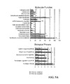


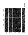


Classifications
-
- C—CHEMISTRY; METALLURGY
- C12—BIOCHEMISTRY; BEER; SPIRITS; WINE; VINEGAR; MICROBIOLOGY; ENZYMOLOGY; MUTATION OR GENETIC ENGINEERING
- C12N—MICROORGANISMS OR ENZYMES; COMPOSITIONS THEREOF; PROPAGATING, PRESERVING, OR MAINTAINING MICROORGANISMS; MUTATION OR GENETIC ENGINEERING; CULTURE MEDIA
- C12N5/00—Undifferentiated human, animal or plant cells, e.g. cell lines; Tissues; Cultivation or maintenance thereof; Culture media therefor
- C12N5/06—Animal cells or tissues; Human cells or tissues
- C12N5/0602—Vertebrate cells
- C12N5/0696—Artificially induced pluripotent stem cells, e.g. iPS
-
- C—CHEMISTRY; METALLURGY
- C12—BIOCHEMISTRY; BEER; SPIRITS; WINE; VINEGAR; MICROBIOLOGY; ENZYMOLOGY; MUTATION OR GENETIC ENGINEERING
- C12N—MICROORGANISMS OR ENZYMES; COMPOSITIONS THEREOF; PROPAGATING, PRESERVING, OR MAINTAINING MICROORGANISMS; MUTATION OR GENETIC ENGINEERING; CULTURE MEDIA
- C12N2501/00—Active agents used in cell culture processes, e.g. differentation
- C12N2501/60—Transcription factors
- C12N2501/602—Sox-2
-
- C—CHEMISTRY; METALLURGY
- C12—BIOCHEMISTRY; BEER; SPIRITS; WINE; VINEGAR; MICROBIOLOGY; ENZYMOLOGY; MUTATION OR GENETIC ENGINEERING
- C12N—MICROORGANISMS OR ENZYMES; COMPOSITIONS THEREOF; PROPAGATING, PRESERVING, OR MAINTAINING MICROORGANISMS; MUTATION OR GENETIC ENGINEERING; CULTURE MEDIA
- C12N2501/00—Active agents used in cell culture processes, e.g. differentation
- C12N2501/60—Transcription factors
- C12N2501/603—Oct-3/4
-
- C—CHEMISTRY; METALLURGY
- C12—BIOCHEMISTRY; BEER; SPIRITS; WINE; VINEGAR; MICROBIOLOGY; ENZYMOLOGY; MUTATION OR GENETIC ENGINEERING
- C12N—MICROORGANISMS OR ENZYMES; COMPOSITIONS THEREOF; PROPAGATING, PRESERVING, OR MAINTAINING MICROORGANISMS; MUTATION OR GENETIC ENGINEERING; CULTURE MEDIA
- C12N2501/00—Active agents used in cell culture processes, e.g. differentation
- C12N2501/60—Transcription factors
- C12N2501/604—Klf-4
-
- C—CHEMISTRY; METALLURGY
- C12—BIOCHEMISTRY; BEER; SPIRITS; WINE; VINEGAR; MICROBIOLOGY; ENZYMOLOGY; MUTATION OR GENETIC ENGINEERING
- C12N—MICROORGANISMS OR ENZYMES; COMPOSITIONS THEREOF; PROPAGATING, PRESERVING, OR MAINTAINING MICROORGANISMS; MUTATION OR GENETIC ENGINEERING; CULTURE MEDIA
- C12N2501/00—Active agents used in cell culture processes, e.g. differentation
- C12N2501/60—Transcription factors
- C12N2501/606—Transcription factors c-Myc
-
- C—CHEMISTRY; METALLURGY
- C12—BIOCHEMISTRY; BEER; SPIRITS; WINE; VINEGAR; MICROBIOLOGY; ENZYMOLOGY; MUTATION OR GENETIC ENGINEERING
- C12N—MICROORGANISMS OR ENZYMES; COMPOSITIONS THEREOF; PROPAGATING, PRESERVING, OR MAINTAINING MICROORGANISMS; MUTATION OR GENETIC ENGINEERING; CULTURE MEDIA
- C12N2506/00—Differentiation of animal cells from one lineage to another; Differentiation of pluripotent cells
- C12N2506/13—Differentiation of animal cells from one lineage to another; Differentiation of pluripotent cells from connective tissue cells, from mesenchymal cells
- C12N2506/1307—Differentiation of animal cells from one lineage to another; Differentiation of pluripotent cells from connective tissue cells, from mesenchymal cells from adult fibroblasts
-
- C—CHEMISTRY; METALLURGY
- C12—BIOCHEMISTRY; BEER; SPIRITS; WINE; VINEGAR; MICROBIOLOGY; ENZYMOLOGY; MUTATION OR GENETIC ENGINEERING
- C12N—MICROORGANISMS OR ENZYMES; COMPOSITIONS THEREOF; PROPAGATING, PRESERVING, OR MAINTAINING MICROORGANISMS; MUTATION OR GENETIC ENGINEERING; CULTURE MEDIA
- C12N2510/00—Genetically modified cells
Definitions
- the present invention relates to methods for inducing pluripotency in human somatic cells, and for maintaining pluripotency in human embryonic stem cells, with PRDM14 or NFRKB.
- hESCs human embryonic stem cells
- hESCs have the capacity for extensive self-renewal under in vitro culture conditions.
- a second hallmark of these cells is their ability to undergo multi-lineage differentiation; also defined as pluripotency.
- pluripotency the robust self-renewal capability of these pluripotent hESCs makes them a renewable source for the generation of functional cell-types or tissues for potential therapeutic applications and drug discovery.
- hESCs provide an opportunity to study early human developmental biology—an area of study where it is difficult to acquire experimental data.
- hESCs In addition to the ability to self-renew and differentiate, hESCs share many similarities with mouse embryonic stem cells (mESCs) [2, 3]. Both of them express genes which are associated with pluripotency [4-6].
- POU5F1 coding for the protein OCT4
- NANOG both key components of the core transcriptional regulatory network [7-9] are highly expressed in undifferentiated ESCs [10-15] and upon differentiation, the expression of these genes is reduced.
- These and other transcription regulators, including the co-activator p300 show extensive co-localization at genomic sites and this binding configuration may be important for the expression of pluripotency-specific genes [9, 16, 17].
- FGF/MEK fibroblast growth factor/Mitogen-Activated Protein Kinase Kinase pathway
- the TGF ⁇ /Activin/Nodal pathway maintains the self-renewal of hESCs but not mESCs, through the up-regulation of NANOG expression [19, 20].
- Leukemia inhibitory factor (LIF) and bone morphogenetic protein 4 (BMP4) are known to sustain mESCs, but LIF does not support hESCs and BMP4 induces hESCs to differentiate [4].
- hESCs and mESCs are morphologically distinct where mESCs form dome-shaped colonies while the hESC colonies are flat.
- certain surface molecules like SSEA-3 and SSEA-4 are present in undifferentiated hESCs but not mESCs [4].
- ESCs The differences between both mouse and human ESCs could be due to species-specific differences in embryonic development.
- the ESCs could be derived from cells originating from different developmental stages. Consistent with this idea is the identification of post-implantation murine epiblast-derived stem cells which show characteristics of hESCs [21, 22]. Hence, it is useful to understand the differences between these ESCs and the molecular basis for the differences. Furthermore, to harness the full potential of hESCs, it is valuable to dissect the mechanisms that maintain the identity of hESCs.
- the present invention provides methods of inducing pluripotency in a human somatic cell, including for example a human fibroblast cell.
- the present invention provides a method of inducing pluripotency in a human somatic cell, the method comprising culturing the human somatic cell in the presence of (i) OCT4 and SOX2; (ii) at least one of KLF4 and c-MYC; and (iii) at least one of PRDM14 and NFRKB.
- culturing may comprise contacting the human somatic cell, with the OCT4 and the SOX2, with the at least one of KLF4 and c-MYC and with the at least one of PRDM14 and NFRKB so that the OCT4, the SOX2, the at least one of KLF4 and c-MYC and the at least one of PRDM14 and NFRKB are taken up by the human somatic cell.
- culturing may comprise expressing the OCT4, the SOX2, the at least one of KLF4 and c-MYC and the at least one of PRDM14 and NFRKB in the human somatic cell.
- OCT4, SOX2, at least one of KLF4 and c-MYC and at least one of PRDM14 and NFRKB may be expressed from one or more expression vectors, which may be viral vectors.
- OCT4, SOX2, KLF4 and PRDM14 are expressed in the human somatic cell.
- OCT4, SOX2, c-MYC and PRDM14 are expressed in the human somatic cell.
- OCT4, SOX2, KLF4, c-MYC and PRDM14 are expressed in the human somatic cell.
- OCT4, SOX2, KLF4 and NFRKB are expressed in the human somatic cell.
- OCT4, SOX2, c-MYC and NFRKB are expressed in the human somatic cell.
- OCT4, SOX2, KLF, c-MYC and NFRKB are expressed in the human somatic cell.
- the human somatic cell may be partially differentiated prior to the culturing or may be fully differentiated prior to the culturing.
- the human somatic cell is a fibroblast, including a human fibroblast prior to the culturing.
- the present invention provides a method of inducing pluripotency in a human fibroblast, the method comprising expressing in the human fibroblast (i) OCT4 and SOX2; (ii) at least one of KLF4 and c-MYC; and (iii) at least one of PRDM14 and NFRKB from one or more expression vectors; and culturing the human fibroblast under conditions suitable for growth of embryonic stem cells.
- the present invention provides a method of inducing pluripotency in a human fibroblast, the method comprising expressing in the human fibroblast (i) PRDM14 or NFRKB; together with (ii) OCT4, SOX2 and KLF4.
- the method may further comprise expressing c-MYC in the human fibroblast.
- the present invention provides a human somatic cell comprising (i) OCT4 and SOX2; (ii) at least one of KLF4 and c-MYC; and (iii) at least one of PRDM14 and NFRKB.
- the human somatic cell expresses each of OCT4, SOX2 and KLF4 and at least one of PRDM14 and NFRKB.
- the human somatic cell expresses each of OCT4, SOX2 and c-MYC and at least one of PRDM14 and NFRKB.
- the human somatic cell expresses each of OCT4, SOX2, KLF4 and c-MYC and at least one of PRDM14 and NFRKB.
- pluripotency has been induced in the cell.
- the human somatic cell is a fibroblast.
- the present invention provides a human somatic cell comprising one or more expression vectors encoding (i) OCT4 and SOX2; (ii) at least one of KLF4 and c-MYC; and (iii) at least one of PRDM14 and NFRKB.
- the human somatic cell is a fibroblast, including a human fibroblast.
- the present invention provides a human somatic cell, including a human fibroblast cell, produced according to the described methods.
- the human somatic cell may be one in which pluripotency has been induced.
- the present invention further provides methods of maintaining pluripotency of a human embryonic stem cell (hESC).
- hESC human embryonic stem cell
- the present invention provides a method of maintaining pluripotency of a hESC comprising culturing the hESC in the presence of (i) OCT4 and SOX2; (ii) at least one of KLF4 and c-MYC; and (iii) at least one of PRDM14 and NFRKB.
- culturing may comprise contacting the hESC with the OCT4 and the SOX2, with the at least one of KLF4 and c-MYC and with the at least one of PRDM14 and NFRKB so that the OCT4, the SOX2, the at least one of KLF4 and c-MYC and the at least one of PRDM14 and NFRKB are taken up by the hESC.
- culturing may comprise expressing the OCT4, the SOX2, the at least one of KLF4 and c-MYC and the at least one of PRDM14 and NFRKB in the hESC.
- OCT4, SOX2, at least one of KLF4 and c-MYC and at least one of PRDM14 and NFRKB may be expressed from one or more expression vectors, which may be viral vectors.
- FIG. 1 Genome-wide screen for regulators that maintain hESC identity.
- 1 A Schematic representation of siRNA screen. H1 hESC line with a GFP reporter gene driven by the POU5F1 promoter was used for the screen.
- 21,121 Dharmacon SMARTpooled siRNA were seeded onto 67 384-well plates and the POU5F1-GFP reporter hESCs were reverse transfected on these plates.
- individual siRNAs for 200 high confidence candidates from the primary screen were reverse transfected into the reporter cells and two other hESC lines HES2 and HES3.
- 1 B Montage representing the images for GFP fluorescence and Hoechst staining for a typical 384-well plate is shown.
- the hESCs were imaged 4 days post transfection.
- the negative control siRNA (NT: non-targeting) and the positive controls siRNA (GFP and POU5F1) were printed at the designated wells as indicated.
- FIG. 2 Characterization of POU5F1-GFP H1 hESC line.
- 2 A POU5F1-GFP reporter cells transfected with non-targeting (NT) siRNA exhibited strong fluorescence while reporter cells transfected with POU5F1 siRNA resulted in a reduction in GFP expression. Nuclei of the cells were counterstained with Hoechst. Scale bars represent 50 ⁇ m.
- 2 B Fluorescent activated cell sorting for GFP quantification.
- Graphed lines are shown for non-reporter cells (H1 hESC), POU5F1-GFP reporter cells transfected with POU5F1 siRNA (POU5F1 siRNA) and reporter cells transfected with non-targeting siRNA (NT siRNA).
- POU5F1 siRNA POU5F1 siRNA
- NT siRNA non-targeting siRNA
- 2 C Karyotypic analysis of POU5F1-GFP reporter cells. The cells have a normal karyotype with 46XY chromosomes.
- 2 D Teratoma formation assay for POU5F1-GFP reporter cells. Teratoma of ectoderm, mesoderm and endoderm lineages were obtained after 8 weeks of injection into SCID mice.
- FIG. 3 Gene ontology analysis of F av .
- 3 A Transcription factors that have been shown to be involved in the maintenance of mESC identity. The human counterparts of these genes were identified from this genome wide siRNA screen and they are ranked based on the z-score. The percentage of ranking of these genes out of the 21,121 genes is indicated in bracket.
- 3 B Components of the mediator complex are identified among the top hits.
- 3 C Components of the INO80 complex are identified among the top hits.
- FIG. 4 Gene ontology analysis of N av .
- 4 A Gene ontology analysis on the molecular function of the top 200 genes ranked by N av scores (Panther classification). Candidate genes involved in cell survivability were enriched in GO categories for nucleic acid binding and ribosomal proteins, mRNA splicing and processing factor and DNA-directed RNA polymerase.
- 4 B Transcripts encoding for RNA polymerase subunits were among the top 200 hits (ranked by N av score).
- 4 C Transcripts encoding for ribosomal subunits were found among the top hits.
- FIG. 5 Pathway analyses.
- 5 A Gene Ontology analysis of the 566 genes with z-score>2. Graphs represent the functional categorization of the biological process and molecular function categories that are over-represented. Categories with p-value ⁇ 0.05 are indicated with * and categories with p-value ⁇ 0.01 are indicated with **.
- 5 B Reactome analysis. The 566 genes (identifiers) were analyzed using the web-resource Reactome. The reactome map were plotted using Skypainter tool to determine which events (reactions and/or pathways) are statistically over-represented in the set of genes submitted. 113 identifiers could be matched to the 333 out of 4374 events. 12 categories with p-value ⁇ 0.05 are over-represented.
- FIG. 6 Secondary validation of 200 genes.
- 6 A Deconvoluted siRNA screen on H1-GFP, HES2 and HES3 hESCs lines. 200 genes from the 566 genes with z-score>2 were subjected to further validation by deconvoluting the pooled mixture of 4 siRNAs. The screen was performed on 3 different hESCs lines and different stemness markers were used for analysis. H1-GFP hESCs line were analyzed for GFP, OCT4 and NANOG expression, HES2 and HES3 hESCs lines were analyzed for OCT4 and NANOG expression. Genes were considered positive hits if 2 or more siRNA down-regulate GFP/OCT4/NANOG expression.
- FIG. 6 B Venn diagram showing the overlapping hits for the different marker of analysis in each of the different cell line. 126 genes are validated by GFP, OCT4 and NANOG downregulation in H1-GFP hESCs. 86 genes in HES2 and 124 genes in HES3 were validated based on OCT4 and NANOG downregulation.
- FIG. 6 C Venn diagram showing the common overlapping genes among the 3 different hESC lines based on OCT4 or NANOG stemness marker for analysis. 93 common genes are involved in the downregulation of OCT4 and 54 common genes are involved in the down regulation of NANOG in all the 3 hESCs lines.
- FIG. 6 D Graphs depicting the GFP VS OCT4, GFP VS NANOG correlation for H1-GFP hESCs and NANOG VS OCT4 correlation for each of H1-GFP, HES2 and HES3 hESCs.
- FIG. 7 PRDM14 and NFRKB can enhance reprogramming of human fibroblasts to iPSCs.
- 7 B PRDM14 and NFRKB are required for reprogramming of human somatic cell.
- the hiPSCs can differentiate into ectodermal (indicated by NESTIN staining) and mesodermal cells (indicated by ⁇ -smooth muscle actin staining) using EB-mediated method.
- Definitive endodermal cells (as shown by staining for SOX17) derived with activin A induction and trophectodermal cells (as shown by staining for p57kip2) derived with combined BMP4 induction and FGF inhibition (PD0325901) were also obtained from these hiPSCs.
- the scale bars represent 200 ⁇ m.
- ( 7 F) Teratoma formation assay for OSKM+PRDM14 hiPSCs and OSKM+NFRKB hiPSCs.
- Tissues derived from all three germ layers were obtained. Tissues shown correspond to pigmented epithelium, neural rosettes, muscle and gut epithelium. Bars represent 50 ⁇ m.
- 7 G Microarray profiling of hiPSCs. Heatmap showing the gene expression profile of 1,000 hESC-associated genes and fibroblast-associated genes in H1, H9 hESCs, two hiPSC lines and MRC-5. The selection of genes was based on the fold differences in their expression levels in hESCs and fibroblasts. The genes were sorted according to the average expression ratios and mean-centred around the fibroblasts signal. The greyscale bar indicates the gene expression normalized to fibroblasts in log 2 scale.
- FIG. 8 Characterization of PRDM14 and NFRKB reprogrammed hiPSCs.
- PRDM14 and NFRKB can enhance reprogramming mediated by OCT4, SOX2 and KLF4.
- 8 B Both OSK+PRDM14 hiPSCs and OSK+NFRKB hiPSCs showed normal karyotype.
- the hiPSCs can differentiate into ectodermal (indicated by NESTIN staining) and mesodermal cells (indicated by ⁇ -smooth muscle actin staining) using EB-mediated method.
- Definitive endodermal cells (as shown by staining for SOX17) derived with activin A induction and trophectodermal cells (as shown by staining for p57kip2) derived with combined BMP4 induction and FGF inhibition (PD0325901) were also obtained from these hiPSCs.
- the scale bars represent 200 ⁇ m.
- ( 8 E) Teratoma formation assay for OSK+PRDM14 hiPSCs and OSK+NFRKB hiPSCs.
- Tissues derived from all three germ layers were obtained. Tissues shown correspond to pigmented epithelium, muscle, gut epithelium and kidney-like tissues. Bars represent 50 ⁇ m.
- 8 F Microarray profiling of hiPSCs. Heatmap showing the gene expression profile of 1,000 hESC-associated genes and fibroblast-associated genes in H1, H9 hESCs, two hiPSC lines and MRC-5. The selection of genes was based on the fold differences in their expression levels in hESCs and fibroblasts. The genes were sorted according to the average expression ratios and mean-centred around the fibroblasts signal. The greyscale bar indicates the gene expression normalized to fibroblasts in log 2 scale.
- the hiPSCs can differentiate into ectodermal (indicated by NESTIN staining) and mesodermal cells (indicated by ⁇ -smooth muscle actin staining) using EB-mediated method.
- Definitive endodermal cells (as shown by staining for SOX17) derived with activin A induction and trophectodermal cells (as shown by staining for p57kip2) derived with combined BMP4 induction and FGF inhibition (PD0325901) were also obtained from these hiPSCs.
- the scale bars represent 200 ⁇ m.
- FIG. 9 PRDM14 and the maintenance of H1 hESCs.
- 9 A Immunofluorescence staining for stemness markers. PRDM14 and control knockdown H1 hESCs were stained for OCT4, TRA-1-60 and TRA-1-81 after 4 days of knocked down. The nuclei of the cells were counterstained with Hoechst. Scale bars represent 50 ⁇ m.
- 9 B Analysis of PRDM14, OCT4 and NANOG protein levels by western blot.
- 9 C Quantification of pluripotency-related transcript levels. Pluripotency-associated genes SOX2, HELLS and DPPA4 were quantified for mRNA expression changes by qPCR.
- FIG. 10 Validation for PRDM14 knockdown in HES3 hESCs.
- 10 A PRDM14 and control knockdown HES3 hESCs were stained for hESC markers (OCT4, TRA-1-60, TRA-1-81 and SSEA-4) and alkaline phosphatase after 4 days of knockdown. The nuclei of the cells were counterstained with Hoechst. Scale bars represent 50 ⁇ m.
- 10 B Quantification for pluripotency-related transcript levels.
- FIG. 11 Validation for PRDM14 for PRDM14 Knockdown in H9 hESCs.
- 11 A PRDM14 and control knockdown H9 hESCs were stained for hESC markers (OCT4, TRA-1-60, TRA-1-81 and SSEA-4) and alkaline phosphatase after 4 days of knockdown. The nuclei of the cells were counterstained with Hoechst. Scale bars represent 50 ⁇ m.
- 11 B Quantification for pluripotency-related transcript levels.
- FIG. 12 Prdm14 is not required for the maintenance of mouse ESCs and is deficient in mouse EpiSCs.
- 12 A Knockdown of Prdm14 in mESCs with 3 different shRNA constructs did not induce differentiation as indicated by the alkaline phosphatase staining and morphology. Scale bar represents 50 uM.
- 12 C Genes expressed at high and low level in EpiSCs.
- FIG. 13 Validation of PRDM14 ChIP-seq dataset.
- 13 A H1 hESCs were transfected with the PRDM14 or control knockdown construct and harvested 72 hrs post transfection. 21 genomic loci bound by PRDM14 from the ChIP-seq analysis were chosen for validation. C1 and C2 are control regions showing only background level of enrichment.
- 13 B H1 hESCs were transfected with the HA-tagged PRDM14 expression construct and harvested 72 hrs post transfection. An antibody specific for the HA tag was used in the ChIP against chromatin extracts from cells transfected with the PRDM14 expression construct and untransfected cells.
- C1 and C2 are control regions showing only background level of enrichment.
- FIG. 14 PRDM14 regulates POU5F1 enhancer.
- PRDM14 shows co-binding with OCT4, SOX2, NANOG and co-activator p300. Greyscale intensity in the heat map reflects the co-localization frequency of each of the transcription factors (the descending frequency of localization ranges from white to grey to dark grey).
- PRDM14 motif predicted by the de novo motif-discovery algorithm CisFinder.
- C ChIP-seq binding profile of PRDM14, OCT4, NANOG, CTCF at POU5F1 locus. Control ChIP-seq library was obtained from sequencing of input DNA.
- PRDM14 binds to CR2 probe.
- Probe containing putative PRDM14 motif were incubated with purified recombinant PRDM14 DNA binding domain (DBD) protein. Introduction of mutations in the putative motif disrupt the PRDM14 DBD/DNA complex.
- DBD DNA binding domain
- Native PRDM14 binds to CR2 probe. Probes containing putative PRDM14 motif were incubated with nuclear extract prepared from hESCs. Introduction of mutations in the putative motif disrupt the PRDM14 DBD/DNA complex. Supershift assay was performed by adding PRDM14 antibodies to the EMSA sample.
- PRDM14 regulates CR2 enhancer but not CR4 enhancer in hESC.
- CR2 and CR4 of the POU5F1 upstream regulatory region were each cloned downstream of the luciferase reporter gene driven by a POU5F1 proximal promoter ( ⁇ 350 bp).
- Each of the constructs was transfected into H1 hESCs to test for enhancer activity.
- PRDM14 is bound to CR2 but not CR4 region. ChIP assay was performed using a PRDM14 antibody.
- NANOG binds to PRDM14 in human ES cells.
- PRDM14 and NANOG synergistically enhance reprogramming of human fibroblasts.
- FIG. 15 Co-motif analysis. The distribution and enrichment of sequence motifs around PRDM14 ChIP-seq peaks. Except for the PRDM14 motif which was found de novo in this study, the other 3 motifs V_OCT4 — 01, V_SP1_Q6 — 01 and V_AP2_Q6 shown are the most enriched from the TRANSFAC database. The enrichment score reflects the number of motif matches compared to the expected number of matches in windows around the ChIP-seq peaks.
- FIG. 16 PRDM14 regulates CR2 enhancer in different hESCs.
- 16 A Map of luciferase reporters used to test enhancer activity of CR2 and CR4.
- CR2 and CR4 of the POU5F1 upstream regulatory region were each cloned downstream of the luciferase reporter gene driven by a POU5F1 proximal promoter ( ⁇ 350 bp).
- 16 B Each of the constructs was transfected into HES2 and HES3 hESCs to test for enhancer activity.
- FIG. 17 Mapping of functional domains for transcriptional regulation and reprogramming.
- 17 A A schematic summary of different molecules generated for PRDM14 functional study. N, N-terminal; SET, PR/SET domain; DBD, DNA binding domain; ⁇ , deletion.
- 17 B 3 copies of PRDM14 motif found in CR2 enhancer were inserted in tandem before a minimal promoter found in pGL4.23 luciferase reporter vector. Full length cDNA of PRDM14 or different mutant versions of PRDM14 were transfected into 293T cells to test for their ability to regulate transcription. Luciferase vector without PRDM14 motif was used as control. The reporter activity was normalized to the activity of control vector in the presence of PRDM14 or deletion mutants.
- FIG. 18 Regulation of target genes by PRDM14.
- 18 A Venn diagram showing the intersection of PRDM14-bound genes with genes that were down-regulated after PRDM14 depletion. This subset of PRDM14 bound genes are positively regulated by PRDM14.
- 18 B Venn diagram showing the intersection of PRDM14-bound genes with genes that were induced after PRDM14 depletion.
- 18 C GO analysis of the overlapping genes shown in ( 18 A).
- 18 D GO analysis of the overlapping genes shown in ( 18 B).
- 18 E Schematic representation of a model for the transcriptional regulatory network governed by PRDM14 in hESC.
- FIG. 19 Validation of gene expression (from the different Gene Ontology group) upon PRDM14 depletion.
- FIG. 20 Transcription factors that co-localize to PRDM14 sites at PRDM14 regulated genes.
- 20 A Top 20 enriched PWMs at PRDM14 sites.
- 20 B Motif logo for V_OCT4 — 01 TRANSFAC PWM.
- 20 C Examples of 10 PWM not enriched at PRDM14 sites.
- PRDM14 shows co-binding with OCT4, SOX2, NANOG and co-activator p300. Greyscale intensity in the heat map reflects the co-localization frequency of each of the transcription factors (the descending frequency of localization ranges from white to grey to dark grey).
- FIG. 21 PRDM14 recruits polycomb group proteins in hESCs and during reprogramming.
- 21 A Co-localization analysis for PRDM14 and histone modifications. Greyscale intensity in the heat map reflects the co-localization frequency of each of transcription factors (the descending frequency of localization ranges from white to grey to dark grey).
- 21 B PRDM14 interacts with Ezh2 in hESC. Co-IP assays were performed using hESC whole cell lysate with anti-PRDM14 and anti-Ezh2 antibody. Western was carried out with anti-PRDM14 or anti-Ezh2 antibody. Anti-GST antibody was used in the control IP sample.
- 21 C PRDM14 interacts with Ezh2 in 293T cells.
- 293T cell were co-transfected with cDNA encoding HA tagged PRDM14 and Myc tagged EZH2 protein. Whole cell lysate was used for co-IP with anti-HA and anti-Myc antibody. Anti-GST antibody was used in the control IP.
- 21 E Recruitment of Ezh2 is reduced with PRDM14 depletion.
- 21 G H3K27me3 is induced at PRDM14 targets in fibroblasts.
- PRDM14 represses the genes shown in ( 21 D) in fibroblasts.
- PR domain-containing protein 14 PRDM14
- NFRKB nuclear factor related kappaB binding protein
- the present invention thus relates to methods of inducing pluripotency in a human somatic cell, to methods of maintaining hESCs and to human somatic cells comprising OCT4, SOX2, at least one of KLF4 and c-MYC and at least one of PRDM14 and NFRKB.
- somatic cells could be reprogrammed into induced pluripotent stem cells (iPSCs) through the co-expression of four transcription factors, OCT4, SOX2, KLF4 and c-MYC [27, 42-44]. This is significant since the reprogramming of somatic cells provides unprecedented opportunities for generating patient-specific pluripotent cells which may be used as in vitro models for studying and developing applications for treating human diseases [28-31].
- iPSCs induced pluripotent stem cells
- OCT4 is a protein encoded by the human POU5F1 gene [71].
- the POU family of transcription factors consists of at least 14 members, which often act synergistically during vertebrate development with the SOX family of transcription factors [72-74].
- the role of OCT4 as an inducer of pluripotency is well established [27, 57].
- the sequence of two isoforms of the human OCT4 gene, generated by alternative splicing, is available for example in GenBank under DQ486515 and DQ486516.
- the reference to OCT4 in the present application may encompass either of the OCT4 isoforms.
- a representative amino acid sequence for human OCT4 is provided below:
- SOX2 is a transcription factor that is critical to maintain self-renewal of undifferentiated stem cells, and is one of the key transcription factors required in iPSCs [45].
- the SOX family of transcription factors consists of at least 20 members [72-74].
- specificity in transcriptional control may be achieved as a result of selective heterodimerization.
- SOX2/OCT4 pairs are important factors in embryonic stem (ES) cells [8, 64].
- the sequence of the human SOX2 gene is available, for example, in GenBank under BC013923.
- reference herein to SOX2 may include for example mutated versions of SOX proteins, such as described International Patent Application PCT/SG2010/000423.
- a representative amino acid sequence for human SOX2 is provided below:
- KLF4 is a member of a family of proteins characterized by their three Cyst His2 zinc fingers located at the C-terminus, each of which is separated by a highly conserved H/C link. KLF4 interacts with p300 histone acetyltransferase to regulate gene transcription by modulating histone acetylation [76]. KLF4 has been shown to play a role in reprogramming human somatic cells into iPSCs [27]. The sequence of the human KLF4 gene is available, for example, in GenBank under AF105036. A representative amino acid sequence for human KLF4 is provided below:
- c-MYC is a member of a family of transcription factors containing a basic Helix-Loop-Helix Leucine. It has been proposed that the role of c-MYC in establishing iPSCs may be as a booster of reprogramming rather than a controller of maintenance [27].
- the sequence of the human c-MYC gene may be found, for example, in GenBank under NCBI RefSeq NM — 002467.
- a representative amino acid sequence for human c-MYC is provided below:
- reference herein to OCT4, SOX2, KLF4 and c-MYC includes those embodiments described above, as well as sequence variants or fragments (e.g. fragments of at least 25, 50, 100, 150, 200, 250, 300, 350, 400 or more amino acids in length) which retain the ability to direct the specific function of OCT4, SOX2, KLF4 and c-MYC, respectively, including for example either the induction or maintenance of pluripotency.
- any such variants or fragments may be used in the methods of the present invention, for example, either in methods involving contacting the human somatic cells with OCT4, SOX2, KLF4 or c-MYC or methods involving expressing OCT4, SOX2, KLF4 or c-MYC in the human somatic cell.
- the OCT4, SOX2, KLF4 and c-MYC used in the present invention may be obtained from cDNA found in Addgene plasmids 17217, 17218, 17219 and 17220, respectively [27].
- Polypeptides or peptides that have substantial identity to proteins encoded by the cDNA found in the Addgene plasmids or substantial identity to the representative amino acid sequences provided herein for OCT4, SOX2, KLF4 and c-MYC may also be used.
- nucleotide sequences encoding any of these polypeptides, peptides or proteins, or nucleotide sequences having substantial identity thereto, are also encompassed by the present invention.
- sequences are considered to have substantial identity if, when optimally aligned (with gaps permitted), they share at least approximately 50% sequence identity, or if the sequences share defined functional motifs.
- optimally aligned sequences may be considered to be substantially identical (i.e., to have substantial identity) if they share at least 60%, 70%, 75%, 80%, 85%, 90%, 95%, 96%, 97%, 98%, or 99% identity over a specified region.
- identity refers to sequence similarity between two polypeptides molecules. Identity can be determined by comparing each position in the aligned sequences.
- a degree of identity between amino acid sequences is a function of the number of identical or matching amino acids at positions shared by the sequences, for example, over a specified region.
- Optimal alignment of sequences for comparisons of identity may be conducted using a variety of algorithms, as are known in the art, including the ClustalW program, available at http://clustalw.genome.ad.ip, the local homology algorithm of Smith and Waterman, 1981 , Adv. Appl. Math 2: 482, the homology alignment algorithm of Needleman and Wunsch, 1970 , J. Mol. Biol. 48:443, the search for similarity method of Pearson and Lipman, 1988 , Proc. Natl. Acad. Sci.
- Sequence identity may also be determined using the BLAST algorithm, described in Altschul et al., 1990 , J. Mol. Biol. 215:403-10 (using the published default settings).
- the “BLAST 2 Sequences” tool available through the National Center for Biotechnology Information (through the interne at http://www.ncbi.nlm.nih.gov/BLAST/bl2seq/wblast2.cqi) may be used, selecting the “blastp” program at the following default settings: expect threshold 10; word size 3; matrix BLOSUM 62; gap costs existence 11, extension 1.
- the person skilled in the art can readily and properly align any given sequence and deduce sequence identity and/or homology by mere visual inspection
- the presence of either PRDM14 or NFRKB during culture of human somatic cells enhances reprogramming of the human somatic cells to iPSCs (i.e. imparting pluripotency). This is observed when the cells are cultured in the presence of OCT4, SOX2, at least one of KLF4 and c-MYC, and at least one of PRDM14 and NFRKB. While OCT2 and SOX2 may be required for the generation of iPSCs, PRDM14 and NFRKB are able to substitute for either KLF4 or c-MYC.
- the present methods provide a method of using OCT4, SOX2 and KLF4 or c-MYC, together with at least one of PRDM14 and NFRKB to induce pluripotency by converting a non-pluripotent cell to a pluripotent cell, including in vitro methods.
- a method of inducing pluripotency in a human somatic cell comprising culturing the human somatic cell in the presence of (i) OCT4 and SOX2; (ii) at least one of KLF4 and c-MYC; and (iii) at least one of PRDM14 and NFRKB.
- the human somatic cells may be cultured in conditions appropriate for maintaining the particular human somatic cell type.
- Appropriate conditions for culturing human somatic cell types are known in the art.
- human fibroblast cells may be cultured in 15% fetal bovine serum (FBS)/Dulbecco's Modified Eagle Medium (DMEM).
- FBS fetal bovine serum
- DMEM Dulbecco's Modified Eagle Medium
- the human somatic cells are cultured in the presence of OCT4, SOX2, at least one of KLF4 and c-MYC, and at least one of PRDM14 and NFRKB.
- the culture conditions may be adjusted during the method from conditions supporting the human somatic cells to conditions which are suitable for the growth and maintenance of embryonic stem cells.
- the change in culture conditions may be performed, for example, upon the introduction of OCT4, SOX2, at least one of KLF4 and c-MYC, and at least one of PRDM14 and NFRKB, or shortly thereafter (e.g. about 24 hours).
- the cells may then be maintained and expanded in such conditions which are suitable for the growth and maintenance of embryonic stem cells.
- the present invention provides methods of reprogramming human somatic cells to iPSCs, which are known to be comparable to hESCs [27].
- iPSCs which are known to be comparable to hESCs [27].
- These human iPSCs are able to be maintained in the presence of OCT4, SOX2, at least one of KLF4 and c-MYC, and at least one of PRDM14 and NFRKB.
- OCT4, SOX2, at least one of KLF4 and c-MYC, and at least one of PRDM14 and NFRKB a role for these regulators in maintaining pluripotency in hESCs is further shown by the fact that reduction in expression of one of these key regulators, PRDM14, results in a loss of pluripotency.
- PRDM14 is required to maintain the expression of a key pluripotency gene, POU5F1 (coding for the protein OCT4).
- POU5F1 coding for the protein OCT4
- ESA electrophoretic mobility shift assay
- PRDM14 shows co-binding with OCT4, SOX2, NANOG and the co-activator p300, indicating that the PRDM14 circuitry is integrated into the core hESC transcriptional regulatory network, and is an important regulatory factor in maintaining pluripotency.
- Also contemplated therefore is a method of maintaining pluripotency of a human embryonic stem cell (hESC) comprising culturing the hESC in the presence of (i) OCT4 and SOX2; (ii) at least one of KLF4 and c-MYC; and (iii) at least one of PRDM14 and NFRKB.
- hESC human embryonic stem cell
- the hESCs may be cultured under conditions suitable for the growth and maintenance of embryonic stem cells.
- culturing the cell in the presence of OCT4 and SOX2, at least one of KLF4 and c-MYC, and at least one of PRDM14 and NFRKB includes contacting the cell with the various pluripotency protein factors so that the pluripotency protein factors are taken up by the cell, as well as transfecting or transducing the cell with nucleic acids encoding the various pluripotency protein factors and expressing the pluripotency protein factors. Procedures for accomplishing such culturing methods are widely known in the art and any of these known culturing techniques may be used in the present invention.
- reference to pluripotency protein factors is a reference to OCT4, SOX2, KLF4, c-MYC, PRDM14 and NFRKB, either alone or in combination.
- expression of the pluripotency protein factors may involve expression from an expression vector.
- the expression vector may be of any suitable structure which provides expression of the pluripotency protein factors.
- a suitable promoter will be operably linked to the coding region for the particular pluripotency protein factor.
- a coding sequence is operably linked to a promoter if the promoter activates the transcription of the coding sequence.
- the expression vector may for example be a plasmid, bacteriophage, DNA virus, RNA virus, or retrovirus vector.
- a pMX retroviral plasmid may be used to carry cDNA of human OCT4, SOX2, KLF4, c-MYC, PRDM14 or NFRKB. It will be understood that the pluripotency protein factors may be co-expressed from one or more expression vectors.
- the cells may be cultured under conditions suitable for the growth and maintenance of embryonic stem cells.
- Conditions that are suitable for growth and maintenance embryonic stem cells are known. Exemplary conditions are described herein which were used for the culture of the hESC lines H1, H9, HES2, HES3 and H1 POU5F1-GFP lines. Further, commercial medium for human embryonic stem cell culture is available (e.g. HEScGRO from Millipore; StemPro from Invitrogen) and other culture media and culture conditions are described, for example, in Lu et al. (2006) PNAS, 103(15): 5688-5693. In some circumstances, it may be desirable to use feeder cells to promote stem cell growth in culture, in keeping with standard embryonic stem cell culture techniques.
- cell refers to and includes a single cell, as well as a plurality of cells or a population of cells, where context permits, and unless otherwise specified. Similarly, reference to “cells” also includes reference to a single cell where context permits, unless otherwise specified.
- the cell or cells may be grown in e.g. batch culture or in tissue culture plates.
- the human somatic cell may be any human somatic cell, for example any human somatic cell that is partially differentiated or fully differentiated, including an in vitro cell, a cell in culture, or an explanted cell from a subject.
- the cell may be of any type of partially or fully differentiated human somatic cell, including for example a human fibroblast cell.
- the hESC may be any type of undifferentiated hESC.
- the undifferentiated hES cell or cells are typically originally obtained from a blastocyst as is known in the art, but may be previously expanded.
- pluripotency refers to a state where the cell has the potential to differentiate into any of the three germ layers: endoderm, mesoderm or ectoderm. Pluripotency may be assessed according to any means known in the art. For example, the expression levels of known pluripotency markers, such as for example NANOG, TRA-1-60, TRA-1-81, SSEA-4 or alkaline phosphatase, may be determined. An increased expression of any of these genes as compared to levels in differentiated human somatic cells may be considered indicative of pluripotency. Also, cells may be identified as pluripotent by their ability to differentiate into different lineages via EB (embryoid body)-mediated or growth factor-induced in vitro differentiation or by teratoma formation assays.
- EB epidermality
- hESCs refers to the maintenance of these cells in their undifferentiated, pluripotent state. Techniques described above for determining pluripotency may also be used to determine if cells are maintained as embryonic stem cells. Other known means may also be used. For example, the expression profile of hESC-associated genes, such as for example SOX2, HELLS and DPPA4, may be determined. Continued expression of these genes during culture may indicate maintenance of hESCs. In contrast, an increased expression of proteins that are expressed in differentiated cells types, such as RUNX1, MAFB and IGFBP5, may signify differentiation, and therefore a loss of hESCs.
- differentiated cells types such as RUNX1, MAFB and IGFBP5
- PRDM14 is a pluripotency protein factor belongs to a family of PR (PRDI-BFI and RIZ) domain proteins, a common domain found in histone modifying enzymes [50, 51]. In this family, there are predicted to be at least 17 PRDM proteins in primates and 16 PRDM proteins in rodents [70]. The proteins share a characteristic domain organization, each (with the exception of PRDM11) having an N-terminal PR domain followed by a variable number of zinc-finger repeats [70].
- the sequence of the human PRDM14 gene may be found, for example, in GenBank under AF319458. A representative sequence for human PRDM14 is provided below:
- PRDM14 may encompass sequence variants or fragments (e.g. fragments of at least 25, 50, 100, 150, 200, 250, 300, 350, 400 or more amino acids in length) which retain the ability to direct the specific function of PRDM14, including for example either the induction or maintenance of pluripotency. Any such variants or fragments may be used in the methods of the present invention, for example, either in methods involving contacting the human somatic cells with PRDM14 or methods involving expressing PRDM14 in the human somatic cell.
- Polypeptides or peptides that have substantial identity to PRDM14 protein sequences known in the art or substantial identity to the representative amino acid sequences provided herein for PRDM14 may also be used.
- nucleotide sequences encoding any of these polypeptides, peptides or proteins, or nucleotide sequences having substantial identity thereto, are also encompassed by the present invention. The meaning of substantial identity has been described earlier herein and applies equally to nucleotide and amino acid sequences of PRDM14.
- Prdm14 is critical for the establishment of the germ cell lineage [52].
- Prdm14 is essential for the derivation of embryonic germ cells from primordial germ cells (PGCs).
- PGCs primordial germ cells
- the knockout animals do not show early embryonic lethal phenotype, unlike the knockout phenotype of other key regulators essential for the maintenance of pluripotency of mESCs [13, 15, 57, 58].
- Prdm14 does not appear to be required to maintain mESC and pluripotent stem cells of the blastocysts [23, 25, 52].
- the expression of Pou5f1 is maintained in the absence of Prdm14.
- the present invention indicates that PRDM14 is critical for activating POU5F1 in hESCs.
- PRDM14 maintains pluripotency and promotes the acquisition of pluripotency of the germ cell lineage and hESCs through distinctive mechanisms. These differences may arise through cell-type specific or species-specific differences in regulating Pou5f1. Also, it is shown that PRDM14 associates with NANOG, another transcription factor important for the establishment of ground state pluripotency [58]. PRDM14 and NANOG can function synergistically to activate POU5F1 enhancer as well as to mediate reprogramming. Therefore, PRDM14 is connected via multiple points to the pluripotency-specific transcriptional regulatory network.
- NFRKB which is thought to be a component of the chromatin-remodelling INO80 complex, is a member of a family of transcription factors and has been shown to bind in vitro to several of the kappa B regulatory elements [77].
- the sequence of the human NFRKB gene may be found, for example, in GenBank under BC063280.
- a representative amino acid sequence for human NFRKB is provided below:
- NFRKB may encompass sequence variants or fragments (e.g. fragments of at least 25, 50, 100, 150, 200, 250, 300, 350, 400 or more amino acids in length) which retain the ability to direct the specific function of NFRKB, including for example either the induction or maintenance of pluripotency. Any such variants or fragments may be used in the methods of the present invention, for example, either in methods involving contacting the human somatic cells with NFRKB or methods involving expressing NFRKB in the human somatic cell.
- Polypeptides or peptides that have substantial identity to NFRKB protein sequences known in the art or substantial identity to the representative amino acid sequences provided herein for NFRKB may also be used.
- nucleotide sequences encoding any of these polypeptides, peptides or proteins, or nucleotide sequences having substantial identity thereto, are also encompassed by the present invention. The meaning of substantial identity has been described earlier herein and applies equally to nucleotide and amino acid sequences of NFRKB.
- the present methods are based on the finding that PRDM14 and NFRKB enhance reprogramming of human somatic cells.
- the profound effect of these pluripotency protein factors is exemplified by the conversion of human somatic cells to iPSCs when cultured with OCT4 and SOX2 and at least one of KLF4 and c-MYC.
- the method of inducing pluripotency in a human somatic cell, or maintaining pluripotency in a hESC may comprise expression in the human somatic cell or the hESC of PRDM14 or NFRKB together with OCT4, SOX2 and KLF4.
- the method may further comprise expressing c-MYC.
- the presence of c-MYC may further enhance generation of iPSCs.
- KLF4 may be substituted with c-MYC.
- individual colonies of induced pluripotent cells may be selected and then expanded in order to obtain a clonal population of an induced pluripotent cell, in accordance with standard cell culture techniques.
- the present invention also provides a human somatic cell comprising (i) OCT4 and SOX2; (ii) at least one of KLF4 and c-MYC; and (iii) at least one of PRDM14 and NFRKB.
- the cell may comprise one or more expression vectors which provide for the expression these pluripotency protein factors.
- the present methods may be used to conveniently produce a human somatic cell that comprises OCT4, SOX2 and at least one of KLF4 and c-MYC together with at least one of PRDM14 and NFRKB.
- the cell may have taken up the pluripotency protein factors or may have been transfected or transduced the cell with nucleic acid for the pluripotency protein factors and thereby expresses the pluripotency protein factors.
- the human somatic cell that comprises OCT4, SOX2, at least one of KLF4 and c-MYC together with at least one of PRDM14 and NFRKB may be one in which pluripotency has been induced.
- the cell in which pluripotency is to be induced may already express one or more of the pluripotency protein factors, such as OCT4, SOX2, KLF4 and c-MYC and one or both of PRDM14 and NFRKB, but culture in the presence of OCT4, SOX2, at least one of KLF4 and c-MYC and at least one of PRDM14 and NFRKB according to the present invention will enhance reprogramming.
- the pluripotency protein factors such as OCT4, SOX2, KLF4 and c-MYC and one or both of PRDM14 and NFRKB
- the induced pluripotent stem cells and hESCs of the present invention may be used in any therapeutic, clinical or research application to which embryonic stem cells generally are capable of being used.
- patient-specific pluripotent cells of the present invention may have application as in vitro models for studying and developing applications for treating human diseases [28-31]. This includes their use in therapies relating to regenerative medicine and tissue replacement after injury or disease. It also includes uses in the treatment of diseases such as, for example, blood and immune-system related genetic diseases, cancers, and disorders; juvenile diabetes; Parkinson's; blindness and spinal cord injuries.
- Candidates for maintaining hESC identity were identified using a siRNA screen that uses GFP fluorescence as an indicator for the undifferentiated state of hESCs.
- a GFP reporter gene was placed under the control of a 3 kb POU5F1 upstream regulatory region which had been previously characterized [32]. This reporter construct was introduced into H1 hESC to generate a stable hESC line ( FIG. 1A ). When hESCs differentiate, the POU5F1 gene regulatory element will be silenced and correspondingly, the GFP expression will be reduced. The reliability and robustness of this reporter line was tested with a positive control siRNA (targeting POU5F1) and a negative control siRNA (non-targeting siRNA).
- siRNA screen was performed using the Dharmacon SMARTpooled siRNA library targeting 21,121 human genes.
- the siRNAs were printed onto 67 matrigel coated 384-well plates where each well contained a mixture of 4 siRNAs targeting a single gene.
- negative controls non-targeting siRNA
- positive controls GFP siRNA and POU5F1 siRNA
- the screen was carried out in duplicates and the cells were imaged for GFP and Hoechst fluorescence 4 days after transfection ( FIGS. 1A and 1B ).
- the Z′ factor for the entire screen was 0.76 and it was reproducibly greater than 0.5 as observed from the pilot screen.
- the mean of the z-score for GFP fluorescence reduction (F av ) and nuclei number reduction (N av ) were calculated to generate a candidate list (data not shown).
- POU5F1 which is a key regulator in the maintenance of both mouse and human ESCs, ranked first for GFP fluorescence reduction, with a z-score of 5.15.
- several known pluripotency regulators including HCFC1, TCL1A, ZSCAN10, ZIC3, NANOG, ZNF143 and RIF1 were identified among the top 5% of the hits ( FIG. 3A ).
- PRDM14 and NFRKB demonstrated significant GFP fluorescence reduction with a z-score of 3.79 and 3.13, respectively. This correlates to PRDM14 and NFRKB being ranked 10 th and 21 st , respectively, out of the 21,121 genes tested.
- FIG. 1C A F av z-score>2 (2 standard deviations from the negative controls) was set as the cutoff for a gene to be considered a potential candidate and 566 genes were obtained ( FIG. 1C ).
- GO analysis of the 566 genes showed enrichment for transcription factors and translation factors ( FIG. 5A ).
- Reactome analysis also revealed enrichment of pathways involved in transcription and translation ( FIG. 5B , additional data not shown) [34].
- the STRING database was used to uncover protein-protein interactions among the protein products encoded by the 566 genes ( FIGS. 5C and 5D ).
- a secondary validation screen for 200 candidates was performed. The majority of these candidates are found within the top 50% of the 566 primary hits.
- the pooled siRNAs for each gene were deconvoluted into 4 individual siRNAs, i.e. a total 800 of siRNAs targeting 200 genes.
- a multi-parametric approach was adopted where the importance of each gene in the maintenance of hESCs was assessed by different stemness markers of analysis and subjecting this mode of analysis on different hESCs cell lines. Since OCT4 and NANOG are key markers for pluripotency, in which depletion will induce differentiation, these 2 factors in addition to GFP (for the H1-GFP reporter cell only) were utilized as markers for assessing hESCs' stemness state.
- a threshold cutoff of 0.75 (equivalent to 2 S.D. from the negative control in the primary screen) was set and the number of siRNAs that down-regulated OCT4/NANOG/POU5F1-GFP expression to below the threshold were scored. The candidates were considered positive if the assay of assessment was scored by at least 2 siRNAs.
- the validation rate based on the reduction of GFP reporter, OCT4 and NANOG expression were 86.1%, 87.6% and 63.4% respectively and 127 common genes were obtained with the 3 stemness markers of assessment ( FIGS. 6A and 6B ). The 127 common genes are listed in Table 1 below:
- HES2 and HES3 The secondary screen was extended to 2 other hESC lines; HES2 and HES3.
- HES2 hESCs 86 common genes were obtained based on OCT4 and NANOG expression and the validation rate was 75.2% and 43.6%, respectively ( FIGS. 6A and 6B ).
- the 86 common genes are listed in Table 2 below:
- the secondary validation assay enabled the selection of genes for further functional assays for pluripotency.
- PRDM14 and NFRKB were identified for further analysis.
- Pluripotency can be reinstated in somatic cells through the introduction of defined transcription factors [41].
- OCT4, SOX2, KLF4 and c-MYC can reprogram human fibroblasts into human iPSCs (hiPSCs) [27, 42-44].
- hiPSCs human iPSCs
- NANOG, UTF1 and SALL4 have been shown to mediate reprogramming in conjunction with OCT4, SOX2 and/or KLF4 [44-46].
- NFRKB and YAP1 human fibroblasts were transduced with retroviruses containing expression constructs for OCT4 (0), SOX2 (S), KLF4 (K), and c-MYC (M) to generate hiPSC colonies that resembled hESCs.
- S SOX2
- K KLF4
- M c-MYC
- shRNAs targeting PRDM14 or NFRKB were introduced along with the OSKM retroviruses.
- knockdown of PRDM14 by different shRNA constructs reduced the number of hiPSC colonies formed, indicating that reprogramming of human somatic cells is dependent on PRDM14 ( FIG. 7B ).
- Knockdown of NFRKB had a weaker effect than PRDM14, but also resulted in a reduced number of hiPSC colonies, indicating a role for NFRKB in the reprogramming of human somatic cells ( FIG. 7B ).
- the OSKMP and OSKMN hiPSCs expressed pluripotency markers such as NANOG, TRA-1-60, TRA-1-81, SSEA-4 and alkaline phosphatase and showed normal karyotypes ( FIGS. 7C and 7D ).
- the hiPSCs are pluripotent as determined by in vitro differentiation and teratoma formation assays ( FIGS. 7E and 7F ).
- both OSKMP and OSKMN hiPSCs have a similar gene expression profile to the two hESCs lines, but not the fibroblasts ( FIG. 7G ) and they also showed loss of DNA methylation at the POU5F1 and NANOG promoters ( FIG. 7H ).
- the results indicate that PRDM14 and NFRKB can enhance reprogramming mediated by the OSKM combination of transcription factors.
- c-MYC is not necessary for reprogramming, but it can enhance the generation of iPSCs [47].
- PRDM14 and NFRKB can replace KLF4 since it was possible to generate hiPSCs using OSCP and OSCN ( FIGS. 8H and 8I ). However, in the absence of OCT4 or SOX2, no hiPSCs were obtained.
- PDRM14 is Important for hESC Pluripotency:
- RNAi screen was effective in uncovering novel genes that are important in the maintenance of hESCs and reprogramming, their mechanisms of action remain elusive. Therefore, a candidate gene was selected for further study in order to gain new insights into its link with pluripotency in hESCs.
- PRDM14 is highly expressed in a variety of hESCs [48] and is a target of the core transcription factors in hESCs [8].
- FIGS. 9A , 10 A and 11 A Western blot analysis also showed a downregulation of OCT4 and NANOG with PRDM14 depletion ( FIG. 9B ).
- the expression of other hESC-associated genes such as SOX2, HELLS and DPPA4 was also down-regulated ( FIGS. 9C , 10 B and 11 B).
- proteins that are expressed in differentiated cell-types were up-regulated upon PRDM14 depletion ( FIG. 9D ), indicating differentiation of the hESCs.
- Prdm14 is highly expressed in the primordial germ cells (PGCs) and is essential for the establishment of the germ cell lineage [52]. Prdm14 is not essential for early embryonic development as the knockout mice were born with an expected Mendelian ratio.
- Prdm14 has knocked down Prdm14 in mouse ESC and have found no observable phenotypic change and no significant reduction in OCT4 and SOX2 ( FIGS. 12A and 12B ). This result is consistent with the in vivo phenotype of Prdm14 null embryos.
- mouse epiblast-derived stem cells EpiSCs
- the present inventors also examined and compared Prdm14 levels against that of other genes that are characteristic of mouse EpiSCs. Surprisingly, it was found that Prdm14 is deficient in mouse EpiSCs ( FIGS. 12C and 12D ).
- Prdm14 is differentially regulated in mouse EpiSCs and may only be required for the maintenance of hESCs.
- PRDM14 Regulates the Proximal Enhancer of POU5F1
- chromatin immunoprecipitation coupled with massively parallel short-tag-based sequencing was used to map the in vivo binding loci using our PRDM14 antibody (data not shown).
- the specificity of the PRDM14 ChIP-seq data was validated with two different assays. First, PRDM14 ChIP was performed on PRDM14-depleted hESCs and a reduction of ChIP signals at 21 randomly chosen binding sites was observed ( FIG. 13A ). Second, HA ChIP was performed on hESCs that expressed HA-tagged PRDM14 and an enrichment at the same 21 sites was obtained ( FIG. 13B ). Both assays confirmed that PRDM14 is bound to the tested sites.
- the PRDM14 protein has a putative DNA binding domain at the carboxyl-terminus.
- the DNA binding specificity of its C2H2 zinc finger is unknown.
- the consensus sequence motifs were derived by using the de novo motif-discovery algorithm, CisFinder [53].
- CisFinder [53].
- a motif that does not resemble any known motifs in the TRANSFAC, JASPAR or UniPROBE databases was discovered ( FIG. 14B ).
- the bona fide PRDM14 binding sites were identified through ChIP-seq analysis.
- the ChIP-seq analysis identified 2,755 genes that were bound by PRDM14 (data not shown). Interestingly, a PRDM14 peak was observed at the POU5F1 upstream regulatory region ( FIG. 14C ). This region is known to contain a proximal enhancer and a distal enhancer [54-56]. There is differential usage of these enhancers in vivo.
- the distal enhancer containing a conserved region 4 (CR4), activates Pou5f1 expression in pre-implantation embryos and in primordial germ cells but not in cells of the epiblast [56].
- the proximal enhancer containing a conserved region 2 (CR2) is responsible for epiblast-specific Pou5f1 expression in vivo.
- EMSA it was here confirmed that the CR2 sequence, which contains the PRDM14 motif, is indeed bound directly by recombinant PRDM14 protein ( FIG. 14D ) and native PRDM14 protein ( FIG. 14E ).
- the functional domains of PRDM14 were further mapped by generating constructs expressing different fragments of the protein ( FIG. 17A ). These constructs were co-transfected with a CR2 reporter into 293T cells.
- the putative DNA binding domain (DBD) and the N-terminal region are required for transcriptional activation whereby the absence of these domains reduced the reporter activity.
- the PR/SET domain has transcriptional repression activity as deleting this domain led to a 5-fold enhancement of transcription ( FIG. 17B ).
- FIG. 17C it is shown that all three domains are required for the acquisition of pluripotency using the reprogramming assay.
- the DNA binding activity resides within the C-terminal zinc forger region as deleting this region abolished direct interaction with DNA ( FIG. 17D ). Five of the six zinc fingers are required for DNA binding as well as transcriptional activity ( FIGS. 17E and 17F ).
- FIG. 14A As PRDM14 showed co-localization with NANOG ( FIG. 14A ), the inventors sought to determine whether PRDM14 interacts with NANOG. Co-immunoprecipitation (co-IP) experiments revealed an association between the two proteins ( FIGS. 14H-14J ). To address whether PRDM14 can activate CR2 in somatic cells, the inventors co-transfected PRDM14 expression construct with CR2 reporter into 293T cells ( FIG. 14K ). Although PRDM14 could activate the CR2 reporter, NANOG can further enhance this activity in the presence of PRDM14, indicating a synergistic interaction between the two proteins ( FIG. 14K ). More importantly, a synergistic enhancement of reprogramming with PRDM14 and NANOG was also observed ( FIG. 14L ).
- PRDM14 cooperates with NANOG to activate the CR2 enhancer of POU5F1 in hESC as well as to mediate reprogramming in human somatic cells.
- PRDM14 recruits Polycomb Group Proteins in hESC and During Reprogramming
- PRDM14-activated genes showed that they are enriched for biological processes such as nucleic acid metabolism and mRNA transcription ( FIGS. 18C and 19A ).
- GO analysis of PRDM14-repressed genes showed enrichment for GO categories such as developmental processes, neurogenesis, mesoderm development, ectoderm development and embryogenesis ( FIGS. 18D and 19B ). This finding suggests that PRDM14 can play both positive and negative roles on transcription.
- FIG. 20A 953 vertebrate position weight matrices (PWMs) were scanned for enrichment proximal to PRDM14 sites.
- An OCT4 PWM was among the top 20 significantly enriched PWMs ( FIG. 20A ).
- the OCT4 PWM which is a joint Sox2 and Oct4 motif ( FIG. 20B ), was derived from previous ChIP-seq datasets for Oct4, Sox2 and Nanog in mouse ES cells. Of note, not all PWMs are enriched at PRDM14 sites ( FIG. 20C ).
- co-localization analysis was performed with 10 other ChIP-seq datasets. Again, significant co-localization of PRDM14 with OCT4, SOX2 and NANOG was observed ( FIG. 20D ).
- FIGS. 21D and 21E Upon depletion of PRDM14, the levels of H3K27me3 and Ezh2 at PRDM14 bound targets (HES7, NR2F1, OAF, ZEB1) were reduced ( FIGS. 21D and 21E ). As it has been shown here that PRDM14 has a role in the conversion of human fibroblasts into hiPSCs, the effect of expression of PRDM14 in human fibroblasts was examined. Ectopic expression of PRDM14 led to the induction of H3K27me3 and recruitment of Ezh2 to these PRDM14 targets ( FIGS. 21F , 21 G and 21 H). NR2F1 and ZEB1 are highly expressed in fibroblasts, but these genes are silenced in hESCs.
- NF2F1 and ZEB1 were also downregulated by PRDM14 ( FIG. 21I ).
- over-expression of these genes induced differentiation of hESCs ( FIG. 21J )
- silencing of these genes in fibroblasts will be required for the conversion of somatic cells into iPSCs.
- the hESC lines H1 (WA-01, passage 28), H9 (WA-09, passage 26), HES2 (ES-02, passage 79), HES3 (ES-03, passage 97) and H1 POU5F1-GFP reporter cells (passage 56) were used for the genome-wide RNAi screen [1, 59]. They were cultured feeder-free on matrigel (BD) [60]. Condition medium used for culturing hESCs contained 20% KO serum replacement, 1 mM L-glutamine, 1% non-essential amino acids and 0.1 mM 2-mercaptoethanol and an additional 8 ng/ml of basic fibroblast growth factor (Invitrogen) supplemented to the hESCs unconditioned medium. Medium was changed daily. The hESCs were subcultured with 1 mg/ml collagenase IV (Gibco) every 5-7 days.
- a 3064 bp upstream region of human POU5F1 gene was cloned upstream of a GFP reporter gene into a N-EGFP plasmid with Geneticin (Gibco) drug selection marker.
- 2 ⁇ g of the POU5F1-GFP construct was transfected into the hESCs using 6 ⁇ l of Fugene (Roche). Drug resistant colonies appeared after 2 weeks of drug selection.
- the 384-well plates were coated with 10 ⁇ l of matrigel for 30 mins at 37° C. before removing the excess matrigel. 5 ⁇ l of 500 nM pooled siRNAs (siGenome, Dharmacon) or 5 ul of 500 nM individual siRNAs were printed onto the plate and frozen at ⁇ 20° C. before use.
- a master mix of 0.05 ⁇ l of Dharmafect1 (Dharmacon) transfection reagent and 4.95 ⁇ l of OptiMEM (Invitrogen) mix was added to siRNA plates and incubated for 20 mins.
- the cells were fixed and stained after 4 days of transfection. Medium from the cells was replaced with 30 ⁇ l of 4% paraformaldehyde (Sigma). Cells were fixed for 15 mins before washing with PBS. Hoechst 3342 (1:10,000, Invitrogen) in 0.1% Triton-X/1% BSA was added to each well and stained for 30 mins. The cells were then washed once with PBS and covered in 30 ⁇ l of PBS.
- X is the sample value calculated based on the integrated fluorescent intensity/number of cells.
- the screen was carried out in duplicate for the different stemness marker of analysis in the 3 different hESCs (H1 GFP reporter line, HES2 and HES3). The average of the duplicate data was taken and the value was normalized to the wells transfected with non-targeting siRNA. The value for each well is a relative expression to that of the average of the negative control wells.
- the antibodies used for staining are OCT4 antibody (ab19857, Abcam, 0.6 mg/ml), NANOG antibody (AF1997, R&D), Alexa Fluor 647 anti-goat IgG and Alexa Fluor 594 anti-rabbit IgG (Invitrogen). All antibodies are added in the dilution of 1:600.
- a web-resource Reactome (www.reactome.org) is used for the analysis of reactions and/or pathways that are statistically over-represented from the 566 genes with z-score>2 submitted.
- the Entrez gene ID of the hits were input as gene-identifiers using “sky-painter tool” which calculates a one-tailed. Fisher's exact test for the probability of observing at least N genes from an event if the event is not over-represented among the 566 genes. Events with p-value ⁇ 0.05 are statistically significant and over-represented. These events are highlighted in the map with the accompanying genes listed.
- Protein-protein interaction network is generated using STRING database which comprises a database of known and predicted protein interactions (http://string.embl.de/). 566 genes were input into STRING and 263 genes formed interactions among themselves.
- a medium confidence score criterion is set for the building of the protein network. Active prediction methods used are experiments, databases and text-mining. The resulting network is imported into cytoscape. Stem cells and transcription related genes based on Gene Ontology prediction are indicated in green in the cytoscape. The rest of the genes are indicated in pink.
- a high confidence score criterion is set for the individual protein complexes; INO80 complex, mediator complex, TAF complex, COP9 signalosome, eukaryotic initiation factor complex and spliceosome complex.
- Active prediction methods used for these smaller protein-protein network networks are the same as that for the 263 genes protein-protein network.
- the protein complexes are imported into pathway studio for further text-mining and additional interactions. Pathway studio highlights the entities that have a z-score of greater than 2 in red.
- Peak calling of the PRDM14 ChIP-seq data (12,824,267 uniquely mapped tags) was carried out using MACS [65] with a P value cutoff of 1e-10. 7,002 peaks were called.
- the control ChIP-seq library (sequencing of input DNA) contained 15,975,557 uniquely mapped tags.
- Enriched sequence motifs were identified by de novo motif discovery programs Weeder, MEME [66] and CisFinder [53]. These programs identified the most overrepresented motif in PRDM14 ChIP-seq peak regions. All three programs identified a motif with the core 9-mer GGTCTCTAA as the most or second most enriched motif.
- the logo shown in FIG. 14B is an extended motif identified by CisFinder using as input the sequences of the top 2000 PRDM14 ChIP-seq peaks (peak summit+/ ⁇ 100 bp.). In running CisFinder, we applied clustering of the initial candidate motifs using a match threshold of 0.5.
- RNA extraction was performed using Trizol (Invitrogen) after 4 days of knock down. 500 ng RNA was reverse transcribed using superscript II (Invitrogen) utilizing oligo (dT) 18 primer. mRNA expression changes were quantitated from qPCR using Kappa Sybr green enzyme. Measured transcript was normalized to GAPDH and samples were run in triplicate.
- hESCs or hiPSCs were treated with type IV collagenase and resuspended in 0.9% normal saline at a concentration of 1 ⁇ 10 7 cells/ml. 100 ⁇ l of the cell suspension was injected into the dorsal flanks of SCID mice that were anesthetized with Avertin. Teratomas were formed after 6 to 8 weeks and they were surgically dissected, fixed in Bouin's solution and embedded in paraffin. They were sectioned and analyzed with Mallory's Tetrachrome staining.
- pMXs retroviral plasmids that carry cDNA of human OCT4, SOX2, KLF4 and c-MYC genes were obtained from Addgene (plasmids 17217, 17218, 17219 and 17220) [27].
- cDNA of human PRDM14 gene was cloned into pMX vector for retrovirus mediated over-expression.
- Retroviruses were packaged using Pantropic Retroviral Expression System (Clontech) and concentrated with centrifugal filter devices (Millipore).
- MRC-5 cells obtained from ATCC were cultured in 15% FBS/DMEM.
- Confluent MRC-5 cells were split into 24 wells at one day before being transduced with equal amount of the retroviruses stock in presence of 4 ⁇ g/ml polybrene (Sigma). After 24 hours, the cells were changed to fresh 15% FBS/DMEM medium, and then split from a single 24-well into two 6-wells with pre-seeded CF-1 feeders in the next day. The cultures were then maintained in human ESC culture medium and fed every two days. To expand and characterize hiPSCs, each emerged hESC-like colony was mechanically dissociated to small clamps and transferred into one 6-well with CF-1 feeder.
- hiPSCs were dissociated by collagenase IV treatment and transferred to low attachment 10 cm dishes. After 1 week, embryoid bodies were transferred to gelatin-coated plates and cultured in the same medium for another 6 days.
- hiPSCs were dissociated by collagenase IV treatment and seeded on Matrigel, then induced for definitive endoderm differentiation using 100 ng/ml Activin A [61], or for trophectoderm differentiation using 100 ng/ml BMP4 and 1 ⁇ M PD0325901 [62].
- Bisulfite treatment of DNA was performed with the ImprintTM DNA Modification Kit (Sigma) according to manufacturer's instructions. Amplified products were cloned into the pGEM-T easy vector (Promega), and sequenced with M13 forward and reverse primers.
- PCR amplification was carried out using 500 ng of genomic DNA extracted from MRC-5, H1 hESC and reprogrammed cells for each reaction.
- PRDM14 (SEQ ID NO: 12) 5′-TCGTAGAGAGGCTCCCTCTGTAGGC OCT4: (SEQ ID NO: 13) 5′-CAGGTCCGAGGATCAACCCAGC SOX2: (SEQ ID NO: 14) 5′-GGGTTCTCCTGGGCCATCTTGC KLF4: (SEQ ID NO: 15) 5′-TCCCGCCAGCGGTTATTCGG c-MYC: (SEQ ID NO: 16) 5′-CCTCCTCGTCGCAGTAGAAATACGG NFRKB: (SEQ ID NO: 17) 5′-GCAGAAACTGCTGGAGGTGTTCACG
- 293-T cells were lysed with RIPA buffer (Pierce) supplemented with protease inhibitor cocktail (Roche). Protein concentration was measured with a Bradford assay kit (Bio-Rad). 50 ⁇ g of cell lysate was resolved on a 10% SDS-polyacrylamide gel and transferred to a polyvinylidine difluoride membrane (Millipore). The membrane was blocked with 5% skim milk.
- the blot was incubated with either anti-PRDM14 (1:2000, Home-made), anti-Oct4 (1:5000, Abcam), anti-Nanog (1:800, R&D) or anti-Gapdh (1:5000, Santa-Cruz) primary antibodies for 1 h, washed with PBST and incubated with either horse-radish peroxidase (HRP)-conjugated anti-rabbit IgG (1:5000, Santa Cruz), HRP-conjugated anti-goat IgG (1:5000, Santa Cruz) or HRP-conjugated anti-mouse IgG (1:5000, Santa Cruz), respectively. After washing with PBST, signals were detected using the Western Blotting Luminol Reagents (Santa Cruz).
- HRP horse-radish peroxidase
- a minimal pou5f1 proximal promoter region (350 bp) was cloned into the PGL3 basic vector (Promega), driving the luciferase gene via the cloning site BglII and NcoI.
- the CR2 and CR4 fragments (550 and 500 bp, respectively) were cloned into the PGL3-Pou5f1 pp vector downstream of the luciferase gene via the cloning sites BamHI and SalI.
- reporter vector used to test the functional domains of PRDM14 3 copies of 30 bp CR2 consensus motif was synthesized and cloned into XhoI and BglII site in front of the minimal promoter of pGL4.23 vector (Promega) in tandem.
- H1, HES2 and HES3 hESCs were transfected with the reporter constructs using Fugene (Roche) and E14 mESCs and 293T cells using Lipofectamine 2000 (Invitrogen). Cells were harvested 48-60 hrs after transfection and the luciferase activities were quantified using the Dual-luciferase Reporter Assay System (Promega).
- Recombinant PRDM14 DNA binding domain (His tagged) was used in the gel shift assays. Briefly, a cDNA encoding 179 amino acids of the C terminus of PRDM14 was cloned into the pET42b (Novagen) vector using cloning sites NdeI and XhoI. The fusion construct was transformed into BL21 competent cells (Strategene) according to the supplier's instructions. Purified proteins were dialyzed against a dialysis buffer (10 mM Tris-HCl, pH 7.4, 100 mM NaCl, 10 mM ZnCl 2 and 10% glycerol) at 4° C. for 6 hr.
- a dialysis buffer (10 mM Tris-HCl, pH 7.4, 100 mM NaCl, 10 mM ZnCl 2 and 10% glycerol
- Oligonucleotides (Proligo) labeled with biotin at the 5′ end of the sense strands were annealed with the antisense strands in the annealing buffer (10 mM Tris-HCl, pH8.0, 50 mM NaCl, 1 mM EDTA) and purified with agarose gel DNA extraction kit (Qiagen). DNA concentrations were determined by the NanoDrop ND-1000 spectrophotometer. The gel shift assays were performed using a LightShift Chemiluminescent EMSA kit (Pierce Biotechnologies).
- CR2 probe (SEQ ID NO: 18) CAGCTCTAACCCTAAACAAGTGCTCAACCCTTGAATGGGCCTGGATGGCT CR2 Mutant: (SEQ ID NO: 19) CATTTTTAACCCTAAACAAGTTTTTAACCCTTGAATGGGCCTGGATGGCT
- ChIP assay ChIP assays were performed as described previously [64]. In short, cells were crosslinked with 1% formaldehyde for 10 min at room temperature and the formaldehyde was quenched with 125 mM glycine. Cell lysates were sonicated and chromatin extracts were immunoprecipitated by using the respective antibodies. Quantitative PCR analyses were performed as previously described.
- mRNAs derived from hESCs, hiPSCs and human MRC-5 fibroblasts were reverse transcribed, labeled and analyzed on Illumina microarray platform (HumanRef-8 v3.0 Expression BeadChips). Arrays were processed according to manufacturer's instructions. For each cell type or cell line, biological replicate microarray data were generated. Rank invariant normalization was used to normalize the microarrays. For PRDM14 knockdown, mRNAs derived from PRDM14 shRNA and luciferase shRNA-treated H1 hESCs were reverse transcribed, labeled and similarly analyzed on Illumina microarray platform (HumanRef-8_v3.0 Expression BeadChips). Biological triplicates were included in the profiling of PRDM14-depleted H1 cells. Cluster 3.0 was used for hierarchical clustering and Java Treeview for visualization.
- PRDM14 associated genes were defined by PRDM14 ChIP-seq peak location in +/ ⁇ 20 Kbp distance to RefSeq gene borders (either to 5′ or 3′ end) or in gene. 2,645 genes of 2,755 PRDM14 associated genes were presented on Illumina microarray platform. SAM software [69] was used to define sets of gene up- and down-regulated genes in triplicate data on gene expression in hESC after PRDM14 siRNA knockdown. A 1.2 fold change threshold and q-values less than 15% were used to detect differentially expressed genes: 358 RefSeq genes were defined as down-regulated and 638 RefSeq genes as up-regulated at 3 days after PRDM14 knockdown (see Tables 6 and 7, above); FIGS. 18A and 18B ).
- 1,458 PRDM14 sites (ChIP-seq peaks) associated with 996 PRDM14 regulated genes (+/ ⁇ 20 Kb to the gene borders) were analyzed for co-occurring PWMs found in the TRANSFAC database (158 transcription factor families with 953 vertebrate PWM) ( FIG. 20 ). The frequency of these TRANSFAC PWM occurring within 200 bp from these PRDM14 sites were counted. The p-values were calculated with statistics of binomial distribution using StatXact software.
- H1 hESCs and transfected 293T cells were lysed in the cell lysis buffer (50 mM Tris-HCl, pH 8.0, 150 mM NaCl, 10 uM ZnCl, 0.5% Nonidet P-40, 5% glycerol with protease inhibitor) for 1 h.
- the whole cell lysate was precleared, collected and incubated overnight with beads coated with antibodies at 4° C.
- the beads were washed 4 times with the cell lysis buffer and boiled 10 mins for elution.
- the interacting protein bands are resolved with 10% SDS-PAGE gel and transferred to the PVDF membrane, followed by detection with an appropriate primary antibody, an HRP-conjugated second antibody, and an ECL reagent.
- Antibodies used in Co-IP Anti-GST (sc-469, Santa Cruz), anti-PRDM14 (custom-made), anti-NANOG (AF1997, R&D), anti-HA (sc-7392, Santa Cruz), anti-cMyc (sc-40, Santa Cruz) and anti-EZH2 (Active motif) antibodies were used to pull down the protein complexes.
Landscapes
- Health & Medical Sciences (AREA)
- Engineering & Computer Science (AREA)
- Life Sciences & Earth Sciences (AREA)
- Biomedical Technology (AREA)
- Bioinformatics & Cheminformatics (AREA)
- Wood Science & Technology (AREA)
- Biotechnology (AREA)
- Organic Chemistry (AREA)
- Chemical & Material Sciences (AREA)
- Genetics & Genomics (AREA)
- Zoology (AREA)
- Developmental Biology & Embryology (AREA)
- Microbiology (AREA)
- Transplantation (AREA)
- Biochemistry (AREA)
- General Engineering & Computer Science (AREA)
- General Health & Medical Sciences (AREA)
- Cell Biology (AREA)
- Micro-Organisms Or Cultivation Processes Thereof (AREA)
Abstract
Description
| (SEQ ID NO: 1) |
| MAGHLASDFAFSPPPGGGGDGPGGPEPGWVDPRTWLSFQGP |
| PGGPGIGPGVGPGSEVWGIPPCPPPYEFCGGMAYCGPQVGV |
| GLVPQGGLETSQPEGEAGVGVESNSDGASPEPCTVTPGAVK |
| LEKEKLEQNPEESQDIKALQKELEQFAKLLKQKRITLGYTQ |
| ADVGLTLGVLFGKVFSQTTICRFEALQLSFKNMCKLRPLLQ |
| KWVEEADNNENLQEICKAETLVQARKRKRTSIENRVRGNLE |
| NLFLQCPKPTLQQISHIAQQLGLEKDVVRVWFCNRRQKGKR |
| SSSDYAQREDFEAAGSPFSGGPVSFPLAPGPHFGTPGYGSP |
| HFTALYSSVPFPEGEAFPPVSVTTLGSPMHSN |
| (SEQ ID NO: 2) |
| MYNMMETELKPPGPQQTSGGGGGNSTAAAAGGNQKNSPDRV |
| KRPMNAFMVWSRGQRRKMAQENPKMHNSEISKRLGAEWKLL |
| SETEKRPFIDEAKRLRALHMKEHPDYKYRPRRKTKTLMKKD |
| KYTLPGGLLAPGGNSMASGVGVGAGLGAGVNQRMDSYAHMN |
| GWSNGSYSMMQDQLGYPQHPGLNAHGAAQMQPMHRYDVSAL |
| QYNSMTSSQTYMNGSPTYSMSYSQQGTPGMALGSMGSVVKS |
| EASSSPPVVTSSSHSRAPCQAGDLRDMISMYLPGAEVPEPA |
| APSRLHMSQHYQSGPVPGTAINGTLPLSHM |
| (SEQ ID NO: 3) |
| MRQPPGESDMAVSDALLPSFSTFASGPAGREKTLRQAGAPN |
| NRWREELSHMKRLPPVLPGRPYDLAAATVATDLESGGAGAA |
| CGGSNLAPLPRRETEEFNDLLDLDFILSNSLTHPPESVAAT |
| VSSSASASSSSSPSSSGPASAPSTCSFTYPIRAGNDPGVAP |
| GGTGGGLLYGRESAPPPTAPFNLADINDVSPSGGFVAELLR |
| PELDPVYIPPQQPQPPGGGLMGKFVLKASLSAPGSEYGSPS |
| VISVSKGSPDGSHPVVVAPYNGGPPRTCPKIKQEAVSSCTH |
| LGAGPPLSNGHRPAAHDFPLGRQLPSRTTPTLGLEEVLSSR |
| DCHPALPLPPGFHPHPGPNYPSFLPDQMQPQVPPLHYQGQS |
| RGFVARAGEPCVCWPHFGTHGMMLTPPSSPLELMPPGSCMP |
| EEPKPKRGRRSWPRKRTATHTCDYAGCGKTYTKSSHLKAHL |
| RTHTGEKPYHCDWDGCGWKFARSDELTRHYRKHTGHRPFQC |
| QKCDRAFSRSDHLALHMKRHF |
| (SEQ ID NO: 4) |
| MPLNVSFTNRNYDLDYDSVQPYFYCDEEENFYQQQQQSELQ |
| PPAPSEDIWKKFELLPTPPLSPSRRSGLCSPSYVAVTPFSL |
| RGDNDGGGGSFSTADQLEMVTELLGGDMVNQSFICDPDDET |
| FIKNIIIQDCMWSGFSAAAKLVSEKLASYQAARKDSGSPNP |
| ARGHSVCSTSSLYLQDLSAAASECIDPSVVFPYPLNDSSSP |
| KSCASQDSSAFSPSSDSLLSSTESSPQGSPEPLVLHEETPP |
| TTSSDSEEEQEDEEEIDVVSVEKRQAPGKRSESGSPSAGGH |
| SKPPHSPLVLKRCHVSTHQHNYAAPPSTRKDYPAAKRVKLD |
| SVRVLRQISNNRKCTSPRSSDTEENVKRRTHNVLERQRRNE |
| LKRSFFALRDQIPELENNEKAPKVVILKKATAYILSVQAEE |
| QKLISEEDLLRKRREQLKHKLEQLRNSCA |
| (SEQ ID NO: 5) |
| MALPRPSEAVPQDKVCYPPESSPQNLAAYYTPFPSYGHYRN |
| SLATVEEDFQPFRQLEAAASAAPAMPPFPFRMAPPLLSPGL |
| GLQREPLYDLPWYSKLPPWYPIPHVPREVPPFLSSSHEYAG |
| ASSEDLGHQIIGGDNESGPCCGPDTLIPPPPADASLLPEGL |
| RTSQLLPCSPSKQSEDGPKPSNQEGKSPARFQFTEEDLHFV |
| LYGVTPSLEHPASLHHAISGLLVPPDSSGSDSLPQTLDKDS |
| LQLPEGLCLMQTVFGEVPHFGVFCSSFIAKGVRFGPFQGKV |
| VNASEVKTYGDNSVMWEIFEDGHLSHFIDGKGGTGNWMSYV |
| NCARFPKEQNLVAVQCQGHIFYESCKEIHQNQELLVWYGDC |
| YEKFLDIPVSLQVTEPGKQPSGPSEESAEGYRCERCGKVFT |
| YKYYRDKHLKYTPCVDKGDRKFPCSLCKRSFEKRDRLRIHI |
| LHVHEKHRPHKCSTCGKCFSQSSSLNKHMRVHSGDRPYQCV |
| YCTKRFTASSILRTHIRQHSGEKPFKCKYCGKSFASHAAHD |
| SHVRRSHKEDDGCSCSICGKIFSDQETFYSHMKFHEDY |
| (SEQ ID NO: 6) |
| MDSLDHMLTDPLELGPCGDGHGTRIMEDCLLGGTRVSLPED |
| LLEDPEIFFDVVSLSTWQEVLSDSQREHLQQFLPQFPEDSA |
| EQQNELILALFSGENFRFGNPLHIAQKLFRDGHFNPEVVKY |
| RQLCFKSQYKRYLNSQQQYFHRLLKQILASRSDLLEMARRS |
| GPALPFRQKRPSPSRTPEEREWRTQQRYLKVLREVKEECGD |
| TALSSDEEDLSSWLPSSPARSPSPAVPLRVVPTLSTTDMKT |
| ADKVELGDSDLKIMLKKHHEKRKHQPDHPDLLTGDLTLNDI |
| MTRVNAGRKGSLAALYDLAVLKKKVKEKEEKKKKKIKTIKS |
| EAEDLAEPLSSTEGVAPLSQAPSPLAIPAIKEEPLEDLKPC |
| LGINEISSSFFSLLLEILLLESQASLPMLEERVLDWQSSPA |
| SSLNSWFSAAPNWAELVLPALQYLAGESRAVPSSFSPFVEF |
| KEKTQQWKLLGQSQDNEKELAALFQLWLETKDQAFCKQENE |
| DSSDATTPVPRVRTDYVVRPSTGEEKRVFQEQERYRYSQPH |
| KAFTFRMHGFESVVGPVKGVFDKETSLNKAREHSLLRSDRP |
| AYVTILSLVRDAAARLPNGEGTRAEICELLKDSQFLAPDVT |
| STQVNTVVSGALDRLHYEKDPCVKYDIGRKLWIYLHRDRSE |
| EEFERIHQAQAAAAKARKALQQKPKPPSKVKSSSKESSIKV |
| LSSGPSEQSQMSLSDSSMPPTPVTPVTPTTPALPAIPISPP |
| PVSAVNKSGPSTVSEPAKSSSGVLLVSSPTMPHLGTMLSPA |
| SSQTAPSSQAAARVVSHSGSAGLSQVRVVAQPSLPAVPQQS |
| GGPAQTLPQMPAGPQIRVPATATQTKVVPQTVMATVPVKAQ |
| TTAATVQRPGPGQTGLTVTSLPATASPVSKPATSSPGTSAP |
| SASTAAVIQNVTGQNIIKQVAITGQLGVKPQTGNSIPLTAT |
| NFRIQGKDVLRLPPSSITTDAKGQTVLRITPDMMATLAKSQ |
| VTTVKLTQDLFGTGGNTTGKGISATLHVTSNPVHAADSPAK |
| ASSASAPSSTPTGTTVVKVTPDLKPTEASSSAFRLMPALGV |
| SVADQKGKSTVASSEAKPAATIRIVQGLGVMPPKAGQTITV |
| ATHAKQGASVASGSGTVHTSAVSLPSMNAAVSKTVAVASGA |
| ASTPISISTGAPTVRQVPVSTTVVSTSQAGKLPTRITVPLS |
| VISQPMKGKSVVTAPIIKGNLGANLSGLGRNIILTTMPAGT |
| KLIAGNKPVSFLTAQQLQQLQQQGQATQVRIQTVPASHLQQ |
| GTASGSSKAVSTVVVTTAPSPKQAPEQQ |
| TABLE 1 |
| H1-GFP hESCs - Common Genes for GFP, OCT4 and NANOG |
| 1 | ABP1 |
| 2 | ABTB1 |
| 3 | ADA |
| 4 | ADAMTS1 |
| 5 | ANGPT4 |
| 6 | ANXA4 |
| 7 | APLP2 |
| 8 | BDP1 |
| 9 | BENE |
| 10 | C22ORF16 |
| 11 | CAPN2 |
| 12 | CDC42 |
| 13 | CGGBP1 |
| 14 | COL11A1 |
| 15 | COPS4 |
| 16 | CPSF3 |
| 17 | CREBL2 |
| 18 | CRLF1 |
| 19 | CRSP2 |
| 20 | CYBA |
| 21 | DEFB126 |
| 22 | DKFZP564B147 |
| 23 | DKFZP564K142 |
| 24 | E4F1 |
| 25 | EDF1 |
| 26 | EIF2B1 |
| 27 | EIF2B2 |
| 28 | EIF2B3 |
| 29 | EIF2B4 |
| 30 | EIF2S2 |
| 31 | ELYS |
| 32 | ENPP7 |
| 33 | ETF1 |
| 34 | FAM19A1 |
| 35 | FLJ20898 |
| 36 | FLJ23447 |
| 37 | FLJ25439 |
| 38 | FLJ32954 |
| 39 | FLJ46536 |
| 40 | FLJ90652 |
| 41 | FTSJ1 |
| 42 | GJA8 |
| 43 | GLRB |
| 44 | GLTSCR1 |
| 45 | GPS1 |
| 46 | GUSB |
| 47 | H1FX |
| 48 | HCFC1 |
| 49 | HELZ |
| 50 | HEMK1 |
| 51 | HES6 |
| 52 | HNRPU |
| 53 | IGFBP6 |
| 54 | INCA1 |
| 55 | KIR3DL1 |
| 56 | KIRREL2 |
| 57 | LARS |
| 58 | LCMR1 |
| 59 | LIF |
| 60 | LOC124245 |
| 61 | LOC374654 |
| 62 | LOC390790 |
| 61 | LOC400221 |
| 64 | LOC56901 |
| 65 | LRRC33 |
| 66 | LUC7A |
| 67 | MBTD1 |
| 68 | MCRS1 |
| 69 | MGC10471 |
| 70 | MGC21874 |
| 71 | MGC32871 |
| 72 | MGC39827 |
| 73 | MMP15 |
| 74 | MOCS1 |
| 75 | MR1 |
| 76 | MVP |
| 77 | NANOG |
| 78 | NCBP1 |
| 79 | NEUROD2 |
| 80 | NFRKB |
| 81 | NPEPL1 |
| 82 | NUDT8 |
| 83 | NUP107 |
| 84 | NXF1 |
| 85 | ODF2 |
| 86 | PCF11 |
| 87 | PDZK11 |
| 88 | PHB |
| 89 | POLH |
| 90 | POU5F1 |
| 91 | PPAPDC2 |
| 92 | PRDM14 |
| 93 | PROP1 |
| 94 | PSMD2 |
| 94 | PXN |
| 96 | RASEF |
| 97 | REA |
| 98 | RHOA |
| 99 | RPESP |
| 100 | SAMD7 |
| 101 | SERPINB2 |
| 102 | SF3A1 |
| 103 | SF3A3 |
| 104 | SFPQ |
| 105 | SFRS3 |
| 106 | SFXN3 |
| 107 | SOAT2 |
| 108 | SON |
| 109 | SOX14 |
| 110 | SUV39H2 |
| 111 | TAF2 |
| 112 | TBC1D10 |
| 113 | THRAP2 |
| 114 | TMEM14B |
| 115 | TNRC11 |
| 116 | TPD52L1 |
| 117 | TPM1 |
| 118 | TPR |
| 119 | TRIP |
| 120 | TRPA1 |
| 121 | VMP |
| 122 | YY1 |
| 123 | ZFP36 |
| 124 | ZIC4 |
| 125 | ZNF206 |
| 126 | ZNF35 |
| 127 | ZNF434 |
| TABLE 2 |
| HES2 hESCs - Common Genes for OCT4 and |
| 1 | |
| 2 | |
| 3 | |
| 4 | |
| 5 | |
| 6 | |
| 7 | |
| 8 | |
| 9 | |
| 10 | |
| 11 | |
| 12 | |
| 13 | |
| 14 | CRSP2 |
| 15 | |
| 16 | |
| 17 | |
| 18 | |
| 19 | |
| 20 | |
| 21 | |
| 22 | EP300 |
| 23 | ETF1 |
| 24 | |
| 25 | FLJ90652 |
| 26 | FOXJ3 |
| 27 | FTL |
| 28 | FTSJ1 |
| 29 | |
| 30 | |
| 31 | |
| 32 | HCFC1 |
| 33 | |
| 34 | |
| 35 | HES6 |
| 36 | HIVEP3 |
| 37 | HNRPU |
| 38 | IGFBP6 |
| 39 | |
| 40 | KIAA0274 |
| 41 | |
| 42 | LCMR1 |
| 43 | LIF |
| 44 | LOC390790 |
| 45 | MGC10471 |
| 46 | MGC21874 |
| 47 | MGC39827 |
| 48 | MMP15 |
| 49 | |
| 50 | NANOG |
| 51 | NCBP1 |
| 52 | NFRKB |
| 53 | |
| 54 | |
| 55 | NXF1 |
| 56 | PCF11 |
| 57 | PDZK11 |
| 58 | PHB |
| 59 | POU5F1 |
| 60 | PRDM14 |
| 61 | PRDM9 |
| 62 | PRO2730 |
| 63 | |
| 64 | PSMD2 |
| 65 | PSTPIP2 |
| 66 | PXN |
| 67 | RASEF |
| 68 | REA |
| 69 | |
| 70 | SF3A3 |
| 71 | SFPQ |
| 72 | SFRS3 |
| 73 | SON |
| 74 | SOX14 |
| 75 | SUV39H2 |
| 76 | TAF7 |
| 77 | TBC1D10 |
| 78 | TCL1A |
| 79 | |
| 80 | TPR |
| 81 | VWF |
| 82 | ZFP64 |
| 83 | ZIC4 |
| 84 | ZNF136 |
| 85 | ZNF206 |
| 86 | ZNF434 |
| TABLE 3 |
| HES3 hESCs - Common Genes for OCT4 and NANOG |
| 1 | ADAMTS1 |
| 2 | AGPS |
| 3 | ANGPT4 |
| 4 | ANXA4 |
| 5 | ATOH8 |
| 6 | BCL6B |
| 7 | BDP1 |
| 8 | CAPN2 |
| 9 | CDC42 |
| 10 | CDX2 |
| 11 | CGGBP1 |
| 12 | COL11A1 |
| 13 | CREBL2 |
| 14 | CRK7 |
| 15 | CRLF1 |
| 16 | DDIT3 |
| 17 | DEFB126 |
| 18 | DKFZP564B147 |
| 19 | E4F1 |
| 20 | EDF1 |
| 21 | EIF2B1 |
| 22 | EIF2B2 |
| 23 | EIF2B3 |
| 24 | EIF2B4 |
| 25 | EIF2S2 |
| 26 | ELYS |
| 27 | ENPP7 |
| 28 | ETF1 |
| 29 | FLJ23447 |
| 30 | FLJ25439 |
| 31 | FLJ25952 |
| 32 | FLJ38508 |
| 33 | FLJ46536 |
| 34 | FLJ90652 |
| 35 | FTSJ1 |
| 36 | FUBP1 |
| 37 | GLTSCR1 |
| 38 | GPS1 |
| 39 | GSPT1 |
| 40 | GUSB |
| 41 | H1FX |
| 42 | HCFC1 |
| 43 | HELZ |
| 44 | HEMK1 |
| 45 | HNRPU |
| 46 | IBSP |
| 47 | IGFBP6 |
| 48 | INCA1 |
| 49 | JMJD2B |
| 50 | KIAA1076 |
| 51 | KIRREL2 |
| 52 | KLK5 |
| 53 | LARS |
| 54 | LCE1E |
| 55 | LCMR1 |
| 56 | LOC124245 |
| 57 | LOC390790 |
| 58 | LOC56901 |
| 59 | LPPR2 |
| 60 | LRRC33 |
| 61 | LUC7A |
| 62 | MAP2K7 |
| 63 | MCRS1 |
| 64 | MGC21874 |
| 65 | MGC39827 |
| 66 | MMP15 |
| 67 | MMP24 |
| 68 | MOCS1 |
| 69 | MR1 |
| 70 | MVP |
| 71 | NANOG |
| 72 | NCBP1 |
| 73 | NFRKB |
| 74 | NPEPL1 |
| 75 | NUP107 |
| 76 | NXF1 |
| 77 | OACT1 |
| 78 | ODF2 |
| 79 | PCF11 |
| 80 | PDZK11 |
| 81 | PHB |
| 82 | PITX1 |
| 83 | POU5F1 |
| 84 | PPAPDC2 |
| 85 | PPP2R3A |
| 86 | PRDM9 |
| 87 | PRO2730 |
| 88 | PROP1 |
| 89 | PSMD2 |
| 90 | PSTPIP2 |
| 91 | PXN |
| 92 | RBM17 |
| 93 | REA |
| 94 | RPESP |
| 94 | RRAS |
| 96 | SAMD7 |
| 97 | SF3A1 |
| 98 | SF3A3 |
| 99 | SFPQ |
| 100 | SFRS3 |
| 101 | SOAT2 |
| 102 | SON |
| 103 | SOX14 |
| 104 | SPI1 |
| 105 | SUV39H2 |
| 106 | SYNCRIP |
| 107 | TAF2 |
| 108 | TAF7 |
| 109 | THRAP4 |
| 110 | TMEM14B |
| 111 | TPD52L1 |
| 112 | TPM1 |
| 113 | TPR |
| 114 | TRIP15 |
| 115 | TRPA1 |
| 116 | ULK2 |
| 117 | VMP |
| 118 | XRCC1 |
| 119 | ZFP64 |
| 120 | ZNF136 |
| 121 | ZNF138 |
| 122 | ZNF206 |
| 123 | ZNF35 |
| 124 | ZNF43 |
| TABLE 4 |
| Genes that down-regulated OCT4 in all 3 |
| 1 | |
| 2 | |
| 3 | |
| 4 | |
| 5 | |
| 6 | |
| 7 | |
| 8 | |
| 9 | |
| 10 | |
| 11 | |
| 12 | |
| 13 | DEFB126 |
| 14 | |
| 15 | |
| 16 | |
| 17 | |
| 18 | |
| 19 | |
| 20 | |
| 21 | ELYS |
| 22 | ENPP7 |
| 23 | ETF1 |
| 24 | |
| 25 | FTSJ1 |
| 26 | FUBP1 |
| 27 | GLTSCR1 |
| 28 | GPS1 |
| 29 | |
| 30 | |
| 31 | |
| 32 | HCFC1 |
| 33 | |
| 34 | |
| 35 | HNRPU |
| 36 | IGFBP6 |
| 37 | INCA1 |
| 38 | JMJD2B |
| 39 | KLK5 |
| 40 | LARS |
| 41 | |
| 42 | LCMR1 |
| 43 | LIF |
| 44 | LOC124245 |
| 45 | LOC390790 |
| 46 | LOC56901 |
| 47 | LPPR2 |
| 48 | LUC7A |
| 49 | |
| 50 | MGC39827 |
| 51 | MMP15 |
| 52 | MMP24 |
| 53 | |
| 54 | |
| 55 | NANOG |
| 56 | NCBP1 |
| 57 | NFRKB |
| 58 | NPEPL1 |
| 59 | |
| 60 | NXF1 |
| 61 | ODF2 |
| 62 | PCF11 |
| 63 | |
| 64 | PHB |
| 65 | POU5F1 |
| 66 | PPAPDC2 |
| 67 | PRDM14 |
| 68 | PRDM9 |
| 69 | |
| 70 | PROP1 |
| 71 | PSMD2 |
| 72 | PSTPIP2 |
| 73 | PXN |
| 74 | |
| 75 | RPESP |
| 76 | RRAS |
| 77 | SAMD7 |
| 78 | SF3A1 |
| 79 | |
| 80 | SFPQ |
| 81 | SFRS3 |
| 82 | SOAT2 |
| 83 | SON |
| 84 | SOX14 |
| 85 | SPI1 |
| 86 | SUV39H2 |
| 87 | |
| 88 | TPD52L1 |
| 89 | |
| 90 | ZFP64 |
| 91 | ZNF136 |
| 92 | ZNF206 |
| 93 | ZNF43 |
| TABLE 5 |
| Genes that down-regulated NANOG in all 3 |
| 1 | |
| 2 | |
| 3 | |
| 4 | |
| 5 | |
| 6 | |
| 7 | |
| 8 | |
| 9 | |
| 10 | |
| 11 | |
| 12 | |
| 13 | |
| 14 | |
| 15 | |
| 16 | |
| 17 | |
| 18 | |
| 19 | |
| 20 | |
| 21 | |
| 22 | HEMK1 |
| 23 | HNRPU |
| 24 | |
| 25 | LCMR1 |
| 26 | LOC390790 |
| 27 | MCRS1 |
| 28 | MGC21874 |
| 29 | |
| 30 | |
| 31 | |
| 32 | NANOG |
| 33 | |
| 34 | |
| 35 | NPEPL1 |
| 36 | NUP107 |
| 37 | NXF1 |
| 38 | PCF11 |
| 39 | |
| 40 | PHB |
| 41 | |
| 42 | PROP1 |
| 43 | PSMD2 |
| 44 | PXN |
| 45 | REA |
| 46 | SF3A1 |
| 47 | SF3A3 |
| 48 | SFPQ |
| 49 | |
| 50 | SON |
| 51 | SOX14 |
| 52 | SUV39H2 |
| 53 | |
| 54 | ZNF206 |
| TABLE 6 |
| Genes down-regulated after depletion of PRDM14 |
| Fold | ||||
| change | ||||
| (3 d after | q- | |||
| knock- | value | |||
| Gene Name | down) | (%) | ||
| GDF3 | 0.18 | 0.4 | ||
| CDCA7 | 0.28 | 0.6 | ||
| NASP | 0.31 | 0.0 | ||
| HEY1 | 0.32 | 0.0 | ||
| PHF17 | 0.33 | 0.3 | ||
| SOX2 | 0.36 | 0.5 | ||
| C18orf56 | 0.36 | 0.0 | ||
| MYCN | 0.36 | 0.2 | ||
| TRIM71 | 0.37 | 2.6 | ||
| ZSCAN2 | 0.37 | 0.4 | ||
| PUNC | 0.37 | 0.4 | ||
| PRDM14 | 0.38 | 0.3 | ||
| IQGAP2 | 0.38 | 0.3 | ||
| MAP7 | 0.38 | 0.3 | ||
| CA4 | 0.38 | 3.5 | ||
| PDCD5 | 0.39 | 0.0 | ||
| METTL7A | 0.39 | 0.0 | ||
| SALL2 | 0.39 | 0.3 | ||
| CHCHD3 | 0.40 | 0.2 | ||
| STMN1 | 0.42 | 0.2 | ||
| GLDC | 0.42 | 1.1 | ||
| FZD5 | 0.43 | 0.3 | ||
| CHST9 | 0.43 | 0.6 | ||
| AK3L1 | 0.43 | 0.0 | ||
| TGIF2 | 0.43 | 0.0 | ||
| ETV4 | 0.43 | 0.5 | ||
| RTN4IP1 | 0.43 | 0.0 | ||
| BCL11A | 0.44 | 2.6 | ||
| AXIN2 | 0.44 | 0.2 | ||
| ZNF649 | 0.44 | 0.6 | ||
| PPFIBP2 | 0.44 | 0.4 | ||
| SUV420H1 | 0.45 | 0.3 | ||
| SLC7A2 | 0.45 | 1.4 | ||
| HAS2 | 0.45 | 1.4 | ||
| KCND2 | 0.45 | 0.2 | ||
| GSTZ1 | 0.45 | 0.2 | ||
| FANCL | 0.47 | 1.4 | ||
| PHLPP | 0.47 | 0.8 | ||
| MTA3 | 0.48 | 0.2 | ||
| FOXO4 | 0.48 | 0.2 | ||
| FAM33A | 0.48 | 0.3 | ||
| COMTD1 | 0.48 | 3.5 | ||
| LRFN5 | 0.48 | 1.1 | ||
| SLC39A8 | 0.48 | 0.2 | ||
| DLG2 | 0.49 | 0.5 | ||
| SUOX | 0.49 | 0.3 | ||
| PPM1H | 0.49 | 0.0 | ||
| TNS3 | 0.51 | 1.1 | ||
| DET1 | 0.51 | 0.8 | ||
| LIN28 | 0.51 | 0.6 | ||
| SEMG1 | 0.52 | 1.9 | ||
| VARS | 0.52 | 1.1 | ||
| RIT2 | 0.52 | 1.9 | ||
| CCDC28B | 0.52 | 0.5 | ||
| ARRB1 | 0.53 | 0.2 | ||
| SEPHS1 | 0.53 | 0.2 | ||
| LAMA3 | 0.53 | 1.9 | ||
| DBC1 | 0.54 | 1.1 | ||
| TIPIN | 0.54 | 1.4 | ||
| RND2 | 0.54 | 0.8 | ||
| TCF7L1 | 0.54 | 6.0 | ||
| USP44 | 0.54 | 6.0 | ||
| MYO10 | 0.55 | 2.6 | ||
| TBC1D16 | 0.55 | 6.0 | ||
| ITPR2 | 0.56 | 0.5 | ||
| CDA | 0.56 | 0.5 | ||
| SOHLH2 | 0.56 | 0.3 | ||
| NOL11 | 0.56 | 0.4 | ||
| SDCCAG10 | 0.56 | 0.0 | ||
| C12orf43 | 0.56 | 0.4 | ||
| KAL1 | 0.56 | 0.6 | ||
| SRBD1 | 0.56 | 6.0 | ||
| MRPS28 | 0.57 | 3.5 | ||
| FOXO1 | 0.57 | 0.8 | ||
| SVOPL | 0.57 | 0.3 | ||
| RASL11B | 0.57 | 1.9 | ||
| EFHC2 | 0.57 | 0.4 | ||
| DNMT1 | 0.57 | 11.1 | ||
| HOOK2 | 0.58 | 0.5 | ||
| SLC35D3 | 0.58 | 0.8 | ||
| FOS | 0.58 | 0.8 | ||
| CACHD1 | 0.58 | 1.4 | ||
| HMMR | 0.58 | 1.4 | ||
| NOC4L | 0.58 | 4.5 | ||
| B4GALT6 | 0.58 | 3.5 | ||
| ZMYND8 | 0.58 | 0.3 | ||
| BMS1 | 0.58 | 0.6 | ||
| BST2 | 0.58 | 3.5 | ||
| SOX13 | 0.59 | 1.4 | ||
| CLYBL | 0.59 | 6.0 | ||
| C12orf35 | 0.59 | 1.9 | ||
| PBX1 | 0.59 | 1.1 | ||
| YTHDF2 | 0.60 | 8.2 | ||
| SNTB1 | 0.60 | 0.4 | ||
| HSD17B4 | 0.60 | 2.6 | ||
| BCOR | 0.61 | 1.9 | ||
| RUNX1T1 | 0.61 | 0.3 | ||
| WDR72 | 0.61 | 1.1 | ||
| SASH1 | 0.61 | 2.6 | ||
| WDR67 | 0.61 | 1.9 | ||
| AFG3L2 | 0.61 | 3.5 | ||
| PLCH1 | 0.61 | 3.5 | ||
| ITPK1 | 0.61 | 0.3 | ||
| MPPED2 | 0.61 | 3.5 | ||
| LRBA | 0.61 | 4.5 | ||
| GRM4 | 0.62 | 6.0 | ||
| SLC29A3 | 0.62 | 1.1 | ||
| RCOR2 | 0.62 | 8.2 | ||
| C17orf61 | 0.62 | 4.5 | ||
| LSM6 | 0.62 | 0.3 | ||
| INTS3 | 0.63 | 3.5 | ||
| FABP3 | 0.63 | 1.9 | ||
| EFCAB4A | 0.63 | 0.2 | ||
| STAP2 | 0.63 | 0.5 | ||
| SP3 | 0.63 | 2.6 | ||
| ANGPT1 | 0.63 | 0.3 | ||
| NOLC1 | 0.63 | 0.5 | ||
| NLK | 0.63 | 1.9 | ||
| DHX30 | 0.63 | 2.6 | ||
| ZFP37 | 0.63 | 0.2 | ||
| C14orf115 | 0.63 | 0.5 | ||
| LOC644096 | 0.64 | 0.2 | ||
| NCL | 0.64 | 3.5 | ||
| RASAL2 | 0.64 | 2.6 | ||
| SNRP70 | 0.64 | 2.6 | ||
| PRRT3 | 0.64 | 6.0 | ||
| C9orf46 | 0.65 | 1.9 | ||
| MSC | 0.65 | 3.5 | ||
| CRLF1 | 0.65 | 1.9 | ||
| PPARGC1B | 0.65 | 3.5 | ||
| LRAT | 0.65 | 8.2 | ||
| MTIF3 | 0.66 | 2.6 | ||
| TFRC | 0.66 | 2.6 | ||
| NLGN1 | 0.66 | 0.3 | ||
| ROR1 | 0.66 | 0.2 | ||
| ERICH1 | 0.66 | 0.4 | ||
| TSN | 0.66 | 8.2 | ||
| NMU | 0.66 | 14.8 | ||
| SEPT11 | 0.66 | 8.2 | ||
| LHFPL4 | 0.66 | 2.6 | ||
| ZNF219 | 0.67 | 0.4 | ||
| JARID2 | 0.67 | 2.6 | ||
| RPS7 | 0.67 | 14.8 | ||
| IER3IP1 | 0.67 | 3.5 | ||
| PRKCB1 | 0.67 | 4.5 | ||
| C1orf106 | 0.67 | 0.2 | ||
| POU5F1 | 0.67 | 14.8 | ||
| DEPDC2 | 0.68 | 0.6 | ||
| WBP4 | 0.68 | 3.5 | ||
| FBXL10 | 0.68 | 0.8 | ||
| GOLM1 | 0.68 | 4.5 | ||
| CCNYL1 | 0.68 | 4.5 | ||
| SPHK2 | 0.68 | 3.5 | ||
| HSD17B8 | 0.68 | 1.4 | ||
| CRYBA4 | 0.68 | 1.4 | ||
| PIAS2 | 0.68 | 6.0 | ||
| LARS2 | 0.69 | 0.0 | ||
| C3orf26 | 0.69 | 1.9 | ||
| C21orf59 | 0.69 | 0.3 | ||
| MAT2B | 0.69 | 0.8 | ||
| EPB41L2 | 0.69 | 0.4 | ||
| KIAA1576 | 0.69 | 1.1 | ||
| PKP4 | 0.69 | 3.5 | ||
| RBM17 | 0.69 | 4.5 | ||
| CEP110 | 0.69 | 1.9 | ||
| FABP6 | 0.69 | 4.5 | ||
| FOXD3 | 0.69 | 0.4 | ||
| RBM23 | 0.69 | 0.3 | ||
| XYLT1 | 0.69 | 4.5 | ||
| TDGF1 | 0.69 | 4.5 | ||
| NICN1 | 0.70 | 1.1 | ||
| ACSS2 | 0.70 | 11.1 | ||
| FLAD1 | 0.70 | 0.6 | ||
| INTS10 | 0.70 | 1.1 | ||
| TIPRL | 0.70 | 0.8 | ||
| SORL1 | 0.70 | 3.5 | ||
| MSRA | 0.70 | 3.5 | ||
| SULF1 | 0.70 | 11.1 | ||
| ABHD12B | 0.70 | 11.1 | ||
| MYO5C | 0.70 | 2.6 | ||
| CPS1 | 0.70 | 14.8 | ||
| CD1D | 0.70 | 1.1 | ||
| CDC25A | 0.70 | 1.4 | ||
| DHX35 | 0.71 | 0.6 | ||
| TMEM144 | 0.71 | 3.5 | ||
| SP8 | 0.71 | 8.2 | ||
| PCMTD2 | 0.71 | 14.8 | ||
| AKR1A1 | 0.71 | 0.8 | ||
| TRERF1 | 0.71 | 6.0 | ||
| TBC1D8 | 0.71 | 1.9 | ||
| ZMYM6 | 0.71 | 1.9 | ||
| WWC1 | 0.71 | 3.5 | ||
| MRPL40 | 0.72 | 6.0 | ||
| KIAA1012 | 0.72 | 1.4 | ||
| POLD3 | 0.72 | 1.9 | ||
| SCG5 | 0.72 | 4.5 | ||
| C6orf70 | 0.72 | 0.8 | ||
| CD40 | 0.72 | 1.9 | ||
| DDX43 | 0.72 | 3.5 | ||
| ZNF23 | 0.72 | 6.0 | ||
| AUTS2 | 0.72 | 1.4 | ||
| RAB37 | 0.72 | 11.1 | ||
| SEPT9 | 0.72 | 6.0 | ||
| ARV1 | 0.72 | 2.6 | ||
| ALDH1A1 | 0.72 | 3.5 | ||
| GMPPA | 0.73 | 4.5 | ||
| FAM108C1 | 0.73 | 4.5 | ||
| GRHL2 | 0.73 | 4.5 | ||
| UGDH | 0.73 | 4.5 | ||
| POLR3GL | 0.73 | 11.1 | ||
| KHDRBS1 | 0.73 | 6.0 | ||
| CCNE2 | 0.74 | 4.5 | ||
| ICA1 | 0.74 | 4.5 | ||
| NSUN6 | 0.74 | 8.2 | ||
| MYO3A | 0.74 | 11.1 | ||
| PLXDC2 | 0.74 | 11.1 | ||
| FBXL6 | 0.74 | 14.8 | ||
| C1orf172 | 0.74 | 2.6 | ||
| UBE2G1 | 0.74 | 11.1 | ||
| RAD1 | 0.74 | 6.0 | ||
| CD247 | 0.74 | 0.8 | ||
| ZFP161 | 0.74 | 0.8 | ||
| MFNG | 0.75 | 6.0 | ||
| ZCWPW1 | 0.75 | 8.2 | ||
| C20orf94 | 0.75 | 6.0 | ||
| ZNF675 | 0.75 | 6.0 | ||
| KIAA0999 | 0.75 | 1.1 | ||
| NFATC1 | 0.75 | 14.8 | ||
| LYRM2 | 0.75 | 6.0 | ||
| HMG20A | 0.75 | 14.8 | ||
| RING1 | 0.75 | 8.2 | ||
| HPS5 | 0.75 | 0.8 | ||
| KIAA0644 | 0.75 | 2.6 | ||
| TTC4 | 0.75 | 0.2 | ||
| THOC1 | 0.75 | 2.6 | ||
| WDR59 | 0.75 | 1.9 | ||
| GNG2 | 0.75 | 1.9 | ||
| GDF11 | 0.76 | 11.1 | ||
| LEPREL1 | 0.76 | 3.5 | ||
| PITX2 | 0.76 | 3.5 | ||
| C21orf63 | 0.76 | 6.0 | ||
| PHLDB2 | 0.76 | 14.8 | ||
| TLN2 | 0.76 | 4.5 | ||
| SNCAIP | 0.76 | 6.0 | ||
| TMEM30B | 0.76 | 1.1 | ||
| AGK | 0.76 | 4.5 | ||
| ATP1B2 | 0.76 | 14.8 | ||
| ACTL6A | 0.76 | 11.1 | ||
| TRAF2 | 0.76 | 6.0 | ||
| NPTX1 | 0.76 | 6.0 | ||
| ZNF341 | 0.76 | 6.0 | ||
| PHF14 | 0.76 | 11.1 | ||
| BAT3 | 0.76 | 3.5 | ||
| GRPEL1 | 0.76 | 0.2 | ||
| MTX2 | 0.76 | 8.2 | ||
| SH2D4B | 0.77 | 1.4 | ||
| ARHGAP8 | 0.77 | 8.2 | ||
| KIAA0182 | 0.77 | 0.2 | ||
| SMAD6 | 0.77 | 3.5 | ||
| LMO3 | 0.77 | 11.1 | ||
| FAM125A | 0.77 | 1.9 | ||
| POU2F1 | 0.77 | 14.8 | ||
| ST6GALNAC6 | 0.77 | 6.0 | ||
| SRF | 0.77 | 8.2 | ||
| SALL3 | 0.77 | 14.8 | ||
| UPK1A | 0.77 | 11.1 | ||
| RALY | 0.77 | 1.9 | ||
| ZCCHC7 | 0.77 | 6.0 | ||
| LAPTM4B | 0.78 | 1.9 | ||
| PSME4 | 0.78 | 6.0 | ||
| C14orf118 | 0.78 | 8.2 | ||
| METT10D | 0.78 | 11.1 | ||
| SFRS12 | 0.78 | 11.1 | ||
| GTF3C4 | 0.78 | 4.5 | ||
| MMAA | 0.78 | 4.5 | ||
| PPP2R2B | 0.78 | 2.6 | ||
| ALS2CR13 | 0.78 | 8.2 | ||
| ZNF74 | 0.78 | 14.8 | ||
| GRRP1 | 0.78 | 6.0 | ||
| KIRREL2 | 0.79 | 11.1 | ||
| TAF15 | 0.79 | 6.0 | ||
| CCDC4 | 0.79 | 11.1 | ||
| ADIPOR2 | 0.79 | 4.5 | ||
| SP110 | 0.79 | 3.5 | ||
| SLC25A13 | 0.79 | 14.8 | ||
| TUBE1 | 0.79 | 0.4 | ||
| SEMA4A | 0.79 | 6.0 | ||
| CECR5 | 0.79 | 8.2 | ||
| PTCH1 | 0.79 | 8.2 | ||
| VPS52 | 0.79 | 1.9 | ||
| PVRL1 | 0.79 | 11.1 | ||
| PANK2 | 0.79 | 11.1 | ||
| SNX13 | 0.79 | 8.2 | ||
| ZCCHC3 | 0.79 | 11.1 | ||
| RFC3 | 0.79 | 14.8 | ||
| MMP25 | 0.79 | 2.6 | ||
| SOX4 | 0.79 | 6.0 | ||
| DSCAML1 | 0.79 | 14.8 | ||
| BNC2 | 0.79 | 11.1 | ||
| KCNQ1 | 0.79 | 14.8 | ||
| WDR25 | 0.79 | 14.8 | ||
| KIAA0406 | 0.80 | 2.6 | ||
| LRP2 | 0.80 | 14.8 | ||
| NLN | 0.80 | 11.1 | ||
| PDZRN3 | 0.80 | 11.1 | ||
| SSH3 | 0.80 | 11.1 | ||
| ADAMTSL1 | 0.80 | 6.0 | ||
| MFAP3L | 0.80 | 8.2 | ||
| NOSTRIN | 0.80 | 6.0 | ||
| CAMK1D | 0.80 | 1.9 | ||
| ZNF607 | 0.80 | 8.2 | ||
| ZNF660 | 0.81 | 4.5 | ||
| TMEM16K | 0.81 | 8.2 | ||
| SH3BGRL2 | 0.81 | 8.2 | ||
| SEMA5B | 0.81 | 6.0 | ||
| E2F3 | 0.81 | 6.0 | ||
| ACADSB | 0.81 | 14.8 | ||
| ZFP90 | 0.81 | 2.6 | ||
| FREM2 | 0.81 | 14.8 | ||
| BCL9 | 0.81 | 1.1 | ||
| BMPR1A | 0.81 | 11.1 | ||
| HOXB1 | 0.81 | 8.2 | ||
| APC | 0.81 | 11.1 | ||
| RBMS1 | 0.81 | 8.2 | ||
| DAAM1 | 0.81 | 14.8 | ||
| HDGF | 0.82 | 1.4 | ||
| WNT3A | 0.82 | 8.2 | ||
| TDRD3 | 0.82 | 4.5 | ||
| SLCO4C1 | 0.82 | 4.5 | ||
| BDH1 | 0.82 | 11.1 | ||
| HERC1 | 0.82 | 3.5 | ||
| ATP4A | 0.82 | 14.8 | ||
| EIF4A3 | 0.82 | 3.5 | ||
| GPC3 | 0.82 | 8.2 | ||
| PCCA | 0.82 | 11.1 | ||
| ERCC6 | 0.82 | 11.1 | ||
| RSF1 | 0.82 | 6.0 | ||
| LRRC2 | 0.82 | 8.2 | ||
| ZNF429 | 0.82 | 8.2 | ||
| SLC44A3 | 0.82 | 14.8 | ||
| PGM1 | 0.82 | 8.2 | ||
| CDCA4 | 0.82 | 11.1 | ||
| LYPD6 | 0.82 | 8.2 | ||
| MYO5A | 0.83 | 14.8 | ||
| SPIRE2 | 0.83 | 14.8 | ||
| ORAI3 | 0.83 | 11.1 | ||
| GABRG2 | 0.83 | 6.0 | ||
| JMJD2C | 0.83 | 1.9 | ||
| CAB39L | 0.83 | 8.2 | ||
| IQCE | 0.83 | 14.8 | ||
| ACTL6B | 0.83 | 8.2 | ||
| AQP2 | 0.83 | 14.8 | ||
| FLJ46082 | 0.83 | 14.8 | ||
| CPSF6 | 0.83 | 6.0 | ||
| BTBD16 | 0.83 | 14.8 | ||
| ART3 | 0.83 | 6.0 | ||
| TRIM9 | 0.83 | 3.5 | ||
| TARBP1 | 0.83 | 11.1 | ||
| TABLE 7 |
| Genes induced after depletion of PRDM14 |
| Fold | ||||
| change (3 | ||||
| days after | q- | |||
| knock- | value | |||
| Gene Name | down) | (%) | ||
| IGFBP5 | 37.31 | 0.0 | ||
| FOXD1 | 17.60 | 0.0 | ||
| FZD2 | 11.44 | 0.0 | ||
| CST1 | 10.06 | 0.0 | ||
| TIMP3 | 9.64 | 0.6 | ||
| PAPPA | 9.45 | 0.0 | ||
| MSX1 | 9.07 | 0.0 | ||
| ANKRD1 | 9.00 | 0.1 | ||
| CCDC92 | 8.77 | 0.0 | ||
| CADM1 | 8.60 | 0.0 | ||
| ALPK2 | 8.46 | 0.0 | ||
| PTHLH | 7.75 | 0.0 | ||
| PRICKLE2 | 7.45 | 0.0 | ||
| S100A6 | 7.41 | 0.0 | ||
| NOX4 | 7.38 | 0.0 | ||
| ITGA5 | 7.29 | 0.0 | ||
| F2RL1 | 7.28 | 0.1 | ||
| UBE2J1 | 6.98 | 0.0 | ||
| TBC1D9 | 6.83 | 0.1 | ||
| PALLD | 6.33 | 0.0 | ||
| PNMA2 | 6.28 | 0.0 | ||
| SPP1 | 6.00 | 0.2 | ||
| RHOB | 5.78 | 0.0 | ||
| MAFB | 5.76 | 0.0 | ||
| CAPN2 | 5.40 | 0.0 | ||
| UNC84B | 5.40 | 0.0 | ||
| PDGFB | 5.33 | 0.1 | ||
| S100A16 | 5.25 | 0.0 | ||
| NUAK2 | 4.85 | 0.2 | ||
| BCAR3 | 4.84 | 0.0 | ||
| ZFHX3 | 4.75 | 0.5 | ||
| PLAUR | 4.73 | 0.2 | ||
| OAF | 4.72 | 0.0 | ||
| GPR177 | 4.55 | 0.3 | ||
| BTG2 | 4.42 | 0.1 | ||
| HNT | 4.39 | 0.2 | ||
| ANTXR2 | 4.33 | 0.0 | ||
| AFAP1L2 | 4.32 | 0.0 | ||
| RUNX1 | 4.30 | 0.2 | ||
| COL1A2 | 4.15 | 0.0 | ||
| RGS20 | 4.14 | 0.1 | ||
| TNFRSF19 | 4.13 | 0.1 | ||
| ASS1 | 4.07 | 0.2 | ||
| PTRF | 4.02 | 0.0 | ||
| SMYD2 | 3.90 | 0.0 | ||
| CAMK2N1 | 3.82 | 0.3 | ||
| GPRC5C | 3.80 | 0.2 | ||
| CALCA | 3.60 | 0.0 | ||
| PHC2 | 3.58 | 0.1 | ||
| FBN2 | 3.56 | 0.4 | ||
| RBM24 | 3.53 | 0.0 | ||
| GPR87 | 3.51 | 0.8 | ||
| SFN | 3.47 | 0.2 | ||
| PAG1 | 3.45 | 0.0 | ||
| SEMA3C | 3.41 | 0.3 | ||
| CLDN11 | 3.38 | 0.2 | ||
| CDC42EP3 | 3.37 | 0.3 | ||
| USP3 | 3.37 | 0.2 | ||
| BMP4 | 3.33 | 0.3 | ||
| LHX1 | 3.31 | 0.0 | ||
| FRMD6 | 3.26 | 0.3 | ||
| NGF | 3.25 | 0.0 | ||
| MN1 | 3.24 | 0.0 | ||
| RHOBTB3 | 3.24 | 0.3 | ||
| BMF | 3.23 | 0.3 | ||
| EMILIN2 | 3.23 | 0.0 | ||
| ATP2B4 | 3.19 | 0.1 | ||
| EFNB2 | 3.17 | 0.0 | ||
| FGF1 | 3.17 | 0.4 | ||
| LPP | 3.16 | 0.2 | ||
| RAB31 | 3.15 | 0.1 | ||
| CDC42EP1 | 3.15 | 0.1 | ||
| CDC2L6 | 3.12 | 0.0 | ||
| SVIL | 3.11 | 0.0 | ||
| ZNF518B | 3.10 | 0.0 | ||
| CDH2 | 3.09 | 0.0 | ||
| RUNX3 | 3.05 | 0.0 | ||
| TMEM123 | 3.04 | 1.1 | ||
| CCDC50 | 3.02 | 0.0 | ||
| NRP2 | 2.98 | 0.2 | ||
| MBNL2 | 2.97 | 0.2 | ||
| FAM24B | 2.96 | 0.4 | ||
| MYH9 | 2.96 | 0.3 | ||
| GJB2 | 2.93 | 0.2 | ||
| CUEDC1 | 2.93 | 0.8 | ||
| LIMCH1 | 2.91 | 0.0 | ||
| SPON1 | 2.89 | 0.0 | ||
| PTPRM | 2.89 | 0.3 | ||
| ANXA2 | 2.86 | 0.1 | ||
| PEAR1 | 2.83 | 0.6 | ||
| SMAD3 | 2.79 | 0.1 | ||
| TSPAN5 | 2.77 | 1.4 | ||
| CYP1B1 | 2.76 | 0.5 | ||
| SLC1A4 | 2.75 | 0.0 | ||
| LIMS1 | 2.75 | 0.1 | ||
| IER5L | 2.73 | 0.5 | ||
| GSN | 2.72 | 1.1 | ||
| CAMK2D | 2.72 | 0.5 | ||
| TNFRSF10B | 2.70 | 0.1 | ||
| SIPA1L2 | 2.68 | 0.0 | ||
| FLRT2 | 2.67 | 0.6 | ||
| SCHIP1 | 2.66 | 0.2 | ||
| CALD1 | 2.66 | 0.6 | ||
| STEAP3 | 2.66 | 0.2 | ||
| MAMDC2 | 2.63 | 1.4 | ||
| SLC25A37 | 2.62 | 0.0 | ||
| IER5 | 2.61 | 0.0 | ||
| CPXM2 | 2.59 | 0.0 | ||
| CDKN2A | 2.57 | 0.8 | ||
| TTC3 | 2.57 | 0.2 | ||
| ARNTL | 2.56 | 0.2 | ||
| ID3 | 2.54 | 1.1 | ||
| PRKCA | 2.54 | 0.1 | ||
| GALNT10 | 2.54 | 0.5 | ||
| C6orf15 | 2.54 | 0.0 | ||
| ACTN1 | 2.52 | 0.2 | ||
| OLFML3 | 2.51 | 0.1 | ||
| FOSL1 | 2.49 | 0.1 | ||
| H2AFY2 | 2.48 | 0.0 | ||
| PQLC1 | 2.48 | 0.1 | ||
| SH3BGRL3 | 2.48 | 0.8 | ||
| TNFRSF21 | 2.46 | 0.3 | ||
| CHST7 | 2.46 | 0.0 | ||
| PGCP | 2.45 | 0.5 | ||
| DUSP10 | 2.45 | 0.0 | ||
| ODZ3 | 2.44 | 0.1 | ||
| LAMC1 | 2.43 | 0.3 | ||
| GRK5 | 2.43 | 0.2 | ||
| CDC42EP4 | 2.42 | 0.2 | ||
| C10orf59 | 2.42 | 0.0 | ||
| AFF3 | 2.38 | 0.2 | ||
| RGS16 | 2.38 | 0.0 | ||
| DDEF1 | 2.38 | 0.2 | ||
| EDEM1 | 2.37 | 0.1 | ||
| ANXA3 | 2.37 | 0.1 | ||
| ARID5B | 2.37 | 0.3 | ||
| COL4A2 | 2.35 | 1.4 | ||
| ALDH1A3 | 2.35 | 0.0 | ||
| H2AFY | 2.34 | 0.2 | ||
| APCDD1 | 2.34 | 0.1 | ||
| SH3PXD2A | 2.34 | 0.2 | ||
| DERA | 2.32 | 0.0 | ||
| WBP5 | 2.32 | 0.1 | ||
| ZHX2 | 2.32 | 0.2 | ||
| HERC4 | 2.30 | 1.1 | ||
| RTN4 | 2.29 | 0.3 | ||
| CES1 | 2.28 | 1.4 | ||
| ETV5 | 2.27 | 0.2 | ||
| SH3RF2 | 2.27 | 0.2 | ||
| WEE1 | 2.27 | 0.8 | ||
| EFNA1 | 2.26 | 0.3 | ||
| NBPF10 | 2.26 | 6.0 | ||
| BDNF | 2.25 | 0.3 | ||
| ARHGAP21 | 2.24 | 0.1 | ||
| S100A11 | 2.24 | 0.4 | ||
| RFTN1 | 2.24 | 0.0 | ||
| EXT1 | 2.24 | 0.2 | ||
| HS6ST2 | 2.23 | 0.0 | ||
| SULF2 | 2.23 | 0.3 | ||
| MEF2D | 2.23 | 0.2 | ||
| ST3GAL1 | 2.23 | 0.0 | ||
| CNTNAP1 | 2.23 | 0.2 | ||
| VPS37B | 2.22 | 0.4 | ||
| MAP1A | 2.21 | 1.1 | ||
| CTSL2 | 2.20 | 0.1 | ||
| GLIPR1L1 | 2.18 | 0.4 | ||
| SIRPA | 2.17 | 0.5 | ||
| COL4A1 | 2.16 | 0.0 | ||
| CDK6 | 2.16 | 0.0 | ||
| DACH1 | 2.15 | 0.0 | ||
| CUGBP2 | 2.15 | 0.2 | ||
| PTPN14 | 2.14 | 0.1 | ||
| SAMD4A | 2.14 | 0.4 | ||
| SETD8 | 2.14 | 1.4 | ||
| CDH13 | 2.13 | 0.1 | ||
| SPAG1 | 2.13 | 0.2 | ||
| EFHD1 | 2.13 | 2.6 | ||
| MMP2 | 2.12 | 1.9 | ||
| TXNRD1 | 2.11 | 2.6 | ||
| TMCO3 | 2.10 | 0.4 | ||
| LOC162073 | 2.10 | 1.4 | ||
| PPARG | 2.10 | 0.1 | ||
| IGF1R | 2.09 | 0.1 | ||
| FOXK1 | 2.07 | 0.4 | ||
| TUBA4A | 2.07 | 0.6 | ||
| ADAM17 | 2.06 | 0.1 | ||
| NBPF20 | 2.04 | 6.0 | ||
| MERTK | 2.04 | 0.0 | ||
| CMBL | 2.03 | 0.0 | ||
| PRDM8 | 2.03 | 1.4 | ||
| CPNE4 | 2.03 | 0.2 | ||
| HMGA2 | 2.03 | 0.0 | ||
| ACVR1B | 2.02 | 0.0 | ||
| PMP22 | 2.02 | 0.2 | ||
| RAI1 | 2.02 | 1.1 | ||
| NTNG1 | 2.02 | 0.3 | ||
| NECAP2 | 2.01 | 0.1 | ||
| ZNF532 | 2.01 | 0.1 | ||
| GOLGA8B | 2.01 | 0.0 | ||
| FILIP1L | 2.01 | 0.2 | ||
| BACH2 | 2.01 | 0.3 | ||
| MYL7 | 2.00 | 0.8 | ||
| CDCP1 | 2.00 | 0.3 | ||
| PPP1R14A | 1.99 | 0.0 | ||
| ZNF800 | 1.99 | 0.5 | ||
| SLC7A5 | 1.99 | 0.3 | ||
| BIN1 | 1.98 | 0.6 | ||
| FAM129A | 1.97 | 4.5 | ||
| LCOR | 1.96 | 0.2 | ||
| BCL2L1 | 1.96 | 0.5 | ||
| NOPE | 1.96 | 0.2 | ||
| GNG3 | 1.96 | 0.2 | ||
| CDX1 | 1.95 | 1.1 | ||
| GDPD5 | 1.95 | 0.5 | ||
| CORO2B | 1.94 | 0.6 | ||
| LSAMP | 1.94 | 0.0 | ||
| STARD13 | 1.93 | 0.0 | ||
| KCNMA1 | 1.92 | 0.0 | ||
| COBL | 1.92 | 0.4 | ||
| PKM2 | 1.92 | 0.2 | ||
| TSPAN14 | 1.91 | 8.2 | ||
| GPC6 | 1.90 | 0.3 | ||
| TSPAN18 | 1.89 | 8.2 | ||
| PKNOX2 | 1.88 | 0.6 | ||
| GPC1 | 1.87 | 0.8 | ||
| KLF9 | 1.87 | 0.6 | ||
| TRIO | 1.87 | 0.6 | ||
| CADM4 | 1.87 | 0.3 | ||
| PPARGC1A | 1.87 | 1.1 | ||
| CLEC16A | 1.86 | 0.6 | ||
| NUAK1 | 1.86 | 1.1 | ||
| SLC35F2 | 1.85 | 0.2 | ||
| ZAK | 1.85 | 0.5 | ||
| TBL2 | 1.85 | 0.4 | ||
| TMEM41B | 1.85 | 0.1 | ||
| ASXL1 | 1.84 | 0.1 | ||
| ELL2 | 1.84 | 0.3 | ||
| TBC1D19 | 1.84 | 0.6 | ||
| LAMB1 | 1.83 | 0.5 | ||
| SPRED2 | 1.83 | 0.1 | ||
| LEMD2 | 1.83 | 0.8 | ||
| PPFIBP1 | 1.81 | 0.3 | ||
| UFM1 | 1.81 | 0.5 | ||
| CDC25B | 1.81 | 0.3 | ||
| USP53 | 1.80 | 0.5 | ||
| C11orf17 | 1.80 | 1.1 | ||
| RRAGD | 1.80 | 0.0 | ||
| NT5C2 | 1.80 | 0.0 | ||
| PDE4D | 1.79 | 0.5 | ||
| COTL1 | 1.78 | 6.0 | ||
| ZNF823 | 1.78 | 0.4 | ||
| CNTNAP2 | 1.78 | 0.2 | ||
| ZMIZ1 | 1.76 | 0.1 | ||
| TMEM16F | 1.76 | 1.4 | ||
| GPR176 | 1.76 | 2.6 | ||
| EFNB1 | 1.75 | 0.3 | ||
| SGPL1 | 1.75 | 1.1 | ||
| PRKCH | 1.75 | 2.6 | ||
| CDH10 | 1.75 | 0.6 | ||
| HERPUD1 | 1.75 | 1.4 | ||
| DUSP12 | 1.75 | 0.6 | ||
| PTK7 | 1.75 | 0.8 | ||
| TMEM14A | 1.74 | 0.2 | ||
| XKR4 | 1.74 | 2.6 | ||
| ACTR3 | 1.74 | 2.6 | ||
| LASS6 | 1.74 | 3.5 | ||
| MTMR3 | 1.73 | 1.9 | ||
| UBE2H | 1.73 | 1.1 | ||
| RAPH1 | 1.73 | 0.0 | ||
| PM20D2 | 1.73 | 0.6 | ||
| CDS2 | 1.72 | 1.4 | ||
| JDP2 | 1.72 | 0.2 | ||
| AFAP1 | 1.71 | 1.4 | ||
| ADAMTS9 | 1.71 | 1.9 | ||
| B4GALT1 | 1.71 | 0.6 | ||
| RCAN2 | 1.71 | 2.6 | ||
| CLDN6 | 1.71 | 2.6 | ||
| FOXA1 | 1.71 | 0.2 | ||
| HIVEP1 | 1.70 | 0.5 | ||
| FNBP1 | 1.70 | 0.0 | ||
| C10orf141 | 1.70 | 0.0 | ||
| NANOS3 | 1.70 | 0.4 | ||
| PARVA | 1.69 | 0.0 | ||
| CNN2 | 1.69 | 0.6 | ||
| JUND | 1.69 | 0.3 | ||
| PIGG | 1.69 | 1.9 | ||
| JUP | 1.69 | 0.8 | ||
| AKAP13 | 1.69 | 0.2 | ||
| PXN | 1.69 | 0.2 | ||
| PHF19 | 1.69 | 0.3 | ||
| IGFBP2 | 1.68 | 0.0 | ||
| LHFP | 1.68 | 4.5 | ||
| TMEM88 | 1.68 | 1.9 | ||
| RRAS | 1.68 | 0.1 | ||
| MBOAT1 | 1.68 | 0.2 | ||
| C2orf18 | 1.68 | 4.5 | ||
| GAD1 | 1.67 | 8.2 | ||
| PARP3 | 1.67 | 0.2 | ||
| PLEKHG3 | 1.66 | 2.6 | ||
| RAB11A | 1.65 | 0.2 | ||
| DDAH1 | 1.65 | 1.4 | ||
| LIMS2 | 1.64 | 1.4 | ||
| FAM110B | 1.64 | 1.9 | ||
| PID1 | 1.64 | 0.3 | ||
| TOX3 | 1.64 | 0.8 | ||
| EGLN1 | 1.63 | 1.1 | ||
| TANC1 | 1.63 | 1.4 | ||
| CDYL | 1.63 | 1.4 | ||
| WNT5B | 1.63 | 0.6 | ||
| PCDH19 | 1.63 | 0.5 | ||
| ABTB2 | 1.63 | 8.2 | ||
| CENTG3 | 1.62 | 3.5 | ||
| JAG1 | 1.62 | 0.8 | ||
| SMCR7L | 1.62 | 2.6 | ||
| GFOD1 | 1.62 | 0.3 | ||
| NBPF3 | 1.62 | 2.6 | ||
| PICALM | 1.62 | 4.5 | ||
| IQCK | 1.62 | 1.9 | ||
| ARID3A | 1.61 | 0.5 | ||
| GRHL3 | 1.61 | 1.9 | ||
| SFRP1 | 1.61 | 0.6 | ||
| KCTD20 | 1.60 | 0.6 | ||
| NAV2 | 1.60 | 6.0 | ||
| PTK2 | 1.60 | 0.0 | ||
| LYL1 | 1.60 | 4.5 | ||
| CAP1 | 1.59 | 0.3 | ||
| KCNK1 | 1.59 | 4.5 | ||
| NENF | 1.59 | 8.2 | ||
| TNS1 | 1.59 | 0.3 | ||
| TCEA3 | 1.58 | 1.4 | ||
| ADCY9 | 1.58 | 0.4 | ||
| C12orf5 | 1.58 | 2.6 | ||
| NCOA6 | 1.58 | 0.2 | ||
| DENND1A | 1.57 | 1.4 | ||
| FYN | 1.57 | 0.2 | ||
| C1orf144 | 1.57 | 1.9 | ||
| NDFIP1 | 1.57 | 3.5 | ||
| DCTN5 | 1.57 | 0.0 | ||
| NUMB | 1.57 | 1.4 | ||
| HCK | 1.57 | 2.6 | ||
| SERPINB8 | 1.57 | 1.1 | ||
| MAPKAPK2 | 1.56 | 1.1 | ||
| RNF24 | 1.56 | 0.3 | ||
| TRAF5 | 1.56 | 0.3 | ||
| LMAN2L | 1.56 | 0.0 | ||
| ZNF521 | 1.56 | 0.2 | ||
| DLGAP4 | 1.55 | 1.1 | ||
| ADAMTS5 | 1.55 | 1.9 | ||
| KIAA1128 | 1.55 | 2.6 | ||
| NFIC | 1.55 | 11.1 | ||
| CDC42BPA | 1.55 | 1.9 | ||
| TMEM131 | 1.54 | 0.5 | ||
| MACF1 | 1.54 | 0.2 | ||
| MAST4 | 1.54 | 6.0 | ||
| IL4R | 1.54 | 0.0 | ||
| P2RX4 | 1.54 | 6.0 | ||
| ZEB2 | 1.53 | 0.3 | ||
| C16orf72 | 1.53 | 1.4 | ||
| KCTD10 | 1.53 | 1.1 | ||
| GARNL4 | 1.53 | 1.4 | ||
| ARHGEF17 | 1.53 | 4.5 | ||
| RNF216 | 1.53 | 3.5 | ||
| KIAA0922 | 1.52 | 1.4 | ||
| ACADL | 1.52 | 2.6 | ||
| MPZL1 | 1.52 | 0.2 | ||
| SPRYD3 | 1.52 | 8.2 | ||
| RARA | 1.52 | 0.8 | ||
| SOCS3 | 1.52 | 2.6 | ||
| DR1 | 1.52 | 4.5 | ||
| GLT25D1 | 1.52 | 0.3 | ||
| CRIP1 | 1.52 | 6.0 | ||
| ARHGEF6 | 1.51 | 0.6 | ||
| SLC46A1 | 1.51 | 0.8 | ||
| TSPAN15 | 1.51 | 0.8 | ||
| SH3GL3 | 1.51 | 6.0 | ||
| COL4A6 | 1.51 | 0.0 | ||
| CENTG2 | 1.51 | 2.6 | ||
| SRGAP1 | 1.51 | 8.2 | ||
| FAM125B | 1.51 | 0.2 | ||
| PIGH | 1.50 | 1.9 | ||
| DDR1 | 1.50 | 4.5 | ||
| CSMD2 | 1.50 | 0.6 | ||
| CSAD | 1.50 | 1.1 | ||
| CEBPG | 1.50 | 0.0 | ||
| PACS1 | 1.50 | 3.5 | ||
| NFKB2 | 1.50 | 3.5 | ||
| GCH1 | 1.49 | 8.2 | ||
| BCAT2 | 1.49 | 0.3 | ||
| CANT1 | 1.49 | 0.8 | ||
| ENDOD1 | 1.49 | 0.6 | ||
| CAP2 | 1.48 | 3.5 | ||
| PRKD1 | 1.48 | 3.5 | ||
| MAN1B1 | 1.48 | 2.6 | ||
| IRS1 | 1.48 | 0.1 | ||
| KIF5B | 1.48 | 8.2 | ||
| BASP1 | 1.48 | 2.6 | ||
| DHX32 | 1.47 | 4.5 | ||
| SLC35B3 | 1.47 | 1.1 | ||
| SPTAN1 | 1.46 | 6.0 | ||
| SFXN5 | 1.46 | 8.2 | ||
| KCTD1 | 1.46 | 8.2 | ||
| CTTN | 1.46 | 8.2 | ||
| C20orf43 | 1.46 | 0.8 | ||
| ANK1 | 1.45 | 2.6 | ||
| PARP6 | 1.45 | 2.6 | ||
| C18orf24 | 1.45 | 0.6 | ||
| GGH | 1.45 | 0.6 | ||
| GLT25D2 | 1.45 | 0.8 | ||
| PURA | 1.45 | 1.1 | ||
| C18orf1 | 1.45 | 3.5 | ||
| HSF2BP | 1.45 | 1.9 | ||
| KIAA1026 | 1.45 | 1.4 | ||
| RGAG4 | 1.44 | 14.8 | ||
| GJA3 | 1.44 | 4.5 | ||
| ABHD14A | 1.44 | 0.5 | ||
| HHAT | 1.44 | 4.5 | ||
| ZNF213 | 1.44 | 0.3 | ||
| AMMECR1L | 1.44 | 6.0 | ||
| GNA14 | 1.44 | 3.5 | ||
| SMYD3 | 1.43 | 2.6 | ||
| IFFO | 1.43 | 3.5 | ||
| SERTAD2 | 1.43 | 0.8 | ||
| OLIG2 | 1.43 | 3.5 | ||
| DNAJB5 | 1.43 | 0.8 | ||
| KLF13 | 1.43 | 4.5 | ||
| BCAS4 | 1.43 | 3.5 | ||
| GABBR2 | 1.43 | 3.5 | ||
| C11orf49 | 1.43 | 2.6 | ||
| TGIF1 | 1.43 | 0.4 | ||
| RFFL | 1.42 | 8.2 | ||
| MLL | 1.42 | 0.0 | ||
| TBC1D20 | 1.42 | 1.4 | ||
| SPCS3 | 1.42 | 11.1 | ||
| TRIM44 | 1.42 | 0.8 | ||
| CHD9 | 1.42 | 1.9 | ||
| COG5 | 1.42 | 1.4 | ||
| TRIP4 | 1.42 | 6.0 | ||
| CNOT6 | 1.42 | 3.5 | ||
| SLMO1 | 1.41 | 2.6 | ||
| GPC4 | 1.41 | 3.5 | ||
| GLRX2 | 1.41 | 8.2 | ||
| DSCR6 | 1.41 | 1.4 | ||
| SLC29A4 | 1.41 | 2.6 | ||
| GTF2IRD2B | 1.41 | 2.6 | ||
| ZNF503 | 1.41 | 1.1 | ||
| FAM116A | 1.40 | 6.0 | ||
| ITFG1 | 1.40 | 3.5 | ||
| ZNF154 | 1.40 | 0.4 | ||
| RHOG | 1.40 | 3.5 | ||
| ATN1 | 1.40 | 8.2 | ||
| MGC24039 | 1.40 | 4.5 | ||
| RABL5 | 1.40 | 8.2 | ||
| YPEL2 | 1.40 | 3.5 | ||
| FAM62B | 1.40 | 0.4 | ||
| TCF12 | 1.39 | 14.8 | ||
| HP1BP3 | 1.39 | 0.5 | ||
| ANP32B | 1.39 | 4.5 | ||
| FBXW7 | 1.39 | 4.5 | ||
| STX12 | 1.39 | 6.0 | ||
| SSR2 | 1.39 | 11.1 | ||
| CALN1 | 1.39 | 1.9 | ||
| BCL7C | 1.39 | 0.4 | ||
| PTPLB | 1.38 | 8.2 | ||
| ZDHHC14 | 1.38 | 2.6 | ||
| GAB2 | 1.38 | 1.9 | ||
| TRH | 1.38 | 4.5 | ||
| CLDN3 | 1.38 | 2.6 | ||
| RAB25 | 1.37 | 0.5 | ||
| YWHAQ | 1.37 | 0.4 | ||
| EEF1G | 1.37 | 8.2 | ||
| NBPF1 | 1.37 | 6.0 | ||
| IGF2BP2 | 1.37 | 4.5 | ||
| SLC35C1 | 1.37 | 11.1 | ||
| KRT8 | 1.37 | 2.6 | ||
| RNF130 | 1.36 | 6.0 | ||
| AVEN | 1.36 | 8.2 | ||
| CCKBR | 1.36 | 11.1 | ||
| MAP3K7 | 1.36 | 1.4 | ||
| TLN1 | 1.36 | 1.9 | ||
| PLAC8 | 1.36 | 1.4 | ||
| MGC70863 | 1.36 | 11.1 | ||
| NKTR | 1.35 | 8.2 | ||
| AADACL1 | 1.35 | 8.2 | ||
| SMAD5 | 1.35 | 11.1 | ||
| CACNA2D3 | 1.35 | 14.8 | ||
| TEGT | 1.35 | 0.3 | ||
| HKDC1 | 1.35 | 4.5 | ||
| MAP4K4 | 1.35 | 1.1 | ||
| ITGA11 | 1.35 | 4.5 | ||
| RP9 | 1.35 | 8.2 | ||
| LHX2 | 1.35 | 6.0 | ||
| DYSF | 1.35 | 14.8 | ||
| AIG1 | 1.35 | 4.5 | ||
| SYT7 | 1.35 | 3.5 | ||
| ROR2 | 1.35 | 8.2 | ||
| FAT | 1.34 | 11.1 | ||
| WDR51B | 1.34 | 4.5 | ||
| DOCK3 | 1.34 | 1.9 | ||
| MEIS1 | 1.34 | 0.3 | ||
| GOLSYN | 1.34 | 8.2 | ||
| ARF5 | 1.34 | 1.1 | ||
| SUPT3H | 1.33 | 6.0 | ||
| IL1RAPL1 | 1.33 | 3.5 | ||
| RNF4 | 1.33 | 0.8 | ||
| FXR2 | 1.33 | 4.5 | ||
| MEX3B | 1.33 | 1.4 | ||
| CPEB4 | 1.33 | 6.0 | ||
| UBE2E2 | 1.32 | 3.5 | ||
| ODF2L | 1.32 | 6.0 | ||
| TTYH3 | 1.32 | 3.5 | ||
| CLASP2 | 1.32 | 14.8 | ||
| PMAIP1 | 1.32 | 6.0 | ||
| C10orf116 | 1.32 | 6.0 | ||
| TMOD3 | 1.32 | 14.8 | ||
| STK24 | 1.32 | 1.4 | ||
| YWHAZ | 1.32 | 4.5 | ||
| IL1RAP | 1.32 | 6.0 | ||
| RGS9 | 1.31 | 6.0 | ||
| MGST3 | 1.31 | 3.5 | ||
| GOT1 | 1.31 | 2.6 | ||
| CSHL1 | 1.31 | 0.6 | ||
| MEX3D | 1.31 | 2.6 | ||
| CYP26B1 | 1.31 | 6.0 | ||
| ARHGEF2 | 1.31 | 6.0 | ||
| UNC5A | 1.31 | 11.1 | ||
| SLC15A4 | 1.31 | 6.0 | ||
| ARHGAP30 | 1.31 | 4.5 | ||
| DLX5 | 1.31 | 0.3 | ||
| KIAA0746 | 1.31 | 1.4 | ||
| RHEB | 1.30 | 0.1 | ||
| TACSTD2 | 1.30 | 1.4 | ||
| NAG | 1.30 | 14.8 | ||
| SNAP25 | 1.30 | 3.5 | ||
| DACT1 | 1.30 | 8.2 | ||
| SLC4A2 | 1.30 | 2.6 | ||
| GBF1 | 1.30 | 11.1 | ||
| UHRF2 | 1.30 | 2.6 | ||
| CCDC109A | 1.30 | 14.8 | ||
| MICAL2 | 1.30 | 14.8 | ||
| ARAF | 1.30 | 4.5 | ||
| PLLP | 1.30 | 4.5 | ||
| PPP1R14C | 1.30 | 8.2 | ||
| THRA | 1.29 | 6.0 | ||
| TEAD3 | 1.29 | 6.0 | ||
| MYO1D | 1.29 | 0.3 | ||
| ACOX3 | 1.29 | 3.5 | ||
| PHF21A | 1.29 | 1.9 | ||
| MTA2 | 1.29 | 14.8 | ||
| SCUBE3 | 1.29 | 4.5 | ||
| MAGED1 | 1.29 | 11.1 | ||
| RRAS2 | 1.29 | 11.1 | ||
| MLL5 | 1.28 | 8.2 | ||
| STARD3 | 1.28 | 0.8 | ||
| TOMM34 | 1.28 | 11.1 | ||
| WDR51A | 1.28 | 3.5 | ||
| CCDC126 | 1.28 | 3.5 | ||
| NELL1 | 1.28 | 1.4 | ||
| DPF3 | 1.28 | 1.9 | ||
| MAN1A1 | 1.28 | 6.0 | ||
| MEMO1 | 1.28 | 4.5 | ||
| FSCN1 | 1.28 | 4.5 | ||
| RAPGEF1 | 1.28 | 14.8 | ||
| SORCS1 | 1.28 | 1.9 | ||
| AATF | 1.28 | 2.6 | ||
| MEIS2 | 1.28 | 4.5 | ||
| CCNG2 | 1.28 | 14.8 | ||
| ANXA11 | 1.28 | 1.1 | ||
| DDAH2 | 1.28 | 11.1 | ||
| SLFN13 | 1.28 | 3.5 | ||
| LCP1 | 1.27 | 14.8 | ||
| SERP1 | 1.27 | 0.5 | ||
| TSPAN9 | 1.27 | 11.1 | ||
| MKX | 1.27 | 1.4 | ||
| ZNF436 | 1.27 | 3.5 | ||
| SEC11A | 1.27 | 4.5 | ||
| SH3GL1 | 1.27 | 8.2 | ||
| CD58 | 1.27 | 6.0 | ||
| ENO1 | 1.27 | 11.1 | ||
| PHGDH | 1.26 | 11.1 | ||
| ANGPT2 | 1.26 | 8.2 | ||
| KIAA1217 | 1.26 | 1.4 | ||
| DIP2C | 1.26 | 8.2 | ||
| ACCN1 | 1.26 | 6.0 | ||
| SOCS5 | 1.26 | 0.8 | ||
| IGF2R | 1.26 | 6.0 | ||
| CSK | 1.26 | 4.5 | ||
| ADCY6 | 1.26 | 8.2 | ||
| TSC22D4 | 1.26 | 14.8 | ||
| C7orf10 | 1.26 | 6.0 | ||
| HIPK2 | 1.25 | 11.1 | ||
| FAIM3 | 1.25 | 6.0 | ||
| FAM135A | 1.25 | 6.0 | ||
| SERAC1 | 1.25 | 2.6 | ||
| CRK | 1.25 | 14.8 | ||
| CACNA1C | 1.25 | 3.5 | ||
| ARID5A | 1.25 | 8.2 | ||
| EMD | 1.25 | 6.0 | ||
| INADL | 1.25 | 8.2 | ||
| F13A1 | 1.24 | 14.8 | ||
| DNAJC15 | 1.24 | 8.2 | ||
| STON1 | 1.24 | 14.8 | ||
| AMOT | 1.24 | 2.6 | ||
| INTS7 | 1.24 | 4.5 | ||
| FAM70A | 1.24 | 14.8 | ||
| COL9A2 | 1.23 | 14.8 | ||
| HMBOX1 | 1.23 | 11.1 | ||
| ZFR | 1.23 | 14.8 | ||
| RALA | 1.23 | 8.2 | ||
| EVI1 | 1.23 | 1.9 | ||
| SOAT1 | 1.23 | 11.1 | ||
| FOXP4 | 1.23 | 14.8 | ||
| TGFA | 1.23 | 0.8 | ||
| OSBPL6 | 1.23 | 11.1 | ||
| SKAP2 | 1.23 | 6.0 | ||
| SNX2 | 1.23 | 8.2 | ||
| HES2 | 1.23 | 11.1 | ||
| SBF2 | 1.23 | 2.6 | ||
| DCLK1 | 1.22 | 8.2 | ||
| MCTP1 | 1.22 | 4.5 | ||
| SLC35F3 | 1.22 | 11.1 | ||
| PLD1 | 1.22 | 11.1 | ||
| UAP1L1 | 1.22 | 14.8 | ||
| PTP4A2 | 1.22 | 8.2 | ||
| MAP6 | 1.22 | 14.8 | ||
| SLC10A7 | 1.21 | 2.6 | ||
| UNC5B | 1.21 | 11.1 | ||
| GFRA1 | 1.21 | 8.2 | ||
| TMEM41A | 1.21 | 14.8 | ||
| C14orf101 | 1.21 | 11.1 | ||
| CBFA2T2 | 1.21 | 3.5 | ||
| FOSL2 | 1.21 | 2.6 | ||
| DYM | 1.21 | 6.0 | ||
| ABHD10 | 1.20 | 14.8 | ||
| PTPRJ | 1.20 | 8.2 | ||
| BPTF | 1.20 | 8.2 | ||
| C20orf194 | 1.20 | 6.0 | ||
| SLC35D1 | 1.20 | 14.8 | ||
| CTBS | 1.20 | 14.8 | ||
| (SEQ ID NO: 7) |
| 5′-ATTTGTTTTTTGGGTAGTTAAAGGTTG; |
| (SEQ ID NO: 8) |
| 5′-ACCAACTATCTTCATCTTAATAACATCCA [63]. |
| (SEQ ID NO: 9) |
| 5′-TGGTTAGGTTGGTTTTAAATTTTTG; |
| (SEQ ID NO: 10) |
| 5′-AACCCACCCTTATAAATTCTCAATTA [63]. |
| (SEQ ID NO: 11) |
| 5′-GACGGCATCGCAGCTTGGATACAC |
| PRDM14: |
| (SEQ ID NO: 12) |
| 5′-TCGTAGAGAGGCTCCCTCTGTAGGC |
| OCT4: |
| (SEQ ID NO: 13) |
| 5′-CAGGTCCGAGGATCAACCCAGC |
| SOX2: |
| (SEQ ID NO: 14) |
| 5′-GGGTTCTCCTGGGCCATCTTGC |
| KLF4: |
| (SEQ ID NO: 15) |
| 5′-TCCCGCCAGCGGTTATTCGG |
| c-MYC: |
| (SEQ ID NO: 16) |
| 5′-CCTCCTCGTCGCAGTAGAAATACGG |
| NFRKB: |
| (SEQ ID NO: 17) |
| 5′-GCAGAAACTGCTGGAGGTGTTCACG |
| CR2 probe: |
| (SEQ ID NO: 18) |
| CAGCTCTAACCCTAAACAAGTGCTCAACCCTTGAATGGGCCTGGATGGCT |
| CR2 Mutant: |
| (SEQ ID NO: 19) |
| CATTTTTAACCCTAAACAAGTTTTTAACCCTTGAATGGGCCTGGATGGCT |
- 1. Thomson, J. A. et al. Embryonic stem cell lines derived from human blastocysts. Science 282, 1145-7 (1998).
- 2. Evans, M. J. & Kaufman, M. H. Establishment in culture of pluripotential cells from mouse embryos. Nature 292, 154-6 (1981).
- 3. Martin, G. R. Isolation of a pluripotent cell line from early mouse embryos cultured in medium conditioned by teratocarcinoma stem cells. Proc Natl Acad Sci USA 78, 7634-8 (1981).
- 4. Yu, J. & Thomson, J. A. Pluripotent stem cell lines.
Genes Dev 22, 1987-97 (2008). - 5. Wei, C. L. et al. Transcriptome profiling of human and murine ESCs identifies divergent paths required to maintain the stem cell state. Stem Cells 23, 166-85 (2005).
- 6. Sun, Y. et al. Cross-species transcriptional profiles establish a functional portrait of embryonic stem cells. Genomics 89, 22-35 (2007).
- 7. Kim, J., Chu, J., Shen, X., Wang, J. & Orkin, S. H. An extended transcriptional network for pluripotency of embryonic stem cells. Cell 132, 1049-61 (2008).
- 8. Boyer, L. A. et al. Core transcriptional regulatory circuitry in human embryonic stem cells. Cell 122, 947-56 (2005).
- 9. Chen, X. et al. Integration of external signaling pathways with the core transcriptional network in embryonic stem cells. Cell 133, 1106-17 (2008).
- 10. Scholer, H. R., Ruppert, S., Suzuki, N., Chowdhury, K. & Gruss, P. New type of POU domain in germ line-specific protein Oct-4. Nature 344, 435-9 (1990).
- 11. Chambers, I. et al. Nanog safeguards pluripotency and mediates germline development. Nature 450, 1230-4 (2007).
- 12. Scholer, H. R., Dressler, G. R., Balling, R., Rohdewohld, H. & Gruss, P. Oct-4: a germline-specific transcription factor mapping to the mouse t-complex.
EMBO J 9, 2185-95 (1990). - 13. Mitsui, K. et al. The homeoprotein Nanog is required for maintenance of pluripotency in mouse epiblast and ES cells. Cell 113, 631-42 (2003).
- 14. Niwa, H., Miyazaki, J. & Smith, A. G. Quantitative expression of Oct-3/4 defines differentiation, dedifferentiation or self-renewal of ES cells. Nat Genet 24, 372-6 (2000).
- 15. Chambers, I. et al. Functional expression cloning of Nanog, a pluripotency sustaining factor in embryonic stem cells. Cell 113, 643-55 (2003).
- 16. Heintzman, N. D. et al. Histone modifications at human enhancers reflect global cell-type-specific gene expression. Nature 459, 108-12 (2009).
- 17. Lister, R. et al. Human DNA methylomes at base resolution show widespread epigenomic differences. Nature 462, 315-22 (2009).
- 18. Ying, Q. L. et al. The ground state of embryonic stem cell self-renewal. Nature 453, 519-23 (2008).
- 19. Xu, R. H. et al. NANOG is a direct target of TGFbeta/activin-mediated SMAD signaling in human ESCs.
Cell Stem Cell 3, 196-206 (2008). - 20. Vallier, L. et al. Signaling pathways controlling pluripotency and early cell fate decisions of human induced pluripotent stem cells. Stem Cells 27, 2655-66 (2009).
- 21. Brons, I. G. et al. Derivation of pluripotent epiblast stem cells from mammalian embryos. Nature 448, 191-5 (2007).
- 22. Tesar, P. J. et al. New cell lines from mouse epiblast share defining features with human embryonic stem cells. Nature 448, 196-9 (2007).
- 23. Ding, L. et al. A genome-scale RNAi screen for Oct4 modulators defines a role of the Pafl complex for embryonic stem cell identity.
Cell Stem Cell 4, 403-15 (2009). - 24. Ivanova, N. et al. Dissecting self-renewal in stem cells with RNA interference. Nature 442, 533-8 (2006).
- 25. Hu, G. et al. A genome-wide RNAi screen identifies a new transcriptional module required for self-renewal. Genes Dev 23, 837-48 (2009).
- 26. Fazzio, T. G., Huff, J. T. & Panning, B. An RNAi screen of chromatin proteins identifies Tip60-p400 as a regulator of embryonic stem cell identity. Cell 134, 162-74 (2008).
- 27. Takahashi, K. et al. Induction of pluripotent stem cells from adult human fibroblasts by defined factors. Cell 131, 861-72 (2007).
- 28. Park, I. H. et al. Disease-specific induced pluripotent stem cells. Cell 134, 877-86 (2008).
- 29. Dimos, J. T. et al. Induced pluripotent stem cells generated from patients with ALS can be differentiated into motor neurons. Science 321, 1218-21 (2008).
- 30. Raya, A. et al. Disease-corrected haematopoietic progenitors from Fanconi anaemia induced pluripotent stem cells. Nature 460, 53-9 (2009).
- 31. Maehr, R. et al. Generation of pluripotent stem cells from patients with
type 1 diabetes. Proc Natl Acad Sci USA 106, 15768-73 (2009). - 32. Chew, J. L. et al. Reciprocal transcriptional regulation of Pou5f1 and Sox2 via the Oct4/Sox2 complex in embryonic stem cells.
Mol Cell Biol 25, 6031-46 (2005). - 33. Watanabe, K. et al. A ROCK inhibitor permits survival of dissociated human embryonic stem cells.
Nat Biotechnol 25, 681-6 (2007). - 34. Joshi-Tope, G. et al. Reactome: a knowledgebase of biological pathways. Nucleic Acids Res 33, D428-32 (2005).
- 35. Conaway, R. C. & Conaway, J. W. The INO80 chromatin remodeling complex in transcription, replication and repair.
Trends Biochem Sci 34, 71-7 (2009). - 36. Casamassimi, A. & Napoli, C. Mediator complexes and eukaryotic transcription regulation: an overview. Biochimie 89, 1439-46 (2007).
- 37. Chamovitz, D. A. Revisiting the COP9 signalosome as a transcriptional regulator.
EMBO Rep 10, 352-8 (2009). - 38. Albright, S. R. & Tjian, R. TAFs revisited: more data reveal new twists and confirm old ideas. Gene 242, 1-13 (2000).
- 39. Jackson; R. J., Hellen, C. U. & Pestova, T. V. The mechanism of eukaryotic translation initiation and principles of its regulation. Nat Rev
Mol Cell Biol 11, 113-27. - 40. Rino, J. & Carmo-Fonseca, M. The spliceosome: a self-organized macromolecular machine in the nucleus?
Trends Cell Biol 19, 375-84 (2009). - 41. Takahashi, K. & Yamanaka, S. Induction of pluripotent stem cells from mouse embryonic and adult fibroblast cultures by defined factors. Cell 126, 663-76 (2006).
- 42. Park, I. H. et al. Reprogramming of human somatic cells to pluripotency with defined factors. Nature 451, 141-6 (2008).
- 43. Lowry, W. E. et al. Generation of human induced pluripotent stem cells from dermal fibroblasts. Proc Natl Acad Sci USA 105, 2883-8 (2008).
- 44. Yu, J. et al. Induced pluripotent stem cell lines derived from human somatic cells. Science 318, 1917-20 (2007).
- 45. Zhao, Y. et al. Two supporting factors greatly improve the efficiency of human iPSC generation.
Cell Stem Cell 3, 475-9 (2008). - 46. Tsubooka, N. et al. Roles of Sall4 in the generation of pluripotent stem cells from blastocysts and fibroblasts.
Genes Cells 14, 683-94 (2009). - 47. Nakagawa, M. et al. Generation of induced pluripotent stem cells without Myc from mouse and human fibroblasts. Nat Biotechnol 26, 101-6 (2008).
- 48. Assou, S. et al. A meta-analysis of human embryonic stem cells transcriptome integrated into a web-based expression atlas.
Stem Cells 25, 961-73 (2007). - 49. Tsuneyoshi, N. et al. PRDM14 suppresses expression of differentiation marker genes in human embryonic stem cells. Biochem Biophys Res Commun 367, 899-905 (2008).
- 50. Huang, S. Histone methyltransferases, diet nutrients and tumour suppressors.
Nat Rev Cancer 2, 469-76 (2002). - 51. Derunes, C. et al. Characterization of the PR domain of RIZ1 histone methyltransferase. Biochem Biophys Res Commun 333, 925-34 (2005).
- 52. Yamaji, M. et al. Critical function of Prdm14 for the establishment of the germ cell lineage in mice.
Nat Genet 40, 1016-22 (2008). - 53. Sharov, A. A. & Ko, M. S. Exhaustive search for over-represented DNA sequence motifs with CisFinder.
DNA Res 16, 261-73 (2009). - 54. Hanna, J. et al. Metastable pluripotent states in NOD-mouse-derived ESCs.
Cell Stem Cell 4, 513-24 (2009). - 55. Nordhoff, V. et al. Comparative analysis of human, bovine, and murine Oct-4 upstream promoter sequences.
Mamm Genome 12, 309-17 (2001). - 56. Yeom, Y. I. et al. Germline regulatory element of Oct-4 specific for the totipotent cycle of embryonal cells. Development 122, 881-94 (1996).
- 57. Nichols, J. et al. Formation of pluripotent stem cells in the mammalian embryo depends on the POU transcription factor Oct4. Cell 95, 379-91 (1998).
- 58. Silva, J. et al. Nanog is the gateway to the pluripotent ground state. Cell 138, 722-37 (2009).
- 59. Richards, M., et al. The transcriptome profile of human embryonic stem cells as defined by SAGE.
Stem Cells 22, 51-64 (2004). - 60. Xu, C., et al. Feeder-free growth of undifferentiated human embryonic stem cells.
Nat Biotechnol 19, 971-4 (2001). - 61. D'Amour, K. A., et al. Efficient differentiation of human embryonic stem cells to definitive endoderm. Nat Biotechnol 23, 1534-41 (2005).
- 62. Xu, R. H., et al. BMP4 initiates human embryonic stem cell differentiation to trophoblast.
Nat Biotechnol 20, 1261-4 (2002). - 63. Hockemeyer, D., et al. A drug-inducible system for direct reprogramming of human somatic cells to pluripotency.
Cell Stem Cell 3, 346-53 (2008). - 64. Loh, Y. H., et al. The Oct4 and Nanog transcription network regulates pluripotency in mouse embryonic stem cells. Nat Genet 38, 431-40 (2006).
- 65. Zhang, Y., et al. Model-based analysis of ChIP-Seq (MACS).
Genome Biol 9, R137 (2008). - 66. Bailey, T. L., et al. MEME SUITE: tools for motif discovery and searching. Nucleic Acids Res 37(Web Server issue), W202-8 (2009).
- 67. Matys, V., et al. TRANSFAC and its module TRANSCompel: transcriptional gene regulation in eukaryotes. Nucleic Acids Res 34(Database issue), D108-10 (2006).
- 68. Mi, H., et al. The PANTHER database of protein families, subfamilies, functions and pathways. Nucleic Acids Res 33(Database issue), D284-8 (2005).
- 69. Tusher, V. G., Tibshirani R., and Chu G. Significance analysis of microarrays applied to the ionizing radiation response. Proc Natl Acad Sci USA 98, 5116-21 (2001).
- 70. Fumasoni, I., et al. Family expansion and gene rearrangements contributed to the functional specialization of PRDM genes in vertebrates.
BMC Evol Biol 7, 187 (2007). - 71. Takeda, J., Seino, S., and Bell, G. I. Human Oct3 gene family: cDNA sequences, alternative splicing, gene organization, chromosomal location, and expression at low levels in adult tissues.
Nucleic Acids Res 20, 4613-20 (1992). - 72. Bowles, J., Schepers, G. and Koopman, P. Phylogeny of the Sox family of developmental transcription factors based on sequence and structural indicators. Dev Biol 227, 239-55 (2000).
- 73. Ryan, A. K. and Rosenfield, M. G. POU domain family values: flexibility, partnerships, and developmental codes. Genes &
Dev 11, 1207-25 (1997). - 74. Wegner, M. From head to toes: the multiple facets of Sox proteins. Nucleic Acid Res 27, 1409-20 (1999).
- 75. Badis, G. et al. Diversity and complexity in DNA recognition by transcription factors. Science 324, 1720-3 (2009).
- 76. Evans, P. M., et al. Kruppel-
like factor 4 is acetylated by p300 and regulates gene transcription via modulation of histone acetylation.J Biol Chem 10, 1074 (2007). - 77. Adams, B. S., et al. Localization of the gene encoding R kappa B (NFRKB, a tissue-specific DNA binding protein, to chromosome 11q24-q25.
Genomics 14, 270-4 (1992).
Claims (19)
Priority Applications (1)
| Application Number | Priority Date | Filing Date | Title |
|---|---|---|---|
| US13/701,437 US9234180B2 (en) | 2010-06-02 | 2011-06-02 | Method for inducing pluripotency in human somatic cells with PRDM14 or NFRKB |
Applications Claiming Priority (3)
| Application Number | Priority Date | Filing Date | Title |
|---|---|---|---|
| US35084310P | 2010-06-02 | 2010-06-02 | |
| PCT/SG2011/000202 WO2011152798A1 (en) | 2010-06-02 | 2011-06-02 | Method for inducing pluripotency in human somatic cells with prdm14 or nfrkb |
| US13/701,437 US9234180B2 (en) | 2010-06-02 | 2011-06-02 | Method for inducing pluripotency in human somatic cells with PRDM14 or NFRKB |
Publications (2)
| Publication Number | Publication Date |
|---|---|
| US20130078720A1 US20130078720A1 (en) | 2013-03-28 |
| US9234180B2 true US9234180B2 (en) | 2016-01-12 |
Family
ID=45066988
Family Applications (1)
| Application Number | Title | Priority Date | Filing Date |
|---|---|---|---|
| US13/701,437 Expired - Fee Related US9234180B2 (en) | 2010-06-02 | 2011-06-02 | Method for inducing pluripotency in human somatic cells with PRDM14 or NFRKB |
Country Status (4)
| Country | Link |
|---|---|
| US (1) | US9234180B2 (en) |
| EP (1) | EP2576766A4 (en) |
| SG (2) | SG186144A1 (en) |
| WO (1) | WO2011152798A1 (en) |
Families Citing this family (2)
| Publication number | Priority date | Publication date | Assignee | Title |
|---|---|---|---|---|
| CN103736103B (en) * | 2013-12-27 | 2016-02-17 | 中山大学附属肿瘤医院 | Application of Tumor Suppressor Gene ATOH8 and Its Coding Protein |
| CN117242172A (en) * | 2020-10-19 | 2023-12-15 | 得克萨斯大学体系董事会 | Methods for generating formative pluripotent stem cells capable of direct induction of primordial germ cells |
Citations (7)
| Publication number | Priority date | Publication date | Assignee | Title |
|---|---|---|---|---|
| GB2450603A (en) | 2007-06-15 | 2008-12-31 | Bayer Schering Pharma Ag | Human pluripotent stem cells produced by the introduction of Oct3/4, Sox2, and Klf4 genes, along with a c-Myc gene or histone deacetylase inhibitor, into post |
| US20090047263A1 (en) * | 2005-12-13 | 2009-02-19 | Kyoto University | Nuclear reprogramming factor and induced pluripotent stem cells |
| WO2009067563A1 (en) | 2007-11-19 | 2009-05-28 | The Regents Of The University Of California | Generation of pluripotent cells from fibroblasts |
| WO2009136867A1 (en) | 2008-05-06 | 2009-11-12 | Agency For Science, Technology And Research | Method of effecting de-differentiation of a cell |
| WO2009157201A1 (en) | 2008-06-26 | 2009-12-30 | Osaka University | Method and kit for preparing ips cells |
| WO2010017562A2 (en) | 2008-08-08 | 2010-02-11 | Mayo Foundation For Medical Education And Research | Induced pluripotent stem cells |
| US20110286978A1 (en) * | 1999-06-30 | 2011-11-24 | Klimanskaya Irina V | Genetically Intact Induced Pluripotent Cells Or Transdifferentiated Cells And Methods For The Production Thereof |
-
2011
- 2011-06-02 US US13/701,437 patent/US9234180B2/en not_active Expired - Fee Related
- 2011-06-02 SG SG2012088803A patent/SG186144A1/en unknown
- 2011-06-02 WO PCT/SG2011/000202 patent/WO2011152798A1/en not_active Ceased
- 2011-06-02 EP EP11790096.9A patent/EP2576766A4/en not_active Withdrawn
- 2011-06-02 SG SG10201504220YA patent/SG10201504220YA/en unknown
Patent Citations (7)
| Publication number | Priority date | Publication date | Assignee | Title |
|---|---|---|---|---|
| US20110286978A1 (en) * | 1999-06-30 | 2011-11-24 | Klimanskaya Irina V | Genetically Intact Induced Pluripotent Cells Or Transdifferentiated Cells And Methods For The Production Thereof |
| US20090047263A1 (en) * | 2005-12-13 | 2009-02-19 | Kyoto University | Nuclear reprogramming factor and induced pluripotent stem cells |
| GB2450603A (en) | 2007-06-15 | 2008-12-31 | Bayer Schering Pharma Ag | Human pluripotent stem cells produced by the introduction of Oct3/4, Sox2, and Klf4 genes, along with a c-Myc gene or histone deacetylase inhibitor, into post |
| WO2009067563A1 (en) | 2007-11-19 | 2009-05-28 | The Regents Of The University Of California | Generation of pluripotent cells from fibroblasts |
| WO2009136867A1 (en) | 2008-05-06 | 2009-11-12 | Agency For Science, Technology And Research | Method of effecting de-differentiation of a cell |
| WO2009157201A1 (en) | 2008-06-26 | 2009-12-30 | Osaka University | Method and kit for preparing ips cells |
| WO2010017562A2 (en) | 2008-08-08 | 2010-02-11 | Mayo Foundation For Medical Education And Research | Induced pluripotent stem cells |
Non-Patent Citations (89)
Also Published As
| Publication number | Publication date |
|---|---|
| EP2576766A1 (en) | 2013-04-10 |
| US20130078720A1 (en) | 2013-03-28 |
| SG10201504220YA (en) | 2015-07-30 |
| EP2576766A4 (en) | 2015-03-18 |
| SG186144A1 (en) | 2013-01-30 |
| WO2011152798A1 (en) | 2011-12-08 |
Similar Documents
| Publication | Publication Date | Title |
|---|---|---|
| US20230282445A1 (en) | Method of nuclear reprogramming | |
| Yu et al. | BMP4 resets mouse epiblast stem cells to naive pluripotency through ZBTB7A/B-mediated chromatin remodelling | |
| CN103562376B (en) | Methods of Rejuvenating Cells | |
| An et al. | Sox2 and Klf4 as the functional core in pluripotency induction without exogenous Oct4 | |
| CA2985714C (en) | Methods for nuclear reprogramming using synthetic transcription factors | |
| US9957484B2 (en) | Methods for promoting cell reprogramming | |
| EP2707479B1 (en) | Use of zscan4 and zscan4-dependent genes for direct reprogramming of somatic cells | |
| Shimada et al. | Accelerated generation of human induced pluripotent stem cells with retroviral transduction and chemical inhibitors under physiological hypoxia | |
| US12146166B2 (en) | Generation of induced pluripotent cells by CRISPR activation | |
| US20210189352A1 (en) | Enhanced reprogramming of somatic cells | |
| US20220283144A1 (en) | Compositions and methods identifying and using stem cell differentiation markers | |
| US9234180B2 (en) | Method for inducing pluripotency in human somatic cells with PRDM14 or NFRKB | |
| Hibaoui et al. | Data in brief: Transcriptome analysis of induced pluripotent stem cells from monozygotic twins discordant for trisomy 21 | |
| JP2025530805A (en) | Engineered Sox/Oct heterodimerization to induce high-grade developmental resetting | |
| Takahashi | Direct reprogramming 101 | |
| CN116529361B (en) | Induction of pluripotent stem cells using polycistronic SOX2, KLF4 and optionally C-MYC production | |
| Habib et al. | Ground-state conditions promote robust Prdm14 reactivation and maintain an active Dlk1-Dio3 region during reprogramming | |
| US20180163180A1 (en) | Method of enhancing somatic cell reprogramming with the acetyllysine reader brd3r | |
| 오성룡 | Functional studies of PHF6 in trophectoderm differentiation | |
| HK40089154A (en) | Generation of induced pluripotent stem cells with polycistronic sox2, klf4, and optionally c-myc | |
| HK40089154B (en) | Generation of induced pluripotent stem cells with polycistronic sox2, klf4, and optionally c-myc | |
| Rehbini | The role of high mobility nucleosomal binding protein (Hmgn2) in undifferentiated mouse epiblast carcinoma stem cells | |
| CHIEN | SCREEN OF NUCLEAR RECEPTORS FOR THE ENHANCED AND ALTERNATIVE GENERATION OF INDUCED PLURIPOTENT STEM CELLS | |
| SHUSHAN | DUAL ROLES FOR TRANSCRIPTION FACTOR ZIC3 IN REGULATING EMBRYONIC STEM CELL PLURIPOTENCY AND DIFFERENTIATION |
Legal Events
| Date | Code | Title | Description |
|---|---|---|---|
| AS | Assignment |
Owner name: AGENCY FOR SCIENCE, TECHNOLOGY AND RESEARCH, SINGA Free format text: ASSIGNMENT OF ASSIGNORS INTEREST;ASSIGNORS:NG, HUCK HUI;CHIA, NA YU;FENG, BO;SIGNING DATES FROM 20121217 TO 20121220;REEL/FRAME:029607/0977 |
|
| AS | Assignment |
Owner name: AGENCY FOR SCIENCE, TECHNOLOGY AND RESEARCH, SINGA Free format text: ASSIGNMENT OF ASSIGNORS INTEREST;ASSIGNORS:NG, HUCK HUI;CHIA, NA YU;FENG, BO;SIGNING DATES FROM 20130705 TO 20130708;REEL/FRAME:030973/0650 |
|
| STCF | Information on status: patent grant |
Free format text: PATENTED CASE |
|
| FEPP | Fee payment procedure |
Free format text: MAINTENANCE FEE REMINDER MAILED (ORIGINAL EVENT CODE: REM.); ENTITY STATUS OF PATENT OWNER: LARGE ENTITY |
|
| LAPS | Lapse for failure to pay maintenance fees |
Free format text: PATENT EXPIRED FOR FAILURE TO PAY MAINTENANCE FEES (ORIGINAL EVENT CODE: EXP.); ENTITY STATUS OF PATENT OWNER: LARGE ENTITY |
|
| STCH | Information on status: patent discontinuation |
Free format text: PATENT EXPIRED DUE TO NONPAYMENT OF MAINTENANCE FEES UNDER 37 CFR 1.362 |
|
| FP | Lapsed due to failure to pay maintenance fee |
Effective date: 20200112 |