US9226960B2 - FGF modulation of in vivo antibody production and humoral immunity - Google Patents

FGF modulation of in vivo antibody production and humoral immunity Download PDF

Info

Publication number
US9226960B2
US9226960B2 US13/888,124 US201313888124A US9226960B2 US 9226960 B2 US9226960 B2 US 9226960B2 US 201313888124 A US201313888124 A US 201313888124A US 9226960 B2 US9226960 B2 US 9226960B2
Authority
US
United States
Prior art keywords
fgf2
mammal
human
antibody
seq
Prior art date
Legal status (The legal status is an assumption and is not a legal conclusion. Google has not performed a legal analysis and makes no representation as to the accuracy of the status listed.)
Expired - Fee Related, expires
Application number
US13/888,124
Other versions
US20130236483A1 (en
Inventor
Andrew B. Bush
Current Assignee (The listed assignees may be inaccurate. Google has not performed a legal analysis and makes no representation or warranty as to the accuracy of the list.)
Individual
Original Assignee
Individual
Priority date (The priority date is an assumption and is not a legal conclusion. Google has not performed a legal analysis and makes no representation as to the accuracy of the date listed.)
Filing date
Publication date
Priority claimed from US12/941,070 external-priority patent/US8435525B1/en
Application filed by Individual filed Critical Individual
Priority to US13/888,124 priority Critical patent/US9226960B2/en
Publication of US20130236483A1 publication Critical patent/US20130236483A1/en
Priority to US14/938,235 priority patent/US9359433B2/en
Application granted granted Critical
Publication of US9226960B2 publication Critical patent/US9226960B2/en
Priority to US15/144,780 priority patent/US9884116B2/en
Expired - Fee Related legal-status Critical Current
Adjusted expiration legal-status Critical

Links

Images

Classifications

    • AHUMAN NECESSITIES
    • A61MEDICAL OR VETERINARY SCIENCE; HYGIENE
    • A61KPREPARATIONS FOR MEDICAL, DENTAL OR TOILETRY PURPOSES
    • A61K39/00Medicinal preparations containing antigens or antibodies
    • A61K39/39Medicinal preparations containing antigens or antibodies characterised by the immunostimulating additives, e.g. chemical adjuvants
    • CCHEMISTRY; METALLURGY
    • C07ORGANIC CHEMISTRY
    • C07KPEPTIDES
    • C07K14/00Peptides having more than 20 amino acids; Gastrins; Somatostatins; Melanotropins; Derivatives thereof
    • C07K14/435Peptides having more than 20 amino acids; Gastrins; Somatostatins; Melanotropins; Derivatives thereof from animals; from humans
    • C07K14/475Growth factors; Growth regulators
    • C07K14/50Fibroblast growth factors [FGF]
    • C07K14/503Fibroblast growth factors [FGF] basic FGF [bFGF]
    • AHUMAN NECESSITIES
    • A61MEDICAL OR VETERINARY SCIENCE; HYGIENE
    • A61KPREPARATIONS FOR MEDICAL, DENTAL OR TOILETRY PURPOSES
    • A61K45/00Medicinal preparations containing active ingredients not provided for in groups A61K31/00 - A61K41/00
    • CCHEMISTRY; METALLURGY
    • C07ORGANIC CHEMISTRY
    • C07KPEPTIDES
    • C07K16/00Immunoglobulins [IGs], e.g. monoclonal or polyclonal antibodies
    • CCHEMISTRY; METALLURGY
    • C07ORGANIC CHEMISTRY
    • C07KPEPTIDES
    • C07K16/00Immunoglobulins [IGs], e.g. monoclonal or polyclonal antibodies
    • C07K16/18Immunoglobulins [IGs], e.g. monoclonal or polyclonal antibodies against material from animals or humans
    • C07K16/22Immunoglobulins [IGs], e.g. monoclonal or polyclonal antibodies against material from animals or humans against growth factors ; against growth regulators
    • AHUMAN NECESSITIES
    • A61MEDICAL OR VETERINARY SCIENCE; HYGIENE
    • A61KPREPARATIONS FOR MEDICAL, DENTAL OR TOILETRY PURPOSES
    • A61K39/00Medicinal preparations containing antigens or antibodies
    • A61K2039/55Medicinal preparations containing antigens or antibodies characterised by the host/recipient, e.g. newborn with maternal antibodies
    • AHUMAN NECESSITIES
    • A61MEDICAL OR VETERINARY SCIENCE; HYGIENE
    • A61KPREPARATIONS FOR MEDICAL, DENTAL OR TOILETRY PURPOSES
    • A61K39/00Medicinal preparations containing antigens or antibodies
    • A61K2039/555Medicinal preparations containing antigens or antibodies characterised by a specific combination antigen/adjuvant
    • A61K2039/55511Organic adjuvants
    • A61K2039/55516Proteins; Peptides
    • AHUMAN NECESSITIES
    • A61MEDICAL OR VETERINARY SCIENCE; HYGIENE
    • A61KPREPARATIONS FOR MEDICAL, DENTAL OR TOILETRY PURPOSES
    • A61K39/00Medicinal preparations containing antigens or antibodies
    • A61K2039/57Medicinal preparations containing antigens or antibodies characterised by the type of response, e.g. Th1, Th2
    • A61K2039/575Medicinal preparations containing antigens or antibodies characterised by the type of response, e.g. Th1, Th2 humoral response

Definitions

  • the invention relates to the field of humoral immunity.
  • Organisms control antibody production at multiple steps during an immune response and this response must be carefully adjusted to the invading pathogen. If the response is excessive, autoimmune defects can damage host tissues, whereas if it is inadequate, the pathogen may persist and threaten survival. Soluble factors have been identified that stimulate the humoral immune response, but our knowledge of negative regulators of this process has been quite limited (Ravetch et al., 2000, Science 290:84). Indeed, few soluble cytokines have been identified whose loss of function leads to enhanced antibody production.
  • a complex set of signaling events orchestrate antibody production.
  • the process begins with antigen presentation to mature peripheral B cells, which proliferate and migrate to germinal centers. Cells possessing B cell receptors with the highest affinity for antigen are favored to survive while their low-affinity counterparts more readily undergo apoptosis.
  • the activated B cells which survive this selection differentiate into memory B cells or antibody-secreting plasma cells. Many B cells also secrete antibody outside of the germinal center selection process in the extrafollicular response (MacLennan et al., 2003, Immunol Rev 194:8).
  • Extrafollicular responses are thought to be important following exposure to T-independent antigens (Fagarasan et al., 2000, Science 290:89; Martin et al., 2001. Immunity 14:617). Once the antigen has been removed, B cells return to a resting state. Turning off B cell activation is necessary both for homeostatic resetting of antibody secretion and also for preventing pathologic autoimmune conditions. Little is known about the soluble factors which control the deactivation process.
  • FGF fibroblast growth factor
  • FGF2 or basic FGF
  • FGF2 was the first identified FGF family member (Abraham et al., 1986, Embo J 5:2523) and is one of the most extensively studied. Expressed in most embryonic and adult tissues, it exists in high and low molecular weight isoforms due to initiation of translation at alternative start sites. It binds to all five receptors with preference for the “c” alternate splice form of receptors 1-3 (Ornitz et al, 1996, J Biol Chem 271:15292). FGF2 has been shown to stimulate widely varying effects, including proliferation, differentiation, apoptosis, and migration. Consequently, the FGF2 signal is interpreted differently depending on cellular context.
  • U.S. Pat. No. 4,994,559 discloses human basic fibroblast growth factor.
  • U.S. Pat. No. 5,229,501 discloses expression and use of human fibroblast growth factor receptor.
  • U.S. Pat. No. 5,228,855 discloses an extracellular form of human fibroblast growth factor receptor.
  • U.S. Pat. No. 5,707,632 discloses receptors for fibroblast growth factors.
  • U.S. Pat. No. 5,891,655 discloses methods for identifying molecules that regulate FGF activity and oligosacharide modulators of FGF receptor activation.
  • U.S. Pat. No. 6,071,885 discloses treatment of FGF-mediated conditions by administration of cardiac glycoside and aglycone derivatives thereof.
  • U.S. Pat. No. 6,350,593 discloses receptors for fibroblast growth factors and methods for evaluating compositions for antagonism to fibroblast growth factors and fibroblast growth factors receptors.
  • U.S. Pat. No. 6,255,454 discloses expression and use of a human fibroblast growth receptor and a soluble version of the receptor.
  • U.S. Pat. No. 6,900,053 discloses antisense modulation of fibroblast growth factor receptor 2 expression.
  • IL-2 is administered to patients with advanced melanoma in order to stimulate the anti-tumor immune response.
  • this biologic acting as a systemic cytokine which directly activates T cells, is beset by harsh side effects, such as dangerous hypotension. What is needed are new methods for enhancing immune function and, in particular, humoral immunity.
  • FGF fibroblast growth factor
  • One embodiment of the invention provides a method for increasing humoral immune response to vaccination with an immunogen, for example, an antigen or a live or killed vaccine, in a mammal or other higher vertebrate, that includes: in conjunction with the vaccination of a mammal to the immunogen other than FGF2, inhibiting the activity of a fibroblast growth factor, such as FGF2, in the mammal, thereby increasing the humoral immune response to the antigen.
  • an immunogen for example, an antigen or a live or killed vaccine
  • the immunogen is other than a fibroblast growth factor and other than a fibroblast growth factor receptor.
  • Another embodiment of the invention provides a method for treating an immune deficiency in a mammal, such as a human, that includes: increasing the production of endogenous antibodies in the mammal by inhibiting the activity of a fibroblast growth factor, such as FGF2, in the mammal.
  • a fibroblast growth factor such as FGF2
  • a further embodiment of the invention provides a method for treating a microbial infection in a mammal, such as a human, that includes: inhibiting the activity of a fibroblast growth factor, such as FGF2 in a mammal in need of treatment for a microbial infection, to an extent effective to increase antibody production in the mammal.
  • the inhibiting step may include or consist of administering a fibroblast growth factor antagonist, such as a FGF2 antagonist, to the mammal in an amount effective to increase antibody production in the mammal.
  • the method may further include the step of administering an antibiotic or anti-viral agent to the mammal which is active against the microbial infection.
  • Another embodiment of the invention provides a method for increasing in vivo antibody production in a mammal, such as a human, that does not have a cancer that includes the step of by inhibiting the activity of a fibroblast growth factor, such as FGF2, in the mammal.
  • a mammal such as a human
  • FGF2 fibroblast growth factor
  • the mammal is a geriatric human.
  • a still further embodiment of the invention provides a method for decreasing antibody production, such as pathological antibody production, in a mammal such as a human, in need of such reduction, by administering to the mammal, in an amount effective to decrease antibody production in the mammal, a fibroblast growth factor or agonist thereof, such as FGF2 or an FGF2 agonist, or an agonist of a receptor that binds a fibroblast growth factor such as FGF2, for example FGFR1, FGFR2 and FGFR3.
  • FIG. 1A shows that FGF2 deficient mice respond more strongly to a Type I Thymus Independent Antigen.
  • FIG. 1B shows the difference in antibody titer of FGF2 deficient animals compared to littermate controls following immunization.
  • FIG. 2A shows FGF2 transgenic mice respond more weakly to a Type I Thymus Independent Antigen.
  • FIG. 2B shows the quantification of antibody titer of FGF2 transgenic animals compared to littermate controls following immunization.
  • FIG. 3 panels A-F, show that FGF2 deficiency affects germinal centers but not syndecan expression.
  • FIG. 4 panels A and B, show shows that ectopic expression of FGF2 does not suppress germinal center formation.
  • FGF2 mutant mice FGF2 deficient mice produce more antibody to a Type I independent antigen while FGF2 over-expressing mice show suppressed antibody production to the same pathogenic stimulus.
  • germinal center formation is compromised in the absence of FGF2.
  • changes in both antibody production and germinal center formation are observed in mice lacking a single copy of FGF2, demonstrating that lymphocytes are particularly sensitive to FGF2 gene dosage.
  • FGF2 ⁇ / ⁇ mice were obtained from two academic sources. These mice display relatively benign defects in wound healing, blood pressure regulation and cortical neurogenesis and do not express detectable levels of FGF2 protein (Ortega et al., 1998. Proc Natl Acad Sci USA 95:5672; Zhou et al., 1998, Nature Medicine 4:20). Both sets of knockouts showed increases in antibody production and data in FIGS. 1 and 3 are for animals obtained from the University of Cincinnati. Heterozygous animals (mixture of 129SvEv:Black Swiss) were mated and heterozygous and null animals were compared to littermate controls. Adult mice of both sexes were used.
  • FGF2 transgenic animals exhibit bone dysplasia and disruption of endothelial homeostasis (Fulgham et al., 1999, Endothelium 6:185; Coffin et al., 1995, Mol Biol Cell 6:1861).
  • Animals (FVBN) heterozygous for the transgene were mated to wild type and adult animals of both sexes were compared to littermate controls. Animals were maintained in a pathogen-free facility, following institutional standards. Protocols adhered to IACUC guidelines.
  • mice were immunized intraperitoneally with 50 ug TNP-LPS (tri-nitrophenol lipopolysachharide) emulsified with complete Freund's adjuvant in PBS (200 ul final volume). Serum was harvested from retro-orbital eye bleeds. After coagulation, bleeds were centrifuged and sodium azide (0.01%) was added. ELISAs for TNP specific antisera were performed on plates coated with TNP-BSA (Biosearch) and primary antisera were bound overnight at 4° C. Goat anti-mouse IgG (all Ig isotypes) coupled to Alkaline Phosphatase was used as secondary antisera (Jackson). The genotype of the serum was unknown to the experimenter.
  • TNP-LPS tri-nitrophenol lipopolysachharide
  • Histochemistry was performed on 5 micrometer histologic sections made from formalin fixed, paraffin-embedded spleens. Sections were blocked in PBST (PBS with 0.1% Tween-20) containing 10% normal rabbit serum, stained with the lectin peanut agglutinin, then biotinlyated anti-peanut agglutinin (Vector Laboratories, Burlingame, Calif.), or rat anti-CD138 (syndecan-1) (Becton Dickinson) followed by biotinylated goat anti-rat IgG secondary antibody (Jackson Immunoresearch). Primary antibody was incubated either overnight at 4° C. or for one hour at room temperature. Removal of either primary or secondary antiserum abolished specific signal.
  • PBST PBS with 0.1% Tween-20
  • Germinal center number was scored by experimenters blind to the source of the sections. At least three serial sections were scored for each spleen. Results are based on three independent experiments from two or more animals per genotype. Data are presented from the final experiment which used the largest number of animals.
  • mice Male wild type mice (C57B16) were sacrificed and spleens were rapidly removed. After dissociation into single cell suspension and red blood cell lysis with NH4Cl, splenocytes were isolated by centrifugation over a Ficoll gradient. Subsequently, B lymphocytes were purified by one of two methods, complement mediated lysis or CD43 negative selection.
  • complement lysis cells were incubated with anti-Thy 1 antibody (J1J), anti-L3T4 (GK 1.5), anti-Ly2 (TIB105, ATCC) and rabbit complement (Sigma) for two hours at 37°.
  • CD43 negative selection was carried out using anti-CD43 (Serotec) and Miltenyi microbeads according to the manufacturer's instructions.
  • FGF2 Regulates the Humoral Immune Response
  • TNP-LPS type I independent antigen
  • FIG. 1 is a diagrammatic representation of FIG. 1 .
  • FIG. 1A data points represent average absorbance from the serum of at least five animals. Asterisks indicate statistical differences at p ⁇ 0.05 (student's t test).
  • FIG. 1B shows the quantification of the difference in antibody titer of FGF2 deficient animals compared to littermate controls at day nineteen after immunization. Data points represent the mean absorbance+/ ⁇ s.e.m. at the indicated dilutions for each genotype. Broken line between curves with corresponding vertical line delineates difference in antibody titer at the same absorbance.
  • FGF2 transgenic mice express a human FGF2 gene driven by the ubiquitously active promoter, phosphoglycerate kinase (Coffin et al., 1995, Mol Biol Cell 6:1861). Different forms of FGF2 protein are produced from the FGF2 gene, including several high and low molecular weight isoforms. In FGF2 transgenic animals, there is a marked increase in the expression of the 18-Kd form of FGF2 in selected tissues, including spleen (Coffin et al., 1995, Mol Biol Cell 6:1861).
  • FIG. 2 is a diagrammatic representation of FIG. 1 .
  • FIG. 2B shows the quantification of antibody titer of FGF2 transgenic animals compared to littermate controls at day twenty one after immunization. Data points represent the mean absorbance+/ ⁇ s.e.m. at the indicated dilutions. Broken line between curves with corresponding vertical line delineates difference in antibody titer at the same absorbance.
  • B cells migrate to germinal centers, where high affinity, somatically mutated antibodies are generated.
  • FGF2 FGF2 fibroblast growth factor 2
  • Lectin staining reveals that the number of germinal centers is substantially reduced approximately two weeks after immunization with TNP-LPS, with six-fold fewer germinal centers formed in null animals ( FIG. 3 , panels a-c; Table 2). Fewer germinal centers are also observed two days after immunization (Table 1). Unexpectedly, germinal centers are also reduced in heterozygous animals.
  • Germinal center formation is dependent on FGF2 gene dosage.
  • FGF2+/+, +/ ⁇ , ⁇ / ⁇ mice were immunized i.p. with 50 ug TNP-LPS. Spleens were stained for expression of germinal centers with peanut agglutinin two days (Table 1) and approximately two weeks (Table 2) after immunization. Significantly fewer germinal centers were formed in FGF2 heterozygous (p ⁇ 0.01) and null mice (p ⁇ 0.01) sixteen days after immunization (Student's t test). Significantly fewer germinal centers were formed in FGF2 null mice (p ⁇ 0.05) two days after immunization.
  • FIG. 3 FGF2 deficiency affects germinal centers but not syndecan expression.
  • FGF2+/+, +/ ⁇ , ⁇ / ⁇ mice were immunized i.p. with 50 ug TNP-LPS.
  • A-C Spleens were stained for expression of germinal centers with peanut agglutinin two weeks after immunization.
  • D-F Expression of syndecan-1 was determined by monoclonal antibody anti-CD138 (BD).
  • Germinal center formation is not affected by ectopic expression of FGF2.
  • FGF2 transgenic mice and littermate controls were immunized i.p. with 50 ug TNP-LPS. Spleens were stained for expression of germinal centers with peanut agglutinin fourteen days after immunization.
  • FIG. 4 is a diagrammatic representation of FIG. 4 .
  • FGF2 transgenic and littermate controls were immunized i.p. with 50 ug TNP-LPS.
  • A,B Spleens were stained for expression of germinal centers with peanut agglutinin two weeks after immunization.
  • FGF2 is one of the more widely expressed members of the FGF family of ligands, with strong expression in multiple tissues.
  • functional studies have demonstrated that both FGF-1 and FGF2 are present in the spleen in forms which can stimulate liver cell proliferation (Suzuki et al., 1992, Biochem Biophys Res Commun 186:1192).
  • FGF signaling inhibits splenic B cell proliferation.
  • FGF2 is postulated to control multiple biological processes.
  • studies with mice lacking this gene have challenged this belief, implicating other FGF family members or suggesting that FGF signaling is not essential (Ortega et al., 1998. Proc Natl Acad Sci USA 95:5672; Zhou et al., 1998, Nature Medicine 4:201; Dono et al., 1998, Embo J 17:4213).
  • FGF signaling is not essential
  • FGF-1 constitutes a plausible candidate because it structurally resembles FGF2 and also is expressed in the spleen (Suzuki et al., 1992, Biochem Biophys Res Commun 186:1192).
  • studies with FGF-1/2 double knock out mice suggest that the mild wound healing and neural phenotypes in FGF2 null mice are not a result of FGF-1 substituting for FGF2 (Miller et al., 2000, Mol Cell Biol 20:2260).
  • the type I independent antigen lipopolysaccharide is a key pathogenic substance in the cell wall of gram negative bacteria.
  • the repeating epitope in this molecule leads to massive engagement of receptors on the surface of B cells, including the BCR, TLR2 and TLR4 (Yang et al., 1998, Nature 395:284; Takeuchi et al., 1999, Immunity 11:443).
  • B cell evolution has developed rapid and vigorous pre-existing defenses against such frequent threats and consequently, antibody secretion in response to this stimulus is robust.
  • the greater response in the absence of FGF2 demonstrates that FGF2 negatively regulates the primary humoral immune response.
  • FC ⁇ RIIB FC receptor
  • FGF2 is both necessary and sufficient to regulate antibody production.
  • FGF2 may regulate a step subsequent to the expression of syndecan-1, such as plasmablast migration, full terminal differentiation, or metabolic function of antibody secreting cells in the bone marrow. Consistent with this latter idea, FGF2 is strongly expressed by multiple cell types in the bone marrow (Brunner et al., 1993, Blood 81:631; Chou et al., 2003, Leuk Res 27:499.).
  • FGF2 may control antibody production either by directly signaling to B cells or indirectly by affecting cells which regulate plasma cell activity.
  • the direct model is consistent with our data showing decreased proliferation in response to FGF signaling of primary mature B lymphocytes (Table 3). While the reduction in cell number is modest, it should be borne in mind that few substances can overcome the strong growth and survival signals turned on by simultaneous CD40 and BCR engagement. In agreement with a direct mode of action, a previous study reported that FGF receptors exist on normal human peripheral blood B cells (Genot, et al., 1989, Cell Immunol 122:424). However, the possibility that other cell types could mediate the observed effects cannot presently be excluded.
  • TNF receptor null animals lack germinal centers but produce substantial antibody titers in response to vesicular stomatitis virus (Karrer et al., 2000, J Immunol 164:768).
  • TNF- ⁇ null animals display dramatic alterations in splenic morphology but their antibody production to LPS is unaffected (Pasparakis et al., 1996, J Exp Med 184:1397).
  • FGF2 plays two distinct and complementary roles in the humoral immune response.
  • FGF2 facilitates germinal center formation, thereby contributing to the generation of activated B cells which defend against pathogenic stimuli.
  • FGF2 reduces plasma cell activity and in so doing provides a limit on antibody production. Since FGF2 exerts opposing forces at different times during the B cell response, its activities in the immune system are certainly complex. Such complexity is consistent with observations in other tissues, where FGF signaling can stimulate radically different effects depending on its temporal and spatial locus of action.
  • Non-limiting examples of vaccines for which the invention may be employed to increase humoral immune response include, Malaria vaccine (M. Esen et al. Vaccine. 2009 Nov. 16; 27(49):6862-8. Safety and immunogenicity of GMZ2—a MSP3-GLURP fusion protein malaria vaccine candidate); HIV vaccine (Hoxie J A. Annu Rev Med. 2010; 61:135-52. Toward an antibody-based HIV-1 vaccine.); Influenza vaccine (Nguyen M L et al Infect Immun.
  • the invention may, for example, be used to increase antibody production and/or humoral immunity in patients, such as human patients, suffering from immunodeficiencies including but not limited to: Common variable immunodeficiency (Rezaei N et al Clin Vaccine Immunol. 2008 April; 15(4):607-11 Serum bactericidal antibody responses to meningococcal polysaccharide vaccination as a basis for clinical classification of common variable immunodeficiency.); primary immunodeficiency disorder (PIDD), Ig deficiency, IgG deficiency; and HIV disease (Acquired Immune Deficiency Syndrome).
  • Common variable immunodeficiency Rezaei N et al Clin Vaccine Immunol. 2008 April; 15(4):607-11 Serum bactericidal antibody responses to meningococcal polysaccharide vaccination as a basis for clinical classification of common variable immunodeficiency.
  • PIDD primary immunodeficiency disorder
  • Ig deficiency IgG defici
  • One embodiment of the invention provides a method for increasing the humoral immune response to vaccination with an immunogen, for example, an antigen or a live vaccine, in a mammal, that includes: in conjunction with the vaccination of a mammal to the immunogen other than FGF2, inhibiting the activity of FGF2 in the mammal, thereby increasing the humoral immune response to the antigen.
  • an immunogen for example, an antigen or a live vaccine
  • the immunogen is other than a fibroblast growth factor and other than a fibroblast growth factor receptor.
  • the mammal may be a human, such as a geriatric human.
  • the mammal which may be human, may have an immune deficiency, such as but not limited to Common variable immunodeficiency; primary immunodeficiency disorder (PIDD), an immunoglobulin deficiency such as IgG deficiency, and HIV disease.
  • an immune deficiency such as but not limited to Common variable immunodeficiency; primary immunodeficiency disorder (PIDD), an immunoglobulin deficiency such as IgG deficiency, and HIV disease.
  • PIDD primary immunodeficiency disorder
  • IgG deficiency immunoglobulin deficiency
  • HIV disease HIV disease.
  • Another embodiment of the invention provides a method for treating an immune deficiency in a mammal, such as a human, that includes: increasing the production of endogenous antibodies in the mammal by inhibiting the activity of FGF2 in the mammal.
  • the mammal does not have cancer.
  • the immune deficiency may be, for example, but is not limited to: Common variable immunodeficiency; primary immunodeficiency disorder (PIDD), an immunoglobulin deficiency such as IgG deficiency, and HIV disease.
  • PIDD primary immunodeficiency disorder
  • IgG deficiency immunoglobulin deficiency
  • HIV disease HIV disease.
  • Non-human mammals also suffer from immunodeficiencies and may be treated according to the invention.
  • the method may be used to treat immunodeficiency associated with feline immunodeficiency virus (FIV) in a cat, such as a domesticated cat.
  • FOV feline immunodeficiency virus
  • a further embodiment of the invention provides a method for treating a microbial infection in a mammal, such as a human, that includes: administering an FGF2 antagonist to a mammal in need of treatment for a microbial infection, wherein the FGF2 antagonist is administered in an amount effective to increase antibody production in the mammal.
  • the method may further include the step of: administering an antibiotic or anti-viral agent to the mammal which is active against the microbial infection.
  • the antibiotic or anti-viral agent is administered such that the effect of the antibiotic or anti-viral agent and that of the FGF2 antagonist are temporally overlapping in the mammal.
  • the microbial infection may, for example, be a bacterial infection, a viral infection or a eukaryotic parasite infection.
  • the method may further include the step of determining that the mammal has a microbial infection prior to administering the FGF2 antagonist.
  • Another embodiment of the invention provides a method for increasing in vivo antibody production in a mammal, such as a human, that does not have a cancer, which includes the step of inhibiting the activity of FGF2 in the mammal.
  • the mammal is a geriatric human or non-human mammal, such as a geriatric domesticated dog or cat.
  • a related embodiment provides a method for enhancing the production of antisera or polyclonal antibodies generally against a desired immunogen in a non-human mammal that includes the steps of: inhibiting FGF2 activity in the non-human mammal according to any of methods and ways described herein and immunizing the non-human mammal with an immunogen that is not a fibroblast growth factor or a fibroblast growth factor receptor, whereby the production of antibodies against the immunogen in the mammal is enhanced, increased and/or accelerated versus a comparable immunization without the inhibition of FGF2 activity.
  • the method may further include the step of retrieving the polyclonal sera from the non-human mammal and optionally the step of isolating.
  • the immunizing step may, for example, include more than one temporally separated immunization with the immunogen and may, for example, be aided by inclusion of an immunization adjuvant.
  • the methods for production of antisera and polyclonal antibodies are well known and long-established in the art. See, for example, U.S. Pat. No. 5,440,021.
  • the increase in antibody production in response to inhibition of FGF2 activity in a mammal is a general characteristic of the invention which is not limited to the type of FGF2 inhibitor that is administered to the mammal to inhibit the activity of FGF2.
  • Preferred types of inhibitors of FGF2 activity include antibodies and binding fragments thereof, both monoclonal and polyclonal, which bind to FGF2 and block its interaction with FGF binding receptors and antibodies, both monoclonal and polyclonal, which bind to an FGF receptor such as FGFR1, FGFR2 and FGFR3 and block binding of the ligand (FGF2) to the receptor.
  • a single chain, monoclonal scFv antibody that neutralizes FGF2 may be used such as that described in Tao et al, Selection and characterization of a human neutralizing antibody to human fibroblast growth factor-2, Biochem Biophys Res Commun. 2010 Apr. 9; 394(3):767-73. Epub 2010 Mar. 17 or one obtained by the method described therein.
  • Antibodies blocking FGFR1 such as those those described in Sun et al., Am J Physiol Endocrinol Metab 292:964-976, 2007, or obtained according to the method of this article may be used.
  • Gorbenk et al, Hybridoma, Volume 28, Number 4, 2009 also describes the production of anti-FGFR1 antibodies and their production.
  • Antibodies contain one or more antigen binding sites that specifically binds with an antigen.
  • Antibodies include, but are not limited to polyclonal, monoclonal, chimeric, and humanized antibodies.
  • Immunologically active portions include monovalent and divalent fragments such as Fv, single chain Fv (scFv), single variable domain (sVD), Fab, Fab′ and F(ab′)2 fragments. Immunologically active portions can be incorporated into multivalent from such as diabodies, triabodies, and the like.
  • Antibodies further include antigen binding fragments displayed on phage, and antibody conjugates.
  • isolated antibody is an antibody that (1) has been partially, substantially, or fully purified from a mixture of components; (2) has been identified and separated and/or recovered from a component of its natural environment; (3) is monoclonal; (4) is free of other proteins from the same species; (5) is expressed by a cell from a different species; or (6) does not occur in nature. Isolated antibodies may, for example, be used as inhibitors of FGF2 activity according to the invention.
  • isolated antibodies include an anti-FGF2 antibody that has been affinity purified using FGF2, an anti-FGF2 antibody that has been made by a hybridoma or other cell line in vitro, a human anti-FGF2 antibody isolated from a library such as a phage library, and a human anti-FGF2 antibody derived from a transgenic mouse.
  • Naturally occurring antibody molecules are composed of two identical heavy chains and two light chains.
  • Each light chain is usually covalently linked to a heavy chain by an interchain disulfide bond, and the two heavy chains are further linked to one another by multiple disulfide bonds at the hinge region.
  • the individual chains fold into domains having similar sizes (about 110-125 amino acids) and structures, but different functions.
  • the light chain comprises one variable domain (V L ) and one constant domain (C L ).
  • the heavy chain comprises one variable domain (V H ) and, depending on the class or isotype of antibody, three or four constant domains (C H 1, C H 2, C H 3 and C H 4).
  • the isotypes are IgA, IgD, IgE, IgG, and IgM, with IgA and IgG further subdivided into subclasses or subtypes.
  • the portion of an antibody consisting of V L and V H domains is designated “Fv” and constitutes the antigen-binding site.
  • a single chain Fv (scFv) is an engineered protein containing a V L domain and a V H domain on one polypeptide chain, wherein the N terminus of one domain and the C terminus of the other domain are joined by a flexible linker.
  • Fab refers to the portion of the antibody consisting of V L -C L (i.e., a light chain) and V H -C H 1 (also designated “Fd”).
  • Antibodies include without limitation single variable domains (sVDs) and antigen binding proteins that comprise sVDs.
  • sVD binding sites can be obtained from antigen specific Fv regions (which comprise both V H and V L domains). Often, it can be shown that the binding affinity and specificity of an Fv region is contributed primarily by one of the variable domains. Alternatively, the scFv can be obtained directly.
  • Direct sources of sVDs include mammals (e.g., camelids) that naturally express antibodies containing only V H domain.
  • phage display libraries can be constructed to express only a single variable domain. For example, a human domain antibody phage display library is commercially available from Domantis (Cambridge, UK).
  • the antibody variable domains show considerable amino acid sequence variablity from one antibody to the next, particularly at the location of the antigen binding site.
  • Three regions, called “complementarity-determining regions” (CDRs) are found in each of V L and V H .
  • the CDRs of an antibody are often referred to as “hypervariable regions.”
  • Fc is the designation for the portion of an antibody which comprises paired heavy chain constant domains.
  • the Fc comprises C H 2 and C H 3 domains.
  • the Fc of an IgA or an IgM antibody further comprises a C H 4 domain.
  • the Fc is associated with Fc receptor binding, activation of complement-mediated cytotoxicity and antibody-dependent cellular-cytotoxicity.
  • complex formation requires Fc constant domains.
  • antibodies of the invention include, but are not limited to, naturally occurring antibodies, bivalent fragments such as (Fab′) 2 , monovalent fragments such as Fab, single chain antibodies, single chain Fv (scFv), single domain antibodies, multivalent single chain antibodies, diabodies, triabodies, and the like that bind specifically with antigens.
  • Antibody fragments also include polypeptides with amino acid sequences substantially similar to the amino acid sequence of the variable or hypervariable regions of the antibodies of the invention. Substantially the same amino acid sequence is defined herein as a sequence with at least 70%, at least about 80%, at least about 90%, at least about 95% or at least about 99% homology or identity to a compared amino acid sequence, as determined by the FASTA search method in accordance with Pearson and Lipman, Proc. Natl. Acad. Sci. USA 85:2444-2448 (1988).
  • Antibodies that may be employed as inhibitors according to the invention also include “chimeric” antibodies and binding fragments thereof. Such antibodies generally comprise variable domains of one antibody and constant domains of a different antibody. Typically, to minimize host immune responses against the antibody and to enhance host responses against the antibody target by retaining antibody effector functions, the constant domains of a chimeric antibody are taken from the same species to which the chimeric antibody will be administered.
  • Antibodies that may be employed as inhibitors according to the invention also include “humanized” antibodies.
  • Humanized variable domains are constructed in which amino acid sequences which comprise one or more complementarity determining regions (CDRs) of non-human origin are grafted to human framework regions (FRs).
  • CDRs complementarity determining regions
  • FRs human framework regions
  • Variable domains have a high degree of structural homology, allowing easy identification of amino acid residues within variable domains which corresponding to CDRs and FRs. See, e.g., Kabat, E. A., et al., 1991, Sequences of Proteins of Immunological Interest. 5th ed. National Center for Biotechnology Information, National Institutes of Health, Bethesda, Md. Thus, amino acids which are likely to participate directly in antigen binding are easily identified. In addition, methods have been developed to preserve or to enhance affinity for antigen of humanized binding domains comprising grafted CDRs. One way is to include in the recipient variable domain the foreign framework residues which influence the conformation of the CDR regions.
  • a second way is to graft the foreign CDRs onto human variable domains with the closest homology to the foreign variable region.
  • CDRs are most easily grafted onto different FRs by first amplifying individual FR sequences using overlapping primers which include desired CDR sequences, and joining the resulting gene segments in subsequent amplification reactions. Grafting of a CDR onto a different variable domain can further involve the substitution of amino acid residues which are adjacent to the CDR in the amino acid sequence or packed against the CDR in the folded variable domain structure which affect the conformation of the CDR.
  • Humanized variable domains of the invention therefore include human domains which comprise one or more non-human CDRs as well as such domains in which additional substitutions or replacements have been made to preserve or enhance binding characteristics.
  • Antibodies with variable domains that have been made less immunogenic by replacing surface-exposed residues so as to make the antibody appear as self to the immune system may also be employed as inhibitors (Padlan, E. A., 1991, Mol. Immunol. 28, 489-98). Antibodies have been modified by this process with no loss of affinity (Roguska et al., 1994, Proc. Natl. Acad. Sci. USA 91, 969-973). Because the internal packing of amino acid residues in the vicinity of the antigen binding site remains unchanged, affinity is preserved. Substitution of surface-exposed residues according to the invention for the purpose of reduced immunogenicity does not mean substitution of CDR residues or adjacent residues which influence binding characteristics.
  • variable domains that are essentially human as when the recipient of the antibody is human.
  • Human antibodies comprise human V H and V L framework regions (FWs) as well as human complementary determining regions (CDRs).
  • FWs human V H and V L framework regions
  • CDRs human complementary determining regions
  • the entire V H and V L variable domains are human or derived from human sequences.
  • the antibodies can be obtained directly from human cells, for example by creating human hybridomas.
  • human antibodies can be obtained from transgenic animals into which unrearranged human Ig gene segments have been introduced and in which the endogenous mouse Ig genes have been inactivated (reviewed in Briiggemann and Taussig, 1997, Curr. Opin. Biotechnol. 8, 455-58).
  • Preferred transgenic animals contain very large contiguous Ig gene fragments that are over 1 Mb in size (Mendez et al., 1997, Nature Genet. 15, 146-56) but human Mabs of moderate affinity can be raised from transgenic animals containing smaller gene loci (See, e.g., Wagner et al., 1994, Eur. J. Immunol. 42, 2672-81; Green et al., 1994, Nature Genet. 7, 13-21).
  • Human antibodies can also be obtained from libraries of antibody V H and/or V L domains.
  • a variable domain library can be obtained from human genomic sequences, or from peripheral blood lymphocyte expressing productively rearranged variable region genes.
  • the human gene library can be synthetic.
  • variable domain libraries can be created which comprise human framework regions with one or more CDRs that are synthesized to include random or partial random sequences.
  • a human V H variable domain library can be created in which members are encoded by a human V H gene segment and a synthetic sequence for the CDR3H region (i.e., a synthetic D H -J H gene segment).
  • a human V L variable domain may be encoded by a human V L gene segment and a synthetic sequence for the CDR3L region (i.e., a synthetic J L gene segment).
  • the human frameworks may be synthetic in that they have a consensus sequence derived from known human antibody sequences or subgroups of human sequences.
  • one or more CDRs is obtained by amplification from human lymphocytes expressing rearranged variable domains and then recombined into a particular human framework.
  • variable domains In order to screen libraries of variable domains, it is common to employ phage display libraries wherein combinations of human heavy and light chain variable domains are displayed on the surface of filamentous phage (see, e.g., McCafferty et al., 1990, Nature 348, 552-54; Aujame et al., 1997, Human Antibodies 8, 155-68). Combinations of variable domains are typically displayed on filamentous phage in the form of Fabs or scFvs. The library is screened for phage bearing combinations of variable domains having desired antigen binding characteristics. Preferred single domain and variable domain combinations display high affinity for a selected antigen and little cross-reactivity to other related antigens.
  • binding domains of the invention include those for which binding characteristics have been improved by mutating CDRs and/or FW regions by direct mutation, methods of affinity maturation, or chain shuffling. It is understood that amino acid residues that are primary determinants of binding of single domain antibodies can be within Kabat defined CDRs, but may include other residues as well.
  • residues important for antigen binding can also potentially include amino acids that would otherwise be located at the interface of a V H -V L heterodimer.
  • phage display is used to screen such mutants to identify those having the desired binding characteristics (see, e.g., Yang et al., J. Mol. Biol., 254: 392-403 (1995)). Mutations can be made in a variety of ways. One way is to randomize individual residues or combinations of residues so that in a population of otherwise identical sequences, all twenty amino acids or a subset thereof are found at particular positions.
  • mutations may be induced over a range of CDR residues by error prone PCR methods (see, e.g., Hawkins et al., J. Mol. Biol., 226: 889-896 (1992)).
  • phage display vectors containing heavy and light chain variable region genes may be propagated in mutator strains of E. coli (see, e.g., Low et al., J. Mol. Biol., 250: 359-368 (1996)).
  • Inhibitors that may be used according to the invention also include antigen binding proteins engineered from non-immunoglobulin scaffolds.
  • affibodies which are derived from an immunoglobulin-binding domain of S. aureus protein A, possess no disulfide bonds and display reversible folding.
  • fibronectin which has an antibody-like structure and displays CDR-like loops. In contrast to antibodies, the fibronectin domain structure does not rely on disulfide bonds, yet displays high thermodynamic stability.
  • Binding sites can be engineered into such scaffolds by, for example, diversifying codons at specified positions and screening for binding to a desired antigen. Codons can be randomized in loops, flat surfaces, cavities, or combinations of such locations.
  • peptide sequences can be inserted, usually in loops.
  • Target-binding variants of resulting libraries can be isolated using selection of screening techniques that are well known in the art, not limited to phage display, ribosome display, bacteria or yeast surface display, and the like.
  • screening techniques that are well known in the art, not limited to phage display, ribosome display, bacteria or yeast surface display, and the like.
  • antigen-binding proteins intended for therapy various strategies are available for minimizing potential immunogenicity.
  • Human scaffolds can be employed, and immunogenicity can be minimized, for example, by PEGylation or T-cell epitope engineering (i.e., minimizing T-cell reactive sequences).
  • Antigen-binding proteins from non-immunoglobulin scaffolds often can be produced more economically than immunoglobulin-type proteins.
  • the absence of disulfide bonds or free cysteines allows for expression of functional molecules in the reducing environment of the bacterial cytoplasm, which usually gives higher yields than periplasmic expression, and is more convenient than refolding in vitro.
  • Binz, H. K. et al. discloses a variety of such antigen-specific binding proteins and techniques for their development.
  • the identification or selection of antibodies or other molecules that inhibit binding of FGF2 or other FGFs to their receptors may be performed according to routine ligand-receptor binding assays, comparing binding in presence and absence of test agent, since the full sequences of FGF2 and its receptors are known in various mammals such as human. See, for example, U.S. Pat. No. 5,440,021 for ligand-receptor binding assays.
  • Another preferred type of inhibitor of FGF2 activity is a soluble FGF2-binding receptor or soluble portion of an FGF-binding receptor, such as a soluble form of FGFR1, FGFR2 and FGFR3.
  • the soluble receptor sequence may, for example match the species in which it will be administered, i.e., a human receptor sequence may be used for a human recipient and so on.
  • FP-1039 is a soluble fusion protein consisting of the extracellular domains of human FGFR1 linked to the Fc region of human Immunoglobulin G1 (IgG1), which may be used as an FGF2 inhibitor/antagonist according to the invention (Five Prime Therapeutics, Inc., San Francisco, Calif.; Keer et al, ASCO 2010, Abstract no. TPS260).
  • FGF2 activity may also be inhibited according to the invention by vaccinating the subject mammal against FGF2 itself or against FGFR1, FGFR2 and/or FGFR3.
  • a peptide vaccine targeting the heparin-binding portion of FGF2 can be used to generate a specific anti-FGF2 antibody response in a mammal according the method of Plum et. al., Generation of a specific immunological response to FGF2 does not affect wound healing or reproduction, Immunopharmacol Immunotoxicol. 2004 February; 26(1):29-41.
  • a composition for intravenous administration for example, to a human, may include 0.1 to 20 mg, such as 0.1 to 10 mg, of the polypeptide, and this may be a daily dose. More generally, dosages from 0.1 mg to about 100 mg per subject per day for one or more days may be used.
  • Methods for preparing administrable compositions are well known to those skilled in the art and are described in more detail in such publications as Remington's Pharmaceutical Science, 19th ed., Mack Publishing Company, Easton, Pa. (1995).
  • Polypeptides for administration to a subject may, for example, be provided in lyophilized form and rehydrated with sterile water before administration.
  • the solution of polypeptide may then be added to an infusion bag containing 0.9% sodium chloride, USP, and, for example administered at a dosage of from 0.5 to 15 mg/kg body weight.
  • the polypeptide can be administered as a bolus injection, for example, at a dosage of 0.5 to 30 mg/kg body weight.
  • FGF2 activity inhibitors include, for example, antisense oligonucleotides targeting FGF2 or one or more of FGFR1, FGFR2 and FGFR3.
  • Still further suitable inhibitors are small molecule inhibitors, for example cardiac glycosides or aglycone derivatives as described in U.S. Pat. No. 6,071,885 and FGF activity modulating oligosaccharides as described in U.S. Pat. No. 5,891,655.
  • TKI258 also known as CHIR-258 described in Sarker et al., Clin Cancer Res, 2008; 14(7) 2075-81, is another suitable small molecule FGF receptor inhibitor.
  • Brivanib a FGFR1 Kinase inhibitor described in Bhide et al, Mol Cancer Ther; 9(2) February 2010, 369-78, is still another suitable small molecule inhibitor.
  • the invention also provides embodiments in which antibody production in vivo is purposefully reduced in a mammal, such as a human, by increasing FGF2 activity in the mammal, for example, by administration of FGF2 to the mammal or administration of an agonist of FGF2 or an agonist of an FGF2 receptor, such as FGFR1, FGFR2 or FGFR3 to the mammal, in an amount effective to decrease antibody production in the mammal.
  • FGF2 is administered to a mammal recipient
  • the peptide sequence may, for example at least substantially or identically match the species in which it will be administered, i.e., a human receptor sequence may be used for a human recipient and so on.
  • This aspect of the invention finds practical application is the suppression of antibody production in acutely toxic states.
  • response to invading pathogens can lead to pathological autoimmune effects, with lymphocyte activity spiraling out of control.
  • administration of FGF2 attenuates the uncontrolled secretion of antibody.
  • autoimmune antibodies are observed in both systemic lupus erythematosus (Cohen D et al., Diagnosis and management of the antiphospholipid syndrome. BMJ. 2010 May 14; 340:c2541) and diverse arthritic disease (Calero I, et al., B cell therapies for rheumatoid arthritis: beyond B cell depletion.
  • rheum Dis Clin North Am 2010 May; 36(2):325-43 including rheumatoid arthritis, psoriatic arthritis, ankylosing spondylitis and juvenile idiopathic arthritis.
  • increasing FGF2 activity in a mammal may be used to decrease or maintain a decreased level of antibody production in organ transplant patients, such as human organ transplant patients in order to decrease negative immune responses to and increase tolerance to the transplanted organ in the patient.
  • one embodiment of the invention provides a method for decreasing antibody production, such as pathological antibody production, in a mammal such as a human in need thereof by administering to the mammal FGF2 or an FGF2 agonist or an agonist of a receptor that binds FGF2 such as FGFR1, FGFR2 and FGFR3 in an amount effect to decrease antibody production in the mammal.
  • the mammal may have and be in need of treatment for systemic lupus erythematosus and diverse arthritic disease, including rheumatoid arthritis, psoriatic arthritis, ankylosing spondylitis and juvenile idiopathic arthritis and the method decreases the production of autoimmune antibodies in these mammals thereby treating the condition.
  • the mammal is an organ transplant patient such as a human organ transplant patient and the method reduces antibody response against the transplanted organ.
  • fibroblast growth factors and their receptors are well characterized in humans and non-human mammals.
  • the following sequences are known and form part of this disclosure: Human FGF2 (NCBI Reference Sequence NM — 002006.4; SEQ ID NO:1 peptide, SEQ ID NO:2 nucleotide), Human FGFR1 (GenBank Accession No.
  • the invention also provides methods for increasing endogenous antibody production in mammals such as humans by administering any of the following enumerated compounds or pharmacologically acceptable salts thereof:
  • BIBF 1120 triple angiokinase inhibitor with sustained receptor blockade and good antitumor efficacy.
  • AZD2171 a highly potent, orally bioavailable, vascular endothelial growth factor receptor-2 tyrosine kinase inhibitor for the treatment of cancer.
  • AZD4547 an orally bioavailable, potent, and selective inhibitor of the fibroblast growth factor receptor tyrosine kinase family.
  • TSU-68 (SU6668) inhibits local tumor growth and liver metastasis of human colon cancer xenografts via anti-angiogenesis.
  • FP1039 (fusion protein) Five Prime, Human Genome Sciences, Glaxo Smith Kline.
  • FP 1039 comprises the extracellular domain of human fibroblast growth factor receptor 1c (FGFR1) linked to the Fc portion of human IgG1.
  • the molecule is designed to trap FGFR1 ligands and prevent binding to FGF receptors.
  • the compounds or pharmacologically acceptable sales thereof may, for example, be administered in therapeutically effective amounts to a mammal such as a human in need of increasing endogenous antibody production who is not in need of treatment for a cancer.
  • the mammal may have an immune deficiency such as a humoral immune deficiency or any of the immune deficiencies described herein.
  • the mammal may, for example, be geriatric.
  • the compounds or pharmaceutically acceptable salts thereof may for example, be administered in an amount effective to increase endogenous antibody production in conjunction with a vaccination with an immunogen (other than FGF2 or an FGF) to improve the humoral immune response to the vaccination.
  • the compounds or pharmaceutically acceptable salts thereof may for example, be administered in an amount effective to increase endogenous antibody production to a mammal, such as a human, in need of treatment for a microbial infection or viral infection, for example, alone or in addition to (or in conjunction with) administration of an antibiotic or antiviral agent.
  • the mammal may be one that is not in need of treatment for cancer.
  • the invention also provides corresponding first and second medical uses for each of the methods of treatment described in this disclosure. Accordingly, the invention provides the use of the agents for modulating humoral immunity and for treatment of the conditions described and also provides use of the agents for the manufacture of medicaments for modulating humoral immunity and for the treatment of the conditions described herein.
  • Non-human mammals with which the invention may be used include, for example, livestock animals, such as Bovidae, for example cows and sheep, and swine, also Equidae such as horses, canines such as companion domesticated dogs and felines such as companion domesticated cats, primates, Lagomorphs such as rabbits and Rodentia such as rats and mice.
  • livestock animals such as Bovidae, for example cows and sheep, and swine, also Equidae such as horses
  • canines such as companion domesticated dogs and felines such as companion domesticated cats, primates, Lagomorphs such as rabbits and Rodentia such as rats and mice.
  • the invention is also applicable in birds such as foul, for example, chickens, turkeys and quail, ducks and geese. Accordingly, the invention provides corresponding embodiments and variations as described herein for mammals but applied to avians, such as the aforementioned avians.
  • the invention also provides corresponding embodiments for each embodiment and variation described herein for a fibroblast growth factor and/or FGF receptor generally, and for other specific fibroblast growth factors such as, but not limited to, FGF1 and FGF3.

Abstract

The invention provides methods for increasing or decreasing antibody production in vivo by inhibiting or promoting the activity of fibroblast growth factor-2 (FGF2) respectively.

Description

CROSS-REFERENCE TO RELATED APPLICATIONS
This application is a continuation-in-part of U.S. patent application Ser. No. 12/941,070 filed Nov. 7, 2010 (now U.S. Pat. No. 8,435,525) which claims the benefit of U.S. provisional patent application Ser. No. 61/324,947 filed Apr. 16, 2010, each of which is incorporated by reference herein in its entirety.
SEQUENCE LISTING
The instant application contains a Sequence Listing which has been submitted in ASCII format via EFS-Web and is hereby incorporated by reference in its entirety. Said ASCII copy, created on May 3, 2013, is named AB001D1Sq.txt and is 275,405 bytes in size.
FIELD OF THE INVENTION
The invention relates to the field of humoral immunity.
BACKGROUND OF INVENTION
Organisms control antibody production at multiple steps during an immune response and this response must be carefully adjusted to the invading pathogen. If the response is excessive, autoimmune defects can damage host tissues, whereas if it is inadequate, the pathogen may persist and threaten survival. Soluble factors have been identified that stimulate the humoral immune response, but our knowledge of negative regulators of this process has been quite limited (Ravetch et al., 2000, Science 290:84). Indeed, few soluble cytokines have been identified whose loss of function leads to enhanced antibody production.
During the humoral immune response, a complex set of signaling events orchestrate antibody production. The process begins with antigen presentation to mature peripheral B cells, which proliferate and migrate to germinal centers. Cells possessing B cell receptors with the highest affinity for antigen are favored to survive while their low-affinity counterparts more readily undergo apoptosis. The activated B cells which survive this selection differentiate into memory B cells or antibody-secreting plasma cells. Many B cells also secrete antibody outside of the germinal center selection process in the extrafollicular response (MacLennan et al., 2003, Immunol Rev 194:8). Extrafollicular responses are thought to be important following exposure to T-independent antigens (Fagarasan et al., 2000, Science 290:89; Martin et al., 2001. Immunity 14:617). Once the antigen has been removed, B cells return to a resting state. Turning off B cell activation is necessary both for homeostatic resetting of antibody secretion and also for preventing pathologic autoimmune conditions. Little is known about the soluble factors which control the deactivation process.
The fibroblast growth factor (FGF) family of extracellular regulators has been shown to control the physiology and development of virtually all higher vertebrate tissues. Twenty-three FGF ligands have been identified in mammals, and these ligands interact with cell surface receptors encoded by five different genes (Wiedemann et al., 2000, Genomics 69:275; Ornitz et al., 2001, Genome Biol 2). Alternative splicing in the ligand-binding domain generates variable forms of the FGF receptors, thereby increasing diversity.
FGF2, or basic FGF, was the first identified FGF family member (Abraham et al., 1986, Embo J 5:2523) and is one of the most extensively studied. Expressed in most embryonic and adult tissues, it exists in high and low molecular weight isoforms due to initiation of translation at alternative start sites. It binds to all five receptors with preference for the “c” alternate splice form of receptors 1-3 (Ornitz et al, 1996, J Biol Chem 271:15292). FGF2 has been shown to stimulate widely varying effects, including proliferation, differentiation, apoptosis, and migration. Consequently, the FGF2 signal is interpreted differently depending on cellular context.
U.S. Pat. No. 4,994,559 discloses human basic fibroblast growth factor.
U.S. Pat. No. 5,229,501 discloses expression and use of human fibroblast growth factor receptor.
U.S. Pat. No. 5,228,855 discloses an extracellular form of human fibroblast growth factor receptor.
U.S. Pat. No. 5,707,632 discloses receptors for fibroblast growth factors.
U.S. Pat. No. 5,891,655 discloses methods for identifying molecules that regulate FGF activity and oligosacharide modulators of FGF receptor activation.
U.S. Pat. No. 6,071,885 discloses treatment of FGF-mediated conditions by administration of cardiac glycoside and aglycone derivatives thereof.
U.S. Pat. No. 6,350,593 discloses receptors for fibroblast growth factors and methods for evaluating compositions for antagonism to fibroblast growth factors and fibroblast growth factors receptors.
U.S. Pat. No. 6,255,454 discloses expression and use of a human fibroblast growth receptor and a soluble version of the receptor.
U.S. Pat. No. 6,900,053 discloses antisense modulation of fibroblast growth factor receptor 2 expression.
Multiple human therapeutics are designed to enhance the immune response, but their use in humans are complicated by severe side effects. For example, exogenous IL-2 is administered to patients with advanced melanoma in order to stimulate the anti-tumor immune response. But this biologic, acting as a systemic cytokine which directly activates T cells, is beset by harsh side effects, such as dangerous hypotension. What is needed are new methods for enhancing immune function and, in particular, humoral immunity.
SUMMARY OF INVENTION
A new role for fibroblast growth factor (FGF) signaling in the negative regulation of the humoral immune response has been discovered by the present inventor. It has been found that antibody production to a Type I Independent antigen is enhanced in the absence of FGF2 and conversely, is suppressed when FGF2 is over-expressed. Therefore, FGF2 is an inhibitor of the humoral immune response. In addition, it has been discovered that splenic germinal centers require FGF2 for efficient formation.
One embodiment of the invention provides a method for increasing humoral immune response to vaccination with an immunogen, for example, an antigen or a live or killed vaccine, in a mammal or other higher vertebrate, that includes: in conjunction with the vaccination of a mammal to the immunogen other than FGF2, inhibiting the activity of a fibroblast growth factor, such as FGF2, in the mammal, thereby increasing the humoral immune response to the antigen. In one variation, the immunogen is other than a fibroblast growth factor and other than a fibroblast growth factor receptor.
Another embodiment of the invention provides a method for treating an immune deficiency in a mammal, such as a human, that includes: increasing the production of endogenous antibodies in the mammal by inhibiting the activity of a fibroblast growth factor, such as FGF2, in the mammal.
A further embodiment of the invention provides a method for treating a microbial infection in a mammal, such as a human, that includes: inhibiting the activity of a fibroblast growth factor, such as FGF2 in a mammal in need of treatment for a microbial infection, to an extent effective to increase antibody production in the mammal. The inhibiting step may include or consist of administering a fibroblast growth factor antagonist, such as a FGF2 antagonist, to the mammal in an amount effective to increase antibody production in the mammal. The method may further include the step of administering an antibiotic or anti-viral agent to the mammal which is active against the microbial infection.
Another embodiment of the invention provides a method for increasing in vivo antibody production in a mammal, such as a human, that does not have a cancer that includes the step of by inhibiting the activity of a fibroblast growth factor, such as FGF2, in the mammal. In one variation, the mammal is a geriatric human.
A still further embodiment of the invention provides a method for decreasing antibody production, such as pathological antibody production, in a mammal such as a human, in need of such reduction, by administering to the mammal, in an amount effective to decrease antibody production in the mammal, a fibroblast growth factor or agonist thereof, such as FGF2 or an FGF2 agonist, or an agonist of a receptor that binds a fibroblast growth factor such as FGF2, for example FGFR1, FGFR2 and FGFR3.
Additional features, advantages, and embodiments of the invention may be set forth or apparent from consideration of the following detailed description, drawings, and claims. Moreover, it is to be understood that both the foregoing summary of the invention and the following detailed description are exemplary and intended to provide further explanation without limiting the scope of the invention as claimed.
BRIEF DESCRIPTION OF THE DRAWINGS
The accompanying drawings, which are included to provide a further understanding of the invention and are incorporated in and constitute a part of this specification, illustrate preferred embodiments of the invention and together with the detail description serve to explain the principles of the invention.
FIG. 1A shows that FGF2 deficient mice respond more strongly to a Type I Thymus Independent Antigen.
FIG. 1B shows the difference in antibody titer of FGF2 deficient animals compared to littermate controls following immunization.
FIG. 2A shows FGF2 transgenic mice respond more weakly to a Type I Thymus Independent Antigen.
FIG. 2B shows the quantification of antibody titer of FGF2 transgenic animals compared to littermate controls following immunization.
FIG. 3, panels A-F, show that FGF2 deficiency affects germinal centers but not syndecan expression.
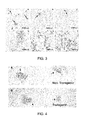
FIG. 4, panels A and B, show shows that ectopic expression of FGF2 does not suppress germinal center formation.
DETAILED DESCRIPTION
It is now shown that the humoral immune response is altered in FGF2 mutant mice. FGF2 deficient mice produce more antibody to a Type I independent antigen while FGF2 over-expressing mice show suppressed antibody production to the same pathogenic stimulus. In addition, germinal center formation is compromised in the absence of FGF2. Surprisingly, changes in both antibody production and germinal center formation are observed in mice lacking a single copy of FGF2, demonstrating that lymphocytes are particularly sensitive to FGF2 gene dosage. These studies provide the first evidence that FGF signaling is a crucial regulator of the humoral immune response and mature B cell function.
Materials and Methods
Mice.
FGF2−/− (homozygous gene knockout) mice were obtained from two academic sources. These mice display relatively benign defects in wound healing, blood pressure regulation and cortical neurogenesis and do not express detectable levels of FGF2 protein (Ortega et al., 1998. Proc Natl Acad Sci USA 95:5672; Zhou et al., 1998, Nature Medicine 4:20). Both sets of knockouts showed increases in antibody production and data in FIGS. 1 and 3 are for animals obtained from the University of Cincinnati. Heterozygous animals (mixture of 129SvEv:Black Swiss) were mated and heterozygous and null animals were compared to littermate controls. Adult mice of both sexes were used. FGF2 transgenic animals exhibit bone dysplasia and disruption of endothelial homeostasis (Fulgham et al., 1999, Endothelium 6:185; Coffin et al., 1995, Mol Biol Cell 6:1861). Animals (FVBN) heterozygous for the transgene were mated to wild type and adult animals of both sexes were compared to littermate controls. Animals were maintained in a pathogen-free facility, following institutional standards. Protocols adhered to IACUC guidelines.
Humoral Immune Response.
Mice were immunized intraperitoneally with 50 ug TNP-LPS (tri-nitrophenol lipopolysachharide) emulsified with complete Freund's adjuvant in PBS (200 ul final volume). Serum was harvested from retro-orbital eye bleeds. After coagulation, bleeds were centrifuged and sodium azide (0.01%) was added. ELISAs for TNP specific antisera were performed on plates coated with TNP-BSA (Biosearch) and primary antisera were bound overnight at 4° C. Goat anti-mouse IgG (all Ig isotypes) coupled to Alkaline Phosphatase was used as secondary antisera (Jackson). The genotype of the serum was unknown to the experimenter. Absorbance (405 nM) was measured in triplicate on a Molecular Devices spectrophotometer. Values were averaged and measurements were taken from absorbance in the middle of the dynamic range. For quantification of difference in antibody titer, serial dilutions were performed and the average value from the serum of all animals (minimum n=5, +/−s.e.m.) was plotted. Omission of either primary or secondary anitsera reduced signal to background levels.
Immunohistochemistry
Histochemistry was performed on 5 micrometer histologic sections made from formalin fixed, paraffin-embedded spleens. Sections were blocked in PBST (PBS with 0.1% Tween-20) containing 10% normal rabbit serum, stained with the lectin peanut agglutinin, then biotinlyated anti-peanut agglutinin (Vector Laboratories, Burlingame, Calif.), or rat anti-CD138 (syndecan-1) (Becton Dickinson) followed by biotinylated goat anti-rat IgG secondary antibody (Jackson Immunoresearch). Primary antibody was incubated either overnight at 4° C. or for one hour at room temperature. Removal of either primary or secondary antiserum abolished specific signal.
Germinal center number was scored by experimenters blind to the source of the sections. At least three serial sections were scored for each spleen. Results are based on three independent experiments from two or more animals per genotype. Data are presented from the final experiment which used the largest number of animals.
Proliferation of B Cells In Vitro.
Adult wild type mice (C57B16) were sacrificed and spleens were rapidly removed. After dissociation into single cell suspension and red blood cell lysis with NH4Cl, splenocytes were isolated by centrifugation over a Ficoll gradient. Subsequently, B lymphocytes were purified by one of two methods, complement mediated lysis or CD43 negative selection. For complement lysis, cells were incubated with anti-Thy 1 antibody (J1J), anti-L3T4 (GK 1.5), anti-Ly2 (TIB105, ATCC) and rabbit complement (Sigma) for two hours at 37°. CD43 negative selection was carried out using anti-CD43 (Serotec) and Miltenyi microbeads according to the manufacturer's instructions. Cells were cultured in RPMI 1640, 10% fetal calf serum for three days in the presence of anti-CD40 (mAb 1C10, generous gift of Hsiou-Chi Liou, Weill Medical College of Cornell University) and anti-IgM Fab′2 fragments (Jackson Immunoresearch). FGF1 (100 ng/ml) and Heparin (10 ug/ml) were added, and the number of cells was determined in triplicate compared to Heparin alone using a Coulter Counter (Coulter) or trypan blue exclusion with the same results.
Results
FGF2 Regulates the Humoral Immune Response
In the course of studies to evaluate the role of FGF signaling in multiple myeloma, we decided to investigate whether B cell function might be altered in FGF mutant mice. If FGF signaling affects mature B cell activity, one would predict that the humoral immune response would be affected by loss of function mutations in one of the FGF family members. To address this issue, we examined the humoral immune response in FGF2 deficient mice, one of the most widely expressed FGFs.
Immunization with a type I independent antigen, TNP-LPS, typically stimulates polyclonal B cell activation and proliferation. This antigen can elicit antibody production in T cell depleted animals, suggesting that the response can be largely independent of T cell help. The humoral response to TNP-LPS was enhanced in the absence of FGF2 (FIG. 1A). The magnitude of the peak response and the decay to baseline are potentiated by FGF2 deficiency. Three weeks after immunization, anti-TNP antibody titers are approximately three-fold higher than littermate controls (FIG. 1B). The size of this potentiation is greater than that seen with the inhibitory FC receptor, FCγRIIB, a gene intrinsic to B cells (Takai et al., 1996, Nature 379:346). Surprisingly, mice lacking a single copy of FGF2 produce more anti-TNP antibody (FIG. 1A, day 13 and day 22 time point). These results demonstrate that FGF2 negatively regulates the primary humoral immune response and is required for the normal inactivation of antibody secretion.
FIG. 1.
FGF2 deficient mice respond more strongly to a Type I Thymus Independent Antigen. Mice were immunized with 50 ug TNP-LPS and anti-TNP specific antibodies were measured by ELISA. In FIG. 1A, data points represent average absorbance from the serum of at least five animals. Asterisks indicate statistical differences at p<0.05 (student's t test). FIG. 1B shows the quantification of the difference in antibody titer of FGF2 deficient animals compared to littermate controls at day nineteen after immunization. Data points represent the mean absorbance+/−s.e.m. at the indicated dilutions for each genotype. Broken line between curves with corresponding vertical line delineates difference in antibody titer at the same absorbance.
To determine whether FGF2 is sufficient to regulate antibody production, we examined the humoral immune response in FGF2 transgenic mice. These animals express a human FGF2 gene driven by the ubiquitously active promoter, phosphoglycerate kinase (Coffin et al., 1995, Mol Biol Cell 6:1861). Different forms of FGF2 protein are produced from the FGF2 gene, including several high and low molecular weight isoforms. In FGF2 transgenic animals, there is a marked increase in the expression of the 18-Kd form of FGF2 in selected tissues, including spleen (Coffin et al., 1995, Mol Biol Cell 6:1861).
FIG. 2.
FGF2 transgenic mice respond more weakly to a Type I Thymus Independent Antigen. Mice were immunized with 50 ug TNP-LPS and anti-TNP specific antibodies were measured by ELISA using TNP-BSA coated plates. Asterisks indicate statistical differences at p<0.05 (student's t test). FIG. 2B shows the quantification of antibody titer of FGF2 transgenic animals compared to littermate controls at day twenty one after immunization. Data points represent the mean absorbance+/−s.e.m. at the indicated dilutions. Broken line between curves with corresponding vertical line delineates difference in antibody titer at the same absorbance.
It was found that antibody production in response to TNP-LPS is significantly diminished in FGF2 transgenic animals, as shown in FIG. 2A. Suppression of antibody production begins relatively late during the primary response, with statistically significant differences not observable until twenty one days after administration of immunogen. The reduction in antibody titers is slightly larger than the enhancement seen in the absence of FGF2 (four-fold). Therefore, FGF2 is both necessary and sufficient to control the humoral immune response. Taken together, these observations identify FGF2 as a soluble regulator of antibody production.
Once activated by antigen, B cells migrate to germinal centers, where high affinity, somatically mutated antibodies are generated. To determine whether germinal centers are affected by FGF2, we examined the number of splenic germinal centers formed in the FGF2 null mice. Lectin staining reveals that the number of germinal centers is substantially reduced approximately two weeks after immunization with TNP-LPS, with six-fold fewer germinal centers formed in null animals (FIG. 3, panels a-c; Table 2). Fewer germinal centers are also observed two days after immunization (Table 1). Unexpectedly, germinal centers are also reduced in heterozygous animals.
TABLE 1
Mouse +/+ +/− −/−
1 4 2 1
2 3 0 0
3 0 0 0
4 8 0 0
5 3 2
6 1 2
7 0 1
8 0 4
9 1 0
10  4 0
11  4 3
12  3 3
13  4
Mean 2.6 1.6 0.25
s.e.m. 0.7 0.4 0.25
N 12 13 4
TABLE 2
Mouse +/+ +/− −/−
1 5 3 11
2 13 0 3
3 11 0 3
4 9 0 0
5 14 0 0
6 8 0.5 0
7 4
Mean 10 0.6 1.7
s.e.m. 1.4 0.5 0.76
N 6 6 7
Tables 1 and 2.
Germinal center formation is dependent on FGF2 gene dosage. FGF2+/+, +/−, −/− mice were immunized i.p. with 50 ug TNP-LPS. Spleens were stained for expression of germinal centers with peanut agglutinin two days (Table 1) and approximately two weeks (Table 2) after immunization. Significantly fewer germinal centers were formed in FGF2 heterozygous (p<0.01) and null mice (p<0.01) sixteen days after immunization (Student's t test). Significantly fewer germinal centers were formed in FGF2 null mice (p<0.05) two days after immunization.
Gross morphologic features of the spleen are similar in the three genotypes. To determine whether plasma cell development is affected in FGF2 deficient animals, we examined the expression of syndecan-1, a cell surface heparin sulfate proteoglycan which is expressed on plasma cells. The number of syndecan positive plasma cells is not noticeably different, suggesting that FGF2 does not influence the adoption of the plasma cell fate in the spleen (FIG. 3, panels d-f). These results demonstrate that splenic germinal center formation is dependent on FGF2 gene dosage.
FIG. 3. FGF2 deficiency affects germinal centers but not syndecan expression. FGF2+/+, +/−, −/− mice were immunized i.p. with 50 ug TNP-LPS. A-C) Spleens were stained for expression of germinal centers with peanut agglutinin two weeks after immunization. D-F) Expression of syndecan-1 was determined by monoclonal antibody anti-CD138 (BD).
TABLE 3
Mouse Transgenic Wild-type
1 3.5 6
2 2 6
3 0 2
4 0.5 0
5 2 2
6 3 4
7 0 1
8 2 2
9 4 8.5
10  2
Mean 1.9 3.5
s.e.m. 0.4 0.9
n 10 9
Table 3.
Germinal center formation is not affected by ectopic expression of FGF2. FGF2 transgenic mice and littermate controls were immunized i.p. with 50 ug TNP-LPS. Spleens were stained for expression of germinal centers with peanut agglutinin fourteen days after immunization.
To determine whether germinal centers were affected by over-expression of FGF2, we performed the same experiment in FGF2 transgenic animals. We find that although there is a trend towards fewer germinal centers when FGF2 is over-expressed, the difference is not statistically significant (Table 3). These data show that over-expression of FGF2 is not sufficient to regulate germinal center formation two weeks after immunization with a Type 1 independent antigen.
FIG. 4.
Ectopic expression of FGF2 does not suppress germinal center formation. FGF2 transgenic and littermate controls were immunized i.p. with 50 ug TNP-LPS. A,B) Spleens were stained for expression of germinal centers with peanut agglutinin two weeks after immunization.
FGF2 is one of the more widely expressed members of the FGF family of ligands, with strong expression in multiple tissues. To determine whether FGF2 is expressed in the spleen we evaluated FGF2 levels by ELISA (R and D Systems). We find that FGF2 is found at 302+/−17 pg/ml (mean+/−s.d. n=4), demonstrating levels that are comparable to those found in other FGF2 responsive tissues. In addition, functional studies have demonstrated that both FGF-1 and FGF2 are present in the spleen in forms which can stimulate liver cell proliferation (Suzuki et al., 1992, Biochem Biophys Res Commun 186:1192).
To determine whether FGF can directly control B cell activation, we explored whether addition of exogenous FGF would affect B cell proliferation in vitro. B cells were purified from spleen and CD40 and BCR signaling were simultaneously activated using stimulating antibodies. Inducing these systems transmits powerful growth and survival signals, leading to rapid proliferation. To investigate whether FGF signaling might affect this response, we incubated the cells in the presence of FGF-1. We used FGF-1 instead of FGF2 because it stimulates the widest range of FGF receptors (8). Under these conditions, B cell number is inhibited by FGF stimulation (Table 3), suggesting that it can directly inhibit antigen stimulated B cells.
TABLE 4
Experiment % Decrease
1 27
2 25
3 10
4 15
5 16
6 25
X 19.7 +/− 2.8
Table 4.
FGF signaling inhibits splenic B cell proliferation. Spleens from adult wild-type mice were dissected and highly enriched populations of B cells were purified. Cells were cultured in serum-containing medium for 3 days in the presence of a CD40 activating monoclonal antibody (1C10) and anti-mouse IgM Fab′2 fragments (Jackson). The values represent the percent decrease in total cell number observed with addition of 100 ng/ml FGF1 (determined in triplicate) as compared to heparin (10 ug/ml) alone. x=mean+/−s.e.m. One sample t test, p<0.01.
Discussion
Using gain and loss-of-function mouse models, it was shown that FGF2 controls the humoral immune response. These observations constitute the first indication that any member of this large family of pleiotropic signaling factors affects the humoral immune response.
Based on its widespread expression and its robust effects on a diverse array of cell types, FGF2 is postulated to control multiple biological processes. However, studies with mice lacking this gene have challenged this belief, implicating other FGF family members or suggesting that FGF signaling is not essential (Ortega et al., 1998. Proc Natl Acad Sci USA 95:5672; Zhou et al., 1998, Nature Medicine 4:201; Dono et al., 1998, Embo J 17:4213). In light of these limited phenotypes, it was not expected that mice lacking a single copy of FGF2 would show abnormalities in immune function. Thus, in contrast to other systems, lymphoid tissue appears to be especially sensitive to FGF2 gene dosage. Since FGF family members are widely expressed, these results raise the possibility that further investigation will uncover additional evidence for FGF-dependent effects on lymphocyte function.
Given the ability of FGF ligands to bind more than one receptor family member, it is surprising that compensation for FGF2 deficiency by one of the twenty-two other FGFs was not observed. In this regard, FGF-1 constitutes a plausible candidate because it structurally resembles FGF2 and also is expressed in the spleen (Suzuki et al., 1992, Biochem Biophys Res Commun 186:1192). On the other hand, studies with FGF-1/2 double knock out mice suggest that the mild wound healing and neural phenotypes in FGF2 null mice are not a result of FGF-1 substituting for FGF2 (Miller et al., 2000, Mol Cell Biol 20:2260). The type I independent antigen lipopolysaccharide is a key pathogenic substance in the cell wall of gram negative bacteria. The repeating epitope in this molecule leads to massive engagement of receptors on the surface of B cells, including the BCR, TLR2 and TLR4 (Yang et al., 1998, Nature 395:284; Takeuchi et al., 1999, Immunity 11:443). B cell evolution has developed rapid and vigorous pre-existing defenses against such frequent threats and consequently, antibody secretion in response to this stimulus is robust. The greater response in the absence of FGF2 demonstrates that FGF2 negatively regulates the primary humoral immune response. The magnitude of the enhanced response is greater than the enhancement seen with FC receptor, FC□RIIB, whose deletion shows no effect on the response to LPS at three weeks post immunization (Takai et al., 1996, Nature 379:346). It is believed that this represents the first example of enhanced antibody production in response to LPS due to genetic deficiency.
Animals over-expressing FGF2 have a suppressed humoral immune response to LPS, demonstrating that the gain of function phenotype is the opposite of the loss of function phenotype. It is concluded that FGF2 is both necessary and sufficient to regulate antibody production.
While not being limited by theory, it is not presently clear which step in the humoral immune response is inhibited by FGF2 signaling. Although the possibility that differences in plasma cell generation take place in other lymphoid tissues cannot be excluded, inhibition occurs without a substantial difference in the number of syndecan positive cells in the spleen (FIG. 3, panels D-F). Hence, FGF2 may regulate a step subsequent to the expression of syndecan-1, such as plasmablast migration, full terminal differentiation, or metabolic function of antibody secreting cells in the bone marrow. Consistent with this latter idea, FGF2 is strongly expressed by multiple cell types in the bone marrow (Brunner et al., 1993, Blood 81:631; Chou et al., 2003, Leuk Res 27:499.).
FGF2 may control antibody production either by directly signaling to B cells or indirectly by affecting cells which regulate plasma cell activity. The direct model is consistent with our data showing decreased proliferation in response to FGF signaling of primary mature B lymphocytes (Table 3). While the reduction in cell number is modest, it should be borne in mind that few substances can overcome the strong growth and survival signals turned on by simultaneous CD40 and BCR engagement. In agreement with a direct mode of action, a previous study reported that FGF receptors exist on normal human peripheral blood B cells (Genot, et al., 1989, Cell Immunol 122:424). However, the possibility that other cell types could mediate the observed effects cannot presently be excluded.
A negative correlation between antibody production and germinal center number was found. At first glance, this observation appears contradictory since one might expect that a reduction in germinal centers would decrease antibody production. However, numerous examples have demonstrated that germinal center number can be uncoupled from the humoral response. TNF receptor null animals lack germinal centers but produce substantial antibody titers in response to vesicular stomatitis virus (Karrer et al., 2000, J Immunol 164:768). Similarly, TNF-α null animals display dramatic alterations in splenic morphology but their antibody production to LPS is unaffected (Pasparakis et al., 1996, J Exp Med 184:1397).
Thus, the work described herein demonstrates that FGF2 plays two distinct and complementary roles in the humoral immune response. FGF2 facilitates germinal center formation, thereby contributing to the generation of activated B cells which defend against pathogenic stimuli. On the other hand, FGF2 reduces plasma cell activity and in so doing provides a limit on antibody production. Since FGF2 exerts opposing forces at different times during the B cell response, its activities in the immune system are certainly complex. Such complexity is consistent with observations in other tissues, where FGF signaling can stimulate radically different effects depending on its temporal and spatial locus of action.
Embodiments Relating to Inhibition of FGF2 Activity in a Mammal
In multiple disease states, vaccination provides inadequate protection and low percentages of seroconversion are observed (Cohen D et al., Diagnosis and management of the antiphospholipid syndrome. BMJ. 2010 May 14; 340:c2541). Non-limiting examples of vaccines for which the invention may be employed to increase humoral immune response include, Malaria vaccine (M. Esen et al. Vaccine. 2009 Nov. 16; 27(49):6862-8. Safety and immunogenicity of GMZ2—a MSP3-GLURP fusion protein malaria vaccine candidate); HIV vaccine (Hoxie J A. Annu Rev Med. 2010; 61:135-52. Toward an antibody-based HIV-1 vaccine.); Influenza vaccine (Nguyen M L et al Infect Immun. 2009 November; 77(11):4714-23. The major neutralizing antibody responses to recombinant anthrax lethal and edema factors are directed to non-cross-reactive epitopes); Influenza Vaccine in geriatric patients (Frasca D, Diaz, A, Romero, M et al. Vaccine. 2010 Oct. 22. Intrinsic defects in B cell response to seasonal influenza vaccination in elderly humans.); and Anthrax vaccine (Nguyen M L et al Infect Immun. 2009 November; 77(11):4714-23. The major neutralizing antibody responses to recombinant anthrax lethal and edema factors are directed to non-cross-reactive epitopes.).
The invention may, for example, be used to increase antibody production and/or humoral immunity in patients, such as human patients, suffering from immunodeficiencies including but not limited to: Common variable immunodeficiency (Rezaei N et al Clin Vaccine Immunol. 2008 April; 15(4):607-11 Serum bactericidal antibody responses to meningococcal polysaccharide vaccination as a basis for clinical classification of common variable immunodeficiency.); primary immunodeficiency disorder (PIDD), Ig deficiency, IgG deficiency; and HIV disease (Acquired Immune Deficiency Syndrome).
One embodiment of the invention provides a method for increasing the humoral immune response to vaccination with an immunogen, for example, an antigen or a live vaccine, in a mammal, that includes: in conjunction with the vaccination of a mammal to the immunogen other than FGF2, inhibiting the activity of FGF2 in the mammal, thereby increasing the humoral immune response to the antigen. In one variation the immunogen is other than a fibroblast growth factor and other than a fibroblast growth factor receptor. The mammal may be a human, such as a geriatric human. The mammal, which may be human, may have an immune deficiency, such as but not limited to Common variable immunodeficiency; primary immunodeficiency disorder (PIDD), an immunoglobulin deficiency such as IgG deficiency, and HIV disease.
Another embodiment of the invention provides a method for treating an immune deficiency in a mammal, such as a human, that includes: increasing the production of endogenous antibodies in the mammal by inhibiting the activity of FGF2 in the mammal. In one variation, the mammal does not have cancer. The immune deficiency may be, for example, but is not limited to: Common variable immunodeficiency; primary immunodeficiency disorder (PIDD), an immunoglobulin deficiency such as IgG deficiency, and HIV disease. Non-human mammals also suffer from immunodeficiencies and may be treated according to the invention. For example, the method may be used to treat immunodeficiency associated with feline immunodeficiency virus (FIV) in a cat, such as a domesticated cat.
A further embodiment of the invention provides a method for treating a microbial infection in a mammal, such as a human, that includes: administering an FGF2 antagonist to a mammal in need of treatment for a microbial infection, wherein the FGF2 antagonist is administered in an amount effective to increase antibody production in the mammal. The method may further include the step of: administering an antibiotic or anti-viral agent to the mammal which is active against the microbial infection. The antibiotic or anti-viral agent is administered such that the effect of the antibiotic or anti-viral agent and that of the FGF2 antagonist are temporally overlapping in the mammal. The microbial infection may, for example, be a bacterial infection, a viral infection or a eukaryotic parasite infection. The method may further include the step of determining that the mammal has a microbial infection prior to administering the FGF2 antagonist.
Another embodiment of the invention provides a method for increasing in vivo antibody production in a mammal, such as a human, that does not have a cancer, which includes the step of inhibiting the activity of FGF2 in the mammal. In one variation, the mammal is a geriatric human or non-human mammal, such as a geriatric domesticated dog or cat.
A related embodiment provides a method for enhancing the production of antisera or polyclonal antibodies generally against a desired immunogen in a non-human mammal that includes the steps of: inhibiting FGF2 activity in the non-human mammal according to any of methods and ways described herein and immunizing the non-human mammal with an immunogen that is not a fibroblast growth factor or a fibroblast growth factor receptor, whereby the production of antibodies against the immunogen in the mammal is enhanced, increased and/or accelerated versus a comparable immunization without the inhibition of FGF2 activity. The method may further include the step of retrieving the polyclonal sera from the non-human mammal and optionally the step of isolating. The immunizing step may, for example, include more than one temporally separated immunization with the immunogen and may, for example, be aided by inclusion of an immunization adjuvant. The methods for production of antisera and polyclonal antibodies are well known and long-established in the art. See, for example, U.S. Pat. No. 5,440,021.
The increase in antibody production in response to inhibition of FGF2 activity in a mammal is a general characteristic of the invention which is not limited to the type of FGF2 inhibitor that is administered to the mammal to inhibit the activity of FGF2. Preferred types of inhibitors of FGF2 activity include antibodies and binding fragments thereof, both monoclonal and polyclonal, which bind to FGF2 and block its interaction with FGF binding receptors and antibodies, both monoclonal and polyclonal, which bind to an FGF receptor such as FGFR1, FGFR2 and FGFR3 and block binding of the ligand (FGF2) to the receptor. For example, a single chain, monoclonal scFv antibody that neutralizes FGF2 may be used such as that described in Tao et al, Selection and characterization of a human neutralizing antibody to human fibroblast growth factor-2, Biochem Biophys Res Commun. 2010 Apr. 9; 394(3):767-73. Epub 2010 Mar. 17 or one obtained by the method described therein. Antibodies blocking FGFR1 such as those those described in Sun et al., Am J Physiol Endocrinol Metab 292:964-976, 2007, or obtained according to the method of this article may be used. Gorbenk et al, Hybridoma, Volume 28, Number 4, 2009 also describes the production of anti-FGFR1 antibodies and their production. Monoclonal antibodies against FGFR3 and their production are described in Qing et al., J. Clin. Invest. 119:1216-1229 (2009) and in Gorbenko et al, Hybridoma, Volume 28, Number 4, 2009, 295-300.
Antibodies contain one or more antigen binding sites that specifically binds with an antigen. Antibodies include, but are not limited to polyclonal, monoclonal, chimeric, and humanized antibodies. Immunologically active portions include monovalent and divalent fragments such as Fv, single chain Fv (scFv), single variable domain (sVD), Fab, Fab′ and F(ab′)2 fragments. Immunologically active portions can be incorporated into multivalent from such as diabodies, triabodies, and the like. Antibodies further include antigen binding fragments displayed on phage, and antibody conjugates.
An “isolated antibody” is an antibody that (1) has been partially, substantially, or fully purified from a mixture of components; (2) has been identified and separated and/or recovered from a component of its natural environment; (3) is monoclonal; (4) is free of other proteins from the same species; (5) is expressed by a cell from a different species; or (6) does not occur in nature. Isolated antibodies may, for example, be used as inhibitors of FGF2 activity according to the invention. Examples of isolated antibodies include an anti-FGF2 antibody that has been affinity purified using FGF2, an anti-FGF2 antibody that has been made by a hybridoma or other cell line in vitro, a human anti-FGF2 antibody isolated from a library such as a phage library, and a human anti-FGF2 antibody derived from a transgenic mouse.
In general, naturally occurring antibody molecules are composed of two identical heavy chains and two light chains. Each light chain is usually covalently linked to a heavy chain by an interchain disulfide bond, and the two heavy chains are further linked to one another by multiple disulfide bonds at the hinge region. The individual chains fold into domains having similar sizes (about 110-125 amino acids) and structures, but different functions. The light chain comprises one variable domain (VL) and one constant domain (CL). The heavy chain comprises one variable domain (VH) and, depending on the class or isotype of antibody, three or four constant domains (C H1, C H2, CH3 and CH4). In mice and humans, the isotypes are IgA, IgD, IgE, IgG, and IgM, with IgA and IgG further subdivided into subclasses or subtypes. The portion of an antibody consisting of VL and VH domains is designated “Fv” and constitutes the antigen-binding site. A single chain Fv (scFv) is an engineered protein containing a VL domain and a VH domain on one polypeptide chain, wherein the N terminus of one domain and the C terminus of the other domain are joined by a flexible linker. “Fab” refers to the portion of the antibody consisting of VL-CL (i.e., a light chain) and VH-CH1 (also designated “Fd”).
Antibodies include without limitation single variable domains (sVDs) and antigen binding proteins that comprise sVDs. sVD binding sites can be obtained from antigen specific Fv regions (which comprise both VH and VL domains). Often, it can be shown that the binding affinity and specificity of an Fv region is contributed primarily by one of the variable domains. Alternatively, the scFv can be obtained directly. Direct sources of sVDs include mammals (e.g., camelids) that naturally express antibodies containing only VH domain. Further, phage display libraries can be constructed to express only a single variable domain. For example, a human domain antibody phage display library is commercially available from Domantis (Cambridge, UK).
The antibody variable domains show considerable amino acid sequence variablity from one antibody to the next, particularly at the location of the antigen binding site. Three regions, called “complementarity-determining regions” (CDRs) are found in each of VL and VH. The CDRs of an antibody are often referred to as “hypervariable regions.”
“Fc” is the designation for the portion of an antibody which comprises paired heavy chain constant domains. In an IgG1 antibody, for example, the Fc comprises C H2 and CH3 domains. The Fc of an IgA or an IgM antibody further comprises a CH4 domain. The Fc is associated with Fc receptor binding, activation of complement-mediated cytotoxicity and antibody-dependent cellular-cytotoxicity. For natural antibodies such as IgA and IgM, which are complexes of multiple IgG like proteins, complex formation requires Fc constant domains.
Finally, the “hinge” region separates the Fab and Fc portions of the antibody, providing for mobility of Fabs relative to each other and relative to Fc, as well as including multiple disulfide bonds for covalent linkage of the two heavy chains. Thus, antibodies of the invention include, but are not limited to, naturally occurring antibodies, bivalent fragments such as (Fab′)2, monovalent fragments such as Fab, single chain antibodies, single chain Fv (scFv), single domain antibodies, multivalent single chain antibodies, diabodies, triabodies, and the like that bind specifically with antigens.
Antibody fragments also include polypeptides with amino acid sequences substantially similar to the amino acid sequence of the variable or hypervariable regions of the antibodies of the invention. Substantially the same amino acid sequence is defined herein as a sequence with at least 70%, at least about 80%, at least about 90%, at least about 95% or at least about 99% homology or identity to a compared amino acid sequence, as determined by the FASTA search method in accordance with Pearson and Lipman, Proc. Natl. Acad. Sci. USA 85:2444-2448 (1988).
Antibodies that may be employed as inhibitors according to the invention also include “chimeric” antibodies and binding fragments thereof. Such antibodies generally comprise variable domains of one antibody and constant domains of a different antibody. Typically, to minimize host immune responses against the antibody and to enhance host responses against the antibody target by retaining antibody effector functions, the constant domains of a chimeric antibody are taken from the same species to which the chimeric antibody will be administered.
Antibodies that may be employed as inhibitors according to the invention also include “humanized” antibodies. Humanized variable domains are constructed in which amino acid sequences which comprise one or more complementarity determining regions (CDRs) of non-human origin are grafted to human framework regions (FRs). For examples, see: Jones, P. T. et al., 1996, Nature 321, 522-25; Riechman, L. et al., 1988, Nature 332, 323-27; and U.S. Pat. No. 5,530,101 to Queen et al. A humanized construct is particularly valuable for elimination of adverse immunogenic characteristics, for example, where an antigen binding domain from a non-human source is desired to be used for treatment in a human. Variable domains have a high degree of structural homology, allowing easy identification of amino acid residues within variable domains which corresponding to CDRs and FRs. See, e.g., Kabat, E. A., et al., 1991, Sequences of Proteins of Immunological Interest. 5th ed. National Center for Biotechnology Information, National Institutes of Health, Bethesda, Md. Thus, amino acids which are likely to participate directly in antigen binding are easily identified. In addition, methods have been developed to preserve or to enhance affinity for antigen of humanized binding domains comprising grafted CDRs. One way is to include in the recipient variable domain the foreign framework residues which influence the conformation of the CDR regions. A second way is to graft the foreign CDRs onto human variable domains with the closest homology to the foreign variable region. Queen, C. et al., 1989, Proc. Natl. Acad. Sci. USA 86, 10029-33. CDRs are most easily grafted onto different FRs by first amplifying individual FR sequences using overlapping primers which include desired CDR sequences, and joining the resulting gene segments in subsequent amplification reactions. Grafting of a CDR onto a different variable domain can further involve the substitution of amino acid residues which are adjacent to the CDR in the amino acid sequence or packed against the CDR in the folded variable domain structure which affect the conformation of the CDR. Humanized variable domains of the invention therefore include human domains which comprise one or more non-human CDRs as well as such domains in which additional substitutions or replacements have been made to preserve or enhance binding characteristics.
Antibodies with variable domains that have been made less immunogenic by replacing surface-exposed residues so as to make the antibody appear as self to the immune system may also be employed as inhibitors (Padlan, E. A., 1991, Mol. Immunol. 28, 489-98). Antibodies have been modified by this process with no loss of affinity (Roguska et al., 1994, Proc. Natl. Acad. Sci. USA 91, 969-973). Because the internal packing of amino acid residues in the vicinity of the antigen binding site remains unchanged, affinity is preserved. Substitution of surface-exposed residues according to the invention for the purpose of reduced immunogenicity does not mean substitution of CDR residues or adjacent residues which influence binding characteristics.
It is often preferable to employ variable domains that are essentially human as when the recipient of the antibody is human. Human antibodies comprise human VH and VL framework regions (FWs) as well as human complementary determining regions (CDRs). Preferably, the entire VH and VL variable domains are human or derived from human sequences. The antibodies can be obtained directly from human cells, for example by creating human hybridomas.
Alternatively, human antibodies can be obtained from transgenic animals into which unrearranged human Ig gene segments have been introduced and in which the endogenous mouse Ig genes have been inactivated (reviewed in Briiggemann and Taussig, 1997, Curr. Opin. Biotechnol. 8, 455-58). Preferred transgenic animals contain very large contiguous Ig gene fragments that are over 1 Mb in size (Mendez et al., 1997, Nature Genet. 15, 146-56) but human Mabs of moderate affinity can be raised from transgenic animals containing smaller gene loci (See, e.g., Wagner et al., 1994, Eur. J. Immunol. 42, 2672-81; Green et al., 1994, Nature Genet. 7, 13-21).
Human antibodies can also be obtained from libraries of antibody VH and/or VL domains. For example, a variable domain library can be obtained from human genomic sequences, or from peripheral blood lymphocyte expressing productively rearranged variable region genes. Furthermore, the human gene library can be synthetic. In one embodiment, variable domain libraries can be created which comprise human framework regions with one or more CDRs that are synthesized to include random or partial random sequences. For example, a human VH variable domain library can be created in which members are encoded by a human VH gene segment and a synthetic sequence for the CDR3H region (i.e., a synthetic DH-JH gene segment). Likewise, a human VL variable domain may be encoded by a human VL gene segment and a synthetic sequence for the CDR3L region (i.e., a synthetic JL gene segment). In another embodiment, the human frameworks may be synthetic in that they have a consensus sequence derived from known human antibody sequences or subgroups of human sequences. In another alternative, one or more CDRs is obtained by amplification from human lymphocytes expressing rearranged variable domains and then recombined into a particular human framework.
In order to screen libraries of variable domains, it is common to employ phage display libraries wherein combinations of human heavy and light chain variable domains are displayed on the surface of filamentous phage (see, e.g., McCafferty et al., 1990, Nature 348, 552-54; Aujame et al., 1997, Human Antibodies 8, 155-68). Combinations of variable domains are typically displayed on filamentous phage in the form of Fabs or scFvs. The library is screened for phage bearing combinations of variable domains having desired antigen binding characteristics. Preferred single domain and variable domain combinations display high affinity for a selected antigen and little cross-reactivity to other related antigens. By screening very large repertoires of antibody fragments, (see e.g., Griffiths et al., 1994, EMBO J. 13, 3245-60) a good diversity of high affinity binding domains are isolated, with many expected to have sub-nanomolar affinities for the desired antigen.
In a physiological immune response, mutation and selection of expressed antibody genes leads to the production of antibodies having high affinity for their target antigen. The VH and VL domains incorporated into antibodies of the invention can similarly be subject to in vitro or in vivo mutation and screening procedures in order to modify affinity and/or specificity. Thus, binding domains of the invention include those for which binding characteristics have been improved by mutating CDRs and/or FW regions by direct mutation, methods of affinity maturation, or chain shuffling. It is understood that amino acid residues that are primary determinants of binding of single domain antibodies can be within Kabat defined CDRs, but may include other residues as well. For sVDs, residues important for antigen binding can also potentially include amino acids that would otherwise be located at the interface of a VH-VL heterodimer. Typically, phage display is used to screen such mutants to identify those having the desired binding characteristics (see, e.g., Yang et al., J. Mol. Biol., 254: 392-403 (1995)). Mutations can be made in a variety of ways. One way is to randomize individual residues or combinations of residues so that in a population of otherwise identical sequences, all twenty amino acids or a subset thereof are found at particular positions. Alternatively, mutations may be induced over a range of CDR residues by error prone PCR methods (see, e.g., Hawkins et al., J. Mol. Biol., 226: 889-896 (1992)). For example, phage display vectors containing heavy and light chain variable region genes may be propagated in mutator strains of E. coli (see, e.g., Low et al., J. Mol. Biol., 250: 359-368 (1996)). These methods of mutagenesis are illustrative of the many methods known to one of skill in the art.
Inhibitors that may be used according to the invention also include antigen binding proteins engineered from non-immunoglobulin scaffolds. For example, affibodies, which are derived from an immunoglobulin-binding domain of S. aureus protein A, possess no disulfide bonds and display reversible folding. Another example is fibronectin, which has an antibody-like structure and displays CDR-like loops. In contrast to antibodies, the fibronectin domain structure does not rely on disulfide bonds, yet displays high thermodynamic stability. Binding sites can be engineered into such scaffolds by, for example, diversifying codons at specified positions and screening for binding to a desired antigen. Codons can be randomized in loops, flat surfaces, cavities, or combinations of such locations. Further, peptide sequences can be inserted, usually in loops. Target-binding variants of resulting libraries can be isolated using selection of screening techniques that are well known in the art, not limited to phage display, ribosome display, bacteria or yeast surface display, and the like. For antigen-binding proteins intended for therapy, various strategies are available for minimizing potential immunogenicity. Human scaffolds can be employed, and immunogenicity can be minimized, for example, by PEGylation or T-cell epitope engineering (i.e., minimizing T-cell reactive sequences).
Antigen-binding proteins from non-immunoglobulin scaffolds often can be produced more economically than immunoglobulin-type proteins. For example, the absence of disulfide bonds or free cysteines allows for expression of functional molecules in the reducing environment of the bacterial cytoplasm, which usually gives higher yields than periplasmic expression, and is more convenient than refolding in vitro. Binz, H. K. et al. (Nat. Biotech. 23:1257-68, 2005) discloses a variety of such antigen-specific binding proteins and techniques for their development.
The identification or selection of antibodies or other molecules that inhibit binding of FGF2 or other FGFs to their receptors may be performed according to routine ligand-receptor binding assays, comparing binding in presence and absence of test agent, since the full sequences of FGF2 and its receptors are known in various mammals such as human. See, for example, U.S. Pat. No. 5,440,021 for ligand-receptor binding assays.
Another preferred type of inhibitor of FGF2 activity is a soluble FGF2-binding receptor or soluble portion of an FGF-binding receptor, such as a soluble form of FGFR1, FGFR2 and FGFR3. The soluble receptor sequence may, for example match the species in which it will be administered, i.e., a human receptor sequence may be used for a human recipient and so on. For example, FP-1039 is a soluble fusion protein consisting of the extracellular domains of human FGFR1 linked to the Fc region of human Immunoglobulin G1 (IgG1), which may be used as an FGF2 inhibitor/antagonist according to the invention (Five Prime Therapeutics, Inc., San Francisco, Calif.; Keer et al, ASCO 2010, Abstract no. TPS260).
FGF2 activity may also be inhibited according to the invention by vaccinating the subject mammal against FGF2 itself or against FGFR1, FGFR2 and/or FGFR3. For example, a peptide vaccine targeting the heparin-binding portion of FGF2 can be used to generate a specific anti-FGF2 antibody response in a mammal according the method of Plum et. al., Generation of a specific immunological response to FGF2 does not affect wound healing or reproduction, Immunopharmacol Immunotoxicol. 2004 February; 26(1):29-41.
For embodiments in which a soluble polypeptide, such as an antibody or soluble receptor, is used to inhibit FGF2 activity, a composition for intravenous administration, for example, to a human, may include 0.1 to 20 mg, such as 0.1 to 10 mg, of the polypeptide, and this may be a daily dose. More generally, dosages from 0.1 mg to about 100 mg per subject per day for one or more days may be used. Methods for preparing administrable compositions are well known to those skilled in the art and are described in more detail in such publications as Remington's Pharmaceutical Science, 19th ed., Mack Publishing Company, Easton, Pa. (1995). Polypeptides for administration to a subject may, for example, be provided in lyophilized form and rehydrated with sterile water before administration. The solution of polypeptide may then be added to an infusion bag containing 0.9% sodium chloride, USP, and, for example administered at a dosage of from 0.5 to 15 mg/kg body weight. Alternatively, for example, the polypeptide can be administered as a bolus injection, for example, at a dosage of 0.5 to 30 mg/kg body weight.
Still other suitable types of FGF2 activity inhibitors include, for example, antisense oligonucleotides targeting FGF2 or one or more of FGFR1, FGFR2 and FGFR3. Still further suitable inhibitors are small molecule inhibitors, for example cardiac glycosides or aglycone derivatives as described in U.S. Pat. No. 6,071,885 and FGF activity modulating oligosaccharides as described in U.S. Pat. No. 5,891,655. TKI258 (also known as CHIR-258) described in Sarker et al., Clin Cancer Res, 2008; 14(7) 2075-81, is another suitable small molecule FGF receptor inhibitor. Brivanib, a FGFR1 Kinase inhibitor described in Bhide et al, Mol Cancer Ther; 9(2) February 2010, 369-78, is still another suitable small molecule inhibitor.
Embodiments Relating to Increasing FGF2 Activity in a Mammal
The invention also provides embodiments in which antibody production in vivo is purposefully reduced in a mammal, such as a human, by increasing FGF2 activity in the mammal, for example, by administration of FGF2 to the mammal or administration of an agonist of FGF2 or an agonist of an FGF2 receptor, such as FGFR1, FGFR2 or FGFR3 to the mammal, in an amount effective to decrease antibody production in the mammal. Where FGF2 is administered to a mammal recipient, the peptide sequence may, for example at least substantially or identically match the species in which it will be administered, i.e., a human receptor sequence may be used for a human recipient and so on.
This aspect of the invention finds practical application is the suppression of antibody production in acutely toxic states. In many cases, response to invading pathogens can lead to pathological autoimmune effects, with lymphocyte activity spiraling out of control. In situations like this, administration of FGF2 attenuates the uncontrolled secretion of antibody.
Similarly, multiple human pathologies result from secretion of autoimmune antibodies. Administration of FGF2 and FGF ligands will serve to attenuate the production of these antibodies and thus ameliorate the autoimmune disease. For example, autoimmune antibodies are observed in both systemic lupus erythematosus (Cohen D et al., Diagnosis and management of the antiphospholipid syndrome. BMJ. 2010 May 14; 340:c2541) and diverse arthritic disease (Calero I, et al., B cell therapies for rheumatoid arthritis: beyond B cell depletion. Rheum Dis Clin North Am 2010 May; 36(2):325-43), including rheumatoid arthritis, psoriatic arthritis, ankylosing spondylitis and juvenile idiopathic arthritis. In addition, increasing FGF2 activity in a mammal may be used to decrease or maintain a decreased level of antibody production in organ transplant patients, such as human organ transplant patients in order to decrease negative immune responses to and increase tolerance to the transplanted organ in the patient.
Accordingly, one embodiment of the invention provides a method for decreasing antibody production, such as pathological antibody production, in a mammal such as a human in need thereof by administering to the mammal FGF2 or an FGF2 agonist or an agonist of a receptor that binds FGF2 such as FGFR1, FGFR2 and FGFR3 in an amount effect to decrease antibody production in the mammal. In one variation, the mammal may have and be in need of treatment for systemic lupus erythematosus and diverse arthritic disease, including rheumatoid arthritis, psoriatic arthritis, ankylosing spondylitis and juvenile idiopathic arthritis and the method decreases the production of autoimmune antibodies in these mammals thereby treating the condition. In another variation, the mammal is an organ transplant patient such as a human organ transplant patient and the method reduces antibody response against the transplanted organ.
The sequences of fibroblast growth factors and their receptors are well characterized in humans and non-human mammals. For example, the following sequences are known and form part of this disclosure: Human FGF2 (NCBI Reference Sequence NM002006.4; SEQ ID NO:1 peptide, SEQ ID NO:2 nucleotide), Human FGFR1 (GenBank Accession No. M34185.1; SEQ ID NO:3 peptide, SEQ ID NO:4 nucleotide), Human FGFR2 (NCBI Reference Sequence NM000141.4; SEQ ID NO:5 peptide, SEQ ID NO:6 nucleotide), Human FGFR3 (NCBI Reference Sequence NM000142.4; SEQ ID NO:7 peptide, SEQ ID NO:8 nucleotide), Human FGFR4 (GenBank Accession No. AF202063.1; SEQ ID NO:9 peptide, SEQ ID NO:10 nucleotide), Bos taurus FGF2 (NCBI Reference Sequence NM174056.3; SEQ ID NO:11 peptide, SEQ ID NO:12 nucleotide), Bos taurus FGFR1 (Genbank Accession No. NM001110207.1; SEQ ID NO:13 peptide, SEQ ID NO:14 nucleotide), Bos taurus FGFR2 (NCBI Reference Sequence XM002698546.1; SEQ ID NO:15 peptide, SEQ ID NO:16 nucleotide); Bos taurus FGFR3 (NCBI Reference Sequence NM174318.3; SEQ ID NO:17 peptide, SEQ ID NO:18 nucleotide), Bos taurus FGFR4 (NCBI Reference Sequence XM002689008.1; SEQ ID NO:19 peptide, SEQ ID NO:20 nucleotide), Sus scrofa FGF2 (NCBI Reference Sequence XM003129213.1; SEQ ID NO:21 peptide, SEQ ID NO:22 nucleotide), Sus scrofa FGFR1 (NCBI Reference Sequence: XM001928678.2; SEQ ID NO:23 peptide, SEQ ID NO:24 nucleotide), Sus scrofa FGFR2 (NCBI Reference Sequence NM 001099924.1; SEQ ID NO:25 peptide, SEQ ID NO:26 nucleotide), Sus scrofa FGFR3 (GenBank Accession No. BV726808.1; SEQ ID NO:27 cds nucleotide), Sus scrofa FGFR4 (NCBI Reference Sequence XM003123682.1; SEQ ID NO:28 peptide, SEQ ID NO:29 nucleotide), Macaca mulatta FGF2 (NCBI Reference Sequence XM001099284.2; SEQ ID NO:30 peptide, SEQ ID NO:31 nucleotide), Macaca fascicularis FGFR1 (GenBank Accession No. AB220417.1; SEQ ID NO:32 peptide, SEQ ID NO:33 nucleotide), Macaca mulatta FGFR2 partial (GenBank Accession No. AY083548.1; SEQ ID NO:34 peptide, SEQ ID NO:35 nucleotide), Macaca mulatta FGFR3 (NCBI Reference Sequence XM002802167.1; SEQ ID NO:36 peptide, SEQ ID NO:37 nucleotide), Macata mulatta FGFR4 (NCBI Reference Sequence XM001087243.2; SEQ ID NO:38 peptide, SEQ ID NO:39 nucleotide), Mus musculus FGF2 (NCBI Reference Sequence NM008006.2; SEQ ID NO:40 peptide, SEQ ID NO:41 nucleotide), Mus musculus FGFR1 (NCBI Reference Sequence NM 010206.2; SEQ ID NO:42 peptide, SEQ ID NO:43 nucleotide), Mus musculus FGFR2 (NCBI Reference Sequence NM010207.2; SEQ ID NO:44 peptide, SEQ ID NO:45 nucleotide), Mus musculus FGFR3 (NCBI Reference Sequence NM008010.4; SEQ ID NO:46 peptide, SEQ ID NO:47 nucleotide), and Mus musculus FGFR4 (NCBI Reference Sequence NM008011.2; SEQ ID NO:48 peptide, SEQ ID NO:49 nucleotide).
Without limitation, the invention also provides methods for increasing endogenous antibody production in mammals such as humans by administering any of the following enumerated compounds or pharmacologically acceptable salts thereof:
1. BIBF1120 (Vargatef) Boehringer Ingelheim,
chemical name: Methyl (3Z)-3-[({4-[N-methyl-2-(4-methylpiperazin-1-yl)acetamido]phenyl}amino)(phenyl)methylidene]-2-oxo-2,3-dihydro-1H-indole-6-carboxylate.
Figure US09226960-20160105-C00001

F. Hilberg et al Cancer Res. 2008 Jun. 15; 68(12):4774-82. doi: 10.1158/0008-5472.CAN-07-6307. BIBF 1120: triple angiokinase inhibitor with sustained receptor blockade and good antitumor efficacy.
2. TKI258 (Dovitinib) Novartis,
chemical name: 4-amino-5-fluoro-3-[5-(4-methylpiperazin-1-yl)-1H-benzo[d]imidazol-2-yl]-3,4-dihydronaphthalen-2(1H)-one
Figure US09226960-20160105-C00002

Trudel S, et al Blood. 2005 Apr. 1; 105(7):2941-8. Epub 2004 Dec. 14. CHIR-258, a novel, multitargeted tyrosine kinase inhibitor for the potential treatment of t(4;14) multiple myeloma.
3. BMS582664 (Brivanib) Bristol Myers Squib
chemical name: (1R)-2-[[4-[(4-fluoro-2-methyl-1H-indol-5-yl)oxy]-5-methylpyrrolo[2,1-f][1,2,4]triazin-6-yl]oxy]-1-methylethyl(2S)-2-aminopropanoate
Figure US09226960-20160105-C00003

Bhide R S et al J Med. Chem. 2006 Apr. 6; 49(7):2143-6. Discovery and preclinical studies of (R)-1-(4-(4-fluoro-2-methyl-1H-indol-5-yloxy)-5-methylpyrrolo[2,1-f][1,2,4]triazin-6-yloxy)propan-2-ol (BMS-540215), an in vivo active potent VEGFR-2 inhibitor.
4. E7080 Eisai
chemical name: 4-[3-Chloro-4-(3-cyclopropylureido)phenoxy]-7-methoxyquinoline-6-carboxamide
Figure US09226960-20160105-C00004

Boss D S et al Br J. Cancer. 2012 May 8; 106(10):1598-604. doi: 10.1038/bjc.2012.154. Epub 2012 Apr. 19. A phase I study of E7080, a multitargeted tyrosine kinase inhibitor, in patients with advanced solid tumours.
5. AZ2171 (Cediranib) Astra Zeneca
chemical name: 4-[(4-Fluoro-2-methyl-1H-indol-5-yl)oxy]-6-methoxyl-7-(3-pyrrolidin-1-ylpropoxy)quinazoline
Figure US09226960-20160105-C00005

Wedge S R et al Cancer Res. 2005 May 15; 65(10):4389-400. AZD2171: a highly potent, orally bioavailable, vascular endothelial growth factor receptor-2 tyrosine kinase inhibitor for the treatment of cancer.
6. AZD4547 Astra Zeneca
chemical name: N-[5-[2-(3,5-Dimethoxyphenyl)ethyl]-2H-pyrazol-3-yl]-4-(3,5-dimethylpiperazin-1-yl)benzamide
Figure US09226960-20160105-C00006

Gavine P R Cancer Res. 2012 Apr. 15; 72(8):2045-56. doi: 10.1158/0008-5472.CAN-11-3034. Epub 2012 Feb. 27. AZD4547: an orally bioavailable, potent, and selective inhibitor of the fibroblast growth factor receptor tyrosine kinase family.
7. TSU68(SU6668) Taiho Pharmaceutical
chemical name: (E)-3-[2,4-Dimethyl-5-[(2-oxoindolin-3-ylidene)methyl]-1H-pyrrol-3-yl]propanoic acid
Figure US09226960-20160105-C00007

Yorozuya K, et al Oncol Rep. 2005 September; 14(3):677-82. TSU-68 (SU6668) inhibits local tumor growth and liver metastasis of human colon cancer xenografts via anti-angiogenesis.
8. BGJ398 Novartis
chemical name: 3-(2,6-Dichloro-3,5-dimethoxy-phenyl)-1-{6-[4-(4-ethyl-piperazin-1-yl)-phenylamino]-pyrimidin-4-yl}-1-methyl-urea
Figure US09226960-20160105-C00008
Guagnano V, et al J Med. Chem. 2011 Oct. 27; 54(20):7066-83. doi: 10.1021/jm2006222. Epub 2011 Sep. 21. Discovery of 3-(2,6-dichloro-3,5-dimethoxy-phenyl)-1-{6-[4-(4-ethyl-piperazin-1-yl)-phenylamino]-pyrimidin-4-yl}-1-methyl-urea (NVP-BGJ398), a potent and selective inhibitor of the fibroblast growth factor receptor family of receptor tyrosine kinase.
9. ENMD2076 Miikana Therapeutics
chemical name: 6-(4-Methylpiperazin-1-yl)-N-(5-methyl-1H-pyrazol-3-yl)-2-[(E)-2-phenylvinyl]pyrimidin-4-amine L-tartrate
Figure US09226960-20160105-C00009

Matulonis U A, et al Eur J. Cancer. 2013 January; 49(1):121-31. doi: 10.1016/j.ejca.2012.07.020. Epub 2012 Aug. 21. ENMD-2076, an oral inhibitor of angiogenic and proliferation kinases, has activity in recurrent, platinum resistant ovarian cancer.
10. AP24534 (Ponatinib) Ariad Pharmaceuticals
chemical name: Benzamide, 3-(2-imidazo[1,2-b]pyridazin-3-ylethynyl)-4-methyl-N-[4-[(4-methyl-1-piperazinyl)methyl]-3-(trifluoromethyl)phenyl]
Figure US09226960-20160105-C00010

Chase A, et al. Haematologica. 2013 January; 98(1):103-6. doi: 10.3324/haematol.2012.066407. Epub 2012 Aug. 8. Ponatinib as targeted therapy for FGFR1 fusions associated with the 8p11 myeloproliferative syndrome.
11. AXL1717 Axelar
chemical name: Furo(3′,′:6,7)naphtho(2,3-d)-1,3-dioxol-6(5aH)-one, 5,8,8a,9-tetrahydro-9-hydroxy-5-(3,4,5-trimethoxyphenyl)-, (5R-(5-alpha,5a-alpha,8a-alpha,9-alpha))-
Ekman S, Acta Oncol. 2011 April; 50(3):441-7. doi: 10.3109/0284186X.2010.499370. Epub 2010 Aug. 11. Clinical Phase I study with an Insulin-like Growth Factor-1 receptor inhibitor: experiences in patients with squamous non-small cell lung carcinoma.
12. FP1039 (fusion protein) Five Prime, Human Genome Sciences, Glaxo Smith Kline. FP 1039 comprises the extracellular domain of human fibroblast growth factor receptor 1c (FGFR1) linked to the Fc portion of human IgG1. The molecule is designed to trap FGFR1 ligands and prevent binding to FGF receptors. Harding et al., Preclinical efficacy of FP-1039 (FGFR1:Fc) in endometrial carcinoma models with activating mutations in FGFR2. 101st Annual Meeting of the American Association for Cancer Research.: abstr. 2597, 17 Apr. 2010
13. MFGR 1877S FGFR3Mab Genentech. Qing J et al J Clin Invest. 2009 May; 119(5):1216-29. doi: 10.1172/JCI38017. Epub 2009 Apr. 20. Antibody-based targeting of FGFR3 in bladder carcinoma and t(4;14)-positive multiple myeloma in mice.
14. Aveo GP369 FGFR2 mAb. Bai A Cancer Res. 2010 Oct. 1; 70(19):7630-9. doi: 10.1158/0008-5472.CAN-10-1489. Epub 2010 Aug. 13. GP369, an FGFR2-IIIb-specific antibody, exhibits potent antitumor activity against human cancers driven by activated FGFR2 signaling.
15. FGFR1 and FGFR3 mAbs Imclone Systems. Sun H D et alAm J Physiol Endocrinol Metab. 2007 March; 292(3):E964-76. Epub 2006 Nov. 28. Monoclonal antibody antagonists of hypothalamic FGFR1 cause potent but reversible hypophagia and weight loss in rodents and monkeys; Deevi D S, Direnzo R, Li H, Malabunga M, Prewett M C Inhibiting FGFR3 for enhancing the cytotoxic effects of cisplatin on bladder cancer cells and possible mechanisms. 2007 AACR-NCI-EORTC International Conference on Molecular Targets and Cancer Therapeutics.: 176-177 (plus poster) abstr. B48, 22 Oct. 2007.
16. FGF2 and FGFR2 mAbs Galaxy Biotech. Wang L, et al. Mol Cancer Ther. 2012 April; 11(4):864-72. doi: 10.1158/1535-7163.MCT-11-0813. Epub 2012 Feb. 16. A novel monoclonal antibody to fibroblast growth factor 2 effectively inhibits growth of hepatocellular carcinoma xenografts; Zhao W M, Clin Cancer Res. 2010 Dec. 1; 16(23):5750-8. doi: 10.1158/1078-0432.CCR-10-0531. Epub 2010 Jul. 29. Monoclonal antibodies to fibroblast growth factor receptor 2 effectively inhibit growth of gastric tumor xenografts.
17. SAR 106881, Sanofi Aventis Research program FGFR agonists
Figure US09226960-20160105-C00011

Guillo, Making agonists from antagonists: SAR106881, a breakthrough in FGFRs activation and a potential treatment to improve peripheral revascularization and reduce neuropathic pain. 240th National Meeting of the American Chemical Society.: (plus oral presentation) abstr. MEDI 23, 22 Aug. 2010.
18. JNJ 42756493 FGFR antagonists Astex Therapeutics/Janssen Research. Squires et al., Development of inhibitors of the fibroblast growth factor receptor (FGFR) kinase using a fragment based approach. 101st Annual Meeting of the American Association for Cancer Research.: abstr. 3626, 17 Apr. 2010.
The compounds or pharmacologically acceptable sales thereof may, for example, be administered in therapeutically effective amounts to a mammal such as a human in need of increasing endogenous antibody production who is not in need of treatment for a cancer. The mammal may have an immune deficiency such as a humoral immune deficiency or any of the immune deficiencies described herein. The mammal may, for example, be geriatric. The compounds or pharmaceutically acceptable salts thereof, may for example, be administered in an amount effective to increase endogenous antibody production in conjunction with a vaccination with an immunogen (other than FGF2 or an FGF) to improve the humoral immune response to the vaccination. The compounds or pharmaceutically acceptable salts thereof, may for example, be administered in an amount effective to increase endogenous antibody production to a mammal, such as a human, in need of treatment for a microbial infection or viral infection, for example, alone or in addition to (or in conjunction with) administration of an antibiotic or antiviral agent. In any of the methods, the mammal may be one that is not in need of treatment for cancer. The invention also provides corresponding first and second medical uses for each of the methods of treatment described in this disclosure. Accordingly, the invention provides the use of the agents for modulating humoral immunity and for treatment of the conditions described and also provides use of the agents for the manufacture of medicaments for modulating humoral immunity and for the treatment of the conditions described herein.
Non-human mammals with which the invention may be used include, for example, livestock animals, such as Bovidae, for example cows and sheep, and swine, also Equidae such as horses, canines such as companion domesticated dogs and felines such as companion domesticated cats, primates, Lagomorphs such as rabbits and Rodentia such as rats and mice. The invention is also applicable in birds such as foul, for example, chickens, turkeys and quail, ducks and geese. Accordingly, the invention provides corresponding embodiments and variations as described herein for mammals but applied to avians, such as the aforementioned avians. The sequences of Gallus gallus FGF2 (NCBI Reference Sequence: NM205433.1; SEQ ID NO:50 peptide, SEQ ID NO:51 nucleotide), Gallus gallus FGFR1 (NCBI Reference Sequence: NM205510.1; SEQ ID NO:52 peptide, SEQ ID NO:53 nucleotide), Gallus gallus FGFR2 (NCBI Reference Sequence: NM205319.1; SEQ ID NO:54 peptide, SEQ ID NO:55 nucleotide), and Gallus gallus FGFR3 (NCBI Reference Sequence: NM205509.2; SEQ ID NO:56 peptide, SEQ ID NO:57 nucleotide) also form part of this disclosure.
While the above examples relate to FGF2 and its receptors, the invention also provides corresponding embodiments for each embodiment and variation described herein for a fibroblast growth factor and/or FGF receptor generally, and for other specific fibroblast growth factors such as, but not limited to, FGF1 and FGF3.
Each of the patents and other publications cited in this disclosure is incorporated by reference in its entirety.
Although the foregoing description is directed to the preferred embodiments of the invention, it is noted that other variations and modifications will be apparent to those skilled in the art, and may be made without departing from the spirit or scope of the invention. Moreover, features described in connection with one embodiment of the invention may be used in conjunction with other embodiments, even if not explicitly stated above.

Claims (13)

What is claimed is:
1. A method for increasing endogenous antibody production in a mammal in need thereof and not in need of treatment for cancer, comprising:
administering to a mammal in need of increased endogenous antibody production and not in need of treatment for cancer, a therapeutically effective amount of a compound selected from the group consisting of: ponatinib;
N-[5-[2-(3,5-Dimethoxyphenyl)ethyl]-2H-pyrazol-3-yl]-4-(3,5-dimethylpiperazin-1-yl)benzamide; and
3-(2,6-Dichloro-3,5-dimethoxy-phenyl)-1-{6-[4-(4-ethyl-piperazin-1-yl)-phenylamino]-pyrimidin-4-yl}-1-methyl-urea,
or a pharmaceutically-acceptable salt thereof.
2. The method of claim 1, wherein the mammal is a human.
3. The method of claim 1, wherein the mammal is a geriatric human.
4. The method of claim 1, wherein the mammal has an immune deficiency.
5. The method of claim 4, wherein the mammal is a human.
6. A method for increasing humoral immune response to vaccination with an immunogen in a mammal, comprising:
in conjunction with the vaccination of a mammal to an immunogen, administering to the mammal a compound selected from the group consisting of: ponatinib;
N-[5-[2-(3,5-Dimethoxyphenyl)ethyl]-2H-pyrazol-3-yl]-4-(3,5-dimethylpiperazin-1-yl)benzamide; and
3-(2,6-Dichloro-3,5-dimethoxy-phenyl)-1-{6-[4-(4-ethyl-piperazin-1-yl)-phenylamino]-pyrimidin-4-yl}-1-methyl-urea,
or a pharmaceutically-acceptable salt thereof.
7. The method of claim 6, wherein the mammal is a human.
8. The method of claim 6, wherein the mammal is not in need of treatment for cancer.
9. The method of claim 8, wherein the mammal is a human.
10. The method of claim 1, wherein the compound is N-[5-[2-(3,5-Dimethoxyphenyl)ethyl]-2H-pyrazol-3-yl]-4-(3,5-dimethylpiperazin-1-yl)benzamide.
11. The method of claim 1, wherein the compound is 3-(2,6-Dichloro-3,5-dimethoxy-phenyl)-1-{6-[4-(4-ethyl-piperazin-1-yl)-phenylamino]-pyrimidin-4-yl}-1-methyl-urea.
12. The method of claim 6, wherein the compound is N-[5-[2-(3,5-Dimethoxyphenyl)ethyl]-2H-pyrazol-3-yl]-4-(3,5-dimethylpiperazin-1-yl)benzamide.
13. The method of claim 6, wherein the compound is 3-(2,6-Dichloro-3,5-dimethoxy-phenyl)-1-{6-[4-(4-ethyl-piperazin-1-yl)-phenylamino]-pyrimidin-4-yl}-1-methyl-urea.
US13/888,124 2010-04-16 2013-05-06 FGF modulation of in vivo antibody production and humoral immunity Expired - Fee Related US9226960B2 (en)

Priority Applications (3)

Application Number Priority Date Filing Date Title
US13/888,124 US9226960B2 (en) 2010-04-16 2013-05-06 FGF modulation of in vivo antibody production and humoral immunity
US14/938,235 US9359433B2 (en) 2010-04-16 2015-11-11 FGF modulation of in vivo antibody production and humoral immunity
US15/144,780 US9884116B2 (en) 2010-04-16 2016-05-02 FGF modulation of in vivo antibody production and humoral immunity

Applications Claiming Priority (3)

Application Number Priority Date Filing Date Title
US32494710P 2010-04-16 2010-04-16
US12/941,070 US8435525B1 (en) 2010-04-16 2010-11-07 FGF modulation of in vivo antibody production and humoral immunity
US13/888,124 US9226960B2 (en) 2010-04-16 2013-05-06 FGF modulation of in vivo antibody production and humoral immunity

Related Parent Applications (1)

Application Number Title Priority Date Filing Date
US12/941,070 Continuation-In-Part US8435525B1 (en) 2010-04-16 2010-11-07 FGF modulation of in vivo antibody production and humoral immunity

Related Child Applications (1)

Application Number Title Priority Date Filing Date
US14/938,235 Continuation US9359433B2 (en) 2010-04-16 2015-11-11 FGF modulation of in vivo antibody production and humoral immunity

Publications (2)

Publication Number Publication Date
US20130236483A1 US20130236483A1 (en) 2013-09-12
US9226960B2 true US9226960B2 (en) 2016-01-05

Family

ID=49114314

Family Applications (3)

Application Number Title Priority Date Filing Date
US13/888,124 Expired - Fee Related US9226960B2 (en) 2010-04-16 2013-05-06 FGF modulation of in vivo antibody production and humoral immunity
US14/938,235 Active US9359433B2 (en) 2010-04-16 2015-11-11 FGF modulation of in vivo antibody production and humoral immunity
US15/144,780 Active US9884116B2 (en) 2010-04-16 2016-05-02 FGF modulation of in vivo antibody production and humoral immunity

Family Applications After (2)

Application Number Title Priority Date Filing Date
US14/938,235 Active US9359433B2 (en) 2010-04-16 2015-11-11 FGF modulation of in vivo antibody production and humoral immunity
US15/144,780 Active US9884116B2 (en) 2010-04-16 2016-05-02 FGF modulation of in vivo antibody production and humoral immunity

Country Status (1)

Country Link
US (3) US9226960B2 (en)

Families Citing this family (1)

* Cited by examiner, † Cited by third party
Publication number Priority date Publication date Assignee Title
US11512141B2 (en) 2017-05-05 2022-11-29 Board Of Regents, The University Of Texas System Fibrinogen-like protein 2 (FGL2) monoclonal antibodies and their use in cancer detection and treatment

Citations (13)

* Cited by examiner, † Cited by third party
Publication number Priority date Publication date Assignee Title
US4994559A (en) 1985-12-17 1991-02-19 Synergen, Inc. Human basic fibroblast growth factor
US5229501A (en) 1991-01-11 1993-07-20 Chiron Corporation Expression and use of human fibroblast growth factor receptor
US5288855A (en) 1989-01-23 1994-02-22 Farmitalia Carlo Erba Extracellular form of the human fibroblast growth factor receptor
US5440021A (en) 1991-03-29 1995-08-08 Chuntharapai; Anan Antibodies to human IL-8 type B receptor
US5530101A (en) 1988-12-28 1996-06-25 Protein Design Labs, Inc. Humanized immunoglobulins
US5707632A (en) 1989-07-06 1998-01-13 The Regents Of The University Of Ca Receptors for fibroblast growth factors
US5891655A (en) 1995-03-15 1999-04-06 Washington University Method of identifying molecules that regulate FGF activity
US5990088A (en) 1993-06-04 1999-11-23 The United States Of America As Represented By The Department Of Health And Human Services Method for treating kaposi's sarcoma with antisense oligonucleotides
US6071885A (en) 1996-02-12 2000-06-06 Scripps Research Institute Treatment of FGF-mediated conditions by administration of a cardiac glycoside or aglycone derivative thereof
WO2003072603A2 (en) 2002-02-28 2003-09-04 Sigma-Tau Industrie Farmaceutiche Riunite S.P.A. Long pentraxin ptx3 functional derivatives for preparing an autologous vaccine for the treatment of tumours
US6900053B2 (en) 2001-09-14 2005-05-31 Isis Pharmaceuticals, Inc. Antisense modulation of fibroblast growth factor receptor 2 expression
WO2007080325A1 (en) 2006-01-13 2007-07-19 Sanofi-Aventis Fgf-receptor agonist dimeric compounds
US20120214740A1 (en) 2007-10-12 2012-08-23 National Institute Of Advanced Industrial Science And Technology Medicinal Compositions Containing Highly Functionalized Chimeric Protein

Family Cites Families (1)

* Cited by examiner, † Cited by third party
Publication number Priority date Publication date Assignee Title
US8435525B1 (en) * 2010-04-16 2013-05-07 Andrew B. Bush FGF modulation of in vivo antibody production and humoral immunity

Patent Citations (15)

* Cited by examiner, † Cited by third party
Publication number Priority date Publication date Assignee Title
US4994559A (en) 1985-12-17 1991-02-19 Synergen, Inc. Human basic fibroblast growth factor
US5530101A (en) 1988-12-28 1996-06-25 Protein Design Labs, Inc. Humanized immunoglobulins
US5288855A (en) 1989-01-23 1994-02-22 Farmitalia Carlo Erba Extracellular form of the human fibroblast growth factor receptor
US5707632A (en) 1989-07-06 1998-01-13 The Regents Of The University Of Ca Receptors for fibroblast growth factors
US6350593B1 (en) 1989-07-06 2002-02-26 Regents Of The University Of California Receptors for fibroblast growth factors
US6255454B1 (en) 1991-01-11 2001-07-03 Chiron Corporation Expression and use of human fibroblast growth factor receptor
US5229501A (en) 1991-01-11 1993-07-20 Chiron Corporation Expression and use of human fibroblast growth factor receptor
US5440021A (en) 1991-03-29 1995-08-08 Chuntharapai; Anan Antibodies to human IL-8 type B receptor
US5990088A (en) 1993-06-04 1999-11-23 The United States Of America As Represented By The Department Of Health And Human Services Method for treating kaposi's sarcoma with antisense oligonucleotides
US5891655A (en) 1995-03-15 1999-04-06 Washington University Method of identifying molecules that regulate FGF activity
US6071885A (en) 1996-02-12 2000-06-06 Scripps Research Institute Treatment of FGF-mediated conditions by administration of a cardiac glycoside or aglycone derivative thereof
US6900053B2 (en) 2001-09-14 2005-05-31 Isis Pharmaceuticals, Inc. Antisense modulation of fibroblast growth factor receptor 2 expression
WO2003072603A2 (en) 2002-02-28 2003-09-04 Sigma-Tau Industrie Farmaceutiche Riunite S.P.A. Long pentraxin ptx3 functional derivatives for preparing an autologous vaccine for the treatment of tumours
WO2007080325A1 (en) 2006-01-13 2007-07-19 Sanofi-Aventis Fgf-receptor agonist dimeric compounds
US20120214740A1 (en) 2007-10-12 2012-08-23 National Institute Of Advanced Industrial Science And Technology Medicinal Compositions Containing Highly Functionalized Chimeric Protein

Non-Patent Citations (45)

* Cited by examiner, † Cited by third party
Title
Abraham et al., 1986. Human basic fibroblast growth factor: nucleotide sequence and genomic organization. Embo J 5:2523.
Bai et al., GP369, an FGFR2-IIIb-specific antibody, exhibits potent antitumor activity against human cancers driven by activated FGFR2 signaling. Cancer Res 2010 70 (19) 7630-9.
Brasile et al., Bioengineered skin allografts: a new method to prevent humoral response. ASAIO Journal May-Jun. 2011 57(3): 239-243.
Brunner et al., 1993. Basic fibroblast growth factor expression in human bone marrow and peripheral blood cells. Blood 81:631.
Bryant et al., Vascular remodeling in response to altered blood flow is mediated by fibroblast growth factor-2. Circ Res, 1999, 84 (3) 323-8.
Camozzi et al., Pentraxin 3 inhibits fibroblast growth factor 2-dependent activation of smooth muscle cells in vitro and neointima formation in vivo. Arterioscler Thromb Vasc Biol. Sep. 2005;25(9):1837-42. Epub Jul. 14, 2005.
Chesi et al., Frequent translocation t(4;14)(p16.3;q32.3) in multiple myeloma is associated with increased expression and activating mutations of fibroblast growth factor receptor 3. Nature Genetics, 1997, 16 (3) 260-4.
Chou et al., 2003. Bone marrow immunohistochemical studies of angiogenic cytokines and their receptors in myelofibrosis with myeloid metaplasia. Leuk Res 27:499.
Coffin et al., 1995. Abnormal bone growth and selective translational regulation in basic fibroblast growth factor (FGF-2) transgenic mice. Mol Biol Cell 6:1861.
Dono et al., 1998. Impaired cerebral cortex development and blood pressure regulation in FGF-2-deficient mice. Embo J 17:4213.
Dutt et al., Drug-sensitive FGFR2 mutations in endometrial carcinoma. Proc Natl Acad Sci U S A. Jun. 24, 2008;105(25):8713-7.
Fagarasan et al., 2000. T-Independent immune response: new aspects of B cell biology. Science 290:89.
Firme et al., FGF signaling inhibits the proliferation of human myeloma cells and reduces c-myc expression. BMC Cell Biol. Dec. 4, 2003;4:17.
Gavine et al., AZD4547: an orally bioavailable, potent, and selective inhibitor of the fibroblast growth factor receptor tyrosine kinase family, Cancer Res. Apr. 15, 2012;72(8):2045-56. doi: 10.1158/0008-5472.CAN-11-3034. Epub Feb. 27, 2012.
Gozgit et al., Ponatinib (AP24534), a multitargeted pan-FGFR inhibitor with activity in multiple FGFR-amplified or mutated cancer models, Mol Cancer Ther 2012, 10(1): 126-137.
Guagnano et al, Discovery of 3-(2,6-dichloro-3,5-dimethoxy-phenyl)-1-{6-[4-(4-ethyl-piperazin-1-yl)-phenylamino]-pyrimidin-4-yl}-1-methyl-urea (NVP-BGJ398), a potent and selective inhibitor of the fibroblast growth factor receptor family of receptor tyrosine kinase. J Med Chem. 2011, 54 (20) 7066-83.
Guagnano et al., FGFR genetic alterations predict for sensitivity to NVP-BGJ398,a selective pan-FGFR inhibitor. Cancer Discov. Sep. 20, 2012, CD-12-0210, Published Online.
Harding et al., Preclinical efficacy of FP-1039 (FGFR1:Fc) in endometrial carcinoma models with activating mutations in FGFR2. 101st Annual Meeting of the American Association for Cancer Research. abstr. 2597, Apr. 17, 2010.
Hori et al., Suppression of solid tumor growth by immunoneutralizing monoclonal antibody against human basic fibroblast growth factor. Cancer Res, 1991, 51 (22) 6180-4.
Immunologic Deficiency Syndromes, in MeSH Database, National Center for Biotechnology Information, Bethesda, MD, USA [online], [retrieved on May 20, 2014]. Retrieved from the Internet: .
Immunologic Deficiency Syndromes, in MeSH Database, National Center for Biotechnology Information, Bethesda, MD, USA [online], [retrieved on May 20, 2014]. Retrieved from the Internet: <URL: http://www.ncbi.nlm.nih.gov/mesh/68007153 >.
Jain, V.K., et al. "Challenges and opportunities in the targeting of fibroblast growth factor receptors in breast cancer." Breast Cancer Research. (2012), vol. 14:208, pp. 1-9. *
Karrer et al., 2000. Antiviral B cell memory in the absence of mature follicular dendritic cell networks and classical germinal centers in TNFR1-/- mice. J Immunol 164:768.
Keer et al. Enrolling a rare patient population: Establishing proof of concept for FP-1039, an FGF "trap," in endometrial cancer patients with the S252W FGFR2 mutation. J Clin Oncol 28: 15s, 2010, ASCO Annual Meeting 2010, Abstract TPS260.
Lee et al., Antibody-producing capacity in human cancer. Br J Cancer. Sep. 1970;24(3):454-63.
MacLennan et al., 2003. Extrafollicular antibody responses. Immunol Rev 194:8.
Martin et al., 2001. Marginal zone and B1 B cells unite in the early response against T-independent blood-borne particulate antigens. Immunity 14:617.
Miller et al., 2000. Compensation by fibroblast growth factor 1 (FGF1) does not account for the mild phenotypic defects observed in FGF2 null mice. Mol Cell Biol 20:2260.
Ornitz et al., 1996. Receptor specificity of the fibroblast growth factor family. J Biol Chem 271:15292.
Ornitz et al., 2001. Fibroblast growth factors. Genome Biol 2.
Ortega et al., 1998. Neuronal defects and delayed wound healing in mice lacking fibroblast growth factor 2. Proc Natl Acad Sci U S A 95:5672.
Pasparakis et al., 1996. Immune and inflammatory responses in TNF alpha-deficient mice: a critical requirement for TNF alpha in the formation of primary B cell follicles, follicular dendritic cell networks and germinal centers, and in the maturation of the humoral immune response. J Exp Med 184:1397.
Qing et al., Antibody-based targeting of FGFR3 in bladder carcinoma and t(4;14)-positive multiple myeloma in mice. J Clin Invest. May 2009;119(5):1216-29.
Ravetch et al., 2000. Immune inhibitory receptors. Science 290:84.
Salzer et al. Common variable immunodeficiency (CVID): exploring the multiple dimensions of a heterogeneous disease. Ann N Y Acad Sci. Feb. 2012;1250:41-9. Epub Feb. 2, 2012.
Severe Combined Immunodeficiency, in MeSH Database, NCBI, Bethesda, MD, USA [online], [retrieved on Feb. 27, 2015]. Retrieved from the Internet: .
Severe Combined Immunodeficiency, in MeSH Database, NCBI, Bethesda, MD, USA [online], [retrieved on Feb. 27, 2015]. Retrieved from the Internet: <URL: http://www.ncbi.nlm.nih.gov/mesh/68016511>.
Takai et al., 1996. Augmented humoral and anaphylactic responses in Fc gamma RII-deficient mice. Nature 379:346.
Takeuchi et al., 1999. Differential roles of TLR2 and TLR4 in recognition of gram-negative and gram-positive bacterial cell wall components. Immunity 11:443.
Tolcher et al., 22nd EORTC-NCI-AACR Symposium on Molecular Targets and Cancer Therapeutics, Nov. 16-19, 2010 Berlin, Germany. Preliminary Results of a Dose Escalation Study of the FGF "trap" FP-1039 (FGFR1:Fc) in Patients with Advanced Malignancies.
Trudel et al., The inhibitory anti-FGFR3 antibody, PRO-001, is cytotoxic to t(4;14) multiple myeloma cells. Blood. May 15, 2006;107(10):4039-46.
Wang et al., A novel monoclonal antibody to fibroblast growth factor 2 effectively inhibits growth of hepatocellular carcinoma xenografts. Mol Cancer Ther. Apr. 2012;11(4):864-72.
Wiedemann et al., 2000. Characterization of a novel protein (FGFRL1) from human cartilage related to FGF receptors. Genomics 69:275.
Yang et al., 1998. Toll-like receptor-2 mediates lipopolysaccharide-induced cellular signalling. Nature 395:284.
Zhou et al., 1998. Fibroblast growth factor 2 control of vascular tone. Nature Medicine 4:201.

Also Published As

Publication number Publication date
US20130236483A1 (en) 2013-09-12
US20160235841A1 (en) 2016-08-18
US20160060334A1 (en) 2016-03-03
US9884116B2 (en) 2018-02-06
US9359433B2 (en) 2016-06-07

Similar Documents

Publication Publication Date Title
US20220118087A1 (en) Methods for treating gi syndrome and graft versus host disease
TW202136314A (en) Anti-ccr8 antibodies and uses thereof
CN108137681A (en) The variant of the optimization of anti-VEGF antibodies
US20140205611A1 (en) Monoclonal antibodies that bind to hgm-csf and medical compositions comprising same
JP2020510653A (en) Multispecific binding proteins targeting CAIX, ANO1, mesothelin, TROP2, CEA or claudin-18.2.
CN104955838A (en) Bispecific EGFR/c-Met antibodies
EA015706B1 (en) Monoclonal antibody or its fragment, binding to il-22ra polypeptide, humanized antibody derived from said antibody, hybridomas and use of said antibody
BR112020024074A2 (en) heavy chain antibodies with cd19 binding
BR112020011343A2 (en) il-2 muteins and their uses
CN110279872A (en) Conjugated body of the cytotoxic molecule with cell bound receptor molecule
US20210130471A1 (en) Proteins binding cd33, nkg2d and cd16
JP7328983B2 (en) Anti-IL-27 antibody and use thereof
US20150266954A1 (en) Il-21 antibodies
CN105749276A (en) Superior Efficacy Of Cd37 Antibodies In Cll Blood Samples
JP2020522474A (en) Proteins that bind to NKG2D, CD16, and tumor associated antigens
JP2020510644A (en) Protein binding to GD2, NKG2D and CD16
BR112019015479A2 (en) ANTIBODY AND ITS USE, CELL, COMPOSITION, POLINUCLEOTID MOLECULE, VECTOR, METHOD TO INHIBIT GCGR SIGNALING, METHOD TO REDUCE OR LOWER GLUCOSE LEVELS, METHOD TO INCREASE THE PEPTIDE LEVEL C, METHOD OF METHOD, TO TREAT DIABETES, METHOD TO TREAT HYPERGLYCEMIA, METHOD TO TREAT A DISEASE, METHOD TO IMPROVE CELL FUNCTION
CN103214555B (en) Peptide inhibitors for mediating stress responses
ES2811527T3 (en) Pharmaceutical compositions containing a mutated leukocidin E
US9884116B2 (en) FGF modulation of in vivo antibody production and humoral immunity
US9107908B2 (en) FGF modulation of in vivo antibody production and humoral immunity
JP2017520554A (en) Methods and pharmaceutical compositions for the treatment of inflammatory bowel disease
WO2022101489A1 (en) Compositions and methods for the treatment of intestinal cancer
WO2014182277A1 (en) Fgf modulation of in vivo antibody production and humoral immunity
TWI832069B (en) Pharmaceutical composition which is for preventing or treating cancer and contains naphthoquinone-based compound and immune checkpoint inhibitor as active ingredient

Legal Events

Date Code Title Description
ZAAA Notice of allowance and fees due

Free format text: ORIGINAL CODE: NOA

ZAAB Notice of allowance mailed

Free format text: ORIGINAL CODE: MN/=.

STCF Information on status: patent grant

Free format text: PATENTED CASE

FEPP Fee payment procedure

Free format text: MAINTENANCE FEE REMINDER MAILED (ORIGINAL EVENT CODE: REM.); ENTITY STATUS OF PATENT OWNER: MICROENTITY

FEPP Fee payment procedure

Free format text: SURCHARGE FOR LATE PAYMENT, MICRO ENTITY (ORIGINAL EVENT CODE: M3554); ENTITY STATUS OF PATENT OWNER: MICROENTITY

MAFP Maintenance fee payment

Free format text: PAYMENT OF MAINTENANCE FEE, 4TH YEAR, MICRO ENTITY (ORIGINAL EVENT CODE: M3551); ENTITY STATUS OF PATENT OWNER: MICROENTITY

Year of fee payment: 4

FEPP Fee payment procedure

Free format text: MAINTENANCE FEE REMINDER MAILED (ORIGINAL EVENT CODE: REM.); ENTITY STATUS OF PATENT OWNER: MICROENTITY

LAPS Lapse for failure to pay maintenance fees

Free format text: PATENT EXPIRED FOR FAILURE TO PAY MAINTENANCE FEES (ORIGINAL EVENT CODE: EXP.); ENTITY STATUS OF PATENT OWNER: MICROENTITY

STCH Information on status: patent discontinuation

Free format text: PATENT EXPIRED DUE TO NONPAYMENT OF MAINTENANCE FEES UNDER 37 CFR 1.362

FP Lapsed due to failure to pay maintenance fee

Effective date: 20240105