US9169316B2 - Production of antibody formats and immunological applications of said formats - Google Patents
Production of antibody formats and immunological applications of said formats Download PDFInfo
- Publication number
- US9169316B2 US9169316B2 US11/818,218 US81821807A US9169316B2 US 9169316 B2 US9169316 B2 US 9169316B2 US 81821807 A US81821807 A US 81821807A US 9169316 B2 US9169316 B2 US 9169316B2
- Authority
- US
- United States
- Prior art keywords
- seq
- vhh
- cea
- antibody
- antibodies
- Prior art date
- Legal status (The legal status is an assumption and is not a legal conclusion. Google has not performed a legal analysis and makes no representation as to the accuracy of the status listed.)
- Expired - Fee Related, expires
Links
Images


Classifications
-
- C—CHEMISTRY; METALLURGY
- C07—ORGANIC CHEMISTRY
- C07K—PEPTIDES
- C07K16/00—Immunoglobulins [IGs], e.g. monoclonal or polyclonal antibodies
- C07K16/18—Immunoglobulins [IGs], e.g. monoclonal or polyclonal antibodies against material from animals or humans
- C07K16/28—Immunoglobulins [IGs], e.g. monoclonal or polyclonal antibodies against material from animals or humans against receptors, cell surface antigens or cell surface determinants
- C07K16/30—Immunoglobulins [IGs], e.g. monoclonal or polyclonal antibodies against material from animals or humans against receptors, cell surface antigens or cell surface determinants from tumour cells
- C07K16/3007—Carcino-embryonic Antigens
-
- A—HUMAN NECESSITIES
- A61—MEDICAL OR VETERINARY SCIENCE; HYGIENE
- A61P—SPECIFIC THERAPEUTIC ACTIVITY OF CHEMICAL COMPOUNDS OR MEDICINAL PREPARATIONS
- A61P35/00—Antineoplastic agents
-
- A—HUMAN NECESSITIES
- A61—MEDICAL OR VETERINARY SCIENCE; HYGIENE
- A61P—SPECIFIC THERAPEUTIC ACTIVITY OF CHEMICAL COMPOUNDS OR MEDICINAL PREPARATIONS
- A61P37/00—Drugs for immunological or allergic disorders
-
- C—CHEMISTRY; METALLURGY
- C07—ORGANIC CHEMISTRY
- C07K—PEPTIDES
- C07K16/00—Immunoglobulins [IGs], e.g. monoclonal or polyclonal antibodies
-
- C—CHEMISTRY; METALLURGY
- C07—ORGANIC CHEMISTRY
- C07K—PEPTIDES
- C07K16/00—Immunoglobulins [IGs], e.g. monoclonal or polyclonal antibodies
- C07K16/18—Immunoglobulins [IGs], e.g. monoclonal or polyclonal antibodies against material from animals or humans
- C07K16/28—Immunoglobulins [IGs], e.g. monoclonal or polyclonal antibodies against material from animals or humans against receptors, cell surface antigens or cell surface determinants
- C07K16/2803—Immunoglobulins [IGs], e.g. monoclonal or polyclonal antibodies against material from animals or humans against receptors, cell surface antigens or cell surface determinants against the immunoglobulin superfamily
- C07K16/283—Immunoglobulins [IGs], e.g. monoclonal or polyclonal antibodies against material from animals or humans against receptors, cell surface antigens or cell surface determinants against the immunoglobulin superfamily against Fc-receptors, e.g. CD16, CD32, CD64
-
- C—CHEMISTRY; METALLURGY
- C07—ORGANIC CHEMISTRY
- C07K—PEPTIDES
- C07K2317/00—Immunoglobulins specific features
- C07K2317/20—Immunoglobulins specific features characterized by taxonomic origin
- C07K2317/21—Immunoglobulins specific features characterized by taxonomic origin from primates, e.g. man
-
- C—CHEMISTRY; METALLURGY
- C07—ORGANIC CHEMISTRY
- C07K—PEPTIDES
- C07K2317/00—Immunoglobulins specific features
- C07K2317/20—Immunoglobulins specific features characterized by taxonomic origin
- C07K2317/22—Immunoglobulins specific features characterized by taxonomic origin from camelids, e.g. camel, llama or dromedary
-
- C—CHEMISTRY; METALLURGY
- C07—ORGANIC CHEMISTRY
- C07K—PEPTIDES
- C07K2317/00—Immunoglobulins specific features
- C07K2317/20—Immunoglobulins specific features characterized by taxonomic origin
- C07K2317/24—Immunoglobulins specific features characterized by taxonomic origin containing regions, domains or residues from different species, e.g. chimeric, humanized or veneered
-
- C—CHEMISTRY; METALLURGY
- C07—ORGANIC CHEMISTRY
- C07K—PEPTIDES
- C07K2317/00—Immunoglobulins specific features
- C07K2317/50—Immunoglobulins specific features characterized by immunoglobulin fragments
- C07K2317/52—Constant or Fc region; Isotype
-
- C—CHEMISTRY; METALLURGY
- C07—ORGANIC CHEMISTRY
- C07K—PEPTIDES
- C07K2317/00—Immunoglobulins specific features
- C07K2317/50—Immunoglobulins specific features characterized by immunoglobulin fragments
- C07K2317/52—Constant or Fc region; Isotype
- C07K2317/522—CH1 domain
-
- C—CHEMISTRY; METALLURGY
- C07—ORGANIC CHEMISTRY
- C07K—PEPTIDES
- C07K2317/00—Immunoglobulins specific features
- C07K2317/50—Immunoglobulins specific features characterized by immunoglobulin fragments
- C07K2317/52—Constant or Fc region; Isotype
- C07K2317/53—Hinge
-
- C—CHEMISTRY; METALLURGY
- C07—ORGANIC CHEMISTRY
- C07K—PEPTIDES
- C07K2317/00—Immunoglobulins specific features
- C07K2317/50—Immunoglobulins specific features characterized by immunoglobulin fragments
- C07K2317/54—F(ab')2
-
- C—CHEMISTRY; METALLURGY
- C07—ORGANIC CHEMISTRY
- C07K—PEPTIDES
- C07K2317/00—Immunoglobulins specific features
- C07K2317/50—Immunoglobulins specific features characterized by immunoglobulin fragments
- C07K2317/55—Fab or Fab'
Definitions
- the invention relates to the production of antibody formats and their immunological applications, more specifically in immunotherapy and immunodiagnostic.
- antibody molecules are immunoglobulins (Ig) belonging to 5 classes: IgM, IgG, IgD, IgE and IgA.
- these molecules comprise a heavy chain (H) and a light chain (L) that is either the kappa chain ( ⁇ ), or the lambda chain ( ⁇ ).
- Each class of immunoglobulins comprises a specific type of H chain: ⁇ chain for IgM, ⁇ for IgG, ⁇ for IgD, ⁇ for IgE and ⁇ for IgA.
- Each chain is formed by domains, each with an inner disulphide bond.
- An L chain has two domains and an H chain has 4 domains.
- the sequence of the domain comprising the amine end of each chain is variable (VH and VL regions), that of other domains is constant (CH1, CH2 and CH3 of the H chain, and CL of the L chain).
- the variable regions V comprise regions of hypervariable sequences called CDR together determining the complementarity.
- the first two domains are followed by a hinge region.
- the L chain is connected to the H chain by a disulphide bond to form a heterodimer.
- This heterodimer is connected to the same heterodimer by several disulphide bonds at the hinge region to form the immunoglobulin.
- fragment Fab antigen binding domain, comprising the VL-CL and VH-CH1 domains
- fragment Fc effector domain, comprising domains (CH2-CH3) 2 ).
- the invention more specifically refers to antibody fragments and different antibody formats created from these fragments, in particular formats of chimerised or humanised, multispecific and/or multivalent antibodies.
- the “antibody formats” as referred to in the invention correspond to different combinations of domains and regions of the types mentioned above.
- chimerised antibody the inventors refer to a VH domain of animal origin fused to constant regions of human immunoglobulin.
- humanised antibody the inventors refer to a human VH domain on which hypervariable regions (CDRs) are grafted from a VH of animal origin, fused to constant regions of a human Ig.
- bispecific antibody refers to a format with two different VH binding two different targets
- biepitopic antibody refers to a format with two different VH binding two different epitopes on the same target.
- valence corresponds to the number of times the same VH is found on the fragment considered.
- the recognition specificity of antibodies to reach a determined target has been used for the diagnosis and treatment of different diseases and, in particular in oncology, where the target may be an antigen associated with a tumour, a growth factor receptor, an oncogene product or a muted “tumour suppressor” gene, or even a molecule linked to angiogenesis or a molecule also expressed on non-tumoural cells, but absent from progenitor cells (as in the case of CD20).
- humanised antibodies such as Herceptine, an anti-HER2/Neu antibody used in association with chemotherapy in certain breast carcinomas
- chimeric antibodies such as Rituximab, an anti-CD20 antibody used in the treatment of follicular B-cell lymphomas.
- New techniques can now be used to obtain fully human antibodies either by selection of variable human domains expressed on phages (so-called “Phage display” technique), or by using transgenic mice producing human antibodies.
- bispecific antibodies has been used to stimulate the immune system and thereby favour the contact between the tumoural target cell and an effector cell. It consists in constructing an antibody endowed with a double specificity. This antibody should be able to bind a molecule produced at the surface of tumoural cells (such as CEA, HER2/Neu, GD2, etc.) and a molecule expressed at the surface of effector cells of the immunity, NK cells, killer T lymphocytes or CTL, polynuclear neutrophils, monocytes and macrophages (such as Fc receptors, etc.).
- a variant of this strategy consists of constructing an antibody linking a molecule produced at the surface of the tumoural cell and a molecule presenting direct or indirect properties of cytotoxicity (radio-element, toxin, prodrug).
- bispecific antibodies were developed by biochemically coupling 2 fragments of antibodies. However, this technique is rarely developed on an industrial scale.
- bispecific antibodies have been genetically developed, such as bispecific antibodies of the scFv type (“diabodies”). Unfortunately, they remain difficult to produce in E. coli in soluble form and they also are not very effective in terms of ADCC.
- VHH Camelidae
- the invention aims at providing antibody formats comprising a part of the totality of VHH or humanised VHH, or human VH domains with properties to recognise the searched for targets and epitopes. It also aims at providing a method for the production of these different constructions. According to another aspect, the invention aims at immunotherapeutic and immunodiagnostic applications of the different formats provided.
- the invention also relates to antibody formats including a part or the totality of the VHH domains of Camelidae, in particular llamas and/or human VH, fused to constant regions of human antibodies.
- the antibody formats are of Fab type and are characterised by the association of two identical or different VHH domains or two human VH domains, or two human VH domains on which are grafted the CDRs of the VHH, one of the domains being fused to the constant region C ⁇ or C ⁇ of a human immunoglobulin, the other to the constant region CH1 from a human immunoglobulin.
- the antibody formats are of the Fab′ type and are characterised by the association of two identical or different VHH domains or two human VH domains, or two human VH domains on which are grafted the CDRs of the VHH, one of the domains being fused to the constant region C ⁇ or C ⁇ of a human immunoglobulin, the other to the constant region CH1 followed by a hinge region H from a human immunoglobulin.
- These chimerised or humanised antibody formats are of monospecific/bivalent, bispecific/monovalent and biepitopic/monovalent types.
- the antibody formats are of F(ab′) 2 type and are characterised by the association of two formats of Fab′ type as defined above.
- These chimerised or humanised antibody formats have a hinge region H, from a human immunoglobulin and allow for monospecific/tetravalent, bispecific/bivalent and biepitopic/bivalent combinations.
- the antibody formats are of F(ab′) 2 type and are characterised by the association of two Fab′ obtained by reduction of formats of the above F(ab′) 2 type.
- These chimerised or humanised antibody formats have a hinge region H from a human immunoglobulin and allow for monospecific/tetravalent to tetraspecifique/monovalent or tetraepitopic/monovalent combinations, including all of the intermediate possibilities.
- the antibody formats are of (HCH2CH3) 2 type (H, representing the hinge region of a human immunoglobulin, CH2 and CH3, representing the second and third constant domain of a heavy chain from a human Ig, and are characterised by the association of two identical VHH or human VH, or two human VH on which are grafted the hypervariable regions of the VHH, each being fused at the region H—CH2-CH3 of a human Ig.
- H representing the hinge region of a human immunoglobulin
- CH2 and CH3 representing the second and third constant domain of a heavy chain from a human Ig
- the antibody formats are of mAb* type (this type refers to variable domains of origin replaced by all or part of the VHH or humanised VHH or human VH domains, fused to constant regions of human antibodies) and are characterised by the association of two identical or different VHH or two human VH, or two human VH on which are grafter hypervariable regions of VHH, one being fused to the C ⁇ or human C ⁇ region, the other to the CH1-H—CH2-CH3 region of a human Ig.
- chimerised or humanised antibody formats allow for monospecific/tetravalent and bispecific/bivalent and biepitopic/bivalent combinations.
- the immunoglobulin is an IgG, corresponding to a human isoform IgG1, IgG2, IgG3 or IgG4, or a human IgA corresponding to an isoform IgA1, IgA2, or any other human Ig.
- the VHH may be replaced by human VH or humanised VHH by the grafting of CDRs from VHH on human VH.
- the VHH correspond to or comprise fragments of Camelidae VHH antibodies, in particular from llamas.
- it involves characteristic fragments in that it consists of a part or the totality of anti-carcinoembryonic antigen (anti-CEA in abbreviated form) or anti-receptor Fc ⁇ RIII (anti-CD16 in abbreviated form) fragments.
- the anti-CEA antibody fragments more specifically comprise an amino acid sequence selected from the group consisting of the sequences SEQ ID NO:77, SEQ ID NO:78, SEQ ID NO:79, SEQ ID NO:80 and SEQ ID NO:105.
- the anti-CD16 antibody fragments in a preferred manner comprise an amino acid sequence selected from the group consisting of the sequences SEQ ID NO:73, SEQ ID NO:74, SEQ ID NO:75, SEQ ID NO:76, SEQ ID NO:103 and SEQ ID NO:104.
- the invention also includes the CDRs of these VHH fragments.
- the invention also includes a method for the production of chimerised or humanised, multispecific and/or multivalent antibodies for immunotherapy or immunodiagnostics, characterised in that it comprises the use of antibody formats defined above.
- the invention specifically aims at a method of said formats comprising anti-CEA and anti-CD16 Camelidae VHH, in particular llama VHH.
- variable domains of anti-CEA and anti-CD16 VHH advantageously produced according to a protocol comprising: the immunisation of Camelidae, in particular of llamas with, as immunogen, a CEA or a CD 16; the purification of B lymphocytes obtained from blood; the construction of a VHH bank; and the isolation of VHH from the bank.
- the construction of the bank comprises: the extraction of whole RNA from B lymphocytes; the reverse transcription of RNA to obtain the corresponding cDNA; the amplification by PCR of genes coding for the variable regions of single heavy chain anti-CD 16 and anti-CEA antibodies; and the ligation of VHH DNA fragments obtained by cutting, by enzymes, of DNA amplified with a phagemid.
- the VHH are isolated from banks by the phage display technique and are purified.
- variable domains of anti-CEA and anti-CD16 VHH are advantageously produced according to a protocol comprising: the immunisation of Camelidae, in particular llamas with, as immunogen, a CEA or a CD16; the purification of B lymphocytes recovered from blood; the construction of a VHH bank; and the isolation of VHH from the bank.
- the construction of the bank comprises: the extraction of whole RNA from B lymphocytes; the reverse transcription of RNA to obtain the corresponding cDNA; the amplification by PCR of genes coding for the variable regions of single heavy chain anti-CD16 and anti-CEA antibodies; and the ligation of fragments of DNA VHH, obtained by cutting by enzymes of amplified DNA with a phagemid.
- the VHH are isolated from banks by the phage display technique and are purified. The different VHH have been validated in terms of specificity and affinity as illustrated by the examples.
- the genes of the selected VHH are then introduced in expression vectors, in particular plasmids, to produce different chimerised multispecific and/or multivalent (anti-CEA/anti-CD16) antibodies, able to bind with tumoral cells expressing the CEA at their surface and recruit the effector cells from the immune system (monocytes, macrophages, NK, polynuclear neutrophils, et al.) that express CD16.
- expression vectors in particular plasmids
- the invention also refers to expression vectors of the antibody formats defined above. It more specifically refers to expression vectors, in particular plasmids containing, between two unique sites of restriction enzymes, the promoters, the signal sequences, the nucleotide sequences able to code for the VHH domains defined above, and the constant regions of a human Ig, or for human VH domains, the CDRs regions of a VHH, and the constant regions of a human Ig.
- the plasmids according to the invention are able to express high quantities of the antibody formats defined above, in soluble forms in bacteria and the regions coding for the antibody domains may easily be transferred to other systems of prokaryotic or even eukaryotic expression.
- the invention therefore refers to plasmids pC ⁇ CH1 ⁇ 1-TAG (SEQ ID NO:98 and SEQ ID NO:112) and pC ⁇ CH1 ⁇ 1 (SEQ ID NO:100 and SEQ ID NO:114) allowing for the production of antibodies of Fab type according to a first means of achievement of the antibody formats defined above.
- These plasmids are more specifically characterised by the insertion of nucleotide sequences coding for the light region C ⁇ , and the constant heavy region CH1 of an Ig in the plasmid p55Flag/RBS/35cmyc6HisGS (SEQ ID NO:94 and SEQ ID NO:110).
- the invention also refers to the plasmids pC ⁇ CH1 H ⁇ 1-TAG (SEQ ID NO:99 and SEQ ID NO:113) and pC ⁇ CH1H ⁇ 1 (SEQ ID NO:101 and SEQ ID NO:115) allowing for the production of antibodies of Fab′ and F(ab′) 2 type according to a second, third and fourth means of achievement of the antibody formats defined above.
- These plasmids are more specifically characterised by the insertion of nucleotide sequences coding for the heavy chain CH1 and the hinge region (H) of an Ig in p55C ⁇ Flag/RBS/35cmyc6HisGS (SEQ ID NO:97 and SEQ ID NO:111).
- the invention also refers to the plasmids pHCH2CH3 ⁇ 1-TAG (SEQ ID NO:95) and pHCH2CH3 ⁇ 1 (SEQ ID NO:96) allowing for the production of antibodies of (HCH2CH3) 2 type according to a fifth means of achievement of the antibody formats defined above.
- These plasmids are more specifically characterised by the insertion of nucleotide sequences coding for the hinge region (H) and the constant regions CH2 and CH3 of an Ig in p55Flag/RBS/35cmyc6HisGS.
- the invention also refers to plasmid pMab ⁇ I* (SEQ ID NO:102 and SEQ ID NO:116) allowing for the production of antibodies of mAb* type according to a sixth means of achievement of the invention.
- This plasmid is more specifically characterised by the insertion of nucleotide sequences coding for the constant heavy region CH1, the hinge region and the constant regions CH2 and CH3 of an Ig in pC ⁇ CH1 ⁇ 1-TAG.
- FIG. 10B The diagrams of these plasmids are illustrated in FIG. 10B and their nucleotide sequences in FIG. 11 .
- the intermediate plasmids used for the construction of the above plasmids also fall within the scope of the invention. More specifically, it involves plasmids p55PhoA6HisGS/N ⁇ (SEQ ID NO:89), p55PhoA6HisGS/NAB′ (SEQ ID NO:90), p55/MCS1 (SEQ ID NO:92), p55Flag/RBS/35 (SEQ ID NO:93 and SEQ ID NO:109), p55Flag/RBS/35cmyc6HisGS (SEQ ID NO:94 and SEQ ID NO:110) and p55C ⁇ Flag/RBS/35cmyc6HisGS (SEQ ID NO:97 and SEQ ID NO:111) constructed to develop the plasmids defined above.
- the genes coding for the VHH or the human VH are introduced between the unique sites in the different plasmids. These genes may be replaced by genes coding for humanised VHH by grafting of CDRs of VHH on human VH. More generally, the plasmids used according to the invention may be designed to contain nucleotide sequences coding for VHH other than anti-CEA or anti-CD16 VHH, or for other human VH, or for other humanised VHH, able to bind on any molecule.
- the invention also refers to plasmid p55PhoA6HisGS ⁇ /NAB ⁇ (SEQ ID NO:91) characterised in that it comprises the nucleotide sequences to produce human VH domains fused to alkaline phosphatase according to the diagram in FIGS. 10A and 11 .
- VH variable human fragments of heavy chains of immunoglobulins
- the antibody formats defined above are of great interest in immunotherapy and immunodiagnostics. They are able to recognise different molecules or bind two different epitopes on the same molecule, and also provide access to new epitopes that are not recognised by the concentional antibodies. They may also be humanised, which opens the way to advantageous prospects, to have antibodies of low immunogenicity after injection in man. The fact that they are obtained in a soluble form is an additional characteristic of interest for these antibodies. Their applications in immunodiagnostics and immunotherapy are also part of the invention.
- FIGS. 1 to 13 that respectively represent:
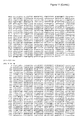
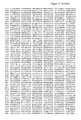
- FIGS. 1 and 2 the amino acid (SEQ ID NOs:73 to 76, 103 and 104) and nucleotide (SEQ ID NOs:81 to 84,106 and 107) sequences of 4 clones of anti-CD16 VHH and the amino acid (SEQ ID NOs:77 to 80 and 105) and nucleotide (SEQ ID NOs:85 to 88 and 108) sequences of 4 anti-CEA clones isolated according to the invention;
- FIGS. 3 and 4 the results by FACS demonstrating the specificity of 8 VHH analysed, and the corresponding bispecific antibodies
- FIG. 5 the results by FACS demonstrating the accessibility on the cells of bispecific antibodies
- FIG. 6 the results of competition tests by ELISA between 2 anti-CD16 VHH and monoclonal anti-CD 16 antibodies
- FIG. 7 the competition profiles on cells by FACS of 2anti-CD16 VHH and the monocolonal anti-CD 16 antibodies
- FIG. 8 the activation results of CD16A by 2 anti-CD16 VHH, and by the bispecific anti-CEA 17/anti-CD16 c21 antibody of type F(ab′) 2 ;
- FIG. 9 the results of cell lysis by NK cells activated by the bi-specific antibodies
- FIGS. 10A and 10B plasmid constructions according to the invention
- FIG. 11 the plasmid sequences of the invention.
- FIGS. 12A and 12B antibody formats of type Fab, Fab′, F(ab′) 2 , (HCH2CH3) 2 and mAb*;
- FIG. 13 electrophoresis gels of antibody fragments of type Fab, Fab′ and F(ab′)2 during different stages of their purification.
- a female llama was immunised with the extracellular region of human recombinant (CD16B) receptor Fc ⁇ RIIIB (described in: Operalaud C et al., 1993).
- a male llama was immunised with the extracellular region of the human recombinant carcinoembryonary antigen (CEA) (described in: Terskikh et al., 1993, and in patent: Terskikh A et al., 1993).
- CEA carcinoembryonary antigen
- the animals were immunised every month with 500 ⁇ g of each immunogen. 100 ml of blood was taken 15 days after each immunisation. For each sample taken, the serums and the purified antibodies (IgGI, 2 and 3) were titrated to detect the presence of antibodies against the different immunogens. The B lymphocytes were then purified on Ficoll gradient (histopaque-1077, Sigma-Aldrich), then washed twice with PBS.
- VHH banks purification of whole RNA, reverse transcription, PCR1, PCR2 and cloning in phagemid pHen1.
- RNA of the B lymphocytes is extracted according to the method using guanidium isothiocyanate (Chomczynski and Sacchi, 1987) REF. After phenol/chloroform extractions in an acid medium, the whole RNA is precipitated with ethanol. The quality of the RNA and the quantification are evaluated on 1% agarose gel. They are then converted into cDNA by reverse transcription.
- RNA Five ⁇ g of whole RNA are hybridised with 1 pmole of oligonucleotide 3′ CH2FORTA4 (Arbabi Ghahroudi et al., 1997) REF or CH2-2 specific to the CH2 domain of the heavy single chain IgG of llama reverse transcribed with 150 U of superscript II (BRL) for 30 min at 50° C.
- the specific oligonucleotides of the hinge regions of IgG 2 and 3, 3′ RC-IgG2 and 3′ RC-IgG3, may also be used.
- the single strand cDNA are purified on beads (BioMag R Carboxyl Terminator, Polyscience Inc.) and eluted with 17 ⁇ l of 10 mM Tris-acetate pH 7.8.
- PCR1 conditions Four ⁇ l of cDNA are amplified by PCR with 0.5 U of Dynazyme Extend DNA polymerase (Finnzymes), 10 pmoles of the same primer 3′ CH2FORTA4 or CH2-2 and 10 pmoles of 4 primers 5′ VH1-4-Sfi specific to the VH domain of human IgG, in a volume of 50 ⁇ l. (94° C., 3 min; 94° C., 1 min; 60° C., 1 min; 72° C., 1 min; 37 cycles then 72° C., 10 min).
- Finnzymes Dynazyme Extend DNA polymerase
- Three fragments of DNA are amplified: one fragment of about 900 bp coding for the VH-CH1-CH2 domains of IgGI; and two fragments of about 600 bp coding for the VHH—CH2 domain of IgG2 and 3.
- PCR2 conditions The 600 bp fragments are purified on 1% agarose gel (“Qiaquick gel extraction” kit, Qiagen) then amplified by PCR with 1 U of Deep Vent (Biolabs) and 10 pmoles of 4 primers 5′VH1-4-Sfi specific for the VH domain of human IgG and 10 pmoles of primer 3′ VHH-NotI. (94° C., 3 min; 94° C., 45 sec; 65° C., 45 sec; 72° C., 45 sec; 15 cycles, then 94° C., 45 sec; 60° C., 45 sec; 72° C., 45 sec; 15 further cycles, then 72° C., 10 min).
- Fragments of about 400 bp coding for the VHH are purified on 1% agarose gel (“Qiaquick gel extraction” kit, Qiagen) assembled and precipitated with ethanol. They are then cut by restriction enzymes NcoI and NotI, or BgII and NotI (Biolabs) to be cloned in phagemid pHen1 (Hoogenboom et al., 1991) REF at sites NcoI and NotI or SfiI and NotI.
- phagemid pHen1 are digested in a 300 ⁇ l volume with 50 U of Sfi1 in the presence of BSA, at 50° C., 16 h; or with 50 U of NcoI in the presence of BSA, at 37° C., 16 h.
- the linearised phagemid is purified on 0.7% agarose gel (“Qiaquick gel extraction” kit, Qiagen).
- the eluted DNA is then cut by 50 U of NotI at 37° C. in a volume of 200 ⁇ l, 16 h.
- the enzyme is destroyed by heat for 15 min at 65° C. and the DNA is extracted with phenol/chloroform and precipitated by ethanol.
- the cut pHen1 is controlled on 0.7% agarose gel, quantified and adjusted to 200 ng/ ⁇ l.
- VHH DNA fragments Five ⁇ g of VHH fragments are cut in a volume of 300 ⁇ l with 50 U of BgII and NotI in the presence of BSA, at 37° C., 16 h; or with 50 U of NcoI and NotI in the presence of BSA, at 37° C., 16 h.
- the enzymes are denatured at 65° C., 15 min; the DNA is then extracted with phenol/chloroform and precipitated with ethanol in the presence of 10 ⁇ g of glycogen (Roche).
- the VHH fragments cut by NcoI and NotI are purified on 1% agarose gel and then controlled on 2% agarose gel, quantified and adjusted at 100 ng/ ⁇ l.
- the ligase is inactivated at 65° C., 15 min, and the ligation product is cut by 20 U of XhoI (Biolabs) to eliminate the non-ligated residual vector, 37° C., 4 h. Six ligations are thereby made.
- the ligation products are then collected in 2 tubes and extracted with phenol/chloroform, precipitated in the presence of 10 ⁇ g of glycogen and taken up in 2 ⁇ 18 ⁇ l ultrapure H 2 O. Two ⁇ l are used by electroporation.
- the VHH bank from the male llama (ref.: 080101) represents 5.4 ⁇ 10 6 clones and the VHH bank from the female llama (ref.: 010301) 10 6 clones.
- phage banks Ten ⁇ l of stock from bank 080101 or 010301 (TG1 cells transformed with phagemids) are inoculated in 50 ml of (2TY, 100 ⁇ g/ml of ampicillin, 2% glucose) and incubated at 37° C. until the OD 600 is equal to 0.5. Five ml of culture are then infected with 5 ml of M13KO7 at 10 13 pfu/ml, 30 min, 37° C., without stirring. After centrifugation, the phage sediment is taken up in 25 ml of (2TY, 100 ⁇ g/ml of ampicillin, 25 ⁇ g/ml kanamycin). The culture is incubated 16 h at 30° C. with stirring. The phages are then precipitated with 1/5 vol of 2.5 M NaCl/20% PEG 6000 and concentrated 25 times in PBS.
- VHH selection Two hundred ⁇ ; of beads coated with streptavidin (Dynabeads M-280, Dynal) are equilibrated with 1 ml of 2% milk/PBS for 45 min at ambient temperature with stirring on a wheel. 10 12 phages from the above production are also equilibrated with 2% milk/PBS in a final volume of 500 ⁇ l for 60 min at ambient temperature with stirring on a wheel.
- the beads are compacted with a magnet, re-suspended with 250 ⁇ l of 2% milk/PBS and incubated with 200 ⁇ l of biotinylated antigen for 30 min at ambient temperature on a wheel. 150, 75 and 25 nM final of biotinyl antigen are used on the 1 st , 2 nd and 3 rd rotation, respectively.
- the antibody phages bound on the beads/antigen-biotin are re-suspended with 200 ⁇ l of PBS and incubated 30 min at 37° C., without stirring, with 1 ml of TG1 rendered competent for the binding of phages to pili (competent cells: from a culture of TG1 in 2YT overnight, a 1/100 dilution is made and 50 ml of 2YT is inoculated at 37° C. while stirring until the OD 600 is close to 0.5). At each selection, the phages are counted and amplified for another round of selection.
- Counting of the selections 1 ⁇ l dilutions are made of TG1 cells transfected with the phages (see above) of 10 ⁇ 2 to 10 ⁇ 5 with 2YT. One, 10 and 100 ⁇ l of each dilution are spread on a Petri dish (2YT/ampicillin 100 ⁇ g/ml/2% glucose). The dishes are incubated for 16 h at 30° C.
- VHH VHH were isolated using this method: four anti-CEA VHH (clones: 3, 17, 25, 43) and four anti-CD16 VHH (clones: c13, c21, c28, c72) were obtained whose amino acid and nucleotide sequences are indicated in FIGS. 1 and 2 .
- VHH cloning The VHH were cloned in plasmid p55PhoA6HisGS/NAB ⁇ (construction described in section 1.3.6, see FIGS. 10A and 11 ) between the SfiI and HindIII restriction sites.
- PCR conditions Fifty ng of VHH were amplified by PCR with 1 U of Deep Vent (Biolabs), 10 pmoles of primers 5′ pJF-VH3-Sfi and 3′ cmyc-6His/HindIII in a final volume of 50 ⁇ l. (94° C., 3 min; 94° C., 45 sec; 52° C., 45 sec; 72° C., 45 sec; 30 cycles then, 72° C., 5 min).
- the PCR products are purified on 1% agarose gel (“Qiaquick gel extraction” kit, Qiagen) and cut with 20 U of BgII and 20 U of HindIII (Biolabs) 16 h at 37° C. Ten ⁇ g of p55PhoA/NAB ⁇ are first cut with 50 U of SfiI16 h at 50° C., then with 20 U of HindIII 12 h at 37° C. The digestion products (vector and PCR fragments) are precipitated in ethanol. The DNA are re-suspended in 20 ⁇ l of H 2 O and quantified on 90.7% agarose gel.
- the ligation is carried out with 200 U of T4 DNA ligase (50 ng of p55PhoA/NAB ⁇ cut by SfiI and HindIII and 10 ng of PCR fragment cut by BgII and HindIII in a volume of 20 ⁇ l, 16 h at 16° C. After the inactivation of the T4 DNA ligase 15 min at 65° C., the non-recombinant vector is eliminated by enzyme digestion with 10 U of XhoI, 2 h at 37° C. After transformation of the ligation and analysis of several recombinant colonies, the VHH of interest are produced in E. coli.
- VHH Production of VHH: An isolated colony is inoculated in 3 ml of 2YT/ampicillin 100 ⁇ g/ml/2% glucose and incubated at 37° C. with stirring. Fifty ml of 2YT/ampicillin 100 ⁇ g/ml/2% glucose are then seeded with a dilution of the above culture and incubated for 16 h at 30° C. with stirring. Four hundred ml of 2YT/ampicillin 100 ⁇ g/ml are inoculated with the equivalent of 0.1 units OD 600 , and incubated at 30° C. with stirring, until the OD 600 is 0.5 to 0.7. The culture is then induced with 400 ⁇ l of IPTG (isopropyl- ⁇ -D-thiogalactopyranoside) 0.1 mM final and cultivated at 30° C. for 16 h.
- IPTG isopropyl- ⁇ -D-thiogalactopyranoside
- IPTG isopropyl- ⁇ -D-thiogalactopyranoside
- the supernatant is recovered (corresponding to the periplasmic fraction) and 150 ⁇ l of DNAse (10 mg/ml) and 5 mM final of MgCl 2 are added, 30 min at ambient temperature.
- the solution is dialysed for 16 h against the equilibrium buffer (50 mM sodium acetate, 0.1M NaCl pH 7.0).
- the column (BD TALONTM Metal affinity, BD Biosciences Clontech) is equilibrated with the equilibration buffer (50 mM sodium acetate, 0.1 M NaCl pH 7.0). The periplasmic fraction is deposited on the column. After washing the column with 5 volumes of equilibration buffer, the VHH is eluted by pH gradient or imidazole (gradient between the equilibration buffer pH 7.0 and the 50 mM sodium acetate solution pH 5.0 or the 200 mM imidazole solution). Each fraction is controlled on a SDS/PAGE gel (15% acrylamide) after colouration with Coomassie blue. The fractions of interest are assembled and dialysed against PBS. The VHH is concentrated on membrane (Amicon Ultra 5000MWCO, Millipore) and assayed with Lowry's colorimetric method using the Biorad Protein Assay kit.
- the equilibration buffer 50 mM sodium acetate, 0.1 M NaCl pH 7.0.
- the bispecific antibodies are purified from the soluble fraction of the periplasma (refer to extraction of the soluble fraction of the periplasma) in two steps. First on a BD TALON column (refer to VHH purification) and then on a protein G (HiTrap protein G 5 ml, Amersham biosciences).
- the “Hi Trap protein G” column is equilibrated in PBS (NaCl 137 mM, KCl 2.67 mM, Na 2 HPO 4 1.2 mM, KH 2 PO 4 1.76 mM pH 7.4).
- the proteins eluted on the BD TALON column and dialysed on PBS are deposited on the protein G.
- the bispecific antibody is eluted with 0.1 M glycine pH 2.7 then buffered with 1 M hepes pH 8.
- SDS/PAGE gel (10% acrylamide) the bispecific antibody is dialysed in 0.1 ⁇ PBS, frozen at ⁇ 80° C. and lyophilised to be concentrated ten times.
- the F(ab′) 2 is separated from the Fab′ on a Tricorn Superdex 200 10/300 GL column (Amersham Biosciences) equilibrated in PBS.
- VHH and bispecific antibodies by ELISA, Biacore, immunofluorescence (flow cytometry, FACS) and by activation tests of CD16.
- ELISA of phages-VHH Five ⁇ g/ml of biotinylated antigen (CEA or CD16) are bound on a streptavidin plate (BioBind Assembly Streptavidin Coated, ThermoLabsystems) previously saturated with 2% milk-PBS. 5 ⁇ 10 10 phages-antibodies are put in contact with the antigen. The antigen/antibody binding is detected by an ELISA comprising a monoclonal antibody directed against protein P8 of the phage (HRP/anti-M13 monoclonal conjugate, Pharmacia).
- VHH Five ⁇ g/ml of biotinylated antibody are binded on a streptavidin plate (BioBind Assembly Streptavidin Coated, ThermoLabsystems) previously saturated in 2% milk-PBS. Each VHH (range from 0.001 ⁇ g/ml to 1 ⁇ g/ml) is bound to the adsorbed antigen in the microwells.
- the binding is detected with a monoclonal antibody directed against the c-myc label (Santa Cruz Biotechnology, Inc) diluted to 1/1000 and a goat polyclonal antibody directed against the IgG of mice coupled with peroxidase diluted to 1/5000 (ref 55556, ICN) in the presence of ABTS (2,2′-Azino-di-(3-ethylbenzthiazoline sulphonate)diammonium salt, Roche).
- ELISA of the bispecific antibodies Ten ⁇ g/ml of antigen (rhCD16 or rhCEA) are passively coated on a MaxiSorp plate (Nunc). After saturation of the plate in PBS/4% milk, the bispecific antibody (F(ab′) 2 , Fab′, Fab) (range from 800 to 0.4 nM) is bound to the antigen adsorbed in the microwells.
- the binding is detected:
- VHH CEA 17 Demonstration of the accessibility of VHH CEA 17 when the VHH CD16 c21 is bound to rhCD16 adsorbed in the microwells with biotinylated rhCEA and streptavidin coupled with alkaline phosphatase diluted to 1/500 (DAKO, cat D0396).
- BIACORE uses the principle of surface plasmon resonance (SPR) to monitor, in real time, the interactions between molecules without their labelling.
- SPR surface plasmon resonance
- One of the partners in the interaction is covalently immobilised on a biosensor while the other is injected in a continuous flow.
- the principle of detection by SPR allows the changes in the mass to be monitored at the surface of the biosensor due to the formation and then the dissociation of the molecular complexes.
- the response, quantified in resonance units (RU) is a direct indication of the rate of binding of the analyte by the measurement of the variation of the refraction index.
- the interactions between the CEA or the CD16 and the VHH were studied on a BIACORE 3000 equipped with a CM5 biosensor on which the monoclonal antibody 9E10 was covalently immobilised following the standard coupling procedure by the amines proposed by BIACORE (activation by NHS/EDC).
- the VHH in buffer: 10 mM HEPES; 150 mM NaC1; 3 mM EDTA; 0.005% surfactant P20) is then injected and then a range of CEA or CD16 is injected on the VHH immobilised on the 9E10.
- the injections are carried out on a control channel that has been subjected to the same coupling chemistry without the injection of protein.
- the affinities of the VHH are indicated in Table 1 below. Equivalent affinities are obtained from the different formats of bispecific antibodies.
- CEA Specificity for CEA: (For effective immunotargeting, it is important that the anti-CEA antibodies do not recognise the NCA, a molecule that is very homologous to the CEA that is expressed at the surface of the granulocytes.)
- Antibodies used for the binding to the cells are:
- the samples are incubated for 45 min, 4° C., in the dark. Between each reaction, a washing is carried out with 2 ml of PBS/1% BSA. At the last step, the cells are taken up with 0.5 ml of PBS.
- CD16 For the effective recognition of the effector cells in the immune system, the anti-CD16 antibodies selected from CD16B should also recognise CD16A. In addition, they should not present a cross-over reaction with CD32 (RFc ⁇ IIA and RFc ⁇ IIB).
- Antibodies used for the binding to cells are:
- Antibodies used for detection are:
- VHH and monoclonal antibodies are diluted in 100 ⁇ l of PBS-1% BSA.
- the results by FACS demonstrate the specificity of the 4 anti-CD16 VHH analysed and the anti-CEA/anti-CD16 bispecific antibodies in Fab, Fab′ and F(ab′) 2 form. They are CD16 specific and do not cross with CD32.
- FIG. 4 One example is illustrated in FIG. 4 .
- the reference monoclonal antibody mAb 3G8 does not bind on the Jurkat cells, K562 CD32A + and IIA.1.6huIIB1 CD32B + that do not express CD16 but on the Jurkat CD 16A + cells, and the granulocytes that express CD16 on their surface.
- CD16 c21 VHH does not bind on cells that express CD16 on their surface.
- VHH CEA 17/VHH CD16 c21 is specific in Jurkat CD16A + and NKL cells both in Fab′ and F(ab′) 2 form.
- Equivalent results are obtained with other anti-CEA/anti-CD16 bispecific antibodies in Fab, Fab′ and F(ab′) 2 form (deriving 8 anti-CEA and anti-CD16 VHH).
- the accessibility of anti-CD16 VHH domain 5 ⁇ 10 5 LS174T cells are incubated for 30 min, in PBS-1% BSA in ice, in the presence of Fab, Fab′ or F(ab′) 2 , (range from 10 ⁇ g/ml to 0.1 ⁇ g/ml). The cells are washed in PBS-BSA 1%.
- the accessibility of the anti-CEA VHH domain 5 ⁇ 10 5 Jurkat CD16A + cells are incubated for 30 min, in PBS-BSA 1% in ice, in the presence of Fab, Fab′ or F(ab′) 2 , (range from 10 ⁇ g/ml to 0.1 g/ml). The cells are washed in PBS-1% BSA.
- the binding of the rhCEA (10 ⁇ g/ml) on the anti-CEA VHH domain of the Fab, Fab′ or F(ab′) 2 is then detected, in two steps, by incubating monoclonal antibody 192 (3 ⁇ /ml) with the cells for 30 min, in ice, and then by incubating the cells with goat F(ab′) 2 anti-IgG of mouse (H+L) marked with FITC (Jackson Immunoresearch Laboratory, cat: 115-096-003), for 30 min in ice. After several washings, the immunofluorescence is analysed by flow cytometry with a FACScalibur 4C4 (Becton Dickinson) using the Cell Quest Pro programme.
- VHH CEA 17/VHH CD16 c21 binds both with the LS174T and Jurkat CD16A + cells. Equivalent results are obtained with the other anti-CEA/anti-CD16 bispecific antibodies in Fab, Fab′ and F(ab′) 2 form (deriving 8 anti-CEA and anti-CD16 VHH).
- ELISA Five ⁇ g/ml of biotinyl VHH (c21, c28) are bound per well in a plate adsorbed with streptavidin (BioBind Assembly Streptavidin Coated, ThermoLabsystems) previously saturated in 2% milk-PBS. The CD16B with a concentration ranging from 0.07 to 20 ⁇ g/ml is then added. Secondly, the monoclonal antibody (3G8 or 7.5.4) at a constant concentration of 5 ⁇ g/ml is added.
- the CD16B-monoclonal antibody binding is detected with a goat F(ab′) 2 anti-IgG of mouse coupled with alkaline phosphatase (SouthernBiotechnology, 1030-04) in the presence of p-nitrophenylphosphatase (Sigma, N9389).
- the competition curves in ELISA are demonstrated in FIG. 6 .
- the CD16 c21 VHH is shifted by monoclonal antibody 7.5.4.
- the CD16 c28 VHH is shifted by monoclonal antibody 3G8.
- FACS Indirect immunofluorescence: 5 ⁇ 10 5 Jurkat-CD16A cells are incubated for 30 min, in PBS-5% BSA in ice, in the presence of Cb16 c21 or c28 VHH (1 to 100 ⁇ g/ml). The cells are then incubated with 0.1 ⁇ g/ml of 3G8 or 1 ⁇ g/ml of 7.5.4 for 30 min in the same conditions, then washed in PBS-5% BSA.
- the binding of 3G8 or 7.5.4 is then detected by incubating the cells with the F(ab′) 2 of a goat antibody anti-IgG of mouse (H+L) marked with FITC (Jackson ImmunoResearch Laboratories Inc., West Grove, Pa., USA, cat No.: 115-096-003) for 30 min in ice. After several washings, the immunofluorescence is analysed by flow cytometry with a FACScalibur 4CA (Becton Dickinson, Mountain View, Calif., USA) using the Cell Quest Pro programme.
- FACScalibur 4CA Becton Dickinson, Mountain View, Calif., USA
- CD16 c21 VHH is shifted by monoclonal antibody 7.5.4.
- CD16 c28 VHH is shifted by monoclonal antibody 3G8.
- a high dose of CD16 c28 VHH is also shifted by mAb 7.5.4.
- the experiments are carried out with Jurkat cells (ATCC TIB-152) transfected with the gene that codes CD16A.
- the cells are cultivated in RPMI 1640 complemented with 10% FCS, 100 U/ml penicillin, 100 ⁇ g/ml streptomycin, 2 mM L-glutamine, 0.5 mg/ml G418.
- 5 ⁇ 10 5 cells are then incubated for 18 h in microplate wells (250 ⁇ l of RPMI containing 10% FCS, 1% PS, 0.5 mg/ml G418).
- Ten ng/ml of phorbol myristate acetate (PMA) concentration not perse inducing the production and secretion of IL2, but necessary as a “second signal” for this production
- PMA phorbol myristate acetate
- the human IL2 produced in the cell supernatants is measured by ELISA using antibodies from the R&D kit (Duoset Human IL2; reference: DY202) and streptavidin coupled with alkaline phosphatase (DAKO, D0396) in the presence of p-nitrophenylphosphate (Sigma, cat 104-405).
- the results of the activation of CD16A are provided in FIG. 8 .
- the two anti-CD16 c21 and c28 VHH activate the production of IL2 of Jurkat CD16A + cells. Higher quantities of c28 are required to obtain an induction of the production and the secretion of IL2 similar to that induced by the c21.
- the anti-CEA 17/anti-CD16 c21 bispecific antibody in form F(ab′)2 also activates the production of IL2 if the Jurkat cells express CD16A at their surface in the absence of bridging via the streptavidin. Equivalent results are obtained from other anti-CD16 VHH and anti-CEA/anti-CD16 bispecific antibodies.
- NKL cells Lysis of tumoural cells by NKL cells in the presence of bispecific antibody:
- NKL cells are used as the cell lines (Robertson et al, 1996(12)) obtained from leukemia with large granulocytic lymphocytes, whose functional properties are similar to that of the NK and whose expression of CD16 was first verified by flow cytometry.
- the target cells used are very NK sensitive HeLa cells obtained from a human leukemia, NK sensitive cells from murine colon C15.4.3 AP (MC38), and MC38 cells transfected with human CEA that are naturally NK resistant.
- the target cells in culture are put into suspension (by tryptic reaction for the HeLa cells, mechanically for the MC38 and NKL cells) and counted using Trypan blue in a Malassay cell. Two thousand cells per well are incubated in 100 ⁇ l with 3.7 ⁇ 10 6 Bq of 51 Cr and the different antibody formats (200, 100 or 50 ⁇ g/ml) 1 h at 37° C. The cells are then washed several times to eliminate the 51 Cr remaining in the medium as well as the non-bound antibodies. The NKL cells in suspension are counted and added to the target cells with an effector/target ratio ranging from 60:1 to 0.2:1.
- genes of human origin inserted The genes coding for regions C ⁇ , CH1, H, CH2 and CH3 respectively correspond to: the domains of genes coding for the constant region of a light kappa chain of human immunoglobulin, for the first constant region, for the hinge region, for the second constant region and for the third constant region of a heavy chain of human immunoglobulin IgG1. These genes were obtained by RT-PCR from an LFB pouch (Laboratoire francais du Fractionêt et des Biotechnologies). This material is subject to the legal authorisations and may be used for the experiments described.
- each plasmid is carried out on ABI 310 sequencer by using the oligonucleotides:
- All of the vectors are designed to allow for the introduction, between 2 unique sites of restriction enzymes, of: different promoters, different signal sequences of type PelB (or other), different RBS sequences, any VHH or humanised VHH domain, any C ⁇ domain, or domains CH1, H, CH2 and CH3 of any type of immunoglobulin.
- This plasmid codes for the mature form of the Alkaline Phosphatase (PhoA) starting at the sixth residue (Proline).
- This plasmid is described in Le Calvez (1996).
- a gene fragment (formed by degenerated oligonucleotides on the third base of codons 2 to 14 coding for the signal sequence of PeIB) is inserted between the NdeI and Eagl sites of plasmid pMCSPhoA′.
- the clones (1 to 60) presenting the best alkaline phosphatase activity were then selected.
- FIGS. 10A and 11 p35PhoA′/N ⁇
- upstream-RsrII SEQ ID NO:14: GGCACATGTGACCTCGCGC Ncol-sup SEQ ID NO:15: GCAACGTACCACGGCAATATCG Ncol-inf SEQ ID NO:16: CGATATTGCCGTGGTACGTTGC downstream-EcoNI SEQ ID NO:17: GCCATCTTTGGTATTTAGCGCC
- plasmid 5 ng
- 10 pmoles of each oligonucleotide upstream-RsrII and NcoI-inf for PCR 1 and NcoI-sup and downstream-EcoNI for PCR 2
- 0.5 U Dynazyme 94° C., 3 min; 94° C., 45 s; 60° C., 45 s; 72° C., 45 s; for 25 cycles then 72° C., 10 min
- the PCR products are purified from a 2% agarose gel (Qiagen Kit extraction gel, final volume 50 ⁇ l).
- FIGS. 10A and 11 p55PhoA6HisGS/N ⁇
- Xhol-SacI SEQ ID NO:18: CCATGGCGGCCGATCCTCGAGAG 6HisGS/HindIII SEQ ID NO: 19: CATGCAGTCCCAAGCTTATTAGCTCCCGTGATGGT GATGATGATGTTTCAGCCCCAGA GCGGCTTTC
- PCR conditions A PCR is carried out with 5 ng of p35PhoA′/N ⁇ vector, 10 pmoles of each oligonucleotide and 0.5 U Dynazyme (94° C., 3 min; 94° C., 1 min; 70° C., 1 min; 72° C., 1 min; 35 cycles then 72° C., 10 min) in a final volume of 50 ⁇ l.
- the PCR product is purified from a 1% agarose gel (Qiagen Kit extraction gel, final volume 50 ⁇ l).
- fragmentXhoI-HindIII Twenty ⁇ l of the PCR fragment and 5 ⁇ l (2.5 ⁇ g) of p55PhoA′ vector (Le Calvez et al. Gene 1996, 170, 51-55) are digested by 10 U of XhoI and HindIII in the presence of BSA. After 16 h of incubation, the enzymes are destroyed for 10 min at 65° C. Each DNA is then precipitated and resuspended with 20 ⁇ l of H 2 O. The ligation is carried out for 16 h at 16° C.
- Competent TG1 bacteria (CaCl 2 technique) are transformed with 5 ⁇ l of ligation product.
- FIGS. 10A and 11 p55PhoA6HisGS/NAB ⁇
- upstream-EcoRV SEQ ID NO:20: CATGAGCTGTCTTCGGTATC ApaI-BstEII-sup SEQ ID NO:21: TAATGGTCCCGCTAACAGCGCGATTTGCTGATGACCCA BstEII-ApaI-inf SEQ ID NO:22: TGGGTCATCAGCAAATCGCGCTGTTAGCGGGACCATTA downstream-MluI SEQ ID NO:23: GAACGAAGCGGCGTCGAAG
- PCR 1 and PCR 2 conditions One ⁇ l (5 ng) of plasmid p55PhoA6HisGS/N ⁇ , 10 pmoles of each oligonucleotide (upstream-EcoRV and BstEII-ApaI-inf for PCR 1 and ApaI-BstEII-sup and downstream-MluI for PCR 2), 0.5 U Dynazyme (94° C., 3 min; 94° C., 45 s; 60° C., 45 s; 72° C., 45 s; 25 cycles then 72° C., 10 min.
- the PCR products are purified from a 2% agarose gel (Qiagen Kit extraction gel, final volume 50 ⁇ l).
- Competent TG1 bacteria (CaCl 2 technique) are transformed with 5 ⁇ l of ligation product.
- FIGS. 10A and 11 p55PhoA6HisGS ⁇ /NAB ⁇
- Phase shift of the PhoA gene at the EagI site creates a single FseI site.
- Five ⁇ l (2.5 ⁇ g) of p55PhoA6HisGS/NAB ⁇ are digested by 10 U of EagI. After 16 h of incubation, the enzyme is destroyed for 10 min at 65° C. The reaction mixture is then precipitated and resuspended with 20 ⁇ l of H 2 O. An equimolar mixture of dGTP and dCTP (33 ⁇ M final) and 2.5 U of Klenow fragment exo (Biolabs) are added in a final volume of 50 ⁇ l 15 min at 25° C.
- the reaction is stopped with 2 ⁇ l of EDTA at 500 mM, 20 min at 75° C.
- the reaction mixture is precipitated in ethanol, resuspended with 5 ⁇ l of H 2 O and ligated with 3 U Weiss of T4 DNA ligase Biolabs in a final volume of 10 ⁇ l Competent TG1 bacteria (CaCl 2 technique) are transformed with 5 ⁇ l of ligation.
- This plasmid allows for the cloning and selection of the best secreted fragments of human antibody VH.
- FIGS. 10A and 11 p55/MCS1
- MCS1 5′ MCS1 SEQ ID NO:24: CATGGCCCAGGTCACCGTCTCCTCAAACCGCGGACTC GAGGCGGCCCAGCCGGCCAT GGCCGCTAGCGCGGCCGCTCTAGATTA 3′ MCS1 SEQ ID NO:25: AGCTTAATCTAGAGCGGCCGCGCTAGCGGCCATGGCC GGCTGGGCCGCCTCGAGTCCG CGGTTTGAGGAGACGGTGACCTGGGC
- reaction mixture (2 h) of 90 ⁇ l, containing 10 U of EagI is added to destroy the original vector.
- This mixture is precipitated with ethanol and resuspended with 10 ⁇ l of H 2 O.
- Competent TG1 bacteria (CaCl 2 technique) are transformed with 5 ⁇ l of mixture.
- Human B lymphocytes are purified by Ficoll gradient from a pouch provided by LFB. The whole RNA is then prepared according to the protocol described in section 1.3.2.
- Hybridisation One ⁇ l of whole DNA is preincubated with 1 pmole of oligonucleotide 3′ C ⁇ for 10 min at 70° C. in a final volume of 8 ⁇ l. The temperature is slowly decreased (45 min) to 37° C.
- Reverse transcription Take 8 ⁇ l and add 0.5 ⁇ l of RNAsine (20 U), 3 ⁇ l of 5 ⁇ buffer (SuperScriptII, Invitrogen), 1 ⁇ l DTT, 100 mM, 2 ⁇ l dNTP 10 mM and incubate for 10 min at 50° C. Then, 0.75 ⁇ l of SuperScript (150 U) are added and the incubation is continued for 30 min at 50° C. and 15 min at 70° C. The cDNA obtained is purified on beads (BioMag Carboxyl Terminated, Polysciences) according to the supplier's recommendations. The final elution is made with 15 ⁇ l of Tris-acetate 10 mM pH 7.8.
- PCR 1 and 2 conditions The PCR1 is carried out with 1 ⁇ l of cDNA, 10 pmoles of each oligonucleotide 5′ C ⁇ and 3′ C ⁇ , 0.5 U Dynazyme (94° C., 3 min; 94° C., 1 min; 60° C., 1 min; 72° C., 1.5 min; 30 cycles then 72° C., 10 min) in a final volume of 50 ⁇ l.
- the PCR2 is carried out from 1 ⁇ l of PCR1 using 0.5 U Deep-Vent, (94° C., 3 min; 94° C., 45 s; 60° C., 45 s; 72° C., 45 s; 25 cycles then 72° C., 5 min) in a final volume of 50 ⁇ l.
- the PCR product is purified from a 2% agarose gel (Qiagen Kit extraction gel, final volume 50 ⁇ l).
- the PCR fragment is sequenced, before cloning, on ABI310 with oligonucleotides 5′ C ⁇ and 3′ C ⁇ .
- the cloning of the C ⁇ domain is carried out between sites BstEII and SacII of p55Flag/RBS/35cmyc6HisGS: 20 ⁇ l of PCR 2 fragment and 5 ⁇ l (2.5 ⁇ g) of p55Flag/RBS/35cmyc6HisGS are digested by 10 U of BstEII and SacII. After 16 h of incubation, the enzymes are destroyed for 10 min at 65° C. Each DNA is then precipitated and resuspended with 20 ⁇ l of H 2 O. Ligation is carried out for 16 h at 16° C.
- Competent TG1 bacteria (CaCl 2 technique) are transformed with 5 ⁇ l of ligate.
- SEQ ID NO:32 CTCGAGGCGGCCCAGCCGGCCATGGCCGCTAGCACCA AGGGCCCATCGG 3′ CH1 ⁇ 1 SEQ ID NO:33: AAGCTTAATCTAGAGCGGCCGCACAAGATTTGGGCTC AACTTTC BstEII-sup SEQ ID NO:34: CCCTCAGCAGCGTAGTGACCGTGCCCTCC BstEII-inf SEQ ID NO:35: GGAGGGCACGGTCACTACGCTGCTGAGGG
- the amplification of the CH1 ⁇ 1 domain is carried out by overlapping PCR to destroy the BstEII site.
- the reverse transcription is carried out exactly as described above for the C ⁇ , but by using oligonucleotide 3′ CH1 ⁇ I.
- the PCR 1 after RT is carried out with 1 ⁇ l of cDNA, 10 pmoles of each oligonucleotide 5′ CH1 ⁇ 1 and 3′ CH1 ⁇ 1, 0.5 U of Dynazyme (94° C., 3 min; 94° C., 1 min; 60° C., 1 min; 72° C., 1.5 min; 30 cycles then 72° C., 10 min) in a final volume of 50 ⁇ l.
- the PCR 2a is carried out from 1 ⁇ l of PCR 1, with oligonucleotides 5′ CH1 ⁇ 1 and BstEII-inf using 0.5 U of Dynazyme (94° C., 3 min; 94° C., 45 s; 60° C., 45 s; 72° C., 45 s; 25 cycles then 72° C., 5 min) in a final volume of 50 ⁇ l.
- the PCR2b is carried out from 1 ⁇ l of PCR 1, with oligonucleotides BstEII-sup and 3′ CH1 ⁇ 1 using 0.5 U of Dynazyme (94° C., 3 min; 94° C., 45 s; 60° C., 45 s; 72° C., 45 s; 25 cycles then 72° C., 5 min) in a final volume of 50 ⁇ l.
- the PCR 3 is carried out from one ⁇ l of each of PCR 2a and PCR 2b, with oligonucleotides 5′ CH1 ⁇ 1 and 3′ CH1 ⁇ 1, using 0.5 U of Deep-Vent (94° C., 3 min; 94° C., 45 s; 60° C., 45 s; 72° C., 45 s; 25 cycles then 72° C., 5 min) in a final volume of 50 ⁇ l.
- the product of PCR 3 is purified from a 2% agarose gel (Qiagen Kit extraction gel, final volume 50 ⁇ l).
- the sequence of the PCR 3 fragment is carried out on ABI 310 sequencer using oligonucleotides: 5′ CH1 ⁇ 1 and 3′ CH1 ⁇ 1.
- the cloning is carried out as described for the C ⁇ domain but between the SfiI and NotI sites of p55C ⁇ Flag/RBS/35cmyc6HisGS.
- the resulting plasmid is more commonly called: PC ⁇ CH1 ⁇ 1-TAG; it allows for the production of antibody fragments of Fab type where each chain has a label ( FIG. 12A ).
- the cloning is carried out exactly according to the conditions described for the insertion of MCS1.
- the resulting intermediate plasmid is called: p55C ⁇ /RBS/35CH1 ⁇ 1cmyc6HisGS.
- the c-myc-6HisGS motif is removed by re-cloning the CH1 ⁇ 1 domain in p55C ⁇ /RBS/35CH1 ⁇ 1cmyc6HisGS.
- PCR is used by amplifying, from 5 ng of plasmid p55C ⁇ Flag/RBS/35CH1 ⁇ 1cmyc6HisGS, the CH1 ⁇ 1 domain with oligonucleotides 5′ CH1 ⁇ 1 and 3′ CH1 ⁇ 1-STOP. Sequence SEQ ID NO:39 of the oligonucleotide used:
- PCR is used by amplifying, from 5 ng of plasmid p55C ⁇ Flag/RBS/35CH1 Hylcmyc6HisGS, the CH1 H ⁇ 1 domain with oligonucleotides 5′ CH1 ⁇ 1 and 3′ CH1 H ⁇ 1-STOP. Sequence SEQ ID NO:40 of the oligonucleotide used:
- the cloning is carried out as described for the CH17 domain between the SfiI and HindIII sites of p55C ⁇ Flag/RBS/35CH1 ⁇ 1cmyc6HisGS.
- the resulting plasmid is more commonly called: pMAbyI*; it allows for the production of antibody fragments of mAb* type ( FIG. 12B ).
- FIGS. 10B and 11 p55HCH2CH3 ⁇ 1cmyc6HisGS (pHCH2CH3 ⁇ 1-TAG) ( FIGS. 10B and 11 )
- SEQ ID NO:42 CCGGCCATGGCCCAGGTCACCGTCTCCTCAGACAAA ACTCACACATGCCC 3′ NotI/H-CH2-CH3 SEQ ID NO:43: AAGCTTAATCTAGAGCGGCCGCTTTACCCGGAGACA GGGAG
- the PCR after RT is carried out with 1 ⁇ l of cDNA, 10 pmoles of each oligonucleotide 5′ BstE2/H—CH2-CH3 and 3′ NotI/H—CH2-CH3, 0.5 U of Dynazyme (94° C., 3 min; 94° C., 1 min; 60° C., 1 min; 72° C., 1.5 min; 30 cycles then 72° C., 10 min) in a final volume of 50 ⁇ l.
- the resulting plasmid is more commonly called: pHCH2CH3 ⁇ 1-TAG; it allows for the production of antibody fragments of (HCH2CH3) 2 type with a label at the end of CH3 ( FIG. 12B ).
- the PCR after RT is carried out with 1 ⁇ l of cDNA, 10 pmoles each of oligonucleotides 5′ BstE2/H—CH2-CH3 and 3′ HindIII/H—CH2-CH3, 0.5 U of Dynazyme (94° C., 3 min; 94° C., 1 min; 60° C., 1 min; 72° C., 1.5 min; 30 cycles then 72° C., 10 min) in a final volume of 50 ⁇ l.
- the resulting plasmid is more commonly called: pHCH2CH3 ⁇ 1; it allows for the production of antibody fragment of (HCH2CH3) 2 type ( FIG. 12B ).
- any VHH may be introduced between the unique sites: Upstream from Ck: between EcoRI and BstEII (or KpnI); Upstream from CH1: between SfiI and NheI; Upstream from H: EcoRI and BstEII.
- Upstream from Ck between EcoRI and BstEII (or KpnI)
- Upstream from CH1 between SfiI and NheI
- Upstream from H EcoRI and BstEII.
- the production and purification of the different antibody fragments with the 6HisGS label are carried out as described above.
- the chromatography stage on base is replaced by an ion exchange column whose characteristics (anions or cations) depend on the characteristics of the antibody fragment.
- Electrophoresis gels are shown in FIG. 13 .
- the Fab′ and F(ab′) 2 are purified on a cobalt column and then on protein G.
- the different antibody fragments are then separated on Superdex 200 (or possibly Superdex 75).
- the principle of the method consists of cloning human VH domains (isolated by RT-PCR from the LFB pouch) in plasmid p55PhoA6HisGS ⁇ /NAB ⁇ ( FIGS. 10A and 11 ).
- This plasmid has the gene coding for the alkaline phosphatase in a reading frame not allowing for its expression.
- the cloning of the VH restores the reading frame of the alkaline phosphatase and allows for the production of fused VH upstream from the alkaline phosphatase (bank for VH-PhoA cloned in TG1 bacteria).
- the different clones are then produced in 96-well microplates and the growth kinetics of the different clones is directly measured every 30 min (OD 620 nm) from the microplates. Thereby, the clones whose growth is not altered by the presence of VH are selected.
- the clones from the microplates are replicated and stored at ⁇ 80° C. After 2 hours of induction at 37° C., or 16 hours of induction at 30° C., 24° C., or even 18° C., the growth is stopped and the phosphatase activity is directly measured from the supernatants in the culture medium. The phosphatase activity is then directly correlated with the number of bacteria found in each microplate well.
- the alkaline phosphatase is only active if it is secreted in the bacterial periplasm, in dimer form with its disulphate bridges correctly formed.
- This approach allows for the selection of the clones producing the most fusion protein VH-PhoA secreted in the bacterial culture medium. It is thereby possible to select the VH that are correctly replicated and whose disulphide bridges are correctly formed, and therefore soluble.
- the selected VH are used as a matrix to exchange the CDR of human VH by the CDR from llama VHH previously described.
- the VH are chosen by selecting the VH whose amino acids at the CDR junctions are equivalent to those of the VHH.
- Hybridisation One ⁇ l of whole RNA (purification described in section 1.3.2) is preincubated with 1 pmole of oligonucleotide (mixture of: 3′ JH1-4-5; 3′ J112; 3′ JH3 and 3′ JH6) for 10 min at 70° C. in a final volume of 8 ⁇ l. The temperature is slowly reduced (45 min) to 37° C.
- Reverse transcription Take 8 ⁇ l and add 0.5 ⁇ l of RNAsine (20 U), 3 ⁇ l of 5 ⁇ buffer (SuperScriptll, Invitrogen), 1 ⁇ l DTT, 100 mM, 2 ⁇ l dNTP 10 mM and incubate for 10 min at 50° C.
- the PCR1 is carried out with 1 ⁇ l of cDNA (obtained by RT-PCR), 10 pmoles of oligonucleotides 5′ (0.625 pmole of each oligonucleotide 5′ whose sequences are indicated below) and 3′ (2.5 pmoles of each oligonucleotide 3′ whose sequences are indicated below), 0.5 U of Dynazyme (95° C., 3 min; then 95° C., 1 min; 58° C., 1 min; 72° C., 1 min; for 35 cycles then 72° C., 10 min).
- the PCR products are deposited on a 2% agarose gel and the bands corresponding to the VH are purified (Qiagen Kit extraction gel).
- the PCR2 is carried out from 1 ⁇ l of PCR1 diluted to 1/1000th, the same quantity of oligonucleotides described above, 0.5 U Deep-Vent for a final volume of 50 ⁇ l. (94° C., 3 min; then 94° C., 1 min; 70° C., 1 min; 72° C., 1.5 min; for 40 cycles then 72° C., 10 min).
- the fragments are purified from 2% agarose gel as described above.
- the different purified fragments of PCR are digested by 10 U of NcoI and XmaI and inserted in cloning vector p55/PhoA6HisGS ⁇ s/NAB ⁇ by ligation.
- the ligation mixture is digested by FseI before transformation of the bacteria.
- the transformation is carried out by electroporation with electrocompetent TG1 bacteria.
- the clones with an inserted VH domain restore the phosphatase activity (blue colonies).
- Controls negative medium control (2YT/ampicillin 100 ⁇ g/ml); negative control of vector p55/PhoA6HisGS ⁇ /NAB ⁇ ; positive control of vector p55PhoA6HisGS/NAB ⁇ .
- Assay of the alkaline phosphatase activity Take 10 ⁇ l of the whole culture (cells+culture medium) and 10 ⁇ l of culture supernatant (for this, centrifuge for 3 min at 910 ⁇ g). To each sample, add 65 ⁇ l 10 mM Tris-HCl pH 8.0 and add 25 ⁇ l of PNPP (paranitrophenyl phosphate) at 1 mg/ml in (diethanolamine pH 9.8 (HCl); MgCl 2 0.5 mM). After 30 min of reaction while stirring, measure the OD at 405 nm.
- the alkaline phosphatase activity measured at 405 nm is corrected according to the number of cells (OD measurement 620 nm) contained in each well at the end of induction.
- Each phosphatase activity, of the clones expressing a VH fused to the alkaline phosphatase, is compared with that of the positive control (non-fused alkaline phosphatase produced by p55PhoA6HisGS/NAB ⁇ ).
Landscapes
- Health & Medical Sciences (AREA)
- Chemical & Material Sciences (AREA)
- Immunology (AREA)
- Organic Chemistry (AREA)
- Life Sciences & Earth Sciences (AREA)
- Medicinal Chemistry (AREA)
- General Health & Medical Sciences (AREA)
- Molecular Biology (AREA)
- Genetics & Genomics (AREA)
- Proteomics, Peptides & Aminoacids (AREA)
- Biophysics (AREA)
- Biochemistry (AREA)
- Oncology (AREA)
- Cell Biology (AREA)
- General Chemical & Material Sciences (AREA)
- Veterinary Medicine (AREA)
- Public Health (AREA)
- Animal Behavior & Ethology (AREA)
- Pharmacology & Pharmacy (AREA)
- Nuclear Medicine, Radiotherapy & Molecular Imaging (AREA)
- Chemical Kinetics & Catalysis (AREA)
- Bioinformatics & Cheminformatics (AREA)
- Engineering & Computer Science (AREA)
- Preparation Of Compounds By Using Micro-Organisms (AREA)
- Peptides Or Proteins (AREA)
- Medicines Containing Antibodies Or Antigens For Use As Internal Diagnostic Agents (AREA)
Abstract
Description
| 3′CH2FORTA4 |
| SEQ ID NO.1: |
| 3′CH2-2 |
| SEQ ID NO:2: |
| 3′RC-IgG2 |
| SEQ ID NO:3: |
| 3′RC-IgG3 |
| SEQ ID NO:4: |
| 5′VH1-Sfi |
| SEQ ID NO:5: |
| GTGCAGCTGGTGCAGTCTGG |
| 5′VH2-Sfi |
| SEQ ID NO:6: |
| GTCACCTTGAAGGAGTCTGG |
| 5′VH3-Sfi |
| SEQ ID NO:7: |
| GTGCAGCTGGTGGAGTTGG |
| 5′VH4-Sfi |
| SEQ ID NO:8: |
| GTGCAGCTGCAGGAGTCGGG |
| 3′VHH-Not |
| SEQ ID NO:9: CACGATTCTGCGGCCGCTGAGGAGAC(AG)GTGACCT |
| GGGTCC |
| 5′ pJF-VH3-Sfi |
| SEQ ID NO: 10: |
| AGCTGGTGG |
| 3′cmyc-6His/HindIII |
| SEQ ID NO: 11: CCGCGCGCGCCAAGACCCAAGCTTGGGCTA(GA)T |
| G(GA)TG(GA)TG(GA)TG(GA) TG(GA)TGTGCGGCCCCATTCAGATC |
| TABLE 1 | ||||
| ka × 105 | kd × 10−3 | KA × 107 | KD × 10−9 | |
| VHH | (1/Ms) | (1/s) | (1/M) | (M) |
| |
1.24 ± 0.014 | 1.68 ± 0.002 | 7.38 | 13.6 |
| Anti-CEA 17 | 1.56 ± 0.014 | 1.3 ± 0.002 | 12 | 8.3 |
| Anti-CEA 25 | 1.13 ± 0.014 | 3.6 ± 0.004 | 3.15 | 31.7 |
| Anti-CEA 43 | 1.78 ± 0.019 | 1.83 ± 0.002 | 9.72 | 10.3 |
| Anti-CD16 c13 | 0.53 ± 0.07 | 5.67 ± 0.02 | 0.94 | 100.6 |
| Anti-CD16 c21 | 2.86 ± 0.02 | 2.79 ± 0.006 | 10.3 | 9.7 |
| Anti-CD16 c28 | 0.42 ± 0.03 | 3.45 ± 0.006 | 1.22 | 81.9 |
| Anti-CD16 c72 | 0.39 ± 0.02 | 3.7 ± 0.006 | 1.06 | 94.6 |
-
- 35A7, anti-CEA monoclonal antibody (specific for CEA).
- 192, anti-CEA monoclonal antibody (that crosses with the NCA).
- 7.5.4 and 3G8 anti-CD16 monoclonal antibody.
- anti-CD16 VHH (c13, c21, c28, c72).
- anti-CEA VHH (3, 17, 25, 43).
- anti-CEA/anti-CD16 bispecific antibodies constructed from the 8 VHH isolated.
-
- 9E10, mouse anti-cmyc monoclonal antibody (200 μg/ml, used at 1/10th) binding at the c-myc label of the purified VHH.
- AP326F, sheep polyclonal antibody anti-IgG of mouse coupled with FITC (Silenus, used at 1/100th).
-
- 0.5×10⁶ cells (autofluorescence measurement of the cells).
- 0.5×10⁶ cells+anti-IgG of mouse-
FITC 20 μg/ml. - 0.5×10⁶ cells+anti-9E10 20 μg/ml, then anti-IgG of mouse-
FITC 20 μg/ml. - 0.5×10⁶ cells+VHH anti-CEA or anti-CD16 1 to 5 μg/ml, then 9E10 20 μg/ml, then anti-IgG of mouse-
FITC 20 μg/ml.
-
- 3G8, anti-CD16 monoclonal antibody (human RFcγIIIA/IIIB), anti-site antibody recognising a conformational epitope and blocking the bond of the IgG to CD16A and CD16B.
- 7.5.4, anti-CD16 monoclonal antibody (human RFcγIIIA/IIIB), antibody recognising a linear epitope, localised outside the binding site of IgG of CD16 and only weakly affecting this bond at high concentrations in competition tests (Vely et al., 1997).
- AT10 anti-CD32 monoclonal antibody (human RFcγIIA/IIB).
- IV.3, anti-CD32 monoclonal antibody (human RFcγlIA).
- anti-CD16 VHH (c13, c21, c28, c72).
- anti-CEA VHH (3, 17, 25, 43).
- 35A7, anti-CEA monoclonal antibody.
- 192, anti-NCA monoclonal antibody (that crosses with CEA).
- anti-CEA/anti-CD16 bispecific antibodies constructed from 8 isolated VHH.
-
- 9E10, mouse anti-cmyc monoclonal antibody (200 μg/ml, used at 1/10th) binding at the c-myc label of the purified VHH.
- Fab′2 of a goat antibody anti-IgG of mouse coupled with FITC (F(ab′)2/FITC) used at 20 μg/ml (Jackson Immunoresearch Lab. Inc., 115-096-003).
-
- 0.5×10⁶ cells (autofluorescence measurement of the cells)
- 0.5×10⁶ cells+(F(ab′)2/FITC) 20 μg/ml
- 0.5×10⁶ cells+anti-9E10 20 μg/ml, then (F(ab′)2/FITC) 20 μg/ml
- 0.5×10⁶ cells+
VHH 1 at 5 μg/ml, then 9E10 20 μg/ml, then (F(ab′)2/FITC) 20 μg/ml - 0.5×10⁶ cells+monoclonal antibodies (35A7, 192, 3G8, 7.5.4, AT10, N.3) 20 μ/ml, then (F(ab′)2/FITC) 20 μg/ml.
At each step, the samples are incubated for 45 min, 4° C., in the dark. Between each reaction, a washing is carried out with 2 ml of PBS/1% BSA. At the last step, the cells are taken up with 0.5 ml of PBS.
-
- All of the non detailed protocols are described in Sambrook, Fritsch and Maniatis, Molecular Cloning: A Laboratory Manual, 2nd ed, Cold Spring Harbor Laboratory Press, 1989.
- The digestions with the restriction enzymes are carried out according to the supplier's recommendations.
| EcoRI−90 of sequence SEQ ID NO:12: GCGCCGACATCATAA |
| CGGTTCTGGC |
| HindIII+88 of sequence SEQ ID NO:13: CGCTACTGCC |
| GCCAGGC |
| upstream-RsrII |
| SEQ ID NO:14: GGCACATGTGACCTCGCGC |
| Ncol-sup |
| SEQ ID NO:15: GCAACGTACCACGGCAATATCG |
| Ncol-inf |
| SEQ ID NO:16: CGATATTGCCGTGGTACGTTGC |
| downstream-EcoNI |
| SEQ ID NO:17: GCCATCTTTGGTATTTAGCGCC |
| Xhol-SacI |
| SEQ ID NO:18: CCATGGCGGCCGATCCTCGAGAG |
| 6HisGS/HindIII |
| SEQ ID NO: 19: CATGCAGTCCCAAGCTTATTAGCTCCCGTGATGGT |
| GATGATGATGTTTCAGCCCCAGA GCGGCTTTC |
| upstream-EcoRV |
| SEQ ID NO:20: CATGAGCTGTCTTCGGTATC |
| ApaI-BstEII-sup |
| SEQ ID NO:21: TAATGGTCCCGCTAACAGCGCGATTTGCTGATGACCCA |
| BstEII-ApaI-inf |
| SEQ ID NO:22: TGGGTCATCAGCAAATCGCGCTGTTAGCGGGACCATTA |
| downstream-MluI |
| SEQ ID NO:23: GAACGAAGCGGCGTCGAAG |
| 5′ MCS1 |
| SEQ ID NO:24: |
| GAGGCGGCCCAGCCGGCCAT GGCCGCTAGCGCGGCCGCTCTAGATTA |
| 3′ MCS1 |
| SEQ ID NO:25: AGCTTAATCTAGAGCGGCCGCGCTAGCGGCCATGGCC |
| GGCTGGGCCGCCTCGAGTCCG CGGTTTGAGGAGACGGTGACCTGGGC |
| 5′ Flag/RBS/35-sup |
| SEQ ID NO:26: GGAGAGTGTGCAGGTGATTACAAAGACGATGACGATA |
| AGTAATAAAGAGGAAACAGAAGTCCATATGAAATACCTATTGCCTACGGCA |
| GCCGCTGGATTGTTATTACTCGCGGCCCAGC |
| SEQ ID NO:117: |
| AAGTAATAAACAGGAAACAGAAGTCCATATGAAATATCTTTTACCTACGGC |
| AGCCGCAGGTTTGTTGTTACTCGCGGCCCAGC |
| 3′ Flag/RBS/35-inf |
| SEQ ID NO:27: GGGCCGCGAGTAATAACAATCCAGCGGCTGCCGTAGG |
| CAATAGGTATTTGATATGGACTTCTGTTTCCTGTTTATTACTTATCGTCAT |
| CGTCTTTGTAATCACCTGCACACTCTCCGC |
| SEQ ID NO:118: GGGCCGCGAGTAACAACAAACCTGCGGCTGCCGTAG |
| GTAAAAGATATTTCATATGGACTTCTGTTTCCTGTTTATTACTTATCGTCA |
| TCGTCTTTGTAATCACCTGCACACTCTCCGC |
The cloning is carried out exactly according to the conditions previously described for the insertion of MCS1. After ligation, the reaction mixture is digested by 10 U of XhoI enzyme.
| 5′ c-myc-6HisGS |
| SEQ ID NO:28: |
| AATGGGGCCGTACATCACCACC ATCACCATGGGAGCTA |
| 3′c-myc-6HisGS |
| SEQ ID NO:29:AGCTTAGCTCCCATGGTGATGGTGGTGATGTACGGCCC |
| CATTCAGATCCTCTTCTGAGA TGAGTTTTTGTTCTGC |
The cloning is carried out exactly according to the conditions previously described for the insertion of MCS1. The ligation mixture is digested by 10 U of XbaI enzyme.
| 5′ Cκ |
| SEQ ID NO:30: GGGGCCAGGGGACCCAGGTCACCGTCTCCTCAGGTAC |
| CGTGGCTGCACCATCTGTCTTC |
| SEQ ID NO:119: |
| CGGTGGCTGCACCATCTGTGT TC |
| 3′ Cκ |
| SEQ ID NO:31: CGTCATCGTCTTTGTAATCACCTGCACACTCTCCGCG |
| GTTGAAGCTCTTTGTCACCG |
| 5′ CH1 γ1 |
| SEQ ID NO:32: |
| AGGGCCCATCGG |
| 3′ CH1 γ1 |
| SEQ ID NO:33: AAGCTTAATCTAGAGCGGCCGCACAAGATTTGGGCTC |
| AACTTTC |
| BstEII-sup |
| SEQ ID NO:34: CCCTCAGCAGCGTAGTGACCGTGCCCTCC |
| BstEII-inf |
| SEQ ID NO:35: GGAGGGCACGGTCACTACGCTGCTGAGGG |
| 3′ CH1Hγ1 |
| SEQ ID NO:36: AAGCTTAATCTAGAGCGGCCGCTGGGCACGGTGGGCA |
| TGTGTGAGTTTTGTCACAAGA TTTGGGCTCAACTTTC |
The cloning is carried out as described for the CH1γ1 domain between the SfiI and NotI sites of p55CκFlag/RBS/35cmyc6HisGS. The resulting plasmid is commonly called: PCκCH1Hγ1-TAG, it allows for the production of antibody fragments of F(ab′)2 type where each chain has a label (
| 5′ RBS/35-sup |
| SEQ ID NO:37: GGAGAGTGTTAATAAACAGGAAACAGAAGTCCATATG |
| AAATACCTATTGCCTACGGCA GCCGCTGGATTGTTATTACTCGCGGCCCA |
| GC |
| SEQ ID NO:120: GGAGAGTGTTAATAAACAGGAAACAGAAGTCCATAT |
| |
| AGC |
| 3′ RBS/35-inf |
| SEQ ID NO:38: GGGCCGCGAGTAATAACAATCCAGCGGCTGCCGTAG |
| GCAATAGGTATTTCATATGGA CTTCTGTTTCCTGTTTATTAACACTCTC |
| CGC |
| SEQ ID NO:121:GGGCCGCGAGTAACAACAAACCTGCGGCTGCCGTAG |
| GTAAAAGATATTTCATATGGAC TTCTGTTTCCTGTTTATTAACACTCTC |
| CGC |
| 3′ CH1γ1-STOP |
| SEQ ID NO:39: CATGCAGTCCCAAGCTTAACAAGATTTGGGCTCAAC |
| TTTC |
The cloning of the PCR fragment is carried out as described for the Cκ domain, but between the SfiI and HindIII sites of plasmid p55Cκ/RBS/35CH171cmyc6HisGS. The resulting plasmid is commonly called: pCκCH1γ1; it allows for the production of antibody fragments of Fab type (
p55Cκ/RBS/35CH1 Hγ1 (pCκCH1Hγ1) (
Elimination of the Flag and c-myc-6HisGS labels from the plasmid p55CκFlag/RBS/35CH1Hγ1cmyc6HisGS by replacement of the DNA fragment included between SacII and SfiI by a new cassette using the paired
| 3′ CH1Hγ1-STOP |
| SEQ ID NO:40: CATGCAGTCCCAAGCTTATGGGCACGGTGGGCATGT |
| GTG |
The cloning of the PCR fragment is carried out between the SfiI and HindIII sites as described above, but from plasmid p55Cκ/RBS/35CH1Hγ1cmyc6HisGS. The resulting plasmid is commonly called: PCκCH1Hγ1, it allows for the production of antibody fragment of F(ab′)2 type (
p55Cκ/RBS/35CH1HCH2CH3γ1 (pMabγI*) (
Insertion of the constant heavy region CH1, of the hinge region (H) and the constant regions CH2 and CH3 of an immunoglobulin of IgG1 type in p55CκFlag/RBS/35CH1γ1cmyc6HisGS. The PCR1, 2a, 2b and 3 are carried out exactly as in the amplification of the CH1 domain described above by replacing
| 3′ HindIII/H-CH2-CH3 |
| SEQ ID NO:41: CCGCCAAAACAGCCAAGCTTATTTACCCGGAGACAG |
| GGAG |
| 5′BstE2/H-CH2-CH3 |
| SEQ ID NO:42: |
| ACTCACACATGCCC |
| 3′ NotI/H-CH2-CH3 |
| SEQ ID NO:43: AAGCTTAATCTAGAGCGGCCGCTTTACCCGGAGACA |
| GGGAG |
| 5′ BstE2/H-CH2-CH3 |
| SEQ ID NO:44: |
| ACTCACACATGCCC |
| 3′ HindIII/H-CH2-C113 |
| SEQ ID NO:45: CCGCCAAAACAGCCAAGCTTATTTACCCGGAGACAG |
| GGAG |
| 5′ EcoRI-PeIB55-PeIBPHen |
| SEQ ID NO:46: |
| CAACAGCAGCAGCTGGGTTATTGCTCGCTGCGCAGCCGGCCATGGCCGAG |
| GTGCAGCTG |
| 5′VH1-Sfi |
| SEQ ID NO:47: |
| GGTGCAGCTGGTGCAGTCTGG |
| 5′VH2-Sfi |
| SEQ ID NO:48: |
| GGTCACCTTGAAGGAGTCTGG |
| 5′ VH3-Sfi |
| SEQ ID NO:49: |
| GGTGCAGCTGGTGGAGTCTGG |
| 5′VH4-Sfi |
| SEQ ID NO:50: |
| GGTGCA GCTGCAGGAGTCGGG |
| 3′ BstEII/KpnI |
| SEQ ID NO:51: GGTGCAGCCACGGTACCTGAGGAGACGGTGACCTG |
| 3′ BstE2/NheI |
| SEQ ID NO:52: GGGCCCTTGGTGCTAGCTGAGGAGACGGTGACCTG |
Reverse transcription: Take 8 μl and add 0.5 μl of RNAsine (20 U), 3 μl of 5× buffer (SuperScriptll, Invitrogen), 1 μl DTT, 100 mM, 2
| 5′VH1a |
| SEQ ID NO:53: CG GCC CAG CCG GCC ATG GCC CAG GTG |
| CTG GTG CAG CAG TCT GG |
| 5′ VH1b |
| SEQ ID NO:54: CG GCC CAG CCG GCC ATG GCC CAG GT(CT) |
| CAG CG(GT) GTG CAG TCT GG |
| 5′ VH1c |
| SEQ ID NO:55: CG GCC CAG CCG GCC ATG GCC (CG)AG |
| GTC CAG CTG GTA CAG TCT GG |
| 5′ VH1d |
| SEQ ID NO:56: CG GCC CAG CCG GCC ATG GCC CA(GA) |
| ATG CAG CTG GTG CAG TCT GG |
| 5′ VH2a |
| SEQ ID NO:57: CG GCC CAG CCG GCC ATG GCC CAG GTC |
| ACC TTG AAG GAG TCT GG |
| 5′ VH2b |
| SEQ ID NO:58: CG GCC CAG CCG GCC ATG GCC CAG ATC |
| ACC TTG AAG GAG TCT GG |
| 5′ VH3a |
| SEQ ID NO:59: CG GCC CAG CCG GCC ATG GCC GAG GTG |
| CAG CTG GTG GAG TCT GG |
| 5′ VH3b |
| SEQ ID NO:60: CG GCC CAG CCG GCC ATG GCC GAA GTG |
| CAG CTG GTG GAG TCT GG |
| 5′ VH3c |
| SEQ ID NO:61: CG GCC CAG CCG GCC ATG GCC CAG GTG |
| CAG CTG GTG GAG TCT GG |
| 5′ VH3d |
| SEQ ID NO:62: CG GCC CAG CCG GCC ATG GCC GAG GTG |
| CAG CTG GTG GAG (AT)C(TC) (GC)G |
| 5′ VH4a |
| SEQ ID NO:63: CG GCC CAG CCG GCC ATG GCC CAG GTG |
| CAG CTG CAG GAG TCG GG |
| 5′ VH4b |
| SEQ ID NO:64: CG GCC CAG CCG GCC ATG GCC CAG CTG |
| CAG CTG CAG GAG TC (GC) GG |
| 5′ VH4c |
| SEQ ID NO:65: CG GCC CAG CCG GCC ATG GCC CAG GTG |
| CAG CTA CAG CAG TGG GG |
| 5′ VH5a |
| SEQ ID NO:66: CG GCC CAG CCG GCC ATG GCC GA(GA) |
| GTG CAG CTG GTG CAG TCT GG |
| 5′ VH6a |
| SEQ ID NO:67: CG GCC CAG CCG GCC ATG GCC CAG) GTA |
| CAG CTG CAG CAG TCA GG |
| 5′ VH7a |
| SEQ ID NO:68: CG GCC CAG CCG GCC ATG GCC CAG GTG |
| CAG CTG GTG CAA TCT GG |
| 3′ JH1-4-5 |
| SEQ ID NO:69: GTC TAG ACG TCC CCC CGG GGA GGA GAC |
| GGT GAC CAG GG |
| 3′ JH2 |
| SEQ ID NO:70: GTC TAG ACG TCC CCC CGG GGA GGA GAC |
| AGT GAC CAG GG |
| 3′ JH3 |
| SEQ ID NO:71: GTC TAG ACG TCC CCC CGG GGA AGA GAC |
| GGT GAC CAT TG |
| 3′ JH6 |
| SEQ ID NO:72: GTC TAG ACG TCC CCC CGG GGA GGA GAC |
| GGT GAC CGT GG |
- 1. Hamers-Casterman C, Atarhouch T, Muyldermans S, Robinson G, Hamers C, Sanga E B, Bendahinan N, Harriers R. Naturally occurring antibodies devoid of light chains. Nature 1993, 363:446-448.
- 2. Teillaud C, Galon J, Zilber M T, Mazieres N, Spagnoli R, Kurrle R, Fridman W H, Sautes C. Soluble CD16 binds peripheral blood mononuclear cells and inhibits pokeweed-nitogen-induced responses. Blood, 1993, 82:3081-3090).
- 3. Terskikh, A, Mach, J P, and Pelegrin A. Marked increase in the secretion of a fully antigenic recombinant CEA obtained by deletion of its hydrophobic tail. Mol Immunol, 1993, 30:921-927.
- 4. Chomczynski P, Sacchi N. Single-step method of RNA isolation by acid guanidinium thiocyanate-phenol-chloroform extraction. Anal Biochem, 1987, 162:156-159.
- 5. Arbabi Ghahroudi M, Desmyter A, Wyns L, Hamers R, Muyldermans S. Selection and identification of single domain antibody fragments from camel heavy-chain antibodies. FEBS Lett, 1997, 414:521-526.
- 6. Vivier E, Rochet N, Ackerly M, Petrini J, Levine H, Daley J, Anderson P. Signaling function of reconstituted CD16: zeta: gamma receptor complex isoforms. Int Immunol, 1992, 4:1313-1323.
- 7. Vély F, Gruel N, Moncuit J, Cochet O, Rouard H, Dard S, Galon J, Sautes C, Fridman W H, Teillaud J-L. A new set of monoclonal antibodies against human FcgammaRIIl (CD32) and FcgammaRIII (CD16): characterization and use in various assays. Hybridoma, 1997, 16:519-528.
- 8. Le Calvez H, Fieschi J, Green J M, Marchesi N, Chauveau J, Baty D. Paratope characterisation by structural modelling of two anti-cortisol single-chain variable fragments produced in E. coli. Mol. Immunol, 1995, 32:185-198.
- 9. Le Calvez H, Green J M, Baty D. Increased efficiency of alkaline phosphatase production levels in Escherichia coli using a degenerate PelB signal sequence. Gene, 1996, 170:51-55.
Claims (10)
Priority Applications (1)
| Application Number | Priority Date | Filing Date | Title |
|---|---|---|---|
| US14/856,016 US10385137B2 (en) | 2004-12-16 | 2015-09-16 | Production of antibody formats and immunological applications of said formats |
Applications Claiming Priority (4)
| Application Number | Priority Date | Filing Date | Title |
|---|---|---|---|
| FR04/13433 | 2004-12-16 | ||
| FR0413433 | 2004-12-16 | ||
| FR0413433A FR2879605B1 (en) | 2004-12-16 | 2004-12-16 | PRODUCTION OF ANTIBODY FORMATS AND IMMUNOLOGICAL APPLICATIONS OF THESE FORMATS |
| PCT/FR2005/003151 WO2006064136A1 (en) | 2004-12-16 | 2005-12-15 | Production of antibody formats and immunological applications of said formats |
Related Parent Applications (1)
| Application Number | Title | Priority Date | Filing Date |
|---|---|---|---|
| PCT/FR2005/003151 Continuation WO2006064136A1 (en) | 2004-12-16 | 2005-12-15 | Production of antibody formats and immunological applications of said formats |
Related Child Applications (1)
| Application Number | Title | Priority Date | Filing Date |
|---|---|---|---|
| US14/856,016 Continuation US10385137B2 (en) | 2004-12-16 | 2015-09-16 | Production of antibody formats and immunological applications of said formats |
Publications (2)
| Publication Number | Publication Date |
|---|---|
| US20090202979A1 US20090202979A1 (en) | 2009-08-13 |
| US9169316B2 true US9169316B2 (en) | 2015-10-27 |
Family
ID=34954794
Family Applications (2)
| Application Number | Title | Priority Date | Filing Date |
|---|---|---|---|
| US11/818,218 Expired - Fee Related US9169316B2 (en) | 2004-12-16 | 2007-06-13 | Production of antibody formats and immunological applications of said formats |
| US14/856,016 Expired - Fee Related US10385137B2 (en) | 2004-12-16 | 2015-09-16 | Production of antibody formats and immunological applications of said formats |
Family Applications After (1)
| Application Number | Title | Priority Date | Filing Date |
|---|---|---|---|
| US14/856,016 Expired - Fee Related US10385137B2 (en) | 2004-12-16 | 2015-09-16 | Production of antibody formats and immunological applications of said formats |
Country Status (7)
| Country | Link |
|---|---|
| US (2) | US9169316B2 (en) |
| EP (3) | EP1824884B1 (en) |
| JP (1) | JP5026279B2 (en) |
| CA (1) | CA2590615C (en) |
| ES (3) | ES2751006T3 (en) |
| FR (1) | FR2879605B1 (en) |
| WO (1) | WO2006064136A1 (en) |
Cited By (17)
| Publication number | Priority date | Publication date | Assignee | Title |
|---|---|---|---|---|
| US20160083476A1 (en) * | 2004-12-16 | 2016-03-24 | Inserm (Institut National De La Sante Et De La Recherche Medicale) | Production of antibody formats and immunological applications of said formats |
| US10066016B2 (en) | 2016-05-20 | 2018-09-04 | Harpoon Therapeutics, Inc. | Single chain variable fragment CD3 binding proteins |
| US10100106B2 (en) | 2016-05-20 | 2018-10-16 | Harpoon Therapeutics, Inc. | Single domain serum albumin binding protein |
| US10543271B2 (en) | 2017-05-12 | 2020-01-28 | Harpoon Therapeutics, Inc. | Mesothelin binding proteins |
| US10730954B2 (en) | 2017-05-12 | 2020-08-04 | Harpoon Therapeutics, Inc. | MSLN targeting trispecific proteins and methods of use |
| US10815311B2 (en) | 2018-09-25 | 2020-10-27 | Harpoon Therapeutics, Inc. | DLL3 binding proteins and methods of use |
| US10844134B2 (en) | 2016-11-23 | 2020-11-24 | Harpoon Therapeutics, Inc. | PSMA targeting trispecific proteins and methods of use |
| US10849973B2 (en) | 2016-11-23 | 2020-12-01 | Harpoon Therapeutics, Inc. | Prostate specific membrane antigen binding protein |
| US10927180B2 (en) | 2017-10-13 | 2021-02-23 | Harpoon Therapeutics, Inc. | B cell maturation antigen binding proteins |
| US10954311B2 (en) | 2015-05-21 | 2021-03-23 | Harpoon Therapeutics, Inc. | Trispecific binding proteins and methods of use |
| US11136403B2 (en) | 2017-10-13 | 2021-10-05 | Harpoon Therapeutics, Inc. | Trispecific proteins and methods of use |
| US11180563B2 (en) | 2020-02-21 | 2021-11-23 | Harpoon Therapeutics, Inc. | FLT3 binding proteins and methods of use |
| US11535668B2 (en) | 2017-02-28 | 2022-12-27 | Harpoon Therapeutics, Inc. | Inducible monovalent antigen binding protein |
| US11623958B2 (en) | 2016-05-20 | 2023-04-11 | Harpoon Therapeutics, Inc. | Single chain variable fragment CD3 binding proteins |
| US12195544B2 (en) | 2018-09-21 | 2025-01-14 | Harpoon Therapeutics, Inc. | EGFR binding proteins and methods of use |
| US12415860B2 (en) | 2018-05-14 | 2025-09-16 | Harpoon Therapeutics, Inc. | Binding moiety for conditional activation of immunoglobulin molecules |
| US12516128B2 (en) | 2019-05-14 | 2026-01-06 | Harpoon Therapeutics, Inc. | EpCAM binding proteins and methods of use |
Families Citing this family (51)
| Publication number | Priority date | Publication date | Assignee | Title |
|---|---|---|---|---|
| EP2658869B1 (en) | 2010-12-30 | 2019-06-12 | INSERM (Institut National de la Santé et de la Recherche Médicale) | Antigen binding formats for use in therapeutic treatments or diagnostic assays |
| UA117218C2 (en) * | 2011-05-05 | 2018-07-10 | Мерк Патент Гмбх | POLYPEPTIDE AGAINST IL-17A, IL-17F AND / OR IL17-A / F |
| CN102229671B (en) * | 2011-06-07 | 2013-04-10 | 南京医科大学 | Anti-IMMT anthropogenic Fab antibody and application thereof in preparing anti-liver cancer drugs |
| EP2847230B1 (en) * | 2012-05-10 | 2020-08-12 | Zymeworks Inc. | Heteromultimer constructs of immunoglobulin heavy chains with mutations in the fc domain |
| US9914785B2 (en) | 2012-11-28 | 2018-03-13 | Zymeworks Inc. | Engineered immunoglobulin heavy chain-light chain pairs and uses thereof |
| CN115160441A (en) * | 2013-09-26 | 2022-10-11 | 埃博灵克斯股份有限公司 | Bispecific nanobodies |
| JP6666848B2 (en) | 2014-02-18 | 2020-03-18 | アンスティチュ ナショナル ドゥ ラ サンテ エ ドゥ ラ ルシェルシュ メディカル | Methods and pharmaceutical compositions for the treatment of diseases mediated by the NRP-1 / OBR complex signaling pathway |
| AU2015279316B2 (en) | 2014-06-27 | 2021-03-04 | Innate Pharma | Multispecific NKp46 binding proteins |
| JP6702893B2 (en) | 2014-06-27 | 2020-06-03 | イナート・ファルマ・ソシエテ・アノニムInnate Pharma Pharma S.A. | Multispecific antigen binding protein |
| RU2609627C2 (en) * | 2014-09-26 | 2017-02-02 | Закрытое Акционерное Общество "Биокад" | High affinity and aggregationally stable antibodies based on vl variable domains and vhh derivative |
| EP3303391A1 (en) | 2015-05-26 | 2018-04-11 | INSERM (Institut National de la Santé et de la Recherche Médicale) | Methods and pharmaceutical compositions (ntsr1 inhibitors) for the treatment of hepatocellular carcinomas |
| WO2016189118A1 (en) | 2015-05-28 | 2016-12-01 | INSERM (Institut National de la Santé et de la Recherche Médicale) | Methods of prognosis and treatment of patients suffering from acute myeloid leukemia |
| WO2016207278A1 (en) | 2015-06-23 | 2016-12-29 | Innate Pharma | Multispecific nk engager proteins |
| US20180327499A1 (en) | 2015-11-13 | 2018-11-15 | INSERM (Institut National de la Santé et de la Recherche Médicale) | Anti- nkg2d single domain antibodies and uses thereof |
| WO2018014260A1 (en) * | 2016-07-20 | 2018-01-25 | Nanjing Legend Biotech Co., Ltd. | Multispecific antigen binding proteins and methods of use thereof |
| WO2018068201A1 (en) | 2016-10-11 | 2018-04-19 | Nanjing Legend Biotech Co., Ltd. | Single-domain antibodies and variants thereof against ctla-4 |
| WO2018090052A1 (en) * | 2016-11-14 | 2018-05-17 | Amgen Inc. | Bispecific or biparatopic antigen binding proteins and uses thereof |
| RU2020122822A (en) * | 2017-12-12 | 2022-01-13 | Макродженикс, Инк. | BISPECIFIC CD16 BINDING MOLECULES AND THEIR USE IN THE TREATMENT OF DISEASES |
| SG11202004158QA (en) | 2017-12-28 | 2020-06-29 | Nanjing Legend Biotech Co Ltd | Single-domain antibodies and variants thereof against tigit |
| CA3084518A1 (en) | 2018-01-15 | 2019-07-18 | Nanjing Legend Biotech Co., Ltd. | Single-domain antibodies and variants thereof against pd-1 |
| EP3894438A1 (en) | 2018-12-13 | 2021-10-20 | INSERM (Institut National de la Santé et de la Recherche Médicale) | New anti tau svqivykpv epitope single domain antibody |
| RU2754760C2 (en) | 2019-04-02 | 2021-09-07 | Закрытое Акционерное Общество "Биокад" | Aqueous pharmaceutical composition of anti-il17a antibody and its application |
| US20220259295A1 (en) | 2019-06-20 | 2022-08-18 | INSERM (Institut National de la Santé et de la Recherche Médicale) | Method useful in tolerance induction therapy and kits therefore |
| US20230040928A1 (en) | 2019-12-09 | 2023-02-09 | INSERM (Institut National de la Santé et de la Recherche Médicale) | Antibodies having specificity to her4 and uses thereof |
| CN113461824A (en) * | 2020-03-31 | 2021-10-01 | 普米斯生物技术(珠海)有限公司 | Platform for constructing multispecific antibody |
| US20230265211A1 (en) | 2020-06-29 | 2023-08-24 | Inserm (Institut National De La Santé Et De Ls Recherche Médicale) | Anti-protein s single-domain antibodies and polypeptides comprising thereof |
| WO2022144836A1 (en) | 2020-12-31 | 2022-07-07 | Sanofi | Multifunctional natural killer (nk) cell engagers binding to nkp46 and cd123 |
| CA3209454A1 (en) | 2021-02-26 | 2022-09-01 | Robertus Cornelis ROOVERS | Antibodies that bind cd123 and gamma-delta t cell receptors |
| CN117529504A (en) | 2021-06-09 | 2024-02-06 | 先天制药公司 | Multispecific antibodies that bind to CD20, NKP46, CD16 and are conjugated to IL-2 |
| EP4380604A1 (en) | 2021-08-05 | 2024-06-12 | Go Therapeutics, Inc. | Anti-glyco-muc4 antibodies and their uses |
| CN118354789A (en) | 2021-09-03 | 2024-07-16 | Go医疗股份有限公司 | Anti-glycemic-cMET antibodies and their uses |
| US20250066498A1 (en) | 2021-09-03 | 2025-02-27 | Go Therapeutics, Inc. | Anti-glyco-lamp1 antibodies and their uses |
| TW202328193A (en) | 2021-09-13 | 2023-07-16 | 美商健生生物科技公司 | Cd33 x vδ2 multispecific antibodies for the treatment of cancer |
| WO2023067138A1 (en) | 2021-10-21 | 2023-04-27 | LAVA Therapeutics N.V. | Uses of gamma delta t cell activating antibodies |
| EP4532023A1 (en) | 2022-05-27 | 2025-04-09 | Sanofi | Natural killer (nk) cell engagers binding to nkp46 and bcma variants with fc-engineering |
| EP4285926A1 (en) | 2022-05-30 | 2023-12-06 | LAVA Therapeutics N.V. | Combination treatment for chronic lymphocytic leukemia |
| WO2024040194A1 (en) | 2022-08-17 | 2024-02-22 | Capstan Therapeutics, Inc. | Conditioning for in vivo immune cell engineering |
| WO2024200573A1 (en) | 2023-03-27 | 2024-10-03 | LAVA Therapeutics N.V. | Nectin-4 binding agents and methods of use |
| EP4438624A1 (en) | 2023-03-27 | 2024-10-02 | LAVA Therapeutics N.V. | Antibodies that bind nectin-4 and gamma-delta t cell receptors |
| KR20260005302A (en) | 2023-04-24 | 2026-01-09 | 사노피 | Use of CD123 NK cell engagers for the treatment of AML and related disorders |
| US12311033B2 (en) | 2023-05-31 | 2025-05-27 | Capstan Therapeutics, Inc. | Lipid nanoparticle formulations and compositions |
| WO2024251884A1 (en) | 2023-06-09 | 2024-12-12 | Innate Pharma | Nk cell engager proteins comprising anti-cd20 and ant-nkp46 antibody, linked to il-2 in treatment of r/r b-nhl |
| WO2025012118A2 (en) | 2023-07-07 | 2025-01-16 | LAVA Therapeutics N.V. | 5t4 binding agents and methods of use |
| US20250064935A1 (en) | 2023-07-13 | 2025-02-27 | Sanofi | Natural killer cells and cd123 nk cell engager combination therapy |
| WO2025076113A1 (en) | 2023-10-05 | 2025-04-10 | Capstan Therapeutics, Inc. | Ionizable cationic lipids with conserved spacing and lipid nanoparticles |
| US20250127728A1 (en) | 2023-10-05 | 2025-04-24 | Capstan Therapeutics, Inc. | Constrained Ionizable Cationic Lipids and Lipid Nanoparticles |
| WO2025114919A1 (en) | 2023-11-28 | 2025-06-05 | Sanofi | Multifunctional natural killer (nk) cell engager combination therapy for treating hematological neoplastic disorders |
| WO2025132833A1 (en) | 2023-12-20 | 2025-06-26 | Institut National de la Santé et de la Recherche Médicale | Anti trypsin-3 single domain antibody |
| US20250302763A1 (en) | 2024-02-22 | 2025-10-02 | Capstan Therapeutics, Inc. | Immune engineering amplification |
| WO2025217452A1 (en) | 2024-04-11 | 2025-10-16 | Capstan Therapeutics, Inc. | Constrained ionizable cationic lipids and lipid nanoparticles |
| WO2025217454A2 (en) | 2024-04-11 | 2025-10-16 | Capstan Therapeutics, Inc. | Ionizable cationic lipids and lipid nanoparticles |
Citations (8)
| Publication number | Priority date | Publication date | Assignee | Title |
|---|---|---|---|---|
| EP0618292A1 (en) | 1993-03-25 | 1994-10-05 | F. Hoffmann-La Roche Ag | CEA derivatives lacking the Hydrophobic C-terminal domain |
| WO1994025591A1 (en) * | 1993-04-29 | 1994-11-10 | Unilever N.V. | PRODUCTION OF ANTIBODIES OR (FUNCTIONALIZED) FRAGMENTS THEREOF DERIVED FROM HEAVY CHAIN IMMUNOGLOBULINS OF $i(CAMELIDAE) |
| US20020155604A1 (en) | 1998-02-19 | 2002-10-24 | Jeffrey A. Ledbetter | Compositions and methods for regulating lymphocyte activation |
| WO2005075515A2 (en) * | 2004-02-06 | 2005-08-18 | Unilever N.V. | Immunoglobulins and method for their modification |
| US20070135621A1 (en) | 2003-04-03 | 2007-06-14 | Laboratoire Francais Du Fractionnement Et Des Biotechnologies | Therapeutic products with enhance ability to immunomodulate cell functions |
| US20070298033A1 (en) | 2003-12-17 | 2007-12-27 | Philippe Gauthier | Human Anti-Idiotypic Antibody Fragments that Mimic Her-2/Neu |
| US20090053233A1 (en) | 2004-12-15 | 2009-02-26 | Laboratoire Francais Du Fractionnement Et Des Biotechnologies | Cytotoxic Antibody Directed Against Type B Lymphoid Hematopoietic Proliferations |
| US20090163410A1 (en) | 2005-12-09 | 2009-06-25 | Daniel Baty | Novel Peptides and the Biological Use Thereof |
Family Cites Families (12)
| Publication number | Priority date | Publication date | Assignee | Title |
|---|---|---|---|---|
| ES2052027T5 (en) | 1988-11-11 | 2005-04-16 | Medical Research Council | IMMUNOGLOBULINE VARIABLE DOMAIN SEQUENCE CLONING. |
| EP0739981A1 (en) | 1995-04-25 | 1996-10-30 | Vrije Universiteit Brussel | Variable fragments of immunoglobulins - use for therapeutic or veterinary purposes |
| AU2152299A (en) | 1997-10-27 | 1999-05-24 | Unilever Plc | Multivalent antigen-binding proteins |
| EP1002861A1 (en) | 1998-10-26 | 2000-05-24 | Unilever Plc | Antigen-binding proteins comprising a linker which confers restricted conformational flexibility |
| CA2410551A1 (en) * | 2000-06-30 | 2002-01-10 | Vlaams Interuniversitair Instituut Voor Biotechnologie Vzw (Vib) | Heterodimeric fusion proteins |
| AU2002229639A1 (en) * | 2000-12-13 | 2002-06-24 | De Haard, Johannes Joseph Wilhelmus | Camelidae antibody arrays |
| GB0110029D0 (en) | 2001-04-24 | 2001-06-13 | Grosveld Frank | Transgenic animal |
| JP4213586B2 (en) | 2001-09-13 | 2009-01-21 | 株式会社抗体研究所 | Camel antibody library production method |
| JP2005289809A (en) * | 2001-10-24 | 2005-10-20 | Vlaams Interuniversitair Inst Voor Biotechnologie Vzw (Vib Vzw) | Mutant heavy chain antibody |
| AU2003244817B2 (en) | 2002-06-28 | 2010-08-26 | Domantis Limited | Antigen-binding immunoglobulin single variable domains and dual-specific constructs |
| GB0230203D0 (en) | 2002-12-27 | 2003-02-05 | Domantis Ltd | Fc fusion |
| FR2879605B1 (en) * | 2004-12-16 | 2008-10-17 | Centre Nat Rech Scient Cnrse | PRODUCTION OF ANTIBODY FORMATS AND IMMUNOLOGICAL APPLICATIONS OF THESE FORMATS |
-
2004
- 2004-12-16 FR FR0413433A patent/FR2879605B1/en not_active Expired - Fee Related
-
2005
- 2005-12-15 ES ES17153644T patent/ES2751006T3/en not_active Expired - Lifetime
- 2005-12-15 WO PCT/FR2005/003151 patent/WO2006064136A1/en not_active Ceased
- 2005-12-15 ES ES11178479.9T patent/ES2627607T3/en not_active Expired - Lifetime
- 2005-12-15 ES ES05826565.3T patent/ES2544237T3/en not_active Expired - Lifetime
- 2005-12-15 EP EP05826565.3A patent/EP1824884B1/en not_active Revoked
- 2005-12-15 EP EP17153644.4A patent/EP3187505B1/en not_active Expired - Lifetime
- 2005-12-15 CA CA 2590615 patent/CA2590615C/en not_active Expired - Lifetime
- 2005-12-15 JP JP2007546119A patent/JP5026279B2/en not_active Expired - Fee Related
- 2005-12-15 EP EP11178479.9A patent/EP2433965B1/en not_active Expired - Lifetime
-
2007
- 2007-06-13 US US11/818,218 patent/US9169316B2/en not_active Expired - Fee Related
-
2015
- 2015-09-16 US US14/856,016 patent/US10385137B2/en not_active Expired - Fee Related
Patent Citations (9)
| Publication number | Priority date | Publication date | Assignee | Title |
|---|---|---|---|---|
| EP0618292A1 (en) | 1993-03-25 | 1994-10-05 | F. Hoffmann-La Roche Ag | CEA derivatives lacking the Hydrophobic C-terminal domain |
| WO1994025591A1 (en) * | 1993-04-29 | 1994-11-10 | Unilever N.V. | PRODUCTION OF ANTIBODIES OR (FUNCTIONALIZED) FRAGMENTS THEREOF DERIVED FROM HEAVY CHAIN IMMUNOGLOBULINS OF $i(CAMELIDAE) |
| US20020155604A1 (en) | 1998-02-19 | 2002-10-24 | Jeffrey A. Ledbetter | Compositions and methods for regulating lymphocyte activation |
| US20040253250A1 (en) * | 1998-02-19 | 2004-12-16 | Xcyte Therapies, Inc. | Compositions and methods for regulating lymphocyte activation |
| US20070135621A1 (en) | 2003-04-03 | 2007-06-14 | Laboratoire Francais Du Fractionnement Et Des Biotechnologies | Therapeutic products with enhance ability to immunomodulate cell functions |
| US20070298033A1 (en) | 2003-12-17 | 2007-12-27 | Philippe Gauthier | Human Anti-Idiotypic Antibody Fragments that Mimic Her-2/Neu |
| WO2005075515A2 (en) * | 2004-02-06 | 2005-08-18 | Unilever N.V. | Immunoglobulins and method for their modification |
| US20090053233A1 (en) | 2004-12-15 | 2009-02-26 | Laboratoire Francais Du Fractionnement Et Des Biotechnologies | Cytotoxic Antibody Directed Against Type B Lymphoid Hematopoietic Proliferations |
| US20090163410A1 (en) | 2005-12-09 | 2009-06-25 | Daniel Baty | Novel Peptides and the Biological Use Thereof |
Non-Patent Citations (43)
| Title |
|---|
| Casset et al (Biochemistry and Biophysical Research Communication, 2003, 307:198-205). * |
| Casset, Roux, Mouchet, Bes, Chardes, Granier, Mani, Pugniere, Laune, Pau, Kaczorek, Lahana, and Rees. A peptide mimetic of an anti-CD4 monoclonal antibody by rational design. Biochemical and Biophysical Research Communications, 2003. vol. 307, pp. 198-205. * |
| Chen et al (Journal of Molecular Biology, 1999, 293:865-881). * |
| Chomczynski, P. et al.; "Single-Step Method of RNA Isolation by Acid Guanidinium Thiocyanate-Phenol-Chloroform Extraction;" Analytical Biochemistry, vol. 162, 1987; pp. 156-159. |
| Colman. Effects of amino acid sequence changes on antibody-antigen interactions. Research in Immunology, 1994. vol. 145, pp. 33-36. * |
| Cortez-Retamozo, Lauwereys, Hassanzadeh, Gobert, Conrath, Muyldermans, Baetselier, and Revets. Efficient tumor targeting by single-domain antibody fragments of camels. International Journal of Cancer, 2002. vol. 98, pp. 456-462. * |
| CREDO, 2013, valence. * |
| De Pascalis et al (Journal of Immunology, 2002, 169:3076-3084). * |
| De Pascalis, Iwahashi, Tamura, Padlan, Gonzales, Santos, Giuliano, Schuck, Schlom, and Kashmir. Grafting of abbreviated complementarity determining regions containing specificity determining residues essential for ligand contact to engineer a less immunogenic humanzied monoclonal antibody. Journal of Immunology, 2002. vol. 169, pp. 3076-3084. * |
| Frenken, Van Der Linden, Hermans, Bos, Ruuls, De Geus, and Verrips. Isolation of antigen specific Llama VHH antibody fragments and their high level secretion by Saccharomyces cerevisiae. Journal of Biotechnology, 2000. vol. 78, pp. 11-21. * |
| Ghahroudi, M. A. et al.; "Selection and Identification of Single Domain Antibody Fragments From Camel Heavy-Chain Antibodies;" FEBS Letters, vol. 414, 1997; pp. 521-526. |
| Hamers-Casterman, C. et al.; "Naturally Occurring Antibodies Devoid of Light Chains;" Nature, vol. 363, Jun. 3, 1993; pp. 446-448. |
| Harmsen and Haard. Properties, production, and applications of camelid single-domain antibody fragments. Applied Microbiology and Biotechnology, 2007. vol. 77, pp. 13-22. * |
| Holm et al (Molecular Immunology, 2007:1075-1084). * |
| International Search Report for PCT/FR2005/003151 dated Mar. 22, 2006, 2 pages. |
| Le Calvez, H. et al.; "Increased Efficiency of Alkaline Phosphatase Production Levels in Escherichia coli Using a Degenerate PeIB Signal Sequence;" Gene, vol. 170, 1996; pp. 51-55. |
| Le Calvez, H. et al.; "Paratope Characterization by Structural Modelling of Two Anti-Cortisol Single-Chain Variable Fragments Produced in E. coli;" Molecular Immunology, vol. 32, No. 3, 1995; pp. 185-198. |
| Mac Callum, Martin and Thornton. Antibody-antigen interactions: contact analysis and binding site topography. Journal of Molecular Biology, 1996. vol. 262, pp. 732-745. * |
| MacCallum et al (Journal of Molecular Biology, 1996, 262:732-745). * |
| Mc Call, Adams, Amoroso, Nielsen, Zhang, Horak, Simmons, Schier, Marks, and Weiner. Isolation and characterization of an anti-CD16 single chain Fv fragment and construction of an anti-HER2/neu/anti-CD16 bispecific svFv that triggers CD16 dependent tumor cytolysis. Molecular Immunology, 1999. vol. 36, pp. 433-446). * |
| Müller Kristian M. et al; "The first constant domain (CH1 and CL) of an antibody used as heterodimerization domain for bispecific miniantibodies;" Federation of European Biochemical Societies-FEBS Letters 442; 1998; pp. 259-264. |
| Muyldermans (Molecular Biotechnology, 2001, 74:277-302). * |
| Muyldermans and Lauwereys. Unique single-domain antigen binding fragments derived from naturally occurring camel heavy-chain antibodies. Journal of Molecular Recognition, 1999. vol. 12, pp. 131-140. * |
| Padlan et al, 1995, FASEB J, 9:133-139. * |
| Paul. Fundamental Immunology, 3rd Edition, 1993. pp. 292-295. * |
| Pluckthun et al (Immunotech, 1997, 3:83-105). * |
| Rahbarizadeh, Rasaee, Forouzandeh, Allameh, Sarrami, Nasiry, and Sadeghizadeh. The production and characterization of novel heavy-chain antibodies against the tandem repeat region of MUC1 mucin. Immunological Investigations, 2005. vol. 34, pp. 431-452. * |
| Riechmann et al (J of Immunol Methods, 1999, 231:25-38). * |
| Roguska et al (Protein Engineering, 9:896-904). * |
| Rudikoff, Giusti, Cook, and Scharff. Single amino acid substitution altering antigen-binding specificity. Proceedings of the National Academy of Sciences, 1982. vol. 79, pp. 1979-1983. * |
| Teillaud, C. et al.; "Soluble CD16 Binds Peripheral Blood Mononuclear Cells and Inhibits Pokeweed-Mitogen-Induced Responses;" Blood, vol. 82, No. 10, Nov. 15, 1993; pp. 3081-3090. |
| Terskikh, A. et al.; "Marked Increase in the Secretion of a Fully Antigenic Recombinant Carcinoembryonic Antigen Obtained by Deletion of its Hydrophobic Tail;" Molecular Immunology, vol. 30, No. 10, 1993; pp. 921-927. |
| Vajdos et al (Journal of Molecular Biology, 2002, 320:415-428). * |
| Vajdos, Adams, Breece, Presta, De Vos, and Sidhu. Comprehensive functional maps of the antigen-binding site of an anti-ErbB2 antibody obtained with shotgun scanning mutagenesis. Journal of Molecular Biology, 2002. vol. 320, pp. 415-428. * |
| Vely, F. et al.; "A New Set of Monoclonal Antibodies Against Human Fc gamma RII (CD32) and Fc gamma RIII (CD16): Characterization and Use in Various Assays;" Hybridoma, vol. 16. No. 6, 1997; pp. 519-528. |
| Vely, F. et al.; "A New Set of Monoclonal Antibodies Against Human Fc γ RII (CD32) and Fc γ RIII (CD16): Characterization and Use in Various Assays;" Hybridoma, vol. 16. No. 6, 1997; pp. 519-528. |
| Vivier, E. et al.; "Signaling Function of Reconstituted CD16: zeta : gamma Receptor Complex Isoforms;" International Immunology, vol. 4, No. 11, 1992; pp. 1313-1323. |
| Vivier, E. et al.; "Signaling Function of Reconstituted CD16: ζ : γ Receptor Complex Isoforms;" International Immunology, vol. 4, No. 11, 1992; pp. 1313-1323. |
| Wright, Shin, and Morrison. Genetically engineered antibodies: progress and prospects. Critical Reviews in Immunology, 1992. vol. 12, pp. 125-168. * |
| Wu et al (Journal of Molecular Biology, 1999, 294:151-162). * |
| Wu, Nie, Huse, and Watkins. Humanization of a murine monoclonal antibody by simultaneous optimization of framework and CDR residues. Journal of Molecular Biology, 1999. vol. 294, pp. 151-162. * |
| Zhu, Hattori, Zhang, Jimenez, Ludwig, Dias, Kussie, Koo, Kim, Lu, Liu, Tejada, Friedrich, Bohlen, Witte, and Rafii. Inhibition of human leukemia in an animal model with human antibodies directed against Vascular Endothelial Growth Factor Receptor 2. Leukemia, 2003. vol. 17, pp. 604-611. * |
| Zuo et al (Protein Engineering, 2000, 13:361-367). * |
Cited By (26)
| Publication number | Priority date | Publication date | Assignee | Title |
|---|---|---|---|---|
| US20160083476A1 (en) * | 2004-12-16 | 2016-03-24 | Inserm (Institut National De La Sante Et De La Recherche Medicale) | Production of antibody formats and immunological applications of said formats |
| US10385137B2 (en) * | 2004-12-16 | 2019-08-20 | Inserm (Institut National De La Sante Et De La Recherche Medicale) | Production of antibody formats and immunological applications of said formats |
| US12084518B2 (en) | 2015-05-21 | 2024-09-10 | Harpoon Therapeutics, Inc. | Trispecific binding proteins and methods of use |
| US10954311B2 (en) | 2015-05-21 | 2021-03-23 | Harpoon Therapeutics, Inc. | Trispecific binding proteins and methods of use |
| US10544221B2 (en) | 2016-05-20 | 2020-01-28 | Harpoon Therapeutics, Inc. | Single chain variable fragment CD3 binding proteins |
| US12528859B2 (en) | 2016-05-20 | 2026-01-20 | Harpoon Therapeutics, Inc. | Single domain serum albumin binding protein |
| US10100106B2 (en) | 2016-05-20 | 2018-10-16 | Harpoon Therapeutics, Inc. | Single domain serum albumin binding protein |
| US10066016B2 (en) | 2016-05-20 | 2018-09-04 | Harpoon Therapeutics, Inc. | Single chain variable fragment CD3 binding proteins |
| US11623958B2 (en) | 2016-05-20 | 2023-04-11 | Harpoon Therapeutics, Inc. | Single chain variable fragment CD3 binding proteins |
| US11453716B2 (en) | 2016-05-20 | 2022-09-27 | Harpoon Therapeutics, Inc. | Single domain serum albumin binding protein |
| US10844134B2 (en) | 2016-11-23 | 2020-11-24 | Harpoon Therapeutics, Inc. | PSMA targeting trispecific proteins and methods of use |
| US10849973B2 (en) | 2016-11-23 | 2020-12-01 | Harpoon Therapeutics, Inc. | Prostate specific membrane antigen binding protein |
| US11535668B2 (en) | 2017-02-28 | 2022-12-27 | Harpoon Therapeutics, Inc. | Inducible monovalent antigen binding protein |
| US10543271B2 (en) | 2017-05-12 | 2020-01-28 | Harpoon Therapeutics, Inc. | Mesothelin binding proteins |
| US10730954B2 (en) | 2017-05-12 | 2020-08-04 | Harpoon Therapeutics, Inc. | MSLN targeting trispecific proteins and methods of use |
| US11607453B2 (en) | 2017-05-12 | 2023-03-21 | Harpoon Therapeutics, Inc. | Mesothelin binding proteins |
| US11136403B2 (en) | 2017-10-13 | 2021-10-05 | Harpoon Therapeutics, Inc. | Trispecific proteins and methods of use |
| US11976125B2 (en) | 2017-10-13 | 2024-05-07 | Harpoon Therapeutics, Inc. | B cell maturation antigen binding proteins |
| US10927180B2 (en) | 2017-10-13 | 2021-02-23 | Harpoon Therapeutics, Inc. | B cell maturation antigen binding proteins |
| US12371504B2 (en) | 2017-10-13 | 2025-07-29 | Harpoon Therapeutics, Inc. | Trispecific proteins and methods of use |
| US12415860B2 (en) | 2018-05-14 | 2025-09-16 | Harpoon Therapeutics, Inc. | Binding moiety for conditional activation of immunoglobulin molecules |
| US12195544B2 (en) | 2018-09-21 | 2025-01-14 | Harpoon Therapeutics, Inc. | EGFR binding proteins and methods of use |
| US11807692B2 (en) | 2018-09-25 | 2023-11-07 | Harpoon Therapeutics, Inc. | DLL3 binding proteins and methods of use |
| US10815311B2 (en) | 2018-09-25 | 2020-10-27 | Harpoon Therapeutics, Inc. | DLL3 binding proteins and methods of use |
| US12516128B2 (en) | 2019-05-14 | 2026-01-06 | Harpoon Therapeutics, Inc. | EpCAM binding proteins and methods of use |
| US11180563B2 (en) | 2020-02-21 | 2021-11-23 | Harpoon Therapeutics, Inc. | FLT3 binding proteins and methods of use |
Also Published As
| Publication number | Publication date |
|---|---|
| JP2008523795A (en) | 2008-07-10 |
| ES2627607T3 (en) | 2017-07-28 |
| US20160083476A1 (en) | 2016-03-24 |
| ES2751006T3 (en) | 2020-03-30 |
| EP2433965A2 (en) | 2012-03-28 |
| CA2590615C (en) | 2015-03-31 |
| FR2879605B1 (en) | 2008-10-17 |
| US20090202979A1 (en) | 2009-08-13 |
| ES2544237T3 (en) | 2015-08-28 |
| EP1824884B1 (en) | 2015-05-13 |
| EP2433965B1 (en) | 2017-03-15 |
| WO2006064136A1 (en) | 2006-06-22 |
| FR2879605A1 (en) | 2006-06-23 |
| CA2590615A1 (en) | 2006-06-22 |
| EP3187505B1 (en) | 2019-07-31 |
| EP1824884A1 (en) | 2007-08-29 |
| US10385137B2 (en) | 2019-08-20 |
| JP5026279B2 (en) | 2012-09-12 |
| EP3187505A1 (en) | 2017-07-05 |
| EP2433965A3 (en) | 2013-03-20 |
Similar Documents
| Publication | Publication Date | Title |
|---|---|---|
| US9169316B2 (en) | Production of antibody formats and immunological applications of said formats | |
| Ahmad et al. | scFv antibody: principles and clinical application | |
| JP6392923B2 (en) | MUC1 * antibody | |
| RU2491294C2 (en) | Anti-cd 16 binding molecules | |
| Muyldermans | Nanobodies: natural single-domain antibodies | |
| Schier et al. | Isolation of high-affinity monomeric human anti-c-erbB-2 single chain Fv using affinity-driven selection | |
| André et al. | In vivo Phage Display: A promising selection strategy for the improvement of antibody targeting and drug delivery properties | |
| Aires da Silva et al. | Recombinant antibodies as therapeutic agents: pathways for modeling new biodrugs | |
| Majidi et al. | Target therapy of cancer: implementation of monoclonal antibodies and nanobodies | |
| CN101432015A (en) | Functional antibodies | |
| JP2021526820A (en) | Anti-oxMIF / anti-CD3 antibody for cancer treatment | |
| CN114685655B (en) | PD-1 binding molecules and uses thereof | |
| Mohseni Nodehi et al. | Enhanced ADCC activity of affinity maturated and Fc-engineered mini-antibodies directed against the AML stem cell antigen CD96 | |
| Nieri et al. | Antibodies for therapeutic uses and the evolution of biotechniques | |
| EP1233790A2 (en) | A33 antigen specific immunoglobulin products in cancer therapy | |
| Loo et al. | Antibody engineering principles and applications | |
| Kipriyanov | Generation of antibody molecules through antibody engineering | |
| Little et al. | Recombinant Antibodies: Construction and Production | |
| KR20250091367A (en) | Canine PDL-1 antibody and uses thereof | |
| CN119855835A (en) | Method for producing bispecific antibodies based on common light chain | |
| Centi et al. | Immunoelectrochemistry for the Detection of Clinical Biomarkers | |
| Zhu | Dual-targeting bispecific antibodies as new therapeutic modalities for cancer | |
| MEDICA et al. | " TOR VERGATA | |
| Zamboni | Design of human single chain fragments for antibody direct enzyme prodrug therapy (ADEPT) | |
| Bronzino et al. | Monoclonal Antibodies and Their Engineered Fragments |
Legal Events
| Date | Code | Title | Description |
|---|---|---|---|
| AS | Assignment |
Owner name: CENTRE NATIONAL DE LA RECHERCHE SCIENTIFIQUE-CNRS, Free format text: ASSIGNMENT OF ASSIGNORS INTEREST;ASSIGNORS:BATY, DANIEL;BEHAR, GHISLAINE;CHARTIER, MARTINE;AND OTHERS;SIGNING DATES FROM 20070829 TO 20070905;REEL/FRAME:020014/0103 Owner name: CENTRE NATIONAL DE LA RECHERCHE SCIENTIFIQUE-CNRS, Free format text: ASSIGNMENT OF ASSIGNORS INTEREST;ASSIGNORS:BATY, DANIEL;BEHAR, GHISLAINE;CHARTIER, MARTINE;AND OTHERS;REEL/FRAME:020014/0103;SIGNING DATES FROM 20070829 TO 20070905 |
|
| AS | Assignment |
Owner name: INSTITUT PAOLI CALMETTES, FRANCE Free format text: ASSIGNMENT OF ASSIGNORS INTEREST;ASSIGNORS:INSERM (INSTITUT NATIONAL DE LA SANTE ET DE LA RECHERCHE MEDICALE);CENTRE NATIONAL DE LA RECHERCHE SCIENTIFIQUE CNRS;SIGNING DATES FROM 20150226 TO 20150305;REEL/FRAME:035101/0483 Owner name: INSTITUT REGIONAL DU CANCER DE MONTPELLIER - VAL D Free format text: ASSIGNMENT OF ASSIGNORS INTEREST;ASSIGNORS:INSERM (INSTITUT NATIONAL DE LA SANTE ET DE LA RECHERCHE MEDICALE);CENTRE NATIONAL DE LA RECHERCHE SCIENTIFIQUE CNRS;SIGNING DATES FROM 20150226 TO 20150305;REEL/FRAME:035101/0483 Owner name: UNIVERSITE D'AIX-MARSEILLE, FRANCE Free format text: ASSIGNMENT OF ASSIGNORS INTEREST;ASSIGNORS:INSERM (INSTITUT NATIONAL DE LA SANTE ET DE LA RECHERCHE MEDICALE);CENTRE NATIONAL DE LA RECHERCHE SCIENTIFIQUE CNRS;SIGNING DATES FROM 20150226 TO 20150305;REEL/FRAME:035101/0483 Owner name: UNIVERSITE DE MONTPELLIER, FRANCE Free format text: ASSIGNMENT OF ASSIGNORS INTEREST;ASSIGNORS:INSERM (INSTITUT NATIONAL DE LA SANTE ET DE LA RECHERCHE MEDICALE);CENTRE NATIONAL DE LA RECHERCHE SCIENTIFIQUE CNRS;SIGNING DATES FROM 20150226 TO 20150305;REEL/FRAME:035101/0483 |
|
| ZAAA | Notice of allowance and fees due |
Free format text: ORIGINAL CODE: NOA |
|
| ZAAB | Notice of allowance mailed |
Free format text: ORIGINAL CODE: MN/=. |
|
| AS | Assignment |
Owner name: INSTITUT REGIONAL DU CANCER DE MONTPELLIER-VAL D'A Free format text: CORRECTIVE ASSIGNMENT TO CORRECT THE ASSIGNEES LISTED AS THEY APPEAR PREVIOUSLY RECORDED AT REEL: 035101 FRAME: 0483. ASSIGNOR(S) HEREBY CONFIRMS THE ASSIGNMENT;ASSIGNORS:INSERM (INSTITUT NATIONAL DE LA SANTE ET DE LA RECHERCHE MEDICALE);CENTRE NATIONAL DE LA RECHERCHE SCIENTIFIQUE - CNRS;SIGNING DATES FROM 20150226 TO 20150305;REEL/FRAME:036585/0045 Owner name: UNIVERSITE D'AIX MARSEILLE, FRANCE Free format text: CORRECTIVE ASSIGNMENT TO CORRECT THE ASSIGNEES LISTED AS THEY APPEAR PREVIOUSLY RECORDED AT REEL: 035101 FRAME: 0483. ASSIGNOR(S) HEREBY CONFIRMS THE ASSIGNMENT;ASSIGNORS:INSERM (INSTITUT NATIONAL DE LA SANTE ET DE LA RECHERCHE MEDICALE);CENTRE NATIONAL DE LA RECHERCHE SCIENTIFIQUE - CNRS;SIGNING DATES FROM 20150226 TO 20150305;REEL/FRAME:036585/0045 Owner name: INSERM (INSTITUT NATIONAL DE LA SANTE ET DE LA REC Free format text: CORRECTIVE ASSIGNMENT TO CORRECT THE SHARE OF OWNERSHIP PREVIOUSLY RECORDED ON REEL 020014 FRAME 103. ASSIGNOR(S) HEREBY CONFIRMS THE ASSIGNEES SHOULD BE LISTED AS THEY APPEAR IN THE PRESENTLY FILED COVER SHEET;ASSIGNORS:BATY, DANIEL;BEHAR, GHISLAINE;CHARTIER, MARTINE;AND OTHERS;SIGNING DATES FROM 20070829 TO 20070905;REEL/FRAME:036585/0035 Owner name: CENTRE NATIONAL DE LA RECHERCHE SCIENTIFIQUE - CNR Free format text: CORRECTIVE ASSIGNMENT TO CORRECT THE SHARE OF OWNERSHIP PREVIOUSLY RECORDED ON REEL 020014 FRAME 103. ASSIGNOR(S) HEREBY CONFIRMS THE ASSIGNEES SHOULD BE LISTED AS THEY APPEAR IN THE PRESENTLY FILED COVER SHEET;ASSIGNORS:BATY, DANIEL;BEHAR, GHISLAINE;CHARTIER, MARTINE;AND OTHERS;SIGNING DATES FROM 20070829 TO 20070905;REEL/FRAME:036585/0035 Owner name: UNIVERSITE DE MONTPELLIER, FRANCE Free format text: CORRECTIVE ASSIGNMENT TO CORRECT THE ASSIGNEES LISTED AS THEY APPEAR PREVIOUSLY RECORDED AT REEL: 035101 FRAME: 0483. ASSIGNOR(S) HEREBY CONFIRMS THE ASSIGNMENT;ASSIGNORS:INSERM (INSTITUT NATIONAL DE LA SANTE ET DE LA RECHERCHE MEDICALE);CENTRE NATIONAL DE LA RECHERCHE SCIENTIFIQUE - CNRS;SIGNING DATES FROM 20150226 TO 20150305;REEL/FRAME:036585/0045 Owner name: INSERM (INSTITUT NATIONAL DE LA SANTE ET DE LA REC Free format text: CORRECTIVE ASSIGNMENT TO CORRECT THE ASSIGNEES LISTED AS THEY APPEAR PREVIOUSLY RECORDED AT REEL: 035101 FRAME: 0483. ASSIGNOR(S) HEREBY CONFIRMS THE ASSIGNMENT;ASSIGNORS:INSERM (INSTITUT NATIONAL DE LA SANTE ET DE LA RECHERCHE MEDICALE);CENTRE NATIONAL DE LA RECHERCHE SCIENTIFIQUE - CNRS;SIGNING DATES FROM 20150226 TO 20150305;REEL/FRAME:036585/0045 Owner name: CENTRE NATIONAL DE LA RECHERCHE SCIENTIFIQUE - CNR Free format text: CORRECTIVE ASSIGNMENT TO CORRECT THE ASSIGNEES LISTED AS THEY APPEAR PREVIOUSLY RECORDED AT REEL: 035101 FRAME: 0483. ASSIGNOR(S) HEREBY CONFIRMS THE ASSIGNMENT;ASSIGNORS:INSERM (INSTITUT NATIONAL DE LA SANTE ET DE LA RECHERCHE MEDICALE);CENTRE NATIONAL DE LA RECHERCHE SCIENTIFIQUE - CNRS;SIGNING DATES FROM 20150226 TO 20150305;REEL/FRAME:036585/0045 Owner name: INSTITUT PAOLI CALMETTES, FRANCE Free format text: CORRECTIVE ASSIGNMENT TO CORRECT THE ASSIGNEES LISTED AS THEY APPEAR PREVIOUSLY RECORDED AT REEL: 035101 FRAME: 0483. ASSIGNOR(S) HEREBY CONFIRMS THE ASSIGNMENT;ASSIGNORS:INSERM (INSTITUT NATIONAL DE LA SANTE ET DE LA RECHERCHE MEDICALE);CENTRE NATIONAL DE LA RECHERCHE SCIENTIFIQUE - CNRS;SIGNING DATES FROM 20150226 TO 20150305;REEL/FRAME:036585/0045 |
|
| STCF | Information on status: patent grant |
Free format text: PATENTED CASE |
|
| MAFP | Maintenance fee payment |
Free format text: PAYMENT OF MAINTENANCE FEE, 4TH YEAR, LARGE ENTITY (ORIGINAL EVENT CODE: M1551); ENTITY STATUS OF PATENT OWNER: LARGE ENTITY Year of fee payment: 4 |
|
| FEPP | Fee payment procedure |
Free format text: MAINTENANCE FEE REMINDER MAILED (ORIGINAL EVENT CODE: REM.); ENTITY STATUS OF PATENT OWNER: LARGE ENTITY |
|
| LAPS | Lapse for failure to pay maintenance fees |
Free format text: PATENT EXPIRED FOR FAILURE TO PAY MAINTENANCE FEES (ORIGINAL EVENT CODE: EXP.); ENTITY STATUS OF PATENT OWNER: LARGE ENTITY |
|
| STCH | Information on status: patent discontinuation |
Free format text: PATENT EXPIRED DUE TO NONPAYMENT OF MAINTENANCE FEES UNDER 37 CFR 1.362 |
|
| FP | Lapsed due to failure to pay maintenance fee |
Effective date: 20231027 |