US8958619B2 - Method of reconstructing tomograms from polychromatic X-ray transmission image and image processing apparatus using the same - Google Patents
Method of reconstructing tomograms from polychromatic X-ray transmission image and image processing apparatus using the same Download PDFInfo
- Publication number
- US8958619B2 US8958619B2 US13/553,033 US201213553033A US8958619B2 US 8958619 B2 US8958619 B2 US 8958619B2 US 201213553033 A US201213553033 A US 201213553033A US 8958619 B2 US8958619 B2 US 8958619B2
- Authority
- US
- United States
- Prior art keywords
- image data
- transmission image
- denotes
- ray
- target
- Prior art date
- Legal status (The legal status is an assumption and is not a legal conclusion. Google has not performed a legal analysis and makes no representation as to the accuracy of the status listed.)
- Expired - Fee Related, expires
Links
Images


Classifications
-
- A—HUMAN NECESSITIES
- A61—MEDICAL OR VETERINARY SCIENCE; HYGIENE
- A61B—DIAGNOSIS; SURGERY; IDENTIFICATION
- A61B5/00—Measuring for diagnostic purposes; Identification of persons
-
- G06T11/005—
-
- G—PHYSICS
- G06—COMPUTING OR CALCULATING; COUNTING
- G06T—IMAGE DATA PROCESSING OR GENERATION, IN GENERAL
- G06T12/00—Tomographic reconstruction from projections
- G06T12/10—Image preprocessing, e.g. calibration, positioning of sources or scatter correction
-
- G—PHYSICS
- G06—COMPUTING OR CALCULATING; COUNTING
- G06T—IMAGE DATA PROCESSING OR GENERATION, IN GENERAL
- G06T1/00—General purpose image data processing
-
- G06T11/006—
-
- G—PHYSICS
- G06—COMPUTING OR CALCULATING; COUNTING
- G06T—IMAGE DATA PROCESSING OR GENERATION, IN GENERAL
- G06T12/00—Tomographic reconstruction from projections
- G06T12/20—Inverse problem, i.e. transformations from projection space into object space
-
- G—PHYSICS
- G06—COMPUTING OR CALCULATING; COUNTING
- G06T—IMAGE DATA PROCESSING OR GENERATION, IN GENERAL
- G06T2211/00—Image generation
- G06T2211/40—Computed tomography
- G06T2211/436—Limited angle
Definitions
- Embodiments of the present disclosure relate to methods of reconstructing tomograms from a polychromatic X-ray transmission image and image processing apparatuses using the methods, and more particularly, to methods of reconstructing tomograms by radiating polychromatic X-rays to a target and detecting polychromatic X-rays transmitted through the target and image processing apparatuses using the methods.
- X-ray tomography is used for testing tissue of diagnostic targets.
- Computed tomography involves acquiring a plurality of tomograms of a target by scanning with X-rays around the target at about 180 degrees, and reconstructing the tomograms to generate an image representing the target.
- Tomosynthesis involves acquiring a plurality of tomograms of a target by scanning it with X-rays within a limited range of angles relative to CT, and reconstructing the tomograms to generate an image representing the target.
- X-ray transmission image data is acquired by detecting X-rays transmitted through a target and tomograms of a target are reconstructed from the X-ray transmission image data.
- tomograms indicating a target it is assumed that X-rays are monochromatic and a target is not affected by photonic energy of X-rays transmitted through the target.
- X-rays have a polychromatic spectrum and materials of a target are affected by X-ray energy when the X-rays are transmitted through the target.
- the assumptions used to reconstruct tomograms do not properly reflect a practical situation.
- methods of reconstructing tomograms indicating a target by detecting polychromatic X-rays transmitted through the target are provided.
- computer readable recording media having recorded thereon a program for executing the methods are provided.
- image processing apparatuses for reconstructing tomograms are provided.
- a method of reconstructing tomograms in which at least one X-ray spectrum is radiated to a target including at least one material to reconstruct the tomograms of the target from an X-ray transmitted through the target is provided.
- the method includes receiving transmission image data that is measured by detecting the X-ray transmitted through the target; obtaining reconstruction information of the target from transmission image data in consideration of a fact that the X-ray has a polychromatic X-ray spectrum; and generating the tomograms of the target from the obtained reconstruction information.
- a computer readable recording medium having recorded thereon a program for executing the above-described method.
- an apparatus for processing an image in which at least one X-ray spectrum is radiated to a target including at least one material to reconstruct the tomograms of the target from an X-ray transmitted through the target.
- the apparatus includes an input unit for receiving transmission image data that is measured by detecting the X-ray transmitted through the target; an image processor for obtaining reconstruction information of the target from transmission image data in consideration of a fact that the X-ray has a polychromatic X-ray spectrum and for generating the tomograms of the target from the obtained reconstruction information; and an output unit for outputting the tomograms to an image display apparatus connected to the apparatus.
- a method of reconstructing tomograms in which at least one X-ray spectrum is radiated through a target comprising at least one material.
- the method includes receiving transmission image data that is measured by detecting the X-ray spectrum radiated through the target and obtaining a cost function of the transmission image data by decoupling an integral of energies of the at least one X-ray spectrum and a line integral of a path along which photons of the at least one X-ray spectrum are radiated, minimizing the cost function having the decoupled form in order to calculate reconstruction information of the target, and constructing the tomograms of the target using the calculated reconstruction information.
- an apparatus of reconstructing tomograms in which at least one X-ray spectrum is radiated through a target comprising at least one material.
- the apparatus includes an input unit to receive transmission image data that is measured by detecting the X-ray spectrum radiated through the target, an image processor to obtain a cost function of the transmission image data by decoupling an integral of energies of the at least one X-ray spectrum and a line integral of a path along which photons of the at least one X-ray spectrum are radiated, to minimize the cost function having the decoupled form in order to calculate reconstruction information of the target, and to reconstruct the tomograms of the target using the calculated reconstruction information, and an output unit to output the tomograms to an image display apparatus.
- FIG. 1 is a schematic diagram of an X-ray imaging system according to one or more embodiments
- FIG. 2 is a diagram illustrating that X-rays radiated towards a target are transmitted to an X-ray detection unit through a target;
- FIG. 3 is a side cross-sectional view illustrating that X-rays radiated towards a target are transmitted to an X-ray detection unit through the target;
- FIG. 4 is a block diagram of an image processing apparatus according to one or more embodiments.
- FIG. 5 is a flowchart of a method of reconstructing an image, according to one or more embodiments.
- FIG. 6 is a detailed flowchart of an operation of obtaining the reconstruction information of the target of the method of reconstructing an image of FIG. 5 .
- the following embodiments relate to a method of reconstructing tomograms from a polychromatic X-ray transmission image. Those matters well known to one of ordinary skill in the art will not be described in detail.
- FIG. 1 is a schematic diagram of an X-ray imaging system according to one or more embodiments.
- the X-ray imaging system of FIG. 1 may include, for example, an X-ray imaging apparatus 110 , an image processing apparatus 120 , and an image display apparatus 130 .
- the X-ray imaging apparatus 110 may include an X-ray radiation unit (not shown) and an X-ray detection unit (not shown).
- the X-ray radiation unit may radiate X-rays onto a target that is a diagnostic target.
- the target may be a part of a body, and in some embodiments, may be a breast or chest.
- the X-ray detection unit may detect an X-ray that has passed through the target and generate an electric signal from the detected X-ray.
- the X-ray radiation unit may radiate an X-ray toward the target, while the X-ray detection unit may detect the radiated X-ray and generate an electric signal from the detected X-ray.
- the electric signal is converted into digital data.
- FIG. 2 is a diagram illustrating that X-rays radiated towards a target 220 are transmitted to an X-ray detection unit 230 through the target 220 .
- X-rays are radiated from an X-ray radiation unit 210 towards the target 220 and may have, but are not limited to, a cone-beam shape. Only X-rays that are transmitted through the target 220 reach the X-ray detection unit 230 .
- the X-ray detection unit 230 may be divided into cells having a predetermined size, as shown in FIG. 2 .
- the target 220 is positioned on a path along which the X-rays are radiated between the X-ray radiation unit 210 and a predetermined cell of the X-ray detection unit 230 .
- X-rays have characteristics whereby X-rays are attenuated while being transmitted through the target 220 .
- a degree of attenuation of the X-rays by the target 220 may be obtained, and accordingly information about the inside of the target 220 may be obtained.
- FIG. 3 is a side cross-sectional view illustrating that X-rays radiated towards a target 320 are transmitted to an X-ray detection unit 330 through the target 320 .
- the target 320 is divided into a plurality of voxels.
- the target 320 may have a three-dimensional (3D) shape and may be composed of at least one material. If the target 320 has a hexahedral shape, a side sectional view of the target 320 has a rectangular shape as shown in FIG. 3 .
- a sectional view of the target 320 may also have a rectangular shape with a predetermined size.
- One or more X-rays having different wavelengths may be transmitted through a single voxel and the X-rays may be transmitted through the voxel along different paths.
- the voxels may be formed of different materials.
- the X-ray detection unit 330 measures transmission image data for each respective cell by detecting polychromatic X-rays that are transmitted through a plurality of voxels of the target 320 and reach cells of the X-ray detection unit 330 .
- the measured transmission image data may be transmitted to the image processing apparatus 120 .
- the image processing apparatus 120 receives the measured transmission image data from the X-ray imaging apparatus 110 .
- the image processing apparatus 120 may reconstruct tomograms by using the transmission image data to generate an image indicating a target.
- the image indicating the target may be a tomogram indicating a sectional view of the target.
- Transmission image data may be modeled in a complex form that is obtained by coupling an integral of energies of X-rays having respective wavelengths constituting a spectrum and a line integral of a path along which photons of X-rays are transmitted.
- Transmission image data measured in an i th cell of an X-ray detection unit may be modeled according to Equation 1 below.
- y i ⁇ S i ( ⁇ )exp( ⁇ ⁇ ( x,y,z, ⁇ ) dl ) d ⁇ (1)
- Equation 1 i denotes an index of a unit cell of the X-ray detection unit, y i denotes transmission image data, S i ( ⁇ ) denotes an intensity of an X-ray having a predetermined wavelength in the X-ray spectrum, ⁇ (x, y, z, ⁇ ) denotes an attenuation coefficient of a target at a predetermined position, and (•)dl denotes a line integral of a path between an X-ray source and the i th cell of the X-ray detection unit.
- the outermost integral in Equation 1 means an integral of all wavelength regions in the X-ray spectrum, that is, an integral of all energy regions in the X-ray spectrum, and is performed when an X-ray has a polychromatic X-ray spectrum.
- Equation 2 Comparing Equation 2 with Equation 1, in Equation 2, an attenuation coefficient is ⁇ c (x,y,z) that is obtained by removing a parameter related to X-ray energy from the attenuation coefficient of Equation 1. Thus, it may be seen that the transmission image data is not affected by X-ray energy. In addition, Equation 2 is obtained by removing the integral of X-ray energies of Equation 1. Thus, it may be seen that the transmission image data is related to a single wavelength, that is, a single energy region in a spectrum.
- transmission image data may be modeled by decoupling an integral of the outermost energy and line integral of a path along which photons of X-rays are transmitted and tomograms may be reconstructed by using a simple method such as filtered back projection.
- X-rays have a polychromatic spectrum and the attenuation characteristics of materials of a target are affected by (i.e., are dependent on) energies of photons of X-rays that are transmitted through a target.
- tomograms are reconstructed using a monochromatic approximation that does not reflect an actual situation, beam hardening artifacts may occur and unique functions of a device, such as material separation, may not be properly performed in a system that actually includes a plurality of X-ray spectrums.
- tomograms indicating a target are reconstructed from transmission image data that is modeled in a complex form that is obtained by coupling an integral of energies of X-rays having respective wavelengths constituting a spectrum and a line integral of a path along which photons of X-rays are transmitted.
- the image display apparatus 130 may receive and display the tomograms generated by the image processing apparatus 120 .
- FIG. 4 is a block diagram of an image processing apparatus 400 according to one or more embodiments.
- the image processing apparatus 400 may include, for example, an input unit 410 , an image processor 420 , an output unit 460 , a storage 470 , and a user interface 480 .
- the input unit 410 receives the transmission image data measured by the X-ray imaging apparatus 110 . That is, the input unit 410 functions as an interface between the outside and the image processing apparatus 400 . The received transmission image data is transmitted to the image processor 420 .
- the image processor 420 receives and processes the transmission image data that is input to the input unit 410 to generate tomograms of a target.
- the image processor 420 may include, for example, a preprocessing unit 425 , a reconstructing unit 430 , and a reconstruction image generating unit 450 .
- the reconstructing unit 430 may include, for example, an initial reconstructing unit 432 , a forward projection unit 434 , an estimating unit 436 , an error parameter calculating unit 438 , an optimizing information calculating unit 440 , a back projection and update unit 442 , and a determination unit 444 .
- the image processor 420 may be manufactured as unique hardware, e.g., chips, for performing functions of such components, or may be embodied by using a central processing unit (CPU) and a unique program stored in storage 470 .
- CPU central processing unit
- the preprocessing unit 425 may perform a basic process for removing noise on the transmission image data that is transmitted from the input unit 410 , or may perform processing such as calibration or correction on the transmission image data.
- the transmission image data on which the basic process is performed by the preprocessing unit 425 is transmitted to the initial reconstructing unit 432 .
- the initial reconstructing unit 432 performs an initial reconstructing process using the transmission image data.
- Reconstruction information for generating tomograms indicating the target may be acquired from the transmission image data on which the initial reconstructing process is performed.
- the term reconstruction information may refer to information regarding a reconstruction space of the target, which is used to reconstruct the tomograms indicating the target and may be a density value of a material of the target at a predetermined position, that is, a predetermined voxel of the target.
- the initial reconstructing unit 432 may use filtered back projection that is well known to those of ordinary skill in the art.
- the reconstruction information may be obtained using another method of acquiring reconstruction information from transmission image data that is measured using a plurality of X-ray spectrums.
- An example of this method is a method disclosed in “A quantitative theory of the Hounsfield unit and its application to dual energy scanning” (written by R. A. Brooks, Journal of Computer Assisted Tomography, Vol. 1, No. 4, pp. 487-493) that was published in 1977.
- the forward projection unit 434 performs forward projection on the reconstruction information.
- the forward projection unit 434 may perform the forward projection on initial reconstruction information or on updated reconstruction information.
- the initial reconstruction information is reconstruction information that is reconstructed from the transmission image data measured by the initial reconstructing unit 432 .
- the updated transmission image data is reconstruction information on which at least one reconstruction process has been performed by the reconstructing unit 430 .
- the forward projection is performed in order to consider paths along which X-rays are transmitted through a target when a polychromatic X-ray spectrum is radiated to the target that is recognized based on the reconstruction information. Thus, a line integral parameter related to the reconstruction information may be calculated in consideration of the paths along which X-rays are transmitted.
- the estimating unit 436 simulates a case in which an X-ray of a polychromatic spectrum is radiated to the target that is recognized from the reconstruction information to estimate the transmission image data.
- the transmission image data may be obtained by using the line integral parameter related to the reconstruction information calculated by the forward projection unit 434 in an equation obtained by modeling the transmission image data.
- the error parameter calculating unit 438 may receive both the transmission image data estimated by the estimating unit 436 and the measured transmission image data input to the initial reconstructing unit 432 .
- An error parameter having a predetermined form is generated using the measured transmission image data and the estimated transmission image data.
- the predetermined form may be a ratio of the estimated transmission image data to the measured transmission image data or a reciprocal of the ratio, that is, a ratio of the measured transmission image data to the estimated transmission image data.
- the optimizing information calculating unit 440 calculates optimization information used to obtain the reconstruction information.
- an algorithm for reconstructing an image may be a predefined cost function for recognizing the reconstruction information.
- the reconstruction information for minimizing the cost function may be obtained.
- the cost function is used since an equation obtained by modeling transmission image data including reconstruction information has a complex form obtained by coupling an energy integral and a line integral. Thus, it is very difficult to obtain the reconstruction information directly from the equation and there has thus far been no analytic solution to solve this problem.
- the cost function regarding transmission image data may be defined by introducing a generalized information theoretic discrepancy (GID) concept and reconstruction information closest to the correct reconstruction information is obtained by minimizing the cost function.
- GID generalized information theoretic discrepancy
- a surrogate function obtained by decoupling an energy integral and a line integral may be obtained by setting two appropriate hypothetical parameters, redefining the cost function in the form of a double minimization related to the two hypothetical parameters, and performing an alternating minimization method related to the two hypothetical parameters on the redefined cost function. That is, the surrogate function having a decoupled form is minimized, instead of directly minimizing the cost function obtained by coupling the two integrals.
- the optimizing information calculating unit 440 may calculate the Hessian matrix and the gradient vector related to line integral parameters of the cost function.
- the Hessian matrix and the gradient vector related to line integral parameters of the cost function may correspond to the optimization information used to obtain the reconstruction information.
- the back projection and update unit 442 obtains the new reconstruction information by using the Hessian matrix and the gradient vector related to line integral parameters, which is calculated by the optimizing information calculating unit 440 .
- a new vector (hereinafter, referred to as the “vector A”) may be generated by back projecting elements of the gradient vector and a new matrix (hereinafter, referred to as the “matrix B”) may be generated by back projecting elements of the Hessian matrix.
- the new reconstruction information may be obtained by calculating a linear summation of the previous reconstruction information and a value obtained by the product of an inverse matrix of the matrix B and the vector A.
- the generated new reconstruction information may be updated instead of the previous reconstruction information.
- the determination unit 444 may determine whether a process for obtaining the new reconstruction information is further performed. According to the present embodiment, since the new reconstruction information is generated by continuing to update the previous reconstruction information in order to be close to the correct reconstruction information, the determination unit 444 determines whether a reconstructing process is finished. For example, when a repeat number of the reconstructing process is previously determined, the reconstruction information is updated by as much as the repeat number, or when an information amount difference between the updated reconstruction information and the previous reconstruction information is a predetermined threshold value or less, it may be determined that updating of the reconstruction information is not necessary and the reconstruction process may be finished.
- the reconstruction image generating unit 450 When the determination unit 444 determines that the reconstruction process will no longer be performed, the reconstruction image generating unit 450 generates images indicating the target, that is, tomograms indicating sectional views indicating the target using the lastly updated reconstruction information.
- the output unit 460 receives the tomograms generated by the reconstruction image generating unit 450 and may output the tomograms on the image display apparatus 130 for displaying tomograms.
- the storage 470 is connected to the image processor 420 and stores data values required in the image processor 420 or data values generated by the image processor 420 .
- the storage 470 may store information regarding a predetermined equation used in the image processor 420 or information regarding the repeat number of the reconstruction process, a threshold value that is previously defined by a user, or the like.
- the storage 470 may store reconstruction information that is generated whenever the reconstruction process is performed in the reconstructing unit 430 .
- the user interface 480 is used to receive an overall instruction regarding an X-ray imaging system from an external source. That is, the user interface 480 may be used to interface with the user.
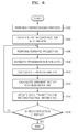
- FIG. 5 is a flowchart of a method of reconstructing an image, according to one or more embodiments.
- the details of the image processing apparatus 400 are applied to the method according to the present embodiments. Details that will be described below with reference to equations are also applied to the image processing apparatus 400 .
- the input unit 410 of the image processing apparatus 400 receives transmission image data measured by the X-ray imaging apparatus 110 .
- the transmission image data is transmitted to the image processor 420 through the input unit 410 of the image processing apparatus 400 .
- the image processor 420 of the image processing apparatus 400 generates reconstruction information for generating an image indicating a target, in particular tomograms indicating the target from the transmission image data.
- the reconstruction information for reconstructing the tomograms is generated in consideration of the fact that X-rays have a polychromatic spectrum and the target is affected by energy of X-rays transmitted through the target.
- the image processor 420 of the image processing apparatus 400 when the reconstruction information for reconstructing the tomograms is determined, the image processor 420 of the image processing apparatus 400 generates tomograms from the determined reconstruction information.
- FIG. 6 is a detailed flowchart of an operation 504 of obtaining the reconstruction information of the target of the method of reconstructing an image of FIG. 5 .
- the preprocessing unit 425 may perform a basic process for removing noise from the transmission image data that is input to the image processor 420 , or may perform processing such as calibration or correction on the transmission image data. These processes may have been previously performed by an imaging apparatus.
- the transmission image data input to the image processor 420 may be modeled according to Equation 1 described above. Equation 3 below may be obtained by performing discretization, that is a process of transferring continuous models or equations into discrete counterparts, on Equation 1.
- Equation 3 may be obtained by approximating an attenuation curve of materials included in the target using linear combination of K basic functions.
- l i,k means a line integral of the product of a density value of each of voxels through which an X-ray with a particular wavelength has been transmitted in the target positioned between an i th cell of an X-ray detection unit and an X-ray source and a distance by which the X-ray is transmitted in each of the voxels. This may be expressed according to Equation 4 below.
- Equation 4 i denotes an index of a cell of the X-ray detection unit, j denotes an index of a voxel that is a unit region of the target, and k denotes an index of a material included in the target.
- a i,j denotes a distance by which an X-ray having a particular wavelength is transmitted in a j th voxel and x j,k denotes an intensity value of a material k of a j th voxel.
- One or more embodiments may also be applied to logarithmic conversion data that is obtained by applying a negative logarithmic value to an equation that is obtained by modeling the transmission image data.
- Transmission image data z i to which logarithmic conversion is applied may be expressed according to Equation 5.
- radiography uses image data that is obtained by applying a negative logarithmic value to data
- a transmission image data model to which logarithmic conversion has been applied may be used.
- the initial reconstructing unit 432 obtains initial reconstruction information using transmission image data input to the initial reconstructing unit 432 . That is, the initial reconstruction information is first reconstruction information that is formed using the measured transmission image data. As described above, the reconstruction information may be a density value of a material of the target at a predetermined position, that is, a predetermined voxel of the target, which is been indicated by x j,k . Such an initial reconstructing process may use a filtered back projection or the like that is well known to those of ordinary skill in the art. Then, a process of forming new reconstruction information based on the reconstruction information is performed and a series of processes for forming new reconstruction information may be repeated.
- the forward projection unit 434 may perform forward projection on the reconstruction information.
- the forward projection is performed in order to consider paths along which X-rays of respective wavelengths are transmitted through a target when a polychromatic X-ray spectrum is radiated to the target that is recognized based on the reconstruction information x j,k .
- a line integral parameter l i,k may be obtained according to Equation 4.
- the estimating unit 436 may simulate a case in which an X-ray of a polychromatic spectrum is radiated to the target that is recognized from the reconstruction information to estimate the transmission image data.
- Transmission image data may be estimated by applying the obtained line integral parameter l i,k to Equation 3 obtained by modeling the transmission image data and the estimated transmission image data may be expressed by ⁇ i .
- the transmission image data to which logarithmic conversion is applied may be estimated by applying the line integral parameter l i,k to Equation 5 that is obtained by applying a negative logarithmic value to Equation 3 and the estimated transmission image data may be expressed by ⁇ circumflex over (z) ⁇ i .
- the error parameter calculating unit 438 may calculate an error parameter.
- the error parameter may be previously defined by the user.
- the error parameter may be a ratio of the estimated transmission image data to the measured transmission image data or a reciprocal of the ratio.
- the error parameter is e i and is defined using Equation 6 below. In this case, e i F denotes a forward form of the error parameter and e i R denotes a reverse form of the error parameter.
- An error parameter in a logarithmic domain that is obtained by applying a negative logarithmic value to transmission image data may be expressed in an equation similar to Equation 6 and may be denoted by d i .
- the error parameter in a logarithmic domain may be expressed using d i F or d i R according to Equation 7 below.
- the optimizing information calculating unit 440 may calculate optimization information used to obtain the reconstruction information.
- the optimization information may correspond to the Hessian matrix and the gradient vector related to line integral parameters.
- the gradient vector and the Hessian matrix are used, which is a result obtained by analyzing an algorithm for obtaining reconstruction information.
- the algorithm for obtaining reconstruction information will be described below.
- Equation 3 an equation obtained by modeling transmission image data has a complex form obtained by coupling an energy integral and a line integral and thus it is very difficult to obtain the reconstruction information x j,k analytically from the equation.
- a cost function may be defined by introducing the GID concept.
- the GID concept is obtained by generalizing an entropy concept between non-negative vectors and a distance between nonnegative values (for example, y i and ⁇ i , or z i and ⁇ circumflex over (z) ⁇ i ) may be defined according to Equation 8 below.
- Equation 8 ⁇ (u) may satisfy Equation 9 below.
- Equation 9 illustrates that ‘u’ for minimizing ⁇ (u) is 1.
- GID may be considered to have a forward form and a reverse form and may be defined according to Equation 10 below.
- ⁇ F ( y, ⁇ ) ⁇ ( y ⁇ )
- R ( y, ⁇ ) ⁇ ( ⁇ y ) (10)
- a radio between the measured transmission image data y i and the estimated transmission image data ⁇ i may be defined as e i F or e i R according to Equation 6, as described above.
- a perimeter dependent upon energy may be introduced as below and a marginal sum of the parameter on energy is equal to the estimated transmission image data ⁇ i .
- Equation 12 An equation obtained by modeling transmission image data with respect to X-rays with a respective energy region, that is, X-rays having respective wavelengths is referred to as m i (E) and is expressed according to Equation 12 below.
- Equation 6 e i F (E) that is not only satisfying Equation 6 but also satisfying Equation 13 as below is simulated.
- e i F (E) may correspond to a distance between a current measured data value and an actual correct answer, for each respective energy band, that is, for each respective wavelength.
- Equation 14 a parameter for a case of reverse GID is defined according to Equation 14 below.
- Equation 15 an equation similar to Equation 13 for a case of forward GID may be expressed according to Equation 15 below.
- Equation 10 may be modified to Equation 16 below.
- Equation 10 may be modified to Equation 17 below.
- an added component in a right section of Equation 16 indicates a distance at an energy region E, and an integral of energies is applied to the outermost region of a cost function by defining the cost function according to sum of distances at respective energy regions E.
- Equation 8 Equation 18
- Equation 19 When u is greater than 0, since ⁇ log(u) is convex downwards, Equation 19 below may be satisfied.
- d i F (E) and d i R (E) may be defined according to Equations 20 below.
- GID with respect to transmission image data to which logarithmic conversion has been applied is expressed according to Equations 21 and 22 below.
- Equation 16 optimization of a right section of Equation 16 corresponds to double minimization of two parameters e i (E) and m i (E).
- e i (E) may be obtained according to Equation 13 or Equation 15.
- ê i (E) is determined, X may be reconstructed by optimizing m i (E) with respect to X.
- Equation 23 a cost function with respect to a parameter m i (E) that is obtained by modeling transmission image data with respect to an X-ray with a particular wavelength may be defined according to Equation 23 below.
- the cost function may be defined by the sum of a distance between a cell i of an X-ray detection unit and an energy E, that is, the sum of c i (E,l i ).
- E the sum of c i (E,l i )
- a relationship between x k to be reconstructed and l i,k may satisfy Equation 4.
- a surrogate function related to l i,k may be considered, instead of minimizing Equation 23 with respect to x k directly, and may be defined according to Equation 24 below.
- Equation 25 is a quadratic equation regarding a parameter l i related to an unknown value.
- a term dependent upon energy is modified to a constant that is used as a coefficient included in the quadratic equation. That is, a cost function
- Equation 25 is obtained by decoupling integral and line integral of energies. Since the form of Equation 25 is the same as an equation for obtaining a surrogate function when transmission image data with respect to an X-ray with a particular wavelength is monochromatic-modeled, Equation 25 may be solved by using known methods of obtaining a line integral.
- Equation 25 Optimization of Equation 25 may be obtained as optimization with respect to respective voxels in consideration of a fact that q i ⁇ (l i ) is convex. That is, an obtained surrogate function may be obtained as a separate surrogate function (SSF). Equation 26 below may be obviously satisfied.
- ⁇ is defined according to Equation 28 below.
- Equation 29 is updated by using a Newton's method according to Equation 29 below.
- x j x ⁇ j - ( ⁇ i ⁇ a i , j ⁇ ⁇ i ⁇ H i ⁇ ⁇ ( l ⁇ i ) ) - 1 ⁇ ( ⁇ i ⁇ a i , j ⁇ g i ⁇ ⁇ ( l ⁇ i ) ) ( 29 )
- ⁇ i ⁇ a i , j ⁇ g ⁇ ( ⁇ ) means vectors obtained by back projecting respective elements of a vector g(•) and
- ⁇ i ⁇ a i , j ⁇ H ⁇ ( ⁇ ) means matrixes obtained by back projecting respective elements of a matrix H(•)
- q i ⁇ (•) may be obtained by quadratic approximation of the original cost function. That is, a minimization algorithm may be obtained by repeatedly performing quadratic approximation on the original cost function.
- Equation 25 g i ⁇ ( ⁇ circumflex over (l) ⁇ i ) and H i ⁇ ( ⁇ circumflex over (l) ⁇ i ) may correspond to a gradient vector and a Hessian matrix of the original cost function.
- the gradient vector g i ⁇ ( ⁇ circumflex over (l) ⁇ i ) and the Hessian matrix H i ⁇ ( ⁇ circumflex over (l) ⁇ i ) may be expressed according to Equations 30 and 31 below, respectively.
- Equation 31 e i is defined in Equation 6
- e i (E) is defined in Equation 13 or Equation 15
- m i (E) is defined in Equation 12.
- Equation 32 d i is defined according to Equation 7
- d i (E) is defined according to Equation 20
- t i (E) is defined according to Equation 19.
- ⁇ (u) that is used in the gradient vector g i ⁇ ( ⁇ circumflex over (l) ⁇ i ) and the Hessian matrix H i ⁇ ( ⁇ circumflex over (l) ⁇ i ) may have various forms. Four types of ⁇ (u) below may be used.
- Equation 29 shows a relationship between previous reconstruction information ⁇ circumflex over (X) ⁇ j and new updated reconstruction information x j .
- the new updated reconstruction information x j is generated by obtaining a gradient vector g i ⁇ ( ⁇ circumflex over (l) ⁇ i ) and a Hessian matrix H i ⁇ ( ⁇ circumflex over (l) ⁇ i ) and performing a predetermining calculation process.
- the optimizing information calculating unit 440 uses an error parameter and a line integral parameter that are calculated in a previous process for obtaining the gradient vector g i ⁇ ( ⁇ circumflex over (l) ⁇ i ) and the Hessian matrix H i ⁇ ( ⁇ circumflex over (l) ⁇ i ). Other data required for the calculation is obtained from the storage 470 and is used to calculated optimization information.
- the back projection and update unit 442 generates new reconstruction information according to Equation 29.
- the back projection and update unit 442 receives the gradient vector g i ⁇ ( ⁇ circumflex over (l) ⁇ i ) and the Hessian matrix H i ⁇ ( ⁇ circumflex over (l) ⁇ i ), which are calculated by the optimizing information calculating unit 440 , receives the reconstruction information from the storage 470 , and performs a predetermined calculation process.
- a first vector may be generated by back projecting elements of the gradient vector and a first matrix is generated by back projecting elements of the Hessian matrix.
- the new reconstruction information may be obtained by calculating linear summation of the previous reconstruction information and a value obtained by the product of an inverse matrix of the first matrix and the first vector.
- the generated new reconstruction information is updated as reconstruction information.
- the determination unit 444 may determine whether a process for obtaining the new reconstruction information is further performed.
- a criterion for the determination may be a repeat number of the reconstruction process, which is previously determined by a user and a data amount difference between the updated reconstruction information and the previous reconstruction information.
- the determination unit 444 performs a new process for generating reconstruction information again. In this case, the determination unit 444 uses the reconstruction information that has been reconstructed so far.
- operation 606 of performing forward projection is performed.
- the determination unit 444 finishes the reconstruction process the lastly updated reconstruction information is used as reconstruction information to reconstruct tomograms indicating the target.
- Equation 29 for obtaining new reconstruction information may be modified to Equations 35 and 36 below.
- g j , s ⁇ ⁇ i ⁇ a i , j , s ⁇ g i , s ⁇ ⁇ ( l ⁇ i , s ) ( 35 )
- g j ⁇ ⁇ s ⁇ g j , s ⁇ ( 36 )
- Equation 29 back projection is performed to elements of the gradient vector.
- Equation 35 shows that a back projected vector corresponding to any one spectrum from among a plurality of spectrums is generated.
- Equation 36 shows a process of adding back projected vectors corresponding to each of the spectrums. As a result, a single vector g j ⁇ is generated in consideration of a plurality of spectrums according to Equation 36.
- H j , s ⁇ ⁇ i ⁇ a i , j , s ⁇ ⁇ i , s ⁇ H i , s ⁇ ⁇ ( l ⁇ i , s ) ( 37 )
- H j ⁇ ⁇ s ⁇ H j , s ⁇ ( 38 )
- Equation 29 elements of the Hessian matrix are back projected.
- Equation 37 shows that a back projected matrix corresponding to any one spectrum from among spectrums is generated.
- Equation 38 shows a process of adding back projected vectors corresponding to each of the spectrums.
- a single matrix H j ⁇ is generated in consideration of a plurality of spectrums according to Equation 38.
- new reconstruction information may be generated using g j ⁇ and H j ⁇ according to Equation 39 below.
- x j ⁇ circumflex over (X) ⁇ j ⁇ ( H j ⁇ ) ⁇ 1 ( g j ⁇ ) (39)
- Equation 39 for generating reconstruction information in consideration of a plurality of spectrums uses a Newton's Method, like in Equation 29 for generating reconstruction information in consideration of a single spectrum.
- a Newton's Method like in Equation 29 for generating reconstruction information in consideration of a single spectrum.
- the embodiments of the method of reconstructing tomograms from a polychromatic X-ray transmission image can be written as computer programs and can be implemented in general-use digital computers that execute the programs using a non-transitory computer readable recording medium.
- non-transitory computer-readable recording medium examples include magnetic media such as hard disks, floppy disks, and magnetic tape; optical media such as CD ROM disks and DVDs; magneto-optical media such as optical disks; and hardware devices that are specially configured to store and perform program instructions, such as read-only memory (ROM), random access memory (RAM), flash memory, and the like.
- program instructions include both machine code, such as produced by a compiler, and files containing higher level code that may be executed by the computer using an interpreter.
- the described hardware devices may be configured to act as one or more software modules in order to perform the operations of the above-described embodiments, or vice versa.
- Any one or more of the software modules described herein may be executed by a controller such as a dedicated processor unique to that unit or by a processor common to one or more of the modules.
- the described methods may be executed on a general purpose computer or processor or may be executed on a particular machine such as the apparatus for processing an, image described herein.
- an image of a target is reconstructed in consideration of a fact that an X-ray has a polychromatic X-ray spectrum and the target is dependent upon energy of the X-ray transmitted through the target, and thus an image without any beam hardening artifacts may be generated.
- measurement data for X-rays with respect to all wavelengths is used to reconstruct an image, there by reconstructing an accurate image.
Landscapes
- Physics & Mathematics (AREA)
- Engineering & Computer Science (AREA)
- General Physics & Mathematics (AREA)
- Theoretical Computer Science (AREA)
- Health & Medical Sciences (AREA)
- Life Sciences & Earth Sciences (AREA)
- Surgery (AREA)
- Public Health (AREA)
- Biomedical Technology (AREA)
- Heart & Thoracic Surgery (AREA)
- Medical Informatics (AREA)
- Molecular Biology (AREA)
- Biophysics (AREA)
- Animal Behavior & Ethology (AREA)
- General Health & Medical Sciences (AREA)
- Pathology (AREA)
- Veterinary Medicine (AREA)
- Analysing Materials By The Use Of Radiation (AREA)
- Apparatus For Radiation Diagnosis (AREA)
- Image Processing (AREA)
- Algebra (AREA)
- Mathematical Analysis (AREA)
- Mathematical Optimization (AREA)
- Mathematical Physics (AREA)
- Pure & Applied Mathematics (AREA)
Abstract
Description
y i =∫S i(ε)exp(−
y i ≈S i(ε0)exp(−
θF(y,ŷ)=θ(y∥ŷ)
θR(y,ŷ)=θ(ŷ∥y) (10)
c i(E,l i)≦q i(E,l i ;{circumflex over (l)} i)=c i(E,{circumflex over (l)} i)+g i(E,{circumflex over (l)} i)T(l i −{circumflex over (l)} i)+½(l i −{circumflex over (l)} i)T H i(E,{circumflex over (l)} i)(l i −{circumflex over (l)} i) (24)
is obtained by decoupling integral and line integral of energies. Since the form of Equation 25 is the same as an equation for obtaining a surrogate function when transmission image data with respect to an X-ray with a particular wavelength is monochromatic-modeled, Equation 25 may be solved by using known methods of obtaining a line integral.
means vectors obtained by back projecting respective elements of a vector g(•) and
means matrixes obtained by back projecting respective elements of a matrix H(•)
x j ={circumflex over (X)} j−(H j ε)−1(g j ε) (39)
Claims (21)
Applications Claiming Priority (2)
| Application Number | Priority Date | Filing Date | Title |
|---|---|---|---|
| KR1020110073246A KR101460615B1 (en) | 2011-07-22 | 2011-07-22 | Method for reconstructing X-ray tomography from a polychromatic X-ray transmission image and image processing apparatus utilizing the method |
| KR10-2011-0073246 | 2011-07-22 |
Publications (2)
| Publication Number | Publication Date |
|---|---|
| US20130182927A1 US20130182927A1 (en) | 2013-07-18 |
| US8958619B2 true US8958619B2 (en) | 2015-02-17 |
Family
ID=46614299
Family Applications (1)
| Application Number | Title | Priority Date | Filing Date |
|---|---|---|---|
| US13/553,033 Expired - Fee Related US8958619B2 (en) | 2011-07-22 | 2012-07-19 | Method of reconstructing tomograms from polychromatic X-ray transmission image and image processing apparatus using the same |
Country Status (3)
| Country | Link |
|---|---|
| US (1) | US8958619B2 (en) |
| EP (1) | EP2549439A3 (en) |
| KR (1) | KR101460615B1 (en) |
Families Citing this family (7)
| Publication number | Priority date | Publication date | Assignee | Title |
|---|---|---|---|---|
| US9713452B2 (en) * | 2014-03-31 | 2017-07-25 | General Electric Company | Generation of monochromatic images |
| US9405990B2 (en) * | 2014-08-19 | 2016-08-02 | Morpho Detection, Llc | X-ray diffraction imaging system with signal aggregation across voxels containing objects and method of operating the same |
| KR20160112494A (en) | 2015-03-19 | 2016-09-28 | (주)바텍이우홀딩스 | Effective oral lesion onset detection method and system in Panorama |
| KR20160112493A (en) | 2015-03-19 | 2016-09-28 | (주)바텍이우홀딩스 | Effective oral lesion onset detection method and system in Computed Tomography |
| US11517197B2 (en) * | 2017-10-06 | 2022-12-06 | Canon Medical Systems Corporation | Apparatus and method for medical image reconstruction using deep learning for computed tomography (CT) image noise and artifacts reduction |
| US10803984B2 (en) | 2017-10-06 | 2020-10-13 | Canon Medical Systems Corporation | Medical image processing apparatus and medical image processing system |
| EP4047344B1 (en) | 2021-02-19 | 2024-08-28 | Helmholtz-Zentrum Dresden - Rossendorf e.V. | A method and device for 3d analyzes of a particulate material |
Citations (5)
| Publication number | Priority date | Publication date | Assignee | Title |
|---|---|---|---|---|
| US20070140410A1 (en) | 2003-10-14 | 2007-06-21 | Koninklijke Philips Electronics N.V. | Coherent-scatter computed tomography |
| KR20090007999A (en) | 2007-07-16 | 2009-01-21 | 삼성테크윈 주식회사 | Surveillance camera system |
| US20090310736A1 (en) | 2006-07-10 | 2009-12-17 | Koninklijke Philips Electronics N.V. | Energy spectrum reconstruction |
| KR20100067326A (en) | 2008-12-11 | 2010-06-21 | 원광대학교산학협력단 | Computed tomography system of nano-spatial resolution |
| US20110262054A1 (en) * | 2006-06-26 | 2011-10-27 | General Electric Company | System and method for iterative image reconstruction |
-
2011
- 2011-07-22 KR KR1020110073246A patent/KR101460615B1/en not_active Expired - Fee Related
-
2012
- 2012-07-19 US US13/553,033 patent/US8958619B2/en not_active Expired - Fee Related
- 2012-07-20 EP EP12177246.1A patent/EP2549439A3/en not_active Withdrawn
Patent Citations (5)
| Publication number | Priority date | Publication date | Assignee | Title |
|---|---|---|---|---|
| US20070140410A1 (en) | 2003-10-14 | 2007-06-21 | Koninklijke Philips Electronics N.V. | Coherent-scatter computed tomography |
| US20110262054A1 (en) * | 2006-06-26 | 2011-10-27 | General Electric Company | System and method for iterative image reconstruction |
| US20090310736A1 (en) | 2006-07-10 | 2009-12-17 | Koninklijke Philips Electronics N.V. | Energy spectrum reconstruction |
| KR20090007999A (en) | 2007-07-16 | 2009-01-21 | 삼성테크윈 주식회사 | Surveillance camera system |
| KR20100067326A (en) | 2008-12-11 | 2010-06-21 | 원광대학교산학협력단 | Computed tomography system of nano-spatial resolution |
Non-Patent Citations (5)
| Title |
|---|
| Chung et al., "Numerical Algorithms for Polyenergetic Digital Breast Tomosynthesis Reconstruction," Society for Industrial and Applied Mathematics, SIAM J. Imaging Sciences, vol. 3, No. 1, 2010, pp. 133-152. |
| European Search Report mailed Sep. 19, 2013 in corresponding European Application No. 12177246.1. |
| Korean Office Action issued Apr. 23, 2014 in corresponding Korean Patent Application 10-2011-0073246. |
| Schneider et al. "Coherent Scatter Computer Tomography Applying a Fan-Beam Geometry," Medical Imaging 2001: Physics of Medical Imaging, Proceedings of SPIE, vol. 4320, 2001. * |
| Srivastava, "Accelerated Statistical Image Reconstruction Algorithms and Simplified Cost Functions for X-Ray Computed Tomography," The University of Michigan, 2008. * |
Also Published As
| Publication number | Publication date |
|---|---|
| EP2549439A3 (en) | 2013-10-30 |
| KR20130011823A (en) | 2013-01-30 |
| EP2549439A2 (en) | 2013-01-23 |
| KR101460615B1 (en) | 2014-11-14 |
| US20130182927A1 (en) | 2013-07-18 |
Similar Documents
| Publication | Publication Date | Title |
|---|---|---|
| Abadi et al. | DukeSim: a realistic, rapid, and scanner-specific simulation framework in computed tomography | |
| JP7245364B2 (en) | sCT Imaging Using CycleGAN with Deformable Layers | |
| US8958619B2 (en) | Method of reconstructing tomograms from polychromatic X-ray transmission image and image processing apparatus using the same | |
| Würfl et al. | Deep learning computed tomography: Learning projection-domain weights from image domain in limited angle problems | |
| US11419568B2 (en) | Multi-energy metal artifact reduction | |
| US7782996B2 (en) | Method for combined bone hardening and scattered radiation correction in X-ray computed tomography | |
| US9836824B2 (en) | De-noised reconstructed image data edge improvement | |
| US11039786B2 (en) | Method and apparatus for determining a patient weight and/or a body mass index | |
| US9600924B2 (en) | Iterative reconstruction of image data in CT | |
| Jadick et al. | A scanner-specific framework for simulating CT images with tube current modulation | |
| US20110268334A1 (en) | Apparatus for Improving Image Resolution and Apparatus for Super-Resolution Photography Using Wobble Motion and Point Spread Function (PSF), in Positron Emission Tomography | |
| US10049446B2 (en) | Accelerated statistical iterative reconstruction | |
| EP2877980B1 (en) | Dose deformation error calculation method and system | |
| JP2014509037A (en) | Processing based on image data model | |
| CN107530037B (en) | Method for generating contrast medium concentrations figure | |
| WO2011133606A2 (en) | Real-time volumetric image reconstruction and 3d tumor localization based on a single x-ray projection image for lung cancer radiotherapy | |
| Lappas et al. | Automatic contouring of normal tissues with deep learning for preclinical radiation studies | |
| US10223814B2 (en) | Information processing apparatus, image reconstruction method, and computer-readable medium | |
| WO2019060688A1 (en) | System and method for low-dose multi-spectral x-ray tomography | |
| Avila et al. | Calibration phantom-based prediction of CT lung nodule volume measurement performance | |
| US8867701B2 (en) | Apparatus for capturing radiation image, medical imaging system, and method of capturing radiation image | |
| CN113424227A (en) | Motion estimation and compensation in Cone Beam Computed Tomography (CBCT) | |
| Abascal et al. | A novel prior-and motion-based compressed sensing method for small-animal respiratory gated CT | |
| CN117751388A (en) | Method of noninvasive medical tomography with uncertainty estimation | |
| Gianoli et al. | A sinogram warping strategy for pre-reconstruction 4D PET optimization |
Legal Events
| Date | Code | Title | Description |
|---|---|---|---|
| AS | Assignment |
Owner name: SAMSUNG ELECTRONICS CO., LTD., KOREA, REPUBLIC OF Free format text: ASSIGNMENT OF ASSIGNORS INTEREST;ASSIGNORS:JANG, KWANG-EUN;SUNG, YOUNG-HUN;LEE, JONG-HA;AND OTHERS;REEL/FRAME:028697/0479 Effective date: 20120725 |
|
| FEPP | Fee payment procedure |
Free format text: PAYOR NUMBER ASSIGNED (ORIGINAL EVENT CODE: ASPN); ENTITY STATUS OF PATENT OWNER: LARGE ENTITY |
|
| STCF | Information on status: patent grant |
Free format text: PATENTED CASE |
|
| CC | Certificate of correction | ||
| MAFP | Maintenance fee payment |
Free format text: PAYMENT OF MAINTENANCE FEE, 4TH YEAR, LARGE ENTITY (ORIGINAL EVENT CODE: M1551) Year of fee payment: 4 |
|
| FEPP | Fee payment procedure |
Free format text: MAINTENANCE FEE REMINDER MAILED (ORIGINAL EVENT CODE: REM.); ENTITY STATUS OF PATENT OWNER: LARGE ENTITY |
|
| LAPS | Lapse for failure to pay maintenance fees |
Free format text: PATENT EXPIRED FOR FAILURE TO PAY MAINTENANCE FEES (ORIGINAL EVENT CODE: EXP.); ENTITY STATUS OF PATENT OWNER: LARGE ENTITY |
|
| STCH | Information on status: patent discontinuation |
Free format text: PATENT EXPIRED DUE TO NONPAYMENT OF MAINTENANCE FEES UNDER 37 CFR 1.362 |
|
| FP | Lapsed due to failure to pay maintenance fee |
Effective date: 20230217 |