US8905997B2 - Therapeutic particles suitable for parenteral administration and methods of making and using same - Google Patents
Therapeutic particles suitable for parenteral administration and methods of making and using same Download PDFInfo
- Publication number
- US8905997B2 US8905997B2 US14/015,549 US201314015549A US8905997B2 US 8905997 B2 US8905997 B2 US 8905997B2 US 201314015549 A US201314015549 A US 201314015549A US 8905997 B2 US8905997 B2 US 8905997B2
- Authority
- US
- United States
- Prior art keywords
- polymer
- therapeutic
- blood vessel
- nih
- seq
- Prior art date
- Legal status (The legal status is an assumption and is not a legal conclusion. Google has not performed a legal analysis and makes no representation as to the accuracy of the status listed.)
- Expired - Fee Related
Links
- LPFWDJFYCTVXBM-UHFFFAOYSA-N CCCC(=O)OCC(COP(=O)(O)OCCNC(=O)CCOCCSCC(N)C(=O)NC(CCCNC(=N)N)C(=O)CC(CCC(=O)O)C(=O)NC(CCCCN)C(=O)CC(C)C(=O)O)OC(=O)CCC Chemical compound CCCC(=O)OCC(COP(=O)(O)OCCNC(=O)CCOCCSCC(N)C(=O)NC(CCCNC(=N)N)C(=O)CC(CCC(=O)O)C(=O)NC(CCCCN)C(=O)CC(C)C(=O)O)OC(=O)CCC LPFWDJFYCTVXBM-UHFFFAOYSA-N 0.000 description 1
- 0 [7*]OC(C)C(=O)OC(C)C(=O)OCC(=O)OCC(=O)[8*]OCCOCSCC(N)C(=O)N[C@H](CCCNC(=N)N)C(=O)CC(CCC(=O)O)C(=O)NC(CCCCN)C(=O)CC(C)C(=O)O Chemical compound [7*]OC(C)C(=O)OC(C)C(=O)OCC(=O)OCC(=O)[8*]OCCOCSCC(N)C(=O)N[C@H](CCCNC(=N)N)C(=O)CC(CCC(=O)O)C(=O)NC(CCCCN)C(=O)CC(C)C(=O)O 0.000 description 1
Images


Classifications
-
- A—HUMAN NECESSITIES
- A61—MEDICAL OR VETERINARY SCIENCE; HYGIENE
- A61K—PREPARATIONS FOR MEDICAL, DENTAL OR TOILETRY PURPOSES
- A61K31/00—Medicinal preparations containing organic active ingredients
- A61K31/33—Heterocyclic compounds
- A61K31/335—Heterocyclic compounds having oxygen as the only ring hetero atom, e.g. fungichromin
- A61K31/337—Heterocyclic compounds having oxygen as the only ring hetero atom, e.g. fungichromin having four-membered rings, e.g. taxol
-
- A61K47/48346—
-
- A—HUMAN NECESSITIES
- A61—MEDICAL OR VETERINARY SCIENCE; HYGIENE
- A61K—PREPARATIONS FOR MEDICAL, DENTAL OR TOILETRY PURPOSES
- A61K31/00—Medicinal preparations containing organic active ingredients
-
- A—HUMAN NECESSITIES
- A61—MEDICAL OR VETERINARY SCIENCE; HYGIENE
- A61K—PREPARATIONS FOR MEDICAL, DENTAL OR TOILETRY PURPOSES
- A61K31/00—Medicinal preparations containing organic active ingredients
- A61K31/21—Esters, e.g. nitroglycerine, selenocyanates
-
- A—HUMAN NECESSITIES
- A61—MEDICAL OR VETERINARY SCIENCE; HYGIENE
- A61K—PREPARATIONS FOR MEDICAL, DENTAL OR TOILETRY PURPOSES
- A61K31/00—Medicinal preparations containing organic active ingredients
- A61K31/33—Heterocyclic compounds
- A61K31/395—Heterocyclic compounds having nitrogen as a ring hetero atom, e.g. guanethidine or rifamycins
- A61K31/435—Heterocyclic compounds having nitrogen as a ring hetero atom, e.g. guanethidine or rifamycins having six-membered rings with one nitrogen as the only ring hetero atom
- A61K31/4353—Heterocyclic compounds having nitrogen as a ring hetero atom, e.g. guanethidine or rifamycins having six-membered rings with one nitrogen as the only ring hetero atom ortho- or peri-condensed with heterocyclic ring systems
-
- A61K47/48238—
-
- A61K47/48915—
-
- A—HUMAN NECESSITIES
- A61—MEDICAL OR VETERINARY SCIENCE; HYGIENE
- A61K—PREPARATIONS FOR MEDICAL, DENTAL OR TOILETRY PURPOSES
- A61K47/00—Medicinal preparations characterised by the non-active ingredients used, e.g. carriers or inert additives; Targeting or modifying agents chemically bound to the active ingredient
- A61K47/50—Medicinal preparations characterised by the non-active ingredients used, e.g. carriers or inert additives; Targeting or modifying agents chemically bound to the active ingredient the non-active ingredient being chemically bound to the active ingredient, e.g. polymer-drug conjugates
- A61K47/51—Medicinal preparations characterised by the non-active ingredients used, e.g. carriers or inert additives; Targeting or modifying agents chemically bound to the active ingredient the non-active ingredient being chemically bound to the active ingredient, e.g. polymer-drug conjugates the non-active ingredient being a modifying agent
- A61K47/62—Medicinal preparations characterised by the non-active ingredients used, e.g. carriers or inert additives; Targeting or modifying agents chemically bound to the active ingredient the non-active ingredient being chemically bound to the active ingredient, e.g. polymer-drug conjugates the non-active ingredient being a modifying agent the modifying agent being a protein, peptide or polyamino acid
- A61K47/66—Medicinal preparations characterised by the non-active ingredients used, e.g. carriers or inert additives; Targeting or modifying agents chemically bound to the active ingredient the non-active ingredient being chemically bound to the active ingredient, e.g. polymer-drug conjugates the non-active ingredient being a modifying agent the modifying agent being a protein, peptide or polyamino acid the modifying agent being a pre-targeting system involving a peptide or protein for targeting specific cells
-
- A—HUMAN NECESSITIES
- A61—MEDICAL OR VETERINARY SCIENCE; HYGIENE
- A61K—PREPARATIONS FOR MEDICAL, DENTAL OR TOILETRY PURPOSES
- A61K47/00—Medicinal preparations characterised by the non-active ingredients used, e.g. carriers or inert additives; Targeting or modifying agents chemically bound to the active ingredient
- A61K47/50—Medicinal preparations characterised by the non-active ingredients used, e.g. carriers or inert additives; Targeting or modifying agents chemically bound to the active ingredient the non-active ingredient being chemically bound to the active ingredient, e.g. polymer-drug conjugates
- A61K47/69—Medicinal preparations characterised by the non-active ingredients used, e.g. carriers or inert additives; Targeting or modifying agents chemically bound to the active ingredient the non-active ingredient being chemically bound to the active ingredient, e.g. polymer-drug conjugates the conjugate being characterised by physical or galenical forms, e.g. emulsion, particle, inclusion complex, stent or kit
- A61K47/6921—Medicinal preparations characterised by the non-active ingredients used, e.g. carriers or inert additives; Targeting or modifying agents chemically bound to the active ingredient the non-active ingredient being chemically bound to the active ingredient, e.g. polymer-drug conjugates the conjugate being characterised by physical or galenical forms, e.g. emulsion, particle, inclusion complex, stent or kit the form being a particulate, a powder, an adsorbate, a bead or a sphere
- A61K47/6927—Medicinal preparations characterised by the non-active ingredients used, e.g. carriers or inert additives; Targeting or modifying agents chemically bound to the active ingredient the non-active ingredient being chemically bound to the active ingredient, e.g. polymer-drug conjugates the conjugate being characterised by physical or galenical forms, e.g. emulsion, particle, inclusion complex, stent or kit the form being a particulate, a powder, an adsorbate, a bead or a sphere the form being a solid microparticle having no hollow or gas-filled cores
- A61K47/6929—Medicinal preparations characterised by the non-active ingredients used, e.g. carriers or inert additives; Targeting or modifying agents chemically bound to the active ingredient the non-active ingredient being chemically bound to the active ingredient, e.g. polymer-drug conjugates the conjugate being characterised by physical or galenical forms, e.g. emulsion, particle, inclusion complex, stent or kit the form being a particulate, a powder, an adsorbate, a bead or a sphere the form being a solid microparticle having no hollow or gas-filled cores the form being a nanoparticle, e.g. an immuno-nanoparticle
- A61K47/6931—Medicinal preparations characterised by the non-active ingredients used, e.g. carriers or inert additives; Targeting or modifying agents chemically bound to the active ingredient the non-active ingredient being chemically bound to the active ingredient, e.g. polymer-drug conjugates the conjugate being characterised by physical or galenical forms, e.g. emulsion, particle, inclusion complex, stent or kit the form being a particulate, a powder, an adsorbate, a bead or a sphere the form being a solid microparticle having no hollow or gas-filled cores the form being a nanoparticle, e.g. an immuno-nanoparticle the material constituting the nanoparticle being a polymer
- A61K47/6935—Medicinal preparations characterised by the non-active ingredients used, e.g. carriers or inert additives; Targeting or modifying agents chemically bound to the active ingredient the non-active ingredient being chemically bound to the active ingredient, e.g. polymer-drug conjugates the conjugate being characterised by physical or galenical forms, e.g. emulsion, particle, inclusion complex, stent or kit the form being a particulate, a powder, an adsorbate, a bead or a sphere the form being a solid microparticle having no hollow or gas-filled cores the form being a nanoparticle, e.g. an immuno-nanoparticle the material constituting the nanoparticle being a polymer the polymer being obtained otherwise than by reactions involving carbon to carbon unsaturated bonds, e.g. polyesters, polyamides or polyglycerol
- A61K47/6937—Medicinal preparations characterised by the non-active ingredients used, e.g. carriers or inert additives; Targeting or modifying agents chemically bound to the active ingredient the non-active ingredient being chemically bound to the active ingredient, e.g. polymer-drug conjugates the conjugate being characterised by physical or galenical forms, e.g. emulsion, particle, inclusion complex, stent or kit the form being a particulate, a powder, an adsorbate, a bead or a sphere the form being a solid microparticle having no hollow or gas-filled cores the form being a nanoparticle, e.g. an immuno-nanoparticle the material constituting the nanoparticle being a polymer the polymer being obtained otherwise than by reactions involving carbon to carbon unsaturated bonds, e.g. polyesters, polyamides or polyglycerol the polymer being PLGA, PLA or polyglycolic acid
-
- A—HUMAN NECESSITIES
- A61—MEDICAL OR VETERINARY SCIENCE; HYGIENE
- A61K—PREPARATIONS FOR MEDICAL, DENTAL OR TOILETRY PURPOSES
- A61K9/00—Medicinal preparations characterised by special physical form
- A61K9/0012—Galenical forms characterised by the site of application
- A61K9/0019—Injectable compositions; Intramuscular, intravenous, arterial, subcutaneous administration; Compositions to be administered through the skin in an invasive manner
-
- A—HUMAN NECESSITIES
- A61—MEDICAL OR VETERINARY SCIENCE; HYGIENE
- A61K—PREPARATIONS FOR MEDICAL, DENTAL OR TOILETRY PURPOSES
- A61K9/00—Medicinal preparations characterised by special physical form
- A61K9/48—Preparations in capsules, e.g. of gelatin, of chocolate
- A61K9/50—Microcapsules having a gas, liquid or semi-solid filling; Solid microparticles or pellets surrounded by a distinct coating layer, e.g. coated microspheres, coated drug crystals
- A61K9/51—Nanocapsules; Nanoparticles
- A61K9/5107—Excipients; Inactive ingredients
- A61K9/5123—Organic compounds, e.g. fats, sugars
-
- A—HUMAN NECESSITIES
- A61—MEDICAL OR VETERINARY SCIENCE; HYGIENE
- A61K—PREPARATIONS FOR MEDICAL, DENTAL OR TOILETRY PURPOSES
- A61K9/00—Medicinal preparations characterised by special physical form
- A61K9/48—Preparations in capsules, e.g. of gelatin, of chocolate
- A61K9/50—Microcapsules having a gas, liquid or semi-solid filling; Solid microparticles or pellets surrounded by a distinct coating layer, e.g. coated microspheres, coated drug crystals
- A61K9/51—Nanocapsules; Nanoparticles
- A61K9/5107—Excipients; Inactive ingredients
- A61K9/513—Organic macromolecular compounds; Dendrimers
- A61K9/5146—Organic macromolecular compounds; Dendrimers obtained otherwise than by reactions only involving carbon-to-carbon unsaturated bonds, e.g. polyethylene glycol, polyamines, polyanhydrides
- A61K9/5153—Polyesters, e.g. poly(lactide-co-glycolide)
-
- A—HUMAN NECESSITIES
- A61—MEDICAL OR VETERINARY SCIENCE; HYGIENE
- A61P—SPECIFIC THERAPEUTIC ACTIVITY OF CHEMICAL COMPOUNDS OR MEDICINAL PREPARATIONS
- A61P9/00—Drugs for disorders of the cardiovascular system
-
- A—HUMAN NECESSITIES
- A61—MEDICAL OR VETERINARY SCIENCE; HYGIENE
- A61P—SPECIFIC THERAPEUTIC ACTIVITY OF CHEMICAL COMPOUNDS OR MEDICINAL PREPARATIONS
- A61P9/00—Drugs for disorders of the cardiovascular system
- A61P9/10—Drugs for disorders of the cardiovascular system for treating ischaemic or atherosclerotic diseases, e.g. antianginal drugs, coronary vasodilators, drugs for myocardial infarction, retinopathy, cerebrovascula insufficiency, renal arteriosclerosis
-
- B—PERFORMING OPERATIONS; TRANSPORTING
- B82—NANOTECHNOLOGY
- B82Y—SPECIFIC USES OR APPLICATIONS OF NANOSTRUCTURES; MEASUREMENT OR ANALYSIS OF NANOSTRUCTURES; MANUFACTURE OR TREATMENT OF NANOSTRUCTURES
- B82Y5/00—Nanobiotechnology or nanomedicine, e.g. protein engineering or drug delivery
Definitions
- NASH Neointimal hyperplasia
- Neointimal hyperplasia can involve smooth muscle cell proliferation, migrations and/or production of the extracellular matrix. With the introduction of arterial stents, the problems of elastic recoil or negative arterial remodeling has been substantially eliminated. However, neointimal hyperplasia is still a primary cause of restenosis after the introduction of a stent in the artery of a patient. Both bare metal stents and drug-eluting stents that typically elute an anti-restenosis agent are available.
- Drug eluting stents are not optimal under all conditions, however. Such stents may not be appropriate for small vessels, and drug eluting stents can hinder vessel healing. For example, drug eluting stents may lead to a higher rate of thrombosis e.g. a year after implantation, when for example antiplatelet therapy (e.g. clopidogrel) is discontinued. For patients needing surgery, for example, patients may suffer fatal heart attacks due to clotting inside of drug-eluting stents, even months or years after surgery, particular if blood thinning medication is stopped (as is often necessary) before surgery. Patients identified as being likely to be non-compliant with antiplatelet therapy may not be suitable candidates for drug-eluting stents.
- antiplatelet therapy e.g. clopidogrel
- bare metal stents may actually provide a safer choice, at least for some patients.
- Improved compositions and methods for delivery of anti-neointimal hyperplasia, or anti-restenosis, agents for local and/or targeted delivery to blood vessels, for example, in conjunction with placement of a bare-metal stents is therefore needed.
- compositions that include therapeutic particles comprising an anti-neointimal hyperplasia (NIH) agent.
- NIH anti-neointimal hyperplasia
- Such therapeutic particles may be capable of releasing said anti-NIH agent to a blood vessel, for example, to a basement vascular membrane of a blood vessel.
- such particles may be capable of releasing an anti-NIH agent for at least about 8 hours when a disclosed therapeutic particle or composition is placed in the blood vessel.
- Such compositions may be used, for example, with a patient receiving a vascular stent, e.g. a bare metal stent, in a blood vessel.
- the disclosure provides a therapeutic composition
- a therapeutic composition comprising a plurality of therapeutic particles each comprising an anti-NIH agent and a basement vascular membrane targeting peptide, wherein a single administration of a dose of the composition to a blood vessel is capable of contacting a blood vessel with a substantially higher surface area density as compared to a surface area density of a blood vessel contacted by a stent comprising the anti-NIH agent.
- the disclosure provides a method of preventing or deterring NIH in a blood vessel of a patient receiving a bare metal stent in a lesion of said blood vessel, comprising administering a composition comprising therapeutic particles, wherein said therapeutic particles comprise a basement vascular membrane targeting peptide and an anti-NIH agent.
- methods disclosed herein include intravenous (e.g., systemic) administration of disclosed compositions and/or particles.
- a method of preventing or deterring NIH in a damaged blood vessel of a patient comprising administering to said patient a therapeutic composition comprising a therapeutic particle, wherein said therapeutic particle may comprise a basement vascular membrane targeting peptide and an anti-NIH agent, and wherein said therapeutic particle substantially biodegrades after delivery of the anti-NIH agent thereby promoting healing of the blood vessel after said therapeutic particle has biodegraded.
- compositions for example, for use in a patient receiving a vascular stent, comprising about 1 to about 20 mole percent targeting co-polymer wherein the targeting co-polymer is chosen from: a) PLA-PEG-basement vascular membrane targeting peptide; b) poly (lactic) acid—co poly (glycolic) acid-PEG-basement vascular membrane targeting peptide; c) DSPE-PEG-basement vascular membrane targeting peptide; and about 0.2 to about 30 weight percent anti-neointimal hyperplasia (NIH) agent (e.g. paclitaxel); about 50 to about 90 weight percent non-targeted polylactic acid or poly-lactic acid-PEG.
- NASH anti-neointimal hyperplasia
- such a targeting co-polymer may include PLA-PEG, wherein for example poly(lactic acid) has a number average molecular weight of about 15 to 20 kDa and/or poly(ethylene)glycol has a number average molecular weight of about 4 to about 6 kDa.
- Contemplated basement vascular membrane targeting peptides may be selected, for example, from: CREKA (SEQ ID NO: 1) or CARLYQKLN (SEQ ID NO: 2).
- FIG. 1 depicts three methods for delivery of compositions of the invention to a target site in a blood vessel, where the blood flow 20 is from left to right in the diagram.
- FIG. 1A illustrates delivery by guide catheter in the blood flow.
- FIG. 1B illustrates use of a balloon catheter for proximal delivery.
- FIG. 1C illustrates the use of a guide catheter with a balloon catheter for distal delivery.
- FIG. 2 depicts the effect of lipid content and number of homogenizer passes on particle size using DSPE-PEG5K.
- FIG. 3 depicts the effect of lipid content and number of homogenizer passes on particle size using DSPE-PEG5K.
- FIG. 4 depicts release of paclitaxel loaded particles at 37° C.
- FIG. 5 depicts release of various paclitaxel loaded particles at 37° C.
- FIG. 6 depicts release of various paclitaxel loaded particles.
- FIG. 7 depicts pharmacokinetics of various paclitaxel loaded particles in a rabbit model.
- FIG. 8 depicts the paclitaxel release rate of disclosed nanoparticles with paclitaxel, in a rabbit model.
- FIG. 9 depicts the paclitaxel blood levels in a disclosed rabbit model.
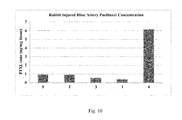
- FIG. 10 depicts injured artery paclitaxel concentration in a rabbit model, after administration of disclosed nanoparticles having paclitaxel.
- Treating includes any effect, e.g., lessening, reducing, modulating, or eliminating, that results in the improvement of the condition, disease, disorder and the like.
- “Pharmaceutically or pharmacologically acceptable” include molecular entities and compositions that do not produce an adverse, allergic or other untoward reaction when administered to an animal, or a human, as appropriate.
- preparations should meet sterility, pyrogenicity, general safety and purity standards as required by FDA Office of Biologics standards.
- compositions may also contain other active compounds providing supplemental, additional, or enhanced therapeutic functions.
- “Individual,” “patient,” or “subject” are used interchangeably and include to any animal, including mammals, such as mice, rats, other rodents, rabbits, dogs, cats, swine, cattle, sheep, horses, or primates, and most preferably humans.
- the compounds and compositions of the invention can be administered to a mammal, such as a human, but can also be other mammals such as an animal in need of veterinary treatment, e.g., domestic animals (e.g., dogs, cats, and the like), farm animals (e.g., cows, sheep, pigs, horses, and the like) and laboratory animals (e.g., rats, mice, guinea pigs, and the like).
- the mammal treated in the methods of the invention is desirably a mammal in whom modulation of NIH is desired.
- “Modulation” includes antagonism (e.g., inhibition), agonism, partial antagonism and/or partial agonism.
- a therapeutically effective amount means the amount of the subject compound or composition that will elicit the biological or medical response of a tissue, system, animal or human that is being sought by the researcher, veterinarian, medical doctor or other clinician.
- the compounds and compositions of the invention are administered in therapeutically effective amounts to treat a disease.
- a therapeutically effective amount of a compound is the quantity required to achieve a desired therapeutic and/or prophylactic effect, such as an amount which results in the prevention of or a decrease in the symptoms associated with NIH.
- pharmaceutically acceptable salt(s) refers to salts of acidic or basic groups that may be present in compounds used in the present compositions.
- Compounds included in the present compositions that are basic in nature are capable of forming a wide variety of salts with various inorganic and organic acids.
- the acids that may be used to prepare pharmaceutically acceptable acid addition salts of such basic compounds are those that form non-toxic acid addition salts, i.e., salts containing pharmacologically acceptable anions, including but not limited to malate, oxalate, chloride, bromide, iodide, nitrate, sulfate, bisulfate, phosphate, acid phosphate, isonicotinate, acetate, lactate, salicylate, citrate, tartrate, oleate, tannate, pantothenate, bitartrate, ascorbate, succinate, maleate, gentisinate, fumarate, gluconate, glucaronate, saccharate, formate, benzoate, glutamate, methanesulfonate, ethanesulfonate, benzenesulfonate, p-toluenesulfonate and pamoate (i.e., 1,1′-methylene-bis-
- Compounds included in the present compositions that include an amino moiety may form pharmaceutically acceptable salts with various amino acids, in addition to the acids mentioned above.
- Compounds included in the present compositions that are acidic in nature are capable of forming base salts with various pharmacologically acceptable cations.
- Examples of such salts include alkali metal or alkaline earth metal salts, such as calcium, magnesium, sodium, lithium, zinc, potassium, and iron salts.
- therapeutic compositions that include a therapeutic particle.
- Such therapeutic particles can include for example an anti-neointimal hyperplasia (NIH) agent or antiinflammatory agent, and may be capable of releasing the anti-NIH agent to a vascular membrane of a blood vessel for at least about 2, 4, 6, 8, 10, 12, or even 24 or more hours, or 1, 2 or 3 or more days, or for about 1, 2, 4 or even 12 or more weeks, when the therapeutic particle is placed in the blood vessel.
- a disclosed therapeutic particle may include basement vascular membrane targeting peptide.
- disclosed therapeutic compositions may be for use, e.g., administered to or with a patient receiving a stent such as a vascular stent, for example, a bare metal stent, or, in some embodiments, a drug-eluting stent.
- a stent such as a vascular stent, for example, a bare metal stent, or, in some embodiments, a drug-eluting stent.
- disclosed compositions may release substantially equal or substantially more effective amount of the anti-NIH agent when placed in the blood vessel as compared to the amount released by a stent comprising the same anti-NIH agent if placed in the blood vessel.
- a disclosed therapeutic composition may release about 10% to about 50% or more, e.g., about 40%, or about 30% of the anti-NIH agent initially, with a more controlled release of the remaining drug over time.
- the anti-NIH agent can be chosen from paclitaxel, sirolimus, zotarolimus and/or everolimus.
- a disclosed therapeutic particle may comprise paclitaxel such that the therapeutic composition releases about 50 ⁇ g to about 1000 ⁇ g, about 50 ⁇ g to about 600 ⁇ g, or about 100 ⁇ g to about 300 ⁇ g, of paclitaxel to a target of interest, e.g. the basement vascular membrane. Such release can occur over a period of about 8 hours to about 8 weeks, such as about 8 hours to about 4 weeks, further such as about 8 hours to about 1 week.
- a disclosed therapeutic particle may comprise sirolimus such that the therapeutic composition releases about 50 ⁇ g to about 250 ⁇ g, such as 50 ⁇ g to about 100 ⁇ g, about 75 ⁇ g to about 150 ⁇ g, of sirolimus to, e.g., the basement vascular membrane.
- a therapeutic particle may comprise zotarolimus, for example, such that the therapeutic composition releases about 50 ⁇ g to about 300 ⁇ g, such as about 50 ⁇ g to about 250 ⁇ g, or about 75 ⁇ g to about 150 ⁇ g, of zotarolimus to e.g., a basement vascular membrane.
- the therapeutic particle may comprise everolimus, for example, such that the therapeutic composition releases about 50 ⁇ g to about 300 ⁇ g, such as about 50 ⁇ g to about 250 ⁇ g, further such as about 75 ⁇ g to about 150 ⁇ g, of everolimus to the basement vascular membrane.
- the release of such drugs can occur over a period of about 8 hours to about 8 weeks, such as about 8 hours to about 4 weeks, further such as about 8 hours to about 1 week.
- Therapeutic particles disclosed herein may include about 5% to about 85% by weight of the anti-NIH agent, such as about 2% to about 35%, or about 10% to about 25%, e.g. about 10%, 15%, or 20% by weight.
- the therapeutic particle may be substantially biodegraded after about 1 month, after about 1 week, such as about 3 days, further such as about 1 day, after placement in the blood vessel.
- compositions may provide a decreased or substantially comparable NIH rate in the patient about 4 weeks, or 3 weeks, or 2 weeks, such as about 1 week, after receiving the vascular stent as compared to the NIH rate obtained by administration of a vascular stent alone.
- the NIH rate may be decreased compared to that from administration of a vascular stent, e.g. a bare metal stent, alone.
- compositions that include a plurality of therapeutic particles each comprising an anti-NIH agent and optionally, a basement vascular membrane targeting peptide, wherein a single administration of a dose of the composition to a blood vessel can be capable of contacting a blood vessel with a substantially higher surface area density as compared to a surface area density of a blood vessel, e.g a surface area density of endothelial cells, as compared to the surface area density contacted by a stent comprising the anti-NIH agent.
- one or more doses of the composition when administered, may contact the blood vessel with a substantially higher surface area density as compared to the density of blood vessel contacted by a stent that includes the anti-NIH agent.
- compositions may contact least a surface area density of about least about 2%, such as at least 5%, such as at least 10% higher than the density of blood vessel contacted by a stent comprising the same or different anti-NIH agent, e.g. a drug-eluting stent.
- Doses of disclosed compositions may be capable of delivering the anti-NIH agent to the blood vessel such that the concentration of the anti-NIH agent in the blood vessel tissue can be about 2 ng/mg to about 100 ng/mg, such as about 15 ng/mg to 50 ng/mg, further such as about 20 ng/mg to 40 ng/mg about 2 days after administration.
- compositions for use in a patient receiving a vascular stent, comprising a plurality of first therapeutic particles, wherein the first therapeutic particles may be capable of localized association with a blood vessel structure, e.g. a basement membrane, smooth muscle cells, endothelial cells, extracellular matrix, or inner elastic lamina, and comprise a first therapeutic agent, e.g. an anti-NIH agent, and wherein a single administration of a dose of the composition to the blood vessel provides a faster endothelial cell healing rate at about 6 months, 4 months, or about 2 months, as compared to the endothelial healing rate of a patient receiving a stent comprising the first therapeutic agent at about 6 months.
- a blood vessel structure e.g. a basement membrane, smooth muscle cells, endothelial cells, extracellular matrix, or inner elastic lamina
- a first therapeutic agent e.g. an anti-NIH agent
- a single administration (or multiple administrations) of a dose of a composition associates with a greater surface area density of the blood vessel structure as compared to a patient receiving a stent comprising the first therapeutic agent.
- Such compositions may further include a plurality of second therapeutic particles.
- the second therapeutic particles may be capable of localized association with a different blood vessel structure than the first therapeutic particles.
- the second therapeutic particles may comprise a second therapeutic agent, which may be different than the first therapeutic agent.
- compositions that include a plurality of first therapeutic particles, wherein the first therapeutic particles may be capable of localized association with a blood vessel structure and comprise a first therapeutic agent such as an anti-NIH agent or anti-inflammatory agent; and a plurality of second therapeutic particles comprising a second therapeutic agent.
- the plurality of first therapeutic particles can be present in a different amount than the plurality of second therapeutic particles.
- the composition can comprise an equal amount of first and second therapeutic particles.
- a target of the first therapeutic agent may be different than a target of the second therapeutic agent.
- Such compositions may release a substantially equal or substantially greater amount of first therapeutic agent than second therapeutic agent, e.g. another anti-NIH agent or anti-inflammatory, in the blood vessel.
- Exemplary second therapeutic agents include, but are not limited to, everolimus, paclitaxel, zotarolimus, pioglitazone, BO-653, rosiglitazone, sirolimus, dexamethasone, rapamycin, tacrolimus, biophosphonates, estrogen, angiopeptin, statin, PDGF inhibitors, ROCK inhibitors, MMP inhibitors, 2-CdA, corticosteroids, including combinations of zotarolimus and dexamethasone, nicotine, hydroxy-methylglutaryl coenzyme A (HMG CoA) reductase inhibitors, statins, niacin, bile acid resins, fibrates, antioxidants, nitric oxide generators, nitric oxide, extracellular matrix synthesis promoters, inhibitors of plaque inflammation, and extracellular degradation, and antithrombotic agents such as clopidogrel.
- HMG CoA hydroxy-methylglutaryl coenzyme A
- Contemplated anti-inflammatories or anti-inflammatory agents which may be useful for restenosis, (e.g. vessel remodeling that occurs in restenosis starts with inflammation in response to the injury caused by angioplasty, and stopping this inflammation has been showed in animal studies to prevent restenosis), include sirolimus, corticosteroids (dexamethasone, prednisolone, triamcinolone acetonide, mometasone, amcinonide, budesonide, acetaminophen, NSAIDS, cox-2 inhibitors, and/or betamethasone.
- Methods contemplated herein include, for example, a method of preventing or deterring NIH in a blood vessel of a patient receiving a bare metal stent in a lesion of the blood vessel, comprising administering a composition comprising disclosed therapeutic particles, such as therapeutic particles that may include a basement vascular membrane targeting peptide and an anti-NIH agent or anti-inflammatory.
- Disclosed methods may provides for decreased or substantially comparable NIH rate at about 2 weeks, such as about 1 week, after receiving the bare metal stent as compared to a patient receiving a stent comprising an anti-NIH agent at about 2 weeks or at about 1 week.
- methods include administering or placing a bare metal stent in a blood vessel, and administering a disclosed therapeutic particle or composition before, after, or substantially simultaneously with the placement of the stent.
- the blood vessel receiving the stent may be less than about 2 mm in length, or between about 2 mm and about 3 mm in length, or may be greater than 3 mm in length.
- a treated blood vessel may be bifurcated or substantially non-bifurcated.
- Such stents for use in the contemplated methods may be a thick or thin strut stent.
- stents may be less than about 14 mm, such as less than about 10 mm, further such as less than about 7 mm in length.
- contemplated stents may be about 14 mm to about 30 mm, such as about 17 mm to about 27 mm, further such as about 20 mm to about 25 mm in length.
- a patient being treated or contemplating treatment may be intolerant or adverse to a particular medication, such as aspirin or clopidogrel.
- a particular medication such as aspirin or clopidogrel.
- Patient populations suitable for treatment with disclosed methods include patients at risk of future surgery, e.g., cardiac or non-cardiac surgery.
- compositions or particles may be administered substantially simultaneously when a patient receives the stent and/or may be administered before or after the patient receives the stent.
- compositions and/or particles may be introduced before or after the introduction of a balloon catheter into the blood vessel.
- the composition may be administered with the same delivery device used to deliver the stent to the patient, or a different delivery device.
- the composition may be administered using a catheter, and/or may be administered intravenously.
- Disclosed compositions may be administered to a patient undergoing, for example, a coronary angioplasty, a peripheral angioplasty, a renal artery angioplasty, or a carotid angioplasty.
- Another embodiment provides a method of preventing or deterring NIH in a damaged blood vessel of a patient, comprising administering to the patient a therapeutic composition comprising a therapeutic particle, wherein the therapeutic particle comprises a basement vascular membrane targeting peptide and an anti-NIH agent, and wherein the therapeutic particle substantially biodegrades after delivery of the anti-NIH agent thereby promoting healing of the blood vessel after the therapeutic particle has biodegraded.
- a therapeutic particle may have substantially degraded about 1 day, such as about 1 week, further such as about 1 month after administration.
- the damaged blood vessel may have been caused by, for example, an implantation of a stent (e.g., a bare metal stent), a balloon angioplasty, or peripheral artery disease.
- a damaged blood vessel may be caused, for example, by balloon angioplasty alone.
- a stent e.g. a bare metal stent or a drug eluting stent
- a stent e.g. a bare metal stent or a drug eluting stent
- a blood vessel may, in some embodiments, have substantially less risk of developing a thrombosis at about 1 month, or greater than about 1 month, about 2 months, further such as greater than about 3 months after administration as compared to a damaged blood vessel receiving a stent comprising an anti-NIH agent, e.g., a drug-eluting stent.
- an anti-NIH agent e.g., a drug-eluting stent.
- Therapeutic particles disclosed herein typically include a polymeric matrix.
- the polymeric matrix comprises one, two or more synthetic or natural polymers.
- the term “polymer,” as used herein, is given its ordinary meaning as used in the art, i.e., a molecular structure comprising one or more repeat units (monomers), connected by covalent bonds. The repeat units may all be identical, or in some cases, there may be more than one type of repeat unit present within the polymer.
- the polymer can be biologically derived, i.e., a biopolymer. Non-limiting examples include peptides or proteins. In some cases, additional moieties may also be present in the polymer, for example biological moieties such as those described below.
- the polymer is said to be a “copolymer.” It is to be understood that in any embodiment employing a polymer, the polymer being employed may be a copolymer in some cases.
- the repeat units forming the copolymer may be arranged in any fashion. For example, the repeat units may be arranged in a random order, in an alternating order, or as a block copolymer, i.e., comprising one or more regions each comprising a first repeat unit (e.g., a first block), and one or more regions each comprising a second repeat unit (e.g., a second block), etc.
- Block copolymers may have two (a diblock copolymer), three (a triblock copolymer), or more numbers of distinct blocks.
- Disclosed particles can include copolymers, which, in some embodiments, describes two or more polymers (such as those described herein) that have been associated with each other, usually by covalent bonding of the two or more polymers together.
- a copolymer may comprise a first polymer and a second polymer, which have been conjugated together to form a block copolymer where the first polymer can be a first block of the block copolymer and the second polymer can be a second block of the block copolymer.
- a block copolymer may, in some cases, contain multiple blocks of polymer, and that a “block copolymer,” as used herein, is not limited to only block copolymers having only a single first block and a single second block.
- a block copolymer may comprise a first block comprising a first polymer, a second block comprising a second polymer, and a third block comprising a third polymer or the first polymer, etc.
- block copolymers can contain any number of first blocks of a first polymer and second blocks of a second polymer (and in certain cases, third blocks, fourth blocks, etc.).
- block copolymers can also be formed, in some instances, from other block copolymers.
- a first block copolymer may be conjugated to another polymer (which may be a homopolymer, a biopolymer, another block copolymer, etc.), to form a new block copolymer containing multiple types of blocks, and/or to other moieties (e.g., to non-polymeric moieties).
- the polymer e.g., copolymer, e.g., block copolymer
- the polymer can be amphiphilic, i.e., having a hydrophilic portion and a hydrophobic portion, or a relatively hydrophilic portion and a relatively hydrophobic portion.
- a hydrophilic polymer can be one generally that attracts water and a hydrophobic polymer can be one that generally repels water.
- a hydrophilic or a hydrophobic polymer can be identified, for example, by preparing a sample of the polymer and measuring its contact angle with water (typically, the polymer will have a contact angle of less than 60°, while a hydrophobic polymer will have a contact angle of greater than about 60°).
- the hydrophilicity of two or more polymers may be measured relative to each other, i.e., a first polymer may be more hydrophilic than a second polymer.
- the first polymer may have a smaller contact angle than the second polymer.
- a polymer e.g., copolymer, e.g., block copolymer
- a biocompatible polymer i.e., the polymer that does not typically induce an adverse response when inserted or injected into a living subject, for example, without significant inflammation and/or acute rejection of the polymer by the immune system, for instance, via a T-cell response.
- the therapeutic particles contemplated herein can be non-immunogenic.
- non-immunogenic refers to endogenous growth factor in its native state which normally elicits no, or only minimal levels of, circulating antibodies, T-cells, or reactive immune cells, and which normally does not elicit in the individual an immune response against itself.
- Biocompatibility typically refers to the acute rejection of material by at least a portion of the immune system, i.e., a nonbiocompatible material implanted into a subject provokes an immune response in the subject that can be severe enough such that the rejection of the material by the immune system cannot be adequately controlled, and often is of a degree such that the material must be removed from the subject.
- One simple test to determine biocompatibility can be to expose a polymer to cells in vitro; biocompatible polymers are polymers that typically will not result in significant cell death at moderate concentrations, e.g., at concentrations of 50 micrograms/10 6 cells.
- a biocompatible polymer may cause less than about 20% cell death when exposed to cells such as fibroblasts or epithelial cells, even if phagocytosed or otherwise uptaken by such cells.
- biocompatible polymers include polydioxanone (PDO), polyhydroxyalkanoate, polyhydroxybutyrate, poly(glycerol sebacate), polyglycolide, polylactide, PLGA, polycaprolactone, or copolymers or derivatives including these and/or other polymers.
- contemplated biocompatible polymers may be biodegradable, i.e., the polymer is able to degrade, chemically and/or biologically, within a physiological environment, such as within the body.
- biodegradable polymers are those that, when introduced into cells, are broken down by the cellular machinery (biologically degradable) and/or by a chemical process, such as hydrolysis, (chemically degradable) into components that the cells can either reuse or dispose of without significant toxic effect on the cells.
- the biodegradable polymer and their degradation byproducts can be biocompatible.
- a contemplated polymer may be one that hydrolyzes spontaneously upon exposure to water (e.g., within a subject), the polymer may degrade upon exposure to heat (e.g., at temperatures of about 37° C.). Degradation of a polymer may occur at varying rates, depending on the polymer or copolymer used. For example, the half-life of the polymer (the time at which 50% of the polymer can be degraded into monomers and/or other nonpolymeric moieties) may be on the order of days, weeks, months, or years, depending on the polymer.
- the polymers may be biologically degraded, e.g., by enzymatic activity or cellular machinery, in some cases, for example, through exposure to a lysozyme (e.g., having relatively low pH).
- the polymers may be broken down into monomers and/or other nonpolymeric moieties that cells can either reuse or dispose of without significant toxic effect on the cells (for example, polylactide may be hydrolyzed to form lactic acid, polyglycolide may be hydrolyzed to form glycolic acid, etc.).
- polymers may be polyesters, including copolymers comprising lactic acid and glycolic acid units, such as poly(lactic acid-co-glycolic acid) and poly(lactide-co-glycolide), collectively referred to herein as “PLGA”; and homopolymers comprising glycolic acid units, referred to herein as “PGA,” and lactic acid units, such as poly-L-lactic acid, poly-D-lactic acid, poly-D,L-lactic acid, poly-L-lactide, poly-D-lactide, and poly-D,L-lactide, collectively referred to herein as “PLA.”
- exemplary polyesters include, for example, polyhydroxyacids; PEGylated polymers and copolymers of lactide and glycolide (e.g., PEGylated PLA, PEGylated PGA, PEGylated PLGA, and derivatives thereof.
- polyesters include, for example, polyanhydrides, poly(ortho ester) PEGylated poly(ortho ester), poly(caprolactone), PEGylated poly(caprolactone), polylysine, PEGylated polylysine, poly(ethylene imine), PEGylated poly(ethylene imine), poly(L-lactide-co-L-lysine), poly(serine ester), poly(4-hydroxy-L-proline ester), poly[ ⁇ -(4-aminobutyl)-L-glycolic acid], and derivatives thereof.
- a polymer may be PLGA.
- PLGA is a biocompatible and biodegradable co-polymer of lactic acid and glycolic acid, and various forms of PLGA can be characterized by the ratio of lactic acid:glycolic acid.
- Lactic acid can be L-lactic acid, D-lactic acid, or D,L-lactic acid.
- the degradation rate of PLGA can be adjusted by altering the lactic acid-glycolic acid ratio.
- PLGA to be used in accordance with the present invention can be characterized by a lactic acid:glycolic acid ratio of approximately 85:15, approximately 75:25, approximately 60:40, approximately 50:50, approximately 40:60, approximately 25:75, or approximately 15:85.
- the ratio of lactic acid to glycolic acid monomers in the polymer of the particle may be selected to optimize for various parameters such as water uptake, therapeutic agent release and/or polymer degradation kinetics can be optimized.
- polymers may be one or more acrylic polymers.
- acrylic polymers include, for example, acrylic acid and methacrylic acid copolymers, methyl methacrylate copolymers, ethoxyethyl methacrylates, cyanoethyl methacrylate, amino alkyl methacrylate copolymer, poly(acrylic acid), poly(methacrylic acid), methacrylic acid alkylamide copolymer, poly(methyl methacrylate), poly(methacrylic acid polyacrylamide, amino alkyl methacrylate copolymer, glycidyl methacrylate copolymers, polycyanoacrylates, and combinations comprising one or more of the foregoing polymers.
- the acrylic polymer may comprise fully-polymerized copolymers of acrylic and methacrylic acid esters with a low content of quaternary ammonium groups.
- polymers can be cationic polymers.
- cationic polymers are able to condense and/or protect negatively charged strands of nucleic acids (e.g. DNA, RNA, or derivatives thereof).
- Amine-containing polymers such as poly(lysine), polyethylene imine (PEI), and poly(amidoamine) dendrimers are contemplated for use, in some embodiments, in a disclosed particle.
- polymers can be degradable polyesters bearing cationic side chains.
- polyesters include poly(L-lactide-co-L-lysine), poly(serine ester), poly(4-hydroxy-L-proline ester).
- a polymer e.g., copolymer, e.g., block copolymer
- poly(ethylene glycol) repeat units can also be referred to as a “PEGylated” polymer.
- Such polymers can control inflammation and/or immunogenicity (i.e., the ability to provoke an immune response) and/or lower the rate of clearance from the circulatory system via the reticuloendothelial system (RES), due to the presence of the poly(ethylene glycol) groups.
- RES reticuloendothelial system
- PEGylation may also be used, in some cases, to decrease charge interaction between a polymer and a biological moiety, e.g., by creating a hydrophilic layer on the surface of the polymer, which may shield the polymer from interacting with the biological moiety.
- the addition of poly(ethylene glycol) repeat units may increase plasma half-life of the polymer (e.g., copolymer, e.g., block copolymer), for instance, by decreasing the uptake of the polymer by the phagocytic system while decreasing transfection/uptake efficiency by cells.
- Particles disclosed herein may or may not contain PEG.
- certain embodiments can be directed towards copolymers containing poly(ester-ether)s, e.g., polymers having repeat units joined by ester bonds (e.g., R—C(O)—O—R′ bonds) and ether bonds (e.g., R—O—R′ bonds).
- a biodegradable polymer such as a hydrolyzable polymer, containing carboxylic acid groups, may be conjugated with poly(ethylene glycol) repeat units to form a poly(ester-ether).
- the molecular weight of the polymers can be optimized for effective treatment as disclosed herein.
- the molecular weight of a polymer may influence particle degradation rate (such as when the molecular weight of a biodegradable polymer can be adjusted), solubility, water uptake, and drug release kinetics.
- the molecular weight of the polymer can be adjusted such that the particle biodegrades in the subject being treated within a reasonable period of time (ranging from a few hours to 1-2 weeks, 3-4 weeks, 5-6 weeks, 7-8 weeks, etc.).
- a disclosed particle can for example comprise a copolymer of PEG and PLGA
- the PEG can have a molecular weight of 1,000-20,000, e.g., 5,000-20,000, e.g., 10,000-20,000
- the PLGA can have a molecular weight of 5,000-100,000, e.g., 20,000-70,000, e.g., 20,000-50,000.
- disclosed therapeutic particles and/or compositions include targeting agents such as dyes, for example Evans blue dye.
- dyes for example Evans blue dye.
- Such dyes may be bound to or associated with a therapeutic particle, or disclosed compositions may include such dyes.
- Evans blue dye may be used, which may bind or associate with albumin, e.g. plasma albumin.
- Disclosed therapeutic particles may, some embodiments, include a targeting moiety, i.e., a moiety able to bind to or otherwise associate with a biological entity, for example, a membrane component, a cell surface receptor, Her-2, the basement membrane of a blood vessel, basement membrane proteins, collagen, collagen IV or the like.
- a targeting moiety i.e., a moiety able to bind to or otherwise associate with a biological entity, for example, a membrane component, a cell surface receptor, Her-2, the basement membrane of a blood vessel, basement membrane proteins, collagen, collagen IV or the like.
- the targeting moiety can be a basement vascular membrane targeting peptide.
- the term “bind” or “binding,” as used herein, refers to the interaction between a corresponding pair of molecules or portions thereof that exhibit mutual affinity or binding capacity, typically due to specific or non-specific binding or interaction, including, but not limited to, biochemical, physiological, and/or chemical interactions. disassociation constant).
- a targeting moiety may target tissue basement membrane, such as the basement membrane of a blood vessel.
- a “basement membrane” refers to a thin membrane upon which is posed about a single layer of cells.
- a basement membrane can be made up of proteins held together by type IV collagen. Epithelial cells are anchored with hemidesmosome to the basement membrane. The end result resembles a layer of tiles attached to a thin sheet. In cases where the endothelium can be disrupted (by disease or trauma, e.g. the process of stent placement), the basement membrane may be exposed and accessible to particles.
- the targeting peptide included in a particle may have a length of at most 200 residues.
- the targeting peptide or peptidomimetic portion of the particle can have a length of at most 50 residues.
- a disclosed particle may include a targeting peptide or peptidomimetic that includes the amino acid sequence AKERC (SEQ ID NO: 3), CREKA (SEQ ID NO: 1), ARYLQKLN (SEQ ID NO: 4), CARYLQKLN (SEQ ID NO: 2) or AXYLZZLN (SEQ ID NO: 5), wherein X and Z can be variable amino acids, or conservative variants or peptidomimetics thereof.
- the poly(amino acid) targeting moiety can be a peptide that includes the amino acid sequence AKERC (SEQ ID NO: 3), CREKA (SEQ ID NO: 1), ARYLQKLN (SEQ ID NO: 4) or AXYLZZLN (SEQ ID NO: 5), wherein X and Z can be variable amino acids, and can have a length of less than 20, 50 or 100 residues. Any peptide, or conservative variants or peptidomimetics thereof, that binds or forms a complex with collagen IV, or the basement membrane of a blood vessel, is contemplated for use as a targeting moiety.
- the targeting moiety can be an isolated peptide or peptidomimetic that can have a length of less than 100 residues and includes the amino acid sequence CREKA (Cys Arg Glu Lys Ala) (SEQ ID NO: 1) or a peptidomimetic thereof.
- Such an isolated peptide or peptidomimetic can have, for example, a length of less than 50 residues or a length of less than 20 residues.
- the invention provides a peptide that includes the amino acid sequence CREKA (SEQ ID NO: 1) and can have a length of less than 20, 50 or 100 residues.
- An exemplary embodiment includes a particle having a portion of the polymer matrix covalently bound to a peptide, such as the basement vascular membrane targeting peptide—e.g., a peptide may form a ligand on the polymer.
- a polymer matrix can be covalently bound to the peptide via the free terminus of e.g., a PEG or e.g., can be covalently bound to the peptide via a carboxyl group at the free terminus of PEG.
- the polymer matrix can be covalently bound to the peptide via a maleimide functional group at the free terminus of PEG.
- the ratio of peptide-bound polymer to free polymer can be selected to optimize the delivery and/or release of the anti-NIH agent to the basement vascular membrane of the blood vessel, or healing rate of the endothelial cells.
- increased ligand density e.g., on a PLGA-PEG copolymer
- target binding cell binding/target uptake
- a certain concentration of nonfunctionalized polymer (e.g., non functionalized PLGA-PEG copolymer) in the therapeutic particle may control inflammation and/or immunogenicity (i.e., the ability to provoke an immune response), may allow a particle to have a circulation half-life that can be therapeutically effective for the treatment of NIH.
- a non-functionalized polymer may lower the rate of clearance from the circulatory system via the reticuloendothelial system.
- a non-functionalized polymer may balance an otherwise high concentration of peptides, which can otherwise accelerate clearance by the subject, resulting in less delivery to the target cells.
- the anti-NIH agent may be associated with the surface of, encapsulated within, surrounded by, and/or dispersed throughout the therapeutic particle. In another embodiment, the anti-NIH agent can be encapsulated within the therapeutic particle.
- Therapeutic compositions disclosed herein may, for example, be locally administered to a designated region of the blood vessel where the NIH occurs.
- the therapeutic composition can be administered via a medical device.
- the medical device can be a drug eluding stent, needle catheter, or stent graft.
- the therapeutic compositions of this invention pass through the endothelial layer of a blood vessel due to plaque damage of the endothelial tissue and bind to collagen IV of the basement membrane.
- contemplated particles may include CREKA bound to PEG (CREKA-PEG) (SEQ ID NO: 6), CREKA bound to PEG that is bound to a lipid (SEQ ID NO: 7) (e.g., CREKA-PEGDSPE (SEQ ID NO: 8)), and CREKA bound to PEG-PLGA (CREKA-PEG-PLGA (SEQ ID NO: 9)).
- CREKA-PEG CREKA-PEG
- PEG-PLGA CREKA-PEG-PLGA
- Exemplary particles may include a compound such as Formula VI and/or Formula VII:
- n 20 to 1720
- R 7 is an alkyl group or H
- R 8 is an ester or amide linkage
- X+Y 20 to 1720
- Z 25 to 455.
- X 0 to 1 mole fraction
- Y 0 to 0.5 mole fraction.
- the polymers of a disclosed particle may be conjugated to a lipid.
- the polymer may be, for example, a lipid-terminated PEG.
- the lipid portion of the polymer can be used for self assembly with another polymer, facilitating the formation of a particle.
- a hydrophilic polymer could be conjugated to a lipid that will self assemble with a hydrophobic polymer.
- lipids can be oils. In general, any oil known in the art can be conjugated to the polymers used in the invention.
- an oil may comprise one or more fatty acid groups or salts thereof.
- a fatty acid group may comprise digestible, long chain (e.g., C 8 -C 50 ), substituted or unsubstituted hydrocarbons.
- a fatty acid group may be a C 10 -C 20 fatty acid or salt thereof.
- a fatty acid group may be a C 15 -C 20 fatty acid or salt thereof.
- a fatty acid may be unsaturated.
- a fatty acid group may be monounsaturated.
- a fatty acid group may be polyunsaturated.
- a double bond of an unsaturated fatty acid group may be in the cis conformation.
- a double bond of an unsaturated fatty acid may be in the trans conformation.
- a fatty acid group may be one or more of butyric, caproic, caprylic, capric, lauric, myristic, palmitic, stearic, arachidic, behenic, or lignoceric acid.
- a fatty acid group may be one or more of palmitoleic, oleic, vaccenic, linoleic, alpha-linolenic, gamma-linoleic, arachidonic, gadoleic, arachidonic, eicosapentaenoic, docosahexaenoic, or erucic acid.
- a disclosed particle can be associated with (e.g., surrounded by) a small molecule amphiphilic compound e.g. having as possible components: 1) a biodegradable polymeric material that forms the core of the particle, which can carry bioactive drugs and release them at a sustained rate after cutaneous, subcutaneous, mucosal, intramuscular, ocular, systemic, oral or pulmonary administration; 2) a small molecule amphiphilic compound that surrounds the polymeric material forming a shell for the particle; and 3) a targeting molecule that can bind to a unique molecular signature on cells, tissues, or organs of the body, such as the basement vascular membrane.
- a small molecule amphiphilic compound e.g. having as possible components: 1) a biodegradable polymeric material that forms the core of the particle, which can carry bioactive drugs and release them at a sustained rate after cutaneous, subcutaneous, mucosal, intramuscular, ocular, systemic, oral or pulmonary administration; 2) a small
- a targeting molecule can be first chemically conjugated to the hydrophilic region of a small molecule amphiphilic compound. This conjugate can be then mixed with a certain ratio of unconjugated small molecule amphiphilic compounds in an aqueous solution containing one or more water-miscible solvents.
- the targeting molecule can be one or a plurality of peptides, small molecules, or combinations thereof.
- the amphiphilic compound can be, but is not limited to, one or a plurality of the following: naturally derived lipids, surfactants, or synthesized compounds with both hydrophilic and hydrophobic moieties.
- the water miscible solvent can be, but is not limited to: acetone, ethanol, methanol, and isopropyl alcohol.
- a biodegradable polymeric material can be mixed with the agent or agents to be encapsulated in a water miscible or partially water miscible organic solvent.
- the biodegradable polymer can be any of the biodegradable polymers disclosed herein, for example, poly(D,L-lactic acid), poly(D,L-glycolic acid), poly( ⁇ -caprolactone), or their copolymers at various molar ratios.
- the carried agent can be, but is not limited to, one or a plurality of the following therapeutic agents discussed below, including, for example, therapeutic drugs, imaging probes, or hydrophobic or lipophobic molecules for medical use.
- the water miscible organic solvent can be but is not limited to: acetone, ethanol, methanol, or isopropyl alcohol.
- the partially water miscible organic solvent can be, but is not limited to: acetonitrile, tetrahydrofuran, ethyl acetate, isopropyl alcohol, isopropyl acetate, or dimethylformamide.
- the resulting polymer solution can then added to the aqueous solution of conjugated and unconjugated amphiphilic compound to yield particles by the rapid diffusion of the organic solvent into the water and evaporation of the organic solvent.
- Contemplated herein are particles that include surface modification, e.g. to enhance arterial uptake.
- surface modifying agents include for example heparin, L-R-phosphatidylethanolamine, cyanoacrylate, epoxide, fibronectin, fibrinogen, ferritin, lipofectin, didodecyldimethylammonium bromide, and DEAEDextran, and any other surface modifying agent disclosed in J Pharm Sci. 1998 October; 87(10):1229-34, which is incorporated herein by reference in it entirety.
- particles having more than one polymer or macromolecule present, and libraries involving such polymers or macromolecules are contemplated herein.
- particles may contain more than one distinguishable polymers (e.g., copolymers, e.g., block copolymers), and the ratios of the two (or more) polymers may be independently controlled, which allows for the control of properties of the particle.
- a first polymer may be a polymeric conjugate comprising a targeting moiety and a biocompatible portion
- a second polymer may comprise a biocompatible portion but not contain the targeting moiety, or the second polymer may contain a distinguishable biocompatible portion from the first polymer.
- Control of the amounts of these polymers within the polymeric particle may thus be used to control various physical, biological, or chemical properties of the particle, for instance, the size of the particle (e.g., by varying the molecular weights of one or both polymers), the surface charge (e.g., by controlling the ratios of the polymers if the polymers have different charges or terminal groups), the surface hydrophilicity (e.g., if the polymers have different molecular weights and/or hydrophilicities), the surface density of the targeting moiety (e.g., by controlling the ratios of the two or more polymers), etc.
- the size of the particle e.g., by varying the molecular weights of one or both polymers
- the surface charge e.g., by controlling the ratios of the polymers if the polymers have different charges or terminal groups
- the surface hydrophilicity e.g., if the polymers have different molecular weights and/or hydrophilicities
- the surface density of the targeting moiety
- a particle may comprise a first polymer comprising a poly(ethylene glycol) and a targeting moiety conjugated to the poly(ethylene glycol), and a second polymer comprising the poly(ethylene glycol) but not the targeting moiety, or comprising both the poly(ethylene glycol) and the targeting moiety, where the poly(ethylene glycol) of the second polymer can have a different length (or number of repeat units) than the poly(ethylene glycol) of the first polymer.
- a particle may comprise a first polymer comprising a first biocompatible portion and a targeting moiety, and a second polymer comprising a second biocompatible portion different from the first biocompatible portion (e.g., having a different composition, a substantially different number of repeat units, etc.) and the targeting moiety.
- a first polymer may comprise a biocompatible portion and a first targeting moiety
- a second polymer may comprise a biocompatible portion and a second targeting moiety different from the first targeting moiety.
- the particle can be a nanoparticle, i.e., the particle can have a characteristic dimension of less than about 1 micrometer, where the characteristic dimension of a particle is the diameter of a perfect sphere having the same volume as the particle.
- a particle may have a characteristic dimension of the particle that may be less than about 300 nm, less than about 200 nm, less than about 150 nm, less than about 100 nm, less than about 50 nm, less than about 30 nm, less than about 10 nm, less than about 3 nm, or less than about 1 nm in some cases.
- a disclosed particle may have a diameter of 50 nm-200 nm.
- the particles disclosed herein can be about 40 nm to about 500 nm in size, for example, may be less than or equal to about 90 nm in size, e.g., about 40 nm to about 80 nm, e.g., about 40 nm to about 60 nm.
- particles less than about 90 nm in size may reduce liver uptake by the subject, and may thereby allow longer circulation in the bloodstream.
- particles disclosed herein may have a surface zeta potential ranging from about ⁇ 80 mV to 50 mV.
- Zeta potential is a measurement of surface potential of a particle.
- the particles can have a zeta potential ranging between 0 mV and ⁇ 50 mV, e.g., between ⁇ 1 mV and 50 mV.
- the particles can have a zeta potential ranging between ⁇ 1 mV and ⁇ 25 mV.
- the particles can have a zeta potential ranging between ⁇ 1.1 mV and ⁇ 10 mV.
- the particles disclosed herein can include liposomes, liposome polymer combinations, dendrimers, and albumin particles that can be functionalized with a peptide ligand.
- a polymeric conjugate to be used in the preparation of disclosed particle may be formed using any suitable conjugation technique.
- two components such as a targeting moiety and a biocompatible polymer, a biocompatible polymer and a poly(ethylene glycol), etc.
- a conjugation reaction may be performed by reacting a polymer that comprises a carboxylic acid functional group (e.g., a poly(ester ether) compound) with a polymer or other moiety (such as a targeting moiety) comprising an amine.
- a targeting moiety such as a poly(amino-acid) ligand
- a reaction may occur as a single-step reaction, i.e., the conjugation can be performed without using intermediates such as N-hydroxysuccinimide or a maleimide.
- a method of preparing therapeutic particles comprising: a) providing an anti-NIH agent; b) providing at least one polymer; optionally c) providing a basement vascular membrane targeting peptide; d) mixing the at least one polymer with the anti-NIH agent to prepare particles; and optionally e) associating the particles with the basement vascular membrane targeting peptide; such that the therapeutic particles are formed.
- at least one polymer can be a copolymer of two or more polymers, such as PLGA and PEG.
- Also provided herein is a method of preparing therapeutic particles comprising: a) providing an anti-NIH agent; b) providing a first polymer; c) providing a second, non-functionalized polymer; optionally d) providing a basement vascular membrane targeting peptide; e) reacting the first polymer with the peptide to prepare a peptide-bound polymer; and f) mixing the peptide-bound polymer with the second, non-functionalized polymer and the anti-NIH agent; such that the therapeutic particles are formed.
- Disclosed particles and/or compositions may be delivered to a blood vessel using a medical device such as a needle catheter, irrigation catheter, balloon catheter, or can be delivered via intravenously e.g., by i.v. infusion.
- a balloon catheter e.g. the GenieTM balloon catheter available from Acrostak
- the balloon can be inflated to provide a pre-dilation of the vessel.
- the therapeutic composition can be, for example, then delivered to the blood vessel, followed by insertion of a bare metal stent, or the stent may be placed first and the composition then delivered.
- Particles may be delivered to a subject in need thereof using delivery devices that have been developed for endovascular local gene transfer such as passive diffusion devices (e.g., double-occlusion balloon, spiral balloon), pressure-driven diffusion devices (e.g., microporous balloon, balloon-in-balloon devices, double-layer channeled perfusion balloon devices, infusion-sleeve catheters, hydrogel-coated balloons), and mechanically or electrically enhanced devices (e.g., needle injection catheter, iontophoretic electric current-enhanced balloons, stent-based system), or any other delivery system disclosed in Radiology 2003; 228:36-49, or Int J Nanomedicine 2007; 2(2):143-61, which are incorporated herein by reference in their entirety.
- passive diffusion devices e.g., double-occlusion balloon, spiral balloon
- pressure-driven diffusion devices e.g., microporous balloon, balloon-in-balloon devices, double-layer channeled perfusion balloon devices, infusion-slee
- a diagnostic/irrigation catheter 30 is used to deliver the therapeutic composition 40 to a blood vessel 10 , such that the delivery of the therapeutic particles is in the blood flow 20 .
- a balloon catheter 60 may be inserted into a blood vessel 10 with the blood flow 20 , proximal to the target delivery site. The balloon is inflated, preventing blood flow into area 50 of the blood vessel 10 , then the therapeutic composition 40 is injected into the catheter 60 , thus localizing its delivery, with proximal landing of the particles.
- a diagnostic/irrigation catheter 30 may be used that includes balloon catheter 60 fed through it ( FIG. 1C ).
- the diagnostic/irrigation catheter 30 is inserted into the blood vessel 10 proximal to the target delivery site.
- the balloon catheter 60 is then fed to a site 50 distal to the target delivery site.
- the balloon is inflated to prevent blood flow 20 , then the therapeutic composition 40 is introduced via the diagnostic/irrigation catheter, such that the delivery is distal.
- An exemplary optional delivery method may include a first balloon catheter fed through a second balloon catheter.
- the first balloon catheter, proximal to the target delivery site, is inflated, followed by inflation of the second balloon catheter that has been fed to a position distal to the target delivery site.
- the therapeutic composition is introduced via the first balloon catheter such that it is trapped between the two balloons, e.g. includes both distal and proximal delivery.
- compositions may be administered intravenously, e.g., systemically.
- a method of preventing or deterring NIH in a blood vessel of a patient receiving a bare metal stent in a lesion of said blood vessel comprising intravenously administering a composition that includes therapeutic particles, wherein said therapeutic particles comprise a basement vascular membrane targeting peptide and an anti-NIH agent.
- Such therapeutic particles may substantially localize in the blood vessel e.g, that receives the bare metal stent.
- intravenous administration may result in blood vessel localization comparable to or more substantially as compared to administration of disclosed compositions using e.g. a guide catheter and/or an angioplasty balloon.
- compositions may provide a decreased or substantially comparable restenosis or NIH rate in the patient after receiving the vascular stent as compared to the restenosis rate obtained by administration of a vascular stent, e.g. a bare metal stent alone.
- NIH or restenosis may be measured in angiographically (e.g, with binary restenosis), or clinically (e.g., target lesion revascularization).
- Binary restenosis, or angiographic restenosis may include 50% or more diameter stenosis (DS) at follow up. It can be measured either by visual inspection or by quantitative coronary angiography (QCA). The percent of binary restenosis may correlate directly with lesion length; vessel diameter; and/or the presence of diabetes.
- compositions comprising particles as disclosed herein formulated together with one or more pharmaceutically acceptable carriers.
- exemplary materials which can serve as pharmaceutically acceptable carriers include, but are not limited to, sugars such as lactose, glucose, and sucrose; starches such as corn starch and potato starch; cellulose and its derivatives such as sodium carboxymethyl cellulose, ethyl cellulose, and cellulose acetate; powdered tragacanth; malt; gelatin; talc; excipients such as cocoa butter and suppository waxes; oils such as peanut oil, cottonseed oil; safflower oil; sesame oil; olive oil; corn oil and soybean oil; glycols such as propylene glycol; esters such as ethyl oleate and ethyllaurate; agar; detergents such as TWEENTM 80; buffering agents such as magnesium hydroxide and aluminum hydroxide; alginic acid; pyrogen-free water; isotonic saline
- compositions of this invention can be administered to a patient by any means known in the art including oral and parenteral routes, and/or systemically, e.g., by IV infusion or injection.
- the disclosed particles may be administered by IV infusion.
- disclosed particles may be locally administered, for example, brought into contact with the blood vessel wall or vascular tissue through a device.
- sterile injectable preparations for example, sterile injectable aqueous or oleaginous suspensions may be formulated according to the known art using suitable dispersing or wetting agents and suspending agents.
- the sterile injectable preparation may also be a sterile injectable solution, suspension, or emulsion in a nontoxic parenterally acceptable diluent or solvent, for example, as a solution in 1,3-butanediol.
- acceptable vehicles and solvents that may be employed are water, Ringer's solution, U.S.P., and isotonic sodium chloride solution.
- sterile, fixed oils are conventionally employed as a solvent or suspending medium.
- any bland fixed oil can be employed including synthetic mono- or diglycerides.
- fatty acids such as oleic acid can be used in the preparation of injectables.
- the inventive conjugate is suspended in a carrier fluid comprising 1% (w/v) sodium carboxymethyl cellulose and 0.1% (v/v) TWEENTM 80.
- the injectable formulations can be sterilized, for example, by filtration through a bacteria-retaining filter, or by incorporating sterilizing agents in the form of sterile solid compositions which can be dissolved or dispersed in sterile water or other sterile injectable medium prior to use.
- Therapeutic particles disclosed herein may be formulated in dosage unit form for ease of administration and uniformity of dosage.
- dosage unit form refers to a physically discrete unit of particle appropriate for the patient to be treated. It will be understood, however, that the total daily usage of the compositions of the present invention will be decided by the attending physician within the scope of sound medical judgment.
- the therapeutically effective dose can be estimated initially either in cell culture assays or in animal models, usually mice, rabbits, dogs, or pigs. The animal model can be also used to achieve a desirable concentration range and route of administration. Such information can then be used to determine useful doses and routes for administration in humans.
- Therapeutic efficacy and toxicity of particles can be determined by standard pharmaceutical procedures in cell cultures or experimental animals, e.g., ED 50 (the dose is therapeutically effective in 50% of the population) and LD 50 (the dose is lethal to 50% of the population).
- the dose ratio of toxic to therapeutic effects is the therapeutic index, and it can be expressed as the ratio, LD 50 /ED 50 .
- Pharmaceutical compositions which exhibit large therapeutic indices may be useful in some embodiments.
- the data obtained from cell culture assays and animal studies can be used in formulating a range of dosage for human use.
- kits that include a disclosed composition and a stent, optionally with instructions for administering any of the compositions described herein by any suitable technique as previously described, for example, orally, intravenously, pump or implantable delivery device, or via another known route of drug delivery.
- a balloon catheter is inserted into a length of 3.1 mm Tygon tubing. Water is injected at the opposite end at physiological blood pressure, 2 psi (103 mmHg). This water is dyed blue in order to ascertain whether the balloon would withstand physiological pressure once inflated.
- a three way valve with syrine is used for injection of a green solution to inflate the balloon and a second syringe to pull vacuum.
- a valve with syringe attached is used for injection of a blue solution through a lumen to distal end of balloon. After infusion of 0.3 mL of blue solution, the pressure inside the tube is about 4 psi (206 mmHg), which is higher than healthy human blood pressure, and the balloon withstands pressure with no leakage downstream.
- mice 16 New Zealand white rabbits were used. Animals were kept in accordance with Institutional Animal Care and Use Committee (IACUC) protocols. To prevent or reduce the occurrence of thrombotic events, animals were treated with acetylsalicylic acid (40 mg, per os [PO]) daily, at least one day prior to the beginning of the study. Animals were anesthetized according to the testing facility standard operating procedures. After induction of anesthesia, the left or right carotid artery was accessed with an incision made in the throat region. An arterial sheath was introduced and advanced into the artery. As an anti-coagulation therapy, an initial bolus of heparin ( ⁇ 70 IU/kg) was given following cannulation of the carotid artery.
- IACUC Institutional Animal Care and Use Committee
- nitroglycerine (0.5 mg/mL) IV was given.
- the iliac artery was circumscribed (from the femoral to the internal iliac branch) and Quantitative Angiography (QA) was performed to document the vessel size.
- QA Quantitative Angiography
- balloon injury was performed in both iliac arteries of each rabbit.
- the appropriate balloon (balloon to artery ratio of 1.3:1 or more if needed) was advanced over the guidewire to traverse the distal portion of the pre-selected injury site.
- the inflated balloon was then retracted from the femoral artery back into the aorta to enable denudation of the target vasculature.
- the balloon was deflated and re-advanced to traverse the target injury site.
- the balloon was deflated while it was in the terminal descending aorta and the denudation procedure repeated one more time (total of 3 times).
- a post-denudation angiogram was performed and TIMI flow was assessed.
- An animal with post-TIMI flow of 0 or 1 received an intra-arterial infusion of nitroglycerine at the discretion of the interventionalist to restore the flow to 2 or 3.
- the guide wire was then advanced to the opposite iliac artery, and the injury procedure was repeated with the appropriate sized balloon for the second artery.
- PLA-PEG-lipid-CREKA (SEQ ID NO: 1) nanoparticles with a diameter of ⁇ 240 nm and labeled with a fluorescent tag were administered with a particle dye load of about 1 wt. %.
- the targeted nanoparticles had a concentration of 6 mg/ml in the administered composition before 50% dilution with contrast media.
- Animals were dosed with 3 mL of 6 mg/mL nanoparticles in 50% contrast media such that for local delivery, 3 mL of solution were delivered to each iliac artery (resulting in 18 mg dose of nanoparticles delivered to all animals).
- Angioplasty balloon After both arteries had been balloon denuded, the guidewire was removed. The balloon was then inflated sufficiently to occlude blood flow in the iliac artery. Two milliliters of the nanoparticle formulation mixed with an appropriate amount of contrast agent (1 mL) for a total delivery volume of 3 mL was administered via the lumen of the balloon catheter while the artery was occluded over a period of one minute. The balloon was then deflated, retracted, and advanced over the guidewire to the other iliac artery. The delivery procedure was repeated in the second artery.
- a 5F guide catheter was advanced to the first iliac artery.
- An angioplasty balloon was then advanced through the guide catheter to the delivery site, approximately 1-2 cm distally from the tip of the guide catheter.
- the balloon was inflated sufficiently to occlude blood flow in the iliac artery.
- Two milliliters of the nanoparticle formulation mixed with an appropriate amount of contrast agent (1 mL) for a total delivery volume of 3 mL was then administered via the guide catheter while the artery was occluded over a period of approximately one minute.
- the balloon was then deflated, and both the balloon and guide catheter were retracted, and advanced to the other iliac artery.
- the delivery procedure was repeated in the second artery.
- the soft tissue surrounding the external iliac arteries was dissected off gently to expose the external artery wall, from about 1 cm proximal to the internal iliac branch to about 1 cm distal to the femoral branch. Between these two branches, a central 2 cm long segment was delimited (in the delivery region), using a dot of black ink at the proximal end and a dot of red ink at the distal end. The ink was allowed to dry, then the artery was explanted, by cutting it from about 1 cm proximal to the internal iliac branch to about 1 cm distal to the femoral branch. The explanted segment was gently rinsed by immersion in approximately 10 milliliters of physiologic saline. Excess saline was gently removed from the explanted iliac vessels by gentle padding on absorbent paper.
- the explanted segments were then sectioned and embedded in an OCT (optimal cutting temperature) cryomold. After freezing in liquid nitrogen, the arteries were cut into 10 sections each, resulting in 320 sections from 16 animals having 2 iliac arteries each. The sections were mounted on slides and viewed at 10 ⁇ magnification for lumen detail and 4 ⁇ magnification for the whole section. The segments were scored on a scale of 0-3 for the amount of fluorescence observed above tissue background, with 0 being no fluorescence, 1 very few points of fluorescence, 2 many points of fluorescence, and 3 being a continuous layer of fluorescence. A total of 1800 images were collected and 1630 were scored. Table 1 provides the average score for each delivery method at the acute and 24 hour time points.
- the emulsion was quenched into a cold water quench, generally at 10:1 quench:emulsion ratio.
- Polysorbate-20 (T-20) was added to the quench to solubilize unencapsulated drug.
- the slurry was then processed with ultrafiltration/diafiltration to remove T-20 and unencapsulated drug. Solids and drug assays were performed on the final slurry to determine drug loading. In order to provide freeze-thaw stability, the final slurries were brought to 10% sucrose (w/w) then stored frozen.
- Particles with placebo containing ⁇ 10 kDa PLA and 5% DSPE-PEG5k were prepared using the procedure of Example 3. No surfactant was used and the particle size after 3 passes on the homogenizer was 153.5 nm.
- DSPE-PEG5k at 1%, 5%, and 10% of solids were prepared as in Example 2 using PLA ⁇ 10 kDa and the effect on particle size was investigated. Additional PEG-lipid decreased the particle size, as shown in FIG. 2 .
- the PLA used was ⁇ 10 kDa.
- the particle size, shown in FIG. 3 was reduced relative to the above, which used DSPE-PEG5k. This may be because the shorter PEG chain may give the PEG-lipid better surfactant qualities. Further, the shorter PEG chain may mean that at a given wt % PEG-lipid, there is a greater number of PEG-lipids present when the PEG chain is shorter.
- Particles with PTXL (paclitaxel) incorporated at a target load of 20%, with 5% DSPE-PEG5k were prepared as in Example 2 (Lot 126)
- the PLA used was ⁇ 10 kDa.
- the final particle size was 174.7 nm, and the PTXL load was 16.1%.
- PTXL was incorporated into particles containing 16.5/5 PLA/PEG copolymer using the procedure of Example 3.
- the final particle size was 85 nm and the PTXL load was 6.6%.
- the particle size was slightly smaller than the target of ⁇ 100 nm (Lot 148)
- Lot 152 was prepared by incorporating PTXL at a target load of 20%, with 5% DSPE-PEG5k, and high MW PLA, ⁇ 85 kDa.
- the final particle size was 242.8 nm, and the PTXL load was 17.2%.
- Lot 156(A) was prepared by incorporating PTXL at a target load of 20%, with 5% DSPE-PEG2k, using PLA with ⁇ 22 kDa.
- the particle size was 133.9 nm and 15.2% drug load.
- Lot 156(A) was prepared by incorporating PTXL at a target load of 20%, with no PEG-lipid, using ⁇ 10 kDa PLA.
- the release testing shown in FIG. 5 , indicates that the cause of the fast release appears to be the low MW ( ⁇ 10 kDa) PLA.
- High MW ( ⁇ 85 kDa) PLA exhibited significantly slower release. This suggests that the release can be altered through the use of higher MW PLAs. It is possible the large particle size played a role in decreasing the release, but previous work has shown little dependence of release on particle size.
- Lot 41-191 incorporated PTXL at a target load of 20%, with 10% DSPE-PEG2k, 20% Alexa fluor 647-PLA, and ⁇ 22 kDa PLA.
- the final particle size was 135.2 nm, and the PTXL load was 16.8%. This is the non-targeted material for CV-INVIV-003.
- Alexa fluor 647-PLA is incorporated so that arteries can be assessed with fluorescence microscopy to assess particle binding.
- PTXL was incorporated at a target load of 20%, with ⁇ 10% DSPE-PEG2k, 20% Alexa fluor 647-PLA, ⁇ 22 kDa PLA, and Smol % of either DSPE-PEG2k-CREKA (SEQ ID NO: 12) (41-205(A)) or DSPE-PEG2k-CARYLQKLN (SEQ ID NO: 13) (41-205(B)).
- the final particle size was 122.8 nm (A) and 145.1 nm (B), and the PTXL load was 14.9% (A) and 16.2% (B).
- Lot 61-1 incorporated PTXL at a target load of 20%, with 20% Alexa fluor 647-PLA, 10 mol % PLA-PEG-CREKA, with 16.5/5 PLA/PEG.
- the final particle size was 103.5 nm, and the PTXL load was 14.2%. This is the copolymer targeted material for CV-INVIV-003. Release profiles are shown in FIG. 6 .
- Rabbits were used as in Example 2.
- One femoral artery in anesthetized rabbits was denuded with a balloon injury model. Briefly, a balloon catheter was overinflated ⁇ 30% then pulled along the artery three times, intended to injury the artery and effectively remove the endothelial cells. The animals were then dosed with a solution of a given formulation, at 1 mg/kg PTXL, including abraxane.
- Abraxane is PTXL in an albumin nanoparticulate formulation, which rapidly dissolved upon administration, mimicking free drug.
- Paclitaxel loaded particles were administered to rabbit injured vessels via IV injection. Each animal had one iliac artery injured via balloon expansion while the other artery was undisturbed. Nanoparticles were injected in the marginal ear vein immediately after injury and blood drawn every 20 minutes to monitor paclitaxel blood levels. The animals were sacrificed after 60 minutes and vessels removed to measure their Paclitaxel content.
- Release rate from formulations 1 through 4 are shown in FIG. 8 .
- the nanoparticles had similar release rates in an in vitro release assay which should allow comparisons between the different formulations in the animals. Blood levels are shown in FIG. 9 .
- the copolymer (formulation 4) had sustained blood concentration compared to the albumin paclitaxel and lipid based nanoparticles.
- FIG. 10 indicates vessel levels.
- the copolymer had significantly higher tissue levels than other formulations.
Abstract
Description

wherein n is 20 to 1720; and

wherein R7 is an alkyl group or H, R8 is an ester or amide linkage, X+Y=20 to 1720, and Z=25 to 455. In other embodiments, X=0 to 1 mole fraction Y=0 to 0.5 mole fraction.
| TABLE 1 | |||||
| Average Score - | Average Score - | ||||
| Delivery | Acute - | 24 hour - | |||
| Delivery | Location | Animals | 1 and 2 | |
|
| Guide Catheter | Local | 1.8 | 0.08 | ||
| 2.1 | 0.00 | ||||
| Angioplasty | Local | 1.9 | 0.09 | ||
| Balloon | 1.7 | 0.04 | |||
| Guide Catheter | Local | 1.1 | 0.13 | ||
| with Angioplasty | 2.2 | 0.12 | |||
| Balloon | |||||
| i.v. | Systemic | 2.5 | 0.07 | ||
| 2.2 | 0.02 | ||||
| TABLE 2 |
| Formulations for animal study |
| PTXL | Particle | |||||
| Nano- | Tageting | Loading | Size | |||
| particle | ligand | Payload | (%) | (nm) | ||
| 1 | lipid | CREKA ( | PTXL | 15 | 123 | |
| ID NO: 1) | ||||||
| 2 | lipid | CARYLQKLN | PTXL | 16 | 145 | |
| (SEQ ID NO: | ||||||
| 2) | ||||||
| 3 | lipid | none | PTXL | 17 | 135 | |
| 4 | copolymer | CREKA ( | PTXL | 14 | 104 | |
| ID NO: 1) | ||||||
| 5 | N/A | N/A | Albumin- | 10 | ~130 | |
| bound | ||||||
| paclitaxel | ||||||
Claims (13)
Priority Applications (1)
| Application Number | Priority Date | Filing Date | Title |
|---|---|---|---|
| US14/015,549 US8905997B2 (en) | 2008-12-12 | 2013-08-30 | Therapeutic particles suitable for parenteral administration and methods of making and using same |
Applications Claiming Priority (5)
| Application Number | Priority Date | Filing Date | Title |
|---|---|---|---|
| US12204608P | 2008-12-12 | 2008-12-12 | |
| US15962509P | 2009-03-12 | 2009-03-12 | |
| US24043309P | 2009-09-08 | 2009-09-08 | |
| US12/636,105 US8563041B2 (en) | 2008-12-12 | 2009-12-11 | Therapeutic particles suitable for parenteral administration and methods of making and using same |
| US14/015,549 US8905997B2 (en) | 2008-12-12 | 2013-08-30 | Therapeutic particles suitable for parenteral administration and methods of making and using same |
Related Parent Applications (1)
| Application Number | Title | Priority Date | Filing Date |
|---|---|---|---|
| US12/636,105 Division US8563041B2 (en) | 2008-12-12 | 2009-12-11 | Therapeutic particles suitable for parenteral administration and methods of making and using same |
Publications (2)
| Publication Number | Publication Date |
|---|---|
| US20140142165A1 US20140142165A1 (en) | 2014-05-22 |
| US8905997B2 true US8905997B2 (en) | 2014-12-09 |
Family
ID=42243330
Family Applications (2)
| Application Number | Title | Priority Date | Filing Date |
|---|---|---|---|
| US12/636,105 Expired - Fee Related US8563041B2 (en) | 2008-12-12 | 2009-12-11 | Therapeutic particles suitable for parenteral administration and methods of making and using same |
| US14/015,549 Expired - Fee Related US8905997B2 (en) | 2008-12-12 | 2013-08-30 | Therapeutic particles suitable for parenteral administration and methods of making and using same |
Family Applications Before (1)
| Application Number | Title | Priority Date | Filing Date |
|---|---|---|---|
| US12/636,105 Expired - Fee Related US8563041B2 (en) | 2008-12-12 | 2009-12-11 | Therapeutic particles suitable for parenteral administration and methods of making and using same |
Country Status (2)
| Country | Link |
|---|---|
| US (2) | US8563041B2 (en) |
| WO (1) | WO2010068866A2 (en) |
Cited By (1)
| Publication number | Priority date | Publication date | Assignee | Title |
|---|---|---|---|---|
| WO2017096361A1 (en) | 2015-12-04 | 2017-06-08 | Merrimack Pharmaceuticals, Inc. | Disulfide-stabilized fabs |
Families Citing this family (62)
| Publication number | Priority date | Publication date | Assignee | Title |
|---|---|---|---|---|
| US9492400B2 (en) | 2004-11-04 | 2016-11-15 | Massachusetts Institute Of Technology | Coated controlled release polymer particles as efficient oral delivery vehicles for biopharmaceuticals |
| WO2007070682A2 (en) | 2005-12-15 | 2007-06-21 | Massachusetts Institute Of Technology | System for screening particles |
| ES2776100T3 (en) | 2006-03-31 | 2020-07-29 | Massachusetts Inst Technology | System for targeted delivery of therapeutic agents |
| US11039942B2 (en) | 2006-06-13 | 2021-06-22 | Sino Medical Sciences Technology Inc. | Drug eluting stent and method of use of the same for enabling restoration of functional endothelial cell layers |
| US9884142B2 (en) * | 2006-06-13 | 2018-02-06 | Alchimedics | Drug eluting stent with a biodegradable release layer attached with an electro-grafted primer coating |
| US9381477B2 (en) | 2006-06-23 | 2016-07-05 | Massachusetts Institute Of Technology | Microfluidic synthesis of organic nanoparticles |
| EP2134830A2 (en) | 2007-02-09 | 2009-12-23 | Massachusetts Institute of Technology | Oscillating cell culture bioreactor |
| WO2008121949A1 (en) | 2007-03-30 | 2008-10-09 | Bind Biosciences, Inc. | Cancer cell targeting using nanoparticles |
| JP2010523595A (en) | 2007-04-04 | 2010-07-15 | マサチューセッツ インスティテュート オブ テクノロジー | Poly (amino acid) targeting part |
| JP2011500569A (en) | 2007-10-12 | 2011-01-06 | マサチューセッツ インスティテュート オブ テクノロジー | Vaccine nanotechnology |
| US8613951B2 (en) | 2008-06-16 | 2013-12-24 | Bind Therapeutics, Inc. | Therapeutic polymeric nanoparticles with mTor inhibitors and methods of making and using same |
| SI2774608T1 (en) | 2008-06-16 | 2020-02-28 | Pfizer Inc. | Drug loaded polymeric nanoparticles and methods of making and using same |
| EP2309991B1 (en) | 2008-06-16 | 2019-03-06 | Pfizer Inc | Therapeutic polymeric nanoparticles comprising vinca alkaloids and methods of making and using same |
| US8277812B2 (en) | 2008-10-12 | 2012-10-02 | Massachusetts Institute Of Technology | Immunonanotherapeutics that provide IgG humoral response without T-cell antigen |
| US8591905B2 (en) | 2008-10-12 | 2013-11-26 | The Brigham And Women's Hospital, Inc. | Nicotine immunonanotherapeutics |
| US8563041B2 (en) | 2008-12-12 | 2013-10-22 | Bind Therapeutics, Inc. | Therapeutic particles suitable for parenteral administration and methods of making and using same |
| EA201100765A1 (en) * | 2008-12-15 | 2012-04-30 | Бинд Биосаиэнсис | Long-term circulation nanoparticles |
| WO2010114770A1 (en) * | 2009-03-30 | 2010-10-07 | Cerulean Pharma Inc. | Polymer-agent conjugates, particles, compositions, and related methods of use |
| EA201171195A8 (en) * | 2009-03-30 | 2014-08-29 | Серулин Фарма Инк. | CONJUGATES, PARTICLES, POLYMER-AGENT COMPOSITIONS AND METHODS OF THEIR APPLICATION |
| EA036522B1 (en) | 2009-12-11 | 2020-11-19 | Пфайзер Инк. | Pharmaceutical composition suitable for lyophilization comprising a plurality of therapeutic particles |
| EA201290499A1 (en) | 2009-12-15 | 2013-01-30 | Байнд Байосайенсиз, Инк. | COMPOSITIONS OF THERAPEUTIC POLYMER NANOPARTICLES WITH HIGH GLASSING TEMPERATURE AND HIGH-MOLECULAR CO-POLYMERS |
| US8822663B2 (en) | 2010-08-06 | 2014-09-02 | Moderna Therapeutics, Inc. | Engineered nucleic acids and methods of use thereof |
| NZ608972A (en) | 2010-10-01 | 2015-09-25 | Moderna Therapeutics Inc | Engineered nucleic acids and methods of use thereof |
| WO2012068531A2 (en) | 2010-11-18 | 2012-05-24 | The General Hospital Corporation | Novel compositions and uses of anti-hypertension agents for cancer therapy |
| US10143778B2 (en) * | 2010-12-21 | 2018-12-04 | Cormatrix Cardiovascular, Inc | Cardiovascular prostheses |
| AU2012236099A1 (en) | 2011-03-31 | 2013-10-03 | Moderna Therapeutics, Inc. | Delivery and formulation of engineered nucleic acids |
| US9464124B2 (en) | 2011-09-12 | 2016-10-11 | Moderna Therapeutics, Inc. | Engineered nucleic acids and methods of use thereof |
| DE19216461T1 (en) | 2011-10-03 | 2021-10-07 | Modernatx, Inc. | MODIFIED NUCLEOSIDES, NUCLEOTIDES AND NUCLEIC ACIDS AND USES THEREOF |
| WO2013090648A1 (en) | 2011-12-16 | 2013-06-20 | modeRNA Therapeutics | Modified nucleoside, nucleotide, and nucleic acid compositions |
| US9283287B2 (en) | 2012-04-02 | 2016-03-15 | Moderna Therapeutics, Inc. | Modified polynucleotides for the production of nuclear proteins |
| US9878056B2 (en) | 2012-04-02 | 2018-01-30 | Modernatx, Inc. | Modified polynucleotides for the production of cosmetic proteins and peptides |
| US9572897B2 (en) | 2012-04-02 | 2017-02-21 | Modernatx, Inc. | Modified polynucleotides for the production of cytoplasmic and cytoskeletal proteins |
| AU2013243948A1 (en) | 2012-04-02 | 2014-10-30 | Moderna Therapeutics, Inc. | Modified polynucleotides for the production of proteins associated with human disease |
| CN104736181A (en) | 2012-08-10 | 2015-06-24 | 北德克萨斯大学健康科学中心 | Drug delivery vehicle comprising conjugates between targeting polyamino acids and fatty acids |
| JP6356678B2 (en) | 2012-09-17 | 2018-07-11 | ファイザー・インク | Method for producing therapeutic nanoparticles |
| ES2921623T3 (en) | 2012-11-26 | 2022-08-30 | Modernatx Inc | terminally modified RNA |
| EP2971010B1 (en) | 2013-03-14 | 2020-06-10 | ModernaTX, Inc. | Formulation and delivery of modified nucleoside, nucleotide, and nucleic acid compositions |
| US8980864B2 (en) | 2013-03-15 | 2015-03-17 | Moderna Therapeutics, Inc. | Compositions and methods of altering cholesterol levels |
| SI3311845T1 (en) | 2013-09-16 | 2020-06-30 | Astrazeneca Ab | Therapeutic polymeric nanoparticles and methods of making and using same |
| WO2015048744A2 (en) | 2013-09-30 | 2015-04-02 | Moderna Therapeutics, Inc. | Polynucleotides encoding immune modulating polypeptides |
| EA201690675A1 (en) | 2013-10-03 | 2016-08-31 | Модерна Терапьютикс, Инк. | POLYNUCLEOTES ENCODING THE RECEPTOR OF LOW DENSITY LIPOPROTEINS |
| DK3116547T3 (en) | 2014-03-14 | 2019-07-15 | Pfizer | Therapeutic nanoparticles comprising a therapeutic agent and methods of making and using them |
| PT3350157T (en) | 2015-09-17 | 2022-03-18 | Modernatx Inc | Compounds and compositions for intracellular delivery of therapeutic agents |
| LT3386484T (en) | 2015-12-10 | 2022-06-10 | Modernatx, Inc. | Compositions and methods for delivery of therapeutic agents |
| WO2017106630A1 (en) | 2015-12-18 | 2017-06-22 | The General Hospital Corporation | Polyacetal polymers, conjugates, particles and uses thereof |
| US10799463B2 (en) | 2015-12-22 | 2020-10-13 | Modernatx, Inc. | Compounds and compositions for intracellular delivery of agents |
| US10397947B2 (en) | 2016-08-12 | 2019-08-27 | Qualcomm Incorporated | Adaptive waveform selection in wireless communications |
| EP3528793A1 (en) * | 2016-10-20 | 2019-08-28 | Pfizer Inc. | Therapeutic polymeric nanoparticles comprising lipids and methods of making and using same |
| WO2018089540A1 (en) | 2016-11-08 | 2018-05-17 | Modernatx, Inc. | Stabilized formulations of lipid nanoparticles |
| US11203569B2 (en) | 2017-03-15 | 2021-12-21 | Modernatx, Inc. | Crystal forms of amino lipids |
| WO2018170306A1 (en) | 2017-03-15 | 2018-09-20 | Modernatx, Inc. | Compounds and compositions for intracellular delivery of therapeutic agents |
| EP3638215A4 (en) | 2017-06-15 | 2021-03-24 | Modernatx, Inc. | Rna formulations |
| US11744801B2 (en) | 2017-08-31 | 2023-09-05 | Modernatx, Inc. | Methods of making lipid nanoparticles |
| EP3470056B1 (en) | 2017-10-16 | 2022-04-27 | Medtronic Vascular Inc. | Therapeutic nanoparticles for treating vascular diseases |
| EP3773477A1 (en) | 2018-04-03 | 2021-02-17 | Vaxess Technologies, Inc. | Microneedle comprising silk fibroin applied to a dissolvable base |
| EP4031524A1 (en) | 2019-09-19 | 2022-07-27 | ModernaTX, Inc. | Branched tail lipid compounds and compositions for intracellular delivery of therapeutic agents |
| CA3147641A1 (en) | 2019-09-23 | 2021-04-01 | Omega Therapeutics, Inc. | Compositions and methods for modulating apolipoprotein b (apob) gene expression |
| AU2020352552A1 (en) | 2019-09-23 | 2022-03-17 | Omega Therapeutics, Inc. | Compositions and methods for modulating hepatocyte nuclear factor 4-alpha (HNF4α) gene expression |
| CN116096886A (en) | 2020-03-11 | 2023-05-09 | 欧米茄治疗公司 | Compositions and methods for modulating fork-box P3 (FOXP 3) gene expression |
| US11524023B2 (en) | 2021-02-19 | 2022-12-13 | Modernatx, Inc. | Lipid nanoparticle compositions and methods of formulating the same |
| WO2023283359A2 (en) | 2021-07-07 | 2023-01-12 | Omega Therapeutics, Inc. | Compositions and methods for modulating secreted frizzled receptor protein 1 (sfrp1) gene expression |
| WO2023250117A2 (en) | 2022-06-24 | 2023-12-28 | Vaxess Technologies, Inc. | Applicator for medicament patch |
Citations (139)
| Publication number | Priority date | Publication date | Assignee | Title |
|---|---|---|---|---|
| US5302401A (en) | 1992-12-09 | 1994-04-12 | Sterling Winthrop Inc. | Method to reduce particle size growth during lyophilization |
| WO1994028874A1 (en) | 1993-06-11 | 1994-12-22 | Unger Evan C | Novel therapeutic delivery systems |
| WO1995003357A1 (en) | 1993-07-23 | 1995-02-02 | Massachusetts Institute Of Technology | Biodegradable particles |
| WO1995035097A1 (en) | 1994-06-18 | 1995-12-28 | University Of Nottingham | Polymer microparticles for drug delivery |
| US5543158A (en) | 1993-07-23 | 1996-08-06 | Massachusetts Institute Of Technology | Biodegradable injectable nanoparticles |
| US5563122A (en) | 1991-12-09 | 1996-10-08 | Asahi Kasei Kogyo Kabushiki Kaisha | Stabilized parathyroid hormone composition |
| EP0805678A1 (en) | 1995-01-05 | 1997-11-12 | THE BOARD OF REGENTS acting for and on behalf of THE UNIVERSITY OF MICHIGAN | Surface-modified nanoparticles and method of making and using same |
| WO1997041837A2 (en) | 1996-05-07 | 1997-11-13 | Alkermes Controlled Therapeutics Inc. Ii | Microparticles |
| US5766635A (en) | 1991-06-28 | 1998-06-16 | Rhone-Poulenc Rorer S.A. | Process for preparing nanoparticles |
| US6007845A (en) | 1994-07-22 | 1999-12-28 | Massachusetts Institute Of Technology | Nanoparticles and microparticles of non-linear hydrophilic-hydrophobic multiblock copolymers |
| WO2000000222A1 (en) | 1998-06-30 | 2000-01-06 | Amgen Inc. | Thermosensitive biodegradable hydrogels for sustained delivery of biologically active agents |
| WO2000019996A1 (en) | 1998-10-01 | 2000-04-13 | Kwangju Institute Of Science And Technology | Controlled drug release system of retinoic acid |
| US6136846A (en) | 1999-10-25 | 2000-10-24 | Supergen, Inc. | Formulation for paclitaxel |
| US6139870A (en) | 1995-12-19 | 2000-10-31 | Aventis Pharma Sa | Stabilized nanoparticles which are filterable under sterile conditions |
| US6201072B1 (en) | 1997-10-03 | 2001-03-13 | Macromed, Inc. | Biodegradable low molecular weight triblock poly(lactide-co- glycolide) polyethylene glycol copolymers having reverse thermal gelation properties |
| US6254890B1 (en) | 1997-12-12 | 2001-07-03 | Massachusetts Institute Of Technology | Sub-100nm biodegradable polymer spheres capable of transporting and releasing nucleic acids |
| US6265609B1 (en) | 1998-07-06 | 2001-07-24 | Guilford Pharmaceuticals Inc. | Thio-substituted pentanedioic acid derivatives |
| US6346274B1 (en) | 1995-03-10 | 2002-02-12 | Roche Diagnostics Gmbh | Polypeptide-containing pharmaceutical forms of administration in the form of microparticles and method for the preparation thereof |
| US20020045582A1 (en) | 1997-12-31 | 2002-04-18 | Alexey L. Margolin | Stabilized protein crystals formulations containing them and methods of making them |
| US6395718B1 (en) | 1998-07-06 | 2002-05-28 | Guilford Pharmaceuticals Inc. | Pharmaceutical compositions and methods of inhibiting angiogenesis using naaladase inhibitors |
| KR20020041712A (en) | 2000-11-28 | 2002-06-03 | 윤덕용 | Process for Preparing Sustained Release Form of Micelle Employing Conjugate of Anticancer Drug and Biodegradable Polymer |
| US20020119916A1 (en) | 2000-12-22 | 2002-08-29 | Hassan Emadeldin M. | Elemental nanoparticles of substantially water insoluble materials |
| WO2002080846A2 (en) | 2001-04-03 | 2002-10-17 | Kosan Biosciences, Inc. | Epothilone derivatives and methods for making and using the same |
| WO2002098885A1 (en) | 2001-02-07 | 2002-12-12 | Beth Israel Deaconess Medical Center | Modified psma ligands and uses related thereto |
| US6528499B1 (en) | 2000-04-27 | 2003-03-04 | Georgetown University | Ligands for metabotropic glutamate receptors and inhibitors of NAALADase |
| WO2003017987A1 (en) | 2001-08-31 | 2003-03-06 | Mcgill University | Biodegradable polymeric nanocapsules and uses thereof |
| US20030068377A1 (en) | 2001-10-03 | 2003-04-10 | Macromed, Incorporated | PLA/PLGA oligomers combined with block copolymers for enhancing solubility of a drug in water |
| WO2003032906A2 (en) | 2001-10-15 | 2003-04-24 | Crititech, Inc. | Delivery of poorly soluble drugs |
| WO2003055469A1 (en) | 2001-12-21 | 2003-07-10 | Celator Technologies Inc. | Improved polymer-lipid delivery vehicles |
| US20030143184A1 (en) | 2000-05-17 | 2003-07-31 | Min-Hyo Seo | Stable polymeric micelle-type drug composition and method for the preparation thereof |
| WO2003086369A2 (en) | 2002-04-05 | 2003-10-23 | Valorisation-Recherche, Societe En Commandite | Stealthy polymeric biodegradable nanospheres and uses thereof |
| US20030232887A1 (en) | 2002-04-10 | 2003-12-18 | Johnson Douglas Giles | Preparation and use of a stable formulation of allosteric effector compounds |
| US20030235619A1 (en) | 2001-12-21 | 2003-12-25 | Christine Allen | Polymer-lipid delivery vehicles |
| US20040054190A1 (en) | 2002-01-10 | 2004-03-18 | Johns Hopkins University | Imaging agents and methods of imaging NAALADase or PSMA |
| US20040071768A1 (en) | 1999-04-01 | 2004-04-15 | Inex Pharmaceuticals Corporation | Compositions and methods for treating cancer |
| US20040081688A1 (en) | 2000-12-27 | 2004-04-29 | Del Curto Maria Dorly | Amphiphilic lipid nanoparticles for peptide and/or protein incorporation |
| US20040086544A1 (en) | 1999-09-30 | 2004-05-06 | Chienna B.V. | Polymers with bioactive agents |
| US20040185170A1 (en) | 2003-03-21 | 2004-09-23 | Shubha Chungi | Method for coating drug-containing particles and formulations and dosage units formed therefrom |
| WO2004084871A1 (en) | 2003-03-26 | 2004-10-07 | Ltt Bio-Pharma Co., Ltd. | Intravenous nanoparticles for targenting drug delivery and sustained drug release |
| WO2004089291A2 (en) | 2003-04-03 | 2004-10-21 | Au Jessie L-S | Tumor-targeting drug-loaded particles |
| US20040219224A1 (en) | 2001-06-21 | 2004-11-04 | Kirill Yakovlevsky | Spherical protein particles and methods for making and using them |
| US20040220081A1 (en) | 2002-10-30 | 2004-11-04 | Spherics, Inc. | Nanoparticulate bioactive agents |
| US20040247624A1 (en) | 2003-06-05 | 2004-12-09 | Unger Evan Charles | Methods of making pharmaceutical formulations for the delivery of drugs having low aqueous solubility |
| US20040247680A1 (en) | 2003-06-06 | 2004-12-09 | Farokhzad Omid C. | Targeted delivery of controlled release polymer systems |
| US6841547B2 (en) | 2003-02-28 | 2005-01-11 | Albert Einstein College Of Medicine Of Yeshevia University | Method for decreasing low density lipoprotein |
| WO2005009357A2 (en) | 2003-07-23 | 2005-02-03 | Pr Pharmaceuticals, Inc. | Controlled release compositions |
| US20050037086A1 (en) | 1999-11-19 | 2005-02-17 | Zycos Inc., A Delaware Corporation | Continuous-flow method for preparing microparticles |
| WO2005020989A1 (en) | 2003-09-02 | 2005-03-10 | Novartis Ag | Cancer treatment with epothilones |
| US20050063976A1 (en) | 1996-05-15 | 2005-03-24 | Altarex --Medical-- Corp. | Combination therapy for treating disease |
| US6890946B2 (en) | 1999-12-23 | 2005-05-10 | Indiana University Research And Technology Corporation | Use of parthenolide to inhibit cancer |
| US6890950B2 (en) | 2002-04-23 | 2005-05-10 | Case Western Reserve University | Lapachone delivery systems, compositions and uses related thereto |
| US6902743B1 (en) | 1995-05-22 | 2005-06-07 | The United States Of America As Represented By The Secretary Of The Army | Therapeutic treatment and prevention of infections with a bioactive material(s) encapuslated within a biodegradable-bio-compatable polymeric matrix |
| US20050136258A1 (en) | 2003-12-22 | 2005-06-23 | Shuming Nie | Bioconjugated nanostructures, methods of fabrication thereof, and methods of use thereof |
| US20050142205A1 (en) | 2003-07-18 | 2005-06-30 | Julia Rashba-Step | Methods for encapsulating small spherical particles prepared by controlled phase separation |
| US6916788B2 (en) | 1999-12-22 | 2005-07-12 | Samyang Corporation | Liquid composition of biodegradable block copolymer for drug delivery system and process for the preparation thereof |
| US20050201972A1 (en) | 2003-10-10 | 2005-09-15 | Samyang Corporation | Amphiphilic block copolymer and polymeric composition comprising the same for drug delivery |
| US20050256071A1 (en) | 2003-07-15 | 2005-11-17 | California Institute Of Technology | Inhibitor nucleic acids |
| US20050266067A1 (en) | 2004-03-02 | 2005-12-01 | Shiladitya Sengupta | Nanocell drug delivery system |
| US20060002971A1 (en) | 2004-07-01 | 2006-01-05 | Yale University | Methods of treatment with drug loaded polymeric materials |
| US20060034925A1 (en) | 2004-04-02 | 2006-02-16 | Au Jessie L | Tumor targeting drug-loaded particles |
| US20060057219A1 (en) | 2002-05-24 | 2006-03-16 | Nanocarrier Co., Ltd. | Method for preparing a polymer micelle pharmaceutical preparation containing drug for injection |
| US20060110460A1 (en) | 2001-10-10 | 2006-05-25 | Eulalia Ferret | Prolonged release biodegradable microspheres and method for preparing same |
| JP2006131577A (en) | 2004-11-09 | 2006-05-25 | Ltt Bio-Pharma Co Ltd | Method for preparing nanoparticle having different particle diameters and containing sealed medicine and nanoparticle obtained by the method |
| WO2006093991A1 (en) | 2005-03-02 | 2006-09-08 | The Cleveland Clinic Foundation | Compounds which bind psma and uses thereof |
| JP2006321763A (en) | 2005-05-20 | 2006-11-30 | Hosokawa Funtai Gijutsu Kenkyusho:Kk | Biocompatibilie nanoparticle and method for production of the same |
| US20070031402A1 (en) | 2005-08-03 | 2007-02-08 | Immunogen Inc. | Immunoconjugate formulations |
| US20070041901A1 (en) | 2002-06-18 | 2007-02-22 | Diener John L | Stabilized aptamers to PSMA and their use as prostate cancer therapeutics |
| US20070043066A1 (en) | 2005-07-11 | 2007-02-22 | Wyeth | Glutamate aggrecanase inhibitors |
| WO2007024323A2 (en) | 2005-06-17 | 2007-03-01 | The University Of North Carolina At Chapel Hill | Nanoparticle fabrication methods, systems, and materials |
| US20070053845A1 (en) | 2004-03-02 | 2007-03-08 | Shiladitya Sengupta | Nanocell drug delivery system |
| WO2007028341A1 (en) | 2005-09-09 | 2007-03-15 | Beijing Diacrid Medical Technology Co., Ltd. | Nano anticancer micelles of vinca alkaloids entrapped in polyethylene glycolylated phospholipids |
| WO2007034479A2 (en) | 2005-09-20 | 2007-03-29 | Yissum Research Development Company | Nanoparticles for targeted delivery of active agents |
| CN1957911A (en) | 2006-12-01 | 2007-05-09 | 济南康泉医药科技有限公司 | Controlled release formulation for anti entity tumour |
| CN1961864A (en) | 2006-12-12 | 2007-05-16 | 济南帅华医药科技有限公司 | Anticancer composition |
| CN1969818A (en) | 2006-12-12 | 2007-05-30 | 济南帅华医药科技有限公司 | Anticancer sustained release injection containing epothilone derivatives |
| CN1969816A (en) | 2006-12-12 | 2007-05-30 | 济南帅华医药科技有限公司 | Anticancer sustained release agent containing epothilone |
| US20070154554A1 (en) | 2005-12-29 | 2007-07-05 | Robert Burgermeister | Polymeric compositions comprising therapeutic agents in crystalline phases, and methods of forming the same |
| WO2007074604A1 (en) | 2005-12-26 | 2007-07-05 | Ltt Bio-Pharma Co., Ltd. | Nanoparticle containing water-soluble non-peptidic low molecular weight substance |
| WO2007110152A2 (en) | 2006-03-24 | 2007-10-04 | Lts Lohmann Therapie-Systeme Ag | Polylactide nanoparticles |
| CN101053553A (en) | 2007-03-16 | 2007-10-17 | 吉林大学 | Biodegradable fluorourcacil polyester medicine-carried nanospheres and its preparation method |
| WO2007133807A2 (en) | 2006-05-15 | 2007-11-22 | Massachusetts Institute Of Technology | Polymers for functional particles |
| WO2008019142A2 (en) | 2006-08-04 | 2008-02-14 | Massachusetts Institute Of Technology | Oligonucleotide systems for targeted intracellular delivery |
| US20080057102A1 (en) | 2006-08-21 | 2008-03-06 | Wouter Roorda | Methods of manufacturing medical devices for controlled drug release |
| WO2008058192A2 (en) | 2006-11-08 | 2008-05-15 | Molecular Insight Pharmaceuticals, Inc. | Heterodimers of glutamic acid |
| US20080124400A1 (en) | 2004-06-24 | 2008-05-29 | Angiotech International Ag | Microparticles With High Loadings Of A Bioactive Agent |
| WO2008105773A2 (en) | 2006-03-31 | 2008-09-04 | Massachusetts Institute Of Technology | System for targeted delivery of therapeutic agents |
| US7422902B1 (en) | 1995-06-07 | 2008-09-09 | The University Of British Columbia | Lipid-nucleic acid particles prepared via a hydrophobic lipid-nucleic acid complex intermediate and use for gene transfer |
| WO2008121949A1 (en) | 2007-03-30 | 2008-10-09 | Bind Biosciences, Inc. | Cancer cell targeting using nanoparticles |
| WO2008124639A2 (en) | 2007-04-04 | 2008-10-16 | Massachusetts Institute Of Technology | Poly (amino acid) targeting moieties |
| WO2008124634A1 (en) | 2007-04-04 | 2008-10-16 | Massachusetts Institute Of Technology | Polymer-encapsulated reverse micelles |
| WO2008139804A1 (en) | 2007-05-14 | 2008-11-20 | Ltt Bio-Pharma Co., Ltd. | Low-molecule drug-containing nanoparticle having sustained release negatively charged group |
| US20090022806A1 (en) | 2006-12-22 | 2009-01-22 | Mousa Shaker A | Nanoparticle and polymer formulations for thyroid hormone analogs, antagonists and formulations and uses thereof |
| US20090053315A1 (en) | 2007-08-21 | 2009-02-26 | Board Of Regents, The University Of Texas System | Thermo-Kinetic Mixing for Pharmaceutical Applications |
| US20090061009A1 (en) | 2007-08-29 | 2009-03-05 | Joseph Schwarz | Composition and Method of Treatment of Bacterial Infections |
| US20090074753A1 (en) | 2004-10-14 | 2009-03-19 | Lynch Samuel E | Platelet-derived growth factor compositions and methods of use thereof |
| CN101396342A (en) | 2006-12-12 | 2009-04-01 | 济南帅华医药科技有限公司 | Anti-cancer sustained-released injection containing epothilone derivate |
| CN101396340A (en) | 2006-12-12 | 2009-04-01 | 济南帅华医药科技有限公司 | Anti-cancer sustained-released injection containing epothilone derivate |
| EA011594B1 (en) | 2004-12-30 | 2009-04-28 | Синвеншен Аг | Combination comprising an agent providing a signal, an implant material and a drug |
| CN101433520A (en) | 2006-12-12 | 2009-05-20 | 济南帅华医药科技有限公司 | Anticancer sustained-release agent containing epothilone |
| RU2007140909A (en) | 2005-04-04 | 2009-05-20 | Синексус, Инк. (Us) | DEVICE AND METHODS FOR TREATING DISEASES OF THE NANOLAIN SINUS |
| WO2009070302A1 (en) | 2007-11-30 | 2009-06-04 | The Johns Hopkins University | Prostate specific membrane antigen (psma) targeted nanoparticles for therapy of prostate cancer |
| US20090155349A1 (en) | 2006-02-23 | 2009-06-18 | Jonathan Daniel Heller | Methods of treating influenza viral infections |
| WO2009074274A1 (en) | 2007-12-10 | 2009-06-18 | Bayer Schering Pharma Aktiengesellschaft | Functionalised solid polymer nanoparticles containing epothilones |
| US20090170753A1 (en) | 2006-07-26 | 2009-07-02 | Christian Welz | Capsofungin formulations |
| WO2009084801A1 (en) | 2007-12-31 | 2009-07-09 | Samyang Corporation | Amphiphilic block copolymer micelle composition containing taxane and manufacturing process of the same |
| EP2106806A1 (en) | 2008-03-31 | 2009-10-07 | Fraunhofer-Gesellschaft zur Förderung der Angewandten Forschung e.V. | Nanoparticles for targeted delivery of active agents to the lung |
| US20090306120A1 (en) | 2007-10-23 | 2009-12-10 | Florencia Lim | Terpolymers containing lactide and glycolide |
| WO2010005725A2 (en) | 2008-06-16 | 2010-01-14 | Bind Biosciences, Inc. | Therapeutic polymeric nanoparticles comprising vinca alkaloids and methods of making and using same |
| WO2010005726A2 (en) | 2008-06-16 | 2010-01-14 | Bind Biosciences Inc. | Therapeutic polymeric nanoparticles with mtor inhibitors and methods of making and using same |
| WO2010005721A2 (en) | 2008-06-16 | 2010-01-14 | Bind Biosciences, Inc. | Drug loaded polymeric nanoparticles and methods of making and using same |
| US20100008998A1 (en) | 2006-09-26 | 2010-01-14 | Samyang Corporation | Submicron nanoparticle of poorly water soluble camptothecin derivatives and process for preparation thereof |
| US20100015050A1 (en) | 2006-12-21 | 2010-01-21 | Wayne State University | Peg and targeting ligands on nanoparticle surface |
| US20100040537A1 (en) | 2008-07-08 | 2010-02-18 | Abbott Laboratories | Prostaglandin E2 Binding Proteins and Uses Thereof |
| US20100087337A1 (en) | 2008-09-10 | 2010-04-08 | Bind Biosciences, Inc. | High Throughput Fabrication of Nanoparticles |
| US20100104645A1 (en) | 2008-06-16 | 2010-04-29 | Bind Biosciences, Inc. | Methods for the preparation of targeting agent functionalized diblock copolymers for use in fabrication of therapeutic targeted nanoparticles |
| WO2010068866A2 (en) | 2008-12-12 | 2010-06-17 | Bind Biosciences | Therapeutic particles suitable for parenteral administration and methods of making and using same |
| US20100166866A1 (en) | 2003-03-26 | 2010-07-01 | Egalet A/S | Matrix compositions for controlled delivery of drug substances |
| WO2010075072A2 (en) | 2008-12-15 | 2010-07-01 | Bind Biosciences | Long circulating nanoparticles for sustained release of therapeutic agents |
| WO2010114768A1 (en) | 2009-03-30 | 2010-10-07 | Cerulean Pharma Inc. | Polymer-epothilone conjugates, particles, compositions, and related methods of use |
| WO2010114770A1 (en) | 2009-03-30 | 2010-10-07 | Cerulean Pharma Inc. | Polymer-agent conjugates, particles, compositions, and related methods of use |
| WO2010117668A1 (en) | 2009-03-30 | 2010-10-14 | Cerulean Pharma Inc. | Polymer-agent conjugates, particles, compositions, and related methods of use |
| US20100303723A1 (en) | 2006-11-20 | 2010-12-02 | Massachusetts Institute Of Technology | Drug delivery systems using fc fragments |
| US20100303900A1 (en) | 2000-05-25 | 2010-12-02 | Alkermes Controlled Therapeutics, Inc. | Preparation of injectable suspensions having improved injectability |
| US20100316725A1 (en) | 2009-05-27 | 2010-12-16 | Elan Pharma International Ltd. | Reduction of flake-like aggregation in nanoparticulate active agent compositions |
| WO2011072218A2 (en) | 2009-12-11 | 2011-06-16 | Bind Biosciences | Stable formulations for lyophilizing therapeutic particles |
| US20110159079A1 (en) | 2002-10-29 | 2011-06-30 | Zhili Li | High Delivery Rates for Lipid Based Drug Formulations, and Methods of Treatment Thereof |
| WO2011079279A2 (en) | 2009-12-23 | 2011-06-30 | Board Of Trustees Of The University Of Illinois | Nanoconjugates and nanoconjugate formulations |
| WO2011084513A2 (en) | 2009-12-15 | 2011-07-14 | Bind Biosciences, Inc. | Therapeutic polymeric nanoparticle compositions with high glass transition temperature or high molecular weight copolymers |
| WO2011084521A2 (en) | 2009-12-15 | 2011-07-14 | Bind Biosciences, Inc. | Therapeutic polymeric nanoparticles comprising epothilone and methods of making and using same |
| WO2011084518A2 (en) | 2009-12-15 | 2011-07-14 | Bind Biosciences, Inc. | Therapeutic polymeric nanoparticles comprising corticosteroids and methods of making and using same |
| WO2011119995A2 (en) | 2010-03-26 | 2011-09-29 | Cerulean Pharma Inc. | Formulations and methods of use |
| US8034765B2 (en) | 2005-08-31 | 2011-10-11 | Abraxis Bioscience, Llc | Compositions and methods for preparation of poorly water soluble drugs with increased stability |
| WO2012040513A1 (en) | 2010-09-22 | 2012-03-29 | The Board Of Regents Of The University Of Texas System | Compositions and methods for the delivery of beta lapachone |
| WO2012054923A2 (en) | 2010-10-22 | 2012-04-26 | Bind Biosciences, Inc. | Therapeutic nanoparticles with high molecular weight copolymers |
| WO2012166923A2 (en) | 2011-05-31 | 2012-12-06 | Bind Biosciences | Drug loaded polymeric nanoparticles and methods of making and using same |
| WO2013044219A1 (en) | 2011-09-22 | 2013-03-28 | Bind Biosciences | Methods of treating cancers with therapeutic nanoparticles |
| WO2013127490A1 (en) | 2012-02-29 | 2013-09-06 | Merck Patent Gmbh | Process for producing nanoparticles laden with active ingredient |
| WO2014043618A1 (en) | 2012-09-17 | 2014-03-20 | Bind Therapeutics, Inc. | Process for preparing therapeutic nanoparticles |
| WO2014043625A1 (en) | 2012-09-17 | 2014-03-20 | Bind Therapeutics, Inc. | Therapeutic nanoparticles comprising a therapeutic agent and methods of making and using same |
Family Cites Families (2)
| Publication number | Priority date | Publication date | Assignee | Title |
|---|---|---|---|---|
| DE102008037621A1 (en) * | 2008-08-14 | 2010-02-18 | Bayer Cropscience Ag | Herbicide combination with dimethoxytriazinyl-substituted difluoromethanesulfonylanilides |
| WO2011082178A1 (en) * | 2009-12-29 | 2011-07-07 | Nanosense Inc. | Fet - based nanotube sensor array |
-
2009
- 2009-12-11 US US12/636,105 patent/US8563041B2/en not_active Expired - Fee Related
- 2009-12-11 WO PCT/US2009/067672 patent/WO2010068866A2/en active Application Filing
-
2013
- 2013-08-30 US US14/015,549 patent/US8905997B2/en not_active Expired - Fee Related
Patent Citations (197)
| Publication number | Priority date | Publication date | Assignee | Title |
|---|---|---|---|---|
| US5766635A (en) | 1991-06-28 | 1998-06-16 | Rhone-Poulenc Rorer S.A. | Process for preparing nanoparticles |
| US5563122A (en) | 1991-12-09 | 1996-10-08 | Asahi Kasei Kogyo Kabushiki Kaisha | Stabilized parathyroid hormone composition |
| US5302401A (en) | 1992-12-09 | 1994-04-12 | Sterling Winthrop Inc. | Method to reduce particle size growth during lyophilization |
| WO1994028874A1 (en) | 1993-06-11 | 1994-12-22 | Unger Evan C | Novel therapeutic delivery systems |
| WO1995003357A1 (en) | 1993-07-23 | 1995-02-02 | Massachusetts Institute Of Technology | Biodegradable particles |
| US5543158A (en) | 1993-07-23 | 1996-08-06 | Massachusetts Institute Of Technology | Biodegradable injectable nanoparticles |
| US5578325A (en) | 1993-07-23 | 1996-11-26 | Massachusetts Institute Of Technology | Nanoparticles and microparticles of non-linear hydrophilic-hydrophobic multiblock copolymers |
| WO1995035097A1 (en) | 1994-06-18 | 1995-12-28 | University Of Nottingham | Polymer microparticles for drug delivery |
| US6007845A (en) | 1994-07-22 | 1999-12-28 | Massachusetts Institute Of Technology | Nanoparticles and microparticles of non-linear hydrophilic-hydrophobic multiblock copolymers |
| EP0805678A1 (en) | 1995-01-05 | 1997-11-12 | THE BOARD OF REGENTS acting for and on behalf of THE UNIVERSITY OF MICHIGAN | Surface-modified nanoparticles and method of making and using same |
| US6346274B1 (en) | 1995-03-10 | 2002-02-12 | Roche Diagnostics Gmbh | Polypeptide-containing pharmaceutical forms of administration in the form of microparticles and method for the preparation thereof |
| US6902743B1 (en) | 1995-05-22 | 2005-06-07 | The United States Of America As Represented By The Secretary Of The Army | Therapeutic treatment and prevention of infections with a bioactive material(s) encapuslated within a biodegradable-bio-compatable polymeric matrix |
| US7422902B1 (en) | 1995-06-07 | 2008-09-09 | The University Of British Columbia | Lipid-nucleic acid particles prepared via a hydrophobic lipid-nucleic acid complex intermediate and use for gene transfer |
| US6139870A (en) | 1995-12-19 | 2000-10-31 | Aventis Pharma Sa | Stabilized nanoparticles which are filterable under sterile conditions |
| WO1997041837A2 (en) | 1996-05-07 | 1997-11-13 | Alkermes Controlled Therapeutics Inc. Ii | Microparticles |
| US20050063976A1 (en) | 1996-05-15 | 2005-03-24 | Altarex --Medical-- Corp. | Combination therapy for treating disease |
| US6201072B1 (en) | 1997-10-03 | 2001-03-13 | Macromed, Inc. | Biodegradable low molecular weight triblock poly(lactide-co- glycolide) polyethylene glycol copolymers having reverse thermal gelation properties |
| US6254890B1 (en) | 1997-12-12 | 2001-07-03 | Massachusetts Institute Of Technology | Sub-100nm biodegradable polymer spheres capable of transporting and releasing nucleic acids |
| US20020045582A1 (en) | 1997-12-31 | 2002-04-18 | Alexey L. Margolin | Stabilized protein crystals formulations containing them and methods of making them |
| WO2000000222A1 (en) | 1998-06-30 | 2000-01-06 | Amgen Inc. | Thermosensitive biodegradable hydrogels for sustained delivery of biologically active agents |
| US6265609B1 (en) | 1998-07-06 | 2001-07-24 | Guilford Pharmaceuticals Inc. | Thio-substituted pentanedioic acid derivatives |
| US6395718B1 (en) | 1998-07-06 | 2002-05-28 | Guilford Pharmaceuticals Inc. | Pharmaceutical compositions and methods of inhibiting angiogenesis using naaladase inhibitors |
| WO2000019996A1 (en) | 1998-10-01 | 2000-04-13 | Kwangju Institute Of Science And Technology | Controlled drug release system of retinoic acid |
| US20040071768A1 (en) | 1999-04-01 | 2004-04-15 | Inex Pharmaceuticals Corporation | Compositions and methods for treating cancer |
| US20040086544A1 (en) | 1999-09-30 | 2004-05-06 | Chienna B.V. | Polymers with bioactive agents |
| US6136846A (en) | 1999-10-25 | 2000-10-24 | Supergen, Inc. | Formulation for paclitaxel |
| US20050037086A1 (en) | 1999-11-19 | 2005-02-17 | Zycos Inc., A Delaware Corporation | Continuous-flow method for preparing microparticles |
| US6916788B2 (en) | 1999-12-22 | 2005-07-12 | Samyang Corporation | Liquid composition of biodegradable block copolymer for drug delivery system and process for the preparation thereof |
| US6890946B2 (en) | 1999-12-23 | 2005-05-10 | Indiana University Research And Technology Corporation | Use of parthenolide to inhibit cancer |
| US6528499B1 (en) | 2000-04-27 | 2003-03-04 | Georgetown University | Ligands for metabotropic glutamate receptors and inhibitors of NAALADase |
| US20030143184A1 (en) | 2000-05-17 | 2003-07-31 | Min-Hyo Seo | Stable polymeric micelle-type drug composition and method for the preparation thereof |
| US20100303900A1 (en) | 2000-05-25 | 2010-12-02 | Alkermes Controlled Therapeutics, Inc. | Preparation of injectable suspensions having improved injectability |
| KR100418916B1 (en) | 2000-11-28 | 2004-02-14 | 한국과학기술원 | Process for Preparing Sustained Release Form of Micelle Employing Conjugate of Anticancer Drug and Biodegradable Polymer |
| KR20020041712A (en) | 2000-11-28 | 2002-06-03 | 윤덕용 | Process for Preparing Sustained Release Form of Micelle Employing Conjugate of Anticancer Drug and Biodegradable Polymer |
| US20020119916A1 (en) | 2000-12-22 | 2002-08-29 | Hassan Emadeldin M. | Elemental nanoparticles of substantially water insoluble materials |
| US20040081688A1 (en) | 2000-12-27 | 2004-04-29 | Del Curto Maria Dorly | Amphiphilic lipid nanoparticles for peptide and/or protein incorporation |
| WO2002098885A1 (en) | 2001-02-07 | 2002-12-12 | Beth Israel Deaconess Medical Center | Modified psma ligands and uses related thereto |
| US6875886B2 (en) | 2001-02-07 | 2005-04-05 | Beth Israel Deaconess Medical Center, Inc. | Modified PSMA ligands and uses related thereto |
| WO2002080846A2 (en) | 2001-04-03 | 2002-10-17 | Kosan Biosciences, Inc. | Epothilone derivatives and methods for making and using the same |
| US20040219224A1 (en) | 2001-06-21 | 2004-11-04 | Kirill Yakovlevsky | Spherical protein particles and methods for making and using them |
| WO2003017987A1 (en) | 2001-08-31 | 2003-03-06 | Mcgill University | Biodegradable polymeric nanocapsules and uses thereof |
| US20050123617A1 (en) | 2001-08-31 | 2005-06-09 | Chang Thomas M. | Biodegradable polymeric nanocapsules and uses thereof |
| US20030068377A1 (en) | 2001-10-03 | 2003-04-10 | Macromed, Incorporated | PLA/PLGA oligomers combined with block copolymers for enhancing solubility of a drug in water |
| US20060110460A1 (en) | 2001-10-10 | 2006-05-25 | Eulalia Ferret | Prolonged release biodegradable microspheres and method for preparing same |
| WO2003032906A2 (en) | 2001-10-15 | 2003-04-24 | Crititech, Inc. | Delivery of poorly soluble drugs |
| WO2003055469A1 (en) | 2001-12-21 | 2003-07-10 | Celator Technologies Inc. | Improved polymer-lipid delivery vehicles |
| US20030235619A1 (en) | 2001-12-21 | 2003-12-25 | Christine Allen | Polymer-lipid delivery vehicles |
| US20040054190A1 (en) | 2002-01-10 | 2004-03-18 | Johns Hopkins University | Imaging agents and methods of imaging NAALADase or PSMA |
| WO2003086369A2 (en) | 2002-04-05 | 2003-10-23 | Valorisation-Recherche, Societe En Commandite | Stealthy polymeric biodegradable nanospheres and uses thereof |
| US20060165987A1 (en) | 2002-04-05 | 2006-07-27 | Patrice Hildgen | Stealthy polymeric biodegradable nanospheres and uses thereof |
| US20030232887A1 (en) | 2002-04-10 | 2003-12-18 | Johnson Douglas Giles | Preparation and use of a stable formulation of allosteric effector compounds |
| US6890950B2 (en) | 2002-04-23 | 2005-05-10 | Case Western Reserve University | Lapachone delivery systems, compositions and uses related thereto |
| US20060057219A1 (en) | 2002-05-24 | 2006-03-16 | Nanocarrier Co., Ltd. | Method for preparing a polymer micelle pharmaceutical preparation containing drug for injection |
| US20070041901A1 (en) | 2002-06-18 | 2007-02-22 | Diener John L | Stabilized aptamers to PSMA and their use as prostate cancer therapeutics |
| US20110159079A1 (en) | 2002-10-29 | 2011-06-30 | Zhili Li | High Delivery Rates for Lipid Based Drug Formulations, and Methods of Treatment Thereof |
| US20040220081A1 (en) | 2002-10-30 | 2004-11-04 | Spherics, Inc. | Nanoparticulate bioactive agents |
| US6841547B2 (en) | 2003-02-28 | 2005-01-11 | Albert Einstein College Of Medicine Of Yeshevia University | Method for decreasing low density lipoprotein |
| US20040185170A1 (en) | 2003-03-21 | 2004-09-23 | Shubha Chungi | Method for coating drug-containing particles and formulations and dosage units formed therefrom |
| US20100166866A1 (en) | 2003-03-26 | 2010-07-01 | Egalet A/S | Matrix compositions for controlled delivery of drug substances |
| WO2004084871A1 (en) | 2003-03-26 | 2004-10-07 | Ltt Bio-Pharma Co., Ltd. | Intravenous nanoparticles for targenting drug delivery and sustained drug release |
| WO2004089291A2 (en) | 2003-04-03 | 2004-10-21 | Au Jessie L-S | Tumor-targeting drug-loaded particles |
| US20040247624A1 (en) | 2003-06-05 | 2004-12-09 | Unger Evan Charles | Methods of making pharmaceutical formulations for the delivery of drugs having low aqueous solubility |
| US20050037075A1 (en) | 2003-06-06 | 2005-02-17 | Farokhzad Omid C. | Targeted delivery of controlled release polymer systems |
| WO2005046572A2 (en) | 2003-06-06 | 2005-05-26 | Massachusetts Institute Of Technology | Targeted delivery of controlled release polymer systems |
| US20040247680A1 (en) | 2003-06-06 | 2004-12-09 | Farokhzad Omid C. | Targeted delivery of controlled release polymer systems |
| US20050256071A1 (en) | 2003-07-15 | 2005-11-17 | California Institute Of Technology | Inhibitor nucleic acids |
| US20050142205A1 (en) | 2003-07-18 | 2005-06-30 | Julia Rashba-Step | Methods for encapsulating small spherical particles prepared by controlled phase separation |
| WO2005009357A2 (en) | 2003-07-23 | 2005-02-03 | Pr Pharmaceuticals, Inc. | Controlled release compositions |
| WO2005020989A1 (en) | 2003-09-02 | 2005-03-10 | Novartis Ag | Cancer treatment with epothilones |
| US20050201972A1 (en) | 2003-10-10 | 2005-09-15 | Samyang Corporation | Amphiphilic block copolymer and polymeric composition comprising the same for drug delivery |
| US20050136258A1 (en) | 2003-12-22 | 2005-06-23 | Shuming Nie | Bioconjugated nanostructures, methods of fabrication thereof, and methods of use thereof |
| US20070053845A1 (en) | 2004-03-02 | 2007-03-08 | Shiladitya Sengupta | Nanocell drug delivery system |
| US20050266067A1 (en) | 2004-03-02 | 2005-12-01 | Shiladitya Sengupta | Nanocell drug delivery system |
| US20060034925A1 (en) | 2004-04-02 | 2006-02-16 | Au Jessie L | Tumor targeting drug-loaded particles |
| US20080124400A1 (en) | 2004-06-24 | 2008-05-29 | Angiotech International Ag | Microparticles With High Loadings Of A Bioactive Agent |
| US20060002852A1 (en) | 2004-07-01 | 2006-01-05 | Yale University | Targeted and high density drug loaded polymeric materials |
| US20060002971A1 (en) | 2004-07-01 | 2006-01-05 | Yale University | Methods of treatment with drug loaded polymeric materials |
| US20090074753A1 (en) | 2004-10-14 | 2009-03-19 | Lynch Samuel E | Platelet-derived growth factor compositions and methods of use thereof |
| JP2006131577A (en) | 2004-11-09 | 2006-05-25 | Ltt Bio-Pharma Co Ltd | Method for preparing nanoparticle having different particle diameters and containing sealed medicine and nanoparticle obtained by the method |
| EA011594B1 (en) | 2004-12-30 | 2009-04-28 | Синвеншен Аг | Combination comprising an agent providing a signal, an implant material and a drug |
| WO2006093991A1 (en) | 2005-03-02 | 2006-09-08 | The Cleveland Clinic Foundation | Compounds which bind psma and uses thereof |
| RU2007140909A (en) | 2005-04-04 | 2009-05-20 | Синексус, Инк. (Us) | DEVICE AND METHODS FOR TREATING DISEASES OF THE NANOLAIN SINUS |
| JP2006321763A (en) | 2005-05-20 | 2006-11-30 | Hosokawa Funtai Gijutsu Kenkyusho:Kk | Biocompatibilie nanoparticle and method for production of the same |
| WO2007024323A2 (en) | 2005-06-17 | 2007-03-01 | The University Of North Carolina At Chapel Hill | Nanoparticle fabrication methods, systems, and materials |
| US20070043066A1 (en) | 2005-07-11 | 2007-02-22 | Wyeth | Glutamate aggrecanase inhibitors |
| US20070031402A1 (en) | 2005-08-03 | 2007-02-08 | Immunogen Inc. | Immunoconjugate formulations |
| US8034765B2 (en) | 2005-08-31 | 2011-10-11 | Abraxis Bioscience, Llc | Compositions and methods for preparation of poorly water soluble drugs with increased stability |
| US20090053293A1 (en) | 2005-09-09 | 2009-02-26 | Beijing Diacrid Medical Technology Co., Ltd. | Nano Anticancer Micelles of Vinca Alkaloids Entrapped in Polyethylene Glycolylated Phospholipids |
| WO2007028341A1 (en) | 2005-09-09 | 2007-03-15 | Beijing Diacrid Medical Technology Co., Ltd. | Nano anticancer micelles of vinca alkaloids entrapped in polyethylene glycolylated phospholipids |
| WO2007034479A2 (en) | 2005-09-20 | 2007-03-29 | Yissum Research Development Company | Nanoparticles for targeted delivery of active agents |
| US20080267876A1 (en) | 2005-09-20 | 2008-10-30 | Yissum Research Development Company | Nanoparticles for Targeted Delivery of Active Agent |
| WO2007074604A1 (en) | 2005-12-26 | 2007-07-05 | Ltt Bio-Pharma Co., Ltd. | Nanoparticle containing water-soluble non-peptidic low molecular weight substance |
| EP1985309A1 (en) | 2005-12-26 | 2008-10-29 | LTT Bio-Pharma Co., Ltd. | Nanoparticle containing water-soluble non-peptidic low molecular weight substance |
| US20090317479A1 (en) | 2005-12-26 | 2009-12-24 | Tsutomu Ishihara | Nanoparticles containing water-soluble non-peptide low-molecular weight drug |
| US20070154554A1 (en) | 2005-12-29 | 2007-07-05 | Robert Burgermeister | Polymeric compositions comprising therapeutic agents in crystalline phases, and methods of forming the same |
| US20090155349A1 (en) | 2006-02-23 | 2009-06-18 | Jonathan Daniel Heller | Methods of treating influenza viral infections |
| US8003128B2 (en) | 2006-03-24 | 2011-08-23 | Lts Lohmann Therapie-Systeme Ag | Polylactide nanoparticles |
| WO2007110152A2 (en) | 2006-03-24 | 2007-10-04 | Lts Lohmann Therapie-Systeme Ag | Polylactide nanoparticles |
| WO2008105773A2 (en) | 2006-03-31 | 2008-09-04 | Massachusetts Institute Of Technology | System for targeted delivery of therapeutic agents |
| US20100266491A1 (en) | 2006-03-31 | 2010-10-21 | Massachusetts Institute Of Technology | System for targeted delivery of therapeutic agents |
| US20080081074A1 (en) | 2006-05-15 | 2008-04-03 | Massachusetts Institute Of Technology | Polymers for functional particles |
| WO2007133807A2 (en) | 2006-05-15 | 2007-11-22 | Massachusetts Institute Of Technology | Polymers for functional particles |
| US20090170753A1 (en) | 2006-07-26 | 2009-07-02 | Christian Welz | Capsofungin formulations |
| WO2008019142A2 (en) | 2006-08-04 | 2008-02-14 | Massachusetts Institute Of Technology | Oligonucleotide systems for targeted intracellular delivery |
| US20080057102A1 (en) | 2006-08-21 | 2008-03-06 | Wouter Roorda | Methods of manufacturing medical devices for controlled drug release |
| US20100008998A1 (en) | 2006-09-26 | 2010-01-14 | Samyang Corporation | Submicron nanoparticle of poorly water soluble camptothecin derivatives and process for preparation thereof |
| WO2008058192A2 (en) | 2006-11-08 | 2008-05-15 | Molecular Insight Pharmaceuticals, Inc. | Heterodimers of glutamic acid |
| US20080193381A1 (en) | 2006-11-08 | 2008-08-14 | Molecular Insight Pharmaceuticals, Inc. | Heterodimers of glutamic acid |
| US20100303723A1 (en) | 2006-11-20 | 2010-12-02 | Massachusetts Institute Of Technology | Drug delivery systems using fc fragments |
| CN1957911A (en) | 2006-12-01 | 2007-05-09 | 济南康泉医药科技有限公司 | Controlled release formulation for anti entity tumour |
| CN101433520A (en) | 2006-12-12 | 2009-05-20 | 济南帅华医药科技有限公司 | Anticancer sustained-release agent containing epothilone |
| CN101396340A (en) | 2006-12-12 | 2009-04-01 | 济南帅华医药科技有限公司 | Anti-cancer sustained-released injection containing epothilone derivate |
| CN101396342A (en) | 2006-12-12 | 2009-04-01 | 济南帅华医药科技有限公司 | Anti-cancer sustained-released injection containing epothilone derivate |
| CN1969816A (en) | 2006-12-12 | 2007-05-30 | 济南帅华医药科技有限公司 | Anticancer sustained release agent containing epothilone |
| CN1969818A (en) | 2006-12-12 | 2007-05-30 | 济南帅华医药科技有限公司 | Anticancer sustained release injection containing epothilone derivatives |
| CN1961864A (en) | 2006-12-12 | 2007-05-16 | 济南帅华医药科技有限公司 | Anticancer composition |
| US20100015050A1 (en) | 2006-12-21 | 2010-01-21 | Wayne State University | Peg and targeting ligands on nanoparticle surface |
| US20090022806A1 (en) | 2006-12-22 | 2009-01-22 | Mousa Shaker A | Nanoparticle and polymer formulations for thyroid hormone analogs, antagonists and formulations and uses thereof |
| CN101053553A (en) | 2007-03-16 | 2007-10-17 | 吉林大学 | Biodegradable fluorourcacil polyester medicine-carried nanospheres and its preparation method |
| WO2008121949A1 (en) | 2007-03-30 | 2008-10-09 | Bind Biosciences, Inc. | Cancer cell targeting using nanoparticles |
| US8246968B2 (en) | 2007-03-30 | 2012-08-21 | Bind Biosciences, Inc. | Cancer cell targeting using nanoparticles |
| WO2008124639A2 (en) | 2007-04-04 | 2008-10-16 | Massachusetts Institute Of Technology | Poly (amino acid) targeting moieties |
| WO2008124634A1 (en) | 2007-04-04 | 2008-10-16 | Massachusetts Institute Of Technology | Polymer-encapsulated reverse micelles |
| US20090074828A1 (en) | 2007-04-04 | 2009-03-19 | Massachusetts Institute Of Technology | Poly(amino acid) targeting moieties |
| WO2008124632A1 (en) | 2007-04-04 | 2008-10-16 | Massachusetts Institute Of Technology | Amphiphilic compound assisted nanoparticles for targeted delivery |
| WO2008139804A1 (en) | 2007-05-14 | 2008-11-20 | Ltt Bio-Pharma Co., Ltd. | Low-molecule drug-containing nanoparticle having sustained release negatively charged group |
| US20090053315A1 (en) | 2007-08-21 | 2009-02-26 | Board Of Regents, The University Of Texas System | Thermo-Kinetic Mixing for Pharmaceutical Applications |
| US20090061009A1 (en) | 2007-08-29 | 2009-03-05 | Joseph Schwarz | Composition and Method of Treatment of Bacterial Infections |
| US20130172406A1 (en) | 2007-09-28 | 2013-07-04 | Bind Biosciences, Inc. | Cancer Cell Targeting Using Nanoparticles |
| US8273363B2 (en) | 2007-09-28 | 2012-09-25 | Bind Biosciences, Inc. | Cancer cell targeting using nanoparticles |
| US8236330B2 (en) | 2007-09-28 | 2012-08-07 | Bind Biosciences, Inc. | Cancer cell targeting using nanoparticles |
| US20090306120A1 (en) | 2007-10-23 | 2009-12-10 | Florencia Lim | Terpolymers containing lactide and glycolide |
| WO2009070302A1 (en) | 2007-11-30 | 2009-06-04 | The Johns Hopkins University | Prostate specific membrane antigen (psma) targeted nanoparticles for therapy of prostate cancer |
| WO2009074274A1 (en) | 2007-12-10 | 2009-06-18 | Bayer Schering Pharma Aktiengesellschaft | Functionalised solid polymer nanoparticles containing epothilones |
| WO2009084801A1 (en) | 2007-12-31 | 2009-07-09 | Samyang Corporation | Amphiphilic block copolymer micelle composition containing taxane and manufacturing process of the same |
| EP2106806A1 (en) | 2008-03-31 | 2009-10-07 | Fraunhofer-Gesellschaft zur Förderung der Angewandten Forschung e.V. | Nanoparticles for targeted delivery of active agents to the lung |
| WO2009121631A2 (en) | 2008-03-31 | 2009-10-08 | Fraunhofer-Gesellschaft zur Förderung der angewandten Forschung e.V. | Nanoparticles for targeted delivery of active agents to the lung |
| US20130189315A1 (en) | 2008-06-16 | 2013-07-25 | Stephen E. Zale | Therapeutic Polymeric Nanoparticles Comprising Vinca Alkaloids and Methods of Making and Using Same |
| US20130243827A1 (en) | 2008-06-16 | 2013-09-19 | Bind Therapeutics, Inc. | Drug Loaded Polymeric Nanoparticles and Methods of Making and Using Same |
| US8623417B1 (en) | 2008-06-16 | 2014-01-07 | Bind Therapeutics, Inc. | Therapeutic polymeric nanoparticles with mTOR inhibitors and methods of making and using same |
| US8617608B2 (en) | 2008-06-16 | 2013-12-31 | Bind Therapeutics, Inc. | Drug loaded polymeric nanoparticles and methods of making and using same |
| US8613954B2 (en) | 2008-06-16 | 2013-12-24 | Bind Therapeutics, Inc. | Drug loaded polymeric nanoparticles and methods of making and using same |
| US8613951B2 (en) | 2008-06-16 | 2013-12-24 | Bind Therapeutics, Inc. | Therapeutic polymeric nanoparticles with mTor inhibitors and methods of making and using same |
| US8609142B2 (en) | 2008-06-16 | 2013-12-17 | Bind Therapeutics, Inc. | Drug loaded polymeric nanoparticles and methods of making and using same |
| US8603534B2 (en) | 2008-06-16 | 2013-12-10 | Bind Therapeutics, Inc. | Drug loaded polymeric nanoparticles and methods of making and using same |
| US20130302433A1 (en) | 2008-06-16 | 2013-11-14 | Bind Therapeutics, Inc. | Drug Loaded Polymeric Nanoparticles and Methods of Making and Using Same |
| US20130236500A1 (en) | 2008-06-16 | 2013-09-12 | Bind Biosciences, Inc. | Drug Loaded Polymeric Nanoparticles and Methods of Making and Using Same |
| WO2010005725A2 (en) | 2008-06-16 | 2010-01-14 | Bind Biosciences, Inc. | Therapeutic polymeric nanoparticles comprising vinca alkaloids and methods of making and using same |
| WO2010005726A2 (en) | 2008-06-16 | 2010-01-14 | Bind Biosciences Inc. | Therapeutic polymeric nanoparticles with mtor inhibitors and methods of making and using same |
| US8420123B2 (en) | 2008-06-16 | 2013-04-16 | Bind Biosciences, Inc. | Drug loaded polymeric nanoparticles and methods of making and using same |
| US20100104645A1 (en) | 2008-06-16 | 2010-04-29 | Bind Biosciences, Inc. | Methods for the preparation of targeting agent functionalized diblock copolymers for use in fabrication of therapeutic targeted nanoparticles |
| US8318211B2 (en) | 2008-06-16 | 2012-11-27 | Bind Biosciences, Inc. | Therapeutic polymeric nanoparticles comprising vinca alkaloids and methods of making and using same |
| US8318208B1 (en) | 2008-06-16 | 2012-11-27 | Bind Biosciences, Inc. | Drug loaded polymeric nanoparticles and methods of making and using same |
| US20120276162A1 (en) | 2008-06-16 | 2012-11-01 | Zale Stephen E | Drug Loaded Polymeric Nanoparticles and Methods of Making and Using Same |
| US8293276B2 (en) | 2008-06-16 | 2012-10-23 | Bind Biosciences, Inc. | Drug loaded polymeric nanoparticles and methods of making and using same |
| WO2010005721A2 (en) | 2008-06-16 | 2010-01-14 | Bind Biosciences, Inc. | Drug loaded polymeric nanoparticles and methods of making and using same |
| WO2010005723A2 (en) | 2008-06-16 | 2010-01-14 | Bind Biosciences, Inc. | Drug loaded polymeric nanoparticles and methods of making and using same |
| US8206747B2 (en) | 2008-06-16 | 2012-06-26 | Bind Biosciences, Inc. | Drug loaded polymeric nanoparticles and methods of making and using same |
| US20100040537A1 (en) | 2008-07-08 | 2010-02-18 | Abbott Laboratories | Prostaglandin E2 Binding Proteins and Uses Thereof |
| US20100087337A1 (en) | 2008-09-10 | 2010-04-08 | Bind Biosciences, Inc. | High Throughput Fabrication of Nanoparticles |
| WO2010068866A2 (en) | 2008-12-12 | 2010-06-17 | Bind Biosciences | Therapeutic particles suitable for parenteral administration and methods of making and using same |
| US8563041B2 (en) | 2008-12-12 | 2013-10-22 | Bind Therapeutics, Inc. | Therapeutic particles suitable for parenteral administration and methods of making and using same |
| US20140093579A1 (en) | 2008-12-15 | 2014-04-03 | Bind Therapeutics, Inc. | Long Circulating Nanoparticles for Sustained Release of Therapeutic Agents |
| WO2010075072A2 (en) | 2008-12-15 | 2010-07-01 | Bind Biosciences | Long circulating nanoparticles for sustained release of therapeutic agents |
| US20110217377A1 (en) | 2008-12-15 | 2011-09-08 | Zale Stephen E | Long Circulating Nanoparticles for Sustained Release of Therapeutic Agents |
| US20130034608A1 (en) | 2008-12-15 | 2013-02-07 | Zale Stephen E | Long Circulating Nanoparticles for Sustained Release of Therapeutic Agents |
| US20100216804A1 (en) | 2008-12-15 | 2010-08-26 | Zale Stephen E | Long Circulating Nanoparticles for Sustained Release of Therapeutic Agents |
| WO2010117668A1 (en) | 2009-03-30 | 2010-10-14 | Cerulean Pharma Inc. | Polymer-agent conjugates, particles, compositions, and related methods of use |
| WO2010114768A1 (en) | 2009-03-30 | 2010-10-07 | Cerulean Pharma Inc. | Polymer-epothilone conjugates, particles, compositions, and related methods of use |
| WO2010114770A1 (en) | 2009-03-30 | 2010-10-07 | Cerulean Pharma Inc. | Polymer-agent conjugates, particles, compositions, and related methods of use |
| US20100316725A1 (en) | 2009-05-27 | 2010-12-16 | Elan Pharma International Ltd. | Reduction of flake-like aggregation in nanoparticulate active agent compositions |
| US8357401B2 (en) | 2009-12-11 | 2013-01-22 | Bind Biosciences, Inc. | Stable formulations for lyophilizing therapeutic particles |
| US20110275704A1 (en) | 2009-12-11 | 2011-11-10 | Greg Troiano | Stable Formulations for Lyophilizing Therapeutic Particles |
| WO2011072218A2 (en) | 2009-12-11 | 2011-06-16 | Bind Biosciences | Stable formulations for lyophilizing therapeutic particles |
| US8603535B2 (en) | 2009-12-11 | 2013-12-10 | Bind Therapeutics, Inc. | Stable formulations for lyophilizing therapeutic particles |
| US8637083B2 (en) | 2009-12-11 | 2014-01-28 | Bind Therapeutics, Inc. | Stable formulations for lyophilizing therapeutic particles |
| US8211473B2 (en) | 2009-12-11 | 2012-07-03 | Bind Biosciences, Inc. | Stable formulations for lyophilizing therapeutic particles |
| US20130243863A1 (en) | 2009-12-11 | 2013-09-19 | Bind Biosciences, Inc. | Stable Formulations for Lyophilizing Therapeutic Particles |
| US20130230568A1 (en) | 2009-12-11 | 2013-09-05 | Bind Therapeutics, Inc. | Stable Formulations for Lyophilizing Therapeutic Particles |
| WO2011084513A2 (en) | 2009-12-15 | 2011-07-14 | Bind Biosciences, Inc. | Therapeutic polymeric nanoparticle compositions with high glass transition temperature or high molecular weight copolymers |
| WO2011084518A2 (en) | 2009-12-15 | 2011-07-14 | Bind Biosciences, Inc. | Therapeutic polymeric nanoparticles comprising corticosteroids and methods of making and using same |
| US20140030351A1 (en) | 2009-12-15 | 2014-01-30 | Bind Therapeutics, Inc. | Therapeutic Polymeric Nanoparticle Compositions with High Glass Transition Temperature or High Molecular Weight Copolymers |
| US8518963B2 (en) | 2009-12-15 | 2013-08-27 | Bind Therapeutics, Inc. | Therapeutic polymeric nanoparticle compositions with high glass transition temperature or high molecular weight copolymers |
| US20110294717A1 (en) | 2009-12-15 | 2011-12-01 | Ali Mir M | Therapeutic Polymeric Nanoparticle Compositions with High Glass Transition Temperature or High Molecular Weight Copolymers |
| WO2011084521A2 (en) | 2009-12-15 | 2011-07-14 | Bind Biosciences, Inc. | Therapeutic polymeric nanoparticles comprising epothilone and methods of making and using same |
| US20130115293A1 (en) | 2009-12-15 | 2013-05-09 | Abhimanyu Sabnis | Therapeutic Polymeric Nanoparticles Comprising Corticosteroids and Methods of Making and Using Same |
| US20130108668A1 (en) | 2009-12-15 | 2013-05-02 | Maria Figueiredo | Therapeutic Polymeric Nanoparticles Comprising Epothilone and Methods of Making and Using Same |
| US20130101672A1 (en) | 2009-12-23 | 2013-04-25 | Board Of Trustees Of The University Of Illinois | Nanoconjugates and nanoconjugate formulations |
| WO2011079279A2 (en) | 2009-12-23 | 2011-06-30 | Board Of Trustees Of The University Of Illinois | Nanoconjugates and nanoconjugate formulations |
| WO2011119995A2 (en) | 2010-03-26 | 2011-09-29 | Cerulean Pharma Inc. | Formulations and methods of use |
| WO2012040513A1 (en) | 2010-09-22 | 2012-03-29 | The Board Of Regents Of The University Of Texas System | Compositions and methods for the delivery of beta lapachone |
| WO2012054923A2 (en) | 2010-10-22 | 2012-04-26 | Bind Biosciences, Inc. | Therapeutic nanoparticles with high molecular weight copolymers |
| WO2012166923A2 (en) | 2011-05-31 | 2012-12-06 | Bind Biosciences | Drug loaded polymeric nanoparticles and methods of making and using same |
| WO2013044219A1 (en) | 2011-09-22 | 2013-03-28 | Bind Biosciences | Methods of treating cancers with therapeutic nanoparticles |
| WO2013127490A1 (en) | 2012-02-29 | 2013-09-06 | Merck Patent Gmbh | Process for producing nanoparticles laden with active ingredient |
| WO2014043618A1 (en) | 2012-09-17 | 2014-03-20 | Bind Therapeutics, Inc. | Process for preparing therapeutic nanoparticles |
| WO2014043625A1 (en) | 2012-09-17 | 2014-03-20 | Bind Therapeutics, Inc. | Therapeutic nanoparticles comprising a therapeutic agent and methods of making and using same |
Non-Patent Citations (123)
| Title |
|---|
| "Docetaxel Dosage," [retrieved on Mar. 28, 2013] http://www.drugs.com/dosage/docetaxel.html. |
| "Taxotere Dosage," [retrieved on Mar. 28, 2013]. http://www.drugs.com/dosage/taxotere.html. |
| Abdelwahed et al., "Freeze-Drying of Nanoparticles: Formulation, Process and Storage Considerations," Adv. Drug Deliv. Rev. (2006) 58:1688-1713. |
| Abizaid et al., "Sirolimus-Eluting Stents Inhibits Neointimal Hyperplasia in Diabetic Patients," Eur. Heart J. (2006) 25:104-112. |
| Adams et al., "Amphiphilic Block Copolymers for Drug Delivery", J. Pharm. Sci. (2003) 92, 1343-1355. |
| Altmann (Epothilone B and its analogs-a new family of anticancer agents, Mini Rev Med Chem. (2003) 3(2):149-158; Abstract Only [retrieved from Bentham Science, ], 1 page. |
| Altmann (Epothilone B and its analogs—a new family of anticancer agents, Mini Rev Med Chem. (2003) 3(2):149-158; Abstract Only [retrieved from Bentham Science, < URL: http://www.eurekaselect.com/80911/artcile>], 1 page. |
| Altmann (Epothilone B and its analogs-a new family of anticancer agents, Mini Rev Med Chem. (2003) 3(2):149-158; Abstract Only [retrieved from PUBMED, ]), 1 page. |
| Altmann (Epothilone B and its analogs—a new family of anticancer agents, Mini Rev Med Chem. (2003) 3(2):149-158; Abstract Only [retrieved from PUBMED, < URL: http://www.ncbi.nlm.nih.gov/pubmed/12570848>]), 1 page. |
| Avgoustakis, "Pegylated poly(lactide) and poly(lactide-co-glycolide) nanoparticles: Preparation, properties and possible applications in drug delivery," Current Drug Delivery. (2004) 1(4):321-333. |
| Barinka et al., "Interactions Between Human Glutamate Carboxypeptidase II and Urea-Based Inhibitors: Structural Characterization," J. Med. Chem. (2008) 51:7737-7743. |
| Barinka et al., "Structural Insight into the Pharmacophore Pocket of Human Glutamate Carboxypeptidase II," J. Med. Chem. (2007) 50:3267-3273. |
| Bilati et al., "Nanoprecipitation Versus Emulsion-based Techniques for the Encapsulation of Proteins into Biodegradable Nanoparticles and Process-related Stability Issues," AAPS PharmSciTech. (2005) 6(4):E594-E604. |
| Blindt et al., "A Novel Drug-Eluting Stent Coated with an Integrin-Binding Cyclic Arg-Gly-Asp Peptide Inhibits Neointimal Hyperplasia by Recruiting Endothelial Progenitor Cells," J. Amer. Coll. Cardiol. (2006) 47(9):1786-1795. |
| Caliceti et al., "Effective Protein Release from PEG/PLA Nano-particles Produced by Compressed Gas Anti-Solvent Precipitation Techniques," Journal of Controlled Release. (2004) 94:195-205. |
| Chandran, et al., "Characterization of a Targeted Nanoparticle Functionalized with a Urea-Based Inhibitor of Prostate-Specific Membrane Antigen (PSMA)," Cancer Biol. Ther. (2008) 7:4:1-9. |
| Chen et al., "Radiohalogenated Prostate-Specific Membrane Antigen (PSMA)-Based Ureas as Imaging Agents for Prostate Cancer," J. Med. Chem. (2008) 51(24):7933-7943. |
| Cheng et al., "Formulation of Functionalized PLGA-PEG Nanoparticles for in Vivo Targeted Drug Delivery," Biomaterials. (2007) 28:869-879. |
| Dancey et al., "Therapeutic Targets" mTOR and Related Pathways, Cancer Biol. Ther. (2006) 5:9: 1065-1073. |
| Davaran, "Preparation and in Vitro Evaluation of Linear and Star-Branched PLGA Nanoparticles for Insulin Delivery," J. Bioact. Compat. Polym. (2008) 23:115-131. |
| De Jaeghere et al., "Formulation and Lyoprotection of Poly(lactic acid-co-ethylene oxide) Nanoparticles: Influence on Physical Stability and in Vitro Cell Uptake," Pharm. Res. (1999) 16(6):859-866. |
| De Jaeghere et al., "Freeze-Drying and Lyopreservation of Diblock and Triblock Poly(Lactic Acid)-Poly(Ethylene Oxide) (PLA-PEO) Copolymer Nanoparticles," Pharmaceutical Development and Technology. (2000) 5(4):473-483. |
| Dong et al., "(Methoxy poly(ethylene glycol)-poly(lactide) (MPEG-PLA) nanoparticles for controlled delivery of anticancer drugs," (2004) Biomaterials. 25:2843-2849. |
| Dong et al., "In vitro and in vivo evaluation of methoxy polyethylene glycol-polylactide (MPEG-PLA) nanoparticles for small-molecule drug chemotherapy," (2007) Biomaterials. |
| Eurasian Official Action for EA 201170038, dated Aug. 12, 2013. |
| Eurasian Official Action for EA 201170040, dated Jun. 29, 2012. |
| Eurasian Search Report for Application No. EA 201100765, dated Aug. 2, 2013. |
| Eurasian Search Report for Application No. EA 201170039, dated Nov. 21, 2011. |
| Eurasian Search Report for Application No. EA 201290497, dated Jan. 15, 2013. |
| Eurasian Search Report for Application No. EA201170038, dated Jul. 8, 2011. |
| European Examination Report for EP 09794913.5, dated Jul. 16, 2012. |
| Ewesuedo et al., "Chapter 1: Systemically Administrated Drugs." Drug Delivery Systems in Cancer Therapy. Ed. D.M. Brown. Totowa:Humana, 2003, pp. 3-14. |
| Extended European Search Report for Application No. 09835578.7, dated May 18, 2012. |
| Extended European Search Report for Application No. EP 09794913.5 mailed Jul. 8, 2011. |
| Extended European Search Report for Application No. EP 09794915.0, mailed Jan. 25, 2012. |
| Extended European Search Report for Application No. EP 10836748.3, mailed Mar. 21, 2013. |
| Extended European Search Report for Application No. EP 11186037.5, mailed Mar. 2, 2012. |
| Extended European Search Report for EP 09794913.5 mailed Jul. 4, 2013, 9 pages. |
| Extended European Search Report for EP 09794917.6 mailed Aug. 7, 2013, 8 pages. |
| Extended European Search Report for EP 10842554.7 mailed Jul. 10, 2013, 9 pages. |
| Extended European Search Report for EP 10842556.2 mailed Jul. 8, 2013, 9 pages. |
| Extended European Search Report for EP 10842557.0 mailed Jul. 8, 2013, 11 pages. |
| Extended European Search Report for EP 11835279.8 mailed Feb. 28, 2014, 8 pages. |
| Extended European Search Report for EP 13162786.1 mailed Aug. 30, 2013, 7 pages. |
| Extended European Search Report for EP 13162789.5 mailed Aug. 30, 2013, 7 pages. |
| Farokhzad et al., "Nanoparticle-Aptamer Bioconjugates: A New Approach for Targeting Prostate Cancer Cells," Cancer Res. (Nov. 1, 2004) 64:7668-7672. |
| Farokhzad et al., "Targeted Nanoparticle-Aptamer Bioconjugates for Cancer Chemotherapy in Vivo," Proc. Natl. Acad. Sci. USA. (2006) 103(16):6315-6320. |
| Feng et al., "Nanoparticles of Biodegradable Polymers for Clinical Administration of Paclitaxel," Current Medicinal Chemistry. (2004) 11:413-424. |
| Foss et al., "Radiolabeled Small-Molecule Ligands for Prostate-Specific Membrane Antigen: In Vivo Imaging in Experimental Models of Prostate Cancer," Clin. Cancer Res. (2005) 11(11):4022-4028. |
| Foss, Poster Session: Novel Probes and Activation Strategies, Part 3, "Synthesis and Validation of a Novel Small-Molecule Fluorescent Probe for PSMA Expression in Human Tumor Neovasculature," 4th Annual Meeting for the Society for Molecular Imaging, (Sep. 7-10, 2005.). |
| Fournier et al., "Experimental Studies and Preliminary Clinical Trial of Vinorelbine-loaded Polymeric Bioresorbable Implants for the Local Treatment of Solid Tumors," Cancer Research. (1991) 51:5384-5391. |
| Galsky et al., "Cabazitaxel," Nature Reviews. (2010) 9:677-678. |
| Gao et al., "In Vivo Cancer Targeting and Imaging with Semiconductor Quantum Dots," Nat. Biotechnol. (2004) 22, 8: 969-976. |
| Gill et al., "Modulated Differential Scanning Calorimetry," J. Thermal Analysis. (1993) 40:931-939. |
| Govender et al., "Defining the Drug Incorporation Properties of PLA-PEG Nanoparticles," Int. J. Pharm. (2000) 199:95-110. |
| Gref et al., "Biodegradable Long-Circulating Polymeric Nanospheres," Science. (1994) 263:1600-1603. |
| Gref et al., "Development and Characterization of CyA-loaded Poly(lactic acid)-poly(ethylene glycol)PEG Micro- and Nanoparticles. Comparison with Conventional PLA Particulate Carriers." Eur. J. Pharm. Biopharm. (2001) 51:111-118. |
| Gref et al., "Stealth' Corona-Core Nanoparticles Surface Modified by Polyethylene Glycol (PEG): Influences of the Corona (PEG Chaing Length and Surface Density) and of the Core Composition on Phagocytic Uptake and Plasma Protein Adsorption," Colloids and Surfaces B: Biointerfaces. (2000) 301-313. |
| Gu et al., "Precise Engineering of Targeted Nanoparticles by Using Self-Assembled Biointegrated Block Copolymers", Proc. Natl. Acad. Sci. USA. (2008) 105:2586-2591. |
| Heald et al., "Poly(lactic acid)-Poly(ethylene oxide) (PLA-PEG) Nanoparticles: NMR Studies of the Central Solidlike PLA Core and the Liquid PEG Corona," Langmuir. (2002) 18:3669-3675. |
| Hederstrom et al., "Purification and Surface Modification of Polymeric Nanoparticles for Medical Applications" Master's Thesis. SINTEF Materials and Chemistry, Trondheim, Norway, Mar. 3, 2008. |
| Heldman et al., "Paclitaxel stent coating inhibits neointimal hyperplasia at 4 weeks in a porcine model of coronary restenosis," Circulation. (2001) 103:2289-2295. |
| Hrkach et al., "Preclinical Development and Clinical Translation of a PSMA-Targeted Docetaxel Nanoparticle with a Differentiated Pharmacological Profile," Sci. Trans. Med. (2012). 4:1-11. |
| Humblet et al., "An HPLC/Mass Spectrometry Platform for the Development of Multimodality Contrast Agents and Targeted Therapeutics: Prostate-Specific Membrane Antigen Small Molecule Derivatives," Contrast Med. Mol. Imaging. (2006) 1:196-211. |
| Humblet et al., "High-Affinity Near-Infrared Fluorescent Small-Molecule Contrast Agents for in Vivo Imaging of Prostate-Specific Membrane Antigen," Mol. Imaging. ( 2005)4:448-462. |
| International Preliminary Report on Patentability for PCT/US2010/060575 dated Jun. 19, 2012, 11 pages. |
| International Search Report for Application No. PCT/US08/13158 dated Jan. 20, 2009 and mailed Feb. 17, 2009. |
| International Search Report for Application No. PCT/US08/58873 dated Aug. 15, 2008 and mailed Aug. 28, 2008. |
| International Search Report for Application No. PCT/US09/47513 dated Jan. 18, 2010 and mailed Jan. 18, 2010. |
| International Search Report for Application No. PCT/US09/67672 dated Aug. 20, 2010 and mailed Aug. 23, 2010. |
| International Search Report for Application No. PCT/US09/68028 dated Aug. 9, 2010 and mailed Aug. 23, 2010. |
| International Search Report for Application No. PCT/US10/59879 dated Aug. 30, 2011 and mailed Aug. 30, 2011. |
| International Search Report for Application No. PCT/US10/60564 dated Sep. 29, 2011 and mailed Sep. 29, 2011. |
| International Search Report for Application No. PCT/US10/60570 dated Aug. 25, 2011 and mailed Aug. 25, 2011. |
| International Search Report for Application No. PCT/US10/60575 dated Aug. 25, 2011 and mailed Aug. 25, 2011. |
| International Search Report for Application No. PCT/US11/057498 dated May 9, 2012 and mailed May 10, 2012. |
| International Search Report for Application No. PCT/US2012/040215 dated Nov. 16, 2012 and mailed Nov. 16, 2012. |
| International Search Report for Application No. PCT/US2012/056891 dated Jan. 4, 2013 and mailed Jan. 4, 2013. |
| International Search Report for Application No. PCT/US2013/059936, dated Feb. 4, 2014 and mailed Feb. 4, 2014, 8 pages. |
| International Search Report for Application No. PCT/US2013/059949, dated Jan. 2, 2014 and mailed Jan. 2, 2014, 5 pages. |
| International Search Report for PCT/US09/47515 dated Jan. 18, 2010 and mailed Jan. 19, 2010. |
| International Search Report for PCT/US09/47517 dated Feb. 23, 2010 and mailed Mar. 2, 2010. |
| International Search Report for PCT/US09/47518 dated Mar. 5, 2010 and mailed Mar. 5, 2010. |
| Jayaprakash et al., "Design and Synthesis of a PSMA Inhibitor-Doxorubicin Conjugate for Targeted Prostrate Cancer Therapy," ChemMedChem 2006, 1, pp. 299-302. |
| Jeong et al., "Effect of cryoprotectants on the reconstitution of surfactant-free nanoparticles of poly(DL-lactide-co-glycolide)," J. of Microencapsulation. (2005) 22(6):593-601. |
| Jiang et al., "Preparation of PLA and PLGA Nanoparticles by Binary Organic Solvent Diffusion Method," J. Cent. South Univ. Technol. (2003) 10(3):202-206. |
| Kimura et al., "Local Delivery of Imatinib Mesylate (STI571)-Incorporated Nanoparticle Ex Vivo Suppresses Vein Graft Neointima Formation," Cancer Res. (2008) 118:S65-S70. |
| Koziara et al., "Blood Compatibility of Cetyl Alcohol/Polysorbate-Based Nanoparticles," Pharma. Res. (2005) 22(11):1821-1828. |
| Kozikowski et al., "Synthesis of Urea-Based Inhibitors as Active Site Probes of Glutamate Carboxypeptidase II: Efficacy as Analgesic Agents," J. Med. Chem. (2004) 47:1729-1738. |
| Kozikowski et al., Design of Remarkably Simple, Yet Potent Urea-Based Inhibitors of Glutamate Carboxypeptidase II (NAALADase), J. Med. Chem. (2001) 44:298-301. |
| Kwon, "Long Acting Porous Microparticle for Pulmonary Protein Delivery," Int. J. Pharm. (2007) 333:5-9. |
| Li et al., "Post-Operative Imatinib in Patients with Intermediate or High Risk Gastrointestinal Stromal Tumor," EJSO. (2011) 37:319-324. |
| Lyseng-Williamson et al., "Docetaxel A Review of its Use in Metastatic Breast Cancer," Drugs. (2005) 65(17):2513-16. |
| Majer et al., "Synthesis and Biological Evaluation of Thiol-Based Inhibitors of Glutamate Carboxypeptidase II: Discovery of an Orally Active GCP II Inhibitor," J. Med. Chem. (2003) 46:1989-1996. |
| Maresca et al., "A Series of Halogenated Heterodimeric Inhibitors of Prostate Specific Membrane Antigen (PSMA) as Radiolabeled Probes for Targeting Prostate Cancer," J. Med. Chem. (2009) 52(2):347-57. |
| Matsumoto et al., "Preparation of Nanoparticles consisted of poly(L-lactide)-poly(ethylene glycol)-poly(L-lactide) and Their Evaluation in Vitro," International J. of Pharmaceutics. (1999) 185:93-101. |
| Mease et al., "N-[N-[(S)-1,3-Dicarboxypropyl] Carbamoyl]-4-[18F] Fluorobenzyl-L-Cysteine, [18F] DCFBC: A New Imaging Probe for Prostate Cancer," Clin. Cancer Res. (2008) 14(10):3036-3043. |
| Merck (Betamethasone, Merck Index (Knovel, copyright 2006, 2012)), 3 pages. |
| Misra et al., "Production of Multimeric Prostate-Specific Membrance Antigen Small-Molecule Radiotracers Using a Solid-Phase 99m Tc Preloading Strategy," J. Nuclear Med. (2007) 48(8):1379-1389. |
| Murugesan et al., "Pegylated Poly(lactide-co-glycolidel (PLGA) Nanoparticulate Delivery of Docetaxel: Synthesis of Diblock Copolymers, Optimization of Preparation Variables on Formulation Characteristics and in Vitro Release Studies." J. Biomed. Nanotechnol. (2007) 3:52-60. |
| Musumeci et al., "Lyoprotected Nanosphere Formulations for Paclitaxel Controlled Delivery." J. Nanosci. Nanotech. (2006) 6:3118-3125. |
| Musumeci et al., "PLA/PLGA Nanoparticles for Sustained Release of Docetaxel," Int. J. Pharm. (2006) 325:172-179. |
| Ojer, "Spray-Drying of Poly(anhydride) Nanoparticles for Drug/Antigen Delivery," J. Drug Del. Sci. Tech. (2010) 20(5):353-359. |
| Oliver et al., "Conformational and SAR Analysis of NAALADase and PSMA Inhibitors," Biorg. Med. Chem. (2003) 11:4455-4461. |
| Olivier, "Drug Transport to Brain with Targeted Nanoparticles," The Journal of the American Society for Experimental NeuroTherapeutics. (2005) 2:108-119. |
| Omelczuk et al., "The Influence of Polymer Glass Transition Temperature and Molecular Weight on Drug Release from Tablets Containing Poly(DL-lactic acid)." Pharm. Res. (1992) 9(1):26-32. |
| Peracchia et al., "PEG-coated nanospheres from amphiphilic diblock and multiblock copolymers: Investigation of their drug encapsulation and release characteristics," Journal of Controlled Release. (1996) 46:223-231. |
| Pomper, Martin G., Russell H. Morgan Department of Radiology and Radiological Science, Johns Hopkins University, "New Developments in Molecular Imaging of Prostate Cancer," Topical Symposium on: Advanced Molecular Imaging Techniques in the Detection, Diagnosis, Therapy, and Follow-up of Prostate Cancer, Palazzo Barberini, Rome, Dec. 6, 2005. |
| Pourcelle, "PCL-PEG-based Nanoparticles Grafted with GRGDS Peptide: Preparation and Surface Analysis by XPS," Biomacromolecules. (2007) 8:3977-3983. |
| Pulkkinen et al., "Three-Step Tumor Targeting of Paclitaxel Using Biotinylated PLA-PEG Nanoparticles and Avidin-Biotin Technology: Formulation Development and in Vitro Anticancer Activity", Eur. J. Pharm. Biopharm. (2008) 70:66-74. |
| Riley et al., "Colloidal Stability and Drug Incorporation Aspects of Micellar-like PLA-PEG Nanoparticles," Colloids Surf. B: Biointer. (1999) 16:147-59. |
| Sapra et al., "Ligand-Targeted Liposomal Anticancer Drugs," Prog. Lipid Res. (2003) 42:439-462. |
| Senthilkumar et al., "Long Circulating PEGylated Poly(D,L-lactide-co-glycolide) Nanoparticulate Delivery of Docetaxel to Solid Tumors," J. Drug Target. (2008) 424-435. |
| Stolnik et al., "(Polylactide-poly(ethylene glycol) micellar-like particles as potential drug carriers: production, colloidal properties and biological performance," (2001) Journal Drug Targeting. 9:361-378. |
| Sweetman, "Martindale: The Complete Drug Reference," 33rd ed., 2002, Pharmaceutical Press, entry for Docetaxel, p. 534. |
| Tamilvanan et al., "Manufacturing Techniques and Excipients Used During the Design of Biodegradable Polymer-Based Microspheres Containing Therapeutic Peptide/Protein for Parenteral Controlled Drug Delivery," J. Pharm. Sci. Tech. (2008) 62(2):125-154. |
| Tang, Hailun, et al., "Prostate Targeting Ligands Based on N-Acetylated α-Linked Acidic Dipeptidase," Biochem. Biophys. Res. Comm. 307 (2003), pp. 8-14. |
| Tobio et al., "Stealth PLA-PEG Nanoparticles as Protein Carriers for Nasal Administration," Pharm. Res. (1998) 15(2):270-275. |
| Verrecchia et al., "Non-stealth (poly(lactic acid/albumin) ) and stealth (poly(lactic acid-polyethylene glycol) ) nanoparticles as injectable drug carriers," J. of Controlled Release. (1995) 36:49-61. |
| Vicari et al., "Paclitaxel Loading in PLGA Nanospheres Affected the in Vitro Drug Cell Accumulation and Antiproliferative Activity," BMC Cancer. (2008) 8:212. |
| Yamamoto et al., "Long-Circulating Poly(ethylene glycol)-poly(D,L-lactide) block copolymer micelles with Modulated Surface Charge," Journal of Controlled Release. (2001) 77:27-38. |
| Zhang et al., "Neointimal Hyperplasia Persists at Six Months after Siroli Mus-Eluting Stent Implantation in Diabetic Porcine," Cardiovasc. Diabetol. (2007) 6:16:1-7. |
| Zhou et al., "NAAG Peptidase Inhibitors and Their Potential for Diagnosis and Therapy," Nature Rev. Drug Discov. (2005) 4:1015-1026. |
Cited By (1)
| Publication number | Priority date | Publication date | Assignee | Title |
|---|---|---|---|---|
| WO2017096361A1 (en) | 2015-12-04 | 2017-06-08 | Merrimack Pharmaceuticals, Inc. | Disulfide-stabilized fabs |
Also Published As
| Publication number | Publication date |
|---|---|
| WO2010068866A3 (en) | 2010-10-21 |
| US20140142165A1 (en) | 2014-05-22 |
| US8563041B2 (en) | 2013-10-22 |
| US20100226986A1 (en) | 2010-09-09 |
| WO2010068866A2 (en) | 2010-06-17 |
Similar Documents
| Publication | Publication Date | Title |
|---|---|---|
| US8905997B2 (en) | Therapeutic particles suitable for parenteral administration and methods of making and using same | |
| US20230190656A1 (en) | Compositions and methods relating to reduced mucoadhesion | |
| Lü et al. | Current advances in research and clinical applications of PLGA-based nanotechnology | |
| Wang et al. | The use of polymer-based nanoparticles and nanostructured materials in treatment and diagnosis of cardiovascular diseases: Recent advances and emerging designs | |
| US20170035694A1 (en) | Therapeutic polymeric nanoparticles comprising corticosteroids and methods of making and using same | |
| US11058805B2 (en) | Compositions and devices incorporating water-insoluble therapeutic agents and methods of the use thereof | |
| US20100074927A1 (en) | Delivery of therapeutic compounds via microparticles or microbubbles | |
| US20090074828A1 (en) | Poly(amino acid) targeting moieties | |
| US20030199425A1 (en) | Compositions and methods for treatment of hyperplasia | |
| EP2335694A1 (en) | Thyroid hormone analogs and methods of use. | |
| JPWO2007058190A1 (en) | Drug release control composition and drug release medical device | |
| JP2014531456A (en) | Therapeutic nanoparticles and methods of treating cancer | |
| US20200276131A1 (en) | Delivery Agents Targeted to Degraded Elastic Fibers | |
| JP2014177478A (en) | Nanoparticle-coated medical devices and formulations for treating vascular disease | |
| US20160022835A1 (en) | Targeted Polymeric Inflammation-Resolving Nanoparticles | |
| Ho et al. | Amphiphilic micelles of poly (2-methyl-2-carboxytrimethylene carbonate-co-D, L-lactide)-graft-poly (ethylene glycol) for anti-cancer drug delivery to solid tumours | |
| Mills et al. | Nanoparticle based medicines: approaches for evading and manipulating the mononuclear phagocyte system and potential for clinical translation | |
| Tsung et al. | Biodegradable polymers in drug delivery systems | |
| JP2018500327A (en) | Nanoparticles for lipid homeostasis | |
| Yang et al. | Intravascular site-specific delivery of a therapeutic antisense for the inhibition of restenosis | |
| He et al. | Recent advances of poly (ether-ether) and poly (ether-ester) block copolymers in biomedical applications | |
| US20080200377A1 (en) | Polymer particles | |
| Fatima et al. | Polymeric nanoparticles for potential drug delivery applications in cancer | |
| Zhang | The Application of Targeted Nanodrugs with Dual Responsiveness of PH and Ros in Preventing and Treating Vascular Restenosis | |
| Raj et al. | Polymers in drug delivery and targeting |
Legal Events
| Date | Code | Title | Description |
|---|---|---|---|
| AS | Assignment |
Owner name: BIND THERAPEUTICS, INC., MASSACHUSETTS Free format text: CHANGE OF NAME;ASSIGNOR:BIND BIOSCIENCES, INC.;REEL/FRAME:034079/0389 Effective date: 20130401 Owner name: BIND BIOSCIENCES, INC., MASSACHUSETTS Free format text: ASSIGNMENT OF ASSIGNORS INTEREST;ASSIGNORS:GRAYSON, AMY;ZALE, STEPHEN E.;DEWITT, DAVID;SIGNING DATES FROM 20100429 TO 20100504;REEL/FRAME:034052/0354 |
|
| STCF | Information on status: patent grant |
Free format text: PATENTED CASE |
|
| AS | Assignment |
Owner name: PFIZER INC., NEW YORK Free format text: ASSIGNMENT OF ASSIGNORS INTEREST;ASSIGNOR:BIND THERAPEUTICS, INC.;REEL/FRAME:041472/0512 Effective date: 20160801 |
|
| AS | Assignment |
Owner name: PFIZER INC., NEW YORK Free format text: CORRECTIVE ASSIGNMENT TO CORRECT THE TO REMOVE PATENT APPLICATION NUMBER 14/105549 PREVIOUSLY RECORDED ON REEL 041472 FRAME 0512. ASSIGNOR(S) HEREBY CONFIRMS THE ASSIGNMENT.;ASSIGNOR:BIND THERAPEUTICS, INC.;REEL/FRAME:042094/0453 Effective date: 20160801 |
|
| MAFP | Maintenance fee payment |
Free format text: PAYMENT OF MAINTENANCE FEE, 4TH YEAR, LARGE ENTITY (ORIGINAL EVENT CODE: M1551) Year of fee payment: 4 |
|
| FEPP | Fee payment procedure |
Free format text: MAINTENANCE FEE REMINDER MAILED (ORIGINAL EVENT CODE: REM.); ENTITY STATUS OF PATENT OWNER: LARGE ENTITY |
|
| LAPS | Lapse for failure to pay maintenance fees |
Free format text: PATENT EXPIRED FOR FAILURE TO PAY MAINTENANCE FEES (ORIGINAL EVENT CODE: EXP.); ENTITY STATUS OF PATENT OWNER: LARGE ENTITY |
|
| STCH | Information on status: patent discontinuation |
Free format text: PATENT EXPIRED DUE TO NONPAYMENT OF MAINTENANCE FEES UNDER 37 CFR 1.362 |
|
| FP | Lapsed due to failure to pay maintenance fee |
Effective date: 20221209 |