US8831709B2 - Method for 3-dimensional fluorescence tomographic imaging - Google Patents
Method for 3-dimensional fluorescence tomographic imaging Download PDFInfo
- Publication number
- US8831709B2 US8831709B2 US11/576,001 US57600105A US8831709B2 US 8831709 B2 US8831709 B2 US 8831709B2 US 57600105 A US57600105 A US 57600105A US 8831709 B2 US8831709 B2 US 8831709B2
- Authority
- US
- United States
- Prior art keywords
- moments
- fluorescent
- normalized
- fluorophore
- moment
- Prior art date
- Legal status (The legal status is an assumption and is not a legal conclusion. Google has not performed a legal analysis and makes no representation as to the accuracy of the status listed.)
- Expired - Fee Related, expires
Links
Images


Classifications
-
- G—PHYSICS
- G01—MEASURING; TESTING
- G01N—INVESTIGATING OR ANALYSING MATERIALS BY DETERMINING THEIR CHEMICAL OR PHYSICAL PROPERTIES
- G01N21/00—Investigating or analysing materials by the use of optical means, i.e. using sub-millimetre waves, infrared, visible or ultraviolet light
- G01N21/62—Systems in which the material investigated is excited whereby it emits light or causes a change in wavelength of the incident light
- G01N21/63—Systems in which the material investigated is excited whereby it emits light or causes a change in wavelength of the incident light optically excited
- G01N21/64—Fluorescence; Phosphorescence
- G01N21/6428—Measuring fluorescence of fluorescent products of reactions or of fluorochrome labelled reactive substances, e.g. measuring quenching effects, using measuring "optrodes"
-
- A—HUMAN NECESSITIES
- A61—MEDICAL OR VETERINARY SCIENCE; HYGIENE
- A61B—DIAGNOSIS; SURGERY; IDENTIFICATION
- A61B5/00—Measuring for diagnostic purposes; Identification of persons
- A61B5/0059—Measuring for diagnostic purposes; Identification of persons using light, e.g. diagnosis by transillumination, diascopy, fluorescence
- A61B5/0062—Arrangements for scanning
- A61B5/0066—Optical coherence imaging
-
- A—HUMAN NECESSITIES
- A61—MEDICAL OR VETERINARY SCIENCE; HYGIENE
- A61B—DIAGNOSIS; SURGERY; IDENTIFICATION
- A61B5/00—Measuring for diagnostic purposes; Identification of persons
- A61B5/0059—Measuring for diagnostic purposes; Identification of persons using light, e.g. diagnosis by transillumination, diascopy, fluorescence
- A61B5/0073—Measuring for diagnostic purposes; Identification of persons using light, e.g. diagnosis by transillumination, diascopy, fluorescence by tomography, i.e. reconstruction of 3D images from 2D projections
-
- G—PHYSICS
- G01—MEASURING; TESTING
- G01N—INVESTIGATING OR ANALYSING MATERIALS BY DETERMINING THEIR CHEMICAL OR PHYSICAL PROPERTIES
- G01N21/00—Investigating or analysing materials by the use of optical means, i.e. using sub-millimetre waves, infrared, visible or ultraviolet light
- G01N21/17—Systems in which incident light is modified in accordance with the properties of the material investigated
- G01N21/47—Scattering, i.e. diffuse reflection
- G01N21/4795—Scattering, i.e. diffuse reflection spatially resolved investigating of object in scattering medium
-
- G—PHYSICS
- G01—MEASURING; TESTING
- G01N—INVESTIGATING OR ANALYSING MATERIALS BY DETERMINING THEIR CHEMICAL OR PHYSICAL PROPERTIES
- G01N21/00—Investigating or analysing materials by the use of optical means, i.e. using sub-millimetre waves, infrared, visible or ultraviolet light
- G01N21/62—Systems in which the material investigated is excited whereby it emits light or causes a change in wavelength of the incident light
- G01N21/63—Systems in which the material investigated is excited whereby it emits light or causes a change in wavelength of the incident light optically excited
- G01N21/64—Fluorescence; Phosphorescence
- G01N21/645—Specially adapted constructive features of fluorimeters
- G01N21/6456—Spatial resolved fluorescence measurements; Imaging
-
- G06T11/006—
-
- G—PHYSICS
- G06—COMPUTING OR CALCULATING; COUNTING
- G06T—IMAGE DATA PROCESSING OR GENERATION, IN GENERAL
- G06T12/00—Tomographic reconstruction from projections
- G06T12/20—Inverse problem, i.e. transformations from projection space into object space
-
- G—PHYSICS
- G06—COMPUTING OR CALCULATING; COUNTING
- G06T—IMAGE DATA PROCESSING OR GENERATION, IN GENERAL
- G06T2211/00—Image generation
- G06T2211/40—Computed tomography
- G06T2211/424—Iterative
Definitions
- This invention relates to the field of optical characterization and molecular imaging of biological tissues. More specifically the invention relates to the detection of fluorophores in tissues by optical methods.
- Optical techniques based on the Near-infrared spectral window have made significant progress in biomedical research in recent years.
- the relative low absorption and low scattering in the 600-1000 nm spectral range allow detection of photons that have traveled through several centimeters of biological tissue [1].
- Coupled with accurate models of light propagation, NIR techniques enable imaging of deep tissue with boundary measurements using non-ionizing, low dose radiation.
- NIR optical imaging is expected to play a key role in breast cancer detection, characterization [3, 4, 5, 6, 7, 8] and monitoring through therapy [9]; brain functional imaging [10, 11, 12, 13] and stroke monitoring [14, 15]; muscle physiological and peripheral vascular disease imaging [16, 17].
- NIR techniques rely on endogenous contrast such as tissue hemodynamics.
- Another potential application of NIR technique is to monitor exogenous contrast.
- NIR fluorescence optical imaging is rapidly evolving as a new modality to monitor functional data in either human or animal tissue.
- the developments of new contrast agents that target specific molecular events [19, 20, 21] are particularly promising.
- detection can be achieved in the early stages of molecular changes prior to structural modification [25].
- the endogenous fluorescence in the NIR spectral window is weak leading to extraordinarily fluorescence sensitivity.
- NIR molecular imaging is still confined to small animal models [26] and the translation to human imaging is foreseen as imminent.
- the technical problems encountered in imaging large tissues are challenging.
- sensitive instrumentation [27] robust and accurate models for fluorescent light propagation are needed.
- Tomographic algorithms in the continuous mode [28] and in the frequency domain [29, 30] have been proposed. Both numerical and analytical models exist and have been applied successfully to experimental data.
- time-domain algorithms there is a need for the time-domain algorithms.
- the present invention provides a method that overcomes the deficiencies of the prior art by providing a method to estimate the concentration of a fluorophore as a function of position within an object such as a biological tissue.
- expressions for moments of the fluorescence response function are derived and used to reconstruct fluorophore(s) distribution in a volume of interest.
- higher moments advantageously provide information that is less overwhelmed by the interactions at the surface of the volume.
- the 3-Dimensional (3D) distribution of the fluorophore concentration is recovered by performing a model based inverse problem.
- a method for Fluorescent Diffuse Optical Tomography (DOT) expressed within the normalized Born approach is provided.
- DOT Fluorescent Diffuse Optical Tomography
- TPSF Time Point Spread Function
- Enhanced performance of fluorescence DOT was achieved using these new analytical solutions when compared to current formulations.
- FIG. 1 is a typical TPSF and respective moments
- FIG. 3 is an example of a sensitivity matrix for m 0 ⁇ 2 ⁇ m 2 ⁇ 2 for the same set up as in FIG. 2 ;
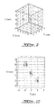
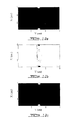
- FIG. 5 is a representation of the phantom simulated
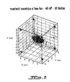
- FIG. 6 is a reconstructed phantom with values based on the 0 th moment only in which the number of iterations in the ART algorithm was set to 100;
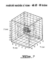
- FIG. 7 is a reconstructed phantom with values based on the 2 nd moment only in which the number of iterations in the ART algorithm was set to 200;
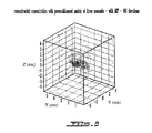
- FIG. 8 is a reconstructed phantom with values based on the 0 th , 1 st and 2 nd moments in which the number of iterations in the ART algorithm was set to 200;
- FIG. 9 is a configuration used for the simulations in which the source (detectors) locations are depicted by dots;
- FIG. 10 is a configuration used for the simulations in which the source (detectors) locations are depicted by dots;
- FIG. 11 shows the results of repartition of energy, mean times and variance of 1,000 randomly generated noised TPSF
- FIG. 12 is an example of sensitivity matrices in which a) and b) correspond respectively to m 0 ⁇ 2 (r s ,r d ) and m 2 ⁇ 2 (r s ,r d ) ⁇ m 0 ⁇ 2 (r s ,r d ) for a 6 cm thick slab with source-detector facing each other and a 0.1 ⁇ M background of Cy 7, c) and d) correspond to the same parameters for a 0.1 ⁇ M background of Cy 5.5 and e) and f) correspond to the same parameters for a 0.1 ⁇ M background of Cy 3B;
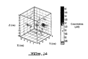
- FIG. 13 is an example of a reconstruction from synthetic data for Cy 7: a) O th moment only, b) 0 th , 1 st and 2 nd moments; Cy 5.5: c) 0 th moment only, d) 0 th , 1 st and 2 nd moments; and Cy 3B: e) 0 th moment only, f) 0 th , 1 st and 2 nd moments in which the quantitative values are expressed in ⁇ M;
- FIG. 14 is an example of a reconstruction from synthetic data for Cy 5.5 using all three moments noisy data.
- FIG. 15 is a schematic representation of a various geometries using source-detector pairs.
- ⁇ (r,t) is the photon fluence rate
- ⁇ a is the linear absorption coefficient
- v is the speed of light in the medium
- S(r,t) is the source term (assumed to be a ⁇ function in our case).
- other expressions for modeling light propagation such as the radiation transfer equation, can be used as would be obvious to one skilled in the art.
- the light propagation can be modeled numerically or using techniques such as Monte Carlo simulations again the person skilled in the art would be familiar with these techniques.
- N ex ⁇ ( r , t ) - 1 ⁇ ⁇ N ex ⁇ ( r , t ) + ⁇ ⁇ ⁇ ⁇ ⁇ ⁇ 1 ⁇ ( r , t ) ⁇ [ N tot ⁇ ( r , t ) - 2 ⁇ ⁇ n ex ⁇ ( r , t ) ] ( 2 )
- N ex (r,t) is the concentration of excited molecules at position r and time t
- N tot (r,t) is the concentration of total molecules of fluorophores (excited or not)
- ⁇ is the radiative lifetime of the fluorescent compound (sec.
- ⁇ is the absorption cross section of the fluorophore (cm 2 ) and ⁇ ⁇ 1 (r,t) is the photon fluence rate (number of photons s ⁇ 1 cm ⁇ 2 ) at the excitation wavelength ⁇ 1 .
- N ex ⁇ ( r , ⁇ ) ⁇ ⁇ N tot ⁇ ( r ) 1 - i ⁇ ⁇ ⁇ ⁇ ⁇ ⁇ ⁇ ⁇ ⁇ ⁇ 1 ⁇ ( r , ⁇ ) ( 3 )
- ⁇ 1 is the angular frequency at the excitation wavelength ⁇ 1 .
- the time domain and the frequency domain are linked through Fourier transform. Therefore the above derivation can also be used for fluorescence measurements performed in the time-domain.
- the time domain may also be linked to continuous wave (CW) measurements by integration of the total temporal point spread function (TPSF).
- the total fluorescent field is the sum of the contributions of all the secondary fluorescent sources over the entire volume.
- the fluorescent field detected at a position r d is modeled by:
- ⁇ ⁇ ⁇ ⁇ 2 ⁇ ( r s , r d , ⁇ ) ⁇ ⁇ ⁇ ⁇ ⁇ volume ⁇ N ex ⁇ ( r , ⁇ ) ⁇ ⁇ ⁇ ⁇ ⁇ 2 ⁇ ( r , r d , ⁇ ) ⁇ d 3 ⁇ r ( 4 )
- ⁇ ⁇ 2 (r,r d , ⁇ 2 ) represent a propagation term of the fluorescent field from the element of volume at r to the detector position r d at the reemission wavelength ⁇ 2 .
- ⁇ ⁇ ⁇ ⁇ 2 ⁇ ( r s , r d , ⁇ ) ⁇ ⁇ ⁇ volume ⁇ ⁇ ⁇ ⁇ ⁇ 1 ⁇ ( r s , r , ⁇ ) ⁇ Q eff ⁇ N tot ⁇ ( r ) 1 - i ⁇ ⁇ ⁇ ⁇ ⁇ ⁇ ⁇ ⁇ ⁇ ⁇ ⁇ 2 ⁇ ( r , r d , ⁇ ) ⁇ d 3 ⁇ r ( 5 )
- Q eff q ⁇ .
- ⁇ is the quantum efficiency, product of q the quenching factor, ⁇ the quantum yield and ⁇ absorption cross section of the fluorophore.
- ⁇ N tot (r) corresponds to the absorption coefficient of the fluorophore and can also be expressed as ⁇ C tot (r) where ⁇ is the extinction coefficient (cm ⁇ 1 Mol ⁇ 1 ) and C tot (r) is the concentration of the fluorophore at position r.
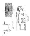
- FIG. 15 is a schematic representation of a various geometries using source-detector pairs. The Figure reproduces some of Li's schematics.
- FIG. 15A shows a pair formed of a source fiber and a detector fiber, in which a source-detector geometry is arranged so that a directional photon flux r is perpendicular to a detector surface.
- FIG. 15B shows another source-detector configuration in which a detector is shown a position indicated as (0,0) cm along a plane formed by axes x and y, while a source is positioned at (0,5) cm on the same plane.
- FIG. 15C shows a block diagram of a setup including a source formed of a diode laser coupled with a source fiber and a detector fiber optically coupled with an avalanche photo diode (APD).
- a sample is placed between the source-detector pair.
- a step motor moves the source in a 2-dimensional plane.
- a single side band (SSB) in-phase/quadrature-phase (I/Q) demodulator connected to the APD receives a signal from the detector, via an amplifier (A), on a radiofrequency (RF) port.
- SSB single side band
- I/Q in-phase/quadrature-phase
- I and Q outputs of the demodulator are filtered by direct current (DC) filters and converted by an analog to digital converter (ADC) before being applied to a computer (PC).
- the PC controls a frequency generator and the step motor.
- a power splitter directs an output of the frequency generator to both the diode laser and a local port (LO) of the demodulator.
- a general purpose interface bus (GPIB) relays digital signals.
- equation (7) is defined in the frequency domain.
- analytical solutions in the time domain are provided. Such analytical solutions for the absorption case have been proposed in the past for the 0 th , 1 st and 2 nd moment of the TPSF [33]. The correspondence of these moments to the TPSF is illustrated in FIG. 1 .
- the 0 th moment corresponds to the integration of the counts (equivalent to the continuous wave mode)
- the 1 st moment corresponds to the mean time of arrival of the photon
- the 2 nd moment to the variance of arrival of the photon.
- the concentration can be estimated using one or more moments.
- boundary conditions were implemented using the extrapolated boundary conditions [35, incorporated herein by reference].
- ART solves this linear system by sequentially projecting a solution estimate onto the hyperplanes defined by each row of the linear system.
- the technique is used in an iterative scheme and the projection at the end of the k th iteration becomes the estimate for the (k+1) th iteration. This projection process can be expressed mathematically as [38]:
- x j ( k + 1 ) x j ( k ) + ⁇ ⁇ b i - ⁇ i ⁇ ⁇ a ij ⁇ x j ( k ) ⁇ i ⁇ ⁇ a ij ⁇ a ij ⁇ ⁇ i ⁇ ⁇ a ij ( 14 )
- x j (k) is the k th estimate of j th element of the object function
- b i the i th measurement a ij the i-j th element of the weight matrix A
- ⁇ the relaxation parameter ⁇ the relaxation parameter.
- the relaxation parameter adjusts the projection step for each iterations.
- a small ⁇ value makes the inversion more robust but also slows conversion.
- the selection of ⁇ can be done empirically [39, 40, 41, 42, incorporated herein by reference].
- We have set ⁇ 0.1 based on previous studies [43].
- the data can be acquired in the Frequency Domain at several frequencies to reconstruct the TPSF via the Fourier Transform.
- Photon propagation is often referred to as a banana shape.
- the measurements are highly sensitive to the surface.
- Such dependence of the data type can be visualized through the mapping of the sensitivity matrix. Indeed, each line of the linear system described in equation (12) represents the dependence to a local perturbation for the corresponding source-detector pair. Thus by mapping this local dependence, we render the spatial sensitivity of this particular source-detector pair for this specific configuration and specific data type.
- the spatial dependence profile of the 2 nd normalized fluorescent moment ( FIG. 3 ) possess distinctive features.
- the 2 nd normalized fluorescent moment still exhibits some strong dependence from the surface voxels, but also from deeper voxel.
- the profile presents a distinguishing depression in the line connecting the source detector pair.
- FIG. 4 where we used the properties of Cy 3B for the simulated chromophore.
- the 2 nd normalized fluorescent moment is characterized by a sharp and well-demarcated hollow dependence.
- Such typical features are related to the fact the fluorescent mean time t ⁇ 2 (r s ,r d ) is subtracted in equation (11).
- the measured mean-time is always greater than the mean time of propagation for the shorter path, i.e. for the voxels located on the line connecting the source-detector pair. Then if the contribution of the lifetime is small enough, the 2 nd normalized fluorescent moment will exhibit reduced (eventually negative) contribution for these voxels.
- this coordinate is the depth relative to a surface of the object in which the fluorophore is embedded. This may be accomplished, for example, by considering a region of interest as a unique voxel resolved in one dimension only.
- the method for estimating the concentration of fluorophores described above can be applied to biological tissues such as brain and breast tissue.
- the fluorophore can be endogenous or exogenous and the concentration of several fluorophores may be determined simultaneously when using multiple excitation and emission wavelengths.
- the reconstructions presented in this section highlight the benefit of the time domain normalized moments formulation over the traditional 0 th normalized moment.
- the higher moment of the fluorescent TPSF provides information that is less overwhelmed by the surface interactions. The gain is important when a background fluorophore concentration exists, as it is generally the case in molecular imaging. Strong surface concentrations that are generally considered as plaguing artifacts in continuous wave fluorescent imaging are avoided in reconstruction with higher moments or their combination when a background fluorophore distribution exists.
- the synthetic phantom was probed with a 25 ⁇ 25 constellation of source detectors. This constellation was distributed evenly 1.5 cm apart in both dimensions.
- the phantom configuration is provided in FIG. 9 .
- TPSF Poisson noise of the temporal distribution of photon time of flights.
- the TPSF was normalized at 500 counts at the maximum bin mimicking real acquisition scenarios. From the noised TPSF, we estimated one set of energy, meantime and variance. The same estimation was performed over 1,000 trials. The statistics of these estimates were used as our noise model. An example of noisy moments value distribution is given in FIG. 11 .
- a Gaussian distribution approximated the noise model.
- the different values of the noise model employed for the three moments evaluated herein are Measure ⁇ (%), Energy: 2, Meantime: 0.2, and Variance: 2.
- FIG. 12 We propose in FIG. 12 some examples of sensitivity matrices for the transmittance case. We limited our to depict slices across the discrete volume, but by construction, the banana shapes are in 3D. The optical and fluorochrome properties characterizing this medium are provided in Table 2 and Table 3.
- FIG. 12 underline interesting features of the time domain moment fluorescent DOT.
- the normalized 0 th order Born approximation in continuous mode is highly sensitive to surface voxels.
- the spatial dependence profile of the 2 nd normalized fluorescent moment possesses distinctive features.
- the 2 nd normalized fluorescent moment still exhibits some strong dependence from the surface voxels, but also from deeper voxels.
- the profile presents a distinguishing depression in the line connecting the source-detector pair. This fact is striking in the case of FIG. 12 d ) where we used the properties of Cy 7 for the simulated chromophore.
- the 2 nd normalized fluorescent moment is characterized by a sharp and well-demarcated hollow dependence.
- Such typical features are related to the fact that the fluorescent mean time t ⁇ 2 (r s ,r d ) is subtracted in Eq (11).
- the measured mean-time is always greater than the mean time of propagation for the shorter path, i.e. for the voxels located on the line connecting the source-detector pair. Then if the contribution of the lifetime is small enough, the 2 nd normalized fluorescent moment will exhibit reduced (eventually negative) contribution for these voxels. This property is dependent on the lifetime of the fluorochrome investigated. This hollow distribution is still present for the Cy 5.5 case but disappears for the Cy 3B simulations. In this last case, the contribution of the lifetime is predominant for these shorter path voxels and the spatial distribution of 2 nd normalized fluorescent moment is not markedly different than the 0 th normalized fluorescent moment.
- the algorithm is still performing satisfactorily in the case of noise. Even though the 2 nd normalized moments are sensitive to noise, the incorporation of this information benefits the inverse problem. The objects are reconstructed with fidelity and the surface artifacts are still minimized due to the inherent spatial information of the 2 nd normalized moment.
Landscapes
- Health & Medical Sciences (AREA)
- Life Sciences & Earth Sciences (AREA)
- Physics & Mathematics (AREA)
- Pathology (AREA)
- General Health & Medical Sciences (AREA)
- Immunology (AREA)
- Chemical & Material Sciences (AREA)
- General Physics & Mathematics (AREA)
- Nuclear Medicine, Radiotherapy & Molecular Imaging (AREA)
- Biochemistry (AREA)
- Engineering & Computer Science (AREA)
- Analytical Chemistry (AREA)
- Heart & Thoracic Surgery (AREA)
- Surgery (AREA)
- Optics & Photonics (AREA)
- Radiology & Medical Imaging (AREA)
- Biophysics (AREA)
- Biomedical Technology (AREA)
- Veterinary Medicine (AREA)
- Medical Informatics (AREA)
- Molecular Biology (AREA)
- Public Health (AREA)
- Animal Behavior & Ethology (AREA)
- Theoretical Computer Science (AREA)
- Chemical Kinetics & Catalysis (AREA)
- Investigating, Analyzing Materials By Fluorescence Or Luminescence (AREA)
- Algebra (AREA)
- Mathematical Analysis (AREA)
- Mathematical Optimization (AREA)
- Mathematical Physics (AREA)
- Pure & Applied Mathematics (AREA)
Abstract
Description
where Nex(r,t) is the concentration of excited molecules at position r and time t, Ntot(r,t) is the concentration of total molecules of fluorophores (excited or not), τ is the radiative lifetime of the fluorescent compound (sec. or nanoseconds), σ is the absorption cross section of the fluorophore (cm2) and Φλ1(r,t) is the photon fluence rate (number of photons s−1 cm−2) at the excitation wavelength λ1. Considering that the number of excited molecule is low compared to the total molecules and working in the frequency domain yields the expression for the concentration of excited molecules:
where ω1 is the angular frequency at the excitation wavelength λ1. The time domain and the frequency domain are linked through Fourier transform. Therefore the above derivation can also be used for fluorescence measurements performed in the time-domain. Furthermore the time domain may also be linked to continuous wave (CW) measurements by integration of the total temporal point spread function (TPSF).
where Φλ2(r,rd,ω2) represent a propagation term of the fluorescent field from the element of volume at r to the detector position rd at the reemission wavelength λ2. Then, by using equation (4) we obtain the fluorescent term:
Where Qeff=q·η. σ is the quantum efficiency, product of q the quenching factor, η the quantum yield and σ absorption cross section of the fluorophore. Note that the product σNtot(r) corresponds to the absorption coefficient of the fluorophore and can also be expressed as εCtot(r) where ε is the extinction coefficient (cm−1 Mol−1) and Ctot(r) is the concentration of the fluorophore at position r.
where
is the system's Green function with k2=(−vμλj a+iω)/Dλj at the considered wavelength λjε(λ1, λ2).
-
- Normalized 1st moment
-
- Normalized 2nd moment
Where
b=A·x (13)
where b is a vector holding the measurements for each source-detector pair, A is the matrix of the forward model (weight matrix), and x is the vector of unknowns (object function). ART solves this linear system by sequentially projecting a solution estimate onto the hyperplanes defined by each row of the linear system. The technique is used in an iterative scheme and the projection at the end of the kth iteration becomes the estimate for the (k+1)th iteration. This projection process can be expressed mathematically as [38]:
where xj (k) is the kth estimate of jth element of the object function, bi the ith measurement, aij the i-jth element of the weight matrix A and λ the relaxation parameter.
| TABLE 1 |
| Parameters used in the simulation. |
| μa λ1 (cm−1) | 0.06 | Cbackground (μM/L) | 0.1 |
| μs ′λ1 (cm−1) | 10.00 | Cinclusion (μM/L) | 1.0 |
| μs ′λ2 (cm−1) | 10.00 | τ (ns) | 1.0 |
| Dimensions (cm) | 10 × 6 × 10 | ε (cm−1 · M−1) | 190,000 |
| Voxel size (cm) | 0.3 × 0.3 × 0.3 | η (%) | 0.23 |
| TABLE 2 |
| Parameters used in the simulations. |
| μa λ1 (cm−1) | 0.06 | Dimensions (cm) | 9 × 6 × 9 | ||
| μa λ2 (cm−1) | 0.06 | Cbackground (μM) | 0.1 | ||
| μs ′λ1 (cm−1) | 10.00 | Cinclusion (μM) | 1.0 | ||
| μs ′λ2 (cm−1) | 10.00 | Voxel size (cm) | 0.36 × 0.3 × 0.36 | ||
| TABLE 3 |
| Fluorochrome investigated herein. |
| Compound | τ (ns) | ε (cm−1 · M−1) | η (%) | ||
| |
<0.3 | 200 000 | 0.28 | ||
| Cy 5.5 | 1.0 | 190 000 | 0.23 | ||
| Cy 3-B | 2.8 | 130 000 | 0.67 | ||
- [1] Yodh A G, Chance B. Spectroscopy and imaging with diffusing light. Physics Today (1995); 48:34-40.
- [2] Jobsis F. Noninvasive infrared monitoring of cerebral and myocardial sufficiency and circulatory parameters. Science (1977); 198:1264-67.
- [3] Tromberg B, Shah N, Lanning R, Cerussi A, Espinoza J, Pham T, et al. Non-invasive in vivo characterization of breast tumors using photon migration spectroscopy. Neoplasia (2000); 2:26-40.
- [4] Dehghani, H., Pogue, B. W., Poplack, S. P., Paulsen, K. D., “Multiwavelength three-dimensional near-infrared tomography of the breast: initial simulation, phantom, and clinical results”, Applied Optics, 42(1) 135-145, 2003.
- [5] Jiang H, Iftimia N, Eggert J, Fajardo L, Klove K. Near-infrared optical imaging of the breast with model-based reconstruction. Acad. Radiology (2002); 9:186-94.
- [6] Franceschini M, Moesta K, Fantini S, Gaida G, Gratton E, Jess H et al. Frequency-domain techniques enhance optical mammography: Initial clinical results. PNAS (1997); 94:6468-73.
- [7] Colak S, van der Mark M, Hooft G, Hoogenraad J, van der Linden E, Kuijpers F. Clinical optical tomography and NIR spectroscopy for breast cancer detection. IEEE Journal of selected topics in quantum electronics (1999); 5:1143-58.
- [8] Intes X, Djeziri S, Ichalalene Z, Mincu N, Wang Y, Kasrai R, Polyzos M, Hall D, Boas D, St-Jean P, Lesage F, Khayat M. Time-Domain Optical Mammography Softscan®: Initial Results on Detection and Characterization of Breast Tumors. In the same proceedings.
- [9] Jakubowski D B, Cerussi A E, Bevilacqua F, Shah N, Hsiang D, Butler J, Tromberg B J. Monitoring neoadjuvant chemotherapy in breast cancer using quantitative diffuse optical spectroscopy: a case study. J Biomed Opt. (2004); 9:230-238.
- [10] Strangman G, Boas D, Sutton J. Non-invasive neuroimaging using Near-Infrared light. Biol. Psychiatry (2002); 52:679-93.
- [11] Villringer A, Chance B. Non-invasive optical spectroscopy and imaging of human brain function. Trends Neurosci. (1997); 20:435-42.
- [12] Chance B, Nioka S, Chen Y. Shining new light on brain function. OE magazine (2003); 3:16-9.
- [13] Steinbrink J, Kohl M, Obrig H, Curio G, Syre F, Thomas F, et al. Somatosensory evoked fast optical intensity changes detected non-invasively in the adult human head. Neuroscience Letters (2000); 291:105-8.
- [14] Stankovic M, Maulik D, Rosenfeld W, Stubblefield P, Kofinas A, Gratton E, et al. Role of frequency domain optical spectroscopy in the detection of neonatal brain hemorrhage—a newborn piglet study. J. Matern Fetal Med. (2000); 9:142-9.
- [15] Hebden J C, Gibson A, Austin T, Yusof R M, Everdell N, Delpy D T, Arridge S R, Meek J H, Wyatt J S. Imaging changes in blood volume and oxygenation in the newborn infant brain using three-dimensional optical tomography. Phys Med Biol. (2004); 49:1117-1130.
- [16] Quaresima V, Lepanto R, Ferrari M. The use of near infrared spectroscopy in sports medicine. J Sports Med Phys Fitness (2003); 43:1-13.
- [17] Wolf U, Wolf M, Choi J, Levi M, Choudhury D, Hull S, et al. Localized irregularities in hemoglobin flow and oxygenation in calf muscle in patients with peripheral vascular disease detected with near-infrared spectrophotometry. Vasc Surg. (2003); 37:1017-26.
- [18] Weissleder R and Mahmood U. Molecular imaging. Radiology. 2001 May; 219(2):316-33.
- [19] Weissleder R, Ntziachritos V. Shedding light onto live molecular targets. Nature Medicine (2003); 9:123-8.
- [20] Frangioni J V. In vivo near-infrared fluorescence imaging. Current Opinion in Chemical Biology (2003); 7:626-34.
- [21] Licha K. Contrast agents for optical imaging. Topics in Current Chemistry (2002); 222: 1-29.
- [22] Achilefu S, Dorshow R, Bugaj J, Rajagopalan R. Novel receptor-targeted fluorescent contrast agents for in-vivo tumor imaging. Invest. Radiol. (2000); 35:479-85.
- [23] Chen Y, Zheng G, Zhang Z, Blessington D, Zhang M, H. Li, et al. Metabolism Enhanced Tumor Localization by Fluorescence Imaging: In Vivo Animal Studies. Optics Letters (2003); 28:2070-2.
- [24] Weissleder R, Tung C H, Mahmood U, Bogdanov A. In vivo imaging with protease-activated near-infrared fluorescent probes. Nat. Biotech. (1999); 17:375-8.
- [25] Weinberg R. How Does Cancer Arise. Sci. Am. (1996); 275:62-71.
- [26] Lewis J, Achilefu S, Garbow J R, Laforest R, Welch M J. Small animal imaging: current technology and perspectives for oncological imaging. European Journal of Cancer (2002); 38:2173-88.
- [27] Ntziachristos V, Ripoll J, Weissleder R. Would near-infrared fluorescence signals propagate through large human organs for clinical studies? Opt. Lett. (2002); 27:333-335.
- [28] Ntziachristos V, Weissleder R. Experimental three-dimensional fluorescence reconstruction of diffuse media by use of a normalized Born approximation. Opt. Lett. (2001); 26:893-895.
- [29] Eppstein M J, Hawrysz D J, Godavarty A, Sevick-Muraca E M. Three-dimensional, Bayesian image reconstruction from sparse and noisy data sets: Near-infrared fluorescence tomography. Proc. Nat. Acad. Sci. Am. (2002); 99: 9619-9624.
- [30] Milstein A B, Stott J J, Oh S, Boas D A, Millane R P, Bouman C A, Webb K J. Fluorescence optical diffusion tomography using multiple-frequency data. J. Opt. Soc. Am. A (2004); 21: 1035-1049.
- [31] Xingde Li. Fluorescence and diffusive wave diffraction tomographic probes in turbid media PhD University of Pennsylvania (1996).
- [32] O'Leary M. Imaging with diffuse photon density waves. PhD University of Pennsylvania (1996).
- [33] Hillman E. Experimental and theoretical investigations of near infrared tomographic imaging methods and clinical applications. PhD University College London (2002).
- [34] Liebert A, Wabnitz H, Grosenick D, Moller M, Macdonald R and Rinnerberg H. Evaluation of optical properties of highly scattering media by moments of distributions of times of flight of photons. Appl. Opt. (2003); 42:5785-5792.
- [35] Haskell R C, Svaasand L O, Tsay T, Feng T, McAdams M S, Tromberg B J. Boundary conditions for the diffusion equation in radiative transfer. J. Opt. Soc. Am A (1994); 11; 2727-41.
- [36] Gaudette R J, Brooks D H, DiMarzio C A, Kilmer M E, Miller E L, Gaudette T. Boas D A. A comparison study of linear reconstruction techniques for diffuse optical tomographic imaging of absorption coefficient. Phys Med Biol. (2000); 45:1051-1070.
- [37] Gordon R, Bender R and Herman G Algebraic reconstruction techniques (ART) for the three dimensional electron microscopy and X-Ray photography. J. Theoret. Biol. (1970); 69:471-482.
- [38] Kak A and Slaney M, “Computerized tomographic Imaging”, IEE Press, N-Y (1987).
- [39] Ros D, Falcon C, Juvells I and Pavia J. The influence of a relaxation parameter on SPECT iterative reconstruction algorithms Phys. Med. Biol. (1996); 41: 925-937.
- [40] Herman G & Meyer L. Algebraic Reconstruction Techniques can be made computationally efficient. IEEE Transactions on Medical Imaging (1993); 12:600-609.
- [41] Gaudette R, Brook D, DiMarzio C, Kilmer M, Miller E, Gaudette T & Boas D. A comparison study of linear reconstruction techniques for diffuse optical tomographic imaging of absorption coefficient. Phys. Med. Biol. (2000); 45:1051-1070.
- [42] van der Sluis A and van der Vorst H. SIRT- and CG-type methods for the iterative solution of sparse linear least-squares problems. Linear Algebr. Appl. (1990); 130:257-302.
- [43] Intes X, Ntziachristos V, Culver J, Yodh A G and Chance B. Projection access order in Algebraic Reconstruction Techniques for Diffuse Optical Tomography. Phys. Med. Biol. (2002); 47:N1-N10.
Claims (11)
Priority Applications (1)
| Application Number | Priority Date | Filing Date | Title |
|---|---|---|---|
| US11/576,001 US8831709B2 (en) | 2004-09-24 | 2005-09-26 | Method for 3-dimensional fluorescence tomographic imaging |
Applications Claiming Priority (3)
| Application Number | Priority Date | Filing Date | Title |
|---|---|---|---|
| US61252104P | 2004-09-24 | 2004-09-24 | |
| PCT/CA2005/001469 WO2006032151A1 (en) | 2004-09-24 | 2005-09-26 | Method for fluorescence tomographic imaging |
| US11/576,001 US8831709B2 (en) | 2004-09-24 | 2005-09-26 | Method for 3-dimensional fluorescence tomographic imaging |
Publications (2)
| Publication Number | Publication Date |
|---|---|
| US20080260647A1 US20080260647A1 (en) | 2008-10-23 |
| US8831709B2 true US8831709B2 (en) | 2014-09-09 |
Family
ID=36089821
Family Applications (1)
| Application Number | Title | Priority Date | Filing Date |
|---|---|---|---|
| US11/576,001 Expired - Fee Related US8831709B2 (en) | 2004-09-24 | 2005-09-26 | Method for 3-dimensional fluorescence tomographic imaging |
Country Status (4)
| Country | Link |
|---|---|
| US (1) | US8831709B2 (en) |
| EP (1) | EP1806999A1 (en) |
| CA (1) | CA2581592C (en) |
| WO (1) | WO2006032151A1 (en) |
Families Citing this family (15)
| Publication number | Priority date | Publication date | Assignee | Title |
|---|---|---|---|---|
| EP1806999A1 (en) | 2004-09-24 | 2007-07-18 | ART Advanced Research Technologies Inc. | Method for fluorescence tomographic imaging |
| FR2900253A1 (en) * | 2006-04-25 | 2007-10-26 | Commissariat Energie Atomique | METHOD FOR RECONSTRUCTING THE DISTRIBUTION OF OPTICAL PROPERTIES OF AN INHOMOGENIC DIFFUSING MEDIUM |
| FR2904691B1 (en) | 2006-08-02 | 2009-03-06 | Commissariat Energie Atomique | METHOD AND DEVICE FOR 3D RECONSTRUCTION OF THE DISTRIBUTION OF FLUORESCENT ELEMENTS |
| FR2938181B1 (en) | 2008-11-13 | 2012-03-09 | Commissariat Energie Atomique | METHOD AND DEVICE FOR LOCATING FLUOROPHORES OR ABSORBERS IN A SURROUNDING ENVIRONMENT |
| AU2010286592B2 (en) * | 2009-08-28 | 2015-08-13 | Visen Medical, Inc. | Systems and methods for tomographic imaging in diffuse media using a hybrid inversion technique |
| FR2950431B1 (en) * | 2009-09-24 | 2011-12-09 | Commissariat Energie Atomique | DEVICE AND METHOD FOR SPATIAL RECONSTRUCTION OF FLUORESCENCE CARTOGRAPHY |
| TWI412940B (en) * | 2009-10-06 | 2013-10-21 | 國立交通大學 | Image reconstruction method, device and computer program for diffuse optical tomography |
| FR2951283B1 (en) | 2009-10-08 | 2013-02-15 | Commissariat Energie Atomique | METHOD AND DEVICE FOR DIFFUSED EXCITATION IN IMAGING |
| FR2968921B1 (en) | 2010-12-15 | 2013-01-11 | Commissariat Energie Atomique | METHOD FOR LOCATING AN OPTICAL MARKER IN A DIFFUSING MEDIUM |
| JP5782314B2 (en) | 2011-07-07 | 2015-09-24 | 浜松ホトニクス株式会社 | Biological measurement device and image creation method |
| FR2984503B1 (en) * | 2011-12-16 | 2014-01-17 | Commissariat Energie Atomique | METHOD FOR RECONSTRUCTING OPTICAL PROPERTIES OF A DIFFUSING MEDIUM USING A COMBINATION OF A PLURALITY OF MELLIN-LAPLACE TRANSFORMS OF A SIZE INCLUDING A TIME DISTRIBUTION OF A RECEIVED SIGNAL, AND ASSOCIATED RECONSTRUCTION SYSTEM |
| FR2985023B1 (en) * | 2011-12-23 | 2016-05-06 | Commissariat Energie Atomique | SYSTEM FOR RECONSTRUCTING OPTICAL PROPERTIES OF A DIFFUSING MEDIUM, COMPRISING A PULSE RADIATION SOURCE AND AT LEAST TWO DETECTORS OF TWO DIFFERENT TYPES, AND ASSOCIATED RECONSTRUCTION METHOD |
| JP5907039B2 (en) * | 2012-10-22 | 2016-04-20 | 株式会社島津製作所 | Fluorescence image reconstruction method and apparatus |
| DE102019000066B4 (en) * | 2019-01-09 | 2020-10-08 | Becker & Hickl Gmbh | Process for the visualization of fluorescence lifetime imaging data |
| CN120814819B (en) * | 2025-07-14 | 2026-02-10 | 东莞理工学院 | Photon weight-fused diffuse light brain tissue blood oxygen concentration variation imaging method and related products |
Citations (7)
| Publication number | Priority date | Publication date | Assignee | Title |
|---|---|---|---|---|
| US5577137A (en) | 1995-02-22 | 1996-11-19 | American Research Corporation Of Virginia | Optical chemical sensor and method using same employing a multiplicity of fluorophores contained in the free volume of a polymeric optical waveguide or in pores of a ceramic waveguide |
| WO2002041760A2 (en) | 2000-11-27 | 2002-05-30 | The General Hospital | Fluorescence-mediated molecular tomography |
| US20020115092A1 (en) | 2000-11-08 | 2002-08-22 | The Scripps Research Institute | Energy transfer labels with mechanically linked fluorophores |
| US20020136972A1 (en) * | 1999-05-25 | 2002-09-26 | Hall Lachlan Everett | Infrared chromophores |
| CA2491748A1 (en) | 2002-07-18 | 2004-01-29 | Mauna Kea Technologies | Method and equipment for fibre optic high-resolution, in particular confocal, fluorescence imaging |
| WO2005043138A1 (en) | 2003-10-31 | 2005-05-12 | Art Advanced Research Technologies Inc. | A time-domain method and apparatus for determining the depth and concentration of a fluorophore in a turbid medium |
| EP1806999A1 (en) | 2004-09-24 | 2007-07-18 | ART Advanced Research Technologies Inc. | Method for fluorescence tomographic imaging |
-
2005
- 2005-09-26 EP EP05789395A patent/EP1806999A1/en not_active Withdrawn
- 2005-09-26 CA CA2581592A patent/CA2581592C/en not_active Expired - Fee Related
- 2005-09-26 WO PCT/CA2005/001469 patent/WO2006032151A1/en not_active Ceased
- 2005-09-26 US US11/576,001 patent/US8831709B2/en not_active Expired - Fee Related
Patent Citations (8)
| Publication number | Priority date | Publication date | Assignee | Title |
|---|---|---|---|---|
| US5577137A (en) | 1995-02-22 | 1996-11-19 | American Research Corporation Of Virginia | Optical chemical sensor and method using same employing a multiplicity of fluorophores contained in the free volume of a polymeric optical waveguide or in pores of a ceramic waveguide |
| US20020136972A1 (en) * | 1999-05-25 | 2002-09-26 | Hall Lachlan Everett | Infrared chromophores |
| US20020115092A1 (en) | 2000-11-08 | 2002-08-22 | The Scripps Research Institute | Energy transfer labels with mechanically linked fluorophores |
| WO2002041760A2 (en) | 2000-11-27 | 2002-05-30 | The General Hospital | Fluorescence-mediated molecular tomography |
| CA2491748A1 (en) | 2002-07-18 | 2004-01-29 | Mauna Kea Technologies | Method and equipment for fibre optic high-resolution, in particular confocal, fluorescence imaging |
| WO2005043138A1 (en) | 2003-10-31 | 2005-05-12 | Art Advanced Research Technologies Inc. | A time-domain method and apparatus for determining the depth and concentration of a fluorophore in a turbid medium |
| US20070158585A1 (en) * | 2003-10-31 | 2007-07-12 | Art, Advanced Research Technologies Inc. | Time-domain method and apparatus for determining the depth and concentration of a fluorophore in a turbid medium |
| EP1806999A1 (en) | 2004-09-24 | 2007-07-18 | ART Advanced Research Technologies Inc. | Method for fluorescence tomographic imaging |
Non-Patent Citations (4)
| Title |
|---|
| Canadian Office Action issued in Canadian Application No. 2,581,592 on Apr. 18, 2012. |
| D1 "Time Domain Optical Imaging", Arridge S R; BioMedical Imaging: macro to Nano; 2004, IEEE International Symposium on Arlington, VA, USA , Apr. 15-18, 2004; pp. 1486-1489; XP010774148. |
| Gao et al. "Improvement of image quality in diffuse optical tomography by use of full time-resolved data". Applied Optics. vol. 41, No. 4, Feb. 1, 2002. * |
| Xingde Li, Dissertation in Physis, U. Penn., Title: Fluorescence and Diffusive Wave Diffraction Tomographic Probes in Turbid Media (1998). |
Also Published As
| Publication number | Publication date |
|---|---|
| EP1806999A1 (en) | 2007-07-18 |
| CA2581592A1 (en) | 2006-03-30 |
| CA2581592C (en) | 2014-04-15 |
| US20080260647A1 (en) | 2008-10-23 |
| WO2006032151A1 (en) | 2006-03-30 |
Similar Documents
| Publication | Publication Date | Title |
|---|---|---|
| Lam et al. | Time domain fluorescent diffuse optical tomography: analytical expressions | |
| Luke et al. | Optical wavelength selection for improved spectroscopic photoacoustic imaging | |
| Intes et al. | Diffuse optical tomography with physiological and spatial a priori constraints | |
| Dehghani et al. | Near infrared optical tomography using NIRFAST: Algorithm for numerical model and image reconstruction | |
| US8831709B2 (en) | Method for 3-dimensional fluorescence tomographic imaging | |
| Lin et al. | Fluorescence diffuse optical tomography with functional and anatomical a priori information: feasibility study | |
| Zhang et al. | Coregistered tomographic x-ray and optical breast imaging: initial results | |
| CN102137618B (en) | Quantitative multi-spectral opto-acoustic tomography (MSOT) of tissue biomarkers | |
| Boverman et al. | Spatio-temporal imaging of the hemoglobin in the compressed breast with diffuse optical tomography | |
| Hillman et al. | Sub‐millimeter resolution 3D optical imaging of living tissue using laminar optical tomography | |
| Intes et al. | Non-PET functional imaging techniques: optical | |
| Mohajerani et al. | Optical and optoacoustic model-based tomography: theory and current challenges for deep tissue imaging of optical contrast | |
| Liu et al. | 4-D reconstruction for dynamic fluorescence diffuse optical tomography | |
| Ge et al. | Three‐dimensional fluorescence‐enhanced optical tomography using a hand‐held probe based imaging system | |
| Jeeva et al. | Reconstruction of optical scanned images of inhomogeneities in biological tissues by Monte Carlo simulation | |
| Mo et al. | Quantitative characterization of optical and physiological parameters in normal breasts using time-resolved spectroscopy: in vivo results of 19 Singapore women | |
| Bai et al. | Fluorescence molecular tomography | |
| Hyde et al. | A statistical approach to inverting the born ratio | |
| Lam et al. | Time domain fluorescent diffuse optical tomography | |
| Venugopal et al. | Multimodal diffuse optical imaging for biomedical applications | |
| Guerra et al. | An iterative method of light fluence distribution estimation for quantitative photoacoustic imaging | |
| Ntziachristos et al. | Fluorescence molecular tomography: New detection schemes for acquiring high information content measurements | |
| Srinivasan et al. | Improved quantification of fluorescence in 3-D in a realistic mouse phantom | |
| Da Silva et al. | From bench-top small animal diffuse optical tomography towards clinical imaging | |
| Konecky | Diffuse optical tomography: Imaging physics and image formation principles |
Legal Events
| Date | Code | Title | Description |
|---|---|---|---|
| AS | Assignment |
Owner name: ART, ADVANCED RESEARCH TECHNOLOGIES INC., CANADA Free format text: ASSIGNMENT OF ASSIGNORS INTEREST;ASSIGNORS:INTES, XAVIER;LESAGE, FREDERIC;LAM, SIRITHY;REEL/FRAME:019071/0200;SIGNING DATES FROM 20050105 TO 20051110 Owner name: ART, ADVANCED RESEARCH TECHNOLOGIES INC., CANADA Free format text: ASSIGNMENT OF ASSIGNORS INTEREST;ASSIGNORS:INTES, XAVIER;LESAGE, FREDERIC;LAM, SIRITHY;SIGNING DATES FROM 20050105 TO 20051110;REEL/FRAME:019071/0200 |
|
| AS | Assignment |
Owner name: DORSKY WORLDWIDE CORP., VIRGIN ISLANDS, BRITISH Free format text: ASSIGNMENT OF ASSIGNORS INTEREST;ASSIGNOR:ART ADVANCED RESEARCH TECHNOLOGIES INC.;REEL/FRAME:026466/0001 Effective date: 20091211 Owner name: NEW ART ADVANCED RESEARCH TECHNOLOGIES INC., QUEBE Free format text: ASSIGNMENT OF ASSIGNORS INTEREST;ASSIGNOR:ART ADVANCED RESEARCH TECHNOLOGIES INC.;REEL/FRAME:026481/0304 Effective date: 20061127 Owner name: ART ADVANCED RESEARCH TECHNOLOGIES INC., QUEBEC Free format text: CHANGE OF NAME;ASSIGNOR:NEW ART ADVANCED RESEARCH TECHNOLOGIES INC.;REEL/FRAME:026479/0052 Effective date: 20061127 |
|
| AS | Assignment |
Owner name: SOFTSCAN HEALTHCARE GROUP LTD., VIRGIN ISLANDS, BR Free format text: CHANGE OF NAME;ASSIGNOR:DORSKY WORLDWIDE CORP.;REEL/FRAME:026469/0916 Effective date: 20110608 |
|
| STCF | Information on status: patent grant |
Free format text: PATENTED CASE |
|
| MAFP | Maintenance fee payment |
Free format text: PAYMENT OF MAINTENANCE FEE, 4TH YR, SMALL ENTITY (ORIGINAL EVENT CODE: M2551) Year of fee payment: 4 |
|
| FEPP | Fee payment procedure |
Free format text: MAINTENANCE FEE REMINDER MAILED (ORIGINAL EVENT CODE: REM.); ENTITY STATUS OF PATENT OWNER: SMALL ENTITY |
|
| LAPS | Lapse for failure to pay maintenance fees |
Free format text: PATENT EXPIRED FOR FAILURE TO PAY MAINTENANCE FEES (ORIGINAL EVENT CODE: EXP.); ENTITY STATUS OF PATENT OWNER: SMALL ENTITY |
|
| STCH | Information on status: patent discontinuation |
Free format text: PATENT EXPIRED DUE TO NONPAYMENT OF MAINTENANCE FEES UNDER 37 CFR 1.362 |
|
| FP | Lapsed due to failure to pay maintenance fee |
Effective date: 20220909 |