US8759093B2 - Method for determining the selectivity of a pre-synaptic neuromuscular blocking substance - Google Patents
Method for determining the selectivity of a pre-synaptic neuromuscular blocking substance Download PDFInfo
- Publication number
- US8759093B2 US8759093B2 US11/659,445 US65944505A US8759093B2 US 8759093 B2 US8759093 B2 US 8759093B2 US 65944505 A US65944505 A US 65944505A US 8759093 B2 US8759093 B2 US 8759093B2
- Authority
- US
- United States
- Prior art keywords
- muscle tissue
- neuromuscular blocking
- blocking substance
- buffer
- synaptic neuromuscular
- Prior art date
- Legal status (The legal status is an assumption and is not a legal conclusion. Google has not performed a legal analysis and makes no representation as to the accuracy of the status listed.)
- Expired - Fee Related, expires
Links
Images


Classifications
-
- C—CHEMISTRY; METALLURGY
- C12—BIOCHEMISTRY; BEER; SPIRITS; WINE; VINEGAR; MICROBIOLOGY; ENZYMOLOGY; MUTATION OR GENETIC ENGINEERING
- C12N—MICROORGANISMS OR ENZYMES; COMPOSITIONS THEREOF; PROPAGATING, PRESERVING, OR MAINTAINING MICROORGANISMS; MUTATION OR GENETIC ENGINEERING; CULTURE MEDIA
- C12N5/00—Undifferentiated human, animal or plant cells, e.g. cell lines; Tissues; Cultivation or maintenance thereof; Culture media therefor
- C12N5/06—Animal cells or tissues; Human cells or tissues
- C12N5/0602—Vertebrate cells
- C12N5/0652—Cells of skeletal and connective tissues; Mesenchyme
- C12N5/0661—Smooth muscle cells
-
- G—PHYSICS
- G01—MEASURING; TESTING
- G01N—INVESTIGATING OR ANALYSING MATERIALS BY DETERMINING THEIR CHEMICAL OR PHYSICAL PROPERTIES
- G01N33/00—Investigating or analysing materials by specific methods not covered by groups G01N1/00 - G01N31/00
- G01N33/48—Biological material, e.g. blood, urine; Haemocytometers
- G01N33/50—Chemical analysis of biological material, e.g. blood, urine; Testing involving biospecific ligand binding methods; Immunological testing
- G01N33/5005—Chemical analysis of biological material, e.g. blood, urine; Testing involving biospecific ligand binding methods; Immunological testing involving human or animal cells
- G01N33/5008—Chemical analysis of biological material, e.g. blood, urine; Testing involving biospecific ligand binding methods; Immunological testing involving human or animal cells for testing or evaluating the effect of chemical or biological compounds, e.g. drugs, cosmetics
- G01N33/5082—Supracellular entities, e.g. tissue, organisms
-
- G—PHYSICS
- G01—MEASURING; TESTING
- G01N—INVESTIGATING OR ANALYSING MATERIALS BY DETERMINING THEIR CHEMICAL OR PHYSICAL PROPERTIES
- G01N33/00—Investigating or analysing materials by specific methods not covered by groups G01N1/00 - G01N31/00
- G01N33/48—Biological material, e.g. blood, urine; Haemocytometers
- G01N33/483—Physical analysis of biological material
-
- G—PHYSICS
- G01—MEASURING; TESTING
- G01N—INVESTIGATING OR ANALYSING MATERIALS BY DETERMINING THEIR CHEMICAL OR PHYSICAL PROPERTIES
- G01N33/00—Investigating or analysing materials by specific methods not covered by groups G01N1/00 - G01N31/00
- G01N33/15—Medicinal preparations ; Physical properties thereof, e.g. dissolubility
Definitions
- the invention relates to a method for determining the selectivity of a pre-synaptic neuromuscular blocking substance.
- the determination of the quantity of pre-synaptic neuromuscular blocking substance contained in a sample is generally made through the measurement of the lethal dose LD 50 for this substance in mice or rats. This method is in particular used presently for the determination of the quantity of active botulinum toxin. Such LD 50 methods are synonym of a large number of animals killed.
- the present invention offers a process for determining the selectivity of a pre-synaptic neuromuscular blocking substance.
- This process uses an ex vivo method which spares the life of a significant number of animals compared to the usual LD 50 methods, which method is described in a PCT patent application by the same Applicant, i.e. PCT application No. PCT/GB2004/000697.
- PCT application No. PCT/GB2004/000697 discloses a method for determining the quantity of a pre-synaptic neuromuscular blocking substance in a sample which comprises the following steps:
- the muscle tissue could be a smooth muscle and a rib (striated) muscle is used in each example.
- a first aspect of the invention relates to a method for determining the quantity of a pre-synaptic neuromuscular blocking substance in a sample which comprises the following steps:
- the instant invention also offers a method for determining the selectivity of a pre-synaptic neuromuscular blocking substance comprising the following steps:
- the quotient of the result obtained after step (h) by the result obtained after step (d) indicates the selectivity of the pre-synaptic neuromuscular blocking substance tested for smooth muscles compared to striated muscles; the higher above 1 this quotient is, the more selective for smooth muscles the pre-synaptic neuromuscular blocking substance is.
- the quotient of the result obtained after step (d) by the result obtained after step (h) indicates the selectivity of the pre-synaptic neuromuscular blocking substance tested for striated muscles compared to smooth muscles; the higher above 1 this quotient is, the more selective for striated muscles the pre-synaptic neuromuscular blocking substance is.
- the striated muscle and the smooth muscle tissues will be taken from the same animal species, and more preferably from the same animal.
- pre-synaptic neuromuscular blocking substance should be understood in the present application a substance that prevents and/or inhibits transmission of the chemical messages and signals involved in pre-synaptic neuromuscular activity.
- pre-synaptic neuromuscular blocking substances are substances that inhibit acetylcholine (ACH) synthesis or release; those include notably biological toxins (such as botulinum neurotoxins and bungarotoxins) and chemicals (such as hemicholinium or triethylcholine which inhibit ACH synthesis, aminoglycoside antibiotics which inhibit ACH release or tubocurarine and similar compounds).
- ACH acetylcholine
- Preferred pre-synaptic neuromuscular blocking substances according to this invention will be botulinum neurotoxins and bungarotoxins ( ⁇ -bungarotoxin being preferred among the bungarotoxins).
- botulinum neurotoxins or botulinum toxins
- botulinum neurotoxin complexes whether of type A, B, C, D, E, F or G
- high purity botulinum neurotoxins whether of type A, B, C, D, E, F or G
- Botulinum toxin type A includes all types of botulinum toxin type A, including A1, A2 and A3.
- botulinum neurotoxin complex (whether of type A, B, C, D, E, F or G) should be understood in the present application a botulinum neurotoxin (whether of type A, B, C, D, E, F or G) associated with at least another non-toxic protein.
- botulinum neurotoxin (whether of type A, B, C, D, E, F or G) is meant, in the present application, botulinum neurotoxin (whether of type A, B, C, D, E, F or G) outside from complexes including at least another protein.
- a high purity botulinum neurotoxin (type A, B, C, D, E, F or G) does not contain significant quantities of any other Clostridium spp derived protein than botulinum neurotoxin (type A, B, C, D, E, F or G).
- muscle tissue is meant, in the present application, a muscular fibre sample comprising one or more muscle fibres. If not further specified, a muscle tissue may be, in the present application, a striated muscle tissue or a smooth muscle tissue.
- the muscle tissue is immersed in a buffer, such as a physiological buffer.
- the buffer may comprise an energy source.
- the energy source may be an ATP energy source, for example one or more of the following: ATP, a sugar such as glucose and/or creatine (including creatine phosphate), a fatty acid, an amino acid, glycogen and pyruvic acid.
- the buffer may be oxygenated, particularly for longer assays.
- the buffer is an oxygenated physiological buffer containing glucose.
- the buffer in which the striated muscle tissue is immersed will preferably contain at least 10 mM of glucose (e.g. 11 mM).
- the buffer will be saturated in oxygen (e.g. by bubbling oxygen or a 95/5 O 2 /CO 2 mixture through the buffer).
- the buffer will preferably contain from 100 to 200 mM of NaCl, from 1 to 5 mM of KCl, from 10 to 15 mM NaHCO 3 , from 0.5 to 2 mM of MgCl 2 and from 1 to 5 mM of CaCl 2 .
- the pH of the buffer will preferably be about 7.4.
- the buffer in which the smooth muscle tissue is immersed will preferably be a Krebs-Henseleit type buffer.
- Krebs-Henseleit type buffer should be understood a buffer that contains D-glucose (preferably at least 1 g/l and more preferably about 2.1 g/l), magnesium sulphate (preferably at least 0.1 g/l and more preferably about 0.29 g/l), monobasic potassium phosphate KH 2 PO 4 (preferably at least 0.1 g/l and more preferably about 0.16 g/l), potassium chloride (preferably at least 0.25 g/l and more preferably about 0.35 g/l), sodium chloride (preferably at least 5 g/l and more preferably about 6.9 g/l), calcium chloride (preferably at least 0.25 g/l and more preferably about 0.282 g/l) and sodium bicarbonate NaHCO 3 (preferably at least 1.5 g/l and more preferably about 2.1 g/l).
- An alternative preferred buffer for immersing the smooth muscle tissue is a standard type Krebs buffer.
- standard Krebs type buffer should be understood a buffer that contains D-glucose (preferably at least 10 mmol/l and more preferably about 11.5 mmol/l), sodium chloride (preferably at least 100 mmol/l and more preferably about 113 mmol/l), potassium chloride (preferably at least 2.5 mmol/l and more preferably about 4.7 mmol/l), calcium chloride (preferably at least 1 mmol/l and more preferably about 1.25 mmol/l), magnesium sulphate (preferably at least 1 mmol/l and more preferably about 1.2 mmol/l), sodium bicarbonate NaHCO 3 (preferably at least 20 mmol/l and more preferably about 25 mmol/l) and monobasic potassium phosphate KH 2 PO 4 (preferably at least 1 mmol/l and more preferably about 1.2 mmol/l).
- the abovementioned methods will be such that the electrical stimulation of step (iii) of the method for determining the quantity of a pre-synaptic neuromuscular blocking substance in a sample or step (c) or step (g) of the method for determining the selectivity of a pre-synaptic neuromuscular blocking substance is carried out at a voltage at least equal to the supramaximal voltage V SM .
- supramaximal voltage is understood the minimum voltage to get the maximum twitch response of the muscle tissue.
- the effect induced used for the comparison of step (iv) of the method for determining the quantity of a pre-synaptic neuromuscular blocking substance in a sample or the measurement of step (d) or step (h) of the method for determining the selectivity of a pre-synaptic neuromuscular blocking substance is the time to paralysis of the muscle tissue (also named “lifetime” in this application).
- the time to paralysis may be determined based (variant A1) on the muscle contraction distance (paralysis being achieved once the contraction distance is equal to zero) or (variant A2) on the muscle twitch frequency (paralysis being achieved once the twitch frequency is equal to zero).
- the effect induced used for the comparison of step (iv) of the method for determining the quantity of a pre-synaptic neuromuscular blocking substance in a sample or the measurement of step (d) or step (h) of the method for determining the selectivity of a pre-synaptic neuromuscular blocking substance is the variation in the contraction rate of the muscle tissue.
- the effect induced used for the comparison of step (iv) of the method for determining the quantity of a pre-synaptic neuromuscular blocking substance in a sample or the measurement of step (d) or step (h) of the method for determining the selectivity of a pre-synaptic neuromuscular blocking substance is the variation in the contraction distance of the muscle tissue.
- the effect induced used for the comparison of step (iv) of the method for determining the quantity of a pre-synaptic neuromuscular blocking substance in a sample or the measurement of step (d) or step (h) of the method for determining the selectivity of a pre-synaptic neuromuscular blocking substance is the variation in the force of contraction of the muscle tissue.
- the effect induced used for the comparison of step (iv) of the method for determining the quantity of a pre-synaptic neuromuscular blocking substance in a sample or the measurement of step (d) or step (h) of the method for determining the selectivity of a pre-synaptic neuromuscular blocking substance is the variation in the end plate potential or the miniature end plate potential of the muscle tissue.
- Preferred methods for measuring toxin effect use the force (or voltage) vs time curve and measurement via either:
- Combinations of the variants A may be used by the person skilled in the art in order to achieve an improvement in the accuracy of the results obtained.
- the person skilled in the art may think of combining subvariant A1 and subvariant A2.
- the pre-synaptic neuromuscular blocking substance will be a botulinum neurotoxin.
- the botulinum neurotoxin may be selected from botulinum neurotoxin type A, botulinum neurotoxin type B and botulinum neurotoxin type F. More preferably, the botulinum neurotoxin will be selected from botulinum neurotoxin type A and botulinum neurotoxin type B.
- the botulinum neurotoxin will be botulinum neurotoxin type A, notably a botulinum neurotoxin type A complex (like the active principles of the commercial products Dysport® or Botox®).
- the method for determining the quantity of a pre-synaptic neuromuscular blocking substance in a sample will be more sensitive at lower concentrations (for example 0 to 100 LD 50 units/ml, and preferably 0 to 50 or 0 to 10 LD 50 units/ml) while it may not work when high concentrations in pre-synaptic neuromuscular blocking substances are present in the sample (the muscle tissue remaining paralysed despite electrical stimulation).
- the sample to be tested will preferably be prepared in at least two or three dilutions (for example, non diluted, diluted 10 times and diluted 100 times) on which the invention method will be carried out; in that way, higher concentrations in pre-synaptic neuromuscular blocking substances can also be determined.
- the sensitivity of the method described previously can be improved as mentioned below.
- the striated muscle tissue will be constituted by a piece of rib muscle obtained from a mouse or a rat.
- this piece will have a dimension of at least 2 mm by 10 mm.
- the striated muscle tissue could for example have a size corresponding to a 2-rib section of the rib muscle.
- the smooth muscle tissue will be constituted by a piece of bladder of a pig or a rat. Preferably, this piece will have a dimension of at least 2 mm by 10 mm.
- Other common species sources include guinea pig and rabbit.
- Other tissue sources that may be used include anal canal/sphincter muscle, iris spincter and associated dilator muscles, and intestinal tissue, preferably duodenal and possibly Spincter of Oddi.
- each electrical stimulation will always consist in applying a voltage V S which is at least equal to the minimum voltage V mst or V msm that is needed to induce the contraction of the muscle tissue, V S being besides inferior or equal to a voltage which is slightly above V mst or V msm .
- the “voltage that is slightly above V mst or V msm ” may be V mst or V msm plus 3 Volts, V mst or V msm plus 2 Volts or V mst or V msm plus 1.5 Volt.
- the stimulation voltage applied may be chosen as V mst or V msm plus 1 Volt.
- Further possible features of the invention include the use of a video camera combined with a video recorder.
- the films produced can then be analysed and the effect of the pre-synaptic neuromuscular blocking substance precisely evaluated.
- the quantity of pre-synaptic neuromuscular blocking substance present in the sample can then be derived from the effect observed for the sample compared to that observed for the reference.
- the force displacement transducer used to measure the force of contraction of the muscle tissue can be associated with an automatic real-time electronic data capture system.
- the electrical stimulator will send at specified time intervals the chosen voltage V S , which each time will bring about a certain effect. Using the mean effect observed in these conditions will allow to make a more accurate determination of the quantity of pre-synaptic neuromuscular blocking substance present in the sample.
- a way of increasing sensitivity for the method consists in carrying out the method over a longer period of time, allowing more data to be captured (for example over a period of at least 5, 10 or 30 minutes and of up to 1, 2, 4, 8, 12, 24, 48, 72 hours or even more).
- the method could be carried out until a reduction in a certain proportion of the force of contraction of the muscle tissue is measured (e.g. a reduction of 10, 20, 25, 30, 40, 50, 60, 70, 75, 80 or 90%).
- the life span of the muscle tissue needs to be extended compared to the more general method explained earlier.
- oxygen and glucose are provided in a regular manner to the muscle tissue.
- One way to achieve this is to exchange at regular intervals an oxygenated physiological buffer bath containing glucose (or other ATP source) with a new one in order to have the consumed oxygen and glucose (or other ATP source) replaced (wherein said intervals are preferably not less than 1 minute and not more than 24 hours, e.g. every 1, 2, 5, 10, 15 or 60 minutes).
- Another way consists in using a bath wherein oxygen is constantly bubbled, which allows to keep the oxygen concentration of the bath constant; additionally, glucose (or other ATP source) may be added at regular intervals to replace the glucose (or other ATP source) consumed by the muscle tissue.
- glucose or other ATP source
- a flow-through bath system can be used, which has the advantage of keeping constant glucose (or other ATP source) and optionally oxygen levels.
- the oxygenated physiological buffer containing glucose (or other ATP source) is pumped in at one end of the vessel in which the muscle tissue is immersed and pumped out at the other end.
- train pulse stimulation is meant stimulations lasting a time t S separated from each other by periods lasting a time t P during which no stimulation is exerted.
- the time t S will preferably be from. 50 ⁇ s to 500 ms, more preferably from 100 ⁇ s to 250 ms and even more preferably from 100 ⁇ s to 1 ms (e.g. 200 ⁇ s or about 200 ⁇ s);
- the time t P will preferably be from 0.1 to 10 s, and more preferably from 0.5 and 2 s (e.g.
- the ratio t S /t P will preferably be from 1:2 to 1:50 000, more preferably from 1:5 to 1:20 000 and even more preferably from 1:500 to 1:10 000 (e.g. about 1:5 000).
- the present invention also provides a method for determining the quantity of neutralising antibodies to a pre-synaptic neuromuscular blocking substance in a sample which comprises the following steps:
- FIG. 1 shows the relative contraction distance of an intercostal rat preparation measured in function of time with varying Dysport® concentrations (0, 1, 10 and 50 U) according to the procedure described in Example 1 (striated muscle system).
- the X-axis shows Time (s), while the Y-axis shows Distance (normalised to contraction initial size).
- the X-axis shows Dysport Concentration Time (LD50 Units/ml), while the Y-axis shows Time (s).
- FIG. 4 shows a 2-rib tissue section attached to the force displacement transducer in a static bath setup.
- the meaning of the figures are the following:
- Error bars illustrate ⁇ S.E.M.
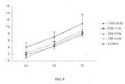
- the X-axis shows % Reduction in Maximal Twitch Force, while the Y-axis shows Time (Hours).
- Error bars illustrate ⁇ S.E.M.
- the X-axis shows % Reduction in Maximal Twitch Force, while the Y-axis shows Time (Hours).
- FIG. 7 shows a bladder tissue section attached to the force displacement transducer in a tissue organ bath setup.
- the meaning of the figures are the following:
- the X-axis shows % Reduction in Maximal Twitch Force, while the Y-axis shows Time (Hours).
- 1 Speywood or Dysport unit or 1 U corresponds to the median intraperitoneal LD 50 dose of botulinum toxin in mice.
- Examples 1 to 3 are aimed at determining the quantity of a pre-synaptic neuromuscular blocking substance in a sample whereas Example 4 is aimed at determining the selectivity of such a substance with respect to smooth or striated muscles.
- the modified Ringers buffer identified hereafter as “Liley's Ringers buffer” is prepared by diluting the following in water:
- glucose 11 mM
- a gas mixture of 95% O 2 and 5% CO 2 is bubbled through the buffer solution to yield the Lillies Ringers buffer.
- PBS Phosphate Buffered Saline solution
- Wistar rats (approximate weight 275 g) are sacrificed by neck dislocation following CO 2 exposure (approximately 3 min to induce loss of consciousness).
- the rib cage is dissected from each animal, placed in Liley's Ringers buffer and transported to the experiment place (journey time: approximately 15 min). There the rib cage is separated into two sections by careful dissection along the spinal column. The tissues are stored in oxygenated buffer prior to carrying out the experimental procedures.
- Each intercostal preparation (half rib cage) is placed into a Petri dish containing Lillies Ringers buffer.
- one intercostal nerve is carefully dissected to reveal approximately 1-2 mm of nerve bundle.
- the preparation can be revived in freshly oxygenated Liley's Ringers buffer for approximately 15-20 minutes before being returned to a Petri dish containing 10 ml of oxygenated Liley's Ringers buffer.
- the dissected intercostal nerve is then connected via a suction electrode to a stimulator (Grass Instruments Model S48), with a return contact electrode placed in the media.
- the minimum voltage V mst needed to induce muscle contraction is determined. If stimulation cannot be achieved below 10V, another nerve is dissected and the preparation revived prior to continuation.
- the nerve is stimulated with a pulsed voltage (5-9V, 1 Hz), the voltage chosen always being 1 V above the threshold voltage V m required to achieve stimulation and muscle contraction.
- Video microscopy of the section is carried out with a Nikon SMZ800 stereomicroscope equipped with JVC TKC1481EG video camera connected to a combined TV/video recorder.
- Dysport® active principle: botulinum toxin type A
- PBS directly above the intercostal preparation
- 50 Speywood or Dysport units (U) per ml dose 500 U of toxin is added to the culture dish (10 ml buffer) to yield a final concentration of 50 U/ml.
- 10 U/ml dose 100 U is added to the culture dish to yield a final concentration of 10 U/ml.
- 10 U is added to the culture dish yielding a final concentration of 1 U/ml.
- placebo which has the same composition as Dysport®, except that botulinum toxin is absent
- the full contents of the vial in 0.2 ml PBS
- the recorded video clips are converted to MPEG files.
- each movie is cut into 2-minute sections and these sections are slowed to 1 ⁇ 2 their initial speed using Adobe® Premiere® 5.1 software.
- Analysis is then performed by counting the number of twitches in a 10 s period (20 s on half speed clips) and averaging the number of twitches over this 10 s period to yield a value of twitch frequency.
- Contraction distance can also be measured by playing the movie with a superimposed scale bar (of arbitrary units—nominally a 6-7 point scale), the data being averaged to give the contraction distance over each 10 s period, or alternatively said distance is compared to the initial contraction distance.
- the relative contraction distance i.e. the contraction distance in the presence of toxin divided by the contraction distance in the absence of toxin
- the relative contraction distance is reduced more or less quickly as a function of the Dysport® concentration.
- FIG. 2 shows results regarding the distance twitch lifetime (i.e. the time needed from the moment the toxin is added until the point when the contraction distance becomes zero), a dose-dependant reduction in the muscle contraction distance as a function of the Dysport® concentration can be observed.
- the twitch frequency lifetime (i.e. the time needed from the moment the toxin is added until the point when the twitch frequency becomes zero) is also reduced by Dysport® in a dose-dependant manner as is shown in FIG. 3 .
- the modified Ringers buffer or “Liley's Ringers buffer” used in this Example is the same as for Example 1.
- the Gelatine Phosphate Buffer (GPB) used in this Example is prepared by diluting the following in 1 liter of water:
- One half of the rib cage is placed in a Petri dish containing approximately 10 ml of Liley's Ringers buffer and an intercostal nerve is carefully dissected to reveal approximately 1-2 mm of nerve bundle. This nerve is then connected via a suction electrode to a stimulator (Grass Instruments Model S48) with a return electrode placed in the media. The minimum voltage V m needed to induce muscle contraction is determined. If stimulation cannot be achieved below 10 V (1 Hz, 200 ⁇ sec duration), another nerve is dissected. The 2-rib section containing the dissected nerve is dissected from the half rib cage, ensuring as much excess muscle tissue as possible remains either side of the 2 ribs for later attachment to the force displacement transducer.
- Three metal staples are attached to the non-stimulated muscle tissue on either side of the two ribs.
- One side of the 2-rib section is attached to the fixed foot via the three staples whilst the other side was affixed to the free foot.
- the fixed foot is clamped securely in place while the free foot was attached to the force displacement transducer (Grass Instruments Model FT03) via approximately 4 cm of cotton thread.
- the fixed tissue is immersed in approximately 500 ml Liley's Ringers buffer, and a return electrode placed within the buffer.
- the dissected intercostal nerve was connected via a suction electrode to the stimulator. This system is shown in FIG. 4 .
- the 2-rib tissue sections are stimulated for approximately 90 minutes at 15 V, 200 ⁇ sec duration using train pulse stimulation (1 pulse/second for the first 5 seconds of every 30 second period).
- the required concentration of toxin is reconstituted in GPB immediately prior to application.
- the toxin delivery is via one of two methods:
- Twitch force readings recorded from the force displacement transducer are first amplified throughout a Grass Instruments AC/DC strain gage amplifier (Model P122) and signals are then recorded using Grass PolyVIEWTM software.
- the system using a striated muscle is identical to that of Example 3, except that a pig rib cage muscle with an intercostals nerve is used instead of the rat rib cage muscle.
- the system using a smooth muscle tissue sample is analogous to that described previously for the striated muscle (rat rib cage muscle) preparation, except that a rat bladder muscle preparation is used instead of the rat rib cage muscle preparation and Krebs-Henseleit buffer is used instead of Lillies Ringers buffer.
- 30 cm of surgical suture is attached to one end of the tissue strip, and secured with three knots.
- a 15 cm length of surgical suture is threaded half way through the tissue, leaving 7.5 cm either side.
- a knot is then half-tied using each end of the suture; the knot is pulled tight over the tissue and a glass hook, before two further knots are tied to secure the attachment.
- the hook is placed into a tissue bath containing 37° C. Krebs buffer gassed with 95% O 2 /5% CO 2 .
- the tissues are hooked up to the force transducer in the same order.
- Three knots are tied around a metal S-hook using the 30 cm length of suture. The S-hook is then attached to the force transducer.
- the PolyVIEWTM recording software is started at this point. Each piece of tissue is then pre-tensioned by quickly raising the force transducer until the equivalent of a 10 g force (5 V) is measured on the P122 strain gauge amplifier (Grass, Astro Med) digital display. The tissue strips undergo an exponential stretch-relaxation observed as a reduction in force measured. The relaxation is allowed to occur under non-stimulation conditions for 1 hour prior to experimental stimulation of the tissue. This system is shown in FIG. 7 .
- the smooth muscle tissue sections are stimulated for approximately 90 minutes at 20 V, 300 ⁇ sec duration at 20 Hz using train pulse stimulation (1 train of pulses for the first 5 seconds of every 60 second period).
- the required concentration of toxin is reconstituted in GPB immediately prior to application.
- Direct application method The smooth muscle section is exposed to the air/liquid interface by removal of some of the buffer within the tissue bath. The toxin is applied directly onto the exposed tissue in a drop wise fashion, coating the muscle in the toxin solution. The tissue is left exposed for a further 15 minutes to enable uptake of the toxin before covering that with the original Krebs Henseleit buffer.
- Contraction force readings recorded from the force displacement transducer are first amplified throughout a Grass Instruments AC/DC strain gage amplifier (Model P122) and signals are then recorded using Grass PolyVIEWTM software.
- Examples 4 are used for determining selectivity of pre-synaptic neuromuscular blocking substances, namely biologically active botulinum toxin fragments.
- the same pre-determined nanomolar quantity of biologically active botulinum toxin fragments is used in each of the striated muscle and smooth muscle systems instead of an unknown amount.
- the ratios of the times needed to get a certain reduction in twitch force e.g. the times to get a reduction of 50%
- twitch force e.g. 50%
- smooth muscles and in striated muscles can be used as indicators of the selectivity of said biologically active botulinum toxin fragments with respect to smooth or striated muscles.
Landscapes
- Health & Medical Sciences (AREA)
- Life Sciences & Earth Sciences (AREA)
- Engineering & Computer Science (AREA)
- Biomedical Technology (AREA)
- Chemical & Material Sciences (AREA)
- Immunology (AREA)
- Urology & Nephrology (AREA)
- Hematology (AREA)
- Molecular Biology (AREA)
- Cell Biology (AREA)
- Physics & Mathematics (AREA)
- Biochemistry (AREA)
- General Health & Medical Sciences (AREA)
- Bioinformatics & Cheminformatics (AREA)
- Biotechnology (AREA)
- General Physics & Mathematics (AREA)
- Analytical Chemistry (AREA)
- Pathology (AREA)
- Medicinal Chemistry (AREA)
- Food Science & Technology (AREA)
- Microbiology (AREA)
- Organic Chemistry (AREA)
- Genetics & Genomics (AREA)
- Wood Science & Technology (AREA)
- Zoology (AREA)
- Tropical Medicine & Parasitology (AREA)
- Toxicology (AREA)
- Biophysics (AREA)
- Rheumatology (AREA)
- General Engineering & Computer Science (AREA)
- Investigating Or Analysing Biological Materials (AREA)
- Measuring Or Testing Involving Enzymes Or Micro-Organisms (AREA)
- Peptides Or Proteins (AREA)
- Medicines Containing Antibodies Or Antigens For Use As Internal Diagnostic Agents (AREA)
Abstract
Description
- (i) determining the minimum voltage Vm needed to induce the contraction of muscle tissue, said muscle tissue being connected to an electrical stimulator through a motor nerve;
- (ii) adding the sample containing the pre-synaptic neuromuscular blocking substance;
- (iii) electrically stimulating, at a voltage at least equal to Vm, the muscle tissue at certain time intervals;
- (iv) comparing the effect induced by the sample to the effect induced by a reference substance and thereby determining the quantity of the pre-synaptic neuromuscular blocking substance in the sample.
- (i) determining the minimum voltage Vmsm needed to induce the contraction of a smooth muscle tissue, said smooth muscle tissue being linked to an electrical stimulator through an electrically conducting liquid;
- (ii) adding the sample containing the pre-synaptic neuromuscular blocking substance;
- (iii) electrically stimulating, at a voltage at least equal to Vmsm, the smooth muscle tissue at certain time intervals;
- (iv) comparing the effect induced by the sample to the effect induced by a reference substance and thereby determining the quantity of the pre-synaptic neuromuscular blocking substance in the sample.
- (a) determining the minimum voltage Vmst needed to induce the contraction of a striated muscle tissue, said striated muscle tissue being connected to an electrical stimulator through a motor nerve;
- (b) adding to the system of step (a) a pre-determined quantity of a pre-synaptic neuromuscular blocking substance;
- (c) electrically stimulating, at a voltage at least equal to Vmst, the muscle tissue at certain time intervals;
- (d) measuring the effect induced by the pre-determined quantity of a pre-synaptic neuromuscular blocking substance on the striated muscle tissue;
- (e) determining the minimum voltage Vmsm needed to induce the contraction of a smooth muscle tissue, said smooth muscle tissue being linked to an electrical stimulator through an electrically conducting liquid;
- (f) adding to the system of step (e) the same pre-determined quantity of the same pre-synaptic neuromuscular blocking substance as for step (b);
- (g) electrically stimulating, at a voltage at least equal to Vmsm, the muscle tissue at certain time intervals;
- (h) measuring the effect induced by said pre-determined quantity of the pre-synaptic neuromuscular blocking substance on the smooth muscle tissue;
- (i) calculating the quotient of the result obtained after step (h) by the result obtained after step (d), or, conversely, the quotient of the result obtained after step (d) by the result obtained after step (h).
-
- i. the time for the muscle contraction force to reach a certain percentage (for example 75% or 50%) of the maximum peak (contraction) height,
- ii. the area under the curve (which may be adjusted for maximum peak height, or
- iii. comparison of the slope of part or all of the curve (which may be related to the maximum peak height of the curve).
- (i) determining the minimum voltage Vmsm needed to induce the contraction of smooth muscle tissue, said smooth muscle tissue being linked to an electrical stimulator through an electrically conducting liquid;
- (ii) adding a mixture of the sample to be tested containing the neutralising antibodies to the pre-synaptic neuromuscular blocking substance and a determined quantity of said pre-synaptic neuromuscular blocking substance, said mixture having been pre-incubated at a temperature from 0 to 45° C. for a period from about 15 to about 120 minutes;
- (iii) electrically stimulating, at a voltage at least equal to Vmsm, the smooth muscle tissue at certain time intervals;
- (iv) comparing the effect induced by the mixture to the effect induced by the determined quantity of said pre-synaptic neuromuscular blocking substance and thereby determining the quantity of neutralising antibodies to the pre-synaptic neuromuscular blocking substance in the sample.
- 1=Force displacement transducer
- 2=Electrode
- 3=Suction electrode
- 4=Fixed attachment
- 5=Staple
- 6=Gas disperser
- 7=Suture material
- 8=Free attachment
- 1=Force transducer
- 2=Glass hook clamp
- 3=Electrode
- 4=Electrode clamp
- 5=Overflow
- 6=Jacket-in
- 7=Glass hook
- 8=Suture
- 9=Rat bladder strip
- 10=Electrode coil
| NaCl | 138.8 | mM | ||
| KCl | 4 | | ||
| NaHCO | ||||
| 3 | 12 | mM | ||
| KH2PO4 | 1 | | ||
| MgCl | ||||
| 2 | 1 | | ||
| CaCl | ||||
| 2 | 2 | mM | ||
| Phosphate buffer | 0.01 | M | ||
| KCl | 0.0027 | M | ||
| NaCl | 0.137 | M | ||
| pH at 25 ° C. | 7.4 | |||
| Gelatin | 2 | g | ||
| NaHPO4, 2 H2O | 10 | g | ||
-
- A) Direct application—The 2-rib section is exposed to the air/liquid interface by removal of some of the buffer within the tissue bath. Using a Hamilton syringe, the toxin is applied directly onto the exposed tissue in a drop wise fashion, coating the muscle in the toxin solution. The tissue is left exposed for a further 15 minutes to enable uptake of the toxin before covering that with the original Ringers buffer. If necessary, any dislodged, dissected nerves are reconnected to the suction electrode.
- B) Immersion—Using a Hamilton syringe, the toxin is applied directly into the tissue bath in close proximity to (but not directly onto) the 2-rib section.
| Component | Concentration (g/I) |
| D-glucose | 2.1 |
| Magnesium sulphate (MgSO4) | 0.29 |
| Monobasic potassium phosphate (KH2PO4) | 0.16 |
| Potassium chloride (NaCl) | 0.35 |
| Sodium chloride (NaCl) | 6.9 |
| Calcium chloride (CaCl2) | 0.282 |
| Sodium bicarbonate NaHCO3 | 2.1 |
-
- 1) The detrusor muscle is dissected from the neck of the bladder in strips (ca. 2 mm×2 mm×10 mm); and
- 2) The tissue is mounted in individual 10 ml tissue baths fitted with tissue electrodes and filled with Krebs-Henselett buffer.
Claims (14)
Applications Claiming Priority (3)
| Application Number | Priority Date | Filing Date | Title |
|---|---|---|---|
| GB0417366A GB2416849A (en) | 2004-08-04 | 2004-08-04 | Method for determining the quantity of a pre-synaptic neuromuscular blocking substance in a sample |
| GB0417366.2 | 2004-08-04 | ||
| PCT/GB2005/003032 WO2006013354A1 (en) | 2004-08-04 | 2005-08-02 | Method for determining the selectivity of a pre-synaptic neuromuscular blocking substance |
Publications (2)
| Publication Number | Publication Date |
|---|---|
| US20080213820A1 US20080213820A1 (en) | 2008-09-04 |
| US8759093B2 true US8759093B2 (en) | 2014-06-24 |
Family
ID=32982512
Family Applications (1)
| Application Number | Title | Priority Date | Filing Date |
|---|---|---|---|
| US11/659,445 Expired - Fee Related US8759093B2 (en) | 2004-08-04 | 2005-08-02 | Method for determining the selectivity of a pre-synaptic neuromuscular blocking substance |
Country Status (6)
| Country | Link |
|---|---|
| US (1) | US8759093B2 (en) |
| EP (1) | EP1776589B1 (en) |
| AT (1) | ATE459003T1 (en) |
| DE (1) | DE602005019575D1 (en) |
| GB (1) | GB2416849A (en) |
| WO (1) | WO2006013354A1 (en) |
Families Citing this family (5)
| Publication number | Priority date | Publication date | Assignee | Title |
|---|---|---|---|---|
| KR20120109508A (en) | 2009-11-18 | 2012-10-08 | 메르츠 파마 게엠베하 운트 코. 카가아 | Assay for quantifying clostridial neurotoxin |
| AU2011239668B2 (en) * | 2010-04-15 | 2014-04-17 | Cardiac Pacemakers, Inc. | Autonomic modulation using transient response with intermittent neural stimulation |
| JP5877617B2 (en) | 2011-09-29 | 2016-03-08 | セルスナップ、リミテッド・ライアビリティ・カンパニー | Compositions and methods for toxin productivity testing |
| SG11201510685UA (en) | 2013-06-28 | 2016-01-28 | Merz Pharma Gmbh & Co Kgaa | Means and methods for the determination of the biological activity of neurotoxin polypeptides in cells |
| CN114621921A (en) * | 2020-12-11 | 2022-06-14 | 深圳先进技术研究院 | Method for extracting neurite contact body |
Citations (1)
| Publication number | Priority date | Publication date | Assignee | Title |
|---|---|---|---|---|
| GB2398636A (en) | 2003-02-21 | 2004-08-25 | Ipsen Ltd | Method for determining the quantity of pre-synaptic neuromuscular blocking substance contained in a sample |
-
2004
- 2004-08-04 GB GB0417366A patent/GB2416849A/en not_active Withdrawn
-
2005
- 2005-08-02 AT AT05767609T patent/ATE459003T1/en not_active IP Right Cessation
- 2005-08-02 US US11/659,445 patent/US8759093B2/en not_active Expired - Fee Related
- 2005-08-02 WO PCT/GB2005/003032 patent/WO2006013354A1/en not_active Ceased
- 2005-08-02 EP EP05767609A patent/EP1776589B1/en not_active Expired - Lifetime
- 2005-08-02 DE DE602005019575T patent/DE602005019575D1/en not_active Expired - Lifetime
Patent Citations (2)
| Publication number | Priority date | Publication date | Assignee | Title |
|---|---|---|---|---|
| GB2398636A (en) | 2003-02-21 | 2004-08-25 | Ipsen Ltd | Method for determining the quantity of pre-synaptic neuromuscular blocking substance contained in a sample |
| WO2004074838A1 (en) | 2003-02-21 | 2004-09-02 | Ipsen Limited | Quantification of botulinum toxin |
Non-Patent Citations (12)
| Title |
|---|
| Birmingham et al, Br. J. Pharmac. 70, 1980, pp. 501-506, A Comparison of the Skeletal Neuromuscular and Autonomic . . . . |
| Chang et al, Naunyn-Schmiedberg's Arch. Pharmacol. 282, 1974, pp. 129-142, Comparison of the Presynaptic Actions of . . . . |
| Foldes, Life Sciences, vol. 28, 1981, pp. 1585-1590, The Significance of Physiological [Ca2+] and [Mg2+] for in vitro . . . . |
| Göschel et al, Experimental Neurology 147, 1997, pp. 96-102, Botulinum A Toxin Therapy: Neutralizing and Nonneutralizing . . . . |
| Healy et al, Br. J. Anaesth. 54, 1982, pp. 1307-1311, In Vitro Comparison Between the Neuromuscular and Ganglion Blocking . . . . |
| James et al, Am. J. Physiol. Gastrointest Liver Physiol. 285, 2003, pp. G291-G297, Inhibitory effects of botulinumtoxin . . . . |
| Longhurst et al, Jour. of Pharm. & Toxicol. Methods 45, 2001, pp. 91-108, Pharmacological Techniques for the In Vitro . . . . |
| Pearce et al, Toxicom, vol. 35, No. 9, 1997, pp. 1373-1412, Pharmacologic Characterization of Botulinum Toxin for Basic . . . . |
| Sheridan et al, J. Appl. Toxicol. 19, 1999, pp. S29-S33, Comparison of In Vivo and In Vitro Mouse Bioassays for . . . . |
| Smith et al. "Effect of stimulation intensity and botulinum toxin isoform on rat bladder strip contractions", Brain Research Bulletin, 2003, 61:165-171. * |
| Vizi et al. "Side effects of nondepolarizing muscle relaxants: relationship to their antinicotinic and antimuscarinic actions", Pharmacol. Ther. 1997, 73(2):75-89. * |
| Wohlfarth et al, Naunyn-Schmiedeberg's Arch Pharmacol. 355, 1997 pp. 335-340, Boluinum A toxins: units versus units. |
Also Published As
| Publication number | Publication date |
|---|---|
| EP1776589B1 (en) | 2010-02-24 |
| GB2416849A (en) | 2006-02-08 |
| EP1776589A1 (en) | 2007-04-25 |
| GB0417366D0 (en) | 2004-09-08 |
| WO2006013354A1 (en) | 2006-02-09 |
| US20080213820A1 (en) | 2008-09-04 |
| ATE459003T1 (en) | 2010-03-15 |
| DE602005019575D1 (en) | 2010-04-08 |
Similar Documents
| Publication | Publication Date | Title |
|---|---|---|
| Chesler et al. | Intracellular pH of astrocytes increases rapidly with cortical stimulation | |
| Forsberg et al. | Ionized calcium in human cerebrospinal fluid and its influence on intrinsic and synaptic excitability of hippocampal pyramidal neurons in the rat | |
| Miele et al. | The source of physiologically stimulated glutamate efflux from the striatum of conscious rats. | |
| Errico et al. | Persistent increase of D-aspartate in D-aspartate oxidase mutant mice induces a precocious hippocampal age-dependent synaptic plasticity and spatial memory decay | |
| JP2011254825A (en) | Quantification of botulinum toxin | |
| Varaschin et al. | Histamine H3 receptors decrease dopamine release in the ventral striatum by reducing the activity of striatal cholinergic interneurons | |
| US8759093B2 (en) | Method for determining the selectivity of a pre-synaptic neuromuscular blocking substance | |
| Sekiguchi et al. | Excitatory action of N-acetylaspartylglutamate on Purkinje cells in guinea pig cerebellar slices: an intrasomatic study | |
| Mori-Okamoto et al. | Electrophysiological and pharmacological actions of N-acetylaspartylglutamate intracellularly studied in cultured chick cerebellar neurons | |
| Medina et al. | Cardiac up-regulation of NBCe1 emerges as a beneficial consequence of voluntary wheel running in mice | |
| Miles et al. | Release and uptake of catecholamines in the bed nucleus of the stria terminalis measured in the mouse brain slice | |
| Barnes et al. | Effects of ethosuxirnide and tetramethylsuccinimide on cultured cortical neurons | |
| Coates et al. | Identification of carbonic anhydrase activity in bullfrog olfactory receptor neurons: histochemical localization and role in CO2 chemoreception | |
| Rosen et al. | Developmental changes in the muscarinic stimulation of canine Purkinje fibers. | |
| KR100888379B1 (en) | Mechanism of astrocyte-neuron signaling | |
| Patel et al. | Microelectrode investigation of neuroneal ageing from a single identified neurone | |
| Spasic et al. | Two distinct electrophysiological mechanisms underlie extensive depolarization elicited by 2, 4 diaminobutyric acid in leech Retzius neurons | |
| de Mello | Modulation of junctional permeability in cardiac fibers | |
| Okuda et al. | Hemorrhage-induced regional brain thyrotropin-releasing hormone release in conscious rats measured by microdialysis | |
| Onimaru et al. | Cell responses of the Ventrolateral Medulla to PAR1 activation and changes in respiratory rhythm in Newborn Rat En Bloc Brainstem-spinal cord preparations | |
| Aldossary | Investigating changes in angiotensin ii signalling, respiratory variability and carotid body function in response to chronic hypoxia | |
| Han | Histaminergic modulation of striatal development | |
| Nori et al. | Adaptation of the endplate in skeletal muscle of Homer 2 2/2 mice | |
| SU938990A1 (en) | Method of evaluating agricultural animal sperm | |
| Mächler et al. | Investigation of Cellular Compartmentation of Brain Energy Metabolism using Genetically Encoded FRET Sensors |
Legal Events
| Date | Code | Title | Description |
|---|---|---|---|
| AS | Assignment |
Owner name: IPSEN LIMITED, UNITED KINGDOM Free format text: ASSIGNMENT OF ASSIGNORS INTEREST;ASSIGNORS:FRANCE, RICHARD;QUIRK, ROBIN;RICCALTON-BANKS, LISA;AND OTHERS;REEL/FRAME:020414/0527;SIGNING DATES FROM 20070131 TO 20070205 Owner name: IPSEN LIMITED, UNITED KINGDOM Free format text: ASSIGNMENT OF ASSIGNORS INTEREST;ASSIGNORS:FRANCE, RICHARD;QUIRK, ROBIN;RICCALTON-BANKS, LISA;AND OTHERS;SIGNING DATES FROM 20070131 TO 20070205;REEL/FRAME:020414/0527 |
|
| AS | Assignment |
Owner name: IPSEN DEVELOPMENTS LIMITED,UNITED KINGDOM Free format text: CHANGE OF NAME;ASSIGNOR:IPSEN LIMITED;REEL/FRAME:023958/0182 Effective date: 20081212 Owner name: IPSEN DEVELOPMENTS LIMITED, UNITED KINGDOM Free format text: CHANGE OF NAME;ASSIGNOR:IPSEN LIMITED;REEL/FRAME:023958/0182 Effective date: 20081212 |
|
| AS | Assignment |
Owner name: IPSEN BIOPHARM LIMITED,UNITED KINGDOM Free format text: ASSIGNMENT OF ASSIGNORS INTEREST;ASSIGNOR:IPSEN DEVELOPMENTS LIMITED;REEL/FRAME:024452/0646 Effective date: 20100423 Owner name: IPSEN BIOPHARM LIMITED, UNITED KINGDOM Free format text: ASSIGNMENT OF ASSIGNORS INTEREST;ASSIGNOR:IPSEN DEVELOPMENTS LIMITED;REEL/FRAME:024452/0646 Effective date: 20100423 |
|
| FEPP | Fee payment procedure |
Free format text: PAYOR NUMBER ASSIGNED (ORIGINAL EVENT CODE: ASPN); ENTITY STATUS OF PATENT OWNER: LARGE ENTITY |
|
| FEPP | Fee payment procedure |
Free format text: MAINTENANCE FEE REMINDER MAILED (ORIGINAL EVENT CODE: REM.) |
|
| LAPS | Lapse for failure to pay maintenance fees |
Free format text: PATENT EXPIRED FOR FAILURE TO PAY MAINTENANCE FEES (ORIGINAL EVENT CODE: EXP.) |
|
| STCH | Information on status: patent discontinuation |
Free format text: PATENT EXPIRED DUE TO NONPAYMENT OF MAINTENANCE FEES UNDER 37 CFR 1.362 |
|
| FP | Lapsed due to failure to pay maintenance fee |
Effective date: 20180624 |