US8709723B2 - Integrated analyses of breast and colorectal cancers - Google Patents
Integrated analyses of breast and colorectal cancers Download PDFInfo
- Publication number
- US8709723B2 US8709723B2 US13/461,268 US201213461268A US8709723B2 US 8709723 B2 US8709723 B2 US 8709723B2 US 201213461268 A US201213461268 A US 201213461268A US 8709723 B2 US8709723 B2 US 8709723B2
- Authority
- US
- United States
- Prior art keywords
- genes
- copy number
- alterations
- breast
- amplifications
- Prior art date
- Legal status (The legal status is an assumption and is not a legal conclusion. Google has not performed a legal analysis and makes no representation as to the accuracy of the status listed.)
- Active
Links
Images


Classifications
-
- C—CHEMISTRY; METALLURGY
- C12—BIOCHEMISTRY; BEER; SPIRITS; WINE; VINEGAR; MICROBIOLOGY; ENZYMOLOGY; MUTATION OR GENETIC ENGINEERING
- C12Q—MEASURING OR TESTING PROCESSES INVOLVING ENZYMES, NUCLEIC ACIDS OR MICROORGANISMS; COMPOSITIONS OR TEST PAPERS THEREFOR; PROCESSES OF PREPARING SUCH COMPOSITIONS; CONDITION-RESPONSIVE CONTROL IN MICROBIOLOGICAL OR ENZYMOLOGICAL PROCESSES
- C12Q1/00—Measuring or testing processes involving enzymes, nucleic acids or microorganisms; Compositions therefor; Processes of preparing such compositions
- C12Q1/68—Measuring or testing processes involving enzymes, nucleic acids or microorganisms; Compositions therefor; Processes of preparing such compositions involving nucleic acids
- C12Q1/6876—Nucleic acid products used in the analysis of nucleic acids, e.g. primers or probes
- C12Q1/6883—Nucleic acid products used in the analysis of nucleic acids, e.g. primers or probes for diseases caused by alterations of genetic material
- C12Q1/6886—Nucleic acid products used in the analysis of nucleic acids, e.g. primers or probes for diseases caused by alterations of genetic material for cancer
-
- A—HUMAN NECESSITIES
- A61—MEDICAL OR VETERINARY SCIENCE; HYGIENE
- A61P—SPECIFIC THERAPEUTIC ACTIVITY OF CHEMICAL COMPOUNDS OR MEDICINAL PREPARATIONS
- A61P35/00—Antineoplastic agents
-
- C—CHEMISTRY; METALLURGY
- C12—BIOCHEMISTRY; BEER; SPIRITS; WINE; VINEGAR; MICROBIOLOGY; ENZYMOLOGY; MUTATION OR GENETIC ENGINEERING
- C12Q—MEASURING OR TESTING PROCESSES INVOLVING ENZYMES, NUCLEIC ACIDS OR MICROORGANISMS; COMPOSITIONS OR TEST PAPERS THEREFOR; PROCESSES OF PREPARING SUCH COMPOSITIONS; CONDITION-RESPONSIVE CONTROL IN MICROBIOLOGICAL OR ENZYMOLOGICAL PROCESSES
- C12Q2600/00—Oligonucleotides characterized by their use
- C12Q2600/156—Polymorphic or mutational markers
Definitions
- the disclosed invention was made using funds from the U.S. government, particularly National Institutes of Health grants CA 043460, CA 057345, CA 062924, and CA 121113. The U.S. government therefore retains certain rights in the invention.
- This invention is related to the area of classifying, characterizing, detecting and diagnosing cancers.
- it relates to breast and colorectal cancers.
- cancer is the result of the sequential mutations of oncogenes and tumor suppressor genes (1).
- oncogenes and tumor suppressor genes have been accomplished through analyses of individual candidate genes chosen on the basis of functional or biologic data implicating them in the tumorigenic process.
- Recent advances in genomic technologies and bioinformatics have permitted simultaneous evaluation of many genes, thereby offering more comprehensive and unbiased information (2, 3).
- sequence of large families of genes, and even the human genes in the Reference Sequence (RefSeq) database have been determined in subsets of human cancers (4, 5).
- the alterations detected by sequencing represent only one category of genetic change that occurs in human cancer.
- alterations include gains (amplifications) and losses (deletions) of discrete chromosomal sequences that occur during tumor progression.
- Dramatic amplifications of oncogenes such as ERBB2 (6) or MYC (7) and deletions of tumor suppressor genes such as CDKN2A (8), PTEN (9, 10) and SMAD4 (11) have demonstrated the importance of these mechanisms of genetic alteration in particular tumor types.
- a comprehensive picture of genetic alterations in human cancer should therefore include the integration of sequence based alterations together with copy number gains and losses.
- a method of characterizing a breast or colon tumor in a human is provided.
- Table 3 or SI Table 6 is determined in a breast or colon tumor by determining at least one somatic mutation in a gene in the pathway in a test sample relative to a normal sample of the human.
- the breast or colon tumor is assigned to a first group of breast or colon tumors that have a somatic mutation in at least one gene in said pathway.
- a method of detecting or diagnosing a breast or colon tumor or minimal residual disease of a breast or colon tumor or molecular relapse of a breast or colon tumor in a human is provided.
- a genomic amplification of at least one genomic region is determined in a test sample of a tumor or suspected tumor of the human.
- the genomic region is selected from the group consisting of those listed in SI Table 4 or Table 1.
- the human is identified as likely to have a breast or colon tumor, minimal residual disease, or molecular relapse of breast or colon tumor when the amplification is determined.
- a method is provided of detecting or diagnosing a breast or colon tumor or minimal residual disease of a breast or colon tumor or molecular relapse of a breast or colon tumor in a human.
- a genomic deletion of at least one genomic region is determined in a test sample of a tumor or suspected tumor of the human.
- the genomic region is selected from the group consisting of those listed in SI Table 5 or Table 2.
- the human is identified as likely to have a breast or colon tumor, minimal residual disease, or molecular relapse of breast or colon tumor when the homozygous deletion is determined.
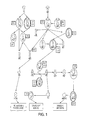
- FIG. 1 shows alterations in the combined FGF, EGFR, ERBB2 and PI3K pathways. Genes affected by copy number alterations are circled in red, while those altered by point mutations are circled in blue. The number of breast (B) and colorectal (C) tumors containing alterations are indicated in boxes adjacent to each gene.
- FIG. 2A-2B shows genomic landscape of a copy number and nucleotide alterations in two typical cancer samples.
- FIG. 2A indicates breast cancer alterations while FIG. 2B indicates colorectal cancer alterations.
- the telomere of the short arm of chromosome 1 is represented in the rear left corner of the green plane and ascending chromosomal positions continue in the direction of the arrow. Chromosomal positions that follow the front edge of the plane are continued at the back edge of the plane of the adjacent row and chromosomes are appended end to end. Peaks indicate the 60 highest-ranking candidate cancer genes for each tumor type, with peak heights reflecting the passenger probability scores.
- the yellow and blue peaks correspond to genes that are altered by copy number changes, while those altered only by point mutations are purple.
- the dots represent genes that were altered by copy number changes (red squares) or point mutations (white circles) in the B9C breast or Mx27 colorectal tumor samples. Altered genes participating in significant gene groups or pathways ( FIG. 13 ; SI Table 6) are indicated as black circles or squares.
- FIG. 3 (SI FIG. 1 .) shows schematic of experimental approach for integration of copy number and sequence alterations in breast and colorectal cancers
- FIG. 4 shows detection of amplifications and homozygous deletions using Illumina arrays and Digital Karyotyping.
- Digital Karyotyping results are shown in the top graphs, with the chromosomal coordinates indicated on the horizontal axis and the Digital Karyotyping tag density ratio indicated on the vertical axis.
- Illumina array results are shown in the bottom graphs, with the chromosomal coordinates indicated on the horizontal axis and the Log R Ratio indicated on the vertical axis.
- Digital Karyotyping data were used to validate the Illumina arrays and to develop approaches for sensitive and specific detection of focal amplifications and homozygous deletions.
- FIG. 5 (Table 1) Top candidate cancer genes in breast and colorectal cancer amplifications.
- FIG. 6 (Table 2) Top candidate cancer genes in breast and colorectal cancer homozygous deletions.
- FIG. 7A to 7B (Table 3) Candidate cancer pathways altered in breast and colorectal cancers.
- FIG. 8 (SI Table 1) Comparison between IlluminaTM array and Digital Karyotyping copy number analyses.
- FIG. 9 (SI Table 2) Copy number changes detected by Digital Karyotyping in colorectal cancer
- FIG. 10A to 10Z (SI Table 3) Amplifications and homozygous deletions detected by IlluminaTM, arrays in breast and colorectal cancers
- FIG. 11A to 11L Amplified genes in breast and colorectal cancers.
- FIG. 12A to 12E (SI Table 5) Homozygously deleted genes in breast and colorectal cancers.
- FIG. 13A to 13I Pathways enriched for copy number alterations and point mutations.
- FIG. 14A to 14B (SI Table 7) Breast and colorectal cancer samples used in these analyses.
- the inventors have developed means of diagnosing, classifying, characterizing, and detecting breast and colorectal tumors based on somatic mutations in genes in pathways, including point mutations, genomic amplifications, and genomic deletions.
- Xenografts or cell lines derived from breast and colorectal cancers were examined to obtain high resolution analyses of copy number and nucleotide alterations. Tumors were evaluated with microarrays containing at least 317,000 SNP probes and selected samples were also evaluated with Digital Karyotyping (24). This latter method provides a highly quantitative measure of gene copy number and was used to validate the sensitivity and specificity of the microarray data. The sequences of the 18,191 genes from the RefSeq database previously determined for breast and colorectal cancers were integrated with these results, providing a genome-wide analysis of sequence and copy number alterations.
- the integrated mutational analysis described here provides a global picture of the genetic alterations of breast and colorectal cancers.
- the combination of sequencing and copy number analysis at the whole genome level permits the identification of genes and pathways that may not be easily detected by either analysis alone.
- the analysis of point mutations can provide independent information that can help identify candidate target genes in regions of amplification or HD.
- gene groups and pathways can be affected by sequence and copy number changes, a combined analysis can highlight the groups that are enriched for these somatic alterations.
- copy number changes can also provide general insights into the functional effects of point mutations. Single nucleotide substitutions in genes that are observed to be deleted are more likely to be inactivating, while substitutions in genes that are amplified are more likely to be activating. This was confirmed by the observation of HDs and point mutations in TP53, SMAD2, SMAD3, and PTEN all of which are thought to be tumor suppressors. If copy number changes faithfully reflect the overall effect of target genes, one would expect to infrequently see both amplifications and HDs of the same set of genes in human tumors.
- cancer genome landscapes are complex, they may be better understood by placing all genetic alterations within defined cellular pathways.
- Our analyses identified several converging gene pathways, including the ERBB2, EGFR and PI3K pathways, that were affected by copy number changes and point alterations in both breast and colorectal cancers.
- many pathways implicated in colorectal tumor progression (Notch, AKT, and MAPK) were enriched for alterations.
- gene groups contained genes that were both amplified and others that were deleted, suggesting that different genes within the same group or pathway may be affected through alternate mechanisms. This is consistent with the observation that most signaling pathways contain both positive and negative regulators and alterations in any of these can lead to dysregulated signaling.
- Mutations including homozygous deletions, genomic amplifications, and point mutations can be determined by any means known in the art, including but not limited to the methods described below. Sequencing, digital karyotyping, and hybridization to SNP arrays, are non-limiting examples of techniques which can be used. DNA sequencing can be performed using any techniques which are known in the art, for example, based on chemical degradation, enzymatic synthesis, ligation, hybridization, etc. Enzymes which can be used include but are not limited to polymerases and ligases. Synthesized or degraded nucleic acids can be analyzed using techniques which separate molecules based on length or mass, for example. Sequence determinations can be performed manually or in an automated fashion.
- oligonucleotides can be employed as probes or primers, both of which may hybridize to the analyte. Some methods utilize dideoxynucleotides which act as monomers and terminators of DNA synthesis.
- Mutation, deletion, or amplification determination involves one or more ex vivo samples which are processed in order to analyze the genetic material (or sometimes the proteins encoded by the genetic material). Typically this involves purification or enrichment of nucleic acids and removal or de-enrichment of other cellular components, such as protein, lipid, carbohydrates. The nucleic acids are further reacted chemically or enzymatically to yield readily detectable products which correspond to the nucleic acids in the ex vivo samples. Determination of a somatic mutation is done by comparing a tumor sample or characteristic to a normal sample of the same individual. Differences can be observed and recorded by a human or a machine or a computer.
- Changes in copy number of a genomic segment can be determined by any means known in the art.
- fragments (enzymatically generated or random) are generated and ligated together to form a chain or concatenate.
- the concatenates can be sequenced, and underrepresented or overrepresented fragments of the genome can be noted.
- genomic DNA fragments can be hybridized to an array of oligonucleotides and their relative prevalence scored. Such techniques may detect deletions or amplifications.
- Changes in copy number may be from diploid to homozygous deletion, or amplifications ranging from diploid to at least 5-, at least 6-, at least 7-, at least 8-, at least 9-, at least 10-, at least 15-, at least 20-, at least 25-fold of diploid.
- Tumors or patients bearing tumors can be divided into or assigned to groups based on the presence or absence of a particular somatic mutation.
- the group with the mutation may optionally contain tumors with a particular mutation in a particular gene, tumors with mutations in a single gene, or tumors with mutations in a single pathway.
- Groups comprising tumors with mutations in a single gene or a single pathway may be the same or different types of mutations.
- Groups that are divided on the basis of a mutation in a gene or in a pathway may be used to evaluate drugs or other therapeutic treatments. This permits the determination of groups which are susceptible or refractory to the treatment. Thus patients who are susceptible can be successfully treated, and patients who are refractory can avoid expensive, potentially hazardous, and ultimately ineffective treatments.
- the mutations in genes and pathways can also be used to detect or diagnose breast or colon tumors, or minimal residual disease of such tumors, or molecular relapse of such tumors.
- the mutations and genes and pathways which have been found are characteristic of these cancers and can be used to identify them in various stages of disease. Characteristic mutations are not necessarily present in all or even in a majority of tumors of the breast or colon.
- Mutations found in tumors can be determined or confirmed by comparison to normal tissue. Somatic mutations are ones that occur in the tumor but are not found in normal tissue of the individual. Thus a comparison between tumor and normal can be used for identification and confirmation.
- DK Digital Karyotyping
- the Illumina platform employs a two step procedure based on oligo hybridization and single base extension for analysis of genomic SNPs (25). The combination of these two steps leads to greater fidelity of SNP calls and decreases false hybridization signals.
- fluorescence intensity measurements we developed an approach to detect amplifications resulting in 12 or more copies per nucleus (6-fold or greater amplification compared to the diploid genome) as well as deletions of both copies of a gene (HDs) (see SI Methods).
- Each HD affected the coding region of one gene on average, while an average amplicon contained two genes.
- the average numbers of protein-coding genes that were affected by either amplification or HD were 24 and 9 per breast and colorectal cancer, respectively.
- Table 1 lists the loci that were amplified in at least one tumor and had the highest probability of containing driver genes as determined by the combined mutation analysis (a complete list of amplifications is provided in SI Table 3 and amplified genes in SI Table 4). For genes to be considered potential targets of the amplification, the entire coding region of the gene was required to be contained within a focal amplicon. A few candidate genes in this list (e.g. CCNE1 (cyclin E) and ERBB2) were amplified in multiple tumors but were not found to be mutated by sequencing. The majority of candidate genes, however, harbored point mutations in some tumors and amplifications in others. The most striking aspect of this list of candidate genes is that only some of them had been implicated in cancer in the past.
- CCNE1 cyclin E
- ERBB2 cyclin E
- MYC MYC
- ERBB2 HER2/NEU
- CCNE1 CCND1
- EGFR EGFR
- FGFR2 FGFR2
- Table 2 similarly lists the loci that were homozygously deleted in at least one tumor and had the highest probability of containing drivers as determined by the combined mutation analysis (a complete list of HDs is provided in SI Table 3 and homozygously deleted genes in SI Table 5). For each of these genes, a portion of the coding region was affected by the HD. A number of genes previously known to be inactivated in colorectal or breast tumorigenesis, such as CDKN2A, PTEN, and TP53 are found in this list. We also identified genes, such as CHD5, MAP2K4, SMAD2, and SMAD3 that have been previously shown to be deleted in other tumor types, but not in colorectal or breast cancers.
- HDs as well as point mutations were found in OMA1 and ZNF521 in colorectal cancers and in MANEA, PCDH8, SATL1, and ZNF674 in breast cancers.
- PCDH8 is mutated and homozygously deleted in breast cancer (33).
- GSEA gene set enrichment analysis
- DNA samples from tumor derived xenografts and cell lines were obtained and purified.
- DK libraries were generated and analyzed as previously described (24, 44).
- the Illumina SNP arrays were used to analyze tumor samples.
- Bioinformatic analyses were used to determine focal amplifications and HDs. Statistical methods were employed to determine the likelihood that genetic alterations occurred at a frequency higher than the passenger rate, and to identify gene groups enriched for copy number and sequence alterations.
- DNA samples were obtained from xenografts and cell lines of ductal breast and colorectal carcinoma. Normal DNA samples were obtained from matched normal tissue or peripheral blood. Twenty two of the DNA samples include those used in the Discovery Screen of Sjöblom et al. and Wood et al. (1, 2). All tumor samples analyzed for copy number analyses are listed in SI Table 9.
- the colorectal cancer samples used were cell lines (10) or xenografts (26), each developed from a liver metastasis of a different patient.
- the breast cancer samples used were cell lines (22) and xenografts (23), each developed from a different patient.
- Homozygous deletions were identified using a sliding window size of 175 virtual tags ( ⁇ 700 kb in size). Windows with a tag density ratio (observed tags in window/expected tags in window) ⁇ 0.01 were considered to represent putative homozygous deletions and were further examined. Regions of homozygous deletions were defined as containing no experimental tags and the boundaries were determined as the outermost virtual tags with no matching experimental tags.
- Amplifications were identified using sliding windows of variable sizes, as the most accurate window size for detection and quantification of amplifications is the exact size of the altered region.
- Windows with tag density ratios ⁇ 6 were considered to represent amplified regions. Boundaries of the amplified region are determined by the outermost tag contained in a window with a tag density ratio >3 or by the virtual tag position after which there is sharp decline in the observed experimental tags.
- the Illumina Infinium II Whole Genome Genotyping Assay employing the BeadChip platform was used to analyze tumor samples at 317,503 (317 k), 555,351 (550 k V1), or 561,466 (550 k V3) SNP loci from the Human HapMap collection. All SNP positions were based on hg17 (NCBI Build 35, May 2004) version of the human genome reference sequence.
- the genotyping assay is a two step procedure that is based on hybridization to a 50 nucleotide oligo, followed by a two-color fluorescent single base extension.
- the image files of fluorescence intensities were processed using Illumina BeadStation software to provide intensity values for each SNP position.
- the normalized experimental intensity value (R) was compared to the intensity values for that SNP from a training set of normal samples and represented as a ratio (called the “Log R Ratio”) of log 2(Rexperimental/Rtraining set).
- Digital Karyotyping was used to inform and optimize the criteria for detection of focal homozygous deletions and high-copy amplifications using the Illumina arrays.
- Three colorectal cancer samples (Co44, Co82 and Co84) were assessed by Digital Karyotyping tag libraries as well as the Illumina arrays (SI Table 1). From these analyses criteria were developed to permit sensitive and specific detection of the Digital Karyotyping alterations using the Illumina platform as described below. These criteria were subsequently used to analyze an additional 46 breast and 33 colorectal cancers.
- HDs Homozygous deletions
- the first and last SNPs of the identified HD region were considered to be the boundaries of the alteration for subsequent analyses.
- the deletion breakpoint would be expected to be located between the boundary deleted SNPs and adjacent non-deleted SNPs; use of the inner deleted SNP boundaries provides the most conservative approach as use of the outer boundaries may include non-deleted regions.
- Karyotyping were defined as regions having at least one SNP with a LogR ratio ⁇ 1.4, at least one in ten SNPs with a LogR ratio ⁇ 1, and an average LogR ratio of the entire region of ⁇ 0.9. The boundaries of amplified regions were delimited by the outermost SNPs with LogR ratios ⁇ 1. Similar to analyses of homozygous deletions, we removed all amplifications that had identical boundaries and occurred in multiple samples.
- a second set of criteria were used to remove large chromosomal regions or entire chromosomes that showed copy number gains. These large alterations, called “complex amplifications”, were thus distinguished from small focal alterations, called “simple amplifications”. Based on observations from Digital Karyotyping, several steps were used to identify and remove complex amplifications. First, amplifications >3 Mb in size and groups of nearby amplifications (within 1 Mb) that were also >3 Mb in size were considered complex.
- Amplifications or groups of amplifications that occurred at a frequency of ⁇ 4 amplifications in a 10 Mb region, or ⁇ 5 amplifications per chromosome were deemed to be complex.
- the amplifications remaining after these filtering steps were considered to be simple amplifications and were further examined.
- the complex regions were not included in subsequent statistical analyses but those containing candidate cancer genes are indicated in Table 1.
- the passenger probability is an a posteriori probability that integrates information from the somatic mutation analysis of Wood et al. (2) with the data presented in this article.
- the passenger probabilities reported in Wood et al. (2) serve as a priori probabilities. These are available for three different scenarios of passenger mutation rates and results are presented separately for each. If a gene was not found to be mutated in Wood et al. (2) the prior passenger probability is set to the estimated proportion of passengers in the RefSeq set.
- a likelihood ratio for “driver” versus “passenger” was evaluated using as evidence the number of samples in which a gene was found to be amplified (or deleted). Analysis is carried out separately by type of array, and then combined by multiplication of the relevant likelihood terms.
- the passenger term is the probability that the gene in question is amplified (deleted). For each sample, we begin by computing the probability that the observed amplifications (deletions) will include the gene in question by chance. Inclusion of all available SNPs is required for amplification, while any overlap of SNPs is sufficient for deletions. Specifically, if in a specific sample N SNPs are typed, and K amplifications are found, whose sizes, in terms of SNPs involved, are A1 . .
- AK a gene with G SNPs will be included at random with probability ( A 1 ⁇ G+ 1)/ N + . . . +( AK ⁇ G+ 1)/ N for amplifications and ( A 1+ G ⁇ 1)/ N + . . . +( AK+G ⁇ 1)/ N for deletions.
Landscapes
- Chemical & Material Sciences (AREA)
- Health & Medical Sciences (AREA)
- Life Sciences & Earth Sciences (AREA)
- Organic Chemistry (AREA)
- Proteomics, Peptides & Aminoacids (AREA)
- Pathology (AREA)
- Immunology (AREA)
- Zoology (AREA)
- Wood Science & Technology (AREA)
- Engineering & Computer Science (AREA)
- Analytical Chemistry (AREA)
- Genetics & Genomics (AREA)
- General Health & Medical Sciences (AREA)
- Microbiology (AREA)
- Biotechnology (AREA)
- Oncology (AREA)
- General Engineering & Computer Science (AREA)
- Bioinformatics & Cheminformatics (AREA)
- Biochemistry (AREA)
- Molecular Biology (AREA)
- Physics & Mathematics (AREA)
- Biophysics (AREA)
- Hospice & Palliative Care (AREA)
- Animal Behavior & Ethology (AREA)
- Nuclear Medicine, Radiotherapy & Molecular Imaging (AREA)
- Chemical Kinetics & Catalysis (AREA)
- Medicinal Chemistry (AREA)
- Pharmacology & Pharmacy (AREA)
- Public Health (AREA)
- Veterinary Medicine (AREA)
- General Chemical & Material Sciences (AREA)
- Measuring Or Testing Involving Enzymes Or Micro-Organisms (AREA)
Abstract
Description
-
- 1. Vogelstein, B. & Kinzler, K. W. (2004) Cancer genes and the pathways they control. Nat Med 10:789-99.
- 2. Bardelli, A. & Velculescu, V. E. (2005) Mutational analysis of gene families in human cancer. Curr Opin Genet Dev 15:5-12.
- 3. Strausberg, R. L., Levy, S. & Rogers, Y. H. (2008) Emerging DNA sequencing technologies for human genomic medicine. Drug Discov Today 13:569-77.
- 4. Greenman, C., et al. (2007) Patterns of somatic mutation in human cancer genomes. Nature 446:153-8.
- 5. Wood, L. D., et al. (2007) The genomic landscapes of human breast and colorectal cancers. Science 318:1108-13.
- 6. Slamon, D. J., et al. (1987) Human breast cancer: correlation of relapse and survival with amplification of the HER-2/neu oncogene. Science 235:177-82.
- 7. Collins, S. & Groudine, M. (1982) Amplification of endogenous myc-related DNA sequences in a human myeloid leukaemia cell line. Nature 298:679-81.
- 8. Kamb, A., et al. (1994) A cell cycle regulator potentially involved in genesis of many tumor types. Science 264:436-40.
- 9. Li, J., et al. (1997) PTEN, a putative protein tyrosine phosphatase gene mutated in human brain, breast, and prostate cancer. Science 275:1943-7.
- 10. Steck, P. A., et al. (1997) Identification of a candidate tumour suppressor gene, MMAC1, at chromosome 10q23.3 that is mutated in multiple advanced cancers. Nat Genet 15:356-62.
- 11. Hahn, S. A., et al. (1996) Dpc4, a Candidate Tumor Suppressor Gene At Human Chromosome 18q21.1. Science 271:350-353.
- 12. Pinkel, D. & Albertson, D. G. (2005) Array comparative genomic hybridization and its applications in cancer.
Nat Genet 37 Suppl:S11-7. - 13. Camps, J., et al. (2008) Chromosomal breakpoints in primary colon cancer cluster at sites of structural variants in the genome. Cancer Res 68:1284-95.
- 14. Weir, B. A., et al. (2007) Characterizing the cancer genome in lung adenocarcinoma. Nature 450:893-8.
- 15. Haverty, P. M., et al. (2008) High-resolution genomic and expression analyses of copy number alterations in breast tumors. Genes Chromosomes Cancer 47:530-42.
- 16. Mullighan, C. G., et al. (2007) Genome-wide analysis of genetic alterations in acute lymphoblastic leukaemia. Nature 446:758-64.
- 17. Harada, T., et al. (2008) Genome-wide DNA copy number analysis in pancreatic cancer using high-density single nucleotide polymorphism arrays. Oncogene 27:1951-60.
- 18. Stark, M. & Hayward, N. (2007) Genome-wide loss of heterozygosity and copy number analysis in melanoma using high-density single-nucleotide polymorphism arrays. Cancer Res 67:2632-42.
- 19. Nagayama, K., et al. (2007) Homozygous deletion scanning of the lung cancer genome at a 100-kb resolution. Genes Chromosomes Cancer 46:1000-10.
- 20. Zhao, X., et al. (2005) Homozygous deletions and chromosome amplifications in human lung carcinomas revealed by single nucleotide polymorphism array analysis. Cancer Res 65:5561-70.
- 21. Jones, S., et al. (2008) Comparative lesion sequencing provides insights into tumor evolution. Proc Natl Acad Sci USA 105:4283-8.
- 22. Cairns, P., et al. (1995) Frequency of homozygous deletion at p16/CDKN2 in primary human tumours. Nat Genet 11:210-2.
- 23. Liggett, W. H., Jr. & Sidransky, D. (1998) Role of the p16 tumor suppressor gene in cancer. J Clin Oncol 16:1197-206.
- 24. Wang, T. L., et al. (2002) Digital karyotyping. Proc Natl Acad Sci USA 99:16156-61.
- 25. Steemers, F. J., et al. (2006) Whole-genome genotyping with the single-base extension assay. Nat Methods 3:31-3.
- 26. Peiffer, D. A., et al. (2006) High-resolution genomic profiling of chromosomal aberrations using Infinium whole-genome genotyping. Genome Res 16:1136-48.
- 27. Conrad, D. F., Andrews, T. D., Carter, N. P., Hurles, M. E. & Pritchard, J. K. (2006) A high-resolution survey of deletion polymorphism in the human genome. Nat Genet 38:75-81.
- 28. Sebat, J., et al. (2004) Large-scale copy number polymorphism in the human genome. Science 305:525-8.
- 29. Volik, S., et al. (2003) End-sequence profiling: sequence-based analysis of aberrant genomes. Proc Natl Acad Sci USA 100:7696-701.
- 30. Bignell, G. R., et al. (2007) Architectures of somatic genomic rearrangement in human cancer amplicons at sequence-level resolution. Genome Res 17:1296-303.
- 31. Sjoblom, T., et al. (2006) The consensus coding sequences of human breast and colorectal cancers. Science 314:268-74.
- 32. Wang, Z., et al. (2004) Three classes of genes mutated in colorectal cancers with chromosomal instability. Cancer Res 64:2998-3001.
- 33. Yu, J. S., et al. (2008) PCDH8, the human homolog of PAPC, is a candidate tumor suppressor of breast cancer. Oncogene.
- 34. Ekins, S., Nikolsky, Y., Bugrim, A., Kirillov, E. & Nikolskaya, T. (2007) Pathway mapping tools for analysis of high content data. Methods Mol Biol 356:319-50.
- 35. Subramanian, A., et al. (2005) Gene set enrichment analysis: a knowledge-based approach for interpreting genome-wide expression profiles. Proc Natl Acad Sci USA 102:15545-50.
- 36. Collins, F. S. & Barker, A. D. (2007) Mapping the cancer genome. Pinpointing the genes involved in cancer will help chart a new course across the complex landscape of human malignancies. Sci Am 296:50-7.
- 37. Hoglund, M., et al. (2002) Dissecting karyotypic patterns in colorectal tumors: two distinct but overlapping pathways in the adenoma-carcinoma transition. Cancer Res 62:5939-46.
- 38. Paez, J. G., et al. (2004) Genome coverage and sequence fidelity of phi29 polymerase-based multiple strand displacement whole genome amplification. Nucleic Acids Res 32:e71.
- 39. Arriola, E., et al. (2007) Evaluation of Phi29-based whole-genome amplification for microarray-based comparative genomic hybridisation. Lab Invest 87:75-83.
- 40. Popescu, N. C. (2004) Fragile sites and cancer genes on the short arm of
chromosome 8. Lancet Oncol 5:77; discussion 77. - 41. Tanner, M., et al. (2006) Topoisomerase IIalpha gene amplification predicts favorable treatment response to tailored and dose-escalated anthracycline-based adjuvant chemotherapy in HER-2/neu-amplified breast cancer: Scandinavian Breast Group Trial 9401. J Clin Oncol 24:2428-36.
- 42. Coon, J. S., et al. (2002) Amplification and overexpression of topoisomerase IIalpha predict response to anthracycline-based therapy in locally advanced breast cancer. Clin Cancer Res 8:1061-7.
- 43. Varshaysky, A. (2007) Targeting the absence: homozygous DNA deletions as immutable signposts for cancer therapy. Proc Natl Acad Sci USA 104:14935-40.
- 44. Leary, R. J., Cummins, J., Wang, T. L. & Velculescu, V. E. (2007) Digital karyotyping. Nat Protoc 2:1973-86.
(A1−G+1)/N+ . . . +(AK−G+1)/N
for amplifications and
(A1+G−1)/N+ . . . +(AK+G−1)/N
for deletions.
- 1. Sjoblom, T., Jones, S., Wood, L. D., Parsons, D. W., Lin, J., Barber, T. D., Mandelker, D., Leary, R. J., Ptak, J., Silliman, N., Szabo, S., Buckhaults, P., Farrell, C., Meeh, P., Markowitz, S. D., Willis, J., Dawson, D., Willson, J. K., Gazdar, A. F., Hartigan, J., Wu, L., Liu, C., Parmigiani, G., Park, B. H., Bachman, K. E., Papadopoulos, N., Vogelstein, B., Kinzler, K. W. & Velculescu, V. E. (2006) The consensus coding sequences of human breast and colorectal cancers. Science 314:268-74.
- 2. Wood, L. D., Parsons, D. W., Jones, S., Lin, J., Sjoblom, T., Leary, R. J., Shen, D., Boca, S. M., Barber, T., Ptak, J., Silliman, N., Szabo, S., Dezso, Z., Ustyanksky, V., Nikolskaya, T., Nikolsky, Y., Karchin, R., Wilson, P. A., Kaminker, J. S., Zhang, Z., Croshaw, R., Willis, J., Dawson, D., Shipitsin, M., Willson, J. K., Sukumar, S., Polyak, K., Park, B. H., Pethiyagoda, C. L., Pant, P. V., Ballinger, D. G., Sparks, A. B., Hartigan, J., Smith, D. R., Suh, E., Papadopoulos, N., Buckhaults, P., Markowitz, S. D., Parmigiani, G., Kinzler, K. W., Velculescu, V. E. & Vogelstein, B. (2007) The genomic landscapes of human breast and colorectal cancers. Science 318:1108-13.
- 3. Saha, S., Bardelli, A., Buckhaults, P., Velculescu, V. E., Rago, C., St Croix, B., Romans, K. E., Choti, M. A., Lengauer, C., Kinzler, K. W. & Vogelstein, B. (2001) A phosphatase associated with metastasis of colorectal cancer. Science 294:1343-6.
- 4. Wang, T. L., Maierhofer, C., Speicher, M. R., Lengauer, C., Vogelstein, B., Kinzler, K. W. & Velculescu, V. E. (2002) Digital karyotyping. Proc Natl Acad Sci USA 99:16156-61.
- 5. Leary, R. J., Cummins, J., Wang, T. L. & Velculescu, V. E. (2007) Digital karyotyping. Nat Protoc 2:1973-86.
- 6. Conrad, D. F., Andrews, T. D., Carter, N. P., Hurles, M. E. & Pritchard, J. K. (2006) A high-resolution survey of deletion polymorphism in the human genome. Nat Genet 38:75-81.
- 7. Sebat, J., Lakshmi, B., Troge, J., Alexander, J., Young, J., Lundin, P., Maner, S., Massa, H., Walker, M., Chi, M., Navin, N., Lucito, R., Healy, J., Hicks, J., Ye, K., Reiner, A., Gilliam, T. C., Trask, B., Patterson, N., Zetterberg, A. & Wigler, M. (2004) Large-scale copy number polymorphism in the human genome. Science 305:525-8.
- 8. Thomas, M. A. & Taub, A. E. (1982) Calculating binomial probabilities when the trial probabilities are unequal. Journal of Statistical Computation and Simulation 14:125-131.
- 9. Smyth, G. K. (2005) in Bioinformatics and Computational Biology Solutions using R and Bioconductor, eds. Gentleman, V., Carey, S., Dudoit, R. & Irizarry, W. H. (Springer, New York), pp. 397-420.
- 10. Storey, J. D. & Tibshirani, R. (2003) Statistical significance for genomewide studies. Proc Natl Acad Sci USA 100:9440-5.
Claims (4)
Priority Applications (1)
| Application Number | Priority Date | Filing Date | Title |
|---|---|---|---|
| US13/461,268 US8709723B2 (en) | 2008-12-02 | 2012-05-01 | Integrated analyses of breast and colorectal cancers |
Applications Claiming Priority (3)
| Application Number | Priority Date | Filing Date | Title |
|---|---|---|---|
| US11910308P | 2008-12-02 | 2008-12-02 | |
| US12/619,726 US20100136560A1 (en) | 2008-12-02 | 2009-11-17 | Integrated Analyses of Breast and Colorectal Cancers |
| US13/461,268 US8709723B2 (en) | 2008-12-02 | 2012-05-01 | Integrated analyses of breast and colorectal cancers |
Related Parent Applications (1)
| Application Number | Title | Priority Date | Filing Date |
|---|---|---|---|
| US12/619,726 Division US20100136560A1 (en) | 2008-12-02 | 2009-11-17 | Integrated Analyses of Breast and Colorectal Cancers |
Publications (2)
| Publication Number | Publication Date |
|---|---|
| US20130035404A1 US20130035404A1 (en) | 2013-02-07 |
| US8709723B2 true US8709723B2 (en) | 2014-04-29 |
Family
ID=42223167
Family Applications (2)
| Application Number | Title | Priority Date | Filing Date |
|---|---|---|---|
| US12/619,726 Abandoned US20100136560A1 (en) | 2008-12-02 | 2009-11-17 | Integrated Analyses of Breast and Colorectal Cancers |
| US13/461,268 Active US8709723B2 (en) | 2008-12-02 | 2012-05-01 | Integrated analyses of breast and colorectal cancers |
Family Applications Before (1)
| Application Number | Title | Priority Date | Filing Date |
|---|---|---|---|
| US12/619,726 Abandoned US20100136560A1 (en) | 2008-12-02 | 2009-11-17 | Integrated Analyses of Breast and Colorectal Cancers |
Country Status (1)
| Country | Link |
|---|---|
| US (2) | US20100136560A1 (en) |
Families Citing this family (8)
| Publication number | Priority date | Publication date | Assignee | Title |
|---|---|---|---|---|
| CN105243295B (en) | 2010-11-30 | 2018-08-17 | 香港中文大学 | Detection of genetic or molecular aberrations associated with cancer |
| EP2768985B1 (en) * | 2011-10-21 | 2019-03-20 | Chronix Biomedical | Colorectal cancer associated circulating nucleic acid biomarkers |
| CA2858688A1 (en) | 2011-12-08 | 2013-06-13 | Five3 Genomics, Llc | Mdm2-containing double minute chromosomes and methods therefore |
| US11261494B2 (en) | 2012-06-21 | 2022-03-01 | The Chinese University Of Hong Kong | Method of measuring a fractional concentration of tumor DNA |
| HUE058263T2 (en) | 2015-02-10 | 2022-07-28 | Univ Hong Kong Chinese | Detecting mutations for cancer screening and fetal analysis |
| SG11201906397UA (en) | 2017-01-25 | 2019-08-27 | Univ Hong Kong Chinese | Diagnostic applications using nucleic acid fragments |
| CN111051536A (en) | 2017-07-26 | 2020-04-21 | 香港中文大学 | Improved cancer screening using cell-free viral nucleic acids |
| GB2595718A (en) * | 2020-06-04 | 2021-12-08 | Cancer Research Tech Ltd | Methods for predicting treatment response in cancers |
Citations (1)
| Publication number | Priority date | Publication date | Assignee | Title |
|---|---|---|---|---|
| WO2007134210A2 (en) | 2006-05-12 | 2007-11-22 | Genentech, Inc. | Methods and compositions for the diagnosis and treatment of cancer |
-
2009
- 2009-11-17 US US12/619,726 patent/US20100136560A1/en not_active Abandoned
-
2012
- 2012-05-01 US US13/461,268 patent/US8709723B2/en active Active
Patent Citations (1)
| Publication number | Priority date | Publication date | Assignee | Title |
|---|---|---|---|---|
| WO2007134210A2 (en) | 2006-05-12 | 2007-11-22 | Genentech, Inc. | Methods and compositions for the diagnosis and treatment of cancer |
Non-Patent Citations (4)
| Title |
|---|
| Leary et al. PNAS Oct. 21, 2008 vol. 105 No. 42 pp. 16224-16229 pre-pub online Oct. 13, 2008. * |
| R. J. Leary et al., "Integrated Analysis of Homozygous Deletions, Focal Amplifications, and Sequence Alterations in Breast and Colorectal Cancers," PNAS, Oct. 21, 2008, vol. 105, No. 42, pp. 16224-16229. |
| Wang, Tian-Li et al. "Digital karyotyping," Dec. 10, 2002 PNAS vol. 99 No. 25 pp. 16156-16161. |
| Wang, Tian-Li et al. Digital karyotyping. Dec. 10, 2002 PNAS vol. 99 No. 25 pp. 16156-16161. * |
Also Published As
| Publication number | Publication date |
|---|---|
| US20100136560A1 (en) | 2010-06-03 |
| US20130035404A1 (en) | 2013-02-07 |
Similar Documents
| Publication | Publication Date | Title |
|---|---|---|
| JP7844597B2 (en) | Methods and materials for evaluating the loss of heterozygosity | |
| US8709723B2 (en) | Integrated analyses of breast and colorectal cancers | |
| US10577662B2 (en) | Methods for predicting anti-cancer response | |
| US20220010385A1 (en) | Methods for detecting inactivation of the homologous recombination pathway (brca1/2) in human tumors | |
| Carrasco et al. | High-resolution genomic profiles define distinct clinico-pathogenetic subgroups of multiple myeloma patients | |
| Mosse et al. | Neuroblastomas have distinct genomic DNA profiles that predict clinical phenotype and regional gene expression | |
| Caprini et al. | Identification of key regions and genes important in the pathogenesis of sezary syndrome by combining genomic and expression microarrays | |
| US10711308B2 (en) | Mutation signatures for predicting the survivability of myelodysplastic syndrome subjects | |
| AU2009238613A1 (en) | Gene expression profiling based identification of genomic signature of high-risk multiple myeloma and uses thereof | |
| CA2864481A1 (en) | Methods for predicting anti-cancer response | |
| WO2021036620A1 (en) | Application of a group of genes related to ovarian cancer prognosis | |
| WO2017112738A1 (en) | Methods for measuring microsatellite instability | |
| US20240093302A1 (en) | Non-invasive cancer detection based on dna methylation changes | |
| US11015223B2 (en) | Methods for determining response to a hypomethylating agent | |
| JP2005524388A (en) | Single nucleotide polymorphisms of paclitaxel responsiveness prediction and their combination | |
| Nowak et al. | Genome-wide DNA-mapping of CD34+ cells from patients with myelodysplastic syndrome using 500K SNP arrays identifies significant regions of deletion and uniparental disomy | |
| US12606871B2 (en) | Molecular signature | |
| WO2017106365A1 (en) | Methods for measuring mutation load | |
| EP4728522A1 (en) | Method for hrd detection in targeted cfdna samples using de novo mutational signatures | |
| Pellagatti | Genomic approaches in MDS | |
| Claßen-von Spee et al. | Bioinformatic Evaluation and Comparison of Parallel aSNP and aCGH Analyses of Myelodysplastic Syndromes Patients with Normal Karyotype | |
| Fedorova et al. | Analysis of point mutations of the BRCA1 gene by hybridization with hydrogel microarrays | |
| Ruosaari | Microarrays in Lung Cancer Research: From Comparative Analyses to Verified Findings |
Legal Events
| Date | Code | Title | Description |
|---|---|---|---|
| STCF | Information on status: patent grant |
Free format text: PATENTED CASE |
|
| MAFP | Maintenance fee payment |
Free format text: PAYMENT OF MAINTENANCE FEE, 4TH YR, SMALL ENTITY (ORIGINAL EVENT CODE: M2551) Year of fee payment: 4 |
|
| AS | Assignment |
Owner name: THE JOHNS HOPKINS UNIVERSITY, MARYLAND Free format text: ASSIGNMENT OF ASSIGNORS INTEREST;ASSIGNORS:VOGELSTEIN, BERT;KINZLER, KENNETH W.;LEARY, REBECCA J.;AND OTHERS;REEL/FRAME:051482/0571 Effective date: 20091125 |
|
| MAFP | Maintenance fee payment |
Free format text: PAYMENT OF MAINTENANCE FEE, 8TH YR, SMALL ENTITY (ORIGINAL EVENT CODE: M2552); ENTITY STATUS OF PATENT OWNER: SMALL ENTITY Year of fee payment: 8 |
|
| MAFP | Maintenance fee payment |
Free format text: PAYMENT OF MAINTENANCE FEE, 12TH YR, SMALL ENTITY (ORIGINAL EVENT CODE: M2553); ENTITY STATUS OF PATENT OWNER: SMALL ENTITY Year of fee payment: 12 |