US8110404B2 - Luminescent lanthanide binding chelates - Google Patents
Luminescent lanthanide binding chelates Download PDFInfo
- Publication number
- US8110404B2 US8110404B2 US12/092,604 US9260408A US8110404B2 US 8110404 B2 US8110404 B2 US 8110404B2 US 9260408 A US9260408 A US 9260408A US 8110404 B2 US8110404 B2 US 8110404B2
- Authority
- US
- United States
- Prior art keywords
- lanthanide chelate
- lanthanide
- biomolecule
- chelate
- diaza
- Prior art date
- Legal status (The legal status is an assumption and is not a legal conclusion. Google has not performed a legal analysis and makes no representation as to the accuracy of the status listed.)
- Expired - Fee Related, expires
Links
- 229910052747 lanthanoid Inorganic materials 0.000 title claims abstract description 63
- 150000002602 lanthanoids Chemical class 0.000 title claims abstract description 61
- 230000027455 binding Effects 0.000 title description 16
- 229910021644 lanthanide ion Inorganic materials 0.000 claims abstract description 16
- 239000013522 chelant Substances 0.000 claims description 63
- 238000002866 fluorescence resonance energy transfer Methods 0.000 claims description 17
- 239000003504 photosensitizing agent Substances 0.000 claims description 15
- 238000000034 method Methods 0.000 claims description 13
- 229960000956 coumarin Drugs 0.000 claims description 12
- 125000004433 nitrogen atom Chemical group N* 0.000 claims description 11
- MJXYFLJHTUSJGU-UHFFFAOYSA-N 7-amino-4-methyl-1h-quinolin-2-one Chemical compound NC1=CC=C2C(C)=CC(=O)NC2=C1 MJXYFLJHTUSJGU-UHFFFAOYSA-N 0.000 claims description 9
- 229910052757 nitrogen Inorganic materials 0.000 claims description 9
- OZFOKTZBDJXZTE-UHFFFAOYSA-N 1,4,7-oxadiazonane Chemical compound C1CNCCOCCN1 OZFOKTZBDJXZTE-UHFFFAOYSA-N 0.000 claims description 8
- PWJHXHMUGFXPSN-UHFFFAOYSA-N 1,7-dioxa-4,10-diazacyclododecane Chemical compound C1COCCNCCOCCN1 PWJHXHMUGFXPSN-UHFFFAOYSA-N 0.000 claims description 8
- 229910052771 Terbium Inorganic materials 0.000 claims description 8
- 230000021615 conjugation Effects 0.000 claims description 8
- ZYGHJZDHTFUPRJ-UHFFFAOYSA-N coumarin Chemical compound C1=CC=C2OC(=O)C=CC2=C1 ZYGHJZDHTFUPRJ-UHFFFAOYSA-N 0.000 claims description 8
- 238000002372 labelling Methods 0.000 claims description 8
- 125000005439 maleimidyl group Chemical group C1(C=CC(N1*)=O)=O 0.000 claims description 5
- 125000004430 oxygen atom Chemical group O* 0.000 claims description 5
- MAOBFOXLCJIFLV-UHFFFAOYSA-N (2-aminophenyl)-phenylmethanone Chemical compound NC1=CC=CC=C1C(=O)C1=CC=CC=C1 MAOBFOXLCJIFLV-UHFFFAOYSA-N 0.000 claims description 4
- NLMDJJTUQPXZFG-UHFFFAOYSA-N 1,4,10,13-tetraoxa-7,16-diazacyclooctadecane Chemical compound C1COCCOCCNCCOCCOCCN1 NLMDJJTUQPXZFG-UHFFFAOYSA-N 0.000 claims description 4
- STHIZMRUXPMSCW-UHFFFAOYSA-N 1,4,10-trioxa-7,13-diazacyclopentadecane Chemical compound C1COCCNCCOCCOCCN1 STHIZMRUXPMSCW-UHFFFAOYSA-N 0.000 claims description 4
- PMPXWUQZYKQHAD-UHFFFAOYSA-N 1,4,7,13-tetraoxa-10,16-diazacyclooctadecane Chemical compound C1COCCNCCOCCOCCOCCN1 PMPXWUQZYKQHAD-UHFFFAOYSA-N 0.000 claims description 4
- WBMPYOXBHLVYMK-UHFFFAOYSA-N 1-amino-10h-acridin-9-one Chemical compound N1C2=CC=CC=C2C(=O)C2=C1C=CC=C2N WBMPYOXBHLVYMK-UHFFFAOYSA-N 0.000 claims description 4
- KHUFHLFHOQVFGB-UHFFFAOYSA-N 1-aminoanthracene-9,10-dione Chemical compound O=C1C2=CC=CC=C2C(=O)C2=C1C=CC=C2N KHUFHLFHOQVFGB-UHFFFAOYSA-N 0.000 claims description 4
- HEQOJEGTZCTHCF-UHFFFAOYSA-N 2-amino-1-phenylethanone Chemical compound NCC(=O)C1=CC=CC=C1 HEQOJEGTZCTHCF-UHFFFAOYSA-N 0.000 claims description 4
- IEUANVROPGDRIN-UHFFFAOYSA-N 2-aminochromeno[3,2-b]pyridin-10-one Chemical compound C1=CC=C2C(=O)C3=NC(N)=CC=C3OC2=C1 IEUANVROPGDRIN-UHFFFAOYSA-N 0.000 claims description 4
- BELZLSZHWJZDNM-UHFFFAOYSA-N 2-aminofluoren-1-one Chemical compound C1=CC=C2C3=CC=C(N)C(=O)C3=CC2=C1 BELZLSZHWJZDNM-UHFFFAOYSA-N 0.000 claims description 4
- QWZHDKGQKYEBKK-UHFFFAOYSA-N 3-aminochromen-2-one Chemical compound C1=CC=C2OC(=O)C(N)=CC2=C1 QWZHDKGQKYEBKK-UHFFFAOYSA-N 0.000 claims description 4
- 235000001671 coumarin Nutrition 0.000 claims description 4
- GLNDAGDHSLMOKX-UHFFFAOYSA-N coumarin 120 Chemical compound C1=C(N)C=CC2=C1OC(=O)C=C2C GLNDAGDHSLMOKX-UHFFFAOYSA-N 0.000 claims description 4
- -1 crown ether lanthanide Chemical class 0.000 claims description 4
- 230000003993 interaction Effects 0.000 claims description 4
- JUIKUQOUMZUFQT-UHFFFAOYSA-N 2-bromoacetamide Chemical group NC(=O)CBr JUIKUQOUMZUFQT-UHFFFAOYSA-N 0.000 claims description 3
- CUHSUPOXQHQJPB-UHFFFAOYSA-N 3-amino-6-chloro-5-(2-chlorophenyl)-1-methylquinolin-2-one Chemical compound C=12C=C(N)C(=O)N(C)C2=CC=C(Cl)C=1C1=CC=CC=C1Cl CUHSUPOXQHQJPB-UHFFFAOYSA-N 0.000 claims description 3
- 230000001268 conjugating effect Effects 0.000 claims description 3
- FHBSGPWHCCIQPG-UHFFFAOYSA-N hydroxy-methyl-oxo-sulfanylidene-$l^{6}-sulfane Chemical group CS(S)(=O)=O FHBSGPWHCCIQPG-UHFFFAOYSA-N 0.000 claims description 3
- ZBKFYXZXZJPWNQ-UHFFFAOYSA-N isothiocyanate group Chemical group [N-]=C=S ZBKFYXZXZJPWNQ-UHFFFAOYSA-N 0.000 claims description 3
- GTDPSWPPOUPBNX-UHFFFAOYSA-N ac1mqpva Chemical compound CC12C(=O)OC(=O)C1(C)C1(C)C2(C)C(=O)OC1=O GTDPSWPPOUPBNX-UHFFFAOYSA-N 0.000 claims description 2
- 125000001142 dicarboxylic acid group Chemical group 0.000 claims description 2
- NMHMNPHRMNGLLB-UHFFFAOYSA-N phloretic acid Chemical group OC(=O)CCC1=CC=C(O)C=C1 NMHMNPHRMNGLLB-UHFFFAOYSA-N 0.000 claims description 2
- 150000002170 ethers Chemical class 0.000 abstract description 4
- QHHFAXFIUXRVSI-UHFFFAOYSA-N 2-[carboxymethyl(ethyl)amino]acetic acid Chemical group OC(=O)CN(CC)CC(O)=O QHHFAXFIUXRVSI-UHFFFAOYSA-N 0.000 abstract description 2
- 108010034119 Myosin Subfragments Proteins 0.000 description 22
- 238000005259 measurement Methods 0.000 description 12
- 238000012546 transfer Methods 0.000 description 12
- 108010085238 Actins Proteins 0.000 description 11
- 102000007469 Actins Human genes 0.000 description 11
- CSFWHPXNORHQTJ-UHFFFAOYSA-N [9-(2-carboxyphenyl)-6-(dimethylamino)-8-[(2-iodoacetyl)amino]xanthen-3-ylidene]-dimethylazanium;chloride Chemical compound [Cl-].C=12C=CC(=[N+](C)C)C=C2OC2=CC(N(C)C)=CC(NC(=O)CI)=C2C=1C1=CC=CC=C1C(O)=O CSFWHPXNORHQTJ-UHFFFAOYSA-N 0.000 description 10
- 238000006243 chemical reaction Methods 0.000 description 7
- 150000003983 crown ethers Chemical class 0.000 description 7
- 210000004317 gizzard Anatomy 0.000 description 7
- 108060008487 Myosin Proteins 0.000 description 6
- 102000003505 Myosin Human genes 0.000 description 6
- WDLRUFUQRNWCPK-UHFFFAOYSA-N Tetraxetan Chemical compound OC(=O)CN1CCN(CC(O)=O)CCN(CC(O)=O)CCN(CC(O)=O)CC1 WDLRUFUQRNWCPK-UHFFFAOYSA-N 0.000 description 6
- 125000006159 dianhydride group Chemical group 0.000 description 6
- 230000005284 excitation Effects 0.000 description 6
- GZCRRIHWUXGPOV-UHFFFAOYSA-N terbium atom Chemical compound [Tb] GZCRRIHWUXGPOV-UHFFFAOYSA-N 0.000 description 6
- WFDIJRYMOXRFFG-UHFFFAOYSA-N Acetic anhydride Chemical compound CC(=O)OC(C)=O WFDIJRYMOXRFFG-UHFFFAOYSA-N 0.000 description 5
- 0 C.C.[1*]C(=O)CN(CCN1CCOCCN(CCN(CC([2*])=O)CC(=O)O)CC1)CC(=O)O Chemical compound C.C.[1*]C(=O)CN(CCN1CCOCCN(CCN(CC([2*])=O)CC(=O)O)CC1)CC(=O)O 0.000 description 5
- QPCDCPDFJACHGM-UHFFFAOYSA-N N,N-bis{2-[bis(carboxymethyl)amino]ethyl}glycine Chemical compound OC(=O)CN(CC(O)=O)CCN(CC(=O)O)CCN(CC(O)=O)CC(O)=O QPCDCPDFJACHGM-UHFFFAOYSA-N 0.000 description 5
- 239000000872 buffer Substances 0.000 description 5
- 235000018102 proteins Nutrition 0.000 description 5
- 102000004169 proteins and genes Human genes 0.000 description 5
- 108090000623 proteins and genes Proteins 0.000 description 5
- 239000000523 sample Substances 0.000 description 5
- 238000003786 synthesis reaction Methods 0.000 description 5
- TWRXJAOTZQYOKJ-UHFFFAOYSA-L Magnesium chloride Chemical compound [Mg+2].[Cl-].[Cl-] TWRXJAOTZQYOKJ-UHFFFAOYSA-L 0.000 description 4
- 239000002253 acid Substances 0.000 description 4
- 230000015572 biosynthetic process Effects 0.000 description 4
- 150000004697 chelate complex Chemical class 0.000 description 4
- 230000000694 effects Effects 0.000 description 4
- 229910052751 metal Inorganic materials 0.000 description 4
- 239000002184 metal Substances 0.000 description 4
- 239000002904 solvent Substances 0.000 description 4
- DVLFYONBTKHTER-UHFFFAOYSA-N 3-(N-morpholino)propanesulfonic acid Chemical compound OS(=O)(=O)CCCN1CCOCC1 DVLFYONBTKHTER-UHFFFAOYSA-N 0.000 description 3
- 230000008901 benefit Effects 0.000 description 3
- 239000013078 crystal Substances 0.000 description 3
- 238000001514 detection method Methods 0.000 description 3
- VHJLVAABSRFDPM-QWWZWVQMSA-N dithiothreitol Chemical compound SC[C@@H](O)[C@H](O)CS VHJLVAABSRFDPM-QWWZWVQMSA-N 0.000 description 3
- 238000002474 experimental method Methods 0.000 description 3
- 238000004020 luminiscence type Methods 0.000 description 3
- 150000002678 macrocyclic compounds Chemical class 0.000 description 3
- 239000000203 mixture Substances 0.000 description 3
- 230000010287 polarization Effects 0.000 description 3
- 239000000243 solution Substances 0.000 description 3
- MQPSNACUPPXMSB-UHFFFAOYSA-N 1,7-diazacyclododecane Chemical compound C1CCNCCCCCNCC1 MQPSNACUPPXMSB-UHFFFAOYSA-N 0.000 description 2
- PBVAJRFEEOIAGW-UHFFFAOYSA-N 3-[bis(2-carboxyethyl)phosphanyl]propanoic acid;hydrochloride Chemical compound Cl.OC(=O)CCP(CCC(O)=O)CCC(O)=O PBVAJRFEEOIAGW-UHFFFAOYSA-N 0.000 description 2
- 108010043137 Actomyosin Proteins 0.000 description 2
- IJGRMHOSHXDMSA-UHFFFAOYSA-N Atomic nitrogen Chemical compound N#N IJGRMHOSHXDMSA-UHFFFAOYSA-N 0.000 description 2
- 102000004310 Ion Channels Human genes 0.000 description 2
- 239000007993 MOPS buffer Substances 0.000 description 2
- 238000003556 assay Methods 0.000 description 2
- QVGXLLKOCUKJST-UHFFFAOYSA-N atomic oxygen Chemical compound [O] QVGXLLKOCUKJST-UHFFFAOYSA-N 0.000 description 2
- 125000004432 carbon atom Chemical group C* 0.000 description 2
- 230000003197 catalytic effect Effects 0.000 description 2
- 150000001875 compounds Chemical class 0.000 description 2
- 239000000975 dye Substances 0.000 description 2
- GNBHRKFJIUUOQI-UHFFFAOYSA-N fluorescein Chemical compound O1C(=O)C2=CC=CC=C2C21C1=CC=C(O)C=C1OC1=CC(O)=CC=C21 GNBHRKFJIUUOQI-UHFFFAOYSA-N 0.000 description 2
- 230000006872 improvement Effects 0.000 description 2
- PGLTVOMIXTUURA-UHFFFAOYSA-N iodoacetamide Chemical group NC(=O)CI PGLTVOMIXTUURA-UHFFFAOYSA-N 0.000 description 2
- 229910001629 magnesium chloride Inorganic materials 0.000 description 2
- 238000012986 modification Methods 0.000 description 2
- 230000004048 modification Effects 0.000 description 2
- 239000002773 nucleotide Substances 0.000 description 2
- 125000003729 nucleotide group Chemical group 0.000 description 2
- 229910052760 oxygen Inorganic materials 0.000 description 2
- 239000001301 oxygen Substances 0.000 description 2
- 230000008569 process Effects 0.000 description 2
- 230000035945 sensitivity Effects 0.000 description 2
- 230000003595 spectral effect Effects 0.000 description 2
- WCEPRISXWGVUQB-UHFFFAOYSA-N tert-butyl 2-[2-bromoethyl-[2-[(2-methylpropan-2-yl)oxy]-2-oxoethyl]amino]acetate Chemical compound CC(C)(C)OC(=O)CN(CCBr)CC(=O)OC(C)(C)C WCEPRISXWGVUQB-UHFFFAOYSA-N 0.000 description 2
- XLYOFNOQVPJJNP-UHFFFAOYSA-N water Substances O XLYOFNOQVPJJNP-UHFFFAOYSA-N 0.000 description 2
- QBPPRVHXOZRESW-UHFFFAOYSA-N 1,4,7,10-tetraazacyclododecane Chemical compound C1CNCCNCCNCCN1 QBPPRVHXOZRESW-UHFFFAOYSA-N 0.000 description 1
- RAEOEMDZDMCHJA-UHFFFAOYSA-N 2-[2-[bis(carboxymethyl)amino]ethyl-[2-[2-[bis(carboxymethyl)amino]ethyl-(carboxymethyl)amino]ethyl]amino]acetic acid Chemical compound OC(=O)CN(CC(O)=O)CCN(CC(=O)O)CCN(CCN(CC(O)=O)CC(O)=O)CC(O)=O RAEOEMDZDMCHJA-UHFFFAOYSA-N 0.000 description 1
- YOETUEMZNOLGDB-UHFFFAOYSA-N 2-methylpropyl carbonochloridate Chemical compound CC(C)COC(Cl)=O YOETUEMZNOLGDB-UHFFFAOYSA-N 0.000 description 1
- PATQAHLJQRPGRQ-UHFFFAOYSA-N 3-(2,5-dioxopyrrol-1-yl)propanehydrazide Chemical compound NNC(=O)CCN1C(=O)C=CC1=O PATQAHLJQRPGRQ-UHFFFAOYSA-N 0.000 description 1
- KWMUSDDZNYHIBR-UHFFFAOYSA-N 3-amino-1h-quinolin-2-one Chemical class C1=CC=C2NC(=O)C(N)=CC2=C1 KWMUSDDZNYHIBR-UHFFFAOYSA-N 0.000 description 1
- OORWFOZQFZBUPU-UHFFFAOYSA-N ATTO 465-4 Chemical compound [O-]Cl(=O)(=O)=O.C12=CC(N)=CC=C2C=C2C=CC(N)=CC2=[N+]1CCCC(=O)NCCN1C(=O)C=CC1=O OORWFOZQFZBUPU-UHFFFAOYSA-N 0.000 description 1
- MKVXEIDFTRERFL-UHFFFAOYSA-N C.C.CC1=CC(=O)NC2=CC(NC(=O)CN(CCN3CCOCCN(CCN(COCO)CC(=O)O)CC3)CC(=O)O)=CC=C12.CC1=CC(=O)NC2=CC(NC(=O)CN(CCN3CCOCCN(CCN(COCO)CC(=O)O)CCOCC3)CC(=O)O)=CC=C12 Chemical compound C.C.CC1=CC(=O)NC2=CC(NC(=O)CN(CCN3CCOCCN(CCN(COCO)CC(=O)O)CC3)CC(=O)O)=CC=C12.CC1=CC(=O)NC2=CC(NC(=O)CN(CCN3CCOCCN(CCN(COCO)CC(=O)O)CCOCC3)CC(=O)O)=CC=C12 MKVXEIDFTRERFL-UHFFFAOYSA-N 0.000 description 1
- WQBOTKRTYSLIAN-UHFFFAOYSA-N C1CCNCCOCC1.C1COCCNCCOCCN1.CC(=O)OC(C)=O.CCN(CC)CC(Br)Br.CCN(CCN1CCOCCN(CCN(CC)CC(=O)OC(C)(C)C)CCOCC1)COC(=O)C(C)(C)C.Cl.O=C(O)CN(CCN1CCOCCN(CCN(CC(=O)O)CC(=O)O)CCOCC1)CC(=O)O.O=C1CC(=O)CN(CCN2CCOCCN(CCN3CC(=O)CC(=O)C3)CCOCC2)C1 Chemical compound C1CCNCCOCC1.C1COCCNCCOCCN1.CC(=O)OC(C)=O.CCN(CC)CC(Br)Br.CCN(CCN1CCOCCN(CCN(CC)CC(=O)OC(C)(C)C)CCOCC1)COC(=O)C(C)(C)C.Cl.O=C(O)CN(CCN1CCOCCN(CCN(CC(=O)O)CC(=O)O)CCOCC1)CC(=O)O.O=C1CC(=O)CN(CCN2CCOCCN(CCN3CC(=O)CC(=O)C3)CCOCC2)C1 WQBOTKRTYSLIAN-UHFFFAOYSA-N 0.000 description 1
- SWQUQENAWBDGGU-UHFFFAOYSA-N C=C1C=C(C)C2=C(C=C(NC(=O)CN(CCN3CCOCCN(CCN(COC=O)CC(=O)NNC(=O)CCN4C(=O)C=CC4=O)CC3)COC=O)C=C2)N1.C=C1C=C(C)C2=CC=C(NC(=O)CN(CCN3CCOCCN(CCN(COC=O)CC(=O)NNC(=O)CCN4C(=O)C=CC4=O)CCOCC3)COC=O)C=C2N1.C=C1C=C(C)C2=CC=C(NC(=O)CN(CCN3CCOCCN(CCN(COC=O)COC=O)CCOCC3)COC=O)C=C2N1.C=C1C=CC2=C(C=C(NC(=O)CN(CCN3CCOCCN(CCN(COC=O)CC(=O)ON4C(=O)CCC4=O)CC3)CC(=O)O)C=C2C)N1 Chemical compound C=C1C=C(C)C2=C(C=C(NC(=O)CN(CCN3CCOCCN(CCN(COC=O)CC(=O)NNC(=O)CCN4C(=O)C=CC4=O)CC3)COC=O)C=C2)N1.C=C1C=C(C)C2=CC=C(NC(=O)CN(CCN3CCOCCN(CCN(COC=O)CC(=O)NNC(=O)CCN4C(=O)C=CC4=O)CCOCC3)COC=O)C=C2N1.C=C1C=C(C)C2=CC=C(NC(=O)CN(CCN3CCOCCN(CCN(COC=O)COC=O)CCOCC3)COC=O)C=C2N1.C=C1C=CC2=C(C=C(NC(=O)CN(CCN3CCOCCN(CCN(COC=O)CC(=O)ON4C(=O)CCC4=O)CC3)CC(=O)O)C=C2C)N1 SWQUQENAWBDGGU-UHFFFAOYSA-N 0.000 description 1
- JFAUTCOYHRKYGE-UHFFFAOYSA-N C=C1C=C(C)C2=CC=C(NC(=O)CN(CCN3CCOCCN(CCN(COC=O)COC=O)CC3)COC=O)C=C2N1.C=C1C=CC2=C(C=C(NC(=O)CN(CCN3CCOCCN(CCN(COC=O)CC(=O)NC(CSS(C)(=O)=O)C(=O)O)CC3)CC(=O)O)C=C2C)N1.C=C1C=CC2=C(C=C(NC(=O)CN(CCN3CCOCCN(CCN(COC=O)CC(=O)NNC(=O)CCC4C(=O)C=CC4=O)CC3)CC(=O)O)C=C2C)N1 Chemical compound C=C1C=C(C)C2=CC=C(NC(=O)CN(CCN3CCOCCN(CCN(COC=O)COC=O)CC3)COC=O)C=C2N1.C=C1C=CC2=C(C=C(NC(=O)CN(CCN3CCOCCN(CCN(COC=O)CC(=O)NC(CSS(C)(=O)=O)C(=O)O)CC3)CC(=O)O)C=C2C)N1.C=C1C=CC2=C(C=C(NC(=O)CN(CCN3CCOCCN(CCN(COC=O)CC(=O)NNC(=O)CCC4C(=O)C=CC4=O)CC3)CC(=O)O)C=C2C)N1 JFAUTCOYHRKYGE-UHFFFAOYSA-N 0.000 description 1
- CUPCLBBVVGNSHE-UHFFFAOYSA-N CC(c(c(N1)c2)ccc2NC(CN(CCN(CCN(CC(NNC(CCN(C(C=C2)=O)C2=O)=O)=O)CC(O)=O)CC(O)=O)CC(O)=O)=O)=CC1=O Chemical compound CC(c(c(N1)c2)ccc2NC(CN(CCN(CCN(CC(NNC(CCN(C(C=C2)=O)C2=O)=O)=O)CC(O)=O)CC(O)=O)CC(O)=O)=O)=CC1=O CUPCLBBVVGNSHE-UHFFFAOYSA-N 0.000 description 1
- KLNYHPNMSNHMJL-WMTCLNHOSA-K CC1=CC(=O)NC2=C1C=CC(NC(=O)CN(CCN(CCN(CC(=O)[O-])CC(=O)NNC(=O)CCN1C(=O)C=CC1=O)CC(=O)[O-])CC(=O)[O-])=C2.[3H][B+3] Chemical compound CC1=CC(=O)NC2=C1C=CC(NC(=O)CN(CCN(CCN(CC(=O)[O-])CC(=O)NNC(=O)CCN1C(=O)C=CC1=O)CC(=O)[O-])CC(=O)[O-])=C2.[3H][B+3] KLNYHPNMSNHMJL-WMTCLNHOSA-K 0.000 description 1
- SSDZHRVNLUMFBD-UHFFFAOYSA-N CC1=CC(=O)NC2=CC(NC(=O)CN(CCN3CCOCCN(CCN(CC(=O)O)CC(=O)NNC(=O)CCN4C(=O)C=CC4=O)CCOCC3)CC(=O)O)=CC=C12.CC1=CC(=O)NC2=CC(NC(=O)CN(CCN3CCOCCN(CCN4CC(=O)CC(=O)C4)CCOCC3)CC(=O)O)=CC=C12.CN1CCOCCN(C)CC1.CN1CCOCCN(C)CCOCC1.NNC(=O)CCN1C(=O)C=CC1=O.O=C1CC(=O)CN(CCN2CCOCCN(CCN3CC(=O)CC(=O)C3)CCOCC2)C1 Chemical compound CC1=CC(=O)NC2=CC(NC(=O)CN(CCN3CCOCCN(CCN(CC(=O)O)CC(=O)NNC(=O)CCN4C(=O)C=CC4=O)CCOCC3)CC(=O)O)=CC=C12.CC1=CC(=O)NC2=CC(NC(=O)CN(CCN3CCOCCN(CCN4CC(=O)CC(=O)C4)CCOCC3)CC(=O)O)=CC=C12.CN1CCOCCN(C)CC1.CN1CCOCCN(C)CCOCC1.NNC(=O)CCN1C(=O)C=CC1=O.O=C1CC(=O)CN(CCN2CCOCCN(CCN3CC(=O)CC(=O)C3)CCOCC2)C1 SSDZHRVNLUMFBD-UHFFFAOYSA-N 0.000 description 1
- GTSVMLZIHJFYGP-UHFFFAOYSA-N CC1=CC(=O)NC2=CC(NC(=O)CN3CCN(CC(=O)O)CCN(CC(=O)O)CCN(CC(=O)O)CC3)=CC=C12.O=C(O)CN1CCN(CC(=O)O)CCN(CC(=O)O)CCN(CC(=O)O)CC1 Chemical compound CC1=CC(=O)NC2=CC(NC(=O)CN3CCN(CC(=O)O)CCN(CC(=O)O)CCN(CC(=O)O)CC3)=CC=C12.O=C(O)CN1CCN(CC(=O)O)CCN(CC(=O)O)CCN(CC(=O)O)CC1 GTSVMLZIHJFYGP-UHFFFAOYSA-N 0.000 description 1
- PAQTWVOXYPRYNQ-UHFFFAOYSA-N CC1=CC(NC(=O)CN(CCN2CCOCCN(CCN(CC(=O)O)CC(=O)NC(CSS(C)(=O)=O)C(=O)O)CC2)CC(=O)O)=CC2=C1C=CC(=O)N2.CC1=CC(NC(=O)CN(CCN2CCOCCN(CCN(CC(=O)O)CC(=O)NNC(=O)CCC3C(=O)C=CC3=O)CC2)CC(=O)O)=CC2=C1C=CC(=O)N2 Chemical compound CC1=CC(NC(=O)CN(CCN2CCOCCN(CCN(CC(=O)O)CC(=O)NC(CSS(C)(=O)=O)C(=O)O)CC2)CC(=O)O)=CC2=C1C=CC(=O)N2.CC1=CC(NC(=O)CN(CCN2CCOCCN(CCN(CC(=O)O)CC(=O)NNC(=O)CCC3C(=O)C=CC3=O)CC2)CC(=O)O)=CC2=C1C=CC(=O)N2 PAQTWVOXYPRYNQ-UHFFFAOYSA-N 0.000 description 1
- SMPIJYKOKJDHNK-UHFFFAOYSA-N CC1=CC(NC(=O)CN(CCN2CCOCCN(CCN(CC(=O)O)CC(=O)ON3C(=O)CCC3=O)CC2)CC(=O)O)=CC2=C1C=CC(=O)N2 Chemical compound CC1=CC(NC(=O)CN(CCN2CCOCCN(CCN(CC(=O)O)CC(=O)ON3C(=O)CCC3=O)CC2)CC(=O)O)=CC2=C1C=CC(=O)N2 SMPIJYKOKJDHNK-UHFFFAOYSA-N 0.000 description 1
- KCXVZYZYPLLWCC-UHFFFAOYSA-N EDTA Chemical compound OC(=O)CN(CC(O)=O)CCN(CC(O)=O)CC(O)=O KCXVZYZYPLLWCC-UHFFFAOYSA-N 0.000 description 1
- 108090000790 Enzymes Proteins 0.000 description 1
- 102000004190 Enzymes Human genes 0.000 description 1
- 229910052693 Europium Inorganic materials 0.000 description 1
- 244000118681 Iresine herbstii Species 0.000 description 1
- 239000007836 KH2PO4 Substances 0.000 description 1
- 239000002616 MRI contrast agent Substances 0.000 description 1
- 108010085220 Multiprotein Complexes Proteins 0.000 description 1
- 102000007474 Multiprotein Complexes Human genes 0.000 description 1
- 241000283973 Oryctolagus cuniculus Species 0.000 description 1
- 108010039918 Polylysine Proteins 0.000 description 1
- 229920005654 Sephadex Polymers 0.000 description 1
- 239000012507 Sephadex™ Substances 0.000 description 1
- 238000004224 UV/Vis absorption spectrophotometry Methods 0.000 description 1
- 230000002378 acidificating effect Effects 0.000 description 1
- 150000001412 amines Chemical class 0.000 description 1
- 238000004458 analytical method Methods 0.000 description 1
- 150000008064 anhydrides Chemical class 0.000 description 1
- 239000000427 antigen Substances 0.000 description 1
- 102000036639 antigens Human genes 0.000 description 1
- 108091007433 antigens Proteins 0.000 description 1
- 239000012736 aqueous medium Substances 0.000 description 1
- 125000004429 atom Chemical group 0.000 description 1
- 239000012620 biological material Substances 0.000 description 1
- 244000309464 bull Species 0.000 description 1
- 238000004364 calculation method Methods 0.000 description 1
- 150000007942 carboxylates Chemical group 0.000 description 1
- 150000001732 carboxylic acid derivatives Chemical group 0.000 description 1
- 238000012512 characterization method Methods 0.000 description 1
- 239000003795 chemical substances by application Substances 0.000 description 1
- 239000002872 contrast media Substances 0.000 description 1
- 238000001816 cooling Methods 0.000 description 1
- XUJNEKJLAYXESH-UHFFFAOYSA-N cysteine Natural products SCC(N)C(O)=O XUJNEKJLAYXESH-UHFFFAOYSA-N 0.000 description 1
- 235000018417 cysteine Nutrition 0.000 description 1
- 230000007423 decrease Effects 0.000 description 1
- 238000009795 derivation Methods 0.000 description 1
- 239000000539 dimer Substances 0.000 description 1
- 238000010494 dissociation reaction Methods 0.000 description 1
- 230000005593 dissociations Effects 0.000 description 1
- 230000002255 enzymatic effect Effects 0.000 description 1
- 125000001495 ethyl group Chemical group [H]C([H])([H])C([H])([H])* 0.000 description 1
- DEFVIWRASFVYLL-UHFFFAOYSA-N ethylene glycol bis(2-aminoethyl)tetraacetic acid Chemical compound OC(=O)CN(CC(O)=O)CCOCCOCCN(CC(O)=O)CC(O)=O DEFVIWRASFVYLL-UHFFFAOYSA-N 0.000 description 1
- OGPBJKLSAFTDLK-UHFFFAOYSA-N europium atom Chemical compound [Eu] OGPBJKLSAFTDLK-UHFFFAOYSA-N 0.000 description 1
- 230000005281 excited state Effects 0.000 description 1
- 238000010438 heat treatment Methods 0.000 description 1
- 230000007062 hydrolysis Effects 0.000 description 1
- 238000006460 hydrolysis reaction Methods 0.000 description 1
- 238000003384 imaging method Methods 0.000 description 1
- 238000010348 incorporation Methods 0.000 description 1
- 239000003446 ligand Substances 0.000 description 1
- LGAILEFNHXWAJP-BMEPFDOTSA-N macrocycle Chemical group N([C@H]1[C@@H](C)CC)C(=O)C(N=2)=CSC=2CNC(=O)C(=C(O2)C)N=C2[C@H]([C@@H](C)CC)NC(=O)C2=CSC1=N2 LGAILEFNHXWAJP-BMEPFDOTSA-N 0.000 description 1
- 238000004949 mass spectrometry Methods 0.000 description 1
- 229910021645 metal ion Inorganic materials 0.000 description 1
- 125000002496 methyl group Chemical group [H]C([H])([H])* 0.000 description 1
- 239000003068 molecular probe Substances 0.000 description 1
- 229910000402 monopotassium phosphate Inorganic materials 0.000 description 1
- 235000019796 monopotassium phosphate Nutrition 0.000 description 1
- 210000003205 muscle Anatomy 0.000 description 1
- 230000009871 nonspecific binding Effects 0.000 description 1
- 238000005580 one pot reaction Methods 0.000 description 1
- 238000010831 paired-sample T-test Methods 0.000 description 1
- 229920000656 polylysine Polymers 0.000 description 1
- 102000040430 polynucleotide Human genes 0.000 description 1
- 108091033319 polynucleotide Proteins 0.000 description 1
- 239000002157 polynucleotide Substances 0.000 description 1
- GNSKLFRGEWLPPA-UHFFFAOYSA-M potassium dihydrogen phosphate Chemical compound [K+].OP(O)([O-])=O GNSKLFRGEWLPPA-UHFFFAOYSA-M 0.000 description 1
- 108090000765 processed proteins & peptides Proteins 0.000 description 1
- 102000004196 processed proteins & peptides Human genes 0.000 description 1
- 125000006239 protecting group Chemical group 0.000 description 1
- 230000004850 protein–protein interaction Effects 0.000 description 1
- 238000006862 quantum yield reaction Methods 0.000 description 1
- 239000010453 quartz Substances 0.000 description 1
- 230000001105 regulatory effect Effects 0.000 description 1
- 238000002165 resonance energy transfer Methods 0.000 description 1
- 238000002864 sequence alignment Methods 0.000 description 1
- VYPSYNLAJGMNEJ-UHFFFAOYSA-N silicon dioxide Inorganic materials O=[Si]=O VYPSYNLAJGMNEJ-UHFFFAOYSA-N 0.000 description 1
- 210000002027 skeletal muscle Anatomy 0.000 description 1
- 238000002415 sodium dodecyl sulfate polyacrylamide gel electrophoresis Methods 0.000 description 1
- 238000004611 spectroscopical analysis Methods 0.000 description 1
- 238000010561 standard procedure Methods 0.000 description 1
- 239000000126 substance Substances 0.000 description 1
- 239000000758 substrate Substances 0.000 description 1
- 230000002194 synthesizing effect Effects 0.000 description 1
- 230000002123 temporal effect Effects 0.000 description 1
- GFISHBQNVWAVFU-UHFFFAOYSA-K terbium(iii) chloride Chemical compound Cl[Tb](Cl)Cl GFISHBQNVWAVFU-UHFFFAOYSA-K 0.000 description 1
- 150000003573 thiols Chemical class 0.000 description 1
- 238000006478 transmetalation reaction Methods 0.000 description 1
- 239000003643 water by type Substances 0.000 description 1
Classifications
-
- C—CHEMISTRY; METALLURGY
- C07—ORGANIC CHEMISTRY
- C07D—HETEROCYCLIC COMPOUNDS
- C07D413/00—Heterocyclic compounds containing two or more hetero rings, at least one ring having nitrogen and oxygen atoms as the only ring hetero atoms
- C07D413/14—Heterocyclic compounds containing two or more hetero rings, at least one ring having nitrogen and oxygen atoms as the only ring hetero atoms containing three or more hetero rings
-
- C—CHEMISTRY; METALLURGY
- C07—ORGANIC CHEMISTRY
- C07D—HETEROCYCLIC COMPOUNDS
- C07D413/00—Heterocyclic compounds containing two or more hetero rings, at least one ring having nitrogen and oxygen atoms as the only ring hetero atoms
- C07D413/02—Heterocyclic compounds containing two or more hetero rings, at least one ring having nitrogen and oxygen atoms as the only ring hetero atoms containing two hetero rings
- C07D413/12—Heterocyclic compounds containing two or more hetero rings, at least one ring having nitrogen and oxygen atoms as the only ring hetero atoms containing two hetero rings linked by a chain containing hetero atoms as chain links
Definitions
- the field of the invention is luminescent lanthanide binding chelates.
- Luminescence Resonance Energy Transfer is a modification and improvement on the widely used technique of fluorescence resonance energy transfer (FRET), and can be widely used in accurately determining the distances between two sites bearing energy donor and energy acceptor respectively in a bio-molecule [1].
- FRET fluorescence resonance energy transfer
- one of the energy donors is a luminescent lanthanide atom enhanced by a small chelate (1):
- the acceptor is a conventional (organic) fluorophore.
- LRET has great distance accuracy and range; ability to resolve multiple D-A distances; great ability to isolate signal from proteins labeled with both donor and acceptor, even in the presence of proteins labeled only with donor or only with acceptor; and less sensitivity of energy transfer to orientation of dyes (which is often unknown).
- a 100-fold improvement in signal to background (S/B) is achieved with LRET.
- energy transfer can be measured with essentially no contaminating background, a stark-contrast to FRET.
- donor emission and acceptor emission both intensity and lifetime—can be independently measured.
- sensitized emission can be measured with no background.
- Contaminating background in FRET when trying to measure energy transfer via an increase in acceptor fluorescence arises from two sources: direct excitation of the acceptor by the excitation light and donor emission at wavelengths where one looks for acceptor emission. In LRET both sources are eliminated.
- donor emission is dark.
- acceptor such as fluorescein
- Samples that contain donor-only or acceptor-only can be spectrally and temporally discriminated against with LRET.
- LRET sensitized emission from acceptor arises only from donor-acceptor labeled complex.
- This ability to measure energy transfer even in complex labeling mixtures is essential for the LRET studies on ion channels [5, 13].
- the current chelate-complex (1) works moderately well with both terbium and europium.
- the disadvantage of such chelate-complexes is that either the relatively low stability constant or fast dissociation and transmetalation kinetics limits their application in physiological environment.
- the lanthanide complex of 1,4,7,10-tetraazacyclododecane N,N′,N′′,N′′′-tetraacetic acid (DOTA) (2A) has been shown to be an excellent lanthanide chelate with a large thermal and kinetic stability constant, and has been widely used as a contrast agent in MRI imaging. Its non-reactive form of luminescent chelate, (DOTA)-cs124 (2B) has been synthesized.
- crown ethers The class of macrocycles known as crown ethers has been widely studied since their metal ion-coordinating capabilities were first reported by C. J. Pedersen ( J. Am. Chem. Soc. 1967, 89, 7017). Derivations of the crown ether include the replacement of one or more of the ring's oxygen atoms with nitrogen atoms resulting in azacrown ethers and/or the attachment of one or more side chains to the ring to form a so-called lariat or armed crown ether. There are numerous publications on the metal-complexing properties of diazacrown ethers containing side chains attached to the nitrogen atoms of the macrocycle (see e.g. Chi et al, Bull. Korean Chem. Soc .
- One aspect of the invention is a crown ether lanthanide chelate of Formula I:
- the dotted line (----) represents a single bond or [CH 2 —O—CH 2 ]n′;
- R 1 and R 2 are independently selected from OH, a photosensitizer, a linker optionally conjugated to a biomolecule, and a biomolecule; and
- n and n′ are independent integers; wherein one or more oxygen and/or carbon atoms of the central ring of Formula I may be optionally replaced by a protected nitrogen atom.
- n is 1 and the dotted line represents a single bond.
- n is 1, the dotted line represents a single bond, R 1 is OH, and R 2 is OH.
- n is 1 and the dotted line represents CH 2 —O—CH 2 .
- n is 1, the dotted line represents CH 2 —O—CH 2 , R 1 is OH, and R 2 is OH.
- R 1 is a photosensitizer selected from the group consisting of an aminoquinolone, an aminocoumarin, an aminoacetophenone, an aminobenzophenone, an aminofluorenone, an aminoxantone, an amino-azaxanthone, an aminoanthraquinone, and an aminoacridone sensitizer.
- the photosensitizer is selected from the group consisting of carbostyril 124 (7-amino-4-methyl-2-quinolinol), coumarin 120 (7-amino-4-methyl-2-coumarin), and coumarin 124 (7-amino-4-(trifluoromethyl)-2-coumarin).
- R 2 is a linker for conjugation to a biomolecule.
- the linker is a thiol-reactive or amine-reactive linker.
- the linker is selected from the group consisting of a maleimide moiety, a bromoacetamide moiety, a pyridyldithio moiety, an iodoacetamide moiety, a methanethiosulfonate moiety, an isothiocyanate moiety, and an N-hydroxysuccinimide ester moiety.
- R 1 is a photosensitizer and R 2 is a biomolecule or a linker optionally conjugated to a biomolecule.
- the lanthanide chelate of Formula I is complexed with a lanthanide ion selected from the group consisting of Tb 3+ , Eu 3+ , Lu 3+ , Dy 3+ , and Gd 3+ .
- Another aspect of the invention is a method for determining an interaction between biomolecules based on fluorescence resonance energy transfer, the method comprising: conjugating a lanthanide chelate of Formula I via a linker at the R 2 position to a first biomolecule, wherein R 1 is a photosensitizer; labeling a second biomolecule with a fluorescent energy acceptor; and measuring the resulting fluorescence.
- the lanthanide has the following Formula (I):
- n and n′ are independent (i.e. the same or different) integers.
- the central, crown ether ring of Formula I can be of any size, lanthanide binding capacity decreases with increased ring size.
- n and n′ are each independently integers from 1 to 10, preferably from 1 to 5, and more preferably from 1 to 3.
- Equivalent crown ether ring structures may have one or more oxygen or carbon atoms in the ring replaced by a nitrogen atom, resulting in a triaza, tetraaza, etc. crown ethers, provided that the additional nitrogen atoms are protected (e.g. with a methyl, ethyl, or other protecting group) such that only two of the nitrogen atoms of the ring carry the dicarboxylic acid side chains.
- Exemplary configurations of the central ring of Formula I include 1-oxa-4,7-diazacyclononane; 1,7-dioxa-4,10-diazacyclododecane; 1,7-diaza-4,10,13-trioxacyclopentadecane; 1,7-diaza 4,10,13,16-tetraoxacyclooctadecane; 1,10-diaza 4,7,13,16-tetraoxacyclooctadecane, etc.
- n is 1 and the dotted line represents a single bond (i.e.
- the ring is 1-oxa-4,7-diazacyclononane).
- n is 1, the dotted line represents a single bond, R 1 is OH, and R 2 is OH.
- n is 1 and the dotted line represents CH 2 —O—CH 2 (i.e. the ring is 1,7-dioxa, 4,10-diazacyclododecane).
- n is 1, the dotted line represents CH 2 —O—CH 2 , R 1 is OH, and R 2 is OH.
- R 1 and/or R 2 is a photosensitizer.
- Suitable photosensitizers are known in the art (see e.g. U.S. Pat. Nos. 5,639,615 and 6,740,756) and include, for example, aminoquinolones, aminocoumarins, aminoacetophenones, aminobenzophenones, aminofluorenones, aminoxantones, amino-azaxanthones, aminoanthraquinones, and aminoacridones.
- R 1 is a photosensitizer selected from the group consisting of carbostyril 124 (7-amino-4-methyl-2-quinolinol), coumarin 120 (7-amino-4-methyl-2-coumarin), and coumarin 124 (7-amino-4-(trifluoromethyl)-2-coumarin).
- R 1 and/or R 2 is a linker for conjugation to a biomolecule.
- the linker is thiol-reactive (see e.g. Ge P, Selvin P R, Bioconjug Chem. (2003) 14:870-876; Chen J, Selvin P R, Bioconjug Chem. (1999) 10:311-315) or amine-reactive (see Li M, Selvin P R, Bioconjug Chem. (1997) 8:127-132).
- Exemplary linkers for conjugation to a biomolecule include a maleimide moiety, a bromoacetamide moiety, a pyridyldithio moiety, an iodoacetamide moiety, a methanethiosulfonate moiety, an isothiocyanate moiety, and an N-hydroxysuccinimide (NHS) ester moiety.
- a maleimide moiety a bromoacetamide moiety, a pyridyldithio moiety, an iodoacetamide moiety, a methanethiosulfonate moiety, an isothiocyanate moiety, and an N-hydroxysuccinimide (NHS) ester moiety.
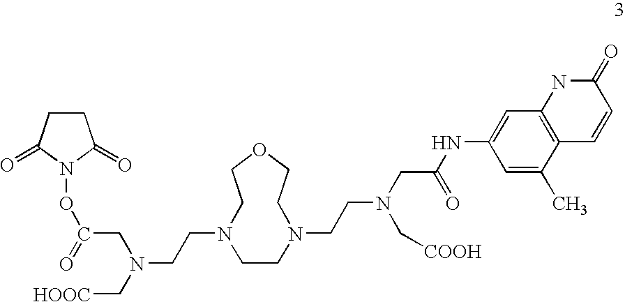
- Synthesis of a lanthanide chelate having a maleimide linker is detailed in Example 1 (see structure 17).
- An exemplary lanthanide chelate having an NHS ester linker is depicted below (3).
- R 1 and/or R 2 is a biomolecule optionally conjugated to the lanthanide chelate via a linker.
- biomolecules include proteins, polynucleotides, peptides, living cells, etc.
- R 1 and R 2 are polylysine molecules, suitable for use in MRI applications.
- the chelate may be directly conjugated to the biomolecule, for example as when using anhydride-based conjugation with an amine- or thiol-containing biomolecule such as an amine-modified DNA (e.g. DNA having a 5′C6-amino linker; see Li M, Selvin P R, Bioconjug Chem. (1997) 8:127-132).
- the lanthanide chelate is complexed with a lanthanide ion.
- the lanthanide ion is selected from Tb 3+ , Eu 3+ , Lu 3+ , Dy 3+ , and Gd 3+ .
- the disclosed lanthanide chelates are useful in the same applications as prior lanthanide chelates (e.g. Gd-DTPA), such as FRET, LRET, MRI, etc, or as phasing agents in solving the crystal structures of biomolecules.
- One aspect of the invention is a method for determining an interaction between biomolecules based on fluorescence resonance energy transfer (FRET). The method comprises conjugating a lanthanide chelate of Formula I via a linker at the R 2 position to a first biomolecule, wherein R 1 is a photosensitizer; labeling a second biomolecule with a fluorescent energy acceptor; and measuring the resulting fluorescence.
- FRET fluorescence resonance energy transfer
- the fluorescent energy acceptor can be any conventional fluorophore used in FRET assays such as tetramethylrhodamine iodoacetamide (TMRIA), fluorescein iodoacetanide (FIA), ATTO 465 maleimide, etc.
- the first and second biomolecules may be any molecule pair analyzable in FRET-based assays, for example as in FRET-based detection of antibody/antigen binding, enzyme/substrate reactions, receptor/ligand binding, etc. Resulting fluorescence is measured using routine methodology (see Example 2).
- these new compounds are similar to that of DTPA (8), with the key features include that they are 10- or 9-dentate chelates with four ionizable carboxylate groups.
- TTHA 9 is a 10-dentate linear chelate.
- the Tb 3+ lifetime of its TTHA-cs124 complex is longer than that of DTPA-cs124 (2.10 ms vs 1.55 ms) [24].
- these chelates are more open and flexible compared to that of DOTA chelates, the binding of lanthanide ions to the chelates is quicker than that of DOTA and lanthanide ions.
- these chelates can also serve as good MRI contrast agents by binding with Gd 3+ [25].
- the free carboxylic acid form of the chelate can be easily converted to dianhydride form (7).
- the dianhydride form allows attachment of an antenna molecule and either an amine-reactive group or a thiol-reactive group to the chelate to make a luminescent probe for LRET or FRET experiments.
- amine or thiol-reactive lanthanide chelates To synthesize amine or thiol-reactive lanthanide chelates, similar methods used for synthesizing DTPA based chelates are employed.
- the formed dianhydride form of chelate is consequently reacted with an antenna molecule (e.g. cs124) and a bi-functional thiol-reactive or amine-reactive compound in a one-pot reaction to form corresponding luminescent chelates.
- an antenna molecule e.g. cs124
- the dianhydride form of chelate (13) can react with cs124 first in a ⁇ 1:0.7 molar ratio, followed by reaction with ⁇ -maleimidopropionic acid hydrazide (EMPH) (16) to form a thio-reactive maleimide form of luminescent lanthanide probe (17):
- TMRIA 5-tetramethylrhodamine iodoacetamide
- HMM purified heavy meromyosin
- Donor labeling Donor chelate is placed on the light chain domain as follows.
- a thio-reactive maleimide form of luminescent lanthanide chelate (17) is prepared as described in Example 1.
- a solution of TbCl 3 is added at a 0.9:1 molar ratio to chelate at pH 7 at millimolar concentration, and the metal is allowed to bind for 30 min on ice.
- An approx. 20-fold excess of the metal-containing chelate is then added to chicken gizzard regulatory light chain (RLC) in exchange buffer with 5 mM tris-(2-carboxyethyl)phosphine hydrochloride (TCEP).
- RLC chicken gizzard regulatory light chain
- TCEP tris-(2-carboxyethyl)phosphine hydrochloride
- Gizzard RLC contains a unique cysteine (Cys108, equivalent in position to Val103 on the skeletal RLC, based on sequence alignment (Collins, J. Muscle Res. Cell Motil . (1991) 12:3-25).
- the two are mixed at concentrations identical to those used for exchange (5-8 ⁇ M HMM, 25-80 ⁇ M gizzard RLC). This mixture is left on ice for 15 min instead of being heated to 34° C., and then passed over a G75 column. During the heating step of the exchange reaction, 1 mM ADP is used to preserve the enzymatic activity of the HMM. In the presence of 1 mM ADP, both the K + -ATPase and actin-activated Mg 2+ -ATPase activities of HMM are unchanged after gizzard RLC exchange relative to untreated HMM.
- HMM Lanthanide luminescence measurements: All terbium emission data is recorded on a laboratory-built spectrophotometer described previously [9] and upgraded to include a CCD for spectral measurements [4]. Samples are placed in a quartz cuvette (either 3 mm ⁇ 3 mm or 2 mm ⁇ 2 mm inner dimensions) at room temperature. The concentration of HMM is typically 1 ⁇ M in rigor buffer. The concentration of actin, when present, is typically 4-10 ⁇ M. This actin concentration ensures complete binding of HMM.
- the terbium donor is excited with 400-1600 excitation pulses from a nitrogen laser (337 nm, 5-ns pulsewidth, 40-Hz repetition rate), and terbium emission (546 nm) is acquired after passing through a grating spectrometer with a photon-counting photomultiplier attached to a multichannel analyzer (2-ms resolution).
- Curve-fitting and energy transfer analysis Multiexponential fits are made with Tablecurve (Jandel Scientific, Marin, Calif.). Donor-only data are fit to two exponentials and show no residual structure. Donor-acceptor data are fit to three exponentials and also show no residual structure. The efficiency of energy transfer is calculated from the lifetimes of donor luminescence as 1 ⁇ ( ⁇ D/A /t D ), where ⁇ D and ⁇ D/A are the donor excited state lifetimes in the absence and presence of acceptor, respectively. For each experiment, donor-only and donor-acceptor samples are prepared simultaneously, and all energy transfer calculations pair the donor-acceptor measurement with the corresponding donor-only control.
- This pairwise method of comparison yields highly reproducible results and is superior to determining energy transfer by comparing the average of donor-only lifetimes to the average of donor-acceptor lifetimes.
- a paired sample t-test is used to determine the statistical significance of differences in energy transfer measurements between experimental conditions (HMM alone, HMM+actin, HMM+actin+ATP, HMM+actin+ADP).
- Polarization measurements Steady-state anisotropy measurements [(I ⁇ ⁇ I ⁇ ) ⁇ (I ⁇ +2 I ⁇ )] of TMRIA bound to myosin are performed according to standard methods using 514-nm vertically polarized excitation, a rotatable analyzer, and a second analyzer placed at 45° to eliminate detection polarization effects. In addition, an aperture is placed in the emission path to limit the numerical aperture, and a CCD is used as the detector [7]. Blank measurements on unlabeled myosin and unlabeled myosin bound to actin are subtracted from all signals. Measurements are performed at room temperature at ⁇ 0.5 ⁇ M TMRIA in a 3 mm ⁇ 3 mm cuvette.
- results show that LRET measurements on HMM are capable of measuring the requisite distances between catalytic and light chain domains, that the measured distance in the absence of nucleotide is consistent with the crystal structure, and that myosin adopts a different conformation upon binding actin and actin plus ADP.
Landscapes
- Chemical & Material Sciences (AREA)
- Organic Chemistry (AREA)
- Plural Heterocyclic Compounds (AREA)
Abstract
Description

and the acceptor is a conventional (organic) fluorophore.

But it has its limitations as well. The binding of DOTA and lanthanide ions is a kinetically slow process [23]. Furthermore, as for luminescent lanthanide probes, amine- or thiol-reactive groups facilitate attachment to a biomolecule. However, neither amine-reactive nor thio-reactive forms of DOTA-based fluorescent chelates have been reported.

or a dianhydride thereof wherein: the dotted line (----) represents a single bond or [CH2—O—CH2]n′; R1 and R2 are independently selected from OH, a photosensitizer, a linker optionally conjugated to a biomolecule, and a biomolecule; and n and n′ are independent integers; wherein one or more oxygen and/or carbon atoms of the central ring of Formula I may be optionally replaced by a protected nitrogen atom.

or is a dianhydride thereof (e.g. see structure 13). In Formula I the dotted line (----) represents a single bond or [CH2—O—CH2]n′; R1 and R2 are independently selected from OH, a photosensitizer, a linker optionally conjugated to a biomolecule, and a biomolecule; and n and n′ are independent (i.e. the same or different) integers. While the central, crown ether ring of Formula I can be of any size, lanthanide binding capacity decreases with increased ring size. Thus, in preferred embodiments, n and n′ are each independently integers from 1 to 10, preferably from 1 to 5, and more preferably from 1 to 3. Equivalent crown ether ring structures may have one or more oxygen or carbon atoms in the ring replaced by a nitrogen atom, resulting in a triaza, tetraaza, etc. crown ethers, provided that the additional nitrogen atoms are protected (e.g. with a methyl, ethyl, or other protecting group) such that only two of the nitrogen atoms of the ring carry the dicarboxylic acid side chains.


Yet, a significant difference of these chelates is that they have macrocycle units incubated in the backbone, which can increase their binding ability to lanthanide ions. Because lanthanide ions can take up to 10 coordination atoms, the new chelates provide better protection to the lanthanide ions from solvent molecule attacks, and thus longer lifetime in aqueous media compared to that of DTPA chelate (3), which is an 8-dentate chelate. For instance, TTHA (9) is a 10-dentate linear chelate. The Tb3+ lifetime of its TTHA-cs124 complex is longer than that of DTPA-cs124 (2.10 ms vs 1.55 ms) [24]. And also because the structures of these chelates are more open and flexible compared to that of DOTA chelates, the binding of lanthanide ions to the chelates is quicker than that of DOTA and lanthanide ions. In addition to forming a stronger binding luminescent lanthanide probes, these chelates can also serve as good MRI contrast agents by binding with Gd3+ [25].


| TABLE 1 |
| Photophysics Data of Luminescent Lanthanide Binding Chelates |
| τH2O/ | No of | Relative | ||||
| Metal | Chelates | τH2O | τD2O | τD2 | waters | Brightness |
| Tb3+ | DTPA-cs124 | 1.55 | 2.63 | 0.59 | 1.1(24) | 1 |
| N2O-cs124 | 1.89 | 1.91 | 0.99 | 0.02 | 1.3 | |
| N2O2-cs124 | 2.50 | 2.88 | 0.87 | 0.22 | 0.2 | |
| N2O2-cs124- | 1.93(81%) | 0.4 | ||||
| EMPH | 0.72(19%) | |||||
| Eu3+ | DTPA-cs124 | 0.62 | 2.42 | 0.26 | 1.26(24) | 1 |
| N2O-cs124 | 1.0 | 2.5 | 0.4 | 0.63 | 0.7 | |
| N2O2-cs124 | N/A | |||||
| N2O2-cs124- | 1.03(41%) | 0.3 | ||||
| EMPH | 0.60(22%) | |||||
| 0.04(37%) | ||||||
- (1) Selvin, P. R. Annual Review of Biophysics and Biomolecular Structure 31, 275-302 (2002).
- (2) Xiao, M.; Selvin, P. R. J. Am. Chem. Soc. 123, 7067-7073 (2001).
- (3) Reifenberger, J. G. et al, Biophysics. J. 82, 430a (2002).
- (4) Selvin, P. R. et al. Inorg. Chem. 35, 700-705 (1996).
- (5) Cha, A., et al. Nature, 402, 809-813 (1999).
- (6) Xiao, M., et al. Proc. Nat'l. Acad. Sci., USA, 95, 15309-15314 (1998)
- (7) Selvin, P. R. IEEE J of Selected Topics In Quantum Electronics: Lasers in Biology 2, 1077-1087 (1996).
- (8) Selvin, P. R. in Applied Fluorescence in Chemistry, Biology and Medicine (eds. Rettig, W.; Strebmenl, B.; Schrader, S.& Seifert, H.) 457-487 (Springer Verlag, New York, 1999).
- (9) Selvin, P. R.; Hearst, J. E. Proc. Natl. Acad. Sci, USA 91, 10024-10028 (1994).
- (10) Selvin, P. R.; Hearst, J. E. U.S. Pat. No. 5,622,821 (1994).
- (11) Xiao, M., et al. Proc. Nat'l. Acad. Sci., USA, 95, 15309-15314 (1998).
- (12) Xiao, M., et al. Nat. Struct. Biol, 10, 402-8 (2003).
- (13) Posson, D. et al. Nature. 436(7052):848-51 (2005).
- (14) Heyduk et al. J. Biol. Chem. 272, 19763-19770 (1997).
- (15) Heyduk et al. Anal. Biochem. 248, 216-227 (1997).
- (16) Callaci, S. et al. Mol. Cell. 3, 229-238 (1999).
- (17) Heyduk. E.; Heyduk, T. J. Biol. Chem. 274, 3315-3322 (1999).
- (18) Xu, J.; Root, D. D. J. Struct. Biol. 123, 150-161 (1998).
- (19) Root, D. D. Proc. Natl. Acad. Sci., USA 94, 5685-5690 (1997).
- (20) Mathis, G. Clinical Chem. 39, 1953-1959 (1993).
- (21) Mathis, G. Clinical Chem. 41, 1391-1397 (1995).
- (22) Vazquez-Ibar, J. L. et al. Proc. Natl. Acad. Sci. USA 99, 3487-3492 (2002).
- (23) Desreux, J. F. Inorg. Chem. 19, 1319-1324 (1980).
- (24) Li, M. & Selvin, P. R. J. Am. Chem. Soc., 117, 8132-8138 (1995).
- (25) Caravan, P.; et al Chem. Rev. 99, 2293-2352 (1999).
- (26) Williams, M. A. & Rapoport, H. J. Org. Chem. 58, 1151-1158 (1993).
Claims (21)


Applications Claiming Priority (1)
| Application Number | Priority Date | Filing Date | Title |
|---|---|---|---|
| PCT/US2005/041337 WO2007055700A1 (en) | 2005-11-14 | 2005-11-14 | Luminescent lanthanide binding chelates |
Publications (2)
| Publication Number | Publication Date |
|---|---|
| US20080299675A1 US20080299675A1 (en) | 2008-12-04 |
| US8110404B2 true US8110404B2 (en) | 2012-02-07 |
Family
ID=38023556
Family Applications (1)
| Application Number | Title | Priority Date | Filing Date |
|---|---|---|---|
| US12/092,604 Expired - Fee Related US8110404B2 (en) | 2005-11-14 | 2005-11-14 | Luminescent lanthanide binding chelates |
Country Status (2)
| Country | Link |
|---|---|
| US (1) | US8110404B2 (en) |
| WO (1) | WO2007055700A1 (en) |
Families Citing this family (3)
| Publication number | Priority date | Publication date | Assignee | Title |
|---|---|---|---|---|
| US20120115128A1 (en) * | 2009-05-07 | 2012-05-10 | The Board Of Trustees Of The University Of Illinois | Selective protein labeling |
| CN109152845B (en) * | 2016-04-14 | 2022-07-12 | 宝力泰锐克斯有限公司 | Comprising at least two (-CH) s within a ring2-CH2Conjugates of linkers of-O-) units and conjugation reagents |
| CN118767158A (en) * | 2023-04-07 | 2024-10-15 | 成都科岭源医药技术有限公司 | Antibody-drug conjugate and preparation method and use thereof |
Citations (1)
| Publication number | Priority date | Publication date | Assignee | Title |
|---|---|---|---|---|
| US4500356A (en) * | 1984-02-24 | 1985-02-19 | The Dow Chemical Company | Methylenephosphonic acid derivatives of bis(aminoalkyl)piperazines as cement set retarding agents |
Family Cites Families (1)
| Publication number | Priority date | Publication date | Assignee | Title |
|---|---|---|---|---|
| US5622821A (en) * | 1994-06-29 | 1997-04-22 | The Regents Of The University Of California | Luminescent lanthanide chelates and methods of use |
-
2005
- 2005-11-14 US US12/092,604 patent/US8110404B2/en not_active Expired - Fee Related
- 2005-11-14 WO PCT/US2005/041337 patent/WO2007055700A1/en not_active Ceased
Patent Citations (1)
| Publication number | Priority date | Publication date | Assignee | Title |
|---|---|---|---|---|
| US4500356A (en) * | 1984-02-24 | 1985-02-19 | The Dow Chemical Company | Methylenephosphonic acid derivatives of bis(aminoalkyl)piperazines as cement set retarding agents |
Non-Patent Citations (1)
| Title |
|---|
| De Jong et al. CAS Accession No. 1983:470686. * |
Also Published As
| Publication number | Publication date |
|---|---|
| WO2007055700A1 (en) | 2007-05-18 |
| US20080299675A1 (en) | 2008-12-04 |
Similar Documents
| Publication | Publication Date | Title |
|---|---|---|
| JP3814291B2 (en) | Luminescent lanthanide chelates and uses thereof | |
| Parker et al. | The design of responsive luminescent lanthanide probes and sensors | |
| Mandal et al. | A detailed spectroscopic study on the interaction of Rhodamine 6G with human hemoglobin | |
| Zhang et al. | New class of tetradentate β-diketonate-europium complexes that can be covalently bound to proteins for time-gated fluorometric application | |
| Lipchik et al. | Time-resolved luminescence detection of spleen tyrosine kinase activity through terbium sensitization | |
| Vaasa et al. | High-affinity bisubstrate probe for fluorescence anisotropy binding/displacement assays with protein kinases PKA and ROCK | |
| Sun et al. | Synthesis and spectroscopic characterization of 4-butoxyethoxy-N-octadecyl-1, 8-naphthalimide as a new fluorescent probe for the determination of proteins | |
| Ma et al. | Insight into fluorescence imaging and bioorthogonal reactions in biological analysis | |
| Dey et al. | Naphthyridine based fluorescent receptors for the recognition of uric acid | |
| Chao et al. | A fluorescent sensor recognized by the FA1 site for highly sensitive detection of HSA | |
| WO2001063265A1 (en) | Measuring method using long life fluorescence of excitation type | |
| WO2022077052A1 (en) | Probe compounds for mitochondial membrane imaging | |
| Kyrychenko et al. | Joint refinement of FRET measurements using spectroscopic and computational tools | |
| Gao et al. | Mitochondrial directed ratiometric fluorescent probe for quantitive detection of sulfur dioxide derivatives | |
| Chen et al. | An NBD tertiary amine is a fluorescent quencher and/or a weak green-light fluorophore in H 2 S-specific probes | |
| Mukherjee et al. | Switching of Trp-214 intrinsic rotamer population in human serum albumin: An insight into the aftermath of embracing therapeutic bioorganic luminophore azapodophyllotoxin into sudlow site I | |
| US8110404B2 (en) | Luminescent lanthanide binding chelates | |
| Wang et al. | Time‐Resolved Luminescent Sensing and Imaging for Enzyme Catalytic Activity Based on Responsive Probes | |
| Ge et al. | New 9-or 10-dentate luminescent lanthanide chelates | |
| Krawczyk et al. | Synthesis, photophysical and biological properties of a new oxazolone fluorescent probe for bioimaging: an experimental and theoretical study | |
| Azab et al. | A new luminescent bio-probe of Europium (III)-complex for sensing some biomolecules and CT-DNA | |
| EP2810648A1 (en) | Targeting domain-domain interaction for the identification of kinase modulators | |
| Turro et al. | Lanthanide ions as luminescent probes of proteins and nucleic acids | |
| US9625466B2 (en) | Signal amplification methods for the imaging of protein synthesis and neurotransmitter transport | |
| Zhang et al. | A water-soluble 1, 8-naphthyridine-based imidazolium molecular gripper for fluorescence sensing and discriminating of GMP |
Legal Events
| Date | Code | Title | Description |
|---|---|---|---|
| AS | Assignment |
Owner name: ILLINOIS BOARD OF TRUSTEES, UNIVERSITY OF, ILLINOI Free format text: ASSIGNMENT OF ASSIGNORS INTEREST;ASSIGNORS:SELVIN, PAUL R.;GE, PINGHUA;REEL/FRAME:017289/0619 Effective date: 20051110 |
|
| AS | Assignment |
Owner name: BOARD OF TRUSTEES OF THE UNIVERSITY OF ILLINOIS, I Free format text: ASSIGNMENT OF ASSIGNORS INTEREST;ASSIGNORS:SELVIN, PAUL R.;GE, PINGHUA;REEL/FRAME:020898/0853 Effective date: 20051110 |
|
| ZAAA | Notice of allowance and fees due |
Free format text: ORIGINAL CODE: NOA |
|
| ZAAB | Notice of allowance mailed |
Free format text: ORIGINAL CODE: MN/=. |
|
| STCF | Information on status: patent grant |
Free format text: PATENTED CASE |
|
| FPAY | Fee payment |
Year of fee payment: 4 |
|
| MAFP | Maintenance fee payment |
Free format text: PAYMENT OF MAINTENANCE FEE, 8TH YR, SMALL ENTITY (ORIGINAL EVENT CODE: M2552); ENTITY STATUS OF PATENT OWNER: SMALL ENTITY Year of fee payment: 8 |
|
| FEPP | Fee payment procedure |
Free format text: MAINTENANCE FEE REMINDER MAILED (ORIGINAL EVENT CODE: REM.); ENTITY STATUS OF PATENT OWNER: SMALL ENTITY |
|
| LAPS | Lapse for failure to pay maintenance fees |
Free format text: PATENT EXPIRED FOR FAILURE TO PAY MAINTENANCE FEES (ORIGINAL EVENT CODE: EXP.); ENTITY STATUS OF PATENT OWNER: SMALL ENTITY |
|
| STCH | Information on status: patent discontinuation |
Free format text: PATENT EXPIRED DUE TO NONPAYMENT OF MAINTENANCE FEES UNDER 37 CFR 1.362 |
|
| FP | Lapsed due to failure to pay maintenance fee |
Effective date: 20240207 |