US8084262B2 - Biomarker for farnesyl pathway - Google Patents

Biomarker for farnesyl pathway Download PDF

Info

Publication number
US8084262B2
US8084262B2 US12/295,343 US29534307A US8084262B2 US 8084262 B2 US8084262 B2 US 8084262B2 US 29534307 A US29534307 A US 29534307A US 8084262 B2 US8084262 B2 US 8084262B2
Authority
US
United States
Prior art keywords
felinine
derivative
subject treated
zoledronate
urine
Prior art date
Legal status (The legal status is an assumption and is not a legal conclusion. Google has not performed a legal analysis and makes no representation as to the accuracy of the status listed.)
Expired - Fee Related, expires
Application number
US12/295,343
Other versions
US20090130770A1 (en
Inventor
Frank Dieterle
Goetz Schlotterbeck
Hans Senn
Laura Suter-Dick
Current Assignee (The listed assignees may be inaccurate. Google has not performed a legal analysis and makes no representation or warranty as to the accuracy of the list.)
Hoffmann La Roche Inc
Original Assignee
Hoffmann La Roche Inc
Priority date (The priority date is an assumption and is not a legal conclusion. Google has not performed a legal analysis and makes no representation as to the accuracy of the date listed.)
Filing date
Publication date
Application filed by Hoffmann La Roche Inc filed Critical Hoffmann La Roche Inc
Assigned to F. HOFFMANN-LA ROCHE AG reassignment F. HOFFMANN-LA ROCHE AG ASSIGNMENT OF ASSIGNORS INTEREST (SEE DOCUMENT FOR DETAILS). Assignors: DIETERLE, FRANK, SCHLOTTERBECK, GOETZ, SENN, HANS, SUTER-DICK, LAURA
Assigned to HOFFMAN-LA ROCHE, INC. reassignment HOFFMAN-LA ROCHE, INC. ASSIGNMENT OF ASSIGNORS INTEREST (SEE DOCUMENT FOR DETAILS). Assignors: F. HOFFMAN-LA ROCHE AG
Publication of US20090130770A1 publication Critical patent/US20090130770A1/en
Application granted granted Critical
Publication of US8084262B2 publication Critical patent/US8084262B2/en
Expired - Fee Related legal-status Critical Current
Adjusted expiration legal-status Critical

Links

Images

Classifications

    • GPHYSICS
    • G01MEASURING; TESTING
    • G01NINVESTIGATING OR ANALYSING MATERIALS BY DETERMINING THEIR CHEMICAL OR PHYSICAL PROPERTIES
    • G01N33/00Investigating or analysing materials by specific methods not covered by groups G01N1/00 - G01N31/00
    • G01N33/48Biological material, e.g. blood, urine; Haemocytometers
    • G01N33/50Chemical analysis of biological material, e.g. blood, urine; Testing involving biospecific ligand binding methods; Immunological testing
    • G01N33/74Chemical analysis of biological material, e.g. blood, urine; Testing involving biospecific ligand binding methods; Immunological testing involving hormones or other non-cytokine intercellular protein regulatory factors such as growth factors, including receptors to hormones and growth factors
    • G01N33/743Steroid hormones
    • GPHYSICS
    • G01MEASURING; TESTING
    • G01NINVESTIGATING OR ANALYSING MATERIALS BY DETERMINING THEIR CHEMICAL OR PHYSICAL PROPERTIES
    • G01N33/00Investigating or analysing materials by specific methods not covered by groups G01N1/00 - G01N31/00
    • G01N33/48Biological material, e.g. blood, urine; Haemocytometers
    • G01N33/50Chemical analysis of biological material, e.g. blood, urine; Testing involving biospecific ligand binding methods; Immunological testing
    • G01N33/68Chemical analysis of biological material, e.g. blood, urine; Testing involving biospecific ligand binding methods; Immunological testing involving proteins, peptides or amino acids
    • G01N33/6803General methods of protein analysis not limited to specific proteins or families of proteins
    • GPHYSICS
    • G01MEASURING; TESTING
    • G01NINVESTIGATING OR ANALYSING MATERIALS BY DETERMINING THEIR CHEMICAL OR PHYSICAL PROPERTIES
    • G01N33/00Investigating or analysing materials by specific methods not covered by groups G01N1/00 - G01N31/00
    • G01N33/48Biological material, e.g. blood, urine; Haemocytometers
    • G01N33/50Chemical analysis of biological material, e.g. blood, urine; Testing involving biospecific ligand binding methods; Immunological testing
    • G01N33/94Chemical analysis of biological material, e.g. blood, urine; Testing involving biospecific ligand binding methods; Immunological testing involving narcotics or drugs or pharmaceuticals, neurotransmitters or associated receptors
    • YGENERAL TAGGING OF NEW TECHNOLOGICAL DEVELOPMENTS; GENERAL TAGGING OF CROSS-SECTIONAL TECHNOLOGIES SPANNING OVER SEVERAL SECTIONS OF THE IPC; TECHNICAL SUBJECTS COVERED BY FORMER USPC CROSS-REFERENCE ART COLLECTIONS [XRACs] AND DIGESTS
    • Y10TECHNICAL SUBJECTS COVERED BY FORMER USPC
    • Y10TTECHNICAL SUBJECTS COVERED BY FORMER US CLASSIFICATION
    • Y10T436/00Chemistry: analytical and immunological testing
    • Y10T436/17Nitrogen containing
    • YGENERAL TAGGING OF NEW TECHNOLOGICAL DEVELOPMENTS; GENERAL TAGGING OF CROSS-SECTIONAL TECHNOLOGIES SPANNING OVER SEVERAL SECTIONS OF THE IPC; TECHNICAL SUBJECTS COVERED BY FORMER USPC CROSS-REFERENCE ART COLLECTIONS [XRACs] AND DIGESTS
    • Y10TECHNICAL SUBJECTS COVERED BY FORMER USPC
    • Y10TTECHNICAL SUBJECTS COVERED BY FORMER US CLASSIFICATION
    • Y10T436/00Chemistry: analytical and immunological testing
    • Y10T436/18Sulfur containing

Definitions

  • FPP farnesyl diphosphate
  • Felinine (2-amino-7-hydroxy-5,5-dimethyl-4-thiaheptanoic acid) (HOOCCH(NH 2 )CH 2 SC(CH 3 )2CH 2 CH 2 OH) is an important sulphur containing amino acid known to be present in large amounts in the urine of male cats. It has been postulated that felinine plays a role as a male pheromone designed to attract female cats (Hendriks W H, Woolhouse A D, Tartelin M F, Moughan P J (1995b) Synthesis of felinine, 2-amino-7-hydroxy-5,5-dimethyl-4-thiaheptanoic acid. Bioorg Chem 23: 89-100). So far, felinine has not been shown to be present in other species than cat.
  • felinylglycine In addition to the previously described presence of free felinine, the presence of several felinine-containing metabolites, including N-acetyl felinine, felinylglycine and unaltered ⁇ -glutamylfelinylglycine was shown in cat urine.
  • the renal metabolism of ⁇ -glutamylfelinylglycine in cats generally occurs in a similar manner to glutathione S-conjugates as in other animal species, although the detection of felinylglycine indicates that subtle differences may exist.
  • the present invention relates to a novel biomarker which can be used in methods to determine the extent to which a compound interferes with steroid metabolism.
  • Said biomarker can be any naturally occurring derivative of 2-amino-7-hydroxy-5,5-dimethyl-4-thiaheptanoic acid, which is also known as felinine.
  • Such derivatives include compounds of the formula
  • R, R′, R′′ and R′′′ are independently: H, CH 3 , COCH 3 , COH, OCH 3 , OC 2 H 5 , Gly, Gln, Glu, Ala, Val, Met, Phosphate, or Pyrophosphate.
  • Preferred embodiments of the marker are gamma-glutamylfelinylglycine (3-methylbutanolgluthatione), felinylglycine, felinine or N-acetyl-felinine.
  • the marker hereinbefore described is used in a method of determining the extent of interference of a substance with steroid metabolism comprising:
  • a body fluid may be any fluid derivable from a body, such as blood, plasma, serum or urine.
  • Intact tissue is any type of issue that was removed from the body prior to analysis according to the present invention.
  • a tissue extract is any extract from a tissue that was removed from the body, which is obtained using extraction methods well known in the art. The method herein described is an ex-vivo, in situ or in vitro method carried out on samples (body fluids, intact tissues, tissue extracts) after they were removed from the body.
  • the extent of interference with farnesyl diphosphate production is determined.
  • the body fluid is urine, plasma, or serum.
  • the presence or absence of N-acetyl-felinine is determined in urine of a subject.
  • the term “extent of interference with steroid metabolism” relates to the correlation between production of felinine derivatives and steroid metabolism, such as inhibition of FPP.
  • higher levels of felinine derivatives correlate with strong interference (e.g. stronger inhibition of FPP), while low or undetectable levels of felinine derivatives correlate with a weaker interference (e.g. a more subtle inhibition of FPP, which may be caused by a different mechanism of inhibition and leads to a lower inhibition, e.g. of protein prenylation).
  • a weaker interference e.g. a more subtle inhibition of FPP, which may be caused by a different mechanism of inhibition and leads to a lower inhibition, e.g. of protein prenylation.
  • subject as used herein relates to any mammal, preferably to a human subject. It is to be understood, though, that none of the methods herein described are performed directly on the body of subject. Thus, they are not in vivo methods.
  • N-acetyl-felinine could be used to differentiate between different bisphosphonates, ibandronate and zoledronate.
  • Bisphosphonates are analogs of pyrophosphate in which the geminal oxygen has been substituted by carbon (see FIG. 1 in Mini-Reviews in Medicinal Chemistry, 2004, Vol 4, p. 712)
  • a method of differentiating between ibandronate and zoledronate in a subject treated with a ibandronate or zoledronate comprising determining the presence or absence of N-acetyl-felinine or another natural derivative of felinine in a body fluid, intact tissue or tissue extract of a subject treated with said substance, wherein the absence of N-acetyl-felinine or another natural derivative of felinine is indicative of whether the patient is treated with ibandronate or zoledronate.
  • the body extract is urine, plasma, or serum.
  • the presence or absence of N-acetyl-felinine is determined in urine of a subject.
  • the identification and quantification can either be achieved by NMR or LC/GC-MS.
  • N-acetyl-felinine or any naturally occurring derivative of felinine can also be used as a marker for bisphosphonate-induced toxicity, in particular kidney toxicity.
  • kidney and liver toxicity was observed in histopathology but not so for Ibandronate.
  • the present invention provides for the use of a felinine derivative of the formula
  • a number of metabolic pathways depend on protein prenylation. Decrease in protein prenylation, therefore, affects such metabolic pathways.
  • FIG. 1 a, b Pathways for the synthesis of N-acetyl-felinine in cats. Pathways for the synthesis of N-acetyl-felinine reported for cat species. Two syntheses are proposed, which both need isopentenyl (link to steroid synthesis!). One synthesis is assumed to happen in the kidney, the other synthesis is assumed to start in the liver, whereby the tripeptide is transported via blood to the kidney and metabolized to n-acetyl-felinine. The yellow box marks structures and reactions assumed in the kidney, the blue box marks reactions assumed to happen in the liver and the magenta box marks the tripeptide, which is transported from the liver to the kidney by blood. A differentiation between the liver synthesis and the pure kidney synthesis should be possible by looking for the tripeptide in plasma.
  • FIG. 2 NMR spectra of three rats before dosing
  • FIG. 3 a )-c) Time course of bin 1.32 ppm (Methyl signals of N-Acetyl felinine) relative to creatinine: Note: Ibandronat dosed rats show no increase in bin 1.32!
  • FIG. 4 a )-c) Statistical analysis of rat urine NMR spectra reveals an important signal at 1.32 ppm
  • FIG. 5 NMR spectra of three rats 96 h after dosing: New signal at 1.32 ppm visible for Zoledronate dosed rats only.
  • FIG. 6 2D, 1H, 13C HMBC spectrum of urine of rats dosed with 6 mg/kg Zoledronate for structure elucidation of N-acetyl felinine
  • FIG. 7 LC-MS investigation of rat urine: Rat control group; No signal found (green extracted ion chromatogram and in the middle MS trace for RT 9.6 min) for expected mass of N-acteyl felinine (m/z 248 in neg. mode).
  • FIG. 8 LC-MS investigation of rat urine: Rat of group dosed with 6 mg/kg Zoledronate, 96 h after dosing: Strong signal found (red extracted ion chromatogram and in the middle MS trace for RT 9.6 min) for expected mass of N-acteyl felinine (m/z 248 in neg. mode). MS/MS shows expected fragmentation pattern (m/z 119)
  • FIG. 9 LC-MS investigation of rat urine: Rat of group dosed with 3 mg/kg Ibandronate, 96 h after dosing; No signal found for expected mass of N-acteyl felinine (m/z 248 in neg. mode).
  • FIG. 10 LC-MS investigation of rat urine: Rat control group spiked with N-acetyl felinine; Strong signal found for expected mass of N-acteyl felinine (m/z 248 in neg. mode).
  • Urine samples were collected in metabolism cages at 0 to 4° C. (automatically refrigerated by a Tecniplast sampling/cooling unit) at the intervals given in the sampling schedule below into sample tubes containing 1 ml of an aqueous Cs-azide (1%) solution. Before aliquoting, urine volumes were determined.
  • the volume was determined and the samples were centrifuged at 3000 u/min [500 g] for 10 min.
  • N-acetyl-felinine was identified in urine samples on basis of 1D and 2D NMR spectra. Spectra were acquired at 600.13 MHz on a Bruker AV 600 spectrometer and at 500.13 MHz on Bruker DRX-500 spectrometer. Urine samples were prepared and measured and the data evaluated as described elsewhere (Keun, H. C.; Ebbels, T. M. D.; Antti, H.; Bollard, M. E.; Beckonert, O.; Schlotterbeck, G.; Senn, H,; Niederhauser, U.; Holmes, E.; Lindon, J. C.; Nicholson, J. K. Chem. Res. Toxicol. 2002, 15, 1380-1386).
  • 1 H NMR spectra for Metabonomic analysis were measured at 300K.
  • the water resonance were suppressed by use of standard pulse-sequences (e.g noesyprld, irradiation for 2 s relaxation delay and during 100 ms mixing time).
  • pulse-sequences e.g noesyprld, irradiation for 2 s relaxation delay and during 100 ms mixing time.
  • For each sample as many transients were added as is needed to provide spectra high enough in signal to noise.
  • the multivariate data analysis identifies signals, patterns of signals, and metabolites derived thereof, which are significantly different between individuals and groups of individuals.
  • PCA principal component analysis
  • PLS projection to latent structures, partial least squares
  • the assignment is performed with the help of an internal data base of metabolites, with the help of pathway mapping procedures (KEGG database) and with the help of a spectroscopic structure elucidation for unknown metabolites when feasible.
  • N-acetyl-felinine was found in spectra of rats dosed with Zoledronate (see FIG. 5 ). A signal at 1.32 ppm was observed in the 1D NMR spectra and the structure was elucidated on basis of 2D NMR experiments (see FIG. 6 ).
  • N-acetyl-felinine was also determined in LC-MS experiments of urine samples.
  • LC-MS analysis of urine was performed using an Agilent 1100 HPLC system coupled to a Bruker Esquire 3000+ mass spectrometer equipped with an electrospray interface.
  • Urine samples were diluted 4-fold with distilled water and 2 ⁇ l injections were made from a well plate maintained at 4° C. into a 150 mm ⁇ 3.9 mm Waters Atlantis dC18 3 ⁇ m HPLC column maintained in an oven at 30° C. The column was eluted at a flow rate of 1 ml/min; mobile phase A consisted of 20 mM ammonium acetate, and mobile phase B of acetonitrile.
  • N-acetyl-felinine can also be measured in positive mode.
  • the corresponding mass of the M+H + is found at m/z 250.

Landscapes

  • Life Sciences & Earth Sciences (AREA)
  • Health & Medical Sciences (AREA)
  • Engineering & Computer Science (AREA)
  • Molecular Biology (AREA)
  • Urology & Nephrology (AREA)
  • Immunology (AREA)
  • Biomedical Technology (AREA)
  • Chemical & Material Sciences (AREA)
  • Hematology (AREA)
  • Physics & Mathematics (AREA)
  • Medicinal Chemistry (AREA)
  • Pathology (AREA)
  • Cell Biology (AREA)
  • Food Science & Technology (AREA)
  • Microbiology (AREA)
  • Biotechnology (AREA)
  • Analytical Chemistry (AREA)
  • Biochemistry (AREA)
  • General Health & Medical Sciences (AREA)
  • General Physics & Mathematics (AREA)
  • Bioinformatics & Cheminformatics (AREA)
  • Pharmacology & Pharmacy (AREA)
  • Bioinformatics & Computational Biology (AREA)
  • Biophysics (AREA)
  • Proteomics, Peptides & Aminoacids (AREA)
  • Endocrinology (AREA)
  • Investigating Or Analysing Biological Materials (AREA)

Abstract

The invention generally provides methods to determine the extent to which a compound interferes with steroid metabolism, comprising a novel biomarker of a felinine derivative. The invention also provides such novel felinine derivative biomarker.

Description

Bisphosphonates and especially Aminobisphosphonate inhibit a key enzyme within the mevalonate pathway, farnesyl diphosphate (FPP) synthase (Green J R. Bisphosphonates: preclinical review. The Oncologist 2004; 9(Suppl4):3-13). Inhibition of this enzyme leads to a reduction in protein prenylation (Rogers M J, Gordon S, Benford H L, Coxon F P, Luckman S P, Mönkkönen J, Frith J C. Cellular and molecular mechanisms of action of bisphosphonates. Cancer. 2000; 88(12 Suppl):2961-78). As a consequence, the production of FPP and, thus, the biosynthesis of squalene and sterol (cholesterol) is affected. But also the isoprenylation of signaling proteins which involves the transfer of a farnesyl or geranylgeranyl group onto a cysteine amino acid residue of the protein are hampered. Isoprenylation is a posttranslational modification necessary for full activation of several key proteins involved in a number of metabolic pathways which underlie cell survival, cytoskeletal organisation and cancer progression (Green J R. Bisphosphonates: preclinical review. The Oncologist 2004; 9(Suppl4):3-13; Wakeling A E. Inhibitors of growth factor signalling. Endocr Relat Cancer. 2005 July; 12 Suppl 1:S183-7). The pathways in which bisphosphonates interfere are described eg. In Reszka and Rodan, Mini-Reviews in Medicinal Chemistry, 2004, Vol 4, p. 712, FIG. 3.
Felinine (2-amino-7-hydroxy-5,5-dimethyl-4-thiaheptanoic acid) (HOOCCH(NH2)CH2SC(CH3)2CH2CH2OH) is an important sulphur containing amino acid known to be present in large amounts in the urine of male cats. It has been postulated that felinine plays a role as a male pheromone designed to attract female cats (Hendriks W H, Woolhouse A D, Tartelin M F, Moughan P J (1995b) Synthesis of felinine, 2-amino-7-hydroxy-5,5-dimethyl-4-thiaheptanoic acid. Bioorg Chem 23: 89-100). So far, felinine has not been shown to be present in other species than cat.
Renal metabolites of a derivative of felinine, γ-glutamylfelinylglycine, were recently identified in the blood of domestic cats (W. H. Hendricks, D. R. Harding, K. J. Rutherfuurd-Marwick (2004), Isolation and characterization of renal metabolites of, γ-glutamylfelinylglycine in the urine of the domestic cat (Felis catus), Comp Biochem Physiol B Biochem Mol Biol 139, 245-251). This tripeptide is metabolised by the kidney in a similar manner to glutathione-S-conjugates in other animal species. In addition to the previously described presence of free felinine, the presence of several felinine-containing metabolites, including N-acetyl felinine, felinylglycine and unaltered γ-glutamylfelinylglycine was shown in cat urine. The renal metabolism of γ-glutamylfelinylglycine in cats generally occurs in a similar manner to glutathione S-conjugates as in other animal species, although the detection of felinylglycine indicates that subtle differences may exist.
DESCRIPTION OF THE INVENTION
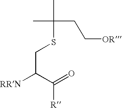
The present invention relates to a novel biomarker which can be used in methods to determine the extent to which a compound interferes with steroid metabolism. Said biomarker can be any naturally occurring derivative of 2-amino-7-hydroxy-5,5-dimethyl-4-thiaheptanoic acid, which is also known as felinine. Such derivatives include compounds of the formula
Figure US08084262-20111227-C00001
Wherein R, R′, R″ and R′″ are independently: H, CH3, COCH3, COH, OCH3, OC2H5, Gly, Gln, Glu, Ala, Val, Met, Phosphate, or Pyrophosphate.
Preferred embodiments of the marker are gamma-glutamylfelinylglycine (3-methylbutanolgluthatione), felinylglycine, felinine or N-acetyl-felinine.
The marker hereinbefore described is used in a method of determining the extent of interference of a substance with steroid metabolism comprising:
determining the presence or absence of said marker in a body fluid, and intact tissue or a tissue extract of a subject treated with said substance, wherein the presence of N-acetyl-felinine is indicative of a substance which interferes with steroid metabolism, and the absence of N-acetyl-felinine is indicative of a substance which does not interfere with steroid metabolism.
A body fluid may be any fluid derivable from a body, such as blood, plasma, serum or urine. Intact tissue is any type of issue that was removed from the body prior to analysis according to the present invention. A tissue extract is any extract from a tissue that was removed from the body, which is obtained using extraction methods well known in the art. The method herein described is an ex-vivo, in situ or in vitro method carried out on samples (body fluids, intact tissues, tissue extracts) after they were removed from the body.
In one embodiment of the present invention, the extent of interference with farnesyl diphosphate production is determined. Preferably, the body fluid is urine, plasma, or serum. In a most preferred embodiment, the presence or absence of N-acetyl-felinine is determined in urine of a subject.
The term “extent of interference with steroid metabolism” relates to the correlation between production of felinine derivatives and steroid metabolism, such as inhibition of FPP. Thus, higher levels of felinine derivatives correlate with strong interference (e.g. stronger inhibition of FPP), while low or undetectable levels of felinine derivatives correlate with a weaker interference (e.g. a more subtle inhibition of FPP, which may be caused by a different mechanism of inhibition and leads to a lower inhibition, e.g. of protein prenylation). Thus, there is a correlation between production of felinine derivatives and farnesyl diphosphate production.
The term “subject” as used herein relates to any mammal, preferably to a human subject. It is to be understood, though, that none of the methods herein described are performed directly on the body of subject. Thus, they are not in vivo methods.
It was found that N-acetyl-felinine could be used to differentiate between different bisphosphonates, ibandronate and zoledronate.
Bisphosphonates are analogs of pyrophosphate in which the geminal oxygen has been substituted by carbon (see FIG. 1 in Mini-Reviews in Medicinal Chemistry, 2004, Vol 4, p. 712)
Ibandronate structure see e.g. FIG. 2 in Mini-Reviews in Medicinal Chemistry, 2004, Vol 4, p. 712
Zoledronate structure see eg. FIG. 2 in Mini-Reviews in Medicinal Chemistry, 2004, Vol 4, p. 712
Although they can both inhibit FPP synthase and, thus, interfere with sterol synthesis, they do so to a different extent. Thus, it was found that in a given time interval, zoledronate stimulation caused high levels of N-acetyl-felinine in urine, while with ibandronate stimulation, N-acetyl-felinine levels in urine were undetectable. Thus, according to the present invention, a method of differentiating between ibandronate and zoledronate in a subject treated with a ibandronate or zoledronate is also provided, said method comprising determining the presence or absence of N-acetyl-felinine or another natural derivative of felinine in a body fluid, intact tissue or tissue extract of a subject treated with said substance, wherein the absence of N-acetyl-felinine or another natural derivative of felinine is indicative of whether the patient is treated with ibandronate or zoledronate. Preferably, the body extract is urine, plasma, or serum. In a most preferred embodiment, the presence or absence of N-acetyl-felinine is determined in urine of a subject.
The identification and quantification can either be achieved by NMR or LC/GC-MS.
According to the present invention, N-acetyl-felinine or any naturally occurring derivative of felinine can also be used as a marker for bisphosphonate-induced toxicity, in particular kidney toxicity. In Zoledronate treated animals kidney and liver toxicity was observed in histopathology but not so for Ibandronate.
Furthermore, the present invention provides for the use of a felinine derivative of the formula
Figure US08084262-20111227-C00002
    • wherein R, R′, R″ and R′″ are independently: H, CH3, COCH3, COH, OCH3, OC2H5, Gly, Gln, Glu, Ala, Val, Met, Phosphate, or Pyrophosphate, as a marker for changes in post-translational protein prenylation of signaling proteins.
A number of metabolic pathways depend on protein prenylation. Decrease in protein prenylation, therefore, affects such metabolic pathways.
SHORT DESCRIPTION OF FIGURES
FIG. 1 a, b): Pathways for the synthesis of N-acetyl-felinine in cats. Pathways for the synthesis of N-acetyl-felinine reported for cat species. Two syntheses are proposed, which both need isopentenyl (link to steroid synthesis!). One synthesis is assumed to happen in the kidney, the other synthesis is assumed to start in the liver, whereby the tripeptide is transported via blood to the kidney and metabolized to n-acetyl-felinine. The yellow box marks structures and reactions assumed in the kidney, the blue box marks reactions assumed to happen in the liver and the magenta box marks the tripeptide, which is transported from the liver to the kidney by blood. A differentiation between the liver synthesis and the pure kidney synthesis should be possible by looking for the tripeptide in plasma.
FIG. 2: NMR spectra of three rats before dosing
FIG. 3 a)-c): Time course of bin 1.32 ppm (Methyl signals of N-Acetyl felinine) relative to creatinine: Note: Ibandronat dosed rats show no increase in bin 1.32!
FIG. 4 a)-c): Statistical analysis of rat urine NMR spectra reveals an important signal at 1.32 ppm
FIG. 5: NMR spectra of three rats 96 h after dosing: New signal at 1.32 ppm visible for Zoledronate dosed rats only.
FIG. 6: 2D, 1H, 13C HMBC spectrum of urine of rats dosed with 6 mg/kg Zoledronate for structure elucidation of N-acetyl felinine
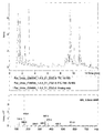
FIG. 7: LC-MS investigation of rat urine: Rat control group; No signal found (green extracted ion chromatogram and in the middle MS trace for RT 9.6 min) for expected mass of N-acteyl felinine (m/z 248 in neg. mode).
FIG. 8: LC-MS investigation of rat urine: Rat of group dosed with 6 mg/kg Zoledronate, 96 h after dosing: Strong signal found (red extracted ion chromatogram and in the middle MS trace for RT 9.6 min) for expected mass of N-acteyl felinine (m/z 248 in neg. mode). MS/MS shows expected fragmentation pattern (m/z 119)
FIG. 9: LC-MS investigation of rat urine: Rat of group dosed with 3 mg/kg Ibandronate, 96 h after dosing; No signal found for expected mass of N-acteyl felinine (m/z 248 in neg. mode).
FIG. 10: LC-MS investigation of rat urine: Rat control group spiked with N-acetyl felinine; Strong signal found for expected mass of N-acteyl felinine (m/z 248 in neg. mode).
EXAMPLES
1.1 Test System
Test system Crl:CD ®(SD)IGS.BR (SPF) Sprague
Dawley rats
Rationale Standard strain for all COMET studies.
Source Charles River Deutschland GmbH
Sulzfeld/Germany
Group allocation Groups 1-5: 10 males each
Total number of animals 50 males
Age at delivery Approximately 6 weeks (8 weeks at
treatment start)
Identification Cage card and ear tattoo.
Randomization Computer-generated random algorithm

1.2 Allocation
No. of animals
(Animal numbers)
Dose Subgroup A Subgroup B
Allo- Dose Volume No. of Necropsy 48 hours Necropsy 168 hrs
cation Compound mg/kg] [ml/kg] doses after application after application
 1* Vehicle 0 2 1 5 (1-5)  5 (6-10) 
2 Compound 1 1 2 1 5 (11-15) 5 (16-20)
3 Compound 1 3 2 1 5 (21-25) 5 (26-30)
4 Compound 2 3 2 1 5 (31-35) 5 (36-40)
5 Compound 2 6 2 1 5 (41-45) 5 (46-50)
*Control animals were treated with vehicle, only.

1.3 Husbandry
Conditions Standard Laboratory Conditions. Air-conditioned with 10-15 air changes per hour, and continuously monitored environment with target ranges for temperature 22±3° C. and for relative humidity between 30-70%. 12 hours fluorescent light/12 hours dark cycle, with music during the light period.
Accommodation Individually in Makrolon type—3 cages with wire mesh tops and standardized softwood bedding (‘Lignocel’ Schill AG, CH-4132 Muttenz/Switzerland) during most days of the acclimatization period as well as individually in metabolism cages (>300 g, Tecniplast, Italy) on three days (day-7, day-4 and day-1) of the acclimatization period and the entire sampling period.
Diet Purina chow 5002 certified rodent diet non-pelleted (PMI® Nutition International, LLC P.O. Box 19798, Brentwood, Mo. 63144 U.S.A.) ad libitum. Provided to animals from the beginning of the acclimatization period.
Water Community tap-water, from Itingen ad libitum. Results of representative bacteriological, chemical and contaminant analyses are presented in Appendix I. See pp. 110-113
1.4 Test Items
  • Compound 1: Ibandronate in saline solution
  • Compound 2: Zoledronate in saline solution
  • Control/Vehicle: Saline Solution (0.9%)
    1.5 Urine Sampling
Urine samples were collected in metabolism cages at 0 to 4° C. (automatically refrigerated by a Tecniplast sampling/cooling unit) at the intervals given in the sampling schedule below into sample tubes containing 1 ml of an aqueous Cs-azide (1%) solution. Before aliquoting, urine volumes were determined.
Study Day Sample time points relative to the application time
Absolute day of Dates of point and sample labels for samples sent to Roche
urine sample urine sample Groups 1-5
Days collection collection Time Subgr. A Subgr. B Label
−12 −12 to −11 (24 h) 14.-15.04.04 Arriv. to −264 h X X −264 h
−4 −4 to −3 (0-24 h) 22.-23.04.04 −96 h to −72 h X X −72 h
−1 −1 (pre-dose) 25.04.04 −24 h to −16 h X X −16 h
−1 −1 to 1 (pre-dose)* 25.-26.04.04 −16 h to 0 h X X 0 h
1 1 (1st dose) 26.04.04 0 h to 8 h X X 8 h
1 1 to 2 26.-27.04.04 8 h to 24 h X X 24 h
2 2 to 3* 27.-28.04.04 24 h to 48 h X X 48 h
3 3 to 4 28.-29.04.04 48 h to 72 h X 72 h
4 4 to 5 29.-30.04.04 72 h to 96 h X 96 h
5 5 to 6 30.-01.05.04 96 h to 120 h X 120 h
6 6 to 7 01.-02.05.04 120 h to 144 h X 144 h
7 7 to 8* 02.-03.05.04 144 h to 168 h X 168 h
*this samples were undergo additional special urine analysis as described below.
Immediately after reaching the end of the sampling period, the volume was determined and the samples were centrifuged at 3000 u/min [500 g] for 10 min.
1.6 Determination of N-Acetyl-Felinine by NMR and LC-MS
N-acetyl-felinine was identified in urine samples on basis of 1D and 2D NMR spectra. Spectra were acquired at 600.13 MHz on a Bruker AV 600 spectrometer and at 500.13 MHz on Bruker DRX-500 spectrometer. Urine samples were prepared and measured and the data evaluated as described elsewhere (Keun, H. C.; Ebbels, T. M. D.; Antti, H.; Bollard, M. E.; Beckonert, O.; Schlotterbeck, G.; Senn, H,; Niederhauser, U.; Holmes, E.; Lindon, J. C.; Nicholson, J. K. Chem. Res. Toxicol. 2002, 15, 1380-1386).
Aliquots of 400 microliter urine are transferred into well plates. For adjustment of urinary pH 200 microliter of buffer [ca. 0.2M Na2HPO4/NaH2PO4, ca. 1 mM TSP (3-trimethylsilyl-D4-propionic acid), ca. 3 mM sodium azide] is added to each well. Prepared well plates are centrifuged at ca. 3,000 rpm at 4° C. for 5-10 min to remove insoluble material.
1H NMR spectra for Metabonomic analysis (e.g. at 500 Mhz, 64 k data points, 12019.2 Hz spectral width) were measured at 300K. The water resonance were suppressed by use of standard pulse-sequences (e.g noesyprld, irradiation for 2 s relaxation delay and during 100 ms mixing time). For each sample as many transients were added as is needed to provide spectra high enough in signal to noise. Fourier transformation was done by use of a matched window function (e.g. lb=1) followed by (automatic or manual) phasing and base-line correction. Referencing was done with respect to TSP added (defined as 0.0 ppm). Acquisition and processing of spectra was performed with Bruker XWinNMR 3.5 software.
Each spectrum recorded was reduced in size for simplified statistical analysis (e.g. For NMR spectra recorded according to the standard protocol 245 variables will be calculated by equidistant integration between 0.2-10.0 ppm). Regions containing misleading information (e.g. solvents, urea cover a region for 4.5 up to 6.0 ppm) were excluded or merged (e.g. the region from 2.66 to 2.74 ppm serves as a value indicative for citrate) prior to analysis. To take account variations in urine concentration, all data-reduced spectra were normalised to a constant integrated intensity of 100 units.
The multivariate data analysis identifies signals, patterns of signals, and metabolites derived thereof, which are significantly different between individuals and groups of individuals. Thereby widespread PCA (principal component analysis) is used for identifying significant patterns of signals and assigning these patterns to samples and groups of samples. PLS (projection to latent structures, partial least squares) is also used for finding significant patterns of signals, whereby in contrast to PCA the information of dose-groups is used for boosting the identification of significant changes. The assignment is performed with the help of an internal data base of metabolites, with the help of pathway mapping procedures (KEGG database) and with the help of a spectroscopic structure elucidation for unknown metabolites when feasible.
N-acetyl-felinine was found in spectra of rats dosed with Zoledronate (see FIG. 5). A signal at 1.32 ppm was observed in the 1D NMR spectra and the structure was elucidated on basis of 2D NMR experiments (see FIG. 6).
LC-MS
In addition N-acetyl-felinine was also determined in LC-MS experiments of urine samples.
LC-MS analysis of urine was performed using an Agilent 1100 HPLC system coupled to a Bruker Esquire 3000+ mass spectrometer equipped with an electrospray interface. Urine samples were diluted 4-fold with distilled water and 2 μl injections were made from a well plate maintained at 4° C. into a 150 mm×3.9 mm Waters Atlantis dC18 3 μm HPLC column maintained in an oven at 30° C. The column was eluted at a flow rate of 1 ml/min; mobile phase A consisted of 20 mM ammonium acetate, and mobile phase B of acetonitrile. A gradient elution started with 100% A for 5 min was linearly increased to 95% B in 15 min and maintained at 95% B for 3 min, returned to 100% A over 1 min, and then reequilibrated over a final 5 min prior to injection of the next sample. The mass spectrometer was operated in negative mode with a dry gas flow of 81/min, nebulizer pressure of 27 psi and dry temperature of 330° C. The instrument was set to acquire over the mass range m/z 50-800. N-acetyl-felinine was found under this conditions at a retention time of 9.6 min with the expected m/z 248 (see FIG. 8) in samples of rats dosed with Zoledronate. MS/MS experiments revealed the expected fragment m/z 119.
In LC-MS N-acetyl-felinine can also be measured in positive mode. Here the corresponding mass of the M+H+ is found at m/z 250.

Claims (5)

1. A method of determining the extent of interference of a bisphosphonate with steroid metabolism comprising:
determining the presence or absence of a felinine derivative selected from the group consisting of gamma-glutamylfelinylglycine (3-methylbutanolgluthatione), felinylglycine, felinine or N-acetyl-felinine in a body fluid, and intact tissue or a tissue extract of a subject treated with said bisphosphonate, wherein the presence of said felinine derivative is indicative of a bisphosphonate which interferes with steroid metabolism, and the absence of said felinine derivative is indicative of a bisphosphonate which does not interfere with steroid metabolism
wherein the extent of interference with farnesyl diphosphate production is determined, and wherein further the subject is a rat, and wherein further the bisphosphonate is selected from the group consisting of ibandronate and zoledronate.
2. The method of claim 1, wherein said interference is determined in a body fluid which is urine, plasma or serum.
3. A method of differentiating between ibandronate and zoledronate in a subject treated with aminobisphosphonate compound, comprising determining the presence or absence of a felinine derivative of the formula
Figure US08084262-20111227-C00003
wherein R, R′, R″ and R′″ are independently.: H:, CH3, COCH3, COH, OCH3, OCH2H5,
Gly, Gln, Glu, Ala, Val, Met, Phosphate, or Pyrophosphate in a body fluid, intact tissue or tissue extract of a subject treated with said substance, wherein the presence of said felinine derivative is indicative of the subject treated with zoledronate, and wherein the absence of said felinine derivative is indicative of the subject treated with ibandronate.
4. The method of claim 3, wherein said body fluid is urine, plasma or serum.
5. A method of differentiating between ibandronate and zoledronate in a subject treated with aminobisphosphonate compound, comprising determining the presence or absence of a felinine derivative, wherein said felinine derivative is gamma-glutamylfelinylglycine (3-methylbutanolgluthatione), felinylglycine, felinine or N-acetyl-felinine, in a body fluid, intact tissue or tissue extract of a subject treated with said substance, wherein the presence of said felinine derivative is indicative of the subject treated with zoledronate, and wherein the absence of said felinine derivative is indicative of the subject treated with ibandronate.
US12/295,343 2006-04-05 2007-03-28 Biomarker for farnesyl pathway Expired - Fee Related US8084262B2 (en)

Applications Claiming Priority (4)

Application Number Priority Date Filing Date Title
EP06112264 2006-04-05
EP06112264.4 2006-04-05
EP06112264 2006-04-05
PCT/EP2007/002747 WO2007112906A1 (en) 2006-04-05 2007-03-28 Biomarker for farnesyl pathway

Publications (2)

Publication Number Publication Date
US20090130770A1 US20090130770A1 (en) 2009-05-21
US8084262B2 true US8084262B2 (en) 2011-12-27

Family

ID=36607409

Family Applications (1)

Application Number Title Priority Date Filing Date
US12/295,343 Expired - Fee Related US8084262B2 (en) 2006-04-05 2007-03-28 Biomarker for farnesyl pathway

Country Status (6)

Country Link
US (1) US8084262B2 (en)
EP (1) EP2005191A1 (en)
JP (1) JP5202513B2 (en)
CN (1) CN101416062B (en)
CA (1) CA2647274C (en)
WO (1) WO2007112906A1 (en)

Families Citing this family (2)

* Cited by examiner, † Cited by third party
Publication number Priority date Publication date Assignee Title
US20150011423A1 (en) * 2011-09-14 2015-01-08 Basf Se Means and methods for assessing kidney toxicity
WO2016170475A1 (en) * 2015-04-24 2016-10-27 Nestec Sa Use of l-felinine to attract cats to eliminate in a litter box

Non-Patent Citations (4)

* Cited by examiner, † Cited by third party
Title
Daniels et al, "One-pot synthesis of L-felinine" Tetrahedron Letters 1999, 40, 4463-4465. *
Hendriks et al. "Isolation and characterisation of renal metabolites of gamma-glutamylfeinylglycine in the urine of the domestic cat (Felis catus)". 2004. Comparative Biochemistry and Physiology, Part B. vol. 139. pp. 245-251. *
Hendriks et al., "Felinine: A Urinary Amino Acid of Felidae", Comparative Biochemistry and Physiology, vol. 112B, No. 4, pp. 581-588 (1995).
Reszka et al., "Nitrogen-Containing Bisphosphonate Mechanism of Action", Mini Reviews in Medicinal Chemistry, vol. 4, No. 7, pp. 711-719 (2004).

Also Published As

Publication number Publication date
CA2647274A1 (en) 2007-10-11
US20090130770A1 (en) 2009-05-21
EP2005191A1 (en) 2008-12-24
CA2647274C (en) 2014-02-11
WO2007112906A1 (en) 2007-10-11
JP2009532686A (en) 2009-09-10
JP5202513B2 (en) 2013-06-05
CN101416062B (en) 2013-06-26
CN101416062A (en) 2009-04-22

Similar Documents

Publication Publication Date Title
Römpp et al. Mass spectrometry imaging with high resolution in mass and space
US9110076B2 (en) Method for quantifying modified peptides
Andreasen et al. A fatal poisoning involving Bromo-Dragonfly
US11835505B2 (en) Quantitation of tamoxifen and metabolites thereof by mass spectrometry
AU2008222912B2 (en) The quantitative determination of risedronate in urine by SPE-LC-MS-MS
AU2012338908B2 (en) Release reagent for vitamin D compounds
Häkkinen et al. Analysis of free, mono-and diacetylated polyamines from human urine by LC–MS/MS
Bao et al. 2-Hydroxy-5-nitro-3-(trifluoromethyl) pyridine as a Novel Matrix for Enhanced MALDI Imaging of Tissue Metabolites
US8084262B2 (en) Biomarker for farnesyl pathway
Glauser et al. Mass spectrometry‐based metabolomics oriented by correlation analysis for wound‐induced molecule discovery: identification of a novel jasmonate glucoside
Li et al. Determination of betaine metabolites and dimethylsulfoniopropionate in coral tissues using liquid chromatography–time-of-flight mass spectrometry and stable isotope-labeled internal standards
Kupfermann et al. Rapid and sensitive quantitative analysis of alkyl phosphates in urine after organophosphate poisoning
Duong et al. Serum Amine Profiling through Fluorine-Labeled 19F NMR Analysis
LJ Graham et al. Stable isotope labeling by amino acids in cell culture (SILAC)-an introduction for biologists
Lee et al. Analytical trends in mass spectrometry based metabolomics approaches of neurochemicals for diagnosis of neurodegenerative disorders

Legal Events

Date Code Title Description
AS Assignment

Owner name: F. HOFFMANN-LA ROCHE AG, SWITZERLAND

Free format text: ASSIGNMENT OF ASSIGNORS INTEREST;ASSIGNORS:DIETERLE, FRANK;SCHLOTTERBECK, GOETZ;SENN, HANS;AND OTHERS;REEL/FRAME:022361/0338

Effective date: 20080910

Owner name: HOFFMAN-LA ROCHE, INC., NEW JERSEY

Free format text: ASSIGNMENT OF ASSIGNORS INTEREST;ASSIGNOR:F. HOFFMAN-LA ROCHE AG;REEL/FRAME:022362/0519

Effective date: 20080917

REMI Maintenance fee reminder mailed
LAPS Lapse for failure to pay maintenance fees
STCH Information on status: patent discontinuation

Free format text: PATENT EXPIRED DUE TO NONPAYMENT OF MAINTENANCE FEES UNDER 37 CFR 1.362

STCH Information on status: patent discontinuation

Free format text: PATENT EXPIRED DUE TO NONPAYMENT OF MAINTENANCE FEES UNDER 37 CFR 1.362

FP Lapsed due to failure to pay maintenance fee

Effective date: 20151227