US7892826B2 - Human cell clones having an endogeneous urotensin II receptor - Google Patents
Human cell clones having an endogeneous urotensin II receptor Download PDFInfo
- Publication number
- US7892826B2 US7892826B2 US11/501,559 US50155906A US7892826B2 US 7892826 B2 US7892826 B2 US 7892826B2 US 50155906 A US50155906 A US 50155906A US 7892826 B2 US7892826 B2 US 7892826B2
- Authority
- US
- United States
- Prior art keywords
- receptor
- urotensin
- cells
- cell
- sjrh30
- Prior art date
- Legal status (The legal status is an assumption and is not a legal conclusion. Google has not performed a legal analysis and makes no representation as to the accuracy of the status listed.)
- Expired - Fee Related, expires
Links
Images


Classifications
-
- G—PHYSICS
- G01—MEASURING; TESTING
- G01N—INVESTIGATING OR ANALYSING MATERIALS BY DETERMINING THEIR CHEMICAL OR PHYSICAL PROPERTIES
- G01N33/00—Investigating or analysing materials by specific methods not covered by groups G01N1/00 - G01N31/00
- G01N33/48—Biological material, e.g. blood, urine; Haemocytometers
- G01N33/50—Chemical analysis of biological material, e.g. blood, urine; Testing involving biospecific ligand binding methods; Immunological testing
- G01N33/74—Chemical analysis of biological material, e.g. blood, urine; Testing involving biospecific ligand binding methods; Immunological testing involving hormones or other non-cytokine intercellular protein regulatory factors such as growth factors, including receptors to hormones and growth factors
-
- G—PHYSICS
- G01—MEASURING; TESTING
- G01N—INVESTIGATING OR ANALYSING MATERIALS BY DETERMINING THEIR CHEMICAL OR PHYSICAL PROPERTIES
- G01N33/00—Investigating or analysing materials by specific methods not covered by groups G01N1/00 - G01N31/00
- G01N33/48—Biological material, e.g. blood, urine; Haemocytometers
- G01N33/50—Chemical analysis of biological material, e.g. blood, urine; Testing involving biospecific ligand binding methods; Immunological testing
- G01N33/5005—Chemical analysis of biological material, e.g. blood, urine; Testing involving biospecific ligand binding methods; Immunological testing involving human or animal cells
-
- G—PHYSICS
- G01—MEASURING; TESTING
- G01N—INVESTIGATING OR ANALYSING MATERIALS BY DETERMINING THEIR CHEMICAL OR PHYSICAL PROPERTIES
- G01N33/00—Investigating or analysing materials by specific methods not covered by groups G01N1/00 - G01N31/00
- G01N33/48—Biological material, e.g. blood, urine; Haemocytometers
- G01N33/50—Chemical analysis of biological material, e.g. blood, urine; Testing involving biospecific ligand binding methods; Immunological testing
- G01N33/53—Immunoassay; Biospecific binding assay; Materials therefor
- G01N33/566—Immunoassay; Biospecific binding assay; Materials therefor using specific carrier or receptor proteins as ligand binding reagents where possible specific carrier or receptor proteins are classified with their target compounds
-
- G—PHYSICS
- G01—MEASURING; TESTING
- G01N—INVESTIGATING OR ANALYSING MATERIALS BY DETERMINING THEIR CHEMICAL OR PHYSICAL PROPERTIES
- G01N2333/00—Assays involving biological materials from specific organisms or of a specific nature
- G01N2333/435—Assays involving biological materials from specific organisms or of a specific nature from animals; from humans
- G01N2333/705—Assays involving receptors, cell surface antigens or cell surface determinants
- G01N2333/72—Assays involving receptors, cell surface antigens or cell surface determinants for hormones
- G01N2333/726—G protein coupled receptor, e.g. TSHR-thyrotropin-receptor, LH/hCG receptor, FSH
-
- G—PHYSICS
- G01—MEASURING; TESTING
- G01N—INVESTIGATING OR ANALYSING MATERIALS BY DETERMINING THEIR CHEMICAL OR PHYSICAL PROPERTIES
- G01N2500/00—Screening for compounds of potential therapeutic value
Definitions
- the present invention relates to human cell clones and methods of using the cell clones. Particularly, the present invention relates to human cell clones that are useful for functional analyses of the biological activity of an endogeneous urotensin II receptor, and their use in identifying compounds that regulate the biological activity of an Urotensin II receptor.
- Urotensin II is a somatosatin-like cyclized peptide that is conserved across many species including fish, frog, mouse, rat, pig, and human (Coulouarn et al., 1999, FEBS. Lett. 457(1): 28-32).
- G-protein-coupled receptor 14 also known as sensory epithelium neuropeptide-like receptor (SENR) was recently identified as to function as an U-II receptor (UT receptor, Ames et al., 1999, Nature 401(6750): 282-6).
- GPR14 also known as sensory epithelium neuropeptide-like receptor (SENR)
- UT receptor Ames et al., 1999, Nature 401(6750): 282-6).
- Human U-II binds to recombinant human GPR14 with high affinity and the binding is functionally coupled to calcium mobilization.
- U-II cardiovascular diseases
- CHF congestive heart failure
- pulmonary hypertension pulmonary hypertension
- chronic renal failure A number of non-peptide UT receptor antagonists have been developed with the aim of dampening harmful effects of over-activated UT receptors (see, i.e., Douglas et al, 2004, Trends Pharmacol Sci. 25: 76-85).
- U-II exhibits significant species differences, as well as regional and functional differences between vessels (Douglas et al., 2000, Br. J. Pharmacol. 131(7): 1262-74).
- Molecules identified as antagonists for the rat receptor can behave as agonists against the monkey receptor (Behm, et al., 2004, European Journal of Pharmacology 492(2-3): 113-116). Thus, it is critical to confirm the effect of a putative drug-like molecule on the biological activities of an endogeneous human UT receptor in a cellular functional assay.
- Human cell clones are now obtained that have increased number of endogeneous urotensin II binding sites per cell.
- the cell clones have been used for identifying compounds that regulate the biological activity of the UT receptor.
- one general aspect of the invention is a human cell clone that is a sub-clone of SJRH30 (ATCC® Number: CRL-2061TM), having at least about 50% more endogeneous urotensin II binding sites cell ⁇ 1 than those of the parental SJRH30.
- the invention provides a cell culture comprising cells of a human cell clone that is a sub-clone of SJRH30 (ATCC® Number: CRL-2061TM), having at least about 50% more endogeneous urotensin II binding sites cells ⁇ 1 than those of the parental SJRH30.
- Another general aspect of the invention is a method of identifying a compound that increases the biological activity of an urotensin II receptor, comprising the steps of: a) administering a test compound in a buffer solution to a cell culture comprising cells of a human cell clone that is a sub-clone of SJRH30 (ATCC® Number: CRL-2061TM), said cell culture having at least about 50% more endogeneous urotensin II binding sites cells ⁇ 1 than those of the parental SJRH30; b) measuring the biological activity of the urotensin II receptor of the cells in the cell culture; c) repeating steps a) and b), but omitting the test compound from the buffer solution; and d) comparing the result obtained from step b) with that from step c).
- the invention also provides a method of identifying a compound that decreases the biological activity of an urotensin II receptor, comprising the steps of: a) administering a test compound in a buffer solution to a cell culture comprising cells of a human cell clone that is a sub-clone of SJRH30 (ATCC® Number: CRL-2061TM), said cell culture having at least about 50% more endogeneous urotensin II binding sites cells ⁇ 1 than those of the parental SJRH30; b) administering an urotensin II or a functional equivalent thereof to the cell culture; c) measuring the biological activity of the urotensin II receptor of the cells in the cell culture; d) repeating steps a), b), and c), omitting the test compound from the buffer solution; and e) comparing the result obtained from step c) with that from step d).
- the step of measuring the biological activity of the urotensin II receptor comprises measuring calcium mobilization in the cells.
- the invention further provides a method of identifying a compound that binds to an endogeneous urotensin II binding site, comprising the steps of: a) administering a test compound in a buffer solution to a cell culture comprising cells of a human cell clone that is a sub-clone of SJRH30 (ATCC® Number: CRL-2061TM), said cell culture having at least about 50% more endogeneous urotensin II binding sites cell ⁇ 1 than those of the parental SJRH30; and b) measuring the amount of the test compound bound to the endogeneous urotensin H binding sites of the cells in the cell culture.
- FIG. 1 illustrates that three sub-clones of SJRH30, 10A7, 6D9, and 4G5, more efficiently mobilized calcium in response to U-II, as compared to SJRH30.
- U-II was added at the 10 second interval in concentrations ranging from 0 to 100 nM: 1 A—multiple well overlay plot of 10A7; 1 B—multiple well overlay plot of 6D9; 1 C—multiple well overlay plot of 4G5; and 1 D—multiple well overlay plot of SJRH30.
- FIGS. 2A and 2B illustrate [ 125 I] hU-II bound to intact cells of SJRH30 (RMS-13) cells ( FIG. 2A ) and the 6D9 clone ( FIG. 2B ) in a time-dependent manner.
- FIGS. 2A and 2B illustrate [ 125 I] hU-II bound to intact cells of SJRH30 (RMS-13) cells ( FIG. 2A ) and the 6D9 clone ( FIG. 2B ) in a time-dependent manner.
- 2A and 2B show that the 6D9 cells had more specific binding to hU-II than the SJRH30 (RMS-13) cells: filled square—specific binding of hU-II to 6D9 cells; filled square—specific binding of hU-II to SJRH30 (RMS-13) cells; filled triangle—nonspecific binding of hU-II to 6D9 cells; and filled triangle—nonspecific binding of hU-II to SJRH30 (RMS-13) cells.
- FIG. 3 shows increased gene expression of the UT receptor in human cell clones, 10A7, 6D9, and 4G5 as compared to SJRH30.
- FIG. 4 shows that the 6D9 clone still responded to hU-II in a concentration dependent manner after 17 passages of the cell cultures (filled square) or following freeze-and-thawed (filled triangle).
- FIG. 5 illustrates the optimal 6D9 cell density for assaying the hU-II coupled calcium mobilization with a Day 1 ( 5 A) or Day 2 ( 5 B) cell culture: filled square—35,000 cells/well; filled triangle—30,000 cells/well; open circle—25,000 cells/well; and open square—20,000 cells/well.
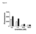
- FIG. 6 shows that in the 6D9 cells, urantide, an UT receptor antagonist, blocked calcium mobilization coupled to U-II stimulation: 10 nM U-II (open bar) or 30 nM U-II (filled bar).
- FIG. 7 illustrates consistent calcium mobilization results obtained from 6D9 cells using a 384-well FLIPR assay: A. multiple well overlay plot with U-II added at the arrow; B. average, from 6 wells, dose response curve for hU-II with an EC 50 of 7.875e ⁇ 008 ; and C. average, from 6 wells, dose response curve for urantide with an IC 50 of 1.33e ⁇ 009 (80 nM hU-II used).
- FLIPR fluorescence imaging plate reader
- GPCR G protein coupled receptor
- hU-II human Urotensin-II
- PBS phosphate buffer saline
- UT receptor Urotensin-II receptor
- a “cell clone” refers to a population of cells derived from a single common ancestral cell by mitosis in eukaryotes, such as a human cell clone, or by binary fission in prokaryotes. Although cells within a cell clone are presumed to be genetically identical, mutational events may abrogate the genetic homogeneity.
- sub-clone is a process whereby a cell clone is obtained from one or few ancestral cells of a parental cell clone.
- cells within the sub-cloned cell clone have less genetic variability to one to another than cells within the parental cell clone.
- sub-clone can also refer to the cell clone resulting from the sub-cloning process. To ensure genetic stability, the clone is frozen in early passage and cultures replaced with the frozen stocks at regular intervals.
- urotensin-II refers to a peptide having a conserved cyclic hexapeptide, SEQ ID NO: 1, CFWKYC, wherein an intramolecular disulfide bond was formed between the two cysteines of the hexapeptide.
- Examples of “urotensin-II” include, but are not limited to, those listed in Table 1, with an intramolecular disulfide bond between the two cysteines of the hexapeptide of SEQ ID NO: 1.
- Urotensin-II also includes the so-called U-II-related peptide (URP), for example, consisting essentially of SEQ ID NO: 10, ACFWKYCV, with an intramolecular disulfide bond formed between the two cysteines (Sugo et al., 2003, Biochem Biophys Res Commun 2003; 310:860-8).
- URP U-II-related peptide
- An “urotensin-II” can be isolated from a natural source, such as an U-II producing animal.
- An “urotensin-II” can also be synthesized via any in vitro method, such as an in vitro peptide synthesis reaction.
- a “functional equivalent of urotensin-II” is a chemical entity that has all or part of the biological activity of urotensin-II, i.e., to bind to an urotensin-II receptor, and the binding can be functionally coupled to calcium mobilization.
- “functional equivalent of urotensin-II” include, but are not limited to, modifications or truncations of urotensin-II, or fusion proteins comprising urotensin-II, that maintain all or part of the biological activities of an urotensin-II.
- “Functional equivalent of urotensin-II” also includes, but are not limited, to the non-peptide U-II mimetics, non-peptide UT receptor agonists, inverse agonists or antagonists.
- the “functional equivalent of urotensin-II” can be from either natural or non-natural sources. Non-natural sources include, for example, recombinant or synthetic sources.
- An “urotensin II receptor”, can (1) have greater than about 60% amino acid sequence identity to a human U-II receptor (NCBI protein accession number: NP — 061822); (2) bind to antibodies, e.g., polyclonal or monoclonal antibodies, raised against a human U-II receptor (NCBI protein accession number: NP — 061822); or (3) be encoded by a polynucleotide that specifically hybridizes under stringent hybridization conditions to a nucleic acid molecule having a sequence that has greater than about 60% nucleotide sequence identity to the coding region of a human U-II receptor cDNA (NCBI nucleotide accession number: NM — 018949).
- Stringent hybridization conditions has the meaning known in the art, as described in Sambrook et al., Molecular Cloning: A Laboratory Manual , Second Edition, Cold Spring Harbor Laboratory, Cold Spring Harbor, New York, (1989).
- An exemplary stringent hybridization condition comprises hybridization in 6 ⁇ sodium chloride/sodium citrate (SSC) at about 45° C., followed by one or more washes in 0.2 ⁇ SSC and 0.1% SDS at 50-65° C.
- the “U-II receptor” has greater than about 65, 70, 75, 80, 85, 90, or 95 percent amino acid sequence identity to a human U-II receptor (NCBI protein accession number: NP — 061822).
- exemplary U-II receptor includes human U-II receptor, which includes structural and functional polymorphisms of the human U-II receptor depicted in NCBI protein accession number: NP — 061822.
- Polymorphism refers to a set of genetic variants at a particular genetic locus among individuals in a population.
- U-II receptor also includes orthologs of the human U-II receptor in other animals such as rat (i.e., NCBI protein accession NO: NP — 065412), mouse (i.e., NCBI protein accession NO: NP — 663415), pig, dog and monkey.
- rat i.e., NCBI protein accession NO: NP — 065412
- mouse i.e., NCBI protein accession NO: NP — 663415
- pig dog and monkey.
- an “endogeneous urotensin II binding site” refers to a site on which an urotensin II can specifically bind to, and the site is naturally produced by or associated with the cell. “Naturally” in this context means that the U-II binding site is not recombinantly made, i.e., not genetically altered or modified by artificial means. In one embodiment, an endogeneous urotensin II binding site is found on an endogeneous urotensin II receptor. The number of the endogeneous urotensin II binding sites of a cell can be calculated using any methods known to a person skilled in the art.
- the number of endogeneous urotensin II binding sites of a cell can be calculated from the U-II receptor-ligand binding curve resulting from an U-II receptor-binding assay.
- Example 2 (infra) describes a specific example of how to measure and calculate the number of endogeneous urotensin II binding sites per cell.
- the “biological activity of an urotensin II receptor” refers to an activity exerted by the urotensin II receptor as determined in vivo, or in vitro, according to standard techniques. Such an activity can be a direct activity such as the ability of an urotensin II receptor to bind to an urotensin II (U-II) or an analog thereof, and the binding can be functionally coupled to calcium mobilization.
- a biological activity of an urotensin II receptor can also be an indirect activity, such as a signal transduction activity mediated by the urotensin II receptor via its interaction with one or more than one additional protein or other molecule(s), including but not limited to, interactions that occur in a multi-step, serial fashion.
- an urotensin II receptor has the biological activity of mediating the function of U-II or a functional derivative thereof as an endothelium independent vasoconstrictor or an endothelium dependent vasodilator.
- a “signal transduction” is the cascade of processes by which an extracellular signal interacts with a receptor at a cell surface, causing a change in the level of a second messenger, and ultimately effects a change in the cell function.
- a “signal transduction activity mediated by urotensin II receptor” refers to a signal transduction, wherein the extracellular signal is urotensin II or a functional equivalent thereof.
- a “signal transduction activity mediated by urotensin II receptor” is the cascade of processes by which urotensin II binds to an urotensin II receptor at a cell surface, causing a change in the level of a second messenger, such as calcium or cyclic AMP, and ultimately effects a change in the cell's function.
- the change in the cell's function can be the change of any cellular process urotensin II is involved in. Changes in the cell's function often lead to changes of the animal physiology.
- a “signal transduction activity mediated by urotensin II receptor” can be an endothelium independent vasoconstriction or an endothelium dependent vasodilation triggered by urotensin II.
- calcium mobilization refers to the process whereby the concentration of intracellular free Ca 2+ , also denoted [Ca 2+ ] i , increases or decreases during signal transduction. [Ca 2+ ] i increases due to, for example, release of Ca 2+ from internal storage, or increased influx of Ca 2+ across the plasma membrane and into the cell.
- test molecule As described herein, a “test molecule”, “test compound”, or “candidate compound”, used interchangeably herein, each means a molecule that is subjected to the assay systems and methods described herein.
- Test compounds or candidate compounds encompass numerous chemical classes, although typically they are organic compounds. Preferably, they are small organic compounds, i.e., those having a molecular weight of more than 50 Kd yet less than about 2500 Kd.
- candidate compounds comprise functional chemical groups necessary for structural interactions with polypeptides, and typically include at least an amine, carbonyl, hydroxyl or carboxyl group, preferably at least two of the functional chemical groups and more preferably at least three of the functional chemical groups.
- the candidate compounds can comprise cyclic carbon or heterocyclic structure and/or aromatic or polyaromatic structures substituted with one or more of the above-identified functional groups.
- Candidate compounds also can be biomolecules such as peptides, saccharides, fatty acids, sterols, isoprenoids, purines, pyrimidines, derivatives or structural analogs of the above, or combinations thereof and the like.
- the compound is a nucleic acid
- the compound typically is a DNA or RNA molecule, although modified nucleic acids having non-natural bonds or subunits are also contemplated.
- Candidate compounds can be obtained from a wide variety of sources including libraries of synthetic or natural compounds. For example, numerous means are available for random and directed synthesis of a wide variety of organic compounds and biomolecules, including expression of randomized oligonucleotides, synthetic organic combinatorial libraries, phage display libraries of random peptides, and the like. Candidate compounds can also be obtained using any of the numerous approaches in combinatorial library methods known in the art, including: biological libraries; spatially addressable parallel solid phase or solution phase libraries: synthetic library methods requiring deconvolution; the “one-bead one-compound” library method; and synthetic library methods using affinity chromatography selection (Lam (1997) Anticancer Drug Des. 12:145). Alternatively, libraries of natural compounds in the form of bacterial, fungal, plant and animal extracts are available or readily produced. Additionally, natural and synthetically produced libraries and compounds can be readily modified through conventional chemical, physical, and biochemical means.
- known pharmacological agents can be subjected to directed or random chemical modifications such as acylation, alkylation, esterification, amidation, etc. to produce structural analogs of the agents.
- Candidate compounds can be selected randomly or can be based on existing compounds that bind to and/or modulate the function of chloride channel activity. Therefore, a source of candidate agents is libraries of molecules based on a known compound that increases or decreases the biological activity of a U-II receptor, in which the structure of the known compound is changed at one or more positions of the molecule to contain more or fewer chemical moieties or different chemical moieties.
- the structural changes made to the molecules in creating the libraries of analog activators/inhibitors can be directed, random, or a combination of both directed and random substitutions and/or additions.
- One of ordinary skill in the art in the preparation of combinatorial libraries can readily prepare such libraries.
- reagents such as salts, buffers, neutral proteins (e.g., albumin), detergents, etc.
- Other reagents that improve the efficiency of the assay such as nuclease inhibitors, antimicrobial agents, and the like can also be used.
- high throughput refers to an assay design that allows easy screening of multiple samples simultaneously, and provides a capacity for robotic manipulation. Another desired feature of high throughput assays is an assay design that is optimized to reduce reagent usage, or minimize the number of manipulations in order to achieve the analysis desired. Examples of high throughput assay formats include 96-well or 384-well plates, levitating droplets, and “lab on a chip” microchannel chips used for liquid handling experiments.
- One general aspect of the invention is a human cell clone that has increased endogeneous urotensin II binding sites, enabling robust and efficient functional analyses of the biological activity of endogeneous UT receptor in the cell clone.
- the invention provides a human cell clone that is a sub-clone of SJRH30 (ATCC® Number: CRL-2061TM), having at least about 50% more endogeneous urotensin II binding sites cell ⁇ 1 than those of the parental SJRH30.
- the invention further provides a cell culture comprising cells of a human cell clone that is a sub-clone of SJRH30 (ATCC® Number: CRL-2061TM), having at least about 50% more endogeneous urotensin II binding sites cells ⁇ 1 than those of the parental SJRH30.
- the cell clones of the invention have at least about 100%, 150%, 200%, 250%, 300%, 350%, 400%, 450%, 500% or 550% or more endogeneous urotensin II binding sites cell ⁇ 1 than those of the parental SJRH30.
- the cell clones of the invention can be a sub-clone of SJRH30 selected from 6D9 (ATCC® Number: PTA-7749), 4G5 (ATCC® Number: PTA-7747), or 10A7 (ATCC® Number: PTA-7748), which has endogeneous urotensin II binding sites cell ⁇ 1 that are about 5.2 fold, 2.7 fold, or 3.3 fold, respectively, of those of the parental SJRH30.
- the cells of the invention exhibited an increase in the amount of U-II receptor mRNA as well as an increase in total receptor binding to U-II.
- the biological activity of an endogeneous U-II receptor can be detected more robustly from the cells of the invention as compared to prior known cells.
- the present invention enabled a HTS screening of the biological activity of an endogeneous U-II receptor in a cellular assay.
- the present invention provides methods of using the human cell clone of the invention to identify compounds that increase or decrease the biological activity of an urotensin II receptor.
- the inventive assay methods can be used to detect test compounds that increase or decrease the biological activity of an U-II receptor in any manner.
- Compounds that increase or decrease the biological activity of an U-II receptor can be compounds that interact directly with the U-II receptor in such a way as to affect the biological activity of U-II receptor.
- such a compound can bind to the U-II receptor and affect the interaction of the receptor with U-II or other protein/molecule, such as a functional derivative of U-II, i.e., an U-II mimetic, an agonist or antagonist of U-II.
- Compounds that increase or decrease the biological activity of an U-II receptor can also be compounds that interact indirectly with the U-II receptor in such a way as to affect the biological activity of U-II receptor.
- such a compound can bind to protein(s) or molecules other than the U-II receptor, and affect the signal transduction activity of the U-II receptor.
- a test compound in a buffer is administered to cells of a human cell clone of the invention.
- the biological activity of the urotensin II receptor of the cells can be measured, and compared to that of a control wherein the buffer rather than the test compound is administered to the cells.
- the test compound that increases the biological activity of the U-II receptor can thus be identified and subject to further analyses.
- cells of a human cell clone of the invention are exposed to a test compound and are exposed to a compound that is known to activate the U-II receptor.
- the cells can be exposed to the test compound either before, after, or simultaneously with the exposure of the cells to the compound that is known to activate the U-II receptor.
- the biological activity of the urotensin II receptor of the cells can be measured, and compared to that of a control wherein the cells are exposed to the U-II receptor activating compound only, not the test compound.
- the test compound that inhibits the biological activity of the U-II receptor can thus be identified and subject to further analyses.
- the biological activity of the urotensin H receptor can be measured using means known to a person skilled in the art. For example, it can be measured for the ability of the U-II receptor to bind to a U-II, a functional derivative of U-II, or any other compound that is known to bind to the receptor.
- the biological activity of the U-II receptor can also be measured by monitoring calcium mobilization. Activation of the U-II receptor is coupled with an increase in [Ca 2+ ] i .
- the methods of the invention can be combined with other means of testing a compound for its ability to increase or decrease the U-II receptor's biological activity.
- compounds that increase or decrease the U-II receptor's biological activity can be further tested in an animal model for its ability to cause changes in animal physiology. For example, it was observed that administering U-II to a rat induced an increase in the redness or skin temperature of the rat ear (Qi et al., U.S. patent application Ser. No.
- the parental SJRH30 cell line (ATCC NO: CRL-2061TM) and RPMI 1640 culture media (ATCC 30-2001) were obtained from ATCC (Manassas, Va.).
- FBS (Cat. No. SH30070.03) was from Hyclone (Logan, Utah).
- the Calcium 3 dye (Cat. No. R7182) was from Molecular Devices (Sonnyvale, Calif.).
- Probenecid (Cat. No. P-8761) and human urotensin II (Cat. No. U-7257) were from Sigma (St Louis, Mo.).
- Urantide was purchased from Peptide International (Cat. No. PUT-3639-P1, Louisville, Ky.). Branched DNA kits and probes were from Genospectra (Freemont, Calif.).
- cells of SJRH30 and its subclones were cultured in RPMI medium containing 10% FBS.
- Cells were split at a 1:5 to 1:10 ratio two times per week by enzyme dissociation. In this process, cells are rinsed with calcium and magnesium free PBS, a trypsin solution is added and the cells incubated until they have released from the bottom of the flask. At this time, cells are triturated, and a portion transferred to a new flask containing fresh medium.
- Non-wash Ca 2+ dye was formulated to incorporate extracellular fluorescence quenchers and avoid the need to wash away extracellular Ca 2+ dye after loading the cells with the dye (Monteith and Bird, 2005, supra).
- SJRH30 cells were trypsinized as described above and diluted to a concentration of 1 cell/100 ul volume in RPMI (10% FBS). Cells were plated at 100 ul/well into 10 ⁇ 96 well culture plates. After the cells were grown for 1-2 weeks on the 96-well culture plates, the replicate plates were made by washing the cells with Ca/Mg free PBS (Mediatech), adding trypsin to the washed cells (Gibco, Grand Island, N.Y.) and incubating the cells for 5 minutes, then triturating, i.e., pipeting up and down, the cells and placing 10 ul of the cells into 200 ul growth medium in separate black clear bottom 96 well plates. After about 1 week incubation of the black clear bottom 96 well plates, the wells of the plates were inspected for the presence of cell colonies. The ability of the cells to mobilize calcium in response to U-II was determined using Calcium 3 and FLIPR.
- SJRH30 cells responded well to dilution sub-cloning. At the about 1-cell/100 ul volume, approximately 59% of the wells contained colonies. The wells contained cells of various morphologies; spindly, elongated and epithelium like highlighting the heterogeneous nature of the parent cell line. Cells were evaluated for response to U-II using calcium mobilization. Wells were considered positive for U-II mediated calcium mobilization if they exhibited >1000 positive Fluorescence Change when U-II (1 nM) was added. Fluorescent change is measured on a well by well basis by taking the maximum fluorescence signal and subtracting the minimal signal for each well.
- the three cell clones are designated 10A7, 6D9 and 4G5, all of which were deposited to the American Type Culture Collection (ATCC®) at 10801 University Boulevard, Manassas, Va. 20110 on Aug. 1, 2006.
- the 10A7, 6D9 and 4G5 sub-clones of SJRH30 were designed as PTA-7748, PTA-7749 and PTA-7747, respectively. All three cell clones mobilized calcium to a similar extent as did the parental SJRH30 cells when the cells were exposed to a calcium ionophore (data not shown).
- the three cell clones differed in their ability to mobilize calcium in response to U-II with 6D9>4G5>10A7>SJRH30 ( FIG. 1 ).
- the cells of the three clones responded to U-II in a dose responsive manner, while the parental SJRH30 showed minimal to no signal (data not shown).
- the U-II specific antagonist, Urantide blocked the calcium mobilization coupled to U-II stimulation ( FIG. 6 ), indicating that the measured calcium mobilization is indeed due to activation of U-II receptor.
- Newly thawed 6D9 cells responded to U-II in a similar fashion as that of the 6D9 cells prior to freezing, indicating that the cells were stable upon freezing and thawing ( FIG. 4 ).
- the cells survived in cultures for over 3 months (17 passages) and were still responsive to U-II ( FIG. 4 ).
- Radioligand-binding studies were performed to characterize the cell clones 6D9, 4G5, 10A7, and SJRH30.
- [ 125 I]-U-II binding experiments cells were plated in 24-well Costar plates in complete medium for 24 h to reach 60% to 80% confluence.
- the binding medium used was Dulbecco's modified Eagle's medium (DMEM) containing 2 mg/ml BSA and 25 mM HEPES (pH 7.4).
- DMEM Dulbecco's modified Eagle's medium
- the cells were washed at room temperature 1 ⁇ with the binding medium, and were incubated with 0.1 ml per well of binding medium containing the indicated amount of [ 125 I]-U-II in the absence or presence of 1 ⁇ M human UII for 3 h.
- the cells were washed 3 ⁇ with the binding medium and solubilized in 1% SDS and 0.5 N NaOH. Radioactivity was quantified by gamma counting.
- Branched-DNA analysis was performed to measure the relative amount of mRNA of the UT receptor in the cell clones 6D9, 4G5, 10A7, and SJRH30.
- CE capture and label probes
- LE The probes within the lysis buffer serve certain functions: CE's have complementary sequences to both the message and to a capture plate so serves to adhere the message to the plate.
- the LE has sequences complementary to the message as well as to the bDNA and serves as a scaffold for binding the amplifying system. Following lysis of the cells, the samples are transferred to a capture plate.
- the capture plate is coated with an oligonucleotide that binds the CE thus capturing the message. After hybridization, the plate is washed and the Amplifier is added (bDNA). After hybridization, the samples are washed and the label is added (DNA coupled alkaline phosphatase). The samples are hybridized, washed and the luminescent substrate, dioxetene, is added and the samples are incubated to allow the reaction to occur and the plates are read on a luminometer.
- the subclones expressed increased the amount of UT receptor mRNA as compared to the SJRH30 ( FIG. 3 ). This suggests that the increased UII binding sites of cells of the subclones are likely due to the increased expression of UII receptors of the cells.
- This Example teaches a high throughput approach using FLIPR on a 384-well format.
- Other formats of high throughput assays can be conducted using protocols similar to this example.
- the optimal cell density for maximal response to U-II was about 30,000 per ml after the cells were incubated in plates for either 1 or 2 days ( FIG. 5 ).
- detecting the fluorescent signal of Calcium 3 between about 30-45 minutes after the dye was added to the cells appeared to decrease the variability of the assay.
Landscapes
- Health & Medical Sciences (AREA)
- Life Sciences & Earth Sciences (AREA)
- Engineering & Computer Science (AREA)
- Immunology (AREA)
- Molecular Biology (AREA)
- Biomedical Technology (AREA)
- Chemical & Material Sciences (AREA)
- Hematology (AREA)
- Urology & Nephrology (AREA)
- Food Science & Technology (AREA)
- Biochemistry (AREA)
- Cell Biology (AREA)
- Biotechnology (AREA)
- Medicinal Chemistry (AREA)
- Physics & Mathematics (AREA)
- Analytical Chemistry (AREA)
- Microbiology (AREA)
- General Health & Medical Sciences (AREA)
- General Physics & Mathematics (AREA)
- Pathology (AREA)
- Tropical Medicine & Parasitology (AREA)
- Endocrinology (AREA)
- Measuring Or Testing Involving Enzymes Or Micro-Organisms (AREA)
- Peptides Or Proteins (AREA)
- Preparation Of Compounds By Using Micro-Organisms (AREA)
Abstract
Description
| TABLE 1 |
| Examples of urotensin-II from various species |
| Species | Sequence of urotensin-II | ||
| Human | SEQ ID NO: 2, ETPDCFWKYCV | |||
| Frog | SEQ ID NO: 3, AGNLSECFWKYCV | |||
| Trout | SEQ ID NO: 4, GGNSECFWKYCV | |||
| Carp α | SEQ ID NO: 5, GGGAECFWKYCV | |||
| Porcine - 1 | SEQ ID NO: 6, GTPSECFWKYCV | |||
| Porcine - 2 | SEQ ID NO: 7, GPPSECFWKYCV | |||
| Rat - 1 | SEQ ID NO: 8, HGTAPECFWKYCI | |||
| Mouse | SEQ ID NO: 9, HGAAPECFWKYCI | |||
Claims (10)
Priority Applications (1)
| Application Number | Priority Date | Filing Date | Title |
|---|---|---|---|
| US11/501,559 US7892826B2 (en) | 2005-08-15 | 2006-08-09 | Human cell clones having an endogeneous urotensin II receptor |
Applications Claiming Priority (2)
| Application Number | Priority Date | Filing Date | Title |
|---|---|---|---|
| US70822105P | 2005-08-15 | 2005-08-15 | |
| US11/501,559 US7892826B2 (en) | 2005-08-15 | 2006-08-09 | Human cell clones having an endogeneous urotensin II receptor |
Publications (2)
| Publication Number | Publication Date |
|---|---|
| US20070082368A1 US20070082368A1 (en) | 2007-04-12 |
| US7892826B2 true US7892826B2 (en) | 2011-02-22 |
Family
ID=37684872
Family Applications (1)
| Application Number | Title | Priority Date | Filing Date |
|---|---|---|---|
| US11/501,559 Expired - Fee Related US7892826B2 (en) | 2005-08-15 | 2006-08-09 | Human cell clones having an endogeneous urotensin II receptor |
Country Status (2)
| Country | Link |
|---|---|
| US (1) | US7892826B2 (en) |
| WO (1) | WO2007021683A2 (en) |
Citations (2)
| Publication number | Priority date | Publication date | Assignee | Title |
|---|---|---|---|---|
| US5223409A (en) | 1988-09-02 | 1993-06-29 | Protein Engineering Corp. | Directed evolution of novel binding proteins |
| WO2006012470A1 (en) | 2004-07-22 | 2006-02-02 | Cumbre Pharmaceuticals Inc. | Rifamycin derivatives for treating microbial infections |
Family Cites Families (1)
| Publication number | Priority date | Publication date | Assignee | Title |
|---|---|---|---|---|
| US7939052B2 (en) * | 2005-05-12 | 2011-05-10 | Janssen Pharmaceutica Nv | Method of measuring the biological activity of an urotensin II receptor |
-
2006
- 2006-08-09 US US11/501,559 patent/US7892826B2/en not_active Expired - Fee Related
- 2006-08-09 WO PCT/US2006/030878 patent/WO2007021683A2/en not_active Ceased
Patent Citations (3)
| Publication number | Priority date | Publication date | Assignee | Title |
|---|---|---|---|---|
| US5223409A (en) | 1988-09-02 | 1993-06-29 | Protein Engineering Corp. | Directed evolution of novel binding proteins |
| US5571698A (en) | 1988-09-02 | 1996-11-05 | Protein Engineering Corporation | Directed evolution of novel binding proteins |
| WO2006012470A1 (en) | 2004-07-22 | 2006-02-02 | Cumbre Pharmaceuticals Inc. | Rifamycin derivatives for treating microbial infections |
Non-Patent Citations (24)
Also Published As
| Publication number | Publication date |
|---|---|
| US20070082368A1 (en) | 2007-04-12 |
| WO2007021683A3 (en) | 2007-04-19 |
| WO2007021683A2 (en) | 2007-02-22 |
Similar Documents
| Publication | Publication Date | Title |
|---|---|---|
| Iturrioz et al. | Identification and pharmacological properties of E339–3D6, the first nonpeptidic apelin receptor agonist | |
| Gao et al. | Connective tissue growth factor (CCN2) induces adhesion of rat activated hepatic stellate cells by binding of its C-terminal domain to integrin αvβ3 and heparan sulfate proteoglycan | |
| Aramori et al. | Coupling of two endothelin receptor subtypes to differing signal transduction in transfected Chinese hamster ovary cells. | |
| JP6654580B2 (en) | Detection of autoantibodies to TSH receptor | |
| Doan et al. | Biochemical and pharmacological characterization of nuclear urotensin‐II binding sites in rat heart | |
| Titus et al. | A new homogeneous high-throughput screening assay for profiling compound activity on the human ether-a-go-go-related gene channel | |
| Ruiz-Opazo et al. | Molecular characterization of a dual endothelin-1/angiotensin II receptor | |
| Fabry et al. | Monitoring of the internalization of neuropeptide Y on neuroblastoma cell line SK‐N‐MC | |
| Barr et al. | Agonist-independent activation of Gz by the 5-hydroxytryptamine1A receptor co-expressed in Spodoptera frugiperda cells: Distinguishing inverse agonists from neutral antagonists | |
| Zhang et al. | Autophagic flux detection: significance and methods involved | |
| Chen et al. | Angiotensin IV induces tyrosine phosphorylation of focal adhesion kinase and paxillin in proximal tubule cells | |
| Ji et al. | Novel signaling of dynorphin at κ-opioid receptor/bradykinin B2 receptor heterodimers | |
| Wu-Wong et al. | Pharmacology of endothelin receptor antagonists ABT-627, ABT-546, A-182086 and A-192621: in vitro studies | |
| Narváez et al. | Study of GPCR homo-and heteroreceptor complexes in specific neuronal cell populations using the in situ proximity ligation assay | |
| Connolly et al. | Interplay between intracellular loop 1 and helix VIII of the angiotensin II type 2 receptor controls its activation | |
| Durroux et al. | Fluorescent pseudo-peptide linear vasopressin antagonists: design, synthesis, and applications | |
| Stevens et al. | Resolution of inverse agonist-induced up-regulation from constitutive activity of mutants of the α1b-adrenoceptor | |
| US7892826B2 (en) | Human cell clones having an endogeneous urotensin II receptor | |
| US7655396B1 (en) | Methods for detecting receptor modulator activity | |
| Bawolak et al. | Effects of inactivation‐resistant agonists on the signalling, desensitization and down‐regulation of bradykinin B2 receptors | |
| Dumont et al. | BODIPY®‐conjugated neuropeptide Y ligands: new fluorescent tools to tag Y1, Y2, Y4 and Y5 receptor subtypes | |
| Smalley et al. | Ligand internalization and recycling by human recombinant somatostatin type 4 (h sst4) receptors expressed in CHO‐K1 cells | |
| Gera et al. | Design of fluorescent bradykinin analogs: application to imaging of B2 receptor-mediated agonist endocytosis and trafficking and angiotensin-converting enzyme | |
| Otsomaa et al. | Discovery and characterization of ORM-11372, a unique and positively inotropic sodium-calcium exchanger/inhibitor | |
| Huwiler et al. | Characterization of serotonin 5-hydroxytryptamine-1A receptor activation using a phospho-extracellular-signal regulated kinase 2 sensor |
Legal Events
| Date | Code | Title | Description |
|---|---|---|---|
| AS | Assignment |
Owner name: ORTHO-MCNEIL PHARMACEUTICAL, INC., NEW JERSEY Free format text: ASSIGNMENT OF ASSIGNORS INTEREST;ASSIGNORS:MINOR, LISA;QI, JENSON;WANG, YUANPING;REEL/FRAME:018334/0109 Effective date: 20060927 |
|
| AS | Assignment |
Owner name: ORTHO-MCNEIL-JANSSEN PHARMACEUTICALS, INC., NEW JE Free format text: MERGER;ASSIGNORS:ORTHO-MCNEIL PHARMACEUTICAL, INC.;JANSSEN PHARMACEUTICA INC.;REEL/FRAME:025628/0554 Effective date: 20071230 Owner name: JANSSEN PHARMACEUTICA N.V., BELGIUM Free format text: ASSIGNMENT OF ASSIGNORS INTEREST;ASSIGNOR:ORTHO-MCNEIL-JANSSEN PHARMACEUTICALS, INC.;REEL/FRAME:025628/0590 Effective date: 20110113 |
|
| STCF | Information on status: patent grant |
Free format text: PATENTED CASE |
|
| FPAY | Fee payment |
Year of fee payment: 4 |
|
| MAFP | Maintenance fee payment |
Free format text: PAYMENT OF MAINTENANCE FEE, 8TH YEAR, LARGE ENTITY (ORIGINAL EVENT CODE: M1552) Year of fee payment: 8 |
|
| FEPP | Fee payment procedure |
Free format text: MAINTENANCE FEE REMINDER MAILED (ORIGINAL EVENT CODE: REM.); ENTITY STATUS OF PATENT OWNER: LARGE ENTITY |
|
| LAPS | Lapse for failure to pay maintenance fees |
Free format text: PATENT EXPIRED FOR FAILURE TO PAY MAINTENANCE FEES (ORIGINAL EVENT CODE: EXP.); ENTITY STATUS OF PATENT OWNER: LARGE ENTITY |
|
| STCH | Information on status: patent discontinuation |
Free format text: PATENT EXPIRED DUE TO NONPAYMENT OF MAINTENANCE FEES UNDER 37 CFR 1.362 |
|
| FP | Lapsed due to failure to pay maintenance fee |
Effective date: 20230222 |