US7820807B2 - Gene constructs coding for CD40-binding activating antibodies - Google Patents
Gene constructs coding for CD40-binding activating antibodies Download PDFInfo
- Publication number
- US7820807B2 US7820807B2 US12/420,481 US42048109A US7820807B2 US 7820807 B2 US7820807 B2 US 7820807B2 US 42048109 A US42048109 A US 42048109A US 7820807 B2 US7820807 B2 US 7820807B2
- Authority
- US
- United States
- Prior art keywords
- cells
- antibodies
- pta
- antibody
- binding
- Prior art date
- Legal status (The legal status is an assumption and is not a legal conclusion. Google has not performed a legal analysis and makes no representation as to the accuracy of the status listed.)
- Expired - Fee Related
Links
Images


Classifications
-
- A—HUMAN NECESSITIES
- A61—MEDICAL OR VETERINARY SCIENCE; HYGIENE
- A61K—PREPARATIONS FOR MEDICAL, DENTAL OR TOILETRY PURPOSES
- A61K39/00—Medicinal preparations containing antigens or antibodies
- A61K39/395—Antibodies; Immunoglobulins; Immune serum, e.g. antilymphocytic serum
- A61K39/39533—Antibodies; Immunoglobulins; Immune serum, e.g. antilymphocytic serum against materials from animals
- A61K39/39541—Antibodies; Immunoglobulins; Immune serum, e.g. antilymphocytic serum against materials from animals against normal tissues, cells
-
- A—HUMAN NECESSITIES
- A61—MEDICAL OR VETERINARY SCIENCE; HYGIENE
- A61P—SPECIFIC THERAPEUTIC ACTIVITY OF CHEMICAL COMPOUNDS OR MEDICINAL PREPARATIONS
- A61P35/00—Antineoplastic agents
-
- A—HUMAN NECESSITIES
- A61—MEDICAL OR VETERINARY SCIENCE; HYGIENE
- A61P—SPECIFIC THERAPEUTIC ACTIVITY OF CHEMICAL COMPOUNDS OR MEDICINAL PREPARATIONS
- A61P37/00—Drugs for immunological or allergic disorders
- A61P37/02—Immunomodulators
-
- C—CHEMISTRY; METALLURGY
- C07—ORGANIC CHEMISTRY
- C07K—PEPTIDES
- C07K16/00—Immunoglobulins [IGs], e.g. monoclonal or polyclonal antibodies
- C07K16/18—Immunoglobulins [IGs], e.g. monoclonal or polyclonal antibodies against material from animals or humans
- C07K16/28—Immunoglobulins [IGs], e.g. monoclonal or polyclonal antibodies against material from animals or humans against receptors, cell surface antigens or cell surface determinants
- C07K16/2803—Immunoglobulins [IGs], e.g. monoclonal or polyclonal antibodies against material from animals or humans against receptors, cell surface antigens or cell surface determinants against the immunoglobulin superfamily
- C07K16/2818—Immunoglobulins [IGs], e.g. monoclonal or polyclonal antibodies against material from animals or humans against receptors, cell surface antigens or cell surface determinants against the immunoglobulin superfamily against CD28 or CD152
-
- C—CHEMISTRY; METALLURGY
- C07—ORGANIC CHEMISTRY
- C07K—PEPTIDES
- C07K16/00—Immunoglobulins [IGs], e.g. monoclonal or polyclonal antibodies
- C07K16/18—Immunoglobulins [IGs], e.g. monoclonal or polyclonal antibodies against material from animals or humans
- C07K16/28—Immunoglobulins [IGs], e.g. monoclonal or polyclonal antibodies against material from animals or humans against receptors, cell surface antigens or cell surface determinants
- C07K16/2878—Immunoglobulins [IGs], e.g. monoclonal or polyclonal antibodies against material from animals or humans against receptors, cell surface antigens or cell surface determinants against the NGF-receptor/TNF-receptor superfamily, e.g. CD27, CD30, CD40, CD95
-
- A—HUMAN NECESSITIES
- A61—MEDICAL OR VETERINARY SCIENCE; HYGIENE
- A61K—PREPARATIONS FOR MEDICAL, DENTAL OR TOILETRY PURPOSES
- A61K39/00—Medicinal preparations containing antigens or antibodies
- A61K2039/505—Medicinal preparations containing antigens or antibodies comprising antibodies
-
- A—HUMAN NECESSITIES
- A61—MEDICAL OR VETERINARY SCIENCE; HYGIENE
- A61K—PREPARATIONS FOR MEDICAL, DENTAL OR TOILETRY PURPOSES
- A61K38/00—Medicinal preparations containing peptides
-
- C—CHEMISTRY; METALLURGY
- C07—ORGANIC CHEMISTRY
- C07K—PEPTIDES
- C07K2317/00—Immunoglobulins specific features
- C07K2317/30—Immunoglobulins specific features characterized by aspects of specificity or valency
- C07K2317/31—Immunoglobulins specific features characterized by aspects of specificity or valency multispecific
Definitions
- This invention relates to a series of novel molecules and monoclonal antibodies that bind to and stimulate antigen presenting cells via the CD40 receptor expressed on such antigen presenting cells.
- the immune system is capable of killing autologous cells when they become infected by virus or when they transform into cancer cells. Such a potentially dangerous mechanism is under tight control.
- the immune system's T-killer cells CTL
- the precursor T-killer cell To be activated, the precursor T-killer cell must recognize its specific antigen peptide, presented by MHC class I molecules on professional antigen presenting cells (APC). This antigen specific cellular interaction is, however, not enough to fully activate the CTL, notwithstanding the co-stimulatory signals from the APC.
- T-helper cell that recognises the same antigen on the same APC as the CTL is needed to fully activate the CTL.
- the specific T-helper cell would supply cytokines such as IL-2 needed for the activation of the CTL.
- Guerder and Matzinger J. Exp. Med. 176:553 (1992)
- the “licensing” model for CTL activation. In this model it was suggested that the T-helper cell, when recognising its antigen on a professional APC, would deliver an activation signal to the APC that as a result would be able to subsequently activate a CTL without the need for the T-helper cell to be present.
- DC circulate through and are resident in the body tissues and at sites of antigen deposition or introduction. After taking up antigens, they migrate to the draining lymph nodes where they present antigen to the T cells. It is well known that a DC needs to be activated to perform optimally. Resting DC express only low levels of MHC and co-stimulatory molecules and are poor stimulators of T cells. DC can be activated by inflammatory cytokines and bacterial products, which results in up-regulation of MHC and co-stimulatory molecules. Therefore, DC that have encountered antigens under inflammatory conditions will readily activate T-helper cells when they arrive in the draining lymph nodes. It is thus very likely that the combination of inflammatory cytokines at the site of antigen uptake and the CD40L-CD40 interaction during the T-helper cell interaction result in an optimal capacity to license the DC for CTL activation.
- the CD40 molecule belongs to the TNF receptor family of type I transmembrane proteins.
- the members of this gene family (which include among others, the two receptors for TNF, the low-affinity nerve growth factor receptor and the T cell activation antigen CD27, CD30, and CD95) are characterized by sequence homology in their cysteine-rich extracellular domains (Armitage et al., Current Opinion in Immunology 6:407 (1994)).
- the known ligands for the members of the TNF receptor family are homologous as well.
- TNF- ⁇ is a soluble cytokine, it is initially synthesized as a membrane associated molecule.
- TNF/CD40L receptor and the TNF/CD40 families are type 11 trans-membrane proteins. These include: hTNF- ⁇ , hLT, hLT- ⁇ , hCD40L, hCD27L, hCD30L, cfECP1, myx VRh, mCD30, hCD27, hFas, m4-1BB, rOX-40, hTNFR-h, hTNFR-II, hTNFP-1 and hLNGFR.
- CD40 is best known for its function in B-cell activation. The molecule is constitutively expressed on all B cells.
- CD40L-CD40 interaction can stimulate the proliferation of purified B cells and, in combination with cytokines, mediate immunoglobulin production.
- cytokines include IFN- ⁇ and TNF- ⁇ .
- Stimulation of monocytes via CD40 results in the secretion of pro-inflammatory cytokines such as IL-1 and TNF- ⁇ , toxic free radical intermediates such as nitric oxide and up-regulation of the B7 co-stimulatory molecules.
- CD40 Human DC isolated from peripheral blood can also express the CD40 molecule (Caux et al., J. Exp. Med. 180:263 (1994)). Ligation of CD40 on DC results in enhanced survival of these cells when cultured in vitro. As with monocytes, stimulation of DC via CD40 results in secretion of pro-inflammatory cytokines such as IL-12 and TNF- ⁇ and up-regulation of the CD80/86 co-stimulatory molecules. In addition, it was recently demonstrated that activation of CD40 induces the capacity to stimulate the activation of killer T cells (Schoenberger et al., Nature 393:480 (1998)). Accordingly, activating CD40 by binding it with a ligand, such as an antibody, would induce a number of humoral and cytotoxic effects, useful in inhibiting tumors.
- a ligand such as an antibody
- the invention includes molecules able to bind to and activate CD40 expressed on both professional and non-professional APCs. These agonistic molecules, following binding to a cell surface receptor, induce intracellular signal transduction, leading to the activation of the APCs expressing CD40.
- the molecules of the invention include monoclonal antibodies, fragments thereof, peptides, oligonucleotides, and other chemical entities. Also included are peptides and genes inducing expression of anti-CD40 antibodies.
- Such molecules can be used in combination, or in a bispecific or multivalent form, including as bispecific antibodies, to cross-link CD40 on the same cell, or to cross-link CD40 present on different cells. Either such cross-linking could cause a synergistic or additive agonistic effect.
- the invention also provides a gene construct coding for an anti-CD40 monoclonal antibody or a fragment of an anti-CD40 monoclonal antibody.
- the anti-CD40 antibody is produced by a hybridoma selected from the group consisting of hybridomas deposited with American Type Culture Collection (ATCC) as deposit designation PTA-2993, PTA-2994, PTA-2995, PTA-2996, PTA-2997, PTA-2998 and PTA-2999.
- the present invention also provides a cell transfected with a gen construct coding fro the anti-CD40 monoclonal antibody.
- anti-CD40 antibodies produced by hybridoma clones generated by the methods of the invention are referred to as follows, in relation to the hybridomas deposited with the American Type Culture Collection (ATCC) and given the following ATCC deposit accession numbers: clone 4 (hybridoma MAb 186-4-1, ATTC Accession No. PTA-2996), clone 7 (hybridoma MAb 186-7-2, ATCC Accession No. PTA-2997), clone 15 (hybridoma MAb 186-15-1, ATCC Accession No. PTA-2998), clone 21 (hybridoma MAb 186-21-1, ATCC Accession No.
- ATCC American Type Culture Collection
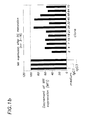
- FIG. 1 shows the induction of maturation of monocyte derived DC by anti-CD40 monoclonal antibodies.
- Monocyte derived immature dendritic cells were cultured for two days in the presence of anti-CD40 monoclonal antibodies or isotype matched control antibodies and then studied by FACS analysis for up-regulation of expression of CD80 and down-regulation of the mannose receptor. Shown are the combined results of several experiments with the percentage of cells expressing CD83 in FIG. 1 a and the relative decrease in mean fluorescence intensity (MFI) of the mannose receptor expression in unstimulated cells (MFI arbitrary taken as a value of 100) compared to stimulated cells in FIG. 1 b.
- MFI mean fluorescence intensity
- FIG. 2 shows induction of maturation of monocyte derived DC by anti-CD40 monoclonal antibodies (clones 7, 15, 21, 48, 64 and 70).
- Monocyte derived immature dendritic cells were cultured for two days in the presence of anti-CD40 monoclonal antibody or isotype matched control antibodies and then studied by FACS analysis for up-regulation of expression of CD80, CD83 and CD86 and down-regulation of expression of the mannose receptor. Data of one representative experiment are shown: the CD80 ( FIG. 2 a ), CD86 ( FIG. 2 c ) and mannose receptor ( FIG. 2 d ) expression are indicated as mean fluorescence intensity, whereas CD83 is indicated as the percentage of cells expressing this marker for mature dendritic cells ( FIG. 2 b ).
- FIG. 3 shows induction of IL-12p70 secretion by monocyte derived DC after stimulation with CD40 agonist antibodies and IFN- ⁇ .
- Monocyte derived immature DC were cultured for two days in the presence of anti-CD40 monoclonal antibodies or isotype control antibodies alone or in combination with IFN- ⁇ . Induction of IL-12p70 production requires the combination of two different stimuli.
- FIG. 4 shows IL-12p70 production induced by CD40 agonist antibodies and IFN- ⁇ is blocked by pre-incubation with CD40-Fc. Pre-incubation of the CD40 agonist antibodies with an excess of CD40-Fc abolished the ability of the anti-CD40 monoclonal antibodies to induce, in combination with IFN- ⁇ , IL-12 production in monocyte derived DC.
- FIG. 5 shows CD40 agonist monoclonal antibodies prime DC with an enhanced ability to induce CD8+ T cell responses.
- Monocyte derived DC were either left un-stimulated, or pre-activated with CD40 agonist antibody with or without IFN- ⁇ and subsequently co-cultured with purified autologous CD8+ T cells in the presence of a flu matrix peptide representing a dominant HLA-A2 restricted epitope recognized by CD8+ T cells.
- the induction of CD8+ T cell responses by CD40 activated DC was studied by analyzing both the expansion of flu peptide specific CD8+ T cells 9 ( FIG. 5 a ) and the increase in CD8+ T cells that produce IFN- ⁇ ( FIG. 5 b ).
- the molecules described and claimed include monoclonal antibodies, fragments thereof, peptides and other chemical entities.
- Monoclonal antibodies can be made by the conventional method of immunization of a mammal, followed by isolation of the B cell producing the monoclonal antibodies of interest and fusion with a myeloma cell.
- the preferred monoclonal antibodies include chimeric antibodies, humanized antibodies, human antibodies, DeImmunizedTM antibodies, single-chain antibodies and fragments, including Fab, F(ab′) 2 Fv and other fragments which retain the antigen binding function of the parent antibody.
- Single chain antibodies (“ScFv”) and the method of their construction are described in U.S. Pat. No. 4,946,778.
- Chimeric antibodies are produced by recombinant processes well known in the art, and have an animal variable region and a human constant region. Humanized antibodies correspond more closely to the sequence of human antibodies than do chimeric antibodies. In a humanized antibody, only the complementarity determining regions (CDRs), which are responsible for antigen binding and specificity, are non-human derived and have an amino acid sequence corresponding to the non-human antibody, and substantially all of the remaining portions of the molecule (except, in some cases, small portions of the framework regions within the variable region) are human derived and have an amino acid sequence corresponding to a human antibody. See L. Riechmann et al., Nature; 332: 323-327 1988; U.S. Pat. No. 5,225,539 (Medical Research Council); U.S. Pat. Nos. 5,585,089; 5,693,761; 5,693,762 (Protein Design Labs, Inc.).
- CDRs complementarity determining regions
- Human antibodies can be made by several different methods, including by use of human immunoglobulin expression libraries (Stratagene Corp., La Jolla, Calif.; Cambridge Antibody Technology Ltd., London, England) to produce fragments of human antibodies (VH, VL, Fv, Fd, Fab, or (Fab′) 2 ), and use of these fragments to construct whole human antibodies by fusion of the appropriate portion thereto, using techniques similar to those for producing chimeric antibodies.
- Human antibodies can also be produced in transgenic mice with a human immunoglobulin genome. Such mice are available from Abgenix, Inc., Fremont, Calif., and Medarex, Inc., Annandale, N.J. In addition to connecting the heavy and light chain Fv regions to form a single chain peptide, Fab can be constructed and expressed by similar means (M. J. Evans et al., J. Immunol. Meth., 184:123-138 1995).
- DeImmunizedTM antibodies are antibodies in which the potential T cell epitopes have been eliminated, as described in International Patent Application PCT/GB98/01473. Therefore, immunogenicity in humans is expected to be eliminated or substantially reduced when they are applied in vivo.
- wholly and partially human antibodies described above are less immunogenic than wholly murine or non-human-derived antibodies, as are the fragments and single chain antibodies. All these molecules (or derivatives thereof) are therefore less likely to evoke an immune or allergic response. Consequently, they are better suited for in vivo administration in humans than wholly non-human antibodies, especially when repeated or long-term administration is necessary, as may be needed for treatment of psoriasis or other inflammatory skin conditions.
- Bispecific antibodies can be used as cross-linking agents between CD40 of the same cell, or CD40 on two different cells. Such bispecific antibodies would have one specificity for each of two different epitopes on CD40. Bispecifics in which one specificity is a strong activator of binding of sCD40L to CD40, and one specificity is a partial or non-inhibitor of binding of sCD40L to CD40, could synergize the agonistic effect on cross-linking.
- bispecific antibodies include linking single chain Fv with peptide couplers, including Ser-Cys, (Gly) 4 -Cys, (His) 6 -(Gly) 4 -Cys, chelating agents, and chemical or disulfide couplings including bismaleimidohexane and bismaleimidocaproyl.
- Non-antibody molecules can be isolated or screened from compound libraries by conventional means.
- An automated system for generating and screening a compound library is described in U.S. Pat. Nos. 5,901,069 and 5,463,564.
- a more focused approach involves three-dimensional modeling of the binding site, and then making a family of molecules which fit the model. These are then screened for those with optimal binding characteristics.
- Another approach is to generate recombinant peptide libraries, and then screen them for those which bind to the epitope of CD40 of interest. See, e.g., U.S. Pat. No. 5,723,322.
- This epitope is the same as that bound by the monoclonal antibodies described in the examples below.
- Molecules can, in fact, be generated or isolated with relative ease in accordance with techniques well known in the art once the epitope is known.
- Another approach is to induce endogenous production of the desired anti-CD40 antibodies, by administering a peptide or an antibody which induces such production, or through gene therapy, where a gene encoding an appropriate anti-CD40 molecule or a fragment thereof is administered.
- the method of making and administering any of these molecules is well known in the art.
- the molecules can be administered by any of a number of routes. In the case of peptides and antibodies, because they are subject to degradation in the gastro-intestinal tract, they would preferably be injected. Other compounds of the invention could also be injected. The injections could be intramuscular, intravenous or sub-cutaneous.
- Non-peptide molecules of the invention could be administered orally, including by suspension, tablets and the like.
- Liquid formulations could be administered by inhalation of lyophilized or aeorosolized microcapsules. Suppositories could also be used.
- Additional pharmaceutical vehicles could be used to control the duration of action of the molecules of the invention. They could be entrapped in microcapsules prepared by coacervation techniques or by interfacial polymerization (hydroxymethylcellullose or gelatin microcapsules) in colloidal drug delivery systems (for example, liposomes, albumin microspheres, micro-emulsions, nanoparticles and nanocapsules) or in macro-emulsions.
- colloidal drug delivery systems for example, liposomes, albumin microspheres, micro-emulsions, nanoparticles and nanocapsules
- Excipients for example, salts, various bulking agents, additional buffering agents, chelating agents, antioxidants, cosolvents and the like can be included in the final formulation. Specific examples include tris(hydroxymethyl) aminomethane salts (“Tris buffer”) and disodium edetate.
- the dosage and scheduling for the formulation which is selected can be determined by standard procedures, well known in the art. Such procedures involve extrapolating an estimated dosing schedule from animal models, and then determining the optimal dosage in a human clinical dose ranging study.
- the EBV-transformed B-cell line JY and the myeloid derived cell line THP1 were cultured in T75 culture flasks routinely in Iscove's modified Dulbecco's medium (IMDM) to which 50 .quadrature.g/ml gentamycin and 2% heat inactivated foetal calf serum was added (FCSi; BioWhittaker, Verviers, Belgium).
- IMDM Iscove's modified Dulbecco's medium
- FCSi g/ml gentamycin and 2% heat inactivated foetal calf serum was added
- PBMC Peripheral mononuclear blood cells
- LymphoprepTM 1.077 g/ml density centrifugation and resuspended in Ca 2+ /Mg 2+ -free PBS-0.1% BSA.
- Autologous PBMC were stored in RPMI 1640 supplemented with 2 mM L-glutamine, 10% FCSi, 50 ⁇ g/ml gentamycin and 10% DMSO at ⁇ 196° C. (for CD8 T cell purification, see below).
- Monocytes were purified from PBMC by immunomagnetic depletion (monocyte-enrichment cocktail containing Mabs against CD2, CD3, CD16, CD19, CD56, CD66b and glycophorin A; StemSepTM from StemCell Technologies, Vancouver, Canada). Monocyte (>90% CD14+) preparations devoid of neutrophilic granulocytes, platelets, lymphocytes and NK cells were subsequently cultured in serum-free culture medium, StemSpanTM (StemCell Technologies), supplemented with 10 ng/ml GM-CSF and 20 ng/ml IL-4 (both cytokines from PeproTech, Rocky Hill, N.J., USA) at 37° C./5% CO 2 during 6-7 days.
- Monocyte-enrichment cocktail containing Mabs against CD2, CD3, CD16, CD19, CD56, CD66b and glycophorin A; StemSepTM from StemCell Technologies, Vancouver, Canada.
- monocytes were seeded at a cell density of 1 ⁇ 10 6/2 ml/10 cm 2 polystyrene surface (coated with 12 mg/ml/10 cm 2 poly-hydroxethylmethacrylate; Sigma) and fresh GM-CSF/IL-4 was added at day 2 and 5.
- the nonadherent cells (with a dendritic morphology) were collected and displayed the following (flow cytometry, see below) phenotypic profile: CD1a + , CD14 ⁇ , CD40 + , C80 + , CD83 ⁇ , CD86 + , HLA-DR + and mannose receptor ++ .
- CD8 T lymphocytes were purified by immunomagnetic depletion of other cell types (CD8-enrichment cocktail with Mabs against CD4, CD14, CD16, CD56 and glycophorin A; StemCell Technologies). This procedure resulted in >90% CD3 + /CD8 + lymphocytes devoid of monocytes, neutrophilic granulocytes, platelets, B and CD4 lymphocytes, and NK cells.
- soluble CD40 ligand (sCD40L) binding by anti-CD40 monoclonal antibodies (Mabs) was demonstrated by using JY cells, which express high levels of CD40. These cells (0.1 ⁇ 10 6 cells/100 ml PBS-0.1% BSA/sample) were pre-incubated with anti-CD40 Mabs for 15 min. at 21° C., and then thoroughly washed in PBS-0.1% BSA, followed by an incubation with a soluble fusion protein consisting of the extracellular domain of human CD40L fused to the extracellular domain of murine CD8 ⁇ (CD40L-mCD8a; Kordia, Leiden, The Netherlands and Tanox Pharma BV, Amsterdam, The Netherlands) for 15 min. at 21° C. Subsequently, CD40L-mCD8a was detected by using rat anti-mouse CD8 ⁇ coupled to phycoerythrin, and analyzed by flow cytometry.
- CD40L-mCD8a was detected by using rat anti-m
- M2 and G28-5 compete for the CD40L binding site, and 5C3 binds to a region distinct from the CD40L binding site.
- EA-5 partially inhibits the binding of CD40L to its receptor (Pound et al., Int Immunol 1999, 11, p 11-20).
- CD40L activated CD4+ T cells As a source of membrane CD40L activated CD4+ T cells are used. To this purpose expression of CD40L on T cells is induced through culturing plastic non-adherent PBMC with PMA and ionomycine for 6 hrs in IMDM+5% human pooled AB serum. CD40-Fc (IgG4 made by Tanox Inc Houston USA) is directly added at a saturating dose to the activated T cells or after pre-incubation of CD40Fc with excess of anti-CD40 Mabs.
- IgG4 made by Tanox Inc Houston USA
- CD40-Fc CD40L activated CD4+ (CD3+CD8 ⁇ ) T cells is monitored through FACS analysis after staining with PE conjugated goat anti human IgG-Fc, FITC conjugated CD3 and PERCP conjugated CD8.
- ELISA plates (Immunon 2) were coated overnight at room temperature with 0.5 ⁇ g/ml, 50 ⁇ l per well of goat-anti-human IgG (Fc). Next the plates were treated with 1% BLOTTO for 60 min at room temperature. After washing 4 times with PBS/Tween, 50 ⁇ l/well of CD40Fc plus 50 ⁇ l of supernatants of the fusion wells were added and incubated for 1 hour at room temperature. After another 4 washings with PBS/Tween, 50 ⁇ l of goat-anti-mouse IgG (Fc)-HRP conjugate was added and incubated for 1 hour. After 4 washings the substrate TMB was added at 100 ⁇ l/well to the plates which were incubated for 30 min. The reaction was stopped by addition of 50 ⁇ l/well of 0.2 M H 2 SO 4 and the plates were read with an ELISA reader at 450/590 nm THP-1 assay
- THP-1 cells were first cultured for two days in 10 ml of IMDM+2% of human type AB serum in the presence of 5 ⁇ 10 2 U/ml IFN-.quadrature. Next the IFN-g treated THP-1 cells were washed once in IMDM+2% human type AB serum. 10 4 THP-1 cells per 96 w plate well were cultured for two days in 120 ⁇ l of culture medium diluted 1:2 with hybridoma supernatant. As controls CD154-mCD8 was used at 40 .quadrature.g/ml maximum and 2 ⁇ dilutions and LPS at 20 ng/ml maximum and 2 ⁇ dilutions.
- ELISA plates were coated with mouse anti human IL-8 antibody (Serotec) at 5 ⁇ g/ml, 100 .quadrature.l/well for 2 hrs at room temperature on a plate shaker. The plates were then incubated with 1% BLOTTO for one hour on the plate shaker at room temperature. After four washings with PBS/Tween, 80 ⁇ l of supernatants harvested from the THP-1 plate were added to the ELISA plate.
- IL-8 was diluted with 1% BLOTTO to 1000 pg/ml, 300 pg/ml, 100 g/ml 30 pg/ml, 10 pg/ml, 3 pg/ml, 1 pg/ml.
- the ELISA plates were incubated for one hr at room temperature on the plate shaker. After four washings with PBS/Tween, 100 ⁇ l/well mouse-anti IL-8 biotin conjugate (Serotec) was added at 1:1000 dilution in 1% BLOTTO and the plates were incubated for one hour at room temperature. After four washings with PBS/Tween, 100 .quadrature.l/well AMDX SA-HRP at 1:1000 dilution in 1% BLOTTO was added to the wells and the plates were incubated for 1 hour at room temperature on the plate shaker.
- PBS/Tween 100 ⁇ l/well mouse-anti IL-8 biotin conjugate
- TMB substrate 100 .quadrature.l of TMB substrate was added to each well and the plates were incubated for 30 minutes at room temperature on the plate shaker. The reaction was stopped by addition of 50 ⁇ l/well of 0.2 M H 2 SO 4 and the plates were read with an ELISA reader at 450/590 nm.
- Immature DC (see above) are cultured in the presence of anti-CD40 Mabs under serum-free condition (StemSpanTM) at 37° C./5% CO2 for 48 hours.
- StemSpanTM serum-free condition
- CD40L-mCD8u, LPS and a combination of IL-1 ⁇ and TNF- ⁇ are used as well-established controls for DC maturation.
- the change from immature to mature DC is determined by: (1) phenotype (CD1a + , CD14 ⁇ , CD40 +++ , CD80 +++ , CD83 + , CD86 +++ , HLA-DR +++ , mannose receptors), (2) IL-12p70 production (commercially available kit), and by (3) the capability of inducing influenza-matrix peptide specific autologous cytotoxic CD8 +++ T lymphocytes (see below).
- Immature DC are cultured in the presence of anti-CD40 Mabs (1 ⁇ g/ml) with or without IFN-g (1000 U/ml) for 48 hrs.
- IL-12p70 secretion was measured in the supernatant using a commercially available kit from Diaclone Research, Becanson, France. Inhibition of IL-12 production was obtained by preincubation of anti-CD40 Mabs with 10 times excess of CD40-Fc (IgG4 made by Tanox Inc Houston USA) for 15 min at room temperature.
- Mature DC generated by agonistic anti-CD40 Mabs are loaded with a synthetic influenza matrix peptide (Flu-peptide 58-66); 1 ⁇ 10 6 DC/Flu-peptide 5 ⁇ g/ml StemSpanTM) and co-cultured with 0.5 ⁇ 10 6 purified autologous CD8+ T lymphocytes at 37° C./5% CO 2 during 7 days.
- a synthetic influenza matrix peptide Flu-peptide 58-66
- DC/Flu-peptide 5 ⁇ g/ml StemSpanTM 1 ⁇ 10 6 DC/Flu-peptide 5 ⁇ g/ml StemSpanTM
- Cytotoxicity of the CD8+ T lymphocytes is determined by: (1) enumeration of the number of IFN- ⁇ producing T cells, which are representative for activated CTL (using flow cytometry with an IFN- ⁇ detection kit from Miltenyi Biotec, Bergisch Gladbach, Germany), and (2) a convential assay measuring cytolysis by CTL of target cells loaded with Flu-peptide.
- mice Two immunization protocols were used to generate anti-CD40 monoclonal antibodies.
- Female BALB/c mice were injected intraperitoneally with SF-9 cells expressing CD40 (3 ⁇ 10 6 cells/mouse), which were washed with PBS twice before injection.
- the mice received a booster injection with SF-9 cells.
- Fourteen days after the last the last the injection the spleen cells were isolated and 10 8 cells were used for cell fusion with 10 8 SP2/0 murine myeloma cells using polyethylene glycol.
- the fused cells were resuspended in D15 (a modified DMEM medium) supplemented with HAT, followed by plating on fifty-one 96 wells plates.
- mice were injected intraperitoneally with 2.5 ⁇ 10 6 monocyte-derived immature DC. At days 14, 35 and 55 mice received booster injections with monocyte-derived DC from different donors. At around day 100-120, spleen cells will be isolated and fused with murine myeloma cells in analogy to the above protocol. Supernatants of wells with growing hybridomas will be screened for the presence of CD40 binding antibodies in the ELISA. Hybridoma supernatants containing CD40 binding antibodies will be subsequently screened for potential agonistic activity as described for the hybridoma's originating from B cells isolated from the BALB/c immunized with CD40 expressing SF-9 cells
- the selected supernatants containing CD40 binding antibodies were subsequently tested for their ability to induce IL-8 production in the CD40 expressing monocytic cell line THP-1, which had been pre-incubated with IFN- ⁇ .
- most supernatants contained anti-CD40 antibodies, which displayed agonistic activity in this assay.
- Supernatants were arbitrarily subdivided into four different groups on the basis of their performance in the THP-1 assay (strong agonists with an OD of >2.000, intermediate agonists with an OD between 1.000-2.000, low agonists with an OD between 0.375-0.999 and non-agonists with an OD ⁇ 0.375).
- DC derived from monocytes after culture with GM-CSF and IL-4 represent immature DC.
- Anti-CD40 monoclonal antibodies have been assayed for their capacity to induce maturation of these CD40 expressing immature DC.
- stimulation of monocyte-derived DC with sCD40L results in their differentiation into DC with a mature phenotype.
- sCD40L in combination with IFN- ⁇ stimulates monocyte-derived DC to secrete IL-12p70.
- mature DC express CD83.
- mature DC display enhanced expression on a per cell basis of the co-stimulatory molecules CD80 and CD86, decreased expression of the mannose receptor and loss of the ability to efficiently take up molecules, as shown for dextran-FITC.
- the phenotypical changes that accompany the differentiation of immature to mature DC were monitored by FACS-analysis as a read-out for induction of DC maturation by the anti-CD40 monoclonal antibodies.
- Antibodies were first used on their own to stimulate monocyte-derived DC. As shown in FIG. 1 (combined results of several experiments showing CD83 up-regulation and mannose receptor down-regulation) and in FIG.
- CD40 binding antibodies were tested and were found to induce phenotypical maturation of monocyte derived DC, as is indicated by the increased percentage of cells expressing the CD83 marker, the increased expression on a per cell basis (mean fluorescence intensity; MFI) of CD80 and CD86 and decreased expression of the mannose receptor.
- MFI mean fluorescence intensity
- IL-12p70 production of monocyte derived DC was tested after stimulation with the CD40 monoclonal antibodies and IFN- ⁇ since dendritic cells require stimulation through at least two different pathways to produce IL-12p70 (Kalinski et al Blood 1997 90:1926).
- Our results show that apart from induction of phenotypical maturation, the CD40 agonist antibodies also induced IL-12 production in DC when used together with IFN- ⁇ ( FIG. 3 ).
- pre-incubation of the CD40 monoclonal antibodies with excess of CD40-Fc inhibited induction of IL-12 production demonstrated that the agonistic effect of the antibodies is really exerted through CD40 and not through other membrane expressed molecules on the DC ( FIG. 4 ).
- T cell help to CTL was found to be mediated through CD40 activated DC.
- Antigen dependent interaction of helper T cells with DC did not only result in the activation of the helper T cell, but through CD40L-CD40 interaction also in the activation of the DC. Only in their activated stage DC were able to prime CTL responses. In the absence of helper T cells no DC activation and therefore no CTL priming occurred. However, by means of in vivo administration of an anti-mouse CD40 stimulatory antibody, T cell help could be efficiently bypassed and DC directly activated.
- CTL activation was analyzed in this experiment through measurement of production of IFN- ⁇ by activated CTL and enumeration of expansion of CTL with PE conjugated HLA-A2/influenza matrix peptide tetramers.
- FIG. 5 a and b the stimulation of monocyte derived DC with CD40 monoclonal antibodies led to increased ability of these cells to induce a flu peptide directed CD8+ T cell response. For most antibodies this effect was elevated when, in addition to the monoclonal antibodies, IFN- ⁇ was used in the pre-activation of the dendritic cells.
- Anti-CD40 antibodies that synergize with sCD40L in the induction of CD40 mediated activation of DC most likely show co-binding with sCD40L to CD40 and thus do not display strong blocking of binding of sCD40L to CD40.
- the percentage of inhibition of sCD40L binding to CD40 on JY EBV transformed B cells by the monoclonal antibodies was tested. This analysis revealed that there was strong variation in the degree that the monoclonal antibodies could inhibit the binding of sCD40L to CD40. Some antibody samples almost completely inhibited sCD40L binding, whereas other antibody samples could only partially block sCD40L binding or had no effect at all (table 2).
- the level of IL 12p70 produced by the DC after stimulation by the combination of one of these antibodies with sCD40L and IFN- ⁇ will be enhanced compared to the level induced by sCD40L and IFN- ⁇ alone.
- two anti-CD40 antibodies may also show synergism with each other in the induction of IL-12p70 secretion. This synergism may occur most noticeably between antibodies that block binding of sCD40L to CD40 and those that are partial or non-inhibitors of this interaction, as these antibodies are expected to bind different epitopes on CD40.
- bi-specific antibody with specificity for CD40 on one side and a determinant on T cells on the other side potentially has the benefit of bringing the activated DC in close contact with surrounding T cells. If the antibody part that recognizes the T cell determinant has agonistic properties, the additional benefit may be that the attracted T cell will be stimulated both through the signals delivered by the activated DC and the agonistic properties of the T cell part of the bispecific antibody. This possibility will be evaluated by comparing the effect of the addition of the CD40 monoclonal antibodies and the bispecific antibodies in the above described DC-CTL co-culture system, using flu peptide specific CD8+ T cell responses as read out.
Abstract
Description
Claims (6)
Priority Applications (1)
| Application Number | Priority Date | Filing Date | Title |
|---|---|---|---|
| US12/420,481 US7820807B2 (en) | 2000-02-01 | 2009-04-08 | Gene constructs coding for CD40-binding activating antibodies |
Applications Claiming Priority (4)
| Application Number | Priority Date | Filing Date | Title |
|---|---|---|---|
| US17893400P | 2000-02-01 | 2000-02-01 | |
| US09/773,866 US7172759B2 (en) | 2000-02-01 | 2001-02-01 | Induction of cytotoxic T lymphocyte responses using anti-CD40 antibodies |
| US11/613,409 US7547438B2 (en) | 2000-02-01 | 2006-12-20 | CD40-binding activating antibodies |
| US12/420,481 US7820807B2 (en) | 2000-02-01 | 2009-04-08 | Gene constructs coding for CD40-binding activating antibodies |
Related Parent Applications (1)
| Application Number | Title | Priority Date | Filing Date |
|---|---|---|---|
| US11/613,409 Division US7547438B2 (en) | 2000-02-01 | 2006-12-20 | CD40-binding activating antibodies |
Publications (2)
| Publication Number | Publication Date |
|---|---|
| US20090311268A1 US20090311268A1 (en) | 2009-12-17 |
| US7820807B2 true US7820807B2 (en) | 2010-10-26 |
Family
ID=22654507
Family Applications (3)
| Application Number | Title | Priority Date | Filing Date |
|---|---|---|---|
| US09/773,866 Expired - Lifetime US7172759B2 (en) | 2000-02-01 | 2001-02-01 | Induction of cytotoxic T lymphocyte responses using anti-CD40 antibodies |
| US11/613,409 Expired - Fee Related US7547438B2 (en) | 2000-02-01 | 2006-12-20 | CD40-binding activating antibodies |
| US12/420,481 Expired - Fee Related US7820807B2 (en) | 2000-02-01 | 2009-04-08 | Gene constructs coding for CD40-binding activating antibodies |
Family Applications Before (2)
| Application Number | Title | Priority Date | Filing Date |
|---|---|---|---|
| US09/773,866 Expired - Lifetime US7172759B2 (en) | 2000-02-01 | 2001-02-01 | Induction of cytotoxic T lymphocyte responses using anti-CD40 antibodies |
| US11/613,409 Expired - Fee Related US7547438B2 (en) | 2000-02-01 | 2006-12-20 | CD40-binding activating antibodies |
Country Status (9)
| Country | Link |
|---|---|
| US (3) | US7172759B2 (en) |
| EP (2) | EP1253942A4 (en) |
| JP (1) | JP2003524644A (en) |
| CN (2) | CN100490895C (en) |
| AU (2) | AU3662101A (en) |
| BR (1) | BR0108001A (en) |
| CA (1) | CA2398998C (en) |
| MX (1) | MXPA02007473A (en) |
| WO (1) | WO2001056603A1 (en) |
Cited By (3)
| Publication number | Priority date | Publication date | Assignee | Title |
|---|---|---|---|---|
| US20080050340A1 (en) * | 2006-05-03 | 2008-02-28 | Regents Of The University Of Colorado | Cd40 agonist antibody /type 1 interferon synergistic adjuvant combination, conjugates containing and use thereof as a therapeutic to enhance cellular immunity |
| US8778345B2 (en) | 2011-04-29 | 2014-07-15 | Apexigen, Inc. | Anti-CD40 antibodies |
| US9676861B2 (en) | 2012-10-30 | 2017-06-13 | Apexigen, Inc. | Anti-CD40 antibodies and methods of use |
Families Citing this family (55)
| Publication number | Priority date | Publication date | Assignee | Title |
|---|---|---|---|---|
| US6407218B1 (en) * | 1997-11-10 | 2002-06-18 | Cytimmune Sciences, Inc. | Method and compositions for enhancing immune response and for the production of in vitro mabs |
| CA2223225A1 (en) * | 1997-11-28 | 1999-05-28 | Canadian Red Cross Society | Method for inhibiting in vivo immune response |
| US20110041190A1 (en) * | 2002-10-31 | 2011-02-17 | Tykocinski Mark L | Novel chimeric proteins |
| AU3662101A (en) * | 2000-02-01 | 2001-08-14 | Tanox Inc | Cd40-binding apc-activating molecules |
| US7063845B2 (en) | 2000-04-28 | 2006-06-20 | Gemini Science, Inc. | Human anti-CD40 antibodies |
| PT1391464E (en) | 2001-04-27 | 2007-11-15 | Kirin Pharma Kk | Anti-cd40 monoclonal antibody |
| AU2002347404A1 (en) * | 2001-09-14 | 2003-04-01 | Cytos Biotechnology Ag | In vivo activation of antigen presenting cells for enhancement of immune responses induced by virus like particles |
| DE60234375D1 (en) | 2001-09-14 | 2009-12-24 | Cytos Biotechnology Ag | PACKAGING IMMUNSTIMULATING CpG IN VIRUS LIKE PARTICLES: PREPARATION METHOD AND USE |
| AR039067A1 (en) * | 2001-11-09 | 2005-02-09 | Pfizer Prod Inc | ANTIBODIES FOR CD40 |
| US7537767B2 (en) | 2003-03-26 | 2009-05-26 | Cytis Biotechnology Ag | Melan-A- carrier conjugates |
| RU2351362C2 (en) | 2003-03-26 | 2009-04-10 | Цитос Байотекнолоджи Аг | CONJUGATES OF PEPTIDE Melan-A, VIRUS-LIKE PARTICLE ANALOGUE |
| CA2548179A1 (en) * | 2003-12-02 | 2005-07-21 | Cytimmune Sciences, Inc. | Methods and compositions for the production of monoclonal antibodies |
| US20050136055A1 (en) | 2003-12-22 | 2005-06-23 | Pfizer Inc | CD40 antibody formulation and methods |
| JP5484732B2 (en) | 2005-12-14 | 2014-05-07 | サイトス バイオテクノロジー アーゲー | Immunostimulatory nucleic acid package particles for the treatment of hypersensitivity |
| MX2008015529A (en) | 2006-06-12 | 2009-01-13 | Cytos Biotechnology Ag | Processes for packaging oligonucleotides into virus-like particles of rna bacteriophages. |
| US20090074711A1 (en) * | 2006-09-07 | 2009-03-19 | University Of Southhampton | Human therapies using chimeric agonistic anti-human cd40 antibody |
| CA2706700A1 (en) * | 2007-11-08 | 2009-05-14 | Cytimmune Sciences, Inc. | Compositions and methods for generating antibodies |
| AU2011309689B2 (en) | 2010-09-28 | 2015-01-15 | Hadasit Medical Research Services & Development Ltd. | Compositions and methods for treatment of hematological malignancies |
| PT2683406T (en) | 2011-03-11 | 2019-07-08 | Beth Israel Deaconess Medical Ct Inc | Anti-cd40 antibodies and uses thereof |
| CN103793594A (en) * | 2013-12-30 | 2014-05-14 | 上海交通大学医学院附属瑞金医院 | Method for constructing related intestinal canal-pancreatic islet regulation axis function evaluation model and application of model |
| EP3189132B1 (en) * | 2014-09-04 | 2020-06-24 | Stemcell Technologies Inc. | Soluble antibody complexes for t cell or nk cell activation and expansion |
| CN107249318A (en) | 2014-12-10 | 2017-10-13 | 明尼苏达大学董事会 | For cell, tissue and the organ of the genetic modification for treating disease |
| JP6401297B2 (en) * | 2014-12-25 | 2018-10-10 | 株式会社日立製作所 | Insulin secretion capacity analysis apparatus, insulin secretion capacity analysis system including the apparatus, and insulin secretion capacity analysis method |
| MA41459A (en) | 2015-02-03 | 2017-12-12 | Als Therapy Development Inst | ANTI-CD40L ANTIBODIES AND METHODS FOR TREATING CD40L ILLNESSES OR DISORDERS |
| WO2016130667A1 (en) * | 2015-02-10 | 2016-08-18 | Ohio State Innovation Foundation | Chlamydia-activated b cell platforms and methods thereof |
| WO2016166568A1 (en) | 2015-04-16 | 2016-10-20 | Juno Therapeutics Gmbh | Methods, kits and apparatus for expanding a population of cells |
| CN111375066B (en) | 2015-07-16 | 2023-04-25 | 百欧肯治疗有限公司 | Compositions and methods for treating cancer |
| EP3307322B1 (en) | 2015-09-04 | 2021-02-17 | Primatope Therapeutics Inc. | Humanized anti-cd40 antibodies and uses thereof |
| ES2906823T3 (en) | 2015-09-30 | 2022-04-20 | Janssen Biotech Inc | Agonist antibodies that specifically bind to human CD40 and methods of use |
| MA45488A (en) | 2015-10-22 | 2018-08-29 | Juno Therapeutics Gmbh | CELL CULTURE PROCESSES, KITS AND APPARATUS |
| MA45489A (en) | 2015-10-22 | 2018-08-29 | Juno Therapeutics Gmbh | CELL CULTURE PROCESSES, ASSOCIATED KITS AND APPARATUS |
| KR20180072821A (en) | 2015-11-03 | 2018-06-29 | 얀센 바이오테크 인코포레이티드 | Antibodies that specifically bind to PD-1 and uses thereof |
| CA3004924A1 (en) | 2015-11-10 | 2017-05-18 | Ohio State Innovation Foundation | Methods and compositions related to accelerated humoral affinity |
| WO2017156349A1 (en) | 2016-03-10 | 2017-09-14 | Cold Genesys, Inc. | Methods of treating solid or lymphatic tumors by combination therapy |
| CN109195994A (en) * | 2016-04-22 | 2019-01-11 | 鳄鱼生物科学公司 | For the novel bispecific polypeptide of CD137 |
| IL263223B2 (en) | 2016-05-27 | 2023-03-01 | Abbvie Biotherapeutics Inc | Anti-cd40 antibodies and their uses |
| US20170342169A1 (en) | 2016-05-27 | 2017-11-30 | Abbvie Biotherapeutics Inc. | Bispecific binding proteins |
| BR112019000512A2 (en) | 2016-07-14 | 2019-04-24 | Genmab A/S | antibody, nucleic acid, expression vector, host cell, composition, methods of treating a disease, to produce a bispecific antibody and to detect if cross-linking between cd40 and cd137 expressing cells occurs in a sample, use of a multispecific antibody and kit |
| CA3033661A1 (en) | 2016-08-12 | 2018-02-15 | Janssen Biotech, Inc. | Engineered antibodies and other fc-domain containing molecules with enhanced agonism and effector functions |
| CN109843916B (en) | 2016-08-12 | 2023-10-31 | 詹森生物科技公司 | Fc-engineered anti-TNFR superfamily member antibodies with enhanced agonistic activity and methods of use thereof |
| EP4342912A2 (en) * | 2016-09-23 | 2024-03-27 | Merus N.V. | Binding molecules that modulate a biological activity expressed by a cell |
| CA3055433A1 (en) * | 2017-03-29 | 2018-10-04 | Glycotope Gmbh | Humanized anti-cd40 antibodies |
| US11866465B2 (en) | 2017-04-27 | 2024-01-09 | Juno Therapeutics Gmbh | Oligomeric particle reagents and methods of use thereof |
| SG10202111207TA (en) | 2017-05-24 | 2021-11-29 | Als Therapy Development Inst | Therapeutic anti-cd40 ligand antibodies |
| JP7369113B2 (en) | 2017-07-20 | 2023-10-25 | アプティーボ リサーチ アンド デベロップメント エルエルシー | Carcinoembryonic antigen binding protein, related compounds and methods |
| SG11202104355SA (en) | 2018-10-31 | 2021-05-28 | Juno Therapeutics Gmbh | Methods for selection and stimulation of cells and apparatus for same |
| US20210393691A1 (en) | 2018-11-06 | 2021-12-23 | Juno Therapeutics, Inc. | Process for producing genetically engineered t cells |
| CN111548415B (en) * | 2019-03-04 | 2021-04-20 | 北京天广实生物技术股份有限公司 | Antibodies that specifically bind to CD40 and uses thereof |
| AU2020377043A1 (en) | 2019-10-30 | 2022-06-02 | Juno Therapeutics Gmbh | Cell selection and/or stimulation devices and methods of use |
| AR120832A1 (en) | 2019-12-20 | 2022-03-23 | Amgen Inc | MULTISPECIFIC ANTIBODY CONSTRUCTS CD40 AGONISTS TARGETING MESOTHELIN FOR THE TREATMENT OF SOLID TUMORS |
| CN111235208B (en) * | 2020-02-28 | 2022-10-28 | 江苏大学 | Method for evaluating influence of ionic liquid on sugar metabolism |
| CN115835873A (en) | 2020-05-13 | 2023-03-21 | 朱诺治疗学股份有限公司 | Method for generating donor batch cells expressing recombinant receptor |
| WO2022036495A1 (en) | 2020-08-17 | 2022-02-24 | Utc Therapeutics Inc. | Lymphocytes-antigen presenting cells co-stimulators and uses thereof |
| EP4334341A2 (en) | 2021-05-06 | 2024-03-13 | Juno Therapeutics GmbH | Methods for stimulating and transducing t cells |
| WO2023213969A1 (en) | 2022-05-05 | 2023-11-09 | Juno Therapeutics Gmbh | Viral-binding protein and related reagents, articles, and methods of use |
Citations (15)
| Publication number | Priority date | Publication date | Assignee | Title |
|---|---|---|---|---|
| US4946778A (en) | 1987-09-21 | 1990-08-07 | Genex Corporation | Single polypeptide chain binding molecules |
| US5225539A (en) | 1986-03-27 | 1993-07-06 | Medical Research Council | Recombinant altered antibodies and methods of making altered antibodies |
| US5463564A (en) | 1994-09-16 | 1995-10-31 | 3-Dimensional Pharmaceuticals, Inc. | System and method of automatically generating chemical compounds with desired properties |
| US5534254A (en) | 1992-02-06 | 1996-07-09 | Chiron Corporation | Biosynthetic binding proteins for immuno-targeting |
| US5585089A (en) | 1988-12-28 | 1996-12-17 | Protein Design Labs, Inc. | Humanized immunoglobulins |
| US5674492A (en) | 1993-12-23 | 1997-10-07 | Immunex Corporation | Method of preventing or treating disease characterized by neoplastic cells expressing CD40 |
| US5723322A (en) | 1994-12-20 | 1998-03-03 | Michigan Biotechnology Institute | Process for making succinic acid, microorganisms for use in the process and methods of obtaining the microorganisms |
| US5801227A (en) | 1993-10-01 | 1998-09-01 | Fanslow, Iii; William C. | Antibodies to CD40 |
| WO1999061057A2 (en) | 1998-05-23 | 1999-12-02 | Tanox, Inc. | Molecules targeting cd40 and tumor cells |
| US6106835A (en) | 1991-04-19 | 2000-08-22 | Tanox, Inc. | Modified binding molecules specific for T or B lymphocytes and their use as in vivo immune modulators |
| WO2002011763A1 (en) | 2000-04-19 | 2002-02-14 | Tanox, Inc. | Cd40 antagonists for use in treating psoriasis and other inflammatory skin conditions |
| US6497876B1 (en) | 1996-07-10 | 2002-12-24 | Immunex Corp. | Method of stimulating an immune response with activated dendritic cells |
| US20030022860A1 (en) | 1998-05-23 | 2003-01-30 | Melief Cornelis J.M. | CD40 binding molecules and CTL peptides for treating tumors |
| US6632992B2 (en) | 2000-07-19 | 2003-10-14 | Yamaha Corporation | System and method for distributing music data with advertisement |
| US7172759B2 (en) * | 2000-02-01 | 2007-02-06 | Pangenetics Bv | Induction of cytotoxic T lymphocyte responses using anti-CD40 antibodies |
Family Cites Families (4)
| Publication number | Priority date | Publication date | Assignee | Title |
|---|---|---|---|---|
| US5637481A (en) * | 1993-02-01 | 1997-06-10 | Bristol-Myers Squibb Company | Expression vectors encoding bispecific fusion proteins and methods of producing biologically active bispecific fusion proteins in a mammalian cell |
| WO1998001473A1 (en) | 1996-07-05 | 1998-01-15 | Novo Nordisk A/S | Method for the production of precursors of insulin, precursors of insulin analogues, and insulin like peptides |
| DE29611914U1 (en) | 1996-07-09 | 1997-11-06 | Bosch Gmbh Robert | Ignition coil for an internal combustion engine |
| US6946129B1 (en) * | 1999-06-08 | 2005-09-20 | Seattle Genetics, Inc. | Recombinant anti-CD40 antibody and uses thereof |
-
2001
- 2001-02-01 AU AU3662101A patent/AU3662101A/en active Pending
- 2001-02-01 WO PCT/US2001/003378 patent/WO2001056603A1/en active Application Filing
- 2001-02-01 EP EP01908789A patent/EP1253942A4/en not_active Ceased
- 2001-02-01 CN CNB018072453A patent/CN100490895C/en not_active Expired - Fee Related
- 2001-02-01 MX MXPA02007473A patent/MXPA02007473A/en unknown
- 2001-02-01 JP JP2001556502A patent/JP2003524644A/en active Pending
- 2001-02-01 BR BR0108001-6A patent/BR0108001A/en not_active IP Right Cessation
- 2001-02-01 US US09/773,866 patent/US7172759B2/en not_active Expired - Lifetime
- 2001-02-01 CA CA2398998A patent/CA2398998C/en not_active Expired - Lifetime
- 2001-02-01 EP EP08157131A patent/EP1975182A1/en not_active Withdrawn
- 2001-02-01 AU AU2001236621A patent/AU2001236621B2/en not_active Ceased
- 2001-04-26 CN CNA018117716A patent/CN1942767A/en active Pending
-
2006
- 2006-12-20 US US11/613,409 patent/US7547438B2/en not_active Expired - Fee Related
-
2009
- 2009-04-08 US US12/420,481 patent/US7820807B2/en not_active Expired - Fee Related
Patent Citations (19)
| Publication number | Priority date | Publication date | Assignee | Title |
|---|---|---|---|---|
| US5225539A (en) | 1986-03-27 | 1993-07-06 | Medical Research Council | Recombinant altered antibodies and methods of making altered antibodies |
| US4946778A (en) | 1987-09-21 | 1990-08-07 | Genex Corporation | Single polypeptide chain binding molecules |
| US5693761A (en) | 1988-12-28 | 1997-12-02 | Protein Design Labs, Inc. | Polynucleotides encoding improved humanized immunoglobulins |
| US5693762A (en) | 1988-12-28 | 1997-12-02 | Protein Design Labs, Inc. | Humanized immunoglobulins |
| US5585089A (en) | 1988-12-28 | 1996-12-17 | Protein Design Labs, Inc. | Humanized immunoglobulins |
| US6106835A (en) | 1991-04-19 | 2000-08-22 | Tanox, Inc. | Modified binding molecules specific for T or B lymphocytes and their use as in vivo immune modulators |
| US5534254A (en) | 1992-02-06 | 1996-07-09 | Chiron Corporation | Biosynthetic binding proteins for immuno-targeting |
| US5801227A (en) | 1993-10-01 | 1998-09-01 | Fanslow, Iii; William C. | Antibodies to CD40 |
| US5674492A (en) | 1993-12-23 | 1997-10-07 | Immunex Corporation | Method of preventing or treating disease characterized by neoplastic cells expressing CD40 |
| US5463564A (en) | 1994-09-16 | 1995-10-31 | 3-Dimensional Pharmaceuticals, Inc. | System and method of automatically generating chemical compounds with desired properties |
| US5901069A (en) | 1994-09-16 | 1999-05-04 | 3-Dimensional Pharmaceuticals, Inc. | System, method, and computer program product for at least partially automatically generating chemical compounds with desired properties from a list of potential chemical compounds to synthesize |
| US5723322A (en) | 1994-12-20 | 1998-03-03 | Michigan Biotechnology Institute | Process for making succinic acid, microorganisms for use in the process and methods of obtaining the microorganisms |
| US6497876B1 (en) | 1996-07-10 | 2002-12-24 | Immunex Corp. | Method of stimulating an immune response with activated dendritic cells |
| WO1999061057A2 (en) | 1998-05-23 | 1999-12-02 | Tanox, Inc. | Molecules targeting cd40 and tumor cells |
| US20030022860A1 (en) | 1998-05-23 | 2003-01-30 | Melief Cornelis J.M. | CD40 binding molecules and CTL peptides for treating tumors |
| US7172759B2 (en) * | 2000-02-01 | 2007-02-06 | Pangenetics Bv | Induction of cytotoxic T lymphocyte responses using anti-CD40 antibodies |
| US7547438B2 (en) * | 2000-02-01 | 2009-06-16 | Pangenetics Bv | CD40-binding activating antibodies |
| WO2002011763A1 (en) | 2000-04-19 | 2002-02-14 | Tanox, Inc. | Cd40 antagonists for use in treating psoriasis and other inflammatory skin conditions |
| US6632992B2 (en) | 2000-07-19 | 2003-10-14 | Yamaha Corporation | System and method for distributing music data with advertisement |
Non-Patent Citations (35)
Cited By (14)
| Publication number | Priority date | Publication date | Assignee | Title |
|---|---|---|---|---|
| US9095608B2 (en) | 2006-05-03 | 2015-08-04 | The Regents Of The University Of Colorado | Immunostimulatory regimen comprising administering type 1 interferon and agonistic anti-CD40 antibody |
| US20100317111A1 (en) * | 2006-05-03 | 2010-12-16 | Ross Kedl | Cd40 agonist antibody/type 1 interferon synergistic adjuvant combination, conjugates containing and use thereof as a therapeutic to enhance cellular immunity |
| US7993648B2 (en) | 2006-05-03 | 2011-08-09 | The Regents of the Universitry of Colorado | Immunostimulatory regimen comprising administering type 1 interferon and agonistic anti-CD40 antibody |
| US8137672B2 (en) | 2006-05-03 | 2012-03-20 | The Regents Of The University Of Colorado | Immunostimulatory regimen comprising administering type 1 interferon and agonistic anti-CD40 antibody |
| US8361471B2 (en) | 2006-05-03 | 2013-01-29 | The Regents Of The University Of Colorado | Immunostimulatory regimen comprising administering type 1 interferon and agonistic anti-CD40 antibody |
| US10329338B2 (en) | 2006-05-03 | 2019-06-25 | The Regents Of The University Of Colorado | Nucleic acid construct encoding an agonistic anti-CD40 antibody and a type I interferon |
| US20080050340A1 (en) * | 2006-05-03 | 2008-02-28 | Regents Of The University Of Colorado | Cd40 agonist antibody /type 1 interferon synergistic adjuvant combination, conjugates containing and use thereof as a therapeutic to enhance cellular immunity |
| US8957193B2 (en) | 2011-04-29 | 2015-02-17 | Apexigen, Inc. | Polynucleotides encoding anti-CD40 antibodies |
| US9266956B2 (en) | 2011-04-29 | 2016-02-23 | Apexigen, Inc. | Methods of inhibiting proliferation of CD40-expressing cancer cells with anti-CD40 antibodies |
| US9556278B2 (en) | 2011-04-29 | 2017-01-31 | Apexigen, Inc. | Anti-CD40 antibodies |
| US8778345B2 (en) | 2011-04-29 | 2014-07-15 | Apexigen, Inc. | Anti-CD40 antibodies |
| US11001637B2 (en) | 2011-04-29 | 2021-05-11 | Apexigen, Inc. | Anti-CD40 antibodies |
| US9676861B2 (en) | 2012-10-30 | 2017-06-13 | Apexigen, Inc. | Anti-CD40 antibodies and methods of use |
| US9994640B2 (en) | 2012-10-30 | 2018-06-12 | Apexigen, Inc. | Anti-CD40 antibodies |
Also Published As
| Publication number | Publication date |
|---|---|
| EP1253942A4 (en) | 2004-06-16 |
| CN1942767A (en) | 2007-04-04 |
| JP2003524644A (en) | 2003-08-19 |
| EP1253942A1 (en) | 2002-11-06 |
| AU2001236621B2 (en) | 2006-06-15 |
| CA2398998A1 (en) | 2001-08-09 |
| MXPA02007473A (en) | 2003-09-22 |
| US7547438B2 (en) | 2009-06-16 |
| US7172759B2 (en) | 2007-02-06 |
| CA2398998C (en) | 2014-04-22 |
| US20070098720A1 (en) | 2007-05-03 |
| US20090311268A1 (en) | 2009-12-17 |
| CN1443077A (en) | 2003-09-17 |
| CN100490895C (en) | 2009-05-27 |
| US20010026932A1 (en) | 2001-10-04 |
| BR0108001A (en) | 2004-01-06 |
| WO2001056603A1 (en) | 2001-08-09 |
| EP1975182A1 (en) | 2008-10-01 |
| AU3662101A (en) | 2001-08-14 |
Similar Documents
| Publication | Publication Date | Title |
|---|---|---|
| US7820807B2 (en) | Gene constructs coding for CD40-binding activating antibodies | |
| AU2001236621A2 (en) | Cd40-binding apc-activating molecules | |
| AU2001236621A1 (en) | Cd40-binding apc-activating molecules | |
| JP6975841B2 (en) | Humanized antigen-binding domain for CD19 and how to use it | |
| JP6290751B2 (en) | Methods and compositions for increasing the effectiveness of therapeutic antibodies using NK cell enhancing compounds | |
| JP2022065073A (en) | Bcma binding molecules and methods of use thereof | |
| WO2002088186A1 (en) | Anti-cd40 monoclonal antibody | |
| KR20030096337A (en) | Anti-CD40 Monoclonal Antibody | |
| JP2022513778A (en) | Chimeric antigen receptor and T cell receptor and how to use | |
| CZ291071B6 (en) | Bispecific molecule useful for lysis of tumor cells, process of its preparation, monoclonal antibody, pharmaceutical preparation and kit as well as purification of tumor cells | |
| CN113402619B (en) | Targeting B7H3 co-expression IL-21 fully human chimeric antigen receptor, iNKT cell and application thereof | |
| US20220064254A1 (en) | Anti-hk2 chimeric antigen receptor (car) | |
| Wheeler et al. | Co-ligation of surface IgM and CD40 on naive B lymphocytes generates a blast population with an ambiguous extrafollicular/germinal centre cell phenotype | |
| WO1995015340A1 (en) | Monoclonal antibodies to antigens expressed by human dendritic cells | |
| EP0503646A1 (en) | Monoclonal antibodies recognizing lymphocyte function associated antigen-3 | |
| EP1618891A1 (en) | Method of inducing differentiation and proliferating regulatory t cell by anti-cd52 antibody and medicinal composition therefor | |
| De Jong et al. | Regulation of T-cell differentiation by CD2 and CD28 accessory molecules. | |
| AU4909199A (en) | Immunological reagent specifically interacting with the extracellular domain of the human zeta chain | |
| CN112638949B (en) | anti-B7S 1 polypeptides and uses thereof | |
| JP2022518187A (en) | antibody | |
| Woodhead | Phenotypic and functional studies on human peripheral blood dendritic cells | |
| MXPA06000841A (en) | Methods and compositions for increasing the efficiency of therapeutic antibodies using nk cell potentiating compounds |
Legal Events
| Date | Code | Title | Description |
|---|---|---|---|
| FEPP | Fee payment procedure |
Free format text: PAYOR NUMBER ASSIGNED (ORIGINAL EVENT CODE: ASPN); ENTITY STATUS OF PATENT OWNER: SMALL ENTITY |
|
| STCF | Information on status: patent grant |
Free format text: PATENTED CASE |
|
| CC | Certificate of correction | ||
| FPAY | Fee payment |
Year of fee payment: 4 |
|
| AS | Assignment |
Owner name: BIOCEROS HOLDING B.V., NETHERLANDS Free format text: ASSIGNMENT OF ASSIGNORS INTEREST;ASSIGNOR:PANGENETICS B.V.;REEL/FRAME:035106/0428 Effective date: 20150211 |
|
| AS | Assignment |
Owner name: CLUSTER40 THERAPEUTICS B.V., NETHERLANDS Free format text: CHANGE OF NAME;ASSIGNOR:BIOCEROS HOLDING B.V.;REEL/FRAME:039735/0016 Effective date: 20160729 |
|
| AS | Assignment |
Owner name: MYCENAX BIOTECH INC., TAIWAN Free format text: ASSIGNMENT OF ASSIGNORS INTEREST;ASSIGNOR:CLUSTER40 THERAPEUTICS B.V.;REEL/FRAME:043119/0407 Effective date: 20170704 |
|
| MAFP | Maintenance fee payment |
Free format text: PAYMENT OF MAINTENANCE FEE, 8TH YR, SMALL ENTITY (ORIGINAL EVENT CODE: M2552) Year of fee payment: 8 |
|
| FEPP | Fee payment procedure |
Free format text: MAINTENANCE FEE REMINDER MAILED (ORIGINAL EVENT CODE: REM.); ENTITY STATUS OF PATENT OWNER: SMALL ENTITY |
|
| LAPS | Lapse for failure to pay maintenance fees |
Free format text: PATENT EXPIRED FOR FAILURE TO PAY MAINTENANCE FEES (ORIGINAL EVENT CODE: EXP.); ENTITY STATUS OF PATENT OWNER: SMALL ENTITY |
|
| STCH | Information on status: patent discontinuation |
Free format text: PATENT EXPIRED DUE TO NONPAYMENT OF MAINTENANCE FEES UNDER 37 CFR 1.362 |
|
| FP | Lapsed due to failure to pay maintenance fee |
Effective date: 20221026 |