US7807394B2 - Nitro-imidazole hypoxia imaging agents - Google Patents
Nitro-imidazole hypoxia imaging agents Download PDFInfo
- Publication number
- US7807394B2 US7807394B2 US12/098,390 US9839008A US7807394B2 US 7807394 B2 US7807394 B2 US 7807394B2 US 9839008 A US9839008 A US 9839008A US 7807394 B2 US7807394 B2 US 7807394B2
- Authority
- US
- United States
- Prior art keywords
- alkyl
- group
- mmol
- compound
- alkylenyl
- Prior art date
- Legal status (The legal status is an assumption and is not a legal conclusion. Google has not performed a legal analysis and makes no representation as to the accuracy of the status listed.)
- Expired - Fee Related, expires
Links
- 206010021143 Hypoxia Diseases 0.000 title abstract description 48
- YZEUHQHUFTYLPH-UHFFFAOYSA-N 2-nitroimidazole Chemical compound [O-][N+](=O)C1=NC=CN1 YZEUHQHUFTYLPH-UHFFFAOYSA-N 0.000 title abstract description 11
- 230000007954 hypoxia Effects 0.000 title description 19
- 239000012216 imaging agent Substances 0.000 title description 8
- 125000005466 alkylenyl group Chemical group 0.000 claims description 57
- 150000001875 compounds Chemical class 0.000 claims description 53
- 125000006273 (C1-C3) alkyl group Chemical group 0.000 claims description 35
- 230000002285 radioactive effect Effects 0.000 claims description 31
- 125000002887 hydroxy group Chemical group [H]O* 0.000 claims description 24
- 125000000217 alkyl group Chemical group 0.000 claims description 18
- 125000004432 carbon atom Chemical group C* 0.000 claims description 17
- 125000004178 (C1-C4) alkyl group Chemical group 0.000 claims description 16
- 125000002924 primary amino group Chemical group [H]N([H])* 0.000 claims description 16
- 150000003573 thiols Chemical class 0.000 claims description 16
- 125000004001 thioalkyl group Chemical group 0.000 claims description 12
- 125000004169 (C1-C6) alkyl group Chemical group 0.000 claims description 11
- 125000003545 alkoxy group Chemical group 0.000 claims description 11
- 125000000229 (C1-C4)alkoxy group Chemical group 0.000 claims description 4
- 125000000446 sulfanediyl group Chemical group *S* 0.000 claims description 4
- CBOIHMRHGLHBPB-UHFFFAOYSA-N hydroxymethyl Chemical compound O[CH2] CBOIHMRHGLHBPB-UHFFFAOYSA-N 0.000 claims description 3
- 125000001425 triazolyl group Chemical group 0.000 claims description 3
- 125000001475 halogen functional group Chemical group 0.000 claims 6
- 206010028980 Neoplasm Diseases 0.000 abstract description 66
- 230000001146 hypoxic effect Effects 0.000 abstract description 29
- 238000002600 positron emission tomography Methods 0.000 abstract description 19
- 150000003852 triazoles Chemical class 0.000 abstract description 7
- 238000001727 in vivo Methods 0.000 abstract description 5
- 230000002708 enhancing effect Effects 0.000 abstract description 2
- 238000011503 in vivo imaging Methods 0.000 abstract description 2
- 230000000302 ischemic effect Effects 0.000 abstract description 2
- XEKOWRVHYACXOJ-UHFFFAOYSA-N Ethyl acetate Chemical compound CCOC(C)=O XEKOWRVHYACXOJ-UHFFFAOYSA-N 0.000 description 90
- YMWUJEATGCHHMB-UHFFFAOYSA-N Dichloromethane Chemical compound ClCCl YMWUJEATGCHHMB-UHFFFAOYSA-N 0.000 description 55
- 238000006243 chemical reaction Methods 0.000 description 54
- KRHYYFGTRYWZRS-BJUDXGSMSA-N ac1l2y5h Chemical compound [18FH] KRHYYFGTRYWZRS-BJUDXGSMSA-N 0.000 description 51
- 235000019439 ethyl acetate Nutrition 0.000 description 44
- OKKJLVBELUTLKV-UHFFFAOYSA-N Methanol Chemical compound OC OKKJLVBELUTLKV-UHFFFAOYSA-N 0.000 description 36
- VYPSYNLAJGMNEJ-UHFFFAOYSA-N Silicium dioxide Chemical compound O=[Si]=O VYPSYNLAJGMNEJ-UHFFFAOYSA-N 0.000 description 36
- WEVYAHXRMPXWCK-UHFFFAOYSA-N Acetonitrile Chemical compound CC#N WEVYAHXRMPXWCK-UHFFFAOYSA-N 0.000 description 32
- LFQSCWFLJHTTHZ-UHFFFAOYSA-N Ethanol Chemical compound CCO LFQSCWFLJHTTHZ-UHFFFAOYSA-N 0.000 description 32
- 210000004027 cell Anatomy 0.000 description 32
- HEDRZPFGACZZDS-MICDWDOJSA-N Trichloro(2H)methane Chemical compound [2H]C(Cl)(Cl)Cl HEDRZPFGACZZDS-MICDWDOJSA-N 0.000 description 30
- 239000000243 solution Substances 0.000 description 30
- XLYOFNOQVPJJNP-UHFFFAOYSA-N water Substances O XLYOFNOQVPJJNP-UHFFFAOYSA-N 0.000 description 29
- 239000000741 silica gel Substances 0.000 description 28
- 229910002027 silica gel Inorganic materials 0.000 description 28
- 238000000034 method Methods 0.000 description 27
- 229910001868 water Inorganic materials 0.000 description 27
- 239000003480 eluent Substances 0.000 description 24
- 201000011510 cancer Diseases 0.000 description 23
- 239000000203 mixture Substances 0.000 description 20
- 125000004429 atom Chemical group 0.000 description 19
- 238000003384 imaging method Methods 0.000 description 19
- KRHYYFGTRYWZRS-UHFFFAOYSA-M Fluoride anion Chemical compound [F-] KRHYYFGTRYWZRS-UHFFFAOYSA-M 0.000 description 18
- 239000007787 solid Substances 0.000 description 18
- 238000005160 1H NMR spectroscopy Methods 0.000 description 17
- DKGAVHZHDRPRBM-UHFFFAOYSA-N Tert-Butanol Chemical compound CC(C)(C)O DKGAVHZHDRPRBM-UHFFFAOYSA-N 0.000 description 16
- 229940125904 compound 1 Drugs 0.000 description 16
- 239000000463 material Substances 0.000 description 16
- 210000002700 urine Anatomy 0.000 description 16
- ZMANZCXQSJIPKH-UHFFFAOYSA-N Triethylamine Chemical compound CCN(CC)CC ZMANZCXQSJIPKH-UHFFFAOYSA-N 0.000 description 15
- 0 *C[Y]CN1C=CN=C1[N+](=O)[O-] Chemical compound *C[Y]CN1C=CN=C1[N+](=O)[O-] 0.000 description 14
- 125000003118 aryl group Chemical group 0.000 description 14
- 238000004949 mass spectrometry Methods 0.000 description 14
- VLKZOEOYAKHREP-UHFFFAOYSA-N n-Hexane Chemical class CCCCCC VLKZOEOYAKHREP-UHFFFAOYSA-N 0.000 description 14
- 239000011541 reaction mixture Substances 0.000 description 14
- 210000001519 tissue Anatomy 0.000 description 14
- 210000004369 blood Anatomy 0.000 description 13
- 239000008280 blood Substances 0.000 description 13
- 239000000523 sample Substances 0.000 description 13
- 238000000746 purification Methods 0.000 description 12
- 238000000163 radioactive labelling Methods 0.000 description 12
- 125000006552 (C3-C8) cycloalkyl group Chemical group 0.000 description 11
- 239000012230 colorless oil Substances 0.000 description 11
- 125000005843 halogen group Chemical group 0.000 description 11
- 239000000047 product Substances 0.000 description 11
- 230000015572 biosynthetic process Effects 0.000 description 10
- 125000001072 heteroaryl group Chemical group 0.000 description 10
- 239000012044 organic layer Substances 0.000 description 10
- BWHMMNNQKKPAPP-UHFFFAOYSA-L potassium carbonate Chemical compound [K+].[K+].[O-]C([O-])=O BWHMMNNQKKPAPP-UHFFFAOYSA-L 0.000 description 10
- 239000000700 radioactive tracer Substances 0.000 description 10
- 239000002904 solvent Substances 0.000 description 10
- DNHBKQXETVPSLC-UHFFFAOYSA-N 2-nitro-1-prop-2-ynylimidazole Chemical compound [O-][N+](=O)C1=NC=CN1CC#C DNHBKQXETVPSLC-UHFFFAOYSA-N 0.000 description 9
- 229910002092 carbon dioxide Inorganic materials 0.000 description 9
- 125000000592 heterocycloalkyl group Chemical group 0.000 description 9
- CSNNHWWHGAXBCP-UHFFFAOYSA-L Magnesium sulfate Chemical compound [Mg+2].[O-][S+2]([O-])([O-])[O-] CSNNHWWHGAXBCP-UHFFFAOYSA-L 0.000 description 8
- 241000700159 Rattus Species 0.000 description 8
- 238000011282 treatment Methods 0.000 description 8
- VEXZGXHMUGYJMC-UHFFFAOYSA-N Hydrochloric acid Chemical compound Cl VEXZGXHMUGYJMC-UHFFFAOYSA-N 0.000 description 7
- 241000699666 Mus <mouse, genus> Species 0.000 description 7
- 238000012879 PET imaging Methods 0.000 description 7
- -1 iso-propylenyl Chemical group 0.000 description 7
- 125000005647 linker group Chemical group 0.000 description 7
- 238000001953 recrystallisation Methods 0.000 description 7
- CIWBSHSKHKDKBQ-JLAZNSOCSA-N Ascorbic acid Natural products OC[C@H](O)[C@H]1OC(=O)C(O)=C1O CIWBSHSKHKDKBQ-JLAZNSOCSA-N 0.000 description 6
- RTZKZFJDLAIYFH-UHFFFAOYSA-N Diethyl ether Chemical compound CCOCC RTZKZFJDLAIYFH-UHFFFAOYSA-N 0.000 description 6
- KFZMGEQAYNKOFK-UHFFFAOYSA-N Isopropanol Chemical compound CC(C)O KFZMGEQAYNKOFK-UHFFFAOYSA-N 0.000 description 6
- PXIPVTKHYLBLMZ-UHFFFAOYSA-N Sodium azide Chemical compound [Na+].[N-]=[N+]=[N-] PXIPVTKHYLBLMZ-UHFFFAOYSA-N 0.000 description 6
- CSCPPACGZOOCGX-WFGJKAKNSA-N acetone d6 Chemical compound [2H]C([2H])([2H])C(=O)C([2H])([2H])[2H] CSCPPACGZOOCGX-WFGJKAKNSA-N 0.000 description 6
- 150000001345 alkine derivatives Chemical class 0.000 description 6
- 235000010323 ascorbic acid Nutrition 0.000 description 6
- 239000011668 ascorbic acid Substances 0.000 description 6
- 229960005070 ascorbic acid Drugs 0.000 description 6
- 239000003795 chemical substances by application Substances 0.000 description 6
- 125000000753 cycloalkyl group Chemical group 0.000 description 6
- 238000006073 displacement reaction Methods 0.000 description 6
- 238000002347 injection Methods 0.000 description 6
- 239000007924 injection Substances 0.000 description 6
- TYQCGQRIZGCHNB-JLAZNSOCSA-N l-ascorbic acid Chemical compound OC[C@H](O)[C@H]1OC(O)=C(O)C1=O TYQCGQRIZGCHNB-JLAZNSOCSA-N 0.000 description 6
- 238000002360 preparation method Methods 0.000 description 6
- 238000003756 stirring Methods 0.000 description 6
- 238000003786 synthesis reaction Methods 0.000 description 6
- APOYTRAZFJURPB-UHFFFAOYSA-N 2-methoxy-n-(2-methoxyethyl)-n-(trifluoro-$l^{4}-sulfanyl)ethanamine Chemical compound COCCN(S(F)(F)F)CCOC APOYTRAZFJURPB-UHFFFAOYSA-N 0.000 description 5
- VYVBDPPXTISKMT-UHFFFAOYSA-N [1-(4-methylphenyl)sulfonyloxy-3-[4-[(2-nitroimidazol-1-yl)methyl]triazol-1-yl]propan-2-yl] acetate Chemical compound C1=C(CN2C(=NC=C2)[N+]([O-])=O)N=NN1CC(OC(=O)C)COS(=O)(=O)C1=CC=C(C)C=C1 VYVBDPPXTISKMT-UHFFFAOYSA-N 0.000 description 5
- MTBNRMPQUKMHHP-UHFFFAOYSA-N [1-azido-3-(4-methylphenyl)sulfonyloxypropan-2-yl] acetate Chemical compound [N-]=[N+]=NCC(OC(=O)C)COS(=O)(=O)C1=CC=C(C)C=C1 MTBNRMPQUKMHHP-UHFFFAOYSA-N 0.000 description 5
- 238000010828 elution Methods 0.000 description 5
- 230000029142 excretion Effects 0.000 description 5
- 230000014759 maintenance of location Effects 0.000 description 5
- 229910052760 oxygen Inorganic materials 0.000 description 5
- 229910000027 potassium carbonate Inorganic materials 0.000 description 5
- 239000002243 precursor Substances 0.000 description 5
- 230000005855 radiation Effects 0.000 description 5
- 238000011894 semi-preparative HPLC Methods 0.000 description 5
- 235000010378 sodium ascorbate Nutrition 0.000 description 5
- PPASLZSBLFJQEF-RKJRWTFHSA-M sodium ascorbate Substances [Na+].OC[C@@H](O)[C@H]1OC(=O)C(O)=C1[O-] PPASLZSBLFJQEF-RKJRWTFHSA-M 0.000 description 5
- 229960005055 sodium ascorbate Drugs 0.000 description 5
- PPASLZSBLFJQEF-RXSVEWSESA-M sodium-L-ascorbate Chemical compound [Na+].OC[C@H](O)[C@H]1OC(=O)C(O)=C1[O-] PPASLZSBLFJQEF-RXSVEWSESA-M 0.000 description 5
- 238000011146 sterile filtration Methods 0.000 description 5
- GSOZMIFQWCNBHZ-UHFFFAOYSA-N (3-azido-2-hydroxypropyl) 4-methylbenzenesulfonate Chemical compound CC1=CC=C(S(=O)(=O)OCC(O)CN=[N+]=[N-])C=C1 GSOZMIFQWCNBHZ-UHFFFAOYSA-N 0.000 description 4
- AGCPPSNKHQEFRX-UHFFFAOYSA-N 1-azidoethanol Chemical compound CC(O)N=[N+]=[N-] AGCPPSNKHQEFRX-UHFFFAOYSA-N 0.000 description 4
- RMFRVPCPYRNDJE-UHFFFAOYSA-N 1-fluoro-3-[4-[(2-nitroimidazol-1-yl)methyl]triazol-1-yl]propan-2-ol Chemical compound N1=NN(CC(CF)O)C=C1CN1C([N+]([O-])=O)=NC=C1 RMFRVPCPYRNDJE-UHFFFAOYSA-N 0.000 description 4
- PGRNTIDECABJKF-UHFFFAOYSA-N 2-[4-[(2-nitroimidazol-1-yl)methyl]triazol-1-yl]ethanol Chemical compound N1=NN(CCO)C=C1CN1C([N+]([O-])=O)=NC=C1 PGRNTIDECABJKF-UHFFFAOYSA-N 0.000 description 4
- FSKJVNDEVKJKAS-UHFFFAOYSA-N 2-azidopropane-1,3-diol Chemical compound OCC(CO)N=[N+]=[N-] FSKJVNDEVKJKAS-UHFFFAOYSA-N 0.000 description 4
- UVQPFNDBXKEGFH-UHFFFAOYSA-N 2-nitro-1-prop-1-ynylimidazole Chemical compound CC#CN1C=CN=C1[N+]([O-])=O UVQPFNDBXKEGFH-UHFFFAOYSA-N 0.000 description 4
- ZSSPATINHRVRTN-UHFFFAOYSA-N 3-fluoro-2-[4-[(2-nitroimidazol-1-yl)methyl]triazol-1-yl]propan-1-ol Chemical compound N1=NN(C(CF)CO)C=C1CN1C([N+]([O-])=O)=NC=C1 ZSSPATINHRVRTN-UHFFFAOYSA-N 0.000 description 4
- PVVVCERWZQLGGT-UHFFFAOYSA-N 4-(azidomethyl)-2,2-dimethyl-1,3-dioxolane Chemical compound CC1(C)OCC(CN=[N+]=[N-])O1 PVVVCERWZQLGGT-UHFFFAOYSA-N 0.000 description 4
- VHYFNPMBLIVWCW-UHFFFAOYSA-N 4-Dimethylaminopyridine Chemical compound CN(C)C1=CC=NC=C1 VHYFNPMBLIVWCW-UHFFFAOYSA-N 0.000 description 4
- SPXOTSHWBDUUMT-UHFFFAOYSA-M 4-nitrobenzenesulfonate Chemical compound [O-][N+](=O)C1=CC=C(S([O-])(=O)=O)C=C1 SPXOTSHWBDUUMT-UHFFFAOYSA-M 0.000 description 4
- YYROPELSRYBVMQ-UHFFFAOYSA-N 4-toluenesulfonyl chloride Chemical compound CC1=CC=C(S(Cl)(=O)=O)C=C1 YYROPELSRYBVMQ-UHFFFAOYSA-N 0.000 description 4
- 102000004190 Enzymes Human genes 0.000 description 4
- 108090000790 Enzymes Proteins 0.000 description 4
- 241001465754 Metazoa Species 0.000 description 4
- JGFZNNIVVJXRND-UHFFFAOYSA-N N,N-Diisopropylethylamine (DIPEA) Chemical compound CCN(C(C)C)C(C)C JGFZNNIVVJXRND-UHFFFAOYSA-N 0.000 description 4
- 229910019142 PO4 Inorganic materials 0.000 description 4
- HOKUHBQGCWLJBK-UHFFFAOYSA-N [3-hydroxy-2-[4-[(2-nitroimidazol-1-yl)methyl]triazol-1-yl]propyl] acetate Chemical compound N1=NN(C(CO)COC(=O)C)C=C1CN1C([N+]([O-])=O)=NC=C1 HOKUHBQGCWLJBK-UHFFFAOYSA-N 0.000 description 4
- 238000007792 addition Methods 0.000 description 4
- 150000001540 azides Chemical group 0.000 description 4
- 239000012267 brine Substances 0.000 description 4
- 239000013078 crystal Substances 0.000 description 4
- 231100000433 cytotoxic Toxicity 0.000 description 4
- 230000001472 cytotoxic effect Effects 0.000 description 4
- 238000002784 cytotoxicity assay Methods 0.000 description 4
- 231100000263 cytotoxicity test Toxicity 0.000 description 4
- 239000003085 diluting agent Substances 0.000 description 4
- 230000000694 effects Effects 0.000 description 4
- 230000034659 glycolysis Effects 0.000 description 4
- 125000000623 heterocyclic group Chemical group 0.000 description 4
- 238000004128 high performance liquid chromatography Methods 0.000 description 4
- 150000002430 hydrocarbons Chemical group 0.000 description 4
- 229910052943 magnesium sulfate Inorganic materials 0.000 description 4
- NWMKSJCWXGTRLJ-UHFFFAOYSA-N n-[[1-(2-hydroxyethyl)triazol-4-yl]methyl]-2-(2-nitroimidazol-1-yl)acetamide Chemical compound N1=NN(CCO)C=C1CNC(=O)CN1C([N+]([O-])=O)=NC=C1 NWMKSJCWXGTRLJ-UHFFFAOYSA-N 0.000 description 4
- 239000003921 oil Substances 0.000 description 4
- 235000019198 oils Nutrition 0.000 description 4
- KJIFKLIQANRMOU-UHFFFAOYSA-N oxidanium;4-methylbenzenesulfonate Chemical compound O.CC1=CC=C(S(O)(=O)=O)C=C1 KJIFKLIQANRMOU-UHFFFAOYSA-N 0.000 description 4
- NBIIXXVUZAFLBC-UHFFFAOYSA-K phosphate Chemical compound [O-]P([O-])([O-])=O NBIIXXVUZAFLBC-UHFFFAOYSA-K 0.000 description 4
- 239000010452 phosphate Substances 0.000 description 4
- 102000004169 proteins and genes Human genes 0.000 description 4
- 108090000623 proteins and genes Proteins 0.000 description 4
- 239000000377 silicon dioxide Substances 0.000 description 4
- HPALAKNZSZLMCH-UHFFFAOYSA-M sodium;chloride;hydrate Chemical compound O.[Na+].[Cl-] HPALAKNZSZLMCH-UHFFFAOYSA-M 0.000 description 4
- 230000001225 therapeutic effect Effects 0.000 description 4
- 238000002560 therapeutic procedure Methods 0.000 description 4
- FAKHBKIYWQJVES-UHFFFAOYSA-N (2-azido-3-hydroxypropyl) benzoate Chemical compound [N-]=[N+]=NC(CO)COC(=O)C1=CC=CC=C1 FAKHBKIYWQJVES-UHFFFAOYSA-N 0.000 description 3
- QWENRTYMTSOGBR-UHFFFAOYSA-N 1H-1,2,3-Triazole Chemical compound C=1C=NNN=1 QWENRTYMTSOGBR-UHFFFAOYSA-N 0.000 description 3
- FDXPMNDORQBUDN-UHFFFAOYSA-N 2-bromo-n-prop-2-ynylacetamide Chemical compound BrCC(=O)NCC#C FDXPMNDORQBUDN-UHFFFAOYSA-N 0.000 description 3
- ZCXUVYAZINUVJD-AHXZWLDOSA-N 2-deoxy-2-((18)F)fluoro-alpha-D-glucose Chemical compound OC[C@H]1O[C@H](O)[C@H]([18F])[C@@H](O)[C@@H]1O ZCXUVYAZINUVJD-AHXZWLDOSA-N 0.000 description 3
- QTBSBXVTEAMEQO-UHFFFAOYSA-N Acetic acid Chemical group CC(O)=O QTBSBXVTEAMEQO-UHFFFAOYSA-N 0.000 description 3
- WFDIJRYMOXRFFG-UHFFFAOYSA-N Acetic anhydride Chemical compound CC(=O)OC(C)=O WFDIJRYMOXRFFG-UHFFFAOYSA-N 0.000 description 3
- XYLBQEURGVWBPN-UHFFFAOYSA-N CC(C)(C)C1=CN(C(C)(C)C)N=N1.CC(C)(C)C1=CN=NN1C(C)(C)C Chemical compound CC(C)(C)C1=CN(C(C)(C)C)N=N1.CC(C)(C)C1=CN=NN1C(C)(C)C XYLBQEURGVWBPN-UHFFFAOYSA-N 0.000 description 3
- IAZDPXIOMUYVGZ-UHFFFAOYSA-N Dimethylsulphoxide Chemical compound CS(C)=O IAZDPXIOMUYVGZ-UHFFFAOYSA-N 0.000 description 3
- WQZGKKKJIJFFOK-GASJEMHNSA-N Glucose Natural products OC[C@H]1OC(O)[C@H](O)[C@@H](O)[C@@H]1O WQZGKKKJIJFFOK-GASJEMHNSA-N 0.000 description 3
- 241000124008 Mammalia Species 0.000 description 3
- 241000699670 Mus sp. Species 0.000 description 3
- UIIMBOGNXHQVGW-UHFFFAOYSA-M Sodium bicarbonate Chemical compound [Na+].OC([O-])=O UIIMBOGNXHQVGW-UHFFFAOYSA-M 0.000 description 3
- HEMHJVSKTPXQMS-UHFFFAOYSA-M Sodium hydroxide Chemical compound [OH-].[Na+] HEMHJVSKTPXQMS-UHFFFAOYSA-M 0.000 description 3
- 239000008186 active pharmaceutical agent Substances 0.000 description 3
- 238000013459 approach Methods 0.000 description 3
- 238000003556 assay Methods 0.000 description 3
- QVGXLLKOCUKJST-UHFFFAOYSA-N atomic oxygen Chemical compound [O] QVGXLLKOCUKJST-UHFFFAOYSA-N 0.000 description 3
- 125000003178 carboxy group Chemical group [H]OC(*)=O 0.000 description 3
- 230000003197 catalytic effect Effects 0.000 description 3
- 238000001311 chemical methods and process Methods 0.000 description 3
- 238000001816 cooling Methods 0.000 description 3
- 238000001514 detection method Methods 0.000 description 3
- 201000010099 disease Diseases 0.000 description 3
- 208000037265 diseases, disorders, signs and symptoms Diseases 0.000 description 3
- 238000009509 drug development Methods 0.000 description 3
- 229940088679 drug related substance Drugs 0.000 description 3
- 238000001704 evaporation Methods 0.000 description 3
- 230000002349 favourable effect Effects 0.000 description 3
- 229910052731 fluorine Inorganic materials 0.000 description 3
- 239000012634 fragment Substances 0.000 description 3
- 239000008103 glucose Substances 0.000 description 3
- 238000006460 hydrolysis reaction Methods 0.000 description 3
- 238000005259 measurement Methods 0.000 description 3
- 230000001404 mediated effect Effects 0.000 description 3
- 238000012986 modification Methods 0.000 description 3
- 230000004048 modification Effects 0.000 description 3
- 210000000056 organ Anatomy 0.000 description 3
- 239000001301 oxygen Substances 0.000 description 3
- 238000006213 oxygenation reaction Methods 0.000 description 3
- 238000001959 radiotherapy Methods 0.000 description 3
- 102000005962 receptors Human genes 0.000 description 3
- 108020003175 receptors Proteins 0.000 description 3
- 230000004044 response Effects 0.000 description 3
- 125000001424 substituent group Chemical group 0.000 description 3
- 210000004881 tumor cell Anatomy 0.000 description 3
- CJXRDHKNWHBZPL-UHFFFAOYSA-N (2-azido-3-fluoropropyl) benzoate Chemical compound [N-]=[N+]=NC(CF)COC(=O)C1=CC=CC=C1 CJXRDHKNWHBZPL-UHFFFAOYSA-N 0.000 description 2
- WGTHAWZTWCHTBV-UHFFFAOYSA-N (2-azido-3-hydroxypropyl) acetate Chemical compound CC(=O)OCC(CO)N=[N+]=[N-] WGTHAWZTWCHTBV-UHFFFAOYSA-N 0.000 description 2
- CTEIQEFIWKXRFK-UHFFFAOYSA-N (2-phenyl-1,3-dioxan-5-yl) 4-methylbenzenesulfonate Chemical compound C1=CC(C)=CC=C1S(=O)(=O)OC1COC(C=2C=CC=CC=2)OC1 CTEIQEFIWKXRFK-UHFFFAOYSA-N 0.000 description 2
- PDVFSPNIEOYOQL-UHFFFAOYSA-N (4-methylphenyl)sulfonyl 4-methylbenzenesulfonate Chemical compound C1=CC(C)=CC=C1S(=O)(=O)OS(=O)(=O)C1=CC=C(C)C=C1 PDVFSPNIEOYOQL-UHFFFAOYSA-N 0.000 description 2
- REDNYVMNIXZUDI-UHFFFAOYSA-N 1-(2-fluoroethyl)-4-[(2-nitroimidazol-1-yl)methyl]triazole Chemical compound [O-][N+](=O)C1=NC=CN1CC1=CN(CCF)N=N1 REDNYVMNIXZUDI-UHFFFAOYSA-N 0.000 description 2
- AUZPMIQENQLFBV-UHFFFAOYSA-N 1-azido-3-fluoropropan-2-ol Chemical compound FCC(O)CN=[N+]=[N-] AUZPMIQENQLFBV-UHFFFAOYSA-N 0.000 description 2
- QSOGZKQDCUCPSD-UHFFFAOYSA-N 1-nitroprop-1-yne Chemical compound CC#C[N+]([O-])=O QSOGZKQDCUCPSD-UHFFFAOYSA-N 0.000 description 2
- 238000010176 18-FDG-positron emission tomography Methods 0.000 description 2
- IOKQVLZEUPIIKC-UHFFFAOYSA-N 2-(2-nitroimidazol-1-yl)-n-prop-2-ynylacetamide Chemical compound [O-][N+](=O)C1=NC=CN1CC(=O)NCC#C IOKQVLZEUPIIKC-UHFFFAOYSA-N 0.000 description 2
- MGFQSWSVFOJPLV-UHFFFAOYSA-N 2-[4-[(2-nitroimidazol-1-yl)methyl]triazol-1-yl]ethyl 4-methylbenzenesulfonate Chemical compound C1=CC(C)=CC=C1S(=O)(=O)OCCN1N=NC(CN2C(=NC=C2)[N+]([O-])=O)=C1 MGFQSWSVFOJPLV-UHFFFAOYSA-N 0.000 description 2
- DZFVNKKIVKUKEJ-UHFFFAOYSA-N 2-[4-[[[2-(2-nitroimidazol-1-yl)acetyl]amino]methyl]triazol-1-yl]ethyl 4-methylbenzenesulfonate Chemical compound C1=CC(C)=CC=C1S(=O)(=O)OCCN1N=NC(CNC(=O)CN2C(=NC=C2)[N+]([O-])=O)=C1 DZFVNKKIVKUKEJ-UHFFFAOYSA-N 0.000 description 2
- GDPAFQRBCUSHJH-UHFFFAOYSA-N 2-azido-3-fluoropropan-1-ol Chemical compound OCC(CF)N=[N+]=[N-] GDPAFQRBCUSHJH-UHFFFAOYSA-N 0.000 description 2
- AZKSAVLVSZKNRD-UHFFFAOYSA-M 3-(4,5-dimethylthiazol-2-yl)-2,5-diphenyltetrazolium bromide Chemical compound [Br-].S1C(C)=C(C)N=C1[N+]1=NC(C=2C=CC=CC=2)=NN1C1=CC=CC=C1 AZKSAVLVSZKNRD-UHFFFAOYSA-M 0.000 description 2
- 229960000549 4-dimethylaminophenol Drugs 0.000 description 2
- QACWTGGKNGOTBK-UHFFFAOYSA-N 5-azido-2-phenyl-1,3-dioxane Chemical compound O1CC(N=[N+]=[N-])COC1C1=CC=CC=C1 QACWTGGKNGOTBK-UHFFFAOYSA-N 0.000 description 2
- ZCYVEMRRCGMTRW-UHFFFAOYSA-N 7553-56-2 Chemical group [I] ZCYVEMRRCGMTRW-UHFFFAOYSA-N 0.000 description 2
- NLXLAEXVIDQMFP-UHFFFAOYSA-N Ammonia chloride Chemical compound [NH4+].[Cl-] NLXLAEXVIDQMFP-UHFFFAOYSA-N 0.000 description 2
- VHUUQVKOLVNVRT-UHFFFAOYSA-N Ammonium hydroxide Chemical compound [NH4+].[OH-] VHUUQVKOLVNVRT-UHFFFAOYSA-N 0.000 description 2
- 206010002091 Anaesthesia Diseases 0.000 description 2
- XKRFYHLGVUSROY-UHFFFAOYSA-N Argon Chemical compound [Ar] XKRFYHLGVUSROY-UHFFFAOYSA-N 0.000 description 2
- IJGRMHOSHXDMSA-UHFFFAOYSA-N Atomic nitrogen Chemical compound N#N IJGRMHOSHXDMSA-UHFFFAOYSA-N 0.000 description 2
- 206010006187 Breast cancer Diseases 0.000 description 2
- 208000026310 Breast neoplasm Diseases 0.000 description 2
- WKBOTKDWSSQWDR-UHFFFAOYSA-N Bromine atom Chemical compound [Br] WKBOTKDWSSQWDR-UHFFFAOYSA-N 0.000 description 2
- 108700012439 CA9 Proteins 0.000 description 2
- KEZVXKDQLZOLHI-JVVVGQRLSA-N CCC(CO[11CH3])N1C=C(CN2C=CN=C2[N+](=O)[O-])N=N1 Chemical compound CCC(CO[11CH3])N1C=C(CN2C=CN=C2[N+](=O)[O-])N=N1 KEZVXKDQLZOLHI-JVVVGQRLSA-N 0.000 description 2
- MRSDEZUIBPHANR-KXMUYVCJSA-N CCC(C[18F])N1C=C(CN2C=CN=C2[N+](=O)[O-])N=N1 Chemical compound CCC(C[18F])N1C=C(CN2C=CN=C2[N+](=O)[O-])N=N1 MRSDEZUIBPHANR-KXMUYVCJSA-N 0.000 description 2
- LTZKICVRGAMJAN-XCGROIGPSA-N CCCN1C=C(CN2C=CN=C2[N+](=O)[O-])N=N1.O=C(CN1C=CN=C1[N+](=O)[O-])NC1CC(O)C(N2C=C(C[18F])N=N2)CC1O.O=C(CN1C=CN=C1[N+](=O)[O-])NCC1=CN(CC[18F])N=N1.O=[N+]([O-])C1=NC=CN1CC(O)C(O)CN1C=C(C[18F])N=N1.O=[N+]([O-])C1=NC=CN1CC(O)C1=CN(CC[18F])N=N1.O=[N+]([O-])C1=NC=CN1CC1=CN(C(CO)C[18F])N=N1.O=[N+]([O-])C1=NC=CN1CC1=CN(CC(O)C(O)C[18F])N=N1.O=[N+]([O-])C1=NC=CN1CC1=CN(CC(O)C[18F])N=N1 Chemical compound CCCN1C=C(CN2C=CN=C2[N+](=O)[O-])N=N1.O=C(CN1C=CN=C1[N+](=O)[O-])NC1CC(O)C(N2C=C(C[18F])N=N2)CC1O.O=C(CN1C=CN=C1[N+](=O)[O-])NCC1=CN(CC[18F])N=N1.O=[N+]([O-])C1=NC=CN1CC(O)C(O)CN1C=C(C[18F])N=N1.O=[N+]([O-])C1=NC=CN1CC(O)C1=CN(CC[18F])N=N1.O=[N+]([O-])C1=NC=CN1CC1=CN(C(CO)C[18F])N=N1.O=[N+]([O-])C1=NC=CN1CC1=CN(CC(O)C(O)C[18F])N=N1.O=[N+]([O-])C1=NC=CN1CC1=CN(CC(O)C[18F])N=N1 LTZKICVRGAMJAN-XCGROIGPSA-N 0.000 description 2
- 102100024423 Carbonic anhydrase 9 Human genes 0.000 description 2
- 206010008342 Cervix carcinoma Diseases 0.000 description 2
- ZAMOUSCENKQFHK-UHFFFAOYSA-N Chlorine atom Chemical compound [Cl] ZAMOUSCENKQFHK-UHFFFAOYSA-N 0.000 description 2
- IAZDPXIOMUYVGZ-WFGJKAKNSA-N Dimethyl sulfoxide Chemical compound [2H]C([2H])([2H])S(=O)C([2H])([2H])[2H] IAZDPXIOMUYVGZ-WFGJKAKNSA-N 0.000 description 2
- PXGOKWXKJXAPGV-UHFFFAOYSA-N Fluorine Chemical compound FF PXGOKWXKJXAPGV-UHFFFAOYSA-N 0.000 description 2
- 102000042092 Glucose transporter family Human genes 0.000 description 2
- 108091052347 Glucose transporter family Proteins 0.000 description 2
- 102000005548 Hexokinase Human genes 0.000 description 2
- 108700040460 Hexokinases Proteins 0.000 description 2
- 238000006736 Huisgen cycloaddition reaction Methods 0.000 description 2
- 102000016878 Hypoxia-Inducible Factor 1 Human genes 0.000 description 2
- 108010028501 Hypoxia-Inducible Factor 1 Proteins 0.000 description 2
- ZSSPATINHRVRTN-LMANFOLPSA-N O=[N+]([O-])C1=NC=CN1CC1=CN(C(CO)C[18F])N=N1 Chemical compound O=[N+]([O-])C1=NC=CN1CC1=CN(C(CO)C[18F])N=N1 ZSSPATINHRVRTN-LMANFOLPSA-N 0.000 description 2
- 206010039491 Sarcoma Diseases 0.000 description 2
- VMHLLURERBWHNL-UHFFFAOYSA-M Sodium acetate Chemical compound [Na+].CC([O-])=O VMHLLURERBWHNL-UHFFFAOYSA-M 0.000 description 2
- WQDUMFSSJAZKTM-UHFFFAOYSA-N Sodium methoxide Chemical compound [Na+].[O-]C WQDUMFSSJAZKTM-UHFFFAOYSA-N 0.000 description 2
- 208000006105 Uterine Cervical Neoplasms Diseases 0.000 description 2
- 108010073929 Vascular Endothelial Growth Factor A Proteins 0.000 description 2
- 102000005789 Vascular Endothelial Growth Factors Human genes 0.000 description 2
- 108010019530 Vascular Endothelial Growth Factors Proteins 0.000 description 2
- ULOLHPSCYBABAZ-BJUDXGSMSA-N [11CH3]OCC(CO)N1C=C(CN2C=CN=C2[N+](=O)[O-])N=N1 Chemical compound [11CH3]OCC(CO)N1C=C(CN2C=CN=C2[N+](=O)[O-])N=N1 ULOLHPSCYBABAZ-BJUDXGSMSA-N 0.000 description 2
- ZCXUVYAZINUVJD-GLCXRVCCSA-N [18F]fluorodeoxyglucose Chemical compound OC[C@H]1OC(O)[C@H]([18F])[C@@H](O)[C@@H]1O ZCXUVYAZINUVJD-GLCXRVCCSA-N 0.000 description 2
- 239000002253 acid Substances 0.000 description 2
- 125000000304 alkynyl group Chemical group 0.000 description 2
- 230000037005 anaesthesia Effects 0.000 description 2
- 238000004458 analytical method Methods 0.000 description 2
- XRWSZZJLZRKHHD-WVWIJVSJSA-N asunaprevir Chemical compound O=C([C@@H]1C[C@H](CN1C(=O)[C@@H](NC(=O)OC(C)(C)C)C(C)(C)C)OC1=NC=C(C2=CC=C(Cl)C=C21)OC)N[C@]1(C(=O)NS(=O)(=O)C2CC2)C[C@H]1C=C XRWSZZJLZRKHHD-WVWIJVSJSA-N 0.000 description 2
- 230000008236 biological pathway Effects 0.000 description 2
- 230000036765 blood level Effects 0.000 description 2
- 210000004556 brain Anatomy 0.000 description 2
- GDTBXPJZTBHREO-UHFFFAOYSA-N bromine Substances BrBr GDTBXPJZTBHREO-UHFFFAOYSA-N 0.000 description 2
- 229910052794 bromium Inorganic materials 0.000 description 2
- 239000003560 cancer drug Substances 0.000 description 2
- 230000001413 cellular effect Effects 0.000 description 2
- 201000010881 cervical cancer Diseases 0.000 description 2
- 239000000460 chlorine Substances 0.000 description 2
- 229910052801 chlorine Inorganic materials 0.000 description 2
- 238000013375 chromatographic separation Methods 0.000 description 2
- 238000004587 chromatography analysis Methods 0.000 description 2
- 229940125961 compound 24 Drugs 0.000 description 2
- ARUVKPQLZAKDPS-UHFFFAOYSA-L copper(II) sulfate Chemical compound [Cu+2].[O-][S+2]([O-])([O-])[O-] ARUVKPQLZAKDPS-UHFFFAOYSA-L 0.000 description 2
- 229910000366 copper(II) sulfate Inorganic materials 0.000 description 2
- JZCCFEFSEZPSOG-UHFFFAOYSA-L copper(II) sulfate pentahydrate Chemical compound O.O.O.O.O.[Cu+2].[O-]S([O-])(=O)=O JZCCFEFSEZPSOG-UHFFFAOYSA-L 0.000 description 2
- 238000011161 development Methods 0.000 description 2
- ZUOUZKKEUPVFJK-UHFFFAOYSA-N diphenyl Chemical compound C1=CC=CC=C1C1=CC=CC=C1 ZUOUZKKEUPVFJK-UHFFFAOYSA-N 0.000 description 2
- 238000007876 drug discovery Methods 0.000 description 2
- NLFBCYMMUAKCPC-KQQUZDAGSA-N ethyl (e)-3-[3-amino-2-cyano-1-[(e)-3-ethoxy-3-oxoprop-1-enyl]sulfanyl-3-oxoprop-1-enyl]sulfanylprop-2-enoate Chemical compound CCOC(=O)\C=C\SC(=C(C#N)C(N)=O)S\C=C\C(=O)OCC NLFBCYMMUAKCPC-KQQUZDAGSA-N 0.000 description 2
- 230000001747 exhibiting effect Effects 0.000 description 2
- 238000002474 experimental method Methods 0.000 description 2
- 239000011737 fluorine Substances 0.000 description 2
- 125000000524 functional group Chemical group 0.000 description 2
- 210000000232 gallbladder Anatomy 0.000 description 2
- 229910052736 halogen Inorganic materials 0.000 description 2
- 150000002367 halogens Chemical class 0.000 description 2
- 125000005842 heteroatom Chemical group 0.000 description 2
- 230000001965 increasing effect Effects 0.000 description 2
- 229910052740 iodine Inorganic materials 0.000 description 2
- 239000010410 layer Substances 0.000 description 2
- 210000004185 liver Anatomy 0.000 description 2
- 210000004072 lung Anatomy 0.000 description 2
- 239000012139 lysis buffer Substances 0.000 description 2
- 230000003211 malignant effect Effects 0.000 description 2
- 230000002503 metabolic effect Effects 0.000 description 2
- BDAGIHXWWSANSR-UHFFFAOYSA-N methanoic acid Natural products OC=O BDAGIHXWWSANSR-UHFFFAOYSA-N 0.000 description 2
- 210000003205 muscle Anatomy 0.000 description 2
- VMGAPWLDMVPYIA-HIDZBRGKSA-N n'-amino-n-iminomethanimidamide Chemical compound N\N=C\N=N VMGAPWLDMVPYIA-HIDZBRGKSA-N 0.000 description 2
- VXEWIGDXMLFRIF-UHFFFAOYSA-N n-[[1-(2-fluoroethyl)triazol-4-yl]methyl]-2-(2-nitroimidazol-1-yl)acetamide Chemical compound [O-][N+](=O)C1=NC=CN1CC(=O)NCC1=CN(CCF)N=N1 VXEWIGDXMLFRIF-UHFFFAOYSA-N 0.000 description 2
- 150000004957 nitroimidazoles Chemical class 0.000 description 2
- 239000012038 nucleophile Substances 0.000 description 2
- 230000003287 optical effect Effects 0.000 description 2
- 239000003960 organic solvent Substances 0.000 description 2
- 150000002905 orthoesters Chemical class 0.000 description 2
- 239000008363 phosphate buffer Substances 0.000 description 2
- 239000002244 precipitate Substances 0.000 description 2
- 230000002062 proliferating effect Effects 0.000 description 2
- 150000005838 radical anions Chemical class 0.000 description 2
- 238000011127 radiochemotherapy Methods 0.000 description 2
- BOLDJAUMGUJJKM-LSDHHAIUSA-N renifolin D Natural products CC(=C)[C@@H]1Cc2c(O)c(O)ccc2[C@H]1CC(=O)c3ccc(O)cc3O BOLDJAUMGUJJKM-LSDHHAIUSA-N 0.000 description 2
- 229920006395 saturated elastomer Polymers 0.000 description 2
- 238000002603 single-photon emission computed tomography Methods 0.000 description 2
- 239000001632 sodium acetate Substances 0.000 description 2
- 235000017281 sodium acetate Nutrition 0.000 description 2
- 229910052717 sulfur Inorganic materials 0.000 description 2
- 230000004083 survival effect Effects 0.000 description 2
- 238000011285 therapeutic regimen Methods 0.000 description 2
- 210000003462 vein Anatomy 0.000 description 2
- UAOUIVVJBYDFKD-XKCDOFEDSA-N (1R,9R,10S,11R,12R,15S,18S,21R)-10,11,21-trihydroxy-8,8-dimethyl-14-methylidene-4-(prop-2-enylamino)-20-oxa-5-thia-3-azahexacyclo[9.7.2.112,15.01,9.02,6.012,18]henicosa-2(6),3-dien-13-one Chemical compound C([C@@H]1[C@@H](O)[C@@]23C(C1=C)=O)C[C@H]2[C@]12C(N=C(NCC=C)S4)=C4CC(C)(C)[C@H]1[C@H](O)[C@]3(O)OC2 UAOUIVVJBYDFKD-XKCDOFEDSA-N 0.000 description 1
- SRKDUHUULIWXFT-UHFFFAOYSA-N (2,2-dimethyl-1,3-dioxolan-4-yl)methyl 4-methylbenzenesulfonate Chemical compound C1=CC(C)=CC=C1S(=O)(=O)OCC1OC(C)(C)OC1 SRKDUHUULIWXFT-UHFFFAOYSA-N 0.000 description 1
- CEGXZKXILQSJHO-AKNDJLBFSA-N (3R,4S,5R)-3,4,5,6-tetrahydroxyhexan(18F)oyl fluoride Chemical compound [18F]C(=O)C[C@@H](O)[C@H](O)[C@H](O)CO CEGXZKXILQSJHO-AKNDJLBFSA-N 0.000 description 1
- KDLLNMRYZGUVMA-ZXXIGWHRSA-N (8r,9s,13s,14s,16r,17r)-16-fluoro-13-methyl-6,7,8,9,11,12,14,15,16,17-decahydrocyclopenta[a]phenanthrene-3,17-diol Chemical compound OC1=CC=C2[C@H]3CC[C@](C)([C@H]([C@H](F)C4)O)[C@@H]4[C@@H]3CCC2=C1 KDLLNMRYZGUVMA-ZXXIGWHRSA-N 0.000 description 1
- 125000000027 (C1-C10) alkoxy group Chemical group 0.000 description 1
- 125000000008 (C1-C10) alkyl group Chemical group 0.000 description 1
- NDQXKKFRNOPRDW-UHFFFAOYSA-N 1,1,1-triethoxyethane Chemical compound CCOC(C)(OCC)OCC NDQXKKFRNOPRDW-UHFFFAOYSA-N 0.000 description 1
- HTSGKJQDMSTCGS-UHFFFAOYSA-N 1,4-bis(4-chlorophenyl)-2-(4-methylphenyl)sulfonylbutane-1,4-dione Chemical compound C1=CC(C)=CC=C1S(=O)(=O)C(C(=O)C=1C=CC(Cl)=CC=1)CC(=O)C1=CC=C(Cl)C=C1 HTSGKJQDMSTCGS-UHFFFAOYSA-N 0.000 description 1
- ASOKPJOREAFHNY-UHFFFAOYSA-N 1-Hydroxybenzotriazole Chemical compound C1=CC=C2N(O)N=NC2=C1 ASOKPJOREAFHNY-UHFFFAOYSA-N 0.000 description 1
- HIIJZYSUEJYLMX-JZRMKITLSA-N 1-fluoranyl-3-(2-nitroimidazol-1-yl)propan-2-ol Chemical compound [18F]CC(O)CN1C=CN=C1[N+]([O-])=O HIIJZYSUEJYLMX-JZRMKITLSA-N 0.000 description 1
- HIIJZYSUEJYLMX-UHFFFAOYSA-N 1-fluoro-3-(2-nitroimidazol-1-yl)propan-2-ol Chemical compound FCC(O)CN1C=CN=C1[N+]([O-])=O HIIJZYSUEJYLMX-UHFFFAOYSA-N 0.000 description 1
- 238000001644 13C nuclear magnetic resonance spectroscopy Methods 0.000 description 1
- 238000004293 19F NMR spectroscopy Methods 0.000 description 1
- SNTWKPAKVQFCCF-UHFFFAOYSA-N 2,3-dihydro-1h-triazole Chemical compound N1NC=CN1 SNTWKPAKVQFCCF-UHFFFAOYSA-N 0.000 description 1
- SYOANZBNGDEJFH-UHFFFAOYSA-N 2,5-dihydro-1h-triazole Chemical compound C1NNN=C1 SYOANZBNGDEJFH-UHFFFAOYSA-N 0.000 description 1
- OIFAHDAXIUURLN-UHFFFAOYSA-N 2-(fluoromethyl)oxirane Chemical compound FCC1CO1 OIFAHDAXIUURLN-UHFFFAOYSA-N 0.000 description 1
- TVTJUIAKQFIXCE-HUKYDQBMSA-N 2-amino-9-[(2R,3S,4S,5R)-4-fluoro-3-hydroxy-5-(hydroxymethyl)oxolan-2-yl]-7-prop-2-ynyl-1H-purine-6,8-dione Chemical compound NC=1NC(C=2N(C(N(C=2N=1)[C@@H]1O[C@@H]([C@H]([C@H]1O)F)CO)=O)CC#C)=O TVTJUIAKQFIXCE-HUKYDQBMSA-N 0.000 description 1
- LSTRKXWIZZZYAS-UHFFFAOYSA-N 2-bromoacetyl bromide Chemical compound BrCC(Br)=O LSTRKXWIZZZYAS-UHFFFAOYSA-N 0.000 description 1
- WPHUUIODWRNJLO-UHFFFAOYSA-N 2-nitrobenzenesulfonyl chloride Chemical compound [O-][N+](=O)C1=CC=CC=C1S(Cl)(=O)=O WPHUUIODWRNJLO-UHFFFAOYSA-N 0.000 description 1
- 125000005917 3-methylpentyl group Chemical group 0.000 description 1
- OSWFIVFLDKOXQC-UHFFFAOYSA-N 4-(3-methoxyphenyl)aniline Chemical compound COC1=CC=CC(C=2C=CC(N)=CC=2)=C1 OSWFIVFLDKOXQC-UHFFFAOYSA-N 0.000 description 1
- 208000023275 Autoimmune disease Diseases 0.000 description 1
- BWKDAAFSXYPQOS-UHFFFAOYSA-N Benzaldehyde glyceryl acetal Chemical compound O1CC(O)COC1C1=CC=CC=C1 BWKDAAFSXYPQOS-UHFFFAOYSA-N 0.000 description 1
- 206010005003 Bladder cancer Diseases 0.000 description 1
- 206010005949 Bone cancer Diseases 0.000 description 1
- 208000018084 Bone neoplasm Diseases 0.000 description 1
- 208000003174 Brain Neoplasms Diseases 0.000 description 1
- VZQVRWSLGFCJOG-UHFFFAOYSA-N C#CCN1C=CN=C1[N+](=O)[O-].CC(=O)OC(CN=[N+]=[N-])COS(=O)(=O)C1=CC=C(C)C=C1.CC(=O)OC(COS(=O)(=O)C1=CC=C(C)C=C1)CN1C=C(CN2C=CN=C2[N+](=O)[O-])N=N1.CC1(C)OCC(CN=[N+]=[N-])O1.CC1=CC=C(S(=O)(=O)OCC(O)CN=[N+]=[N-])C=C1.CC1=CC=C(S(=O)(=O)OCC2COC(C)(C)O2)C=C1 Chemical compound C#CCN1C=CN=C1[N+](=O)[O-].CC(=O)OC(CN=[N+]=[N-])COS(=O)(=O)C1=CC=C(C)C=C1.CC(=O)OC(COS(=O)(=O)C1=CC=C(C)C=C1)CN1C=C(CN2C=CN=C2[N+](=O)[O-])N=N1.CC1(C)OCC(CN=[N+]=[N-])O1.CC1=CC=C(S(=O)(=O)OCC(O)CN=[N+]=[N-])C=C1.CC1=CC=C(S(=O)(=O)OCC2COC(C)(C)O2)C=C1 VZQVRWSLGFCJOG-UHFFFAOYSA-N 0.000 description 1
- CTHQIXNSXPWEDZ-UHFFFAOYSA-N C#CCN1C=CN=C1[N+](=O)[O-].CC(=O)OCC(CO)N1C=C(CN2C=CN=C2[N+](=O)[O-])N=N1.CC(=O)OCC(CO)N=[N+]=[N-].CC(=O)OCC(COS(=O)(=O)C1=C([N+](=O)[O-])C=CC=C1)N1C=C(CN2C=CN=C2[N+](=O)[O-])N=N1.CCOC(C)(OCC)OCC.Cl.OC1COC(C2=CC=CC=C2)OC1.[N-]=[N+]=NC(CO)CO.[N-]=[N+]=NC1COC(C2=CC=CC=C2)OC1 Chemical compound C#CCN1C=CN=C1[N+](=O)[O-].CC(=O)OCC(CO)N1C=C(CN2C=CN=C2[N+](=O)[O-])N=N1.CC(=O)OCC(CO)N=[N+]=[N-].CC(=O)OCC(COS(=O)(=O)C1=C([N+](=O)[O-])C=CC=C1)N1C=C(CN2C=CN=C2[N+](=O)[O-])N=N1.CCOC(C)(OCC)OCC.Cl.OC1COC(C2=CC=CC=C2)OC1.[N-]=[N+]=NC(CO)CO.[N-]=[N+]=NC1COC(C2=CC=CC=C2)OC1 CTHQIXNSXPWEDZ-UHFFFAOYSA-N 0.000 description 1
- YNINMHVFVDKEOQ-UHFFFAOYSA-N C#CCN1C=CN=C1[N+](=O)[O-].CC1=CC=C(S(=O)(=O)OCCN2C=C(CN3C=CN=C3[N+](=O)[O-])N=N2)C=C1.O=[N+]([O-])C1=NC=CN1.O=[N+]([O-])C1=NC=CN1CC1=CN(CCF)N=N1.O=[N+]([O-])C1=NC=CN1CC1=CN(CCO)N=N1 Chemical compound C#CCN1C=CN=C1[N+](=O)[O-].CC1=CC=C(S(=O)(=O)OCCN2C=C(CN3C=CN=C3[N+](=O)[O-])N=N2)C=C1.O=[N+]([O-])C1=NC=CN1.O=[N+]([O-])C1=NC=CN1CC1=CN(CCF)N=N1.O=[N+]([O-])C1=NC=CN1CC1=CN(CCO)N=N1 YNINMHVFVDKEOQ-UHFFFAOYSA-N 0.000 description 1
- XGZSOKIZUNYGQI-UHFFFAOYSA-N C#CCN1C=CN=C1[N+](=O)[O-].CCN=[N+]=[N-] Chemical compound C#CCN1C=CN=C1[N+](=O)[O-].CCN=[N+]=[N-] XGZSOKIZUNYGQI-UHFFFAOYSA-N 0.000 description 1
- FKJUABGUNVVIKP-UHFFFAOYSA-N C#CCN1C=CN=C1[N+](=O)[O-].FCC1CO1.O=[N+]([O-])C1=NC=CN1CC1=CN(CC(O)CF)N=N1.[N-]=[N+]=NCC(O)CF Chemical compound C#CCN1C=CN=C1[N+](=O)[O-].FCC1CO1.O=[N+]([O-])C1=NC=CN1CC1=CN(CC(O)CF)N=N1.[N-]=[N+]=NCC(O)CF FKJUABGUNVVIKP-UHFFFAOYSA-N 0.000 description 1
- RNGCMELMKBOXBR-UHFFFAOYSA-N C#CCNC(=O)CBr.C#CCNC(=O)CN1C=CN=C1[N+](=O)[O-].CC1=CC=C(S(=O)(=O)OCCN2C=C(CNC(=O)CN3C=CN=C3[N+](=O)[O-])N=N2)C=C1.O=C(Br)CBr.O=C(CN1C=CN=C1[N+](=O)[O-])NCC1=CN(CCF)N=N1.O=C(CN1C=CN=C1[N+](=O)[O-])NCC1=CN(CCO)N=N1 Chemical compound C#CCNC(=O)CBr.C#CCNC(=O)CN1C=CN=C1[N+](=O)[O-].CC1=CC=C(S(=O)(=O)OCCN2C=C(CNC(=O)CN3C=CN=C3[N+](=O)[O-])N=N2)C=C1.O=C(Br)CBr.O=C(CN1C=CN=C1[N+](=O)[O-])NCC1=CN(CCF)N=N1.O=C(CN1C=CN=C1[N+](=O)[O-])NCC1=CN(CCO)N=N1 RNGCMELMKBOXBR-UHFFFAOYSA-N 0.000 description 1
- DNZYFICNBYDZML-UHFFFAOYSA-N C.C#CCN1C=CN=C1[N+](=O)[O-].CCOC(OCC)(OCC)C1=CC=CC=C1.CO[Na].O=[N+]([O-])C1=NC=CN1CC1=CN(C(CO)CF)N=N1.[N-]=[N+]=NC(CF)COC(=O)C1=CC=CC=C1.[N-]=[N+]=NC(CO)CF.[N-]=[N+]=NC(CO)CO.[N-]=[N+]=NC(CO)COC(=O)C1=CC=CC=C1 Chemical compound C.C#CCN1C=CN=C1[N+](=O)[O-].CCOC(OCC)(OCC)C1=CC=CC=C1.CO[Na].O=[N+]([O-])C1=NC=CN1CC1=CN(C(CO)CF)N=N1.[N-]=[N+]=NC(CF)COC(=O)C1=CC=CC=C1.[N-]=[N+]=NC(CO)CF.[N-]=[N+]=NC(CO)CO.[N-]=[N+]=NC(CO)COC(=O)C1=CC=CC=C1 DNZYFICNBYDZML-UHFFFAOYSA-N 0.000 description 1
- GIOVDCCLJKTSDL-RFLNPVNASA-N CC(=O)OC(COS(=O)(=O)C1=CC=C(C)C=C1)CN1C=C(CN2C=CN=C2[N+](=O)[O-])N=N1.CC(=O)OC(C[18F])CN1C=C(CN2C=CN=C2[N+](=O)[O-])N=N1.O=[N+]([O-])C1=NC=CN1CC1=CN(CC(O)C[18F])N=N1 Chemical compound CC(=O)OC(COS(=O)(=O)C1=CC=C(C)C=C1)CN1C=C(CN2C=CN=C2[N+](=O)[O-])N=N1.CC(=O)OC(C[18F])CN1C=C(CN2C=CN=C2[N+](=O)[O-])N=N1.O=[N+]([O-])C1=NC=CN1CC1=CN(CC(O)C[18F])N=N1 GIOVDCCLJKTSDL-RFLNPVNASA-N 0.000 description 1
- PGWNDDCCBRKAQK-RFLNPVNASA-N CC(=O)OCC(COCC1=C([N+](=O)[O-])C=CC=C1)N1C=C(CN2C=CN=C2[N+](=O)[O-])N=N1.CC(=O)OCC(C[18F])N1C=C(CN2C=CN=C2[N+](=O)[O-])N=N1.O=[N+]([O-])C1=NC=CN1CC1=CN(C(CO)C[18F])N=N1 Chemical compound CC(=O)OCC(COCC1=C([N+](=O)[O-])C=CC=C1)N1C=C(CN2C=CN=C2[N+](=O)[O-])N=N1.CC(=O)OCC(C[18F])N1C=C(CN2C=CN=C2[N+](=O)[O-])N=N1.O=[N+]([O-])C1=NC=CN1CC1=CN(C(CO)C[18F])N=N1 PGWNDDCCBRKAQK-RFLNPVNASA-N 0.000 description 1
- VVBGTZUBZHCPOV-RFLNPVNASA-N CC(=O)OCC(COS(=O)(=O)C1=C([N+](=O)[O-])C=CC=C1)N1C=C(CN2C=CN=C2[N+](=O)[O-])N=N1.CC(=O)OCC(C[18F])N1C=C(CN2C=CN=C2[N+](=O)[O-])N=N1.O=[N+]([O-])C1=NC=CN1CC1=CN(C(CO)C[18F])N=N1 Chemical compound CC(=O)OCC(COS(=O)(=O)C1=C([N+](=O)[O-])C=CC=C1)N1C=C(CN2C=CN=C2[N+](=O)[O-])N=N1.CC(=O)OCC(C[18F])N1C=C(CN2C=CN=C2[N+](=O)[O-])N=N1.O=[N+]([O-])C1=NC=CN1CC1=CN(C(CO)C[18F])N=N1 VVBGTZUBZHCPOV-RFLNPVNASA-N 0.000 description 1
- PMBFKOYUYMMGHI-UHFFFAOYSA-N CC(C)(C)C1CCCC(C(C)(C)C)C1.CC(C)(C)CCCC(C)(C)C Chemical compound CC(C)(C)C1CCCC(C(C)(C)C)C1.CC(C)(C)CCCC(C)(C)C PMBFKOYUYMMGHI-UHFFFAOYSA-N 0.000 description 1
- CZNQPJNJHSQNOB-QUZDTPALSA-N CC1=CC=C(S(=O)(=O)OCCN2C=C(CN3C=CN=C3[N+](=O)[O-])N=N2)C=C1.O=[N+]([O-])C1=NC=CN1CC1=CN(CC[18F])N=N1 Chemical compound CC1=CC=C(S(=O)(=O)OCCN2C=C(CN3C=CN=C3[N+](=O)[O-])N=N2)C=C1.O=[N+]([O-])C1=NC=CN1CC1=CN(CC[18F])N=N1 CZNQPJNJHSQNOB-QUZDTPALSA-N 0.000 description 1
- NPJQPQCJUZHMPM-XNHBWTERSA-N CC1=CC=C(S(=O)(=O)OCCN2C=C(CNC(=O)CN3C=CN=C3[N+](=O)[O-])N=N2)C=C1.O=C(CN1C=CN=C1[N+](=O)[O-])NCC1=CN(CC[18F])N=N1 Chemical compound CC1=CC=C(S(=O)(=O)OCCN2C=C(CNC(=O)CN3C=CN=C3[N+](=O)[O-])N=N2)C=C1.O=C(CN1C=CN=C1[N+](=O)[O-])NCC1=CN(CC[18F])N=N1 NPJQPQCJUZHMPM-XNHBWTERSA-N 0.000 description 1
- RNMPYRQDKSHFPT-UHFFFAOYSA-N CCN1C=C(CN2C=CN=C2[N+](=O)[O-])N=N1 Chemical compound CCN1C=C(CN2C=CN=C2[N+](=O)[O-])N=N1 RNMPYRQDKSHFPT-UHFFFAOYSA-N 0.000 description 1
- YSURLWHXGGQYKX-XCGROIGPSA-N CCN1C=C(CN2C=CN=C2[N+](=O)[O-])N=N1.O=C(CN1C=CN=C1[N+](=O)[O-])NC1CC(O)C(N2C=C(C[18F])N=N2)CC1O.O=C(CN1C=CN=C1[N+](=O)[O-])NCC1=CN(CC[18F])N=N1.O=[N+]([O-])C1=NC=CN1CC(O)C(O)CN1C=C(C[18F])N=N1.O=[N+]([O-])C1=NC=CN1CC(O)N1=CN(CC[18F])N=C1.O=[N+]([O-])C1=NC=CN1CC1=CN(C(CO)C[18F])N=N1.O=[N+]([O-])C1=NC=CN1CC1=CN(CC(O)C(O)C[18F])N=N1.O=[N+]([O-])C1=NC=CN1CC1=CN(CC(O)C[18F])N=N1 Chemical compound CCN1C=C(CN2C=CN=C2[N+](=O)[O-])N=N1.O=C(CN1C=CN=C1[N+](=O)[O-])NC1CC(O)C(N2C=C(C[18F])N=N2)CC1O.O=C(CN1C=CN=C1[N+](=O)[O-])NCC1=CN(CC[18F])N=N1.O=[N+]([O-])C1=NC=CN1CC(O)C(O)CN1C=C(C[18F])N=N1.O=[N+]([O-])C1=NC=CN1CC(O)N1=CN(CC[18F])N=C1.O=[N+]([O-])C1=NC=CN1CC1=CN(C(CO)C[18F])N=N1.O=[N+]([O-])C1=NC=CN1CC1=CN(CC(O)C(O)C[18F])N=N1.O=[N+]([O-])C1=NC=CN1CC1=CN(CC(O)C[18F])N=N1 YSURLWHXGGQYKX-XCGROIGPSA-N 0.000 description 1
- OKTJSMMVPCPJKN-UHFFFAOYSA-N Carbon Chemical compound [C] OKTJSMMVPCPJKN-UHFFFAOYSA-N 0.000 description 1
- CURLTUGMZLYLDI-UHFFFAOYSA-N Carbon dioxide Chemical compound O=C=O CURLTUGMZLYLDI-UHFFFAOYSA-N 0.000 description 1
- 208000024172 Cardiovascular disease Diseases 0.000 description 1
- 102000002745 Choline Kinase Human genes 0.000 description 1
- 108010018888 Choline kinase Proteins 0.000 description 1
- 206010009944 Colon cancer Diseases 0.000 description 1
- 206010052360 Colorectal adenocarcinoma Diseases 0.000 description 1
- 208000035473 Communicable disease Diseases 0.000 description 1
- 229940126062 Compound A Drugs 0.000 description 1
- RYGMFSIKBFXOCR-UHFFFAOYSA-N Copper Chemical compound [Cu] RYGMFSIKBFXOCR-UHFFFAOYSA-N 0.000 description 1
- 230000004543 DNA replication Effects 0.000 description 1
- 208000000461 Esophageal Neoplasms Diseases 0.000 description 1
- 102000003688 G-Protein-Coupled Receptors Human genes 0.000 description 1
- 108090000045 G-Protein-Coupled Receptors Proteins 0.000 description 1
- 208000022072 Gallbladder Neoplasms Diseases 0.000 description 1
- 208000032612 Glial tumor Diseases 0.000 description 1
- 206010018338 Glioma Diseases 0.000 description 1
- 102000009465 Growth Factor Receptors Human genes 0.000 description 1
- 108010009202 Growth Factor Receptors Proteins 0.000 description 1
- NLDMNSXOCDLTTB-UHFFFAOYSA-N Heterophylliin A Natural products O1C2COC(=O)C3=CC(O)=C(O)C(O)=C3C3=C(O)C(O)=C(O)C=C3C(=O)OC2C(OC(=O)C=2C=C(O)C(O)=C(O)C=2)C(O)C1OC(=O)C1=CC(O)=C(O)C(O)=C1 NLDMNSXOCDLTTB-UHFFFAOYSA-N 0.000 description 1
- 206010061218 Inflammation Diseases 0.000 description 1
- PIWKPBJCKXDKJR-UHFFFAOYSA-N Isoflurane Chemical compound FC(F)OC(Cl)C(F)(F)F PIWKPBJCKXDKJR-UHFFFAOYSA-N 0.000 description 1
- 208000008839 Kidney Neoplasms Diseases 0.000 description 1
- 206010023825 Laryngeal cancer Diseases 0.000 description 1
- 108090000543 Ligand-Gated Ion Channels Proteins 0.000 description 1
- 102000004086 Ligand-Gated Ion Channels Human genes 0.000 description 1
- 206010058467 Lung neoplasm malignant Diseases 0.000 description 1
- 206010025323 Lymphomas Diseases 0.000 description 1
- 208000032271 Malignant tumor of penis Diseases 0.000 description 1
- 102000003939 Membrane transport proteins Human genes 0.000 description 1
- 108090000301 Membrane transport proteins Proteins 0.000 description 1
- 206010027476 Metastases Diseases 0.000 description 1
- 208000003445 Mouth Neoplasms Diseases 0.000 description 1
- NQTADLQHYWFPDB-UHFFFAOYSA-N N-Hydroxysuccinimide Chemical compound ON1C(=O)CCC1=O NQTADLQHYWFPDB-UHFFFAOYSA-N 0.000 description 1
- 206010029113 Neovascularisation Diseases 0.000 description 1
- 206010030155 Oesophageal carcinoma Diseases 0.000 description 1
- 102000043276 Oncogene Human genes 0.000 description 1
- 108700020796 Oncogene Proteins 0.000 description 1
- 235000019502 Orange oil Nutrition 0.000 description 1
- 206010033128 Ovarian cancer Diseases 0.000 description 1
- 206010061535 Ovarian neoplasm Diseases 0.000 description 1
- 206010061902 Pancreatic neoplasm Diseases 0.000 description 1
- 208000002471 Penile Neoplasms Diseases 0.000 description 1
- 206010034299 Penile cancer Diseases 0.000 description 1
- 108050003267 Prostaglandin G/H synthase 2 Proteins 0.000 description 1
- 102100038280 Prostaglandin G/H synthase 2 Human genes 0.000 description 1
- 206010060862 Prostate cancer Diseases 0.000 description 1
- 208000000236 Prostatic Neoplasms Diseases 0.000 description 1
- 208000015634 Rectal Neoplasms Diseases 0.000 description 1
- 206010038389 Renal cancer Diseases 0.000 description 1
- 208000000453 Skin Neoplasms Diseases 0.000 description 1
- 208000021712 Soft tissue sarcoma Diseases 0.000 description 1
- 208000000102 Squamous Cell Carcinoma of Head and Neck Diseases 0.000 description 1
- 208000005718 Stomach Neoplasms Diseases 0.000 description 1
- NINIDFKCEFEMDL-UHFFFAOYSA-N Sulfur Chemical compound [S] NINIDFKCEFEMDL-UHFFFAOYSA-N 0.000 description 1
- 208000024313 Testicular Neoplasms Diseases 0.000 description 1
- 206010057644 Testis cancer Diseases 0.000 description 1
- 108091023040 Transcription factor Proteins 0.000 description 1
- 102000040945 Transcription factor Human genes 0.000 description 1
- 206010066901 Treatment failure Diseases 0.000 description 1
- 102000001742 Tumor Suppressor Proteins Human genes 0.000 description 1
- 108010040002 Tumor Suppressor Proteins Proteins 0.000 description 1
- 206010046431 Urethral cancer Diseases 0.000 description 1
- 206010046458 Urethral neoplasms Diseases 0.000 description 1
- 208000007097 Urinary Bladder Neoplasms Diseases 0.000 description 1
- 208000002495 Uterine Neoplasms Diseases 0.000 description 1
- VEPJXWVPOYMCNK-UHFFFAOYSA-N [2-[4-[(2-nitroimidazol-1-yl)methyl]triazol-1-yl]-3-(2-nitrophenyl)sulfonyloxypropyl] acetate Chemical compound C1=C(CN2C(=NC=C2)[N+]([O-])=O)N=NN1C(COC(=O)C)COS(=O)(=O)C1=CC=CC=C1[N+]([O-])=O VEPJXWVPOYMCNK-UHFFFAOYSA-N 0.000 description 1
- 210000001015 abdomen Anatomy 0.000 description 1
- 230000003187 abdominal effect Effects 0.000 description 1
- XSQUKJJJFZCRTK-BJUDXGSMSA-N ac1l9ph1 Chemical compound N[11C](N)=O XSQUKJJJFZCRTK-BJUDXGSMSA-N 0.000 description 1
- YGYAWVDWMABLBF-BJUDXGSMSA-N ac1l9pip Chemical compound Cl[11C](Cl)=O YGYAWVDWMABLBF-BJUDXGSMSA-N 0.000 description 1
- 238000009825 accumulation Methods 0.000 description 1
- 150000001298 alcohols Chemical class 0.000 description 1
- 150000001299 aldehydes Chemical class 0.000 description 1
- HSFWRNGVRCDJHI-UHFFFAOYSA-N alpha-acetylene Natural products C#C HSFWRNGVRCDJHI-UHFFFAOYSA-N 0.000 description 1
- PNEYBMLMFCGWSK-UHFFFAOYSA-N aluminium oxide Inorganic materials [O-2].[O-2].[O-2].[Al+3].[Al+3] PNEYBMLMFCGWSK-UHFFFAOYSA-N 0.000 description 1
- 150000001412 amines Chemical class 0.000 description 1
- 125000006295 amino methylene group Chemical group [H]N(*)C([H])([H])* 0.000 description 1
- 235000019270 ammonium chloride Nutrition 0.000 description 1
- 230000033115 angiogenesis Effects 0.000 description 1
- 239000003957 anion exchange resin Substances 0.000 description 1
- 239000002246 antineoplastic agent Substances 0.000 description 1
- 230000006907 apoptotic process Effects 0.000 description 1
- 229910052786 argon Inorganic materials 0.000 description 1
- 125000004104 aryloxy group Chemical group 0.000 description 1
- 239000012298 atmosphere Substances 0.000 description 1
- 238000010462 azide-alkyne Huisgen cycloaddition reaction Methods 0.000 description 1
- 239000000090 biomarker Substances 0.000 description 1
- 239000004305 biphenyl Substances 0.000 description 1
- 235000010290 biphenyl Nutrition 0.000 description 1
- 230000036770 blood supply Effects 0.000 description 1
- 208000024055 brain glioblastoma Diseases 0.000 description 1
- 201000011609 brain glioblastoma multiforme Diseases 0.000 description 1
- 210000000481 breast Anatomy 0.000 description 1
- ATDGTVJJHBUTRL-BJUDXGSMSA-N bromoformonitrile Chemical compound Br[11C]#N ATDGTVJJHBUTRL-BJUDXGSMSA-N 0.000 description 1
- 125000000484 butyl group Chemical group [H]C([*])([H])C([H])([H])C([H])([H])C([H])([H])[H] 0.000 description 1
- 125000005467 butylenyl group Chemical group 0.000 description 1
- VNWKTOKETHGBQD-BJUDXGSMSA-N carbane Chemical compound [11CH4] VNWKTOKETHGBQD-BJUDXGSMSA-N 0.000 description 1
- 229910052799 carbon Inorganic materials 0.000 description 1
- OKTJSMMVPCPJKN-BJUDXGSMSA-N carbon-11 Chemical compound [11C] OKTJSMMVPCPJKN-BJUDXGSMSA-N 0.000 description 1
- 150000007942 carboxylates Chemical class 0.000 description 1
- 150000001735 carboxylic acids Chemical class 0.000 description 1
- 230000015556 catabolic process Effects 0.000 description 1
- 230000010261 cell growth Effects 0.000 description 1
- 230000004663 cell proliferation Effects 0.000 description 1
- 230000004637 cellular stress Effects 0.000 description 1
- 210000003679 cervix uteri Anatomy 0.000 description 1
- 239000003153 chemical reaction reagent Substances 0.000 description 1
- 230000000973 chemotherapeutic effect Effects 0.000 description 1
- 238000002512 chemotherapy Methods 0.000 description 1
- 229940044683 chemotherapy drug Drugs 0.000 description 1
- 150000001805 chlorine compounds Chemical class 0.000 description 1
- OEYIOHPDSNJKLS-UHFFFAOYSA-N choline Chemical compound C[N+](C)(C)CCO OEYIOHPDSNJKLS-UHFFFAOYSA-N 0.000 description 1
- 229960001231 choline Drugs 0.000 description 1
- 208000029742 colonic neoplasm Diseases 0.000 description 1
- 229940125851 compound 27 Drugs 0.000 description 1
- 238000012790 confirmation Methods 0.000 description 1
- GBRBMTNGQBKBQE-UHFFFAOYSA-L copper;diiodide Chemical compound I[Cu]I GBRBMTNGQBKBQE-UHFFFAOYSA-L 0.000 description 1
- 125000004122 cyclic group Chemical group 0.000 description 1
- 238000006352 cycloaddition reaction Methods 0.000 description 1
- 125000000113 cyclohexyl group Chemical group [H]C1([H])C([H])([H])C([H])([H])C([H])(*)C([H])([H])C1([H])[H] 0.000 description 1
- 239000002254 cytotoxic agent Substances 0.000 description 1
- 229940127089 cytotoxic agent Drugs 0.000 description 1
- 231100000599 cytotoxic agent Toxicity 0.000 description 1
- 230000003247 decreasing effect Effects 0.000 description 1
- 238000006731 degradation reaction Methods 0.000 description 1
- 206010012601 diabetes mellitus Diseases 0.000 description 1
- 238000009792 diffusion process Methods 0.000 description 1
- 208000010643 digestive system disease Diseases 0.000 description 1
- 229940079593 drug Drugs 0.000 description 1
- 239000003814 drug Substances 0.000 description 1
- 238000001035 drying Methods 0.000 description 1
- 238000005516 engineering process Methods 0.000 description 1
- 201000004101 esophageal cancer Diseases 0.000 description 1
- 125000001495 ethyl group Chemical group [H]C([H])([H])C([H])([H])* 0.000 description 1
- 125000005469 ethylenyl group Chemical group 0.000 description 1
- 125000002534 ethynyl group Chemical group [H]C#C* 0.000 description 1
- 238000011156 evaluation Methods 0.000 description 1
- 208000024519 eye neoplasm Diseases 0.000 description 1
- 210000002950 fibroblast Anatomy 0.000 description 1
- 238000003818 flash chromatography Methods 0.000 description 1
- 150000002221 fluorine Chemical class 0.000 description 1
- 230000037406 food intake Effects 0.000 description 1
- 235000019253 formic acid Nutrition 0.000 description 1
- 201000010175 gallbladder cancer Diseases 0.000 description 1
- 229960002963 ganciclovir Drugs 0.000 description 1
- 239000007789 gas Substances 0.000 description 1
- 206010017758 gastric cancer Diseases 0.000 description 1
- 210000001035 gastrointestinal tract Anatomy 0.000 description 1
- 230000014509 gene expression Effects 0.000 description 1
- 230000000762 glandular Effects 0.000 description 1
- 150000002303 glucose derivatives Chemical class 0.000 description 1
- 230000012010 growth Effects 0.000 description 1
- 150000004820 halides Chemical class 0.000 description 1
- 201000000459 head and neck squamous cell carcinoma Diseases 0.000 description 1
- 230000036541 health Effects 0.000 description 1
- 210000002216 heart Anatomy 0.000 description 1
- 238000006077 hetero Diels-Alder cycloaddition reaction Methods 0.000 description 1
- 125000005553 heteroaryloxy group Chemical group 0.000 description 1
- 229940088597 hormone Drugs 0.000 description 1
- 239000005556 hormone Substances 0.000 description 1
- 125000004435 hydrogen atom Chemical group [H]* 0.000 description 1
- NPZTUJOABDZTLV-UHFFFAOYSA-N hydroxybenzotriazole Substances O=C1C=CC=C2NNN=C12 NPZTUJOABDZTLV-UHFFFAOYSA-N 0.000 description 1
- 208000027866 inflammatory disease Diseases 0.000 description 1
- 230000002757 inflammatory effect Effects 0.000 description 1
- 230000004054 inflammatory process Effects 0.000 description 1
- 150000002484 inorganic compounds Chemical class 0.000 description 1
- 238000002721 intensity-modulated radiation therapy Methods 0.000 description 1
- 230000003834 intracellular effect Effects 0.000 description 1
- 238000010253 intravenous injection Methods 0.000 description 1
- 125000005468 isobutylenyl group Chemical group 0.000 description 1
- 229960002725 isoflurane Drugs 0.000 description 1
- 150000002576 ketones Chemical class 0.000 description 1
- 210000003734 kidney Anatomy 0.000 description 1
- 201000010982 kidney cancer Diseases 0.000 description 1
- 206010023841 laryngeal neoplasm Diseases 0.000 description 1
- 208000032839 leukemia Diseases 0.000 description 1
- 239000003446 ligand Substances 0.000 description 1
- 208000012987 lip and oral cavity carcinoma Diseases 0.000 description 1
- 238000004895 liquid chromatography mass spectrometry Methods 0.000 description 1
- 201000007270 liver cancer Diseases 0.000 description 1
- 210000005229 liver cell Anatomy 0.000 description 1
- 208000014018 liver neoplasm Diseases 0.000 description 1
- 230000004807 localization Effects 0.000 description 1
- 201000005202 lung cancer Diseases 0.000 description 1
- 208000020816 lung neoplasm Diseases 0.000 description 1
- 239000006166 lysate Substances 0.000 description 1
- 208000015486 malignant pancreatic neoplasm Diseases 0.000 description 1
- 239000003550 marker Substances 0.000 description 1
- 239000012528 membrane Substances 0.000 description 1
- 239000002207 metabolite Substances 0.000 description 1
- CURLTUGMZLYLDI-BJUDXGSMSA-N methanedione Chemical compound O=[11C]=O CURLTUGMZLYLDI-BJUDXGSMSA-N 0.000 description 1
- 125000002496 methyl group Chemical group [H]C([H])([H])* 0.000 description 1
- UGFAIRIUMAVXCW-BJUDXGSMSA-N methylidyneoxidanium Chemical compound [O+]#[11C-] UGFAIRIUMAVXCW-BJUDXGSMSA-N 0.000 description 1
- OBBCSXFCDPPXOL-UHFFFAOYSA-N misonidazole Chemical compound COCC(O)CN1C=CN=C1[N+]([O-])=O OBBCSXFCDPPXOL-UHFFFAOYSA-N 0.000 description 1
- 229950010514 misonidazole Drugs 0.000 description 1
- 238000002156 mixing Methods 0.000 description 1
- 239000002808 molecular sieve Substances 0.000 description 1
- 238000012544 monitoring process Methods 0.000 description 1
- JJLSADOCAFGEER-UHFFFAOYSA-N n-[4-[[[2-oxo-2-(2-pyridin-2-ylhydrazinyl)acetyl]amino]sulfamoyl]phenyl]acetamide Chemical compound C1=CC(NC(=O)C)=CC=C1S(=O)(=O)NNC(=O)C(=O)NNC1=CC=CC=N1 JJLSADOCAFGEER-UHFFFAOYSA-N 0.000 description 1
- 125000001624 naphthyl group Chemical group 0.000 description 1
- 230000004770 neurodegeneration Effects 0.000 description 1
- 208000015122 neurodegenerative disease Diseases 0.000 description 1
- 229910052757 nitrogen Inorganic materials 0.000 description 1
- 208000002154 non-small cell lung carcinoma Diseases 0.000 description 1
- 231100000065 noncytotoxic Toxicity 0.000 description 1
- 125000005492 nosylate group Chemical group 0.000 description 1
- 201000008106 ocular cancer Diseases 0.000 description 1
- 239000010502 orange oil Substances 0.000 description 1
- 150000002894 organic compounds Chemical class 0.000 description 1
- 150000002902 organometallic compounds Chemical class 0.000 description 1
- 230000002018 overexpression Effects 0.000 description 1
- 210000000496 pancreas Anatomy 0.000 description 1
- 201000002528 pancreatic cancer Diseases 0.000 description 1
- 208000008443 pancreatic carcinoma Diseases 0.000 description 1
- 230000037361 pathway Effects 0.000 description 1
- 125000003538 pentan-3-yl group Chemical group [H]C([H])([H])C([H])([H])C([H])(*)C([H])([H])C([H])([H])[H] 0.000 description 1
- 125000001147 pentyl group Chemical group C(CCCC)* 0.000 description 1
- 230000035699 permeability Effects 0.000 description 1
- 230000003285 pharmacodynamic effect Effects 0.000 description 1
- 239000003880 polar aprotic solvent Substances 0.000 description 1
- 125000003367 polycyclic group Chemical group 0.000 description 1
- 230000008569 process Effects 0.000 description 1
- 230000035755 proliferation Effects 0.000 description 1
- JKANAVGODYYCQF-UHFFFAOYSA-N prop-2-yn-1-amine Chemical compound NCC#C JKANAVGODYYCQF-UHFFFAOYSA-N 0.000 description 1
- YORCIIVHUBAYBQ-UHFFFAOYSA-N propargyl bromide Chemical compound BrCC#C YORCIIVHUBAYBQ-UHFFFAOYSA-N 0.000 description 1
- 125000001436 propyl group Chemical group [H]C([*])([H])C([H])([H])C([H])([H])[H] 0.000 description 1
- 125000005470 propylenyl group Chemical group 0.000 description 1
- 238000000425 proton nuclear magnetic resonance spectrum Methods 0.000 description 1
- 238000011002 quantification Methods 0.000 description 1
- 239000002534 radiation-sensitizing agent Substances 0.000 description 1
- 230000005258 radioactive decay Effects 0.000 description 1
- 239000002287 radioligand Substances 0.000 description 1
- 239000012217 radiopharmaceutical Substances 0.000 description 1
- 229940121896 radiopharmaceutical Drugs 0.000 description 1
- 230000002799 radiopharmaceutical effect Effects 0.000 description 1
- 206010038038 rectal cancer Diseases 0.000 description 1
- 201000001275 rectum cancer Diseases 0.000 description 1
- 230000022983 regulation of cell cycle Effects 0.000 description 1
- 208000023504 respiratory system disease Diseases 0.000 description 1
- 230000000717 retained effect Effects 0.000 description 1
- 125000006413 ring segment Chemical group 0.000 description 1
- 150000003839 salts Chemical class 0.000 description 1
- 238000005070 sampling Methods 0.000 description 1
- 125000003548 sec-pentyl group Chemical group [H]C([H])([H])C([H])([H])C([H])([H])C([H])(*)C([H])([H])[H] 0.000 description 1
- 230000035945 sensitivity Effects 0.000 description 1
- 230000019491 signal transduction Effects 0.000 description 1
- 201000000849 skin cancer Diseases 0.000 description 1
- 201000002314 small intestine cancer Diseases 0.000 description 1
- 150000003384 small molecules Chemical class 0.000 description 1
- 239000011734 sodium Substances 0.000 description 1
- URGAHOPLAPQHLN-UHFFFAOYSA-N sodium aluminosilicate Chemical compound [Na+].[Al+3].[O-][Si]([O-])=O.[O-][Si]([O-])=O URGAHOPLAPQHLN-UHFFFAOYSA-N 0.000 description 1
- 229910000030 sodium bicarbonate Inorganic materials 0.000 description 1
- 125000003003 spiro group Chemical group 0.000 description 1
- 210000000952 spleen Anatomy 0.000 description 1
- 239000011550 stock solution Substances 0.000 description 1
- 201000011549 stomach cancer Diseases 0.000 description 1
- 238000006467 substitution reaction Methods 0.000 description 1
- 239000011593 sulfur Substances 0.000 description 1
- 239000006228 supernatant Substances 0.000 description 1
- LMBFAGIMSUYTBN-MPZNNTNKSA-N teixobactin Chemical compound C([C@H](C(=O)N[C@@H]([C@@H](C)CC)C(=O)N[C@@H](CO)C(=O)N[C@H](CCC(N)=O)C(=O)N[C@H]([C@@H](C)CC)C(=O)N[C@@H]([C@@H](C)CC)C(=O)N[C@@H](CO)C(=O)N[C@H]1C(N[C@@H](C)C(=O)N[C@@H](C[C@@H]2NC(=N)NC2)C(=O)N[C@H](C(=O)O[C@H]1C)[C@@H](C)CC)=O)NC)C1=CC=CC=C1 LMBFAGIMSUYTBN-MPZNNTNKSA-N 0.000 description 1
- 201000003120 testicular cancer Diseases 0.000 description 1
- 125000005490 tosylate group Chemical group 0.000 description 1
- 230000009466 transformation Effects 0.000 description 1
- 238000000844 transformation Methods 0.000 description 1
- 229940086542 triethylamine Drugs 0.000 description 1
- 150000008648 triflates Chemical class 0.000 description 1
- IECKAVQTURBPON-UHFFFAOYSA-N trimethoxymethylbenzene Chemical compound COC(OC)(OC)C1=CC=CC=C1 IECKAVQTURBPON-UHFFFAOYSA-N 0.000 description 1
- 208000029729 tumor suppressor gene on chromosome 11 Diseases 0.000 description 1
- 230000003827 upregulation Effects 0.000 description 1
- 201000005112 urinary bladder cancer Diseases 0.000 description 1
- 206010046766 uterine cancer Diseases 0.000 description 1
- 206010046885 vaginal cancer Diseases 0.000 description 1
- 208000013139 vaginal neoplasm Diseases 0.000 description 1
- 238000003260 vortexing Methods 0.000 description 1
- 239000003643 water by type Substances 0.000 description 1
Images


Classifications
-
- C—CHEMISTRY; METALLURGY
- C07—ORGANIC CHEMISTRY
- C07D—HETEROCYCLIC COMPOUNDS
- C07D403/00—Heterocyclic compounds containing two or more hetero rings, having nitrogen atoms as the only ring hetero atoms, not provided for by group C07D401/00
- C07D403/02—Heterocyclic compounds containing two or more hetero rings, having nitrogen atoms as the only ring hetero atoms, not provided for by group C07D401/00 containing two hetero rings
- C07D403/06—Heterocyclic compounds containing two or more hetero rings, having nitrogen atoms as the only ring hetero atoms, not provided for by group C07D401/00 containing two hetero rings linked by a carbon chain containing only aliphatic carbon atoms
-
- A—HUMAN NECESSITIES
- A61—MEDICAL OR VETERINARY SCIENCE; HYGIENE
- A61K—PREPARATIONS FOR MEDICAL, DENTAL OR TOILETRY PURPOSES
- A61K31/00—Medicinal preparations containing organic active ingredients
- A61K31/33—Heterocyclic compounds
- A61K31/395—Heterocyclic compounds having nitrogen as a ring hetero atom, e.g. guanethidine or rifamycins
- A61K31/41—Heterocyclic compounds having nitrogen as a ring hetero atom, e.g. guanethidine or rifamycins having five-membered rings with two or more ring hetero atoms, at least one of which being nitrogen, e.g. tetrazole
- A61K31/4192—1,2,3-Triazoles
-
- A—HUMAN NECESSITIES
- A61—MEDICAL OR VETERINARY SCIENCE; HYGIENE
- A61P—SPECIFIC THERAPEUTIC ACTIVITY OF CHEMICAL COMPOUNDS OR MEDICINAL PREPARATIONS
- A61P35/00—Antineoplastic agents
-
- C—CHEMISTRY; METALLURGY
- C07—ORGANIC CHEMISTRY
- C07B—GENERAL METHODS OF ORGANIC CHEMISTRY; APPARATUS THEREFOR
- C07B59/00—Introduction of isotopes of elements into organic compounds ; Labelled organic compounds per se
- C07B59/002—Heterocyclic compounds
-
- C—CHEMISTRY; METALLURGY
- C07—ORGANIC CHEMISTRY
- C07D—HETEROCYCLIC COMPOUNDS
- C07D403/00—Heterocyclic compounds containing two or more hetero rings, having nitrogen atoms as the only ring hetero atoms, not provided for by group C07D401/00
- C07D403/02—Heterocyclic compounds containing two or more hetero rings, having nitrogen atoms as the only ring hetero atoms, not provided for by group C07D401/00 containing two hetero rings
- C07D403/12—Heterocyclic compounds containing two or more hetero rings, having nitrogen atoms as the only ring hetero atoms, not provided for by group C07D401/00 containing two hetero rings linked by a chain containing hetero atoms as chain links
Definitions
- the present invention relates to novel radioactively labeled bioreducible tracers useful for detecting hypoxic cells in vivo.
- the present application relates to a novel hypoxia imaging agent that displays low background uptake leading to good tumor to background ratios.
- Positron Emission Tomography is a molecular imaging technology that is used effectively for the detection of disease.
- PET imaging systems create images based on the distribution of positron-emitting isotopes in the tissue of a patient.
- the isotopes are typically administered to a patient by injection of probe molecules that comprise a positron-emitting isotope, such as F-18, C-11, N-13 or O-15, covalently attached to a molecule that is readily metabolized or localized in cells (e.g., glucose) or that chemically binds to receptor sites within cells.
- the isotope is administered to the patient as an ionic solution or by inhalation.
- One of the most widely used positron-emitter labeled PET molecular imaging probes is 2-deoxy-2-[ 18 F]fluoro-D-glucose ([ 18 F]FDG).
- PET scanning using the glucose analog [ 18 F]FDG which primarily targets glucose transporters and hexokinase, is an accurate clinical tool for the early detection, staging, and restaging of cancer.
- PET-FDG imaging is also used to monitor cancer chemotherapy and chemoradiotherapy because early changes in glucose utilization have been shown to correlate with outcome predictions.
- a characteristic feature of tumor cells is their accelerated glycolysis rate, which results from the high metabolic demands of rapidly proliferating tumor tissue.
- FDG is taken up by cancer cells via glucose transporters and is phosphorylated by hexokinase to FDG-6 phosphate. FDG-6 phosphate cannot proceed any further in the glycolysis chain, or leave the cell due to its charge, allowing cells with high glycolysis rates to be detected.
- FDG-PET imaging for monitoring cancer
- Accumulation in inflammatory tissue limits the specificity of FDG-PET.
- nonspecific FDG uptake may also limit the sensitivity of PET for tumor response prediction.
- Therapy induced cellular stress reactions have been shown to cause a temporary increase in FDG-uptake in tumor cell lines treated by radiotherapy and chemotherapeutic drugs.
- physiologically high normal background activity e.g. in the brain
- PET imaging tracers are being developed to target other enzyme-mediated transformations in cancer tissue, such as 3′-[F-18]Fluoro-3′-deoxythymidine (FLT) for DNA replication, and [C-11](methyl)choline for choline kinase, as well as ultra high-specific activity receptor-ligand binding (e.g., 16 ⁇ [F-18]fluoroestradiol) and potentially gene expression (e.g., [F-18]fluoro-ganciclovir).
- FLT 3′-[F-18]Fluoro-3′-deoxythymidine
- [C-11](methyl)choline for choline kinase
- ultra high-specific activity receptor-ligand binding e.g. 16 ⁇ [F-18]fluoroestradiol
- gene expression e.g., [F-18]fluoro-ganciclovir
- biomarkers are needed that show a very high affinity to, and specificity for, tumor targets to support cancer drug development and to provide health care providers with a means to accurately diagnose diseases and monitor treatment.
- imaging probes could dramatically improve the patient's outcome, allowing smaller tumors to be detected, with only nanomole quantities of the tracer injected into patients.
- hypoxia inducible factor-1 leads to the up-regulation of several proteins necessary for tumor survival including vascular endothelial growth factor (VEGF), carbonic anhydrase-IX (CA-IX), and glycolysis enzymes.
- Hypoxic tumors are clinically problematic: they resist both the effects of radiation and cytotoxic therapy which can result in treatment failure.
- Adams, G. Hypoxia - mediated drugs for radiation and chemotherapy . Cancer, 1981. 48: p. 696-707; Moulder, J. and S. Rockwell, Hypoxic fractions of solid tumors: experimental techniques, methods of analysis, and a survey of existing data .
- hypoxic cancer cells have been linked to malignant cancers that are known to spread invasively throughout the patient.
- Tumour oxygenation predicts for the likelihood of distant metastases in human soft tissue sarcoma . Cancer Res., 1996. 56: p. 941-943; Hockel, M., et al., Association between tumor hypoxia and malignant progression in advanced cancer of the cervix . Cancer Res., 1996. 56: p. 941-943.
- Several types of human cancers are well known to become hypoxic including breast, cervical cancer and non-small cell lung cancer.
- Vaupel, P., et al. Oxygenation of human tumours: evaluation of tissue oxygen distribution in breast cancers by computerized O 2 tension measurements . Cancer Res., 1991, 51: p. 3316-3322.
- hypoxic tumors respond poorly to both traditional radiation and cytotoxic therapies, several alternate approaches exist for treating hypoxic cancer cells including hyperbaric O 2 , ARCON, radiosensitizers and bioreductive cytotoxic agents.
- hyperbaric O 2 ARCON
- radiosensitizers and bioreductive cytotoxic agents.
- bioreductive agents containing the nitroimidazole chemotype are reduced intracellularly, forming radical anion metabolites that eventually become trapped intracellularly.
- the radical anion reacts with O 2 and returns to its premetabolized state.
- [ 18 F]F-MISO the fluorine analog of the hypoxic cell radiosensitizer misonidazole.
- [ 18 F]F-MISO successfully identifies hypoxic tumors in patients, however, its slow diffusion into hypoxic tumors requires longer uptake times before imaging and, in addition, high background uptake leads to small tumor to background ratios.
- several groups have prepared nitroimidazoles with less lipophilic character in an attempt to increase the tumor to background ratio by increasing the tracer's washout from normoxic tissue.
- hypoxia tracers While these hypoxic imaging agents show clinical promise, there exists an unmet need for hypoxia tracers that possess enhanced pharmacokinetic profiles leading to peak signal to noise ratios in shorter periods for faster and potentially more accurate hypoxia assessments.
- the present invention relates to novel hypoxia imaging agents that display low background uptake leading to high tumor to background ratios. More specifically, the present invention relates to novel 2-nitroimidazole based hypoxia imaging agents that display rapid, predominantly renal clearance, leading to low background uptake, low abdominal uptake and generally high tumor to background ratios.
- the present invention relates to novel radioactively labeled bioreducible tracers useful for detecting hypoxic tumors in vivo.
- the tracers consist of a 2-nitroimidazole moiety, a triazole, metabolically stable linker with pharmacokinetics enhancing substituents, and a radioisotope suitable for single photon emission computed tomography (SPECT) or positron emission tomography (PET) imaging.
- SPECT single photon emission computed tomography
- PET positron emission tomography
- the preferred in vivo imaging modality is positron emission tomography. Because hypoxic cells resist cytotoxic and radiation therapies, and also possess an increased propensity of proliferation and propagation into nearby tissues, accurate assessment of the hypoxic nature of a patient's cancer can guide and greatly affect the therapeutic regimen and outcome.
- X is a (C 1 -C 10 )alkylenyl, unsubstituted or substituted with 1, 2, 3 or 4 X 1 , wherein one of the (C 1 -C 10 )alkylenyl carbon atoms is optionally replaced by a group selected from —CO—, —CONR′—, —NR′CO—, —NR′—, —O— and —S—, or wherein 2, 3 or 4 contiguous atoms of the (C 1 -C 10 )alkylenyl group form an unsubstituted or substituted (C 3 -C 8 )cycloalkyl or a (C 3 -C 8 )heterocycloalkyl ring, unsubstituted or substituted with 1, 2, 3 or 4 X 1 , or a combination thereof;
- each X 1 is independently hydroxyl, thiol, amino, alkyl, alkoxy, thioalkyl, or halo;
- Y is a triazolyl of the formula
- Z is an (C 1 -C 10 )alkylenyl group wherein one of the carbon atoms is optionally replaced by a group selected from —CO—, —CONR′′, —NR′′CO—, —NR′′—, —O— and —S—, and wherein the (C 1 -C 10 )alkylenyl group is unsubstituted or substituted with 1, 2, 3 or 4 X 1 ;
- A is a radioactive element
- R′ and R′′ are each independently H or are each independently selected from the group consisting of (C 1 -C 6 )alkyl, —CO(C 1 -C 3 )alkyl, —CONH(C 1 -C 3 )alkyl and —CO 2 (C 1 -C 3 )alkyl.
- X is a (C 1 -C 4 )alkylenyl group optionally substituted with 1, 2 or 3 hydroxyl groups or 1, 2 or 3 —NH 2 or —NH(C 1 -C 4 )alkyl group.
- X is selected from the group consisting of —CH 2 —, —CH 2 —CH 2 —, —CH 2 —CH 2 —CH 2 —, —CH 2 —CH 2 —CH 2 —, —CH 2 —CH(OH)—, —CH(OH)—CH 2 —, —CH(OH)—CH 2 —, —CH 2 —CH(OH)—CH 2 —, —CH 2 —CH 2 —CH(OH)— and —CH 2 —CH(OH)—CH(OH)—CH 2 —.
- X is selected from the group consisting of —CONR′—, —(C 1 -C 4 )alkylCONR′—, —CONR′(C 1 -C 4 )alkyl-, —(C 1 -C 4 )alkylCONR′(C 1 -C 4 )alkyl, wherein R′ is H or (C 1 -C 3 )alkyl.
- any one of X, Z or A comprises of a 11 C.
- X is a (C 1 -C 10 )alkylenyl, unsubstituted or substituted with 1, 2, 3 or 4 X 1 , wherein one of the (C 1 -C 10 )alkylenyl carbon atoms is optionally replaced by a group selected from —CO—, —CONR′—, —NR′CO—, —NR′—, —O— and —S—, or wherein 2, 3 or 4 contiguous atoms of the (C 1 -C 10 )alkylenyl group form an unsubstituted or substituted (C 3 -C 8 )cycloalkyl or a (C 3 -C 8 )heterocycloalkyl ring, unsubstituted or substituted with 1, 2, 3 or 4 X 1 , or a combination thereof;
- each X 1 is independently selected from the group consisting of hydroxyl, thiol, amino, alkyl, alkoxy, thioalkyl and halo;
- Z is an (C 1 -C 10 )alkylenyl group wherein one of the carbon atoms is optionally replaced by a group selected from —CO—, —CONR′′—, —NR′′CO—, —NR′′—, —O— and —S—, are unsubstituted or substituted with 1, 2, 3 or 4 X 1 ;
- A is a radioactive element
- R′ and R′′ are each independently H or are each independently selected from the group consisting of (C 1 -C 6 )alkyl, —CO(C 1 -C 3 )alkyl, —CONH(C 1 -C 3 )alkyl and —CO 2 (C 1 -C 3 )alkyl.
- Z is an (C 1 -C 10 )alkylenyl group wherein one of the carbon atoms of the (C 1 -C 10 )alkylenyl group is optionally replaced by a group selected from —CO—, —CONR′′—, —NR′′CO—, —NR′′—, —O— and —S—, and the (C 1 -C 10 )alkylenyl group is unsubstituted or substituted with 1, 2, 3 or 4 X 1 ;
- R′′ is H or is selected from the group consisting of (C 1 -C 6 )alkyl, —CO(C 1 -C 3 )alkyl, —CONH(C 1 -C 3 )alkyl and —CO 2 (C 1 -C 3 )alkyl;
- each X 1 is independently hydroxyl, thiol, amino, alkyl, alkoxy, thioalkyl or halo; and A is a radioactive element.
- Z is an (C 1 -C 4 )alkylenyl group optionally substituted with 1, 2 or 3 groups independently selected from the group consisting of hydroxyl, thiol, amino, (C 1 -C 4 )alkyl, (C 1 -C 4 )alkoxy, thio(C 1 -C 4 )alkyl and halo.
- Z is selected from the group consisting of —CH 2 —, —CH 2 —CH 2 —, —CH 2 —CH 2 —CH 2 —, —CH 2 —CH 2 —CH 2 —, —CH 2 —CH(OH)—, —CH(OH)—CH 2 —, —CH(OH)—CH 2 —CH 2 —, —CH(CH 2 OH)—CH 2 —, —CH 2 —CH(OH)—CH 2 —, —CH 2 —CH 2 —CH(OH)— and —CH 2 —CH(OH)—CH(OH)—CH 2 —.
- A is 18 F or 11 C-Me. In a particular variation of the above, A is 18 F. In one variation of the above, there is provided a compound of the formula:
- X is a (C 1 -C 10 )alkylenyl, unsubstituted or substituted with 1, 2, 3 or 4 X 1 , wherein one of the (C 1 -C 10 )alkylenyl carbon atoms is optionally replaced by a group selected from —CO—, —CONR′—, —NR′CO—, —NR′—, —O— and —S—, or wherein 2, 3 or 4 contiguous atoms of the (C 1 -C 10 )alkylenyl group form an unsubstituted or substituted (C 3 -C 8 )cycloalkyl or a (C 3 -C 8 )heterocycloalkyl ring, unsubstituted or substituted with 1, 2, 3 or 4 X 1 , or a combination thereof;
- R′ is H or is selected from the group consisting of (C 1 -C 6 )alkyl, —CO(C 1 -C 3 )alkyl, —CONH(C 1 -C 3 )alkyl and —CO 2 (C 1 -C 3 )alkyl; and
- each X 1 is independently selected from the group consisting of hydroxyl, thiol, amino, (C 1 -C 4 )alkyl, (C 1 -C 4 )alkoxy, thio(C 1 -C 4 )alkyl and halo.
- X is (C 1 -C 5 )alkylenyl, unsubstituted or substituted with 1 or 2 X 1 , or wherein one of the (C 1 -C 10 )alkylenyl carbon atoms is optionally replaced by a group selected from —CONR′— or —NR′CO—, or wherein 2, 3 or 4 contiguous atoms of the (C 1 -C 10 )alkylenyl group form an unsubstituted or substituted (C 3 -C 8 )cycloalkyl unsubstituted or substituted with 1, 2 or 3 X 1 , wherein X 1 is —OH or NH 2 .
- X is a (C 1 -C 10 )alkylenyl, unsubstituted or substituted with 1, 2, 3 or 4 X 1 , wherein one of the (C 1 -C 10 )alkylenyl carbon atoms is optionally replaced by a group selected from —CO—, —CONR′—, —NR′CO—, —NR′—, —O— and —S—, or wherein 2, 3 or 4 contiguous atoms of the (C 1 -C 10 )alkylenyl group form an unsubstituted or substituted (C 3 -C 8 )cycloalkyl or a (C 3 -C 8 )heterocycloalkyl ring, unsubstituted or substituted with 1, 2, 3 or 4 X 1 , or a combination thereof;
- R′ is H or is selected from the group consisting of (C 1 -C 6 )alkyl, —CO(C 1 -C 3 )alkyl, —CONH(C 1 -C 3 )alkyl and —CO 2 (C 1 -C 3 )alkyl; and
- each X 1 is independently selected from the group consisting of hydroxyl, thiol, amino, alkyl, alkoxy, thioalkyl and halo.
- a method for detecting hypoxia in cells comprising: a) administering to a mammal a compound of the formula I:
- X is a (C 1 -C 10 )alkylenyl, unsubstituted or substituted with 1, 2, 3 or 4 X 1 , wherein one of the (C 1 -C 10 )alkylenyl carbon atoms is optionally replaced by a group selected from —CO—, —CONR′—, —NR′CO—, —NR′—, —O— and —S—, or wherein 2, 3 or 4 contiguous atoms of the (C 1 -C 10 )alkylenyl group form an unsubstituted or substituted (C 3 -C 8 )cycloalkyl or a (C 3 -C 8 )heterocycloalkyl ring, unsubstituted or substituted with 1, 2, 3 or 4 X 1 , or a combination thereof;
- each X 1 is independently selected from the group consisting of hydroxyl, thiol, amino, alkyl, alkoxy, thioalkyl and halo;
- Y is a triazolyl of the formula
- Z is an (C 1 -C 10 )alkylenyl group wherein one of the carbon atoms of the (C 1 -C 10 )alkylenyl group is optionally replaced by a group selected from —CO—, —CONR′—, —NR′CO—, —NR′—, —O— and —S—, and the (C 1 -C 10 )alkylenyl group is unsubstituted or substituted with 1, 2, 3 or 4 X 1 ;
- A is a radioactive element
- R′ and R′′ are each independently H or are each independently selected from the group consisting of (C 1 -C 6 )alkyl, —CO(C 1 -C 3 )alkyl, —CONH(C 1 -C 3 )alkyl and —CO 2 (C 1 -C 3 )alkyl; and b) detecting by PET the presence of retained radioactive element in hypoxic cells of the mammal.
- X is a (C 1 -C 4 )alkylenyl group optionally substituted with 1, 2 or 3 hydroxyl groups or 1, 2 or 3-NH 2 or —NH(C 1 -C 4 )alkyl group.
- X is selected from the group consisting of —CH 2 —, —CH 2 —CH 2 —, —CH 2 —CH 2 —CH 2 —, —CH 2 —CH 2 —CH 2 —, —CH 2 —CH(OH)—, —CH(OH)—CH 2 —, —CH(OH)—CH 2 —, —CH 2 —CH(OH)—CH 2 —, —CH 2 —CH 2 —CH(OH)— and —CH 2 —CH(OH)—CH(OH)—CH 2 —.
- X is selected from the group consisting of —CONR′—, —(C 1 -C 4 )alkylCONR′—, —CONR′(C 1 -C 4 )alkyl- and —(C 1 -C 4 )alkylCONR′(C 1 -C 4 )alkyl-, and wherein R′ is H or (C 1 -C 3 )alkyl.
- the compound is of formula II or III
- X is a (C 1 -C 10 )alkylenyl, unsubstituted or substituted with 1, 2, 3 or 4 X 1 , wherein one of the (C 1 -C 10 )alkylenyl carbon atoms is optionally replaced by a group selected from —CO—, —CONR′—, —NR′CO—, —NR′—, —O— and —S—, or wherein 2, 3 or 4 contiguous atoms of the (C 1 -C 10 )alkylenyl group form an unsubstituted or substituted (C 3 -C 8 )cycloalkyl or a (C 3 -C 8 )heterocycloalkyl ring, unsubstituted or substituted with 1, 2, 3 or 4 X 1 , or a combination thereof;
- each X 1 is independently selected from the group consisting of hydroxyl, thiol, amino, alkyl, alkoxy, thioalkyl and halo;
- Z is an (C 1 -C 10 )alkylenyl group wherein one of the carbon atoms of the (C 1 -C 10 )alkylenyl group is optionally replaced by a group selected from —CO—, —CONR′′—, —NR′′CO—, —NR′′—, —O— and —S—, and the (C 1 -C 10 )alkylenyl group is unsubstituted or substituted with 1, 2, 3 or 4 X 1 ;
- A is a radioactive element
- R′ and R′′ are each independently H or are each independently selected from the group consisting of (C 1 -C 6 )alkyl, —CO(C 1 -C 3 )alkyl, —CONH(C 1 -C 3 )alkyl and —CO 2 (C 1 -C 3 )alkyl.
- the compound is of the formula II.
- Z is (C 1 -C 4 )alkylenyl group optionally substituted with 1, 2 or 3 hydroxyl groups or 1, 2 or 3 hydroxy(C 1 -C 4 )alkyl groups.
- Z is selected from the group consisting of —CH 2 —, —CH 2 —CH 2 —, —CH 2 —CH 2 —CH 2 —, —CH 2 —CH 2 —CH 2 —, —CH 2 —CH(OH)—, —CH(OH)—CH 2 —, —CH(OH)—CH 2 —CH 2 —, —CH(CH 2 OH)—CH 2 —, —CH 2 —CH(OH)—CH 2 —, —CH 2 —CH 2 —CH(OH)— and —CH 2 —CH(OH)—CH(OH)—CH 2 —.
- A is 18 F or 11 C-Me.
- A is 18 F.
- the compound comprises the formula:
- hypoxic cells are tumor cells or ischemic cells.
- X is a (C 1 -C 10 )alkylenyl, unsubstituted or substituted with 1, 2, 3 or 4 X 1 , wherein one of the (C 1 -C 10 )alkylenyl carbon atoms is optionally replaced by a group selected from —CO—, —CONR′—, —NR′CO—, —NR′—, —O— and —S—, or wherein 2, 3 or 4 contiguous atoms of the (C 1 -C 10 )alkylenyl group form an unsubstituted or substituted (C 3 -C 8 )cycloalkyl or a (C 3 -C 8 )heterocycloalkyl ring, unsubstituted or substituted with 1, 2, 3 or 4 X 1 , or a combination thereof’
- each X 1 is independently selected from the group consisting of hydroxyl, thiol, amino, alkyl, alkoxy, thioalkyl and halo;
- Z is an (C 1 -C 10 )alkylenyl group wherein one of the carbon atoms of the (C 1 -C 10 )alkylenyl group is optionally replaced by a group selected from —CO—, —CONR′′—, —NR′′CO—, —NR′′—, —O— and —S—, and the (C 1 -C 10 )alkylenyl group is unsubstituted or substituted with 1, 2, 3 or 4 X 1 ;
- A is a radioactive element
- R′ and R′′ are each independently H or are each independently selected from the group consisting of (C 1 -C 6 )alkyl, —CO(C 1 -C 3 )alkyl, —CONH(C 1 -C 3 )alkyl and —CO 2 (C 1 -C 3 )alkyl;
- step b) radiolabeling the product of step a) by the displacement of A′ with a radiolabeling agent to form compound II or III or a mixture thereof, wherein A is either 18 F or 11 C.
- the compound is of the formula
- the radiolabeling agent is K 18 F.
- FIG. 1 Bio-distribution of Compound 1 in male white mouse 120 min post IV injection (200 ⁇ l of a 5 mM solution).
- FIG. 2 Plasma and Urine Pharmacology Study of Compound 1 in Wild-type Rat (1 ml at 10 mM concentration).
- FIG. 3 Cytotoxicity Assay (A172 cells incubated with different concentration of compounds)
- FIG. 4 Cytotoxicity Assay (LS174T cells incubated with different concentration of compounds).
- FIG. 5 Cytotoxicity Assay (AML12 cells incubated with different concentration of compounds).
- FIG. 6 PET Images.
- novel compounds described herein contain the requisite 2-nitroimidazole core that is necessary for imaging hypoxic tumors.
- intracellular bioreduction under hypoxic conditions modifies the 2-nitroimidazole core which then undergoes covalently-mediated localization within the cell.
- the bioreduced nitroimidazole returns to its native state and diffuses freely into and out of the cell.
- novel compounds disclosed herein also display favorable imaging pharmacokinetic properties.
- the compounds possess favorable clearance properties via renal excretion, thus exhibiting lower uptake in the liver and gut regions. These compounds also clear quickly from both blood and muscle tissue, thus leading to desirable tumor to background ratios.
- these favorable clearance properties translate into peak tumor to background ratios at a timepoint earlier than the “gold standard” [ 18 F]F-MISO.
- These tracers are highly selective for hypoxic tumors over non-hypoxic tumors. In xenograph mice and rats, the tracer preferentially localizes within hypoxic tumors and, in most cases, with tumor to background ratios greater than 2:1.
- This class of compounds appears to be benign on the cellular level in that, for example, Compound 1 exhibits no cytotoxic effects in either normal human or cancer cells at concentrations up to 10,000 nM.
- Alkyl refers to a hydrocarbon chain, typically ranging from about 1 to 20 atoms in length. Such hydrocarbon chains may be branched or straight chain, although typically straight chain is preferred. Exemplary alkyl groups include ethyl, propyl, butyl, pentyl, 1-methylbutyl, 1-ethylpropyl, 3-methylpentyl, and the like. As used herein, “alkyl” includes cycloalkyl when three or more carbon atoms are referenced.
- Alkylenyl refers to a hydrocarbon chain, typically ranging from about 1 to 20 atoms in length, or 1 to 10 carbon atoms in length, that is divalently bonded to two different groups. Such hydrocarbon chains may be branched or straight chain.
- alkylenyl groups include ethylenyl, propylenyl, iso-propylenyl, butylenyl, iso-butylenyl, pentylenyl, 1-methylbutylenyl, 1-ethylpropylenyl, 3-methylpentylenyl, and the like, wherein the divalent bonds may be at any of the carbon atoms of the alkylenyl group, or as specifically indicated.
- alkylenyl also includes cycloalkylenyl when three or more carbon atoms are referenced.
- Aryl means one or more aromatic rings, each of 5 or 6 core carbon atoms.
- Aryl includes multiple aryl rings that may be fused, as in naphthyl or unfused, as in biphenyl.
- Aryl rings may also be fused or unfused with one or more cyclic hydrocarbon, heteroaryl, or heterocyclic rings.
- aryl includes heteroaryl.
- a “biological target” can be any biological molecule involved in biological pathways associated with any of various diseases and conditions, including cancer (e.g., leukemia, lymphomas, brain tumors, breast cancer, lung cancer, prostate cancer, gastric cancer, as well as skin cancer, bladder cancer, bone cancer, cervical cancer, colon cancer, esophageal cancer, eye cancer, gallbladder cancer, liver cancer, kidney cancer, laryngeal cancer, oral cancer, ovarian cancer, pancreatic cancer, penile cancer, glandular tumors, rectal cancer, small intestine cancer, sarcoma, testicular cancer, urethral cancer, uterine cancer, and vaginal cancer), diabetes, neurodegenerative diseases, cardiovascular diseases, respiratory diseases, digestive system diseases, infectious diseases, inflammatory diseases, autoimmune diseases, and the like.
- cancer e.g., leukemia, lymphomas, brain tumors, breast cancer, lung cancer, prostate cancer, gastric cancer, as well as skin cancer, bladder cancer, bone cancer, cervical cancer, colon cancer, esoph
- Exemplary biological pathways include, for example, cell cycle regulation (e.g., cellular proliferation and apoptosis), angiogenesis, signaling pathways, tumor suppressor pathways, inflammation (COX-2), oncogenes, and growth factor receptors.
- the biological target may also be referred to as the “target biomacromolecule” or the “biomacromolecule.”
- the biological target can be a receptor, such as enzyme receptors, ligand-gated ion channels, G-protein-coupled receptors, and transcription factors.
- the biological target is preferably a protein or protein complex, such as enzymes, membrane transport proteins, hormones, and antibodies.
- Fragment “A” when the atoms form an unsubstituted (C 3 -C 8 )cycloalkyl group such as a cyclohexyl group, the fragment may form as shown in Fragment “B.”
- Cycloalkyl refers to a saturated, partially unsaturated or unsaturated cyclic hydrocarbon chain, including bridged, fused, or spiro cyclic rings, preferably made up of 3 to about 12 carbon atoms, more preferably 3 to 8 carbon atoms.
- Heteroaryl is an aryl group containing from one to four heteroatoms, preferably N, O, or S, or a combination thereof. Heteroaryl rings may also be fused with one or more cyclic hydrocarbon, heterocyclic, aryl, or heteroaryl rings.
- Heterocycle or “heterocyclic” means one or more rings of 3-12 atoms, preferably 5-7 atoms, with or without unsaturation or aromatic character, and having at least one ring atom which is not a carbon. Preferred heteroatoms include sulfur, oxygen, and nitrogen.
- leaving group refers to groups that are readily displaced, for example, by a nucleophile, such as an amine, a thiol or an alcohol nucleophile or its salt.
- a nucleophile such as an amine, a thiol or an alcohol nucleophile or its salt.
- Such leaving groups are well known and include, for example carboxylates, N-hydroxysuccinimide, N-hydroxybenzotriazole, halides, triflates, tosylates, nosylates, —OR and —SR and the like.
- Linker refers to a chain comprising 1 to 10 atoms and may comprise of 1, 2 or 3 adjacent or non-adjacent atoms or groups, such as C, —NR—, O, S, —S(O)—, —S(O) 2 —, CO, —C(NR)— and the like, and wherein R is H or is selected from the group consisting of (C 1-10 )alkyl, (C 3-8 )cycloalkyl, aryl(C 1-5 )alkyl, heteroaryl(C 1-5 )alkyl, amino, aryl, heteroaryl, hydroxy, (C 1-10 )alkoxy, aryloxy, heteroaryloxy, each substituted or unsubstituted.
- the linker may comprise of the groups: —CH 2 —, —CH 2 —CH 2 —, —CH 2 —O—CH 2 —, —CH 2 —CH 2 —O—CH 2 —CH 2 —, —CH 2 —NH—CH 2 —, —CH 2 —CH 2 —NH—CH 2 —CH 2 —, —CH 2 —NHC(O)—CH 2 —, —CH 2 —C(O)NH—CH 2 —, —CH 2 —C(O)—CH 2 — etc. . . .
- the linker chain may also comprise part of a saturated, unsaturated or aromatic ring, including polycyclic and heteroaromatic rings.
- variables X and Z may be the linker or linker chain.
- the representation of “(C 1-3 )alkyl”, for example, is used interchangeably with “C 1 -C 4 alkyl” to mean the same.
- radiochemical is intended to encompass any organic, inorganic or organometallic compound comprising a covalently-attached radioactive isotope, any inorganic radioactive ionic solution (e.g., Na[ 18 F]F ionic solution), or any radioactive gas (e.g., [ 11 C]CO 2 ), particularly including radioactive molecular imaging probes intended for administration to a patient (e.g., by inhalation, ingestion or intravenous injection) for tissue imaging purposes, which are also referred to in the art as radiopharmaceuticals, radiotracers or radioligands.
- any inorganic radioactive ionic solution e.g., Na[ 18 F]F ionic solution
- radioactive gas e.g., [ 11 C]CO 2
- the present invention is primarily directed to synthesis of positron-emitting molecular imaging probes for use in PET imaging systems, the invention could be readily adapted for synthesis of any radioactive compound comprising a radionuclide, including radiochemicals useful in other imaging systems, such as single photon emission computed tomography (SPECT).
- SPECT single photon emission computed tomography
- radioactive isotope or “radioactive element” refers to isotopes exhibiting radioactive decay (i.e., emitting positrons) and radiolabeling agents comprising a radioactive isotope (e.g., [ 11 C]methane, [ 11 C]carbon monoxide, [ 11 C]carbon dioxide, [ 11 C]phosgene, [ 11 C]urea, [ 11 C]cyanogen bromide, as well as various acid chlorides, carboxylic acids, alcohols, aldehydes, and ketones containing carbon-11).
- radioactive isotope e.g., [ 11 C]methane, [ 11 C]carbon monoxide, [ 11 C]carbon dioxide, [ 11 C]phosgene, [ 11 C]urea, [ 11 C]cyanogen bromide, as well as various acid chlorides, carboxylic acids, alcohols, aldehydes, and ketones containing carbon-11.
- isotopes or elements are also referred to in the art as radio
- Radioactive isotopes are named herein using various commonly used combinations of the name or symbol of the element and its mass number (e.g., 18 F, F-18, or fluorine-18).
- Exemplary radioactive isotopes include I-124, F-18 fluoride, C-11, N-13, and O-15, which have half-lives of 4.2 days, 110 minutes, 20 minutes, 10 minutes, and 2 minutes, respectively.
- the radioactive isotope is preferably dissolved in an organic solvent, such as a polar aprotic solvent.
- the radioactive isotopes used in the present method include F-18, C-11, I-123, I-124, I-127, I-131, Br-76, Cu-64, Tc-99m, Y-90, Ga-67, Cr-51, Ir-192, Mo-99, Sm-153 and Tl-201.
- the radioactive isotope used in the present method is F-18.
- Other radioactive isotopes that may be employed include: As-72, As-74, Br-75, Co-55, Cu-61, Cu-67, Ga-68, Ge-68, I-125, I-132, In-111, Mn-52, Pb-203 and Ru-97.
- “Substituted” or a “substituent” as used herein means that a compound or functional group comprising one or more hydrogen atom of which is substituted by a group (a substituent) such as a —C 1-5 alkyl, C 2-5 alkenyl, halogen or halo (chlorine, fluorine, bromine, iodine atom), —CF 3 , nitro, amino (—NH 2 , —NHR, —NR 2 , etc. . . .
- oxo i.e., forming —C( ⁇ O)—
- —OH carboxyl
- COOH carboxyl
- —COOC 1-5 alkyl —OC 1-5 alkyl, —CONHC 1-5 alkyl, —NHCOC 1-5 alkyl, —OSOC 1-5 alkyl, —SO OC 1-5 alkyl, —SOONHC 1-5 alkyl, —NHSO 2 C 1-5 alkyl, aryl, heteroaryl and the like, each of which may be further substituted.
- Thioalkyl means a C 1-10 alkyl-S— group that may be unsubstituted or substituted with a —C 1-5 alkyl, C 2-5 alkenyl, halogen or halo (chlorine, fluorine, bromine, iodine atom), —CF 3 , nitro, amino (—NH 2 , —NHR, —NR 2 , etc. . . .
- oxo i.e., forming —C( ⁇ O)—
- Triazole as used herein means either 1,3,4- or 1,2,3-triazole, or mixtures thereof.
- the “triazole” is a 1,2,3-triazole, substituted in the 1- and 5-positions (“syn”) or in the 1- and 4-positions (“anti”) or mixtures thereof.
- the 1,2,3-triazole is substituted in the 1- and 4-positions.
- Click chemistry provides chemists an opportunity to rapidly produce libraries of candidate imaging agents, from which potential small molecule PET imaging tracers with optimal pharmacodynamic and pharmacokinetic properties may be identified.
- Click chemistry is a modular approach to chemical synthesis that utilizes only the most practical and reliable chemical transformations. Click chemistry techniques are described, for example, in the following references, which are incorporated herein by reference in their entirety:
- the triazole comprising a radioactive isotope or radioactive element may be prepared by the displacement or substitution of A with the radioactive isotope or the radioactive element, as disclosed herein.
- 2-Nitro-1-(prop-2-ynyl)-1H-imidazole (10) To a round bottom flask was added 2-nitroimidazole (500 mg, 4.42 mmol), propargyl bromide (631 mg, 5.31 mmol), potassium carbonate (733 mg, 5.31 mmol) and DMF (5 mL). The reaction was stirred overnight at RT. TLC (EtOAc) indicated complete reaction. The reaction was poured onto water (20 mL) and extracted into EtOAc (3 ⁇ 20 mL). The combined organics were washed with water (5 ⁇ 20 mL).
- 3-Azido-2-hydroxypropyl 4-methylbenzenesulfonate 14 (1.83 g, 6.70 mmol) was dissolved in CH 2 Cl 2 (15 mL) and treated with TEA (1.4 mL, 10.00 mmol), a catalytic amount of DMAP and acetic anhydride (0.95 mL, 10.00 mmol) at room temperature.
- 2-Bromo-N-(prop-2-ynyl)acetamide (18): To a round bottom flask containing 2-bromoacetyl bromide (10 g, 49.5 mmol), TEA (5.01 g, 49.5 mmol) and DCM (20 mL) at 0° C., was added dropwise propargyl amine (2.73 g, 49.5 mmol) as a solution in DCM (10 mL) over 1 hr. The reaction stirred overnight at RT. The reaction was poured onto water (20 mL) and washed with sat'd NaHCO 3 (1 ⁇ 20 mL). The organic layer was washed with 1N HCl (1 ⁇ 20 mL).
- 2-Phenyl-1,3-dioxan-5-yl 4-methylbenzenesulfonate To a 2 liter round bottom flask was added 2-phenyl-1,3-dioxan-5-ol (50 g, 277 mmol), triethyl amine (42.1 g, 416 mmol), DMAP (3.39 g, 27.7 mmol) and dichloromethane (1 L). The reaction was cooled to 0° C. To this solution was added tosyl chloride (58.2 g, 305 mmol) and the reaction was stirred overnight at RT. The reaction was poured onto water (800 mL) and extracted into DCM. The organic layers were combined and concentrated to dryness.
- 2-Azido-3-hydroxypropyl acetate (24) 2-Azidopropane-1,3-diol 23 (10.46 g, 89.3 mmol) was dissolved in CH 2 Cl 2 (450 mL) and treated with a catalytic amount of p-toluene sulfonic acid monohydrate (340 mg, 1.8 mmol) and triethylorthoacetate (24.4 mL, 134 mmol) at room temperature for 1 hr. After the formation of the orthoester was complete, a stoichiometric amount of water (2.4 mL, 134 mmol) was added to the mixture. The mixture was then stirred for 40 min and then concentrated in vacuo.
- the reaction was stirred at RT for 2 hrs.
- the reaction was concentrated in vacuo onto silica gel and purified on a silica gel cartridge using MeOH:DCM (2% MeOH:DCM to 10% MeOH:DCM) as the eluent to afford 9.74 g of a pale yellow solid.
- MeOH:DCM 2% MeOH:DCM to 10% MeOH:DCM
- the material was further purified via recrystallization using EtOAc:Hex to afford 4.5 g (30.5% yield) of a white solid.
- Neat 23 (1.48 g, 12.65 mmol) was dissolved in CH 2 Cl 2 (127 mL) and treated with a catalytic amount of p-toluene sulfonic acid monohydrate (0.048 g, 20 ⁇ mol) and (trimethoxymethyl)benzene (3.26 mL, 18.98 mmol) in CH 2 Cl 2 at room temperature for 1 hr. After the formation of the orthoester was complete, a stoichiometric amount of water (340 ⁇ L, 18.98 mmol) was added to the mixture. The mixture was then stirred for 40 min and concentrated in vacuo.
- the reaction takes place in an Explora® RN chemistry module, which is controlled by a computer using the GINA-Star software.
- [F-18]Fluoride ion preparation Aqueous [F-18]fluoride ion, which is produced in the cyclotron target, is delivered to and then trapped on an anion exchange resin cartridge located on the chemistry module. The F-18 is then eluted into the reaction vessel using a solution of potassium carbonate (3 mg) in water (0.4 mL). Kryptofix® 222 (20 mg) dissolved in acetonitrile (1 mL) is then added to the reaction vessel.
- the solution is concentrated to dryness to remove the acetonitrile and water (70-95° C., under reduced pressure (250 mbar) and a stream of argon), which will afford a relatively dry, highly activated form of [F-18]fluoride ion.
- a solution of the precursor 12, (20 ⁇ mol) in acetonitrile (0.9 mL) is added to the dried [F-18]fluoride ion.
- the mixture is heated to approximately 110° C. for 10 minutes to allow reaction with the [F-18]fluoride ion.
- the Explora® RN chemistry module is equipped with both UV and Geiger Mueller (GM) detectors.
- GM Geiger Mueller
- the product is collected from the column as monitored by flow-through radioactivity and UV (254 nm) detectors.
- the retention time of the [F-18]8 is approximately 8-10 minutes.
- the fraction containing the [F-18]-labeled tracer is passed through a sterile filter (0.22 ⁇ m) and collected into a sterile vial.
- the volume of the drug substance collected is typically 8-10 mL.
- a solution of the precursor 16, (15 ⁇ 5 mg, 20-40 ⁇ mol) dissolved in a mixture of acetonitrile (0.5 mL) and t-BuOH (0.5 mL) is added to the reaction vessel containing the anhydrous [F-18]Fluoride.
- the vessel is heated to 125 ⁇ 15° C. for 7.5 ⁇ 2.5 minutes to induce displacement of the nosylate leaving group by [F-18]fluoride.
- aqueous hydrochloric acid (1.0 N, 0.8 mL) is added and the mixture heated to 110 ⁇ 5° C. for 7.5 ⁇ 2.5 minutes. This hydrolyzes the acetate group converting it to a hydroxyl group. This hydrolysis reaction results in the formation of crude [F-18]3.
- the reaction mixture is cooled and neutralized by addition of sodium acetate (2.0 M, 0.4 mL).
- the Explora® RN chemistry module is equipped with both UV and Geiger Mueller (GM) detectors.
- GM Geiger Mueller
- the product is collected from the column as monitored by flow-through radioactivity and UV (254 nm) detectors.
- the retention time of the [F-18]3 is approximately 8-10 minutes.
- the fraction containing the [F-18]-labeled tracer is passed through a sterile filter (0.22 ⁇ m) and collected into a sterile vial.
- the volume of the drug substance collected is typically 8-10 mL.
- a solution of the precursor 21, (15 ⁇ 5 mg, 20-40 ⁇ mol) dissolved in a mixture of acetonitrile (0.5 mL) and t-BuOH (0.5 mL) is added to the reaction vessel containing the anhydrous [F-18]Fluoride.
- the vessel is heated to 125 ⁇ 15° C. for 7.5 ⁇ 2.5 minutes to induce displacement of the nosylate leaving group by [F-18]fluoride.
- the Explora® RN chemistry module is equipped with both UV and Geiger Mueller (GM) detectors.
- GM Geiger Mueller
- the product is collected from the column as monitored by flow-through radioactivity and UV (254 nm) detectors.
- the retention time of the [F-18]5 is approximately 7-9 minutes.
- the fraction containing the [F-18]-labeled tracer is passed through a sterile filter (0.22 ⁇ m) and collected into a sterile vial.
- the volume of the drug substance collected is typically 8-10 mL.
- a solution of the precursor 26, (15 ⁇ 5 mg, 20-40 ⁇ mol) dissolved in a mixture of acetonitrile (0.5 mL) and t-BuOH (0.5 mL) is added to the reaction vessel containing the anhydrous [F-18]Fluoride.
- the vessel is heated to 125 ⁇ 15° C. for 7.5 ⁇ 2.5 minutes to induce displacement of the nosylate leaving group by [F-18]fluoride.
- aqueous hydrochloric acid (1.0 N, 0.8 mL) is added and the mixture heated to 110 ⁇ 5° C. for 7.5 ⁇ 2.5 minutes. This hydrolyzes the acetate group converting it to a hydroxyl group. This hydrolysis reaction results in the formation of crude [F-18]1.
- the reaction mixture is cooled and neutralized by addition of sodium acetate (2.0 M, 0.4 mL).
- reaction mixture and sterile filtration The reaction mixture containing crude [F-18]1 is passed through a alumina sep-pak lite (to remove unreacted [F-18]fluoride) and then transferred to the HPLC sample loop.
- the crude reaction material was purified via chromatographic separation using a semi-preparative HPLC column (Waters ACE AQ reverse phase semi-prep column (250 ⁇ 10 mm), p/n ACE-126-2510, 250 ⁇ 10 mm, 5% ethanol in 21 mM 95% Phosphate Buffer, 5.0 mL/min).
- the column effluent is monitored using UV (254 nm) and radiometric detectors connected in series.
- the Explora® RN chemistry module is equipped with both UV and Geiger Mueller (GM) detectors.
- the product is collected from the column as monitored by flow-through radioactivity and UV (254 nm) detectors.
- the retention time of the [F-18]1 is approximately 17 ⁇ 5 minutes.
- a sterile empty collection vial is preloaded with 5-10 mL of sterile diluent containing 1-6% ethanol, 75 mg/mL of ascorbic acid and 95% 21 mM phosphate.
- the purified [F-18]1 fraction eluted from the HPLC purification column is processed through a 0.2 ⁇ m sterile filter into the preloaded collection vial.
- Radioactive [F-18]fluoride is transferred into a 3 mL V-vial with 0.5 mL of K222/K 2 CO 3 solution (660 mg of K222 and 210 mg of K 2 CO 3 in 9 mL of MeCN and 9 mL of H 2 O).
- the [F-18]Fluoride is azeotropically dried with 3 ⁇ 1 mL MeCN under Ar flow.
- a solution of the precursor 26, (16 ⁇ 5 mg, 20-40 ⁇ mol) dissolved in acetonitrile (1.0 mL) is added to the reaction vessel containing the anhydrous [F-18]Fluoride.
- the vessel is heated to 95 ⁇ 5° C. for 7.5 ⁇ 2.5 minutes to induce displacement of the nosylate leaving group by [F-18]fluoride.
- reaction mixture Purification of reaction mixture and sterile filtration: The crude reaction material was purified via chromatographic separation using a semi-preparative HPLC column (Phenomenex Luna reverse phase semi-prep column (250 ⁇ 10 mm, 10 ⁇ ), 6% ethanol, 5.0 mL/min). The column effluent is monitored using UV (254 nm) and radiometric detectors connected in series.
- the Explora® RN chemistry module is equipped with both UV and Geiger Mueller (GM) detectors.
- the product is collected from the column as monitored by flow-through radioactivity and UV (254 nm) detectors.
- the retention time of the [F-18]1 is approximately 17 ⁇ 5 minutes.
- a sterile empty collection vial is preloaded with 5-10 mL of sterile diluent containing 1-6% ethanol, 75 mg/mL of ascorbic acid and 95% 21 mM phosphate.
- the purified [F-18]1 fraction eluted from the HPLC purification column is processed through a 0.2 ⁇ m sterile filter into the preloaded collection vial.
- Blood and urine samples were collected at 30, 60, 90 and 120 minutes. Samples were immediately placed on ice to prevent compound degradation. After blood and urine sampling were completed at 120 minutes, the mouse was sacrificed by cervical dislocation. The following whole organs were harvested within 30-40 minutes in the following order; lower GI, upper GI, spleen, pancreas, gall bladder, liver, heart, kidneys, lungs, about 0.3 gram of muscle from both legs, about 0.4 gram of skin at the back, the entire brain and about 0.4 gram of fat inside the abdomen.
- lysis buffer about 5 ml/gram tissue
- 50 ⁇ L of lysis buffer was added to the blood and urine samples, followed by briefly vortexing.
- 200 ⁇ l of organ homogenate was sampled into a 1.5 ml Eppendorf tube. All samples were heated at 100° C. for 2 minutes to denature the proteins and then placed in ice for 15 minutes. Then 40 ⁇ l of formic acid was added to the organ samples and 20 ⁇ l was added to the blood and urine samples. All tubes were vortexed to achieve complete mixing of sample with acid. Samples were then placed on ice for 15 minutes before being centrifuged at 13000 rpm for 15 minutes at 4° C. 30 ⁇ l of the supernatant was transferred to an HPLC vial for MSD analysis.
- a standard curve with a wide range of Compound 1 concentrations was prepared using the same method described above for tissue lysate treatment. The corresponding peak of the compound was identified first in the standards. The areas of the peaks were measured and converted into a standard correlation curve for estimation of Compound 1 concentration in the samples. The amount of compound in each sample is expressed as % injected dose/gram tissue (% ID/g tissue).
- Compound 1 solution was prepared as described previously. One ml of 10 mM Compound 1 solution was injected intravenously (tail vein) into rats under isofluorane anesthesia. Blood and urine samples were collected before injection and at 15, 30, 60, 90 and 120 minutes after injection. Samples treatments and LC/MS measurement are the same as the mouse samples described in 1A.
- the blood levels of Compound 1 are 0.67 and 0.15% ID/g at 30 and 120 minutes, respectively.
- the urine level was 5.3% ID/g at 30 minute.
- the urine excretion index (averaged urine % ID/g divided by blood % ID/g at 120 minute) was 36.62.
- Compound 1 in rat urine excretion and blood clearance is fast given the high % ID/g in the urine and low % ID/g in the blood.
- the assay is based on the ability of viable cells to reduce 3-[4,5-dimethylthiazol-2-yl]-2,5-diphenyl tetrazolium bromide to colored formazan crystals.
- Ls174T human colorectal adenocarcinoma, A172 human brain glioblastoma, MRC5 human normal lung fibroblast, and ALM12 mouse normal liver cell lines were selected for this study.
- Cells were cultured with different concentration of Compound 1 (0, 100, 500, 1000, 5000, 10000 nM) for 24 hours and then incubated with 0.5 mg/ml of 3-[4,5-dimethylthiazol-2-yl]-2,5-diphenyl tetrazolium bromide for 1 hour.
- Cells were lysed and formazan crystals were dissolved with 2-propanol for 1 hour and the optical density of the resultant colored 2-propanol was measured. The percentages of cell survival were calculated based on the optical densities.
Landscapes
- Chemical & Material Sciences (AREA)
- Organic Chemistry (AREA)
- Health & Medical Sciences (AREA)
- Veterinary Medicine (AREA)
- General Health & Medical Sciences (AREA)
- Medicinal Chemistry (AREA)
- Public Health (AREA)
- Pharmacology & Pharmacy (AREA)
- Life Sciences & Earth Sciences (AREA)
- Animal Behavior & Ethology (AREA)
- General Chemical & Material Sciences (AREA)
- Nuclear Medicine, Radiotherapy & Molecular Imaging (AREA)
- Chemical Kinetics & Catalysis (AREA)
- Epidemiology (AREA)
- Medicines Containing Antibodies Or Antigens For Use As Internal Diagnostic Agents (AREA)
- Pharmaceuticals Containing Other Organic And Inorganic Compounds (AREA)
- Medicines That Contain Protein Lipid Enzymes And Other Medicines (AREA)
- Plural Heterocyclic Compounds (AREA)
Abstract

Description

wherein:


wherein X is a (C1-C10)alkylenyl, unsubstituted or substituted with 1, 2, 3 or 4 X1, wherein one of the (C1-C10)alkylenyl carbon atoms is optionally replaced by a group selected from —CO—, —CONR′—, —NR′CO—, —NR′—, —O— and —S—, or wherein 2, 3 or 4 contiguous atoms of the (C1-C10)alkylenyl group form an unsubstituted or substituted (C3-C8)cycloalkyl or a (C3-C8)heterocycloalkyl ring, unsubstituted or substituted with 1, 2, 3 or 4 X1, or a combination thereof;

wherein:


wherein:

wherein:


wherein X is a (C1-C10)alkylenyl, unsubstituted or substituted with 1, 2, 3 or 4 X1, wherein one of the (C1-C10)alkylenyl carbon atoms is optionally replaced by a group selected from —CO—, —CONR′—, —NR′CO—, —NR′—, —O— and —S—, or wherein 2, 3 or 4 contiguous atoms of the (C1-C10)alkylenyl group form an unsubstituted or substituted (C3-C8)cycloalkyl or a (C3-C8)heterocycloalkyl ring, unsubstituted or substituted with 1, 2, 3 or 4 X1, or a combination thereof;


wherein: X is a (C1-C10)alkylenyl, unsubstituted or substituted with 1, 2, 3 or 4 X1, wherein one of the (C1-C10)alkylenyl carbon atoms is optionally replaced by a group selected from —CO—, —CONR′—, —NR′CO—, —NR′—, —O— and —S—, or wherein 2, 3 or 4 contiguous atoms of the (C1-C10)alkylenyl group form an unsubstituted or substituted (C3-C8)cycloalkyl or a (C3-C8)heterocycloalkyl ring, unsubstituted or substituted with 1, 2, 3 or 4 X1, or a combination thereof;


wherein, X is a (C1-C10)alkylenyl, unsubstituted or substituted with 1, 2, 3 or 4 X1, wherein one of the (C1-C10)alkylenyl carbon atoms is optionally replaced by a group selected from —CO—, —CONR′—, —NR′CO—, —NR′—, —O— and —S—, or wherein 2, 3 or 4 contiguous atoms of the (C1-C10)alkylenyl group form an unsubstituted or substituted (C3-C8)cycloalkyl or a (C3-C8)heterocycloalkyl ring, unsubstituted or substituted with 1, 2, 3 or 4 X1, or a combination thereof’


-
- 1. Kolb, H. C.; Finn, M. G.; Sharpless, K. B. Angewandte Chemie, International Edition 2001, 40, 2004-2021.
- 2. Kolb, H. C.; Sharpless, K. B. Drug Discovery Today 2003, 8, 1128-1137.
- Rostovtsev, V. V.; Green, L. G.; Fokin, V. V.; Sharpless, K. B. Angewandte Chemie, International Edition 2002, 41, 2596-2599.
- 3. Tornøe, C. W.; Christensen, C.; Meldal, M. Journal of Organic Chemistry 2002, 67, 3057-3064.
- 4. Wang, Q.; Chan, T. R.; Hilgraf, R.; Fokin, V. V.; Sharpless, K. B.; Finn, M. G. Journal of the American Chemical Society 2003, 125, 3192-3193.
- 5. Lee, L. V.; Mitchell, M. L.; Huang, S.-J.; Fokin, V. V.; Sharpless, K. B.; Wong, C.-H. Journal of the American Chemical Society 2003, 125, 9588-9589.
- 6. Lewis, W. G.; Green, L. G.; Grynszpan, F.; Radic, Z.; Carlier, P. R.; Taylor, P.; Finn, M. G.; Barry, K. Angew. Chem., Int. Ed. 2002, 41, 1053-1057.
- 7. Manetsch, R.; Krasinski, A.; Radic, Z.; Raushel, J.; Taylor, P.; Sharpless, K. B.; Kolb, H. C. Journal of the American Chemical Society 2004, 126, 12809-12818.
- 8. Mocharla, V. P.; Colasson, B.; Lee, L. V.; Roeper, S.; Sharpless, K. B.; Wong, C.-H.; Kolb, H. C. Angew. Chem. Int. Ed. 2005, 44, 116-120.
- 9. M. Whiting, J. Muldoon, Y.-C. Lin, S. M. Silverman, W. Lindstrom, A. J. Olson, H. C. Kolb, M. G. Finn, K. B. Sharpless, J. H. Elder, V. V. Fokin, Angew. Chem. 2006, 118, 1463-1467; Angew. Chem. Int. Ed. Engl. 2006, 45, 1435-1439.

General Scheme for the Synthesis of 1,5-disubstituted (“syn”) triazoles:

General Scheme for the Synthesis of 1,4-disubstituted (“anti”) triazoles:

| Duration (cv) | 0 | 1.8 | 19.6 | 5.4 | 0 | 1.8 | ||
| | 0 | 0 | 100 | 100 | 0 | 0 | ||
Preparation of Compounds 8 and 12:


-
- Radiochemical yield: 2.05% decay corrected
- Radiochemical purity: 100%

-
- Radiochemical yield: 38.42% decay corrected
- Radiochemical purity: 100%

-
- Radiochemical yield: 12.14% decay corrected
- Radiochemical purity: 40%

-
- Radiochemical yield: 34% decay corrected
- Radiochemical purity: 100%

-
- Radiochemical yield: 34% decay corrected
- Radiochemical purity: 100%
Biological Data for HX Series
Animal Experiments
1) Bio-Distribution and Bio-Stability of HX4
1A: Study in Mice
Claims (16)


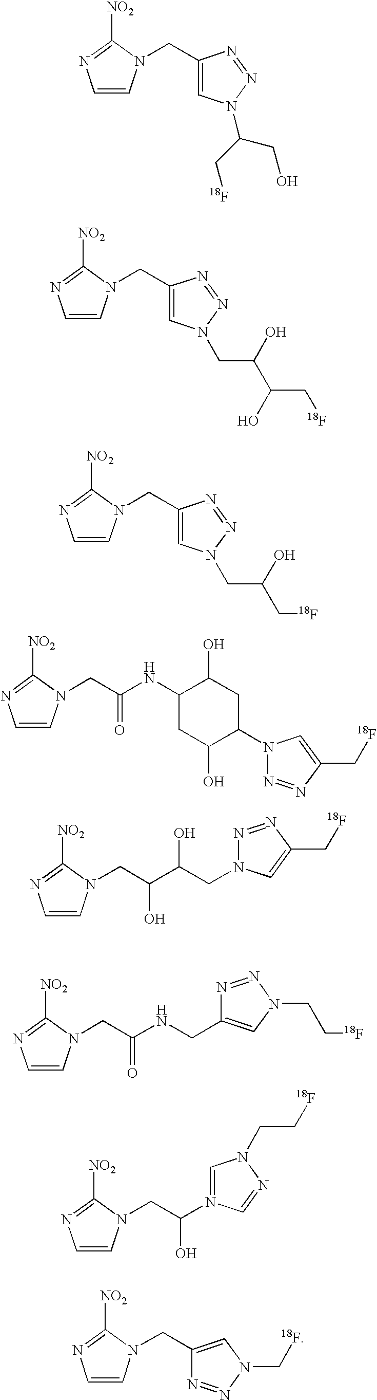
Priority Applications (2)
| Application Number | Priority Date | Filing Date | Title |
|---|---|---|---|
| US12/098,390 US7807394B2 (en) | 2007-04-05 | 2008-04-04 | Nitro-imidazole hypoxia imaging agents |
| US12/836,926 US7977361B2 (en) | 2007-04-05 | 2010-07-15 | Nitro-imidazole hypoxia imaging agent |
Applications Claiming Priority (3)
| Application Number | Priority Date | Filing Date | Title |
|---|---|---|---|
| US92193207P | 2007-04-05 | 2007-04-05 | |
| US96425407P | 2007-08-10 | 2007-08-10 | |
| US12/098,390 US7807394B2 (en) | 2007-04-05 | 2008-04-04 | Nitro-imidazole hypoxia imaging agents |
Related Child Applications (1)
| Application Number | Title | Priority Date | Filing Date |
|---|---|---|---|
| US12/836,926 Division US7977361B2 (en) | 2007-04-05 | 2010-07-15 | Nitro-imidazole hypoxia imaging agent |
Publications (2)
| Publication Number | Publication Date |
|---|---|
| US20090010846A1 US20090010846A1 (en) | 2009-01-08 |
| US7807394B2 true US7807394B2 (en) | 2010-10-05 |
Family
ID=39831553
Family Applications (2)
| Application Number | Title | Priority Date | Filing Date |
|---|---|---|---|
| US12/098,390 Expired - Fee Related US7807394B2 (en) | 2007-04-05 | 2008-04-04 | Nitro-imidazole hypoxia imaging agents |
| US12/836,926 Expired - Fee Related US7977361B2 (en) | 2007-04-05 | 2010-07-15 | Nitro-imidazole hypoxia imaging agent |
Family Applications After (1)
| Application Number | Title | Priority Date | Filing Date |
|---|---|---|---|
| US12/836,926 Expired - Fee Related US7977361B2 (en) | 2007-04-05 | 2010-07-15 | Nitro-imidazole hypoxia imaging agent |
Country Status (7)
| Country | Link |
|---|---|
| US (2) | US7807394B2 (en) |
| EP (1) | EP2132200B1 (en) |
| JP (1) | JP5347164B2 (en) |
| KR (1) | KR101523257B1 (en) |
| CN (1) | CN101652360B (en) |
| CA (1) | CA2683433C (en) |
| WO (1) | WO2008124651A2 (en) |
Cited By (3)
| Publication number | Priority date | Publication date | Assignee | Title |
|---|---|---|---|---|
| US20110002850A1 (en) * | 2007-04-05 | 2011-01-06 | Siemens Medical Solutions Usa, Inc. | Nitro-Imidazole Hypoxia Imaging Agent |
| EP2511006A2 (en) | 2011-04-13 | 2012-10-17 | Siemens Medical Solutions USA, Inc. | System, device and method for preparing tracers and transferring materials during radiosynthesis |
| US9023316B2 (en) | 2010-04-08 | 2015-05-05 | Siemens Medical Solutions Usa, Inc. | Synthesis of 18F-labeled tracers in hydrous organic solvents |
Families Citing this family (19)
| Publication number | Priority date | Publication date | Assignee | Title |
|---|---|---|---|---|
| CN101709060B (en) * | 2009-12-02 | 2013-05-01 | 北京师范大学 | F-triazole ring-polyethylene glycol-metronidazole compound and preparation method thereof |
| DK3323810T3 (en) | 2010-02-08 | 2022-03-28 | Lantheus Medical Imaging Inc | AUTOMATED REACTION SYSTEM, CASSETTE AND EQUIPMENT FOR SYNTHESIS OF IMAGE PRODUCTS |
| CN101921263A (en) * | 2010-04-16 | 2010-12-22 | 兰州大学 | 2-nitroimidazole derivatives and their use and preparation method |
| ES2763815T3 (en) | 2010-05-11 | 2020-06-01 | Lantheus Medical Imaging Inc | Compositions, methods and systems for the synthesis and use of imaging agents |
| CN108484449A (en) | 2011-09-09 | 2018-09-04 | 蓝瑟斯医学影像公司 | Composition, method and system for synthesizing and using developer |
| JPWO2013054788A1 (en) | 2011-10-14 | 2015-03-30 | 住友重機械工業株式会社 | Charged particle beam irradiation system and charged particle beam irradiation planning method |
| EP2793871A4 (en) * | 2011-12-23 | 2015-07-22 | Auckland Uniservices Ltd | Compounds and methods for selective imaging and/or ablation |
| WO2014062856A1 (en) | 2012-10-16 | 2014-04-24 | Halozyme, Inc. | Hypoxia and hyaluronan and markers thereof for diagnosis and monitoring of diseases and conditions and related methods |
| ES2580334T3 (en) | 2014-01-13 | 2016-08-23 | Yang, Guanghua | Compositions of dendrimers, methods of synthesis, and their uses |
| AU2015322871A1 (en) * | 2014-09-25 | 2017-03-23 | Nihon Medi-Physics Co., Ltd. | Renal imaging agent |
| US10695446B2 (en) | 2015-05-01 | 2020-06-30 | Vanderbilt University | Composition and method for detecting hypoxia |
| US10695450B2 (en) | 2016-07-26 | 2020-06-30 | Laboratoires Cyclopharma | Synthesis of a radioactive agent composition |
| CN106977501A (en) * | 2017-03-20 | 2017-07-25 | 华东师范大学 | A kind of hypoxemia activation prodrug based on the alkylol of 2 nitroimidazole 1 |
| FR3068695B1 (en) * | 2017-07-05 | 2019-08-16 | Centre National De La Recherche Scientifique | COMPOUNDS USEFUL AS HYPOXIA IMAGING AGENT |
| CN111372940A (en) * | 2017-09-19 | 2020-07-03 | 阿尔伯塔大学理事会 | Bioreduction-activated compounds, prodrugs thereof, radiopharmaceuticals, compositions and their use in multimodal therapeutic control of hypoxic diseases including cancer |
| CA3076256A1 (en) | 2017-09-19 | 2019-03-28 | The Governors Of The University Of Alberta | Markers, conjugates, compositions and methods for hypoxia imaging, mapping, and therapy |
| CN115210229A (en) | 2020-01-03 | 2022-10-18 | 博格有限责任公司 | Polycyclic amides as UBE2K modulators for the treatment of cancer |
| CN111233821B (en) * | 2020-01-20 | 2022-12-27 | 上海交通大学医学院附属仁济医院 | Novel antimycin derivative containing 3-hydroxybenzene acid group and preparation method and application thereof |
| CN114478489B (en) * | 2022-01-18 | 2023-04-28 | 中国科学院精密测量科学与技术创新研究院 | Preparation method and application of fluorine-19 modified nitroimidazole compound and derivative thereof |
Citations (3)
| Publication number | Priority date | Publication date | Assignee | Title |
|---|---|---|---|---|
| WO1996004249A1 (en) | 1994-08-05 | 1996-02-15 | Sri International | Fluorinated 2-nitroimidazole analogs for detecting hypoxic tumor cells |
| US5674693A (en) * | 1994-03-18 | 1997-10-07 | Natural Pharmacia International Inc. | Derivatives of 2-nitro-imidazoles as hypoxic cell markers |
| WO2001012575A1 (en) | 1999-08-11 | 2001-02-22 | Universite Catholique De Louvain | Methods for preparing perfluorinated [18f]-radiolabelled nitroimidazole derivatives for cellular hypoxia detection |
Family Cites Families (1)
| Publication number | Priority date | Publication date | Assignee | Title |
|---|---|---|---|---|
| CA2683433C (en) * | 2007-04-05 | 2016-10-11 | Siemens Medical Solutions Usa, Inc. | Nitro-imidazole hypoxia imaging agents |
-
2008
- 2008-04-04 CA CA2683433A patent/CA2683433C/en not_active Expired - Fee Related
- 2008-04-04 US US12/098,390 patent/US7807394B2/en not_active Expired - Fee Related
- 2008-04-04 CN CN200880011415.1A patent/CN101652360B/en not_active Expired - Fee Related
- 2008-04-04 EP EP08745183.7A patent/EP2132200B1/en not_active Not-in-force
- 2008-04-04 JP JP2010502341A patent/JP5347164B2/en not_active Expired - Fee Related
- 2008-04-04 KR KR1020097023183A patent/KR101523257B1/en not_active Expired - Fee Related
- 2008-04-04 WO PCT/US2008/059505 patent/WO2008124651A2/en not_active Ceased
-
2010
- 2010-07-15 US US12/836,926 patent/US7977361B2/en not_active Expired - Fee Related
Patent Citations (3)
| Publication number | Priority date | Publication date | Assignee | Title |
|---|---|---|---|---|
| US5674693A (en) * | 1994-03-18 | 1997-10-07 | Natural Pharmacia International Inc. | Derivatives of 2-nitro-imidazoles as hypoxic cell markers |
| WO1996004249A1 (en) | 1994-08-05 | 1996-02-15 | Sri International | Fluorinated 2-nitroimidazole analogs for detecting hypoxic tumor cells |
| WO2001012575A1 (en) | 1999-08-11 | 2001-02-22 | Universite Catholique De Louvain | Methods for preparing perfluorinated [18f]-radiolabelled nitroimidazole derivatives for cellular hypoxia detection |
Non-Patent Citations (31)
Cited By (6)
| Publication number | Priority date | Publication date | Assignee | Title |
|---|---|---|---|---|
| US20110002850A1 (en) * | 2007-04-05 | 2011-01-06 | Siemens Medical Solutions Usa, Inc. | Nitro-Imidazole Hypoxia Imaging Agent |
| US7977361B2 (en) * | 2007-04-05 | 2011-07-12 | Siemens Medical Solutions Usa, Inc. | Nitro-imidazole hypoxia imaging agent |
| US9023316B2 (en) | 2010-04-08 | 2015-05-05 | Siemens Medical Solutions Usa, Inc. | Synthesis of 18F-labeled tracers in hydrous organic solvents |
| US10639608B2 (en) | 2010-04-08 | 2020-05-05 | Siemens Medical Solutions Usa, Inc. | System, device and method for preparing tracers and transferring materials during radiosynthesis |
| EP2511006A2 (en) | 2011-04-13 | 2012-10-17 | Siemens Medical Solutions USA, Inc. | System, device and method for preparing tracers and transferring materials during radiosynthesis |
| EP2799091A2 (en) | 2011-04-13 | 2014-11-05 | Siemens Medical Solutions USA, Inc. | System, device and method for preparing tracers and transferring materials during radiosynthesis |
Also Published As
| Publication number | Publication date |
|---|---|
| US20110002850A1 (en) | 2011-01-06 |
| KR101523257B1 (en) | 2015-05-27 |
| WO2008124651A2 (en) | 2008-10-16 |
| KR20090130106A (en) | 2009-12-17 |
| CA2683433A1 (en) | 2008-10-16 |
| US7977361B2 (en) | 2011-07-12 |
| JP5347164B2 (en) | 2013-11-20 |
| JP2010523596A (en) | 2010-07-15 |
| US20090010846A1 (en) | 2009-01-08 |
| WO2008124651A3 (en) | 2009-04-02 |
| CA2683433C (en) | 2016-10-11 |
| EP2132200B1 (en) | 2016-03-23 |
| EP2132200A2 (en) | 2009-12-16 |
| CN101652360B (en) | 2014-06-25 |
| CN101652360A (en) | 2010-02-17 |
Similar Documents
| Publication | Publication Date | Title |
|---|---|---|
| US7807394B2 (en) | Nitro-imidazole hypoxia imaging agents | |
| US20060263293A1 (en) | Click chemistry method for synthesizing molecular imaging probes | |
| US8354092B2 (en) | Development of molecular imaging probes for carbonic anhydrase-IX using click chemistry | |
| US10358434B2 (en) | 2-(3-pyridinyl)-1H-benzimidazole derivative compound and medicament containing same | |
| Cao et al. | Synthesis of novel PEG-modified nitroimidazole derivatives via “hot-click” reaction and their biological evaluation as potential PET imaging agent for tumors | |
| US7432295B2 (en) | Preparation of compounds useful for the detection of hypoxia | |
| Liu et al. | Development of STING probes and visualization of STING in multiple tumor types | |
| Wong et al. | Synthesis and 18 F-radiolabeling of thymidine AMBF 3 conjugates | |
| Pisaneschi et al. | Synthesis of a new fluorine‐18 glycosylated ‘click’cyanoquinoline for the imaging of epidermal growth factor receptor | |
| US7230115B1 (en) | Preparation of compounds useful for the detection of hypoxia | |
| JP2019043882A (en) | Therapeutic effect predictors for alternative anticancer drugs | |
| Zeng et al. | Development of Preladenant-Based Radiotracers for Imaging A2AR in Tumors | |
| Sand | Synthetic studies toward [18F]-fluorination of solid-supported silyl ethers | |
| HK1232878B (en) | 2-(3-pyridinyl)-1h-benzimidazole derivative compound and medicine containing same | |
| HK1207852A1 (en) | Labelled quinoxaline derivatives as multimodal radiopharmaceuticals and their precursors |
Legal Events
| Date | Code | Title | Description |
|---|---|---|---|
| AS | Assignment |
Owner name: SIEMENS MEDICAL SOLUTIONS USA, INC., PENNSYLVANIA Free format text: ASSIGNMENT OF ASSIGNORS INTEREST;ASSIGNORS:KOLB, HARTMUTH C.;WALSH, JOSEPH C.;GANGADHARMATH, UMESH B.;AND OTHERS;REEL/FRAME:021559/0042;SIGNING DATES FROM 20080619 TO 20080731 Owner name: SIEMENS MEDICAL SOLUTIONS USA, INC., PENNSYLVANIA Free format text: ASSIGNMENT OF ASSIGNORS INTEREST;ASSIGNORS:KARIMI, FARHAD;DUCLOS, BRIAN A.;REEL/FRAME:021560/0226;SIGNING DATES FROM 20080605 TO 20080610 Owner name: SIEMENS MEDICAL SOLUTIONS USA, INC., PENNSYLVANIA Free format text: ASSIGNMENT OF ASSIGNORS INTEREST;ASSIGNORS:KOLB, HARTMUTH C.;WALSH, JOSEPH C.;GANGADHARMATH, UMESH B.;AND OTHERS;SIGNING DATES FROM 20080619 TO 20080731;REEL/FRAME:021559/0042 Owner name: SIEMENS MEDICAL SOLUTIONS USA, INC., PENNSYLVANIA Free format text: ASSIGNMENT OF ASSIGNORS INTEREST;ASSIGNORS:KARIMI, FARHAD;DUCLOS, BRIAN A.;SIGNING DATES FROM 20080605 TO 20080610;REEL/FRAME:021560/0226 |
|
| AS | Assignment |
Owner name: SIEMENS MOLECULAR IMAGING, INC., TENNESSEE Free format text: ASSIGNMENT OF ASSIGNORS INTEREST;ASSIGNOR:SIEMENS MEDICAL SOLUTIONS USA, INC.;REEL/FRAME:030038/0836 Effective date: 20130219 |
|
| FPAY | Fee payment |
Year of fee payment: 4 |
|
| FEPP | Fee payment procedure |
Free format text: MAINTENANCE FEE REMINDER MAILED (ORIGINAL EVENT CODE: REM.) |
|
| LAPS | Lapse for failure to pay maintenance fees |
Free format text: PATENT EXPIRED FOR FAILURE TO PAY MAINTENANCE FEES (ORIGINAL EVENT CODE: EXP.); ENTITY STATUS OF PATENT OWNER: LARGE ENTITY |
|
| STCH | Information on status: patent discontinuation |
Free format text: PATENT EXPIRED DUE TO NONPAYMENT OF MAINTENANCE FEES UNDER 37 CFR 1.362 |
|
| FP | Lapsed due to failure to pay maintenance fee |
Effective date: 20181005 |