US7780952B2 - Agents for magnetic imaging method - Google Patents
Agents for magnetic imaging method Download PDFInfo
- Publication number
- US7780952B2 US7780952B2 US10/516,781 US51678104A US7780952B2 US 7780952 B2 US7780952 B2 US 7780952B2 US 51678104 A US51678104 A US 51678104A US 7780952 B2 US7780952 B2 US 7780952B2
- Authority
- US
- United States
- Prior art keywords
- bis
- nutrient
- acid
- mri detectable
- amino
- Prior art date
- Legal status (The legal status is an assumption and is not a legal conclusion. Google has not performed a legal analysis and makes no representation as to the accuracy of the status listed.)
- Expired - Fee Related, expires
Links
- YOOSBMUYQHBONG-UHFFFAOYSA-N CC(C)(C)OC(=O)NCCCCN(CCC#N)C(=O)OC(C)(C)C Chemical compound CC(C)(C)OC(=O)NCCCCN(CCC#N)C(=O)OC(C)(C)C YOOSBMUYQHBONG-UHFFFAOYSA-N 0.000 description 2
- AGIUPQUTLBAVDK-UHFFFAOYSA-N CC(C)(C)OC(=O)NCCCCN(CCCN)C(=O)OC(C)(C)C Chemical compound CC(C)(C)OC(=O)NCCCCN(CCCN)C(=O)OC(C)(C)C AGIUPQUTLBAVDK-UHFFFAOYSA-N 0.000 description 2
- YCYZNZYGUXBMGZ-UHFFFAOYSA-N NCCCCNCCCNCC1=CC=C(NC(=O)CN2CCN(CC(=O)O)CCN(CC(=O)O)CCN(C(=O)O)CC2)C=C1 Chemical compound NCCCCNCCCNCC1=CC=C(NC(=O)CN2CCN(CC(=O)O)CCN(CC(=O)O)CCN(C(=O)O)CC2)C=C1 YCYZNZYGUXBMGZ-UHFFFAOYSA-N 0.000 description 2
- 0 C*CN(CCN(C[*-])CC(NCCNC(C(CCCNC(N)=N)N)=O)=O)CCN(C*C)CC(NCCNC(C(CCCNC(N)=N)N)=O)=O Chemical compound C*CN(CCN(C[*-])CC(NCCNC(C(CCCNC(N)=N)N)=O)=O)CCN(C*C)CC(NCCNC(C(CCCNC(N)=N)N)=O)=O 0.000 description 1
- GPCDVOIAKKFLOM-UHFFFAOYSA-N CC(C)(C)OC(=O)NCCCCN(CCCNCC1=CC=C(N)C=C1)C(=O)OC(C)(C)C Chemical compound CC(C)(C)OC(=O)NCCCCN(CCCNCC1=CC=C(N)C=C1)C(=O)OC(C)(C)C GPCDVOIAKKFLOM-UHFFFAOYSA-N 0.000 description 1
- FPZMZZULCALIGN-UHFFFAOYSA-N CC(C)(C)OC(=O)NCCCCN(CCCNCC1=CC=C([N+](=O)[O-])C=C1)C(=O)OC(C)(C)C Chemical compound CC(C)(C)OC(=O)NCCCCN(CCCNCC1=CC=C([N+](=O)[O-])C=C1)C(=O)OC(C)(C)C FPZMZZULCALIGN-UHFFFAOYSA-N 0.000 description 1
- QWVKHBSHZICENP-UHFFFAOYSA-A CC(O)CN1CCN(CC(=O)[O-])CCN(CC(=O)[O-])CCN(CC(=O)[O-])CC1.CNC(=O)CN(CCN(CCN(CC(=O)[O-])CC(=O)NC)CC(=O)[O-])CC(=O)[O-].O=C([O-])CN(CCN(CC(=O)[O-])CC(=O)[O-])CCN(CC(=O)[O-])CC(=O)[O-].O=C([O-])CN1CCN(CC(=O)[O-])CCN(CC(=O)[O-])CCN(CC(=O)[O-])CC1.[Gd+3].[Gd+3].[Gd+3].[Gd+3] Chemical compound CC(O)CN1CCN(CC(=O)[O-])CCN(CC(=O)[O-])CCN(CC(=O)[O-])CC1.CNC(=O)CN(CCN(CCN(CC(=O)[O-])CC(=O)NC)CC(=O)[O-])CC(=O)[O-].O=C([O-])CN(CCN(CC(=O)[O-])CC(=O)[O-])CCN(CC(=O)[O-])CC(=O)[O-].O=C([O-])CN1CCN(CC(=O)[O-])CCN(CC(=O)[O-])CCN(CC(=O)[O-])CC1.[Gd+3].[Gd+3].[Gd+3].[Gd+3] QWVKHBSHZICENP-UHFFFAOYSA-A 0.000 description 1
- JOBYWZOYPOEIAK-UHFFFAOYSA-N CCN(CC)CCN(CCN(CC)CC)C(C)CCC(=O)NC(C)CCCNC(=N)NCC1=C(C)C(C)=C(OC)C=C1C Chemical compound CCN(CC)CCN(CCN(CC)CC)C(C)CCC(=O)NC(C)CCCNC(=N)NCC1=C(C)C(C)=C(OC)C=C1C JOBYWZOYPOEIAK-UHFFFAOYSA-N 0.000 description 1
- CTAYDOOUJXWXRV-UHFFFAOYSA-N CCN1CCN(C)CCN(CC(=O)NC2=CC=C(CNCCCN(CCCCNC(=O)OC(C)(C)C)C(=O)OC(C)(C)C)C=C2)CCN(CC)CC1 Chemical compound CCN1CCN(C)CCN(CC(=O)NC2=CC=C(CNCCCN(CCCCNC(=O)OC(C)(C)C)C(=O)OC(C)(C)C)C=C2)CCN(CC)CC1 CTAYDOOUJXWXRV-UHFFFAOYSA-N 0.000 description 1
- SQFRTFPBISWYEO-UHFFFAOYSA-N CCOc1ccc(CC(CN(CCN(CC(=O)O)CC(=O)O)CC(=O)O)N(CC(=O)O)CC(=O)O)cc1.O=C(O)CN(CCN(CC(=O)O)CC(=O)O)CCN(CC(=O)O)C(COCc1ccccc1)C(=O)O Chemical compound CCOc1ccc(CC(CN(CCN(CC(=O)O)CC(=O)O)CC(=O)O)N(CC(=O)O)CC(=O)O)cc1.O=C(O)CN(CCN(CC(=O)O)CC(=O)O)CCN(CC(=O)O)C(COCc1ccccc1)C(=O)O SQFRTFPBISWYEO-UHFFFAOYSA-N 0.000 description 1
- WLCNZYVOUPOLHO-UHFFFAOYSA-N N#CCCNCCCCN Chemical compound N#CCCNCCCCN WLCNZYVOUPOLHO-UHFFFAOYSA-N 0.000 description 1
- JJQJRRFFUUEUJC-UHFFFAOYSA-K N=C(N)NCCCC(N)C(=O)NCCNC(=O)CN(CCN(CCN(CC(=O)[O-])CC(=O)NCCNC(=O)C(N)CCCNC(=N)N)CC(=O)[O-])CC(=O)[O-].[Gd+3] Chemical compound N=C(N)NCCCC(N)C(=O)NCCNC(=O)CN(CCN(CCN(CC(=O)[O-])CC(=O)NCCNC(=O)C(N)CCCNC(=N)N)CC(=O)[O-])CC(=O)[O-].[Gd+3] JJQJRRFFUUEUJC-UHFFFAOYSA-K 0.000 description 1
- FXXOVMKCGAFIJF-UHFFFAOYSA-I N=C(N)NCCCCNC(=O)CCC(C(=O)[O-])N(CCN(CC(=O)[O-])CC(=O)[O-])CCN(CC(=O)[O-])CC(=O)[O-].[Gd+3].[Na+].[Na+] Chemical compound N=C(N)NCCCCNC(=O)CCC(C(=O)[O-])N(CCN(CC(=O)[O-])CC(=O)[O-])CCN(CC(=O)[O-])CC(=O)[O-].[Gd+3].[Na+].[Na+] FXXOVMKCGAFIJF-UHFFFAOYSA-I 0.000 description 1
- YYDVVBUYAAWFKG-UHFFFAOYSA-K N=C(N)NCCCCNC(=O)CN(CCN(CCN(CC(=O)[O-])CC(=O)NCCCCNC(=N)N)CC(=O)[O-])CC(=O)[O-].[Gd+3] Chemical compound N=C(N)NCCCCNC(=O)CN(CCN(CCN(CC(=O)[O-])CC(=O)NCCCCNC(=N)N)CC(=O)[O-])CC(=O)[O-].[Gd+3] YYDVVBUYAAWFKG-UHFFFAOYSA-K 0.000 description 1
- DTHFNEIRNVFKHW-QDVCFFRGSA-H N=C(N)NCCC[C@H](NC(=O)CCC(C(=O)[O-])N(CCN(CC(=O)[O-])CC(=O)[O-])CCN(CC(=O)[O-])CC(=O)[O-])C(=O)[O-].[Gd+3].[Na+].[Na+] Chemical compound N=C(N)NCCC[C@H](NC(=O)CCC(C(=O)[O-])N(CCN(CC(=O)[O-])CC(=O)[O-])CCN(CC(=O)[O-])CC(=O)[O-])C(=O)[O-].[Gd+3].[Na+].[Na+] DTHFNEIRNVFKHW-QDVCFFRGSA-H 0.000 description 1
- ZJXVYNIMUBEBIP-QFPZOENFSA-H NC(=O)CCC(/N=C\OCCC(C(=O)[O-])N(CCN(CC(=O)[O-])CC(=O)[O-])CCN(CC(=O)[O-])CC(=O)[O-])C(=O)[O-].[Gd+3].[Na+].[Na+].[Na+] Chemical compound NC(=O)CCC(/N=C\OCCC(C(=O)[O-])N(CCN(CC(=O)[O-])CC(=O)[O-])CCN(CC(=O)[O-])CC(=O)[O-])C(=O)[O-].[Gd+3].[Na+].[Na+].[Na+] ZJXVYNIMUBEBIP-QFPZOENFSA-H 0.000 description 1
- DDWJKTVDRKOOJW-UHFFFAOYSA-I NC(=O)CCC(NC(=O)CN(CCN(CCN(CC(=O)[O-])CC(=O)NC(CCC(N)=O)C(=O)[O-])CC(=O)[O-])CC(=O)[O-])C(=O)[O-].[Gd+3].[Na+].[Na+] Chemical compound NC(=O)CCC(NC(=O)CN(CCN(CCN(CC(=O)[O-])CC(=O)NC(CCC(N)=O)C(=O)[O-])CC(=O)[O-])CC(=O)[O-])C(=O)[O-].[Gd+3].[Na+].[Na+] DDWJKTVDRKOOJW-UHFFFAOYSA-I 0.000 description 1
- WTUHDOGAWZJZHT-UHFFFAOYSA-K NCCCCNCCCNCC1=CC=C(NC(=O)CN2CCN(CC(=O)[O-])CCN(CC(=O)[O-])CCN(C(=O)[O-])CC2)C=C1.[Gd+3] Chemical compound NCCCCNCCCNCC1=CC=C(NC(=O)CN2CCN(CC(=O)[O-])CCN(CC(=O)[O-])CCN(C(=O)[O-])CC2)C=C1.[Gd+3] WTUHDOGAWZJZHT-UHFFFAOYSA-K 0.000 description 1
- GXBDGDUPYFLMRQ-UHFFFAOYSA-K NCCCNCCCCNC(=O)CN(CCN(CCN(CC(=O)[O-])CC(=O)NCCCCNCCCN)CC(=O)[O-])CC(=O)[O-].[Gd+3] Chemical compound NCCCNCCCCNC(=O)CN(CCN(CCN(CC(=O)[O-])CC(=O)NCCCCNCCCN)CC(=O)[O-])CC(=O)[O-].[Gd+3] GXBDGDUPYFLMRQ-UHFFFAOYSA-K 0.000 description 1
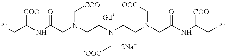
- YTPBRWOKRZYWLK-UHFFFAOYSA-I O=C([O-])CN(CCN(CC(=O)[O-])CC(=O)NC(CC1=CC=CC=C1)C(=O)[O-])CCN(CC(=O)[O-])CC(=O)NC(CC1=CC=CC=C1)C(=O)[O-].[Gd+3].[Na+].[Na+] Chemical compound O=C([O-])CN(CCN(CC(=O)[O-])CC(=O)NC(CC1=CC=CC=C1)C(=O)[O-])CCN(CC(=O)[O-])CC(=O)NC(CC1=CC=CC=C1)C(=O)[O-].[Gd+3].[Na+].[Na+] YTPBRWOKRZYWLK-UHFFFAOYSA-I 0.000 description 1
- YIEYCGULSPAWAH-APLPGFJPSA-K O=C([O-])CN(CCN(CC(=O)[O-])CC(=O)NC1C(O)O[C@@H](CO)[C@@H](O)[C@@H]1O)CCN(CC(=O)NC1C(O)O[C@H](CO)[C@H](O)[C@H]1O)C(=O)[O-].[Gd+3] Chemical compound O=C([O-])CN(CCN(CC(=O)[O-])CC(=O)NC1C(O)O[C@@H](CO)[C@@H](O)[C@@H]1O)CCN(CC(=O)NC1C(O)O[C@H](CO)[C@H](O)[C@H]1O)C(=O)[O-].[Gd+3] YIEYCGULSPAWAH-APLPGFJPSA-K 0.000 description 1
- NHPISLFZZDQLFB-UHFFFAOYSA-K [Gd+3].[NH3+]C(CCCCNC(=O)CN(CCN(CCN(CC(=O)[O-])CC(=O)NCCCCC([NH3+])C(=O)[O-])CC(=O)[O-])CC(=O)[O-])C(=O)[O-] Chemical compound [Gd+3].[NH3+]C(CCCCNC(=O)CN(CCN(CCN(CC(=O)[O-])CC(=O)NCCCCC([NH3+])C(=O)[O-])CC(=O)[O-])CC(=O)[O-])C(=O)[O-] NHPISLFZZDQLFB-UHFFFAOYSA-K 0.000 description 1
Images


Classifications
-
- C—CHEMISTRY; METALLURGY
- C07—ORGANIC CHEMISTRY
- C07C—ACYCLIC OR CARBOCYCLIC COMPOUNDS
- C07C237/00—Carboxylic acid amides, the carbon skeleton of the acid part being further substituted by amino groups
- C07C237/02—Carboxylic acid amides, the carbon skeleton of the acid part being further substituted by amino groups having the carbon atoms of the carboxamide groups bound to acyclic carbon atoms of the carbon skeleton
- C07C237/04—Carboxylic acid amides, the carbon skeleton of the acid part being further substituted by amino groups having the carbon atoms of the carboxamide groups bound to acyclic carbon atoms of the carbon skeleton the carbon skeleton being acyclic and saturated
- C07C237/12—Carboxylic acid amides, the carbon skeleton of the acid part being further substituted by amino groups having the carbon atoms of the carboxamide groups bound to acyclic carbon atoms of the carbon skeleton the carbon skeleton being acyclic and saturated having the nitrogen atom of at least one of the carboxamide groups bound to an acyclic carbon atom of a hydrocarbon radical substituted by carboxyl groups
-
- A—HUMAN NECESSITIES
- A61—MEDICAL OR VETERINARY SCIENCE; HYGIENE
- A61K—PREPARATIONS FOR MEDICAL, DENTAL OR TOILETRY PURPOSES
- A61K41/00—Medicinal preparations obtained by treating materials with wave energy or particle radiation ; Therapies using these preparations
- A61K41/009—Neutron capture therapy, e.g. using uranium or non-boron material
- A61K41/0095—Boron neutron capture therapy, i.e. BNCT, e.g. using boronated porphyrins
-
- A—HUMAN NECESSITIES
- A61—MEDICAL OR VETERINARY SCIENCE; HYGIENE
- A61K—PREPARATIONS FOR MEDICAL, DENTAL OR TOILETRY PURPOSES
- A61K49/00—Preparations for testing in vivo
- A61K49/06—Nuclear magnetic resonance [NMR] contrast preparations; Magnetic resonance imaging [MRI] contrast preparations
- A61K49/08—Nuclear magnetic resonance [NMR] contrast preparations; Magnetic resonance imaging [MRI] contrast preparations characterised by the carrier
- A61K49/085—Nuclear magnetic resonance [NMR] contrast preparations; Magnetic resonance imaging [MRI] contrast preparations characterised by the carrier conjugated systems
-
- C—CHEMISTRY; METALLURGY
- C07—ORGANIC CHEMISTRY
- C07C—ACYCLIC OR CARBOCYCLIC COMPOUNDS
- C07C237/00—Carboxylic acid amides, the carbon skeleton of the acid part being further substituted by amino groups
- C07C237/02—Carboxylic acid amides, the carbon skeleton of the acid part being further substituted by amino groups having the carbon atoms of the carboxamide groups bound to acyclic carbon atoms of the carbon skeleton
- C07C237/04—Carboxylic acid amides, the carbon skeleton of the acid part being further substituted by amino groups having the carbon atoms of the carboxamide groups bound to acyclic carbon atoms of the carbon skeleton the carbon skeleton being acyclic and saturated
- C07C237/10—Carboxylic acid amides, the carbon skeleton of the acid part being further substituted by amino groups having the carbon atoms of the carboxamide groups bound to acyclic carbon atoms of the carbon skeleton the carbon skeleton being acyclic and saturated having the nitrogen atom of at least one of the carboxamide groups bound to an acyclic carbon atom of a hydrocarbon radical substituted by nitrogen atoms not being part of nitro or nitroso groups
-
- C—CHEMISTRY; METALLURGY
- C07—ORGANIC CHEMISTRY
- C07C—ACYCLIC OR CARBOCYCLIC COMPOUNDS
- C07C271/00—Derivatives of carbamic acids, i.e. compounds containing any of the groups, the nitrogen atom not being part of nitro or nitroso groups
- C07C271/06—Esters of carbamic acids
- C07C271/08—Esters of carbamic acids having oxygen atoms of carbamate groups bound to acyclic carbon atoms
- C07C271/10—Esters of carbamic acids having oxygen atoms of carbamate groups bound to acyclic carbon atoms with the nitrogen atoms of the carbamate groups bound to hydrogen atoms or to acyclic carbon atoms
- C07C271/22—Esters of carbamic acids having oxygen atoms of carbamate groups bound to acyclic carbon atoms with the nitrogen atoms of the carbamate groups bound to hydrogen atoms or to acyclic carbon atoms to carbon atoms of hydrocarbon radicals substituted by carboxyl groups
-
- C—CHEMISTRY; METALLURGY
- C07—ORGANIC CHEMISTRY
- C07C—ACYCLIC OR CARBOCYCLIC COMPOUNDS
- C07C279/00—Derivatives of guanidine, i.e. compounds containing the group, the singly-bound nitrogen atoms not being part of nitro or nitroso groups
- C07C279/04—Derivatives of guanidine, i.e. compounds containing the group, the singly-bound nitrogen atoms not being part of nitro or nitroso groups having nitrogen atoms of guanidine groups bound to acyclic carbon atoms of a carbon skeleton
- C07C279/12—Derivatives of guanidine, i.e. compounds containing the group, the singly-bound nitrogen atoms not being part of nitro or nitroso groups having nitrogen atoms of guanidine groups bound to acyclic carbon atoms of a carbon skeleton being further substituted by nitrogen atoms not being part of nitro or nitroso groups
-
- C—CHEMISTRY; METALLURGY
- C07—ORGANIC CHEMISTRY
- C07C—ACYCLIC OR CARBOCYCLIC COMPOUNDS
- C07C279/00—Derivatives of guanidine, i.e. compounds containing the group, the singly-bound nitrogen atoms not being part of nitro or nitroso groups
- C07C279/04—Derivatives of guanidine, i.e. compounds containing the group, the singly-bound nitrogen atoms not being part of nitro or nitroso groups having nitrogen atoms of guanidine groups bound to acyclic carbon atoms of a carbon skeleton
- C07C279/14—Derivatives of guanidine, i.e. compounds containing the group, the singly-bound nitrogen atoms not being part of nitro or nitroso groups having nitrogen atoms of guanidine groups bound to acyclic carbon atoms of a carbon skeleton being further substituted by carboxyl groups
Definitions
- the present invention relates to new MRI detectable species suitable for use as contrast agents in a magnetic imaging method, and to the injectable compositions containing them for the magnetic resonance imaging (MRI) of living subjects and for the diagnosing of tumors.
- MRI magnetic resonance imaging
- the invention also refers to the injectable compositions of some of said new MRI detectable species for the therapy of tumors.
- the present invention also relates to the new intermediates in the preparation of the new MRI detectable species of the invention.
- Magnetic resonance imaging is a nuclear magnetic resonance technique that is used clinically to distinguish between different tissues or organs in the human or animal body through the spatial localization of water protons in the tissues or organs.
- the signal that is obtained by this technique and is then converted into an imaging depends in fact on the water proton concentrations and on the relaxation rates within the different types of tissues.
- contrast agents i.e. agents that influence the local relaxation behaviour of the observed nuclei in certain tissues or organs, because if MRI is performed without employing a contrast agent, differentiation of the tissue of interest from the surrounding tissues in the resulting image may be difficult.
- paramagnetic contrast agents involve materials which contain unpaired electrons.
- the unpaired electrons act as small magnets within the main magnetic field to increase the rate of longitudinal (T 1 ) and transverse (T 2 ) relaxation.
- paramagnetic contrast agents are used for their ability to decrease T 1 (positive contrast agents) and in use they enhance image intensity from the regions to which they distribute.
- Paramagnetic contrast agents typically comprise metal ions, such as for instance transition metal ions, which provide a source of unpaired electrons. Particularly preferred and therefore studied in more depth, resulted to be Gd 3+ (with 7 unpaired electrons) and Mn 2+ (with 5 unpaired electrons). Particular attention has been paid to the Gd 3+ ion as this ion shows a very high magnetic moment coupled to a relaxation rate relatively long at the magnetic fields of interest in the MR area (in the range of nanoseconds). However these metal ions are also generally highly toxic or at least poorly tolerated and need to be strongly coordinated with a ligand that occupies the major number of coordination sites.
- metal ions such as for instance transition metal ions
- the first contrast agent for MRI approved by the Regulatory authorities was GdDTPA (Magnevist®, by Schering AG), followed by GdDOTA (Dotarem®, by Guerbet SA), GdDTPA-BMA (Omniscan®, by Nycomed Imaging AS), and GdHPDO3A (ProHance®, by Bracco Imaging S.p.A.).
- GdDTPA Magneticnevist®, by Schering AG
- GdDOTA Dotarem®, by Guerbet SA
- GdDTPA-BMA Optamine-BMA
- GdHPDO3A ProHance®, by Bracco Imaging S.p.A.
- contrast agents share some similar pharmacological features as they all diffuse from plasma into the extracellular fluids and are excreted through the kidney via glomerular filtration. They are particularly useful for the diagnosis of hematoencephalic barrier lesions.
- Gd EOB-DTPA ovist®, by Shering AG
- Gd BOPTA MultiHance®, by Bracco Imaging S.p.A.
- Ferromagnetic materials which are employed for their ability to decrease T 2 . Ferromagnetic materials have high, positive magnetic susceptibilities and maintain their magnetism in the absence of an applied field. Ferromagnetic materials for use as MRI contrast agents are for instance described in WO 86/01112 and WO 85/043301.
- a third class of magnetic materials that can be used in MRI are those generally indicated as superparamagnetic materials. Analogously to paramagnetic materials, the superparamagnetic ones do not maintain their magnetism in the absence of an externally applied magnetic field. Superparamagnetic materials can have magnetic susceptibilities nearly as high as ferromagnetic materials and higher than the paramagnetic ones. As generally used, also superparamagnetic materials alter the MR image by decreasing T 2 and therefore result in a darkening of the tissues or fluids where they are present or accumulate versus the lighter background where they are not present.
- Iron oxides such as magnetite and gamma ferric oxide exhibit ferrromagnetism or superparamagnetism depending on the size of the crystals comprising the material, with larger crystals (typically with an average size larger than 0.3 ⁇ M) being ferromagnetic.
- the paramagnetic, ferromagnetic or superparamagnetic compounds containing the suitably selected targeting moiety bind to the relevant receptors on the surface of the tumor cells to be targeted and is possibly internalised.
- the number of receptors per cell is generally lower than the number of MRI detectable metal ions required to have a MR signal visible with the actual MRI technologies. To increase the contrast and make the signal more visible it is therefore necessary to increase the concentration of the contrast agent in the cell or on the surface of the cell.
- a compound capable to increase internalization of the receptor-binding compound or increasing the number of MRI detectable metal units linked to the targeting moiety (via dendrimers or multimers).
- particularly useful is the internalization route based on receptor mediated (fluid) endocytosis that allows the entrapment of a huge number of paramagnetic units.
- the optimum contrast agent should provide a contrast sufficient to clearly distinguish between normal, healthy cells and tumor cells using the available equipment and without creating any toxicity problem.
- the present invention provides new compounds suitable for use in the manufacture of contrast agents, said compounds containing an MRI detectable moiety and being functionalised in such a way to be easily internalised into the cells.
- the present invention provides for a MRI detectable species that is useful for internalising into tumor cells an amount of a MRI detectable metal that is distinguishably higher than the amount internalised in normal healthy cells.
- MRI detectable species that contain at least one MRI detectable moiety that is bound, either directly or through a spacer, to at least one molecule of a nutrient or pseudo-nutrient.
- the nutrient or pseudo-nutrient transporters or transporting systems that are present into the human body, will in fact recognize the nutrient or pseudo-nutrient molecule and will carry it, together with the at least one MRI detectable moiety that is linked thereto, into the cells where said MRI detectable moiety or moieties will thus concentrate to provide a more evident visualization of the cell.
- the present invention provides a pharmaceutical composition
- a pharmaceutical composition comprising an effective amount of a MRI detectable species of the invention together with at least one pharmaceutically acceptable carrier.
- said pharmaceutical composition will be in the form of an injectable composition.
- Said injectable composition can be employed for diagnostic purpose using the MRI technology to visualize tumor cells and, when in the new compounds the MRI detectable moiety is a chelated complex of a paramagnetic metal.
- the present invention also relates to a process for the preparation of the new compounds by conjugating the MRI detectable moiety D or a precursor thereof with the nutrient or pseudo-nutrient molecule, either directly or through a spacer, and to the new intermediates obtained therein.
- FIG. 1 reports the differential uptake of the compound of Example 1 by a human hepatoma cell line with respect to hepatocytes
- FIG. 2 reports the differential uptake of the compound of Example 3 by a human hepatoma cell line with respect to hepatocytes
- FIG. 3 summarises the differential uptake of the compound of Example 3 by various cancer cell lines.
- FIG. 4 reports the differential uptake of the compound of Example 8 by rat hepatocytes with respect to human hepatoma cells HTC.
- FIG. 5 reports the differential uptake of the compound of Example 8 in a panel of cancer cell lines with respect to healthy hepatocytes and 3T3 cells.
- FIG. 6 reports the differential uptake of the compound of Example 8 in HTC cells as function of the concentration of glutamine.
- a first object of the present invention is a MRI detectable species of following formula (I) D p -S n -N m (I)
- the MRI detectable species of formula (I) must contain at least one MRI detectable moiety D and at least one nutrient or pseudo-nutrient molecule N bound together.
- the number of nutrient or pseudo-nutrient molecules will be up to 5, preferably up to 4, more preferably up to 3 and even more preferably up to 2.
- said spacer will preferably contain a multiplicity of binding sites equal to the number of MRI detectable moieties D, i.e., the spacer S will provide the backbone on which several D moieties or a cluster of D moieties are bound.
- the MRI detectable species of formula (I) contains more than one MRI detectable moiety D
- p typically will be an integer of from 1 to 10, preferably of from 1 to 8, more preferably of from 1 to 6, and even more preferably of from 1 to 4, i.e., 1, 2, 3 or 4; n is typically 0 or an integer of from 1 to 5, preferably 0, or an integer of from 1 to 3, more preferably 0 or an integer of from 1 to 2, and even more preferably 0 or 1; and m is typically an integer of from 1 to 5, preferably of from 1 to 4, more preferably of from 1 to 3, and even more preferably 1 or 2.
- the MRI detectable species of the present invention can be represented as a characterizable compound of formula (I) as indicated above, there may be instances where the MRI detectable species according to the present invention can only be represented as a combination of moieties (D and N and optionally S) bonded or otherwise associated, e.g. conjugated, with each other. As used herein however the term “MRI detectable species of formula (I)” will include also these compositions of matter.
- D is any MRI detectable moiety, i.e. any moiety which affects local electromagnetic fields (i.e. any paramagnetic, superparamagnetic or ferromagnetic species), that contains at least one portion capable of being linked to at least one molecule of nutrient or pseudo-nutrient N, either directly or through a spacer S.
- any MRI detectable moiety i.e. any moiety which affects local electromagnetic fields (i.e. any paramagnetic, superparamagnetic or ferromagnetic species) that contains at least one portion capable of being linked to at least one molecule of nutrient or pseudo-nutrient N, either directly or through a spacer S.
- the MRI detectable moiety D therefore can be a coated ferromagnetic particle, a coated superparamagnetic particle or a chelated complex of a paramagnetic metal ion wherein the coating of the ferromagnetic or superparamagnetic particles or the chelator of the paramagnetic metal contain at least one site for a possible link to a spacer S or to a nutrient/pseudo-nutrient molecule N.
- suitably coated ferromagnetic or superparamagnetic particles are for instance those described in U.S. Pat. Nos. 4,770,183, 4,827,945, 5,707,877, 6,123,920, and 6,207,134 where the coating materials, i.e., polymers such as polysaccharides, carbohydrates, polypeptides, organosilanes, proteins, and the like, gelatin-aminodextran, or starch and polyalkylene oxides, can be functionalised to allow binding of the particle to the spacer or to the nutrient/pseudo-nutrient molecule.
- the coating materials i.e., polymers such as polysaccharides, carbohydrates, polypeptides, organosilanes, proteins, and the like, gelatin-aminodextran, or starch and polyalkylene oxides
- the MRI detectable moiety D is a paramagnetic metal ion complexed with a chelating ligand L.
- Preferred paramagnetic metal ions include ions of transition and lanthanide metals (i.e. metals having atomic number of 21 to 29, 42, 43, 44, or 57 to 71).
- transition and lanthanide metals i.e. metals having atomic number of 21 to 29, 42, 43, 44, or 57 to 71.
- ions of Mn, Fe, Co, Ni, Eu, Gd, Dy, Tm, and Yb are preferred, with those of Mn, Fe, Eu, Gd, and Dy being more preferred and Gd being the most preferred.
- chelator or “chelating ligand” is intended to refer to a compound containing donor atoms that can combine by coordinative bonding with a metal atom to form a cyclic structure (coordination cage) called “chelation complex” or “chelate”.
- Suitable chelating ligands L that are or can be functionalised in such a way to allow binding of the paramagnetic chelation complex D to the spacer or nutrient/pseudo-nutrient molecule as in formula (I) above, include the residue of a polyaminopolycarboxylic acid, either linear or cyclic, in racemic or optically active form, such as ethylenediaminotetracetic acid (EDTA), diethylenetriaminopentaacetic acid (DTPA), N-[2-[bis(carboxymethyl)amino]-3-(4-ethoxyphenyl)propyl]-N-[2-[bis(carboxymethyl)amino]ethyl]-L-glycine (EOB-DTPA), N,N-bis[2-[bis(carboxymethyl)amino]ethyl]-L-glutamic acid (DTPA-GLU), N,N-Bis[2-[bis(carboxymethyl)amino]e
- Suitable chelating ligands L as well as the processes for their preparation are described for instance in the following patents: GB-A-2,137,612, EP-A-230,893, EP-A-255,471, EP-A-299,795, EP-A-325,762, EP-A-565,930, EP-A-594,734, U.S. Pat. No.
- Preferred chelating ligands L are linear and macrocyclic polyaminopolycarboxylic acids, in racemic or optically active form.
- ethylenediaminotetracetic acid EDTA
- diethylenetriaminopentaacetic acid DTPA
- N-[2-[bis(carboxymethyl)amino]-3-(4-ethoxyphenyl)propyl]-N-[2-[bis(carboxymethyl)amino]ethyl]-L-glycine EOB-DTPA
- N,N-bis[2-[bis(carboxymethyl)amino]ethyl]-L-glutamic acid DTPA-GLU
- N,N-Bis[2-[bis(carboxymethyl)amino]ethyl]-L- ⁇ -glutamyl-L-glutamine, N,N-bis[2-[bis(carboxymethyl)amino]ethyl]-L-lysine DPTA-LYS
- N,N-bis[2-(carboxymethyl[(methylcarbamoyl)methyl]amino)ethyl]glycine DTPA
- ethylenediaminotetracetic acid EDTA
- diethylenetriaminopentaacetic acid DTPA
- 4-carboxy-5,8,11-tris(carboxymethyl)-1-phenyl-2-oxa-5,8,11-triazatridecan-13-oic acid BOPTA
- DO TA 1,4,7,10-tetraazacyclododecan-1,4,7,10-tetraacetic acid
- DO3A 1,4,7,10-tetraazacyclododecan-1,4,7-triacetic acid
- HPDO3A 10-(2-hydroxypropyl)-1,4,7,10-tetraazacyclododecan-1,4,7-triacetic acid
- HPDO3A ( ⁇ , ⁇ ′, ⁇ ′′, ⁇ ′′′)tetramethyl-1,4,7,10-tetraazacyclododecan-1,4,7,10-tetraacetic acid (DOTMA), as well as the analogs thereof where the methyl groups are replaced by higher alky
- the term “nutrient” refers to any substance or compound that is essential for sustaining the cell life in the organism concerned, while the term “pseudo-nutrient” refers to any substance or compound that is nonessential during health but is required in the diet in certain pathophysiologic states because cell utilisation exceeds the capacity for endogenous biosynthesis or to any substance or compound that is capable of being utilised by the cells in any of their vital functions or as an assimilable source of nutrients.
- nutrients are typically monosaccharides and the essential amino acids.
- the term “monosaccharides” includes glucose, which is the most preferred one, fructose and other hexoses.
- the term “essential amino-acid” generally refers to the L-isomers of the ⁇ -amino acids which are found in nature.
- Typical examples of “pseudo-nutrients” are the polyamines, the derivatives of the essential amino acids and the non-essential amino-acids.
- polyamines are e.g. putrescine, spermidine, and spermine, that are present in all the cells and play an important role in several essential cell functions through their interaction with DNA, RNA, proteins, and lipids.
- putrescine e.g. putrescine, spermidine, and spermine
- spermidine e.g. putrescine, spermidine, and spermine
- the uptake of these compounds by the rapidly growing cells as well as by the tumor cells is known to be very high.
- a suitable example of essential amino-acid derivative suitable as pseudo-nutrients is e.g. agmatine, the compound obtainable by decarboxylation of arginine, that can be used by the cell as a nitrogen source and is known to be internalised by the cells through the same transport system used for polyamines.
- Non essential amino-acids are e.g. the D-isomers of the natural essential amino-acids, any mixture of the L- and D-isomers, as well as synthetic ⁇ -amino acids which are not found in nature.
- An example of molecule suitable as pseudo-nutrient is glutamine that is not an essential amino-acid but becomes a conditionally essential amino acid in the host with cancer. Glutamine in fact is known to be the main source of nitrogen for tumor cells that compete with the host for circulating glutamine.
- nutrient or pseudo-nutrient molecule may however be employed as the nutrient or pseudo-nutrient molecule. More particularly any substance or compound that is recognized by at least one transporting system of the living organism that receives the MRI detectable species (I) and that is internalised by the tumor cells in a higher amount with respect to the normal healthy cells, can suitably be employed.
- Examples of known transporting systems are for instance the ASC system that is expressed by different types of cells, that typically transports neutral apolar or neutral polar amino-acids, such as alanine, leucine, valine, methionine, serine, cysteine, glutamine, threonine, asparagine; the N system that specifically transports amino-acids that contains nitrogen in the side chain, such as glutamine, asparagine and histidine; the B 0,+ system that transports also cationic amino-acids and is expressed only in certain organs, i.e.
- neutral apolar or neutral polar amino-acids such as alanine, leucine, valine, methionine, serine, cysteine, glutamine, threonine, asparagine
- the N system that specifically transports amino-acids that contains nitrogen in the side chain, such as glutamine, asparagine and histidine
- the B 0,+ system that transports also cationic amino-acids and
- the Y + system that is ubiquitous with the only exception of epatocytes and mediates the uptake of cationic amino-acids as well as that of glutamine; the L system for the transport of neutral apolar amino-acids, such as leucine, isoleucine, valine, phenylalanine, tyrosine, tryptophane, methionine, and of some neutral polar amino-acids, such as glutamine, serine and threonine; the T system that preferentially transports neutral aromatic amino-acids; and the various families of glucose transporters, GLUT1, GLUT2, GLUT3, GLUT4, and GLUT5, which are expressed in the various tissues and are responsible for the cellular uptake of glucose and the other hexoses.
- each N can be independently selected among the nutrient and pseudo-nutrient molecules as indicated above. In a preferred embodiment however when more than one molecule N is linked to the MRI detectable moiety D, all the N molecules are of the same compound or substance.
- saccharides and essential amino acids are the preferred molecules, wherein the most preferred ones are glucose, neutral amino acids such as alanine and phenylalanine, and cationic amino acids such as lysine and arginine.
- polyamines such as putrescine and spermidine
- non essential amino-acids such as glutamine
- essential amino acid derivatives such as agmatine
- Each of the molecules of nutrient or pseudo-nutrient N can be linked to the MRI detectable moiety D either directly or through a spacer S.
- the MRI detectable moiety D When the MRI detectable moiety D is linked to a nutrient or pseudo-nutrient N through a direct bond, said bond usually involves interaction between functional groups located on the nutrient molecule N and on the MRI detectable moiety, i.e. on the coating of the ferromagnetic or superparamagnetic particle or—in the preferred embodiment of the present invention according to which D is a paramagnetic chelated complex—on the chelating ligand L.
- functional groups located on the nutrient molecule N and on the MRI detectable moiety i.e. on the coating of the ferromagnetic or superparamagnetic particle or—in the preferred embodiment of the present invention according to which D is a paramagnetic chelated complex—on the chelating ligand L.
- non limitative examples of chemically reactive functional groups which may be employed to this purpose include amino, hydroxy, thiol, carboxy, carbonyl and the like groups.
- the chelating ligand L may be attached directly to the nutrient or pseudo-nutrient molecule N via one of the metal coordinating groups of the chelant which may form an ester, amide, thioester, or thioamide bond with an amine, thiol, or hydroxy group present on the N molecule.
- the ligand L will contain free carboxyl groups, and a direct covalent bond with the nutrient or pseudo-nutrient molecule N can be obtained through formation of an ester or, preferably, an amide bond with respectively a hydroxy or amino group possibly present in the N molecule.
- the N molecule and the chelating ligand L may be directly linked via a functionality attached to the chelant L backbone, and in such a case it is also possible to devise a ligand L bearing as a substituent in its backbone e.g. the residue of an amino-acid or amino-acid derivative, a polyamine, or a glucose molecule N.
- these two elements can also be connected through a homo- or hetero-bifunctional linker, i.e. a spacer S.
- said spacer S will contain at least two specific reactive moieties separated by a spacing arm, wherein one of the reactive moieties will provide for a covalent bonding with the MRI detectable moiety D and the other with the nutrient or pseudo-nutrient molecule N.
- the spacing arm may typically consist of an alkylidene, alkenylidene, alkynylidene, cycloalkylidene, arylidene, or aralkylidene radical that can be substituted and be interrupted by heteroatoms such as oxygen, nitrogen, and sulphur.
- said spacer arm consists of an aliphatic, straight or branched chain, that effectively separates the reactive moieties of the spacer so that ideally the spatial configuration of the molecule of nutrient or pseudo-nutrient N is not influenced by the presence of the MRI detectable moiety D and the molecule of nutrient or pseudo-nutrient N is thus more easily recognized by the transporter.
- Said chain may be interrupted by groups such as, —O—, —S—, —CO—, —NR—, —CS— and the like groups or by aromatic rings such as phenylene radicals, and may bear substituents such as —OR, —SR, —NRR 1 , —COOR, —CONRR 1 , and the like substituents, wherein R and R 1 , each independently, may be a hydrogen atom or an organic group.
- the reactive moieties in said bifunctional spacer that may be the same or, preferably, different, need to be capable of reaction with the functional groups present in the MRI detectable moiety D and in the nutrient or pseudo-nutrient molecule N, i.e. need to be able to react with carboxyl, amino, hydroxyl, sulphydryl, carbonyl, and the like groups.
- Examples of functional groups capable of reaction with carboxylic groups include diazo compounds such as diazoacetate esters and diazoacetamides, which-react with high specificity to generate ester groups.
- Carboxylic acid modifying reagents such as carbodiimides, e.g. 1-cyclohexyl-3-(2-morpholinyl-4-ethyl)carbodiimide (CMC) and 1-ethyl-3-(3-dimethylaminopropyl)carbodiimide (EDC), may also be usefully employed.
- Other useful carboxylic acid modifying reagents include isoxazolium derivatives, chloroformates, and N-carbalkoxydihydroquinolines.
- Examples of reactive moieties capable of reaction with sulphydryl groups include ⁇ -haloacetyl compounds and maleimide derivatives.
- Examples of reactive moieties capable of reaction with amino groups include alkylating and acylating agents.
- Representative of the alkylating agents are ⁇ -haloacetyl compounds, maleimide derivatives, reactive aryl halides and alkyl halides, ⁇ -haloalkyl ethers, aldehydes and ketones capable of Schiff's base formation with amino groups (the adducts formed usually being stabilised through reduction to give a stable amine), epoxide derivatives, such as epichlorohydrin and bisoxiranes.
- Examples of acylating agents include isocyanates and isothiocyanates, acid anhydrides, acid halides, active esters, and those useful reagents for amide bond formation widely known and conventionally used for peptide syntheses.
- Said spacer may also contain more than two functional groups.
- said spacer may contain a multiplicity of possible binding sites or be a molecular aggregate with a multiplicity of built-in or pendant groups which bind covalently or non-covalently (.g. coordinatively) with the MRI detectable moieties D and the nutrient or pseudo-nutrient molecules N in such a way to anchor said moieties thereto with a strength and for a time sufficient to bring the MRI detectable species (I) into the cells.
- the nature of the spacer S may have a critical bearing on the stability of the end MRI detectable species and on the capability for the transporting systems to recognize the nutrient or pseudo-nutrient molecule. As indicated in fact it should bind the MRI detectable moiety D to the recognized nutrient or pseudo-nutrient molecule N for an adequate period of time that would allow the MRI detectable moiety D to be internalised by the tumor cells in an amount sufficient to give a clearly distinguishable contrast imaging. It should also bind the MRI detectable moiety D to the nutrient or pseudo-nutrient molecule in a way that would ensure an appropriate spatial conformation that allows the nutrient or pseudo-nutrient molecule to be easily recognized by the transport system.
- the MRI detectable moiety D is a paramagnetic metal chelated complex
- said optional spacer S should also ensure that water molecules may have access to the chelated paramagnetic ion (as the entity of the signal will depend on the rate of exchange of the water molecule of the coordination cage with the water bulk).
- each of them can be independently selected as indicated above. According to a preferred embodiment, however, in such a case all the n molecules of spacer S are identical.
- reaction conditions used for obtaining the MRI detectable species (I) will vary depending on the particular type of reactive moieties employed but are analogous to those known in the literature for similar general reactions and can be easily devised by any skilled technician.
- the spacer In general for the preparation of the MRI detectable species (I), it is possible to conjugate first the spacer, if any, with the nutrient or pseudo-nutrient molecules N and then conjugate the obtained intermediate product with the MRI detectable moiety or moieties D; or alternatively, it is also possible to conjugate first the MRI detectable moiety or moieties D with the spacer or spacers, if any, and then conjugate the obtained intermediate with the nutrient or pseudo-nutrient molecule(s) N.
- D is a paramagnetic metal chelated complex
- the process for the preparation of these compounds will comprise conjugating one or more chelating ligands L to one or more nutrient or pseudo-nutrient molecules N either directly or through one or more spacers S, by any suitable sequence of steps, to get a suitable precursor of the desired end compound (I), said precursor having following general formula (II) L p -S n -N m (II)
- a further preferred embodiment of the present invention are the corresponding complexes compounds, according to general formula (I), of the above indicated list of compounds.
- ions of Mn, Fe, Co, Ni, Eu, Gd, Dy, Tm, and Yb are preferred, with those of Mn, Fe, Eu, Gd, and Dy and Gd being more preferred and Gd being the most preferred.
- the direct incorporation is the preferred one to obtain the compounds of formula (I) wherein D is a paramagnetic metal chelated complex starting from the intermediates of formula (II).
- the MRI detectable species (I) wherein D is a paramagnetic chelated complex can be easily obtained by merely exposing or mixing an aqueous solution of the functionalized chelators (II) with a paramagnetic metal salt in an aqueous solution having a pH comprised between about 4 and about 9, and preferably comprised between about 5 and about 8.
- the salt can be any salt but preferably a water soluble salt of the paramagnetic metal, such as a halide. Said salts are preferably selected so as not to interfere with the binding of the metal ion with the functionalised chelator (II).
- the funtionalised chelator (II) is preferably in aqueous solution at a pH of between about 5 and about 8. It can be mixed with buffer salts such as citrate, acetate, phosphate and borate, to produce the optimum pH.
- Preferred paramagnetic metal ions include ions of transition and lanthanide metals (i.e. metals having atomic number of 21 to 29, 42, 43, 44, or 57 to 71). In particular ions of Mn, Fe, Co, Ni, Eu, Gd, Dy, Tm, and Yb are preferred, with those of Mn, Fe, Eu, Gd, and Dy being more preferred and Gd being the most preferred.
- the MRI detectable species (I) wherein D is a paramagnetic chelated complex should have a zero residual charge on the coordination cage in order not to alter the residual charges possibly present on the nutrient or pseudo-nutrient moiety of the end species (I).
- Preferred cations of inorganic bases suitable for salifying the paramagnetic chelated complexes comprise the ions of alkali metals or alkaline-earth metals such as potassium, sodium, magnesium, calcium, and the like metals, including any mixed salt.
- Preferred cations of organic bases suitable for this purpose comprise those obtained by protonation of primary, secondary and tertiary amines, such as ethanolamine, diethanolamine, morpholine, glucamine, N-methylglucamine, N,N-dimethylglucamine, basic amino-acids such as lysine, arginine, and ornithine, and the like organic bases.
- the MRI detectable species (I) wherein D is a paramagnetic chelation complex should contain at least one molecule of water in the coordination cage of the chelation complex so as to guarantee a rlp higher than 1 s ⁇ 1 mM ⁇ 1 .
- Higher relaxivities (such as higher than 4 s ⁇ 1 mM ⁇ 1 , or higher than 8 s ⁇ 1 mM ⁇ 1 , or higher than 10 s ⁇ 1 mM ⁇ 1 or higher than 15 s ⁇ 1 mM ⁇ 1 ) can be advantageous but not strictly necessary because of the high amount of internalized MRI detectable moiety D.
- the MRI detectable species (I) according to the present invention may be administered to patients for imaging in an amount sufficient to give the desired contrast with the particular technique used in the MRI. Generally, dosages of from about 0.001 to about 5.0 mmoles of MRI detectable species (I) per kg of body weight are sufficient to obtain the desired contrast. For most MRI applications preferred dosages of imaging metal compound will be in the range of from 0.01 to 2.5 mmoles per kg of body weight.
- the MRI detectable species (I) of the present invention can be employed for the manufacture of a contrast medium for use in a method of diagnosis by MRI involving administering said contrast medium to a human or animal being and generating an image of at least part of said human or animal being.
- the MRI detectable species (I) can be employed for the manufacture of a contrast medium for use in a method of diagnosing tumors, said method involving administering said contrast medium to a human or animal being and detecting the major uptake of said detectable species by tumor cells with respect to normal cells.
- the MRI detectable species (I) of the present invention may be formulated with conventional pharmaceutical aids, such as emulsifiers, stabilisers, anti-oxidant agents, osmolality adjusting agents, buffers, pH adjusting agents, and the like agents, and may be in a form suitable for parenteral administration, e.g. for infusion or injection.
- conventional pharmaceutical aids such as emulsifiers, stabilisers, anti-oxidant agents, osmolality adjusting agents, buffers, pH adjusting agents, and the like agents.
- the MRI detectable species (I) according to the present invention may be in conventional administration forms such as solutions, suspensions, or dispersions in physiologically acceptable carriers media, such as water for injection.
- Parenterally administrable forms should be sterile and free from physiologically unacceptable agents, and have low osmolality to minimize irritation and other adverse effects upon administration.
- These parenterally administrable solutions can be prepared as customarily done with injectable solutions. They may contain additives, such as anti-oxidants, stabilizers, buffers, etc., which are compatible with the chemical structure of the MRI detectable species (I) and which will not interfere with the manufacture, storage and use thereof.
- Said pharmaceutical compositions may also contain suitable agents that may increase the amount of MRI detectable moiety internalised.
- suitable agents that may increase the amount of MRI detectable moiety internalised.
- Said “internalization adjuvant” may be administered simultaneously with the MRI detectable species (I) and in such a case the formulation may contain both ingredients. Alternatively it can be administered in advance with respect to the MRI detectable species (I) and in such a case two separate formulations should be provided that may be presented in the form of a kit.
- a mixture of agmatine free base (3.27 g; 0.02531 mol) and DMSO (70 ml) is stirred at 50° C. for two hours.
- a solution of the compound obtained in step a) above (4.20 g, 0.01175 mol) in dimethylsulfoxide (DMSO) (15 ml) is added portionwise over a period of two hours and the mixture is allowed to react at 50° C. for 4 hours and then at room temperature for additional 20 hours. At the end of the reaction time the solution is clear but a lower yellow phase of a jelly consistency is present.
- the DMSO phase is recovered by decantation and acetone is added thereto until precipitation of a solid product is complete.
- the lower yellow phase is dissolved in a small amount of water and acetone is added thereto to get an additional crop of solid precipitate.
- the two crops are combined and washed several times with fresh acetone, left under stirring overnight.
- aqueous solution of the chelating ligand obtained in step b) above (7.0 g; 0.0122 mol) is brought to pH 6.5 by the addition of NaOH and an equimolar amount of GdCl 3 (3.22 g; 0.0122 mol) is then slowly stirred in at room temperature. During the addition the pH of the reaction mixture is monitored and adjusted to 6.5 with NaOH. Once the addition of GdCl 3 is over, the solution is brought to pH 8.5 and filtered on a 22 ⁇ m syringe filter. A small amount of the chelating ligand obtained in step b) above is then added to the filtrate and the pH is brought to 7. Removal of the solvent under vacuum yields the compound of the title as a white solid.
- the gadolinium complex of the chelating ligand of step b) above has been prepared by following the same general procedure described in step c) of Example 1.
- the gadolinium complex of the chelating ligand of step b) above has been prepared by following the same general procedure described in step c) of Example 1.
- N-tertbutoxycarbonyl-L-lysine (0.950 g; 0.00386 mol) and DMSO (10 ml) are charged into a 100-ml reaction flask and stirred at 50° C. until complete solution.
- a solution of diethylenetriaminopentaacetic acid bis-anhydride obtained as described in step a) of Example 1 (0.690 g; 0.00193 mol) in DMSO (5 ml) is slowly added thereto over a period of about one hour. After about 24 hours methanolic KOH is added to bring the pH of the reaction mixture to 8 and then acetone is added to precipitate the product which is then washed several times with fresh acetone. Upon removal of the solvent the obtained solid is dried at 40° C.
- the gadolinium complex of the chelating ligand of step b) above has been prepared by following the same general procedure described in step c) of Example 1.
- a solution of phenyalanine (0.66 g; 0.002 mole) in DMSO (10 ml) is loaded into a 100-ml reaction flask and the mixture is heated to 50° C. under stirring.
- a solution of diethylenetriaminopentaacetic acid bis-anhydride obtained as described in step a) of Example 1 (0.715 g; 0.002 mol) in DMSO (6 ml) is slowly added thereto over a period of about two hour.
- the reaction mixture is allowed to react at 50° C. for 4 hours and at room temperature for additional 20 hours. At the end of this time the solution is clear but a lower yellow phase of a jelly consistency is present.
- the DMSO phase is recovered by decantation and acetone is added thereto until precipitation of a solid product is complete.
- the lower yellow phase is dissolved in a small amount of water and acetone is added thereto to get an additional crop of solid precipitate.
- the two crops are combined and washed several times under stirring with fresh acetone: The surnatant is then removed and the solid is taken up with water and washed again with fresh acetone.
- the gadolinium complex of the chelating ligand of step b) above has been prepared by following the same general procedure described in step c) of Example 1.
- a solution of diethylenetriaminopentaacetic acid bis-anhydride obtained as described in step a) of Example 1 (0.357 g; 0.001 mol) in DMSO (5 ml) is added over a period of two hours to a solution of N-(3-aminopropyl)-1,4-butanediamine (spermidine, 1.45 g; 0.01 mol) in DMSO (20 ml) stirred at 50° C. in a 100-ml reaction flask. The reaction mixture is allowed to react at 50° C. for 4 hours and then at room temperature for additional 20 hours. At the end of this time the solution is clear but a lower yellow phase of a jelly consistency is present.
- DMSO N-(3-aminopropyl)-1,4-butanediamine
- the DMSO phase is recovered by decantation and acetone is added thereto until precipitation of a solid product is complete.
- the lower yellow phase is dissolved in a small amount of water and acetone is added thereto to get an additional crop of solid precipitate.
- the two crops are combined and washed several times under stirring with fresh acetone.
- the surnatant is then removed and the solid is taken up with water and washed again with fresh acetone.
- the gadolinium complex of the chelating ligand of step b) above has been prepared by following the same general procedure described in step c) of Example 1.
- 2-amino-5-guanidino-valeric acid (arginine, 0.871 g; 0.005 mol) is dissolved in an aqueous solution at pH 9 and a strong excess of benzyl chloroformate is then slowly added thereto.
- the thus obtained corresponding tri-carbobenzyloxy derivative is isolated, dissolved in methylene chloride and added with di-cyclohexylcarbodiimide.
- the obtained solution is then slowly added to a solution of ethylenediamine (1.5 g; 0.025 mol) in methylene chloride.
- the 2-aminoethylamide of the tri-carbobenzyloxy protected arginine is recovered, dissolved in DMSO and a solution of diethylentriaminopentaneacetic acid bis-anhydride (1.78 g; 0.005 mol) in DMSO is then added to the obtained solution heated to 50° C.
- the reaction mixture is stirred at this temperature for 4 hours and then cooled to room temperature.
- the pH is adjusted to 8 by the addition of methanolic KOH and the raw product that precipitates is washed with acetone under stirring overnight.
- the solid is then recovered, dissolved in methanol and the protecting carbobenzyloxy groups are removed by catalytic hydrogenation over Pd/C 5%. Any residual carbamic acid that may possibly be present is then removed by acidification with HCl yielding the desired compound.
- the gadolinium complex of the chelating ligand of step b) above has been prepared by following the same general procedure described in step c) of Example 1.
- Trifluoroacetic acid (12.4 mL; 162.3 mmol) was added to a solution of the compound obtained in the step a) (5.03 g; 5.4 mmol) in CH 2 Cl 2 (100 mL) and stirred at room temperature for 48 h. The solvent was evaporated and the residue was dissolved in trifluoroacetic acid (10 mL). After 48 h at room temperature the acid was evaporated and the crude was treated with Et 2 O, then filtered. The solid was suspended twice in Et 2 O and filtered to give a white solid (3.76 g, 4.1 mmol). Yield 75%.
- N-hydroxysuccinimide (1.41 g; 12.29 mmol) was added to a solution of N,N-bis[2-[bis(1,1-dimethylethoxy)-2-oxoethyl]amino]ethyl]-L-glutamic acid 1-(1,1-dimethylethyl) ester (7.64 g; 10.24 mmol) in CH 2 Cl 2 (100 mL).
- the mixture was cooled at 0° C. and a solution of EDCI (2.36 g; 12.29 mmol) in CH 2 Cl 2 (45 mL) was added dropwise. The mixture was stirred at room temperature, monitoring the reaction by TLC.
- step a) The compound obtained ad indicated in step a) (1.48 g; 1.72 mmol) was dissolved in CH 2 Cl 2 (15 mL) and TFA (3.3 mL; 43 mmol) was added to the solution. The mixture was stirred at room temperature. After 4 days the solvent was removed under reduce pressure to give 1.56 g of crude product. The raw materials was dissolved in pure TFA (5 mL) and the mixture was stirred at room temperature overnight. TFA was removed under reduced pressure and the residue was crystallized with Et 2 O and filtered to give a white crystalline solid. Because of the presence of Et 2 O and TFA in the final product, the solid was dissolved in water again and evaporated under reduce pressure for three times. The final product was obtained as white crystalline solid (0.77 g; 1.33 mmol). Yield: 97%.
- the compound as obtained in the previous step b) was dissolved in 10 mL of water and NaOH 1M was added (2.3 mL). Then NaOH 0.1M was slowly added until the pH of final solution was 7. Gd 2 O 3 (210 mg; 0.58 mmol) was suspended and the mixture was heated at 50° C. overnight. Because of pH of the solution raised to 11.8, HCl 1M (0.6 mL) was added to adjust the pH to 5. The mixture was maintained at room temperature for further 48 h then filtered (Millipore HA 0.45) to eliminate the unreacted Gd 2 O 3 . The pH of the solution was adjusted to 6.4 and the solvent was evaporated under reduced pressure to give 0.95 g (1.22 mmol) of white crystalline solid.
- N-hydroxysuccinimide (773 mg; 6.72 mmol) was added to a solution of N,N-bis[2-[bis(1,1-dimethylethoxy)-2-oxoethyl]amino]ethyl]-L-glutamic acid 1-(1,1-dimethylethyl) ester (4.18 g; 5.6 mmol) in CH 2 Cl 2 (55 mL).
- the mixture was cooled at 0° C. and a solution of EDCI (1.29 g; 6.72 mmol) in CH 2 Cl 2 (25 mL) was added dropwise. The mixture was stirred at room temperature, monitoring the reaction by TLC.
- step a) The compound obtained in step a) (2.5 g; 2.14 mmol) was dissolved in a mixture of TFA (63 mL) and thioanisole (7 mL). The solution was stirred at room temperature. After 2 days the solvent was removed under reduce pressure and the residue was dissolved in HCl 1 N (18 mL) and evaporated; this procedure was repeated three times. The residue was dissolved in water and extracted with CHCl 3 . The aqueous solution was evaporated to obtained a crude (1.79 g) that was dissolved in water (6 mL), adjusted to pH 1.7 by addition of NaOH 1M and purified by percolation through Amberlite XAD 1600T resin column to give the product (1.03 g; 1.66 mmol) as white crystalline solid. Yield: 78%
- the compound as obtained in the step b) was dissolved in water (15 mL) and NaOH 1M was added (2.5 mL) until the pH of the solution was 3.9 (0.5 mL).
- Gd 2 O 3 (217 mg; 0.6 mmol) was added and the solution was heated to 50° C. overnight. Because of pH of the solution raised to 12.5, HCl 1M (0.35 mL) was added to adjust the pH to 6.5.
- the mixture was maintained at room temperature for further 3 days, then it was filtered (Millipore HA 0.45 filter) to eliminate the unreacted Gd 2 O 3 .
- the pH was adjusted to 6.90 and the solvent was evaporated under reduced pressure to give 1.08 g (1.32 mmol) of white crystalline solid.
- step b) The product as obtained in step b) (2.07 g, 6.066 mmol) and NaOH (0.6 g, 0.015 mol) were dissolved in 94% EtOH (30 ml). After addition of Ni-Raney as catalyst, the resulting mixture was hydrogenated under pressure (10 bar). After filtration through celite, the solvent volume was reduced to 5 cm 3 and the product was extracted with CHCl 3 (5 ⁇ 50 cm 3 .) The collected organic fractions were dried over Na 2 SO 4 and filtered. After removal of the solvent, a pale yellow oil was obtained which was dried under reduced pressure (1.826 g, 5.29 mmol, yield 87.2%).
- step c) To a solution of the compound as obtained in step c) (1.64 g, 4.30 mmol) in Et 2 O (30 cm 3 ) 4-nitrobenzaldehyde (0.716 g, 4.30 mmol) dissolved in a mixture of Et 2 O and THF (20 cm 3 , 1:1 v/v) was added in 1 h. The resulting mixture was stirred at RT for 24 h and then the solvent was removed at the rotary evaporator. The reddish oil obtained was dissolved in EtOH (30 cm 3 ) and NaBH 4 (0.488 g, 12.9 mmol) was added in small portions. The resulting solution was stirred at RT for 5 h.
- step d) A mixture of the compound obtained in step d) (0.183 g, 0.382 mmol) in EtOH (20 cm 3 ) and of FeO(OH) as catalyst (20 mg) was heated to 60° C. under N 2 atmosphere. Then hydrazine hydrate (19.5 mg, 0.60 mmol) was added and the resulting mixture was stirred 18 h at RT. After filtration through celite the solvent was removed at the rotary evaporator to yield a yellow oil which was dried under reduced pressure. (0.112 g, 0.248 mmol, yield 65.0%).
- step g) The compound obtained in step g) (100.4 mg, 0.10 mmol) was dissolved in a mixture CH 2 Cl 2 /TFA (1:1, v/v, 20 cm 3 ) and stirred at RT for 4 h after which a reddish oil was formed. After decantation of the solution, MeOH (10 cm 3 ) was added and a yellow solid was formed. The solid was then collected and dried under reduced pressure (45.4 mg, 0.071 mmol, yield 71.3%).
- the gadolinium complex of the chelating ligand of step h) above has been prepared by following the same general procedure described in step c) of Example 1.
- Rat hepatocytes were cultured in M199 medium supplemented with 2 mg/ml bovine serum albumin (BSA), 3.6 mg/ml Hepes, 100 U/ml penicillin, 100 U/ ⁇ g streptomycin, and 10 nmol/l insulin;
- HEPG2 cells were cultured in RPMI 1640 medium supplemented with 10% foetal bovine serum, 100 U/ml penicillin and 100 U/ ⁇ g streptomycin.
- the uptake experiments were performed in 5 ml EBSS, Earl's Balanced Salt Solution, (CaCl 2 0.266 g/l; KCl 0.4 g/l; NaCl 6.8 g/l; glucose 1 g/l; MgSO 4 0.204 g/l; NaH 2 PO 4 0.144 g/l; NaHCO 3 2.2 g/l).
- the substrate concentration was for both cell lines 0.8 mM.
- the cells were cultured as in Example 12 above.
- the uptake experiments were performed in 5 ml EBSS without glucose to avoid competition between this compound and the test compound.
- the substrate concentration was 5.1 mM and a time course analysis was done.
- the uptake of the compound of Example 3 in a panel of cancer cell lines compared with healthy hepatocytes was evaluated by culturing the various cancer cell lines and performing the uptake experiments as described above.
- the substrate concentration was 5.1 mM and the uptake was measured after 2 hours from the administration of the test compound.
- Rat hepatocytes were cultured in M199 medium supplemented with 2 mg/ml bovine serum albumin (BSA), 3.6 mg/ml Hepes, 100 U/ml penicillin, 100 U/ ⁇ g streptomycin, and 10 nmol/l insulin; HTC cells were cultured in RPMI 1640 medium supplemented with 10% foetal bovine serum, 100 U/ml penicillin and 100 U/ ⁇ g streptomycin.
- BSA bovine serum albumin
- HR cells were cultured in RPMI 1640 medium supplemented with 10% foetal bovine serum, 100 U/ml penicillin and 100 U/ ⁇ g streptomycin.
- the uptake experiments were performed in 5 ml EBSS, Earl's Balanced Salt Solution, (CaCl 2 0.266 g/l; KCl 0.4 g/l; NaCl 6.8 g/l; glucose 1 g/l; MgSO 4 0.204 g/l; NaH 2 PO 4 0.144 g/l; NaHCO 3 2.2 g/l).
- the uptake of the compound of Example 8 in a panel of cancer cell lines compared with healthy hepatocytes and 3T3 cell was evaluated by culturing the various cancer cell lines and performing the uptake experiments as described above.
- the substrate concentration was 1.6 mM and the uptake was measured after 6 hours from the administration of the test compound.
- the uptake of the compound of example 8 in HTC cells as a function of the concentration of glutamine in the inoculation medium was evaluated by culturing the cancer cell lines and performing the uptake experiments as described above.
- the substrate concentration was 1.3 mM and the uptake was measured after 1, 2 and 10 hours from the administration of the test compound.
- Cells were incubated at 37° C. for 6 hours with a fixed amount of compound of example 8 and increasing concentration of the competitor glutamine (from 0.5 to 10 mM).
Abstract
Description


Dp-Sn-Nm (I)
-
- D is a MRI detectable moiety
- S is a spacer
- N is a molecule of a nutrient or pseudo-nutrient
- n is 0 or an integer
- m is an integer and
- p is an integer.
Lp-Sn-Nm (II)
-
- L is a chelating ligand
- S is a spacer
- N is a molecule of a nutrient or pseudo-nutrient
- n is 0 or an integer
- m is an integer and
- p is an integer.

a) Preparation of Diethylenetriaminopentaacetic Acid bis-anhydride

a) Preparation of 6,16-dicarbonyl-5,19-dicarboxy-5,8,11,14,17-pentaaza-8,11,14-tricarboxymethyl-heneicosandioic Acid Diamide

(gadolinium complex of N,N′-glucosamin-diethylenetriaminopentaacetic Acid bis-amide)
a) Preparation of 3,6,9-triaza-3,6,9-tricarboxymethylundecanoic Acid bis-glucopyranosylamide

a) 2,24-diamino-8,18-dicarbonyl-7,10,13,16,19-pentaaza-10,13,16-tricarboxymethyl-pentaheicosandioic Acid
a) Preparation of 2,16-dibenzyl-4,13-dicarbonyl-3,6,9,12,15-pentaaza-6,9,12-tricarboxymethyl-heptadecandioic Acid

a) Preparation of 10,20-dicarbonyl-4,9,12,15,18,21,26-heptaaza-12,15,18-tricarboxy-methyl-nonaheicosan-1,29-diamine

a) Preparation of 4,26-diamino-5,10,20,25-tetracarbonyl-12,15,18-tricarboxymethyl-6,9,12,15,18,21,24-heptaaza-nonaheicosan-1,29-diguanidina

a) Preparation of N,N-Bis[2-[bis(1,1-dimethylethoxy)-2-oxoethyl]amino]ethyl]-L-γ-glutamyl-L-glutamine bis(1,1-dimethylethyl) Ester

a) Preparation of N,N-Bis[2-[bis(1,1-dimethylethoxy)-2-oxoethyl]amino]ethyl]-L-γ-glutamyl-agmatine

a) Preparation of N,N-bis[2-[bis(1,1-dimethylethoxy)-2-oxoethyl]amino]ethyl]-L-γ-glutamyl[2-amino-5-[N′[(2,3,6-trimethyl-4-methoxy)benzensulfonyl]]guanidine-1-(1,1-dimethylethyl)ester]-L-glutamic Acid 1-(1,1-dimethylethyl) Ester


a) Preparation of 3-[(4-Aminobutyl)amino]propanenitrile


Claims (12)
Dp—Sn—Nm (I)
Priority Applications (1)
| Application Number | Priority Date | Filing Date | Title |
|---|---|---|---|
| US12/830,067 US8961927B2 (en) | 2002-06-05 | 2010-07-02 | Agents for magnetic imaging method |
Applications Claiming Priority (4)
| Application Number | Priority Date | Filing Date | Title |
|---|---|---|---|
| EP02012531 | 2002-06-05 | ||
| EP02012531A EP1369134A1 (en) | 2002-06-05 | 2002-06-05 | New agents for magnetic imaging method |
| EP02012531.6 | 2002-06-05 | ||
| PCT/EP2003/005761 WO2003103722A1 (en) | 2002-06-05 | 2003-06-02 | New agents for magnetic imaging method |
Related Child Applications (1)
| Application Number | Title | Priority Date | Filing Date |
|---|---|---|---|
| US12/830,067 Division US8961927B2 (en) | 2002-06-05 | 2010-07-02 | Agents for magnetic imaging method |
Publications (2)
| Publication Number | Publication Date |
|---|---|
| US20050175543A1 US20050175543A1 (en) | 2005-08-11 |
| US7780952B2 true US7780952B2 (en) | 2010-08-24 |
Family
ID=29433117
Family Applications (2)
| Application Number | Title | Priority Date | Filing Date |
|---|---|---|---|
| US10/516,781 Expired - Fee Related US7780952B2 (en) | 2002-06-05 | 2003-06-02 | Agents for magnetic imaging method |
| US12/830,067 Expired - Fee Related US8961927B2 (en) | 2002-06-05 | 2010-07-02 | Agents for magnetic imaging method |
Family Applications After (1)
| Application Number | Title | Priority Date | Filing Date |
|---|---|---|---|
| US12/830,067 Expired - Fee Related US8961927B2 (en) | 2002-06-05 | 2010-07-02 | Agents for magnetic imaging method |
Country Status (7)
| Country | Link |
|---|---|
| US (2) | US7780952B2 (en) |
| EP (2) | EP1369134A1 (en) |
| JP (1) | JP4469273B2 (en) |
| AT (1) | ATE420664T1 (en) |
| AU (1) | AU2003238192A1 (en) |
| DE (1) | DE60325849D1 (en) |
| WO (1) | WO2003103722A1 (en) |
Cited By (1)
| Publication number | Priority date | Publication date | Assignee | Title |
|---|---|---|---|---|
| US20100311962A1 (en) * | 2002-06-05 | 2010-12-09 | Silvio Aime | Agents For Magnetic Imaging Method |
Families Citing this family (15)
| Publication number | Priority date | Publication date | Assignee | Title |
|---|---|---|---|---|
| JP2004307356A (en) * | 2003-04-02 | 2004-11-04 | Hamamatsu Kagaku Gijutsu Kenkyu Shinkokai | New dendrimer and contrast medium |
| WO2006027244A2 (en) * | 2004-09-10 | 2006-03-16 | Bracco Imaging S.P.A. | Paramagnetic metal polyamino complexes for mri visualization of internalized polynucleotides |
| EP1809351A2 (en) * | 2004-09-28 | 2007-07-25 | Mallinckrodt, Inc. | Container with constrained quality maintenance agent |
| WO2006095745A1 (en) * | 2005-03-07 | 2006-09-14 | National University Corporation Shizuoka University | Diethylenetriaminepentaacetic acid derivative, gadolinium- diethylenetriaminepentaacetic acid derivative complex, mri contrast medium, and contrast medium specific to hypervascular tumor |
| WO2007142804A2 (en) * | 2006-06-01 | 2007-12-13 | Mallinckrodt Inc. | Preparation of complexing agents and metal ion complexes in aqueous media |
| JPWO2008044443A1 (en) * | 2006-10-06 | 2010-02-04 | 国立大学法人静岡大学 | Gadolinium compound and contrast medium for MRI |
| US7875747B2 (en) * | 2006-10-10 | 2011-01-25 | Afton Chemical Corporation | Branched succinimide dispersant compounds and methods of making the compounds |
| CN100560138C (en) * | 2007-02-02 | 2009-11-18 | 厦门大学 | A kind of NMR contrast agent and preparation method thereof |
| KR101098136B1 (en) | 2009-09-02 | 2011-12-26 | 경북대학교 산학협력단 | Paramagnetic Hetero-polymetallic Complexes with High relaxivity, a preparation thereof and MRI contrast agents comprising the same |
| WO2011133115A1 (en) * | 2010-04-22 | 2011-10-27 | Singapore Health Services Pte. Ltd. | Tumour radiolabelling imaging agents comprising ornithine and derivatives thereof |
| FR2986332A1 (en) * | 2012-01-30 | 2013-08-02 | Centre Nat Rech Scient | PERSISTENT LUMINESCENCE NANOPARTICLES FOR REAL-TIME OPTICAL IMAGING, OPTICAL-MRI MULTIMODAL IMAGING IN VIVO, AND THERANOSTICS |
| US20150015252A1 (en) * | 2012-03-09 | 2015-01-15 | New York University | Methods and Kits for Detecting Non-Luminescent or Weakly Luminescent Metals |
| FR3046172B1 (en) * | 2015-12-24 | 2019-05-31 | Guerbet | MACROCYCLIC LIGANDS WITH GROUP (S) PICOLINATE (S), THEIR COMPLEXES AND THEIR MEDICAL USES |
| WO2017178301A1 (en) * | 2016-04-13 | 2017-10-19 | Bracco Imaging Spa | Contrast agents |
| CN115894301A (en) * | 2022-10-25 | 2023-04-04 | 武汉大学中南医院 | Dimerized gadolinium-based T1 magnetic resonance contrast agent, and preparation method and application thereof |
Citations (18)
| Publication number | Priority date | Publication date | Assignee | Title |
|---|---|---|---|---|
| US4622420A (en) | 1980-03-18 | 1986-11-11 | The Regents Of The University Of California | Chelating agents and method |
| US4822594A (en) | 1987-01-27 | 1989-04-18 | Gibby Wendell A | Contrast enhancing agents for magnetic resonance images |
| WO1992004919A1 (en) | 1990-09-13 | 1992-04-02 | Mallinckrodt Medical, Inc. | Novel magnetic resonance imaging agents |
| WO1993003351A1 (en) | 1991-08-09 | 1993-02-18 | The Regents Of The University Of California | Amino acid, ester and/or catechol contrast agents for mri |
| JPH05503104A (en) | 1989-12-11 | 1993-05-27 | イー・アイ・デユポン・ドウ・ヌムール・アンド・カンパニー | Partially fluorinated compounds and polymers |
| WO1994002068A1 (en) | 1992-07-21 | 1994-02-03 | The General Hospital Corporation | System of drug delivery to the lymphatic tissues |
| US5330743A (en) | 1992-11-12 | 1994-07-19 | Magnetic Research, Inc. | Aminosaccharide contrast agents for magnetic resonance images |
| WO1995014492A2 (en) * | 1993-11-29 | 1995-06-01 | Access Pharmaceuticals, Inc. | In vivo agents comprising radioactive metal-ion chelates with acidic saccharides and glycosaminoglycans, giving improved site-selective localization, uptake mechanism, sensitivity and kinetic-spatial profiles |
| JPH07224050A (en) | 1993-12-30 | 1995-08-22 | Guerbet Sa | Polyamino acid ligand and metal complex from it |
| JPH07330773A (en) | 1994-06-07 | 1995-12-19 | Toyo Hatsuka Kogyo Kk | Porphyrin derivative and its use |
| US5620675A (en) * | 1992-06-23 | 1997-04-15 | Diatech, Inc. | Radioactive peptides |
| WO1997032862A1 (en) | 1996-03-08 | 1997-09-12 | Bracco S.P.A. | Polychelants, their complexes with metal ions, their preparation and their uses |
| WO1998005626A1 (en) | 1996-08-02 | 1998-02-12 | Bracco S.P.A. | Diagnostic imaging contrast agent with improved in-serum-relaxivity |
| AU5655498A (en) | 1996-12-04 | 1998-06-29 | Schering Aktiengesellschaft | Macrocyclic metal complex carboxylic acids, Their use as well as process for their production |
| WO1998047541A1 (en) | 1997-04-24 | 1998-10-29 | Nycomed Imaging As | Contrast agents |
| WO1999025389A2 (en) | 1997-11-17 | 1999-05-27 | Research Corporation Technologies, Inc. | Magnetic resonance imaging agents for the detection of physiological agents |
| WO1999059640A2 (en) | 1998-05-16 | 1999-11-25 | Bracco International B.V. | Metal complexes derivatized with folate for use in diagnostic and therapeutic applications |
| EP1199312A2 (en) | 2000-10-11 | 2002-04-24 | Sumitomo Chemical Company, Limited | Process for producing an amide compound |
Family Cites Families (3)
| Publication number | Priority date | Publication date | Assignee | Title |
|---|---|---|---|---|
| US5364613A (en) * | 1989-04-07 | 1994-11-15 | Sieving Paul F | Polychelants containing macrocyclic chelant moieties |
| US5562894A (en) * | 1991-08-09 | 1996-10-08 | Regents Of The University Of California | Amino-acyl-type and catecholamine-type contrast agents for MRI |
| EP1369134A1 (en) * | 2002-06-05 | 2003-12-10 | Bracco Imaging S.p.A. | New agents for magnetic imaging method |
-
2002
- 2002-06-05 EP EP02012531A patent/EP1369134A1/en not_active Withdrawn
-
2003
- 2003-06-02 JP JP2004510841A patent/JP4469273B2/en not_active Expired - Fee Related
- 2003-06-02 AT AT03735518T patent/ATE420664T1/en not_active IP Right Cessation
- 2003-06-02 AU AU2003238192A patent/AU2003238192A1/en not_active Abandoned
- 2003-06-02 WO PCT/EP2003/005761 patent/WO2003103722A1/en active Application Filing
- 2003-06-02 DE DE60325849T patent/DE60325849D1/en not_active Expired - Lifetime
- 2003-06-02 EP EP03735518A patent/EP1509254B1/en not_active Expired - Lifetime
- 2003-06-02 US US10/516,781 patent/US7780952B2/en not_active Expired - Fee Related
-
2010
- 2010-07-02 US US12/830,067 patent/US8961927B2/en not_active Expired - Fee Related
Patent Citations (27)
| Publication number | Priority date | Publication date | Assignee | Title |
|---|---|---|---|---|
| US4622420A (en) | 1980-03-18 | 1986-11-11 | The Regents Of The University Of California | Chelating agents and method |
| US4822594A (en) | 1987-01-27 | 1989-04-18 | Gibby Wendell A | Contrast enhancing agents for magnetic resonance images |
| JPH05503104A (en) | 1989-12-11 | 1993-05-27 | イー・アイ・デユポン・ドウ・ヌムール・アンド・カンパニー | Partially fluorinated compounds and polymers |
| WO1992004919A1 (en) | 1990-09-13 | 1992-04-02 | Mallinckrodt Medical, Inc. | Novel magnetic resonance imaging agents |
| WO1993003351A1 (en) | 1991-08-09 | 1993-02-18 | The Regents Of The University Of California | Amino acid, ester and/or catechol contrast agents for mri |
| JPH07502725A (en) | 1991-08-09 | 1995-03-23 | ザ リージェンツ オブ ザ ユニバーシティ オブ カリフォルニア | Amino acid, ester and/or catechol contrast agents for MRI |
| US5620675A (en) * | 1992-06-23 | 1997-04-15 | Diatech, Inc. | Radioactive peptides |
| JPH07509467A (en) | 1992-07-21 | 1995-10-19 | ザ ゼネラル ホスピタル コーポレーション | Drug transport system to lymphoid tissue |
| WO1994002068A1 (en) | 1992-07-21 | 1994-02-03 | The General Hospital Corporation | System of drug delivery to the lymphatic tissues |
| US5330743A (en) | 1992-11-12 | 1994-07-19 | Magnetic Research, Inc. | Aminosaccharide contrast agents for magnetic resonance images |
| WO1995014492A2 (en) * | 1993-11-29 | 1995-06-01 | Access Pharmaceuticals, Inc. | In vivo agents comprising radioactive metal-ion chelates with acidic saccharides and glycosaminoglycans, giving improved site-selective localization, uptake mechanism, sensitivity and kinetic-spatial profiles |
| JPH07224050A (en) | 1993-12-30 | 1995-08-22 | Guerbet Sa | Polyamino acid ligand and metal complex from it |
| JPH07330773A (en) | 1994-06-07 | 1995-12-19 | Toyo Hatsuka Kogyo Kk | Porphyrin derivative and its use |
| WO1997032862A1 (en) | 1996-03-08 | 1997-09-12 | Bracco S.P.A. | Polychelants, their complexes with metal ions, their preparation and their uses |
| US6719958B1 (en) * | 1996-03-08 | 2004-04-13 | Dibra S.P.A. | Polychelants, their complexes with metal ions, their preparation and their uses |
| JP2000506152A (en) | 1996-03-08 | 2000-05-23 | ブラッコ・エッセ・ピ・ア | Polychelants, their complexes with metal ions, their preparation and their use |
| WO1998005626A1 (en) | 1996-08-02 | 1998-02-12 | Bracco S.P.A. | Diagnostic imaging contrast agent with improved in-serum-relaxivity |
| JP2001522348A (en) | 1996-08-02 | 2001-11-13 | ブラッコ・エッセ・ピ・ア | Diagnostic imaging agents with improved serum relaxivity (In-Serum-Relaxity) |
| AU5655498A (en) | 1996-12-04 | 1998-06-29 | Schering Aktiengesellschaft | Macrocyclic metal complex carboxylic acids, Their use as well as process for their production |
| JP2001504843A (en) | 1996-12-04 | 2001-04-10 | シエーリング アクチエンゲゼルシヤフト | Macrocyclic metal complex carboxylic acid, its use and its production |
| WO1998047541A1 (en) | 1997-04-24 | 1998-10-29 | Nycomed Imaging As | Contrast agents |
| JP2002511845A (en) | 1997-04-24 | 2002-04-16 | ニユコメド・イメージング・アクシエセルカペト | Contrast agent |
| JP2001523650A (en) | 1997-11-17 | 2001-11-27 | リサーチ コーポレイション テクノロジーズ,インコーポレイテッド | Magnetic resonance contrast agents for the detection of physiological drugs |
| WO1999025389A2 (en) | 1997-11-17 | 1999-05-27 | Research Corporation Technologies, Inc. | Magnetic resonance imaging agents for the detection of physiological agents |
| WO1999059640A2 (en) | 1998-05-16 | 1999-11-25 | Bracco International B.V. | Metal complexes derivatized with folate for use in diagnostic and therapeutic applications |
| JP2002515462A (en) | 1998-05-16 | 2002-05-28 | ブラッコ インターナショナル ベスローテン フエンノートシャップ | Borate-derivatized metal complexes for diagnostic and therapeutic applications |
| EP1199312A2 (en) | 2000-10-11 | 2002-04-24 | Sumitomo Chemical Company, Limited | Process for producing an amide compound |
Non-Patent Citations (3)
| Title |
|---|
| Lawaczeck R. et al; "Gadolinium Neutron Capture Therapy (GDNCT) With MRI Contrast Media In Vitro Studies" Cancer Neutron Capture Therapy, 1996 pp. 859-864, XP000951499, Pleneum Press, New York. |
| Lawaczeck R. et al; "Gadolinium Neutron Capture Therapy (GDNCT) With MRI Contrast Media In Vitro Studies" Cancer Neutron Capture Therapy, XX, XX, 1996 pp. 859-864, XP000951499. |
| PCT Search Report for PCT/EP03/05761, mail date Sep. 22, 2003. |
Cited By (2)
| Publication number | Priority date | Publication date | Assignee | Title |
|---|---|---|---|---|
| US20100311962A1 (en) * | 2002-06-05 | 2010-12-09 | Silvio Aime | Agents For Magnetic Imaging Method |
| US8961927B2 (en) * | 2002-06-05 | 2015-02-24 | Bracco Imaging S.P.A. | Agents for magnetic imaging method |
Also Published As
| Publication number | Publication date |
|---|---|
| JP2005528453A (en) | 2005-09-22 |
| US8961927B2 (en) | 2015-02-24 |
| EP1369134A1 (en) | 2003-12-10 |
| EP1509254B1 (en) | 2009-01-14 |
| DE60325849D1 (en) | 2009-03-05 |
| US20100311962A1 (en) | 2010-12-09 |
| EP1509254A1 (en) | 2005-03-02 |
| US20050175543A1 (en) | 2005-08-11 |
| AU2003238192A1 (en) | 2003-12-22 |
| JP4469273B2 (en) | 2010-05-26 |
| ATE420664T1 (en) | 2009-01-15 |
| WO2003103722A1 (en) | 2003-12-18 |
Similar Documents
| Publication | Publication Date | Title |
|---|---|---|
| US8961927B2 (en) | Agents for magnetic imaging method | |
| US11401262B2 (en) | Dimeric contrast agents | |
| US7164016B2 (en) | Macrocyclic metal complexes and their use for the production of conjugates with biomolecules | |
| US20070014725A1 (en) | Conjugates of macrocyclic metal complexes with biomolecules and their use for the production of agents for NMR diagnosis and radiodiagnosis as well as radiotherapy | |
| EP0325762B1 (en) | Macrocyclic chelating agents and chelates thereof | |
| SK177997A3 (en) | Cascade polymer complexes | |
| US5405601A (en) | Functionalized tripodal ligands for imaging applications | |
| US5132409A (en) | Macrocyclic chelating agents and chelates thereof | |
| US10793533B2 (en) | Dimeric contrast agents | |
| US20190167819A1 (en) | Contrast agents | |
| CA2092596A1 (en) | Tetraazacyclododecane derivative and its use | |
| IL145018A (en) | Perfluoroalkylamide, the production thereof and the use thereof in diagnostics | |
| KR101451446B1 (en) | Metal chelates having a perfluorinated peg group, method for the production thereof, and use thereof | |
| US20060292079A1 (en) | Conjugates of hydroxypyridinone derivative metal complexes with biomolecules and their use for MRI diagnosis | |
| US20030194371A1 (en) | (Ethylene)-(propylene) - triaminepentaacetic acid derivatives, process for their production, and their use for the production of pharmaceutical agents | |
| US20120064002A1 (en) | Enantiomer-pure (4S,8S)- and (4R,8R)-4-p-Nitrobenzyl-8-methyl-3,6,9-triaza-3N,6N,9N-tricarboxymethyl-1,11-undecanedioic Acid and Derivatives Thereof, Process for their Production and Use for the Production of Pharmaceutical Agents | |
| US20040258620A1 (en) | (Ethylene)-( propylene)-triaminepentaacetic acid derivatives, process for their production, and their use for the production of pharmaceutical agents | |
| RU2059642C1 (en) | Gadolinium chelates and a method of their synthesis | |
| AU726604B2 (en) | Cascade polymer complexes, process for their production and pharmaceutical agents containing said complexes |
Legal Events
| Date | Code | Title | Description |
|---|---|---|---|
| AS | Assignment |
Owner name: BRACCO IMAGING S.P.A., ITALY Free format text: ASSIGNMENT OF ASSIGNORS INTEREST;ASSIGNORS:AIME, SILVIO;GENINATTI, SIMONETTA CHRICH;MAINERO, VALENTINA;AND OTHERS;REEL/FRAME:016583/0585 Effective date: 20041119 |
|
| STCF | Information on status: patent grant |
Free format text: PATENTED CASE |
|
| FPAY | Fee payment |
Year of fee payment: 4 |
|
| MAFP | Maintenance fee payment |
Free format text: PAYMENT OF MAINTENANCE FEE, 8TH YEAR, LARGE ENTITY (ORIGINAL EVENT CODE: M1552) Year of fee payment: 8 |
|
| FEPP | Fee payment procedure |
Free format text: MAINTENANCE FEE REMINDER MAILED (ORIGINAL EVENT CODE: REM.); ENTITY STATUS OF PATENT OWNER: LARGE ENTITY |
|
| LAPS | Lapse for failure to pay maintenance fees |
Free format text: PATENT EXPIRED FOR FAILURE TO PAY MAINTENANCE FEES (ORIGINAL EVENT CODE: EXP.); ENTITY STATUS OF PATENT OWNER: LARGE ENTITY |
|
| STCH | Information on status: patent discontinuation |
Free format text: PATENT EXPIRED DUE TO NONPAYMENT OF MAINTENANCE FEES UNDER 37 CFR 1.362 |
|
| FP | Lapsed due to failure to pay maintenance fee |
Effective date: 20220824 |