US7627155B2 - Fast geometric flows based white matter fiber tract segmentation in DT-MRI - Google Patents
Fast geometric flows based white matter fiber tract segmentation in DT-MRI Download PDFInfo
- Publication number
- US7627155B2 US7627155B2 US11/489,103 US48910306A US7627155B2 US 7627155 B2 US7627155 B2 US 7627155B2 US 48910306 A US48910306 A US 48910306A US 7627155 B2 US7627155 B2 US 7627155B2
- Authority
- US
- United States
- Prior art keywords
- coin
- ntsp
- coincidence
- measure
- tensor
- Prior art date
- Legal status (The legal status is an assumption and is not a legal conclusion. Google has not performed a legal analysis and makes no representation as to the accuracy of the status listed.)
- Expired - Fee Related, expires
Links
Images


Classifications
-
- G—PHYSICS
- G06—COMPUTING OR CALCULATING; COUNTING
- G06T—IMAGE DATA PROCESSING OR GENERATION, IN GENERAL
- G06T7/00—Image analysis
- G06T7/10—Segmentation; Edge detection
- G06T7/149—Segmentation; Edge detection involving deformable models, e.g. active contour models
-
- G—PHYSICS
- G06—COMPUTING OR CALCULATING; COUNTING
- G06T—IMAGE DATA PROCESSING OR GENERATION, IN GENERAL
- G06T7/00—Image analysis
- G06T7/10—Segmentation; Edge detection
- G06T7/12—Edge-based segmentation
-
- G—PHYSICS
- G06—COMPUTING OR CALCULATING; COUNTING
- G06T—IMAGE DATA PROCESSING OR GENERATION, IN GENERAL
- G06T2207/00—Indexing scheme for image analysis or image enhancement
- G06T2207/10—Image acquisition modality
- G06T2207/10072—Tomographic images
- G06T2207/10088—Magnetic resonance imaging [MRI]
- G06T2207/10092—Diffusion tensor magnetic resonance imaging [DTI]
-
- G—PHYSICS
- G06—COMPUTING OR CALCULATING; COUNTING
- G06T—IMAGE DATA PROCESSING OR GENERATION, IN GENERAL
- G06T2207/00—Indexing scheme for image analysis or image enhancement
- G06T2207/30—Subject of image; Context of image processing
- G06T2207/30004—Biomedical image processing
- G06T2207/30016—Brain
Definitions
- the present invention relates to image segmentation of major white matter bundles in the human brain using diffusion tensor imaging (DTI) datasets.
- DTI diffusion tensor imaging
- Diffusion tensor imaging has become an important diagnostic imaging technique in medical applications and can be used in neuron navigation and surgery.
- the Diffusion Tensor measured in nerve fibers may be highly anisotropic and provide a way to identify fiber tracts.
- Region based segmentation methods using DTI may not work for the whole tract.
- DTI methods applying evolving surfaces can improve the segmentation of white matter fiber tract.
- Current level set methods can be relatively slow and not completely accurate in segmenting white matter fiber tracts.
- One aspect of the present invention presents a novel method and system for segmentation of White Matter Fiber Tract in MRI imaging.
- It is a further aspect of the present invention to provide an expression for the surface evolution velocity as F mean(NTSP(D i , D i+1 ), NTSP(D i , D i+2 ))+ ⁇ COIN .
- COIN 3 ( FA ) * N ⁇ Di * N max ⁇ ( ⁇ 1 , ⁇ 2 , ⁇ 3 ) .
- a system that can process a DT-MRI image that includes White Matter Fiber Tract, comprising a processor and application software operable on the processor is also provided in accordance with one aspect of the present invention.
- the application software can perform all of the methods described herein.
- FIG. 1 illustrates the choice of adjacent voxels.
- FIG. 2 shows an evolving curve in a 2D tensor field.
- FIG. 3 shows a segmentation within a tensor field.
- FIGS. 4 to 6 show segmentations of the corpus callosum.
- FIG. 7 shows sectional segmentation contours of corpus callosum.
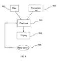
- FIG. 8 is a diagram of a computer system that is used to perform the steps described herein in accordance with one aspect of the present invention.
- a curvature flow is a curve or surface that evolves at each point along its normal with a velocity depending on the curvature at that point.
- a geometric flow is a curve or surface that evolves with a velocity depending on external properties determined by the image features.
- Equation (1) becomes
- D i :D i ⁇ 1 Trace(D i D i ⁇ 1 )
- NTSP(D i , D i ⁇ 2 ) is defined similarly.
- TSP tensor scalar product
- the TSP is often normalized to avoid influence by the relative size of the two tensors, as described in the earlier cited article by Lisa Jonasson et al.
- the fundamental assumption of the segmentation technique in the cited article by Lisa Jonasson et al. is that adjacent voxels in a tract have similar diffusion properties.
- the present model still applies this assumption, but with the purpose to obtain a faster geometric flow.
- FIG. 2 shows an example of a 2D tensor field to demonstrate the background of the presented model.
- the black ellipse depicts the evolving curve
- the horizontal black arrows and the vertical grey arrows show normal directions at corresponding locations.
- evolution speed along the horizontal arrows should be larger than that along the vertical arrows.
- FIG. 1 shows the choice of adjacent voxels with respect to the normal of the surface, as shown in [1] L. Jonasson, X. Bresson, P Hagmann, O. Cuisenaire, R. Meuli, and J. Thiran, “White matter fiber trace segmentation in DT-MRI using geometric flows,” Medical Image Analysis., vol. 9, pp. 223-236, 2005.
- FIG. 2 shows the evolving curve (black ellipse) superimposed inside a semi-circle shaped 2D tensor field.
- the other candidate is:
- COIN 3 ( FA ) * N ⁇ Di * N max ⁇ ( ⁇ 1 , ⁇ 2 , ⁇ 3 ) ( 7 ⁇ a ) where ⁇ 1 , ⁇ 2 , ⁇ 3 are the eigen values of Di*N.
- COIN 1 The definition of COIN 1 is quite intuitive: if the normal direction is coincident with the principle direction of tensor, the evolution speed is higher.
- FA is used by Mariana Lazar et al. in [2] M. Lazar, D. Weinstein, and J. Tsuruda et al, “White matter tractography using diffusion tensor deflection,” Human Brain Mapping , vol. 18, pp. 306-321, 2003. Segmentation using COIN 3 is less sensitive to image noise and experimental results show that it is more efficient than COIN 1 .
- equation (5) the similarity is defined using D i+1 , D i+2 which are neighbors of D i along the positive normal direction instead of negative normal direction. This is done to avoid overshooting.
- Certain termination criteria are used to determine the boundaries of tracts. It is preferred to use two criteria. Evolution of the curve/surface is terminated at locations where the similarity measure NTSP is less than a threshold or where the gradient of the similarity NTSP is less than a threshold. These two criteria combined together can accurately catch the boundary for segmentation purposes.
- L. Jonasson, X. Bresson, P Hagmann, O. Cuisenaire, R. Meuli, and J. Thiran, “White matter fiber trace segmentation in DT-MRI using geometric flows,” Medical Image Analysis ., vol. 9, pp. 223-236, 2005 only the first criterion is used. This does not work well for segmentation of vector/tensor field composed of homogeneous regions.
- the similarity map of the vector/tensor field is a piecewise constant function. Locations with a high gradient of similarity are on the boundary of the segmentation. Using a threshold of similarity is not able to identify this boundary and consequently would not be sufficient as a segmentation criterion.
- FIG. 3 compares the segmentation quality of the method presented as an aspect of the present invention and the method applied in article [1].
- the method as described in this cited article has been applied to the recreated synthetic tensor fields.
- the results as depicted in FIG. 3 show that the method presented as one aspect of the present invention is more accurate.
- FIG. 4 and FIG. 5 show segmentation of corpus callosum using different views obtained using the proposed model.
- FIG. 6 shows results obtained using the model applied in [1].
- FIG. 7 shows 2D sectional contour superimposed over corresponding FA images. It is clear that the process of the present invention catches more accurate corpus callosum detail than the model that is applied in [1].
- L. Jonasson, X. Bresson, P Hagmann, O. Cuisenaire, R. Meuli, and J. Thiran “White matter fiber trace segmentation in DT-MRI using geometric flows,” Medical Image Analysis ., vol. 9, pp. 223-236, 2005.
- FIG. 8 illustrates a computer system that can be used in accordance with one aspect of the present invention.
- the system is provided with data 901 representing the to be displayed MRI image.
- An instruction set or program 902 comprising the methods of the present invention is provided and combined with the data in a processor 903 , which can process the instructions of 902 applied to the data 901 and show the resulting image on a display 904 .
- the processor can be dedicated hardware, a GPU, a CPU or any other computing device that can execute the instructions of 902 .
- An input device 905 like a mouse, or track-ball or other input device allows a user to initiate the segmentation process. Consequently the system as shown in FIG. 8 provides an interactive system for image segmentation.
- any type of computer system can be used, although it is preferred to use a computer system having sufficient processing power.
- a stand alone PC a multiprocessor PC, a main frame computer, a parallel processing computer or any other type of computer can be used.
Landscapes
- Engineering & Computer Science (AREA)
- Computer Vision & Pattern Recognition (AREA)
- Physics & Mathematics (AREA)
- General Physics & Mathematics (AREA)
- Theoretical Computer Science (AREA)
- Software Systems (AREA)
- Magnetic Resonance Imaging Apparatus (AREA)
Abstract
Description
where F is an image based speed function, H is an intrinsic speed depending on the curvature of the surface S, N is the normal of the surface and t is time.
F=mean(NTSP(D i ,D i−1),NTSP(D i ,D i−2)), (3)
wherein Di is the diffusion tensor in the current voxel and Di−p is the diffusion tensor in grid found by following the normal to the surface p voxels backwards from the original voxel i, see
with Di:Di−1=Trace(DiDi−1), NTSP(Di, Di−2) is defined similarly. One of the most common measures of similarity between two tensors is the tensor scalar product (TSP) and is a measure of the overlap between two tensors. The TSP is often normalized to avoid influence by the relative size of the two tensors, as described in the earlier cited article by Lisa Jonasson et al. The fundamental assumption of the segmentation technique in the cited article by Lisa Jonasson et al. is that adjacent voxels in a tract have similar diffusion properties. The present model still applies this assumption, but with the purpose to obtain a faster geometric flow.
F=mean(NTSP(D i ,D i+1),NTSP(D i ,D i+2))+βCOIN, (5)
where COIN is a measure of coincidence of the normal direction and the tensor field and β is a constant.
COIN 1 =N·PE (6)
with PE the principal eigenvector of the current diffusion tensor Di, and N the normal direction.
with FA the fractional anisotropy value of tensor Di.
where λ1, λ2, λ3 are the eigen values of Di*N.
Claims (12)
COIN 1 =N·PE.
COIN 1 =N·PE.
Priority Applications (2)
| Application Number | Priority Date | Filing Date | Title |
|---|---|---|---|
| US11/489,103 US7627155B2 (en) | 2005-10-26 | 2006-07-19 | Fast geometric flows based white matter fiber tract segmentation in DT-MRI |
| CN2006101365357A CN1955980B (en) | 2005-10-26 | 2006-10-26 | Fast geometric flows based white matter fiber tract segmentation in DT-MRI |
Applications Claiming Priority (2)
| Application Number | Priority Date | Filing Date | Title |
|---|---|---|---|
| US73047305P | 2005-10-26 | 2005-10-26 | |
| US11/489,103 US7627155B2 (en) | 2005-10-26 | 2006-07-19 | Fast geometric flows based white matter fiber tract segmentation in DT-MRI |
Publications (2)
| Publication Number | Publication Date |
|---|---|
| US20070092120A1 US20070092120A1 (en) | 2007-04-26 |
| US7627155B2 true US7627155B2 (en) | 2009-12-01 |
Family
ID=37985438
Family Applications (1)
| Application Number | Title | Priority Date | Filing Date |
|---|---|---|---|
| US11/489,103 Expired - Fee Related US7627155B2 (en) | 2005-10-26 | 2006-07-19 | Fast geometric flows based white matter fiber tract segmentation in DT-MRI |
Country Status (2)
| Country | Link |
|---|---|
| US (1) | US7627155B2 (en) |
| CN (1) | CN1955980B (en) |
Cited By (2)
| Publication number | Priority date | Publication date | Assignee | Title |
|---|---|---|---|---|
| US20140233819A1 (en) * | 2013-02-20 | 2014-08-21 | Industry-University Cooperation Foundation Hanyang University | Method and apparatus for acquiring nerve fiber structure information of object by using mri system |
| US11000204B2 (en) | 2016-04-01 | 2021-05-11 | University-Industry Cooperation Group Of Kyung Hee University | Device and method for reconstructing low-frequency conductivity images using MRI without current injection |
Families Citing this family (7)
| Publication number | Priority date | Publication date | Assignee | Title |
|---|---|---|---|---|
| US7268551B2 (en) * | 2004-04-14 | 2007-09-11 | Mclean Hospital Corporation | Inter-subject coherence in DT-MRI |
| US8320647B2 (en) | 2007-11-20 | 2012-11-27 | Olea Medical | Method and system for processing multiple series of biological images obtained from a patient |
| WO2011011554A1 (en) * | 2009-07-21 | 2011-01-27 | The Regents Of The University Of California | Methods for the identification and targeting of brain regions and structures and treatments related thereto |
| CN101866485B (en) * | 2010-06-10 | 2012-02-01 | 西北工业大学 | Brain cortex surface maximum principal direction field diffusion method for three-dimensional brain magnetic resonance image |
| TWI478103B (en) * | 2012-08-10 | 2015-03-21 | 國立臺灣大學 | Diffusion spectrum contrast conversion method using highly deformed differential homeomorphic metric mapping method |
| US11328426B2 (en) * | 2017-10-03 | 2022-05-10 | Mint Labs Inc. | Fiber tracking and segmentation |
| CN115937223B (en) * | 2023-01-06 | 2023-07-14 | 北京理工大学 | A single-sample new category white matter tract segmentation method, device, equipment and medium |
Citations (2)
| Publication number | Priority date | Publication date | Assignee | Title |
|---|---|---|---|---|
| US5969524A (en) * | 1997-04-14 | 1999-10-19 | The United States Of America As Represented By The Department Of Health And Human Services | Method to significantly reduce bias and variance of diffusion anisotrophy measurements |
| US7034531B1 (en) * | 2003-01-09 | 2006-04-25 | The General Hospital Corporation | Diffusion MRI using spherical shell sampling |
-
2006
- 2006-07-19 US US11/489,103 patent/US7627155B2/en not_active Expired - Fee Related
- 2006-10-26 CN CN2006101365357A patent/CN1955980B/en not_active Expired - Fee Related
Patent Citations (2)
| Publication number | Priority date | Publication date | Assignee | Title |
|---|---|---|---|---|
| US5969524A (en) * | 1997-04-14 | 1999-10-19 | The United States Of America As Represented By The Department Of Health And Human Services | Method to significantly reduce bias and variance of diffusion anisotrophy measurements |
| US7034531B1 (en) * | 2003-01-09 | 2006-04-25 | The General Hospital Corporation | Diffusion MRI using spherical shell sampling |
Non-Patent Citations (2)
| Title |
|---|
| Jonasson, L., et al., "White Matter Fiber Tract Segmentation in DT-MRI Using Geometric Flows",Medical Image Analysis, vol. 9, pp. 223-226 (2005). |
| Lazar, M., et al., "White Matter Tractograhy Using Diffusion Tensor Deflection", Human Brain Mapping, 18:306-321(2003). |
Cited By (3)
| Publication number | Priority date | Publication date | Assignee | Title |
|---|---|---|---|---|
| US20140233819A1 (en) * | 2013-02-20 | 2014-08-21 | Industry-University Cooperation Foundation Hanyang University | Method and apparatus for acquiring nerve fiber structure information of object by using mri system |
| US9436869B2 (en) * | 2013-02-20 | 2016-09-06 | Samsung Electronics Co., Ltd. | Method and apparatus for acquiring nerve fiber structure information of object by using MRI system |
| US11000204B2 (en) | 2016-04-01 | 2021-05-11 | University-Industry Cooperation Group Of Kyung Hee University | Device and method for reconstructing low-frequency conductivity images using MRI without current injection |
Also Published As
| Publication number | Publication date |
|---|---|
| CN1955980B (en) | 2011-10-19 |
| CN1955980A (en) | 2007-05-02 |
| US20070092120A1 (en) | 2007-04-26 |
Similar Documents
| Publication | Publication Date | Title |
|---|---|---|
| Yushkevich et al. | Structure-specific statistical mapping of white matter tracts | |
| US9092849B2 (en) | Bidirectional blood vessel segmentation | |
| Prados et al. | Control theory and fast marching techniques for brain connectivity mapping | |
| US20200116808A1 (en) | Cerebrovascular segmentation from mra images | |
| Reid et al. | How many streamlines are required for reliable probabilistic tractography? Solutions for microstructural measurements and neurosurgical planning | |
| US7627155B2 (en) | Fast geometric flows based white matter fiber tract segmentation in DT-MRI | |
| Wen et al. | Brain tissue classification based on DTI using an improved fuzzy C-means algorithm with spatial constraints | |
| US9390549B2 (en) | Shape data generation method and apparatus | |
| Paknezhad et al. | Automatic basal slice detection for cardiac analysis | |
| Gruslys et al. | A new fast accurate nonlinear medical image registration program including surface preserving regularization | |
| Jia et al. | Intermediate templates guided groupwise registration of diffusion tensor images | |
| Hameeteman et al. | Carotid wall volume quantification from magnetic resonance images using deformable model fitting and learning-based correction of systematic errors | |
| Wu et al. | TPS-HAMMER: Improving HAMMER registration algorithm by soft correspondence matching and thin-plate splines based deformation interpolation | |
| Davatzikos | Measuring biological shape using geometry-based shape transformations | |
| Krishnaswamy et al. | A novel 3D-to-3D Diffeomorphic registration algorithm with applications to left ventricle segmentation in MR and ultrasound sequences | |
| Savadjiev et al. | Local white matter geometry from diffusion tensor gradients | |
| US10789713B2 (en) | Symplectomorphic image registration | |
| Chen et al. | Shape statistics variational approach for the outer contour segmentation of left ventricle MR images | |
| Queirós et al. | Fast left ventricle tracking in CMR images using localized anatomical affine optical flow | |
| Kop et al. | Kidney segmentation from ultrasound images using gradient vector force | |
| Asiri et al. | Advanced computational modeling for brain tumor detection: Enhancing segmentation accuracy using ICA-I and ICA-II techniques | |
| He et al. | 3-D B-spline wavelet-based local standard deviation (BWLSD): its application to edge detection and vascular segmentation in magnetic resonance angiography | |
| Saran et al. | Vessel segmentation in MRI using a variational image subtraction approach | |
| Jonasson et al. | White matter mapping in DT-MRI using geometric flows | |
| Gooya et al. | Generalization of geometrical flux maximizing flow on Riemannian manifolds for improved volumetric blood vessel segmentation |
Legal Events
| Date | Code | Title | Description |
|---|---|---|---|
| AS | Assignment |
Owner name: SIEMENS CORPORATE RESEARCH, INC., NEW JERSEY Free format text: ASSIGNMENT OF ASSIGNORS INTEREST;ASSIGNORS:WANG, ZHIZHOU;GUO, WEIHONG;REEL/FRAME:018249/0995 Effective date: 20060908 |
|
| AS | Assignment |
Owner name: SIEMENS MEDICAL SOLUTIONS USA, INC.,PENNSYLVANIA Free format text: ASSIGNMENT OF ASSIGNORS INTEREST;ASSIGNOR:SIEMENS CORPORATE RESEARCH, INC.;REEL/FRAME:019309/0669 Effective date: 20070430 Owner name: SIEMENS MEDICAL SOLUTIONS USA, INC., PENNSYLVANIA Free format text: ASSIGNMENT OF ASSIGNORS INTEREST;ASSIGNOR:SIEMENS CORPORATE RESEARCH, INC.;REEL/FRAME:019309/0669 Effective date: 20070430 |
|
| STCF | Information on status: patent grant |
Free format text: PATENTED CASE |
|
| CC | Certificate of correction | ||
| FPAY | Fee payment |
Year of fee payment: 4 |
|
| FPAY | Fee payment |
Year of fee payment: 8 |
|
| FEPP | Fee payment procedure |
Free format text: MAINTENANCE FEE REMINDER MAILED (ORIGINAL EVENT CODE: REM.); ENTITY STATUS OF PATENT OWNER: LARGE ENTITY |
|
| LAPS | Lapse for failure to pay maintenance fees |
Free format text: PATENT EXPIRED FOR FAILURE TO PAY MAINTENANCE FEES (ORIGINAL EVENT CODE: EXP.); ENTITY STATUS OF PATENT OWNER: LARGE ENTITY |
|
| STCH | Information on status: patent discontinuation |
Free format text: PATENT EXPIRED DUE TO NONPAYMENT OF MAINTENANCE FEES UNDER 37 CFR 1.362 |
|
| FP | Lapsed due to failure to pay maintenance fee |
Effective date: 20211201 |