US7609870B2 - Methods and systems for enhancing images - Google Patents
Methods and systems for enhancing images Download PDFInfo
- Publication number
- US7609870B2 US7609870B2 US10/925,317 US92531704A US7609870B2 US 7609870 B2 US7609870 B2 US 7609870B2 US 92531704 A US92531704 A US 92531704A US 7609870 B2 US7609870 B2 US 7609870B2
- Authority
- US
- United States
- Prior art keywords
- image
- pixels
- pixel
- smoothed
- dataset
- Prior art date
- Legal status (The legal status is an assumption and is not a legal conclusion. Google has not performed a legal analysis and makes no representation as to the accuracy of the status listed.)
- Expired - Fee Related, expires
Links
Images


Classifications
-
- G—PHYSICS
- G06—COMPUTING OR CALCULATING; COUNTING
- G06T—IMAGE DATA PROCESSING OR GENERATION, IN GENERAL
- G06T5/00—Image enhancement or restoration
- G06T5/70—Denoising; Smoothing
-
- G—PHYSICS
- G06—COMPUTING OR CALCULATING; COUNTING
- G06T—IMAGE DATA PROCESSING OR GENERATION, IN GENERAL
- G06T5/00—Image enhancement or restoration
- G06T5/20—Image enhancement or restoration using local operators
-
- G—PHYSICS
- G06—COMPUTING OR CALCULATING; COUNTING
- G06T—IMAGE DATA PROCESSING OR GENERATION, IN GENERAL
- G06T5/00—Image enhancement or restoration
- G06T5/73—Deblurring; Sharpening
-
- G—PHYSICS
- G06—COMPUTING OR CALCULATING; COUNTING
- G06T—IMAGE DATA PROCESSING OR GENERATION, IN GENERAL
- G06T7/00—Image analysis
- G06T7/10—Segmentation; Edge detection
- G06T7/136—Segmentation; Edge detection involving thresholding
-
- G—PHYSICS
- G06—COMPUTING OR CALCULATING; COUNTING
- G06T—IMAGE DATA PROCESSING OR GENERATION, IN GENERAL
- G06T2207/00—Indexing scheme for image analysis or image enhancement
- G06T2207/10—Image acquisition modality
- G06T2207/10072—Tomographic images
- G06T2207/10081—Computed x-ray tomography [CT]
-
- G—PHYSICS
- G06—COMPUTING OR CALCULATING; COUNTING
- G06T—IMAGE DATA PROCESSING OR GENERATION, IN GENERAL
- G06T2207/00—Indexing scheme for image analysis or image enhancement
- G06T2207/20—Special algorithmic details
- G06T2207/20004—Adaptive image processing
- G06T2207/20012—Locally adaptive
-
- G—PHYSICS
- G06—COMPUTING OR CALCULATING; COUNTING
- G06T—IMAGE DATA PROCESSING OR GENERATION, IN GENERAL
- G06T2207/00—Indexing scheme for image analysis or image enhancement
- G06T2207/30—Subject of image; Context of image processing
- G06T2207/30004—Biomedical image processing
- G06T2207/30061—Lung
- G06T2207/30064—Lung nodule
Definitions
- the invention relates generally to computer tomography (CT) imaging systems, and more particularly, to image filters in CT lung applications.
- CT computer tomography
- CT Computer Tomography
- an X-ray source emits a fan-shaped beam towards a scan subject, such as a patient.
- the beam after being attenuated by the scan subject, impinges upon an array of radiation detectors, which, in turn produce electrical signals indicative of the attenuated beam.
- the electrical signals are then transmitted to a data processing unit for analysis and image reconstruction.
- CT lung images Various techniques to reconstruct CT images, such as CT lung images, are known. These techniques involve dividing each acquired CT image slice into a matrix of volume elements (voxels). Each voxel may be traversed during the scan by numerous X-ray photons, and the intensity of the transmitted radiation is measured by the detectors. These intensity readings are used to calculate the density or attenuation value of a body part such as a tissue at each point in the slice. Specific attenuation values are assigned to each individual voxel. In addition, each image pixel is assigned a numerical value (CT number), which is the average of all the attenuation values contained within the corresponding voxel. The final image is then reconstructed from the matrix of image voxels as a corresponding matrix of picture elements (pixels).
- CT number numerical value
- a good contrast and a good spatial resolution are required.
- an absolute difference between two objects in the image may be easy to perceive.
- a good spatial resolution ensures that even very minute structures within an object in the image are easily identifiable.
- a method for enhancing an image that includes a plurality of image pixels includes segmenting the image by comparing an image pixel CT number to a plurality of predetermined thresholds. The method further includes filtering the plurality of image pixels based on the comparison and generating an output image based on the filtered image pixels.
- a computed tomography (CT) system in another exemplary embodiment, includes a computed tomography scanner, and a controller for controlling the operation of the computed tomography scanner to enhance a CT image having a plurality of image pixels.
- the controller is configured to segment the image by comparing an image pixel CT number of the plurality of image pixels to a plurality of predetermined thresholds, filter the plurality of image pixels based on the comparison, and generate an output image based on the filtered image pixels.
- FIG. 1 is a flowchart illustrating a method to enhance an image, in accordance with an exemplary embodiment of the invention.
- FIG. 2 is a flowchart illustrating a method to enhance an image, in accordance with another exemplary embodiment of the invention.
- FIGS. 3 a and 3 b is a flowchart illustrating a method to enhance an image, in accordance with yet another exemplary embodiment of the invention.
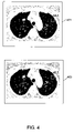
- FIG. 4 illustrates a lung scan image obtained by using known image reconstruction techniques and an exemplary lung scan image obtained by using the enhanced filtering described in various embodiments of the invention.
- FIG. 5 is a block diagram depicting a CT system used to enhance an image, in accordance with an exemplary embodiment of the invention.
- Various embodiments of the invention provide a method for enhancing an image that includes a plurality of image pixels.
- the image may be enhanced by increasing its contrast to noise ratio, thereby enabling better visualization of minute parts of the image.
- a post processing method for medical applications, such as CT lung application that enhances contrast of vessels and tumors in lungs and reduces noise of the surrounding lung tissues, is described.
- FIG. 1 is a flowchart illustrating a method to enhance an image, in accordance with an exemplary embodiment of the invention.
- the image is composed of a plurality of image pixels and each image pixel has a CT number associated with it.
- the image is segmented by comparing an image pixel CT number to a plurality of predetermined threshold values.
- the threshold values depend on the type of image application. For example, the threshold values for a CT lung application may be different from the threshold values determined for a CT heart application.
- the pixels of the segmented image are filtered based on the comparison.
- filtering includes image smoothing and deconvolution operations.
- an output image is generated using the filtered image pixels.
- the output image is an enhanced image, which enables better visualization of the minute parts of the image.
- FIG. 2 is a flowchart illustrating a method to enhance an image, in accordance with another exemplary embodiment of the invention.
- the image is composed of pixels, and each pixel is associated with a CT number.
- the image is a medical image, such as images used in CT Lung applications.
- a smoothing operation is performed on an original image (im) to generate a smoothed (im_s) image dataset.
- standard smoothing algorithms such as Gaussian filter, Uniform filter, Triangular filter, and the like are used, in order to reduce noise and prepare the original image (im) for further processing such as segmentation.
- the smoothing operation is performed by averaging the CT numbers of a plurality of adjacent image pixels.
- a deconvolution operation is performed on the original image (im) to generate a deconvolution (im_d) image dataset.
- standard deconvolution algorithms such as Empirical Point-spread function, Calculated Point-spread function, Nearest Neighbor algorithm, and the like are used, in order to remove out of focus light and sharpen the original image (im).
- the smoothed image dataset (im_s) is segmented based on a plurality of predetermined threshold values.
- the smoothed image dataset (im_s) is segmented by using the CT numbers of the image pixels.
- the smoothed image dataset (im_s) pixels are filtered based on the relative value of their CT number.
- filtering is performed by applying a different filter to each segment.
- image pixels with CT numbers smaller than a predetermined low threshold are smoothed, whereas image pixels with CT numbers greater than a predetermined high threshold are assigned the value of the corresponding pixel in the original image (im).
- the image pixels that have CT numbers between the predetermined high and the low thresholds are filtered by using a deconvolution function.
- an output image is generated by using the filtered pixels.
- the filtered pixels are modulated by using a gain factor, in order to ensure that the smoothing and deconvolution operations have little impact on the image pixels near the edge of the output image.
- the gain factor is a function of the position of an image pixel. In another embodiment of the invention, the gain factor is a function of the CT number of an image pixel.
- FIGS. 3 a and 3 b is a flowchart illustrating a method to enhance an image, in accordance with yet another exemplary embodiment of the invention.
- a smoothing operation is performed on an original image (im) to generate a smoothed (im_s) image dataset.
- deconvolution operation is performed on the original image (im) to generate a deconvolution (im_d) image dataset.
- the smoothed image dataset (im_s) is segmented based on a plurality of predetermined threshold values.
- four sets of threshold values for example, low (t_low), high (t_high), and transition threshold values (t_tran1) and (t_tran2) are established.
- the low threshold value (t_low) signifies the background region of an image whereas the high threshold (t_high) signifies the soft tissue region in an image.
- the transition thresholds (t_tran1 and t_tran2) are established in order to make an enhanced output image look natural and have no abrupt transition regions.
- the smoothed image dataset (im_s) pixels are filtered based on their CT numbers.
- an output image pixel is obtained by assigning to it a filtered CT number, determined by using corresponding pixels of the smoothed image dataset (im_s) and the deconvolution image dataset (im_d), and a first scaling factor.
- the first scaling factor is determined by interpolating the pixel CT number between the first predetermined threshold and the first transition threshold.
- an output image pixel is obtained by assigning to it a filtered CT number corresponding to a pixel of the deconvolution image dataset.
- an output image pixel is obtained by assigning to it a filtered CT number.
- the output image pixel is determined by using the corresponding original image (im) and the deconvolution image dataset (im_d), and a second scaling factor.
- the second scaling factor is determined by interpolating the pixel CT number between the second predetermined threshold and the second transition threshold.
- an output image pixel (im_o) is obtained by assigning to it a filtered CT number corresponding to a pixel of the original image (im).
- the output image pixels (im_o) are modulated by a gain factor in order to obtain the final output image (im_out). Modulation of im_o is performed to ensure that the smoothing and deconvolution operations do not have much impact on the pixels near the edge of the original image.
- the gain factor is a polynomial expression that is based on the position of a pixel relative to the centre of the image.
- the position dependent gain factor (gain_pd) has a value of 1 near the image center and gradually rolls off to 0 towards the edges.
- the gain factor is expressed as a polynomial expression based on the pixel location relative to the center of the image:
- FIG. 4 illustrates exemplary lung scan images.
- Lung scan image 401 is obtained by using known image reconstruction methods.
- Lung scan image 403 is obtained by using enhanced post processing filtering in an embodiment of the invention.
- Lung scan image 403 illustrates sharper images of the vessels and nodules in the lungs than 401 .
- the enhanced post processing filtering reduces the standard deviation of the lung tissues surrounding the vessels and nodules, as depicted by lung scan images 401 and 403 .
- the reduction of the standard deviation enables reduction in noise, smoothing and enhancing of the contrast of minute parts of the image.
- Lung scan image 401 has a CT number standard deviation value of 64.4 whereas lung scan image 403 has a CT number standard deviation value of 45.7. Therefore, lung scan image 403 has an improved contrast to noise ratio of the lung nodules.
- FIG. 5 is a block diagram depicting a CT system used to enhance an image, in accordance with an exemplary embodiment of the invention.
- CT system 500 includes a CT scanner 501 and a controller 503 .
- controller 503 controls the operation of CT scanner 501 , and is configured to enhance a CT image, wherein the image includes a plurality of image pixels.
- the CT image is produced as a result of a scan performed by CT scanner 501 .
- controller 503 enhances the CT image by following the steps of segmenting the image by comparing an image pixel CT number to a set of predetermined thresholds; filtering the image pixels based on the comparison; and generating an output image based on the filtered image pixels.
- controller 503 is configured to perform a smoothing operation on the CT image in order to generate a smoothed image dataset and average the CT numbers of a plurality of adjacent pixels of the CT image.
- controller 503 is configured to perform a deconvolution operation on the CT image to generate a deconvolution image dataset.
- controller 503 is further configured to assign a CT number to an image pixel that is equal to a CT number of a corresponding pixel in the smoothed image dataset, if the CT number of the image pixel is less than or equal to a first predetermined threshold. Controller 503 assigns a CT number to an image pixel that is equal to a CT number of a corresponding image pixel, if the CT number of the image pixel is greater than or equal to a second predetermined threshold. Controller 503 further assigns a CT number to an image pixel that is equal to a CT number of a corresponding pixel in the deconvolution image dataset, if the CT number of the image pixel is greater than the first predetermined threshold and is less than the second predetermined threshold.
- controller 503 is further configured to assign a CT number to an image pixel that is equal to a CT number, determined by using the CT number of a corresponding pixel of the smoothed image dataset, a corresponding pixel of the deconvolution image dataset, and a first transition scaling factor, if the CT number of the image pixel is greater than the first predetermined threshold and less than a first predetermined transition threshold.
- Controller 503 assigns a CT number to an image pixel that is equal to a CT number, determined by using the CT number of a corresponding pixel of the image pixels, a corresponding pixel of the deconvolution image dataset, and a second transition scaling factor, if the CT number of the image pixel is less than the second predetermined threshold and is greater than a second predetermined transition threshold. Controller 503 further assigns a CT number to an image pixel that is equal to a CT number of a corresponding pixel in the deconvolution image dataset, if the CT number of the image pixel is less than the second predetermined transition threshold and greater than the first predetermined transition threshold.
- the various embodiments of the invention provide an improved post-processing image filter that enhances the contrast of minute objects in the image and reducing noise of the surrounding lung tissues. Further, the various embodiments of the invention provide an improved post-processing image filter for filtering medical images.
- the post processing image filter enables higher spatial resolution and better contrast noise ratio for tumor detection and sizing, while keeping the radiation dose fed to a patient low.
- a technical effect of the invention is to provide enhanced images with increased high contrast resolution.
- Other technical effects include the reduction of noise of body parts such as lung tissues to enhance contrast to noise ratio enabling better visualization of body parts such as vessels and nodules in the lung.
- the various embodiments or components thereof may be implemented as part of a computer system.
- the computer system may include a computer, an input device, a display unit and an interface, for example, for accessing the Internet.
- the computer may include a microprocessor.
- the microprocessor may be connected to a communication bus.
- the computer may also include a memory.
- the memory may include Random Access Memory (RAM) and Read Only Memory (ROM).
- the computer system further may include a storage device, which may be a hard disk drive or a removable storage drive such as a floppy disk drive, optical disk drive, and the like.
- the storage device can also be other similar means for loading computer programs or other instructions into the computer system.
- the term “computer” may include any processor-based or microprocessor-based system including systems using microcontrollers, reduced instruction set circuits (RISC), application specific integrated circuits (ASICs), logic circuits, and any other circuit or processor capable of executing the functions described herein.
- RISC reduced instruction set circuits
- ASICs application specific integrated circuits
- the above examples are exemplary only, and are thus not intended to limit in any way the definition and/or meaning of the term “computer”.
- the computer system executes a set of instructions that are stored in one or more storage elements, in order to process input data.
- the storage elements may also hold data or other information as desired or needed.
- the storage element may be in the form of an information source or a physical memory element within the processing machine.
- the set of instructions may include various commands that instruct the processing machine to perform specific operations such as the processes of the various embodiments of the invention.
- the set of instructions may be in the form of a software program.
- the software may be in various forms such as system software or application software. Further, the software may be in the form of a collection of separate programs, a program module within a larger program or a portion of a program module.
- the software also may include modular programming in the form of object-oriented programming.
- the processing of input data by the processing machine may be in response to user commands, or in response to results of previous processing, or in response to a request made by another processing machine.
- the terms “software” and “firmware” are interchangeable, and include any computer program stored in memory for execution by a computer, including RAM memory, ROM memory, EPROM memory, EEPROM memory, and non-volatile RAM (NVRAM) memory.
- RAM memory random access memory
- ROM memory read-only memory
- EPROM memory erasable programmable read-only memory
- EEPROM memory electrically erasable programmable read-only memory
- NVRAM non-volatile RAM
Landscapes
- Engineering & Computer Science (AREA)
- Physics & Mathematics (AREA)
- General Physics & Mathematics (AREA)
- Theoretical Computer Science (AREA)
- Computer Vision & Pattern Recognition (AREA)
- Image Processing (AREA)
- Apparatus For Radiation Diagnosis (AREA)
Abstract
Description
im — d=im−(im−decon(im))*gain_decon —(1)
where decon(*) denotes deconvolution operation, and the gain_decon is expressed as:
where
a(k) represents polynomial coefficients and t_low represents a low threshold. In an embodiment, the values assigned to parameters in equation (2) are m=5, a(1)=0.378, a(2)=13.02, a(3)=−38.16, a(4)=44.193, and a(5)=−18.93. The gain_decon factor has value of zero for tval=0, and 0.5 for tval=1 and a maximum value of 1 around tval=0.75.
if im — s<t_low, then im — o=im — s. —(3)
Assigning im_s to im_o ensures that the background of output image im_o is smoothed.
scale1=3.0*t 2−2.0*t 3 for t_low≦pixel<t_trans1, where t=(pixel−t_low)/(t_trans1−t_low) —(4)
In an embodiment, this filtering is represented as:
if t_low<=im — s<=t_tran1, then im — o=im — s+(im — d−im — s)*scale1 —(5)
if t_tran1<im — s<t_tran2, then im — o=im — d —(6)
scale2=3.0*t 2−2.0*t 3 for t_trans2≦pixel≦t_high, where t=(pixel−t_trans2)/(t_high−t_trans2) —(7)
This filtering is represented as:
if t_tran2<=im — s<=t_high, then im — o=im — d+(im−im — d)*scale2. —(8)
if im_s>t_high, then im — o=im. —(9)
gain— pd(ij)=1.0+0.16*dt−2.75*dt^2+12.2*dt^3−18.9*dt^4+8.1*dt^5 —(10)
In another embodiment of the invention, the gain factor is expressed as a polynomial expression based on the pixel location relative to the center of the image:
Where dt=((i−255)^2+(j−255)^2)^0.5/256, for 512×512 image matrix size.
Claims (16)
Priority Applications (1)
| Application Number | Priority Date | Filing Date | Title |
|---|---|---|---|
| US10/925,317 US7609870B2 (en) | 2004-08-24 | 2004-08-24 | Methods and systems for enhancing images |
Applications Claiming Priority (1)
| Application Number | Priority Date | Filing Date | Title |
|---|---|---|---|
| US10/925,317 US7609870B2 (en) | 2004-08-24 | 2004-08-24 | Methods and systems for enhancing images |
Publications (2)
| Publication Number | Publication Date |
|---|---|
| US20060045371A1 US20060045371A1 (en) | 2006-03-02 |
| US7609870B2 true US7609870B2 (en) | 2009-10-27 |
Family
ID=35943151
Family Applications (1)
| Application Number | Title | Priority Date | Filing Date |
|---|---|---|---|
| US10/925,317 Expired - Fee Related US7609870B2 (en) | 2004-08-24 | 2004-08-24 | Methods and systems for enhancing images |
Country Status (1)
| Country | Link |
|---|---|
| US (1) | US7609870B2 (en) |
Cited By (3)
| Publication number | Priority date | Publication date | Assignee | Title |
|---|---|---|---|---|
| US20070280519A1 (en) * | 2004-03-22 | 2007-12-06 | Hagen Spies | Method, Computer Program Product And Apparatus For Enhancing A Computerized Tomography Image |
| US20100266178A1 (en) * | 2005-11-30 | 2010-10-21 | The Research Foundation Of State University Of New York | System and method for acceleration of image reconstruction |
| US8953902B2 (en) * | 2012-07-06 | 2015-02-10 | Morpho Detection, Llc | Systems and methods for thin object imaging |
Families Citing this family (6)
| Publication number | Priority date | Publication date | Assignee | Title |
|---|---|---|---|---|
| US8213734B2 (en) * | 2006-07-07 | 2012-07-03 | Sony Ericsson Mobile Communications Ab | Active autofocus window |
| US7656990B2 (en) * | 2006-09-19 | 2010-02-02 | The Board Of Trustees Of The Leland Stanford Junior University | Adaptive anisotropic filtering of projection data for computed tomography |
| US8045776B2 (en) * | 2007-03-06 | 2011-10-25 | General Electric Company | Geometry-dependent filtering in CT method and apparatus |
| US9197885B2 (en) | 2014-03-20 | 2015-11-24 | Gopro, Inc. | Target-less auto-alignment of image sensors in a multi-camera system |
| US8988509B1 (en) | 2014-03-20 | 2015-03-24 | Gopro, Inc. | Auto-alignment of image sensors in a multi-camera system |
| US12236583B2 (en) * | 2021-11-23 | 2025-02-25 | Revvity Health Sciences, Inc. | Three-dimensional luminescence imaging |
Citations (6)
| Publication number | Priority date | Publication date | Assignee | Title |
|---|---|---|---|---|
| US4571635A (en) | 1984-02-17 | 1986-02-18 | Minnesota Mining And Manufacturing Company | Method of image enhancement by raster scanning |
| US5265142A (en) | 1992-05-08 | 1993-11-23 | General Electric Company | Image reconstruction technique for a computer tomography system |
| US5594767A (en) | 1995-11-02 | 1997-01-14 | General Electric Company | Methods and apparatus for enhancing image sharpness |
| US6449330B1 (en) * | 2001-06-28 | 2002-09-10 | Ge Medical Systems Global Technology Company, Llc | Methods and apparatus for artifact reduction in computed tomographic imaging |
| US20030099405A1 (en) | 2001-11-21 | 2003-05-29 | Avinash Gopal B. | CT dose reduction filter with a computationally efficient implementation |
| US7177483B2 (en) | 2002-08-29 | 2007-02-13 | Palo Alto Research Center Incorporated. | System and method for enhancement of document images |
-
2004
- 2004-08-24 US US10/925,317 patent/US7609870B2/en not_active Expired - Fee Related
Patent Citations (6)
| Publication number | Priority date | Publication date | Assignee | Title |
|---|---|---|---|---|
| US4571635A (en) | 1984-02-17 | 1986-02-18 | Minnesota Mining And Manufacturing Company | Method of image enhancement by raster scanning |
| US5265142A (en) | 1992-05-08 | 1993-11-23 | General Electric Company | Image reconstruction technique for a computer tomography system |
| US5594767A (en) | 1995-11-02 | 1997-01-14 | General Electric Company | Methods and apparatus for enhancing image sharpness |
| US6449330B1 (en) * | 2001-06-28 | 2002-09-10 | Ge Medical Systems Global Technology Company, Llc | Methods and apparatus for artifact reduction in computed tomographic imaging |
| US20030099405A1 (en) | 2001-11-21 | 2003-05-29 | Avinash Gopal B. | CT dose reduction filter with a computationally efficient implementation |
| US7177483B2 (en) | 2002-08-29 | 2007-02-13 | Palo Alto Research Center Incorporated. | System and method for enhancement of document images |
Cited By (5)
| Publication number | Priority date | Publication date | Assignee | Title |
|---|---|---|---|---|
| US20070280519A1 (en) * | 2004-03-22 | 2007-12-06 | Hagen Spies | Method, Computer Program Product And Apparatus For Enhancing A Computerized Tomography Image |
| US7809178B2 (en) * | 2004-03-22 | 2010-10-05 | Contextvision Ab | Method, computer program product and apparatus for enhancing a computerized tomography image |
| US20100266178A1 (en) * | 2005-11-30 | 2010-10-21 | The Research Foundation Of State University Of New York | System and method for acceleration of image reconstruction |
| US8687869B2 (en) * | 2005-11-30 | 2014-04-01 | The Research Foundation Of State Of University Of New York | System and method for acceleration of image reconstruction |
| US8953902B2 (en) * | 2012-07-06 | 2015-02-10 | Morpho Detection, Llc | Systems and methods for thin object imaging |
Also Published As
| Publication number | Publication date |
|---|---|
| US20060045371A1 (en) | 2006-03-02 |
Similar Documents
| Publication | Publication Date | Title |
|---|---|---|
| CN102013089B (en) | Iterative CT image filter for noise reduction | |
| JP7106405B2 (en) | Medical image processing device, medical imaging device and medical image processing program | |
| US8938110B2 (en) | Enhanced image data/dose reduction | |
| US8861886B2 (en) | Enhanced visualization for medical images | |
| US9265475B2 (en) | Methods and apparatus for scatter correction for CBCT system and cone-beam image reconstruction | |
| US6463167B1 (en) | Adaptive filtering | |
| US9189832B2 (en) | Method and system for noise reduction in low dose computed tomography | |
| CN100353378C (en) | Image processing apparatus for reducing noise from image | |
| US8538099B2 (en) | Method and system for controlling image reconstruction | |
| EP2646975B1 (en) | Contrast to noise ratio (cnr) enhancer | |
| CN102376084B (en) | Use anisotropic noise model to the iterative image filtering of CT image | |
| CN108292430A (en) | The method for carrying out Automatic Optimal is generated for the quantitative figure in being imaged to functional medicine | |
| JPWO2005110232A1 (en) | Image processing apparatus and method | |
| US7609870B2 (en) | Methods and systems for enhancing images | |
| US8824753B2 (en) | Task oriented noise suppression in medical images | |
| CN117830456B (en) | Method, device and electronic device for correcting metal artifacts in images | |
| JP6921711B2 (en) | Image processing equipment, image processing methods, and programs | |
| US11113810B2 (en) | X-ray CT scanner, image generation method, and image generation program | |
| Bertram et al. | Monte-Carlo scatter correction for cone-beam computed tomography with limited scan field-of-view | |
| EP3404618B1 (en) | Poly-energetic reconstruction method for metal artifacts reduction | |
| US11158095B2 (en) | System and method for reducing artifact bloom in a reconstructed object | |
| Yu et al. | Adaptive modulation of bilateral filtering based on a practical noise model for streaking and noise reduction in multi-slice CT | |
| Zimeras et al. | Object segmentation and shape reconstruction using computer-assisted segmentation tools | |
| Joemai | Technical advances in multi-slice computed tomography: dose assessment and |
Legal Events
| Date | Code | Title | Description |
|---|---|---|---|
| AS | Assignment |
Owner name: GENERAL ELECTRIC COMPANY, NEW YORK Free format text: ASSIGNMENT OF ASSIGNORS INTEREST;ASSIGNOR:LI, JIANYING;REEL/FRAME:015728/0555 Effective date: 20040823 |
|
| FEPP | Fee payment procedure |
Free format text: PAYOR NUMBER ASSIGNED (ORIGINAL EVENT CODE: ASPN); ENTITY STATUS OF PATENT OWNER: LARGE ENTITY |
|
| CC | Certificate of correction | ||
| REMI | Maintenance fee reminder mailed | ||
| LAPS | Lapse for failure to pay maintenance fees | ||
| STCH | Information on status: patent discontinuation |
Free format text: PATENT EXPIRED DUE TO NONPAYMENT OF MAINTENANCE FEES UNDER 37 CFR 1.362 |
|
| STCH | Information on status: patent discontinuation |
Free format text: PATENT EXPIRED DUE TO NONPAYMENT OF MAINTENANCE FEES UNDER 37 CFR 1.362 |
|
| FP | Lapsed due to failure to pay maintenance fee |
Effective date: 20131027 |