US7279331B2 - Differentiation of bone marrow cells into neuronal cells and uses therefor - Google Patents
Differentiation of bone marrow cells into neuronal cells and uses therefor Download PDFInfo
- Publication number
- US7279331B2 US7279331B2 US10/213,526 US21352602A US7279331B2 US 7279331 B2 US7279331 B2 US 7279331B2 US 21352602 A US21352602 A US 21352602A US 7279331 B2 US7279331 B2 US 7279331B2
- Authority
- US
- United States
- Prior art keywords
- neuronal
- cells
- cell
- differentiation
- isolated
- Prior art date
- Legal status (The legal status is an assumption and is not a legal conclusion. Google has not performed a legal analysis and makes no representation as to the accuracy of the status listed.)
- Expired - Fee Related, expires
Links
Images


Classifications
-
- C—CHEMISTRY; METALLURGY
- C12—BIOCHEMISTRY; BEER; SPIRITS; WINE; VINEGAR; MICROBIOLOGY; ENZYMOLOGY; MUTATION OR GENETIC ENGINEERING
- C12N—MICROORGANISMS OR ENZYMES; COMPOSITIONS THEREOF; PROPAGATING, PRESERVING, OR MAINTAINING MICROORGANISMS; MUTATION OR GENETIC ENGINEERING; CULTURE MEDIA
- C12N15/00—Mutation or genetic engineering; DNA or RNA concerning genetic engineering, vectors, e.g. plasmids, or their isolation, preparation or purification; Use of hosts therefor
- C12N15/09—Recombinant DNA-technology
- C12N15/63—Introduction of foreign genetic material using vectors; Vectors; Use of hosts therefor; Regulation of expression
- C12N15/79—Vectors or expression systems specially adapted for eukaryotic hosts
- C12N15/85—Vectors or expression systems specially adapted for eukaryotic hosts for animal cells
-
- A—HUMAN NECESSITIES
- A61—MEDICAL OR VETERINARY SCIENCE; HYGIENE
- A61K—PREPARATIONS FOR MEDICAL, DENTAL OR TOILETRY PURPOSES
- A61K31/00—Medicinal preparations containing organic active ingredients
- A61K31/045—Hydroxy compounds, e.g. alcohols; Salts thereof, e.g. alcoholates
-
- A—HUMAN NECESSITIES
- A61—MEDICAL OR VETERINARY SCIENCE; HYGIENE
- A61K—PREPARATIONS FOR MEDICAL, DENTAL OR TOILETRY PURPOSES
- A61K31/00—Medicinal preparations containing organic active ingredients
- A61K31/095—Sulfur, selenium, or tellurium compounds, e.g. thiols
- A61K31/10—Sulfides; Sulfoxides; Sulfones
-
- A—HUMAN NECESSITIES
- A61—MEDICAL OR VETERINARY SCIENCE; HYGIENE
- A61K—PREPARATIONS FOR MEDICAL, DENTAL OR TOILETRY PURPOSES
- A61K31/00—Medicinal preparations containing organic active ingredients
- A61K31/185—Acids; Anhydrides, halides or salts thereof, e.g. sulfur acids, imidic, hydrazonic or hydroximic acids
- A61K31/19—Carboxylic acids, e.g. valproic acid
- A61K31/195—Carboxylic acids, e.g. valproic acid having an amino group
- A61K31/197—Carboxylic acids, e.g. valproic acid having an amino group the amino and the carboxyl groups being attached to the same acyclic carbon chain, e.g. gamma-aminobutyric acid [GABA], beta-alanine, epsilon-aminocaproic acid or pantothenic acid
- A61K31/198—Alpha-amino acids, e.g. alanine or edetic acid [EDTA]
-
- A—HUMAN NECESSITIES
- A61—MEDICAL OR VETERINARY SCIENCE; HYGIENE
- A61K—PREPARATIONS FOR MEDICAL, DENTAL OR TOILETRY PURPOSES
- A61K31/00—Medicinal preparations containing organic active ingredients
- A61K31/33—Heterocyclic compounds
- A61K31/335—Heterocyclic compounds having oxygen as the only ring hetero atom, e.g. fungichromin
- A61K31/365—Lactones
- A61K31/375—Ascorbic acid, i.e. vitamin C; Salts thereof
-
- A—HUMAN NECESSITIES
- A61—MEDICAL OR VETERINARY SCIENCE; HYGIENE
- A61P—SPECIFIC THERAPEUTIC ACTIVITY OF CHEMICAL COMPOUNDS OR MEDICINAL PREPARATIONS
- A61P17/00—Drugs for dermatological disorders
- A61P17/02—Drugs for dermatological disorders for treating wounds, ulcers, burns, scars, keloids, or the like
-
- A—HUMAN NECESSITIES
- A61—MEDICAL OR VETERINARY SCIENCE; HYGIENE
- A61P—SPECIFIC THERAPEUTIC ACTIVITY OF CHEMICAL COMPOUNDS OR MEDICINAL PREPARATIONS
- A61P19/00—Drugs for skeletal disorders
-
- A—HUMAN NECESSITIES
- A61—MEDICAL OR VETERINARY SCIENCE; HYGIENE
- A61P—SPECIFIC THERAPEUTIC ACTIVITY OF CHEMICAL COMPOUNDS OR MEDICINAL PREPARATIONS
- A61P21/00—Drugs for disorders of the muscular or neuromuscular system
-
- A—HUMAN NECESSITIES
- A61—MEDICAL OR VETERINARY SCIENCE; HYGIENE
- A61P—SPECIFIC THERAPEUTIC ACTIVITY OF CHEMICAL COMPOUNDS OR MEDICINAL PREPARATIONS
- A61P25/00—Drugs for disorders of the nervous system
-
- A—HUMAN NECESSITIES
- A61—MEDICAL OR VETERINARY SCIENCE; HYGIENE
- A61P—SPECIFIC THERAPEUTIC ACTIVITY OF CHEMICAL COMPOUNDS OR MEDICINAL PREPARATIONS
- A61P25/00—Drugs for disorders of the nervous system
- A61P25/08—Antiepileptics; Anticonvulsants
-
- A—HUMAN NECESSITIES
- A61—MEDICAL OR VETERINARY SCIENCE; HYGIENE
- A61P—SPECIFIC THERAPEUTIC ACTIVITY OF CHEMICAL COMPOUNDS OR MEDICINAL PREPARATIONS
- A61P25/00—Drugs for disorders of the nervous system
- A61P25/14—Drugs for disorders of the nervous system for treating abnormal movements, e.g. chorea, dyskinesia
- A61P25/16—Anti-Parkinson drugs
-
- A—HUMAN NECESSITIES
- A61—MEDICAL OR VETERINARY SCIENCE; HYGIENE
- A61P—SPECIFIC THERAPEUTIC ACTIVITY OF CHEMICAL COMPOUNDS OR MEDICINAL PREPARATIONS
- A61P25/00—Drugs for disorders of the nervous system
- A61P25/28—Drugs for disorders of the nervous system for treating neurodegenerative disorders of the central nervous system, e.g. nootropic agents, cognition enhancers, drugs for treating Alzheimer's disease or other forms of dementia
-
- A—HUMAN NECESSITIES
- A61—MEDICAL OR VETERINARY SCIENCE; HYGIENE
- A61P—SPECIFIC THERAPEUTIC ACTIVITY OF CHEMICAL COMPOUNDS OR MEDICINAL PREPARATIONS
- A61P35/00—Antineoplastic agents
-
- A—HUMAN NECESSITIES
- A61—MEDICAL OR VETERINARY SCIENCE; HYGIENE
- A61P—SPECIFIC THERAPEUTIC ACTIVITY OF CHEMICAL COMPOUNDS OR MEDICINAL PREPARATIONS
- A61P37/00—Drugs for immunological or allergic disorders
-
- C—CHEMISTRY; METALLURGY
- C12—BIOCHEMISTRY; BEER; SPIRITS; WINE; VINEGAR; MICROBIOLOGY; ENZYMOLOGY; MUTATION OR GENETIC ENGINEERING
- C12N—MICROORGANISMS OR ENZYMES; COMPOSITIONS THEREOF; PROPAGATING, PRESERVING, OR MAINTAINING MICROORGANISMS; MUTATION OR GENETIC ENGINEERING; CULTURE MEDIA
- C12N5/00—Undifferentiated human, animal or plant cells, e.g. cell lines; Tissues; Cultivation or maintenance thereof; Culture media therefor
- C12N5/06—Animal cells or tissues; Human cells or tissues
- C12N5/0602—Vertebrate cells
- C12N5/0618—Cells of the nervous system
- C12N5/0619—Neurons
-
- A—HUMAN NECESSITIES
- A61—MEDICAL OR VETERINARY SCIENCE; HYGIENE
- A61K—PREPARATIONS FOR MEDICAL, DENTAL OR TOILETRY PURPOSES
- A61K35/00—Medicinal preparations containing materials or reaction products thereof with undetermined constitution
- A61K35/12—Materials from mammals; Compositions comprising non-specified tissues or cells; Compositions comprising non-embryonic stem cells; Genetically modified cells
-
- A—HUMAN NECESSITIES
- A61—MEDICAL OR VETERINARY SCIENCE; HYGIENE
- A61K—PREPARATIONS FOR MEDICAL, DENTAL OR TOILETRY PURPOSES
- A61K48/00—Medicinal preparations containing genetic material which is inserted into cells of the living body to treat genetic diseases; Gene therapy
-
- C—CHEMISTRY; METALLURGY
- C12—BIOCHEMISTRY; BEER; SPIRITS; WINE; VINEGAR; MICROBIOLOGY; ENZYMOLOGY; MUTATION OR GENETIC ENGINEERING
- C12N—MICROORGANISMS OR ENZYMES; COMPOSITIONS THEREOF; PROPAGATING, PRESERVING, OR MAINTAINING MICROORGANISMS; MUTATION OR GENETIC ENGINEERING; CULTURE MEDIA
- C12N2500/00—Specific components of cell culture medium
- C12N2500/30—Organic components
- C12N2500/38—Vitamins
-
- C—CHEMISTRY; METALLURGY
- C12—BIOCHEMISTRY; BEER; SPIRITS; WINE; VINEGAR; MICROBIOLOGY; ENZYMOLOGY; MUTATION OR GENETIC ENGINEERING
- C12N—MICROORGANISMS OR ENZYMES; COMPOSITIONS THEREOF; PROPAGATING, PRESERVING, OR MAINTAINING MICROORGANISMS; MUTATION OR GENETIC ENGINEERING; CULTURE MEDIA
- C12N2500/00—Specific components of cell culture medium
- C12N2500/90—Serum-free medium, which may still contain naturally-sourced components
-
- C—CHEMISTRY; METALLURGY
- C12—BIOCHEMISTRY; BEER; SPIRITS; WINE; VINEGAR; MICROBIOLOGY; ENZYMOLOGY; MUTATION OR GENETIC ENGINEERING
- C12N—MICROORGANISMS OR ENZYMES; COMPOSITIONS THEREOF; PROPAGATING, PRESERVING, OR MAINTAINING MICROORGANISMS; MUTATION OR GENETIC ENGINEERING; CULTURE MEDIA
- C12N2501/00—Active agents used in cell culture processes, e.g. differentation
- C12N2501/80—Neurotransmitters; Neurohormones
- C12N2501/815—Dopamine
-
- C—CHEMISTRY; METALLURGY
- C12—BIOCHEMISTRY; BEER; SPIRITS; WINE; VINEGAR; MICROBIOLOGY; ENZYMOLOGY; MUTATION OR GENETIC ENGINEERING
- C12N—MICROORGANISMS OR ENZYMES; COMPOSITIONS THEREOF; PROPAGATING, PRESERVING, OR MAINTAINING MICROORGANISMS; MUTATION OR GENETIC ENGINEERING; CULTURE MEDIA
- C12N2501/00—Active agents used in cell culture processes, e.g. differentation
- C12N2501/999—Small molecules not provided for elsewhere
-
- C—CHEMISTRY; METALLURGY
- C12—BIOCHEMISTRY; BEER; SPIRITS; WINE; VINEGAR; MICROBIOLOGY; ENZYMOLOGY; MUTATION OR GENETIC ENGINEERING
- C12N—MICROORGANISMS OR ENZYMES; COMPOSITIONS THEREOF; PROPAGATING, PRESERVING, OR MAINTAINING MICROORGANISMS; MUTATION OR GENETIC ENGINEERING; CULTURE MEDIA
- C12N2506/00—Differentiation of animal cells from one lineage to another; Differentiation of pluripotent cells
- C12N2506/13—Differentiation of animal cells from one lineage to another; Differentiation of pluripotent cells from connective tissue cells, from mesenchymal cells
- C12N2506/1346—Differentiation of animal cells from one lineage to another; Differentiation of pluripotent cells from connective tissue cells, from mesenchymal cells from mesenchymal stem cells
- C12N2506/1353—Differentiation of animal cells from one lineage to another; Differentiation of pluripotent cells from connective tissue cells, from mesenchymal cells from mesenchymal stem cells from bone marrow mesenchymal stem cells (BM-MSC)
Definitions
- Pluripotent stem cells have been detected in multiple tissues in the adult mammal, participating in normal replacement and repair, while undergoing self-renewal (Hay, 1966, Regeneration, Holt, Rinehart and Winston, N.Y.; McKay, 1999, Nature Med. 5:261–262; Lemiscka, 1999, Ann. N.Y. Acad. Sci. 872:274–288; Owens and Friedenstein, 1988, Ciba Foundation Syp. 136, Chichester, U.K. pp. 42–60; Prockop, 1997, Science 276:71–74; Ferrari et al., 1998, Science 279:1528–1530; Caplan, 1991, J. Orthop. Res.
- a subclass of bone marrow stem cells is one prototype, capable of differentiating into osteogenic, chondrogenic, adipogenic and other mesenchymal lineages in vitro (Owens and Friedenstein, 1988, Ciba Foundation Symp. 136, Chichester, U.K. pp.
- Neural stem cells are capable of undergoing expansion and differentiating into neurons, astrocytes and oligodendrocytes in vitro (Reynolds and Weiss, 1992, Science 255:1707–1710; Johansson et al., 1999, Cell 96:25–34; Gage et al., 1995, Annu. Rev. Neurosci. 18:159–192; Vescovi et al., 1993, Neuron 11:951–966). NSCs back transplanted into the adult rodent brain survive and differentiate into neurons and glia, raising the possibility of therapeutic potential (Lundberg et al., 1997, Exp. Neurol.
- the present invention includes a method of inducing differentiation of an isolated marrow stromal cell into a neuronal cell.
- the method comprises contacting the isolated marrow stromal cell with at least one neuronal differentiation-inducing compound. This induces differentiation of the isolated marrow stromal cell into a neuronal cell.
- the isolated marrow stromal cell is a rat cell.
- the isolated marrow stromal cell is a human cell.
- the neuronal differentiation-inducing compound is an anti-oxidant.
- the anti-oxidant is selected from the group consisting of beta-mercaptoethanol, dimethylsulfoxide, butylated hydroxytoluene, butylated hydroxyanisole, ascorbic acid, dimethylfumarate, and n-acetylcysteine.
- the anti-oxidant is beta-mercaptoethanol.
- the anti-oxidant is dimethylsulfoxide. In yet another aspect the anti-oxidant is dimethylsulfoxide and butylated hydroxyanisole.
- the neuronal differentiation-inducing compound is also a growth factor in another aspect.
- the growth factor is selected from the group consisting of platelet-derived growth factor, fibroblast growth factor 2 and nerve growth factor.
- the invention further includes a method of producing an isolated neuronal cell.
- the method comprises isolating a marrow stromal cell, contacting the marrow stromal cell with a neuronal differentiation-inducing compound wherein the compound induces the isolated marrow stromal cell to differentiate into an isolated neuronal cell, thereby producing an isolated neuronal cell.
- the invention includes a method of treating a human patient having a disease, disorder or condition of the central nervous system.
- the method comprises obtaining a bone marrow sample from a human donor, isolating stromal cells from the bone marrow sample, inducing the stromal cells to differentiate into isolated neuronal cells, and administering the isolated neuronal cells to the central nervous system of the human patient.
- the presence of the isolated neuronal cells in the central nervous system of the human patient effects treatment of the disease, disorder or condition.
- the disease, disorder or condition of the central nervous system is selected from the group consisting of Alzheimer's disease, Parkinson's disease, Huntington's disease, amyotrophic lateral sclerosis, a tumor, a trauma, elderly dementia, Tay-Sach's disease, Sandhoff's disease, Hurler's syndrome, Krabbe's disease, birth-induced traumatic central nervous system injury, epilepsy, multiple sclerosis, trauma, tumor, stroke, and spinal cord injury.
- the isolated neuronal cells prior to administering the isolated neuronal cells, are transfected with an isolated nucleic acid encoding a therapeutic protein, wherein when the protein is expressed in the cells the protein serves to effect treatment of the disease, disorder or condition.
- the isolated neuronal cells are transfected with an isolated nucleic acid encoding a cytokine, a chemokine, a neurotrophin, another trophic protein, a growth factor, an antibody, or glioma toxic protein.
- the present invention further includes a method of treating a human patient in need of neuronal cells.
- the method comprises obtaining marrow stromal cells from a human patient, propagating the marrow stromal cells in culture under conditions that induce their differentiation into neuronal cells, transplanting the neuronal cells into the human patient in need of the neuronal cells, thereby treating the human patient in need of neuronal cells.
- the invention also includes an isolated neuronal cell made by the method of inducing differentiation of an isolated marrow cell into a neuronal cell.
- the method comprises contacting the isolated marrow stromal cell with at least one neuronal differentiation-inducing compound.
- the contact between the isolated marrow stromal cell and the neuronal differentiation-inducing compound induces differentiation of the isolated marrow stromal cell into the neuronal cell of the invention.
- the neuronal cell made by this method is a rodent cell. In another aspect, the neuronal cell is a rat cell. In a preferred aspect, the neuronal cell made by this method is a human neuronal cell.
- a preferred embodiment of the invention includes an isolated neuronal cell transfected with a therapeutic protein.
- the neuronal cell is isolated by the method of inducing differentiation of an isolated marrow cell into a neuronal cell recited above.
- the neuronal cell is then transfected with an isolated nucleic acid encoding a therapeutic protein that when expressed, will effect treatment of a disease, disorder, or condition of the central nervous system.
- the therapeutic protein encoded by the isolated nucleic acid is a cytokine, a chemokine, a neurotrophin, another trophic protein, a growth factor, an antibody, or glioma toxic protein.
- the invention encompasses diseases, disorders, or conditions of the central nervous system including, but are not limited to, Alzheimer's disease, Parkinson's disease, Huntington's disease, amyotrophic lateral sclerosis, a tumor, a trauma, elderly dementia, Tay-Sach's disease, Sandhoffs disease, Hurler's syndrome, Krabbe's disease, birth-induced traumatic central nervous system injury, epilepsy, multiple sclerosis, trauma, tumor, stroke, and spinal cord injury.
- the transfected neuronal cell made by the method of inducing differentiation of an isolated marrow stromal cell is a rat cell or a rodent cell.
- the transfected neuronal cell is a human cell.
- the invention also includes an isolated neuronal cell produced by a method comprising isolating a marrow stromal cell and contacting it with a neuronal differentiation-inducing compound. This induces the isolated marrow stromal cell to differentiate into isolated neuronal cells.
- a transfected isolated neuronal cell produced by isolating a marrow stromal cell and contacting it with a neuronal differentiation-inducing compound is also included in the invention.
- the isolated neuronal cell produced by this method is then transfected with an isolated nucleic acid encoding a therapeutic protein, that, when expressed in the neuronal cell, will effect treatment of a disease, disorder, or condition of the central nervous system.
- the therapeutic protein encoded by the isolated nucleic acid is a cytokine, a chemokine, a neurotrophin, another trophic protein, a growth factor, an antibody, or a glioma-toxic protein.
- the transfected neuronal cell produced by contacting a neuronal differentiation-inducing compound with a marrow stromal cell is a rat cell.
- the transfected neuronal cell is a rodent cell.
- the transfected neuronal cell is a human cell.
- FIG. 1A is a graph depicting fluorescent cell sorting of passage 1 rMSCs using mouse monoclonal antibodies that specifically bind with cell surface marker CD11b (CD11/integrin alpha M /Mac-1 alpha chain; Pharmingen, San Diego, Calif.) (unfilled peaks).
- the secondary antibody used was anti-mouse antibody conjugated with fluoresceine isothiocyanate (FITC).
- FITC fluoresceine isothiocyanate
- An isotype control is included in each experiment to identify background fluorescence (filled peaks). Number of cells analyzed (Events) is plotted on the Y-axis, while intensity of staining is plotted on the X-axis.
- FIG. 1B is a graph depicting fluorescent cell sorting of passage 1 rMSCs using mouse monoclonal antibodies that specifically bind with cell surface marker CD45/leukocyte common antigen (Pharmingen) (unfilled peaks).
- the secondary antibody is anti-mouse antibody conjugated with fluoresceine isothiocyanate (FITC).
- FITC fluoresceine isothiocyanate
- An isotype control is included in each experiment to identify background fluorescence (filled peaks). Number of cells analyzed (Events) is plotted on the Y-axis, while intensity of staining is plotted on the X-axis.
- FIG. 1C is a graph depicting fluorescent cell sorting of passage 1 rMSCs using mouse monoclonal antibodies that specifically bind with cell surface marker CD90/Thy-1/CD90.1/Thy1.1 (Pharmingen) (unfilled peaks).
- the secondary antibody is anti-mouse antibody conjugated with fluoresceine isothiocyanate (FITC).
- FITC fluoresceine isothiocyanate
- An isotype control is included in each experiment to identify background fluorescence (filled peaks). Number of cells analyzed (Events) is plotted on the Y-axis, while intensity of staining is plotted on the X-axis.
- FIG. 2 is an image depicting neuronal differentiation of rMSCs at various time points after treatment. Briefly, the neuronal differentiation protocol disclosed herein was initiated at 0 minutes and followed for 210 minutes.
- FIG. 2A represents 0 minutes
- FIG. 2B represents 30 minutes
- FIG. 2C represents 60 minutes
- FIG. 3D represents 90 minutes
- FIG. 2E represents 120 minutes
- FIG. 2F represents 150 minutes
- FIG. 2G represents 180 minutes
- FIG. 2H represents 210 minutes.
- a flat rMSC in FIG. 2A is identified ( ⁇ ) prior to differentiation. Retraction of cell body and process elaboration is evident with increasing time.
- FIG. 3A is an image depicting neuron-specific enolase (NSE) expression in differentiating neurons using an anti-NSE polyclonal antibody (Polysciences, Warrington, Pa.).
- NSE neuron-specific enolase
- FIG. 3B is an image depicting that the morphologies of rMSC-derived neurons include simple bipolar ( ⁇ ) and complex multipolar cells with highly branched processes (arrow). Intense NSE staining is evident in both neuronal cell types.
- FIG. 3C is an image depicting that NSE-positive neurons displaying pyramidal morphologies are sometimes generated using the protocols disclosed elsewhere herein. Contact with a transitional cell (light brown) is maintained via a single unbranched process.
- FIG. 3D is an image depicting a NSE-positive neuron elaborating a long process with evident varicosities (arrows). The data disclosed herein demonstrate that the neuronal cell body is in intimate contact with a transitional cell.
- FIG. 3E is an image depicting that clusters of rMSC-derived neurons of varying morphologies form complex networks.
- FIG. 3F is an image of a Western blot analysis disclosing expression of low levels of NSE in uninduced rMSCs (U).
- the data disclosed herein demonstrate that a significant increase in NSE expression is evident at 5 hours post BME treatment (I). Comparable levels of tubulin are detected in each lane, indicating equal loading of samples.
- FIG. 4A is an image depicting NeuN expression in rMSC-derived neurons using a monoclonal anti-NeuN antibody (Chemicon, Temeeula, Calif.). Briefly, the data disclosed herein demonstrate that NeuN can be detected in the nucleus and surrounding cytoplasm of rMSC-derived neurons (arrow).
- FIG. 4B is an image depicting NeuN expression in rMSC-derived neurons using a monoclonal anti-NeuN antibody (Chemicon). Briefly, the data disclosed herein demonstrate that NeuN can be detected in the nucleus and surrounding cytoplasm of rMSC-derived neurons (arrow). Further, the data disclosed herein demonstrate that anti-NeuN antibody staining does not extend into the processes of positive cells. The image further depicts that transitional cells ( ) and undifferentiated rMSCs ( ⁇ ) do not express NeuN.
- FIG. 5A is an image depicting expression of NF-M and tau by differentiating cells.
- rMSC-derived neurons were immunostained to detect expression of NF-M using an anti-NF-M polyclonal antibody (Chemicon).
- the data disclosed herein demonstrate that cells that exhibit neuronal morphologies express NF-M in both cell bodies (arrow) and processes (*).
- Flat, undifferentiated rMSCs (>) do not stain for NF-M expression.
- FIG. 5B is an image depicting that pre-adsorption of anti-NF-M antibody (Chemicon) with 20 micrograms of purified NF-M protein overnight at 4° C. eliminated staining of rMSC-derived neurons, indicating specificity of the NF-M staining.
- FIG. 5C is an image depicting rMSC-derived neurons stained for expression of tau using anti-tau polyclonal antibody (Sigma Chemical Co., St. Louis, Mo.).
- the data disclosed herein demonstrate that cells displaying neuronal morphologies (arrows) stain dark brown for tau expression within the cell body and extending into the processes (*).
- FIG. 5D is an image depicting rMSC-derived neurons stained for expression of tau using anti-tau polyclonal antibody (Sigma Chemical Co., St. Louis, Mo.).
- the data disclosed herein demonstrate that cells displaying neuronal morphologies (arrows) stain dark brown for tau expression within the cell body and extending into the processes (*).
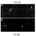
- FIG. 6A is an image depicting FM1–43 labeling of rMSC-derived neurons.
- the data disclosed herein demonstrate that rMSC-derived neurons depolarized using KCl demonstrate intense labeling of terminal putative growth cones (indicated by unfilled triangle).
- FIG. 6B is an image depicting FM1–43 labeling of rMSC-derived neurons.
- the data disclosed herein demonstrate that rMSC-derived neurons depolarized using KCl demonstrate intense labeling of terminal putative growth cones (indicated by unfilled triangle).
- FIG. 8A is an image depicting differentiation of human MSCs.
- the data disclosed herein demonstrate that human MSCs differentiate into neurons and express high levels of NSE (dark brown).
- a lighter stained transitional cell (indicated by ) is depicted at lower left.
- FIG. 8B is an image depicting that an NSE-positive hMSC-derived neuron elaborates a process exhibiting neuronal-like terminal bulb morphology.
- FIG. 8C is an image depicting a phase-contrast image of paired NSE-positive neurons.
- the data disclosed herein demonstrate growth cone morphologies with filopodial extensions (double arrow). The image is enlarged 50% to show detail.
- FIG. 9 is an image depicting nestin and trkA expression in differentiating rMSC-derived neurons.
- FIGS. 9A–9C demonstrate cells stained for nestin expression at 5 hours, 1 day, and 6 days, respectively.
- the invention is based on the discovery that contacting marrow stromal cells with a neural-differentiation inducing agent mediates differentiation of the cells into neuronal-like cells expressing a variety of neuron-specific markers (e.g., NeuN, neurofilament-M, neuron-specific enolase [NSE], tau, nestin, trkA, and the like).
- a neural-differentiation inducing agent mediates differentiation of the cells into neuronal-like cells expressing a variety of neuron-specific markers (e.g., NeuN, neurofilament-M, neuron-specific enolase [NSE], tau, nestin, trkA, and the like).
- the cells exhibit other neuron-like phenotypic characteristics such as, but not limited to, spherical and refactile cell bodies exhibiting typical neuronal perikaryal appearance, cell bodies extending long processes terminating in growth cones and filopodia typical of neurons, and labeling of the growth cones by the fluorescent dye FM1–43, which typically labels neuronal transmitter release and synaptic vesicle recycling.
- the methods disclosed herein induce marrow stromal cell differentiation into neuronal cells.
- Such methods are crucial in the development of cell-based therapeutics for treatment of central nervous system (CNS) disorders, diseases or conditions. Indeed, prior to the present invention, the lack of source of neuronal cells, which can be introduced into the CNS of a human patient, has severely impeded the development of CNS therapeutics.
- the invention includes a method of inducing an isolated marrow stromal cell to differentiate into an isolated neuronal cell.
- Embodiments of the method of the invention are described in the Examples section herein.
- cells are isolated from a donor, stromal cells are obtained therefrom, usually using a cell-sorting method, and the stromal cells are subsequently cultured in vitro.
- the donor may be a rat, for example, or the donor may be a human.
- the invention is intended to encompass a mammalian donor and should not be limited to the specific donors disclosed herein.
- the cells are pre-treated with an effective amount of a neuronal differentiation-inducing compound which is introduced into the cell culture for a period of time.
- the length of time may vary according to the precise method being contemplated and should not be construed as limiting the invention in any way.
- the cells are transferred to a serum-free medium containing an amount of the same neuronal differentiation-inducing compound.
- Neuronal morphology is evident within about an hour, see FIG. 2 for example, and the morphology becomes more evident steadily over time. Neuronal marker expression is also apparent within about 30 minutes after treatment.
- Neuronal cells so differentiated also eventually express several protein markers, including but not limited to, tyrosine hydroxylase, tubulin, choline acetyltransferase, synaptophysin, and TOAD, which are all proteins necessarily associated with neurons and neuronal processes.
- protein markers including but not limited to, tyrosine hydroxylase, tubulin, choline acetyltransferase, synaptophysin, and TOAD, which are all proteins necessarily associated with neurons and neuronal processes.
- neuronal cells are useful in treating patients afflicted with diseases of the cholinergic and catecholaminergic systems, and more generally, patients afflicted with diseases of the central nervous system.
- antioxidants serve as the neuronal differentiation-inducing compounds, including but not limited to, beta-mercaptoethanol, dimethylsulfoxide, butylated hydroxytoluene, butylated hydroxyanisole, ascorbic acid, dimethylfumarate, and n-acetylcysteine.
- Particularly preferred embodiments as demonstrated in the Examples section herein disclosed include beta-mercaptoethanol, dimethylsufloxide and a combination of dimethylsulfoxide and butylated hydroxyanisole as the favored antioxidants.
- the invention is not limited to those antioxidants disclosed herein and should be construed to include all antioxidants, as well as other compounds which induce neuronal differentiation of marrow stromal cells.
- the invention also contemplates use of growth factors as the neuronal differentiation-inducing compounds in the method of inducing differentiation of MSCs to neuronal cells.
- growth factors include, but are not limited to fibroblast growth factor 2, platelet-derived growth factor, and nerve growth factor, as well as related agents.
- Neuronal identity can be confirmed by staining the differentiated neuronal cells for detection of neuron-specific markers.
- markers are neurofilament-M (NF-M), tau protein, Neu-N, neuron-specific enolase (NSE), nestin, and trkA.
- NF-M neurofilament-M
- tau protein tau protein
- Neu-N neuron-specific enolase
- NSE neuron-specific enolase
- nestin trkA
- Progressive differentiation of the marrow stromal cell to the neuronal cell corresponds with an increase in each of these markers, indicating that neuronal cells are produced.
- Further characterization can be accomplished using known immunocytochemical and antibody techniques. For example, immunocytochemical analysis of these neuronal cells reveals that the cells also express proteins that are associated with naturally-differentiated neurons. Such proteins include, but are not limited to tubulin, TOAD, and synaptophysin. Antibody detection of choline acetyltransferase and
- Neuronal cells so differentiated are useful in treating patients afflicted with any of a wide variety of central nervous system diseases, disorders, or conditions.
- the invention also includes a method for producing an isolated neuronal cell from isolated marrow stromal cells.
- the method comprises differentiating an isolated marrow stromal cell in the same general manner as recited above, thereby producing an isolated neuronal cell.
- the isolated neuronal cell recited in both of the methods above may be transfected with an isolated nucleic acid encoding a therapeutic protein.
- the therapeutic protein when expressed, will treat a patient having a disease, disorder, or condition of the central nervous system.
- beneficial proteins are well-known in the art and are set forth in, for example, WO 96/30031 and WO 99/43286. Such examples include, but are not limited to, cytokines, chemokines, neurotrophins, other trophic proteins, growth factors, antibodies, and glioma toxic protein.
- the transfected neuronal cells encoding such proteins are administered to a patient, the neuronal cells will beneficially influence cells which are already present in the central nervous system.
- transfected neuronal cells which are introduced into the central nervous system may be used to repair any central nervous system damage, and/or to combat tumors of the central nervous system.
- CNS diseases diseases, disorders, or conditions
- CNS diseases diseases, disorders, or conditions
- diseases include, but are not limited to, genetic diseases of the CNS (e.g., Tay-Sach's, Sandhoff s disease, Hurler's syndrome, Krabbe's disease), birth-induced traumatic CNS injury, adult CNS diseases, disorders or conditions (e.g., Parkinson's, Alzheimer's, and Huntington's diseases, elderly dementia, epilepsy, amyotropic lateral sclerosis, multiple sclerosis, trauma, tumors, stroke, and the like) and degenerative diseases and traumatic injury of the spinal cord.
- genetic diseases of the CNS e.g., Tay-Sach's, Sandhoff s disease, Hurler's syndrome, Krabbe's disease
- birth-induced traumatic CNS injury e.g., Parkinson's, Alzheimer's, and Huntington's diseases, elderly dementia, epilepsy, amyotropic lateral sclerosis, multiple sclerosis, trauma, tumors, stroke,
- transfected neuronal cells may be used for treatment of a number of genetic diseases of the central nervous system, including, but not limited to, Tay-Sachs disease and the related Sandhoffs disease, Hurler's syndrome and related mucopolysaccharidoses and Krabbe's disease.
- these diseases also produce lesions in the spinal cord and peripheral nerves and they also have non-neurological effects. While the non-neurological effects of these diseases may be treatable by bone marrow transplantation, the central nervous system effects do not improve despite bone marrow transplantation.
- the method of the present invention is useful to address the central nervous system effects of these types of diseases.
- head trauma during birth or following birth is treatable by introducing these neuronal cells directly into the central nervous system of the children.
- Central nervous system tumor formation in children is also treatable using the methods of the present invention.
- Adult diseases of the central nervous system are also treatable by administering isolated neuronal cells to the adult.
- Such adult diseases include but are not limited to, Parkinson's disease, Alzheimer's disease, spinal cord injury, stroke, trauma, tumors, degenerative diseases of the spinal cord such as amyotropic lateral sclerosis, Huntington's disease and epilepsy. Treatment of multiple sclerosis is also contemplated.
- Treatment of spinal cord injuries is also possible using the method of the present invention.
- Prior art methods of treating spinal cord injuries involve using fibroblast cells to deliver neurotrophins to the site of spinal cord lesions in animals.
- the neurotrophins delivered in this manner, reduce the lesion or otherwise treat the injury.
- fibroblasts produce large amounts of collagen, causing fibrosis at the site of the lesion, thus negating the beneficial effects of the treatment.
- Delivery of neurotrophins to spinal cord lesions using transfected neuronal cells is advantageous over prior art methods because neuronal cells do not produce large amounts of collagen and therefore should not cause fibrosis.
- the invention further includes a method of treating a human patient having a disease, disorder, or condition of the central nervous system by administering the differentiated neuronal cells of the invention to the central nervous system of the patient.
- Methods of treating a human patient using MSCs are described in WO 96/30031 and WO 99/43286, which are incorporated by reference as if set forth in their entirety herein.
- Methods of administering differentiated neuronal cells to a patient are identical to those used for MSCs as described in WO 96/30031 and WO 99/43286.
- the methods encompass introduction of an isolated nucleic acid encoding a beneficial protein into differentiated neuronal cells and also encompassusing differentiated neuronal cells themselves in cell-based therapeutics where a patient is in need of the administration of such cells.
- the differentiated neuronal cells are preferably administered to a human, and further, the neuronal cells are preferably administered to the central nervous system of the human. In some instances, the differentiated neuronal cells are administered to the corpus striatum portion of the human brain.
- the precise site of administration of the neuronal cells will depend on any number of factors, including but not limited to, the site of the lesion to be treated, the type of disease being treated, the age of the human and the severity of the disease, and the like. Determination of the site of administration is well within the skill of the artisan versed in the administration of cells to mammals.
- the mode of administration of the differentiated neural cells to the central nervous system of the human may vary depending on several factors including but not limited to, the type of disease being treated, the age of the human, whether the neuronal cells have isolated DNA introduced therein, and the like.
- An example of administration of neuronal cells directly into brain tissue is provided herein in the experimental details section.
- cells are introduced into the brain of a mammal by first creating a hole in the cranium through which the cells are passed into the brain tissue.
- Cells may be introduced by direct injection, by using a shunt, or by any other means used in the art for the introduction of compounds into the central nervous system.
- an element means one element or more than one element.
- central nervous system should be construed to include brain and/or the spinal cord of a mammal.
- the term may also include the eye and optic nerve in some instances.
- stromal cells As used herein, “stromal cells”, “isolated marrow stromal cells”, and “MSCs” are used interchangeably and are meant to refer to the small fraction of cells in bone marrow which can serve as stem cell-like precursors of osteocytes, chondrocytes, and adipocytes and which are isolated from bone marrow by their ability adhere to plastic dishes. Marrow stromal cells may be derived from any animal. In some embodiments, stromal cells are derived from primates, preferably humans.
- anti-oxidant is meant to refer to those substances that inhibit oxidation or reactions promoted by oxygen or peroxides.
- anti-oxidants include, but are not limited to, beta-mercaptoethanol, dimethylsulfoxide, butylated hydroxytoluene, butylated hydroxyanisole, ascorbic acid, dimethylfumarate, and n-acetylcysteine.
- the terms “beneficial protein” and “therapeutic protein” are used interchangeably and are meant to refer to a protein which can compensate for the protein encoded by a defective gene and/or insufficient gene expression that is causally linked to the disease or symptoms of the disease, disorder or condition characterized by a gene defect.
- the presence of the protein alleviates, reduces, prevents or causes to be alleviated, reduced or prevented, the causes and/or symptoms that characterize the disease, disorder or condition.
- a disease, disorder or condition which can be treated with a beneficial or therapeutic protein is meant to refer to a disease, disorder or condition that can be treated or prevented by the presence of a protein which alleviates, reduces, prevents or causes to be alleviated, reduced or prevented, the causes and/or symptoms that characterize the disease, disorder or condition.
- Diseases, disorders and conditions which can be treated with a beneficial protein include diseases, disorders and conditions characterized by a gene defect as well as those which are not characterized by a gene defect but which nonetheless can be treated or prevented by the presence of a protein which alleviates, reduces, prevents or causes to be alleviated, reduced or prevented, the causes and/or symptoms that characterize the disease, disorder or condition.
- isolated nucleic acid should be construed to refer to a nucleic acid sequence, or segment, or fragment which has been purified from the sequences which flank it in a naturally occurring state, e.g., a DNA fragment which has been removed from the sequences which are normally adjacent to the fragment e.g., the sequences adjacent to the fragment in a genome in which it naturally occurs.
- nucleic acids which have been substantially purified from other components which naturally accompany the nucleic acid, e.g., RNA or DNA or proteins which naturally accompany it in the cell.
- transfected cells is meant to refer to cells to which a gene construct has been provided using any technology used to introduce nucleic acid molecules into cells such as, but not limited to, classical transfection (calcium phosphate or DEAE dextran mediated transfection), electroporation, microinjection, liposome-mediated transfer, chemical-mediated transfer, ligand mediated transfer or recombinant viral vector transfer.
- classical transfection calcium phosphate or DEAE dextran mediated transfection
- electroporation microinjection
- liposome-mediated transfer liposome-mediated transfer
- chemical-mediated transfer ligand mediated transfer or recombinant viral vector transfer.
- differentiated should be construed to mean the induction of a differentiated phenotype in an undifferentiated cell by coculturing the undifferentiated cell in the presence of a substantially homogeneous population of differentiated cells, in the presence of products of differentiated cells or in the presence of an inducer of cell differentiation.
- neuronal cell as used herein should be construed to mean an MSC differentiated such that it expresses at least one of the following neuronal markers: neuron-specific enolase (NSE), NeuN, neurofilament M, or tau protein.
- NSE neuron-specific enolase
- NeuN neurofilament M
- tau protein tau protein
- nerve as used herein should be construed to mean a nerve cell capable of receiving and conducting electrical impulses from the central nervous system.
- a nerve cell or “neuron” typically comprises a cell body, an axon, axon terminals, and dendrites.
- neuronal differentiation-inducing compound refers to those compounds capable of inducing differentiation of a stromal cell into a neuronal cell. These compounds include, but are not limited to antioxidants, trophic factors, and growth factors.
- Bone marrow stromal cells exhibit multiple traits of a stem cell population. They can be greatly expanded in vitro, and induced to differentiate into multiple mesenchymal cell types (see, e.g., WO 96/30031; WO 99/43286). However, differentiation to non-mesenchymal fates has not been demonstrated. Here, adult rat stromal cells were expanded as undifferentiated cells in culture for more than 14 passages, indicating their proliferative capacity.
- a novel treatment protocol induced the stromal cells to exhibit a neuronal phenotype, expressing various neuron-specific markers, i.e., neuron-specific enolase (NSE), NeuN, neurofilament-M, tau, nestin, and trkA.
- NSE neuron-specific enolase
- NeuN neurofilament-M
- tau tau
- nestin nestin
- trkA neuron-specific enolase
- the refractile cell bodies of the treated cells extended long processes terminating in growth cones and filopodia typical of neuronal cells.
- Rat MSCs were originally cultured in alpha-Modified Eagle's Medium (alpha-MEM) supplemented with 20% FBS, 2 mM L-glutamine, 100 units per milliliter penicillin, 100 milligrams per milliliter streptomycin and 25 nanogramps per milliliter amphotericin B. For each passage the cells were plated at about 8,000 cells per square centimeter and grown to confluency. At passage 6 the cells were transferred to DMEM (pH 8.0)/20% FBS without additional supplementation, and maintained beyond passage 14.
- the rat MSCs were obtained with a protocol and procedures approved by the Institutional IACUC. The human samples were obtained from volunteers with informed consent and according to a protocol approved by the Institutional Review Board.
- rMSCs were fixed with 4% paraformaldehyde, incubated with primary antibody overnight at 4° C., incubated with secondary antibody for one hour, followed by exposure to avidin-biotin complex for one hour at 25° C.
- DAB Diaminobenzidene
- Cultures were treated with DMSO/BHA in serum-free media (SFM) for approximately 4 hours. The cells were maintained for an additional 30 minutes in artificial cerebral spinal fluid (aCSF)/BHA. Cells were labeled in aCSF containing 1 millimolar FM1–43 and 75 mM KCl for 60 seconds. The labeling mixture was removed, the cultures were washed twice with aCSF, and the cells were incubated in aCSF for 60 minutes to reduce background staining. Cultures were fixed with 4% paraformaldehyde, and soaked for 24 hours in phosphate buffered saline (PBS) before analysis.
- PBS phosphate buffered saline
- Rat mesenchymal stromal cells were isolated from the femurs of adult rats and propagated in vitro (Azizi et al., 1998, Proc. Natl. Acad. Sci. USA 95:3908–3913).
- the data disclosed in FIG. 1A demonstrate that the distribution of cells stained with antibody to CD11b (unfilled) does not differ from that of isotype control (filled), indicating the rMSC cultures do not contain significant numbers of contaminating CD11b-expressing cells.
- the data disclosed in FIG. 1B also demonstrate that the intensity of staining does not differ between CD45 antibody (unfilled) and control (filled) profiles, indicating that cultured rMSCs are not contaminated by CD45-expressing cells.
- FIG. 1A Fluorescent cell sorting at passage one also demonstrated that the cells were negative for CD11b ( FIG. 1A ), and CD45 ( FIG. 1B ), which are cell surface markers associated with lymphohematopoietic cells. Therefore, there was no evidence of hematopoietic precursors in the cultures.
- rMSCs were initially maintained in sub-confluent cultures in media supplemented with 1 mM beta-mercaptoethanol (BME) for 24 hours. Under these conditions no changes in morphology were evident.
- BME beta-mercaptoethanol
- the cells were transferred to serum-free medium containing 1–10 millimolar BME (SFM/BME).
- SFM/BME millimolar BME
- the percentage of cells adopting a neuronal morphology increased at higher BME concentrations, and was enhanced by BME pretreatment.
- FIG. 2C Responsive cells progressively assumed neuronal morphological characteristics over the first 3 hours. Initially, cytoplasm in the flat rMSCs retracted towards the nucleus, forming a contracted multipolar, cell body, leaving membranous, process-like extensions peripherally (0–90 minutes).
- Treated cells exhibited increased expression of the neuronal marker NSE within 30 minutes of treatment. Over the subsequent 2 hours cell bodies became increasingly spherical and retractile, exhibiting a typical neuronal perikaryal appearance. Processes continued to elaborate, developing growth cone-like terminal expansions and filopodial extensions (see, e.g., FIGS. 2G and 2H ). Cellular processes exhibited primary and secondary branches, and underwent dynamic growth. Retraction, as well as extension, was evident as demonstrated by the fact that the cell marked by an arrow at 120 minutes ( FIG. 2E ) was initially contacted by a neighboring process (marked by “>”), which retracted by 180 minutes ( FIG. 2G ), with loss of contact.
- NSE neuronal marker neuron-specific enolase
- FIG. 3A Progressive transition of rMSCs to a neuronal phenotype coincided with increased expression of NSE ( FIG. 3A ).
- Cells at intermediate stages in the differentiation sequence ( ) exhibited transitional morphologies and light brown staining, indicating synchrony of morphologic and molecular differentiation.
- rMSC-derived neurons displayed distinct neuronal morphologies ( FIG. 3B ), ranging from simple bipolar ( ⁇ ) to large, extensively branched multipolar cells (arrow).
- FIG. 3C Clusters of differentiated cells exhibited intense NSE positivity, and processes formed extensive networks ( FIG. 3E ). Even within these clusters, typical, flat rMSCs (>) were only lightly stained, consistent with their undifferentiated state.
- FIG. 3F Western blot analysis confirmed the expression of low levels of NSE protein in uninduced rMSCs. Induction of the neuronal phenotype resulted in a dramatic increase in NSE expression, consistent with the immunocytochemical data.
- NeuN a neuron-specific marker expressed in post-mitotic cells
- NeuN staining was confined to the nucleus and surrounding cytoplasm of positive cells, and did not extend into the processes.
- Nestin an intermediate filament protein, is expressed in neuroepithelial neuronal precursor stem cells, with expression decreasing as the neruon matures.
- Experimental data shows that when the MSC-differentiated neuronal cells are stained to detect nestin, the expression of nestin decreases over time ( FIGS. 9A–9C ).
- staining for trkA a high-affinity nerve growth factor receptor which is present in neurons, demonstrates that trkA levels remain unchanged throughout the maturation process of the MSC-differentiated neuronal cell ( FIGS. 9D–9F ).
- rMSCs were treated with other anti-oxidants, e.g., dimethylsulfoxide (DMSO), butylated hydroxyanisole (BHA), or butylated hydroxytoluene (BHT), ascorbic acid, dimethylfumarate, n-acetylcysteine, and the like, both alone and in combination with each other.
- DMSO dimethylsulfoxide
- BHA butylated hydroxyanisole
- BHT butylated hydroxytoluene
- ascorbic acid dimethylfumarate
- n-acetylcysteine n-acetylcysteine
- Each anti-oxidant treatment e.g., DMSO, BHA, BHT, ascorbic acid, dimethylfumarate, n-acetylcysteine, and the like, both alone and in combination
- DMSO dimethylfumarate
- n-acetylcysteine elicited neuronal morphologies with a time course similar to the effects of BME.
- preliminary data suggested that treatment using about 2% (v/v) DMSO and about 200 millimolar BHA (DMSO/BHA) was preferred although a wide range of concentrations elicited neuronal differentiation.
- NF-M neurofilament-M
- NF-M neurofilament-M
- DMSO/BHA treated cultures were then examined for the presence of tau, a neuron-specific microtubule-associated protein expressed by differentiating neurons (Kosik and Finch, 1987, J. Neurosci. 7:3142–3153).
- the data disclosed herein indicate that the method described herein induce neuronal differentiation of marrow stromal cells.
- rMSCs To determine whether individual rMSCs exhibit stem cell characteristics of self-renewal and pluripotentiality, individual clones were analyzed. To establish clones, rMSCs were plated at approximately 10 cells per square centimeters, grown to 50–150 cells per colony, isolated with cloning cylinders, transferred to separate wells and eventually to individual flasks. Single cells replicated as typical rMSCs and differentiated into NSE-positive neurons after BME treatment.
- FIGS. 7A–7D Analysis of four distinct clonal lines is shown in FIGS. 7A–7D .
- Each individual clone generated refractile, process-bearing, NSE-positive cells following BME treatment. Undifferentiated rMSCs (>) and transitional cells ( ) were evident in each clonal line. Therefore, clones derived from a single cell can give rise to both rMSCs and neurons, indicating stem cell characteristics.
- hMSCs The neuronal potential of MSCs was not unique to rodents as demonstrated by the following experiments using MSCs obtained from humans (hMSCs). hMSCs were isolated from a healthy adult donor and grown in vitro (Bjornson et al., 1999, Science 283:534–537). hMSCs resembled their rodent counterparts, growing as large flat cells in the undifferentiated state.
- FIGS. 8A and 8B Cells from passage two were subjected to the neuronal differentiation protocol and stained for NSE or NF-M expression.
- hMSCs After BME treatment, hMSCs attained neuronal characteristics and increased NSE expression in a time frame similar to that observed for rMSCs. Contracted cell bodies elaborated processes and stained strongly for NSE expression within 3 hours ( FIGS. 8A and 8B ). Transitional cells were also evident ( ). Many processes elaborated by hMSC-derived neurons exhibited terminal bulbs (arrow in 8 B), which may represent growth cones. Growth cone morphologies with filopodial extensions ( ⁇ ) were clearly evident on the processes elaborated by paired neurons depicted in the image in FIG. 8C . These cells also expressed NF-M, consistent with their neuronal differentiation ( FIG. 8D ).
- rat and human MSCs retain the capacity to differentiate into non-mesenchymal derivatives, specifically neurons, suggesting that intrinsic genomic mechanisms of commitment, lineage restriction and cell fate are mutable.
- Environmental signals apparently can elicit the expression of pluripotentiality that extends well beyond the accepted fate restrictions of cells originating in classical embryonic germ layers.
- These adult cells are both self-renewing and multipotential (Owens and Friedenstein, 1988, Ciba Foundation Symp. 136, Chichester, U.K. pp. 42–60; Prockop, 1997, Science 276; 71–74; Ferrari et al., 1998, Science 279:1528–1530; Caplan, 1991, J. Orthop. Res.
- peripheral mesenchymal cells can differentiate into neurons in vitro.
- the present invention provides, for the first time, methods of directing differentiation of MSCs into neuronal cells in vitro.
- MSCs are useful in the treatment of a wide variety of neurologic diseases disorders and conditions, and these cells offer significant advantages over other so-called “stem” cells. That is, bone marrow cells are readily accessible, obviating the risks of obtaining neural stem cells from the brain, and provide a renewable population which can be expanded in vitro thereby allowing complex gene manipulations to be performed for ex vivo gene therapy and/or for cell therapy for CNS diseases, disorders or conditions that require administering cells to a CNS site.
- autologous transplantation overcomes the ethical and immunologic concerns associated with the use of fetal tissue.
- MSCs grow rapidly in culture, precluding the need for immortalization, and differentiate into neurons exclusively using the protocols disclosed herein.
- neuronal cells differentiated from MSCs as described herein express various neuron-related proteins.
- immunocytochemical analysis of these differentiated neurons revealed the expression of beta-3 tubulin.
- TOAD-64 a neuronal protein of unknown function, is also detectable using immunocytochemical techniques, as well as synaptophysin, which is associated with synapses and synaptic vesicles.
- these cells Using polyclonal and monoclonal antibody-based procedures, these cells have been demonstrated to express choline acetyltransferase, an enzyme responsible for the synthesis of the neurotransmitter acetylcholine.
- tyrosine hydroxylase the rate-limiting enzyme in catecholamine biosynthesis, was also detected immunocytochemically in a population of these differentiated neurons.
- the differentiated neurons may be therapeutically beneficial to treating those diseases affecting cholinergic and catecholaminergic systems, such as, for example, Alzheimer's disease, Parkinson's disease, or schizophrenia.
- the neurons were transplanted, using sterile technique and known and accepted neurosurgical procedures (1997, Grill et al.; 1995, Gage et al.; 1994, Dunnett et al.), into the hippocampus or striatum of the brain or the dorsal horn of the spinal cord of individual rats. Each rat received a transplant to one of the aforementioned areas. The rats were returned to their cages and received standard postoperative care with access to food and water ad libatum.
- Rats receiving the neuronal transplant were examined 42 days after the transplantation operation took place.
- fluorescence microscopy to detect bisbenzimide-positive transplanted cells
- histologic studies of the hippocampal and striatal regions of the brain revealed that the transplanted neurons survived in the hippocampus. This result indicates that long-term survival of the transplanted, differentiated neurons is possible.
- An examination of the rats receiving the transplanted neurons in the dorsal horn of the spinal cord demonstrated a survival period of at least three days. Further, the processes of the transplanted neurons in this area grew to at least two to three times in length than the cell body diameter.
- transplanted, differentiated neurons express many neuronal proteins, retain viability in vivo, and seemingly exert no detectable deleterious effect on the living animal.
- these neurons create a potential therapeutic treatment for a variety of brain and spinal cord diseases, including, but not limited to, Alzheimer's disease, Parkinson's disease, Schizophrenia, and spinal cord injury resulting from trauma or degeneration.
Landscapes
- Health & Medical Sciences (AREA)
- Life Sciences & Earth Sciences (AREA)
- Engineering & Computer Science (AREA)
- Chemical & Material Sciences (AREA)
- General Health & Medical Sciences (AREA)
- Veterinary Medicine (AREA)
- Animal Behavior & Ethology (AREA)
- Medicinal Chemistry (AREA)
- Public Health (AREA)
- Pharmacology & Pharmacy (AREA)
- Biomedical Technology (AREA)
- Bioinformatics & Cheminformatics (AREA)
- Organic Chemistry (AREA)
- Neurology (AREA)
- Epidemiology (AREA)
- Chemical Kinetics & Catalysis (AREA)
- General Chemical & Material Sciences (AREA)
- Nuclear Medicine, Radiotherapy & Molecular Imaging (AREA)
- Genetics & Genomics (AREA)
- Neurosurgery (AREA)
- Wood Science & Technology (AREA)
- Zoology (AREA)
- Biotechnology (AREA)
- General Engineering & Computer Science (AREA)
- Biochemistry (AREA)
- Microbiology (AREA)
- Cell Biology (AREA)
- Physical Education & Sports Medicine (AREA)
- Plant Pathology (AREA)
- Molecular Biology (AREA)
- Biophysics (AREA)
- Physics & Mathematics (AREA)
- Hospice & Palliative Care (AREA)
- Pain & Pain Management (AREA)
- Immunology (AREA)
- Psychiatry (AREA)
- Psychology (AREA)
- Dermatology (AREA)
- Orthopedic Medicine & Surgery (AREA)
- Micro-Organisms Or Cultivation Processes Thereof (AREA)
Abstract
Description
Claims (11)
Priority Applications (3)
| Application Number | Priority Date | Filing Date | Title |
|---|---|---|---|
| US10/213,526 US7279331B2 (en) | 2000-02-11 | 2002-08-06 | Differentiation of bone marrow cells into neuronal cells and uses therefor |
| US11/849,870 US8486698B2 (en) | 2000-02-11 | 2007-09-04 | Differentiation of bone marrow cells into neuronal cells and uses therefor |
| US13/939,986 US20130302889A1 (en) | 2000-02-11 | 2013-07-11 | Differentiation of Bone Marrow Cells into Neuronal Cells and Uses Therefor |
Applications Claiming Priority (3)
| Application Number | Priority Date | Filing Date | Title |
|---|---|---|---|
| US18185000P | 2000-02-11 | 2000-02-11 | |
| PCT/US2001/004282 WO2001059072A1 (en) | 2000-02-11 | 2001-02-09 | Differentiation of bone marrow cells into neuronal cells and uses therefor |
| US10/213,526 US7279331B2 (en) | 2000-02-11 | 2002-08-06 | Differentiation of bone marrow cells into neuronal cells and uses therefor |
Related Parent Applications (1)
| Application Number | Title | Priority Date | Filing Date |
|---|---|---|---|
| PCT/US2001/004282 Continuation WO2001059072A1 (en) | 2000-02-11 | 2001-02-09 | Differentiation of bone marrow cells into neuronal cells and uses therefor |
Related Child Applications (1)
| Application Number | Title | Priority Date | Filing Date |
|---|---|---|---|
| US11/849,870 Continuation US8486698B2 (en) | 2000-02-11 | 2007-09-04 | Differentiation of bone marrow cells into neuronal cells and uses therefor |
Publications (2)
| Publication Number | Publication Date |
|---|---|
| US20030203484A1 US20030203484A1 (en) | 2003-10-30 |
| US7279331B2 true US7279331B2 (en) | 2007-10-09 |
Family
ID=22666063
Family Applications (3)
| Application Number | Title | Priority Date | Filing Date |
|---|---|---|---|
| US10/213,526 Expired - Fee Related US7279331B2 (en) | 2000-02-11 | 2002-08-06 | Differentiation of bone marrow cells into neuronal cells and uses therefor |
| US11/849,870 Expired - Fee Related US8486698B2 (en) | 2000-02-11 | 2007-09-04 | Differentiation of bone marrow cells into neuronal cells and uses therefor |
| US13/939,986 Abandoned US20130302889A1 (en) | 2000-02-11 | 2013-07-11 | Differentiation of Bone Marrow Cells into Neuronal Cells and Uses Therefor |
Family Applications After (2)
| Application Number | Title | Priority Date | Filing Date |
|---|---|---|---|
| US11/849,870 Expired - Fee Related US8486698B2 (en) | 2000-02-11 | 2007-09-04 | Differentiation of bone marrow cells into neuronal cells and uses therefor |
| US13/939,986 Abandoned US20130302889A1 (en) | 2000-02-11 | 2013-07-11 | Differentiation of Bone Marrow Cells into Neuronal Cells and Uses Therefor |
Country Status (10)
| Country | Link |
|---|---|
| US (3) | US7279331B2 (en) |
| EP (2) | EP1272614B1 (en) |
| JP (1) | JP2003521935A (en) |
| AT (1) | ATE370225T1 (en) |
| AU (2) | AU3685401A (en) |
| CA (1) | CA2399623A1 (en) |
| DE (1) | DE60129943T2 (en) |
| ES (1) | ES2292564T3 (en) |
| HK (1) | HK1052369B (en) |
| WO (1) | WO2001059072A1 (en) |
Cited By (8)
| Publication number | Priority date | Publication date | Assignee | Title |
|---|---|---|---|---|
| US20080131407A1 (en) * | 2000-02-11 | 2008-06-05 | Philadelphia Health And Education Corporation | Differentiation of bone marrow cells into neuronal cells and uses therefor |
| US20100130607A1 (en) * | 2007-02-08 | 2010-05-27 | Ralf Gold | Neuroprotection in demyelinating diseases |
| WO2011046570A1 (en) | 2009-10-16 | 2011-04-21 | The University Of Medicine And Dentistry Of New Jersey | Method for treating chronic nerve tissue injury using a cell therapy strategy |
| WO2011047289A1 (en) | 2009-10-16 | 2011-04-21 | University Of Medicine And Dentistry Of New Jersey | Closed system separation of adherent bone marrow stem cells for regenerative medicine applications |
| US20110124615A1 (en) * | 2003-09-09 | 2011-05-26 | Fumapharm Ag | Use of fumaric acid derivatives for treating cardiac insufficiency, and asthma |
| WO2012156968A2 (en) | 2011-05-19 | 2012-11-22 | Ariel - University Research And Development Company, Ltd. | Use of mesenchymal stem cells for the improvement of affective and cognitive function |
| US8399514B2 (en) | 2007-02-08 | 2013-03-19 | Biogen Idec Ma Inc. | Treatment for multiple sclerosis |
| US8921533B2 (en) | 2011-07-25 | 2014-12-30 | Chromatin Technologies | Glycosylated valproic acid analogs and uses thereof |
Families Citing this family (36)
| Publication number | Priority date | Publication date | Assignee | Title |
|---|---|---|---|---|
| DE19853487A1 (en) | 1998-11-19 | 2000-05-25 | Fumapharm Ag Muri | Use of dialkyl fumarate for treating transplant rejection and autoimmune disease |
| US20030104997A1 (en) | 2001-09-05 | 2003-06-05 | Black Ira B. | Multi-lineage directed induction of bone marrow stromal cell differentiation |
| US9969980B2 (en) * | 2001-09-21 | 2018-05-15 | Garnet Biotherapeutics | Cell populations which co-express CD49c and CD90 |
| US20040259254A1 (en) * | 2001-10-30 | 2004-12-23 | Osamu Honmou | Method for inducing differentiation mesodermal stem cells, es cells or immortalized cells into nervous system cells |
| EP1471970A4 (en) | 2002-01-14 | 2006-08-02 | Univ Illinois | NEW MULTIPOTENTE MAMMALIAN STEM CELLS AND COMPOSITIONS, METHOD OF PREPARING THEM AND METHOD FOR THEIR ADMINISTRATION |
| DK1479767T3 (en) * | 2002-02-06 | 2013-11-25 | Sanbio Inc | Method of differentiating / inducing bone marrow to nerve cells by transferring Notch gene |
| US9969977B2 (en) | 2002-09-20 | 2018-05-15 | Garnet Biotherapeutics | Cell populations which co-express CD49c and CD90 |
| EP2399990B1 (en) | 2003-06-27 | 2015-07-22 | DePuy Synthes Products, Inc. | Cells derived from post-partum umbilical cord for use in treatment of disease of the heart and circulatory system |
| US8518390B2 (en) | 2003-06-27 | 2013-08-27 | Advanced Technologies And Regenerative Medicine, Llc | Treatment of stroke and other acute neural degenerative disorders via intranasal administration of umbilical cord-derived cells |
| US8790637B2 (en) | 2003-06-27 | 2014-07-29 | DePuy Synthes Products, LLC | Repair and regeneration of ocular tissue using postpartum-derived cells |
| US9572840B2 (en) | 2003-06-27 | 2017-02-21 | DePuy Synthes Products, Inc. | Regeneration and repair of neural tissue using postpartum-derived cells |
| US9592258B2 (en) | 2003-06-27 | 2017-03-14 | DePuy Synthes Products, Inc. | Treatment of neurological injury by administration of human umbilical cord tissue-derived cells |
| US8491883B2 (en) | 2003-06-27 | 2013-07-23 | Advanced Technologies And Regenerative Medicine, Llc | Treatment of amyotrophic lateral sclerosis using umbilical derived cells |
| US7875272B2 (en) | 2003-06-27 | 2011-01-25 | Ethicon, Incorporated | Treatment of stroke and other acute neuraldegenerative disorders using postpartum derived cells |
| CA2589041C (en) | 2004-12-23 | 2019-08-20 | DePuy Synthes Products, Inc. | Postpartum cells derived from umbilical cord tissue, and methods of making and using the same |
| EP1835924B1 (en) | 2004-12-23 | 2013-08-21 | Ethicon, Incorporated | Treatment of parkinson's disease and related disorders using postpartum derived cells |
| PT1855700E (en) * | 2005-03-07 | 2013-12-10 | Sanbio Inc | Use of neuronal precursor cells for treatment of central nervous system lesions |
| US8480797B2 (en) | 2005-09-12 | 2013-07-09 | Abela Pharmaceuticals, Inc. | Activated carbon systems for facilitating use of dimethyl sulfoxide (DMSO) by removal of same, related compounds, or associated odors |
| EP1966229B1 (en) | 2005-09-12 | 2015-10-21 | Abela Pharmaceuticals, Inc. | Systems for removing dimethyl sulfoxide (dmso) or related compounds, or odors associated with same |
| EP1937286B1 (en) | 2005-09-12 | 2016-03-09 | Abela Pharmaceuticals, Inc. | Compositions comprising dimethyl sulfoxide (dmso) |
| WO2007033180A1 (en) | 2005-09-12 | 2007-03-22 | Abela Pharmaceuticals, Inc. | Materials for facilitating administration of dimethyl sulfoxide (dmso) and related compounds |
| WO2007070870A1 (en) | 2005-12-16 | 2007-06-21 | Ethicon, Inc. | Compositions and methods for inhibiting adverse immune response in histocompatibility-mismatched transplantation |
| US8741638B2 (en) | 2005-12-19 | 2014-06-03 | DePuy Synthes Products, LLC | In vitro expansion of postpartum-derived cells in roller bottles |
| US9125906B2 (en) | 2005-12-28 | 2015-09-08 | DePuy Synthes Products, Inc. | Treatment of peripheral vascular disease using umbilical cord tissue-derived cells |
| KR100814504B1 (en) | 2006-11-03 | 2008-03-18 | 전북대학교산학협력단 | How to differentiate bone marrow mesenchymal stem cells into neurons |
| NZ582616A (en) | 2007-06-15 | 2012-07-27 | Garnet Biotherapeutics Inc | Treatment of diseases and disorders using self-renewing colony forming cells cultured and expanded in vitro |
| ES2525718T3 (en) | 2007-10-05 | 2014-12-29 | DePuy Synthes Products, LLC | Repair and regeneration of renal tissue by cells derived from human umbilical cord tissue |
| EP2217252A2 (en) * | 2007-10-29 | 2010-08-18 | Hadasit Medical Research Services & Development Limited | Human stem cell-derived neural precursors for treatment of autoimmune diseases of the central nervous system |
| PT2222839E (en) | 2007-12-03 | 2015-11-30 | Sanbio Inc | Methods and compositions for modulating differentiation of pluripotential cells |
| US8236538B2 (en) | 2007-12-20 | 2012-08-07 | Advanced Technologies And Regenerative Medicine, Llc | Methods for sterilizing materials containing biologically active agents |
| BRPI0921494A2 (en) | 2008-11-03 | 2018-10-30 | Prad Reasearch And Development Ltd | method of planning a underground forming sampling operation, method of controlling a underground forming sampling operation, method of controlling a drilling operation for an underground formation, and method of sampling during the drilling operation. |
| US10179900B2 (en) | 2008-12-19 | 2019-01-15 | DePuy Synthes Products, Inc. | Conditioned media and methods of making a conditioned media |
| JP5518893B2 (en) | 2008-12-19 | 2014-06-11 | アドバンスト・テクノロジーズ・アンド・リジェネレイティブ・メディスン・エルエルシー | Treatment of lungs and lung diseases and disorders |
| EP2411504B1 (en) | 2009-03-26 | 2017-05-10 | DePuy Synthes Products, Inc. | Human umbilical cord tissue cells as therapy for alzheimer's disease |
| WO2011053874A1 (en) | 2009-10-30 | 2011-05-05 | Tandem Abela Development Group Llc | Dimethyl sulfoxide (dmso) and methylsulfonylmethane (msm) formulations to treat osteoarthritis |
| EP2794854B1 (en) | 2011-12-23 | 2018-06-20 | DePuy Synthes Products, Inc. | Detection of human umbilical cord tissue-derived cells |
Citations (7)
| Publication number | Priority date | Publication date | Assignee | Title |
|---|---|---|---|---|
| WO1995000632A1 (en) | 1993-06-18 | 1995-01-05 | Amgen Inc. | Medium for long-term proliferation and development of cells |
| WO1996030031A1 (en) | 1995-03-28 | 1996-10-03 | Thomas Jefferson University | Isolated stromal cells and methods of using the same |
| US5639618A (en) | 1994-05-13 | 1997-06-17 | Plurion, Inc. | Method of isolating a lineage specific stem cell in vitro |
| WO1999043286A2 (en) | 1998-02-24 | 1999-09-02 | Mcp Hahnemann University | Isolated stromal cells for use in the treatment of diseases of the central nervous system |
| WO1999056759A1 (en) | 1998-05-07 | 1999-11-11 | University Of South Florida | Bone marrow cells as a source of neurons for brain and spinal cord repair |
| US6090622A (en) | 1997-03-31 | 2000-07-18 | The Johns Hopkins School Of Medicine | Human embryonic pluripotent germ cells |
| WO2001083715A2 (en) | 2000-05-01 | 2001-11-08 | THE GOVERNMENT OF THE UNITED STATES OF AMERICA as represented by the Secretary, | Derivation of midbrain dopaminergic neurons from embryonic stem cells |
Family Cites Families (1)
| Publication number | Priority date | Publication date | Assignee | Title |
|---|---|---|---|---|
| ES2292564T3 (en) * | 2000-02-11 | 2008-03-16 | Philadelphia Health And Education Corporation | DIFFERENTIATION OF CELLS OF THE OSEA MEDULA IN NEURON CELLS AND ASSOCIATED USES. |
-
2001
- 2001-02-09 ES ES01909061T patent/ES2292564T3/en not_active Expired - Lifetime
- 2001-02-09 AU AU3685401A patent/AU3685401A/en active Pending
- 2001-02-09 AU AU2001236854A patent/AU2001236854B2/en not_active Ceased
- 2001-02-09 EP EP01909061A patent/EP1272614B1/en not_active Expired - Lifetime
- 2001-02-09 EP EP06012529A patent/EP1749884A1/en not_active Withdrawn
- 2001-02-09 DE DE60129943T patent/DE60129943T2/en not_active Expired - Lifetime
- 2001-02-09 WO PCT/US2001/004282 patent/WO2001059072A1/en not_active Ceased
- 2001-02-09 HK HK03104584.1A patent/HK1052369B/en not_active IP Right Cessation
- 2001-02-09 CA CA002399623A patent/CA2399623A1/en not_active Abandoned
- 2001-02-09 AT AT01909061T patent/ATE370225T1/en not_active IP Right Cessation
- 2001-02-09 JP JP2001558212A patent/JP2003521935A/en active Pending
-
2002
- 2002-08-06 US US10/213,526 patent/US7279331B2/en not_active Expired - Fee Related
-
2007
- 2007-09-04 US US11/849,870 patent/US8486698B2/en not_active Expired - Fee Related
-
2013
- 2013-07-11 US US13/939,986 patent/US20130302889A1/en not_active Abandoned
Patent Citations (7)
| Publication number | Priority date | Publication date | Assignee | Title |
|---|---|---|---|---|
| WO1995000632A1 (en) | 1993-06-18 | 1995-01-05 | Amgen Inc. | Medium for long-term proliferation and development of cells |
| US5639618A (en) | 1994-05-13 | 1997-06-17 | Plurion, Inc. | Method of isolating a lineage specific stem cell in vitro |
| WO1996030031A1 (en) | 1995-03-28 | 1996-10-03 | Thomas Jefferson University | Isolated stromal cells and methods of using the same |
| US6090622A (en) | 1997-03-31 | 2000-07-18 | The Johns Hopkins School Of Medicine | Human embryonic pluripotent germ cells |
| WO1999043286A2 (en) | 1998-02-24 | 1999-09-02 | Mcp Hahnemann University | Isolated stromal cells for use in the treatment of diseases of the central nervous system |
| WO1999056759A1 (en) | 1998-05-07 | 1999-11-11 | University Of South Florida | Bone marrow cells as a source of neurons for brain and spinal cord repair |
| WO2001083715A2 (en) | 2000-05-01 | 2001-11-08 | THE GOVERNMENT OF THE UNITED STATES OF AMERICA as represented by the Secretary, | Derivation of midbrain dopaminergic neurons from embryonic stem cells |
Non-Patent Citations (51)
| Title |
|---|
| Abboud et al., "Peptide growth factors stimulate macrophage colony-stimulating factor in murine stromal cells," Blood, 78(1):103-109, Jul. 1, 1991. |
| Azizi et al., 1998, Proc. Natl. Acad. Sci. USA 95:3908-3913. |
| Betz et al. 1992, J. Neurosci. 12:363-375. |
| Betz et al. 1992, Science 255:200-203. |
| Bjornson et al. 1999, Science 283:534-537. |
| Bruder et al. 1998, Clin. Orthop. Relat. Res. 355S:S247-S256. |
| Caplan 1991, J. Orthop. Res. 9:641-650. |
| Carden et al. 1987, Neurosci. 7:3489-3504. |
| Correia et al., Annals of Medicine, 2005, vol. 37, pp. 487-498. * |
| Diefenbach et al. 1999, J. Neurosci. 19:9436-9444. |
| Fernandez et al., Surgical Neurology, 2006, vol. 65, pp. 223-237. * |
| Ferrari et al. 1998, Science 279:1528-1530. |
| Flax et al. 1998, Nature Biotech. 16:1033-1039. |
| Gage et al. 1995, Annu. Rev. Neurosci. 18:159-192. |
| Gage et al. 1995, Proc. Natl. Acad. Sci. USA 92:11879-11883. |
| Horwitz et al. 1999, Nature Med. 5:309-313. |
| Ishii et al. 1993, Neurosci. Lett. 163:159-162. |
| Johansson et al., 1999, Cell 96:25-34. |
| Kopen et al. 1999, Proc. Natl. Acad. Sci. 96:10711-10716. |
| Kosik et al. 1987, J. Neurosci. 7:3142-3153. |
| Kuznetsov et al. 1997, Brit. J. Haemotology 97:561-570. |
| Lemiscka 1999, Ann. N.Y. Acad. Sci. 872:274-288. |
| Lundberg et al. 1996, Brain Res. 737:295-300. |
| Lundberg et al. 1997, Exp. Neurol. 145:342-360. |
| Majumdar et al. 1998, J. Cell Physiol. 176:57-66. |
| McKay 1999 Nature Med. 5:261-262. |
| Mena et al., "Glia protect fetal midbrain dopamine neurons in culture from L-DOPA toxicity through multiple mechanisms," J. Neural Transmission, 104(4-5):317-328, 1997. |
| Morshead et al. 1994, Neuron 13:1071-1082. |
| Nakajima et al., "Retinoids (all-trans and 9-cis retinoic acid) stimulate production of macrophange colony-stimulating factor and granulocyte-macrophane colony-stimulating factor by human bone marrow stromal cells," Blood, 84(12):4107-4115, Dec. 15, 1994. |
| Neuhuber et al., Journal of Neuroscience Research, 2004, vol. 77, pp. 192-204. * |
| Owens et al. 1988, Ciba Foundation Symp. 136, Chichester, U.K. pp. 42-60. |
| Pechumer et al. 1993, Lab. Invest. 69:743-749. |
| Pereira et al. 1995, Proc. Natl. Acad. Sci. USA 92:4857-4861. |
| Pittenger et al. 1999, Science 284:143-147. |
| Prockop 1997, Science 276:71-74. |
| Reid et al. 1991, Clin. Pathol. 44:483-486. |
| Renfranz et al. 1991, Cell 66:713-729. |
| Reynolds et al. 1992, Science 255:1707-1710. |
| Richards et al. 1992, Proc. Natl. Acad. Sci. USA 89:8591-8595. |
| Rickard et al., "Isolation and characterization of osteoblast precursor cells from human bone marrow," J. Bone Mineral Res., 11(3):312-324, 1996. |
| Sakai et al., 1995, Experimental Cell Research 217:395-403. |
| Sanchez-Ramos et al., 2000, Experimental Neurology 164:247-256. |
| Sarnat et al. 1998, Brain Res. 20:88-94. |
| Satomura et al., "Receptor tyrosine kinase expression in human bone marrow stromal cells," J. Cell Physiol., 177:426-438, 1994. |
| Strelau et al., 2000, J. Neurosci. 20(23):8597-8603. |
| Svendsen et al. 1997, Exp. Neurol. 148:135-146. |
| Tanaka et al., "Effect of platelet-derived growth factor on DNA synthesis and gene expression in bone marrow stromal cells derived from adult and old rats," J. Cell Physiol., 164:367-375, 1995. |
| vanObberghen et al. 1988, J. Neurosci. Res. 19:450-456. |
| Vescovi et al. 1993, Neuron 11:951-966. |
| Woodbury et al., 2000, J. Neurosci. 61:364-370. |
| Yan et al., 2001, J. Neurochem. 76:307-311. |
Cited By (10)
| Publication number | Priority date | Publication date | Assignee | Title |
|---|---|---|---|---|
| US20080131407A1 (en) * | 2000-02-11 | 2008-06-05 | Philadelphia Health And Education Corporation | Differentiation of bone marrow cells into neuronal cells and uses therefor |
| US8486698B2 (en) * | 2000-02-11 | 2013-07-16 | University Of Medicine And Dentistry Of New Jersey | Differentiation of bone marrow cells into neuronal cells and uses therefor |
| US20110124615A1 (en) * | 2003-09-09 | 2011-05-26 | Fumapharm Ag | Use of fumaric acid derivatives for treating cardiac insufficiency, and asthma |
| US8980832B2 (en) | 2003-09-09 | 2015-03-17 | Biogen Idec International Gmbh | Use of fumaric acid derivatives for treating cardiac insufficiency, and asthma |
| US20100130607A1 (en) * | 2007-02-08 | 2010-05-27 | Ralf Gold | Neuroprotection in demyelinating diseases |
| US8399514B2 (en) | 2007-02-08 | 2013-03-19 | Biogen Idec Ma Inc. | Treatment for multiple sclerosis |
| WO2011046570A1 (en) | 2009-10-16 | 2011-04-21 | The University Of Medicine And Dentistry Of New Jersey | Method for treating chronic nerve tissue injury using a cell therapy strategy |
| WO2011047289A1 (en) | 2009-10-16 | 2011-04-21 | University Of Medicine And Dentistry Of New Jersey | Closed system separation of adherent bone marrow stem cells for regenerative medicine applications |
| WO2012156968A2 (en) | 2011-05-19 | 2012-11-22 | Ariel - University Research And Development Company, Ltd. | Use of mesenchymal stem cells for the improvement of affective and cognitive function |
| US8921533B2 (en) | 2011-07-25 | 2014-12-30 | Chromatin Technologies | Glycosylated valproic acid analogs and uses thereof |
Also Published As
| Publication number | Publication date |
|---|---|
| CA2399623A1 (en) | 2001-08-16 |
| WO2001059072A1 (en) | 2001-08-16 |
| AU2001236854B2 (en) | 2005-04-14 |
| JP2003521935A (en) | 2003-07-22 |
| US20130302889A1 (en) | 2013-11-14 |
| EP1272614A4 (en) | 2003-05-07 |
| EP1749884A1 (en) | 2007-02-07 |
| HK1052369B (en) | 2007-12-21 |
| EP1272614A1 (en) | 2003-01-08 |
| AU3685401A (en) | 2001-08-20 |
| DE60129943D1 (en) | 2007-09-27 |
| ES2292564T3 (en) | 2008-03-16 |
| ATE370225T1 (en) | 2007-09-15 |
| US20030203484A1 (en) | 2003-10-30 |
| DE60129943T2 (en) | 2008-05-08 |
| HK1052369A1 (en) | 2003-09-11 |
| US20080131407A1 (en) | 2008-06-05 |
| US8486698B2 (en) | 2013-07-16 |
| EP1272614B1 (en) | 2007-08-15 |
Similar Documents
| Publication | Publication Date | Title |
|---|---|---|
| US7279331B2 (en) | Differentiation of bone marrow cells into neuronal cells and uses therefor | |
| AU2001236854A1 (en) | Differentiation of bone marrow cells into neuronal cells and uses therefor | |
| Woodbury et al. | Adult rat and human bone marrow stromal cells differentiate into neurons | |
| US7364900B2 (en) | Multi-lineage directed induction of bone marrow stromal cell differentiation | |
| Scintu et al. | Differentiation of human bone marrow stem cells into cells with a neural phenotype: diverse effects of two specific treatments | |
| Kim et al. | Differentiation of adult bone marrow stem cells into neuroprogenitor cells in vitro | |
| McCoy et al. | Autologous transplants of Adipose-Derived Adult Stromal (ADAS) cells afford dopaminergic neuroprotection in a model of Parkinson's disease | |
| Fujii et al. | Dopaminergic differentiation of stem cells from human deciduous teeth and their therapeutic benefits for Parkinsonian rats | |
| Reynolds et al. | Clonal and population analyses demonstrate that an EGF-responsive mammalian embryonic CNS precursor is a stem cell | |
| CN103396993B (en) | The oligodendroglia of myelinization again and the treatment for spinal cord injury of derived from primate embryonic stem cells | |
| Lambert et al. | Differentiation of human adipose-derived adult stem cells into neuronal tissue: does it work? | |
| Sanchez‐Ramos | Neural cells derived from adult bone marrow and umbilical cord blood | |
| Weiss et al. | Transplantation of porcine umbilical cord matrix cells into the rat brain | |
| US20200332254A1 (en) | Oligodendrocytes Derived from Human Embryonic Stem Cells for Remyelination and Treatment of Spinal Cord Injury | |
| US7547545B2 (en) | Directed in vitro differentiation of marrow stromal cells into neural cell progenitors | |
| Daadi et al. | Dopaminergic neurons from midbrain-specified human embryonic stem cell-derived neural stem cells engrafted in a monkey model of Parkinson’s disease | |
| US20100021434A1 (en) | Isolated Oligodendrocyte-Like Cells and Populations Comprising Same for the Treatment of CNS Diseases | |
| Jurga et al. | Neurogenic potential of human umbilical cord blood: Neural‐like stem cells depend on previous long‐term culture conditions | |
| Baghbaderani et al. | Bioreactor expansion of human neural precursor cells in serum‐free media retains neurogenic potential | |
| KR20230165846A (en) | Dopaminergic progenitor cells and methods of use | |
| Soares et al. | Adult neural stem cells from the mouse subventricular zone are limited in migratory ability compared to progenitor cells of similar origin | |
| Chen et al. | Neural progenitor cells derived from the adult rat subventricular zone: characterization and transplantation | |
| Schumm et al. | Enhanced viability and neuronal differentiation of neural progenitors by chromaffin cell co-culture | |
| US20060177928A1 (en) | Method of generating neural stem cells | |
| HK1100851A (en) | Differentiation of bone marrow cells into neuronal cells and uses therefor |
Legal Events
| Date | Code | Title | Description |
|---|---|---|---|
| AS | Assignment |
Owner name: PHILADELPHIA HEALTH AND EDUCATION CORPORATION, PEN Free format text: ASSIGNMENT OF ASSIGNORS INTEREST;ASSIGNORS:BLACK, IRA B.;WODBURY, DALE L.;PROCKOP, DARWIN M.;AND OTHERS;REEL/FRAME:013989/0508;SIGNING DATES FROM 20021008 TO 20030409 Owner name: UNIVERSITY OF MEDICINE AND DENTISTRY OF NEW JERSEY Free format text: ASSIGNMENT OF ASSIGNORS INTEREST;ASSIGNORS:BLACK, IRA B.;WODBURY, DALE L.;PROCKOP, DARWIN M.;AND OTHERS;REEL/FRAME:013989/0508;SIGNING DATES FROM 20021008 TO 20030409 |
|
| STCF | Information on status: patent grant |
Free format text: PATENTED CASE |
|
| AS | Assignment |
Owner name: NATIONAL INSTITUTES OF HEALTH (NIH), U.S. DEPT. OF Free format text: CONFIRMATORY LICENSE;ASSIGNOR:UNIVERSITY OF MED/DENT NJ NEWARK;REEL/FRAME:020878/0036 Effective date: 20030314 |
|
| AS | Assignment |
Owner name: SEED INTELLECTUAL PROPERTY LAW GROUP PLLC, WASHING Free format text: LIEN;ASSIGNORS:ARTECEL, INC.;COGNATE BIOSERVICES, INC.;THERADIGM, INC.;REEL/FRAME:022619/0065 Effective date: 20090403 Owner name: SEED INTELLECTUAL PROPERTY LAW GROUP PLLC,WASHINGT Free format text: LIEN;ASSIGNORS:ARTECEL, INC.;COGNATE BIOSERVICES, INC.;THERADIGM, INC.;REEL/FRAME:022619/0065 Effective date: 20090403 |
|
| REMI | Maintenance fee reminder mailed | ||
| FPAY | Fee payment |
Year of fee payment: 4 |
|
| SULP | Surcharge for late payment | ||
| FEPP | Fee payment procedure |
Free format text: PAYOR NUMBER ASSIGNED (ORIGINAL EVENT CODE: ASPN); ENTITY STATUS OF PATENT OWNER: SMALL ENTITY |
|
| AS | Assignment |
Owner name: RUTGERS, THE STATE UNIVERSITY OF NEW JERSEY, NEW J Free format text: NUNC PRO TUNC ASSIGNMENT;ASSIGNOR:THE UNIVERSITY OF MEDICINE AND DENTISTRY OF NEW JERSEY;REEL/FRAME:033525/0369 Effective date: 20130628 |
|
| REMI | Maintenance fee reminder mailed | ||
| FPAY | Fee payment |
Year of fee payment: 8 |
|
| SULP | Surcharge for late payment |
Year of fee payment: 7 |
|
| FEPP | Fee payment procedure |
Free format text: MAINTENANCE FEE REMINDER MAILED (ORIGINAL EVENT CODE: REM.); ENTITY STATUS OF PATENT OWNER: SMALL ENTITY |
|
| LAPS | Lapse for failure to pay maintenance fees |
Free format text: PATENT EXPIRED FOR FAILURE TO PAY MAINTENANCE FEES (ORIGINAL EVENT CODE: EXP.); ENTITY STATUS OF PATENT OWNER: SMALL ENTITY |
|
| STCH | Information on status: patent discontinuation |
Free format text: PATENT EXPIRED DUE TO NONPAYMENT OF MAINTENANCE FEES UNDER 37 CFR 1.362 |
|
| FP | Lapsed due to failure to pay maintenance fee |
Effective date: 20191009 |