US7183383B2 - Uses of collagen IV - Google Patents
Uses of collagen IV Download PDFInfo
- Publication number
- US7183383B2 US7183383B2 US10/393,193 US39319303A US7183383B2 US 7183383 B2 US7183383 B2 US 7183383B2 US 39319303 A US39319303 A US 39319303A US 7183383 B2 US7183383 B2 US 7183383B2
- Authority
- US
- United States
- Prior art keywords
- injury
- collagen
- proximal tubular
- renal proximal
- tubular cells
- Prior art date
- Legal status (The legal status is an assumption and is not a legal conclusion. Google has not performed a legal analysis and makes no representation as to the accuracy of the status listed.)
- Expired - Fee Related, expires
Links
- 102000008186 Collagen Human genes 0.000 title claims abstract description 255
- 108010035532 Collagen Proteins 0.000 title claims abstract description 255
- 229920001436 collagen Polymers 0.000 title claims abstract description 255
- 208000027418 Wounds and injury Diseases 0.000 claims abstract description 101
- 208000014674 injury Diseases 0.000 claims abstract description 101
- 230000006378 damage Effects 0.000 claims abstract description 100
- 238000000034 method Methods 0.000 claims abstract description 26
- 230000003915 cell function Effects 0.000 claims abstract description 13
- 210000004027 cell Anatomy 0.000 claims description 329
- 230000000694 effects Effects 0.000 claims description 74
- 102000006495 integrins Human genes 0.000 claims description 66
- 108010044426 integrins Proteins 0.000 claims description 66
- 239000012528 membrane Substances 0.000 claims description 52
- 230000008021 deposition Effects 0.000 claims description 41
- 230000027455 binding Effects 0.000 claims description 39
- 230000004807 localization Effects 0.000 claims description 37
- 102000010834 Extracellular Matrix Proteins Human genes 0.000 claims description 35
- 108010037362 Extracellular Matrix Proteins Proteins 0.000 claims description 35
- 210000002744 extracellular matrix Anatomy 0.000 claims description 22
- 231100000167 toxic agent Toxicity 0.000 claims description 22
- 239000003440 toxic substance Substances 0.000 claims description 22
- 239000003814 drug Substances 0.000 claims description 15
- 230000000302 ischemic effect Effects 0.000 claims description 15
- 206010063837 Reperfusion injury Diseases 0.000 claims description 14
- 229940079593 drug Drugs 0.000 claims description 14
- 230000036284 oxygen consumption Effects 0.000 claims description 8
- 206010061481 Renal injury Diseases 0.000 claims description 5
- 208000037906 ischaemic injury Diseases 0.000 claims description 5
- 208000009304 Acute Kidney Injury Diseases 0.000 claims description 4
- 208000020564 Eye injury Diseases 0.000 claims description 4
- 208000013875 Heart injury Diseases 0.000 claims description 4
- 208000004852 Lung Injury Diseases 0.000 claims description 4
- 208000033626 Renal failure acute Diseases 0.000 claims description 4
- 206010069363 Traumatic lung injury Diseases 0.000 claims description 4
- 230000001154 acute effect Effects 0.000 claims description 4
- 201000011040 acute kidney failure Diseases 0.000 claims description 4
- 208000012998 acute renal failure Diseases 0.000 claims description 4
- 208000029028 brain injury Diseases 0.000 claims description 4
- 210000002919 epithelial cell Anatomy 0.000 claims description 4
- 210000000936 intestine Anatomy 0.000 claims description 4
- 208000037806 kidney injury Diseases 0.000 claims description 4
- 210000004072 lung Anatomy 0.000 claims description 4
- 231100000515 lung injury Toxicity 0.000 claims description 4
- 210000003734 kidney Anatomy 0.000 claims description 3
- 208000007788 Acute Liver Failure Diseases 0.000 claims description 2
- 206010000804 Acute hepatic failure Diseases 0.000 claims description 2
- 206010007556 Cardiac failure acute Diseases 0.000 claims description 2
- 208000008964 Chemical and Drug Induced Liver Injury Diseases 0.000 claims description 2
- 206010072268 Drug-induced liver injury Diseases 0.000 claims description 2
- 208000010334 End Stage Liver Disease Diseases 0.000 claims description 2
- 206010067125 Liver injury Diseases 0.000 claims description 2
- 208000024248 Vascular System injury Diseases 0.000 claims description 2
- 208000012339 Vascular injury Diseases 0.000 claims description 2
- 231100000836 acute liver failure Toxicity 0.000 claims description 2
- 210000004556 brain Anatomy 0.000 claims description 2
- 208000020832 chronic kidney disease Diseases 0.000 claims description 2
- 208000011444 chronic liver failure Diseases 0.000 claims description 2
- 208000022831 chronic renal failure syndrome Diseases 0.000 claims description 2
- 231100000753 hepatic injury Toxicity 0.000 claims description 2
- 208000037817 intestinal injury Diseases 0.000 claims description 2
- 208000023569 ischemic bowel disease Diseases 0.000 claims description 2
- 210000004185 liver Anatomy 0.000 claims description 2
- 239000012634 fragment Substances 0.000 abstract description 8
- 239000008194 pharmaceutical composition Substances 0.000 abstract description 7
- MIJPAVRNWPDMOR-ZAFYKAAXSA-N L-ascorbic acid 2-phosphate Chemical compound OC[C@H](O)[C@H]1OC(=O)C(OP(O)(O)=O)=C1O MIJPAVRNWPDMOR-ZAFYKAAXSA-N 0.000 description 124
- PJIHCWJOTSJIPQ-AGFFZDDWSA-N S-(cis-1,2-dichlorovinyl)-L-cysteine Chemical compound OC(=O)[C@@H](N)CS\C(Cl)=C\Cl PJIHCWJOTSJIPQ-AGFFZDDWSA-N 0.000 description 119
- 230000008439 repair process Effects 0.000 description 77
- 210000004379 membrane Anatomy 0.000 description 51
- CIWBSHSKHKDKBQ-JLAZNSOCSA-N Ascorbic acid Chemical compound OC[C@H](O)[C@H]1OC(=O)C(O)=C1O CIWBSHSKHKDKBQ-JLAZNSOCSA-N 0.000 description 50
- 230000000144 pharmacologic effect Effects 0.000 description 44
- 229960002429 proline Drugs 0.000 description 44
- 230000035790 physiological processes and functions Effects 0.000 description 36
- 230000014509 gene expression Effects 0.000 description 33
- 230000015572 biosynthetic process Effects 0.000 description 29
- 238000002474 experimental method Methods 0.000 description 28
- 230000003247 decreasing effect Effects 0.000 description 26
- 238000005805 hydroxylation reaction Methods 0.000 description 26
- 238000003786 synthesis reaction Methods 0.000 description 26
- 230000033444 hydroxylation Effects 0.000 description 24
- ONIBWKKTOPOVIA-UHFFFAOYSA-N Proline Natural products OC(=O)C1CCCN1 ONIBWKKTOPOVIA-UHFFFAOYSA-N 0.000 description 23
- 235000010323 ascorbic acid Nutrition 0.000 description 21
- 239000011668 ascorbic acid Substances 0.000 description 21
- 229960005070 ascorbic acid Drugs 0.000 description 21
- 239000001963 growth medium Substances 0.000 description 21
- 102000012422 Collagen Type I Human genes 0.000 description 20
- 108010022452 Collagen Type I Proteins 0.000 description 20
- ONIBWKKTOPOVIA-BYPYZUCNSA-N L-Proline Chemical compound OC(=O)[C@@H]1CCCN1 ONIBWKKTOPOVIA-BYPYZUCNSA-N 0.000 description 20
- 108090000623 proteins and genes Proteins 0.000 description 19
- 102000004169 proteins and genes Human genes 0.000 description 19
- 102000007547 Laminin Human genes 0.000 description 17
- 108010085895 Laminin Proteins 0.000 description 17
- 230000007423 decrease Effects 0.000 description 17
- 102000016359 Fibronectins Human genes 0.000 description 15
- 108010067306 Fibronectins Proteins 0.000 description 15
- 230000001413 cellular effect Effects 0.000 description 15
- 210000002469 basement membrane Anatomy 0.000 description 14
- 230000006870 function Effects 0.000 description 14
- 230000032258 transport Effects 0.000 description 14
- 241000283973 Oryctolagus cuniculus Species 0.000 description 11
- 239000002356 single layer Substances 0.000 description 11
- 108091006112 ATPases Proteins 0.000 description 10
- 102000057290 Adenosine Triphosphatases Human genes 0.000 description 10
- FAPWRFPIFSIZLT-UHFFFAOYSA-M Sodium chloride Chemical compound [Na+].[Cl-] FAPWRFPIFSIZLT-UHFFFAOYSA-M 0.000 description 10
- 230000008929 regeneration Effects 0.000 description 10
- 238000011069 regeneration method Methods 0.000 description 10
- 230000008859 change Effects 0.000 description 9
- LPMXVESGRSUGHW-UHFFFAOYSA-N Acolongiflorosid K Natural products OC1C(O)C(O)C(C)OC1OC1CC2(O)CCC3C4(O)CCC(C=5COC(=O)C=5)C4(C)CC(O)C3C2(CO)C(O)C1 LPMXVESGRSUGHW-UHFFFAOYSA-N 0.000 description 8
- LPMXVESGRSUGHW-GHYGWZAOSA-N Ouabain Natural products O([C@@H]1[C@@H](O)[C@@H](O)[C@@H](O)[C@H](C)O1)[C@H]1C[C@@H](O)[C@@]2(CO)[C@@](O)(C1)CC[C@H]1[C@]3(O)[C@@](C)([C@H](C4=CC(=O)OC4)CC3)C[C@@H](O)[C@H]21 LPMXVESGRSUGHW-GHYGWZAOSA-N 0.000 description 8
- 102000004079 Prolyl Hydroxylases Human genes 0.000 description 8
- 108010043005 Prolyl Hydroxylases Proteins 0.000 description 8
- DBMJMQXJHONAFJ-UHFFFAOYSA-M Sodium laurylsulphate Chemical compound [Na+].CCCCCCCCCCCCOS([O-])(=O)=O DBMJMQXJHONAFJ-UHFFFAOYSA-M 0.000 description 8
- 244000166550 Strophanthus gratus Species 0.000 description 8
- 230000004913 activation Effects 0.000 description 8
- 238000004113 cell culture Methods 0.000 description 8
- 210000000170 cell membrane Anatomy 0.000 description 8
- 230000007246 mechanism Effects 0.000 description 8
- LPMXVESGRSUGHW-HBYQJFLCSA-N ouabain Chemical compound O[C@@H]1[C@H](O)[C@@H](O)[C@H](C)O[C@H]1O[C@@H]1C[C@@]2(O)CC[C@H]3[C@@]4(O)CC[C@H](C=5COC(=O)C=5)[C@@]4(C)C[C@@H](O)[C@@H]3[C@@]2(CO)[C@H](O)C1 LPMXVESGRSUGHW-HBYQJFLCSA-N 0.000 description 8
- 229960003343 ouabain Drugs 0.000 description 8
- 238000011282 treatment Methods 0.000 description 8
- 239000000872 buffer Substances 0.000 description 7
- 230000005779 cell damage Effects 0.000 description 7
- 230000030833 cell death Effects 0.000 description 7
- 230000010261 cell growth Effects 0.000 description 7
- 230000003993 interaction Effects 0.000 description 7
- 125000001500 prolyl group Chemical group [H]N1C([H])(C(=O)[*])C([H])([H])C([H])([H])C1([H])[H] 0.000 description 7
- 210000001519 tissue Anatomy 0.000 description 7
- 210000005239 tubule Anatomy 0.000 description 7
- JKMHFZQWWAIEOD-UHFFFAOYSA-N 2-[4-(2-hydroxyethyl)piperazin-1-yl]ethanesulfonic acid Chemical compound OCC[NH+]1CCN(CCS([O-])(=O)=O)CC1 JKMHFZQWWAIEOD-UHFFFAOYSA-N 0.000 description 6
- 239000007995 HEPES buffer Substances 0.000 description 6
- 230000006727 cell loss Effects 0.000 description 6
- 239000013553 cell monolayer Substances 0.000 description 6
- MHMNJMPURVTYEJ-UHFFFAOYSA-N fluorescein-5-isothiocyanate Chemical compound O1C(=O)C2=CC(N=C=S)=CC=C2C21C1=CC=C(O)C=C1OC1=CC(O)=CC=C21 MHMNJMPURVTYEJ-UHFFFAOYSA-N 0.000 description 6
- RAXXELZNTBOGNW-UHFFFAOYSA-N imidazole Natural products C1=CNC=N1 RAXXELZNTBOGNW-UHFFFAOYSA-N 0.000 description 6
- 230000005764 inhibitory process Effects 0.000 description 6
- 229960004452 methionine Drugs 0.000 description 6
- 238000013508 migration Methods 0.000 description 6
- YBYRMVIVWMBXKQ-UHFFFAOYSA-N phenylmethanesulfonyl fluoride Chemical compound FS(=O)(=O)CC1=CC=CC=C1 YBYRMVIVWMBXKQ-UHFFFAOYSA-N 0.000 description 6
- 230000001737 promoting effect Effects 0.000 description 6
- 210000000512 proximal kidney tubule Anatomy 0.000 description 6
- XLYOFNOQVPJJNP-UHFFFAOYSA-N water Substances O XLYOFNOQVPJJNP-UHFFFAOYSA-N 0.000 description 6
- 102000016621 Focal Adhesion Protein-Tyrosine Kinases Human genes 0.000 description 5
- 108010067715 Focal Adhesion Protein-Tyrosine Kinases Proteins 0.000 description 5
- WSFSSNUMVMOOMR-UHFFFAOYSA-N Formaldehyde Chemical compound O=C WSFSSNUMVMOOMR-UHFFFAOYSA-N 0.000 description 5
- 108010050808 Procollagen Proteins 0.000 description 5
- 230000008901 benefit Effects 0.000 description 5
- 208000037887 cell injury Diseases 0.000 description 5
- 238000000684 flow cytometry Methods 0.000 description 5
- 239000000463 material Substances 0.000 description 5
- 230000001404 mediated effect Effects 0.000 description 5
- 239000000203 mixture Substances 0.000 description 5
- 230000035755 proliferation Effects 0.000 description 5
- 239000011780 sodium chloride Substances 0.000 description 5
- 238000002415 sodium dodecyl sulfate polyacrylamide gel electrophoresis Methods 0.000 description 5
- 230000000638 stimulation Effects 0.000 description 5
- 230000004960 subcellular localization Effects 0.000 description 5
- 239000000758 substrate Substances 0.000 description 5
- QKNYBSVHEMOAJP-UHFFFAOYSA-N 2-amino-2-(hydroxymethyl)propane-1,3-diol;hydron;chloride Chemical compound Cl.OCC(N)(CO)CO QKNYBSVHEMOAJP-UHFFFAOYSA-N 0.000 description 4
- 241000283707 Capra Species 0.000 description 4
- KCXVZYZYPLLWCC-UHFFFAOYSA-N EDTA Chemical compound OC(=O)CN(CC(O)=O)CCN(CC(O)=O)CC(O)=O KCXVZYZYPLLWCC-UHFFFAOYSA-N 0.000 description 4
- XEEYBQQBJWHFJM-UHFFFAOYSA-N Iron Chemical compound [Fe] XEEYBQQBJWHFJM-UHFFFAOYSA-N 0.000 description 4
- UQSXHKLRYXJYBZ-UHFFFAOYSA-N Iron oxide Chemical compound [Fe]=O UQSXHKLRYXJYBZ-UHFFFAOYSA-N 0.000 description 4
- 235000000069 L-ascorbic acid Nutrition 0.000 description 4
- 239000002211 L-ascorbic acid Substances 0.000 description 4
- TWRXJAOTZQYOKJ-UHFFFAOYSA-L Magnesium chloride Chemical compound [Mg+2].[Cl-].[Cl-] TWRXJAOTZQYOKJ-UHFFFAOYSA-L 0.000 description 4
- BOPGDPNILDQYTO-NNYOXOHSSA-L NADH(2-) Chemical compound C1=CCC(C(=O)N)=CN1[C@H]1[C@H](O)[C@H](O)[C@@H](COP([O-])(=O)OP([O-])(=O)OC[C@@H]2[C@H]([C@@H](O)[C@@H](O2)N2C3=NC=NC(N)=C3N=C2)O)O1 BOPGDPNILDQYTO-NNYOXOHSSA-L 0.000 description 4
- 229920004890 Triton X-100 Polymers 0.000 description 4
- 239000013504 Triton X-100 Substances 0.000 description 4
- 229940072107 ascorbate Drugs 0.000 description 4
- 238000000376 autoradiography Methods 0.000 description 4
- 230000015556 catabolic process Effects 0.000 description 4
- 230000012292 cell migration Effects 0.000 description 4
- 238000004624 confocal microscopy Methods 0.000 description 4
- 238000006731 degradation reaction Methods 0.000 description 4
- JYGXADMDTFJGBT-VWUMJDOOSA-N hydrocortisone Chemical compound O=C1CC[C@]2(C)[C@H]3[C@@H](O)C[C@](C)([C@@](CC4)(O)C(=O)CO)[C@@H]4[C@@H]3CCC2=C1 JYGXADMDTFJGBT-VWUMJDOOSA-N 0.000 description 4
- 239000007788 liquid Substances 0.000 description 4
- 102000005962 receptors Human genes 0.000 description 4
- 108020003175 receptors Proteins 0.000 description 4
- 238000011084 recovery Methods 0.000 description 4
- 230000011664 signaling Effects 0.000 description 4
- DAEPDZWVDSPTHF-UHFFFAOYSA-M sodium pyruvate Chemical compound [Na+].CC(=O)C([O-])=O DAEPDZWVDSPTHF-UHFFFAOYSA-M 0.000 description 4
- 230000004936 stimulating effect Effects 0.000 description 4
- 206010053487 Exposure to toxic agent Diseases 0.000 description 3
- WQZGKKKJIJFFOK-GASJEMHNSA-N Glucose Natural products OC[C@H]1OC(O)[C@H](O)[C@@H](O)[C@@H]1O WQZGKKKJIJFFOK-GASJEMHNSA-N 0.000 description 3
- PEDCQBHIVMGVHV-UHFFFAOYSA-N Glycerine Chemical compound OCC(O)CO PEDCQBHIVMGVHV-UHFFFAOYSA-N 0.000 description 3
- 102100024193 Mitogen-activated protein kinase 1 Human genes 0.000 description 3
- GHAZCVNUKKZTLG-UHFFFAOYSA-N N-ethyl-succinimide Natural products CCN1C(=O)CCC1=O GHAZCVNUKKZTLG-UHFFFAOYSA-N 0.000 description 3
- HDFGOPSGAURCEO-UHFFFAOYSA-N N-ethylmaleimide Chemical compound CCN1C(=O)C=CC1=O HDFGOPSGAURCEO-UHFFFAOYSA-N 0.000 description 3
- 239000011324 bead Substances 0.000 description 3
- WQZGKKKJIJFFOK-VFUOTHLCSA-N beta-D-glucose Chemical compound OC[C@H]1O[C@@H](O)[C@H](O)[C@@H](O)[C@@H]1O WQZGKKKJIJFFOK-VFUOTHLCSA-N 0.000 description 3
- 239000006143 cell culture medium Substances 0.000 description 3
- 230000024245 cell differentiation Effects 0.000 description 3
- 230000004663 cell proliferation Effects 0.000 description 3
- 210000004748 cultured cell Anatomy 0.000 description 3
- 238000000326 densiometry Methods 0.000 description 3
- 230000001419 dependent effect Effects 0.000 description 3
- 238000011161 development Methods 0.000 description 3
- 230000018109 developmental process Effects 0.000 description 3
- 230000008034 disappearance Effects 0.000 description 3
- 238000010494 dissociation reaction Methods 0.000 description 3
- 230000005593 dissociations Effects 0.000 description 3
- 229910052739 hydrogen Inorganic materials 0.000 description 3
- 239000012133 immunoprecipitate Substances 0.000 description 3
- 238000001114 immunoprecipitation Methods 0.000 description 3
- 230000003834 intracellular effect Effects 0.000 description 3
- 208000028867 ischemia Diseases 0.000 description 3
- 230000003907 kidney function Effects 0.000 description 3
- 238000004519 manufacturing process Methods 0.000 description 3
- 239000011159 matrix material Substances 0.000 description 3
- 230000004898 mitochondrial function Effects 0.000 description 3
- 239000000178 monomer Substances 0.000 description 3
- 238000002360 preparation method Methods 0.000 description 3
- 230000004044 response Effects 0.000 description 3
- 230000019491 signal transduction Effects 0.000 description 3
- 239000011734 sodium Substances 0.000 description 3
- 239000000126 substance Substances 0.000 description 3
- 239000006228 supernatant Substances 0.000 description 3
- 230000002459 sustained effect Effects 0.000 description 3
- 230000001225 therapeutic effect Effects 0.000 description 3
- 229920000936 Agarose Polymers 0.000 description 2
- XKRFYHLGVUSROY-UHFFFAOYSA-N Argon Chemical compound [Ar] XKRFYHLGVUSROY-UHFFFAOYSA-N 0.000 description 2
- 101001011741 Bos taurus Insulin Proteins 0.000 description 2
- 239000006144 Dulbecco’s modified Eagle's medium Substances 0.000 description 2
- 102000004190 Enzymes Human genes 0.000 description 2
- 108090000790 Enzymes Proteins 0.000 description 2
- 102000008055 Heparan Sulfate Proteoglycans Human genes 0.000 description 2
- 229920002971 Heparan sulfate Polymers 0.000 description 2
- 101000766306 Homo sapiens Serotransferrin Proteins 0.000 description 2
- ZDXPYRJPNDTMRX-VKHMYHEASA-N L-glutamine Chemical compound OC(=O)[C@@H](N)CCC(N)=O ZDXPYRJPNDTMRX-VKHMYHEASA-N 0.000 description 2
- 229930182816 L-glutamine Natural products 0.000 description 2
- 102000003855 L-lactate dehydrogenase Human genes 0.000 description 2
- 108700023483 L-lactate dehydrogenases Proteins 0.000 description 2
- JVTAAEKCZFNVCJ-UHFFFAOYSA-M Lactate Chemical compound CC(O)C([O-])=O JVTAAEKCZFNVCJ-UHFFFAOYSA-M 0.000 description 2
- 235000013418 Myrtus communis Nutrition 0.000 description 2
- 240000005125 Myrtus communis Species 0.000 description 2
- BAWFJGJZGIEFAR-NNYOXOHSSA-O NAD(+) Chemical compound NC(=O)C1=CC=C[N+]([C@H]2[C@@H]([C@H](O)[C@@H](COP(O)(=O)OP(O)(=O)OC[C@@H]3[C@H]([C@@H](O)[C@@H](O3)N3C4=NC=NC(N)=C4N=C3)O)O2)O)=C1 BAWFJGJZGIEFAR-NNYOXOHSSA-O 0.000 description 2
- 108010033276 Peptide Fragments Proteins 0.000 description 2
- 102000007079 Peptide Fragments Human genes 0.000 description 2
- BELBBZDIHDAJOR-UHFFFAOYSA-N Phenolsulfonephthalein Chemical compound C1=CC(O)=CC=C1C1(C=2C=CC(O)=CC=2)C2=CC=CC=C2S(=O)(=O)O1 BELBBZDIHDAJOR-UHFFFAOYSA-N 0.000 description 2
- LCTONWCANYUPML-UHFFFAOYSA-M Pyruvate Chemical compound CC(=O)C([O-])=O LCTONWCANYUPML-UHFFFAOYSA-M 0.000 description 2
- 102000013009 Pyruvate Kinase Human genes 0.000 description 2
- 108020005115 Pyruvate Kinase Proteins 0.000 description 2
- BUGBHKTXTAQXES-UHFFFAOYSA-N Selenium Chemical compound [Se] BUGBHKTXTAQXES-UHFFFAOYSA-N 0.000 description 2
- UIIMBOGNXHQVGW-DEQYMQKBSA-M Sodium bicarbonate-14C Chemical compound [Na+].O[14C]([O-])=O UIIMBOGNXHQVGW-DEQYMQKBSA-M 0.000 description 2
- 108090000054 Syndecan-2 Proteins 0.000 description 2
- 230000003213 activating effect Effects 0.000 description 2
- 229940024606 amino acid Drugs 0.000 description 2
- 150000001413 amino acids Chemical class 0.000 description 2
- 238000004458 analytical method Methods 0.000 description 2
- 230000003367 anti-collagen effect Effects 0.000 description 2
- 239000012131 assay buffer Substances 0.000 description 2
- MJAMCTLGWXIKOT-UHFFFAOYSA-M benzyl-dimethyl-[2-[2-[2-methyl-4-(2,4,4-trimethylpentan-2-yl)phenoxy]ethoxy]ethyl]azanium;hydroxide Chemical compound [OH-].CC1=CC(C(C)(C)CC(C)(C)C)=CC=C1OCCOCC[N+](C)(C)CC1=CC=CC=C1 MJAMCTLGWXIKOT-UHFFFAOYSA-M 0.000 description 2
- AFYNADDZULBEJA-UHFFFAOYSA-N bicinchoninic acid Chemical compound C1=CC=CC2=NC(C=3C=C(C4=CC=CC=C4N=3)C(=O)O)=CC(C(O)=O)=C21 AFYNADDZULBEJA-UHFFFAOYSA-N 0.000 description 2
- 230000033228 biological regulation Effects 0.000 description 2
- 238000009835 boiling Methods 0.000 description 2
- IXIBAKNTJSCKJM-BUBXBXGNSA-N bovine insulin Chemical compound C([C@@H](C(=O)N[C@@H](CC(C)C)C(=O)N[C@H]1CSSC[C@H]2C(=O)N[C@@H](C)C(=O)N[C@@H](CO)C(=O)N[C@H](C(=O)N[C@H](C(N[C@@H](CO)C(=O)N[C@@H](CC(C)C)C(=O)N[C@@H](CC=3C=CC(O)=CC=3)C(=O)N[C@@H](CCC(N)=O)C(=O)N[C@@H](CC(C)C)C(=O)N[C@@H](CCC(O)=O)C(=O)N[C@@H](CC(N)=O)C(=O)N[C@@H](CC=3C=CC(O)=CC=3)C(=O)N[C@@H](CSSC[C@H](NC(=O)[C@H](C(C)C)NC(=O)[C@H](CC(C)C)NC(=O)[C@H](CC=3C=CC(O)=CC=3)NC(=O)[C@H](CC(C)C)NC(=O)[C@H](C)NC(=O)[C@H](CCC(O)=O)NC(=O)[C@H](C(C)C)NC(=O)[C@H](CC(C)C)NC(=O)[C@H](CC=3NC=NC=3)NC(=O)[C@H](CO)NC(=O)CNC1=O)C(=O)NCC(=O)N[C@@H](CCC(O)=O)C(=O)N[C@@H](CCCNC(N)=N)C(=O)NCC(=O)N[C@@H](CC=1C=CC=CC=1)C(=O)N[C@@H](CC=1C=CC=CC=1)C(=O)N[C@@H](CC=1C=CC(O)=CC=1)C(=O)N[C@@H]([C@@H](C)O)C(=O)N1[C@@H](CCC1)C(=O)N[C@@H](CCCCN)C(=O)N[C@@H](C)C(O)=O)C(=O)N[C@@H](CC(N)=O)C(O)=O)=O)CSSC[C@@H](C(N2)=O)NC(=O)[C@H](CCC(N)=O)NC(=O)[C@H](CCC(O)=O)NC(=O)[C@H](C(C)C)NC(=O)[C@@H](NC(=O)CN)[C@@H](C)CC)C(C)C)NC(=O)[C@H](CCC(N)=O)NC(=O)[C@H](CC(N)=O)NC(=O)[C@@H](NC(=O)[C@@H](N)CC=1C=CC=CC=1)C(C)C)C1=CN=CN1 IXIBAKNTJSCKJM-BUBXBXGNSA-N 0.000 description 2
- 238000004891 communication Methods 0.000 description 2
- 210000002808 connective tissue Anatomy 0.000 description 2
- 230000003436 cytoskeletal effect Effects 0.000 description 2
- 230000002950 deficient Effects 0.000 description 2
- 230000030609 dephosphorylation Effects 0.000 description 2
- 238000006209 dephosphorylation reaction Methods 0.000 description 2
- 239000003937 drug carrier Substances 0.000 description 2
- 230000009977 dual effect Effects 0.000 description 2
- 150000002085 enols Chemical class 0.000 description 2
- 210000001650 focal adhesion Anatomy 0.000 description 2
- 239000000499 gel Substances 0.000 description 2
- 229960001031 glucose Drugs 0.000 description 2
- 230000012010 growth Effects 0.000 description 2
- 229960000890 hydrocortisone Drugs 0.000 description 2
- 230000006872 improvement Effects 0.000 description 2
- 229910052742 iron Inorganic materials 0.000 description 2
- 238000002955 isolation Methods 0.000 description 2
- 229910001629 magnesium chloride Inorganic materials 0.000 description 2
- 159000000003 magnesium salts Chemical class 0.000 description 2
- 238000012423 maintenance Methods 0.000 description 2
- 230000005012 migration Effects 0.000 description 2
- 230000004048 modification Effects 0.000 description 2
- 238000012986 modification Methods 0.000 description 2
- 238000011587 new zealand white rabbit Methods 0.000 description 2
- 238000010899 nucleation Methods 0.000 description 2
- 238000001543 one-way ANOVA Methods 0.000 description 2
- 210000000056 organ Anatomy 0.000 description 2
- 230000003647 oxidation Effects 0.000 description 2
- 238000007254 oxidation reaction Methods 0.000 description 2
- 230000036961 partial effect Effects 0.000 description 2
- 230000001575 pathological effect Effects 0.000 description 2
- 230000010412 perfusion Effects 0.000 description 2
- 229960003531 phenolsulfonphthalein Drugs 0.000 description 2
- 230000026731 phosphorylation Effects 0.000 description 2
- 238000006366 phosphorylation reaction Methods 0.000 description 2
- 239000000047 product Substances 0.000 description 2
- XNSAINXGIQZQOO-SRVKXCTJSA-N protirelin Chemical compound NC(=O)[C@@H]1CCCN1C(=O)[C@@H](NC(=O)[C@H]1NC(=O)CC1)CC1=CN=CN1 XNSAINXGIQZQOO-SRVKXCTJSA-N 0.000 description 2
- LXNHXLLTXMVWPM-UHFFFAOYSA-N pyridoxine Chemical compound CC1=NC=C(CO)C(CO)=C1O LXNHXLLTXMVWPM-UHFFFAOYSA-N 0.000 description 2
- 235000019171 pyridoxine hydrochloride Nutrition 0.000 description 2
- 239000011764 pyridoxine hydrochloride Substances 0.000 description 2
- 229940076788 pyruvate Drugs 0.000 description 2
- 238000007634 remodeling Methods 0.000 description 2
- 239000000523 sample Substances 0.000 description 2
- 229910052711 selenium Inorganic materials 0.000 description 2
- 239000011669 selenium Substances 0.000 description 2
- 239000002002 slurry Substances 0.000 description 2
- FHHPUSMSKHSNKW-SMOYURAASA-M sodium deoxycholate Chemical compound [Na+].C([C@H]1CC2)[C@H](O)CC[C@]1(C)[C@@H]1[C@@H]2[C@@H]2CC[C@H]([C@@H](CCC([O-])=O)C)[C@@]2(C)[C@@H](O)C1 FHHPUSMSKHSNKW-SMOYURAASA-M 0.000 description 2
- 229940054269 sodium pyruvate Drugs 0.000 description 2
- 238000007619 statistical method Methods 0.000 description 2
- 230000004083 survival effect Effects 0.000 description 2
- 229940011671 vitamin b6 Drugs 0.000 description 2
- DGVVWUTYPXICAM-UHFFFAOYSA-N β‐Mercaptoethanol Chemical compound OCCS DGVVWUTYPXICAM-UHFFFAOYSA-N 0.000 description 2
- ONIBWKKTOPOVIA-FXLFCPKBSA-N (2S)-(214C)azolidine-2-carboxylic acid Chemical compound N1[14C@@H](CCC1)C(=O)O ONIBWKKTOPOVIA-FXLFCPKBSA-N 0.000 description 1
- DBSABEYSGXPBTA-RXSVEWSESA-N (2r)-2-[(1s)-1,2-dihydroxyethyl]-3,4-dihydroxy-2h-furan-5-one;phosphoric acid Chemical compound OP(O)(O)=O.OC[C@H](O)[C@H]1OC(=O)C(O)=C1O DBSABEYSGXPBTA-RXSVEWSESA-N 0.000 description 1
- 102000008490 2-Oxoglutarate 5-Dioxygenase Procollagen-Lysine Human genes 0.000 description 1
- 108010020504 2-Oxoglutarate 5-Dioxygenase Procollagen-Lysine Proteins 0.000 description 1
- 208000024985 Alport syndrome Diseases 0.000 description 1
- 108010039627 Aprotinin Proteins 0.000 description 1
- 201000001320 Atherosclerosis Diseases 0.000 description 1
- 108091003079 Bovine Serum Albumin Proteins 0.000 description 1
- 208000018380 Chemical injury Diseases 0.000 description 1
- 208000024869 Goodpasture syndrome Diseases 0.000 description 1
- 241000282412 Homo Species 0.000 description 1
- UFHFLCQGNIYNRP-UHFFFAOYSA-N Hydrogen Chemical compound [H][H] UFHFLCQGNIYNRP-UHFFFAOYSA-N 0.000 description 1
- 108010021625 Immunoglobulin Fragments Proteins 0.000 description 1
- 102000008394 Immunoglobulin Fragments Human genes 0.000 description 1
- 208000001791 Leiomyomatosis Diseases 0.000 description 1
- GDBQQVLCIARPGH-UHFFFAOYSA-N Leupeptin Natural products CC(C)CC(NC(C)=O)C(=O)NC(CC(C)C)C(=O)NC(C=O)CCCN=C(N)N GDBQQVLCIARPGH-UHFFFAOYSA-N 0.000 description 1
- 241000124008 Mammalia Species 0.000 description 1
- 241001465754 Metazoa Species 0.000 description 1
- 108010029485 Protein Isoforms Proteins 0.000 description 1
- 102000001708 Protein Isoforms Human genes 0.000 description 1
- 241000283984 Rodentia Species 0.000 description 1
- 238000010161 Student-Newman-Keuls test Methods 0.000 description 1
- 238000003639 Student–Newman–Keuls (SNK) method Methods 0.000 description 1
- 239000007983 Tris buffer Substances 0.000 description 1
- 241000251539 Vertebrata <Metazoa> Species 0.000 description 1
- 206010047623 Vitamin C deficiency Diseases 0.000 description 1
- 238000004873 anchoring Methods 0.000 description 1
- 229960004405 aprotinin Drugs 0.000 description 1
- 229910052786 argon Inorganic materials 0.000 description 1
- 230000002238 attenuated effect Effects 0.000 description 1
- 229940098773 bovine serum albumin Drugs 0.000 description 1
- UDSAIICHUKSCKT-UHFFFAOYSA-N bromophenol blue Chemical compound C1=C(Br)C(O)=C(Br)C=C1C1(C=2C=C(Br)C(O)=C(Br)C=2)C2=CC=CC=C2S(=O)(=O)O1 UDSAIICHUKSCKT-UHFFFAOYSA-N 0.000 description 1
- 230000008619 cell matrix interaction Effects 0.000 description 1
- 150000001875 compounds Chemical class 0.000 description 1
- 230000002596 correlated effect Effects 0.000 description 1
- 238000004132 cross linking Methods 0.000 description 1
- 238000013461 design Methods 0.000 description 1
- 238000001514 detection method Methods 0.000 description 1
- 238000010790 dilution Methods 0.000 description 1
- 239000012895 dilution Substances 0.000 description 1
- 239000000539 dimer Substances 0.000 description 1
- 229940042399 direct acting antivirals protease inhibitors Drugs 0.000 description 1
- 201000010099 disease Diseases 0.000 description 1
- 208000037265 diseases, disorders, signs and symptoms Diseases 0.000 description 1
- 239000012153 distilled water Substances 0.000 description 1
- 238000009826 distribution Methods 0.000 description 1
- VHJLVAABSRFDPM-QWWZWVQMSA-N dithiothreitol Chemical compound SC[C@@H](O)[C@H](O)CS VHJLVAABSRFDPM-QWWZWVQMSA-N 0.000 description 1
- 230000008846 dynamic interplay Effects 0.000 description 1
- 239000003792 electrolyte Substances 0.000 description 1
- 238000001962 electrophoresis Methods 0.000 description 1
- 230000005183 environmental health Effects 0.000 description 1
- 230000002255 enzymatic effect Effects 0.000 description 1
- 210000000981 epithelium Anatomy 0.000 description 1
- DEFVIWRASFVYLL-UHFFFAOYSA-N ethylene glycol bis(2-aminoethyl)tetraacetic acid Chemical compound OC(=O)CN(CC(O)=O)CCOCCOCCN(CC(O)=O)CC(O)=O DEFVIWRASFVYLL-UHFFFAOYSA-N 0.000 description 1
- 102000013373 fibrillar collagen Human genes 0.000 description 1
- 108060002894 fibrillar collagen Proteins 0.000 description 1
- 239000012530 fluid Substances 0.000 description 1
- 239000007850 fluorescent dye Substances 0.000 description 1
- 238000010230 functional analysis Methods 0.000 description 1
- 239000011521 glass Substances 0.000 description 1
- 210000000585 glomerular basement membrane Anatomy 0.000 description 1
- 239000008103 glucose Substances 0.000 description 1
- 230000013595 glycosylation Effects 0.000 description 1
- 238000006206 glycosylation reaction Methods 0.000 description 1
- 150000008282 halocarbons Chemical class 0.000 description 1
- 208000003215 hereditary nephritis Diseases 0.000 description 1
- 239000000833 heterodimer Substances 0.000 description 1
- 210000004408 hybridoma Anatomy 0.000 description 1
- 239000001257 hydrogen Substances 0.000 description 1
- 238000000338 in vitro Methods 0.000 description 1
- 238000010348 incorporation Methods 0.000 description 1
- 238000011534 incubation Methods 0.000 description 1
- 231100000268 induced nephrotoxicity Toxicity 0.000 description 1
- ZPNFWUPYTFPOJU-LPYSRVMUSA-N iniprol Chemical compound C([C@H]1C(=O)NCC(=O)NCC(=O)N[C@H]2CSSC[C@H]3C(=O)N[C@@H](CCCCN)C(=O)N[C@@H](C)C(=O)N[C@@H](CCCNC(N)=N)C(=O)N[C@H](C(N[C@H](C(=O)N[C@@H](CCCNC(N)=N)C(=O)N[C@@H](CC=4C=CC(O)=CC=4)C(=O)N[C@@H](CC=4C=CC=CC=4)C(=O)N[C@@H](CC=4C=CC(O)=CC=4)C(=O)N[C@@H](CC(N)=O)C(=O)N[C@@H](C)C(=O)N[C@@H](CCCCN)C(=O)N[C@@H](C)C(=O)NCC(=O)N[C@@H](CC(C)C)C(=O)N[C@@H](CSSC[C@H](NC(=O)[C@H](CC(O)=O)NC(=O)[C@H](CCC(O)=O)NC(=O)[C@H](C)NC(=O)[C@H](CO)NC(=O)[C@H](CCCCN)NC(=O)[C@H](CC=4C=CC=CC=4)NC(=O)[C@H](CC(N)=O)NC(=O)[C@H](CC(N)=O)NC(=O)[C@H](CCCNC(N)=N)NC(=O)[C@H](CCCCN)NC(=O)[C@H](C)NC(=O)[C@H](CCCNC(N)=N)NC2=O)C(=O)N[C@@H](CCSC)C(=O)N[C@@H](CCCNC(N)=N)C(=O)N[C@@H]([C@@H](C)O)C(=O)N[C@@H](CSSC[C@H](NC(=O)[C@H](CC=2C=CC=CC=2)NC(=O)[C@H](CC(O)=O)NC(=O)[C@H]2N(CCC2)C(=O)[C@@H](N)CCCNC(N)=N)C(=O)N[C@@H](CC(C)C)C(=O)N[C@@H](CCC(O)=O)C(=O)N2[C@@H](CCC2)C(=O)N2[C@@H](CCC2)C(=O)N[C@@H](CC=2C=CC(O)=CC=2)C(=O)N[C@@H]([C@@H](C)O)C(=O)NCC(=O)N2[C@@H](CCC2)C(=O)N3)C(=O)NCC(=O)NCC(=O)N[C@@H](C)C(O)=O)C(=O)N[C@@H](CCC(N)=O)C(=O)N[C@H](C(=O)N[C@@H](CC=2C=CC=CC=2)C(=O)N[C@H](C(=O)N1)C(C)C)[C@@H](C)O)[C@@H](C)CC)=O)[C@@H](C)CC)C1=CC=C(O)C=C1 ZPNFWUPYTFPOJU-LPYSRVMUSA-N 0.000 description 1
- PGLTVOMIXTUURA-UHFFFAOYSA-N iodoacetamide Chemical compound NC(=O)CI PGLTVOMIXTUURA-UHFFFAOYSA-N 0.000 description 1
- 230000002427 irreversible effect Effects 0.000 description 1
- 230000029795 kidney development Effects 0.000 description 1
- 238000002372 labelling Methods 0.000 description 1
- -1 laminins Polymers 0.000 description 1
- GDBQQVLCIARPGH-ULQDDVLXSA-N leupeptin Chemical compound CC(C)C[C@H](NC(C)=O)C(=O)N[C@@H](CC(C)C)C(=O)N[C@H](C=O)CCCN=C(N)N GDBQQVLCIARPGH-ULQDDVLXSA-N 0.000 description 1
- 108010052968 leupeptin Proteins 0.000 description 1
- 230000000670 limiting effect Effects 0.000 description 1
- 125000003588 lysine group Chemical group [H]N([H])C([H])([H])C([H])([H])C([H])([H])C([H])([H])C([H])(N([H])[H])C(*)=O 0.000 description 1
- 239000012139 lysis buffer Substances 0.000 description 1
- 230000034956 maintenance of cell polarity Effects 0.000 description 1
- 239000003550 marker Substances 0.000 description 1
- 238000005259 measurement Methods 0.000 description 1
- 230000010534 mechanism of action Effects 0.000 description 1
- 108020004999 messenger RNA Proteins 0.000 description 1
- 238000000386 microscopy Methods 0.000 description 1
- 230000003228 microsomal effect Effects 0.000 description 1
- 210000000110 microvilli Anatomy 0.000 description 1
- 230000006686 mitochondrial oxygen consumption Effects 0.000 description 1
- 239000012120 mounting media Substances 0.000 description 1
- 229940126619 mouse monoclonal antibody Drugs 0.000 description 1
- 230000017074 necrotic cell death Effects 0.000 description 1
- 239000012246 nephrotoxicant Substances 0.000 description 1
- 231100001115 nephrotoxicant Toxicity 0.000 description 1
- 238000012907 on board imaging Methods 0.000 description 1
- 210000004789 organ system Anatomy 0.000 description 1
- 239000007800 oxidant agent Substances 0.000 description 1
- 230000001590 oxidative effect Effects 0.000 description 1
- 230000037361 pathway Effects 0.000 description 1
- 239000008188 pellet Substances 0.000 description 1
- 108010091212 pepstatin Proteins 0.000 description 1
- FAXGPCHRFPCXOO-LXTPJMTPSA-N pepstatin A Chemical compound OC(=O)C[C@H](O)[C@H](CC(C)C)NC(=O)[C@H](C)NC(=O)C[C@H](O)[C@H](CC(C)C)NC(=O)[C@H](C(C)C)NC(=O)[C@H](C(C)C)NC(=O)CC(C)C FAXGPCHRFPCXOO-LXTPJMTPSA-N 0.000 description 1
- 239000000137 peptide hydrolase inhibitor Substances 0.000 description 1
- 210000002826 placenta Anatomy 0.000 description 1
- 238000010149 post-hoc-test Methods 0.000 description 1
- 230000004481 post-translational protein modification Effects 0.000 description 1
- 230000001323 posttranslational effect Effects 0.000 description 1
- 230000008569 process Effects 0.000 description 1
- 238000012545 processing Methods 0.000 description 1
- 230000001681 protective effect Effects 0.000 description 1
- 230000027419 protein insertion into membrane Effects 0.000 description 1
- 238000011002 quantification Methods 0.000 description 1
- 230000033300 receptor internalization Effects 0.000 description 1
- 229940045847 receptor mimetic Drugs 0.000 description 1
- 230000002829 reductive effect Effects 0.000 description 1
- 230000001172 regenerating effect Effects 0.000 description 1
- 230000008085 renal dysfunction Effects 0.000 description 1
- 230000008458 response to injury Effects 0.000 description 1
- 239000012723 sample buffer Substances 0.000 description 1
- 208000010233 scurvy Diseases 0.000 description 1
- 230000007781 signaling event Effects 0.000 description 1
- 239000011537 solubilization buffer Substances 0.000 description 1
- 230000009870 specific binding Effects 0.000 description 1
- 238000004611 spectroscopical analysis Methods 0.000 description 1
- 238000010186 staining Methods 0.000 description 1
- 239000013589 supplement Substances 0.000 description 1
- CIHOLLKRGTVIJN-UHFFFAOYSA-N tert‐butyl hydroperoxide Chemical compound CC(C)(C)OO CIHOLLKRGTVIJN-UHFFFAOYSA-N 0.000 description 1
- 238000012360 testing method Methods 0.000 description 1
- 229940124597 therapeutic agent Drugs 0.000 description 1
- 230000008280 toxic mechanism Effects 0.000 description 1
- 102000027257 transmembrane receptors Human genes 0.000 description 1
- 108091008578 transmembrane receptors Proteins 0.000 description 1
- LENZDBCJOHFCAS-UHFFFAOYSA-N tris Chemical compound OCC(N)(CO)CO LENZDBCJOHFCAS-UHFFFAOYSA-N 0.000 description 1
- GPRLSGONYQIRFK-MNYXATJNSA-N triton Chemical compound [3H+] GPRLSGONYQIRFK-MNYXATJNSA-N 0.000 description 1
- 210000004926 tubular epithelial cell Anatomy 0.000 description 1
- 208000037995 tubular obstruction Diseases 0.000 description 1
- 238000007492 two-way ANOVA Methods 0.000 description 1
- 239000011534 wash buffer Substances 0.000 description 1
Images


Classifications
-
- A—HUMAN NECESSITIES
- A61—MEDICAL OR VETERINARY SCIENCE; HYGIENE
- A61K—PREPARATIONS FOR MEDICAL, DENTAL OR TOILETRY PURPOSES
- A61K38/00—Medicinal preparations containing peptides
- A61K38/16—Peptides having more than 20 amino acids; Gastrins; Somatostatins; Melanotropins; Derivatives thereof
- A61K38/17—Peptides having more than 20 amino acids; Gastrins; Somatostatins; Melanotropins; Derivatives thereof from animals; from humans
- A61K38/39—Connective tissue peptides, e.g. collagen, elastin, laminin, fibronectin, vitronectin, cold insoluble globulin [CIG]
Definitions
- the present invention relates generally to the field of cellular injury. More specifically, the present invention relates to the uses of collagen IV in promoting recovery of cellular functions following cellular injury.
- the mesh-like basement membrane provides structural support and influences the growth, function, and survival of many cell types in most organ systems (15).
- Collagens are extracellular matrix (ECM) proteins that form the renal tubular basement membrane with other extracellular matrix proteins, such as laminin and heparan sulfate proteoglycans (29).
- ECM extracellular matrix
- the most abundant type of collagen in the basement membrane of the glomerulus and renal tubules is collagen IV, a globular, non-fibrillar protein. This characteristic distinguishes it from collagen I, the major fibrillar component of connective tissues and the second most abundant extracellular matrix protein in the proximal tubular basement membrane (12, 21).
- Collagen IV forms a triple-helical monomer that consists most often of two ⁇ 1(IV) chains and one ⁇ 2(IV) chain or three ⁇ 1(IV) chains (42,14).
- the collagen IV chains ⁇ 3(IV), ⁇ 4(IV), ⁇ 5(IV), and ⁇ 6(IV) have been identified and can associate in various combinations (15, 20). However, these isoforms have not been detected in the human proximal tubule or in primary cultures of rabbit renal proximal tubular cells (12, 29).
- collagen IV has been shown to play a crucial role in tubular function and kidney development (31). Because collagen IV is an important anchorage substrate for many cell types, especially in the kidney, the regulation of collagen IV synthesis and degradation plays an important role in cell function, growth, migration, and organ remodeling (15).
- renal epithelial cells may die or detach from the extracellular matrix and slough into the tubular lumen. Here they may aggregate with other sloughed cells, forming casts that cause tubular obstruction. Cells that do not die or become detached from the extracellular matrix are thought to dedifferentiate, proliferate, and migrate to denuded areas of the tubule, thus replacing the sloughed cells. The cells of the newly lined tubule may then differentiate, promoting the return of normal tubular function and overall renal function (1).
- the roles of collagens and other extracellular matrix proteins in renal cell survival, migration, and function have been examined (4). Surprisingly, few reports exist regarding the role of collagens in cellular repair and regeneration, although proliferation, migration, and return of normal functions do contribute to renal regeneration following injury (49).
- Ascorbic acid is known to prevent the effects of scurvy, a disease characterized by defective connective tissue resulting from decreased collagen synthesis (40).
- ascorbate acts as an essential iron reducing cofactor in the production of collagens, specifically in the hydroxylation of susceptible proline and lysine residues in procollagen ⁇ chains.
- These hydroxylation reactions are catalyzed by prolyl and lysyl hydroxylases, respectively, and are necessary for the proper folding of procollagen triple helices, as well as other post-translational modifications, including glycosylation and monomer crosslinking (9,24).
- Ascorbic acid also is known to promote the synthesis of both fibrillar and non-fibrillar collagen types in an array of cell types in vitro (10, 13, 33, 45).
- ascorbate has been suggested to act pretranslationally by stimulating mRNA expression of multiple collagen types in various culture systems, independent of its role as an enzymatic cofactor (6, 14, 32, 41, 46).
- Ascorbic acid has been implicated as an important mediator of cell growth and differentiation in a variety of cell types, through its effects on collagen synthesis and deposition (2).
- ascorbic acid has been shown to both stimulate and inhibit cell proliferation depending on ascorbate concentration and cell type (48, 7, 16).
- Ascorbic acid promotes increased cell growth and density, and improvement of key physiological functions including brush border enzyme activity, basal oxygen consumption, and Na + -K + -ATPase activity in primary cultures of rabbit renal proximal tubular cells (RPTC) (36).
- the halocarbon conjugate S-(1,2-dichlorovinyl)-L-cysteine is a model toxicant that produces renal proximal tubular cell necrosis and acute renal failure (23).
- renal proximal tubular cells were grown under physiological concentrations of all culture media supplements including 50 ⁇ M L-ascorbic acid 2-phosphate (AscP).
- L-ascorbic acid 2-phosphate 500 ⁇ M
- renal proximal tubular cells exposed to S-(1,2-dichlorovinyl)-L-cysteine were able to proliferate and repair physiological functions, although L-ascorbic acid 2 phosphate provided no protective effect during injury.
- pharmacological concentrations of ascorbic acid were shown to stimulate collagen IV synthesis and deposition in uninjured renal proximal tubular cells (39).
- Cellular integrins are heterodimeric transmembrane receptors that provide a means for anchorage to extracellular substrates as well as two-way communication between the intracellular and the extracellular environments (Molitoris and Marrs, 1999; Ruoslahti and Engvall, 1997; Schoenwaelder and Burridge, 1999). Activation and clustering of integrins upon binding to extracellular matrix proteins initiate focal adhesion formation and the activation of cytoskeletal signaling cascades involved in cell growth, proliferation, migration, differentiation, and gene expression (Molitoris and Marrs, 1999; Schoenwaelder and Burridge, 1999, Zuk et al., 1998).
- integrins In addition to binding to extracellular matrix substrates and mediating cytoskeletal signaling, integrins also are known to influence the formation and composition of the extracellular matrix (Gotwals, et al., 1996; Riikonen et al., 1995).
- integrins and other proteins such as Na + /K + -ATPases, are localized to the basal membrane, where cells interact with the extracellular matrix as well as neighboring cells. These functions are in contrast to those of the apical membrane, where distinct physiological processes such as Na + -dependent glucose and amino acid transport take place.
- the cellular polarity derived from the distinct functions carried out at separate membrane regions supports, and is critical for, proper renal tubular function (Bush et al., 2000).
- the renal tubular basement membrane is composed mainly of collagens, laminins, and heparan sulfate proteoglycans (Furness, 1996, Miner, 1999).
- the most abundant type of collagen in the basement membrane of the glomerulus and renal tubules is collagen IV, a globular, non-fibrillar protein (Furness, 1996).
- the binding of integrins to collagens and other extracellular matrix proteins is determined largely by the combination of ⁇ and ⁇ integrin subunits that form the functional heterodimer.
- At least eight ⁇ subunits and 17 ⁇ subunits have been identified to date, and they associate non-covalently to form more than 20 heterodimers with various signaling and substrate binding properties (Kreidberg and Symons, 2000).
- Cells most often utilize the integrin heterodimers ⁇ 1 ⁇ 1 and ⁇ 2 ⁇ 1 to bind collagen IV, and the importance of signals derived from collagen-binding integrins (CBIs) in normal cellular activities have been studied (Gardner et al., 1996; Knight et al., 1998; Kuhn and Ebel, 1994).
- tubular epithelial cells may lose polarity, as characterized by decreased localization of integrins in the basal membrane and their redistribution throughout the plasma membrane (Goligorsky and DiBona, 1993; Lieberthal et al., 1997;
- Collagen IV is found in the renal proximal tubular cell basement membrane and is a mediator of renal development and function.
- Pharmacological concentrations of L-ascorbic acid phosphate (AscP) promote the repair of physiological functions in renal proximal tubular cells sublethally injured by S-(1,2-dichlorovinyl)-L-cysteine.
- AscP promotes renal proximal tubular cell repair by stimulating collagen IV synthesis and/or deposition. Renal proximal tubular cells exhibit increased synthesis but decreased deposition of collagen IV following S-(1,2-dichlorovinyl)-L-cysteine exposure.
- renal proximal tubular cells cultured in pharmacological concentrations of AscP maintain collagen IV deposition.
- prolyl hydroxylase is decreased in renal proximal tubular cells after S-(1,2-dichlorovinyl)-L-cysteine injury, an effect that is partially attenuated in injured renal proximal tubular cells cultured in pharmacological concentrations of AscP.
- the addition of exogenous collagen IV to the culture media of S-(1,2-dichlorovinyl)-L-cysteine-injured renal proximal tubular cells promotes the repair of mitochondrial function and Na + /K + -ATPase activity.
- collagen I, laminin, nor fibronectin promotes cell repair.
- collagen IV selectively promotes the repair of physiological processes in sublethally injured renal proximal tubular cells.
- the mechanisms of cell repair were examined by measuring the effects of toxicant injury and stimulation of repair by L-ascorbic acid-2-phosphate, exogenous collagen IV, or function-stimulating integrin antibodies on the expression and subcellular localization of collagen-binding integrins (CBI) in renal proximal tubular cells.
- CBI collagen-binding integrins
- L-ascorbic acid-2-phosphate and exogenous collagen IV act to promote renal proximal tubular cell regeneration through the restoration of interactions between collagen IV and CBI.
- Other objects of the present invention include 1) to determine the fate of collagen binding integrins following sublethal renal proximal tubular cells injury with regards to expression and subcellular localization; and 2) to examine the effect of L-ascorbic acid-2-phosphate, exogenous collagen IV, and function-stimulating CBI antibodies on CBI expression and/or localization following sublethal injury in relation to the repair of physiological functions.
- a method of recovering cellular functions in cells following injury comprising the step of contacting said cells with collagen IV or a natural or mutated fragment thereof.
- a pharmaceutical composition comprising a therapeutically effective amount of collagen IV or a natural or mutated fragment thereof and a pharmaceutically acceptable carrier.
- a method of recovering cellular functions following injury in an individual in need of such treatment comprising the step of administering a therapeutically effective amount of the pharmaceutical composition of the present invention to the individual.
- a method of recovering cellular functions in cells following injury comprising the step of stimulating the collagen IV receptor in the cells.
- FIG. 3 shows a representative autoradiograph of newly synthesized collagen IV in renal proximal tubular cells sublethally injured by S-(1,2-dichlorovinyl)-L-cysteine in the presence of either 50 ⁇ M or 500 ⁇ M L-ascorbic acid-2-phosphate.
- Renal proximal tubular cells were exposed to S-(1,2-dichlorovinyl)-L-cysteine (200 ⁇ M) for 1.75 hours, and metabolically labeled with [ 35 S]-L-methionine for 24 hours on days 1, 4, and 6 following injury.
- Collagen IV immunoprecipitates were subjected to SDS-PAGE and exposed to film for 3 weeks.
- the molecular weight of collagen IV is 206 kilodaltons.
- C50 uninjured renal proximal tubular cells grown in 50 ⁇ M L-ascorbic acid-2-phosphate
- C500 uninjured renal proximal tubular cells grown in 500 ⁇ M L-ascorbic acid-2-phosphate
- D50 S-(1,2-dichlorovinyl)-L-cysteine-treated renal proximal tubular cells grown in 50 ⁇ M L-ascorbic acid-2-phosphate
- D500 S-(1,2-dichlorovinyl)-L-cysteine-treated renal proximal tubular cells grown in 500 ⁇ M AscP.
- FIG. 4 shows newly synthesized collagen IV in renal proximal tubular cells sublethally injured by S-(1,2-dichlorovinyl)-L-cysteine in the presence of either 50 ⁇ M or 500 ⁇ M L-ascorbic acid-2-phosphate.
- Renal proximal tubular cells were exposed to S-(1,2-dichlorovinyl)-L-cysteine (200 ⁇ M) for 1.75 hours, and metabolically labeled with [ 35 S]-L-methionine for 24 hours on days 1, 4, and 6 following injury.
- FIG. 5 shows a representative autoradiograph of newly deposited collagen IV in renal proximal tubular cells sublethally injured by S-(1,2-dichlorovinyl)-L-cysteine in the presence of either 50 ⁇ M or 500 ⁇ M L-ascorbic acid-2-phosphate.
- Renal proximal tubular cells were exposed to S-(1,2-dichlorovinyl)-L-cysteine (200 ⁇ M) for 1.75 hours, and metabolically labeled with [ 35 S]-L-methionine for 24 hours on days 1, 4, and 6 following injury.
- Collagen IV immunoprecipitates were subjected to SDS-PAGE and exposed to film for 3 weeks.
- the molecular weight of collagen IV is 206 kilodaltons.
- C50 uninjured renal proximal tubular cells grown in 50 ⁇ M L-ascorbic acid-2-phosphate
- C500 uninjured renal proximal tubular cells grown in 500 ⁇ M L-ascorbic acid-2-phosphate
- D50 S-(1,2-dichlorovinyl)-L-cysteine-treated renal proximal tubular cells grown in 50 ⁇ M L-ascorbic acid-2-phosphate
- D500 S-(1,2-dichlorovinyl)-L-cysteine-treated renal proximal tubular cells grown in 500 ⁇ M L-ascorbic acid-2-phosphate.
- FIG. 6 shows newly deposited collagen IV in renal proximal tubular cells sublethally injured by S-(1,2-dichlorovinyl)-L-cysteine in the presence of either 50 ⁇ M or 500 ⁇ M L-ascorbic acid-2-phosphate.
- Renal proximal tubular cells were exposed to S-(1,2-dichlorovinyl)-L-cysteine (200 ⁇ M) for 1.75 hours, and metabolically labeled with [ 35 S]-L-methionine for 24 hours on days 1, 4, and 6 following injury.
- FIGS. 11A–11B show total protein content and Na + /K + -ATPase activity in renal proximal tubular cells sublethally injured by S-(1,2-dichlorovinyl)-L-cysteine and cultured in the presence of exogenous extracellular matrix proteins. Renal proximal tubular cells were exposed to S-(1,2-dichlorovinyl)-L-cysteine (200 ⁇ M) for 1.75 hours, and collagen I, collagen IV, laminin, or fibronectin was added immediately following S-(1,2-dichlorovinyl)-L-cysteine exposure and after daily media change at a concentration of 50 ⁇ g/ml.
- FIG. 13 shows binding of monoclonal antibodies to CBI subunits.
- Untreated rabbit renal proximal tubular cells were gently scraped from culture dishes and incubated with non-specific IgG or primary monoclonal antibodies to CBI subunits ⁇ 1 , ⁇ 2 , or ⁇ 1 .
- Fluorescence intensity of FITC-conjugated goat anti-mouse secondary antibodies was measured by flow cytometry.
- FL1-H (x-axis) displays fluorescence intensity on a logarithmic scale.
- Shaded histograms represent fluorescence intensity of renal proximal tubular cells labeled with primary antibodies to CBI subunits.
- Open histograms represent renal proximal tubular cells incubated with a non-specific IgG.
- FIGS. 14A–14F show the expression of collagen-binding integrin subunits ⁇ 1 , ⁇ 2 , and ⁇ 1 in renal proximal tubular cells sublethally injured by S-(1,2-dichlorovinyl)-L-cysteine and cultured in the absence or presence of pharmacological concentrations of L-ascorbic acid-2-phosphate (500 ⁇ M) or exogenous collagen IV (50 ⁇ g/ml).
- L-ascorbic acid-2-phosphate 500 ⁇ M
- exogenous collagen IV 50 ⁇ g/ml
- FIGS. 15A–15H show basal membrane localization of CBI subunit ⁇ 1 in renal proximal tubular cells on day 1 ( FIGS. 15A–15D ) and day 6 ( FIGS. 15E–15H ) after S-(1,2-dichlorovinyl)-L-cysteine exposure.
- FIG. 15A control renal proximal tubular cells
- FIG. 15E sub-confluent control renal proximal tubular cells
- FIGS. 15B and 15F S-(1,2-dichlorovinyl)-L-cysteine-injured renal proximal tubular cells
- FIGS. 15A–15H show basal membrane localization of CBI subunit ⁇ 1 in renal proximal tubular cells on day 1 ( FIGS. 15A–15D ) and day 6 ( FIGS. 15E–15H ) after S-(1,2-dichlorovinyl)-L-cysteine exposure.
- FIG. 15A control renal proxi
- FIGS. 15C and 15G S-(1,2-dichlorovinyl)-L-cysteine-injured renal proximal tubular cells cultured in the presence of pharmacological concentrations of L-ascorbic acid-2-phosphate
- FIGS. 16A–16H show the apical membrane localization of CBI subunit ⁇ 1 in renal proximal tubular cells on day 1 ( FIGS. 16A–16D ) and day 6 ( FIGS. 16E–16H ) after S-(1,2-dichlorovinyl)-L-cysteine exposure.
- FIG. 16A control renal proximal tubular cells
- FIG. 16E sub-confluent control renal proximal tubular cells
- FIGS. B and F S-(1,2-dichlorovinyl)-L-cysteine-injured renal proximal tubular cells
- FIGS. D and H S-(1,2-dichlorovinyl)-L-cysteine-injured renal proximal tubular cells cultured in the presence of pharmacological concentrations of L-ascorbic acid-2-phosphate;
- FIGS. 17A–17H show the basal membrane localization of CBI subunit ⁇ 2 in renal proximal tubular cells on day 1 ( FIGS. 17A–17D ) and day 6 ( FIGS. 17E–17H ) after S-(1,2-dichlorovinyl)-L-cysteine exposure.
- FIG. 17A control renal proximal tubular cells
- FIG. 17E sub-confluent control RPTC
- FIGS. B and F S-(1,2-dichlorovinyl)-L-cysteine-injured renal proximal tubular cells
- FIGS. 18A–18H show the apical membrane localization of CBI subunit ⁇ 2 in renal proximal tubular cells on day 1 ( FIGS. 18A–18D ) and day 6 ( FIGS. 18E–18H ) after S-(1,2-dichlorovinyl)-L-cysteine exposure.
- FIG. 18A control renal proximal tubular cells
- FIG. 18E sub-confluent control RPTC
- FIGS. B and F S-(1,2-dichlorovinyl)-L-cysteine-injured renal proximal tubular cells
- FIGS. 19A–19H show the basal membrane localization of CBI subunit ⁇ 1 in renal proximal tubular cells on day 1 ( FIGS. 19A–19D ) and day 6 ( FIGS. 19E–19H ) after S-(1,2-dichlorovinyl)-L-cysteine exposure.
- FIG. 19A control renal proximal tubular cells
- FIG. 19E sub-confluent control renal proximal tubular cells
- FIGS. 19B and 19F S-(1,2-dichlorovinyl)-L-cysteine-injured renal proximal tubular cells
- FIGS. 19A–19H show the basal membrane localization of CBI subunit ⁇ 1 in renal proximal tubular cells on day 1 ( FIGS. 19A–19D ) and day 6 ( FIGS. 19E–19H ) after S-(1,2-dichlorovinyl)-L-cysteine exposure.
- FIG. 19A control renal
- FIGS. 19C and 19G S-(1,2-dichlorovinyl)-L-cysteine-injured renal proximal tubular cells cultured in the presence of pharmacological concentrations of L-ascorbic acid-2-phosphate
- FIG. 20A–20H show the apical membrane localization of CBI subunit ⁇ 1 in renal proximal tubular cells on day 1 ( FIGS. 20A–20D ) and day 6 ( FIGS. 20E–20H ) after S-(1,2-dichlorovinyl)-L-cysteine exposure.
- FIG. 20A control renal proximal tubular cells
- FIG. 20E sub-confluent control renal proximal tubular cells
- FIGS. 20B and 20F S-(1,2-dichlorovinyl)-L-cysteine-injured renal proximal tubular cells
- FIGS. 20A–20H show the apical membrane localization of CBI subunit ⁇ 1 in renal proximal tubular cells on day 1 ( FIGS. 20A–20D ) and day 6 ( FIGS. 20E–20H ) after S-(1,2-dichlorovinyl)-L-cysteine exposure.
- FIG. 20A
- FIGS. 20C and 20G S-(1,2-dichlorovinyl)-L-cysteine-injured renal proximal tubular cells cultured in the presence of pharmacological concentrations of L-ascorbic acid-2-phosphate
- FIG. 21 shows Na + /K + -ATPase activity in renal proximal tubular cells sublethally injured by S-(1,2-dichlorovinyl)-L-cysteine and cultured in the absence or presence of function-stimulating antibodies to CBI subunits ⁇ 2 or ⁇ 1 (5 ⁇ g/ml).
- FIG. 22A–22F show basal ( FIGS. 22A–22C ) and apical ( FIGS. 22D–22F ) localization of CBI subunit ⁇ 1 on day 6 after S-(1,2-dichlorovinyl)-L-cysteine exposure in renal proximal tubular cells cultured in the absence or presence of CBI subunit ⁇ 1 -stimulating antibodies.
- FIGS. 22A and 22D control renal proximal tubular cells
- FIGS. 22B and 22E S-(1,2-dichlorovinyl)-L-cysteine-injured renal proximal tubular cells
- the present invention is directed to a method of recovering cellular functions in cells following injury, comprising the step of contacting said cells with collagen IV or a natural or mutated fragment thereof.
- concentration of said collagen IV is from about 0.01 mM to about 100 mM.
- the injury is selected from the group consisting of ischemic injury, drug induced injury and a toxicant induced injury.
- Representative injuries include, but are not limited to, drug-induced intestine injury, toxicant-induced intestinal injury, ischemic reperfusion injury of the intestine, ischemic bowel disease, drug-induced liver injury, toxicant-induced liver injury, ischemic/reperfusion injury of the liver, acute liver failure, drug-induced lung injury, toxicant-induced lung injury, ischemic reperfusion injury of the lung, acute lung failure, drug-induced heart injury, toxicant-induced heart injury, ischemic/reperfusion injury of the heart, acute heart failure, drug-induced brain injury, toxicant-induced brain injury, ischemic reperfusion injury of the brain, stroke, drug-induced kidney injury, toxicant-induced kidney injury, ischemic reperfusion injury of the kidney, acute renal failure, drug-induced eye injury, toxicant-induced eye injury, ischemic reperfusion injury of the eye, chronic liver failure, chronic renal failure and vascular injury.
- the present invention is directed to a pharmaceutical composition, comprising a therapeutically effective amount of collagen IV or a natural or mutated fragment thereof and a pharmaceutically acceptable carrier.
- the present invention is further directed to a method of recovering cellular functions following injury in an individual in need of such treatment, comprising the step of administering a therapeutically effective amount of the pharmaceutical composition of the present invention to the individual.
- concentration of said collagen IV is from about 0.01 mg/kg to about 100 mg/kg. Representative injuries which may be treated using this pharmaceutical composition are described above.
- the present invention is directed to a method of recovering cellular functions in cells following injury, comprising the step of stimulating the collagen IV receptor in said cells.
- Representative injuries which may be treated using this method are described above.
- a person having ordinary skill in this art would readily appreciate that the collagen IV receptor could be stimulated using a variety of means.
- Representative examples of techniques to stimulate the collagen IV receptor include using a collagen IV antibody, a collagen IV antibody fragment or a collagen IV receptormimetic.
- compositions may be prepared using a pharmacological concentration of collagen IV disclosed in the present invention. It is not intended that the present invention be limited by the particular nature of the therapeutic preparation, so long as the preparation comprises collagen IV or fragments or analogs thereof.
- These therapeutic preparations can be administered to mammals for veterinary use, such as with domestic animals, and clinical use in humans in a manner similar to other therapeutic agents.
- the dosage required for therapeutic efficacy will vary according to the type of use and mode of administration, as well as the particularized requirements of individual hosts. A person having ordinary skill in this art would readily be able to determine, without undue experimentation, the appropriate dosages and routes of administration of the collagen IV or fragments or analogs thereof of the present invention.
- [ 14 C]-L-Proline (0.275 Ci/mmol) and 4-[ 3 H]-L-proline (24 Ci/mmol) were purchased from New England Nuclear Life Science Products (Boston, Mass.).
- Hyperfilm ECL was purchased from Amersham Life Science (Cleveland, Ohio).
- Hyamine hydroxide was purchased from ICN Radiochemicals (Irvine, Calif.).
- Rat tail collagen I was purchased from Collaborative Biochemical Products (Bedford, Mass.).
- Human cellular fibronectin was purchased from Upstate Biotech (Lake Placid, N.Y.). Human placenta collagen IV, laminin, and all other materials were purchased from Sigma Chemical Co. (St. Louis, Mo.).
- Rabbit renal proximal tubules were isolated using the iron oxide perfusion method and grown in 35-mm tissue culture dishes or 48-well cell culture clusters under improved conditions as previously described (35, 36).
- the cell culture medium was a 1:1 mixture of DMEM/Ham's F-12 (without D-glucose, phenol red, or sodium pyruvate) supplemented with 15 mM HEPES buffer, 2.5 mM L-glutamine, 1 ⁇ M pyridoxine HCl, 15 mM sodium bicarbonate, and 6 mM lactate.
- L-ascorbic acid-2-phosphate 50 ⁇ M or 500 ⁇ M were added to fresh culture medium immediately prior to daily media change.
- L-Ascorbic acid-2-phosphate AscP was used due to the fact that L-ascorbic acid is unstable in culture media. AscP is stable in culture media for ⁇ 7 days at 37° C. and, after intracellular dephosphorylation, has the same effect on cultured cells as L-ascorbic acid (13).
- collagen I (0, 5, 15, or 50 ⁇ g/ml)
- collagen IV (0, 5, 15, or 50 ⁇ g/ml)
- laminin 50 ⁇ g/ml
- cellular fibronectin 50 ⁇ g/ml
- Exogenous collagens I and IV used in this study were triple helical and contained only ⁇ 1 and ⁇ 2 chains in conformations similar to those found in the renal basement membrane (3, 25).
- Laminin was a mixture of biologically active laminin chains found in most epithelial tissues (8, 50) and cellular fibronectin was composed of functional dimers (51).
- renal proximal tubular cells bathed in 37° C. culture medium were gently detached from culture dishes with a rubber policeman and transferred to a 37° C. oxygen consumption (QO 2 ) chamber.
- Basal renal proximal tubular cells QO 2 was measured polarographically using a Clark-type electrode as described previously (36).
- ATPase activity was measured using a modification of a previously described procedure (44). Briefly, renal proximal tubular cells cultured in 48-well cell culture clusters were scraped and incubated in dissociation buffer (5 mM HEPES (pH 7.4), 25 mM imidazole, 1% BSA, 0.065% sodium dodecyl sulfate (SDS)) for 10 min. at room temperature and placed on ice. The dissociated renal proximal tubular cells were then diluted 5-fold with additional dissociation buffer minus SDS.
- dissociation buffer 5 mM HEPES (pH 7.4), 25 mM imidazole, 1% BSA, 0.065% sodium dodecyl sulfate (SDS)
- ATPase activity was measured under linear conditions spectrophotometrically as the oxidation of ⁇ -NADH to NAD + at 37° C.
- pellet solubilization buffer (20 mM Tris-HCl (pH 8.8), 2 mM EDTA, 0.2 mM PMSF, 10 mM N-ethylmaleimide, 1% sodium dodecyl sulfate, and 10 mM dithiothreitol), sonicated, boiled for 5 minutes, and incubated in the presence of iodoacetamide (25 mM) for 30 minutes at 37° C. with shaking.
- pellet solubilization buffer (20 mM Tris-HCl (pH 8.8), 2 mM EDTA, 0.2 mM PMSF, 10 mM N-ethylmaleimide, 1% sodium dodecyl sulfate, and 10 mM dithiothreitol
- Sample aliquots equaling 0.5 mg of total protein were diluted 3-fold in immunoprecipitation buffer (20 mM Tris-HCl (pH 8.8), 2 mM EDTA, 0.2 mM PMSF, 10 mM N-ethylmaleimide) and precleared with 20 ⁇ l (50% slurry) of Protein G-agarose beads for 2 hours. Samples were then incubated overnight with 0.05 mg of an anti-collagen IV monoclonal antibody (clone M3F7) at 4° C. with vigorous shaking. Samples were incubated for 2 hours at 4° C. with shaking in the presence of goat anti-mouse IgG at a concentration in two-fold excess of that of the primary antibody. Immune complexes were precipitated for 2 hours at 4° C. with shaking using 40 ⁇ l (50% slurry) of Protein G-agarose beads.
- immunoprecipitation buffer 20 mM Tris-HCl (pH 8.8), 2 mM EDTA, 0.2
- sample buffer (1 M Tris-HCI, pH 6.8, 10% glycerol, 10% sodium dodecyl sulfate, 0.2% bromophenol blue, and 5% 2-mercaptoethanol) was added to each sample just prior to boiling for 10 minutes to release the collagen IV from the immune complexes. After boiling, samples were centrifuged for 1 minute at 15,000 ⁇ g at 4° C., snap frozen in liquid N 2 , and stored at ⁇ 80° C. until further use.
- Radiolabeled collagen IV was separated by SDS-PAGE and visualized by autoradiography (22). Each radiolabeled collagen IV immunoprecipitate (20 ⁇ l) was subjected to electrophoresis and the gels stained, dried, and exposed to film for 3 weeks at ⁇ 80° C. After film development, densitometry of the resulting 206 kDa bands was calculated using NIH Image software.
- Proline hydroxylation in collagen molecules occurs at the 4-trans position. Therefore, the extent of proline hydroxylation can be estimated by the loss of hydrogen from the 4-trans position of proline in the hydroxylation reaction.
- a previously described dual-labeling technique for the determination of proline hydroxylation was modified using [ 14 C]-L-proline and 4-[ 3 H]-L-proline incorporation into collagen IV and measuring the ratio of [ 3 H]:[ 14 C] in collagen IV molecules (5).
- a greater extent of proline hydroxylation at the 4-trans position of proline will result in a decrease in the ratio of [ 3 H]:[ 14 C] in newly synthesized collagen molecules due to the loss of [ 3 H].
- Renal proximal tubular cells were dual-labeled for 24 hours with [ 14 C]-L-proline (15 RCi/ml) and 4-[ 3 H]-L-proline (62.5 ⁇ Ci/ml) immediately after S-(1,2-dichlorovinyl)-L-cysteine exposure.
- Cell-associated and deposited proteins were harvested, and collagen IV was immunoprecipitated as described above. Immunoprecipitated collagen IV was visualized by SDS-PAGE as described above, the 206 kDa bands in the destained gels were excised, and the protein was extracted with 90% hyamine hydroxide overnight at 37° C.
- [ 14 C]-Proline and 4-[ 3 H]-proline in collagen IV was determined using liquid scintillation spectrometry, and the extent of proline hydroxylation was calculated as the ratio of [ 3 H]:[ 14 C].
- Monolayer protein content was used to measure cell density over a 6-day recovery period in renal proximal tubular cells sublethally injured by S-(1,2-dichlorovinyl)-L-cysteine. There were no significant differences at any time point in monolayer protein content between untreated renal proximal tubular cells grown in 50 ⁇ M or 500 ⁇ M AscP.
- One day after DCVC exposure (200 ⁇ M) renal proximal tubular cells cultured in 50 ⁇ M AscP exhibited a 56% decrease in monolayer protein content compared to control, representing cell death and loss ( FIG. 1 ).
- Basal QO 2 (a measure of mitochondrial function) and Na + /K + -ATPase activity were used as measures of physiological functions in renal proximal tubular cells over the 6 day recovery period following S-(1,2-dichlorovinyl)-L-cysteine exposure.
- Basal QO 2 and Na + /K + -ATPase activity in untreated renal proximal tubular cells grown in 50 ⁇ M and 500 ⁇ M L-ascorbic acid-2-phosphate were equivalent at all time points.
- FIG. 3 shows a representative autoradiograph of newly synthesized, but not deposited, [ 35 S]-labeled collagen IV in renal proximal tubular cells following exposure to S-(1,2-dichlorovinyl)-L-cysteine.
- Densitometrical analysis of the 206 kDa collagen IV band was performed on scanned images of individual autoradiographs from 4 separate experiments ( FIG. 4 ). To illustrate changes in collagen IV synthesis over time in each group, values are expressed as a percent of day 1 controls grown in 50 ⁇ M AscP.
- Collagen IV synthesis on day 1 was decreased in uninjured renal proximal tubular cells grown in 500 ⁇ M AscP compared to those grown in 50 ⁇ M L-ascorbic acid-2-phosphate.
- One day after S-(1,2-dichlorovinyl)-L-cysteine exposure there was an equivalent 1.8-fold increase in collagen IV synthesis in renal proximal tubular cells grown in both 50 ⁇ M and 500 ⁇ M L-ascorbic acid-2-phosphate.
- FIG. 5 shows a representative autoradiograph of newly deposited [ 35 S]-labeled collagen IV in renal proximal tubular cells following exposure to S-(1,2-dichlorovinyl)-L-cysteine.
- Densitometrical analysis of the 206 kDa collagen IV band was performed on scanned images of individual autoradiographs from 3 separate experiments ( FIG. 6 ). To illustrate changes in collagen IV deposition over time in each group, values are expressed as a percent of day 1 controls grown in 50 ⁇ M L-ascorbic acid-2-phosphate.
- Collagen IV deposition was numerically decreased approximately 50% compared to controls in S-(1,2-dichlorovinyl)-L-cysteine-injured renal proximal tubular cells grown in physiological concentrations of L-ascorbic acid-2-phosphate on day 4 after injury, though this decrease was not statistically significant.
- S-(1,2-dichlorovinyl)-L-cysteine-injured renal proximal tubular cells grown in 500 ⁇ M L-ascorbic acid-2-phosphate maintained collagen IV deposition at levels equal to that of controls throughout the experiment. Compared to day 1, a decrease in collagen IV deposition was seen in all groups on days 4 and 6. On day 6, there were no differences in collagen IV deposition between any treatment groups.
- Loss of [ 3 H] due to hydroxylation of the 4-trans position of proline in newly synthesized collagen IV was used as a marker of prolyl hydroxylase activity. Renal proximal tubular cells grown in 50 ⁇ M and 500 ⁇ M AscP were dual labeled with 4-[ 3 H]-L-proline and [ 14 C]-L-proline over a 24 hour period following S-(1,2-dichlorovinyl)L-cysteine exposure, and the loss of [ 3 H] was measured as a decrease in the ratio of 4-[ 3 H]-L-proline:[ 14 C]-L-proline.
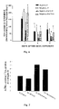
- S-(1,2-dichlorovinyl)-L-cysteine-injured renal proximal tubular cells grown in 50 ⁇ M AscP had a ratio of 4-[ 3 H]-L-proline:[ 14 C]-L-proline that was approximately 80% greater than controls on day 1 following injury indicating a significant decrease in proline hydroxylation ( FIG. 7 ).
- 4-[ 3 H]-L-proline: [ 14 C]-L-proline ratio in S-(1,2-dichlorovinyl)-L-cysteine-injured renal proximal tubular cells grown in 500 ⁇ M AscP was approximately 45% greater than controls on day 1 following injury.
- DCVC-injured renal proximal tubular cells grown in physiological concentrations of L-ascorbic acid-2-phosphate were exposed to exogenous collagen I or collagen IV (0, 5, 15, 50 ⁇ g/ml) immediately following S-(1,2-dichlorovinyl)-L-cysteine exposure and continuously through day 6 after injury.
- Basal QO 2 and monolayer cell density were measured on days 1 and 6 after injury induced by DCVC.
- Exogenous proteins were added directly to culture media at a concentration of 50 ⁇ g/ml, because this concentration of collagen IV was found to promote complete repair ( FIG. 9B ).
- the addition of exogenous collagen I, collagen IV, fibronectin, or laminin to injured renal proximal tubular cells produced no change in monolayer protein content on day 1 or day 6 ( FIG. 11A ). These results suggest that, like collagen I and collagen IV, exogenous laminin or fibronectin does not stimulate injured renal proximal tubular cells to proliferate.
- Na + /K + -ATPase activity was significantly increased in injured renal proximal tubular cells cultured in the presence of collagen IV on day 6 following S-(1,2-dichlorovinyl)-L-cysteine injury ( FIG. 11B ).
- Na + /K + -ATPase activity remained inhibited on day 6 after injury in renal proximal tubular cells cultured in the presence of exogenous collagen I, fibronectin, or laminin ( FIG. 11B ).
- Renal proximal tubular cells that do not die or become detached from the extracellular matrix following ischemic or chemical injury are thought to undergo repair or to dedifferentiate, proliferate, and migrate to denuded areas of the tubules.
- the cells of the newly lined tubule may then differentiate, promoting the return of normal tubular function and overall renal function.
- renal proximal tubular cells sublethally injured by S-(1,2-dichlorovinyl)-L-cysteine neither proliferated nor repaired physiological functions (38).
- renal proximal tubular cells sublethally injured by the oxidant t-butyl hydroperoxide proliferated and repaired physiological functions (37).
- renal proximal tubular cells sublethally injured by S-(1,2-dichlorovinyl)-L-cysteine were cultured in the presence of pharmacological concentrations of L-ascorbic acid-2-phosphate, they proliferated and repaired physiological functions (39).
- the mechanism of action of pharmacological concentrations of ascorbic acid in promoting renal proximal tubular cell repair and regeneration may be through the stimulation of the synthesis and deposition of collagen IV.
- AscP is known to promote collagen synthesis and deposition in cultured cells (13, 33).
- Collagen IV is the most abundant component of the proximal tubular basement membrane, and the regulation of collagen IV synthesis and degradation plays a n important role in cell function, growth, migration, and organ remodeling in many tissues (15). Further, collagen IV synthesis and deposition is increased in control renal proximal tubular cells exposed to L-ascorbic acid-2-phosphate (39). Collagen IV synthesis and deposition in renal proximal tubular cells sublethally injured by S-(1,2-dichlorovinyl)-L-cysteine was examined.
- S-(1,2-dichlorovinyl)-L-cysteine-injured renal proximal tubular cells grown in physiological concentrations of L-ascorbic acid-2-phosphate S-(1,2-dichlorovinyl)-L-cysteine-injured renal proximal tubular cells grown in pharmacological concentrations of L-ascorbic acid-2-phosphate maintained collagen IV deposition at control levels.
- Prolyl hydroxylase is the microsomal enzyme responsible for proline hydroxylation of procollagen ⁇ chains, and ascorbic acid is the preferred iron reducing cofactor for prolyl hydroxylase activity (41).
- proline hydroxylation in injured renal proximal tubular cells grown in pharmacological concentrations of L-ascorbic acid-2-phosphate was greater compared to injured renal proximal tubular cells grown in physiological concentrations of L-ascorbic acid-2-phosphate, suggesting that these cells retain some ability to hydroxylate susceptible proline residues.
- FIG. 4 shows that uninjured renal proximal tubular cells grown in pharmacological concentrations of L-ascorbic acid-2-phosphate synthesize less collagen IV on day 1 after confluence than renal proximal tubular cells grown in physiological concentrations of L-ascorbic acid-2-phosphate.
- renal proximal tubular cells grown in pharmacological L-ascorbic acid-2-phosphate concentrations may decrease collagen synthesis to basal levels sooner than renal proximal tubular cells grown in physiological concentrations of L-ascorbic acid-2-phosphate (36). Further evidence of this effect is observed on day 4, when renal proximal tubular cells cultured in physiological L-ascorbic acid-2-phosphate concentrations exhibit collagen IV synthesis that is decreased to levels of renal proximal tubular cells cultured in pharmacological concentrations of L-ascorbic acid-2-phosphate.
- Collagen IV but not collagen I, laminin, or fibronectin, promoted repair of mitochondrial oxygen consumption (basal QO 2 ) and active Na + transport (Na + /K + -ATPase activity) following injury, implicating collagen IV as an important extracellular matrix protein involved in repair of physiological functions in RPTC.
- S-(1,2-dichlorovinyl)-L-cysteine-injured renal proximal tubular cells cultured in the presence of collagen IV did not exhibit increased cell density on day 6 following S-(1,2-dichlorovinyl)-L-cysteine exposure.
- FITC-conjugated goat anti-mouse IgG and mouse monoclonal antibodies directed against human integrin subunits ⁇ 1 (clone FB12), ⁇ 2 (clone JBS2), and ⁇ 1 (clone B3B11) were purchased from Chemicon International, Inc. (Temecula, Calif.). All other materials were purchased from Sigma Chemical Co. (St. Louis, Mo.).
- Rabbit renal proximal tubules were isolated using the iron oxide perfusion method and grown in 35-mm tissue culture dishes or 48-well cell culture clusters under improved conditions as previously described (Nowak and Schnellmann, 1995; Nowak and Schnellmann, 1996).
- the cell culture medium was a 1:1 mixture of DMEM/Ham's F-12 (without D-glucose, phenol red, or sodium pyruvate) supplemented with 15 mM HEPES buffer, 2.5 mM L-glutamine, 1 ⁇ M pyridoxine HCl, 15 mM sodium bicarbonate, and 6 mM lactate.
- L-ascorbic acid-2-phosphate 50 or 500 ⁇ M were added to the culture medium immediately prior to daily media change.
- L-Ascorbic acid-2-phosphate (AscP) was used because L-ascorbic acid is unstable in culture media. AscP is stable in culture media and, after intracellular dephosphorylation, has the same effect on cultured cells as L ascorbic acid (Hata and Senoo, 1989).
- exogenous collagen IV 50 ⁇ g/ml
- collagen I 50 ⁇ g/ml
- function-stimulating antibodies to collagen-binding integrin subunits ⁇ 2 or ⁇ 1 5 ⁇ g/ml was added daily to the culture media of S-(1,2-dichlorovinyl)-L-cysteine-injured renal proximal tubular cells cultured in the absence of pharmacological concentrations of L-ascorbic acid-2-phosphate.
- Function-stimulating antibodies to collagen-binding integrin ⁇ 1 are not commercially available at this time.
- Renal proximal tubular cells were gently detached from culture dishes with a rubber policeman and transferred to a 37° C. oxygen consumption (QO 2 ) chamber.
- QO 2 in renal proximal tubular cells was measured polarographically in the absence (basal QO 2 ) or presence of ouabain (100 ⁇ M) (ouabain-insensitive QO 2 ) using a Clark-type electrode as described previously (Nowak and Schnellmann, 1995).
- Active Na + transport (ouabain-sensitive QO 2 ) was calculated by subtracting ouabain-insensitive QO 2 from basal QO 2 . Protein concentrations were determined using the bicinchoninic acid microassay according to the manufacturer's instructions (Pierce, Rockford, Ill.).
- Total ATPase activity was measured as described above. Briefly, renal proximal tubular cells cultured in 48-well cell culture clusters were scraped, solubilized in dissociation buffer (5 mM HEPES (pH 7.4), 25 mM imidazole, 1% BSA, 0.065% sodium dodecyl sulfate (SDS)) for 10 min.
- dissociation buffer 5 mM HEPES (pH 7.4), 25 mM imidazole, 1% BSA, 0.065% sodium dodecyl sulfate (SDS)
- ATPase activity was measured under linear conditions spectrophotometrically (340 nm) as the oxidation of ⁇ -NADH to NAD + at 37° C. in the absence or presence of ouabain (0.1 mM). Na + /K + -ATPase activity was calculated as total ATPase activity minus ouabain-insensitive ATPase activity.
- Renal proximal tubular cell monolayers were washed 3 times with ice-cold PBS, gently scraped from culture dishes into cell culture media containing 5% BSA (BSA/media), and transferred to microcentrifuge tubes.
- Renal proximal tubular cells were dissociated by pipeting, incubated on ice with moderate shaking for 20 minutes, centrifuged, and resuspended in 1% BSA/media containing 2 ⁇ g/ml of a specific anti-integrin antibody or non-specific IgG on ice with moderate shaking for 1 hour. After 3 washes with 1% BSA/media, renal proximal tubular cells were incubated for 30 minutes in the dark with a goat-anti mouse FITC-conjugated IgG diluted 1:100 in 1% BSA/media, followed by 3 washes with ice-cold PBS.
- Membrane expression of collagen-binding integrins was determined immediately by flow cytometry using a FACSCalibur four-color cell sorter/analyzer (Beckton Dickinson, San Jose, Calif.) with a blue argon laser for detection of FITC. Specific binding was calculated as total fluorescence minus that in IgG controls.
- Collagen-binding integrin localization was determined using confocal microscopy of renal proximal tubular cell monolayers stained with monoclonal antibodies to integrin subunits ⁇ 1 , ⁇ 2 , and ⁇ 1 .
- Renal proximal tubular cell monolayers were washed 3 times with PBS and fixed with 10% buffered formalin (4% formaldehyde) for 20 minutes at room temperature. After 3 washes with PBS, monolayers were permeabilized in PTB buffer (PBS+0.3% Triton X-100+0.1% BSA) for 10 minutes at room temperature. Monolayers were washed 3 times with 0.1% BSA in PBS and incubated with 8% BSA in PBS for 30 minutes at room temperature.
- BSA (1%) in PBS containing 5 ⁇ g/ml of specific integrin antibodies or non-specific IgG was added to renal proximal tubular cell monolayers and incubated overnight a t 4° C. with moderate shaking. Following 3 washes with PTB buffer, monolayers were incubated for 2 hours in the dark at room temperature with 1% BSA in PBS containing a 1:100 dilution of a FITC-conjugated goat anti-mouse IgG. Monolayers were washed 3 times with PTB, and glass coverslips were applied after the addition of 2–3 drops of mounting media.
- FIG. 13 demonstrates the fluorescence shift observed in response to incubation of rabbit renal proximal tubular cells with the anti-integrin antibodies for individual CBI subunits. Exposure of uninjured renal proximal tubular cells to pharmacological concentrations of L-ascorbic acid-2-phosphate or exogenous collagen IV did not affect collagen-binding integrin. ⁇ 1 , ⁇ 2 , and ⁇ 1 expression.
- sub-confluent renal proximal tubular cell cultures exhibit a morphology and cell density more like that of sublethally injured renal proximal tubular cell cultures.
- basal localization of CBI subunits ⁇ 1 , ⁇ 2 , and ⁇ 1 in renal proximal tubular cells cultured in the absence of L-ascorbic acid-2-phosphate or exogenous collagen IV was still decreased compared to sub-confluent controls ( FIGS. 15F , 17 F, and 19 F).
- FIGS. 16A–D , 18 A–D, and 20 A–D sublethally injured renal proximal tubular cells continued to exhibit CBI is distributed to the apical membrane.
- FIGS. 16F , 18 F, and 20 F injured renal proximal tubular cells cultured in the presence of pharmacological concentrations of AscP or exogenous collagen IV revealed a complete disappearance of OBIs from the apical membrane by day 6 ( FIGS. 16G–H , 18 G–H, and 20 G–H).
- Function-stimulating antibodies to CBI subunits ⁇ 2 and ⁇ 1 were added to the culture media of S-(1,2-dichlorovinyl)-L-cysteine-injured renal proximal tubular cells.
- the function-stimulating antibodies did not affect the degree of S-(1,2-dichlorovinyl)-L-cysteine-induced renal proximal tubular cell injury on day 1 after exposure.
- CBI subunit ⁇ 1 remained delocalized and partially redistributed to the apical membrane in renal proximal tubular cells cultured in the presence of the ⁇ 1 -stimulating antibody ( FIG. 22 ).
- Anchorage-dependent cell growth, proliferation, migration, and differentiation depend on the ability of the cell to recognize anchoring substrates in the extracellular matrix. Localization of ECM-binding integrins to the point of contact provides a strong but dynamic interaction that supports not only cellular attachment but also communication between the cell and the ECM. Given the importance of these interactions and the maintenance of cell polarity, the loss of integrin-extracellular matrix interactions and cell polarity play a key role in cell injury. Likewise, the restoration of integrin-extracellular matrix interactions and cell polarity likely play an equally important role in the return of normal cell function following injury.
- exogenous collagen IV added to the culture media of injured renal proximal tubular cells promotes repair of physiological functions, providing the first evidence that a key extracellular matrix protein in the renal proximal tubule is involved in cell repair following injury.
- renal epithelial cells interact with collagen IV through CBIs, these data suggest that physiological repair in injured renal proximal tubular cells may involve an effect of collagen IV on the expression, localization, and/or function of CBIs.
- sublethally injured cells may experience a loss in cell polarity due to decreased localization of certain proteins to specific areas of the plasma membrane.
- basal membrane protein localization Two potential reasons for a decrease in basal membrane protein localization include a decrease in membrane expression of these proteins due to translational effects or receptor internalization, or the redistribution of these proteins to other areas of the plasma membrane.
- S-(1,2-dichlorovinyl)-L-cysteine-induced sublethal injury to RPTC no changes in total membrane expression of CBIs were evident on day 1.
- confocal microscopy showed that CBI localization to the basal membrane was decreased and accompanied by the appearance of CBIs on the apical membrane.
- the ⁇ 1 subunit can associate with any of at least eleven integrin subunits ( ⁇ 1 – ⁇ 10 and ⁇ v ), each combination producing a heterodimeric integrin with unique binding and signaling properties. Therefore, repair stimulated by the ⁇ 1 antibody may link repair with an ⁇ - ⁇ subunit combination that forms a non-collagen-binding integrin, though association of ⁇ 1 with ⁇ 1 cannot be ruled out at this time. Additionally, the ⁇ 2 -stimulating antibody could be ineffective at activating the ⁇ 1 subunit in rabbit renal proximal tubular cells.
Landscapes
- Health & Medical Sciences (AREA)
- Life Sciences & Earth Sciences (AREA)
- Engineering & Computer Science (AREA)
- Immunology (AREA)
- Pharmacology & Pharmacy (AREA)
- Gastroenterology & Hepatology (AREA)
- Chemical & Material Sciences (AREA)
- Bioinformatics & Cheminformatics (AREA)
- Biomedical Technology (AREA)
- Medicinal Chemistry (AREA)
- Zoology (AREA)
- Proteomics, Peptides & Aminoacids (AREA)
- Epidemiology (AREA)
- Animal Behavior & Ethology (AREA)
- General Health & Medical Sciences (AREA)
- Public Health (AREA)
- Veterinary Medicine (AREA)
- Medicines That Contain Protein Lipid Enzymes And Other Medicines (AREA)
Abstract
Description
- 1. Abbate, M. and Remuzzi, G. Ren Fail 18: 377–388, 1996.
- 2. Alcain, et al., J Bioenerg Biomembr 26: 393–398, 1994.
- 3. Bailey, et al., FEBS Lett 99: 361–366, 1979.
- 4. Cantley, L. G. Am J Physiol Renal Fluid Electrolyte Physiol 271: F1103–F1113, 1996.
- 5. Chojkier, et al., Anal Biochem 108: 385–393, 1980.
- 6. Davidson, et al., J Biol Chem 272: 345–352, 1997.
- 7. Denk, P. O. and Knorr, M. Eur J Ophthalmol 8: 37–41, 1998.
- 8. Dixit, S. N. Connect Tissue Res 14: 31–40, 1985.
- 9. Englard, S. and Seifter, S. Ann Rev Nutr 6: 365–406, 1986.
- 10. Geesin, et al., J Invest Dermatol 90: 420–424, 1988.
- 11. Ghohestani, et al., J Biol Chem 275: 16002–16006, 2000.
- 12. Gibbs, et al., Connect Tissue Res 40: 173–188, 1999.
- 13. Hata, R. I. and Senoo, H. J Cell Physiol 138: 8–16, 1989.
- 14. Houglum, et al., Am. J Clin Nutr 54:1141S-1143S, 1991.
- 15. Hudson, et al., J Biol Chem 268: 26033–26036, 1993.
- 16. Ivanov, et al., Atherosclerosis 140: 25–24, 1998.
- 17. Juva, et al., Science 152: 92–4, 1966.
- 18. Kao, et al., J Biol Chem 254: 2234–2243, 1979.
- 19. Kalluri, et al., J Clin Invest 99: 2470–2478, 1997.
- 20. Kalluri, R. and Cosgrove, D. J Biol Chem 275: 12719–12724, 2000.
- 21. Komsa-Penkova, et al., Biochim Biophys Acta 1297: 171–181, 1996.
- 22. Laemmli, U. K. Nature 227: 680–685, 1970.
- 23. Lash, et al., J Pharmacol Exp Ther 269: 374–383, 1994.
- 24. Levine, M. N Engl J Med 314: 892–902, 1986.
- 25. Linsenmayer, T. F. Cell Biology of the Extracellular Matrix. New York: Plenum Press, 1991, p. 5–37.
- 26. Lowry, et al., J Biol Chem 193: 265–275, 1951.
- 27. Martensson, et al., Nephrol Dial Transplant 10: 1637–1643, 1995.
- 28. Miller, E. J. Biochem 10:1652–1659, 1971.
- 29. Miner, J. H. Kidney Int. 56: 2016–2024, 1999.
- 30. Moore, et al., Toxicol Environ Chem 17: 153–162, 1988.
- 31. Muller, U. and Brandli, A. W. J Cell Sci 112: 3855–3867, 1999.
- 32. Murad, et al., Proc Natl Acad Sci USA 78: 2879–2882, 1981.
- 33. Murad, et al., J Invest Derm 81: 158–162, 1983.
- 34. Niki, et al., Hepatology 23: 1673–1681, 1996.
- 35. Nowak, et al., Am J Physiol Cell Physiol 269: C1317–C1325, 1995.
- 36. Nowak, et al., Am J Physiol Cell Physiol 271: C2072–C2080, 1996.
- 37. Nowak, et al., Am J Physiol Renal Physiol 274: F509–515, 1998.
- 38. Nowak, et al., Am J Physiol Renal Physiol 276: F228–F236, 1999.
- 39. Nowak, et al., Toxicol Appl Pharmacol 167: 37–45, 2000.
- 40. Peterkofsky, B. Am J Clin Nutr 54: 1135S-1140S, 1991.
- 41. Pinnell, S. Yale J Biol Med 58: 553–559, 1985.
- 42. Rosenbloom, et al., J Biol Chem 245:3361–3368, 1970.
- 43. Schmidt, et al., FEBS Lett 312: 174–178, 1992.
- 44. Schwartz, et al., Am J Physiol Renal Fluid Electrolyte Physiol 246: F845–F852, 1984.
- 45. Shapiro, et al., Am J Clin Nutr 54:1209S-1213S, 1991.
- 46. Sullivan, et al., J Biol Chem 269: 22500–22506, 1994.
- 47. Tschank, et al., Arch Biochem Biophys 261: 312–323, 1988.
- 48. Venezian, et al., J Cell Physiol 174; 331–341, 1996.
- 49. Walker, P. D. Lab Invest 70: 229–345, 1994.
- 50. Wewer, et al., J Biol Chem 258: 12654–12660, 1983.
- 51. Yamada, K. M. and Kennedy, D. W. J Cell Biol 80: 492–498, 1979.
Claims (4)
Priority Applications (1)
| Application Number | Priority Date | Filing Date | Title |
|---|---|---|---|
| US10/393,193 US7183383B2 (en) | 2002-03-20 | 2003-03-20 | Uses of collagen IV |
Applications Claiming Priority (2)
| Application Number | Priority Date | Filing Date | Title |
|---|---|---|---|
| US36600202P | 2002-03-20 | 2002-03-20 | |
| US10/393,193 US7183383B2 (en) | 2002-03-20 | 2003-03-20 | Uses of collagen IV |
Publications (2)
| Publication Number | Publication Date |
|---|---|
| US20030181392A1 US20030181392A1 (en) | 2003-09-25 |
| US7183383B2 true US7183383B2 (en) | 2007-02-27 |
Family
ID=28045592
Family Applications (1)
| Application Number | Title | Priority Date | Filing Date |
|---|---|---|---|
| US10/393,193 Expired - Fee Related US7183383B2 (en) | 2002-03-20 | 2003-03-20 | Uses of collagen IV |
Country Status (1)
| Country | Link |
|---|---|
| US (1) | US7183383B2 (en) |
Families Citing this family (3)
| Publication number | Priority date | Publication date | Assignee | Title |
|---|---|---|---|---|
| US20090304686A1 (en) * | 2006-03-08 | 2009-12-10 | Universotair Medisch Centrum Utrecht | Interfering in activation of an immune cell by influencing interaction of lair and collagen |
| AU2007301981A1 (en) * | 2006-09-25 | 2008-04-03 | Universiteit Maastricht | Means and methods for diagnosing and/or treating a subject at risk of developing heart failure |
| WO2016014781A1 (en) * | 2014-07-25 | 2016-01-28 | Rgdi3, Inc. | Collagen iv replacement |
-
2003
- 2003-03-20 US US10/393,193 patent/US7183383B2/en not_active Expired - Fee Related
Non-Patent Citations (4)
| Title |
|---|
| Houglum et al., Am. J. Clin. Nutr., vol. 54, pp. 1141S-1143S, 1991. * |
| Hudson et al., The Journal of Biological Chemistry, vol. 268, No. 35, pp. 26033-26036, Dec. 15, 1993. * |
| Kalluri et al., The Journal of Biological Chemistry, vol. 275, No. 17, pp. 12719-12724, Apr. 28, 2000. * |
| Nowak et al., Toxicology and Applied Pharmacology, vol. 167, pp. 37-45, 2000. * |
Also Published As
| Publication number | Publication date |
|---|---|
| US20030181392A1 (en) | 2003-09-25 |
Similar Documents
| Publication | Publication Date | Title |
|---|---|---|
| Chen et al. | Lack of integrin α1β1 leads to severe glomerulosclerosis after glomerular injury | |
| Finn et al. | Evidence that Wallerian degeneration and localized axon degeneration induced by local neurotrophin deprivation do not involve caspases | |
| Robinson et al. | Oxytocin-induced desensitization of the oxytocin receptor | |
| Liedtke et al. | Elongation of axons during regeneration involves retinal crystallin β b2 (crybb2) | |
| US10330671B2 (en) | Modulation of synaptic maintenance | |
| de Vrieze et al. | Matrix metalloproteinases in osteoclasts of ontogenetic and regenerating zebrafish scales | |
| US10240156B2 (en) | Modulation of synaptic maintenance | |
| US7837993B2 (en) | Methods and compositions for regeneration of aged skeletal muscle tissues | |
| Cheng et al. | Staurosporine, K‐252a, and K‐252b stabilize calcium homeostasis and promote survival of CNS neurons in the absence of glucose | |
| Niewenhuis et al. | Calmodulin inhibitors protect against cadmium-induced testicular damage in mice | |
| US7183383B2 (en) | Uses of collagen IV | |
| Zhan et al. | Nitric oxide induces epidermal stem cell de-adhesion by targeting integrin β1 and Talin via the cGMP signalling pathway | |
| Yasuda et al. | Protective effect of T3 peptide, an active fragment of tumstatin, against ischemia/reperfusion injury in rat heart | |
| KR20160086775A (en) | composition for the prevention or treatment of the symptoms in the stroke comprising the inhibitor of Pin1 | |
| Sataieva et al. | Toxic cardiac effect of cobalt chloride excessive chronic consumption in male Wistar rats | |
| Nony et al. | Interactions between collagen IV and collagen-binding integrins in renal cell repair after sublethal injury | |
| Nony et al. | Collagen IV promotes repair of renal cell physiological functions after toxicant injury | |
| Yi et al. | Infarct reduction in rats following intraventricular administration of either tissue plasminogen activator (tPA) or its non-protease mutant S478A-tPA | |
| US20120309687A1 (en) | Glioprotectant Peptide for Use in the Treatment of Amyotrophic Lateral Sclerosis (ALS) and Methods Related Thereto | |
| WO1997044059A2 (en) | Cartilage type ii collagen as an angiogenic factor | |
| JP7678254B2 (en) | Epidermal basal niche protecting agent, pharmaceutical composition for protecting epidermal basal niche, and cosmetic for protecting epidermal basal niche | |
| Younis et al. | Inhibiting matrix metalloproteinases with prinomastat produces abnormalities in fetal growth and development in rats | |
| Díaz‐Juárez et al. | The role of calcium and nitric oxide during liver enzyme release induced by increased physical forces as evidenced in partially hepatectomized rats | |
| Kim et al. | The insect peptide CopA3 enhances hepatocyte proliferation by directly binding to PTEN and inhibiting its activity | |
| Tam et al. | Neurite outgrowth in dorsal root ganglia induced by islet neogenesis-associated protein peptide involves protein kinase A activation |
Legal Events
| Date | Code | Title | Description |
|---|---|---|---|
| AS | Assignment |
Owner name: THE UNIVERSITY OF ARKANSAS FOR MEDICAL SCIENCES, A Free format text: ASSIGNMENT OF ASSIGNORS INTEREST;ASSIGNORS:NONY, PAUL A.;SCHNELLMANN, RICKY GENE;REEL/FRAME:014009/0700 Effective date: 20030409 |
|
| STCF | Information on status: patent grant |
Free format text: PATENTED CASE |
|
| AS | Assignment |
Owner name: NATIONAL INSTITUTES OF HEALTH (NIH), U.S. DEPT. OF Free format text: EXECUTIVE ORDER 9424, CONFIRMATORY LICENSE;ASSIGNOR:UNIVERSITY OF ARKANSAS MED SCIS LTL ROCK;REEL/FRAME:020857/0749 Effective date: 20061101 |
|
| AS | Assignment |
Owner name: BOARD OF TRUSTEES OF THE UNIVERSITY OF ARKANSAS, A Free format text: RE-RECORD TO CORRECT PATENT NUMBER PREVIOUSLY RECORDED AT R/F 014009/0700;ASSIGNORS:SCHNELLMANN, RICKY GENE;NONY, PAUL A.;SIGNING DATES FROM 20030409 TO 20030411;REEL/FRAME:024838/0819 |
|
| FPAY | Fee payment |
Year of fee payment: 4 |
|
| FEPP | Fee payment procedure |
Free format text: PATENT HOLDER CLAIMS MICRO ENTITY STATUS, ENTITY STATUS SET TO MICRO (ORIGINAL EVENT CODE: STOM); ENTITY STATUS OF PATENT OWNER: MICROENTITY |
|
| FPAY | Fee payment |
Year of fee payment: 8 |
|
| FEPP | Fee payment procedure |
Free format text: MAINTENANCE FEE REMINDER MAILED (ORIGINAL EVENT CODE: REM.); ENTITY STATUS OF PATENT OWNER: MICROENTITY |
|
| LAPS | Lapse for failure to pay maintenance fees |
Free format text: PATENT EXPIRED FOR FAILURE TO PAY MAINTENANCE FEES (ORIGINAL EVENT CODE: EXP.); ENTITY STATUS OF PATENT OWNER: MICROENTITY |
|
| STCH | Information on status: patent discontinuation |
Free format text: PATENT EXPIRED DUE TO NONPAYMENT OF MAINTENANCE FEES UNDER 37 CFR 1.362 |
|
| FP | Lapsed due to failure to pay maintenance fee |
Effective date: 20190227 |