RELATED APPLICATIONS
This application is a division of U.S. patent application Ser. No. 09/626,939, filed Jul. 27, 2000, which is a division of U.S. patent application Ser. No. 08/833,752, filed Apr. 9, 1997, now U.S. Pat. No. 6,448,375, issued Sep. 10, 2002, which is a continuation of Ser. No. 08/810,028, filed on Mar. 3, 1997, now abandoned, which claims priority under 35 U.S.C. 119(a)-(d) to EP 96870021.1, filed Mar. 3, 1996, and EP 96870102.9, filed Aug. 6, 1996.
FIELD OF THE INVENTION
The present invention concerns new peptides and the nucleic acid molecules encoding said peptides, the vector comprising said nucleic acid molecules, the cells transformed by said vector, inhibitors directed against said peptides or said nucleic acid molecules, a pharmaceutical composition and a diagnostic and/or dosage device comprising said products, and non human transgenic animals expressing the peptides according to the invention or the nucleic acid molecules encoding said peptides.
The invention further provides a method for determining ligand binding, detecting expression, screening for drugs binding specifically to said peptides and treatments involving the peptides or the nucleic acid molecules according to the invention.
TECHNOLOGICAL BACKGROUND OF THE ART
Chemotactic cytokines, or chemokines, are small signalling proteins that can be divided in two subfamilies (CC- and CXC-chemokines) depending on the relative position of the first two conversed cysteines. Interleukin 8 (IL-8) is the most studied of these proteins, but a large number of chemokines (Regulated on Activation Normal T-cell Expressed and Secreted (RANTES), Monocyte Chemoattractant Protein 1 (MCP-1), Monocyte Chemoattractant Protein 2 (MCP-2), Monocyte Chemoattractant Protein 3 (MCP-3), Growth-Related gene product a (GROα), Growth-Related gene product β (GRO)β, Growth-Related gene product γ (GROγ), Macrophage Inflammatory Protein 1 α (MIP-1α) and β, etc.) has now been described [4]. Chemokines play fundamental roles in the physiology of acute and chronic inflammatory processes as well as in the pathological dysregulations of these processes, by attracting and simulating specific subsets of leucocytes [32]. RANTES for example is a chemoattractant for monocytes, memory T-cells and eosinophils, and induces the release of histamine by basophils. MCP-1, released by smooth muscle cells in arteriosclerotic lesions, is considered as the factor (or one of the factors) responsible for macrophage attraction and, therefore, for the progressive aggravation of the lesions [4].
MIP-1α, MIP-1β and RANTES chemokines have recently been described as major HIV-suppressive factors produced by CD8+ T-cells [9]. CC-chemokines are also involved in the regulation of human myeloid progenetor cell proliferation [6, 7].
Recent studies have demonstrated that the actions of CC- and CXC-chemokines are mediated by subfamilies of G protein-coupled receptors. To date, despite the numerous functions attributed to chemokines and the increasing number of biologically active ligands, only six functional receptors have been identified in human. Two receptors for interleukin-8 (IL-8) have been described [20, 29]. One (IL-8RA) binds IL-8 specifically, while the other (IL-8RB) binds IL-8 and other CXC-chemokines, like GRO. Among receptors binding CC-chemokines, a receptor, designated CC-chemokine receptor 1 (CCR1), binds both RANTES and MIP-1α [31], and the CC-chemokine receptor 2 (CCR2) binds MCP-1 and MCP-3 [8, 44, 15]. Two additional CC-chemokine receptors were cloned recently the CC-chemokine receptor 3 (CCR3) was found to be activated by RANTES, MIP-1α and MIP-1β [10]; the CC-chemokine receptor 4 (CCR4) responds to MIP-1, RANTES and MCP-1 [37]. In addition to these six functional receptors, a number of orphan receptors have been cloned from human and other species, that are structurally related to either CC- or CXC-chemokine receptors. These include the human BLR1 [13], EBI1 [5], LCR1 [21], the mouse MIP-1 RL1 and MIP-1 RL2 [17] and the bovine PPR1 [25]. Their respective ligand(s) and function(s) are unknown at present.
SUMMARY OF THE INVENTION
The present invention is related to a peptide having at least an amino acid sequence which presents more than 80%, advantageously more than 90%, preferably more than 95%, homology with the amino acid sequence as represented in SEQ ID NO. 1.
Preferably, said peptide has also at least an amino acid sequence which presents more than 80%, advantageously more than 90%, preferably more than 95%, homology with the amino acid sequence as represented in SEQ ID NO. 2.
According to another embodiment of the present invention, the peptide has at least an amino acid sequence which presents more than 80%, advantageously more than 90%, preferably more than 95%, homology with the amino acid sequence as represented in SEQ ID NO. 3.
The present invention is also related to the amino acid sequence of SEQ ID NO. 1, SEQ ID NO. 2, SEQ ID NO. 3 or a portion thereof (represented in the FIG. 1).
A “portion of an amino acid sequence” means one or more amino acid segments having the same or improved binding properties of the whole peptide according to the invention. Said portion could be an epitope which is specifically binded by a ligand of the peptide which could be a known “natural ligand” of said peptide, an agonist or an analog of said ligand, or an inhibitor capable of competitively inhibiting the binding of said ligand to the peptide (including the antagonists of said ligand to the peptide).
Specific examples of said portions of amino acid sequence and their preparation process are described in the publication of Rucker J. et al. (Cell, Vol. 87, pp. 437-446 (1996)) incorporated herein by reference.
According to tho invention, said portion of the amino acid sequence of the peptide according to the invention comprises the N-terminus segment and the first extracellular loop of the peptide.
Therefore, according to the invention, the amino acid sequence as represented in SEQ ID its NO. 1 is the common amino acid sequence of SEQ ID NO. 2 and of SEQ ID NO. 3 (see also FIG. 1). Therefore, a first industrial application of said amino acid sequence is the identification of the homology between said amino acid sequence and the screening of various mutants encoding a different amino acid sequence than the one previously described, and the identification of various types of patient which may present a predisposition or a resistance to the disorders described in the following specification.
Preferably, the peptide according to the invention or a portion thereof is an active CC-chemokine receptor.
Advantageously, the CC-chemokine receptor according to the invention is stimulated by the MIP-1β chemokine at a concentration less or equal to 10 nm, and is advantageously also stimulated by the MIP-1α or RANTES chemokines. However, said chemokine receptor is not stimulated by the MCP-1, MCP-2, MCP-3, IL8 and GROα chemokines.
In addition, the peptide according to the invention or a portion thereof is also a receptor of HIV viruses or a portion of said HIV viruses.
It is meant by “HIV viruses”, HIV-1 or HIV-2 and all the various strains of HIV viruses which are involved in the development of AIDS. It is meant by a “a portion of HIV viruses”, any epitope of said viruses which is able to interact specifically with said receptor. Among said portions of viruses which may be involved in the interaction with the peptide according to the invention, are peptides encoded by the ENV and GAG viruses genes.
Preferably, said portion of HTV viruses is the glycopeptide gp120/160 (membrane-bound gp160 or the free gp derived therefrom) or a portion thereof.
It is meant by a “portion of the glycopeptide gp120/160 ” any epitope, preferably an immuno-dominant epitope, of said glycopeptide which may interact specifically with the peptide according to the invention, such as for instance the V3 loop (third hypervariable domain).
According to another embodiment of the present invention, the peptide according to the invention is an inactive CC-chemokine receptor. An example of such inactive CC-chemokine receptor is encoded by the amino acid sequence as represented in SEQ ID NO. 2.
It is meant by an “inactive CC-chemokine receptor” a receptor which is not stimulated by any known CC-chemokine, especially the MIP-1β, MIP-1 α or RANTES chemokines.
The peptide represented in SEQ ID NO. 3 according to the invention is an 30 inactive receptor which is not a receptor of HIV viruses or of a portion of said HIV viruses, which means that said inactive receptor does not allow the entry of said FHV viruses into a cell which presents at its surface said inactive receptor.
Advantageously, the peptide according to the invention is a human receptor.
The present invention concerns also the nucleic acid molecule having more than 80%, preferably more than 90%, homology with one of the nucleic acid sequences of SEQ ID NO. 1, SEQ ID NO. 2 and SEQ ID NO. 3 shown in the FIG. 1.
Preferably, said nucleic acid molecule has at least the nucleic acid sequence shown in SEQ ED NO. 1, SEQ ID NO. 2 or SEQ ID NO. 3 of FIG. 1 or a portion thereof.
It is meant by a “portion of said nucleic acid molecule” any nucleic acid sequence of a more than 15 nucleotides which could be used in order to detect and/or reconstitute said nucleic acid molecule or its complementary strand. Such portion could be a probe or a primer which could be used in genetic amplification using the PCR, LCR, NASBA or CPR techniques for instance.
The present invention concerns more specifically the nucleic acid molecules encoding the peptide according to the invention. Said nucleic acid molecules are RNA or DNA molecules such as a cDNA molecule or a genornic DNA molecule.
The present invention is also related to a vector comprising the nucleic acid molecule according to the invention. Preferably, said vector is adapted for expression in a cell and comprises the regulatory elements necessary for expressing the amino acid molecule in said cell operatively linked to the nucleic acid sequence according to the invention as to permit expression thereof.
Preferably, said cell is chosen among the group consisting of bacterial cells, yeast cells, insect cells or mammalian cells. The vector according to the invention is a plasmid, preferably a pcDNA3 plasmid, or a virus, preferably a baculovirus, an adenovirus or a semliki forest virus.
The present invention concerns also the cell, preferably a manunalian cell, such as a CHO-K1 or a HEK293 cell, transformed by the vector according to the invention. Advantageously, said cell is non neuronal in origin and is chosen among the group consisting of CHO-K1, HEK293, BHK21, COS-7 cells.
The present invention also concerns the cell (preferably a mammalian cell such as a CHO-K1 cell) transformed by the vector according to the invention and by another vector encoding a protein enhancing the functional response in said cell. Advantageously, said protein is the Gα15 or Gα16 (G protein, a submit). Advantageously, said cell is the cell CHO-K1-pEFIN hCCR5-1/16.
The present invention is also related to a nucleic acid probe comprising a nucleic acid molecule of at least 15 nucleotides capable of specifically hybridizing with a unique sequence included within the sequence of the nucleic acid molecule according to the invention. Said nucleic acid probe may be a DNA or a RNA.
The invention concerns also an antisense oligonucleotide having a sequence capable of specifically hybridizing to an MRNA molecule encoding the peptide according to the invention so as to prevent translation of said MIRNA molecule or an antisense oligonucleotide having a sequence capable of specifically hybridizing to the CDNA molecule encoding the peptide according to the invention.
Said antisense oligonucleotide may comprise chemical analogs of nucleotide or substances which inactivate MRNA, or be included in an RNA molecule endowed with ribozyme activity.
Another aspect of the present invention concerns a ligand or an anti-ligand (preferably an antibody) other than known “natural ligands”, which are chosen among the group consisting of the MIP-1β, MIP-1α or RANTES chemokines, HIV viruses or a portion of said HIV viruses, wherein said ligand is capable of binding to the receptor according to the invention and wherein said anti-ligand is capable of (preferably competitively) inhibiting the binding of said known “natural ligand” or the ligand according to the invention to the peptide according to the invention.
The exclusion in the above identified definition of known chemokines, HIV viruses or a portion of said HIV viruses, does not include variants of said “natural” viruses or said “natural” portion which may be obtained for instance by genetic engineering and which may mimic the interaction of said viruses and portion of said viruses to the peptide according to the invention.
Advantageously, said antibody is a monoclonal antibody which is preferably directed to an epitope of the peptide according to the invention and present on the surface of a cell expressing said peptide.
Preferably, said antibody is produced by the hybridome cell AchCCR5-SAB1A7.
The invention concerns also the pharmaceutical composition comprising either an effective amount of the peptide according to the invention (in order to delude the HIV virus from the natural peptide present at the surface of a mammalian cell and stop the infection of said mammalian cell by the HIV virus), or an effective amount of the above identified described ligand and/or anti-ligand, or an effective amount of oligonucleotide according to the invention, effective to decrease the activity of said peptide by passing through a cell membrane and binding specifically with MRNA encoding the peptide according to the invention in the cell so as to prevent it translation. The pharmaceutical composition comprises also a pharmaceutically acceptable carrier, preferably capable of passing through said cell membrane. Preferably, in said pharmaceutical composition, the oligonucleotide is coupled to a substance, such as a ribozyme, which inactivates MRNA encoding the peptide according to the invention.
Preferably, the pharmaceutically acceptable carrier comprises a structure which binds to a receptor on a cell capable of being taken up by cell after binding to the structure. The structure of the pharmaceutically acceptable carrier in said pharmaceutical composition is capable of binding to a receptor which is specific for a selected cell type.
The present invention concerns also a transgenic non human mammal overexpressing (or expressing ectopically) the nucleic acid molecule encoding the peptide according to the invention.
The present invention also concerns a transgenic non human mammal comprising an homologous recombination knockout of the native peptide according to the invention.
According to a preferred embodiment of the invention, the transgenic non human mammal whose genome comprises antisense nucleic acid complementary to the nucleic acid according to the invention is so placed as to be transcripted into antisense MRNA which is complementary to the MRNA encoding the peptide according to the invention and which hybridizes to MRNA encoding said peptide, thereby reducing its translation. Preferably, the transgenic non human mammal according to the invention comprises a nucleic acid molecule encoding the peptide according to the invention and comprises additionally an inducible promoter or a tissue specific regulatory element.
Preferably, the transgenic non human mammal is a mouse.
The invention relates to a method for determining whether a ligand can be specifically bound to the peptide according to the invention, which comprises contacting a cell transfected with a vector expressing the nucleic acid molecule encoding said peptide with the ligand under conditions permitting binding of ligand to such peptide and detecting the presence of any such ligand bound specifically to said peptide, thereby determining whether the ligand binds specifically to said peptide.
The invention relates to a method for determining whether a ligand can specifically bind to a peptide according to the invention, which comprises preparing a cell extract from cells transfected with a vector expressing the nucleic acid molecule encoding said peptide, isolating a membrane fraction from the cell extract, contacting the ligand with the membrane fraction under conditions permitting binding of the ligand to such peptide and detecting the presence of any ligand bound to said peptide, thereby determining whether the compound is capable of specifically binding to said peptide. Preferably, said method is used when the ligand is not previously known.
The invention relates to a method for determining whether a ligand is an agonist of the peptide according to the invention, which comprises contacting a cell transfected with a vector expressing the nucleic acid molecule encoding said peptide with the ligand under conditions permitting the activation of a functional peptide response from the cell and detecting by means of a bio-assay, such as a modification in a second messenger concentration (preferably calcium ions or inositol phosphates such as IP3) or a modification in the cellular metabolism (preferably determined by the acidification rate of the culture medium), an increase in the peptide activity, thereby determining whether the ligand is a peptide agonist
The invention relates to a method for determining whether a ligand is an agonist of the peptide according to the invention, which comprises preparing a cell extract from cells transfected with a vector expressing the nucleic acid molecule encoding said peptide, isolating a membrane fraction from the cell extract, contacting the membrane fraction with the ligand under conditions permitting the activation of a functional peptide response and detecting by means of a bio-assay, such as a modification in the production of a second messenger (preferably inositol phosphates such as IP3), an increase in the peptide activity, thereby determining whether the ligand is a peptide agonist.
The present invention relates to a method for determining whether a ligand is an antagonist of the peptide according to the invention, which comprises contacting a cell transfected with a vector expressing the nucleic acid molecule encoding said peptide with the ligand in the presence of a known peptide agonist, under conditions permitting the activation of a functional peptide response and detecting by means of a bio-assay, such as a modification in second messenger concentration (preferably calcium ions or inositol phosphates such as IP3) or a modification in the cellular metabolism (preferably determined by the acidification rate of the culture medium), a decrease in the peptide activity, thereby determining whether the ligand is a peptide antagonist.
The present invention relates to a method for determining whether a ligand is an antagonist of the peptide according to the invention, which comprises preparing a cell extract from cells transfected with an expressing the nucleic acid molecule encoding said peptide, isolating a membrane fraction from the cells extract, contacting the membrane fraction with the ligand in the presence of a known peptide agonist, under conditions permitting the activation of a functional peptide response and detecting by means of a bio-assay, such as a modification in the production of a second messenger, a decrease in the peptide activity, thereby determining whether the ligand is a peptide antagonist.
Preferably, the second messenger assay comprises measurement of calcium ions or inositol phosphates such as IP3.
Preferably, the cell used in said method is a mammalian cell non neuronal in origin, such as CHO-K1, HEK293, BHK21, COS-7 cells.
In said method, the ligand is not previously known.
The invention is also related to the ligand isolated and detected by any of the preceding methods.
The present invention concerns also the pharmaceutical composition which comprises an effective amount of an agonist or an antagonist of the peptide according to the invention, effective to reduce the activity of said peptide and a pharmaceutically acceptable carrier.
It is meant by “an agonist or an antagonist of the peptide according to the invention”, all the agonists or antagonists of the known “natural ligand” of the peptide as above described.
Therefore, the previously described methods may be used for the screening of drugs to identify drugs which specifically bind to the peptide according to the invention The invention is also related to the drugs isolated and detected by any of these methods.
The present invention concerns also a pharmaceutical composition comprising said drugs and a pharmaceutically acceptable carrier.
The invention is also related to a method of detecting expression of a peptide according to the invention by detecting the presence of MRNA coding for a peptide, which comprises obtaining total RNA or total MRNA from the cell and contacting the RNA or MRNA so obtained with the nucleic acid-probe according to the invention under hybridizing conditions and detecting the presence of MRNA hybridized to the probe, thereby detecting the expression of the peptide by the cell.
Said hybridization conditions are stringent conditions.
The present invention concerns also the use of the pharmaceutical composition according to the invention for the treatment and/or prevention of inflammatory diseases, including rheumatoid arthritis, glomerulonephritis, asthma, idiopathic pulmonary fibrosis and psoriasis, viral infections including Human Immunodeficiency Viruses 1 and 2 (HIV-1 and 2), cancer including leukaemia, atherosclerosis and/or auto-immune disorders.
The present invention concerns also a method for diagnosing a predisposition or a resistance to a disorder associated with the activity of the peptide according to the invention and/or associated with infectious agents such as HIV viruses in a subject. Said method comprises
a) obtaining nucleic acid molecules encoding the peptide according to the invention from the cells of the subject;
b) possibly performing a restriction digest of said nucleic acid molecules with a panel of restriction enzymes;
c) possibly electrophoretically separating the resulting nucleic acid fragments on a sized gel;
d) contacting the resulting gel or the obtained nucleic acid molecule with a nucleic acid probe labelled with a detectable marker and capable of specifically hybridizing to said nucleic acid molecule (said hybridization being made in stringent hybridization conditions);
e) detecting labelled bands or the in situ nucleic acid molecules which have hybridized to the said nucleic acid molecule labelled with a detectable marker to create a unique band pattern or an in situ marking specific to the subject;
f) preparing other nucleic acid molecules encoding the peptide according to the invention obtained from the cells of other patients for diagnosis by step a-e; and
g) comparing the unique band pattern specific to the nucleic acid molecule of subjects suffering from the disorder from step e and the nucleic acid molecule obtained for diagnosis from step f to determine whether the patterns are the same or different and to diagnose thereby a predisposition or a resistance to the disorder if the patterns are the same or different.
The present invention is also related to a method for diagnosing a predisposition or a resistance to a disorder associated with the activity of a specific allele of the peptide according to the invention or the presence of said peptide at the surface of cells and/or associated with infectious agents such as HIV viruses present in a subject. Said method comprises:
a) obtaining a sample of a body fluid, preferably a blood sample comprising antigen presenting cells, from a subject;
b) adding to said sample a ligand and/or an anti-ligand according to the invention;
c) detecting the cross-reaction between said ligand and/or said anti-ligand and the specific peptide according to the invention; and
d) determining whether the peptide corresponds to a receptor or an inactive receptor according to the invention and diagnosing thereby a predisposition or a resistance to the disorder according to the type of the peptide present in the body fluid of the subject.
The present invention concerns also a diagnostic and/or dosage device, preferably a kit, comprising the peptides, the nucleic acid molecules, the nucleic acid probes, the ligands and/or the anti-ligands according to the invention, their portions (such as primers, probes, epitopes, . . . ) or a mixture thereof, being possibly labelled with a detectable marker.
Said diagnostic and/or dosage device comprises also the reactants for the detection and/or the dotage of antigens, antibodies or nucleic acid sequences through a method selected from the group consisting of in situ hybridization, hybridization or recognition by marked specific antibodies, specially ELISA® (Enzyme Linked Immunosorbent Assay) or RIA® (Radio Immunoassay), methods on filter, on a solid support, in solution, in “sandwich”, on gel, by Dot blot hybridization, by Northern blot hybridization, by Southern blot hybridization, by isotopic or non-isotopic labelling (such as immunofluorescence or biotinylation), by a technique of cold probes, by genetic amplification, particularly PCR, LCR, NASBA or CPR, by a double immunodiffusion, by a counter-immunoelectrophoresis, by haemagglutination and/or a mixture thereof.
A last aspect of the present invention concerns a method of preparing peptides according to the invention, which comprises
a) constructing a vector adapted for expression in a cell which comprises the regulatory elements necessary for the expression of nucleic acid molecules in the cell operatively linked to nucleic acid molecule encoding said peptide so as to permit expression thereof, wherein the cell is preferably selected from the group consisting of bacterial cells, yeast cells, insect cells and mammalian cells;
b) inserting the vector of step a in a suitable host cell;
c) incubating the cell of step b under conditions allowing the expression of the peptide according to the invention;
d) recovering the peptide so obtained; and
e) purifying the peptide so recovered, thereby preparing an isolated peptide -according to the invention.
The deposits of micro-organisms AchCCR5-SAB1A7 and CHO-K1-PEFIN HCCR5-1/16 were made according to the Budapest Treaty in the Belgium Coordinated Collection of Micro-organisms (BCCM), Laboratorium voor Moleculaire Biologic (LMBP), Universiteit Gent, K. L. Ledeganckstraat 35, B-9000 GENT, BELGIUM.
BRIEF DESCRIPTION OF THE FIGURES
FIG. 1 Shows the nucleic acid and amino acid sequences of the invention. FIGS. 1A-1 and 1A-2 show the nucleic acid and amino acid sequence of SEQ ID Nos 1 and 4, respectively. FIGS. 1B-1, 1B-2, and 1B-3 show the nucleic acid and amino acid sequence of SEQ ID Nos 2 and 5, respectively. FIGS. 1D-1 to 1D-3 show the nucleic acid and amino acid sequence of SEQ ID Nos. 3 and 6, respectively.
FIG. 2 represents the amino acids sequence of the active human CCR5 chemokine receptor (SEQ ID NO: 5) according to the invention aligned with that of the human CCR1 (SEQ ID NO: 9), CCR2b (SEQ ID NO: 7), CCR3 (SEQ ID NO: 8), and CCR4 (SEQ ID NO: 10) receptors. Amino acids identical with the active CCR5 sequence are boxed.
FIG. 3 shows the chromosomal organisation of the human CCR2 and CCR5 chemokine receptor genes.
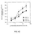
FIG. 4 shows the functional expression of the human active CCR5 receptor in a CHO-K1 cell line.
FIG. 5 represents the distribution of MRNA encoding the CCR5 receptor in a panel of human cell lines of haematopoietic origin.
FIG. 6 represents the structure of the mutant form of human CCR5 receptor. FIG. 6B shows the wild type amino acid sequence (SEQ ID NO: 11), and the location of the 32 base deletion mutation in the nucleic acid (SEQ ID NO: 12) and amino acid sequences (SEQ ID NO: 13).
FIG. 7 represents the quantification of ENV proteins-mediated fusion by luciferase assays.
FIG. 8 represents genotyping of individuals by PCR and segregation of the CCR5 alleles in CEPH families.
FIG. 9 represents the FACS analysis of sera anti-CCR5 on a CCR5-CHO cell line according to the invention.
FIG. 10 represents the inhibition of HIV infectivity with anti-CCR5 antibodies.
DETAILED DESCRIPTION OF THE PREFERRED EMBODIMENTS
Experimentals
Materials
Recombinant human chemokines, including MCP-1, MIP-1α, MIP-1p, RANTES, IL-8 and GROα were obtained from R & D Systems (London, UK). [125I]MIP-1α (specific activity, 2200 Ci/mmol) was obtained from Dupont NEN (Brussels, Belgium). Chemokines obtained from R & D Systems were reported by the supplier as >97% pure on SDS-PAGE (sodium dodecyl sulphate-polyacrylamide gel electrophoresis) and biologically active on a bioassay specific for each ligand. The lyophilised chemokines were dissolved as a 100 μg/ml solution in a sterile phosphate-buffered saline (PBS) and this stock solution was stored at −20° C. in aliquots. Chemokines were diluted to the working concentration immediately before use. All cell lines used in the present study were obtained from the ATCC (Rockville, Md., USA).
Cloning and Sequencing
The mouse MOP020 clone was obtained by low stringency polymerase chain reaction, as described previously [24, 34], using genomic DNA as template. A human genomic DNA library (Stratagene, La Jolla, Calif.), constructed in the lambda DASH vector was screened at low stringency [39] with the MOP020 (511 bp) probe. The positive clones were purified to homogeneity and analysed by Southern blotting. The restriction map of the locus was determined and a relevant XbaI fragment of 4,400 bp was subcloned in pBluescript SK+ (Stratagene). Sequencing was performed on both strands after subcloning in M13mp derivatives, using fluorescent primers and an automated DNA sequencer (Applied Biosystem 370A). Sequence handling and data analysis was carried out using the DNASIS/PROSIS software (Hitachi), and the GCG software package (Genetics Computer Group, Wisconsin).
Expression in Cell Lines
The entire coding region was amplified by PCR as a 1056 bp fragment, using primers including respectively the BamHI and XbaI recognition sequences, and cloned after restriction in the corresponding sites of the eukaryotic expression vector pcdna3 (Invitrogen, San Diego, Calif.). The resulting construct was verified by sequencing, and transfected in CHO-K1 cells as described [35]. Two days after transfection, selection for stably transfected cell lines was initiated by the addition of 400 μg/ml G418 (Gibco), arid resistant clones were isolated at day 10. CHO-K1 cells were cultured using Ham's F12 medium, as previously described [35, 11]. The expression of the active CCR5 receptor in the various cell clones was evaluated by measuring the specific transcript level by Northern blotting, on total RNA prepared from the cells (see below).
Binding Assays
Stably transfected CHO-K1 cells expressing the active CCR5 receptor were grown to confluence and detached from culture dishes by incubation in phosphate-buffered saline (PBS) supplemented with 1 Mm EDTA. Cells were collected by low speed centrifugation and counted in a Neubaeur cell. Binding assays were performed in polyethylene minisorp tubes (Nunc) in a final volume of 200 μl PBS containing 0.2% bovine serum albumin (BSA) and 106 cells, in presence of [125I]-MIP-1α. Non specific binding was determined by addition of 10 Nm unlabelled MIP-1 α. The concentration of labelled ligand was 0.4 Nm (around 100 000 cpm per tube). The incubation was carried out for 2 hours at 4° C., and was: stopped by the rapid addition of 4 ml ice-cold buffer, and immediate collection of cells by vacuum filtration through GF/B glass fiber filters (Whatmann) pre-soaked in 0.5% polyethyleneinimine (Sigma). Filters were washed three times with 4 ml ice-cold buffer and counted in a gamma counter.
Biological Activity
The CHO-K1 cell lines stably transfected with the pcdna3/CCR5 construct or wild type CHO-K1 cells (used as controls) were plated onto the membrane of Transwell cell capsules (Molecular Devices), at a density of 2.5 105 cells/well in Ham's F12 medium. The next day, the capsules were transferred in a microphysiometer (Cytosensor, Molecular Devices), and the cells were allowed to equilibrate for approximately two hours by perfusion of 1 Mm phosphate-buffered (Ph 7.4) RPMI-1640 medium containing 0.2% BSA. Cells were then exposed to various chemokines diluted in the same medium, for a 2 mm duration. Acidification rates were measured at one minute intervals.
Northern Blotting
Total RNA was isolated from transfected CHO-K1 cell lines, from a panel of human cell lines of haematopoietic origin and from a panel of dog tissues, using the RNeasy kit (Qiagen). RNA samples (10 μg per lane) were denatured in presence of glyoxal [26], fractionated on a 1% agarose gel in a 10 Mm phosphate buffer (Ph 7.0), and transferred to nylon membranes (Pall Biodyne A, Glen Cove, N.Y.) as described [42]. After baking, the blots were prehybridized for 4 h at 420° C. in a solution consisting of 50% formamide, 5×Denhardt solution (0×Denhardt: 0.02% Ficoll, 0.02% polyvinylpyrolidone, 0.02% BSA), 5×SSPE (1×SSPE: 0.18 MNaCl, 10 Mm Na phosphate, 1 Mm EDTA Ph 8.3), 0.3% Sodium Dodecyl Sulphate (SDS), 250 μg per ml denatured DNA from herring testes. DNA probes were (α32P)-labelled by random priming [14]. Hybridizations were carried out for 12 h at 42° C. in the same solution containing 10% (wt/vol) dextran sulphate and the heat denatured probe. Filters were washed up to 0.1×SSMC (1×SSC: 150 Mm NaCl, 15 Mm Na Citrate Ph 7.0), 0.1% SDS at 60° C. and autoradiographed at −70° C. using Amersham β-max films.
Results and Discussion
Cloning and Structural Analysis
The sequence homology characterising genes encoding G protein-coupled receptors has allowed the cloning by low stringency polymerase chain reaction (PCR) of new members of this gene family [24, 34]. One of the clones amplified from mouse genomic DNA, named MOP020 presented strong similarities with characterised chemokine receptors, sharing 80% identity with the MCP-1 receptor (CCR2) [8], 65% identity with the MIP-1α/RANTES receptor (CCR1) [31], and 51% identity with IL-8 receptors [20, 30]. The clone was used as a probe to screen a human genomic library. A total of 16 lambda phage clones were isolated. It was inferred from the restriction pattern of each clone and from partial sequence data that all clones were belonging to a single contig in which two different coding sequences were included. One of the coding sequences was identical to the reported CDNA encoding the CCR2 receptor [8, 44]. A 4.400 pb XbaI fragment of a representative clone containing the second region of hybridization was subcloned in Pbluescript SK+. Sequencing revealed a novel gene, tentatively named CCR5, sharing 84% identity with the MOP020 probe, suggesting that MOP020 is the mouse ortholog of CCR5. MOP020 does not correspond to any of the three mouse chemokine receptor genes cloned recently [16], demonstrating the existence of a fourth murine chemokine receptor.
The sequence of CCR5 revealed a single open reading frame of 352 codons encoding a protein of 40,600 Da. The sequence surrounding the proposed initiation codon is in agreement with the consensus as described by Kozak [22], since the nucleotide in −3 is a purine. The hydropathy profile of the deduced amino acid sequence is consistent with the existence of 7 transmembrane segments. Alignment of the CCR5 amino acid sequence with that of other functionally characterised human CC-chemokine receptors is represented in FIG. 2. The highest similarity is found with the CCR2 receptor [8] that shares 75.8% identical residues. There is also 56.3% identity with the CCR1 receptor [31], 58.4% with the CCR3 [10], and 49.1% with the CCR4 [37]. CCR5 represents therefore a new member of the CC-chemokine receptor group [30]. Like the related CCR1 and IL-8 receptors [20, 29, 31, 16] the coding region of CCR5 appears as intronless. From our partial sequencing data, the CCR2 gene is also devoid of intron in the first two thirds of its coding sequence.
Sequence similarities within the chemokine receptor family are higher in the transmembrane-spanning domains, and in intracellular loops. As an example, the identity score between CCR5 and CCR2 goes up to 92% when considering the transmembrane segments only. Lower similarities are found in the N-terminal extracellular domain, and in the extracellular loops. The N-terminal domain of the IL-8 and CCR2 receptors has been shown to be essential for interaction with the ligand [19, 18]. The variability of this region among CC-chemokine receptors presumably contributes to the specificity towards the various ligands of the family
single potential site for N-linked glycosylation was identified in the third extracellular loop of CCR5 (FIG. 1). No glycosylation site was found in the N-terminal domain of the receptor, where most G protein-coupled receptors are glycosylated. The other chemokine receptors CCR1 and CCR2 present such an N-linked glycosylation site in their N-terminal domain [31, 8]. By contrast, the CCR3 receptor [10] does not display glycosylation sites neither in the N-terminus, nor in extracellular loops. The active CCR5 receptor has four cysteines in its extracellular segments, and all four are conserved in the other CC- and CXC-chemokine receptors (FIG. 2). The cysteines located in the first and second extracellular loops are present in most G protein-coupled receptors, and are believed to form a disulphide bridge stabilising the receptor structure [41]. The two other cysteines, in the N-terminal segment, and in the third extracellular loop could similarly form a stabilising bridge specific to the chemokine receptor family. The intracellular domains of CCR5 do not include potential sites for phosphorylation by protein kinase C (PKC) or protein kinase A. PKC sites, involved in heterologous desensitisation are frequent in the third intracellular loop and C-terminus of G protein-coupled receptors. CCR1 is also devoid of PKC sites. In contrast, all CC-chemokine receptors, are rich in serine and threonine residues in the C-terminal domain. These residues represent potential phosphorylation sites by the family of G protein-coupled receptor kinases, and are probably involved in homologous desensitisation [41]. Five of these S/T residues are perfectly aligned in all five receptors (FIG. 2).
Physical Linkage of the CCR5 and CCR2 Genes
As stated above, the 16 clones isolated with the MOP020 probe corresponded to a single contig containing the CCR5 and CCR2 genes. The organisation of this contig was investigated in order to characterise the physical linkage of the two receptor genes in the human genome. A combination of restriction mapping, Southern blotting, fragment subcloning and partial sequencing allowed to determine the respective borders and overlaps of all clones. Out of the 16 clones, 9 turned out to be characterised by a specific restriction map, and their organisation is depicted in FIG. 3. Four of these clones (#11, 18, 21, 22) contained the CCR2 gene alone, four clones (# 7, 13, 15, 16) contained the ChemR13 gene alone and one clone (#9) contains part of both coding sequences. The CCR2 and CCR5 genes are organised in tandem, CCR5 being located downstream of CCR2. The distance separating CCR2 and CCR5 open reading frames is 17.5 kb. The chromosomal localisation of the tandem is presently unknown. Other chemokine receptors have however been located in the human genome: the CCR1 gene was localised by fluorescence in situ hybridization to the p21 region of human chromosome 3 [16]. The two IL-8 receptor genes, and theft pseudogene have been shown to be clustered on the human 2q34-q35 region [1].
Functional Expression and Pharmacology of the Active CCR5 Receptor
Stable CHO-K1 cell lines expressing the active CCR5 receptor were established and were screened on the basis of the level of CCR5 transcripts as determined by Northern blotting. Three clones were selected and tested for biological responses in a microphysiometer, using various CC- and CXC-chemokines as potential agonists. Wild type CHO-K1 dells were used as control to ensure that the observed responses were specific for the transfected receptor, and did not result from the activation of endogenous receptors. The microphysiometer allows the real time detection of receptor activation, by measuring the modifications of cell metabolism resulting from the stimulation of intracellular cascades [33]. Several studies have already demonstrated the potential of microphysiometry in the field of chemokine receptors. Modifications of metabolic activity in human monocytes, in response CC-chemokines, were monitored using this system [43]. Similarly, changes in the acidification rate of THP-1 cells (a human monocytic cell line) in response to MCP-1 and MCP-3 have been measured [36]. The estimation of the EC50 for both proteins, using this procedure, was in agreement with the values obtained by monitoring the intracellular calcium in other studies [8, 15].
Ligands belonging to the CC- and CXC-chemokine classes were tested on the CCR5 transfected CHO-K1 cells. Whereas MIP-1α, MIP-1β and RANTES were found to be potent activators of the new receptor (FIG. 4), the CC-chemokines MCP-1, MCP-2 and MCP-3, and the CXC-chemokines GROα and IL-8 had no effect on the metabolic activity, even at the highest concentrations tested (30 Nm). The biological activity of one of the chemokines inducing no response on CCR5 (IL-8) could be demonstrated on a CHO-K1 cell line transfected with the IL-8A interleukin receptor (Mollereau et al., 1993): IL-8 produced a 160% increase in metabolic activity as determined using the microphysiometer. The biological activity of the MCP-2 and MCP-3 preparations as provided by J. Van Damme have been widely documented [2, 40]. MIP-1α, MIP-1β and RANTES were tested on the wild type CHO-K1 cells, at a 30 Nm-concentration, and none of them induced a metabolic response. On the CCR5 transfected CHO-K1 cell line, all three active ligands (MIP-1a MIP-1β and RANTES) caused a rapid increase in acidification rate, reaching a maximum by the second or third minute after perfusion of the ligand. The acidification rate returned to basal level within 10 minutes. The timing of the cellular response is similar to that observed for chemokines on their natural receptors in human monocytes [43]. When agonists were applied repeatedly to the same cells, the response was strongly reduced as compared to the first stimulation, suggesting the desensitisation of the receptor. All measurements were therefore obtained on the first stimulation of each capsule.
The concentration-effect relation was evaluated for the three active ligands in the 0.3 to 30 Nm range (FIGS. 3B and C). The rank order of potency was MIP-1α>MIP-1β=RANTES. At 30 Nm concentrations, the effect of MIP-1α appeared to saturate (at 156% of baseline level) while MIP-1β and RANTES were still in the ascending phase. Higher concentrations of chemokines could however not be used. The EC50 was estimated around 3 Nm for MIP-1α. The concentrations necessary for obtaining a biological response as determined by using the microphysiometer are in the same range as those measured by intracellular calcium mobilisation for the CCR1 [31], the CCR2A and B [8], and the CCR3 [10] receptors. The ligand specificity of CCR5 is similar to that reported for CCR3 [10]. CCR3 was described as the first cloned receptor responding to MIP-1β. However, MIP-1 at 10 Nm elicits a significant effect on the CCR5, while the same concentration is without effect on the CCR3 transfected cells [10]. These data suggest that CCR5 could be a physiological receptor for MIP-1β.
Binding experiments using [125I]-human MIP-1α as ligand did not allow to demonstrate specific binding to CCR53 expressing CHO-K1 cells, using as much as 0.4 Nm radioligand and 1 million transfected cells per tube. Failure to obtain binding data could be attributed to a relatively low affinity of the receptor for MIP-1α.
Northern Blotting Analysis
Northern blotting performed on a, panel of dog tissues did not allow to detect transcripts for CCR5. Given the role of the chemokine receptor family in mediating chemoattraction and activation of various classes of cells involved in inflammatory and immune responses, the probe was also used to detect specific transcripts in a panel of human cell lines of haematopoietic origin (FIG. 5). The panel included lymphoblastic (Raji) and T lymphoblastic (Jurkat) cell lines, promyeloblastic (KG-1A) and promyelocytic (HL-60) cell lines, a monocytic (THP-1) cell line, an erythroleukemia (HEL 92.1.7) cell line, a megakaryoblastic (MEG-01) cell line, and a myelogenous leukaemia (K-562) cell line. Human peripheral blood mononuclear cells (PBMC), including mature monocytes and lymphocytes, were also tested. CCR5 transcripts (4.4 kb) could be detected only in the KG-1A promyeloblastic cell line, but were not found in the promyelocytic cell line HL-60, in PBMC, or in any of the other cell lines tested. These results suggest that the active CCR5 receptor could be expressed in precursors of the granulocytic lineage. CC-chemokines have been reported to stimulate mature granulocytes [27, 38, 23, 2]. However, recent data have also demonstrated a role of CC- and CXC-chemokines in the regulation of mouse and human myeloid progenitor cell proliferation [6, 7].
CCR5 was shown to respond to MIP-1a, MIP-1β and RANTES, the three chemokines identified as the major HIV-suppressive factors produced by CD8+ T cells [9], and released in higher amounts by CD4+ T lymphocytes from uninfected but multiply exposed individuals [51 ]. CCR5 represents a major co-receptor for macrophage-tropic (M-tropic) HIV-1 primary isolates and strains [45, 50]. M-tropic strains predominate during the asymptomatic phase of the disease in infected individuals, and are considered as responsible for HIV-1 transmission. Strains adapted for growth in transformed T-cell lines (T-tropic strains) use as a co-receptor LESTR (or fusin) [50], an orphan receptor also belonging to the chemokine receptor family, but not yet characterized functionally [21, 52, 53]. Dual-tropic viruses, which may represent transitional forms of the virus in late stages of infection [54] are shown to use both CCR5 and LESTR as co-receptors, as well as the CC-chemokine receptors CCR2b and CCR3 [47]. The broad spectrum of co-receptor usage of dual-tropic viruses suggests that within infected individuals, the virus may evolve at least in part from selection by a variety of co-receptors expressed on different cell types.
Identification of an Inactive ΔCCR5 Receptor
It is known that some individuals remain uninfected despite repeated exposure to HIV-1 [55, 56, 51]. A proportion of these exposed-uninfected individuals results from the relatively low risk of contamination after a single contact with the virus, but it has been postulated that truly resistant individuals do exist. In fact, CD4 lymphocytes isolated from exposed-uninfected individuals are highly resistant to infection by primary M-tropic, but not T-tropic HIV-1 strains. Also, peripheral blood mononuclear cells (PBMC) from different donors are not infected equally with various HIV-1 strains [57-59]. Given the key role played by CCR5 in the fusion event that mediates infection by M-tropic viruses, it is postulated that variants of CCR5 could be responsible for the relative or absolute resistance to HIV-1 infection exhibited by some individuals, and possibly for the variability of disease progression in infected patients [66]. The Inventors selected three HIV-1 infected patients known to be slow progressors, and four seronegative individuals as controls; the full coding region of their CCR5 gene was amplified by PCR and sequenced. Unexpectedly, one of the slow progressors, but also two of the uninfected controls, exhibited heterozygosity at the CCR5 locus for a biallelic polymorphism. The frequent allele corresponded to the published CCR5 sequence, while the minor one displayed a 32 bp deletion within the coding sequence, in a region corresponding to the second extracellular loop of the receptor (FIG. 6). The FIG. 6 is the structure of the mutant form of human CC-chemokine receptor 5. a The amino acid sequence of the non-functional Δccr5 protein is represented. The transmembrane organisation is given by analogy with the predicted transmembrane structure of the wild-type CCR5. Amino acids represented in black correspond to unnatural residues resulting from the frame shift caused by the deletion. The mutant protein lacks the last three transmembrane segments of CCR5, as well as the regions involved in G protein-coupling b, Nucleotide sequence of the CCR5 gene surrounding the deleted region, and translation into the normal receptor (top) or the truncated mutant (ccr5, bottom). The 10-bp direct repeat is represented in italics. The full size coding region of the CCR5 gene was amplified by PCR, using 5′-TCGAGGATCCAAGATGGATTATCAAGT-3′ (SEQ ID NO: 14) and 5′-CTGATCTAGAGCCATGTGCACAACTCT-3′ (SEQ ID NO: 15) as forward and reverse primers' respectively. The PCR products were sequenced on both strands using the same oligonucleotides as primers, as well as internal primers, and fluorochrome-labelled didcoxynucleotides as terminators. The sequencing products were run on an Applied Biosystem sequencer, and ambiguous positions were searched along the coding sequence. When the presence of a deletion was suspected from direct sequencing, the PCR products were cloned after restriction with BamHI and XbaI endonucleases into pcdna3. Several clones were sequenced to confirm the deletion. The deletion was identical in three unrelated individuals investigated by sequencing.
Cloning of the PCR product and sequencing of several clones confirmed the deletion. The deletion causes a frame shift, which is expected to result in premature termination of translation. The protein encoded by this mutant allele (Δccr5) therefore lacks the last three transmembrane segments of the receptor. A 10-bp direct repeat flanking the deleted region (FIG. 6b) on both sides is expected to have promoted the recombination event leading to the deletion. Numerous mutagenesis studies performed on various classes of G protein-coupled receptors, including chemokine receptors, makes it clear that such a truncated protein is certainly not functional in terms of chemokine-induced signal transduction: it lacks the third intracellular loop and C-terminal cytoplasmic domains, the two regions involved primarily in G protein coupling [41]. In order to test whether the truncated protein was able to function as a HIV-1 co-receptor, the Inventors tested its ability to support membrane fusion by both primary M-tropic and dual-tropic virus ENV proteins. The recombinant protein was expressed in quail QT6 cells together with human CD4. The QT6 cells were then mixed with HeLa cells expressing the indicated viral ENV protein and the extent of cell-cell fusion measured using a sensitive and quantitative gene-reporter assay. In contrast to wild-type CCR5, the truncated receptor did not allow fusion with cells expressing the ENV protein from either M-tropic or dual-tropic viruses (FIG. 7). The FIG. 7 represents the quantification of ENV protein-mediated fusion by luciferase assay. To quantify cell-cell fusion events, Japanese quail QT6 fibrosarcoma cells were transfected or cotransfected as indicated with the pcdna3 vector (Invitrogen) containing the coding sequence for wild-type CCR5, the truncated ccr5 mutant, the CCR2b or the Duffy chemokine receptors, or with the PCDNA3 vector alone. The target cells were also transfected with human CD4 expressed from the CMV promoter and the luciferase gene under the control of the T7 promoter. HeLa effector cells were infected (MOI=10) with vaccinia vectors expressing T7-polymerase (vTF1.1) and either the JR-FL (vCB28) or 89.6 (vBD3) envelope proteins. The luciferase activity resulting from cell fusion is expressed as the percentage of the activity (in relative light units) obtained for wild-type CCR5. All transfections were performed with an identical quantity of plasmid DNA using pcdna3 as carrier when necessary. To initiate fusion, target and effector cells were mixed in 24 well plates at 3° C. in the presence of ara-C and rifampicin, and allowed to fuse for 8 hours. Cells were lysed in 150 μl of reporter lysis buffer (Promega) and assayed for luciferase activity according to the manufacturer's instructions (Promega).
Coexpression of Δccr5 with wild-type CCR5 consistently reduced the efficiency of fusion for both JR-FL and 89.6 envelopes, as compared with CCR5 alone. Whether this in vitro inhibitory effect (not shared by the chemokine receptor Duffy, used as control) also occurs in vivo is presently not known. Coexpression with the CCR2b receptor [31], which is the CC-chemokine receptor most closely related to CCR5 but does not promote fusion by M-tropic HIV-1 strains [48], did not rescue the mutation by formation of a hybrid molecule (FIG. 7).
The FIG. 8 represents genotyping of individuals by PCR and segregation of the CCR5 alleles in CEPH families. α, Autoradiography illustrating the pattern resulting from PCR amplification and EcoRI cleavage for individuals homozygous for the wild-type CCR5 allele (CCR5/CCR5), the null Δccr5 allele (Δccr5/Δccr5), and for heterozygotes (CCR5/Δccr5). A 735 bp PCR product is cleaved into a common band of 332 bp for both alleles, and into 403 and 371 bp bands for the wild-type and mutant alleles, respectively. b, Segregation of the CCR5 alleles in two informative families of the CEPH. Half-black and white symbols represent heterozygotes and wild-type homozygotes, respectively. For a few individuals in the pedigrees, DNA was not available (ND: not determined). PCRs were performed on genomic DNA samples, using 5′-CCTGGCTGTCGTCCATGCTG-3′ (SEQ ID NO: 16) and 5′-CTGATCTAGAGCCATGTGCACAACTCT-3′ (SEQ ID NO: 17) as forward and reverse primers respectively. Reaction mixtures consisted in 30 μl of 10 Mm Tris-Hcl buffer Ph 8.0, containing 50 Mm Kcl, 0.75 Mm MgCl2, 0.2 Mm dCTP, dGTP and dTTP, 0.1 Mm dATP, 0.5 μi [a-32P]-DATP, 0.01% gelatine, 5% DMSO, 200 ng target DNA, 60 ng of each of the primers and 1.5 U Taq polymerase. PCR conditions were: 93° C. for 2 min 30; 93° C. for 1 min, 60° C. for 1 min, 72° C. for 1 min, 30 cycles; 72° C. for 6 min. After the PCR reaction, the samples were incubated for 60 min at 37° C. with 10 U EcoRI, and 2 μl of the denatured reaction mixture was applied onto a denaturing 5% polyacrylamidc gel containing 35% formamide and 5.6 M urea. Bands were detected by autoradiography.
Based on the 14 chromosomes tested in the first experiment, the deleted Δccr5 allele appeared rather frequent in the Caucasian population. The accurate frequency was further estimated by testing (FIG. 8a) a large cohort of Caucasian individuals, including unrelated members of the CEPH (Centre d'Etude des Polymorphismes Humains) families, part of the IRIBHN staff, and a bank of anonymous DNA samples from healthy individuals collected by the Genetics Department of the Erasme Hospital in Brussels. From a total of more than 700 healthy individuals, the allele frequencies were found to be 0.908 for the wild-type allele, and 0.092 for the mutant allele (Table 1). The genotype frequencies observed in the population were not significantly different from the expected Hardy-Weinberg distribution (CCR5/CCR5: 0.827 vs 0.824; CCR5/Δccr5: 0.162 vs 0.167; Δccr51/ccr5: 0.011 vs 0.008, p>0.999), suggesting that the null allele has no drastic effect on fitness. Using two informative CEPH families, it was confirmed that—the wild-type CCR5 gene and its Δccr5 variant were allelic, and segregated in a normal mendelian fashion (FIG. 8b). Interestingly, a cohort of 124 DNA samples originating from Central Africa (collected from Zaire, Burkina Fasso, Cameroun, Senegal and Benin) and Japan did not reveal a single Δccr5 mutant allele, suggesting that this allele is either absent or very rare in Asian, African black populations (Table I).
The consequences of the existence of a null allele of CCR5 in the normal Caucasian population were then considered in terms of susceptibility to infection by HIV-1. If, as it is predicted, CCR5 plays a major (not redundant) role in the entry of most primary virus strains into cells, then Δccr5/Δccr5 individuals should be particularly resistant to HIV-1 challenge, both in vitro and in vivo. The frequency of the Δccr5/Δccr5 genotype should therefore be in significantly lower in HIV-1 infected patients, and increased in exposed-uninfected individuals. Also, if heterozygotes have a statistical advantage due to the lower number of functional receptors on their white blood cells, or to the possible dominant-negative properties of the mutant allele, the frequency of heterozygotes (and mutant alleles) should be decreased in HIV-infected populations. These hypotheses were tested by genotyping a large number of seropositive Caucasian individuals (n=645) belonging to cohorts originating from various hospitals from Brussels, Liege and Paris (Table I). Indeed, it was found that within this large series, the frequency of the null Δccr5 allele was significantly reduced from 0.092 to 0.053 (p<10−5). The frequency of heterozygotes was also reduced from 0.162 to 0.106 (p<0.001) and not a single Δccr5/Δccr5 individual could be found (p<0.01).
Altogether, functional and statistical data suggest that CCR5 is indeed the major co-receptor responsible for natural infection by M-tropic HIV-1 strains Individuals homozygous for the null Δccr5 allele (about 1% of the Caucasian population) have apparently a strong resistance to infection. It is unclear at this point whether resistance to HIV-1 is absolute or relative, and whether resistance will vary depending on the mode of viral contamination. Larger cohorts of seropositive individuals will have to be tested in order to clarify this point. Heterozygotes have a milder though significant advantage: assuming an equal probability of contact with HIV, it can be inferred from Table I that heterozygotes have a 39% reduction in their likeliness of becoming seropositive, as compared to individuals homozygous for the wild-type CCR5 allele. Both a decrease in functional CCR5 receptor number, and a dominant-negative effect of Δccr5 in vivo, comparable to what is observed in the in vitro experiments (FIG. 7) are possible explanations for this relative protection. The mutant allele, which can be regarded as a natural knock-out in human, is not accompanied by an obvious phenotype in homozygous individuals. Nevertheless, the lack of overt phenotype, taken together with the relative protection that characterizes heterozygous subjects, suggests that pharmacological agents that selectively block the ability of HIV-1 to utilize CCR5 as a cofactor, could be effective in preventing HIV-1 infection, and would be predicted not be associated with major side effects resulting from CCR5 inactivation. These pharmaceutical agents could be used with other compounds which are able to block other chemokine receptors used as co-receptors by some. HIV-primary isolates in order to infect other cells [47]. The prevalence of the null allele in the Caucasian population raises the question of whether pandemia of HIV (or related viruses using the same co-receptor) have contributed during mankind's evolution to stabilize by selection the mutant ccr5 allele at such a high frequency.
Production of Antibodies Anti-CCR5
Antibodies were produced by genetic immunisation. Six week old females balb/c mice were used. DNA coding for the human CCR5 receptor was inserted in the expression vector pcdna3 under the control of the CMV promotor and 100 μg DNA was injected in the anterior tibial muscle, five days after pre-treatment of this muscle with cardiotoxine (from venom of Naja Nigricolis). Injections were repeated twice at three week intervals. Fifteen days after the last injection, blood was taken from each animal and sera were tested for the presence of anti-CCR5 antibodies.
Test of Sera Using Fluorescence Activated Cell Sorter (FACS)
Sera were tested by fluorescence activated cell sorting using recombinant CHO cells expressing the CCR5 receptor. Briefly, cells were detached using a PBS-EDTA-EGTA solution and incubated into PBS-BSA medium for 30 minutes at room temperature with 5 μl serum on the basis of 100,000 cells per tube. Cells were then washed and incubated for 30 minutes in ice together with anti-mouse antibody labelled with fluorescein. Cells were washed, taken up into 200 μl of a PBS-BSA solution and fluorescence was analysed by FACS (FACSCAN, Becton-Dickinson). 10,000 cells were counted. Wild type CHO or recombinant CHO cells expressing the human CCR2b receptor were used as controls.
When tested by FACS analysis 2 weeks after the last injection (FIG. 9), all the sera from mice immunised with CCR5 CDNA, clearly recognised the native receptor expressed on CHO cells (mean of fluorescence=200), without significant cross reaction with control cells expressing CCR2b (mean of fluorescence=20).
Sera were tested on either a CHO cell line expressing high level of CCR5 receptor (black histogram) or a CHO cell line expressing CCR2b receptor (white histogram) as negative control. Each serum was tested individually.
Antibodies Anti-CCR5 and HIV Infectivity
Peripheral blood mononuclear cells (PBMC) from one donor homozygous from wild type CCR5 gene, were isolated and cultivated 3 days in presence of PHA.
On day 4, 800 μl of cells (105 cells/ml) were incubated with 8 μl of sera from mice immunised with CCR5 CDNA, 30 minutes at 37° C. 1 ml of viral solution (JRCSF HIV strain) is then added and incubated during 2 hours. Cells were then washed twice and cultivated during 15 days.
Aliquot of medium is taken at days 0, 4, 7, 10 and 14 and the dosage of antigen p24 is performed.
14 days after the beginning of the experiment, one serum (serum B0) totally block the production of p24, indicating its ability to block the infection of the lymphocytes by this HIV strain (FIG. 10). Other serums also exhibit a partial or total effect on this infection (serum A2 and B1). All the other sera did not show any effect on this infection.
Production of Monoclonal Antibodies
Mice with the highest title of CCR5 antibodies were selected for monoclonal antibodies production and injected intravenously with 107 recombinant CHO-K1 cells expressing human CCR5 receptors. Three days later, animals were sacrificed and fusion of splenic cells or cells from lymph nodes near the site of injection with SP2/0 myeloma cells, were performed. Fusion protocol used was that of Galfre et al. (Nature 266, 550 (1977)). A selective HAT (hypoxanthine/aminopterin/thymidin) medium is used to select hybridomas and their supernatants are tested by FACS using recombinant CHO cells expressing the human CCR5 receptor, as it was done for the sera. Positives hybridomas are then cloned by limited dilution. Clones that are show positive by FACS analyses are then expanded and produced in ascites in balb/C mice.
| |
TABLE 1 |
| |
|
| |
Seronegative |
Seropositive |
|
| |
|
|
Standard |
|
|
Standard |
|
| |
Number |
Frequency |
error |
Number |
Frequency |
error |
Chi-squared |
| |
|
| Genotypes: |
|
|
|
|
|
|
2 degrees of |
| CCR5/CCR5 |
582 |
0.827 |
0.014 |
645 |
0.892 |
0.012 |
freedom |
| CCR5/ΔCCR5 |
114 |
0.162 |
0.014 |
78 |
0.108 |
0.012 |
17.7 |
| CCRS/ΔCCR5 |
8 |
0.011 |
0.004 |
0 |
0.000 |
<0.001 |
p < 0.0005 |
| Total: |
704 |
1.000 |
|
723 |
1.000 |
| Alleles: |
|
|
|
|
|
|
1 degree of |
| CCR5 |
1278 |
0.908 |
0.008 |
1368 |
0.946 |
0.006 |
freedom |
| ΔCCR5 |
| |
130 |
0.092 |
0.008 |
78 |
0.054 |
0.006 |
p < 0.0005 |
| Total: |
1408 |
1.000 |
|
1446 |
1.000 |
| |
References
1. Ahuja et al. (1992) Nature Genetics 2, 31-36.
2. Alam et al. (1994) J. Immunol. 153, 3155-3159.
3. Alam et al. (1992) J. Exp. Med. 176, 781-786.
4. Baggiolini et al. (1994) Advances in immunology, Academic press. Ed. Dixon, F. J. 55, 97-179.
5. Bikenbach et al. (1 993) J. Virol. 67, 2209-2220.
6. Broxmeyer et al. (1990) Blood 76, 1110-1116.
7. Broxmeyer et al. (1993) J. Immunol. 150, 3448-3458.
8. Charo et al. (1994) Proc. Natl. Acad. Sci. 91, 2752-2756.
9. Cocci et al. (1995) Science 270, 1811-1815.
10. Combadiere et al. (1995) J. Biol. Chem. 270, 16491-16494.
11. Desarnaud et al. (1994) Biochem. J. 299, 367-373.
12. Deveréux et al. (1984) Nucleic Acids Res. 12, 387-395.
13. Dobner et al. (1992) Eur. J. Immunol. 22, 2795-2799.
14. Feinberg et al. (1 983) Anal. Biochem. 132, 6-13.
15. Franci et al. (1995) J. Immunol. 154, 6511-6517.
16. Gao et al. (1995) J. Biol. Chem. 270, 17494-17501.
17. Gao et al. (1993) J. Exp. Med. 177, 1421-1427.
18. Gong et al. (1995) J. Exp. Med. 181, 631-640.
19. Hébert et al. (1993) J. Biol. Chem. 268, 18549-18533.
20. Holmes et al. (1991) Science 253, 1278-1283.
21. Jazin et al. (1993) Regul. Peptides 47, 247-258.
22. Kozak, M. (1989) J. Cell. Biol. 108, 229-241.
23. Kuna et al. (1992) J. Immunol. 149, 636-642.
24. Libert et al. (1989) Science 244, 569-572.
25. Matsuoka et al. (1993) Biochem. Biophys. Res. Commun. 194, 504-511.
26. Mc Master et al. (1977) Proc. Natl Acad. Sci. USA 74, 4835-4838.
27. McColl et al. (1993) J. Immunol. 150, 4550-4560.
28. Mollereau et al. (1993) Genomics 16, 248-251.
29. Murphy, P. M. (1994) Anne Rev. Immunol. 12, 593-633.
30. Murphy et al. (1991) Science 253, 1280-1283.
31. Neote et al. (1993) Cell 72, 415-425.
32. Oppenheim et al. (1991) Ann. Rev. Immunol. 9, 617-648.
33. Owicki et al. (1992) Biosensors Bioelectronics 7, 255-272.
34. Parmentier et al. (1989) Science 246, 1620-1622.
35. Perret et al. (1 990) Biochem. Biophys. Res. Commun. 17, 1044-1050.
36. Pleass et al. (1995) Fourth International Chemokine Symposium, Bath (UK), 27-30 June, P47.
37. Power et al. (1995) J. Biol. Chem. 270, 19495-19500.
38. Rot et al. (1992) J. Exp. Med. 176, 1489-1495.
39. Sambrook et al. (1989) Molecular Cloning: A Laboratory Manual, 2nd Ed., Cold Spring Harbor Laboratory, Cold Spring Harbor, N.Y.
40. Sozzani et al. (1994) J. Immunol. 152, 3615-3622.
41. Strader et al. (1994) Annu. Rev. Biochem. Ed. Dixon, R. A. F. 63, 101-132.
42. Thomas, P. S. (1980). Proc. Natl. Acad. Sci. USA 77, 5201-5205.
43. Vaddi et al. (1994) The FASEB Journal 8, A502.
44. Yamagami et al. (1994) Biochem. Biophys. Res. Commun. 202, 1156-1162.
45. Deng et al. (1996) Nature 381, 661-666.
46. Drajic et al. (1996) Nature 381, 667-673.
47. Doranz et al. (1996) Cell 85, 1149-1158.
48. Choe et al. (1996) Cell 85, 1135-1148.
49. Aklhatib et al. (1996) Science 272, 1955-1958.
50. Feng et al. (1996) Science 272, 872-877.
51. Paxton et al. (1996) Nat. Med. 2, 412-417.
52. Nomura et al. (1993) Int. Immunol. 5, 1239-1249.
53. Loetscher et al. (1994) J. Biol. Chem. 269, 232-237.
54. Collman et al. (1992) J. Virol. 66,7517-7521.
55. Detels et al. (1994) J. Acquir. Immun. Defic. Sundr. 7, 1263-1269.
56. Taylor R. J. (1994) J. NIH Res. 6,29-31.
57. Wainberg et al. (1987) Clin. Exp. Immunol. 1987, 136-142.
58. Williams et al. (1991) Virology 184, 723-728.
59. Spria et al. (1995) J. Virol. 69, 422-429.
60. Haynes et al. (1996) Science 271, 324-328.
61. Ben-Baruch et al. (1995) J. Biol. Chem. 270, 11703-11706.
62. Nussbaum et al. (1994) J. Virol. 68, 5411-5422.
18
1
792
DNA
Homo sapiens
1
gaattccccc aacagagcca agctctccat ctagtggaca gggaagctag cagcaaacct 60
tcccttcact acaaaacttc attgcttggc caaaaagaga gttaattcaa tgtagacatc 120
tatgtaggca attaaaaacc tattgatgta taaaacagtt tgcattcatg gagggcaact 180
aaatacattc taggacttta taaaagatca ctttttattt atgcacaggg tggaacaaga 240
tggattatca agtgtcaagt ccaatctatg acatcaatta ttatacatcg gagccctgcc 300
aaaaaatcaa tgtgaagcaa atcgcagccc gcctcctgcc tccgctctac tcactggtgt 360
tcatctttgg ttttgtgggc aacatgctgg tcatcctcat cctgataaac tgcaaaaggc 420
tgaagagcat gactgacatc tacctgctca acctggccat ctctgacctg tttttccttc 480
ttactgtccc cttctgggct cactatgctg ccgcccagtg ggactttgga aatacaatgt 540
gtcaactctt gacagggctc tattttatag gcttcttctc tggaatcttc ttcatcatcc 600
tcctgacaat cgataggtac ctggctgtcg tccatgctgt gtttgcttta aaagccagga 660
cggtcacctt tggggtggtg acaagtgtga tcacttgggt ggtggctgtg tttgcgtctc 720
tcccaggaat catctttacc agatctcaaa aagaaggtct tcattacacc tgcagctctc 780
attttccata ca 792
2
1477
DNA
Homo sapiens
misc_feature
(1377)..(1377)
Any nucleotide
2
gaattccccc aacagagcca agctctccat ctagtggaca gggaagctag cagcaaacct 60
tcccttcact acaaaacttc attgcttggc caaaaagaga gttaattcaa tgtagacatc 120
tatgtaggca attaaaaacc tattgatgta taaaacagtt tgcattcatg gagggcaact 180
aaatacattc taggacttta taaaagatca ctttttattt atgcacaggg tggaacaaga 240
tggattatca agtgtcaagt ccaatctatg acatcaatta ttatacatcg gagccctgcc 300
aaaaaatcaa tgtgaagcaa atcgcagccc gcctcctgcc tccgctctac tcactggtgt 360
tcatctttgg ttttgtgggc aacatgctgg tcatcctcat cctgataaac tgcaaaaggc 420
tgaagagcat gactgacatc tacctgctca acctggccat ctctgacctg tttttccttc 480
ttactgtccc cttctgggct cactatgctg ccgcccagtg ggactttgga aatacaatgt 540
gtcaactctt gacagggctc tattttatag gcttcttctc tggaatcttc ttcatcatcc 600
tcctgacaat cgataggtac ctggctgtcg tccatgctgt gtttgcttta aaagccagga 660
cggtcacctt tggggtggtg acaagtgtga tcacttgggt ggtggctgtg tttgcgtctc 720
tcccaggaat catctttacc agatctcaaa aagaaggtct tcattacacc tgcagctctc 780
attttccata cagtcagtat caattctgga agaatttcca gacattaaag atagtcatct 840
tggggctggt cctgccgctg cttgtcatgg tcatctgcta ctcgggaatc ctaaaaactc 900
tgcttcggtg tcgaaatgag aagaagaggc acagggctgt gaggcttatc ttcaccatca 960
tgattgttta ttttctcttc tgggctccct acaacattgt ccttctcctg aacaccttcc 1020
aggaattctt tggcctgaat aattgcagta gctctaacag gttggaccaa gctatgcagg 1080
tgacagagac tcttgggatg acgcactgct gcatcaaccc catcatctat gcctttgtcg 1140
gggagaagtt cagaaactac ctcttagtct tcttccaaaa gcacattgcc aaacgcttct 1200
gcaaatgctg ttctattttc cagcaagagg ctcccgagcg agcaagctca gtttacaccc 1260
gatccactgg ggagcaggaa atatctgtgg gcttgtgaca cggactcaag tgggctggtg 1320
acccagtcag agttgtgcac atggcttagt tttcatacac agcctgggct gggggtnggt 1380
tggnngaggt cttttttaaa aggaagttac tgttatagag ggtctaagat tcatccattt 1440
atttggcatc tgtttaaagt agattagatc cgaattc 1477
3
1442
DNA
Homo sapiens
3
gaattccccc aacagagcca agctctccat ctagtggaca gggaagctag cagcaaacct 60
tcccttcact acaaaacttc attgcttggc caaaaagaga gttaattcaa tgtagacatc 120
tatgtaggca attaaaaacc tattgatgta taaaacagtt tgcattcatg gagggcaact 180
aaatacattc taggacttta taaaagatca ctttttattt atgcacaggg tggaacaaga 240
tggattatca agtgtcaagt ccaatctatg acatcaatta ttatacatcg gagccctgcc 300
aaaaaatcaa tgtgaagcaa atcgcagccc gcctcctgcc tccgctctac tcactggtgt 360
tcatctttgg ttttgtgggc aacatgctgg tcatcctcat cctgataaac tgcaaaaggc 420
tgaagagcat gactgacatc tacctgctca acctggccat ctctgacctg tttttccttc 480
ttactgtccc cttctgggct cactatgctg ccgcccagtg ggactttgga aatacaatgt 540
gtcaactctt gacagggctc tattttatag gcttcttctc tggaatcttc ttcatcatcc 600
tcctgacaat cgataggtac ctggctgtcg tccatgctgt gtttgcttta aaagccagga 660
cggtcacctt tggggtggtg acaagtgtga tcacttgggt ggtggctgtg tttgcgtctc 720
tcccaggaat catctttacc agatctcaaa aagaaggtct tcattacacc tgcagctctc 780
attttccata cattaaagat agtcatcttg gggctggtcc tgccgctgct tgtcatggtc 840
atctgctact cgggaatcct aaaaactctg cttcggtgtc gaaatgagaa gaagaggcac 900
agggctgtga ggcttatctt caccatcatg attgtttatt ttctcttctg ggctccctac 960
aacattgtcc ttctcctgaa caccttccag gaattctttg gcctgaataa ttgcagtagc 1020
tctaacaggt tggaccaagc tatgcaggtg acagagactc ttgggatgac gcactgctgc 1080
atcaacccca tcatctatgc ctttgtcggg gagaagttca gaaactacct cttagtcttc 1140
ttccaaaagc acattgccaa acgcttctgc aaatgctgtt ctattttcca gcaagaggct 1200
cccgagcgag caagctcagt ttacacccga tccactgggg agcaggaaat atctgtgggc 1260
ttgtgacacg gactcaagtg ggctggtgac ccagtcagag ttgtgcacat ggcttagttt 1320
tcatacacag cctgggctgg gggtggttgg gaggtctttt ttaaaaggaa gttactgtta 1380
tagagggtct aagattcatc catttatttg gcatctgttt aaagtagatt agatccgaat 1440
tc 1442
4
184
PRT
Homo sapiens
4
Met Asp Tyr Gln Val Ser Ser Pro Ile Tyr Asp Ile Asn Tyr Tyr Thr
1 5 10 15
Ser Glu Pro Cys Gln Lys Ile Asn Val Lys Gln Ile Ala Ala Arg Leu
20 25 30
Leu Pro Pro Leu Tyr Ser Leu Val Phe Ile Phe Gly Phe Val Gly Asn
35 40 45
Met Leu Val Ile Leu Ile Leu Ile Asn Cys Lys Arg Leu Lys Ser Met
50 55 60
Thr Asp Ile Tyr Leu Leu Asn Leu Ala Ile Ser Asp Leu Phe Phe Leu
65 70 75 80
Leu Thr Val Pro Phe Trp Ala His Tyr Ala Ala Ala Gln Trp Asp Phe
85 90 95
Gly Asn Thr Met Cys Gln Leu Leu Thr Gly Leu Tyr Phe Ile Gly Phe
100 105 110
Phe Ser Gly Ile Phe Phe Ile Ile Leu Leu Thr Ile Asp Arg Tyr Leu
115 120 125
Ala Val Val His Ala Val Phe Ala Leu Lys Ala Arg Thr Val Thr Phe
130 135 140
Gly Val Val Thr Ser Val Ile Thr Trp Val Val Ala Val Phe Ala Ser
145 150 155 160
Leu Pro Gly Ile Ile Phe Thr Arg Ser Gln Lys Glu Gly Leu His Tyr
165 170 175
Thr Cys Ser Ser His Phe Pro Tyr
180
5
352
PRT
Homo sapiens
5
Met Asp Tyr Gln Val Ser Ser Pro Ile Tyr Asp Ile Asn Tyr Tyr Thr
1 5 10 15
Ser Glu Pro Cys Gln Lys Ile Asn Val Lys Gln Ile Ala Ala Arg Leu
20 25 30
Leu Pro Pro Leu Tyr Ser Leu Val Phe Ile Phe Gly Phe Val Gly Asn
35 40 45
Met Leu Val Ile Leu Ile Leu Ile Asn Cys Lys Arg Leu Lys Ser Met
50 55 60
Thr Asp Ile Tyr Leu Leu Asn Leu Ala Ile Ser Asp Leu Phe Phe Leu
65 70 75 80
Leu Thr Val Pro Phe Trp Ala His Tyr Ala Ala Ala Gln Trp Asp Phe
85 90 95
Gly Asn Thr Met Cys Gln Leu Leu Thr Gly Leu Tyr Phe Ile Gly Phe
100 105 110
Phe Ser Gly Ile Phe Phe Ile Ile Leu Leu Thr Ile Asp Arg Tyr Leu
115 120 125
Ala Val Val His Ala Val Phe Ala Leu Lys Ala Arg Thr Val Thr Phe
130 135 140
Gly Val Val Thr Ser Val Ile Thr Trp Val Val Ala Val Phe Ala Ser
145 150 155 160
Leu Pro Gly Ile Ile Phe Thr Arg Ser Gln Lys Glu Gly Leu His Tyr
165 170 175
Thr Cys Ser Ser His Phe Pro Tyr Ser Gln Tyr Gln Phe Trp Lys Asn
180 185 190
Phe Gln Thr Leu Lys Ile Val Ile Leu Gly Leu Val Leu Pro Leu Leu
195 200 205
Val Met Val Ile Cys Tyr Ser Gly Ile Leu Lys Thr Leu Leu Arg Cys
210 215 220
Arg Asn Glu Lys Lys Arg His Arg Ala Val Arg Leu Ile Phe Thr Ile
225 230 235 240
Met Ile Val Tyr Phe Leu Phe Trp Ala Pro Tyr Asn Ile Val Leu Leu
245 250 255
Leu Asn Thr Phe Gln Glu Phe Phe Gly Leu Asn Asn Cys Ser Ser Ser
260 265 270
Asn Arg Leu Asp Gln Ala Met Gln Val Thr Glu Thr Leu Gly Met Thr
275 280 285
His Cys Cys Ile Asn Pro Ile Ile Tyr Ala Phe Val Gly Glu Lys Phe
290 295 300
Arg Asn Tyr Leu Leu Val Phe Phe Gln Lys His Ile Ala Lys Arg Phe
305 310 315 320
Cys Lys Cys Cys Ser Ile Phe Gln Gln Glu Ala Pro Glu Arg Ala Ser
325 330 335
Ser Val Tyr Thr Arg Ser Thr Gly Glu Gln Glu Ile Ser Val Gly Leu
340 345 350
6
215
PRT
Homo sapiens
6
Met Asp Tyr Gln Val Ser Ser Pro Ile Tyr Asp Ile Asn Tyr Tyr Thr
1 5 10 15
Ser Glu Pro Cys Gln Lys Ile Asn Val Lys Gln Ile Ala Ala Arg Leu
20 25 30
Leu Pro Pro Leu Tyr Ser Leu Val Phe Ile Phe Gly Phe Val Gly Asn
35 40 45
Met Leu Val Ile Leu Ile Leu Ile Asn Cys Lys Arg Leu Lys Ser Met
50 55 60
Thr Asp Ile Tyr Leu Leu Asn Leu Ala Ile Ser Asp Leu Phe Phe Leu
65 70 75 80
Leu Thr Val Pro Phe Trp Ala His Tyr Ala Ala Ala Gln Trp Asp Phe
85 90 95
Gly Asn Thr Met Cys Gln Leu Leu Thr Gly Leu Tyr Phe Ile Gly Phe
100 105 110
Phe Ser Gly Ile Phe Phe Ile Ile Leu Leu Thr Ile Asp Arg Tyr Leu
115 120 125
Ala Val Val His Ala Val Phe Ala Leu Lys Ala Arg Thr Val Thr Phe
130 135 140
Gly Val Val Thr Ser Val Ile Thr Trp Val Val Ala Val Phe Ala Ser
145 150 155 160
Leu Pro Gly Ile Ile Phe Thr Arg Ser Gln Lys Glu Gly Leu His Tyr
165 170 175
Thr Cys Ser Ser His Phe Pro Tyr Ile Lys Asp Ser His Leu Gly Ala
180 185 190
Gly Pro Ala Ala Ala Cys His Gly His Leu Leu Leu Gly Asn Pro Lys
195 200 205
Asn Ser Ala Ser Val Ser Lys
210 215
7
360
PRT
Homo sapiens
MISC_FEATURE
(325)..(327)
Xaa = any amino acid
7
Met Leu Ser Thr Ser Arg Ser Arg Phe Ile Arg Asn Thr Asn Glu Ser
1 5 10 15
Gly Glu Glu Val Thr Thr Phe Phe Asp Tyr Asp Tyr Gly Ala Pro Cys
20 25 30
His Lys Phe Asp Val Lys Gln Ile Gly Ala Gln Leu Leu Pro Pro Leu
35 40 45
Tyr Ser Leu Val Phe Ile Phe Gly Phe Val Gly Asn Met Leu Val Val
50 55 60
Leu Ile Leu Ile Asn Cys Lys Lys Leu Lys Cys Leu Thr Asp Ile Tyr
65 70 75 80
Leu Leu Asn Leu Ala Ile Ser Asp Leu Leu Phe Ile Ile Thr Leu Pro
85 90 95
Leu Trp Ala His Ser Ala Ala Asn Glu Trp Val Phe Gly Asn Ala Met
100 105 110
Cys Lys Leu Phe Thr Gly Leu Tyr His Ile Gly Tyr Phe Gly Gly Ile
115 120 125
Phe Phe Ile Ile Leu Leu Thr Ile Asp Arg Tyr Leu Ala Ile Val His
130 135 140
Ala Val Phe Ala Leu Lys Ala Arg Thr Val Thr Phe Gly Val Val Thr
145 150 155 160
Ser Val Ile Thr Trp Leu Val Ala Val Phe Ala Ser Val Pro Gly Ile
165 170 175
Ile Phe Thr Lys Cys Gln Lys Glu Asp Ser Val Tyr Val Cys Gly Pro
180 185 190
Tyr Phe Pro Arg Gly Trp Asn Asn Phe His Thr Ile Met Arg Asn Ile
195 200 205
Leu Gly Leu Val Leu Pro Leu Leu Ile Met Val Ile Cys Tyr Ser Gly
210 215 220
Ile Leu Lys Thr Leu Leu Arg Cys Arg Asn Glu Lys Lys Arg His Arg
225 230 235 240
Ala Val Arg Val Ile Phe Thr Ile Met Ile Val Tyr Phe Leu Phe Trp
245 250 255
Thr Pro Tyr Asn Ile Val Ile Leu Leu Asn Thr Phe Gln Glu Phe Phe
260 265 270
Gly Leu Ser Asn Cys Glu Ser Thr Ser Gln Leu Asp Gln Ala Ile Gln
275 280 285
Val Thr Glu Thr Leu Gly Met Thr His Cys Cys Ile Asn Pro Ile Ile
290 295 300
Tyr Ala Phe Val Gly Glu Lys Phe Arg Arg Tyr Ile Ser Val Phe Phe
305 310 315 320
Arg Lys His Ile Xaa Xaa Xaa Phe Cys Lys Gln Cys Pro Val Phe Tyr
325 330 335
Arg Glu Thr Val Asp Gly Val Thr Ser Thr Asn Thr Pro Ser Thr Gly
340 345 350
Glu Gln Glu Val Ser Ala Gly Leu
355 360
8
355
PRT
Homo sapiens
MISC_FEATURE
(231)..(233)
Xaa = amy amino acid
8
Met Thr Thr Ser Ile Asp Thr Val Glu Thr Phe Gly Thr Thr Ser Tyr
1 5 10 15
Tyr Asp Asp Val Gly Leu Leu Cys Glu Lys Ala Asp Thr Arg Ala Leu
20 25 30
Met Ala Gln Phe Val Pro Pro Leu Tyr Ser Leu Val Phe Thr Val Gly
35 40 45
Leu Ile Gly Asn Val Val Val Val Met Ile Leu Ile Lys Tyr Arg Arg
50 55 60
Ile Arg Ile Met Thr Asn Ile Tyr Leu Leu Asn Leu Ala Ile Ser Asp
65 70 75 80
Leu Leu Phe Ile Val Thr Leu Pro Phe Trp Thr His Tyr Val Arg Gly
85 90 95
His Asn Trp Val Phe Gly His Gly Met Cys Asn Leu Ile Ser Gly Phe
100 105 110
Tyr His Thr Gly Leu Tyr Ser Glu Ile Phe Phe Ile Ile Leu Leu Thr
115 120 125
Ile Asp Arg Tyr Leu Ala Ile Val His Ala Val Phe Ala Ile Arg Ala
130 135 140
Arg Thr Val Thr Phe Gly Val Ile Thr Ser Ile Val Thr Trp Gly Ile
145 150 155 160
Ala Val Ile Ala Ala Leu Pro Glu Phe Ile Phe Tyr Glu Thr Glu Glu
165 170 175
Leu Phe Glu Glu Thr Ile Cys Ser Ala Leu Tyr Pro Glu Asp Thr Val
180 185 190
Tyr Ser Trp Arg His Phe His Thr Ile Arg Met Thr Ile Phe Cys Leu
195 200 205
Val Leu Pro Leu Leu Val Met Ala Ile Cys Tyr Thr Gly Ile Ile Lys
210 215 220
Thr Leu Leu Arg Cys Pro Xaa Xaa Xaa Lys Tyr Lys Ala Ile Arg Leu
225 230 235 240
Ile Phe Val Ile Met Ala Val Phe Phe Ile Glu Trp Thr Pro Tyr Asn
245 250 255
Val Ala Ile Leu Ile Ser Ser Tyr Gln Ser Leu Leu Phe Gly Asn Asn
260 265 270
Cys Glu Arg Ser Lys His Leu Asp Leu Val Met Ile Val Thr Glu Val
275 280 285
Ile Ala Tyr Ser His Cys Cys Met Asn Glu Val Ile Tyr Ala Phe Val
290 295 300
Gly Glu Arg Phe Arg Lys Tyr Ile Arg His Phe Phe His Arg His Leu
305 310 315 320
Leu Met His Leu Gly Arg Tyr Ile Pro Phe Leu Pro Xaa Xaa Xaa Ile
325 330 335
Glu Arg Ile Ser Ser Val Ser Pro Ser Thr Ala Glu Pro Glu Ile Ser
340 345 350
Ile Val Phe
355
9
355
PRT
Homo sapiens
9
Met Glu Thr Pro Asn Thr Thr Glu Asp Tyr Asp Thr Thr Thr Glu Phe
1 5 10 15
Asp Tyr Gly Asp Ala Thr Pro Cys Gln Lys Val Asn Glu Arg Ala Phe
20 25 30
Gly Ala Gln Leu Leu Pro Pro Leu Tyr Ser Leu Val Phe Val Ile Gly
35 40 45
Leu Val Gly Asn Ile Leu Val Val Leu Val Leu Val Gln Tyr Lys Arg
50 55 60
Leu Lys Asn Met Thr Ser Ile Tyr Leu Leu Asn Leu Ala Ile Ser Asp
65 70 75 80
Leu Leu Phe Ile Phe Thr Leu Pro Phe Trp Ile Asp Tyr Lys Leu Lys
85 90 95
Asp Asp Trp Val Phe Gly Asp Ala Met Cys Lys Ile Ile Ser Gly Phe
100 105 110
Tyr Tyr Thr Gly Leu Tyr Ser Glu Ile Phe Phe Ile Ile Leu Leu Thr
115 120 125
Ile Asp Arg Tyr Leu Ala Ile Val His Ala Val Phe Ala Ile Arg Ala
130 135 140
Arg Thr Val Thr Phe Gly Val Ile Thr Ser Ile Ile Ile Trp Ala Ile
145 150 155 160
Ala Ile Ile Ala Ser Met Pro Gly Leu Tyr Phe Ser Lys Thr Gln Trp
165 170 175
Glu Phe Thr His His Thr Cys Ser Leu His Phe Pro His Glu Ser Leu
180 185 190
Arg Glu Trp Lys Leu Phe Gln Ala Leu Lys Leu Asn Leu Phe Gly Leu
195 200 205
Val Leu Pro Leu Leu Val Met Ile Ile Cys Tyr Ile Gly Ile Ile Lys
210 215 220
Ile Leu Leu Arg Arg Pro Asn Glu Lys Lys Ser Lys Ala Val Arg Leu
225 230 235 240
Ile Phe Val Ile Met Ile Ile Phe Phe Leu Phe Trp Ile Pro Tyr Asn
245 250 255
Leu Thr Ile Ile Ile Ser Val Phe Gln Asp Phe Leu Phe Thr His Glu
260 265 270
Cys Glu Gln Ser Arg His Leu Asp Leu Ala Val Gln Val Thr Glu Val
275 280 285
Ile Ala Tyr Thr His Cys Cys Val Asn Glu Val Ile Tyr Ala Phe Val
290 295 300
Gly Glu Arg Phe Arg Lys Tyr Ile Arg Gln Leu Glu His Arg Arg Val
305 310 315 320
Ala Val His Leu Val Lys Trp Leu Pro Phe Leu Ser Val Asp Arg Ile
325 330 335
Glu Arg Val Ser Ser Thr Ser Pro Ser Thr Gly Glu His Glu Ile Ser
340 345 350
Ala Gly Phe
355
10
360
PRT
Homo sapiens
MISC_FEATURE
(145)..(147)
Xaa = any amino acid
10
Met Asn Pro Thr Asp Ile Ala Asp Thr Thr Leu Asp Glu Ser Ile Tyr
1 5 10 15
Ser Asn Tyr Tyr Leu Tyr Glu Ser Ile Pro Lys Pro Cys Thr Lys Glu
20 25 30
Gly Ile Lys Ala Phe Gly Glu Leu Phe Leu Pro Pro Leu Tyr Ser Leu
35 40 45
Val Glu Val Phe Gly Leu Ile Gly Asn Ser Val Val Val Leu Val Leu
50 55 60
Phe Lys Tyr Lys Arg Ile Arg Ser Met Thr Asp Val Tyr Leu Leu Asn
65 70 75 80
Leu Ala Ile Ser Asp Leu Leu Phe Val Phe Ser Leu Pro Phe Trp Gly
85 90 95
Tyr Tyr Ala Ala Asp Gln Trp Val Phe Gly Leu Gly Ile Cys Lys Met
100 105 110
Ile Ser Trp Met Tyr Leu Val Gly Phe Tyr Ser Gly Ile Phe Phe Val
115 120 125
Met Ile Met Ser Ile Asp Arg Tyr Leu Ala Ile Val His Ala Val Glu
130 135 140
Xaa Xaa Xaa Ala Arg Thr Ile Ile Tyr Gly Val Ile Thr Ser Leu Ala
145 150 155 160
Thr Trp Ser Val Ala Val Phe Ala Ser Leu Pro Gly Phe Ile Phe Ser
165 170 175
Thr Cys Tyr Thr Glu Arg Asn His Thr Tyr Cys Lys Thr Lys Tyr Ser
180 185 190
Leu Asn Ser Thr Thr Trp Lys Val Leu Ser Ser Leu Glu Ile Asn Ile
195 200 205
Leu Gly Leu Val Ile Pro Leu Gly Ile Met Leu Phe Cys Tyr Ser Met
210 215 220
Ile Ile Arg Thr Leu Gln His Cys Lys Asn Glu Lys Lys Asn Lys Ala
225 230 235 240
Val Lys Met Ile Phe Ala Val Val Val Leu Phe Leu Gly Phe Trp Thr
245 250 255
Pro Tyr Asn Ile Val Leu Phe Leu Glu Thr Leu Val Glu Leu Glu Val
260 265 270
Ile Gln Asp Cys Thr Phe Glu Arg Tyr Leu Asp Tyr Ala Ile Gln Ala
275 280 285
Thr Glu Thr Leu Ala Phe Val His Cys Cys Leu Asn Pro Ile Ile Tyr
290 295 300
Phe Phe Leu Gly Glu Lys Phe Arg Lys Tyr Ile Ile Gln Leu Phe Lys
305 310 315 320
Xaa Xaa Xaa Gly Leu Phe Val Ile Cys Gln Tyr Cys Gly Leu Leu Gln
325 330 335
Ile Tyr Ser Ala Asp Thr Pro Ser Ser Ser Tyr Thr Gln Ser Thr Met
340 345 350
Asp His Asp Leu His Asp Ala Leu
355 360
11
49
PRT
Homo sapiens
11
Phe Pro Tyr Ser Gln Tyr Gln Phe Trp Lys Asn Phe Gln Thr Leu Lys
1 5 10 15
Ile Val Ile Leu Gly Leu Val Leu Pro Leu Leu Val Met Val Ile Cys
20 25 30
Tyr Ser Gly Ile Leu Lys Thr Leu Leu Arg Cys Arg Asn Glu Lys Lys
35 40 45
Arg
12
147
DNA
Homo sapiens
12
tttccataca gtcagtatca attctggaag aatttccaga cattaaagat agtcatcttg 60
gggctggtcc tgccgctgct tgtcatggtc atctgctact cgggaatcct aaaaactctg 120
cttcggtgtc gaaatgagaa gaagagg 147
13
34
PRT
Homo sapiens
13
Phe Pro Tyr Ile Lys Asp Ser His Leu Gly Ala Gly Pro Ala Ala Ala
1 5 10 15
Cys His Gly His Leu Leu Leu Gly Asn Pro Lys Asn Ser Ala Ser Val
20 25 30
Ser Lys
14
27
DNA
Artificial Sequence
primer_bind
(1)..(27)
Primer used to amplify the full size coding
region of the CCR5 gene
14
tcgaggatcc aagatggatt atcaagt 27
15
27
DNA
Artificial Sequence
primer_bind
(1)..(27)
Primer to amplify the full size coding region
of the CCR5 gene
15
ctgatctaga gccatgtgca caactct 27
16
20
DNA
Artificial Sequence
primer_bind
(1)..(20)
Primer used to amplify CCR5 from genomic DNA
samples
16
cctggctgtc gtccatgctg 20
17
27
DNA
Artificial Sequence
primer_bind
(1)..(27)
primer used to amplify CCR5 from genomic DNA
samples
17
ctgatctaga gccatgtgca caactct 27
18
215
PRT
Homo sapiens
18
Met Asp Tyr Gln Val Ser Ser Pro Ile Tyr Asp Ile Asn Tyr Tyr Thr
1 5 10 15
Ser Glu Pro Cys Gln Lys Ile Asn Val Lys Gln Ile Ala Ala Arg Leu
20 25 30
Leu Pro Pro Leu Tyr Ser Leu Val Phe Ile Phe Gly Phe Val Gly Asn
35 40 45
Met Leu Val Ile Leu Ile Leu Ile Asn Cys Lys Arg Leu Lys Ser Met
50 55 60
Thr Asp Ile Tyr Leu Leu Asn Leu Ala Ile Ser Asp Leu Phe Phe Leu
65 70 75 80
Leu Thr Val Pro Phe Trp Ala His Tyr Ala Ala Ala Gln Trp Asp Phe
85 90 95
Gly Asn Thr Met Cys Gln Leu Leu Thr Gly Leu Tyr Phe Ile Gly Phe
100 105 110
Phe Ser Gly Ile Phe Phe Ile Ile Leu Leu Thr Ile Asp Arg Tyr Leu
115 120 125
Ala Val Val His Ala Val Phe Ala Leu Lys Ala Arg Thr Val Thr Phe
130 135 140
Gly Val Val Thr Ser Val Ile Thr Trp Val Val Ala Val Phe Ala Ser
145 150 155 160
Leu Pro Gly Ile Ile Phe Thr Arg Ser Gln Lys Glu Gly Leu His Tyr
165 170 175
Thr Cys Ser Ser His Phe Pro Tyr Ile Lys Asp Ser His Leu Gly Ala
180 185 190
Gly Pro Ala Ala Ala Cys His Gly His Leu Leu Leu Gly Asn Pro Lys
195 200 205
Asn Ser Ala Ser Val Ser Lys
210 215