US6760609B2 - Adaptive calibration pulsed oximetry method and device - Google Patents
Adaptive calibration pulsed oximetry method and device Download PDFInfo
- Publication number
- US6760609B2 US6760609B2 US10/196,715 US19671502A US6760609B2 US 6760609 B2 US6760609 B2 US 6760609B2 US 19671502 A US19671502 A US 19671502A US 6760609 B2 US6760609 B2 US 6760609B2
- Authority
- US
- United States
- Prior art keywords
- tissue
- light
- measurements
- blood
- oxygen saturation
- Prior art date
- Legal status (The legal status is an assumption and is not a legal conclusion. Google has not performed a legal analysis and makes no representation as to the accuracy of the status listed.)
- Expired - Fee Related
Links
- 238000000034 method Methods 0.000 title claims abstract description 54
- 238000002496 oximetry Methods 0.000 title abstract description 39
- 230000003044 adaptive effect Effects 0.000 title description 12
- 239000008280 blood Substances 0.000 claims abstract description 192
- 210000004369 blood Anatomy 0.000 claims abstract description 190
- 238000005259 measurement Methods 0.000 claims abstract description 123
- QVGXLLKOCUKJST-UHFFFAOYSA-N atomic oxygen Chemical compound [O] QVGXLLKOCUKJST-UHFFFAOYSA-N 0.000 claims abstract description 112
- 229910052760 oxygen Inorganic materials 0.000 claims abstract description 112
- 239000001301 oxygen Substances 0.000 claims abstract description 112
- 238000011088 calibration curve Methods 0.000 claims abstract description 109
- 230000000541 pulsatile effect Effects 0.000 claims abstract description 74
- 230000005540 biological transmission Effects 0.000 claims abstract description 51
- 230000003287 optical effect Effects 0.000 claims description 40
- 230000000694 effects Effects 0.000 claims description 13
- 230000035945 sensitivity Effects 0.000 claims 1
- 238000002834 transmittance Methods 0.000 abstract description 6
- 210000001519 tissue Anatomy 0.000 description 132
- 239000000306 component Substances 0.000 description 52
- 210000003491 skin Anatomy 0.000 description 41
- 238000010521 absorption reaction Methods 0.000 description 29
- 230000006870 function Effects 0.000 description 27
- 239000000523 sample Substances 0.000 description 25
- 238000004422 calculation algorithm Methods 0.000 description 15
- 101150006300 CALCR gene Proteins 0.000 description 14
- XLYOFNOQVPJJNP-UHFFFAOYSA-N water Substances O XLYOFNOQVPJJNP-UHFFFAOYSA-N 0.000 description 13
- 108010054147 Hemoglobins Proteins 0.000 description 12
- 102000001554 Hemoglobins Human genes 0.000 description 12
- 238000001228 spectrum Methods 0.000 description 12
- 238000010586 diagram Methods 0.000 description 11
- 238000000926 separation method Methods 0.000 description 11
- 238000013507 mapping Methods 0.000 description 9
- 238000009792 diffusion process Methods 0.000 description 8
- 238000002474 experimental method Methods 0.000 description 8
- 230000006872 improvement Effects 0.000 description 8
- 210000000624 ear auricle Anatomy 0.000 description 7
- 238000000149 argon plasma sintering Methods 0.000 description 6
- 230000008859 change Effects 0.000 description 6
- 230000001605 fetal effect Effects 0.000 description 6
- ZOJBYZNEUISWFT-UHFFFAOYSA-N allyl isothiocyanate Chemical compound C=CCN=C=S ZOJBYZNEUISWFT-UHFFFAOYSA-N 0.000 description 5
- 238000013459 approach Methods 0.000 description 5
- 239000012503 blood component Substances 0.000 description 5
- 230000036770 blood supply Effects 0.000 description 5
- 230000008569 process Effects 0.000 description 5
- 241001465754 Metazoa Species 0.000 description 4
- 238000004458 analytical method Methods 0.000 description 4
- 238000004364 calculation method Methods 0.000 description 4
- 238000006243 chemical reaction Methods 0.000 description 4
- 239000008267 milk Substances 0.000 description 4
- 210000004080 milk Anatomy 0.000 description 4
- 235000013336 milk Nutrition 0.000 description 4
- INGWEZCOABYORO-UHFFFAOYSA-N 2-(furan-2-yl)-7-methyl-1h-1,8-naphthyridin-4-one Chemical compound N=1C2=NC(C)=CC=C2C(O)=CC=1C1=CC=CO1 INGWEZCOABYORO-UHFFFAOYSA-N 0.000 description 3
- 108010064719 Oxyhemoglobins Proteins 0.000 description 3
- 230000008901 benefit Effects 0.000 description 3
- 230000008081 blood perfusion Effects 0.000 description 3
- 210000000988 bone and bone Anatomy 0.000 description 3
- 210000004556 brain Anatomy 0.000 description 3
- 238000005094 computer simulation Methods 0.000 description 3
- 238000005534 hematocrit Methods 0.000 description 3
- 238000000338 in vitro Methods 0.000 description 3
- 239000000203 mixture Substances 0.000 description 3
- 238000012544 monitoring process Methods 0.000 description 3
- 238000002106 pulse oximetry Methods 0.000 description 3
- 230000004044 response Effects 0.000 description 3
- 239000010421 standard material Substances 0.000 description 3
- 238000011282 treatment Methods 0.000 description 3
- 239000002023 wood Substances 0.000 description 3
- 206010002091 Anaesthesia Diseases 0.000 description 2
- CURLTUGMZLYLDI-UHFFFAOYSA-N Carbon dioxide Chemical compound O=C=O CURLTUGMZLYLDI-UHFFFAOYSA-N 0.000 description 2
- 239000004593 Epoxy Substances 0.000 description 2
- 238000007792 addition Methods 0.000 description 2
- 230000037005 anaesthesia Effects 0.000 description 2
- 210000000709 aorta Anatomy 0.000 description 2
- 210000001367 artery Anatomy 0.000 description 2
- 230000003190 augmentative effect Effects 0.000 description 2
- 235000013405 beer Nutrition 0.000 description 2
- 230000008033 biological extinction Effects 0.000 description 2
- 230000036772 blood pressure Effects 0.000 description 2
- 210000004204 blood vessel Anatomy 0.000 description 2
- 238000012937 correction Methods 0.000 description 2
- 230000006378 damage Effects 0.000 description 2
- 108010002255 deoxyhemoglobin Proteins 0.000 description 2
- 238000013461 design Methods 0.000 description 2
- 238000011156 evaluation Methods 0.000 description 2
- 230000014509 gene expression Effects 0.000 description 2
- RVRCFVVLDHTFFA-UHFFFAOYSA-N heptasodium;tungsten;nonatriacontahydrate Chemical compound O.O.O.O.O.O.O.O.O.O.O.O.O.O.O.O.O.O.O.O.O.O.O.O.O.O.O.O.O.O.O.O.O.O.O.O.O.O.O.[Na+].[Na+].[Na+].[Na+].[Na+].[Na+].[Na+].[W].[W].[W].[W].[W].[W].[W].[W].[W].[W].[W] RVRCFVVLDHTFFA-UHFFFAOYSA-N 0.000 description 2
- 230000031700 light absorption Effects 0.000 description 2
- 239000000463 material Substances 0.000 description 2
- 230000003278 mimic effect Effects 0.000 description 2
- 238000010606 normalization Methods 0.000 description 2
- 238000006213 oxygenation reaction Methods 0.000 description 2
- 230000000737 periodic effect Effects 0.000 description 2
- 230000004962 physiological condition Effects 0.000 description 2
- 210000004761 scalp Anatomy 0.000 description 2
- 230000003595 spectral effect Effects 0.000 description 2
- 238000010186 staining Methods 0.000 description 2
- 238000001356 surgical procedure Methods 0.000 description 2
- 238000012360 testing method Methods 0.000 description 2
- 210000003462 vein Anatomy 0.000 description 2
- 238000009423 ventilation Methods 0.000 description 2
- 201000004569 Blindness Diseases 0.000 description 1
- 102000008186 Collagen Human genes 0.000 description 1
- 108010035532 Collagen Proteins 0.000 description 1
- 241001494479 Pecora Species 0.000 description 1
- 208000018262 Peripheral vascular disease Diseases 0.000 description 1
- 208000027418 Wounds and injury Diseases 0.000 description 1
- 239000006096 absorbing agent Substances 0.000 description 1
- 230000009471 action Effects 0.000 description 1
- 238000010009 beating Methods 0.000 description 1
- 208000027119 bilirubin metabolic disease Diseases 0.000 description 1
- 230000017531 blood circulation Effects 0.000 description 1
- 229910002092 carbon dioxide Inorganic materials 0.000 description 1
- 239000001569 carbon dioxide Substances 0.000 description 1
- 230000000747 cardiac effect Effects 0.000 description 1
- 230000019522 cellular metabolic process Effects 0.000 description 1
- 230000035606 childbirth Effects 0.000 description 1
- 229920001436 collagen Polymers 0.000 description 1
- 238000004590 computer program Methods 0.000 description 1
- 230000007423 decrease Effects 0.000 description 1
- 230000006735 deficit Effects 0.000 description 1
- 238000006392 deoxygenation reaction Methods 0.000 description 1
- 230000001419 dependent effect Effects 0.000 description 1
- 210000004207 dermis Anatomy 0.000 description 1
- 238000011161 development Methods 0.000 description 1
- 210000004700 fetal blood Anatomy 0.000 description 1
- 210000003754 fetus Anatomy 0.000 description 1
- 239000000835 fiber Substances 0.000 description 1
- 210000004209 hair Anatomy 0.000 description 1
- 210000003128 head Anatomy 0.000 description 1
- 230000036541 health Effects 0.000 description 1
- 208000036796 hyperbilirubinemia Diseases 0.000 description 1
- 238000005286 illumination Methods 0.000 description 1
- 208000014674 injury Diseases 0.000 description 1
- 238000003780 insertion Methods 0.000 description 1
- 230000037431 insertion Effects 0.000 description 1
- 229940028435 intralipid Drugs 0.000 description 1
- 208000018769 loss of vision Diseases 0.000 description 1
- 231100000864 loss of vision Toxicity 0.000 description 1
- 210000004072 lung Anatomy 0.000 description 1
- 230000007257 malfunction Effects 0.000 description 1
- 230000007246 mechanism Effects 0.000 description 1
- 238000012986 modification Methods 0.000 description 1
- 230000004048 modification Effects 0.000 description 1
- 210000000056 organ Anatomy 0.000 description 1
- 230000035515 penetration Effects 0.000 description 1
- 230000010412 perfusion Effects 0.000 description 1
- 230000009467 reduction Effects 0.000 description 1
- 238000011160 research Methods 0.000 description 1
- 230000000717 retained effect Effects 0.000 description 1
- 238000012546 transfer Methods 0.000 description 1
- 230000002792 vascular Effects 0.000 description 1
- 210000005166 vasculature Anatomy 0.000 description 1
- 230000000007 visual effect Effects 0.000 description 1
- 230000004393 visual impairment Effects 0.000 description 1
Images


Classifications
-
- A—HUMAN NECESSITIES
- A61—MEDICAL OR VETERINARY SCIENCE; HYGIENE
- A61B—DIAGNOSIS; SURGERY; IDENTIFICATION
- A61B5/00—Measuring for diagnostic purposes; Identification of persons
- A61B5/145—Measuring characteristics of blood in vivo, e.g. gas concentration, pH value; Measuring characteristics of body fluids or tissues, e.g. interstitial fluid, cerebral tissue
- A61B5/1455—Measuring characteristics of blood in vivo, e.g. gas concentration, pH value; Measuring characteristics of body fluids or tissues, e.g. interstitial fluid, cerebral tissue using optical sensors, e.g. spectral photometrical oximeters
- A61B5/14551—Measuring characteristics of blood in vivo, e.g. gas concentration, pH value; Measuring characteristics of body fluids or tissues, e.g. interstitial fluid, cerebral tissue using optical sensors, e.g. spectral photometrical oximeters for measuring blood gases
-
- A—HUMAN NECESSITIES
- A61—MEDICAL OR VETERINARY SCIENCE; HYGIENE
- A61B—DIAGNOSIS; SURGERY; IDENTIFICATION
- A61B5/00—Measuring for diagnostic purposes; Identification of persons
- A61B5/145—Measuring characteristics of blood in vivo, e.g. gas concentration, pH value; Measuring characteristics of body fluids or tissues, e.g. interstitial fluid, cerebral tissue
- A61B5/1495—Calibrating or testing of in-vivo probes
Definitions
- This invention relates generally to oximeters that measure arterial blood oxygen saturation (S a O 2 ) levels in tissues. More specifically, this invention relates to oximeters that use the pulsatile component of light of multiple wavelengths to determine the amount of arterial blood oxygen saturation.
- S a O 2 arterial blood oxygen saturation
- the arterial blood oxygen saturation and pulse rate of an individual are of interest for a variety of reasons.
- emergency or surgical care settings can use information regarding oxygen saturation to signal changing physiological factors, the malfunction of anesthesia equipment, or physician error.
- oxygen saturation information can be used to confirm the provision of proper patient ventilation or to optimize a gradual reduction and eventual removal from assisted ventilation.
- Heart rate, blood pressure, and arterial oxygen saturation are among the most critical information that a physician needs to determine an optimal course of treatment. Continuous provision of this information is crucial to allow the physician to immediately adopt a procedural course that will best meet a patient's needs.
- S a O 2 Arterial oxygen saturation (S a O 2 ) is expressed as a percentage ratio of hemoglobin which is bound to oxygen (i.e., oxygenated hemoglobin (HbO 2 or “oxyhemoglobin”)) to the total hemoglobin in the patient's blood (including both oxygenated (HbO 2 ) and non-oxygenated hemoglobin (Hb)), as represented by the following equation:
- the S a O 2 value is generally above 95% since blood traveling through the arteries has just passed through the lungs and has been oxygenated. As blood courses through the capillaries, however, oxygen is off-loaded into the tissues and carbon dioxide is on-loaded into the hemoglobin. Thus, the oxygen saturation levels in the capillaries (S c O 2 ) is always lower than in the arteries. Once the blood has provided oxygen to the body tissue, the blood returns to the heart through the veins. Accordingly, the blood oxygen saturation levels in the veins is even lower still (i.e., about 75%).
- Typical techniques for measuring these characteristics include invasive procedures, such as using an inserted catheter to measure blood pressure and to extract periodic blood samples, or non-invasive techniques.
- invasive procedures such as using an inserted catheter to measure blood pressure and to extract periodic blood samples, or non-invasive techniques.
- invasive procedures are typically more accurate than non-invasive ones, they generally take several minutes to obtain results. These wasted minutes can be crucial in many medical situations as human tissue can begin to degenerate with lack of sufficient oxygen in just a few minutes.
- Non-invasive techniques are therefore generally preferred, not only because they avoid the painful insertion of needles or other instrumentation into a patient's body, but also because they offer a quicker response to changing physiological characteristics of the patient.
- Noninvasive techniques are also desirable when complex blood diagnostic equipment is not available, such as, for example, when a home health care provider performs a routine check-up in a patient's home.
- oximetry has been adopted in the art to refer to noninvasive apparatus and methods for determining blood oxygen saturation levels.
- Conventional types of oximeters include finger oximeters, earlobe oximeters, and fetal oximeters.
- Conventional oximetry systems make use of the fact that the absorption characteristics of different blood components, namely, HbO 2 and Hb, differ depending on which wavelength of light (e.g., infrared or visible portions of the spectrum) is being used.
- typical noninvasive oximetric systems impinge at least both visible and infrared light upon a body part, such as a finger, and then estimate the S a O 2 level using the relative proportions of visible and infrared light transmitted through or reflected by the body tissue.
- Pulsed oximeters are therefore oximeters which measure the arterial component of the blood perfusion, to yield the arterial oxygen saturation (S a O 2 ) level, using the pulsatile component of a light transmission signal.
- Companies have built special circuitry and developed algorithms to obtain good signal-to-noise ratios for this pulsatile measurement. These conventional circuits and algorithms typically yield a pulsatile factor (R) which is based in part on the ratio of the pulsatile component of light measurements at a red wavelength (eg., 600-800 nm) and at an infrared wavelength (eg., 800-1000 nm).
- the pulsatile factor R is equal to a ratio of the pulsatile component divided by the steady-state component of light at the red wavelength to the pulsatile component divided by the steady-state component of light at the infrared wavelength, as shown by the equation:
- the pulsatile factor R therefore properly corrects for variation in the power of the light sources and photodetectors comprising the measurement device.
- the ratio R does not, however, correct for the background tissue optics consisting of tissue thickness, tissue blood perfusion, light scattering, and boundary conditions such as bones and the air/tissue surface.
- pulse oximeters calibrate their pulsatile factor R measurement using non-invasive light transmission or reflection analysis as opposed to direct measurement of arterial S a O 2 measured with catheters inserted in arterial vessels. More specifically, pulse oximeters monitor blood oxygen content by measuring the absorption of light in an arterialized vascular bed. Since oxyhemoglobin (HbO 2 ) and deoxyhemoglobin (Hb) absorb light differently, the relative concentration of each blood component and thus the S a O 2 can be determined by measuring absorbed light at two different wavelengths. Pulse oximetry is now an established standard of care during anesthesia and in neonatal and adult critical care.
- the basic design of conventional pulse oximeter probes includes both red and infrared light emitting diodes (LEDs) and a photodetector (or light transducer). These components are arranged so that the LEDs illuminate a particular section of arterialized tissue.
- the detector collects the light from the LEDs which has been transmitted through the tissue section but not absorbed by the skin, bone, blood and other physiologic absorbers.
- the steady-state (DC) and time-varying (AC) components of this signal are then used to calculate the fraction of the arterial blood which is oxygenated.
- a pulse oximeter can be used to determine both the patient's pulse rate and arterial blood oxygen saturation.
- the intensity of light transmitted through an earlobe, finger, or other body part is a function of the absorption coefficient of both “fixed” and “variable” components.
- “fixed” components include bone, tissue, skin, and hair.
- “variable” components include the volume of blood in the tissue.
- the intensity of light transmitted through the tissue is generally expressed as a function of time. It includes a baseline (or “DC”) component, which varies slowly with time and represents the effect of the fixed components on the light transmission. It further includes a periodic pulsatile (or “AC”) component, which varies more rapidly with time and represents the effect that changing tissue blood volume has on the light. Because the attenuation produced by the fixed tissue components does not contain information about pulse rate and arterial oxygen saturation, the pulsatile signal is of primary interest. In that regard, many of the transmittance oximetry techniques of the prior art eliminate the baseline component from the signal analyzed.
- U.S. Pat. No. 2,706,927 measures light absorption at two wavelengths under a “bloodless” condition and a “normal” condition.
- a “bloodless” condition as much blood as possible is squeezed from the tissue being analyzed.
- light at both wavelengths is transmitted through the tissue and absorption measurements made.
- These measurements indicate the effect that all non-blood tissue components have on the transmission of light through the tissue.
- a second set of measurements is made that indicates the influence of both blood and non-blood components.
- the difference in light transmission measurements between the two conditions is then used to determine the average oxygen saturation of the tissue, including the effects of both arterial and venous blood. This process essentially eliminates the DC, non-blood component from the signal used to determine oxygen saturation.
- the Wood method fails to provide the necessary accuracy. For example, a true bloodless condition cannot be obtained practically. In addition, efforts to obtain a bloodless condition, such as by squeezing the tissue, may result in a different light transmission path for the two conditions. In addition to problems with accuracy, the Wood approach is both inconvenient and time consuming and can cause damage to the tissue.
- New, Jr. et al. discloses yet another pulse transmittance oximeter.
- two LEDs expose a body member, such as a finger, to light having red and infrared wavelengths, with each LED having a one-in-four duty cycle.
- a detector produces a signal in response to the light that is split into two channels.
- the one-in-four duty cycle allows negatively amplified noise signals to be integrated with positively amplified signals including the detector response and noise, thereby eliminating the effect of noise on the signal produced.
- the resultant signals include a substantially constant DC component and an AC component.
- a fixed DC value is subtracted from the signal prior to the conversion. This level is then added back in by a microprocessor after the conversion. Logarithmic analysis is avoided by the microprocessor because for each wavelength of light transmitted through the finger, a quotient of the AC component over the constant DC component is determined. The ratio of the two quotients is then determined and fitted to a curve of independently derived oxygen saturation levels.
- an adjustable drive source for the LEDs is provided.
- Pulsed oximetry has been successful as a trend detector to detect a sudden fall in S a O 2 from the normal value of approx. 95%.
- pulsed oximetery has failed to prove accurate over a broad range of saturation levels.
- the subject-to-subject and tissue site-to-site variation in tissue blood perfusion is too great to allow a single calibration curve to relate R to S a O 2 for all cases.
- Pulsed oximetry needs adaptive calibration to properly interpret R values based on pulsatile light transmission to yield accurate S a O 2 values. There is a significant need for an oximeter with adaptive calibration.
- One object of the present invention is to enable a method of determining an arterial blood oxygen saturation level that is accurate over various oxygen saturation levels.
- Another object of the present invention is to enable a method of determining an arterial blood oxygen saturation level that is adaptively calibrated to give accurate blood oxygen saturation values over a range of oxygen saturation levels.
- the present invention is an oximetry system that uses a plurality of sets of calibration curves, each containing a plurality of calibration curves, to permit accurate calibration of the system over a range of oxygen saturation levels.
- an adaptively calibrated pulse oximeter uses the steady-state component of light transmission measurements to select a proper calibration curve.
- the selected calibration curve is then used to properly interpret the pulsatile factor obtained from the conventional measurement of the pulsatile component of the light signals to yield an accurate arterial blood oxygen saturation level determination.
- one method of determining an arterial oxygen saturation level proceeds by using the DC components of light transmission measurements for both red and infrared light to determine a blood volume fraction/mixed blood oxygen saturation value pair. The unique pair is then used to select an appropriate calibration curve. Once the appropriate calibration curve has been selected, a pulsatile factor can then be used to determine the corresponding arterial blood oxygen saturation value.
- light measurements at the red and infrared wavelengths are taken with the probe in air (or some other standard medium). These calibration measurements are one-time measurements to allow correction for variation in the power of the light source and for variation in the responsivity of the detector. Subsequent measurements of the tissue are normalized by the calibration measurements. The normalized measurements are then used to determine the volume fraction of blood in the tissue and the mixed blood oxygen saturation value from a grid mapping. An appropriate calibration curve from among a plurality of calibration curves can then be selected using the blood volume fraction and the mixed blood oxygen saturation value.
- a plurality of sets of calibration curves can also be provided.
- Each set of calibration curves includes a plurality of calibration curves relating the pulsatile factor to the arterial blood oxygen saturation level.
- the volume blood fraction and the mixed blood oxygen saturation value are used to select an appropriate calibration curve from among the plurality of calibration curves.
- the pulsatile factor can be properly interpreted to yield an arterial blood oxygen saturation value. If the blood volume fraction or mixed blood oxygen saturation value of the patient's tissue site changes, the calibration will change accordingly to adaptively calibrate the determination of arterial blood oxygen saturation values.
- FIG. 1 is a schematic flow diagram illustrating a method of determining an arterial blood oxygen saturation level according to one embodiment of the present invention.
- FIG. 1A is a schematic system level diagram illustrating an oximetry system for implementing the method of FIG. 1 according to another embodiment of the present invention.
- FIG. 2 is a block flow diagram illustrating a basic algorithm for determining the arterial blood oxygen saturation level according to the method illustrated in FIG. 1 .
- FIG. 3 is a grid showing a relationship between light transport factors and blood volume fraction and mixed blood oxygen saturation values as used in the method illustrated in FIG. 1 .
- FIG. 4 is a graph showing a set of calibration curves for a given mixed blood oxygen saturation value for use in the method illustrated in FIG. 1 .
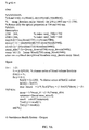
- FIGS. 5A and 5B are a listing of a calibration program for generating calibration curves for use in the method illustrated in FIG. 1 .
- FIG. 6 is a listing of a subroutine for calculating a pulsatile factor R for use in the calibration program of FIG. 5 .
- FIG. 7 is a graph showing calibration factors calculated for use in the method illustrated in FIG. 1 .
- FIGS. 8A and 8B are a program listing of a program for calculating calibration factors for use in the method illustrated in FIG. 1 .
- FIGS. 9A and 9B are a program listing of a computer program for generating the grid mapping of FIG. 3 .
- FIG. 10A is a schematic diagram illustrating use of an oximetry device with different source-detector separations according to an extended embodiment.
- FIG. 10B is a schematic diagram illustrating use of another oximetry device having a plurality of source-detector separations according to another extended embodiment of the invention.
- FIG. 11 is a schematic diagram illustrating use of an oximetry device using multiple light sources of different wavelengths according to yet another extended embodiment of the invention.
- FIG. 12 is a schematic diagram illustrating use of an oximetry device using a modulated light source to obtain measurements for determining a blood oxygen saturation level according to a further extended embodiment of the present invention.
- FIG. 13 is a schematic diagram illustrating use of an oximetry device configured to correlate a detected light transmission with a digitally modulated light stream to obtain measurements for calculating a blood oxygen saturation level according to a still further extended embodiment of the present invention.
- the method and apparatus of this invention can be used to monitor blood oxygen saturation in the emergency room, in surgery, in intensive care, in the neonatal intensive care unit, monitoring the fetus during child birth, and in home use for monitoring peripheral vascular disease, among other uses.
- pulse oximeters measure the pulsatile change in transmission of light through a tissue due to the pulsatile change in tissue blood content caused by the beating heart.
- Such pulsatile measurements are generally made at two, wavelengths (red and infrared) which are combined to yield a pulsatile factor R that is directly related to the arterial blood oxygen saturation value, S a O 2 . Accordingly, by measuring R, the S a O 2 can be determined.
- the calibration curve which maps an R value to an S a O 2 value is not constant.
- the calibration curve of R versus S a O 2 will vary from patient to patient and from tissue site to tissue site.
- the key factor underlying the failure of the prior art is the reliance of previous methods on the assumption of Beer's Law to describe light transport in tissue. Beer's Law would be appropriate if tissue were a cuvette of clear hemoglobin solution such that the tissue was transparent with no light scattering to cause optical turbidity. In such a case, the current calibration curves of R versus S a O 2 would be accurate. Tissue turbidity due to light scattering, however, causes the light transport problem to become nonlinear, and the simple calculation of R consequently does not correct for the background tissue optics.
- the calibration curve varies with changes in the optical properties (which determine an amount of light transport) through the tissue.
- the optical properties which determine an amount of light transport
- the absorption coefficient of the tissue also changes.
- the light transport changes and the calibration also needs to be changed.
- fetal oximeters are more sensitive to such calibration problems than adult oximeters because fetal blood oxygen saturation levels can drop to much lower values than normal adult blood oxygen saturation.
- the calibration curve is more variable at lower blood oxygen saturation levels. The fetal oximeter, therefore, in particular, demands the proper choice of a calibration curve for each blood oxygen saturation level.
- This invention employs findings regarding the influence of tissue optical properties and light transport on the calibration curve to provide an appropriate calibration curve in numerous physiological conditions.
- an adaptive calibration scheme is provided for selecting the proper calibration curve to interpret R to yield S a O 2 .
- DC red steady-state transmitted signal at red wavelength ( ⁇ 800 nm)
- DC infrared steady-state transmitted signal at infrared wavelength (>800 nm)
- f v volume fraction of blood in tissue
- S m O 2 oxygen saturation due to mixture of arterial and venous blood
- R factor based on pulsatile red and infrared signals
- S a O 2 oxygen saturation of arterial blood
- a method for adaptively-calibrating a pulsed oximeter uses the steady-state or DC component of light transmission measurements to select an appropriate calibration curve.
- the selected calibration curve is then used to properly interpret the pulsatile factor (or “R value”) obtained from a standard measurement of the pulsatile components to yield an accurate arterial blood oxygen saturation (S a O 2 ) measurement.
- FIG. 1 is a schematic flow diagram illustrating a method of determining an arterial blood oxygen saturation (S a O 2 ) level according to one embodiment of this invention.
- FIG. 1A is a system level view of an oximetry system for implementing the method of FIG. 1 .
- a method for determining the arterial blood oxygen saturation level begins with a one-time calibration experiment 10 . These experiments compare the DC component of light transmission measurements for both red and infrared wavelengths in air (or some other standard medium) with similar light transmission measurements taken when the probe is placed in contact with the desired tissue. In other words, two calibration measurements, one at each wavelength, are taken in air and compared with two calibration measurements, one at each wavelength, taken in the tissue.
- the two measurements in air are initial, one-time measurements that allow correction for variation in the power of the light source (e.g., light emitting diodes or laser diodes) and variation in the responsivity of the light detector (e.g., light transducer or photodiode).
- the light source e.g., light emitting diodes or laser diodes
- the responsivity of the light detector e.g., light transducer or photodiode
- Subsequent measurements of the tissue are normalized using the air measurements.
- a calibration factor G is also determined for each of the light wavelengths during the initial calibration process 10 .
- the ratios of measurements in tissue to measurements in air can be directly mapped by a grid mapping 30 into values for the blood volume fraction and the mixed blood oxygen saturation, bypassing the need for calibration factors G red and G ir and transport factors T red , T ir incorporating the calibration factors.
- the measurements of air at red and infrared wavelengths can be substituted by measurements of some other standard material at those wavelengths. The ratios of the measurements of tissue to the measurements of the other standard material at red and infrared wavelengths is used to correct for source and photodector variations.
- the grid mapping 30 is the result of calculations based on optical transport theory.
- optical transport theory is used to create a unique one-to-one mapping of T red , T ir (or M red.skin /M red.air , M ir.skin /M ir.air ) into f v , S m O 2 .
- the S m O 2 value is used to select a set of calibration curves 40 relating R to S a O 2 from among a plurality of sets calibration curves 50 .
- the blood volume fraction f v is then used to select an appropriate calibration curve 42 from the selected set of calibration curves 40 .
- Optical transport theory is also used to generate each of the calibration curves.
- the optical properties of the tissue affect the transmission of light through the tissue. Interpretation of pulsatile signals and the choice of the appropriate calibration curve therefore also depend on the baseline optical properties of the tissue. To choose the appropriate calibration curve, therefore, the optical properties of the tissue must first be characterized. Important to this invention, it has been discovered that the non-pulsatile (steady-state or DC) measurements of transmitted light can be used to accomplish this task. As the tissue's blood content or blood oxygen saturation changes, the DC measurements (DC red , DC infrared ) will also change, and the choice of calibration curve can be changed to provide accurate S a O 2 approximations.
- DC steady-state
- the principles according to this invention are fairly straightforward to implement into an oximetry system that is similar in many respects to a conventional oximetry system.
- the basic circuitry and software for obtaining a pulsatile factor R with an optimal signal-to-noise can be retained.
- the DC signals needed to calibrate the system are already measured in conventional systems and used to calculate the pulsatile R, although they are otherwise generally disregarded.
- the improved oximetry system according to this invention therefore, need only be modified to permit adaptive calibration of the system.
- the data obtained from the current oximetry systems needs to be analyzed by an additional software module (or hardwired circuitry) to determine the appropriate calibration curve for interpretation of R to yield a predicted S a O 2 .
- This invention provides an algorithm for selecting the proper calibration curve based on the measured DC signals.
- the light transducer communicates data regarding the transmission of light through (or reflection of light from) the tissue to the data processor 120 through the A/D converter 110 .
- the data processor uses the light transmission data to determine an arterial blood oxygen saturation S a O 2 level of the tissue using software adapted to cause the computer 120 to implement the method described herein.
- the software can be provided to the computer 120 on a computer readable medium such as a floppy disk, a CD ROM, etc., or via any other type of data transfer mechanism.
- a display device 130 is used to convey information regarding the S a O 2 level to the attending physician to facilitate appropriate treatment.
- FIG. 2 is a block diagram illustrating the basic algorithm for determining arterial blood oxygen saturation levels according to one embodiment of this invention.
- this basic algorithm utilizes the same calculation of the pulsatile factor R, based on the AC components of the light transmission measurements, as used in the prior art, it further adds the selection of an appropriate calibration curve. As indicated, the calibration curve is chosen based on the DC components of the light transmission measurements.
- the basic adaptive calibration algorithm begins by using the DC components of light transmission measurements (DC red , DC infrared ) to yield the light transport factors T red , T ir .
- the light transport factors T red , T ir are then mapped into a unique blood volume fraction f v , and a mixed blood oxygen saturation value S m O 2 . These values are representative of absorption and scattering characteristics of the tissue and can be used to adaptively select an appropriate R vs. S a O 2 calibration curve.
- the light transport factors T red , T ir have a well-defined meaning in optical transport theory that depend on the blood volume fraction f v and mixed blood oxygen value S m O 2 of the tissue. This relationship is represented by the grid mapping 30 of FIG. 3 . As indicated by the grid mapping 30 , a light transport factor pair T red , T ir specifies a unique blood volume fraction and mixed blood oxygen value pair f v , S m O 2 .
- the S m O 2 value is used to select a specific set of calibration curves from among a plurality of sets of calibration curves, where each set of calibration curves includes a plurality of R vs. S a O 2 calibration curves.
- Each calibration curve is for a given f v value.
- the f v value can then be used to select an appropriate one of the calibration curves, for the given light transmission properties of the tissue, from the set of calibration curves.
- the selected calibration curve allows the use of the pulsatile factor R to determine the arterial blood oxygen saturation level S a O 2 .
- the values of f v and S m O 2 can jointly and directly select the appropriate calibration curve R versus S a O 2 by means of an algorithm or lookup table.
- a computational model is needed for predicting how the measured pulsatile ratio R will vary for changes in tissue blood content that affect absorption and for changes in tissue scattering properties.
- Absorption and scattering are the two optical properties that determine light transport properties.
- the following section provides a simple computational model for creating a calibration curve.
- the optical properties of tissue are based on the absorption properties of water, bloodless tissue, oxygenated whole blood, and deoxygenated whole blood, and on the scattering properties of skin tissue and blood. These are summarized as follows:
- ⁇ a bloodless tissue 0.75 ⁇ a water + ⁇ a dry tissue absorption
- ⁇ s ′ f v ⁇ s ′ blood +(1 ⁇ f v ) ⁇ s ′ bloodless skin
- ⁇ s ′ blood spectrum from Roggan et al. 1999
- the blood volume fraction, f v Describes the volume of blood per volume of tissue, a dimensionless fraction.
- the arterial oxygen saturation, S a O 2 Describes the oxygen saturation of the arterial blood supply, a value from 0 to 1 (or 0 to 100%).
- the mixed blood oxygen saturation, S m O 2 Describes the oxygen saturation of the combined venous and arterial blood supply, a value for 0 to S a O 2 since S m O 2 ⁇ S a O 2 .
- a program called “calibrate.m” is used to generate the calibration curves for various f v , S a O 2 , and S m O 2 choices.
- a listing of the calibrate.m program is provided as FIG. 5 .
- the program calls a subroutine called “calcR.m” which calculates the pulsatile factor R based on the equation:
- optically-derived ⁇ s ′ of perfused skin, the ⁇ a of the mixed venous and arterial blood supply, and the ⁇ a of the arterial supply.
- the program “calibrate.m” begins by calling a subroutine called “tissueoptics.m” which loads the needed optical properties for all wavelengths from 250 to 1000 nm, then selects only the values for 730 and 940 nm.
- the optical properties are called oxy, deoxy, skin, musp_skin, and musp_bl, for the ⁇ a values of oxygenated blood, deoxygenated blood, and bloodless skin, and for the ⁇ s ′ values of bloodless skin and whole blood, respectively.
- oxy(1) and oxy(2) are the ⁇ a oxy at 730 nm and 940 nm, respectively. All blood values are for 45% Hct blood.
- the fraction was chosen to be 0.80 or 80% of the S a O 2 . Any fraction 0 ⁇ f ⁇ 1 can be chosen. Physically, S m O 2 cannot exceed S a O 2 .
- mua — mixed f v *( SmO 2 *oxy +(1 ⁇ SmO 2)* deoxy )+(1 ⁇ fv ) * skin;
- musp fv*musp — bl +(1 ⁇ fv )* musp — skin;
- the R value returned is assigned to the array element R(j) to facilitate later plotting.
- the S a O 2 value is assigned to the array element SO2(j) for later plotting.
- the curve is plotted.
- the next volume fraction f v is then computed and the process repeats. Accordingly, each calibration curve is for a separate f v .
- a listing of the calcR.m subroutine is provided in FIG. 6 .
- the calcR.m subroutine will now be described in detail.
- the absorption coefficient mua is assigned the value based on the mua_mixed value, which describes the blood content prior to a heart beat.
- the optical diffusion length D and the optical penetration depth delta are calculated.
- the transport factor T1 is calculated according to the equation:
- T 2( j ) exp ( ⁇ d/delta )/(4 *pi*D*d )
- a — 730 ( T 2(1) ⁇ T 1(1))*2/( T 2(1)+ T 1(1)); % RED;
- a — 940 ( T 2(2) ⁇ T 1(2))*2/( T 2(2)+ T 1(2)); % INFRARED;
- M red skin /M red air is equivalent to:
- g1 factor describing how the power of the source is augmented by reflections from the mirrored surfaces of the spiral probe when in skin
- g3 Factor describing how the power of the source is augmented by reflections from the mirrored surfaces of the spiral probe when in air
- g4 Factor describing how transmitted power couples to the detector through the air/epoxy/photodiode interfaces
- ⁇ a absorption coefficient [cm ⁇ 1 ],
- d the effective source-detector distance [cm].
- the experimental testing involved the testing of 24 spiral oximeter probes by taking a measurement in air followed by a measurement in 2% intralipid, for both the red and infrared wavelengths (i.e., 4 measurements).
- the value of light transport factors T for the red and infrared wavelengths were calculated based on the optical properties of the phantom.
- G ir T ir ( M irIL / M irair )
- FIG. 7 shows the values of G red and G ir that were obtained using the above experiment.
- the analysis was accomplished using the MATLAB program “calcG.m,” the listing for which is shown in FIG. 8 .
- Ignoring probe #2 the mean values for G were 1.036 ⁇ 0.031 for red and infrared, respectively.
- a program called “grid.m,” a listing of which is shown in FIG. 9, is used to generate the grid that describes the relationship between f v and S m O 2 and the values T red and T ir .
- the program grid.m calls a subroutine function called “calcDC.m,” listed in FIG. 9A, to begin the operation.
- the program grid.m assumes a background scattering value ⁇ s ′ and uses the two parameters S m O 2 and f v to specify the two optical properties ⁇ a and D which are used to calculate the transport factors T red , T ir .
- the f v and S m O 2 specify the absorption ( ⁇ a ) and scattering ( ⁇ s ′) properties of the tissue, as developed by the calibrate.m program, allowing the selection of the appropriate calibration curve.
- the selected calibration curve is then used to match the calculated pulsatile R value. from the conventional oximetry system with the S a O 2 level.
- the time-averaged DC measurements of a standard calibration material such as air, water, or some other standard medium, (M std.red , M std.ir ) and the DC measurements of tissue (M tissue.red , M tissue.ir ) are derived from an oximeter to yield normalized light measurement values:
- mT red M tissue.red /M std.red
- mT ir M tissue.ir /M std.ir
- a pulsatile factor (R value) is obtained from the pulsatile AC component of the oximeter measurements and is called mR.
- R value a pulsatile factor
- mR a pulsatile factor
- mT red , mT ir , and mR are determined and passed to a subroutine getSaO2([mTred, mTir, mR]) to yield a prediction of the arterial oxygen saturation S a O 2 which is accurate despite variations in the blood volume fraction, f v , or the mixed blood oxygen saturation, S m O 2 .
- the program getSaO2( ) written in the MATLABTM programming language, is listed here:
- the function getSaO2( ) uses the normalized light measurement data [mTred, mTir] to specify the blood volume fraction and mixed blood oxygen saturation values [fv, SmO2], which in turn specify a calibration curve for S a O 2 as a function of R that has been summarized as a second-order polynomial.
- the polynomial is used to convert the pulsatile measurement mR into a predicted S a O 2 .
- the final answer is the predicted S a O 2 .
- the above function getSaO2( ) is the complete master routine which handles the direct feed of measurement data from an oximeter via a master program.
- the details of the functions grid( ) and calibcurve( ) are presented in the following paragraphs. Both grid( ) and calibcurve( ) use two transport functions mTred( ) and mTir( ). First, a description of how mTred( ) and mTir( ) are prepared is provided.
- two light transport factors T red ( ⁇ a , ⁇ s ′) and T ir ( ⁇ a , ⁇ s ′) are specified by measurements in a series of calibrated phantoms of known optical properties consisting of various absorption coefficients ( ⁇ a ) and reduced scattering coefficients ( ⁇ s ′). at the red and infrared wavelengths.
- These transport factors are similar to the transport factors predicted by optical diffusion theory or other transport theories found in the scientific literature but further include the influence of the specific probe geometry. They are computed from the measured values of the phantoms:
- T red ( ⁇ a , ⁇ s ′) M phantom.red ( ⁇ a , ⁇ s ′)/ M std.red ( ⁇ a , ⁇ s ′)
- T ir ( ⁇ a , ⁇ s ′) M phantom.ir ( ⁇ a , ⁇ s ′)/ M std.ir ( ⁇ a , ⁇ s ′)
- mTred Tred (mua, musp) %
- mTred M_red/M_std_red % given tissue absorption (mua) and scattering (musp) %
- the standard was water.
- the function grid( ) uses Tred( ) and Tir( ) in conjunction with an optical properties library which is loaded by the command “load tissueoptics”.
- This library includes the spectral optical absorption properties of oxy-hemoglobin in whole blood (oxy), deoxy-hemoglobin in whole blood (deoxy), and bloodless tissue (mua_tissue), and the optical scattering properties of the tissue (musp_tissue) and of blood (musp_blood).
- the absorption properties of bloodless tissue are based on the absorption properties of water and the non-aqueous components of bloodless tissue.
- This library is prepared from values in the scientific literature. For example, for skin the library is specified by values from the scientific literature as follows:
- tissue is skin
- red 730 nm
- % [mua_red, mua_ir] mua_tissue [0.0276, 0.221]
- % [mua_red, mua_ir] musp_tissue [10.2, 6.94]
- % [musp_red, musp_ir] musp_blood [30.3, 23.6]
- the subroutine grid( ) uses a standard minimization algorithm, called fmins( ) in MATLABTM, and an error evaluation function fvSmO2grid( ) to directly calculate f v and S m O 2 based on the measurements [mTred, mTir]:
- mua_red and mua_ir refer to DC measurements on tissues % and mua_std_red and mua_std_ir refer to measurements % of the standard.
- %%% global Data % Links Data to the subroutine grid( [fv SmO2] ).
- the function calibcurve( ) is used to return a calibration curve for S a O 2 versus R:
- a series of ⁇ a and ⁇ s ′ values for the blood perfused tissue are generated for the red and infrared wavelengths. These values are sent to the subroutines Tred(mua, musp) and Tir(mua, musp) to return predictions of time-averaged measurements (M tissue.red /M std.red ) and (M tissue.ir /M std.ir ).
- an incremental amount of arterial blood is added to the computer-simulated tissue by increasing the ⁇ a and ⁇ s ′ values as if a 0.1% increase in blood volume fraction had occurred due to the added arterial blood.
- the subroutines Tred(mua, musp) and Tir(mua, musp) are used to return predictions of the pulsatile measurements M tissue.red /M std.red and M tissue.ir /M std.ir acquired at the peak in blood volume fraction due to expanded arterial volume.
- the pulsatile R value is calculated according to the equation:
- R [( M tissue.red /M std.red )/( M tissue.red /M std.red )]/[( M tissue.ir /M std.ir )/( M tissue.ir /M std.ir )]
- This polynomial allows immediate evaluation of a measured pulsatile mR value from the oximeter to yield an arterial oxygen saturation value S a O 2 :
- This value SaO2 is the final answer of the algorithm.
- the subroutine calibcurve( ) is listed below.
- the subroutine uses the subroutine calcR(optprops) to compute the expected pulsatile R for a specified set of optical properties specified by the argument optprops which holds the optical properties of the tissue with mixed arterial/venous blood and of arterial blood.
- 51 values of R are generated using calcR( ).
- a second-order polynomial is fitted to the relation of SaO2(1:51) versus R(1:51) to yield the coefficients [C0, C1, C2].
- coefficients [C0, C1, and C2] can be used to interpret the measured pulsatile factor mR received from the oximeter as described above.
- the original disclosure describes the use of a steady-state component of light transmission measurements at two wavelengths through a tissue to select a proper calibration curve for interpretation of the pulsatile component of the light transmission.
- the selected calibration curve allows pulsatile measurements to specify the arterial blood oxygen saturation.
- the standard pulsed oximetry of conventional medical practice relies on a single average calibration based on measurements on a subject population.
- the baseline level of nonpulsatile blood content, however, both arterial and venous, in a tissue can vary significantly from patient to patient, and even from site to site on any given patient.
- Conventional pulsed oximetry is therefore not calibrated for a particular patient or tissue site, but rather for an average patient.
- the principles of the invention described above provide dynamic adaptive calibration for standard pulsed oximetry, which can adjust to any variation in the amount of mixed arterial and venous blood volumes in a tissue.
- a surgically-implanted oximetry device e.g., a light source and a detector
- a surgically-implanted oximetry device might enable performance of pulsed oximetry on blood vessels that carry blood to and from the brain so as to assess the oxygen utilization by the brain and the ability of cardiac output to meet the oxygen requirements of the brain.
- An implanted oximetry device encounters a complex geometry with a surrounding tissue, a vessel wall, and an inner lumen filled with blood. Furthermore, the optical properties of the surrounding tissue may vary. The thickness of the vessel wall may also vary from patient to patient and the size of the inner lumen may vary.
- an embodiment of the invention to conduct more measurements in order to specify the oxygen saturation. To accomplish this, it is desirable to expand the number of measurements that are included in the oximetry method to provide accurate measurements in more complex tissues.
- FIGS. 10A and 10B are schematic diagrams illustrating use of oximetry devices 200 A, 200 B to detect blood oxygen saturation of a sample tissue 250 .
- a number of light sources 210 and/or a number of detectors 220 in an oximetry device 200 A, 200 B are used to provide a range of source-detector separations.
- the light sources 210 (and/or the detectors 220 ) can be arranged, for instance, in an array 208 .
- the circuitry that actuates the light sources 210 and circuitry that processes the output signals from the detectors 220 are not shown in FIGS. 10A and 10B, but can be routinely designed by those skilled in the art.
- each measurement at a different source-detector separation involves a different ensemble of path lengths L through the tissue 250 for the collected photons. Consequently, each measurement is unique and independent. Small source-detector separations probe the vessel wall 262 , while large source-detector separations probe the blood volume 270 within the vessel 260 .
- the oximetry calibration algorithm is preferably adapted to utilize the additional information obtained from these measurements and thereby yield an accurate adaptive calibration for use with pulsatile information.
- an appropriate f v -S m O 2 grid 30 can be selected using measurements of T IR and T RED to yield values of f v and S m O 2 .
- the S m O 2 value can then be used to select a set 40 of calibration curves from a group of sets 50 , and the f v value can be used to select a calibration curve 42 from the selected set 40 .
- additional measurements at several source-detector separations yield a set of measurements (T IR1 , T RED1 , T IR2 , T RED2 , T IR3 , T RED3 , . . . ) that specify the vessel wall thickness (Dwall), the average path length (Lblood) of the ensemble of collected photons that pass through the vessel blood volume 270 , and the optical properties (mua, musp) of the averaged surrounding tissue 250 , vessel wall 262 , and blood-filled vessel lumen 270 that are associated with the nonpulsatile component of the light transmission.
- the optical properties specified are the absorption coefficient mua [cm ⁇ 1 ] and the reduced scattering coefficient musp [cm ⁇ 1 ].
- the combination of parameters (Dwall, Lblood, mua, musp) is used to specify a calibration curve, similar to the calibration curve 42 of the original embodiments, that allows the pulsatile component of transmission to specify the arterial oxygen saturation (S a O 2 ).
- At least five independent measurements are preferably used to specify the five variables (Dwall, Lblood, mua, musp, S a O 2 ).
- the actual position of certain sources and detectors is varied.
- the position information rather than just the source-detector separation information, is then used in specifying the parameters that are in turn used to specify the calibration curve required to interpret pulsatile measurements to yield an arterial blood oxygen saturation (S a O 2 ) value.
- S a O 2 arterial blood oxygen saturation
- additional measurements are preferably obtained using more than two light sources 310 A, 310 B, 310 C of different wavelengths in the visible to near-infrared wavelength range. Shorter wavelengths probe the vessel wall 260 and longer wavelengths probe into the blood-filled vessel lumen 270 . Inclusion of more light sources provides additional measurements.
- a set of transmission measurements (T wavelength1 , T wavelength2 , T wavelength3 , T wavelength4 , T wavelength5 , . . . ) are obtained.
- At least five independent measurements are preferably obtained to determine values for the five variables (Dwall, Lblood, mua, musp, S a O 2 ). Most preferably, both a plurality of wavelengths and a plurality of source-detector separations and positions can be implemented.
- additional measurements are achieved using a modulator 408 (e.g., preferably using amplitude modulation, but alternatively using frequency or phase modulation) to modulate the light source 410 at various high frequencies, such as between 50 kHz and 1 GHz.
- a demodulator 418 measures the phase P and modulation M of the transmitted light received at the detector 420 .
- the measurements at different frequencies of modulation involve different path lengths for collected photons and therefore probe different proportions of the surrounding tissue 250 , vessel wall 262 , and blood-filled lumen 270 .
- Multiple independent measurements of P and M at various frequencies f 1 , f 2 , f 3 , f 4 , . . .
- the light source 510 is modulated digitally as a unique stream of pulses of variable pulse duration using a digital modulator 508 .
- the stream of pulses from the modulator 508 may be a pseudorandom sequence of uniform-duration pulses.
- the detector 520 acquires the signal and uses a correlator 518 (e.g., a specially programmed microprocessor) to decode the received pulse stream and correlate it against the known source stream to specify the delay time DT for transmission to the detector.
- the pulse durations are preferably very short, such as between Ins to 1 ⁇ s.
- the delay times associated with transmission between a set of source and detectors yields a set of measurements (DT1, DT2, DT3, DT4, DT5, . . . ) that in turn specify values for the variables (Dwall, Lblood, mua, musp). These variables are used to specify the appropriate calibration curve to allow the pulsatile measurements to yield an arterial blood oxygen saturation (S a O 2 ) value.
- S a O 2 arterial blood oxygen saturation
- Oximetry devices other than surgically-implanted devices to monitor oxygenation in a blood vessel could also be used.
- an oximetry device could be implanted to monitor the oxygenation of a specific organ.
- the geometry and complexity of the situation may involve a set of variables, here called (x1, x2, x3, x4, . . . ), that are similar to, but different from the previously identified variables (Dwall, Lblood, mua, musp).
- a plurality of wavelengths and source-detector separations and positions, or different frequencies of modulations or variable digital streams controlling light sources can be used to determine the nonpulsatile component of a set of light transmission measurements (m1, m2, m3, m4, m5, . . . ). These measurements can then be used to determine values for the parameters (x1, x2, x3, x4, . . . ) that are in turn used select an appropriate calibration curve. The calibration curve then allows interpretation of the pulsatile component of transmission measurements to yield an arterial blood oxygen saturation (S a O 2 ) value.
- S a O 2 arterial blood oxygen saturation
Priority Applications (1)
| Application Number | Priority Date | Filing Date | Title |
|---|---|---|---|
| US10/196,715 US6760609B2 (en) | 1999-07-14 | 2002-07-15 | Adaptive calibration pulsed oximetry method and device |
Applications Claiming Priority (3)
| Application Number | Priority Date | Filing Date | Title |
|---|---|---|---|
| US14389499P | 1999-07-14 | 1999-07-14 | |
| US09/616,465 US6421549B1 (en) | 1999-07-14 | 2000-07-14 | Adaptive calibration pulsed oximetry method and device |
| US10/196,715 US6760609B2 (en) | 1999-07-14 | 2002-07-15 | Adaptive calibration pulsed oximetry method and device |
Related Parent Applications (1)
| Application Number | Title | Priority Date | Filing Date |
|---|---|---|---|
| US09/616,465 Continuation-In-Part US6421549B1 (en) | 1999-07-14 | 2000-07-14 | Adaptive calibration pulsed oximetry method and device |
Publications (2)
| Publication Number | Publication Date |
|---|---|
| US20030109776A1 US20030109776A1 (en) | 2003-06-12 |
| US6760609B2 true US6760609B2 (en) | 2004-07-06 |
Family
ID=26841505
Family Applications (1)
| Application Number | Title | Priority Date | Filing Date |
|---|---|---|---|
| US10/196,715 Expired - Fee Related US6760609B2 (en) | 1999-07-14 | 2002-07-15 | Adaptive calibration pulsed oximetry method and device |
Country Status (1)
| Country | Link |
|---|---|
| US (1) | US6760609B2 (US06760609-20040706-M00004.png) |
Cited By (86)
| Publication number | Priority date | Publication date | Assignee | Title |
|---|---|---|---|---|
| US20050065414A1 (en) * | 2003-07-24 | 2005-03-24 | Allen Robert V. | Pulse oximeter system |
| US20050197793A1 (en) * | 2004-03-08 | 2005-09-08 | Nellcor Puritan Bennett Incorporated | Pulse oximeter with separate ensemble averaging for oxygen saturation and heart rate |
| US20070078311A1 (en) * | 2005-03-01 | 2007-04-05 | Ammar Al-Ali | Disposable multiple wavelength optical sensor |
| US20080306356A1 (en) * | 2007-06-05 | 2008-12-11 | Kenneth Darryl Kemp | Vascular status monitoring system |
| US7647084B2 (en) | 2005-08-08 | 2010-01-12 | Nellcor Puritan Bennett Llc | Medical sensor and technique for using the same |
| US7647083B2 (en) | 2005-03-01 | 2010-01-12 | Masimo Laboratories, Inc. | Multiple wavelength sensor equalization |
| US7650177B2 (en) | 2005-09-29 | 2010-01-19 | Nellcor Puritan Bennett Llc | Medical sensor for reducing motion artifacts and technique for using the same |
| US7657296B2 (en) | 2005-08-08 | 2010-02-02 | Nellcor Puritan Bennett Llc | Unitary medical sensor assembly and technique for using the same |
| US7657295B2 (en) | 2005-08-08 | 2010-02-02 | Nellcor Puritan Bennett Llc | Medical sensor and technique for using the same |
| US7658652B2 (en) | 2006-09-29 | 2010-02-09 | Nellcor Puritan Bennett Llc | Device and method for reducing crosstalk |
| US7676253B2 (en) | 2005-09-29 | 2010-03-09 | Nellcor Puritan Bennett Llc | Medical sensor and technique for using the same |
| US7680522B2 (en) | 2006-09-29 | 2010-03-16 | Nellcor Puritan Bennett Llc | Method and apparatus for detecting misapplied sensors |
| US7684842B2 (en) | 2006-09-29 | 2010-03-23 | Nellcor Puritan Bennett Llc | System and method for preventing sensor misuse |
| US7689259B2 (en) | 2000-04-17 | 2010-03-30 | Nellcor Puritan Bennett Llc | Pulse oximeter sensor with piece-wise function |
| US7796403B2 (en) | 2006-09-28 | 2010-09-14 | Nellcor Puritan Bennett Llc | Means for mechanical registration and mechanical-electrical coupling of a faraday shield to a photodetector and an electrical circuit |
| US7869849B2 (en) | 2006-09-26 | 2011-01-11 | Nellcor Puritan Bennett Llc | Opaque, electrically nonconductive region on a medical sensor |
| US7880884B2 (en) | 2008-06-30 | 2011-02-01 | Nellcor Puritan Bennett Llc | System and method for coating and shielding electronic sensor components |
| US7881762B2 (en) | 2005-09-30 | 2011-02-01 | Nellcor Puritan Bennett Llc | Clip-style medical sensor and technique for using the same |
| US7887345B2 (en) | 2008-06-30 | 2011-02-15 | Nellcor Puritan Bennett Llc | Single use connector for pulse oximetry sensors |
| US7890153B2 (en) | 2006-09-28 | 2011-02-15 | Nellcor Puritan Bennett Llc | System and method for mitigating interference in pulse oximetry |
| US20110040176A1 (en) * | 2008-02-19 | 2011-02-17 | Helmholtz Zentrum Muenchen Deutsches Forschungszentrum fur Gesundheit und | Method and device for near-field dual-wave modality imaging |
| US7894869B2 (en) | 2007-03-09 | 2011-02-22 | Nellcor Puritan Bennett Llc | Multiple configuration medical sensor and technique for using the same |
| US7899510B2 (en) | 2005-09-29 | 2011-03-01 | Nellcor Puritan Bennett Llc | Medical sensor and technique for using the same |
| US8062221B2 (en) | 2005-09-30 | 2011-11-22 | Nellcor Puritan Bennett Llc | Sensor for tissue gas detection and technique for using the same |
| US20110286000A1 (en) * | 2008-10-01 | 2011-11-24 | Xin-Hua Hu | Methods and Systems for Optically Characterizing a Turbid Material Using a Structured Incident Beam |
| US8068891B2 (en) | 2006-09-29 | 2011-11-29 | Nellcor Puritan Bennett Llc | Symmetric LED array for pulse oximetry |
| US8073518B2 (en) | 2006-05-02 | 2011-12-06 | Nellcor Puritan Bennett Llc | Clip-style medical sensor and technique for using the same |
| US8070508B2 (en) | 2007-12-31 | 2011-12-06 | Nellcor Puritan Bennett Llc | Method and apparatus for aligning and securing a cable strain relief |
| US8071935B2 (en) | 2008-06-30 | 2011-12-06 | Nellcor Puritan Bennett Llc | Optical detector with an overmolded faraday shield |
| US8092993B2 (en) | 2007-12-31 | 2012-01-10 | Nellcor Puritan Bennett Llc | Hydrogel thin film for use as a biosensor |
| US8092379B2 (en) | 2005-09-29 | 2012-01-10 | Nellcor Puritan Bennett Llc | Method and system for determining when to reposition a physiological sensor |
| US8112375B2 (en) | 2008-03-31 | 2012-02-07 | Nellcor Puritan Bennett Llc | Wavelength selection and outlier detection in reduced rank linear models |
| US8133176B2 (en) | 1999-04-14 | 2012-03-13 | Tyco Healthcare Group Lp | Method and circuit for indicating quality and accuracy of physiological measurements |
| US8145288B2 (en) | 2006-08-22 | 2012-03-27 | Nellcor Puritan Bennett Llc | Medical sensor for reducing signal artifacts and technique for using the same |
| US8175667B2 (en) | 2006-09-29 | 2012-05-08 | Nellcor Puritan Bennett Llc | Symmetric LED array for pulse oximetry |
| US8175671B2 (en) | 2006-09-22 | 2012-05-08 | Nellcor Puritan Bennett Llc | Medical sensor for reducing signal artifacts and technique for using the same |
| US8190225B2 (en) | 2006-09-22 | 2012-05-29 | Nellcor Puritan Bennett Llc | Medical sensor for reducing signal artifacts and technique for using the same |
| US8199007B2 (en) | 2007-12-31 | 2012-06-12 | Nellcor Puritan Bennett Llc | Flex circuit snap track for a biometric sensor |
| US8219170B2 (en) | 2006-09-20 | 2012-07-10 | Nellcor Puritan Bennett Llc | System and method for practicing spectrophotometry using light emitting nanostructure devices |
| US8224412B2 (en) | 2000-04-17 | 2012-07-17 | Nellcor Puritan Bennett Llc | Pulse oximeter sensor with piece-wise function |
| US8221319B2 (en) | 2009-03-25 | 2012-07-17 | Nellcor Puritan Bennett Llc | Medical device for assessing intravascular blood volume and technique for using the same |
| US8233954B2 (en) | 2005-09-30 | 2012-07-31 | Nellcor Puritan Bennett Llc | Mucosal sensor for the assessment of tissue and blood constituents and technique for using the same |
| US8260391B2 (en) | 2005-09-12 | 2012-09-04 | Nellcor Puritan Bennett Llc | Medical sensor for reducing motion artifacts and technique for using the same |
| US8265724B2 (en) | 2007-03-09 | 2012-09-11 | Nellcor Puritan Bennett Llc | Cancellation of light shunting |
| US8280469B2 (en) | 2007-03-09 | 2012-10-02 | Nellcor Puritan Bennett Llc | Method for detection of aberrant tissue spectra |
| US8311601B2 (en) | 2009-06-30 | 2012-11-13 | Nellcor Puritan Bennett Llc | Reflectance and/or transmissive pulse oximeter |
| US8315685B2 (en) | 2006-09-27 | 2012-11-20 | Nellcor Puritan Bennett Llc | Flexible medical sensor enclosure |
| US8346328B2 (en) | 2007-12-21 | 2013-01-01 | Covidien Lp | Medical sensor and technique for using the same |
| US8352010B2 (en) | 2005-09-30 | 2013-01-08 | Covidien Lp | Folding medical sensor and technique for using the same |
| US8352009B2 (en) | 2005-09-30 | 2013-01-08 | Covidien Lp | Medical sensor and technique for using the same |
| US8352004B2 (en) | 2007-12-21 | 2013-01-08 | Covidien Lp | Medical sensor and technique for using the same |
| US8364224B2 (en) | 2008-03-31 | 2013-01-29 | Covidien Lp | System and method for facilitating sensor and monitor communication |
| US8364220B2 (en) | 2008-09-25 | 2013-01-29 | Covidien Lp | Medical sensor and technique for using the same |
| US8366613B2 (en) | 2007-12-26 | 2013-02-05 | Covidien Lp | LED drive circuit for pulse oximetry and method for using same |
| US8386002B2 (en) | 2005-09-30 | 2013-02-26 | Covidien Lp | Optically aligned pulse oximetry sensor and technique for using the same |
| US8391941B2 (en) | 2009-07-17 | 2013-03-05 | Covidien Lp | System and method for memory switching for multiple configuration medical sensor |
| US8396527B2 (en) | 2006-09-22 | 2013-03-12 | Covidien Lp | Medical sensor for reducing signal artifacts and technique for using the same |
| US8417309B2 (en) | 2008-09-30 | 2013-04-09 | Covidien Lp | Medical sensor |
| US8417310B2 (en) | 2009-08-10 | 2013-04-09 | Covidien Lp | Digital switching in multi-site sensor |
| US8423112B2 (en) | 2008-09-30 | 2013-04-16 | Covidien Lp | Medical sensor and technique for using the same |
| US8428675B2 (en) | 2009-08-19 | 2013-04-23 | Covidien Lp | Nanofiber adhesives used in medical devices |
| US8433383B2 (en) | 2001-10-12 | 2013-04-30 | Covidien Lp | Stacked adhesive optical sensor |
| US8437822B2 (en) | 2008-03-28 | 2013-05-07 | Covidien Lp | System and method for estimating blood analyte concentration |
| US8442608B2 (en) | 2007-12-28 | 2013-05-14 | Covidien Lp | System and method for estimating physiological parameters by deconvolving artifacts |
| US8452364B2 (en) | 2007-12-28 | 2013-05-28 | Covidien LLP | System and method for attaching a sensor to a patient's skin |
| US8452366B2 (en) | 2009-03-16 | 2013-05-28 | Covidien Lp | Medical monitoring device with flexible circuitry |
| US8483790B2 (en) | 2002-10-18 | 2013-07-09 | Covidien Lp | Non-adhesive oximeter sensor for sensitive skin |
| US8505821B2 (en) | 2009-06-30 | 2013-08-13 | Covidien Lp | System and method for providing sensor quality assurance |
| US8509869B2 (en) | 2009-05-15 | 2013-08-13 | Covidien Lp | Method and apparatus for detecting and analyzing variations in a physiologic parameter |
| US8577434B2 (en) | 2007-12-27 | 2013-11-05 | Covidien Lp | Coaxial LED light sources |
| WO2014011368A1 (en) * | 2012-06-18 | 2014-01-16 | Eso-Technologies, Inc. | Compositions and methods for measurement of oxygen saturation in blood filled structures |
| US8634891B2 (en) | 2009-05-20 | 2014-01-21 | Covidien Lp | Method and system for self regulation of sensor component contact pressure |
| US8649839B2 (en) | 1996-10-10 | 2014-02-11 | Covidien Lp | Motion compatible sensor for non-invasive optical blood analysis |
| US8781544B2 (en) | 2007-03-27 | 2014-07-15 | Cercacor Laboratories, Inc. | Multiple wavelength optical sensor |
| US8801613B2 (en) | 2009-12-04 | 2014-08-12 | Masimo Corporation | Calibration for multi-stage physiological monitors |
| US8897850B2 (en) | 2007-12-31 | 2014-11-25 | Covidien Lp | Sensor with integrated living hinge and spring |
| US8914088B2 (en) | 2008-09-30 | 2014-12-16 | Covidien Lp | Medical sensor and technique for using the same |
| US8965471B2 (en) | 2007-04-21 | 2015-02-24 | Cercacor Laboratories, Inc. | Tissue profile wellness monitor |
| US9010634B2 (en) | 2009-06-30 | 2015-04-21 | Covidien Lp | System and method for linking patient data to a patient and providing sensor quality assurance |
| US9066660B2 (en) | 2009-09-29 | 2015-06-30 | Nellcor Puritan Bennett Ireland | Systems and methods for high-pass filtering a photoplethysmograph signal |
| US9271654B2 (en) | 2009-06-29 | 2016-03-01 | Helmholtz Zentrum Munchen Deutsches Forschungszentrum Fur Gesundheit Und Umwelt (Gmbh) | Thermoacoustic imaging with quantitative extraction of absorption map |
| US9551789B2 (en) | 2013-01-15 | 2017-01-24 | Helmholtz Zentrum Munchen Deutsches Forschungszentrum Fur Gesundheit Und Umwelt (Gmbh) | System and method for quality-enhanced high-rate optoacoustic imaging of an object |
| US9572497B2 (en) | 2008-07-25 | 2017-02-21 | Helmholtz Zentrum Munchen Deutsches Forschungszentrum Fur Gesundheit Und Umwelt (Gmbh) | Quantitative multi-spectral opto-acoustic tomography (MSOT) of tissue biomarkers |
| US9839381B1 (en) | 2009-11-24 | 2017-12-12 | Cercacor Laboratories, Inc. | Physiological measurement system with automatic wavelength adjustment |
| US10292593B2 (en) | 2009-07-27 | 2019-05-21 | Helmholtz Zentrum München Deutsches Forschungszentrum Für Gesundheit Und Umwelt (Gmbh) | Imaging device and method for optoacoustic imaging of small animals |
| US11026584B2 (en) | 2012-12-11 | 2021-06-08 | Ithera Medical Gmbh | Handheld device and method for tomographic optoacoustic imaging of an object |
Families Citing this family (14)
| Publication number | Priority date | Publication date | Assignee | Title |
|---|---|---|---|---|
| DE112006002808A5 (de) * | 2005-11-15 | 2008-09-04 | Weinmann Geräte für Medizin GmbH + Co. KG | Autoadaptive Kalibration |
| US8099146B1 (en) | 2006-03-23 | 2012-01-17 | Pacesetter, Inc. | System and method for calibrating a blood oxygen saturation sensor for use with an implantable medical device |
| US9282924B2 (en) * | 2011-03-31 | 2016-03-15 | Covidien Lp | Medical sensor with temperature control |
| EP2815697B1 (en) * | 2012-02-13 | 2019-09-11 | Girina, Marina Borisovna | Device for assessing regional blood circulation |
| US10687742B2 (en) * | 2015-06-12 | 2020-06-23 | ChroniSense Medical Ltd. | Using invariant factors for pulse oximetry |
| US10470692B2 (en) | 2015-06-12 | 2019-11-12 | ChroniSense Medical Ltd. | System for performing pulse oximetry |
| US11160459B2 (en) | 2015-06-12 | 2021-11-02 | ChroniSense Medical Ltd. | Monitoring health status of people suffering from chronic diseases |
| US11464457B2 (en) | 2015-06-12 | 2022-10-11 | ChroniSense Medical Ltd. | Determining an early warning score based on wearable device measurements |
| US11712190B2 (en) | 2015-06-12 | 2023-08-01 | ChroniSense Medical Ltd. | Wearable device electrocardiogram |
| US11160461B2 (en) | 2015-06-12 | 2021-11-02 | ChroniSense Medical Ltd. | Blood pressure measurement using a wearable device |
| US10952638B2 (en) | 2015-06-12 | 2021-03-23 | ChroniSense Medical Ltd. | System and method for monitoring respiratory rate and oxygen saturation |
| US11000235B2 (en) | 2016-03-14 | 2021-05-11 | ChroniSense Medical Ltd. | Monitoring procedure for early warning of cardiac episodes |
| US11026608B2 (en) * | 2017-01-09 | 2021-06-08 | Vox Biomedical Llc | Cerebral oximetry using time-gated direct sequence spread spectrum |
| CN110573067B (zh) * | 2017-03-02 | 2022-10-11 | 安科医疗私人有限公司 | 无创肱动脉血压测量 |
Citations (18)
| Publication number | Priority date | Publication date | Assignee | Title |
|---|---|---|---|---|
| USRE28990E (en) | 1972-12-04 | 1976-10-05 | Corometrics Medical Systems, Inc. | Bipolar electrode structure for monitoring fetal heartbeat and the like |
| US4086915A (en) | 1975-04-30 | 1978-05-02 | Harvey I. Kofsky | Ear oximetry process and apparatus |
| US4407290A (en) | 1981-04-01 | 1983-10-04 | Biox Technology, Inc. | Blood constituent measuring device and method |
| US4807631A (en) | 1987-10-09 | 1989-02-28 | Critikon, Inc. | Pulse oximetry system |
| US4819646A (en) | 1986-08-18 | 1989-04-11 | Physio-Control Corporation | Feedback-controlled method and apparatus for processing signals used in oximetry |
| US4892101A (en) | 1986-08-18 | 1990-01-09 | Physio-Control Corporation | Method and apparatus for offsetting baseline portion of oximeter signal |
| US5111817A (en) | 1988-12-29 | 1992-05-12 | Medical Physics, Inc. | Noninvasive system and method for enhanced arterial oxygen saturation determination and arterial blood pressure monitoring |
| US5411024A (en) | 1993-12-15 | 1995-05-02 | Corometrics Medical Systems, Inc. | Fetal pulse oximetry sensor |
| US5431159A (en) | 1988-05-05 | 1995-07-11 | Sentinel Monitoring, Inc. | Pulse oximetry |
| US5448991A (en) | 1989-11-01 | 1995-09-12 | Polson; Michael J. R. | Method of measuring the oxygen saturation in pulsating blood flow |
| US5725480A (en) | 1996-03-06 | 1998-03-10 | Abbott Laboratories | Non-invasive calibration and categorization of individuals for subsequent non-invasive detection of biological compounds |
| US5772589A (en) * | 1995-02-13 | 1998-06-30 | Bernreuter; Peter | Measurement process for blood gas analysis sensors |
| US5922607A (en) * | 1995-12-13 | 1999-07-13 | Bernreuter; Peter | Measuring process for blood gas analysis sensors |
| US6064898A (en) | 1998-09-21 | 2000-05-16 | Essential Medical Devices | Non-invasive blood component analyzer |
| US6151107A (en) | 1996-07-26 | 2000-11-21 | Linde Medical Sensors Ag | Method of non-invasive determination of oxygen saturation in tissue in which blood is circulating |
| US6226540B1 (en) | 1995-12-13 | 2001-05-01 | Peter Bernreuter | Measuring process for blood gas analysis sensors |
| US6421549B1 (en) * | 1999-07-14 | 2002-07-16 | Providence Health System-Oregon | Adaptive calibration pulsed oximetry method and device |
| US6493565B1 (en) * | 1993-11-15 | 2002-12-10 | Non-Invasive Technology, Inc. | Examination of biological tissue by monitoring one or more solutes |
-
2002
- 2002-07-15 US US10/196,715 patent/US6760609B2/en not_active Expired - Fee Related
Patent Citations (20)
| Publication number | Priority date | Publication date | Assignee | Title |
|---|---|---|---|---|
| USRE28990E (en) | 1972-12-04 | 1976-10-05 | Corometrics Medical Systems, Inc. | Bipolar electrode structure for monitoring fetal heartbeat and the like |
| US4086915A (en) | 1975-04-30 | 1978-05-02 | Harvey I. Kofsky | Ear oximetry process and apparatus |
| US4407290A (en) | 1981-04-01 | 1983-10-04 | Biox Technology, Inc. | Blood constituent measuring device and method |
| US4407290B1 (US06760609-20040706-M00004.png) | 1981-04-01 | 1986-10-14 | ||
| US4819646A (en) | 1986-08-18 | 1989-04-11 | Physio-Control Corporation | Feedback-controlled method and apparatus for processing signals used in oximetry |
| US4892101A (en) | 1986-08-18 | 1990-01-09 | Physio-Control Corporation | Method and apparatus for offsetting baseline portion of oximeter signal |
| US4807631A (en) | 1987-10-09 | 1989-02-28 | Critikon, Inc. | Pulse oximetry system |
| US5431159A (en) | 1988-05-05 | 1995-07-11 | Sentinel Monitoring, Inc. | Pulse oximetry |
| US5111817A (en) | 1988-12-29 | 1992-05-12 | Medical Physics, Inc. | Noninvasive system and method for enhanced arterial oxygen saturation determination and arterial blood pressure monitoring |
| US5448991A (en) | 1989-11-01 | 1995-09-12 | Polson; Michael J. R. | Method of measuring the oxygen saturation in pulsating blood flow |
| US5820550A (en) | 1989-11-01 | 1998-10-13 | Novametrix Medical Systems Inc. | Pulse oximeter with improved accuracy and response time |
| US6493565B1 (en) * | 1993-11-15 | 2002-12-10 | Non-Invasive Technology, Inc. | Examination of biological tissue by monitoring one or more solutes |
| US5411024A (en) | 1993-12-15 | 1995-05-02 | Corometrics Medical Systems, Inc. | Fetal pulse oximetry sensor |
| US5772589A (en) * | 1995-02-13 | 1998-06-30 | Bernreuter; Peter | Measurement process for blood gas analysis sensors |
| US5922607A (en) * | 1995-12-13 | 1999-07-13 | Bernreuter; Peter | Measuring process for blood gas analysis sensors |
| US6226540B1 (en) | 1995-12-13 | 2001-05-01 | Peter Bernreuter | Measuring process for blood gas analysis sensors |
| US5725480A (en) | 1996-03-06 | 1998-03-10 | Abbott Laboratories | Non-invasive calibration and categorization of individuals for subsequent non-invasive detection of biological compounds |
| US6151107A (en) | 1996-07-26 | 2000-11-21 | Linde Medical Sensors Ag | Method of non-invasive determination of oxygen saturation in tissue in which blood is circulating |
| US6064898A (en) | 1998-09-21 | 2000-05-16 | Essential Medical Devices | Non-invasive blood component analyzer |
| US6421549B1 (en) * | 1999-07-14 | 2002-07-16 | Providence Health System-Oregon | Adaptive calibration pulsed oximetry method and device |
Cited By (148)
| Publication number | Priority date | Publication date | Assignee | Title |
|---|---|---|---|---|
| US8649839B2 (en) | 1996-10-10 | 2014-02-11 | Covidien Lp | Motion compatible sensor for non-invasive optical blood analysis |
| US8133176B2 (en) | 1999-04-14 | 2012-03-13 | Tyco Healthcare Group Lp | Method and circuit for indicating quality and accuracy of physiological measurements |
| US8224412B2 (en) | 2000-04-17 | 2012-07-17 | Nellcor Puritan Bennett Llc | Pulse oximeter sensor with piece-wise function |
| US8078246B2 (en) | 2000-04-17 | 2011-12-13 | Nellcor Puritan Bennett Llc | Pulse oximeter sensor with piece-wise function |
| US7689259B2 (en) | 2000-04-17 | 2010-03-30 | Nellcor Puritan Bennett Llc | Pulse oximeter sensor with piece-wise function |
| US8433383B2 (en) | 2001-10-12 | 2013-04-30 | Covidien Lp | Stacked adhesive optical sensor |
| US8483790B2 (en) | 2002-10-18 | 2013-07-09 | Covidien Lp | Non-adhesive oximeter sensor for sensitive skin |
| US20050065414A1 (en) * | 2003-07-24 | 2005-03-24 | Allen Robert V. | Pulse oximeter system |
| US20050197793A1 (en) * | 2004-03-08 | 2005-09-08 | Nellcor Puritan Bennett Incorporated | Pulse oximeter with separate ensemble averaging for oxygen saturation and heart rate |
| US7039538B2 (en) * | 2004-03-08 | 2006-05-02 | Nellcor Puritant Bennett Incorporated | Pulse oximeter with separate ensemble averaging for oxygen saturation and heart rate |
| US7761127B2 (en) | 2005-03-01 | 2010-07-20 | Masimo Laboratories, Inc. | Multiple wavelength sensor substrate |
| US8849365B2 (en) | 2005-03-01 | 2014-09-30 | Cercacor Laboratories, Inc. | Multiple wavelength sensor emitters |
| US8581732B2 (en) | 2005-03-01 | 2013-11-12 | Carcacor Laboratories, Inc. | Noninvasive multi-parameter patient monitor |
| US8634889B2 (en) | 2005-03-01 | 2014-01-21 | Cercacor Laboratories, Inc. | Configurable physiological measurement system |
| US9351675B2 (en) | 2005-03-01 | 2016-05-31 | Cercacor Laboratories, Inc. | Noninvasive multi-parameter patient monitor |
| US10123726B2 (en) | 2005-03-01 | 2018-11-13 | Cercacor Laboratories, Inc. | Configurable physiological measurement system |
| US9750443B2 (en) | 2005-03-01 | 2017-09-05 | Cercacor Laboratories, Inc. | Multiple wavelength sensor emitters |
| US7729733B2 (en) | 2005-03-01 | 2010-06-01 | Masimo Laboratories, Inc. | Configurable physiological measurement system |
| US10251585B2 (en) | 2005-03-01 | 2019-04-09 | Cercacor Laboratories, Inc. | Noninvasive multi-parameter patient monitor |
| US9241662B2 (en) | 2005-03-01 | 2016-01-26 | Cercacor Laboratories, Inc. | Configurable physiological measurement system |
| US10856788B2 (en) | 2005-03-01 | 2020-12-08 | Cercacor Laboratories, Inc. | Noninvasive multi-parameter patient monitor |
| US7764982B2 (en) | 2005-03-01 | 2010-07-27 | Masimo Laboratories, Inc. | Multiple wavelength sensor emitters |
| US8255027B2 (en) | 2005-03-01 | 2012-08-28 | Cercacor Laboratories, Inc. | Multiple wavelength sensor substrate |
| US8483787B2 (en) | 2005-03-01 | 2013-07-09 | Cercacor Laboratories, Inc. | Multiple wavelength sensor drivers |
| US8190223B2 (en) | 2005-03-01 | 2012-05-29 | Masimo Laboratories, Inc. | Noninvasive multi-parameter patient monitor |
| US9549696B2 (en) | 2005-03-01 | 2017-01-24 | Cercacor Laboratories, Inc. | Physiological parameter confidence measure |
| US8718735B2 (en) | 2005-03-01 | 2014-05-06 | Cercacor Laboratories, Inc. | Physiological parameter confidence measure |
| US8224411B2 (en) | 2005-03-01 | 2012-07-17 | Masimo Laboratories, Inc. | Noninvasive multi-parameter patient monitor |
| US8385996B2 (en) | 2005-03-01 | 2013-02-26 | Cercacor Laboratories, Inc. | Multiple wavelength sensor emitters |
| US11545263B2 (en) | 2005-03-01 | 2023-01-03 | Cercacor Laboratories, Inc. | Multiple wavelength sensor emitters |
| US9167995B2 (en) | 2005-03-01 | 2015-10-27 | Cercacor Laboratories, Inc. | Physiological parameter confidence measure |
| US11430572B2 (en) | 2005-03-01 | 2022-08-30 | Cercacor Laboratories, Inc. | Multiple wavelength sensor emitters |
| US8912909B2 (en) | 2005-03-01 | 2014-12-16 | Cercacor Laboratories, Inc. | Noninvasive multi-parameter patient monitor |
| US9131882B2 (en) | 2005-03-01 | 2015-09-15 | Cercacor Laboratories, Inc. | Noninvasive multi-parameter patient monitor |
| US8301217B2 (en) | 2005-03-01 | 2012-10-30 | Cercacor Laboratories, Inc. | Multiple wavelength sensor emitters |
| US7957780B2 (en) | 2005-03-01 | 2011-06-07 | Masimo Laboratories, Inc. | Physiological parameter confidence measure |
| US8050728B2 (en) | 2005-03-01 | 2011-11-01 | Masimo Laboratories, Inc. | Multiple wavelength sensor drivers |
| US8130105B2 (en) | 2005-03-01 | 2012-03-06 | Masimo Laboratories, Inc. | Noninvasive multi-parameter patient monitor |
| US10327683B2 (en) | 2005-03-01 | 2019-06-25 | Cercacor Laboratories, Inc. | Multiple wavelength sensor emitters |
| US10984911B2 (en) | 2005-03-01 | 2021-04-20 | Cercacor Laboratories, Inc. | Multiple wavelength sensor emitters |
| US7647083B2 (en) | 2005-03-01 | 2010-01-12 | Masimo Laboratories, Inc. | Multiple wavelength sensor equalization |
| US20070078311A1 (en) * | 2005-03-01 | 2007-04-05 | Ammar Al-Ali | Disposable multiple wavelength optical sensor |
| US8929964B2 (en) | 2005-03-01 | 2015-01-06 | Cercacor Laboratories, Inc. | Multiple wavelength sensor drivers |
| US7693559B2 (en) | 2005-08-08 | 2010-04-06 | Nellcor Puritan Bennett Llc | Medical sensor having a deformable region and technique for using the same |
| US7738937B2 (en) | 2005-08-08 | 2010-06-15 | Nellcor Puritan Bennett Llc | Medical sensor and technique for using the same |
| US8311602B2 (en) | 2005-08-08 | 2012-11-13 | Nellcor Puritan Bennett Llc | Compliant diaphragm medical sensor and technique for using the same |
| US7647084B2 (en) | 2005-08-08 | 2010-01-12 | Nellcor Puritan Bennett Llc | Medical sensor and technique for using the same |
| US7657296B2 (en) | 2005-08-08 | 2010-02-02 | Nellcor Puritan Bennett Llc | Unitary medical sensor assembly and technique for using the same |
| US7657294B2 (en) | 2005-08-08 | 2010-02-02 | Nellcor Puritan Bennett Llc | Compliant diaphragm medical sensor and technique for using the same |
| US7657295B2 (en) | 2005-08-08 | 2010-02-02 | Nellcor Puritan Bennett Llc | Medical sensor and technique for using the same |
| US7684843B2 (en) | 2005-08-08 | 2010-03-23 | Nellcor Puritan Bennett Llc | Medical sensor and technique for using the same |
| US8528185B2 (en) | 2005-08-08 | 2013-09-10 | Covidien Lp | Bi-stable medical sensor and technique for using the same |
| US8260391B2 (en) | 2005-09-12 | 2012-09-04 | Nellcor Puritan Bennett Llc | Medical sensor for reducing motion artifacts and technique for using the same |
| US8600469B2 (en) | 2005-09-29 | 2013-12-03 | Covidien Lp | Medical sensor and technique for using the same |
| US7904130B2 (en) | 2005-09-29 | 2011-03-08 | Nellcor Puritan Bennett Llc | Medical sensor and technique for using the same |
| US8092379B2 (en) | 2005-09-29 | 2012-01-10 | Nellcor Puritan Bennett Llc | Method and system for determining when to reposition a physiological sensor |
| US8965473B2 (en) | 2005-09-29 | 2015-02-24 | Covidien Lp | Medical sensor for reducing motion artifacts and technique for using the same |
| US7729736B2 (en) | 2005-09-29 | 2010-06-01 | Nellcor Puritan Bennett Llc | Medical sensor and technique for using the same |
| US8060171B2 (en) | 2005-09-29 | 2011-11-15 | Nellcor Puritan Bennett Llc | Medical sensor for reducing motion artifacts and technique for using the same |
| US7676253B2 (en) | 2005-09-29 | 2010-03-09 | Nellcor Puritan Bennett Llc | Medical sensor and technique for using the same |
| US7650177B2 (en) | 2005-09-29 | 2010-01-19 | Nellcor Puritan Bennett Llc | Medical sensor for reducing motion artifacts and technique for using the same |
| US7899510B2 (en) | 2005-09-29 | 2011-03-01 | Nellcor Puritan Bennett Llc | Medical sensor and technique for using the same |
| US7869850B2 (en) | 2005-09-29 | 2011-01-11 | Nellcor Puritan Bennett Llc | Medical sensor for reducing motion artifacts and technique for using the same |
| US8233954B2 (en) | 2005-09-30 | 2012-07-31 | Nellcor Puritan Bennett Llc | Mucosal sensor for the assessment of tissue and blood constituents and technique for using the same |
| US7881762B2 (en) | 2005-09-30 | 2011-02-01 | Nellcor Puritan Bennett Llc | Clip-style medical sensor and technique for using the same |
| US8386002B2 (en) | 2005-09-30 | 2013-02-26 | Covidien Lp | Optically aligned pulse oximetry sensor and technique for using the same |
| US8062221B2 (en) | 2005-09-30 | 2011-11-22 | Nellcor Puritan Bennett Llc | Sensor for tissue gas detection and technique for using the same |
| US8352009B2 (en) | 2005-09-30 | 2013-01-08 | Covidien Lp | Medical sensor and technique for using the same |
| US8352010B2 (en) | 2005-09-30 | 2013-01-08 | Covidien Lp | Folding medical sensor and technique for using the same |
| US8073518B2 (en) | 2006-05-02 | 2011-12-06 | Nellcor Puritan Bennett Llc | Clip-style medical sensor and technique for using the same |
| US8437826B2 (en) | 2006-05-02 | 2013-05-07 | Covidien Lp | Clip-style medical sensor and technique for using the same |
| US8145288B2 (en) | 2006-08-22 | 2012-03-27 | Nellcor Puritan Bennett Llc | Medical sensor for reducing signal artifacts and technique for using the same |
| US8577436B2 (en) | 2006-08-22 | 2013-11-05 | Covidien Lp | Medical sensor for reducing signal artifacts and technique for using the same |
| US8219170B2 (en) | 2006-09-20 | 2012-07-10 | Nellcor Puritan Bennett Llc | System and method for practicing spectrophotometry using light emitting nanostructure devices |
| US8396527B2 (en) | 2006-09-22 | 2013-03-12 | Covidien Lp | Medical sensor for reducing signal artifacts and technique for using the same |
| US8195264B2 (en) | 2006-09-22 | 2012-06-05 | Nellcor Puritan Bennett Llc | Medical sensor for reducing signal artifacts and technique for using the same |
| US8190224B2 (en) | 2006-09-22 | 2012-05-29 | Nellcor Puritan Bennett Llc | Medical sensor for reducing signal artifacts and technique for using the same |
| US8190225B2 (en) | 2006-09-22 | 2012-05-29 | Nellcor Puritan Bennett Llc | Medical sensor for reducing signal artifacts and technique for using the same |
| US8175671B2 (en) | 2006-09-22 | 2012-05-08 | Nellcor Puritan Bennett Llc | Medical sensor for reducing signal artifacts and technique for using the same |
| US7869849B2 (en) | 2006-09-26 | 2011-01-11 | Nellcor Puritan Bennett Llc | Opaque, electrically nonconductive region on a medical sensor |
| US8315685B2 (en) | 2006-09-27 | 2012-11-20 | Nellcor Puritan Bennett Llc | Flexible medical sensor enclosure |
| US7796403B2 (en) | 2006-09-28 | 2010-09-14 | Nellcor Puritan Bennett Llc | Means for mechanical registration and mechanical-electrical coupling of a faraday shield to a photodetector and an electrical circuit |
| US7890153B2 (en) | 2006-09-28 | 2011-02-15 | Nellcor Puritan Bennett Llc | System and method for mitigating interference in pulse oximetry |
| US8660626B2 (en) | 2006-09-28 | 2014-02-25 | Covidien Lp | System and method for mitigating interference in pulse oximetry |
| US8068891B2 (en) | 2006-09-29 | 2011-11-29 | Nellcor Puritan Bennett Llc | Symmetric LED array for pulse oximetry |
| US7794266B2 (en) | 2006-09-29 | 2010-09-14 | Nellcor Puritan Bennett Llc | Device and method for reducing crosstalk |
| US8175667B2 (en) | 2006-09-29 | 2012-05-08 | Nellcor Puritan Bennett Llc | Symmetric LED array for pulse oximetry |
| US7658652B2 (en) | 2006-09-29 | 2010-02-09 | Nellcor Puritan Bennett Llc | Device and method for reducing crosstalk |
| US7680522B2 (en) | 2006-09-29 | 2010-03-16 | Nellcor Puritan Bennett Llc | Method and apparatus for detecting misapplied sensors |
| US7684842B2 (en) | 2006-09-29 | 2010-03-23 | Nellcor Puritan Bennett Llc | System and method for preventing sensor misuse |
| US7894869B2 (en) | 2007-03-09 | 2011-02-22 | Nellcor Puritan Bennett Llc | Multiple configuration medical sensor and technique for using the same |
| US8265724B2 (en) | 2007-03-09 | 2012-09-11 | Nellcor Puritan Bennett Llc | Cancellation of light shunting |
| US8280469B2 (en) | 2007-03-09 | 2012-10-02 | Nellcor Puritan Bennett Llc | Method for detection of aberrant tissue spectra |
| US8781544B2 (en) | 2007-03-27 | 2014-07-15 | Cercacor Laboratories, Inc. | Multiple wavelength optical sensor |
| US10251586B2 (en) | 2007-04-21 | 2019-04-09 | Masimo Corporation | Tissue profile wellness monitor |
| US10980457B2 (en) | 2007-04-21 | 2021-04-20 | Masimo Corporation | Tissue profile wellness monitor |
| US11647923B2 (en) | 2007-04-21 | 2023-05-16 | Masimo Corporation | Tissue profile wellness monitor |
| US9848807B2 (en) | 2007-04-21 | 2017-12-26 | Masimo Corporation | Tissue profile wellness monitor |
| US8965471B2 (en) | 2007-04-21 | 2015-02-24 | Cercacor Laboratories, Inc. | Tissue profile wellness monitor |
| US20080306356A1 (en) * | 2007-06-05 | 2008-12-11 | Kenneth Darryl Kemp | Vascular status monitoring system |
| US8352004B2 (en) | 2007-12-21 | 2013-01-08 | Covidien Lp | Medical sensor and technique for using the same |
| US8346328B2 (en) | 2007-12-21 | 2013-01-01 | Covidien Lp | Medical sensor and technique for using the same |
| US8366613B2 (en) | 2007-12-26 | 2013-02-05 | Covidien Lp | LED drive circuit for pulse oximetry and method for using same |
| US8577434B2 (en) | 2007-12-27 | 2013-11-05 | Covidien Lp | Coaxial LED light sources |
| US8442608B2 (en) | 2007-12-28 | 2013-05-14 | Covidien Lp | System and method for estimating physiological parameters by deconvolving artifacts |
| US8452364B2 (en) | 2007-12-28 | 2013-05-28 | Covidien LLP | System and method for attaching a sensor to a patient's skin |
| US8199007B2 (en) | 2007-12-31 | 2012-06-12 | Nellcor Puritan Bennett Llc | Flex circuit snap track for a biometric sensor |
| US8070508B2 (en) | 2007-12-31 | 2011-12-06 | Nellcor Puritan Bennett Llc | Method and apparatus for aligning and securing a cable strain relief |
| US8897850B2 (en) | 2007-12-31 | 2014-11-25 | Covidien Lp | Sensor with integrated living hinge and spring |
| US8092993B2 (en) | 2007-12-31 | 2012-01-10 | Nellcor Puritan Bennett Llc | Hydrogel thin film for use as a biosensor |
| US20110040176A1 (en) * | 2008-02-19 | 2011-02-17 | Helmholtz Zentrum Muenchen Deutsches Forschungszentrum fur Gesundheit und | Method and device for near-field dual-wave modality imaging |
| US8437822B2 (en) | 2008-03-28 | 2013-05-07 | Covidien Lp | System and method for estimating blood analyte concentration |
| US8112375B2 (en) | 2008-03-31 | 2012-02-07 | Nellcor Puritan Bennett Llc | Wavelength selection and outlier detection in reduced rank linear models |
| US8364224B2 (en) | 2008-03-31 | 2013-01-29 | Covidien Lp | System and method for facilitating sensor and monitor communication |
| US8071935B2 (en) | 2008-06-30 | 2011-12-06 | Nellcor Puritan Bennett Llc | Optical detector with an overmolded faraday shield |
| US7887345B2 (en) | 2008-06-30 | 2011-02-15 | Nellcor Puritan Bennett Llc | Single use connector for pulse oximetry sensors |
| US7880884B2 (en) | 2008-06-30 | 2011-02-01 | Nellcor Puritan Bennett Llc | System and method for coating and shielding electronic sensor components |
| US9572497B2 (en) | 2008-07-25 | 2017-02-21 | Helmholtz Zentrum Munchen Deutsches Forschungszentrum Fur Gesundheit Und Umwelt (Gmbh) | Quantitative multi-spectral opto-acoustic tomography (MSOT) of tissue biomarkers |
| US8364220B2 (en) | 2008-09-25 | 2013-01-29 | Covidien Lp | Medical sensor and technique for using the same |
| US8914088B2 (en) | 2008-09-30 | 2014-12-16 | Covidien Lp | Medical sensor and technique for using the same |
| US8417309B2 (en) | 2008-09-30 | 2013-04-09 | Covidien Lp | Medical sensor |
| US8423112B2 (en) | 2008-09-30 | 2013-04-16 | Covidien Lp | Medical sensor and technique for using the same |
| US20110286000A1 (en) * | 2008-10-01 | 2011-11-24 | Xin-Hua Hu | Methods and Systems for Optically Characterizing a Turbid Material Using a Structured Incident Beam |
| US8634077B2 (en) * | 2008-10-01 | 2014-01-21 | East Carolina University | Methods and systems for optically characterizing a turbid material using a structured incident beam |
| US8452366B2 (en) | 2009-03-16 | 2013-05-28 | Covidien Lp | Medical monitoring device with flexible circuitry |
| US8221319B2 (en) | 2009-03-25 | 2012-07-17 | Nellcor Puritan Bennett Llc | Medical device for assessing intravascular blood volume and technique for using the same |
| US8509869B2 (en) | 2009-05-15 | 2013-08-13 | Covidien Lp | Method and apparatus for detecting and analyzing variations in a physiologic parameter |
| US8634891B2 (en) | 2009-05-20 | 2014-01-21 | Covidien Lp | Method and system for self regulation of sensor component contact pressure |
| US9271654B2 (en) | 2009-06-29 | 2016-03-01 | Helmholtz Zentrum Munchen Deutsches Forschungszentrum Fur Gesundheit Und Umwelt (Gmbh) | Thermoacoustic imaging with quantitative extraction of absorption map |
| US9010634B2 (en) | 2009-06-30 | 2015-04-21 | Covidien Lp | System and method for linking patient data to a patient and providing sensor quality assurance |
| US8505821B2 (en) | 2009-06-30 | 2013-08-13 | Covidien Lp | System and method for providing sensor quality assurance |
| US8311601B2 (en) | 2009-06-30 | 2012-11-13 | Nellcor Puritan Bennett Llc | Reflectance and/or transmissive pulse oximeter |
| US8391941B2 (en) | 2009-07-17 | 2013-03-05 | Covidien Lp | System and method for memory switching for multiple configuration medical sensor |
| US10292593B2 (en) | 2009-07-27 | 2019-05-21 | Helmholtz Zentrum München Deutsches Forschungszentrum Für Gesundheit Und Umwelt (Gmbh) | Imaging device and method for optoacoustic imaging of small animals |
| US8417310B2 (en) | 2009-08-10 | 2013-04-09 | Covidien Lp | Digital switching in multi-site sensor |
| US8428675B2 (en) | 2009-08-19 | 2013-04-23 | Covidien Lp | Nanofiber adhesives used in medical devices |
| US9066660B2 (en) | 2009-09-29 | 2015-06-30 | Nellcor Puritan Bennett Ireland | Systems and methods for high-pass filtering a photoplethysmograph signal |
| US9649071B2 (en) | 2009-09-29 | 2017-05-16 | Nellcor Puritan Bennett Ireland | Systems and methods for high-pass filtering a photoplethysmograph signal |
| US10750983B2 (en) | 2009-11-24 | 2020-08-25 | Cercacor Laboratories, Inc. | Physiological measurement system with automatic wavelength adjustment |
| US9839381B1 (en) | 2009-11-24 | 2017-12-12 | Cercacor Laboratories, Inc. | Physiological measurement system with automatic wavelength adjustment |
| US11534087B2 (en) | 2009-11-24 | 2022-12-27 | Cercacor Laboratories, Inc. | Physiological measurement system with automatic wavelength adjustment |
| US10729402B2 (en) | 2009-12-04 | 2020-08-04 | Masimo Corporation | Calibration for multi-stage physiological monitors |
| US8801613B2 (en) | 2009-12-04 | 2014-08-12 | Masimo Corporation | Calibration for multi-stage physiological monitors |
| US11571152B2 (en) | 2009-12-04 | 2023-02-07 | Masimo Corporation | Calibration for multi-stage physiological monitors |
| WO2014011368A1 (en) * | 2012-06-18 | 2014-01-16 | Eso-Technologies, Inc. | Compositions and methods for measurement of oxygen saturation in blood filled structures |
| US9220450B2 (en) | 2012-06-18 | 2015-12-29 | Eso-Technologies, Inc. | Compositions and methods for measurement of oxygen saturation in blood filled structures |
| US11026584B2 (en) | 2012-12-11 | 2021-06-08 | Ithera Medical Gmbh | Handheld device and method for tomographic optoacoustic imaging of an object |
| US9551789B2 (en) | 2013-01-15 | 2017-01-24 | Helmholtz Zentrum Munchen Deutsches Forschungszentrum Fur Gesundheit Und Umwelt (Gmbh) | System and method for quality-enhanced high-rate optoacoustic imaging of an object |
Also Published As
| Publication number | Publication date |
|---|---|
| US20030109776A1 (en) | 2003-06-12 |
Similar Documents
| Publication | Publication Date | Title |
|---|---|---|
| US6760609B2 (en) | Adaptive calibration pulsed oximetry method and device | |
| US6421549B1 (en) | Adaptive calibration pulsed oximetry method and device | |
| US11363976B2 (en) | Method for spectrophotometric blood oxygenation monitoring of organs in the body | |
| US9480422B2 (en) | Cyanotic infant sensor | |
| JP4465271B2 (ja) | 対象組織内の血液酸素飽和度を非侵襲的に決定する装置 | |
| US5111817A (en) | Noninvasive system and method for enhanced arterial oxygen saturation determination and arterial blood pressure monitoring | |
| KR100612827B1 (ko) | 비 침습적인 헤모글로빈 농도와 산소 포화도 모니터링방법 및 장치 | |
| EP0850013B1 (en) | Procedure for the determination of fractional oxygen saturation | |
| US5833602A (en) | Process of continuous noninvasive hemometry | |
| US20110105912A1 (en) | Cerebral autoregulation indices | |
| US20140155715A1 (en) | Wavelength switching for pulse oximetry | |
| JP5568461B2 (ja) | 血液パラメータをモニタリングするための装置及び方法 | |
| Nitzan et al. | The measurement of oxygen saturation in arterial and venous blood | |
| JP2005528134A (ja) | 静脈の酸素測定法、静脈の脈動酸素測定法 | |
| EP0512987A1 (en) | Enhanced arterial oxygen saturation determination and arterial blood pressure monitoring | |
| Reuss et al. | The pulse in reflectance pulse oximetry: modeling and experimental studies | |
| EP1771109B1 (en) | Cyanotic infant sensor | |
| US20090030296A1 (en) | Predictive oximetry model and method | |
| IL303206A (en) | Device and method for measuring the oxygen level of a fetus during birth | |
| Cheang | Feasibility of non-contact photoplethysmography | |
| JPH05503856A (ja) | 強化された動脈酸素飽和決定方法および動脈血圧監視方法 | |
| Cysewska-Sobusiak | Optoelectronic blood oximetry as a tool of health safety monitoring | |
| Mould | C zyxwvutsrqponmlkjihg |
Legal Events
| Date | Code | Title | Description |
|---|---|---|---|
| AS | Assignment |
Owner name: PROVIDENCE HEALTH SYSTEM - OREGON, WASHINGTON Free format text: ASSIGNMENT OF ASSIGNORS INTEREST;ASSIGNOR:JACQUES, STEVEN L.;REEL/FRAME:013382/0599 Effective date: 20020801 |
|
| CC | Certificate of correction | ||
| FPAY | Fee payment |
Year of fee payment: 4 |
|
| REMI | Maintenance fee reminder mailed | ||
| FPAY | Fee payment |
Year of fee payment: 8 |
|
| SULP | Surcharge for late payment |
Year of fee payment: 7 |
|
| REMI | Maintenance fee reminder mailed | ||
| LAPS | Lapse for failure to pay maintenance fees | ||
| STCH | Information on status: patent discontinuation |
Free format text: PATENT EXPIRED DUE TO NONPAYMENT OF MAINTENANCE FEES UNDER 37 CFR 1.362 |
|
| FP | Lapsed due to failure to pay maintenance fee |
Effective date: 20160706 |