US6509020B1 - Replication-competent herpes simplex virus - Google Patents
Replication-competent herpes simplex virus Download PDFInfo
- Publication number
- US6509020B1 US6509020B1 US09/381,733 US38173399A US6509020B1 US 6509020 B1 US6509020 B1 US 6509020B1 US 38173399 A US38173399 A US 38173399A US 6509020 B1 US6509020 B1 US 6509020B1
- Authority
- US
- United States
- Prior art keywords
- herpes simplex
- gene
- hsv
- simplex virus
- cells
- Prior art date
- Legal status (The legal status is an assumption and is not a legal conclusion. Google has not performed a legal analysis and makes no representation as to the accuracy of the status listed.)
- Expired - Fee Related
Links
- 241000700584 Simplexvirus Species 0.000 title claims abstract description 77
- 210000004027 cell Anatomy 0.000 claims abstract description 102
- 241000700588 Human alphaherpesvirus 1 Species 0.000 claims abstract description 77
- 210000004881 tumor cell Anatomy 0.000 claims abstract description 72
- 108090000623 proteins and genes Proteins 0.000 claims abstract description 59
- 239000013598 vector Substances 0.000 claims abstract description 53
- 108010072685 Uracil-DNA Glycosidase Proteins 0.000 claims abstract description 50
- 102000006943 Uracil-DNA Glycosidase Human genes 0.000 claims abstract description 38
- 238000000034 method Methods 0.000 claims abstract description 35
- 101150096316 5 gene Proteins 0.000 claims abstract description 24
- 230000014509 gene expression Effects 0.000 claims abstract description 22
- 208000003174 Brain Neoplasms Diseases 0.000 claims abstract description 15
- 239000003443 antiviral agent Substances 0.000 claims abstract description 9
- 241000700605 Viruses Species 0.000 claims description 68
- 230000003612 virological effect Effects 0.000 claims description 32
- 230000004075 alteration Effects 0.000 claims description 13
- 101150032615 ung gene Proteins 0.000 claims description 9
- 241000701074 Human alphaherpesvirus 2 Species 0.000 claims description 8
- 238000003780 insertion Methods 0.000 claims description 7
- 230000037431 insertion Effects 0.000 claims description 7
- 206010006187 Breast cancer Diseases 0.000 claims description 4
- 208000026310 Breast neoplasm Diseases 0.000 claims description 4
- 239000012634 fragment Substances 0.000 claims description 4
- 230000002401 inhibitory effect Effects 0.000 claims description 2
- 206010072813 Breast angiosarcoma Diseases 0.000 claims 1
- 108091026890 Coding region Proteins 0.000 claims 1
- 108020004705 Codon Proteins 0.000 claims 1
- 230000003211 malignant effect Effects 0.000 claims 1
- 210000004556 brain Anatomy 0.000 abstract description 17
- 230000035772 mutation Effects 0.000 abstract description 17
- 238000011282 treatment Methods 0.000 abstract description 15
- 230000002147 killing effect Effects 0.000 abstract description 13
- 230000010076 replication Effects 0.000 abstract description 11
- 210000002569 neuron Anatomy 0.000 abstract description 10
- 238000001727 in vivo Methods 0.000 abstract description 9
- 230000001225 therapeutic effect Effects 0.000 abstract description 8
- 206010020751 Hypersensitivity Diseases 0.000 abstract description 7
- 238000013459 approach Methods 0.000 abstract description 4
- 230000002950 deficient Effects 0.000 abstract description 4
- 206010028980 Neoplasm Diseases 0.000 description 65
- 208000015181 infectious disease Diseases 0.000 description 43
- 229960002963 ganciclovir Drugs 0.000 description 37
- IRSCQMHQWWYFCW-UHFFFAOYSA-N ganciclovir Chemical compound O=C1NC(N)=NC2=C1N=CN2COC(CO)CO IRSCQMHQWWYFCW-UHFFFAOYSA-N 0.000 description 37
- 238000002347 injection Methods 0.000 description 27
- 239000007924 injection Substances 0.000 description 27
- 108020004414 DNA Proteins 0.000 description 18
- 239000000427 antigen Substances 0.000 description 17
- 108091007433 antigens Proteins 0.000 description 17
- 102000036639 antigens Human genes 0.000 description 17
- 241001465754 Metazoa Species 0.000 description 16
- 230000000694 effects Effects 0.000 description 16
- 241000699670 Mus sp. Species 0.000 description 15
- 102000004169 proteins and genes Human genes 0.000 description 15
- 239000002609 medium Substances 0.000 description 14
- 238000002560 therapeutic procedure Methods 0.000 description 14
- 238000004519 manufacturing process Methods 0.000 description 13
- 230000035945 sensitivity Effects 0.000 description 12
- 208000000172 Medulloblastoma Diseases 0.000 description 11
- 239000003814 drug Substances 0.000 description 11
- 208000005017 glioblastoma Diseases 0.000 description 11
- 210000001519 tissue Anatomy 0.000 description 11
- 230000001537 neural effect Effects 0.000 description 10
- 239000000047 product Substances 0.000 description 10
- 239000000523 sample Substances 0.000 description 10
- 229960005486 vaccine Drugs 0.000 description 10
- 108700026244 Open Reading Frames Proteins 0.000 description 9
- 230000000120 cytopathologic effect Effects 0.000 description 9
- 229940079593 drug Drugs 0.000 description 9
- 239000000203 mixture Substances 0.000 description 9
- 230000001105 regulatory effect Effects 0.000 description 9
- 210000003501 vero cell Anatomy 0.000 description 9
- 208000009889 Herpes Simplex Diseases 0.000 description 8
- 231100000111 LD50 Toxicity 0.000 description 8
- 238000011579 SCID mouse model Methods 0.000 description 8
- 230000012010 growth Effects 0.000 description 8
- 230000029812 viral genome replication Effects 0.000 description 8
- 108020004440 Thymidine kinase Proteins 0.000 description 7
- 230000028993 immune response Effects 0.000 description 7
- 238000000338 in vitro Methods 0.000 description 7
- 239000003981 vehicle Substances 0.000 description 7
- 206010018338 Glioma Diseases 0.000 description 6
- 241000282412 Homo Species 0.000 description 6
- 101100508081 Human herpesvirus 1 (strain 17) ICP34.5 gene Proteins 0.000 description 6
- 101150027249 RL1 gene Proteins 0.000 description 6
- FAPWRFPIFSIZLT-UHFFFAOYSA-M Sodium chloride Chemical compound [Na+].[Cl-] FAPWRFPIFSIZLT-UHFFFAOYSA-M 0.000 description 6
- 102000006601 Thymidine Kinase Human genes 0.000 description 6
- 108020005202 Viral DNA Proteins 0.000 description 6
- 238000004458 analytical method Methods 0.000 description 6
- 230000002238 attenuated effect Effects 0.000 description 6
- 210000005013 brain tissue Anatomy 0.000 description 6
- 230000003247 decreasing effect Effects 0.000 description 6
- 238000012217 deletion Methods 0.000 description 6
- 230000037430 deletion Effects 0.000 description 6
- 206010014599 encephalitis Diseases 0.000 description 6
- 238000002474 experimental method Methods 0.000 description 6
- 238000007917 intracranial administration Methods 0.000 description 6
- 108091008146 restriction endonucleases Proteins 0.000 description 6
- 238000012360 testing method Methods 0.000 description 6
- 239000013603 viral vector Substances 0.000 description 6
- OPIFSICVWOWJMJ-AEOCFKNESA-N 5-bromo-4-chloro-3-indolyl beta-D-galactoside Chemical compound O[C@@H]1[C@@H](O)[C@@H](O)[C@@H](CO)O[C@H]1OC1=CNC2=CC=C(Br)C(Cl)=C12 OPIFSICVWOWJMJ-AEOCFKNESA-N 0.000 description 5
- 230000003602 anti-herpes Effects 0.000 description 5
- 201000011510 cancer Diseases 0.000 description 5
- 210000003169 central nervous system Anatomy 0.000 description 5
- 150000001875 compounds Chemical class 0.000 description 5
- 230000006378 damage Effects 0.000 description 5
- 238000011081 inoculation Methods 0.000 description 5
- 206010027191 meningioma Diseases 0.000 description 5
- 239000008194 pharmaceutical composition Substances 0.000 description 5
- 230000003362 replicative effect Effects 0.000 description 5
- 231100000419 toxicity Toxicity 0.000 description 5
- 230000001988 toxicity Effects 0.000 description 5
- 201000003076 Angiosarcoma Diseases 0.000 description 4
- 108090000695 Cytokines Proteins 0.000 description 4
- 102000004127 Cytokines Human genes 0.000 description 4
- 108060006698 EGF receptor Proteins 0.000 description 4
- 102000003974 Fibroblast growth factor 2 Human genes 0.000 description 4
- 108090000379 Fibroblast growth factor 2 Proteins 0.000 description 4
- 208000001258 Hemangiosarcoma Diseases 0.000 description 4
- XQFRJNBWHJMXHO-RRKCRQDMSA-N IDUR Chemical compound C1[C@H](O)[C@@H](CO)O[C@H]1N1C(=O)NC(=O)C(I)=C1 XQFRJNBWHJMXHO-RRKCRQDMSA-N 0.000 description 4
- 241001529936 Murinae Species 0.000 description 4
- 108700008625 Reporter Genes Proteins 0.000 description 4
- 238000002105 Southern blotting Methods 0.000 description 4
- 108010067390 Viral Proteins Proteins 0.000 description 4
- MKUXAQIIEYXACX-UHFFFAOYSA-N aciclovir Chemical compound N1C(N)=NC(=O)C2=C1N(COCCO)C=N2 MKUXAQIIEYXACX-UHFFFAOYSA-N 0.000 description 4
- 208000026935 allergic disease Diseases 0.000 description 4
- 238000002512 chemotherapy Methods 0.000 description 4
- 238000002591 computed tomography Methods 0.000 description 4
- 238000010276 construction Methods 0.000 description 4
- 210000004748 cultured cell Anatomy 0.000 description 4
- 230000034994 death Effects 0.000 description 4
- 238000011161 development Methods 0.000 description 4
- 238000010790 dilution Methods 0.000 description 4
- 239000012895 dilution Substances 0.000 description 4
- 201000010099 disease Diseases 0.000 description 4
- 208000037265 diseases, disorders, signs and symptoms Diseases 0.000 description 4
- 238000009472 formulation Methods 0.000 description 4
- 230000002068 genetic effect Effects 0.000 description 4
- 230000009610 hypersensitivity Effects 0.000 description 4
- 238000003364 immunohistochemistry Methods 0.000 description 4
- 230000004807 localization Effects 0.000 description 4
- 238000002595 magnetic resonance imaging Methods 0.000 description 4
- 201000009020 malignant peripheral nerve sheath tumor Diseases 0.000 description 4
- 239000000546 pharmaceutical excipient Substances 0.000 description 4
- ZJAOAACCNHFJAH-UHFFFAOYSA-N phosphonoformic acid Chemical compound OC(=O)P(O)(O)=O ZJAOAACCNHFJAH-UHFFFAOYSA-N 0.000 description 4
- 238000001959 radiotherapy Methods 0.000 description 4
- 230000006798 recombination Effects 0.000 description 4
- 238000005215 recombination Methods 0.000 description 4
- 230000002829 reductive effect Effects 0.000 description 4
- 239000002356 single layer Substances 0.000 description 4
- 239000011780 sodium chloride Substances 0.000 description 4
- 238000010186 staining Methods 0.000 description 4
- 238000010561 standard procedure Methods 0.000 description 4
- 239000000126 substance Substances 0.000 description 4
- 206010003571 Astrocytoma Diseases 0.000 description 3
- 241000557626 Corvus corax Species 0.000 description 3
- 102000001301 EGF receptor Human genes 0.000 description 3
- 102000004190 Enzymes Human genes 0.000 description 3
- 108090000790 Enzymes Proteins 0.000 description 3
- 206010014967 Ependymoma Diseases 0.000 description 3
- LFQSCWFLJHTTHZ-UHFFFAOYSA-N Ethanol Chemical compound CCO LFQSCWFLJHTTHZ-UHFFFAOYSA-N 0.000 description 3
- 108700039691 Genetic Promoter Regions Proteins 0.000 description 3
- PEDCQBHIVMGVHV-UHFFFAOYSA-N Glycerine Chemical compound OCC(O)CO PEDCQBHIVMGVHV-UHFFFAOYSA-N 0.000 description 3
- 208000037018 Herpes simplex virus encephalitis Diseases 0.000 description 3
- OKKJLVBELUTLKV-UHFFFAOYSA-N Methanol Chemical compound OC OKKJLVBELUTLKV-UHFFFAOYSA-N 0.000 description 3
- 201000004404 Neurofibroma Diseases 0.000 description 3
- 201000010133 Oligodendroglioma Diseases 0.000 description 3
- DNIAPMSPPWPWGF-UHFFFAOYSA-N Propylene glycol Chemical compound CC(O)CO DNIAPMSPPWPWGF-UHFFFAOYSA-N 0.000 description 3
- 108010041388 Ribonucleotide Reductases Proteins 0.000 description 3
- 101150003725 TK gene Proteins 0.000 description 3
- 208000036142 Viral infection Diseases 0.000 description 3
- 229960004150 aciclovir Drugs 0.000 description 3
- 239000004480 active ingredient Substances 0.000 description 3
- 238000010171 animal model Methods 0.000 description 3
- 238000003556 assay Methods 0.000 description 3
- 230000015572 biosynthetic process Effects 0.000 description 3
- 238000004113 cell culture Methods 0.000 description 3
- 230000022534 cell killing Effects 0.000 description 3
- 239000013553 cell monolayer Substances 0.000 description 3
- 238000010353 genetic engineering Methods 0.000 description 3
- 239000001963 growth medium Substances 0.000 description 3
- 238000011065 in-situ storage Methods 0.000 description 3
- 239000000463 material Substances 0.000 description 3
- 235000013336 milk Nutrition 0.000 description 3
- 239000008267 milk Substances 0.000 description 3
- 210000004080 milk Anatomy 0.000 description 3
- 230000004048 modification Effects 0.000 description 3
- 238000012986 modification Methods 0.000 description 3
- 210000005170 neoplastic cell Anatomy 0.000 description 3
- 206010061311 nervous system neoplasm Diseases 0.000 description 3
- 208000007538 neurilemmoma Diseases 0.000 description 3
- 208000029974 neurofibrosarcoma Diseases 0.000 description 3
- 239000002777 nucleoside Substances 0.000 description 3
- 150000003833 nucleoside derivatives Chemical class 0.000 description 3
- 239000013641 positive control Substances 0.000 description 3
- 230000000306 recurrent effect Effects 0.000 description 3
- 206010039667 schwannoma Diseases 0.000 description 3
- 239000007787 solid Substances 0.000 description 3
- 239000000243 solution Substances 0.000 description 3
- 239000000725 suspension Substances 0.000 description 3
- 230000004614 tumor growth Effects 0.000 description 3
- 230000009385 viral infection Effects 0.000 description 3
- XLYOFNOQVPJJNP-UHFFFAOYSA-N water Substances O XLYOFNOQVPJJNP-UHFFFAOYSA-N 0.000 description 3
- MTVWFVDWRVYDOR-UHFFFAOYSA-N 3,4-Dihydroxyphenylglycol Chemical compound OCC(O)C1=CC=C(O)C(O)=C1 MTVWFVDWRVYDOR-UHFFFAOYSA-N 0.000 description 2
- 229920000936 Agarose Polymers 0.000 description 2
- CIWBSHSKHKDKBQ-JLAZNSOCSA-N Ascorbic acid Chemical compound OC[C@H](O)[C@H]1OC(=O)C(O)=C1O CIWBSHSKHKDKBQ-JLAZNSOCSA-N 0.000 description 2
- 230000004543 DNA replication Effects 0.000 description 2
- 238000002965 ELISA Methods 0.000 description 2
- 206010064571 Gene mutation Diseases 0.000 description 2
- WQZGKKKJIJFFOK-GASJEMHNSA-N Glucose Natural products OC[C@H]1OC(O)[C@H](O)[C@@H](O)[C@@H]1O WQZGKKKJIJFFOK-GASJEMHNSA-N 0.000 description 2
- NYHBQMYGNKIUIF-UUOKFMHZSA-N Guanosine Chemical compound C1=NC=2C(=O)NC(N)=NC=2N1[C@@H]1O[C@H](CO)[C@@H](O)[C@H]1O NYHBQMYGNKIUIF-UUOKFMHZSA-N 0.000 description 2
- 208000037952 HSV-1 infection Diseases 0.000 description 2
- 241000700589 Herpes simplex virus (type 1 / strain 17) Species 0.000 description 2
- 241000700328 Herpes simplex virus (type 1 / strain F) Species 0.000 description 2
- 206010019973 Herpes virus infection Diseases 0.000 description 2
- 101150076998 ICP34.5 gene Proteins 0.000 description 2
- 241000699666 Mus <mouse, genus> Species 0.000 description 2
- 102000008730 Nestin Human genes 0.000 description 2
- 108010088225 Nestin Proteins 0.000 description 2
- 206010029260 Neuroblastoma Diseases 0.000 description 2
- 206010029350 Neurotoxicity Diseases 0.000 description 2
- JGSARLDLIJGVTE-MBNYWOFBSA-N Penicillin G Chemical compound N([C@H]1[C@H]2SC([C@@H](N2C1=O)C(O)=O)(C)C)C(=O)CC1=CC=CC=C1 JGSARLDLIJGVTE-MBNYWOFBSA-N 0.000 description 2
- 235000014676 Phragmites communis Nutrition 0.000 description 2
- 241001068263 Replication competent viruses Species 0.000 description 2
- 102000000505 Ribonucleotide Reductases Human genes 0.000 description 2
- 108010090851 Simplexvirus DNA polymerase Proteins 0.000 description 2
- 206010044221 Toxic encephalopathy Diseases 0.000 description 2
- 101150118251 UL23 gene Proteins 0.000 description 2
- ISAKRJDGNUQOIC-UHFFFAOYSA-N Uracil Chemical compound O=C1C=CNC(=O)N1 ISAKRJDGNUQOIC-UHFFFAOYSA-N 0.000 description 2
- OIRDTQYFTABQOQ-UHTZMRCNSA-N Vidarabine Chemical compound C1=NC=2C(N)=NC=NC=2N1[C@@H]1O[C@H](CO)[C@@H](O)[C@@H]1O OIRDTQYFTABQOQ-UHTZMRCNSA-N 0.000 description 2
- 102000013127 Vimentin Human genes 0.000 description 2
- 108010065472 Vimentin Proteins 0.000 description 2
- 239000007864 aqueous solution Substances 0.000 description 2
- 230000008901 benefit Effects 0.000 description 2
- 210000004958 brain cell Anatomy 0.000 description 2
- 201000008275 breast carcinoma Diseases 0.000 description 2
- 230000008859 change Effects 0.000 description 2
- 238000006243 chemical reaction Methods 0.000 description 2
- 238000007428 craniotomy Methods 0.000 description 2
- OPTASPLRGRRNAP-UHFFFAOYSA-N cytosine Chemical class NC=1C=CNC(=O)N=1 OPTASPLRGRRNAP-UHFFFAOYSA-N 0.000 description 2
- 239000008121 dextrose Substances 0.000 description 2
- 230000029087 digestion Effects 0.000 description 2
- ODKNJVUHOIMIIZ-RRKCRQDMSA-N floxuridine Chemical compound C1[C@H](O)[C@@H](CO)O[C@H]1N1C(=O)NC(=O)C(F)=C1 ODKNJVUHOIMIIZ-RRKCRQDMSA-N 0.000 description 2
- 239000012530 fluid Substances 0.000 description 2
- 229960005102 foscarnet Drugs 0.000 description 2
- 230000006870 function Effects 0.000 description 2
- 208000029824 high grade glioma Diseases 0.000 description 2
- 230000001744 histochemical effect Effects 0.000 description 2
- 230000002519 immonomodulatory effect Effects 0.000 description 2
- 210000000987 immune system Anatomy 0.000 description 2
- 230000002458 infectious effect Effects 0.000 description 2
- 230000005764 inhibitory process Effects 0.000 description 2
- 230000002601 intratumoral effect Effects 0.000 description 2
- 230000002101 lytic effect Effects 0.000 description 2
- HQKMJHAJHXVSDF-UHFFFAOYSA-L magnesium stearate Chemical compound [Mg+2].CCCCCCCCCCCCCCCCCC([O-])=O.CCCCCCCCCCCCCCCCCC([O-])=O HQKMJHAJHXVSDF-UHFFFAOYSA-L 0.000 description 2
- 201000011614 malignant glioma Diseases 0.000 description 2
- 230000001404 mediated effect Effects 0.000 description 2
- 239000012528 membrane Substances 0.000 description 2
- 230000001394 metastastic effect Effects 0.000 description 2
- 206010061289 metastatic neoplasm Diseases 0.000 description 2
- 230000004660 morphological change Effects 0.000 description 2
- 239000013642 negative control Substances 0.000 description 2
- 230000001613 neoplastic effect Effects 0.000 description 2
- 210000000653 nervous system Anatomy 0.000 description 2
- 210000005055 nestin Anatomy 0.000 description 2
- 230000007135 neurotoxicity Effects 0.000 description 2
- 231100000228 neurotoxicity Toxicity 0.000 description 2
- 239000002773 nucleotide Substances 0.000 description 2
- 125000003729 nucleotide group Chemical group 0.000 description 2
- 235000020030 perry Nutrition 0.000 description 2
- 230000000144 pharmacologic effect Effects 0.000 description 2
- 238000002962 plaque-reduction assay Methods 0.000 description 2
- 239000013612 plasmid Substances 0.000 description 2
- 230000009467 reduction Effects 0.000 description 2
- 230000000717 retained effect Effects 0.000 description 2
- 210000003594 spinal ganglia Anatomy 0.000 description 2
- 230000002269 spontaneous effect Effects 0.000 description 2
- UCSJYZPVAKXKNQ-HZYVHMACSA-N streptomycin Chemical compound CN[C@H]1[C@H](O)[C@@H](O)[C@H](CO)O[C@H]1O[C@@H]1[C@](C=O)(O)[C@H](C)O[C@H]1O[C@@H]1[C@@H](NC(N)=N)[C@H](O)[C@@H](NC(N)=N)[C@H](O)[C@H]1O UCSJYZPVAKXKNQ-HZYVHMACSA-N 0.000 description 2
- 230000004083 survival effect Effects 0.000 description 2
- 208000024891 symptom Diseases 0.000 description 2
- 230000009885 systemic effect Effects 0.000 description 2
- 238000011269 treatment regimen Methods 0.000 description 2
- IMNIMPAHZVJRPE-UHFFFAOYSA-N triethylenediamine Chemical compound C1CN2CCN1CC2 IMNIMPAHZVJRPE-UHFFFAOYSA-N 0.000 description 2
- 229940126580 vector vaccine Drugs 0.000 description 2
- 229960003636 vidarabine Drugs 0.000 description 2
- 238000001262 western blot Methods 0.000 description 2
- KZDCMKVLEYCGQX-UDPGNSCCSA-N 2-(diethylamino)ethyl 4-aminobenzoate;(2s,5r,6r)-3,3-dimethyl-7-oxo-6-[(2-phenylacetyl)amino]-4-thia-1-azabicyclo[3.2.0]heptane-2-carboxylic acid;hydrate Chemical compound O.CCN(CC)CCOC(=O)C1=CC=C(N)C=C1.N([C@H]1[C@H]2SC([C@@H](N2C1=O)C(O)=O)(C)C)C(=O)CC1=CC=CC=C1 KZDCMKVLEYCGQX-UDPGNSCCSA-N 0.000 description 1
- GUBGYTABKSRVRQ-XLOQQCSPSA-N Alpha-Lactose Chemical compound O[C@@H]1[C@@H](O)[C@@H](O)[C@@H](CO)O[C@H]1O[C@@H]1[C@@H](CO)O[C@H](O)[C@H](O)[C@H]1O GUBGYTABKSRVRQ-XLOQQCSPSA-N 0.000 description 1
- 206010002091 Anaesthesia Diseases 0.000 description 1
- 241000283707 Capra Species 0.000 description 1
- 241000282552 Chlorocebus aethiops Species 0.000 description 1
- 108010035532 Collagen Proteins 0.000 description 1
- 102000008186 Collagen Human genes 0.000 description 1
- 206010009944 Colon cancer Diseases 0.000 description 1
- MIKUYHXYGGJMLM-GIMIYPNGSA-N Crotonoside Natural products C1=NC2=C(N)NC(=O)N=C2N1[C@H]1O[C@@H](CO)[C@H](O)[C@@H]1O MIKUYHXYGGJMLM-GIMIYPNGSA-N 0.000 description 1
- FBPFZTCFMRRESA-KVTDHHQDSA-N D-Mannitol Chemical compound OC[C@@H](O)[C@@H](O)[C@H](O)[C@H](O)CO FBPFZTCFMRRESA-KVTDHHQDSA-N 0.000 description 1
- NYHBQMYGNKIUIF-UHFFFAOYSA-N D-guanosine Natural products C1=2NC(N)=NC(=O)C=2N=CN1C1OC(CO)C(O)C1O NYHBQMYGNKIUIF-UHFFFAOYSA-N 0.000 description 1
- HOOWCUZPEFNHDT-UHFFFAOYSA-N DHPG Natural products OC(=O)C(N)C1=CC(O)=CC(O)=C1 HOOWCUZPEFNHDT-UHFFFAOYSA-N 0.000 description 1
- 230000006820 DNA synthesis Effects 0.000 description 1
- 102100023933 Deoxyuridine 5'-triphosphate nucleotidohydrolase, mitochondrial Human genes 0.000 description 1
- LVGKNOAMLMIIKO-UHFFFAOYSA-N Elaidinsaeure-aethylester Natural products CCCCCCCCC=CCCCCCCCC(=O)OCC LVGKNOAMLMIIKO-UHFFFAOYSA-N 0.000 description 1
- 206010014612 Encephalitis viral Diseases 0.000 description 1
- 102000009024 Epidermal Growth Factor Human genes 0.000 description 1
- 241001331845 Equus asinus x caballus Species 0.000 description 1
- 102000003951 Erythropoietin Human genes 0.000 description 1
- 108090000394 Erythropoietin Proteins 0.000 description 1
- 241000588724 Escherichia coli Species 0.000 description 1
- 101000941893 Felis catus Leucine-rich repeat and calponin homology domain-containing protein 1 Proteins 0.000 description 1
- 108700028146 Genetic Enhancer Elements Proteins 0.000 description 1
- 101150109586 Gk gene Proteins 0.000 description 1
- 102100039620 Granulocyte-macrophage colony-stimulating factor Human genes 0.000 description 1
- UYTPUPDQBNUYGX-UHFFFAOYSA-N Guanine Natural products O=C1NC(N)=NC2=C1N=CN2 UYTPUPDQBNUYGX-UHFFFAOYSA-N 0.000 description 1
- 208000000903 Herpes simplex encephalitis Diseases 0.000 description 1
- 208000029433 Herpesviridae infectious disease Diseases 0.000 description 1
- 101000746373 Homo sapiens Granulocyte-macrophage colony-stimulating factor Proteins 0.000 description 1
- 102000008100 Human Serum Albumin Human genes 0.000 description 1
- 108091006905 Human Serum Albumin Proteins 0.000 description 1
- 101100195053 Human herpesvirus 1 (strain 17) RIR1 gene Proteins 0.000 description 1
- 101150027427 ICP4 gene Proteins 0.000 description 1
- 108010002350 Interleukin-2 Proteins 0.000 description 1
- 102000012411 Intermediate Filament Proteins Human genes 0.000 description 1
- 108010061998 Intermediate Filament Proteins Proteins 0.000 description 1
- GUBGYTABKSRVRQ-QKKXKWKRSA-N Lactose Natural products OC[C@H]1O[C@@H](O[C@H]2[C@H](O)[C@@H](O)C(O)O[C@@H]2CO)[C@H](O)[C@@H](O)[C@H]1O GUBGYTABKSRVRQ-QKKXKWKRSA-N 0.000 description 1
- 208000032420 Latent Infection Diseases 0.000 description 1
- 206010058467 Lung neoplasm malignant Diseases 0.000 description 1
- 206010025323 Lymphomas Diseases 0.000 description 1
- 229930195725 Mannitol Natural products 0.000 description 1
- 206010027406 Mesothelioma Diseases 0.000 description 1
- 206010027476 Metastases Diseases 0.000 description 1
- 101150041636 NEC1 gene Proteins 0.000 description 1
- 229920002274 Nalgene Polymers 0.000 description 1
- 108010025020 Nerve Growth Factor Proteins 0.000 description 1
- 102000015336 Nerve Growth Factor Human genes 0.000 description 1
- 208000012902 Nervous system disease Diseases 0.000 description 1
- 208000008457 Neurologic Manifestations Diseases 0.000 description 1
- 208000025966 Neurological disease Diseases 0.000 description 1
- 206010060860 Neurological symptom Diseases 0.000 description 1
- 108020004485 Nonsense Codon Proteins 0.000 description 1
- 108091028043 Nucleic acid sequence Proteins 0.000 description 1
- 108700020796 Oncogene Proteins 0.000 description 1
- 206010061902 Pancreatic neoplasm Diseases 0.000 description 1
- 229930040373 Paraformaldehyde Natural products 0.000 description 1
- 108091000080 Phosphotransferase Proteins 0.000 description 1
- 239000002202 Polyethylene glycol Substances 0.000 description 1
- 239000004372 Polyvinyl alcohol Substances 0.000 description 1
- 206010060862 Prostate cancer Diseases 0.000 description 1
- 102000001253 Protein Kinase Human genes 0.000 description 1
- 102000004022 Protein-Tyrosine Kinases Human genes 0.000 description 1
- 108090000412 Protein-Tyrosine Kinases Proteins 0.000 description 1
- 206010037660 Pyrexia Diseases 0.000 description 1
- 229920002472 Starch Polymers 0.000 description 1
- 101150068034 UL30 gene Proteins 0.000 description 1
- 241000251539 Vertebrata <Metazoa> Species 0.000 description 1
- 108700005077 Viral Genes Proteins 0.000 description 1
- 239000011149 active material Substances 0.000 description 1
- 239000002671 adjuvant Substances 0.000 description 1
- 230000002411 adverse Effects 0.000 description 1
- 230000037005 anaesthesia Effects 0.000 description 1
- 239000003242 anti bacterial agent Substances 0.000 description 1
- 230000001773 anti-convulsant effect Effects 0.000 description 1
- 230000000118 anti-neoplastic effect Effects 0.000 description 1
- 230000000840 anti-viral effect Effects 0.000 description 1
- 229940088710 antibiotic agent Drugs 0.000 description 1
- 229940125681 anticonvulsant agent Drugs 0.000 description 1
- 239000001961 anticonvulsive agent Substances 0.000 description 1
- 230000005975 antitumor immune response Effects 0.000 description 1
- 239000008365 aqueous carrier Substances 0.000 description 1
- 239000003125 aqueous solvent Substances 0.000 description 1
- 238000011888 autopsy Methods 0.000 description 1
- 230000001580 bacterial effect Effects 0.000 description 1
- WQZGKKKJIJFFOK-VFUOTHLCSA-N beta-D-glucose Chemical compound OC[C@H]1O[C@@H](O)[C@H](O)[C@@H](O)[C@@H]1O WQZGKKKJIJFFOK-VFUOTHLCSA-N 0.000 description 1
- 238000004820 blood count Methods 0.000 description 1
- 239000000872 buffer Substances 0.000 description 1
- 210000000234 capsid Anatomy 0.000 description 1
- 239000002775 capsule Substances 0.000 description 1
- 239000006285 cell suspension Substances 0.000 description 1
- 230000001413 cellular effect Effects 0.000 description 1
- 239000001913 cellulose Substances 0.000 description 1
- 229920002678 cellulose Polymers 0.000 description 1
- 238000005119 centrifugation Methods 0.000 description 1
- 238000012512 characterization method Methods 0.000 description 1
- 239000003795 chemical substances by application Substances 0.000 description 1
- 238000003776 cleavage reaction Methods 0.000 description 1
- 238000011260 co-administration Methods 0.000 description 1
- 229920001436 collagen Polymers 0.000 description 1
- 208000029742 colonic neoplasm Diseases 0.000 description 1
- 230000001010 compromised effect Effects 0.000 description 1
- 239000013078 crystal Substances 0.000 description 1
- 230000001461 cytolytic effect Effects 0.000 description 1
- 231100000433 cytotoxic Toxicity 0.000 description 1
- 230000001472 cytotoxic effect Effects 0.000 description 1
- 229940087451 cytovene Drugs 0.000 description 1
- 108010011219 dUTP pyrophosphatase Proteins 0.000 description 1
- 230000009615 deamination Effects 0.000 description 1
- 238000006481 deamination reaction Methods 0.000 description 1
- 230000006735 deficit Effects 0.000 description 1
- 239000012973 diazabicyclooctane Substances 0.000 description 1
- 230000004069 differentiation Effects 0.000 description 1
- 239000003085 diluting agent Substances 0.000 description 1
- LOKCTEFSRHRXRJ-UHFFFAOYSA-I dipotassium trisodium dihydrogen phosphate hydrogen phosphate dichloride Chemical compound P(=O)(O)(O)[O-].[K+].P(=O)(O)([O-])[O-].[Na+].[Na+].[Cl-].[K+].[Cl-].[Na+] LOKCTEFSRHRXRJ-UHFFFAOYSA-I 0.000 description 1
- 208000037771 disease arising from reactivation of latent virus Diseases 0.000 description 1
- 230000009429 distress Effects 0.000 description 1
- 231100000673 dose–response relationship Toxicity 0.000 description 1
- 239000003937 drug carrier Substances 0.000 description 1
- 238000002651 drug therapy Methods 0.000 description 1
- 210000002257 embryonic structure Anatomy 0.000 description 1
- 239000003995 emulsifying agent Substances 0.000 description 1
- 230000003511 endothelial effect Effects 0.000 description 1
- 238000007824 enzymatic assay Methods 0.000 description 1
- 238000001976 enzyme digestion Methods 0.000 description 1
- 229940105423 erythropoietin Drugs 0.000 description 1
- LVGKNOAMLMIIKO-QXMHVHEDSA-N ethyl oleate Chemical compound CCCCCCCC\C=C/CCCCCCCC(=O)OCC LVGKNOAMLMIIKO-QXMHVHEDSA-N 0.000 description 1
- 229940093471 ethyl oleate Drugs 0.000 description 1
- 235000013861 fat-free Nutrition 0.000 description 1
- 238000001914 filtration Methods 0.000 description 1
- 235000013305 food Nutrition 0.000 description 1
- 230000037433 frameshift Effects 0.000 description 1
- 210000001652 frontal lobe Anatomy 0.000 description 1
- 108010074605 gamma-Globulins Proteins 0.000 description 1
- BTCSSZJGUNDROE-UHFFFAOYSA-N gamma-aminobutyric acid Chemical compound NCCCC(O)=O BTCSSZJGUNDROE-UHFFFAOYSA-N 0.000 description 1
- 229940084388 gammar Drugs 0.000 description 1
- 210000000609 ganglia Anatomy 0.000 description 1
- 230000000574 ganglionic effect Effects 0.000 description 1
- 238000001415 gene therapy Methods 0.000 description 1
- 102000034356 gene-regulatory proteins Human genes 0.000 description 1
- 108091006104 gene-regulatory proteins Proteins 0.000 description 1
- 230000004077 genetic alteration Effects 0.000 description 1
- 231100000118 genetic alteration Toxicity 0.000 description 1
- 239000003102 growth factor Substances 0.000 description 1
- IVSXFFJGASXYCL-UHFFFAOYSA-N guanine Chemical compound O=C1NC(N)=NC2=NC=N[C]21 IVSXFFJGASXYCL-UHFFFAOYSA-N 0.000 description 1
- 229940029575 guanosine Drugs 0.000 description 1
- 238000003306 harvesting Methods 0.000 description 1
- 230000036541 health Effects 0.000 description 1
- 206010073071 hepatocellular carcinoma Diseases 0.000 description 1
- 238000010562 histological examination Methods 0.000 description 1
- 244000052637 human pathogen Species 0.000 description 1
- 238000009396 hybridization Methods 0.000 description 1
- 238000003384 imaging method Methods 0.000 description 1
- 230000001900 immune effect Effects 0.000 description 1
- 230000003053 immunization Effects 0.000 description 1
- 230000002163 immunogen Effects 0.000 description 1
- 230000000091 immunopotentiator Effects 0.000 description 1
- 238000012744 immunostaining Methods 0.000 description 1
- 239000007943 implant Substances 0.000 description 1
- 230000002779 inactivation Effects 0.000 description 1
- 238000011534 incubation Methods 0.000 description 1
- 239000000411 inducer Substances 0.000 description 1
- 239000004615 ingredient Substances 0.000 description 1
- 239000007972 injectable composition Substances 0.000 description 1
- 238000001990 intravenous administration Methods 0.000 description 1
- 210000003292 kidney cell Anatomy 0.000 description 1
- 238000002372 labelling Methods 0.000 description 1
- 239000008101 lactose Substances 0.000 description 1
- 210000003715 limbic system Anatomy 0.000 description 1
- 230000000670 limiting effect Effects 0.000 description 1
- 239000007788 liquid Substances 0.000 description 1
- 239000006193 liquid solution Substances 0.000 description 1
- 239000006194 liquid suspension Substances 0.000 description 1
- 238000007449 liver function test Methods 0.000 description 1
- 201000005202 lung cancer Diseases 0.000 description 1
- 208000020816 lung neoplasm Diseases 0.000 description 1
- 230000002934 lysing effect Effects 0.000 description 1
- ZLNQQNXFFQJAID-UHFFFAOYSA-L magnesium carbonate Chemical compound [Mg+2].[O-]C([O-])=O ZLNQQNXFFQJAID-UHFFFAOYSA-L 0.000 description 1
- 239000001095 magnesium carbonate Substances 0.000 description 1
- 229910000021 magnesium carbonate Inorganic materials 0.000 description 1
- 235000019359 magnesium stearate Nutrition 0.000 description 1
- 208000015486 malignant pancreatic neoplasm Diseases 0.000 description 1
- 239000000594 mannitol Substances 0.000 description 1
- 235000010355 mannitol Nutrition 0.000 description 1
- 108010082117 matrigel Proteins 0.000 description 1
- 238000005259 measurement Methods 0.000 description 1
- 201000001441 melanoma Diseases 0.000 description 1
- 230000004060 metabolic process Effects 0.000 description 1
- RFKMCNOHBTXSMU-UHFFFAOYSA-N methoxyflurane Chemical compound COC(F)(F)C(Cl)Cl RFKMCNOHBTXSMU-UHFFFAOYSA-N 0.000 description 1
- 229960002455 methoxyflurane Drugs 0.000 description 1
- 230000005012 migration Effects 0.000 description 1
- 238000013508 migration Methods 0.000 description 1
- 231100000324 minimal toxicity Toxicity 0.000 description 1
- 239000003226 mitogen Substances 0.000 description 1
- 238000010172 mouse model Methods 0.000 description 1
- 238000002703 mutagenesis Methods 0.000 description 1
- 231100000350 mutagenesis Toxicity 0.000 description 1
- 230000036438 mutation frequency Effects 0.000 description 1
- 229940053128 nerve growth factor Drugs 0.000 description 1
- 201000011682 nervous system cancer Diseases 0.000 description 1
- 229940000041 nervous system drug Drugs 0.000 description 1
- 210000001020 neural plate Anatomy 0.000 description 1
- 210000002241 neurite Anatomy 0.000 description 1
- 230000009251 neurologic dysfunction Effects 0.000 description 1
- 230000000926 neurological effect Effects 0.000 description 1
- 238000010984 neurological examination Methods 0.000 description 1
- 230000002276 neurotropic effect Effects 0.000 description 1
- PGSADBUBUOPOJS-UHFFFAOYSA-N neutral red Chemical compound Cl.C1=C(C)C(N)=CC2=NC3=CC(N(C)C)=CC=C3N=C21 PGSADBUBUOPOJS-UHFFFAOYSA-N 0.000 description 1
- 231100000252 nontoxic Toxicity 0.000 description 1
- 230000003000 nontoxic effect Effects 0.000 description 1
- 108020004707 nucleic acids Proteins 0.000 description 1
- 102000039446 nucleic acids Human genes 0.000 description 1
- 150000007523 nucleic acids Chemical class 0.000 description 1
- 235000015097 nutrients Nutrition 0.000 description 1
- 238000011275 oncology therapy Methods 0.000 description 1
- 230000000174 oncolytic effect Effects 0.000 description 1
- 150000002895 organic esters Chemical class 0.000 description 1
- 239000006179 pH buffering agent Substances 0.000 description 1
- 238000004806 packaging method and process Methods 0.000 description 1
- 201000002528 pancreatic cancer Diseases 0.000 description 1
- 208000008443 pancreatic carcinoma Diseases 0.000 description 1
- 239000012188 paraffin wax Substances 0.000 description 1
- 229920002866 paraformaldehyde Polymers 0.000 description 1
- 239000002245 particle Substances 0.000 description 1
- 230000007918 pathogenicity Effects 0.000 description 1
- 230000037361 pathway Effects 0.000 description 1
- 239000008188 pellet Substances 0.000 description 1
- 229940056360 penicillin g Drugs 0.000 description 1
- 230000000737 periodic effect Effects 0.000 description 1
- 230000002093 peripheral effect Effects 0.000 description 1
- 239000008363 phosphate buffer Substances 0.000 description 1
- 239000002953 phosphate buffered saline Substances 0.000 description 1
- 102000020233 phosphotransferase Human genes 0.000 description 1
- 239000006187 pill Substances 0.000 description 1
- 230000007505 plaque formation Effects 0.000 description 1
- 238000007747 plating Methods 0.000 description 1
- 229920001223 polyethylene glycol Polymers 0.000 description 1
- 229920002451 polyvinyl alcohol Polymers 0.000 description 1
- 230000002980 postoperative effect Effects 0.000 description 1
- OXCMYAYHXIHQOA-UHFFFAOYSA-N potassium;[2-butyl-5-chloro-3-[[4-[2-(1,2,4-triaza-3-azanidacyclopenta-1,4-dien-5-yl)phenyl]phenyl]methyl]imidazol-4-yl]methanol Chemical compound [K+].CCCCC1=NC(Cl)=C(CO)N1CC1=CC=C(C=2C(=CC=CC=2)C2=N[N-]N=N2)C=C1 OXCMYAYHXIHQOA-UHFFFAOYSA-N 0.000 description 1
- 239000000843 powder Substances 0.000 description 1
- 239000002244 precipitate Substances 0.000 description 1
- 230000002028 premature Effects 0.000 description 1
- 238000002360 preparation method Methods 0.000 description 1
- 239000003755 preservative agent Substances 0.000 description 1
- 230000002265 prevention Effects 0.000 description 1
- 238000004393 prognosis Methods 0.000 description 1
- 229940021993 prophylactic vaccine Drugs 0.000 description 1
- 238000011321 prophylaxis Methods 0.000 description 1
- 201000001514 prostate carcinoma Diseases 0.000 description 1
- 108060006633 protein kinase Proteins 0.000 description 1
- 238000001243 protein synthesis Methods 0.000 description 1
- 230000002285 radioactive effect Effects 0.000 description 1
- 230000007420 reactivation Effects 0.000 description 1
- 238000009877 rendering Methods 0.000 description 1
- 238000011160 research Methods 0.000 description 1
- 238000007894 restriction fragment length polymorphism technique Methods 0.000 description 1
- 230000001177 retroviral effect Effects 0.000 description 1
- CVHZOJJKTDOEJC-UHFFFAOYSA-N saccharin Chemical compound C1=CC=C2C(=O)NS(=O)(=O)C2=C1 CVHZOJJKTDOEJC-UHFFFAOYSA-N 0.000 description 1
- 150000003839 salts Chemical class 0.000 description 1
- 230000007017 scission Effects 0.000 description 1
- 229940125723 sedative agent Drugs 0.000 description 1
- 239000000932 sedative agent Substances 0.000 description 1
- 238000012163 sequencing technique Methods 0.000 description 1
- 238000013207 serial dilution Methods 0.000 description 1
- 210000002966 serum Anatomy 0.000 description 1
- 238000001179 sorption measurement Methods 0.000 description 1
- 241000894007 species Species 0.000 description 1
- 210000000273 spinal nerve root Anatomy 0.000 description 1
- 230000007480 spreading Effects 0.000 description 1
- 238000003892 spreading Methods 0.000 description 1
- 206010041823 squamous cell carcinoma Diseases 0.000 description 1
- 239000008107 starch Substances 0.000 description 1
- 235000019698 starch Nutrition 0.000 description 1
- 238000007619 statistical method Methods 0.000 description 1
- 210000000130 stem cell Anatomy 0.000 description 1
- 150000003431 steroids Chemical class 0.000 description 1
- 229960005322 streptomycin Drugs 0.000 description 1
- 238000007920 subcutaneous administration Methods 0.000 description 1
- 239000000758 substrate Substances 0.000 description 1
- 239000006228 supernatant Substances 0.000 description 1
- 238000001356 surgical procedure Methods 0.000 description 1
- 238000013268 sustained release Methods 0.000 description 1
- 239000012730 sustained-release form Substances 0.000 description 1
- 239000003826 tablet Substances 0.000 description 1
- 210000003478 temporal lobe Anatomy 0.000 description 1
- 238000011287 therapeutic dose Methods 0.000 description 1
- 229940021747 therapeutic vaccine Drugs 0.000 description 1
- 231100000820 toxicity test Toxicity 0.000 description 1
- 238000001890 transfection Methods 0.000 description 1
- 230000014616 translation Effects 0.000 description 1
- 239000003656 tris buffered saline Substances 0.000 description 1
- 230000005909 tumor killing Effects 0.000 description 1
- 241001529453 unidentified herpesvirus Species 0.000 description 1
- 229940035893 uracil Drugs 0.000 description 1
- 235000015112 vegetable and seed oil Nutrition 0.000 description 1
- 239000008158 vegetable oil Substances 0.000 description 1
- 108700026220 vif Genes Proteins 0.000 description 1
- 210000005048 vimentin Anatomy 0.000 description 1
- 201000002498 viral encephalitis Diseases 0.000 description 1
- 210000002845 virion Anatomy 0.000 description 1
- 230000010464 virion assembly Effects 0.000 description 1
- 238000012800 visualization Methods 0.000 description 1
- 238000005406 washing Methods 0.000 description 1
- 238000009736 wetting Methods 0.000 description 1
- 239000000080 wetting agent Substances 0.000 description 1
- 210000004885 white matter Anatomy 0.000 description 1
- 229940107931 zovirax Drugs 0.000 description 1
Images


Classifications
-
- C—CHEMISTRY; METALLURGY
- C12—BIOCHEMISTRY; BEER; SPIRITS; WINE; VINEGAR; MICROBIOLOGY; ENZYMOLOGY; MUTATION OR GENETIC ENGINEERING
- C12N—MICROORGANISMS OR ENZYMES; COMPOSITIONS THEREOF; PROPAGATING, PRESERVING, OR MAINTAINING MICROORGANISMS; MUTATION OR GENETIC ENGINEERING; CULTURE MEDIA
- C12N7/00—Viruses; Bacteriophages; Compositions thereof; Preparation or purification thereof
-
- A—HUMAN NECESSITIES
- A61—MEDICAL OR VETERINARY SCIENCE; HYGIENE
- A61K—PREPARATIONS FOR MEDICAL, DENTAL OR TOILETRY PURPOSES
- A61K39/00—Medicinal preparations containing antigens or antibodies
- A61K39/12—Viral antigens
-
- A—HUMAN NECESSITIES
- A61—MEDICAL OR VETERINARY SCIENCE; HYGIENE
- A61K—PREPARATIONS FOR MEDICAL, DENTAL OR TOILETRY PURPOSES
- A61K39/00—Medicinal preparations containing antigens or antibodies
- A61K39/12—Viral antigens
- A61K39/245—Herpetoviridae, e.g. herpes simplex virus
-
- C—CHEMISTRY; METALLURGY
- C12—BIOCHEMISTRY; BEER; SPIRITS; WINE; VINEGAR; MICROBIOLOGY; ENZYMOLOGY; MUTATION OR GENETIC ENGINEERING
- C12N—MICROORGANISMS OR ENZYMES; COMPOSITIONS THEREOF; PROPAGATING, PRESERVING, OR MAINTAINING MICROORGANISMS; MUTATION OR GENETIC ENGINEERING; CULTURE MEDIA
- C12N15/00—Mutation or genetic engineering; DNA or RNA concerning genetic engineering, vectors, e.g. plasmids, or their isolation, preparation or purification; Use of hosts therefor
- C12N15/09—Recombinant DNA-technology
- C12N15/63—Introduction of foreign genetic material using vectors; Vectors; Use of hosts therefor; Regulation of expression
- C12N15/79—Vectors or expression systems specially adapted for eukaryotic hosts
- C12N15/85—Vectors or expression systems specially adapted for eukaryotic hosts for animal cells
- C12N15/86—Viral vectors
-
- A—HUMAN NECESSITIES
- A61—MEDICAL OR VETERINARY SCIENCE; HYGIENE
- A61K—PREPARATIONS FOR MEDICAL, DENTAL OR TOILETRY PURPOSES
- A61K38/00—Medicinal preparations containing peptides
-
- A—HUMAN NECESSITIES
- A61—MEDICAL OR VETERINARY SCIENCE; HYGIENE
- A61K—PREPARATIONS FOR MEDICAL, DENTAL OR TOILETRY PURPOSES
- A61K48/00—Medicinal preparations containing genetic material which is inserted into cells of the living body to treat genetic diseases; Gene therapy
-
- C—CHEMISTRY; METALLURGY
- C12—BIOCHEMISTRY; BEER; SPIRITS; WINE; VINEGAR; MICROBIOLOGY; ENZYMOLOGY; MUTATION OR GENETIC ENGINEERING
- C12N—MICROORGANISMS OR ENZYMES; COMPOSITIONS THEREOF; PROPAGATING, PRESERVING, OR MAINTAINING MICROORGANISMS; MUTATION OR GENETIC ENGINEERING; CULTURE MEDIA
- C12N2710/00—MICROORGANISMS OR ENZYMES; COMPOSITIONS THEREOF; PROPAGATING, PRESERVING, OR MAINTAINING MICROORGANISMS; MUTATION OR GENETIC ENGINEERING; CULTURE MEDIA dsDNA viruses
- C12N2710/00011—Details
- C12N2710/16011—Herpesviridae
- C12N2710/16611—Simplexvirus, e.g. human herpesvirus 1, 2
- C12N2710/16632—Use of virus as therapeutic agent, other than vaccine, e.g. as cytolytic agent
-
- C—CHEMISTRY; METALLURGY
- C12—BIOCHEMISTRY; BEER; SPIRITS; WINE; VINEGAR; MICROBIOLOGY; ENZYMOLOGY; MUTATION OR GENETIC ENGINEERING
- C12N—MICROORGANISMS OR ENZYMES; COMPOSITIONS THEREOF; PROPAGATING, PRESERVING, OR MAINTAINING MICROORGANISMS; MUTATION OR GENETIC ENGINEERING; CULTURE MEDIA
- C12N2710/00—MICROORGANISMS OR ENZYMES; COMPOSITIONS THEREOF; PROPAGATING, PRESERVING, OR MAINTAINING MICROORGANISMS; MUTATION OR GENETIC ENGINEERING; CULTURE MEDIA dsDNA viruses
- C12N2710/00011—Details
- C12N2710/16011—Herpesviridae
- C12N2710/16611—Simplexvirus, e.g. human herpesvirus 1, 2
- C12N2710/16634—Use of virus or viral component as vaccine, e.g. live-attenuated or inactivated virus, VLP, viral protein
-
- C—CHEMISTRY; METALLURGY
- C12—BIOCHEMISTRY; BEER; SPIRITS; WINE; VINEGAR; MICROBIOLOGY; ENZYMOLOGY; MUTATION OR GENETIC ENGINEERING
- C12N—MICROORGANISMS OR ENZYMES; COMPOSITIONS THEREOF; PROPAGATING, PRESERVING, OR MAINTAINING MICROORGANISMS; MUTATION OR GENETIC ENGINEERING; CULTURE MEDIA
- C12N2710/00—MICROORGANISMS OR ENZYMES; COMPOSITIONS THEREOF; PROPAGATING, PRESERVING, OR MAINTAINING MICROORGANISMS; MUTATION OR GENETIC ENGINEERING; CULTURE MEDIA dsDNA viruses
- C12N2710/00011—Details
- C12N2710/16011—Herpesviridae
- C12N2710/16611—Simplexvirus, e.g. human herpesvirus 1, 2
- C12N2710/16641—Use of virus, viral particle or viral elements as a vector
- C12N2710/16643—Use of virus, viral particle or viral elements as a vector viral genome or elements thereof as genetic vector
-
- C—CHEMISTRY; METALLURGY
- C12—BIOCHEMISTRY; BEER; SPIRITS; WINE; VINEGAR; MICROBIOLOGY; ENZYMOLOGY; MUTATION OR GENETIC ENGINEERING
- C12N—MICROORGANISMS OR ENZYMES; COMPOSITIONS THEREOF; PROPAGATING, PRESERVING, OR MAINTAINING MICROORGANISMS; MUTATION OR GENETIC ENGINEERING; CULTURE MEDIA
- C12N2710/00—MICROORGANISMS OR ENZYMES; COMPOSITIONS THEREOF; PROPAGATING, PRESERVING, OR MAINTAINING MICROORGANISMS; MUTATION OR GENETIC ENGINEERING; CULTURE MEDIA dsDNA viruses
- C12N2710/00011—Details
- C12N2710/16011—Herpesviridae
- C12N2710/16611—Simplexvirus, e.g. human herpesvirus 1, 2
- C12N2710/16661—Methods of inactivation or attenuation
Definitions
- the present invention relates to recombinant virus strains capable of killing tumor cells. More specifically, the present invention relates to a mutated replication-competent viruses which contains mutations in two genes, is hypersensitive to antiviral agents such as ganciclovir, is not neurovirulent and does not replicate in non-dividing cells, yet can kill nervous system tumor cells.
- the present invention also relates to recombinant herpesvirus strains, vital vaccines incorporating such strains, methods for making such strains and vaccines, and methods for immunizing a human host against herpes simplex virus using the vaccines.
- Malignant tumors of the nervous system are generally fatal, despite many recent advances in neurosurgical techniques, chemotherapy and radiotherapy.
- high mortality rates persist in malignant medulloblastomas, malignant meningiomas and neurofibrosarcomas, as well as in malignant gliomas.
- Gliomas are the most common primary tumors arising in the human brain. The most malignant glioma, the glioblastoma, represents 29% of all primary brain tumors, some 5,000 new cases per year in the United States alone. Glioblastomas are almost always fatal, with a median survival of less than a year and a 5-year survival of 5.5% or less. Mahaley et al., J. Neurosurg. 71: 826 (1989); Shapiro, et al., J. Neurosurg. 71: 1 (1989): Kim et al., J. Neurosurg. 74: 27 (1991). After glioblastomas are treated with radiotherapy, recurrent disease usually occurs locally; systemic metastases are rare. Hochberg et al., Neurology 30: 907 (1980). Neurologic dysfunction and death in an individual with glioblastoma is due to the local growth of the tumor.
- Efforts to cure primary and metastatic brain tumors have focused on new approaches that make use of genetically modified viruses either to deliver cytotoxic genes to tumor cells or to directly infect and destroy tumor cells in a selective fashion.
- Treatment strategies employing replication-competent HSV-1 mutants may be particularly promising (Hum. Gene Ther. 5, 183-191; Cancer Res. 54, 5745-5751.; J Neuro-Oncol, 19, 137-147: J. Neurosurg, 77, 590-594: Neurosurg. 32, 597-602; Science 252, 854-856; Stereotact. Funct. Neurosurg. 59, 92-99; Nature Med. 1, 938-943; Virol.
- Such mutants like wild-type HSV-1 strains, establish a lytic infection in dividing tumor cells, leading to tumor cell destruction, but establish only a latent infection of the surrounding nondividing brain cells, including neurons. These mutants are attenuated human pathogens and thus, must be examined fully for their safety and utility prior to clinical use.
- the first HSV-1 mutant studied, dlsptk carried a single mutation in the thymidine kinase (TK) gene.
- TK thymidine kinase
- Mutant strain dlsptk was found to have significant antineoplastic efficacy with a minimal level of toxicity in human tumor xenografts in immunodeficient mice (Neurosurg. 32, 597-602; Science 252, 854-856). These effects demonstrated the potential of HSV-1 as a tumor therapy, but at least two concerns regarding the safety of dlsptk limited its potential for human use. Because it lacks a functional TK gene, strain dlsptk cannot be controlled by the antiherpetic drugs acyclovir or ganciclovir.
- HSV-1 strain that is reported to be rendered avirulent by the prevention of expression of an active product of a gene, designated gamma 34.5. that maps to the inverted repeats, flanking the long unique sequence of herpes simplex virus DNA. This gene is not essential for viral growth in cell culture. Viruses from which 34.5 was deleted or which carried premature stop codons in the 34.5 gene are avirulent following intracerebral inoculation of mice.
- G207 glioblastoma xenografts established in immunodeficient mice
- Strain G207 carries a deletion of both copies of the ICP34.5 gene and a mutated ICP6 gene, that encodes the large subunit of the ribonucleotide reductase, an enzyme in the salvage pathway required for efficient DNA synthesis (ROIZMAN, B. and SEARS. A. E., 1990).
- Still another object of the present invention is to provide a mutant HSV-1 vector that can selectively replicate in and kill a tumor cell of non-nervous tissue origin.
- An additional object of the present invention is the production of a replication-competent viral vector, derived from herpes simplex virus, that can be employed in a genetic therapy against tumors by expressing foreign genes to target an immune response that kills the tumor cells.
- Yet another object of the present invention is the production of a mutant herpes simplex virus vector containing a tumor cell-specific promoter so that the vector can be targeted to specific tumor cells.
- a replication-competent herpes simplex virus that is incapable of expressing both (i) a functional gamma 34.5 gene product and (ii) a uracil DNA glycosylase.
- the vector contains alterations in both genes.
- a method for killing tumor cells in a subject comprising the step of administering to the subject a pharmaceutical composition comprising (A) a herpes simplex virus vector that is altered in (i) the gamma 34.5 gene, and (ii) the a uracil DNA glycosylase (UNG) gene; and (B) a pharmaceutically acceptable vehicle for the vector, such that the tumor cells are altered in situ by the vector and the tumor cells are killed.
- a pharmaceutical composition comprising (A) a herpes simplex virus vector that is altered in (i) the gamma 34.5 gene, and (ii) the a uracil DNA glycosylase (UNG) gene
- the tumor cells can be of a nervous-system type selected from the group consisting of astrocytoma, oligodendroglioma, meningioma, neurofibroma, glioblastoma, ependymoma, Schwannoma, neurofibrosarcoma, and medulloblastoma.
- Other kinds of tumor cells which can be killed, pursuant to the present invention include those selected from the group consisting of melanoma cells, pancreatic cancer cells, prostate carcinoma cells, breast cancer cells, lung cancer cells, colon cancer cells lymphoma cells, hepatoma cells and mesothelioma and epidermoid carcinoma cells.
- a method for killing tumor cells in a subject comprising the steps of administering to the subject a herpes simplex virus vector, wherein the vector comprises a tumor cell-specific promoter wherein the promoter controls expression of at least one viral protein necessary for viral replication and wherein the promoter is induced selectively or at a higher level in tumor cells than in normal cells.
- This method can entail the use of a promoter that is selectively capable of expression in nervous-system tumor cells, for example, glioblastoma cells, medulloblastoma cells, meningioma cells, neurofibrosarcoma cells, astrocytoma cells, oligodendroglioma cells, neurofibroma cells, ependymoma cells and Schwannoma cells.
- a promoter that is selectively capable of expression in nervous-system tumor cells, for example, glioblastoma cells, medulloblastoma cells, meningioma cells, neurofibrosarcoma cells, astrocytoma cells, oligodendroglioma cells, neurofibroma cells, ependymoma cells and Schwannoma cells.
- a method for preparing a replication-competent vector of a herpes simplex virus comprising the steps of (A) isolating a viral genome of the herpes simplex virus: and (B) permanently altering the genome so that the virus is (1) sensitive to antiviral agents, (2) kills tumor cells and (3) expresses decreased generalized neurovirulence.
- the vector can be derived from either HSV-1 or HSV-2.
- the present invention further provides for a method of protecting a subject against herpes simplex virus infection, comprising the step of administering to the subject a pharmaceutical composition that is comprised of (A) a herpes simplex virus vector wherein the genome of the virus is altered in (i) the gamma 34.5 gene, and (ii) the a uracil DNA glycosylase gene; and (B) a pharmaceutically acceptable vehicle for the vector.
- a pharmaceutical composition that is comprised of (A) a herpes simplex virus vector wherein the genome of the virus is altered in (i) the gamma 34.5 gene, and (ii) the a uracil DNA glycosylase gene; and (B) a pharmaceutically acceptable vehicle for the vector.
- a method of eliciting an immune response to a tumor cell comprising the step of administering to the subject a pharmaceutical composition comprising (A) a herpes simplex virus, wherein the genome of the virus (i) contains an expressible non-herpes simplex virus nucleotide sequence encoding a desired protein capable of eliciting an immune response in the subject, and (ii) is altered in the gamma 34.5 gene, and the uracil DNA glycosylase gene: and (B) a pharmaceutically acceptable vehicle for the virus.
- the method further comprises the step of co-administration with neurosurgery, chemotherapy or radiotherapy.
- FIG. 1 Genomic Structure of HSV-1 Strain 3616UB.
- Panel A Top line: A schematic representation of the 152 kb wild type HSV-1 genome. The solid boxes represent the terminal and internal repeat elements of the HSV-1 genome. The unique long and unique short sequences are indicated by U L and U S .
- Bottom line Expansion of the left end of the genome beginning at the Bam H1 site at bp 2907 and extending to the Asp 718 site at bp 16269. The UNG open reading frame (ORF) and the position of the LacZ gene insertion in the UNG ORF is indicated.
- Panels B & C A schematic representation of the 152 kb wild type HSV-1 genome. The solid boxes represent the terminal and internal repeat elements of the HSV-1 genome. The unique long and unique short sequences are indicated by U L and U S .
- Bottom line Expansion of the left end of the genome beginning at the Bam H1 site at bp 2907 and extending to the Asp 718 site at bp 16269. The UNG open reading frame (ORF) and the position of the LacZ gene
- FIG. 2 The sensitivity of 3616UB to Ganciclovir (GCV).
- Panel A graphically reports the results of exposure of infected VERO cultures to ganciclovir (GCV).
- the percent of surviving plaques represents the [(mean number of plaques in parallel cultures that were not exposed to GCV)/(mean number of plaques from triplicate cultures following GCV exposure)] ⁇ 100%.
- Panel B is a plaque reduction assay following exposure of infected cultures to increasing doses of GCV.
- the percent plaques represents [(the mean number of plaques from triplicate cultures exposed to the indicated dose of GCV)/(the mean plaques of parallel cultures not exposed to GCV)] ⁇ 100%.
- the GCV doses that showed significantly greater inhibition of 3616UB compared to strain F are denoted by *.
- FIG. 3 In vitro infection and killing of human tumor cell lines by 3616UB.
- Panel A is a compilation of the killing of monolayers of tumor cells following infection with 1 3616UB PFU per 10,000 cells (MOI of 0.000 1). Following infection of duplicate monolayers, daily observations were made to estimate the percent of the monolayer showing virally-induced cytopathic effect as a measure of cell killing. To show that the observed cytopathic effect was due to infection by 3616UB, additional infected cultures of each cell line were fixed at 24 hours PI and immunostained for the presence of HSV-1 antigen. Strain 3616UB-infected DAOY monolayers are shown in panels B and C as a representative cell line. For visualization of uninfected cells, the cultures also were exposed to a monoclonal antibody to vimentin (panel B).
- FIG. 4 HSV-1 antigen production in infected primary neuronal cultures.
- FIG. 5 Localization of HSV-1 antigen in a medulloblastoma tumor xenog raft following injection of 3616UB.
- FIG. 6 HSV-1 treatment of human tumor xenografts established in severe-combined immunodeficient mice.
- Panel A shows the results of two intratumoral injections, seven days apart, of 1 ⁇ 10 6 PFU/ml of 3616UB, 1 ⁇ 10 6 PFU/ml of R3616 or medium alone into human medulloblastoma xenografts of 50mm 3 initial volume. Groups of ten tumors were treated similarly and monitored for growth by measuring tumor volumes. Tumor volumes were determined at points after treatment were compared to the volume at the completion of treatment to establish growth ratios. Average growth ratios are plotted ⁇ SEM. Panel B presents a similar study of 3616UB efficacy in the faster growing human angiosarcoma tumor xenografts. Significant growth ratio differences (ANOVA p ⁇ 0.01) between the treated and mock treated groups are denoted by *.
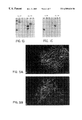
- FIG. 7 HSV antigen production in normal murine brain following intracranial injection of strain 3616UB or F.
- the needle track (top right corner) in a section of a SCID mouse brain that had been stereotactially injected with 3616UB, 48 hours prior, is presented in panel A.
- the section has been immunostained for the presence of HSV-1 protein and even after over-exposure of the film no HSV-1 proteins were evident.
- the needle track (top right corner) in a similar section from a SCID mouse injected with strain F, 24 hours prior, is shown in panel B.
- a large number of infected cells are clustered near the track (B) and spread of the virus is evident throughout the brain with concentrated areas of infected cells near white matter tracks and the ventricles.
- the present invention provides mutant, replication-competent HSV-1 viruses that can enter a tumor cell in situ, make multiple copies, lyse the tumor cell and spread to additional tumor cells with relatively minor effects on the surrounding normal cells.
- mutant refers to a genetically altered or engineered virus that contains a purposefully introduced mutation.
- mutation refers to a change in the chemistry of a gene, a change in the sequence of base pairs in the genomic molecule, that is perpetuated in subsequent progeny of the virus in which it occurs that results in a new species or strain, as distinguished from variation.
- the mutant herpes simplex virus of the present invention has each of the following characteristics: (1) efficacy in killing human brain tumor cells, (2) marked attenuation of generalized neurovirulence to protect the normal brain, (3) multiple deletions so that a single mutation cannot cause reversion to the wild-type viral phenotype, and (4) hypersensitivity to an antiviral agent so that undesired spread of the virus can be prevented.
- the mutant virus of the present invention is capable of replicating in neoplastic cells, and lysing said neoplastic cell, but spares surrounding non-neoplastic tissue.
- Viruses of the instant invention are engineered to contain alterations in the expression of at least two specific HSV-1 genes: (1) the gamma 34.5 gene and (2) the uracil DNA glycosylase gene. Alterations in this regard include any that disrupt the expression of the product of both the gamma 34.5 gene and the uracil DNA glycosylase gene. The presence of such multiple mutations further reduces the possibility of reversion to wild-type pathogenicity.
- the present invention provides methods for sequentially constructing and testing viruses for the ability to effectively kill brain tumor cells without harming surrounding normal brain. Additionally, mutations can be inserted into these vectors to increase their sensitivity to systemically administered drugs.
- Herpes simplex virus UNG-mutants are severely compromised in their ability to productively infect nondividing cells in vivo, Pyles and Thompson, J. Virol. 68. Therefore, these mutants are attenuated for neurovirulence and less likely to propagate following reactivation signal such as fever or UV exposure. Such characteristics are essential to a therapeutic vector that must be of attenuated neurovirulence and amenable to antiviral therapy in the unexpected event of viral encephalitis.
- Herpes simplex virus mutants deficient in only the gamma 34.5 gene, such as R3616 are attenuated for neurovirulence, which reduces the possible damage to normal brain cells.
- R3616 Herpes simplex virus mutants deficient in only the gamma 34.5 gene, such as R3616, are attenuated for neurovirulence, which reduces the possible damage to normal brain cells.
- the decreased neurovirulence of R3616 is putatively associated with the cessation of neuronal protein synthesis, preempted in wild-type herpes simplex virus infection. Chou and Roizman. Proc. Nat'l Acad. Sci. USA 89: 3266 (1992).
- the lack of 34.5 gene product leads to reduced viral replication in confluent primary cells. See Bolovan et al., J.
- the gamma 34.5 gene product can be detected by Western blot or ELISA analysis of infected cell proteins.
- the gamma 34.5 gene is also present in HSV-2. McGeoch et al., J. Gen. Virol. 72: 3057 (1991).
- the gamma 34.5 gene has been sequenced in four strains of HSV-1, namely F, 17, MGH-10 and CVG-2. Chou and Roizman. J. Virol. 64: 1014 (1990).
- the gamma 34.5 gene mutant HSV-1 vectors retain a wild-type level of sensitivity to acyclovir. Markert et al., supra (1993).
- Mutants of gamma 34.5 have been constructed by various investigators using different techniques and in different strains such as mutant 1776 (McKie et al., J. Gen. Virol. 75: 733 (1994)] and 17termA [Bolovan et al., J. Virol. 68: 48 (1994)] in HSV- 1 strain 17.
- the present invention provides a novel alternative approach to eliminating the activity of a virally-encoded.
- DNA metabolism enzyme by direct mutations to those viral functions involved in the editing of the viral DNA.
- the uracil DNA glycosylase function is especially important for efficient viral progeny production.
- the UNG activity corrects misincorporated uracil events and potentially more importantly for HSV-1, the UNG activity removes uracils that arise by the spontaneous deamination of cytosine residues. Because non-dividing cells like neurons have insufficient levels of UNG activity, HSV-1 strains that replicate efficiently in neurons must encode there own. HSV-1 UNG-mutants are therefore reduced in their ability to cause neurovirulence. Pyles and Thompson J. Virol. 68:***-****.
- the UNG gene is located at the genomic designation UL2 (the second open reading frame of the Unique Long viral segment). See Mullaney. et al. 1989, J. Gen. Virol. 70:449-454.
- HSV-1 is a human neurotropic virus that is capable of infecting virtually all vertebrate cells. Natural infections follow either a lytic, replicative cycle or establish latency, usually in peripheral ganglia, where the DNA is maintained indefinitely in an episomal state.
- Replication-competent, recombinant herpes simplex virus vectors of the instant invention contain alterations in expression of two specific herpes simplex virus genes: (1) the gamma 34.5 gene and (2) the uracil DNA glycosylase gene.
- Such alterations render the product of both genes non-functional or reduce their expression such that the mutant herpes simplex virus vector has the properties of the instant invention.
- Ways to achieve such alterations include (a) any method to disrupt the expression of the product of both of these genes or (b) any method to render the expressed gamma 34.5 gene product and uracil DNA glycosylase nonfunctional.
- the mutated herpes simplex virus vector of the instant invention is a replication-competent herpes simplex virus whose genome is altered in the gamma 34.5 gene and the uracil DNA glycosylase gene. Alterations in the gamma 34.5 gene and the uracil DNA glycosylase gene include modifications in either the structural or regulatory sequences of these genes.
- Genetic alterations can be determined by standard methods such as Southern blot hybridization of restriction endonuclease digested viral DNA, sequencing of mutated regions of viral DNA, presence of reporter gene (for insertions), new restriction endonuclease site, enzymatic assay for uracil DNA glycosylase activity, Western blot or ELISA analysis of infected cell proteins with antibodies to UNG or gamma 34.5, and/or lack of replication in confluent primary cells for gamma 34.5. See Bolovan et al., J. Virol. 68: 48 (1994).
- herpes simplex virus vectors The following genetic manipulations of herpes simplex virus provide examples to illustrate the production of mutant herpes simplex virus vectors.
- the engineering of the herpes simplex virus vectors of the instant invention exploit two well-characterized genes, the gamma 34.5 and uracil DNA glycosylase genes, in a biologically well-characterized virus.
- a herpes simplex virus vector that has been mutated in its gamma 34.5 and uracil DNA glycosylase genes can be isolated after mutagenesis or constructed via recombination between the viral genome and genetically-engineered sequences.
- the high rate of recombination in herpes simplex virus and the fact that transfected viral DNA is infectious renders genetic manipulation very straightforward.
- These genetically-altered, replication-competent viruses can be used in the safety and efficacy assays described below.
- HSV-1 contains a double-stranded linear DNA genome, 153 kilobases in length, that has been completely sequenced by McGeoch. McGeoch et al., J. Gen. Virol. 69: 1531 (1988). McGeoch et al., Nucleic Acids Res 14: 1727 (1986): McGeoch et al., J. Mol. Biol. 181: 1 (1985); Perry and McGeoch, J. Gen. Virol. 69: 2831 (1988). DNA replication and virion assembly occurs in the nucleus of infected cells. Late in infection, concatemeric viral DNA is cleaved into genomic length molecules that are packaged into virions. In the CNS, herpes simplex virus spreads transneuronally followed by intraaxonal transport to the nucleus, either retrograde or anterograde, where replication occurs.
- HSV-2 UNG gene like the HSV-1 counterpart, is located at UL2 and has been described in several reports; see for e.g., Worrad and Caradonna. 1988. J. Virol. 62:4774-4777. Gamma 34.5 is also present in HSV-2. McGeoch et al., J. Gen. Virol. 72: 3057 (1991).
- Another way to render a herpes simplex virus incapable of expressing functional gamma 34.5 gene product and uracil DNA glycosylase is to impair their expression.
- the expression of these two genes can be halted by altering the regulatory sequences of the gamma 34.5 and uracil DNA glycosylase genes or by inserting a frame-shift mutation into the nucleotide sequence of the genes.
- the regulatory regions for gamma 34.5 and/or uracil DNA glycosylase can be altered by standard techniques to disrupt the expression of the gamma 34.5 and uracil DNA glycosylase gene (UL2).
- their regulatory sequences could be altered within the viral genome using techniques described above for the alteration of coding sequences.
- the promoter region of gamma 34.5 gene has been mapped and the promoter region for uracil DNA glycosylase UL2 has been partly mapped by Singh and Wagner. Virology 196:220-231.
- the promoter for gamma 34.5 has been mapped to a region within the “a” sequence.
- the “a” sequence also contains sequences for cleavage of unit length DNA from HSV-1 concatamers, packaging of HSV-1 DNA into capsids and inversion of L and S components, Chou and Roizman, J. Virol. 57: 629 (1986).
- the promoter region of UNG has been mapped to the extreme left end of thee unique, long HSV-1 region (base pairs 9000-9500 using the sequence numbering of McGeoch) McGeoch et al., J. Gen. Virol. 69:1531-1574.
- HSV-1 vectors The construction of HSV-1 vectors is described, for example, in U.S. Pat. No. 5,288,641; Roizman and Jenkins, J. Science 229: 1208 (1985); Johnson et al., J. Virol. 66: 2952 (1992); Gage et al., J. Virol. 66: 5509 (1992); Spaete and Frenkel, Cell 30; 295 (1982); Goldstein and Weller, J. Virol.
- herpes simplex virus against gliomas involves providing a means to stop any potential infection of other dividing cells.
- Clinical studies indicate that even wild-type HSV-1 viruses generally do not spread far from the site of initial infection or cause serious systemic disease in immunocompetent individuals. Sacks et al., Ann. Int'l Med. 11: 893 (1989).
- any replication-competent mutant viral vector that is more sensitive to the anti-viral agent than its wild-type parent is deemed hypersensitive to the anti-viral agent, potentially providing a means to prevent an undesired spread of the mutant virus.
- the mutants are tested for their sensitivity to current anti-herpetic drug therapies in order to control unforeseen virulent infections.
- a number of drugs currently are available to treat herpes infections in humans, the most effective being nucleoside analogs that block herpes simplex virus DNA replication.
- herpes simplex virus genes are known to be involved in sensitivity to nucleoside analogs: herpes simplex virus DNA polymerase (UL30, pol), herpes simplex virus thymidine kinase (UL23,tk), and CMV UL97 which shares homology with protein kinases and bacterial phosphotransferases. Furman et al., J. Virol. 32: 77 (1979); Littler et al., Nature 358: 160 (1992); Sullivan et al., Nature 358: 162 (1992).
- UL30, pol herpes simplex virus DNA polymerase
- UL23,tk herpes simplex virus thymidine kinase
- CMV UL97 which shares homology with protein kinases and bacterial phosphotransferases.
- herpes simplex virus DNA polymerase mutants which exhibit hypersensitivity to ganciclovir, including PAAr 5 and AraAr 9. Coen et al., J. Virol. 53: 477 (1985). Unfortunately, intracranial injections of AraAr 9 led to premature death and had no effect on subcutaneous tumor growth. Markert et al., supra. Another mutant herpes simplex virus, the dlsptk virus, is no longer drug sensitive, at least to nucleoside analog drugs, and therefore potentially uncontrollable in vivo.
- Attenuated or decreased generalized neurovirulence means that life-threatening encephalitis does not ensue after infection with the double mutant herpes simplex virus vector of the instant invention. Because herpes simplex virus-induced encephalitis in humans is very difficult to treat and can be fatal, even with adequate pharmacologic measures, decreased generalized neurovirulence is an important feature of the instant invention.
- the mutant virus of the present invention is capable of replicating in neoplastic cells but spares surrounding non-neoplastic tissue.
- HSV-1 strains available from ATCC include HF (ATCC VR-260), MacIntyre (ATCC VR-539), MP (ATCC VR-735) and HSV-2 strains G (ATCC VR-734) and MS (ATCC VR-540).
- any herpes simplex virus gene mutation leading to decreased viral replication in vivo and/or in specific cell populations May be used in the mutated herpes simplex virus vector of the invention.
- Other neurovirulence genes include but are not limited to: (i) dUTPase [Pyles et al., J. Virol. 66: 6706, (1992)], (ii) UL53 [Moyal et al., Virus Res. 26: 99 (1992)], (iii) alpha 22 [Sears et al., J. Virol.
- herpes simplex virus encephalitis is the most commonly reported viral infection of the central nervous system (CNS) in the United States, with an estimated incidence of 2.3 cases per million people.
- CNS central nervous system
- Herpes simplex virus encephalitis is usually localized to the temporal lobe and the limbic system. Histological examination of autopsy cases demonstrates viral antigen at these sites.
- drugs are available to control infection, including acyclovir 9-92-hydroxyethoxy -methyl)guanine.
- herpes simplex virus mutants of the instant invention may be targeted to specific tumor types using tumor cell-specific promoters.
- tumor cell-specific promoter indicates a promoter that is induced selectively or at a higher level in the target tumor cell than in a normal cell.
- Tumor cell-specific promoters include promoters that are induced selectively or at a higher level in a particular cell type or a tumor cell.
- the vectors of the invention also can be designed to selectively replicate in and kill a tumor cell of non-nervous tissue origin.
- the herpes simplex virus vector of the invention is engineered to place at least one viral protein necessary for viral replication under the control of a cell specific or tumor cell-specific promoter.
- the tumor cell-specific promoter is induced selectively or at higher levels in tumor cells than in normal cells.
- HSV-1 and HSV-2 mutants utilize promoters from genes that are highly expressed in the targeted tumor, such as the epidermal growth factor receptor gene promoter (EGFr) or the basic fibroblast growth factor (bFGF) gene promoter or the NESTIN or other tumor associated promoter or enhancer element to drive expression of an essential herpes simplex virus gene (e.g., ICPO or ICP4), under circumstances in which the wild-type essential herpes simplex virus gene would not be expressed.
- EGFr epidermal growth factor receptor gene promoter
- bFGF basic fibroblast growth factor
- NESTIN tumor associated promoter or enhancer element
- an essential herpes simplex virus gene e.g., ICPO or ICP4
- Rendering the essential herpes simplex virus gene non-functional can be achieved by genetic inactivation or replacement of its viral promoter with a tumor cell-specific promoter.
- the instant invention encompasses a host-range conditional herpes simplex virus mutant where an essential viral gene product is under the control of a tumor cell-specific promoter rather than its own viral promoter.
- permissive cells containing the proper regulatory proteins for this specific promoter, the essential viral gene product is expressed and the virus is able to replicate and spread to adjacent cells until a non-permissive cell is infected.
- tumor cell types express phenotypic markers which are turned off in the normal terminally-differentiated cell.
- differentially regulated genes in neural tumors include: (i) nestin, an intermediate filament protein normally expressed in neuroepithelial stem cells, yet not in mature CNS cells, which is ubiquitously expressed in human brain tumors, most prominently in gliomas, (ii) basic fibroblast growth factor (bFGF), a differentiation factor and mitogen for neuroectoderm, which is highly expressed in human gliomas and meningiomas but not in metastatic brain tumors or normal brain tissue and (iii) epidermal growth factor receptor (EGFr), a membrane-bound tyrosine-specific protein kinase that is stimulated by EGF, which is very often overexpressed, altered and the gene amplified in human high grade gliomas but rarely in normal brain.
- nestin an intermediate filament protein normally expressed in neuroepithelial stem cells,
- the mutant herpes simplex virus vector of the instant invention can be employed as a genetic therapy against specific tumors by expressing foreign genes in a tumor cell-specific fashion in order to target an immune response that kills the tumor cells. Tepper and Mule, Human Gene Therapy 5: 153 (1994).
- the instant invention employs the replication-competent herpes simplex virus vector having decreased neurovirulence as a tumor cell modulator or inducer of an immune response against the tumor cells.
- the mutant herpes simplex virus vector of the invention can be further altered to express cytokines in the tumor target cell in order to elicit an immune response against the tumor cells.
- a mutant herpes simplex virus vector can induce viral-mediated killing of tumor cells, which then is amplified by a cytokine-enhanced immune response, a cytokine having been expressed by the vector itself.
- the expression of cytokines, or other gene products, from the mutant herpes simplex virus vector would occur within hours of infection so that sufficient gene products would be synthesized prior to cell killing. Immune systems-mediated-cell killing may even increase the efficacy of the anti tumor immune response. Barba et al., Proc. Nat'l Acad. Sci. USA 91: 4348 (1994).
- Exemplary candidates for treatment according to the present invention include, but are not limited to (i) non-human animals suffering from tumors and neoplasms, (ii) humans suffering from tumors and neoplasms, (iii) animals suffering from nervous system tumors and (iv) patients having malignant brain tumor, including astrocytoma, oligodendroglioma, meningioma, neurofibroma, glioblastoma. ependymoma, Schwannoma, neurofibrosarcoma, and medulloblastoma.
- the treatment will be initiated by direct intraneoplastic inoculation.
- MRI, CT, or other imaging guided stereotactic technique will be used to direct viral inoculation or virus will be inoculated at the time of craniotomy.
- compositions of the present invention would be advantageously administered in the form of injectable compositions.
- a typical composition for such purpose would comprise a pharmaceutically acceptable vehicle.
- the composition could contain human serum albumin in a phosphate buffer containing NaCl.
- Other pharmaceutically acceptable carriers include aqueous solutions, non-toxic excipients, including salts, preservatives, buffers and the like. See REMINGTON'S PHARMACEUTICAL SCIENCES (15th ed.) 1405-1412 & 1461-1487, Mack Publishing Co. (1975), and THE NATIONAL FORMULARY XIV (14th ed.), American Pharmaceutical Association (1975).
- non-aqueous solvents examples include propylene glycol, polyethylene glycol, vegetable oil and injectable organic esters such as ethyloleate.
- Aqueous carriers include water, aqueous solutions saline solutions, parenteral vehicles such as sodium chloride. Ringer's dextrose, etc.
- Intravenous vehicles include fluid and nutrient replenishers. The pH and exact concentration of the various components of the pharmaceutical composition are adjusted according to routine skills in the art, Goodman and Gilman. THE PHARMACOLOGICAL BASIS FOR THERAPEUTICS (7th ed.).
- the herpes simplex, virus vector would be prepared as injectables, either as liquid solutions or suspensions: solid forms suitable for solution in, or suspension in liquid prior to injection may also be prepared.
- the preparation also may be emulsified.
- the active immunogenic ingredient is often mixed with an excipient which is pharmaceutically-acceptable and compatible with the active indent. Suitable excipients are, for example, water, saline, dextrose, glycerol, ethanol, or the like and combinations thereof.
- the vector may contain minor amounts of auxiliary substances such as wetting or emulsifying agents, pH-buffering agents, adjuvants or immunopotentiators which enhance the effectiveness of the vector vaccine.
- oral formulations include such typical excipients as, for example; pharmaceutical grades of mannitol, lactose, starch, magnesium stearate, sodium saccharine, cellulose, magnesium carbonate and the like.
- the compositions take the form of solutions, suspensions, tablets, pills, capsules, sustained release formulations or powders and contain 10%-95% of active ingredient, preferably 25-70%.
- the composition can comprise, in addition to a virus, compounds and/or compositions that will also aid in treating the tumors or the accompanying symptoms, such as, radioactive seed implants, antibiotics, growth factors (e.g., GMCSF or erythropoietin), other forms of genetic therapy, including but not limited to, retroviral and adenoviral vector systems and immunomodulating therapies such as IL-2, IL-12 or others as they are deemed appropriate.
- the mutant replication-competent HSV strain may be administered with other pharmaceuticals that will be used at the clinicians discretion to treat post-operative symptoms, including but not limited to, anti-convulsants, sedatives, steroids and immunomodulating chemicals. Dosages for the above-mentioned additional compounds are established and known to those skilled in the art.
- the viral therapy may also be given in conjunction with general raiotherapy and/or general chemotherapy.
- the compounds useful in the present inventive method may be administered by any suitable means.
- suitable methods of administering the compound to an animal in the context of the present invention, in particular a human are available, and, although more than one route may be used to administer a particular compound, a particular route of administration may provide a more immediate and more effective reaction than another route.
- unit dose refers to physically discrete units suitable for use in humans, each unit containing a predetermined quantity of active material calculated to produce the desired therapeutic effect in association with the required diluent, i.e., carrier or vehicle, and a particular treatment regimen.
- the quantity to be administered both according to number of treatments and amount, depends on the subject to be treated, capacity of the subject's immune system to synthesize antibodies; and degree of protection desired. Precise amounts of active ingredient required to be administered depend on the judgment of the practitioner and are peculiar to each individual. However, suitable dosage ranges are on the order of one to several hundred micrograms of active ingredient per individual. Suitable regimes for initial administration and booster shots also vary but are typified by an initial administration followed in one or two week intervals by one or more subsequent injections or other administration.
- Patients with recurrent glioblastoma that was refractory to standard surgery radiotherapy and chemotherapy would be treated with herpes simplex virus therapy.
- the patient would be scanned using MRI or CT or other technique and the tumor and normal brain registered in stereotactic space.
- the virus would be administered using stereotactically guided neurosurgical techniques.
- a computer tomography (CT) scan or magnetic resonance imaging (MRI) scan computes the stereotactic frame that would be used to accurately inoculate virus into a tumor at one or more locations.
- Virus would be inoculated at a dose of about 10 1 to about 10 7 p.f.u. per inoculation using a ⁇ 2 mm cannula. The number of sites inoculated would depend on the size of the tumor. Patients would be followed with periodic MRI scans and with neurological examination, blood count, and liver function tests.
- craniotomies will be performed for total gross tumor ressection in with recurrent tumor and virus will inoculated in the resected tumor bed in a fashion similar to above.
- the herpes simplex virus vector of the invention can be used as a vaccine to protect an animal against herpes simplex virus infection.
- “protecting” a subject against herpes simplex virus includes both (1) a prophylactic vaccine, i.e., a vaccine used to prevent a future herpes simplex virus infection, and (2) a therapeutic vaccine for treating an existing herpes simplex vital infection.
- the herpes simplex virus sample would be prepared using standard methodology.
- Herpes simplex virus-infected Vero cells would be frozen at ⁇ 70° C. until they are to be used. The material would be thawed and the cell debris would be pelleted by centrifugation. The supernatant fluid would be discarded and the pellet resuspended to its original volume. This material would most closely approximate that used in vaccine manufacture. This suspension would be sonicated three times for 30 seconds.
- Herpes simplex virus plaque titers would be determined by standard procedures. For example, the virus would be titrated in triplicate on monolayers of Vero cells in 6-well plates. After adsorption of samples for 2 hours, cells would be overlayed with media containing 0.6% agarose and incubated at 37° C. in a CO 2 -rich environment for 48 h. A second overlay, the same as above except for addition of neutral red, would be added and the cells would be incubated an additional 24 hours.
- the herpes simplex virus pools would be titrated before filtration. The pools then would be filtered through a Nalgene 0.45 mu m filter sampled refiltered through a second filter and then resampled.
- African Green Monkey kidney cells (VERO; CRL 1586), the human glioblastoma cell line, U87MG (HTB 14), and the human breast carcinoma cell line MDA-MB-231 (HTB 26) were obtained from the ATCC (Rockville, Md.). VERO and U87MG cells were cultured as recommended by the ATCC. MDA-MB-231 cells were cultured in MEM-a medium supplemented with 10% FBS, 100 units/ml of penicillin G, and 100 mg/ml of streptomycin (MED medium). The human medulloblastoma cell line DAOY was obtained from Dr. Walter Hall (Univ. of Minn.; Wen et al., 1995) and cultured in MED medium and passaged once every four days.
- the human neuroblastoma cell lines N (neurite-bearing) and S (substrate-adherent) were cultured as previously described (Foley et al., 1991).
- SKBR3 Heffelfinger et al., 1992
- Dr. Susan Heffelfinger Univ. of Cincinnati
- SK-M SK-M cells also were grown in MED medium and passaged once every four days.
- the SK-M cell line is of endothelial origin.
- Cell culture supplies were purchased from GIBCO/BRL (Gaithersberg. MD). All cells were maintained in a 37° C., 5% CO 2 -humidified environment.
- embryonic rat dorsal root ganglion cultures were kindly provided by Dr. Nancy Ratner (Univ. of Cincinnati). These cultures were prepared from dorsal root ganglia harvested from day E15 rat embryos (Oncogene 11, 35-335.) and then, following plating at a density of ⁇ 2000 cells/collagen-coated coverslip, the cells were cultured for two weeks first in medium to kill active cells (Dulbeccos modified essential medium supplemented with 10% human plasma serum nerve growth factor and fUdR at 0.5%) and then in the same medium without the fUdR.
- strain F The virus strains used in these studies were derived originally from the wild type HSV-1 strain F. Strain F was chosen for this work because it has reduced neurovirulence as compared to other laboratory isolates of wild type HSV-1 (Infect. Immun. 40, 103-112.). Both strain F and its derivative ICP34.5 deletion mutant, R3616 (Science 250, 1262-1266.) were kindly provided by Dr. B. Roizman (Univ. of Chicago).
- Virus stocks were prepared from subconfluent VERO cell monolayers that had been infected 48 hours prior to harvesting. The virus was concentrated from sonicates of infected cells And then titered using standard methods. All virus stocks were stored at ⁇ 80° C. in VERO culture media. HSV-1 genomic DNA for transfection and for restriction endonuclease analyses was prepared as described (J. Virol. 68, 4963-4972.).
- the HSV-1 UNG open reading frame (ORF; UL2, bps 9886-10888) of R3616 was disrupted as the result of recombination of R3616 with a plasmid that contains the E. coli b-galactosidase (LacZ) gene under the control of the SV40 early promoter inserted into the unique Xba I site within the cloned UNG ORF (J. Virol. 68, 4963-4972.).
- Genomic R3616 DNA and the UNG mutant plasmid DNA were cotransfected into VERO cells with Lipofectase (GIBCO/BRL).
- Recombinant viruses were identified by the formation of a blue precipitate following incubation with the chromagen X-Gal (5-bromo-4-chloro-3-indolyl-b-D-galactopyranoside: GIBCO/BRL). LacZ-expressing “blue” plaques were purified to homogeneity by limiting dilution.
- the genomic structure of the 3616UB virus was confirmed by Southern blot, RFLP analysis. Briefly, viral genomic DNA that had been digested with appropriate restriction enzymes was size-separated in 1% agarose and blotted onto Biodyne-B membrane (GIBCO/BRL). The blots were hybridized to 32 P-labeled probes specific for the UNG, LacZ or ICP34.5 genes at 42° C for 24 hours. Following washing, the blots were exposed to XOMAT-AR films (Eastman Kodak: Rochester, N.Y.) for 6-24 hours.
- XOMAT-AR films Eastman Kodak: Rochester, N.Y.
- CB 17 severe combined immunodeficient (SCID) or outbred Swiss Webster mice (Charles River Breeders) were used throughout these studies. Animals were housed in microisolator cages in American Association for Laboratory Animal Care-approved quarters and provided with unlimited access to food and water. Animals were quarantined for 7 days prior to any procedures.
- PFU/lethal dose 50% PFU/LD 50
- PFU/LD 50 ratios were calculated using the method of end-point estimation as described by Reed and Muench (Amer. J. Hyg. 27, 493-497).
- mice were stereotactically injected with either HSV-1 strain F or 3616UB in the right frontal lobe. A volume of 10 ml that contained 1 ⁇ 10 8 PFU/ML of virus was delivered over a 15 min. period. At 24 or 48 hours post injection, animals then were sacrificed and the injected brain hemisphere examined for HSV-1 antigen by immunohistochemistry with a polyclonal HSV-1 antiserum (Accurate Chemical and Scientific Corp.; Westbury, N.Y.).
- Tumor xenografts were produced by flank injection of DAOY or SK-M cells that had been cultured to confluency and then harvested by trypsinization into culture media. Cell density was adjusted to ⁇ 10 6 cells/125ml of culture media for flank injections. For each injection site, 125 ml of cell suspension was mixed with an equal volume of Matrigel (Collaborative Research, Inc.) to enhance tumor xenograft formation prior to injection. For all experiments, tumor cells were injected subcutaneously into both rear flanks of anesthetized animals.
- Intratumoral injections of viral stock were performed when the xenografts reached a volume of 50 or 100 mm 3 (a diameter of 5-10 mm). For each xenograft. an injection of 30 ml that contained 3 ⁇ 10 6 PFU of 3616UB or R3616 was performed, delivering the virus to the approximate center of the tumor. For the therapy studies, groups of 10 tumors were injected with 3 ⁇ 10 6 PFU of 3616UBI or R3616 or an equivalent amount of medium alone. An identical second injection was given 7 days after the first. Tumor growth ratios were established from tumor volumes that were calculated using the formula length ⁇ width 2 /2.
- Viral antigens in tumor xenografts or healthy brain tissue were detected by immunofluoresence (Cancer Res. 51, 6338-6345.).
- immunofluoresence studies paraffin-embedded tissue sections were mounted on Plus slides (Fisher). Following paraffin clearing and rehydration, the sections were rinsed in phosphate-buffered saline and then were blocked with 5% non-fat dry milk in tris-buffered saline, pH 7.4 (TS) for 40 min.
- Viral antigens present in infected cultured cells were detected following fixation with 4% paraformaldehyde for 10 min. at room temperature.
- a polyclonal antiserum to HSV-1 diluted 1:100 in 5% milk/TS was applied to the tissue or cell monolayer and incubated overnight at 4° C. Following extensive rinsing in TS, goat anti-rabbit-FITC (Kirkegaard and Perry), was applied at a dilution of 1:50 in 5% milk/TS and incubated at room temperature for 1 h. Following rinsing, the specimens were mounted with polyvinyl alcohol containing 0.1% 1,4-diazabicyclo(2.2.2)octane (DABCO; HARLOW, E., and LANE. D. (1988). Antibodies: A laboratory maual. p. 418. Cold Spring Harbor Laboratory, Cold Spring Harbor, N.Y.).
- a monoclonal antibody to the intermediate filament protein vimentin was used in some experiments to visualize noninfected cells because it efficiently labeled each of the selected tumor cell lines.
- tissue specimens were incubated with secondary antibody alone as a negative control.
- Photomicrographs were generated on a Zeiss Axiophot using TMAX 100 film (Eastman/Kodak).
- monolayers of DAOY or VERO cells were infected with 3616UB, R3616 or F at an MOI of 0.01 and then triplicate cultures were incubated with medium alone or in medium supplemented with 75 ng/ml GCV. After 48 hours, the infected monolayers were harvested and viral titers determined. The titers attained by each viral strain were established in triplicate wells, averaged and used to establish standard deviations. The average values then were compared under the two different conditions to establish the sensitivity to GCV as the percent of plaque reduction.
- GCV ganciclovir
- the UNG probe hybridized to a single 12.610 bp Asp 718 fragment in strain F and R3616 lanes (panel B), because the wild type UNG locus does not contain an Asp 718 site (see panel A), and to two bands totaling approximately 16.600 bp in 3616UB.
- the LacZ insertional event at the UNG locus was confirmed by the comparison of Eco R1 digestion of the DNA of the three viral strains.
- the LacZ-specific probe hybridized to bands of expected size following digestion with either Eco R1 or Asp 718 (panel C). These bands are not present in either the R3616 or strain F lanes.
- LacZ reporter gene in 3616UB was functional, cell cultures that had been infected with 3616UB were fixed after 24 hours and then stained with the chromagenic substrate for b-galactosidase. Blue staining was detected in all viral plaques, indicating a correlation between herpes infection and LacZ expression. The b-gal activity was located at the plaque margin only, suggesting that LacZ expression from 3616UB provides an indicator of the initial infection of cells (FIG. 3 D). This data indicates that the LacZ gene is functional, allowing for the histochemical localization of the 3616UB-infected cells. Staining of infected monolayers with X-gal also showed that the UNG gene mutation was stable because no LacZ negative plaques (potential UNG revertants) were observed in over 5000 that were examined (data not shown).
- Strain 3616UB generated titers in VERO monolayers that were equivalent to that of its parent strain R3616 and its grandparent strain F. In each of the cultured cell types examined strain 3616UB effectively replicated to titers of ⁇ 10 8 PFU/ml with the same relative kinetics as the wild type grandparental strain F (data not shown).
- strain 3616UB is to be used as a human tumor therapy, it was necessary to establish that the therapy can be controlled using chemical intervention.
- GCV ganciclovir
- the mutations engineered in 3616UB were chosen to prevent this strain from replicating in and killing nondividing cells near the tumor.
- To determine whether 3616UB replicated in a non-dividing cell population primary embryonic rat dorsal root ganglionic neuron cultures were infected at MOIs of 0.001, 1 or 10 with each of the three viral strains. Even 7 days after infection with an MOI of 10, 3616UB did not adversely affect the neuronal cultures. The observation that 3616UB did not replicate in these cultures was confirmed by the lack of immunostaining for HSV-1 antigens and the lack of virally-induced CPE (FIG. 4 A & B).
- HSV-1 STRAIN MOI 3616UB R3616 F 10 none detected 10 PFU/ml 2 ⁇ 10 5 PFU/ml 1 none detected none detected 1 ⁇ 10 5 PFU/ml 0.01 none detected none detected 6 ⁇ 10 3 PFU/ml
- strain F replicated in and eventually led to the destruction of the neuronal cultures at all three MOIs within the 7 day observation period.
- Strain F generated titers as high as 10 5 PFU/ml following infection at MOI of 1 or 10 (Table 1) and led to the production of robust levels of HSV antigen as detected by immunohistochemistry (FIG. 4 C).
- 3616UB's parental strain, R3616 was found to replicate in and produce a small amount of progeny virus after infection of neuronal cultures with high doses of virus (Table 1: MOI of 10).
- the R3616 virus detected at 7 days post infection could only represent virus produced from active replication in the neuronal culture.
- HSV-1 antigens were immunolocalized to no more than 1% of the total neurons suggesting limited replication of this virus (data not shown).
- the therapeutic value of 3616UB was examined by injection of 30 ml of 1 ⁇ 10 8 PFU/ml intratumorally into 10 tumors of 50 mm 3 volume previously established in the flanks of SCID mice. Additional groups of 10 tumors were injected with either R3616 or medium alone for comparison and control, respectively.
- tumor volumes were calculated from measurements of the tumor mass and were used for the determination of growth ratios (FIG. 6 ).
- R3616 the parent of 3616UB, resulted in the death of 1/6 animals following direct intracranial injection of high titer stock, but in the majority of mice did not cause any signs of neurological distress.
- the wild type strain F generated a PFU/LD 50 ratio of 5 ⁇ 10 4 (Table 2).
- HSV-1 protein was not detected in brain sections at or near the needle track at the 24 or 48 hour time point in any of the mice (FIG. 7 A). Histochemical staining of the adjacent sections with X-gal to illustrate the expression of the LacZ reporter gene in 3616UB also showed no evidence of viral infection other than a single blue cell at the needle track (data not shown). Brain sections from positive control mice that were injected with strain F, however, showed infected cells concentrated near the needle track at 24 hours post infection (FIG. 7 B).
Landscapes
- Health & Medical Sciences (AREA)
- Life Sciences & Earth Sciences (AREA)
- Chemical & Material Sciences (AREA)
- Genetics & Genomics (AREA)
- Virology (AREA)
- Engineering & Computer Science (AREA)
- Wood Science & Technology (AREA)
- Organic Chemistry (AREA)
- Microbiology (AREA)
- Zoology (AREA)
- General Health & Medical Sciences (AREA)
- Bioinformatics & Cheminformatics (AREA)
- General Engineering & Computer Science (AREA)
- Biotechnology (AREA)
- Medicinal Chemistry (AREA)
- Immunology (AREA)
- Biomedical Technology (AREA)
- Pharmacology & Pharmacy (AREA)
- Veterinary Medicine (AREA)
- Public Health (AREA)
- Animal Behavior & Ethology (AREA)
- Biochemistry (AREA)
- Epidemiology (AREA)
- Mycology (AREA)
- Physics & Mathematics (AREA)
- Biophysics (AREA)
- Molecular Biology (AREA)
- Plant Pathology (AREA)
- Micro-Organisms Or Cultivation Processes Thereof (AREA)
- Medicines That Contain Protein Lipid Enzymes And Other Medicines (AREA)
- Medicines Containing Antibodies Or Antigens For Use As Internal Diagnostic Agents (AREA)
- Pharmaceuticals Containing Other Organic And Inorganic Compounds (AREA)
- Medicines Containing Material From Animals Or Micro-Organisms (AREA)
Abstract
A promising approach for the therapeutic treatment of brain tumors utilizes replication-competent, neuroattenuated herpes simplex virus-1 (HSV-1) mutants. This approach requires mutation of HSV-1 to eliminate killing of normal, non-dividing cells of the brain (e.g., neurons). The present invention discloses methods for killing malignant brain tumor cells in vivo entails providing replication competent herpes simplex virus vectors to tumor cells. A replication competent herpes simplex virus vector, with defective expression of the gamma 34.5 gene and the uracil DNA glycosylase (UNG) gene, specifically destroys tumor cells, is hypersensitive to anti-viral agents, and is not neurovirulent.
Description
This Application claims priority to PCT Application No. PCT/US98/06124 entitled “REPLICATION-COMPETENT HERPES SIMPLEX VIRUSES” and filed Mar. 27, 1998, now abandoned and U.S. Provisional application No. 60/042,418 filed Mar. 27, 1987.
This invention was made in part with Government support under Grant Nos. NS31145 and T32-CA59268 awarded by the National Institutes of Health. The Government may have certain rights in this invention.
The present invention relates to recombinant virus strains capable of killing tumor cells. More specifically, the present invention relates to a mutated replication-competent viruses which contains mutations in two genes, is hypersensitive to antiviral agents such as ganciclovir, is not neurovirulent and does not replicate in non-dividing cells, yet can kill nervous system tumor cells. The present invention also relates to recombinant herpesvirus strains, vital vaccines incorporating such strains, methods for making such strains and vaccines, and methods for immunizing a human host against herpes simplex virus using the vaccines.
Malignant tumors of the nervous system are generally fatal, despite many recent advances in neurosurgical techniques, chemotherapy and radiotherapy. In particular, there is no standard therapeutic modality that has substantially changed the prognosis for patients diagnosed with malignant brain tumors. For example, high mortality rates persist in malignant medulloblastomas, malignant meningiomas and neurofibrosarcomas, as well as in malignant gliomas.
Gliomas are the most common primary tumors arising in the human brain. The most malignant glioma, the glioblastoma, represents 29% of all primary brain tumors, some 5,000 new cases per year in the United States alone. Glioblastomas are almost always fatal, with a median survival of less than a year and a 5-year survival of 5.5% or less. Mahaley et al., J. Neurosurg. 71: 826 (1989); Shapiro, et al., J. Neurosurg. 71: 1 (1989): Kim et al., J. Neurosurg. 74: 27 (1991). After glioblastomas are treated with radiotherapy, recurrent disease usually occurs locally; systemic metastases are rare. Hochberg et al., Neurology 30: 907 (1980). Neurologic dysfunction and death in an individual with glioblastoma is due to the local growth of the tumor.
Efforts to cure primary and metastatic brain tumors have focused on new approaches that make use of genetically modified viruses either to deliver cytotoxic genes to tumor cells or to directly infect and destroy tumor cells in a selective fashion. Treatment strategies employing replication-competent HSV-1 mutants may be particularly promising (Hum. Gene Ther. 5, 183-191; Cancer Res. 54, 5745-5751.; J Neuro-Oncol, 19, 137-147: J. Neurosurg, 77, 590-594: Neurosurg. 32, 597-602; Science 252, 854-856; Stereotact. Funct. Neurosurg. 59, 92-99; Nature Med. 1, 938-943; Virol. 211, 94-101.) Such mutants, like wild-type HSV-1 strains, establish a lytic infection in dividing tumor cells, leading to tumor cell destruction, but establish only a latent infection of the surrounding nondividing brain cells, including neurons. These mutants are attenuated human pathogens and thus, must be examined fully for their safety and utility prior to clinical use.
The first HSV-1 mutant studied, dlsptk, carried a single mutation in the thymidine kinase (TK) gene. Mutant strain dlsptk was found to have significant antineoplastic efficacy with a minimal level of toxicity in human tumor xenografts in immunodeficient mice (Neurosurg. 32, 597-602; Science 252, 854-856). These effects demonstrated the potential of HSV-1 as a tumor therapy, but at least two concerns regarding the safety of dlsptk limited its potential for human use. Because it lacks a functional TK gene, strain dlsptk cannot be controlled by the antiherpetic drugs acyclovir or ganciclovir. Second, infection with TK mutant strains causes neurovirulence in animal models when used at doses that would be employed for cancer therapy (N. Engl. J. Med. 320, 293-296). For these reasons, investigators have tested the usefulness of other mutations that severely reduce the ability of the virus to replicate in nondividing cells but do not prevent viral replication in actively dividing cells (Cancer Res. 54, 5745-5751: J Neuro-Oncol. 19, 137-147; J. Neurosurg. 77, 590-594; Neurosurg. 32, 597-602; Virol. 211, 94-101). One such mutation, introduced in both copies of the diploid ICP34.5 gene, has been shown in at least two parental HSV-1 backgrounds to result in viral vectors that have the tumor kill efficiency of dlsptk but minimal to no detectable toxicity (J. Neurosurg. 77, 590-594: Neurosurg. 32, 597-602; Virol. 211, 94-101. J. Gen. Virol. 75, 2059-2063.). Importantly, the HSV-1 strains with mutation of only the ICP34.5 genes retain TK activity, allowing for control by antiherpetic drugs that are activated by HSV-encoded TK. In U.S. Pat. No. 5,328,688. Roizman. issued Jul. 12, 1994, there is disclosed an HSV-1 strain that is reported to be rendered avirulent by the prevention of expression of an active product of a gene, designated gamma 34.5. that maps to the inverted repeats, flanking the long unique sequence of herpes simplex virus DNA. this gene is not essential for viral growth in cell culture. Viruses from which 34.5 was deleted or which carried premature stop codons in the 34.5 gene are avirulent following intracerebral inoculation of mice.
More recently, efforts to increase the safety of herpes-based therapy have spurred the development of HSV-1 strains that have mutations in two viral genes and thus are theoretically less likely to be repaired by recombination with a preexisting or subsequent HSV-1 infection. U.S. Pat. No. 5,585,096, Martuza et al., issued Dec. 17, 1996, discloses a method for killing malignant brain tumor cells in vivo by introducing replication-competent herpes simplex virus vectors to tumor cells. A replication-competent herpes simplex virus vector, with defective expression of the gamma 34.5 gene and the ribonucleotide reductase gene, specifically destroys tumor cells and is not neurovirulent.
Initial success with such a strain, designated G207, has been reported for treatment of glioblastoma xenografts established in immunodeficient mice (Nature Med. 1, 938-943). Strain G207 carries a deletion of both copies of the ICP34.5 gene and a mutated ICP6 gene, that encodes the large subunit of the ribonucleotide reductase, an enzyme in the salvage pathway required for efficient DNA synthesis (ROIZMAN, B. and SEARS. A. E., 1990). Herpes simplex viruses and their replication, p. 1795-1841. In B. N. Fields, et al. (ed.), Virology, 2nd ed. Raven Press, New York). In addition to being multiply-mutated, this virus also retains sensitivity to antiherpetic drugs and has minimal toxicity in animal models (Nature Med. 1, 938-943).
It remains of utmost importance to develop a multiple HSV-1 mutant viral strain that has the greatest possible safety and therapeutic value.
It is therefore an object of this invention to provide a replication-competent viral vector, suitable for use in humans, that is capable of killing human tumor cells in vivo, that exhibits hypersensitivity to anti-viral agents and an inability to revert to wild-type virus, and that is not neurovirulent at a dose required to kill tumor cells.
It is another object of the present invention to provide for the production of a replication-competent herpes simplex virus-derived vector that is effective and safe for use in the treatment of malignant brain tumors in humans.
It is a further object of the invention to provide a safe, mutated HSV-1 vector, incapable of reverting to wild-type form through a spontaneous single mutation, for use in the context of a vaccine or tumor therapy.
Still another object of the present invention is to provide a mutant HSV-1 vector that can selectively replicate in and kill a tumor cell of non-nervous tissue origin.
An additional object of the present invention is the production of a replication-competent viral vector, derived from herpes simplex virus, that can be employed in a genetic therapy against tumors by expressing foreign genes to target an immune response that kills the tumor cells.
Yet another object of the present invention is the production of a mutant herpes simplex virus vector containing a tumor cell-specific promoter so that the vector can be targeted to specific tumor cells.
It is also an object of the present invention to provide for production of a replication competent viral vector that is effective and safe for use as a vaccine to protect against infection by herpes simplex virus.
In satisfying these and other objects, there has been provided, in accordance with one aspect of the present invention, a replication-competent herpes simplex virus that is incapable of expressing both (i) a functional gamma 34.5 gene product and (ii) a uracil DNA glycosylase. In a preferred embodiment, the vector contains alterations in both genes.
In accordance with another aspect of the present invention, a method has been provided for killing tumor cells in a subject, comprising the step of administering to the subject a pharmaceutical composition comprising (A) a herpes simplex virus vector that is altered in (i) the gamma 34.5 gene, and (ii) the a uracil DNA glycosylase (UNG) gene; and (B) a pharmaceutically acceptable vehicle for the vector, such that the tumor cells are altered in situ by the vector and the tumor cells are killed. The tumor cells can be of a nervous-system type selected from the group consisting of astrocytoma, oligodendroglioma, meningioma, neurofibroma, glioblastoma, ependymoma, Schwannoma, neurofibrosarcoma, and medulloblastoma. Other kinds of tumor cells which can be killed, pursuant to the present invention, include those selected from the group consisting of melanoma cells, pancreatic cancer cells, prostate carcinoma cells, breast cancer cells, lung cancer cells, colon cancer cells lymphoma cells, hepatoma cells and mesothelioma and epidermoid carcinoma cells.
In accordance with still another aspect of the present invention, a method is provided for killing tumor cells in a subject, comprising the steps of administering to the subject a herpes simplex virus vector, wherein the vector comprises a tumor cell-specific promoter wherein the promoter controls expression of at least one viral protein necessary for viral replication and wherein the promoter is induced selectively or at a higher level in tumor cells than in normal cells. This method can entail the use of a promoter that is selectively capable of expression in nervous-system tumor cells, for example, glioblastoma cells, medulloblastoma cells, meningioma cells, neurofibrosarcoma cells, astrocytoma cells, oligodendroglioma cells, neurofibroma cells, ependymoma cells and Schwannoma cells.
A method also is provided for preparing a replication-competent vector of a herpes simplex virus, comprising the steps of (A) isolating a viral genome of the herpes simplex virus: and (B) permanently altering the genome so that the virus is (1) sensitive to antiviral agents, (2) kills tumor cells and (3) expresses decreased generalized neurovirulence. For example, the vector can be derived from either HSV-1 or HSV-2.
The present invention further provides for a method of protecting a subject against herpes simplex virus infection, comprising the step of administering to the subject a pharmaceutical composition that is comprised of (A) a herpes simplex virus vector wherein the genome of the virus is altered in (i) the gamma 34.5 gene, and (ii) the a uracil DNA glycosylase gene; and (B) a pharmaceutically acceptable vehicle for the vector.
According to still another aspect of the present invention, there has been provided a method of eliciting an immune response to a tumor cell, comprising the step of administering to the subject a pharmaceutical composition comprising (A) a herpes simplex virus, wherein the genome of the virus (i) contains an expressible non-herpes simplex virus nucleotide sequence encoding a desired protein capable of eliciting an immune response in the subject, and (ii) is altered in the gamma 34.5 gene, and the uracil DNA glycosylase gene: and (B) a pharmaceutically acceptable vehicle for the virus. In a preferred embodiment, the method further comprises the step of co-administration with neurosurgery, chemotherapy or radiotherapy.
Other objects, features and advantages of the present invention will become apparent from the following detailed description. It should be understood however, that the detailed description and specific examples, while indicating preferred embodiments of the invention, are given by way of illustration only, since various changes and modifications within the spirit and scope of the invention will become apparent to those skilled in the art from this detailed description.
The present invention can be understood more fully by reference to the following drawings, where:
FIG. 1: Genomic Structure of HSV-1 Strain 3616UB.
Panel A. Top line: A schematic representation of the 152 kb wild type HSV-1 genome. The solid boxes represent the terminal and internal repeat elements of the HSV-1 genome. The unique long and unique short sequences are indicated by UL and US. Bottom line: Expansion of the left end of the genome beginning at the Bam H1 site at bp 2907 and extending to the Asp 718 site at bp 16269. The UNG open reading frame (ORF) and the position of the LacZ gene insertion in the UNG ORF is indicated. Panels B & C. Restriction enzyme digestion of 3616UB DNA with Eco R1 or Asp 718 followed by Southern analysis with a UL2 probe (panel B depicted by the solid box above the UNG ORF) spanning HSV-1 bps 8662-11820 or a Lac Z probe (panel C) indicates that the UNG gene in the 3616UB genome is interrupted by the LacZ gene. The structure of 3616UB (U) is compared to that of R3616 (R) and strain F (F) for each probe and each enzyme. Migration of the 12, 10, 8 and 6 kb DNA size markers (top to bottom) is indicated by dashes on each blot.
FIG. 2: The sensitivity of 3616UB to Ganciclovir (GCV).
Panel A graphically reports the results of exposure of infected VERO cultures to ganciclovir (GCV). The percent of surviving plaques represents the [(mean number of plaques in parallel cultures that were not exposed to GCV)/(mean number of plaques from triplicate cultures following GCV exposure)]×100%. Panel B is a plaque reduction assay following exposure of infected cultures to increasing doses of GCV. The percent plaques represents [(the mean number of plaques from triplicate cultures exposed to the indicated dose of GCV)/(the mean plaques of parallel cultures not exposed to GCV)]×100%. The GCV doses that showed significantly greater inhibition of 3616UB compared to strain F are denoted by *.
FIG. 3: In vitro infection and killing of human tumor cell lines by 3616UB.
Panel A is a compilation of the killing of monolayers of tumor cells following infection with 1 3616UB PFU per 10,000 cells (MOI of 0.000 1). Following infection of duplicate monolayers, daily observations were made to estimate the percent of the monolayer showing virally-induced cytopathic effect as a measure of cell killing. To show that the observed cytopathic effect was due to infection by 3616UB, additional infected cultures of each cell line were fixed at 24 hours PI and immunostained for the presence of HSV-1 antigen. Strain 3616UB-infected DAOY monolayers are shown in panels B and C as a representative cell line. For visualization of uninfected cells, the cultures also were exposed to a monoclonal antibody to vimentin (panel B). The same field of DAOY cells immunofluorescently stained for HSV-1 proteins is shown in panel C. Those cells infected at 24 hours PI by 3616UB are demonstrated by reaction with the HSV antibodies. A similarly infected DAOY culture also Was histochemically stained for 3616UB-induced b-galactosidase activity. Cells infected by 3616UB turn blue after exposure to the chromagen X-gal (panel D). The bar in C is for B and C; bar=10 mm, Bar in D=50 mm.
FIG. 4: HSV-1 antigen production in infected primary neuronal cultures.
Primary rat neuronal cultures infected with either strain 3616UB (panels A and B) or strain F (panels C and D) were photomicrographed in phase (to visualize all neurons, panels A and C) and for immunofluorescent localization of HSV-1 proteins (to illustrate the infection of any neurons, panels B and D) at 6 days post infection. Panels A and B represent the same field of neurons infected with 3616UB showing no morphological changes or any HSV-1 proteins. Panels C and D show a similar neuronal culture infected with strain F to illustrate the presence of morphological changes and HSV proteins. The arrow in panel D indicates two neuronal cell bodies that were not infected by strain F. Panels B and D were generated using equivalent film exposure times, Bar=50 mm.
FIG. 5: Localization of HSV-1 antigen in a medulloblastoma tumor xenog raft following injection of 3616UB.
Following xenograft formation, DAOY tumors were injected with 3616UB to determine if tumor cells were infected and if the virus spread through the tumor. After 48 hours, the animals were sacrificed and the tumors were stained for HSV-1 protein. Tumor cells that contained HSV-1 protein were found throughout the xenograft illustrating that 3616UB infected and spread through the xenograft. Two regions of HSV-1 infection from the same xenograft are shown in panels A and B. Panel A includes the needle track in the upper right corner, Bar=50 mm.
FIG. 6: HSV-1 treatment of human tumor xenografts established in severe-combined immunodeficient mice.
Panel A shows the results of two intratumoral injections, seven days apart, of 1×10⁶ PFU/ml of 3616UB, 1×10⁶ PFU/ml of R3616 or medium alone into human medulloblastoma xenografts of 50mm3 initial volume. Groups of ten tumors were treated similarly and monitored for growth by measuring tumor volumes. Tumor volumes were determined at points after treatment were compared to the volume at the completion of treatment to establish growth ratios. Average growth ratios are plotted±SEM. Panel B presents a similar study of 3616UB efficacy in the faster growing human angiosarcoma tumor xenografts. Significant growth ratio differences (ANOVA p<0.01) between the treated and mock treated groups are denoted by *.
FIG. 7: HSV antigen production in normal murine brain following intracranial injection of strain 3616UB or F.
The needle track (top right corner) in a section of a SCID mouse brain that had been stereotactially injected with 3616UB, 48 hours prior, is presented in panel A. The section has been immunostained for the presence of HSV-1 protein and even after over-exposure of the film no HSV-1 proteins were evident. The needle track (top right corner) in a similar section from a SCID mouse injected with strain F, 24 hours prior, is shown in panel B. A large number of infected cells are clustered near the track (B) and spread of the virus is evident throughout the brain with concentrated areas of infected cells near white matter tracks and the ventricles. The arrows in panel B denote regions of strain F infection, Bar=100 mm.
The present invention provides mutant, replication-competent HSV-1 viruses that can enter a tumor cell in situ, make multiple copies, lyse the tumor cell and spread to additional tumor cells with relatively minor effects on the surrounding normal cells.
As used herein, the term “mutant” refers to a genetically altered or engineered virus that contains a purposefully introduced mutation. As used herein, the term “mutation” refers to a change in the chemistry of a gene, a change in the sequence of base pairs in the genomic molecule, that is perpetuated in subsequent progeny of the virus in which it occurs that results in a new species or strain, as distinguished from variation.
The mutant herpes simplex virus of the present invention has each of the following characteristics: (1) efficacy in killing human brain tumor cells, (2) marked attenuation of generalized neurovirulence to protect the normal brain, (3) multiple deletions so that a single mutation cannot cause reversion to the wild-type viral phenotype, and (4) hypersensitivity to an antiviral agent so that undesired spread of the virus can be prevented. The mutant virus of the present invention is capable of replicating in neoplastic cells, and lysing said neoplastic cell, but spares surrounding non-neoplastic tissue.
Viruses of the instant invention are engineered to contain alterations in the expression of at least two specific HSV-1 genes: (1) the gamma 34.5 gene and (2) the uracil DNA glycosylase gene. Alterations in this regard include any that disrupt the expression of the product of both the gamma 34.5 gene and the uracil DNA glycosylase gene. The presence of such multiple mutations further reduces the possibility of reversion to wild-type pathogenicity. The present invention provides methods for sequentially constructing and testing viruses for the ability to effectively kill brain tumor cells without harming surrounding normal brain. Additionally, mutations can be inserted into these vectors to increase their sensitivity to systemically administered drugs.
Herpes simplex virus UNG-mutants are severely compromised in their ability to productively infect nondividing cells in vivo, Pyles and Thompson, J. Virol. 68. Therefore, these mutants are attenuated for neurovirulence and less likely to propagate following reactivation signal such as fever or UV exposure. Such characteristics are essential to a therapeutic vector that must be of attenuated neurovirulence and amenable to antiviral therapy in the unexpected event of viral encephalitis.
Herpes simplex virus mutants deficient in only the gamma 34.5 gene, such as R3616, are attenuated for neurovirulence, which reduces the possible damage to normal brain cells. Goodman et al., J. Virol. 63: 1153 (1989); Chou et al., Science 250: 1262 (1990). The decreased neurovirulence of R3616 is putatively associated with the cessation of neuronal protein synthesis, preempted in wild-type herpes simplex virus infection. Chou and Roizman. Proc. Nat'l Acad. Sci. USA 89: 3266 (1992). The lack of 34.5 gene product leads to reduced viral replication in confluent primary cells. See Bolovan et al., J. Virol. 68: 48 (1994). The gamma 34.5 gene product can be detected by Western blot or ELISA analysis of infected cell proteins. The gamma 34.5 gene is also present in HSV-2. McGeoch et al., J. Gen. Virol. 72: 3057 (1991). The gamma 34.5 gene has been sequenced in four strains of HSV-1, namely F, 17, MGH-10 and CVG-2. Chou and Roizman. J. Virol. 64: 1014 (1990). The gamma 34.5 gene mutant HSV-1 vectors retain a wild-type level of sensitivity to acyclovir. Markert et al., supra (1993).
Mutants of gamma 34.5 have been constructed by various investigators using different techniques and in different strains such as mutant 1776 (McKie et al., J. Gen. Virol. 75: 733 (1994)] and 17termA [Bolovan et al., J. Virol. 68: 48 (1994)] in HSV-1 strain 17.
The present invention provides a novel alternative approach to eliminating the activity of a virally-encoded. DNA metabolism enzyme, by direct mutations to those viral functions involved in the editing of the viral DNA. Because the HSV-1 genome is composed of 75% guanosine and cytosine nucleotides, the uracil DNA glycosylase function is especially important for efficient viral progeny production. The UNG activity corrects misincorporated uracil events and potentially more importantly for HSV-1, the UNG activity removes uracils that arise by the spontaneous deamination of cytosine residues. Because non-dividing cells like neurons have insufficient levels of UNG activity, HSV-1 strains that replicate efficiently in neurons must encode there own. HSV-1 UNG-mutants are therefore reduced in their ability to cause neurovirulence. Pyles and Thompson J. Virol. 68:***-****.
The UNG gene is located at the genomic designation UL2 (the second open reading frame of the Unique Long viral segment). See Mullaney. et al. 1989, J. Gen. Virol. 70:449-454.
Production of Herpes Simplex Virus Vectors
HSV-1 is a human neurotropic virus that is capable of infecting virtually all vertebrate cells. Natural infections follow either a lytic, replicative cycle or establish latency, usually in peripheral ganglia, where the DNA is maintained indefinitely in an episomal state.
Replication-competent, recombinant herpes simplex virus vectors of the instant invention contain alterations in expression of two specific herpes simplex virus genes: (1) the gamma 34.5 gene and (2) the uracil DNA glycosylase gene. Such alterations render the product of both genes non-functional or reduce their expression such that the mutant herpes simplex virus vector has the properties of the instant invention. Ways to achieve such alterations include (a) any method to disrupt the expression of the product of both of these genes or (b) any method to render the expressed gamma 34.5 gene product and uracil DNA glycosylase nonfunctional.
Numerous methods to disrupt the expression of a gene are known, including the alterations of these genes or their promoter sequences in the HSV-1 genome by insertions, deletions and/or base changes. Roizman and Jenkins. Science 229: 1208 (1985). The mutated herpes simplex virus vector of the instant invention is a replication-competent herpes simplex virus whose genome is altered in the gamma 34.5 gene and the uracil DNA glycosylase gene. Alterations in the gamma 34.5 gene and the uracil DNA glycosylase gene include modifications in either the structural or regulatory sequences of these genes. Genetic alterations can be determined by standard methods such as Southern blot hybridization of restriction endonuclease digested viral DNA, sequencing of mutated regions of viral DNA, presence of reporter gene (for insertions), new restriction endonuclease site, enzymatic assay for uracil DNA glycosylase activity, Western blot or ELISA analysis of infected cell proteins with antibodies to UNG or gamma 34.5, and/or lack of replication in confluent primary cells for gamma 34.5. See Bolovan et al., J. Virol. 68: 48 (1994).
The following genetic manipulations of herpes simplex virus provide examples to illustrate the production of mutant herpes simplex virus vectors. The engineering of the herpes simplex virus vectors of the instant invention exploit two well-characterized genes, the gamma 34.5 and uracil DNA glycosylase genes, in a biologically well-characterized virus.
A herpes simplex virus vector that has been mutated in its gamma 34.5 and uracil DNA glycosylase genes can be isolated after mutagenesis or constructed via recombination between the viral genome and genetically-engineered sequences. The high rate of recombination in herpes simplex virus and the fact that transfected viral DNA is infectious renders genetic manipulation very straightforward. These genetically-altered, replication-competent viruses can be used in the safety and efficacy assays described below.
HSV-1 contains a double-stranded linear DNA genome, 153 kilobases in length, that has been completely sequenced by McGeoch. McGeoch et al., J. Gen. Virol. 69: 1531 (1988). McGeoch et al., Nucleic Acids Res 14: 1727 (1986): McGeoch et al., J. Mol. Biol. 181: 1 (1985); Perry and McGeoch, J. Gen. Virol. 69: 2831 (1988). DNA replication and virion assembly occurs in the nucleus of infected cells. Late in infection, concatemeric viral DNA is cleaved into genomic length molecules that are packaged into virions. In the CNS, herpes simplex virus spreads transneuronally followed by intraaxonal transport to the nucleus, either retrograde or anterograde, where replication occurs.
DNA constructs employing HSV-2 based on those illustrated herein using the HSV-1 genome are encompassed by the present invention. The HSV-2 UNG gene, like the HSV-1 counterpart, is located at UL2 and has been described in several reports; see for e.g., Worrad and Caradonna. 1988. J. Virol. 62:4774-4777. Gamma 34.5 is also present in HSV-2. McGeoch et al., J. Gen. Virol. 72: 3057 (1991).
Impairment of Gene Expression Via Modification of Gamma 34.5 or Uracil DNA Glycosylase Regulatory Sequences
Another way to render a herpes simplex virus incapable of expressing functional gamma 34.5 gene product and uracil DNA glycosylase is to impair their expression. The expression of these two genes can be halted by altering the regulatory sequences of the gamma 34.5 and uracil DNA glycosylase genes or by inserting a frame-shift mutation into the nucleotide sequence of the genes.
The regulatory regions for gamma 34.5 and/or uracil DNA glycosylase can be altered by standard techniques to disrupt the expression of the gamma 34.5 and uracil DNA glycosylase gene (UL2). For example, their regulatory sequences could be altered within the viral genome using techniques described above for the alteration of coding sequences.
The promoter region of gamma 34.5 gene has been mapped and the promoter region for uracil DNA glycosylase UL2 has been partly mapped by Singh and Wagner. Virology 196:220-231. The promoter for gamma 34.5 has been mapped to a region within the “a” sequence. The “a” sequence also contains sequences for cleavage of unit length DNA from HSV-1 concatamers, packaging of HSV-1 DNA into capsids and inversion of L and S components, Chou and Roizman, J. Virol. 57: 629 (1986). The promoter region of UNG has been mapped to the extreme left end of thee unique, long HSV-1 region (base pairs 9000-9500 using the sequence numbering of McGeoch) McGeoch et al., J. Gen. Virol. 69:1531-1574.
The effect of these alterations on the regulatory capacity of gamma 34.5 and UNG genes can be detected by insertion of a reporter gene downstream of the promoter. Sze and Herman, Virus Res. 26: 141 (1992). Because herpes simplex virus genes are regulated differently when present in the cellular genome. the effects of each alteration in the gamma 34.5 or uracil DNA glycosylase regulatory component would be assessed in various mammalian target cells. McKnight et al., in CANCER CELLS 4; DNA TUMOR VIRUSES, Cold Spring Harbor (1986) 163-173.
Additional methods for the construction of engineered viruses are known in the art. Additional methods for the genetic manipulation of DNA sequences are known in the art. Generally, these include Ausubel et al., chapter 16 in CURRENT PROTOCOLS IN MOLECULAR BIOLOGY (John Wiley and Sons, Inc.): U.S. Pat. No. 4,603,112, Paoletti et al., issued July 1986. Virological considerations also are reviewed in Coen D. M., “Molecular Genetics of Animal Viruses,” in VIROLOGY 123-150 (2nd ed.) (Raven Press, 1990).
The construction of HSV-1 vectors is described, for example, in U.S. Pat. No. 5,288,641; Roizman and Jenkins, J. Science 229: 1208 (1985); Johnson et al., J. Virol. 66: 2952 (1992); Gage et al., J. Virol. 66: 5509 (1992); Spaete and Frenkel, Cell 30; 295 (1982); Goldstein and Weller, J. Virol. 62: 196 (1988), Coen, chapter 7, Virology, Raven Press, 1990; Breakefield and DeLuca, The New Biologist, 3: 203 (1991); Leib and Olivo, BioEssays 15: 547 (1993); Glorioso et al., Seminars in Virology 3: 265 (1992); Chou and Roizman. Proc. Natl. Acad Sci. USA, 89: 3266 (1992); Breakfield et al., Molec. Neurobiol. 1: 339 (1987); Shih et al., in: VACCINES 85, Cold Spring Harbor Press (1985) 177-180; Palella et al., Molec. Cell. Biol. 8: 457 (1988): Matz et al., J. Gen. Virol. 64: 2261 (1983); Mocarski et al., Cell 22: 243 (1980); and Coen et al., Science 234: 53 (1986).
Imparting Hypersensitivity to Antiviral Agents
One safety precaution in the therapeutic use of herpes simplex virus against gliomas involves providing a means to stop any potential infection of other dividing cells. Clinical studies indicate that even wild-type HSV-1 viruses generally do not spread far from the site of initial infection or cause serious systemic disease in immunocompetent individuals. Sacks et al., Ann. Int'l Med. 11: 893 (1989).
Any replication-competent mutant viral vector that is more sensitive to the anti-viral agent than its wild-type parent is deemed hypersensitive to the anti-viral agent, potentially providing a means to prevent an undesired spread of the mutant virus. In constructing herpes simplex virus mutants for use in vivo, the mutants are tested for their sensitivity to current anti-herpetic drug therapies in order to control unforeseen virulent infections. A number of drugs currently are available to treat herpes infections in humans, the most effective being nucleoside analogs that block herpes simplex virus DNA replication. Three herpes simplex virus genes are known to be involved in sensitivity to nucleoside analogs: herpes simplex virus DNA polymerase (UL30, pol), herpes simplex virus thymidine kinase (UL23,tk), and CMV UL97 which shares homology with protein kinases and bacterial phosphotransferases. Furman et al., J. Virol. 32: 77 (1979); Littler et al., Nature 358: 160 (1992); Sullivan et al., Nature 358: 162 (1992).
There are a number of herpes simplex virus DNA polymerase mutants which exhibit hypersensitivity to ganciclovir, including PAAr 5 and AraAr 9. Coen et al., J. Virol. 53: 477 (1985). Unfortunately, intracranial injections of AraAr 9 led to premature death and had no effect on subcutaneous tumor growth. Markert et al., supra. Another mutant herpes simplex virus, the dlsptk virus, is no longer drug sensitive, at least to nucleoside analog drugs, and therefore potentially uncontrollable in vivo.
Attenuation for Neurovirulence
Attenuated or decreased generalized neurovirulence means that life-threatening encephalitis does not ensue after infection with the double mutant herpes simplex virus vector of the instant invention. Because herpes simplex virus-induced encephalitis in humans is very difficult to treat and can be fatal, even with adequate pharmacologic measures, decreased generalized neurovirulence is an important feature of the instant invention. The mutant virus of the present invention is capable of replicating in neoplastic cells but spares surrounding non-neoplastic tissue.
Different herpes simplex virus strains vary in neurovirulence and more attenuated strains may be employed in the construction of the double mutant to further decrease neurovirulence. Other HSV-1 strains available from ATCC include HF (ATCC VR-260), MacIntyre (ATCC VR-539), MP (ATCC VR-735) and HSV-2 strains G (ATCC VR-734) and MS (ATCC VR-540).
Alternatively, any herpes simplex virus gene mutation leading to decreased viral replication in vivo and/or in specific cell populations May be used in the mutated herpes simplex virus vector of the invention. Other neurovirulence genes include but are not limited to: (i) dUTPase [Pyles et al., J. Virol. 66: 6706, (1992)], (ii) UL53 [Moyal et al., Virus Res. 26: 99 (1992)], (iii) alpha 22 [Sears et al., J. Virol. 55: 338 (1985)], (iv) US3 [Meignier et al., Virology 162: 251 (1988)], (v) thymidine kinase gene (UL23/ICP36), and (vi) ribonucleotide reductase (ICP 6).
From a clinical perspective, herpes simplex virus encephalitis is the most commonly reported viral infection of the central nervous system (CNS) in the United States, with an estimated incidence of 2.3 cases per million people. Herpes simplex virus encephalitis is usually localized to the temporal lobe and the limbic system. Histological examination of autopsy cases demonstrates viral antigen at these sites. A number of drugs are available to control infection, including acyclovir 9-92-hydroxyethoxy -methyl)guanine. Zovirax Registered TM, adenine arabinoside (Vidarabine Registered TM ), foscarnet (phosphonoformic acid, PFA) and ganciclovir 9(1,3-dehydroxy-2-propoxy)methylguanine, DHPG, 2′NDG, Cytovene Registered TM. See Whitley et al., in Lopez et al., (eds.) IMMUNOBIOLOGY AND PROPHYLAXIS OF HUMAN HERPESVIRUS INFECTIONS, page 243 (1990, Plenum Press, N.Y.); Whitley et al., N. Engl. J. Med. 297: 289 (1977); Oberg, Pharmacol. Ther. 19: 387 (1983); DeArmond, Transplant. Proc. 23: 171 (1991).
Achieving Tumor-Specificity
Because herpes simplex virus has a very broad host range and seems capable of infecting all cell types in the CNS, herpes simplex virus mutants of the instant invention may be targeted to specific tumor types using tumor cell-specific promoters. The term “tumor cell-specific promoter” indicates a promoter that is induced selectively or at a higher level in the target tumor cell than in a normal cell. Tumor cell-specific promoters include promoters that are induced selectively or at a higher level in a particular cell type or a tumor cell.
The vectors of the invention also can be designed to selectively replicate in and kill a tumor cell of non-nervous tissue origin. The herpes simplex virus vector of the invention is engineered to place at least one viral protein necessary for viral replication under the control of a cell specific or tumor cell-specific promoter. The tumor cell-specific promoter is induced selectively or at higher levels in tumor cells than in normal cells.
Such tumor cell-specific. HSV-1 and HSV-2 mutants utilize promoters from genes that are highly expressed in the targeted tumor, such as the epidermal growth factor receptor gene promoter (EGFr) or the basic fibroblast growth factor (bFGF) gene promoter or the NESTIN or other tumor associated promoter or enhancer element to drive expression of an essential herpes simplex virus gene (e.g., ICPO or ICP4), under circumstances in which the wild-type essential herpes simplex virus gene would not be expressed. Rendering the essential herpes simplex virus gene non-functional can be achieved by genetic inactivation or replacement of its viral promoter with a tumor cell-specific promoter.
The instant invention encompasses a host-range conditional herpes simplex virus mutant where an essential viral gene product is under the control of a tumor cell-specific promoter rather than its own viral promoter. In permissive cells, containing the proper regulatory proteins for this specific promoter, the essential viral gene product is expressed and the virus is able to replicate and spread to adjacent cells until a non-permissive cell is infected. These studies are applicable to the replication-competent herpes simplex virus of this invention. These constructs, however, are only replication-competent in the correct cell types (i.e., tumor cells) and are replication-deficient in other cells (i.e., surrounding tissue).
Many tumor cell types express phenotypic markers which are turned off in the normal terminally-differentiated cell. One can take advantage of this altered expression pattern to construct tumor cell-specific viruses. Examples of such differentially regulated genes in neural tumors include: (i) nestin, an intermediate filament protein normally expressed in neuroepithelial stem cells, yet not in mature CNS cells, which is ubiquitously expressed in human brain tumors, most prominently in gliomas, (ii) basic fibroblast growth factor (bFGF), a differentiation factor and mitogen for neuroectoderm, which is highly expressed in human gliomas and meningiomas but not in metastatic brain tumors or normal brain tissue and (iii) epidermal growth factor receptor (EGFr), a membrane-bound tyrosine-specific protein kinase that is stimulated by EGF, which is very often overexpressed, altered and the gene amplified in human high grade gliomas but rarely in normal brain.
Herpes Simplex Virus Vectors Effective for Xenogenization
The mutant herpes simplex virus vector of the instant invention can be employed as a genetic therapy against specific tumors by expressing foreign genes in a tumor cell-specific fashion in order to target an immune response that kills the tumor cells. Tepper and Mule, Human Gene Therapy 5: 153 (1994). In addition, the instant invention employs the replication-competent herpes simplex virus vector having decreased neurovirulence as a tumor cell modulator or inducer of an immune response against the tumor cells. The mutant herpes simplex virus vector of the invention can be further altered to express cytokines in the tumor target cell in order to elicit an immune response against the tumor cells. For example, a mutant herpes simplex virus vector can induce viral-mediated killing of tumor cells, which then is amplified by a cytokine-enhanced immune response, a cytokine having been expressed by the vector itself. The expression of cytokines, or other gene products, from the mutant herpes simplex virus vector would occur within hours of infection so that sufficient gene products would be synthesized prior to cell killing. Immune systems-mediated-cell killing may even increase the efficacy of the anti tumor immune response. Barba et al., Proc. Nat'l Acad. Sci. USA 91: 4348 (1994).
Herpes Simplex Virus Vector-Mediated Destruction of Tumor Cells
Exemplary candidates for treatment according to the present invention include, but are not limited to (i) non-human animals suffering from tumors and neoplasms, (ii) humans suffering from tumors and neoplasms, (iii) animals suffering from nervous system tumors and (iv) patients having malignant brain tumor, including astrocytoma, oligodendroglioma, meningioma, neurofibroma, glioblastoma. ependymoma, Schwannoma, neurofibrosarcoma, and medulloblastoma.
Preferentially, the treatment will be initiated by direct intraneoplastic inoculation. For tumors in the brain, MRI, CT, or other imaging guided stereotactic technique will be used to direct viral inoculation or virus will be inoculated at the time of craniotomy.
The pharmaceutical compositions of the present invention would be advantageously administered in the form of injectable compositions. A typical composition for such purpose would comprise a pharmaceutically acceptable vehicle. For instance, the composition could contain human serum albumin in a phosphate buffer containing NaCl. Other pharmaceutically acceptable carriers include aqueous solutions, non-toxic excipients, including salts, preservatives, buffers and the like. See REMINGTON'S PHARMACEUTICAL SCIENCES (15th ed.) 1405-1412 & 1461-1487, Mack Publishing Co. (1975), and THE NATIONAL FORMULARY XIV (14th ed.), American Pharmaceutical Association (1975). Examples of non-aqueous solvents are propylene glycol, polyethylene glycol, vegetable oil and injectable organic esters such as ethyloleate. Aqueous carriers include water, aqueous solutions saline solutions, parenteral vehicles such as sodium chloride. Ringer's dextrose, etc. Intravenous vehicles include fluid and nutrient replenishers. The pH and exact concentration of the various components of the pharmaceutical composition are adjusted according to routine skills in the art, Goodman and Gilman. THE PHARMACOLOGICAL BASIS FOR THERAPEUTICS (7th ed.).
Typically, the herpes simplex, virus vector would be prepared as injectables, either as liquid solutions or suspensions: solid forms suitable for solution in, or suspension in liquid prior to injection may also be prepared. The preparation also may be emulsified. The active immunogenic ingredient is often mixed with an excipient which is pharmaceutically-acceptable and compatible with the active indent. Suitable excipients are, for example, water, saline, dextrose, glycerol, ethanol, or the like and combinations thereof. In addition, if desired, the vector may contain minor amounts of auxiliary substances such as wetting or emulsifying agents, pH-buffering agents, adjuvants or immunopotentiators which enhance the effectiveness of the vector vaccine.
Additional formulations which are suitable for other modes of administration include oral formulations. Oral formulations include such typical excipients as, for example; pharmaceutical grades of mannitol, lactose, starch, magnesium stearate, sodium saccharine, cellulose, magnesium carbonate and the like. The compositions take the form of solutions, suspensions, tablets, pills, capsules, sustained release formulations or powders and contain 10%-95% of active ingredient, preferably 25-70%.
The composition can comprise, in addition to a virus, compounds and/or compositions that will also aid in treating the tumors or the accompanying symptoms, such as, radioactive seed implants, antibiotics, growth factors (e.g., GMCSF or erythropoietin), other forms of genetic therapy, including but not limited to, retroviral and adenoviral vector systems and immunomodulating therapies such as IL-2, IL-12 or others as they are deemed appropriate. In addition, the mutant replication-competent HSV strain may be administered with other pharmaceuticals that will be used at the clinicians discretion to treat post-operative symptoms, including but not limited to, anti-convulsants, sedatives, steroids and immunomodulating chemicals. Dosages for the above-mentioned additional compounds are established and known to those skilled in the art. The viral therapy may also be given in conjunction with general raiotherapy and/or general chemotherapy.
The compounds useful in the present inventive method may be administered by any suitable means. One skilled in the art will appreciate that many suitable methods of administering the compound to an animal in the context of the present invention, in particular a human, are available, and, although more than one route may be used to administer a particular compound, a particular route of administration may provide a more immediate and more effective reaction than another route.
The term “unit dose” refers to physically discrete units suitable for use in humans, each unit containing a predetermined quantity of active material calculated to produce the desired therapeutic effect in association with the required diluent, i.e., carrier or vehicle, and a particular treatment regimen. The quantity to be administered both according to number of treatments and amount, depends on the subject to be treated, capacity of the subject's immune system to synthesize antibodies; and degree of protection desired. Precise amounts of active ingredient required to be administered depend on the judgment of the practitioner and are peculiar to each individual. However, suitable dosage ranges are on the order of one to several hundred micrograms of active ingredient per individual. Suitable regimes for initial administration and booster shots also vary but are typified by an initial administration followed in one or two week intervals by one or more subsequent injections or other administration.
Treatment of Human Brain Tumors with Replication-competent Viral Vector
Patients with recurrent glioblastoma that was refractory to standard surgery radiotherapy and chemotherapy would be treated with herpes simplex virus therapy. The patient would be scanned using MRI or CT or other technique and the tumor and normal brain registered in stereotactic space. The virus would be administered using stereotactically guided neurosurgical techniques. A computer tomography (CT) scan or magnetic resonance imaging (MRI) scan computes the stereotactic frame that would be used to accurately inoculate virus into a tumor at one or more locations. Virus would be inoculated at a dose of about 101 to about 107 p.f.u. per inoculation using a <2 mm cannula. The number of sites inoculated would depend on the size of the tumor. Patients would be followed with periodic MRI scans and with neurological examination, blood count, and liver function tests.
In an alternate scheme, craniotomies will be performed for total gross tumor ressection in with recurrent tumor and virus will inoculated in the resected tumor bed in a fashion similar to above.
Replication-competent Herpes Simplex Virus Vector Vaccines
The herpes simplex virus vector of the invention can be used as a vaccine to protect an animal against herpes simplex virus infection. In the present context, “protecting” a subject against herpes simplex virus includes both (1) a prophylactic vaccine, i.e., a vaccine used to prevent a future herpes simplex virus infection, and (2) a therapeutic vaccine for treating an existing herpes simplex vital infection. The herpes simplex virus sample would be prepared using standard methodology. Herpes simplex virus-infected Vero cells would be frozen at −70° C. until they are to be used. The material would be thawed and the cell debris would be pelleted by centrifugation. The supernatant fluid would be discarded and the pellet resuspended to its original volume. This material would most closely approximate that used in vaccine manufacture. This suspension would be sonicated three times for 30 seconds.
Herpes simplex virus plaque titers would be determined by standard procedures. For example, the virus would be titrated in triplicate on monolayers of Vero cells in 6-well plates. After adsorption of samples for 2 hours, cells would be overlayed with media containing 0.6% agarose and incubated at 37° C. in a CO2-rich environment for 48 h. A second overlay, the same as above except for addition of neutral red, would be added and the cells would be incubated an additional 24 hours.
The herpes simplex virus pools would be titrated before filtration. The pools then would be filtered through a Nalgene 0.45 mu m filter sampled refiltered through a second filter and then resampled.
The following examples are provided by way of illustration and are not intended to limit the scope of the invention, which is determined by the claims.
Materials and Methods
Cells and Viruses
African Green Monkey kidney cells (VERO; CRL 1586), the human glioblastoma cell line, U87MG (HTB 14), and the human breast carcinoma cell line MDA-MB-231 (HTB 26) were obtained from the ATCC (Rockville, Md.). VERO and U87MG cells were cultured as recommended by the ATCC. MDA-MB-231 cells were cultured in MEM-a medium supplemented with 10% FBS, 100 units/ml of penicillin G, and 100 mg/ml of streptomycin (MED medium). The human medulloblastoma cell line DAOY was obtained from Dr. Walter Hall (Univ. of Minn.; Wen et al., 1995) and cultured in MED medium and passaged once every four days. The human neuroblastoma cell lines N (neurite-bearing) and S (substrate-adherent) were cultured as previously described (Foley et al., 1991). A clonal derivative of the human hemangiosarcoma cell line. SKBR3 (Heffelfinger et al., 1992), was isolated by Dr. Susan Heffelfinger (Univ. of Cincinnati) and was designated SK-M. SK-M cells also were grown in MED medium and passaged once every four days. The SK-M cell line is of endothelial origin. Cell culture supplies were purchased from GIBCO/BRL (Gaithersberg. MD). All cells were maintained in a 37° C., 5% CO2-humidified environment.
For studies of viral replication in a non-dividing cell population. embryonic rat dorsal root ganglion cultures were kindly provided by Dr. Nancy Ratner (Univ. of Cincinnati). These cultures were prepared from dorsal root ganglia harvested from day E15 rat embryos (Oncogene 11, 35-335.) and then, following plating at a density of ˜2000 cells/collagen-coated coverslip, the cells were cultured for two weeks first in medium to kill active cells (Dulbeccos modified essential medium supplemented with 10% human plasma serum nerve growth factor and fUdR at 0.5%) and then in the same medium without the fUdR.
The virus strains used in these studies were derived originally from the wild type HSV-1 strain F. Strain F was chosen for this work because it has reduced neurovirulence as compared to other laboratory isolates of wild type HSV-1 (Infect. Immun. 40, 103-112.). Both strain F and its derivative ICP34.5 deletion mutant, R3616 (Science 250, 1262-1266.) were kindly provided by Dr. B. Roizman (Univ. of Chicago).
Virus stocks were prepared from subconfluent VERO cell monolayers that had been infected 48 hours prior to harvesting. The virus was concentrated from sonicates of infected cells And then titered using standard methods. All virus stocks were stored at −80° C. in VERO culture media. HSV-1 genomic DNA for transfection and for restriction endonuclease analyses was prepared as described (J. Virol. 68, 4963-4972.).
Generation of the 3616UB Virus Strain
To generate 3616UB, the HSV-1 UNG open reading frame (ORF; UL2, bps 9886-10888) of R3616 was disrupted as the result of recombination of R3616 with a plasmid that contains the E. coli b-galactosidase (LacZ) gene under the control of the SV40 early promoter inserted into the unique Xba I site within the cloned UNG ORF (J. Virol. 68, 4963-4972.). Genomic R3616 DNA and the UNG mutant plasmid DNA were cotransfected into VERO cells with Lipofectase (GIBCO/BRL). This strategy was used previously to mutate the UNG gene of HSV-1 strain 17 syn+ (J. Gen. Virol. 70, 449-454; J. Virol. 68, 4963-4972.) This mutation strategy was used to examine the contribution of HSV-1 UNG Activity to viral mutation frequencies and in that study was found to be extremely stable with no loss of the LacZ insert in almost 1000 plaques examined (J. Virol. 68, 4514-4524.). Base pair numbering, restriction endonuclease fragment names and maps are based upon the complete sequence of HSV-1 strain, 17 syn+ compiled by McGeoch and colleagues (J. Gen. Virol. 69, 1531-1574.). Recombinant viruses were identified by the formation of a blue precipitate following incubation with the chromagen X-Gal (5-bromo-4-chloro-3-indolyl-b-D-galactopyranoside: GIBCO/BRL). LacZ-expressing “blue” plaques were purified to homogeneity by limiting dilution.
The genomic structure of the 3616UB virus was confirmed by Southern blot, RFLP analysis. Briefly, viral genomic DNA that had been digested with appropriate restriction enzymes was size-separated in 1% agarose and blotted onto Biodyne-B membrane (GIBCO/BRL). The blots were hybridized to 32P-labeled probes specific for the UNG, LacZ or ICP34.5 genes at 42° C for 24 hours. Following washing, the blots were exposed to XOMAT-AR films (Eastman Kodak: Rochester, N.Y.) for 6-24 hours.
Animal Studies
Four to five week old (20-22 gm), female. CB 17 severe combined immunodeficient (SCID) or outbred Swiss Webster mice (Charles River Breeders) were used throughout these studies. Animals were housed in microisolator cages in American Association for Laboratory Animal Care-approved quarters and provided with unlimited access to food and water. Animals were quarantined for 7 days prior to any procedures.
Neurovirulence, or the capacity of the virus to replicate in murine brain, as indicated by development of fatal encephalitis, was quantified by PFU/lethal dose 50% (PFU/LD50) ratios following freehand, percutaneous intracranial inoculation of 10-fold serial dilutions of strain F (positive control), or the novel strain 3616UB. Groups of 6 mice were injected following Metofane anesthesia, in the left brain hemisphere, monitored for 21 days and scored for death. PFU/LD50 ratios were calculated using the method of end-point estimation as described by Reed and Muench (Amer. J. Hyg. 27, 493-497). To directly assess viral replication in healthy brain tissues. CB 17 SCID mice were stereotactically injected with either HSV-1 strain F or 3616UB in the right frontal lobe. A volume of 10 ml that contained 1×10⁸ PFU/ML of virus was delivered over a 15 min. period. At 24 or 48 hours post injection, animals then were sacrificed and the injected brain hemisphere examined for HSV-1 antigen by immunohistochemistry with a polyclonal HSV-1 antiserum (Accurate Chemical and Scientific Corp.; Westbury, N.Y.).
Tumor xenografts were produced by flank injection of DAOY or SK-M cells that had been cultured to confluency and then harvested by trypsinization into culture media. Cell density was adjusted to ˜106 cells/125ml of culture media for flank injections. For each injection site, 125 ml of cell suspension was mixed with an equal volume of Matrigel (Collaborative Research, Inc.) to enhance tumor xenograft formation prior to injection. For all experiments, tumor cells were injected subcutaneously into both rear flanks of anesthetized animals.
Intratumoral injections of viral stock were performed when the xenografts reached a volume of 50 or 100 mm3 (a diameter of 5-10 mm). For each xenograft. an injection of 30 ml that contained 3×10⁶ PFU of 3616UB or R3616 was performed, delivering the virus to the approximate center of the tumor. For the therapy studies, groups of 10 tumors were injected with 3×10⁶ PFU of 3616UBI or R3616 or an equivalent amount of medium alone. An identical second injection was given 7 days after the first. Tumor growth ratios were established from tumor volumes that were calculated using the formula length×width2/2.
In situ Localization of Viral Antigen
Viral antigens in tumor xenografts or healthy brain tissue were detected by immunofluoresence (Cancer Res. 51, 6338-6345.). For immunofluoresence studies, paraffin-embedded tissue sections were mounted on Plus slides (Fisher). Following paraffin clearing and rehydration, the sections were rinsed in phosphate-buffered saline and then were blocked with 5% non-fat dry milk in tris-buffered saline, pH 7.4 (TS) for 40 min. Viral antigens present in infected cultured cells were detected following fixation with 4% paraformaldehyde for 10 min. at room temperature. A polyclonal antiserum to HSV-1 diluted 1:100 in 5% milk/TS was applied to the tissue or cell monolayer and incubated overnight at 4° C. Following extensive rinsing in TS, goat anti-rabbit-FITC (Kirkegaard and Perry), was applied at a dilution of 1:50 in 5% milk/TS and incubated at room temperature for 1 h. Following rinsing, the specimens were mounted with polyvinyl alcohol containing 0.1% 1,4-diazabicyclo(2.2.2)octane (DABCO; HARLOW, E., and LANE. D. (1988). Antibodies: A laboratory maual. p. 418. Cold Spring Harbor Laboratory, Cold Spring Harbor, N.Y.). A monoclonal antibody to the intermediate filament protein vimentin (DAKO, Carpinteria, Calif.) was used in some experiments to visualize noninfected cells because it efficiently labeled each of the selected tumor cell lines. In all experiments, tissue specimens were incubated with secondary antibody alone as a negative control. Photomicrographs were generated on a Zeiss Axiophot using TMAX 100 film (Eastman/Kodak).
In Vitro Cell Killing
To assay the killing efficiency of HSV-1 strains in vitro. each tumor cell type was cultured to confluency and then infected at a multiplicity of infection (MOI) of either 0.0001, 0.01 or 1. For each experiment, at least 2 cultures were infected in parallel. Strains F and R3616 served as positive controls for cell monolayer infection because both strains previously have been shown to replicate in a number of the cell lines we examined (Science 252, 854-856: Nature Med. 1. 938-943.). Infected monolayers were observed at 12 hour intervals and scored for cytopathic effect (CPE) as a percentage of the monolayer showing evidence of herpetic infection. To confirm that the monolayers were dying as a result of herpetic infection, parallel cultures were fixed with absolute methanol at −20° C. at 24 hour time points and then immunostained for HSV-1 antigen as described in the previous section. For these experiments, mock-infected cultures served as negative controls for both CPE and HSV-1 antigen production.
Ganciclovir Sensitivity
To test the sensitivity of the HSV-1 strains to ganciclovir (GCV), monolayers of DAOY or VERO cells were infected with 3616UB, R3616 or F at an MOI of 0.01 and then triplicate cultures were incubated with medium alone or in medium supplemented with 75 ng/ml GCV. After 48 hours, the infected monolayers were harvested and viral titers determined. The titers attained by each viral strain were established in triplicate wells, averaged and used to establish standard deviations. The average values then were compared under the two different conditions to establish the sensitivity to GCV as the percent of plaque reduction.
To further characterize the GCV sensitivity of 3616UB, duplicate monolayers of DAOY or VERO cells infected with 100 PFU of either 3616UB, R3616 or F were exposed to medium alone (control) or increasing doses of GCV. After 48 hours of culture in the presence of 0.03% human gamma globulin (Gammar) to control secondary plaque formation, plaques were counted in crystal violet-stained monolayers. The mean number of plaques at each dose of GCV was divided by the mean number of plaques in the GCV-free cultures, and multiplied by 100, to obtain a percent plaque inhibition by GCV. The data were then plotted to extrapolate an inhibitory dose 50% (ID 50) of GCV for each virus in both DAOY and VERO cells. Statistical analyses were completed using one way analysis of the variable (ANOVA) tests and GraphPad Instat software.
Results
Generation of 3616UB
To generate the viral multiple-mutant, 3616UB the UNG ORF (bps 9886-10888) in R3616 was disrupted by insertion of a ˜4 kb Xba I fragment containing the LacZ gene under the control of the SV40 promoter (FIG. 1A depicts this strategy). Southern blot analyses indicated that the genomic structure of 3616UB was as expected at the UNG and 34.5 gene loci (FIG. 1, and data not shown). Blots of genomic DNA digested with either Eco R1 or Asp 718 and hybridized to either a probe specific for the UNG locus (HSV bps 8662-11820; panel B), or a LacZ probe (panel C) showed that 3616UB DNA (lanes 1 & 4), relative to strain F or genomic parent R3616 DNA (lanes 3 & 6 and 2 & 5, respectively), contained the expected new Eco R1 and Asp 718 sites, as well as an additional 4 kB in the UNG locus, reflecting the insertion of the LacZ gene in that region. The UNG probe hybridized to a single 12.610 bp Asp 718 fragment in strain F and R3616 lanes (panel B), because the wild type UNG locus does not contain an Asp 718 site (see panel A), and to two bands totaling approximately 16.600 bp in 3616UB. The LacZ insertional event at the UNG locus was confirmed by the comparison of Eco R1 digestion of the DNA of the three viral strains. The LacZ-specific probe hybridized to bands of expected size following digestion with either Eco R1 or Asp 718 (panel C). These bands are not present in either the R3616 or strain F lanes. Finally, to verify that 3616UB contained the ICP34.5 deletion of its parent virus, R3616, Southern blots of the digested viral DNAs were hybridized to a probe for the ICP34.5 locus, in which the 1 kb deletion was observed in both 3616UB and R3616 DNAs (data not shown). Together, these findings support the predicted structure of the engineered 3616UB strain.
To assess whether the LacZ reporter gene in 3616UB was functional, cell cultures that had been infected with 3616UB were fixed after 24 hours and then stained with the chromagenic substrate for b-galactosidase. Blue staining was detected in all viral plaques, indicating a correlation between herpes infection and LacZ expression. The b-gal activity was located at the plaque margin only, suggesting that LacZ expression from 3616UB provides an indicator of the initial infection of cells (FIG. 3D). This data indicates that the LacZ gene is functional, allowing for the histochemical localization of the 3616UB-infected cells. Staining of infected monolayers with X-gal also showed that the UNG gene mutation was stable because no LacZ negative plaques (potential UNG revertants) were observed in over 5000 that were examined (data not shown).
Strain 3616UB generated titers in VERO monolayers that were equivalent to that of its parent strain R3616 and its grandparent strain F. In each of the cultured cell types examined strain 3616UB effectively replicated to titers of ˜108 PFU/ml with the same relative kinetics as the wild type grandparental strain F (data not shown).
Ganciclovir Sensitivity
Because strain 3616UB is to be used as a human tumor therapy, it was necessary to establish that the therapy can be controlled using chemical intervention. To determine whether 3616UB retained sensitivity to antiherpetic drugs, we examined the effect of ganciclovir (GCV) on 3616UB-infected cultured cells. Unexpectedly, 3616UB was found, by 2 independent assays, to be significantly more sensitive to GCV than either the direct parental strain R3616 or the wild type strain F (ANOVA, p≦0.01). Cultures of VERO cells infected with either 3616UB, R3616, or F that were treated with 75 ng/ml of GCV generated less virus than control cultures infected in parallel, but not exposed to GCV. By comparing the percent of plaques that still formed following exposure to this concentration of GCV, 3616UB was identified as the most sensitive to GCV among the three strains (2.59±0.4% for 3616UB vs. 9.85±1.6% and 22.85±1.2% for R3616 and F, respectively; FIG. 2A). These findings were equivalent to the results from similar experiments performed in DAOY monolayers (data not shown).
Consistent with the single dose study, a GCV dose response analysis showed that a much lower dose of GCV was required to reduce the titer of 3616UB than was required to inhibit strain R3616 or F. The dose of GCV that reduced the number of 3616UB plaques by 50% (ID50) was extrapolated from the plotted plaque-reduction assay data and was found to be 50 ng/ml. The ID50 for strain R3616 and strain F in this study was ˜80 ng/ml. Significant reductions in 3616UB titers as compared to strain F titers were observed at GCV doses of 15, 50 and 100 ng/ml (ANOVA p≦0.01); at this same GCV concentration R3616 and F were only minimally inhibited (FIG. 2B).
In Vitro Cytopathic Effect in Cultured Cells
To test that 3616UB would be an effective cytolytic agent for brain tumor cells, we determined whether this virus retained the ability to replicate in, spread through and destroy a cultures monolayer of human tumor cells. Using CPE as an outcome, we examined the results of 3616UB infection of 6 different cell lines including three human cancers that originate in, or commonly metastasize to the brain. To more closely model an in vivo situation, where the number of tumor cells would far exceed the number of injected viral particles, a low multiplicity of infection (MOI of 0.0001) of the monolayers was performed. In every cell line examined, including DAOY (medulloblastoma), U87MG (glioblastoma), N and S cells (neuroblastoma), SK-M (hemangiosarcoma), and MDA-MB-231 (breast carcinoma), 3616UB was found to destroy the monolayer within 5 days (FIG. 3A). As expected, mock-infected control cultures showed no cytopathic effect. The immunolocalization of HSV antigen in the cultures exposed to 3616UB confirmed that 3616UB was replicating in the tumor cells in vitro (FIG. 3B, C & D show the infection of DAOY cells as an example). Viral infection of DAOY cells (see FIG. 3B) was evident as soon as 24 hours after infection with as little virus as 1 3616UB PFU per 10,000 cells. Further evidence of 3616UB infection in DAOY was illustrated by staining with X-gal (FIG. 3D) at 24 hours after infection.
The mutations engineered in 3616UB were chosen to prevent this strain from replicating in and killing nondividing cells near the tumor. To determine whether 3616UB replicated in a non-dividing cell population, primary embryonic rat dorsal root ganglionic neuron cultures were infected at MOIs of 0.001, 1 or 10 with each of the three viral strains. Even 7 days after infection with an MOI of 10, 3616UB did not adversely affect the neuronal cultures. The observation that 3616UB did not replicate in these cultures was confirmed by the lack of immunostaining for HSV-1 antigens and the lack of virally-induced CPE (FIG. 4A & B). As a more quantitative measure of viral replication, duplicate neuronal cultures were harvested at 7 days post infection, sonicated to release infectious virus and then titered on VERO cells. By this measure, 3616UB did not produce any detectable progeny, even at the highest MOI (Table 1).
| TABLE 1 |
| Virus generated by replication in rat primary neuronal cultures. |
| HSV-1 STRAIN |
| MOI | | R3616 | F | |
| 10 | none detected | 10 PFU/ml | 2 × 10⁵ PFU/ml |
| 1 | none detected | none detected | 1 × 10⁵ PFU/ml |
| 0.01 | none detected | none detected | 6 × 10³ PFU/ml |
In contrast, strain F replicated in and eventually led to the destruction of the neuronal cultures at all three MOIs within the 7 day observation period. Strain F generated titers as high as 105 PFU/ml following infection at MOI of 1 or 10 (Table 1) and led to the production of robust levels of HSV antigen as detected by immunohistochemistry (FIG. 4C). Unexpectedly, 3616UB's parental strain, R3616, was found to replicate in and produce a small amount of progeny virus after infection of neuronal cultures with high doses of virus (Table 1: MOI of 10). The R3616 virus detected at 7 days post infection could only represent virus produced from active replication in the neuronal culture. In the neuronal cultures infected with 10 R3616 PFU/cell. HSV-1 antigens were immunolocalized to no more than 1% of the total neurons suggesting limited replication of this virus (data not shown).
Tumor Xenograft Infection by 3616UB
Although 3616UB was found to efficiently infect and spread through cultured monolayers of human tumor cell lines, in vitro tests can not completely predict in vivo efficacy. Therefore, to determine if 3616UB could infect and spread through a tumor, we established flank xenografts of a human tumor cell line that was destroyed completely by 3616UB in vitro. Tumors of the medulloblastoma line DAOY in CB17 SCID mice were observed until they attained volumes of at least 100 mm3. Then, 30 ml of a 1×10⁸ PFU/ml stock of 3616UB was delivered at a single site within the tumor. Mice were sacrificed at 12, 24, 48 or 72 hours after 3616UB injection and the tumors were harvested and excised from surrounding mouse tissues and then processed for immunohistochemistry with antibody to HSV-1.
As early as 12 hours after injection, 3616UB infection at the site of injection was evident by HSV-1 antibody labeling of tumor cells (a representative DAOY tumor section is presented in FIG. 5A). By 48 hours after 3616UB treatment, tumor cells distant to the site of injection also were infected (FIG. 5B). Unexpectedly, considerably less HSV-1 antigens were detected at 72 hours post 3616UB treatment (data not shown). By qualitative examination of each time point, the largest number of labeled foci in 3616UB-infected tumors were detected at 48 hours after infection. Mock-infected xenografts were not labeled by the HSV-1 antibody (data not shown). Based on the presence of substantial amounts of HSV-1 proteins in large foci near the injection site and at sites distant from the injection, 3616UB is capable of infecting and spreading through human medulloblastoma xenografts.
The therapeutic value of 3616UB was examined by injection of 30 ml of 1×10⁸ PFU/ml intratumorally into 10 tumors of 50 mm3 volume previously established in the flanks of SCID mice. Additional groups of 10 tumors were injected with either R3616 or medium alone for comparison and control, respectively. Tumor models of medulloblastoma using slow-growing DAOY cells, and hemangiosarcoma, a faster-growing tumor type that occasionally metastasizes to the brain (Folia Neuropath. 33, 215-220.), utilizing SK-M cells, were studied. Upon completion of the treatment, tumor volumes were calculated from measurements of the tumor mass and were used for the determination of growth ratios (FIG. 6).
By 10 days after completion of treatment, significant growth arrest of tumors was evident in groups treated with either 3616UB or R3616 when compared to the medium alone-treated tumors (p<0.01 by ANOVA analysis; FIG. 6A and 6B). In several animals, full regressions were noted with no palpable tumor present. In all cases of regression, upon cessation of herpes treatment, tumor masses emerged before the end of the study, suggesting a strong suppressive effect of herpes on tumor growth and the possible usefulness of multiple application of the herpes therapy.
Toxicity Assessment of 3616UB
Having established the therapeutic potential of 3616UB, we sought to determine whether this strain caused any neurotoxicity in murine models. Previous studies on R3616, the parent strain of 3616UB, showed that R3616 was significantly neuroattenuated like an analogous ICP34.5 mutant generated in a different HSV-1 wild type background (J. Gen. Virol. 73, 967-970: J. Gen. Virol. 75, 2059-2063). Although it was unlikely that mutation of the UNG gene in R3616 would have increased its neurotoxicity, we examined whether 3616UB infected and/or caused disease in healthy murine brain tissues. Intracranial injection of 10-fold dilutions of virus in groups of 6 Swiss Webster mice was performed in order to establish a PFU/lethal dose 50% (PFU/LD50) ratio. The toxicity of 3616UB was studied in an immunocompetent mouse to determine if the significant HSV-1 antigen load that would be delivered in a therapeutic dose of 3616UB caused immunological damage to healthy tissue near the site of injection. As predicted, infection with 3616UB did not cause any neurological symptoms or fatal encephalitis in any of the mice injected with the highest titers attainable (1×10⁸ PFU/ML: Table 2). R3616, the parent of 3616UB, resulted in the death of 1/6 animals following direct intracranial injection of high titer stock, but in the majority of mice did not cause any signs of neurological distress. The wild type strain F generated a PFU/LD50 ratio of 5×10⁴ (Table 2).
| TABLE 2 |
| Viral toxicity in healthy murine brain tissue as measured by herpetic |
| encephalitis. |
| Survivors** |
| VIRUS | PFU/LD50* | Swiss Webster | CB17 SCID | ||
| 3616UB | >1 × 10⁸ | 6/6 | 6/6 | ||
| R3616 | >2 × 10⁷ | 5/6 | 6/6 | ||
| |
5 × 10⁴ | 0/6 | 0/6 | ||
| *PFU/LD50 ratios were established using the method of Reed and Muench (1938), following injection of groups of 6 Swiss Webster mice with serial 10-fold dilutions of viral stocks. | |||||
| **Groups of 6 mice were injected with the highest attainable titers of the indicated viruses and observed for up to 30 days for the development of fatal encephalitis. | |||||
A second set of toxicity experiments was performed in SCID mice to rigorously test the potential for disease associated with application of high doses of 3616UB in animals that are more sensitive to herpetic infection than are Swiss Webster mice (Archives of Virol. 103, 73-82.). Neither strain 3616UB nor R3616 were found to cause any signs of neurological disease in SCID mice following direct intracranial injection (Table 2). As in the immunocompetent animals, high titers of strain F in SCID mice led to the development of fatal encephalitis in 100% of the animals studied.
To determine if replication of 3616UB was occurring in healthy brain tissue we examined by immunohistochemistry the tissue surrounding the injection for evidence of production of HSV-1 proteins. HSV-1 protein was not detected in brain sections at or near the needle track at the 24 or 48 hour time point in any of the mice (FIG. 7A). Histochemical staining of the adjacent sections with X-gal to illustrate the expression of the LacZ reporter gene in 3616UB also showed no evidence of viral infection other than a single blue cell at the needle track (data not shown). Brain sections from positive control mice that were injected with strain F, however, showed infected cells concentrated near the needle track at 24 hours post infection (FIG. 7B).
Claims (7)
1. A herpes simplex virus vector for inhibiting or treating a malignant brain tumor, malignant leptomeningeal breast tumor, or hemangiosarcoma, wherein the genome of said vector contains alterations in both (i) the gamma 34.5 gene and (ii) uracil DNA glycosylase (UNG) gene, wherein said alterations prevent the expression of said genes or render the expressed products of said genes nonfunctional when a cell is transfected with said vector.
2. The virus vector as recited in claim 1 wherein said herpes simplex virus genome is an HSV-1 genome.
3. The virus vector as recited in claim 1 wherein said UNG gene is disrupted by the insertion of an about 1000 to about 5000 base pair Xba I fragment, containing the LacZ gene sequence, in the UNG gene of HSV-1.
4. A method of preparing the replication-competent herpes simplex virus vector of claim 1 , said method comprising the steps of: (A) isolating a viral genome of a herpes simplex virus; and (B) permanently altering said genome so that neither a functional 34.5 gene product nor a functional uracil DNA glycosylase is expressed when a cell is transfected with said vector and the virus (1) is sensitive to antiviral agents and (2) kills tumor cells.
5. The method of claim 4 , wherein said herpes simplex virus is HSV-1.
6. The method of claim 4 , wherein said herpes simplex virus is HSV-2.
7. The method as recited in claim 4 wherein said herpes simplex virus comprises a UNG (UL2) gene having a LacZ gene in the reading frame between a first and a last codon of a coding sequence of said UNG gene.
Priority Applications (1)
| Application Number | Priority Date | Filing Date | Title |
|---|---|---|---|
| US09/381,733 US6509020B1 (en) | 1997-03-27 | 1998-03-27 | Replication-competent herpes simplex virus |
Applications Claiming Priority (3)
| Application Number | Priority Date | Filing Date | Title |
|---|---|---|---|
| US4241897P | 1997-03-27 | 1997-03-27 | |
| PCT/US1998/006124 WO1998042195A1 (en) | 1997-03-27 | 1998-03-27 | Replication-competent herpes simplex viruses |
| US09/381,733 US6509020B1 (en) | 1997-03-27 | 1998-03-27 | Replication-competent herpes simplex virus |
Publications (1)
| Publication Number | Publication Date |
|---|---|
| US6509020B1 true US6509020B1 (en) | 2003-01-21 |
Family
ID=21921831
Family Applications (1)
| Application Number | Title | Priority Date | Filing Date |
|---|---|---|---|
| US09/381,733 Expired - Fee Related US6509020B1 (en) | 1997-03-27 | 1998-03-27 | Replication-competent herpes simplex virus |
Country Status (3)
| Country | Link |
|---|---|
| US (1) | US6509020B1 (en) |
| CA (1) | CA2284611A1 (en) |
| WO (1) | WO1998042195A1 (en) |
Cited By (3)
| Publication number | Priority date | Publication date | Assignee | Title |
|---|---|---|---|---|
| US20030228281A1 (en) * | 2000-06-01 | 2003-12-11 | Yuman Fong | Use of mutant herpes viruses and anticancer agents in the treatment of cancer |
| WO2004089189A3 (en) * | 2003-04-04 | 2005-07-28 | Sloan Kettering Inst Cancer | Minimally-invasive imaging of viral gene therapy and presurgical applications thereof |
| WO2007005876A3 (en) * | 2005-07-01 | 2007-06-28 | Uab Research Foundation | Chimeric herpes viruses and uses thereof |
Families Citing this family (5)
| Publication number | Priority date | Publication date | Assignee | Title |
|---|---|---|---|---|
| PT1212428E (en) * | 1999-08-31 | 2005-03-31 | Gen Hospital Corp | REDIRECTION OF THE EXPRESSION OF HERPES RANGE 34.5 GENE THROUGH A SPECIFIC CELL AND / OR SPECIFIC TUMOR PROMOTER |
| US6897057B1 (en) * | 1999-08-31 | 2005-05-24 | The General Hospital Corporation | Cell-specific and/or tumor-specific promoter retargeting of herpes γ 34.5 gene expression |
| AU1788502A (en) * | 2000-11-28 | 2002-06-11 | Univ Chicago | Genetically engineered herpes virus for the treatment of cardiovascular disease |
| EP2213300B1 (en) * | 2001-05-09 | 2015-03-25 | Takara Bio, Inc. | Composition and method for treating cancer using herpes virus |
| GB0203285D0 (en) * | 2002-02-12 | 2002-03-27 | Brown Susanne M | An herpes simplex virus complex |
Citations (2)
| Publication number | Priority date | Publication date | Assignee | Title |
|---|---|---|---|---|
| US5328688A (en) | 1990-09-10 | 1994-07-12 | Arch Development Corporation | Recombinant herpes simplex viruses vaccines and methods |
| US5585096A (en) | 1994-06-23 | 1996-12-17 | Georgetown University | Replication-competent herpes simplex virus mediates destruction of neoplastic cells |
-
1998
- 1998-03-27 CA CA002284611A patent/CA2284611A1/en not_active Abandoned
- 1998-03-27 WO PCT/US1998/006124 patent/WO1998042195A1/en not_active Ceased
- 1998-03-27 US US09/381,733 patent/US6509020B1/en not_active Expired - Fee Related
Patent Citations (2)
| Publication number | Priority date | Publication date | Assignee | Title |
|---|---|---|---|---|
| US5328688A (en) | 1990-09-10 | 1994-07-12 | Arch Development Corporation | Recombinant herpes simplex viruses vaccines and methods |
| US5585096A (en) | 1994-06-23 | 1996-12-17 | Georgetown University | Replication-competent herpes simplex virus mediates destruction of neoplastic cells |
Non-Patent Citations (5)
| Title |
|---|
| International Search Report of PCT/US98/06124, May 22, 1998, Jay Williams, (2 pages). |
| Mineta et al . Nature Medicine, vol. 1, No. 9, pp. 938-943, 1995.* * |
| Mullaney et al . J. Gen. Virology, vol. 70, pp. 449-454, 1989.* * |
| Richard B. Pyles and Richard L. Thompson, Evidence that the Herpes Simplex Virus Type 1 Uracil DNA Glycosylase is Required for Efficient Viral Replication and Latency in the Murine Nervous System, Journal of Virology, Aug. 1994 pp. 4963-4972. |
| Thomas A. Winters and Marshall V. Williams, Purification and Characterization of the Herpes Simplex Virus Type 2-Encoded Uracil-DNA Glycosylase, Virology, 1993, vol. 195, pp. 314-326, abstract. |
Cited By (4)
| Publication number | Priority date | Publication date | Assignee | Title |
|---|---|---|---|---|
| US20030228281A1 (en) * | 2000-06-01 | 2003-12-11 | Yuman Fong | Use of mutant herpes viruses and anticancer agents in the treatment of cancer |
| WO2004089189A3 (en) * | 2003-04-04 | 2005-07-28 | Sloan Kettering Inst Cancer | Minimally-invasive imaging of viral gene therapy and presurgical applications thereof |
| WO2007005876A3 (en) * | 2005-07-01 | 2007-06-28 | Uab Research Foundation | Chimeric herpes viruses and uses thereof |
| US20080206199A1 (en) * | 2005-07-01 | 2008-08-28 | The Uab Research Foundation | Chimeric Herpes Viruses and Uses Thereof |
Also Published As
| Publication number | Publication date |
|---|---|
| WO1998042195A1 (en) | 1998-10-01 |
| CA2284611A1 (en) | 1998-10-01 |
Similar Documents
| Publication | Publication Date | Title |
|---|---|---|
| US5585096A (en) | Replication-competent herpes simplex virus mediates destruction of neoplastic cells | |
| EP0839054B1 (en) | Tissue or cell-specific herpes simplex virus replication | |
| Pyles et al. | A novel multiply-mutated HSV-1 strain for the treatment of human brain tumors | |
| US8273568B2 (en) | Replication-competent herpes simplex virus mediates destruction of neoplastic cells | |
| US6602499B1 (en) | Combination viral-based and gene-based therapy of tumors | |
| US6509020B1 (en) | Replication-competent herpes simplex virus | |
| US6139834A (en) | Replication-competent Herpes simplex virus mediates destruction of neplastic cells | |
| JP4551042B2 (en) | Cell-specific and / or tumor-specific promoter retargeting of herpes γ34.5 gene expression | |
| US7514252B2 (en) | Cell-specific and/or tumor-specific promoter retargeting of herpes γ 34.5 gene expression | |
| WO2003006658A1 (en) | Mutant herpes simplex virus that expresses yeast cytosine deaminase | |
| US20040151697A1 (en) | Replication-competent herpes simplex virus mediates destruction of neoplastic cells | |
| Todo et al. | Oncolytic herpes simplex virus (G207) therapy: from basic to clinical |
Legal Events
| Date | Code | Title | Description |
|---|---|---|---|
| AS | Assignment |
Owner name: UNIVERSITY OF CINCINNATI,THE, OHIO Free format text: ASSIGNMENT OF ASSIGNORS INTEREST;ASSIGNORS:PYLES, RICHARD B.;PARYSEK, LINDA M.;WARNICK, RONALD E.;REEL/FRAME:010878/0093;SIGNING DATES FROM 19990928 TO 19991019 |
|
| CC | Certificate of correction | ||
| FPAY | Fee payment |
Year of fee payment: 4 |
|
| FPAY | Fee payment |
Year of fee payment: 8 |
|
| REMI | Maintenance fee reminder mailed | ||
| LAPS | Lapse for failure to pay maintenance fees | ||
| STCH | Information on status: patent discontinuation |
Free format text: PATENT EXPIRED DUE TO NONPAYMENT OF MAINTENANCE FEES UNDER 37 CFR 1.362 |
|
| FP | Lapsed due to failure to pay maintenance fee |
Effective date: 20150121 |