US6433018B1 - Method for reducing hypertrophy and ischemia - Google Patents
Method for reducing hypertrophy and ischemia Download PDFInfo
- Publication number
- US6433018B1 US6433018B1 US09/945,192 US94519201A US6433018B1 US 6433018 B1 US6433018 B1 US 6433018B1 US 94519201 A US94519201 A US 94519201A US 6433018 B1 US6433018 B1 US 6433018B1
- Authority
- US
- United States
- Prior art keywords
- hypertrophy
- ischemia
- set forth
- tyrphostin
- heart
- Prior art date
- Legal status (The legal status is an assumption and is not a legal conclusion. Google has not performed a legal analysis and makes no representation as to the accuracy of the status listed.)
- Expired - Lifetime
Links
- 208000028867 ischemia Diseases 0.000 title claims abstract description 56
- 206010020880 Hypertrophy Diseases 0.000 title claims abstract description 46
- 238000000034 method Methods 0.000 title claims abstract description 41
- 210000002216 heart Anatomy 0.000 claims abstract description 51
- TUCIOBMMDDOEMM-RIYZIHGNSA-N tyrphostin B42 Chemical compound C1=C(O)C(O)=CC=C1\C=C(/C#N)C(=O)NCC1=CC=CC=C1 TUCIOBMMDDOEMM-RIYZIHGNSA-N 0.000 claims abstract description 34
- 229940121730 Janus kinase 2 inhibitor Drugs 0.000 claims abstract description 32
- 210000000056 organ Anatomy 0.000 claims abstract description 26
- 241000124008 Mammalia Species 0.000 claims abstract description 22
- 229940121358 tyrosine kinase inhibitor Drugs 0.000 claims abstract description 17
- 239000008194 pharmaceutical composition Substances 0.000 claims description 15
- 208000007177 Left Ventricular Hypertrophy Diseases 0.000 claims description 10
- 230000006378 damage Effects 0.000 claims description 9
- 239000000203 mixture Substances 0.000 claims description 9
- 210000002966 serum Anatomy 0.000 claims description 6
- 230000009885 systemic effect Effects 0.000 claims description 3
- 230000000451 tissue damage Effects 0.000 abstract description 2
- 231100000827 tissue damage Toxicity 0.000 abstract description 2
- 230000010410 reperfusion Effects 0.000 description 23
- 101150009057 JAK2 gene Proteins 0.000 description 19
- 102000004881 Angiotensinogen Human genes 0.000 description 16
- 108090001067 Angiotensinogen Proteins 0.000 description 16
- 101000617830 Homo sapiens Sterol O-acyltransferase 1 Proteins 0.000 description 15
- 102100021993 Sterol O-acyltransferase 1 Human genes 0.000 description 15
- 101000697584 Streptomyces lavendulae Streptothricin acetyltransferase Proteins 0.000 description 15
- 108020004999 messenger RNA Proteins 0.000 description 15
- 150000001875 compounds Chemical class 0.000 description 14
- 230000000302 ischemic effect Effects 0.000 description 14
- 206010061216 Infarction Diseases 0.000 description 12
- 230000007574 infarction Effects 0.000 description 12
- 230000027455 binding Effects 0.000 description 10
- 210000004027 cell Anatomy 0.000 description 10
- 230000000694 effects Effects 0.000 description 10
- 241000699670 Mus sp. Species 0.000 description 9
- 230000004913 activation Effects 0.000 description 8
- 210000004165 myocardium Anatomy 0.000 description 8
- 230000009467 reduction Effects 0.000 description 8
- 230000036454 renin-angiotensin system Effects 0.000 description 8
- 206010007572 Cardiac hypertrophy Diseases 0.000 description 7
- 241001465754 Metazoa Species 0.000 description 7
- 241000700159 Rattus Species 0.000 description 7
- 101150058731 STAT5A gene Proteins 0.000 description 7
- 108010011005 STAT6 Transcription Factor Proteins 0.000 description 7
- 102100024481 Signal transducer and activator of transcription 5A Human genes 0.000 description 7
- 150000003839 salts Chemical class 0.000 description 7
- 238000011282 treatment Methods 0.000 description 7
- -1 vasiodilators Substances 0.000 description 7
- 102000005862 Angiotensin II Human genes 0.000 description 6
- 101800000733 Angiotensin-2 Proteins 0.000 description 6
- PEDCQBHIVMGVHV-UHFFFAOYSA-N Glycerine Chemical compound OCC(O)CO PEDCQBHIVMGVHV-UHFFFAOYSA-N 0.000 description 6
- 206010020772 Hypertension Diseases 0.000 description 6
- CZGUSIXMZVURDU-JZXHSEFVSA-N Ile(5)-angiotensin II Chemical compound C([C@@H](C(=O)N[C@@H]([C@@H](C)CC)C(=O)N[C@@H](CC=1NC=NC=1)C(=O)N1[C@@H](CCC1)C(=O)N[C@@H](CC=1C=CC=CC=1)C([O-])=O)NC(=O)[C@@H](NC(=O)[C@H](CCCNC(N)=[NH2+])NC(=O)[C@@H]([NH3+])CC([O-])=O)C(C)C)C1=CC=C(O)C=C1 CZGUSIXMZVURDU-JZXHSEFVSA-N 0.000 description 6
- 102100023980 Signal transducer and activator of transcription 6 Human genes 0.000 description 6
- 229950006323 angiotensin ii Drugs 0.000 description 6
- 238000003556 assay Methods 0.000 description 6
- 238000009739 binding Methods 0.000 description 6
- 210000004369 blood Anatomy 0.000 description 6
- 239000008280 blood Substances 0.000 description 6
- 239000006172 buffering agent Substances 0.000 description 6
- 239000000499 gel Substances 0.000 description 6
- 230000005764 inhibitory process Effects 0.000 description 6
- 230000002107 myocardial effect Effects 0.000 description 6
- 108090000623 proteins and genes Proteins 0.000 description 6
- 230000002829 reductive effect Effects 0.000 description 6
- 108020004414 DNA Proteins 0.000 description 5
- 206010019280 Heart failures Diseases 0.000 description 5
- 230000006907 apoptotic process Effects 0.000 description 5
- 230000005961 cardioprotection Effects 0.000 description 5
- 239000003814 drug Substances 0.000 description 5
- 239000000284 extract Substances 0.000 description 5
- 208000031225 myocardial ischemia Diseases 0.000 description 5
- 239000003381 stabilizer Substances 0.000 description 5
- 210000001519 tissue Anatomy 0.000 description 5
- 230000002861 ventricular Effects 0.000 description 5
- 239000002083 C09CA01 - Losartan Substances 0.000 description 4
- 208000006029 Cardiomegaly Diseases 0.000 description 4
- 102000004022 Protein-Tyrosine Kinases Human genes 0.000 description 4
- 108090000412 Protein-Tyrosine Kinases Proteins 0.000 description 4
- FAPWRFPIFSIZLT-UHFFFAOYSA-M Sodium chloride Chemical compound [Na+].[Cl-] FAPWRFPIFSIZLT-UHFFFAOYSA-M 0.000 description 4
- 102000000887 Transcription factor STAT Human genes 0.000 description 4
- 108050007918 Transcription factor STAT Proteins 0.000 description 4
- 125000000217 alkyl group Chemical group 0.000 description 4
- 210000004413 cardiac myocyte Anatomy 0.000 description 4
- 238000011161 development Methods 0.000 description 4
- 229940079593 drug Drugs 0.000 description 4
- 239000003112 inhibitor Substances 0.000 description 4
- 210000003734 kidney Anatomy 0.000 description 4
- KJJZZJSZUJXYEA-UHFFFAOYSA-N losartan Chemical compound CCCCC1=NC(Cl)=C(CO)N1CC1=CC=C(C=2C(=CC=CC=2)C=2[N]N=NN=2)C=C1 KJJZZJSZUJXYEA-UHFFFAOYSA-N 0.000 description 4
- 229960004773 losartan Drugs 0.000 description 4
- 208000010125 myocardial infarction Diseases 0.000 description 4
- 102000004169 proteins and genes Human genes 0.000 description 4
- 108020003175 receptors Proteins 0.000 description 4
- 230000019491 signal transduction Effects 0.000 description 4
- GWCNJMUSWLTSCW-SFQUDFHCSA-N (e)-2-cyano-3-(3,4-dihydroxyphenyl)-n-(4-phenylbutyl)prop-2-enamide Chemical compound C1=C(O)C(O)=CC=C1\C=C(/C#N)C(=O)NCCCCC1=CC=CC=C1 GWCNJMUSWLTSCW-SFQUDFHCSA-N 0.000 description 3
- 201000001320 Atherosclerosis Diseases 0.000 description 3
- KCXVZYZYPLLWCC-UHFFFAOYSA-N EDTA Chemical compound OC(=O)CN(CC(O)=O)CCN(CC(O)=O)CC(O)=O KCXVZYZYPLLWCC-UHFFFAOYSA-N 0.000 description 3
- 102000004190 Enzymes Human genes 0.000 description 3
- 108090000790 Enzymes Proteins 0.000 description 3
- 230000004163 JAK-STAT signaling pathway Effects 0.000 description 3
- 241000283973 Oryctolagus cuniculus Species 0.000 description 3
- 239000000654 additive Substances 0.000 description 3
- 230000037396 body weight Effects 0.000 description 3
- OSASVXMJTNOKOY-UHFFFAOYSA-N chlorobutanol Chemical compound CC(C)(O)C(Cl)(Cl)Cl OSASVXMJTNOKOY-UHFFFAOYSA-N 0.000 description 3
- 230000001684 chronic effect Effects 0.000 description 3
- 230000007423 decrease Effects 0.000 description 3
- 230000014509 gene expression Effects 0.000 description 3
- 230000000004 hemodynamic effect Effects 0.000 description 3
- 230000001969 hypertrophic effect Effects 0.000 description 3
- 210000005240 left ventricle Anatomy 0.000 description 3
- 210000000107 myocyte Anatomy 0.000 description 3
- 239000013615 primer Substances 0.000 description 3
- 230000004044 response Effects 0.000 description 3
- 230000002441 reversible effect Effects 0.000 description 3
- 239000000523 sample Substances 0.000 description 3
- 210000002235 sarcomere Anatomy 0.000 description 3
- 239000004094 surface-active agent Substances 0.000 description 3
- 238000001356 surgical procedure Methods 0.000 description 3
- 230000004083 survival effect Effects 0.000 description 3
- 230000003827 upregulation Effects 0.000 description 3
- UUUHXMGGBIUAPW-UHFFFAOYSA-N 1-[1-[2-[[5-amino-2-[[1-[5-(diaminomethylideneamino)-2-[[1-[3-(1h-indol-3-yl)-2-[(5-oxopyrrolidine-2-carbonyl)amino]propanoyl]pyrrolidine-2-carbonyl]amino]pentanoyl]pyrrolidine-2-carbonyl]amino]-5-oxopentanoyl]amino]-3-methylpentanoyl]pyrrolidine-2-carbon Chemical compound C1CCC(C(=O)N2C(CCC2)C(O)=O)N1C(=O)C(C(C)CC)NC(=O)C(CCC(N)=O)NC(=O)C1CCCN1C(=O)C(CCCN=C(N)N)NC(=O)C1CCCN1C(=O)C(CC=1C2=CC=CC=C2NC=1)NC(=O)C1CCC(=O)N1 UUUHXMGGBIUAPW-UHFFFAOYSA-N 0.000 description 2
- 206010002383 Angina Pectoris Diseases 0.000 description 2
- 206010007559 Cardiac failure congestive Diseases 0.000 description 2
- 208000024172 Cardiovascular disease Diseases 0.000 description 2
- 206010056370 Congestive cardiomyopathy Diseases 0.000 description 2
- 239000003298 DNA probe Substances 0.000 description 2
- 230000004568 DNA-binding Effects 0.000 description 2
- AHCYMLUZIRLXAA-SHYZEUOFSA-N Deoxyuridine 5'-triphosphate Chemical group O1[C@H](COP(O)(=O)OP(O)(=O)OP(O)(O)=O)[C@@H](O)C[C@@H]1N1C(=O)NC(=O)C=C1 AHCYMLUZIRLXAA-SHYZEUOFSA-N 0.000 description 2
- LYCAIKOWRPUZTN-UHFFFAOYSA-N Ethylene glycol Chemical compound OCCO LYCAIKOWRPUZTN-UHFFFAOYSA-N 0.000 description 2
- 241000282412 Homo Species 0.000 description 2
- 101150069380 JAK3 gene Proteins 0.000 description 2
- TWRXJAOTZQYOKJ-UHFFFAOYSA-L Magnesium chloride Chemical compound [Mg+2].[Cl-].[Cl-] TWRXJAOTZQYOKJ-UHFFFAOYSA-L 0.000 description 2
- 108091034117 Oligonucleotide Proteins 0.000 description 2
- 241001494479 Pecora Species 0.000 description 2
- 102000004270 Peptidyl-Dipeptidase A Human genes 0.000 description 2
- 108090000882 Peptidyl-Dipeptidase A Proteins 0.000 description 2
- 102100028255 Renin Human genes 0.000 description 2
- 108090000783 Renin Proteins 0.000 description 2
- 108010044012 STAT1 Transcription Factor Proteins 0.000 description 2
- 108010017324 STAT3 Transcription Factor Proteins 0.000 description 2
- 229940124639 Selective inhibitor Drugs 0.000 description 2
- 102100029904 Signal transducer and activator of transcription 1-alpha/beta Human genes 0.000 description 2
- 102100024040 Signal transducer and activator of transcription 3 Human genes 0.000 description 2
- 206010049418 Sudden Cardiac Death Diseases 0.000 description 2
- 230000009471 action Effects 0.000 description 2
- 150000001413 amino acids Chemical class 0.000 description 2
- 210000002376 aorta thoracic Anatomy 0.000 description 2
- 206010003119 arrhythmia Diseases 0.000 description 2
- QVGXLLKOCUKJST-UHFFFAOYSA-N atomic oxygen Chemical compound [O] QVGXLLKOCUKJST-UHFFFAOYSA-N 0.000 description 2
- HUMNYLRZRPPJDN-UHFFFAOYSA-N benzaldehyde Chemical compound O=CC1=CC=CC=C1 HUMNYLRZRPPJDN-UHFFFAOYSA-N 0.000 description 2
- 230000017531 blood circulation Effects 0.000 description 2
- 230000036772 blood pressure Effects 0.000 description 2
- 210000004556 brain Anatomy 0.000 description 2
- 238000006243 chemical reaction Methods 0.000 description 2
- 229960004926 chlorobutanol Drugs 0.000 description 2
- 230000000295 complement effect Effects 0.000 description 2
- 239000002299 complementary DNA Substances 0.000 description 2
- 230000009918 complex formation Effects 0.000 description 2
- 230000034994 death Effects 0.000 description 2
- 230000007547 defect Effects 0.000 description 2
- 230000007812 deficiency Effects 0.000 description 2
- 201000010099 disease Diseases 0.000 description 2
- 208000037265 diseases, disorders, signs and symptoms Diseases 0.000 description 2
- 230000004064 dysfunction Effects 0.000 description 2
- 238000001962 electrophoresis Methods 0.000 description 2
- 208000018578 heart valve disease Diseases 0.000 description 2
- 210000000936 intestine Anatomy 0.000 description 2
- 230000007774 longterm Effects 0.000 description 2
- 238000012423 maintenance Methods 0.000 description 2
- 239000002207 metabolite Substances 0.000 description 2
- 238000000386 microscopy Methods 0.000 description 2
- 210000003205 muscle Anatomy 0.000 description 2
- 230000003680 myocardial damage Effects 0.000 description 2
- 239000002736 nonionic surfactant Substances 0.000 description 2
- 235000015097 nutrients Nutrition 0.000 description 2
- 239000001301 oxygen Substances 0.000 description 2
- 229910052760 oxygen Inorganic materials 0.000 description 2
- WEXRUCMBJFQVBZ-UHFFFAOYSA-N pentobarbital Chemical compound CCCC(C)C1(CC)C(=O)NC(=O)NC1=O WEXRUCMBJFQVBZ-UHFFFAOYSA-N 0.000 description 2
- 230000010412 perfusion Effects 0.000 description 2
- 125000001997 phenyl group Chemical group [H]C1=C([H])C([H])=C(*)C([H])=C1[H] 0.000 description 2
- YBYRMVIVWMBXKQ-UHFFFAOYSA-N phenylmethanesulfonyl fluoride Chemical compound FS(=O)(=O)CC1=CC=CC=C1 YBYRMVIVWMBXKQ-UHFFFAOYSA-N 0.000 description 2
- 230000026731 phosphorylation Effects 0.000 description 2
- 238000006366 phosphorylation reaction Methods 0.000 description 2
- DCWXELXMIBXGTH-UHFFFAOYSA-N phosphotyrosine Chemical compound OC(=O)C(N)CC1=CC=C(OP(O)(O)=O)C=C1 DCWXELXMIBXGTH-UHFFFAOYSA-N 0.000 description 2
- 229920002401 polyacrylamide Polymers 0.000 description 2
- 210000002307 prostate Anatomy 0.000 description 2
- 239000003223 protective agent Substances 0.000 description 2
- 239000011780 sodium chloride Substances 0.000 description 2
- 229910000162 sodium phosphate Inorganic materials 0.000 description 2
- 239000000243 solution Substances 0.000 description 2
- 125000001424 substituent group Chemical group 0.000 description 2
- 239000000758 substrate Substances 0.000 description 2
- 239000000725 suspension Substances 0.000 description 2
- 208000011580 syndromic disease Diseases 0.000 description 2
- 238000007910 systemic administration Methods 0.000 description 2
- 238000013518 transcription Methods 0.000 description 2
- 230000035897 transcription Effects 0.000 description 2
- LWIHDJKSTIGBAC-UHFFFAOYSA-K tripotassium phosphate Chemical compound [K+].[K+].[K+].[O-]P([O-])([O-])=O LWIHDJKSTIGBAC-UHFFFAOYSA-K 0.000 description 2
- 239000005483 tyrosine kinase inhibitor Substances 0.000 description 2
- 238000011179 visual inspection Methods 0.000 description 2
- 108091032973 (ribonucleotides)n+m Proteins 0.000 description 1
- LNOBZXNCABUBKK-UHFFFAOYSA-N 2,3,5-triphenyltetrazolium Chemical compound C1=CC=CC=C1C(N=[N+]1C=2C=CC=CC=2)=NN1C1=CC=CC=C1 LNOBZXNCABUBKK-UHFFFAOYSA-N 0.000 description 1
- JKMHFZQWWAIEOD-UHFFFAOYSA-N 2-[4-(2-hydroxyethyl)piperazin-1-yl]ethanesulfonic acid Chemical compound OCC[NH+]1CCN(CCS([O-])(=O)=O)CC1 JKMHFZQWWAIEOD-UHFFFAOYSA-N 0.000 description 1
- QKNYBSVHEMOAJP-UHFFFAOYSA-N 2-amino-2-(hydroxymethyl)propane-1,3-diol;hydron;chloride Chemical compound Cl.OCC(N)(CO)CO QKNYBSVHEMOAJP-UHFFFAOYSA-N 0.000 description 1
- LCSKNASZPVZHEG-UHFFFAOYSA-N 3,6-dimethyl-1,4-dioxane-2,5-dione;1,4-dioxane-2,5-dione Chemical group O=C1COC(=O)CO1.CC1OC(=O)C(C)OC1=O LCSKNASZPVZHEG-UHFFFAOYSA-N 0.000 description 1
- 229920000936 Agarose Polymers 0.000 description 1
- 101800000734 Angiotensin-1 Proteins 0.000 description 1
- 102400000344 Angiotensin-1 Human genes 0.000 description 1
- 108010039627 Aprotinin Proteins 0.000 description 1
- IJGRMHOSHXDMSA-UHFFFAOYSA-N Atomic nitrogen Chemical compound N#N IJGRMHOSHXDMSA-UHFFFAOYSA-N 0.000 description 1
- BTBUEUYNUDRHOZ-UHFFFAOYSA-N Borate Chemical compound [O-]B([O-])[O-] BTBUEUYNUDRHOZ-UHFFFAOYSA-N 0.000 description 1
- 241000283690 Bos taurus Species 0.000 description 1
- 101100298998 Caenorhabditis elegans pbs-3 gene Proteins 0.000 description 1
- 229940127291 Calcium channel antagonist Drugs 0.000 description 1
- 241000282472 Canis lupus familiaris Species 0.000 description 1
- 102000013602 Cardiac Myosins Human genes 0.000 description 1
- 108010051609 Cardiac Myosins Proteins 0.000 description 1
- 208000002330 Congenital Heart Defects Diseases 0.000 description 1
- 102000008130 Cyclic AMP-Dependent Protein Kinases Human genes 0.000 description 1
- 108010049894 Cyclic AMP-Dependent Protein Kinases Proteins 0.000 description 1
- FBPFZTCFMRRESA-KVTDHHQDSA-N D-Mannitol Chemical compound OC[C@@H](O)[C@@H](O)[C@H](O)[C@H](O)CO FBPFZTCFMRRESA-KVTDHHQDSA-N 0.000 description 1
- 108020001019 DNA Primers Proteins 0.000 description 1
- 108020003215 DNA Probes Proteins 0.000 description 1
- 108010008286 DNA nucleotidylexotransferase Proteins 0.000 description 1
- 102100033215 DNA nucleotidylexotransferase Human genes 0.000 description 1
- 239000003155 DNA primer Substances 0.000 description 1
- 229920002307 Dextran Polymers 0.000 description 1
- SHIBSTMRCDJXLN-UHFFFAOYSA-N Digoxigenin Natural products C1CC(C2C(C3(C)CCC(O)CC3CC2)CC2O)(O)C2(C)C1C1=CC(=O)OC1 SHIBSTMRCDJXLN-UHFFFAOYSA-N 0.000 description 1
- 201000010046 Dilated cardiomyopathy Diseases 0.000 description 1
- 241000283086 Equidae Species 0.000 description 1
- 241000282326 Felis catus Species 0.000 description 1
- 229940123457 Free radical scavenger Drugs 0.000 description 1
- 208000034826 Genetic Predisposition to Disease Diseases 0.000 description 1
- DHMQDGOQFOQNFH-UHFFFAOYSA-N Glycine Chemical compound NCC(O)=O DHMQDGOQFOQNFH-UHFFFAOYSA-N 0.000 description 1
- 108010004889 Heat-Shock Proteins Proteins 0.000 description 1
- DGAQECJNVWCQMB-PUAWFVPOSA-M Ilexoside XXIX Chemical compound C[C@@H]1CC[C@@]2(CC[C@@]3(C(=CC[C@H]4[C@]3(CC[C@@H]5[C@@]4(CC[C@@H](C5(C)C)OS(=O)(=O)[O-])C)C)[C@@H]2[C@]1(C)O)C)C(=O)O[C@H]6[C@@H]([C@H]([C@@H]([C@H](O6)CO)O)O)O.[Na+] DGAQECJNVWCQMB-PUAWFVPOSA-M 0.000 description 1
- 102000042838 JAK family Human genes 0.000 description 1
- 108091082332 JAK family Proteins 0.000 description 1
- 102000006503 Janus Kinase 2 Human genes 0.000 description 1
- 108010019437 Janus Kinase 2 Proteins 0.000 description 1
- YQEZLKZALYSWHR-UHFFFAOYSA-N Ketamine Chemical compound C=1C=CC=C(Cl)C=1C1(NC)CCCCC1=O YQEZLKZALYSWHR-UHFFFAOYSA-N 0.000 description 1
- 238000006000 Knoevenagel condensation reaction Methods 0.000 description 1
- 239000012839 Krebs-Henseleit buffer Substances 0.000 description 1
- GUBGYTABKSRVRQ-QKKXKWKRSA-N Lactose Natural products OC[C@H]1O[C@@H](O[C@H]2[C@H](O)[C@@H](O)C(O)O[C@@H]2CO)[C@H](O)[C@@H](O)[C@H]1O GUBGYTABKSRVRQ-QKKXKWKRSA-N 0.000 description 1
- 102000043136 MAP kinase family Human genes 0.000 description 1
- 108091054455 MAP kinase family Proteins 0.000 description 1
- 229930195725 Mannitol Natural products 0.000 description 1
- 208000029549 Muscle injury Diseases 0.000 description 1
- 229910020700 Na3VO4 Inorganic materials 0.000 description 1
- 206010028851 Necrosis Diseases 0.000 description 1
- 206010028980 Neoplasm Diseases 0.000 description 1
- 239000000020 Nitrocellulose Substances 0.000 description 1
- 239000004677 Nylon Substances 0.000 description 1
- 241000282520 Papio Species 0.000 description 1
- 208000031481 Pathologic Constriction Diseases 0.000 description 1
- RVGRUAULSDPKGF-UHFFFAOYSA-N Poloxamer Chemical compound C1CO1.CC1CO1 RVGRUAULSDPKGF-UHFFFAOYSA-N 0.000 description 1
- 239000002202 Polyethylene glycol Substances 0.000 description 1
- 108010021757 Polynucleotide 5'-Hydroxyl-Kinase Proteins 0.000 description 1
- 102000008422 Polynucleotide 5'-hydroxyl-kinase Human genes 0.000 description 1
- 229920001213 Polysorbate 20 Polymers 0.000 description 1
- 241000288906 Primates Species 0.000 description 1
- 102000003923 Protein Kinase C Human genes 0.000 description 1
- 108090000315 Protein Kinase C Proteins 0.000 description 1
- 102000052575 Proto-Oncogene Human genes 0.000 description 1
- 108700020978 Proto-Oncogene Proteins 0.000 description 1
- 101000796258 Rattus norvegicus Angiotensinogen Proteins 0.000 description 1
- 101000885869 Rattus norvegicus Glyceraldehyde-3-phosphate dehydrogenase Proteins 0.000 description 1
- 241000283984 Rodentia Species 0.000 description 1
- 101150001535 SRC gene Proteins 0.000 description 1
- 102000004265 STAT2 Transcription Factor Human genes 0.000 description 1
- 108010081691 STAT2 Transcription Factor Proteins 0.000 description 1
- 102000005886 STAT4 Transcription Factor Human genes 0.000 description 1
- 108010019992 STAT4 Transcription Factor Proteins 0.000 description 1
- 101150063267 STAT5B gene Proteins 0.000 description 1
- 102100024474 Signal transducer and activator of transcription 5B Human genes 0.000 description 1
- 229930006000 Sucrose Natural products 0.000 description 1
- CZMRCDWAGMRECN-UGDNZRGBSA-N Sucrose Chemical compound O[C@H]1[C@H](O)[C@@H](CO)O[C@@]1(CO)O[C@@H]1[C@H](O)[C@@H](O)[C@H](O)[C@@H](CO)O1 CZMRCDWAGMRECN-UGDNZRGBSA-N 0.000 description 1
- 102000040945 Transcription factor Human genes 0.000 description 1
- 108091023040 Transcription factor Proteins 0.000 description 1
- 239000007983 Tris buffer Substances 0.000 description 1
- 239000013504 Triton X-100 Substances 0.000 description 1
- 229920004890 Triton X-100 Polymers 0.000 description 1
- 0 [1*]C(=O)/C(C#N)=C/c1ccc([3*])c([2*])c1 Chemical compound [1*]C(=O)/C(C#N)=C/c1ccc([3*])c([2*])c1 0.000 description 1
- ZKHQWZAMYRWXGA-KNYAHOBESA-N [[(2r,3s,4r,5r)-5-(6-aminopurin-9-yl)-3,4-dihydroxyoxolan-2-yl]methoxy-hydroxyphosphoryl] dihydroxyphosphoryl hydrogen phosphate Chemical compound C1=NC=2C(N)=NC=NC=2N1[C@@H]1O[C@H](COP(O)(=O)OP(O)(=O)O[32P](O)(O)=O)[C@@H](O)[C@H]1O ZKHQWZAMYRWXGA-KNYAHOBESA-N 0.000 description 1
- 239000002253 acid Substances 0.000 description 1
- 150000007513 acids Chemical class 0.000 description 1
- 239000012190 activator Substances 0.000 description 1
- 230000000996 additive effect Effects 0.000 description 1
- 150000001408 amides Chemical class 0.000 description 1
- 229940124326 anaesthetic agent Drugs 0.000 description 1
- 238000002399 angioplasty Methods 0.000 description 1
- ORWYRWWVDCYOMK-HBZPZAIKSA-N angiotensin I Chemical compound C([C@@H](C(=O)N[C@@H]([C@@H](C)CC)C(=O)N[C@@H](CC=1NC=NC=1)C(=O)N1[C@@H](CCC1)C(=O)N[C@@H](CC=1C=CC=CC=1)C(=O)N[C@@H](CC=1NC=NC=1)C(=O)N[C@@H](CC(C)C)C(O)=O)NC(=O)[C@@H](NC(=O)[C@H](CCCN=C(N)N)NC(=O)[C@@H](N)CC(O)=O)C(C)C)C1=CC=C(O)C=C1 ORWYRWWVDCYOMK-HBZPZAIKSA-N 0.000 description 1
- 238000010171 animal model Methods 0.000 description 1
- 239000003963 antioxidant agent Substances 0.000 description 1
- 230000003078 antioxidant effect Effects 0.000 description 1
- 229940027983 antiseptic and disinfectant quaternary ammonium compound Drugs 0.000 description 1
- 210000000709 aorta Anatomy 0.000 description 1
- 229960004405 aprotinin Drugs 0.000 description 1
- 239000007864 aqueous solution Substances 0.000 description 1
- 230000006793 arrhythmia Effects 0.000 description 1
- 210000001367 artery Anatomy 0.000 description 1
- 230000003416 augmentation Effects 0.000 description 1
- 230000003305 autocrine Effects 0.000 description 1
- 238000000376 autoradiography Methods 0.000 description 1
- 238000010009 beating Methods 0.000 description 1
- 229960000686 benzalkonium chloride Drugs 0.000 description 1
- CADWTSSKOVRVJC-UHFFFAOYSA-N benzyl(dimethyl)azanium;chloride Chemical compound [Cl-].C[NH+](C)CC1=CC=CC=C1 CADWTSSKOVRVJC-UHFFFAOYSA-N 0.000 description 1
- 239000002876 beta blocker Substances 0.000 description 1
- 229940097320 beta blocking agent Drugs 0.000 description 1
- 230000008436 biogenesis Effects 0.000 description 1
- 230000033228 biological regulation Effects 0.000 description 1
- 210000004204 blood vessel Anatomy 0.000 description 1
- 239000000872 buffer Substances 0.000 description 1
- 201000011510 cancer Diseases 0.000 description 1
- 239000002775 capsule Substances 0.000 description 1
- 230000009084 cardiovascular function Effects 0.000 description 1
- 210000001168 carotid artery common Anatomy 0.000 description 1
- 230000003915 cell function Effects 0.000 description 1
- 230000001413 cellular effect Effects 0.000 description 1
- 230000033077 cellular process Effects 0.000 description 1
- 210000000038 chest Anatomy 0.000 description 1
- 229940112822 chewing gum Drugs 0.000 description 1
- 235000015218 chewing gum Nutrition 0.000 description 1
- 230000001447 compensatory effect Effects 0.000 description 1
- 208000028831 congenital heart disease Diseases 0.000 description 1
- 230000008828 contractile function Effects 0.000 description 1
- 230000008602 contraction Effects 0.000 description 1
- 239000013068 control sample Substances 0.000 description 1
- 238000013270 controlled release Methods 0.000 description 1
- 210000000805 cytoplasm Anatomy 0.000 description 1
- 230000003247 decreasing effect Effects 0.000 description 1
- 206010012601 diabetes mellitus Diseases 0.000 description 1
- 235000005911 diet Nutrition 0.000 description 1
- 230000037213 diet Effects 0.000 description 1
- 230000004069 differentiation Effects 0.000 description 1
- QONQRTHLHBTMGP-UHFFFAOYSA-N digitoxigenin Natural products CC12CCC(C3(CCC(O)CC3CC3)C)C3C11OC1CC2C1=CC(=O)OC1 QONQRTHLHBTMGP-UHFFFAOYSA-N 0.000 description 1
- SHIBSTMRCDJXLN-KCZCNTNESA-N digoxigenin Chemical compound C1([C@@H]2[C@@]3([C@@](CC2)(O)[C@H]2[C@@H]([C@@]4(C)CC[C@H](O)C[C@H]4CC2)C[C@H]3O)C)=CC(=O)OC1 SHIBSTMRCDJXLN-KCZCNTNESA-N 0.000 description 1
- 201000011304 dilated cardiomyopathy 1A Diseases 0.000 description 1
- 230000010339 dilation Effects 0.000 description 1
- 238000010790 dilution Methods 0.000 description 1
- 239000012895 dilution Substances 0.000 description 1
- 229910001873 dinitrogen Inorganic materials 0.000 description 1
- BNIILDVGGAEEIG-UHFFFAOYSA-L disodium hydrogen phosphate Chemical compound [Na+].[Na+].OP([O-])([O-])=O BNIILDVGGAEEIG-UHFFFAOYSA-L 0.000 description 1
- 229910000397 disodium phosphate Inorganic materials 0.000 description 1
- 239000003937 drug carrier Substances 0.000 description 1
- 210000003989 endothelium vascular Anatomy 0.000 description 1
- 230000007613 environmental effect Effects 0.000 description 1
- 125000001495 ethyl group Chemical group [H]C([H])([H])C([H])([H])* 0.000 description 1
- 230000002349 favourable effect Effects 0.000 description 1
- MHMNJMPURVTYEJ-UHFFFAOYSA-N fluorescein-5-isothiocyanate Chemical compound O1C(=O)C2=CC(N=C=S)=CC=C2C21C1=CC=C(O)C=C1OC1=CC(O)=CC=C21 MHMNJMPURVTYEJ-UHFFFAOYSA-N 0.000 description 1
- 238000012757 fluorescence staining Methods 0.000 description 1
- 238000009472 formulation Methods 0.000 description 1
- 230000006870 function Effects 0.000 description 1
- 239000003193 general anesthetic agent Substances 0.000 description 1
- 230000036541 health Effects 0.000 description 1
- 208000019622 heart disease Diseases 0.000 description 1
- 230000004217 heart function Effects 0.000 description 1
- 210000005003 heart tissue Anatomy 0.000 description 1
- 230000003284 homeostatic effect Effects 0.000 description 1
- WGCNASOHLSPBMP-UHFFFAOYSA-N hydroxyacetaldehyde Natural products OCC=O WGCNASOHLSPBMP-UHFFFAOYSA-N 0.000 description 1
- 238000013115 immunohistochemical detection Methods 0.000 description 1
- 238000001727 in vivo Methods 0.000 description 1
- 230000006698 induction Effects 0.000 description 1
- 230000002401 inhibitory effect Effects 0.000 description 1
- ZPNFWUPYTFPOJU-LPYSRVMUSA-N iniprol Chemical compound C([C@H]1C(=O)NCC(=O)NCC(=O)N[C@H]2CSSC[C@H]3C(=O)N[C@@H](CCCCN)C(=O)N[C@@H](C)C(=O)N[C@@H](CCCNC(N)=N)C(=O)N[C@H](C(N[C@H](C(=O)N[C@@H](CCCNC(N)=N)C(=O)N[C@@H](CC=4C=CC(O)=CC=4)C(=O)N[C@@H](CC=4C=CC=CC=4)C(=O)N[C@@H](CC=4C=CC(O)=CC=4)C(=O)N[C@@H](CC(N)=O)C(=O)N[C@@H](C)C(=O)N[C@@H](CCCCN)C(=O)N[C@@H](C)C(=O)NCC(=O)N[C@@H](CC(C)C)C(=O)N[C@@H](CSSC[C@H](NC(=O)[C@H](CC(O)=O)NC(=O)[C@H](CCC(O)=O)NC(=O)[C@H](C)NC(=O)[C@H](CO)NC(=O)[C@H](CCCCN)NC(=O)[C@H](CC=4C=CC=CC=4)NC(=O)[C@H](CC(N)=O)NC(=O)[C@H](CC(N)=O)NC(=O)[C@H](CCCNC(N)=N)NC(=O)[C@H](CCCCN)NC(=O)[C@H](C)NC(=O)[C@H](CCCNC(N)=N)NC2=O)C(=O)N[C@@H](CCSC)C(=O)N[C@@H](CCCNC(N)=N)C(=O)N[C@@H]([C@@H](C)O)C(=O)N[C@@H](CSSC[C@H](NC(=O)[C@H](CC=2C=CC=CC=2)NC(=O)[C@H](CC(O)=O)NC(=O)[C@H]2N(CCC2)C(=O)[C@@H](N)CCCNC(N)=N)C(=O)N[C@@H](CC(C)C)C(=O)N[C@@H](CCC(O)=O)C(=O)N2[C@@H](CCC2)C(=O)N2[C@@H](CCC2)C(=O)N[C@@H](CC=2C=CC(O)=CC=2)C(=O)N[C@@H]([C@@H](C)O)C(=O)NCC(=O)N2[C@@H](CCC2)C(=O)N3)C(=O)NCC(=O)NCC(=O)N[C@@H](C)C(O)=O)C(=O)N[C@@H](CCC(N)=O)C(=O)N[C@H](C(=O)N[C@@H](CC=2C=CC=CC=2)C(=O)N[C@H](C(=O)N1)C(C)C)[C@@H](C)O)[C@@H](C)CC)=O)[C@@H](C)CC)C1=CC=C(O)C=C1 ZPNFWUPYTFPOJU-LPYSRVMUSA-N 0.000 description 1
- 230000003993 interaction Effects 0.000 description 1
- 230000002452 interceptive effect Effects 0.000 description 1
- 230000031146 intracellular signal transduction Effects 0.000 description 1
- 238000007912 intraperitoneal administration Methods 0.000 description 1
- 239000007928 intraperitoneal injection Substances 0.000 description 1
- 238000001990 intravenous administration Methods 0.000 description 1
- 208000037906 ischaemic injury Diseases 0.000 description 1
- 239000007951 isotonicity adjuster Substances 0.000 description 1
- 229960003299 ketamine Drugs 0.000 description 1
- 238000002372 labelling Methods 0.000 description 1
- 239000008101 lactose Substances 0.000 description 1
- 231100000636 lethal dose Toxicity 0.000 description 1
- 238000011866 long-term treatment Methods 0.000 description 1
- 210000004072 lung Anatomy 0.000 description 1
- 239000012139 lysis buffer Substances 0.000 description 1
- 229910001629 magnesium chloride Inorganic materials 0.000 description 1
- CUONGYYJJVDODC-UHFFFAOYSA-N malononitrile Chemical compound N#CCC#N CUONGYYJJVDODC-UHFFFAOYSA-N 0.000 description 1
- 239000000594 mannitol Substances 0.000 description 1
- 235000010355 mannitol Nutrition 0.000 description 1
- 239000003550 marker Substances 0.000 description 1
- 230000001404 mediated effect Effects 0.000 description 1
- 239000012528 membrane Substances 0.000 description 1
- 230000002503 metabolic effect Effects 0.000 description 1
- 125000002496 methyl group Chemical group [H]C([H])([H])* 0.000 description 1
- 238000002156 mixing Methods 0.000 description 1
- 238000010369 molecular cloning Methods 0.000 description 1
- 239000003147 molecular marker Substances 0.000 description 1
- 230000009456 molecular mechanism Effects 0.000 description 1
- BQJCRHHNABKAKU-KBQPJGBKSA-N morphine Chemical class O([C@H]1[C@H](C=C[C@H]23)O)C4=C5[C@@]12CCN(C)[C@@H]3CC5=CC=C4O BQJCRHHNABKAKU-KBQPJGBKSA-N 0.000 description 1
- 229940126619 mouse monoclonal antibody Drugs 0.000 description 1
- 230000010016 myocardial function Effects 0.000 description 1
- 230000017074 necrotic cell death Effects 0.000 description 1
- 229920001220 nitrocellulos Polymers 0.000 description 1
- 239000002773 nucleotide Substances 0.000 description 1
- 125000003729 nucleotide group Chemical group 0.000 description 1
- 229920001778 nylon Polymers 0.000 description 1
- 229920001542 oligosaccharide Polymers 0.000 description 1
- 150000002482 oligosaccharides Chemical class 0.000 description 1
- 230000003647 oxidation Effects 0.000 description 1
- 238000007254 oxidation reaction Methods 0.000 description 1
- QNGNSVIICDLXHT-UHFFFAOYSA-N para-ethylbenzaldehyde Natural products CCC1=CC=C(C=O)C=C1 QNGNSVIICDLXHT-UHFFFAOYSA-N 0.000 description 1
- 230000001575 pathological effect Effects 0.000 description 1
- 230000037361 pathway Effects 0.000 description 1
- 229960001412 pentobarbital Drugs 0.000 description 1
- 239000000546 pharmaceutical excipient Substances 0.000 description 1
- WVDDGKGOMKODPV-ZQBYOMGUSA-N phenyl(114C)methanol Chemical compound O[14CH2]C1=CC=CC=C1 WVDDGKGOMKODPV-ZQBYOMGUSA-N 0.000 description 1
- 239000008363 phosphate buffer Substances 0.000 description 1
- 239000006187 pill Substances 0.000 description 1
- 201000003144 pneumothorax Diseases 0.000 description 1
- 229920001993 poloxamer 188 Polymers 0.000 description 1
- 229920001223 polyethylene glycol Polymers 0.000 description 1
- 239000000256 polyoxyethylene sorbitan monolaurate Substances 0.000 description 1
- 235000010486 polyoxyethylene sorbitan monolaurate Nutrition 0.000 description 1
- 235000010482 polyoxyethylene sorbitan monooleate Nutrition 0.000 description 1
- 229920002503 polyoxyethylene-polyoxypropylene Polymers 0.000 description 1
- 229920000136 polysorbate Polymers 0.000 description 1
- 229950008882 polysorbate Drugs 0.000 description 1
- 229920000053 polysorbate 80 Polymers 0.000 description 1
- 229910000160 potassium phosphate Inorganic materials 0.000 description 1
- 235000011009 potassium phosphates Nutrition 0.000 description 1
- 230000003389 potentiating effect Effects 0.000 description 1
- 238000002360 preparation method Methods 0.000 description 1
- 230000008569 process Effects 0.000 description 1
- 238000012545 processing Methods 0.000 description 1
- 230000035755 proliferation Effects 0.000 description 1
- 230000002035 prolonged effect Effects 0.000 description 1
- 238000011321 prophylaxis Methods 0.000 description 1
- 238000001243 protein synthesis Methods 0.000 description 1
- 238000005086 pumping Methods 0.000 description 1
- 230000001698 pyrogenic effect Effects 0.000 description 1
- 150000003856 quaternary ammonium compounds Chemical class 0.000 description 1
- 239000002516 radical scavenger Substances 0.000 description 1
- 239000011541 reaction mixture Substances 0.000 description 1
- 230000021419 recognition of apoptotic cell Effects 0.000 description 1
- 238000011084 recovery Methods 0.000 description 1
- 230000029058 respiratory gaseous exchange Effects 0.000 description 1
- 230000036387 respiratory rate Effects 0.000 description 1
- 208000004124 rheumatic heart disease Diseases 0.000 description 1
- 210000003705 ribosome Anatomy 0.000 description 1
- 239000012723 sample buffer Substances 0.000 description 1
- 231100000241 scar Toxicity 0.000 description 1
- 238000012216 screening Methods 0.000 description 1
- 238000000926 separation method Methods 0.000 description 1
- AJPJDKMHJJGVTQ-UHFFFAOYSA-M sodium dihydrogen phosphate Chemical compound [Na+].OP(O)([O-])=O AJPJDKMHJJGVTQ-UHFFFAOYSA-M 0.000 description 1
- 239000001488 sodium phosphate Substances 0.000 description 1
- 239000007787 solid Substances 0.000 description 1
- 239000002904 solvent Substances 0.000 description 1
- 238000010186 staining Methods 0.000 description 1
- 230000036262 stenosis Effects 0.000 description 1
- 208000037804 stenosis Diseases 0.000 description 1
- 210000001562 sternum Anatomy 0.000 description 1
- 230000000638 stimulation Effects 0.000 description 1
- 239000005720 sucrose Substances 0.000 description 1
- 150000005846 sugar alcohols Chemical class 0.000 description 1
- 208000024891 symptom Diseases 0.000 description 1
- 235000020357 syrup Nutrition 0.000 description 1
- 239000006188 syrup Substances 0.000 description 1
- 230000001839 systemic circulation Effects 0.000 description 1
- 239000003826 tablet Substances 0.000 description 1
- 229940124597 therapeutic agent Drugs 0.000 description 1
- 210000001541 thymus gland Anatomy 0.000 description 1
- 231100000419 toxicity Toxicity 0.000 description 1
- 230000001988 toxicity Effects 0.000 description 1
- 230000014616 translation Effects 0.000 description 1
- LENZDBCJOHFCAS-UHFFFAOYSA-N tris Chemical compound OCC(N)(CO)CO LENZDBCJOHFCAS-UHFFFAOYSA-N 0.000 description 1
- IHIXIJGXTJIKRB-UHFFFAOYSA-N trisodium vanadate Chemical compound [Na+].[Na+].[Na+].[O-][V]([O-])([O-])=O IHIXIJGXTJIKRB-UHFFFAOYSA-N 0.000 description 1
- 238000011144 upstream manufacturing Methods 0.000 description 1
- 230000006442 vascular tone Effects 0.000 description 1
- 210000005166 vasculature Anatomy 0.000 description 1
- 239000003981 vehicle Substances 0.000 description 1
- 235000012431 wafers Nutrition 0.000 description 1
- 239000011534 wash buffer Substances 0.000 description 1
- 238000005303 weighing Methods 0.000 description 1
- BPICBUSOMSTKRF-UHFFFAOYSA-N xylazine Chemical compound CC1=CC=CC(C)=C1NC1=NCCCS1 BPICBUSOMSTKRF-UHFFFAOYSA-N 0.000 description 1
- 229960001600 xylazine Drugs 0.000 description 1
Images


Classifications
-
- A—HUMAN NECESSITIES
- A61—MEDICAL OR VETERINARY SCIENCE; HYGIENE
- A61K—PREPARATIONS FOR MEDICAL, DENTAL OR TOILETRY PURPOSES
- A61K31/00—Medicinal preparations containing organic active ingredients
- A61K31/16—Amides, e.g. hydroxamic acids
- A61K31/165—Amides, e.g. hydroxamic acids having aromatic rings, e.g. colchicine, atenolol, progabide
-
- A—HUMAN NECESSITIES
- A61—MEDICAL OR VETERINARY SCIENCE; HYGIENE
- A61K—PREPARATIONS FOR MEDICAL, DENTAL OR TOILETRY PURPOSES
- A61K31/00—Medicinal preparations containing organic active ingredients
- A61K31/275—Nitriles; Isonitriles
- A61K31/277—Nitriles; Isonitriles having a ring, e.g. verapamil
-
- A—HUMAN NECESSITIES
- A61—MEDICAL OR VETERINARY SCIENCE; HYGIENE
- A61K—PREPARATIONS FOR MEDICAL, DENTAL OR TOILETRY PURPOSES
- A61K31/00—Medicinal preparations containing organic active ingredients
-
- A—HUMAN NECESSITIES
- A61—MEDICAL OR VETERINARY SCIENCE; HYGIENE
- A61P—SPECIFIC THERAPEUTIC ACTIVITY OF CHEMICAL COMPOUNDS OR MEDICINAL PREPARATIONS
- A61P43/00—Drugs for specific purposes, not provided for in groups A61P1/00-A61P41/00
-
- A—HUMAN NECESSITIES
- A61—MEDICAL OR VETERINARY SCIENCE; HYGIENE
- A61P—SPECIFIC THERAPEUTIC ACTIVITY OF CHEMICAL COMPOUNDS OR MEDICINAL PREPARATIONS
- A61P9/00—Drugs for disorders of the cardiovascular system
-
- A—HUMAN NECESSITIES
- A61—MEDICAL OR VETERINARY SCIENCE; HYGIENE
- A61P—SPECIFIC THERAPEUTIC ACTIVITY OF CHEMICAL COMPOUNDS OR MEDICINAL PREPARATIONS
- A61P9/00—Drugs for disorders of the cardiovascular system
- A61P9/04—Inotropic agents, i.e. stimulants of cardiac contraction; Drugs for heart failure
-
- A—HUMAN NECESSITIES
- A61—MEDICAL OR VETERINARY SCIENCE; HYGIENE
- A61P—SPECIFIC THERAPEUTIC ACTIVITY OF CHEMICAL COMPOUNDS OR MEDICINAL PREPARATIONS
- A61P9/00—Drugs for disorders of the cardiovascular system
- A61P9/10—Drugs for disorders of the cardiovascular system for treating ischaemic or atherosclerotic diseases, e.g. antianginal drugs, coronary vasodilators, drugs for myocardial infarction, retinopathy, cerebrovascula insufficiency, renal arteriosclerosis
-
- A—HUMAN NECESSITIES
- A61—MEDICAL OR VETERINARY SCIENCE; HYGIENE
- A61P—SPECIFIC THERAPEUTIC ACTIVITY OF CHEMICAL COMPOUNDS OR MEDICINAL PREPARATIONS
- A61P9/00—Drugs for disorders of the cardiovascular system
- A61P9/12—Antihypertensives
Definitions
- This invention relates to methods for reducing hypertrophy and ischemia.
- An increase in the size of the heart (cardiac hypertrophy) in humans is the compensatory response of the myocardium (cardiac muscle) to increased work as a result of an increase in blood pressure or blood volume (hemodynamic overload).
- the myocardium can increase in size but is not capable of increasing cell number.
- Two patterns of hypertrophy can occur depending on the stimulus, either pressure-overloaded hypertrophy or volume-overloaded hypertrophy.
- Pressure-overloaded hypertrophy typically occurs as a result of hypertension.
- the ventricles develop concentric hypertrophy, and exhibit an increased ratio of wall thickness to cavity radius.
- Volume-overloaded hypertrophy generally occurs as a result of a defect in one of the valves of the heart.
- the ventricles develop hypertrophy with dilatation (eccentric hypertrophy), resulting in a proportionate increase in ventricular radius and wall thickness.
- the hypertrophied heart With prolonged hemodynamic overload, however, when the hypertrophied heart can no longer meet the increased demand in workload, the heart begins to dilate, stretching the sarcomeres and increasing the force of contraction and stroke volume. The increased stretching of the myocytes further perpetuates the hypertrophy.
- Hypertrophy of the myocardium may become increasingly harmful due to the increased metabolic requirements of the enlarged heart.
- Molecular changes have been observed in the myocytes during development of myocardial hypertrophy. Such changes include the rapid induction of proto-oncogenes and heat shock protein genes, quantitative and qualitative changes in gene expression, and increased rate of protein synthesis (Ruwhof et al., (2000) Cardio. Res ., 47:23-37). Changes that occur in the hypertrophied heart may contribute to the development of heart failure. Moreover, ischemic heart disease and arrhythmias may develop, increasing the risk of death.
- Ischemia is an imbalance between the supply and demand of the heart for oxygenated blood. In addition to insufficient oxygen, ischemia is also caused by a reduced availability of nutrient substrates and inadequate removal of metabolites. In the majority of cases, myocardial ischemia occurs as a result of the narrowing or obstruction of an artery due to atherosclerosis.
- myocardial ischemia occurs as a result of the narrowing or obstruction of an artery due to atherosclerosis.
- Four ischemic syndromes may result depending on the rate of development and severity of the arterial narrowing and the myocardial response. The ischemic syndromes are angina pectoris, myocardial infarction, chronic ischemic heart disease, and sudden cardiac death.
- the tissue and systemic renin-angiotensin systems play a major role in regulation of pathological cardiovascular functions, such as in hypertension (Raizada et al., (1993) Cellular and Molecular Biology of the Renin-Angiotensin System , 515-555), left ventricular hypertrophy (Lavie et al., (1991) Drugs 42:945-946), ischemic dilated cardiomyopathy, and heart failure Raynolds et al., (1993) Lancet 342:1073-1075).
- the renin-angiotensin system also exists in other organs and tissues, including the kidneys, prostate, brain, intestines, and the vasculature.
- Renin is an enzyme that was first isolated from the kidneys over a hundred years ago. Angiotensinogen is cleaved by renin to yield the inactive decapeptide angiotensin I.
- the vascular endothelium especially in the lungs, has an enzyme known as angiotensin converting enzyme (ACE) which cleaves off two amino acids to form the octapeptide, angiotensin II.
- ACE angiotensin converting enzyme
- Angiotensin II is one component of the renin-angiotensin system that is prominently involved in virtually all aspects of the renin-angiotensin activity. The angiotensin II then exerts its effects on target organs and tissues by binding its receptor.
- angiotensin II to its transmembrane domain G-protien coupled receptor (AT 1 and/or AT 2 ) can activate several different intracellular signal transduction pathways that use the well-known signal transducers, such as protein kinase A, protein kinase C, MAP kinase, and src (Sadoshima et al., (1993) Circ. Res . 73:413-423; Duff et al., (1995) Cardiovasc. Res . 30:511-517; Booz et al., (1995) Cardiovasc. Res . 30:537-543; Schieffer et al., (1996) Hypertension 27:476-480; Bernstein et al., (1996) Trends Cardiovasc. Med . 6:179-197).
- signal transducers such as protein kinase A, protein kinase C, MAP kinase, and src (Sadoshima et al., (19
- angiotensin II also activates the Janus-associated kinase/signal transducer and activator of transcription (Jak/STAT) pathway.
- the components of the Jak/STAT pathway are present in a latent state in the cytoplasm of unstimulated cells. Binding of angiotensin II to its receptor leads to activation of Jak, a tyrosine kinase that phosphorylates STAT proteins and allows them to translocate to the nucleus.
- the phosphorylated STAT functions as a transcription factor (Ihle (1996) Cell 84:331-334) that recognizes and binds, in a sequence-specific fashion, to cis-regulatory elements in the promoter of target genes.
- Jak family consists of Jak1, Jak2, Jak3, and Tyk2. Seven STAT proteins have been identified in mammalin cells, STAT1, STAT2, STAT3, STAT4, STAT5A, STAT5B, and STAT6.
- Jaks are crucial components of diverse signal transduction pathways that govern important cellular functions, including cell survival, proliferation, differentiation and apoptosis. Interfering with Jak activity may lead to the loss of a vital signal transduction pathway, thereby disrupting normal cellular processes needed for cell survival. Therefore, it is important to selectively inhibit particular Jaks that are involved in various disease states.
- Inhibitors of Jaks include tyrphostins, which are a class of compounds that inhibit protein tyrosine kinases.
- the tyrosine kinases that are inhibited depends on the substituents that are present on the tyrphostin.
- tyrophostin AG490 selectively inhibits Jak2 and has been proposed for treating cancer (Meydan N, et al. (1996) Nature 379:645).
- Tyrphostin AG556 is a protein tyrosine kinase inhibitor that reduces myocardial damage due to ischemia (Altavilla D., et al, (2000) Life Sciences 67:2615).
- the reference does not disclose the molecular mechanism of action of tyrphostin AG556.
- tyrphostin AG556 is a selective Jak2 inhibitor.
- treatments include administering drugs, such as vasiodilators, beta-blockers, free-radical scavengers, and calcium antagonists.
- drugs such as vasiodilators, beta-blockers, free-radical scavengers, and calcium antagonists.
- Another type of treatment is surgery and includes by-pass surgery and angioplasty. Virtually all of these methods have performed poorly in vivo, and have been ineffective for favorable long-term results.
- Cardiovascular disease is the predominant cause of death in all industrialized countries. Diseases such as diabetes, hypertension, myocardial hypertrophy, ischemia and heart failure are on the rise.
- Heart muscle cannot currently be regenerated. As a consequence, affected individuals must contend with damaged heart tissue for the rest of their lives. Therefore, restoring normal cardiac function to heart muscles damaged by cardiovascular disease has been a long-term goal of cardiology.
- the method comprises administering to said mammal an effective amount of a pharmaceutical composition comprising a selective Jak2 inhibitor.
- the invention in another embodiment, relates to a method for reducing ischemia of an organ in a mammal at risk for said ischemia.
- the method comprises administering to said mammal an effective amount of a pharmaceutical composition comprising a selective Jak2 inhibitor.
- FIG. 1 depicts a model for transverse aortic constriction (TAC) in mice.
- FIG. 2 depicts cardioprotection of left ventricular hypertrophy (LVH) by tyrphostin AG490.
- FIG. 2A Visual inspection of cross section of the heart demonstrates a decrease in LVH in tyrphostin AG490 treated animals.
- FIG. 2B Histogram demonstrates a decrease in ratio of heart weight to body weight in tyrphostin AG490 treated animals.
- FIG. 2C Light microscopy of cardiomyocytes of left ventricle demonstrates a decrease in hypertrophy in tyrphostin AG490 treated animals.
- FIG. 3 depicts ANF inhibition by tyrphostin AG490 during cardiac hypertrophy.
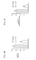
- FIG. 4 A depicts the effects of tyrphostin AG490 on myocardial function.
- FIG. 4 B depicts reduction of infarct size by tyrphostin AG490 during ischemia/reperfusion.
- FIG. 4 C depicts reduction of apoptosis of cardiomyocytes by tyrphostin AG490 during ischemia/reperfusion.
- FIG. 5 depicts up-regulation of angiotensinogen mRNA during ischemia/reperfusion (I/R) is mediated by STATs.
- FIG. 5A Angiotensinogen mRNA is increased during ischemia/reperfusion.
- FIG. 5B St-domain/STAT binding activity is increased in hearts subjected to ischemia/reperfusion.
- FIG. 5C STAT5A and STAT6 are activated in ischemic hearts.
- FIG. 6 A depicts Jak2 inhibition by tyrophostin AG490 during ischemia/reperfusion (I/R).
- FIG. 6 B depicts reduction of St-domain/STAT binding activity by tyrphostin AG490 in hearts subjected to ischemia/reperfusion.
- FIG. 6 C depicts inhibition of angiotensinogen mRNA by tyrphostin AG490 in hearts subjected to ischemia/reperfusion.
- the present invention is based on the discovery by the inventors that a specific signaling pathway is responsible for the onset and maintenance of the renin-angiotensin system in hypertrophy and ischemia.
- the inventors have discovered that the activation of Jak2, during hypertrophy and ischemia, activates specific STAT proteins, specifically STAT5A and STAT6.
- the inventors have discovered that administration of a Jak2 inhibitor significantly reduces the myocardial damage caused by hypertrophy and ischemia.
- the invention relates to a method for reducing hypertrophy of an organ in a mammal at risk for hypertrophy.
- the method comprises administering to said mammal an effective amount of a pharmaceutical composition comprising a selective Jak2 inhibitor.
- Hypertrophy is the enlarging of an organ.
- the increase in size may, for example, be due to an increase in workload due to some physical defect in the organ itself or one of the biological systems supporting the organ.
- Some organs are subject to hypertrophy. Some examples include the heart, kidney, and prostate.
- Myocardial hypertrophy for example, is hypertrophy of the heart, which is typically caused by either myocardial valve damage or high blood pressure. Myocardial hypertrophy may also result from a dilation or expansion of the heart in response to heart muscle damage that causes weak muscle action. Hypertrophic damage may lead, for example, to myocardial infarction, congestive heart failure, and cardiomyopathy.
- Left ventricular hypertrophy is the medical term for enlargement of the left ventricle of the heart.
- the left ventricle is the heart's main pumping chamber, and pumps oxygenated blood via the aorta through the systemic circulation.
- Hypertrophy may be assessed, for example, by any method known to those skilled in the art.
- the weight of the organ relative to the body weight of the mammal may be expressed as a ratio, as described in Example 1 and depicted in FIG. 2 B.
- the invention in another embodiment, relates to a method for reducing ischemia of an organ in a mammal at risk for ischemia.
- the method comprises administering to said mammal an effective amount of a pharmaceutical composition comprising a selective Jak2 inhibitor.
- Ischemia is a deficiency of oxygenated blood.
- the deficiency of blood may, for example, be caused by functional constriction or obstruction of a blood vessel.
- the lack of oxygen and/or reduced availability of nutrient substrates and inadequate removal of metabolites may result in tissue damage, for example, apoptosis and/or necrosis of cells.
- organs are subject to ischemia. Some examples include, but are not limited to, the heart, brain, kidney, and intestines.
- Ischemic heart disease is often caused by a reduction in coronary blood flow relative to myocardial demand.
- the reduction in blood flow may result from a variety of reasons, and typically occurs as a result of atherosclerosis.
- ischemic damage As a result of ischemic damage to the heart muscle, the damaged area ceases to contract. Symptoms of such damage include, but are not limited to, cardiac arrhythmias, angina, myocardial infarction, congestive heart failure, and sudden cardiac death.
- Ischemia may be assessed by any method known to those skilled in the art.
- An assessment of ischemic damage may be made, for example, by measuring the infarct (scar) size of the organ, as described in Example 2 and depicted in FIG. 4 B.
- reducing hypertrophy of an organ means a significant reduction in the size of a hypertrophic organ relative to a healthy organ.
- Reducing ischemia of an organ means a significant reduction in the infarct size of an ischemic organ.
- Hypertrophy or ischemia is considered significantly reduced if the size of the hypertrophic organ or the infarct size of the ischemic organ is reduced by at least 10%, preferably at least about 25%, more preferably at least about 50%, even more preferably at least about 75%, and most preferably about 100%.
- Mammals include, for example, humans, baboons and other primates, as well as pet animals such as dogs and cats, laboratory animals such as rats and mice, and farm animals such as horses, sheep, and cows.
- a mammal at risk for hypertrophy or ischemia may be susceptible for any number or reasons, including a genetic predisposition and/or environmental insult.
- reasons for susceptibility to hypertrophy include, but are not limited to, familial history of high blood pressure, valvular heart disease, and side effects of medication.
- Valvular heart disease includes, for example, congenital heart disease and rheumatic heart disease.
- reasons for susceptibility to ischemia include, but are not limited to, familial history of atherosclerosis, diet and lifestyle, surgical procedures, and side effects of medication.
- a Jak2 inhibitor is any compound that selectively inhibits the phosphorylation of the Jak2 protein in the Jak/STAT pathway.
- the compound may directly inhibit Jak2, or a component upstream of Jak2.
- the inhibition of the Jak2 protein must be sufficient to substantially inhibit and preferably prevent the Jak/STAT cascade.
- the Jak2 inhibitor may be any type of compound.
- the compound may be a small organic molecule or a biological compound, such as an antibody or an enzyme.
- Jak2 inhibitors include some members of a class of small organic molecules called tyrphostins. Tyrphostins inhibit the activity of protein tyrosine kinases and have the basic structure shown below:
- the tyrphostin may be any tyrphostin that selectively inhibits Jak2.
- Some examples of tyrphostins include the various structures described in Meydan et al., (1996) Nature , 379:645-648; Levitzki et al, (1995) Science , 267:1782-1788; and PCT application WO 98/06391. These structures are incorporated herein by reference.
- a preferred class of tyrphostins for use are those wherein:
- R 1 C 6 H 5 —CH 2 —NH
- R 2 and R 3 H, OH, lower alkyl, F, NO 2 , CF 3 , C 6 H 5 —SO 2 , O—R 4 , O—CO—R 4 , or R 4
- R 4 phenyl or lower alkyl
- lower alkyl C 1 -C 4 branched or unbranched alkyl (for example, methyl or ethyl).
- R 2 and R 3 may be the same or different except R 2 and R 3 cannot both be H.
- R 2 and R 3 are OH.
- the preferred substituent for R 1 is C 6 H 5 —CH 2 —NH.
- the preferred compound is known as Tyrphostin AG490, which is a selective, specific, and potent Jak2 protein tyrosine kinase inhibitor.
- the structure of AG490 is shown below:
- the tyrphostins may be made by methods known in the art, for example, as described in the PCT application WO 98/06391. Briefly, the typhostins may be synthesized by knoevenagel condensation of the appropriate benzaldehyde with malononitrile or the appropriate substituted amide.
- a compound is considered a selective inhibitor of Jak2 when the compound inhibits Jak2 activity to an extent significantly greater than it inhibits the activity of other members of the Jak family, e.g., Jak1, Jak3, and Tyk2.
- the selective inhibitor inhibits Jak2 at least 2-fold more than it inhibits other members of the Jak family, more preferably at least about 5-fold more, and most preferably at least about 10-fold more.
- Jak2 inhibitors as defined herein also include pharmaceutically acceptable salts.
- pharmaceutically acceptable salts may be formed by treating the compounds identified above with salt-forming acids and bases which do not substantially increase the toxicity of the compound.
- the Jak2 inhibitor is administered in a pharmaceutical composition.
- the pharmaceutical composition may be manufactured by known means.
- the pharmaceutical compositions are preferably sterile, non-pyrogenic and isotonic preparations, optionally with one or more of the pharmaceutically acceptable additives listed below.
- the pharmaceutical composition may be any composition suitable for pharmaceutical use in a mammal, especially a human.
- the composition may, for example, be in the form of a solid, a solution, or a suspension.
- compositions of the Jak2 inhibitors of the invention are preferably stable compositions which may comprise one or more of the following: a stabilizer, a surfactant, preferably a nonionic surfactant, and optionally a salt and/or a buffering agent.
- the pharmaceutical composition may be in the form of an aqueous solution, or in a lyophilized form.
- the stabilizer may, for example, be an amino acid, such as for instance, glycine; or an oligosaccharide, such as for example, sucrose, tetralose, lactose or a dextran.
- the stabilizer may be a sugar alcohol, such as for instance, mannitol; or a combination thereof.
- the stabilizer or combination of stabilizers constitutes from about 0.1% to about 10% by weight of the Jak2 inhibitor.
- the surfactant is preferably a nonionic surfactant, such as a polysorbate.
- suitable surfactants include Tween20, Tween80; a polyethylene glycol or a polyoxyethylene polyoxypropylene glycol, such as Pluronic F-68 at from about ) 001% (w/v) to about 10% (w/v),
- the salt or buffering agent may be any salt or buffering agent, such as for example, sodium chloride, or sodium/potassium phosphate, respectively.
- the buffering agent maintains the pH of the pharmaceutical composition in the range of about 5.5 to about 7.5.
- the salt and/or buffering agent is also useful to maintain the osmolarity at a level suitable for administration to a human or an animal.
- the salt or buffering agent is present at a roughly isotonic concentration of about 150 mM to about 300 mM.
- the pharmaceutical compositions of the present invention may additionally contain one or more conventional additive.
- additives include a solubilizer such as for example, glycerol; an antioxidant such as for example, benzalkonium chloride (a mixture of quaternary ammonium compounds, known as “quats”), benzyl alcohol, chloretone or chlorobutanol; anaesthetic agent such as for example a morphine derivative; or an isotonic agent etc., such as described above.
- a solubilizer such as for example, glycerol
- an antioxidant such as for example, benzalkonium chloride (a mixture of quaternary ammonium compounds, known as “quats”), benzyl alcohol, chloretone or chlorobutanol
- anaesthetic agent such as for example a morphine derivative
- an isotonic agent etc. such as described above.
- the pharmaceutical compositions may be stored under nitrogen gas in vials sealed with impermeable stoppers.
- an effective amount of a Jak2 inhibitor is the amount which reduces hypertrophy and/or ischemia of the organ.
- Optimal doses can be determined by those skilled in the art based on a number of parameters including, for example, age, sex, weight, severity of the condition being treated, the compound being administered, and the route of administration.
- an effective amount of Jak2 inhibitor can be that amount that would produce a blood serum volume level of between about 0.01 ⁇ M to about 50 ⁇ M, preferably between about 1.0 ⁇ M to about 5 ⁇ M.
- the Jak2 inhibitor can be administered by any suitable method, as is known in the art.
- the Jak2 inhibitor can be administered topically or systemically. Systemic administration is preferred.
- Adminstration using controlled release delivery systems, as is known in the art, is also contemplated herein.
- Systemic administration includes both parenteral and enteral routes.
- Jak2 inhibitors such as tyrphostins can easily be administered intravenously, which is a preferred route of delivery.
- Intravenous administration can be accomplished by mixing the Jak2 inhibitor in a suitable pharmaceutical carrier (vehicle) or excipient as understood by practitioners in the art.
- Oral or enteral administration includes, for example, formulations such as tablets, capsules, pills, troches, elixirs, suspensions, syrups, wafers, chewing gum and the like.
- the Jak2 inhibitor may be administered as a protective agent before hypertrophy and/or ischemia occurs.
- the Jak2 inhibitor may be used as a prophylactic treatment to prevent hypertrophy and/or ischemia in a mammal at risk for hypertrophy and/or ischemia.
- the Jak2 inhibitor may be administered at a time after the hypertrophy and/or ischemia occurs in order to minimize and/or reverse the hypertrophy and/or ischemia, as well as to prevent further damage resulting from hypertrophy and/or ischemia.
- the Jak2 inhibitor When administering the Jak2 inhibitor after hypertrophy and/or ischemia has occurred, it is preferred that the Jak2 inhibitor be administered as soon thereafter as possible. Jak2 can also be administered while the hypertrophy and/or ischemia is occurring.
- This example demonstrates cardioprotection from left ventricular hypertrophy by Tyrphostin AG 490.
- TAC transverse aortic constriction
- FIG. 1 left ventricular hypertrophy
- mice Male C57/BL6 mice, weighing 20 to 24 grams, were anesthetized by intra-peritoneal injection of a cocktail of ketamine (100 mg/kg) and xylazine (5 mg/ml). The mice were shaved, restrained, and orally intubated (under direct vision via a vertical cervical incision) using a 22 guage blunt feeding needle. Respiration was artificially controlled (tidal volume of 0.1 to 0.3 ml) at a respiratory rate of 110 to 150 breaths/minute using a ventilator (Harvard Apparatus Rodent Ventilator, model 683).
- a median stemotomy was performed and the sternum retracted.
- the thymus was retracted anteriorly and the aortic arch identified and ligated (using 8.0 nylon suture; Ethicon) between the innominate and left common carotid artery with an overlying 27-guage needle; and then the needle removed to leave a discrete region of stenosis.
- the chest was then closed in two layers (using 6.0 vicryl suture, Ethicon) and the pneumothorax evacuated.
- Some mice were subjected to a sham operation in which the aortic arch was visualized but not banded. The mice were then extubated and monitored post-op for 3 to 12 hours. The survival rate at the end of the learning period is greater than 90%.
- mice Nine days post-op, the hearts were removed from heparinized (500 U) mice and euthanized with a lethal dose of pentobarbital (150 mg/kg). The hearts were analyzed by visual inspection of a cross-section of the heart (FIG. 2 A), determination of heart to body weight ratio (FIG. 2 B), and light microscopy of the cardiomyocytes in the left ventricular (FIG. 2 C), and activation of artiel natuiretic factor (ANF), a specific molecular marker for hypertrophy (FIG. 3 ). Based on these determinations, all trans-aortic constricted mice developed well-defined left ventricular hypertrophy.
- tyrphostin AG490 To determine whether tyrphostin AG490 could reverse the hypertrophy induced by traverse aortic constriction, tyrphostin AG490 (5 ⁇ M) was administered to the mice, intra-peritoneal, 24 hours before being subjected to transverse aortic constriction and every 24 hours thereafter for the duration of the study (9 days). Chronic administration of tyrophostin AG490 caused a remarkable reversal of hypertrophy (see FIGS. 2A, 2 B, 2 C, and 3 ).
- dp/dt values were markedly higher in groups at both concentrations throughout most of the reperfusion period compared with the control reperfused group, the difference being apparent at R-30 (3818 ⁇ 49.46 and 4156 ⁇ 238 versus 3382 ⁇ 68.8), R-60 (3362 ⁇ 53.14 and 3840 ⁇ 140 versus 2878 ⁇ 237), R-90 (2840 ⁇ 88 and 3194.7 ⁇ 228 versus 1842 ⁇ 162), and R-120 (2552 ⁇ 58.9 and 2626 ⁇ 269 versus 1543 ⁇ 94).
- tyrphostin AG490 To gain insight into the physiological basis for cardioprotection afforded by tyrphostin AG490, the extent of cardiomyocyte infarct size and apoptosis were measured. On termination of treatment with tyrphostin AG490, hearts were immersed in 1% triphenyl tetrazolium solution in phosphate buffer (Na 2 HPO 4 88 mmol/l, NaH 2 PO 4 1.8 mmol/l) for 10 min. at 37° C. and stored at ⁇ 70° C. for processing.
- phosphate buffer Na 2 HPO 4 88 mmol/l, NaH 2 PO 4 1.8 mmol/l
- Frozen hearts (ventricular tissue) were sliced transversely in a plane perpendicular to the apicobasal axis into 0.5 mm thick sections, blotted dry, placed between microscope slides, and scanned on a Hewlett-Packard Scanjet 5p single-pass flatbed scanner.
- NIH 1.61 image processing software each digitized image was subjected to equivalent degrees of background subtraction, brightness, and contrast enhancement for improved clarity and distinctness. Risk (equivalent to total left ventricular muscle mass) and infarct zones of each slice were traced, and the respective areas were calculated in terms of pixels. The weight of each slice was then recorded to facilitate the expression of total and infarct masses of each slice in grams.
- the risk and infarct volumes of each slice in cubic centimeters were then calculated on the basis of slice weight to correct for any errors due to nonuniformity of heart slice thickness.
- the risk volumes and infarct volumes of each slice were summed to obtain the risk and infarct volumes for the whole heart.
- Infarct size was taken to be the percent infarct volume/risk volume for any one heart.
- Immunohistochemical detection of apoptotic cells was carried out by use of terminal dUTP nick end-labeling (TUNEL), in which residues of digoxigenin-labeled dUTP are catalytically incorporated into the DNA by terminal deoxynucleotidyl transferase II.
- TUNEL terminal dUTP nick end-labeling
- the cells were incubated with a sheep polyclonal anti-digoxigenin antibody followed by a FITC-conjugated rabbit anti-sheep IgG as a secondary antibody.
- the heart sections were washed in PBS 3 times, blocked with normal rabbit serum, and incubated with mouse monoclonal antibody recognizing cardiac myosin heavy chain (Biogenesis Ltd) followed by staining with TRIRC-conjugated rabbit anti-mouse IgG (200:1 dilution, Dako, Japan). The fluorescence staining was viewed with a confocal laser microscope (Olympus Co). The apoptoic cells were counted and expressed as percentage of total myocyte population.
- tyrphostin AG490 reduced myocardial infarct size (FIG. 4B) and caused a marked lowering of apoptotic cell death (FIG. 4 C), thereby, attributing, at least in part, to the recovery of contractile function upon treatment with tyrphostin AG490.
- This example demonstrates upregulation of rat heart angiotensinogen mRNA during ischemia/reperfusion.
- Ischemia was induced by a modified Langendorf-reperfusion method in rat hearts.
- Hearts from adult male rats were randomly divided into 4 groups and subjected to ischemia/reperfusion.
- ischemic group hearts were perfused with Krebs-Henseleit buffer for 60 minutes, followed by 30 minutes of global ischemia.
- ischemic/reperfused group hearts were perfused for 60 minutes, followed by 30 minutes of global ischemia and 120 minutes of reperfusion. Control group hearts were perfused for the same lengths of time.
- Rat hearts subjected to ischemia/referfusion were tested to determine whether activation of the renin-angiotensin system, as reflected by an increase in angiotensinogen mRNA, occurs in ischemic injury.
- the level of angiotensinogen mRNA was analyzed by primer extension assay using gene-specific DNA probes.
- a DNA primer spanning the complementary sequence of the rat angiotensinogen cDNA between nucleotides 302 and 279 (5′-AGGAGATGAAAGGGGTGGATGTAT-3′) was end-labeled and used to evaluate the expression of angiotensinogen mRNA in total RNA isolated from the rat heart.
- the primer extension protocol was performed according to instructions provided by the supplier (Progema).
- Rat GAPDH cDNA specific primer was used as control. There was a marked increase in mRNA level after 30 minutes of ischemia and 120 minutes of reperfusion (FIG. 5 A). The increase in mRNA was sensitive to blockage of the AT 1 , receptor, because pretreatment with losartan (L) reduced it almost entirely to the level of the control sample (C). The levels of the ribosomal marker L32 mRNA, used as control, remained unchanged.
- This example demonstrates STAT activation during ischemia/reperfusion.
- Nuclear extracts were examined in hearts subjected to global ischemia to determine whether there is enhanced STAT binding activity to the St domain of the angiotensinogen promoter.
- the nuclear heart extracts were examined by the electrophoretic gel mobility-shift assay that exployed the use of the chemically synthesized oligonucleotide sequence of the St domain.
- the St-domain DNA probe for protein binding was a double-stranded oligonucleotide containing the sequence 5′-GGGTtcCTGGAAGGG-3′ and complementary strand 5′-CCCTTCCAGgaACCC-3′, respectively. These probes were end-labeled by polynucleotide kinase and [ ⁇ - 32 P]ATP.
- Binding reaction mixture containing 0.5 ng of labeled DNA (1,000 cpm), 2 ⁇ g of poly(dI-C), and 1-12 ⁇ g of protein in buffer containing 20 MM Hepes, 3% glycerol, 1.5 mM MgCl 2 , 1 mM DTT, 2 mM EDTA and 50 mM KCl, pH 7.5 was allowed to incubate at 4° C. for 30 min. The reactions were analyzed by electrophoresis on 8% polyacrylamide gel in 0.375 ⁇ TBE (0.33 mM Tris borate, pH 8.7 and 1.0 mM EDTA). After electrophoresis, the gels were dried and subjected to autoradiography.
- This example demonstrates the effect of Jak2 inhibition on STAT/DNA binding and angiotensinogen mRNA.
- Rats were pretreated with 5 or 50 ⁇ mol/L of tyrphostin AG490 24 h prior to ischemia/reperfusion followed by chronic administration of tyrphostin AG490 during the process of ischemia/reperfusion.
- a phosphotyrosine assay was performed. Briefly, nuclear extracts from hearts subjected to ischemia/reperfusion in presence of absence of tyrphostin AG490 were immunoprecipitated with anti-phosphotyrosine antibodies (4G10). Fifty microliters of 50% protein A-agarose, prewashed in lysis buffer (Upstate Biotechnology) was then added and the mixture was incubated for 2 hr at 4° C.
- Each sample was washed with washing buffer containing 150 mM NaCl, 50 mM Tris-HCL (pH 7.4), 5 mM EDTA, 0.25% Triton X-100, 2 mM phenylmethylsulfonyl fluoride, aprotinin (0.2 unit/ml), 1 mM Na 3 VO 4 , and 1 mM NaF.
- Samples were eluted in 2X Laemmli's sample buffer. Proteins were separated on a 7.5% SDS/polyacrylamide gel and transferred to nitrocellulose membrane, Nitropure (Micron Separations, Westboro, Mass.).
- Blots were probed with polyclonal antibody against Jak2 and developed according to the chemiluminescence protocol.
- Administration of tyrphostin AG490 in perfusion medium was inhibitory at both 5 and 50 lmol/L for phosphorylation of Jak2, which was activated readily in the ischemic heart in absence of the inhibitor (FIG. 6 A).
Landscapes
- Health & Medical Sciences (AREA)
- General Health & Medical Sciences (AREA)
- Chemical & Material Sciences (AREA)
- Veterinary Medicine (AREA)
- Pharmacology & Pharmacy (AREA)
- Medicinal Chemistry (AREA)
- Animal Behavior & Ethology (AREA)
- Public Health (AREA)
- Life Sciences & Earth Sciences (AREA)
- Bioinformatics & Cheminformatics (AREA)
- Engineering & Computer Science (AREA)
- Nuclear Medicine, Radiotherapy & Molecular Imaging (AREA)
- General Chemical & Material Sciences (AREA)
- Organic Chemistry (AREA)
- Chemical Kinetics & Catalysis (AREA)
- Cardiology (AREA)
- Epidemiology (AREA)
- Heart & Thoracic Surgery (AREA)
- Urology & Nephrology (AREA)
- Vascular Medicine (AREA)
- Hospice & Palliative Care (AREA)
- Medicines That Contain Protein Lipid Enzymes And Other Medicines (AREA)
- Pharmaceuticals Containing Other Organic And Inorganic Compounds (AREA)
- Acyclic And Carbocyclic Compounds In Medicinal Compositions (AREA)
Abstract
Description


Claims (19)
Priority Applications (13)
| Application Number | Priority Date | Filing Date | Title |
|---|---|---|---|
| US09/945,192 US6433018B1 (en) | 2001-08-31 | 2001-08-31 | Method for reducing hypertrophy and ischemia |
| NZ531693A NZ531693A (en) | 2001-08-31 | 2002-07-23 | Method for reducing hypertension and heart failure |
| US10/488,279 US7235588B2 (en) | 2001-08-31 | 2002-07-23 | Method for reducing hypertension and heart failure |
| BR0212252-9A BR0212252A (en) | 2001-08-31 | 2002-07-23 | Methods to Reduce Hypertension and Heart Failure in a Mammal |
| CA2458798A CA2458798C (en) | 2001-08-31 | 2002-07-23 | Method for reducing hypertension and heart failure |
| PCT/US2002/023444 WO2003020202A2 (en) | 2001-08-31 | 2002-07-23 | Method for reducing hypertension and heart failure |
| EP02753409A EP1427401A4 (en) | 2001-08-31 | 2002-07-23 | METHOD FOR REDUCING HYPERTENSION AND CARDIAC INSUFFICIENCY |
| MXPA04001913A MXPA04001913A (en) | 2001-08-31 | 2002-07-23 | Method for reducing hypertension and heart failure. |
| AU2002313700A AU2002313700B2 (en) | 2001-08-31 | 2002-07-23 | Method for reducing hypertension and heart failure |
| KR10-2004-7003111A KR20040050894A (en) | 2001-08-31 | 2002-07-23 | Method for reducing hypertension and heart failure |
| JP2003524516A JP2005505545A (en) | 2001-08-31 | 2002-07-23 | Methods for reducing hypertension and heart failure |
| ZA2004/02540A ZA200402540B (en) | 2001-08-31 | 2004-03-31 | Method for reducing hypertension and heart failure |
| JP2010180146A JP2010285455A (en) | 2001-08-31 | 2010-08-11 | Methods for reducing hypertension and heart failure |
Applications Claiming Priority (1)
| Application Number | Priority Date | Filing Date | Title |
|---|---|---|---|
| US09/945,192 US6433018B1 (en) | 2001-08-31 | 2001-08-31 | Method for reducing hypertrophy and ischemia |
Related Child Applications (2)
| Application Number | Title | Priority Date | Filing Date |
|---|---|---|---|
| US10/488,279 Continuation-In-Part US7235588B2 (en) | 2001-08-31 | 2002-07-23 | Method for reducing hypertension and heart failure |
| US10488279 Continuation-In-Part | 2002-07-23 |
Publications (1)
| Publication Number | Publication Date |
|---|---|
| US6433018B1 true US6433018B1 (en) | 2002-08-13 |
Family
ID=25482762
Family Applications (2)
| Application Number | Title | Priority Date | Filing Date |
|---|---|---|---|
| US09/945,192 Expired - Lifetime US6433018B1 (en) | 2001-08-31 | 2001-08-31 | Method for reducing hypertrophy and ischemia |
| US10/488,279 Expired - Lifetime US7235588B2 (en) | 2001-08-31 | 2002-07-23 | Method for reducing hypertension and heart failure |
Family Applications After (1)
| Application Number | Title | Priority Date | Filing Date |
|---|---|---|---|
| US10/488,279 Expired - Lifetime US7235588B2 (en) | 2001-08-31 | 2002-07-23 | Method for reducing hypertension and heart failure |
Country Status (11)
| Country | Link |
|---|---|
| US (2) | US6433018B1 (en) |
| EP (1) | EP1427401A4 (en) |
| JP (2) | JP2005505545A (en) |
| KR (1) | KR20040050894A (en) |
| AU (1) | AU2002313700B2 (en) |
| BR (1) | BR0212252A (en) |
| CA (1) | CA2458798C (en) |
| MX (1) | MXPA04001913A (en) |
| NZ (1) | NZ531693A (en) |
| WO (1) | WO2003020202A2 (en) |
| ZA (1) | ZA200402540B (en) |
Cited By (28)
| Publication number | Priority date | Publication date | Assignee | Title |
|---|---|---|---|---|
| US20040029902A1 (en) * | 2002-02-01 | 2004-02-12 | Rajinder Singh | 2,4-Pyrimidinediamine compounds and their uses |
| US20040266661A1 (en) * | 2001-08-31 | 2004-12-30 | Siddiqui Mohammed A. Q. | Method for reducing hypertension and heart failure |
| US20050038450A1 (en) * | 2003-06-03 | 2005-02-17 | Ping Hu | Methods and apparatus for minimally invasive transverse aortic banding |
| US20050153927A1 (en) * | 2003-12-17 | 2005-07-14 | Alcon, Inc. | Use of serum amyloid a gene in diagnosis and treatment of glaucoma and identification of anti-glaucoma agents |
| US20050234049A1 (en) * | 2003-07-30 | 2005-10-20 | Rajinder Singh | Methods of treating or preventing autoimmune diseases with 2, 4-pyrimidinediamine compounds |
| US20050277680A1 (en) * | 2003-12-11 | 2005-12-15 | Board Of Regents, The University Of Texas System | Compounds for treatment of cell proliferative diseases |
| US20060293311A1 (en) * | 2005-06-08 | 2006-12-28 | Rigel Pharmaceuticals, Inc. | Compositions and methods for inhibition of the jak pathway |
| WO2007098507A2 (en) | 2006-02-24 | 2007-08-30 | Rigel Pharmaceuticals, Inc. | Compositions and methods for inhibition of the jak pathway |
| US20070203161A1 (en) * | 2006-02-24 | 2007-08-30 | Rigel Pharmaceuticals, Inc. | Compositions and methods for inhibition of the jak pathway |
| US20070208043A1 (en) * | 2003-12-17 | 2007-09-06 | Alcon, Inc. | Use of Serum Amyloid A Gene in Diagnosis and Treatment of Glaucoma and Identification of Anti-Glaucoma Agents |
| US20070232668A1 (en) * | 2006-03-31 | 2007-10-04 | The Board Of Regents Of The University Of Texas System | Orally Bioavailable Caffeic Acid Related Anticancer Drugs |
| WO2007084861A3 (en) * | 2006-01-13 | 2008-03-20 | Harvard College | Xenohormesis based compositions and methods |
| WO2007101232A3 (en) * | 2006-02-28 | 2008-11-27 | Cytopia Res Pty Ltd | Inhibition of jak2 as a treatment of pulmonary arterial hypertension |
| US20090258864A1 (en) * | 2008-02-15 | 2009-10-15 | Rigel Pharmaceuticals, Inc. | Pyrimidine-2-amine compounds and their use as inhibitors of jak kinases |
| US20090298823A1 (en) * | 2008-04-22 | 2009-12-03 | Portola Pharmaceuticals, Inc. | Inhibitors of protein kinases |
| US20090318407A1 (en) * | 2008-04-16 | 2009-12-24 | Portola Pharmaceuticals, Inc. | Inhibitors of protein kinases |
| US20090318545A1 (en) * | 2008-06-09 | 2009-12-24 | Cornell Reasearch Foundation, Inc. | Mast cell inhibition in diseases of the retina and vitreous |
| US20100113481A1 (en) * | 2003-12-17 | 2010-05-06 | Alcon Research, Ltd. | Use of serum amyloid a gene in diagnosis and treatment of glaucoma and identification of anti-glaucoma agents |
| US20110028503A1 (en) * | 2009-07-28 | 2011-02-03 | Rigel Pharmaceuticals, Inc. | Compositions and methods for inhibition of the jak pathway |
| WO2012061428A2 (en) | 2010-11-01 | 2012-05-10 | Portola Pharmaceuticals, Inc. | Nicotinamides as jak kinase modulators |
| US8637675B2 (en) | 2008-07-08 | 2014-01-28 | Board Of Regents, The University Of Texas System | Inhibitors of proliferation and activation of signal transducer and activators of transcription (STATS) |
| US8952027B2 (en) | 2008-04-16 | 2015-02-10 | Portola Pharmaceuticals, Inc. | Inhibitors of syk and JAK protein kinases |
| US9359308B2 (en) | 2011-11-23 | 2016-06-07 | Portola Pharmaceuticals, Inc. | Pyrazine kinase inhibitors |
| US9573964B2 (en) | 2005-11-16 | 2017-02-21 | Cti Biopharma Corp. | Oxygen linked pyrimidine derivatives |
| US9624242B2 (en) | 2008-12-11 | 2017-04-18 | Cti Biopharma Corp. | 11-2(2-pyrrolidin-1-yl-ethoxy)-14,19-dioxa-5,7,26-triaza-tetracyclo[19.3.1.1(2,6).1(8,12)]heptacosa-1(25),2(26),3,5,8,10,12(27),16,21,23-decaene citrate salt |
| US9676756B2 (en) | 2012-10-08 | 2017-06-13 | Portola Pharmaceuticals, Inc. | Substituted pyrimidinyl kinase inhibitors |
| WO2018002673A1 (en) | 2016-07-01 | 2018-01-04 | N4 Pharma Uk Limited | Novel formulations of angiotensin ii receptor antagonists |
| US10517876B2 (en) | 2005-11-16 | 2019-12-31 | Cti Biopharma Corp. | Oxygen linked pyrimidine derivatives |
Families Citing this family (6)
| Publication number | Priority date | Publication date | Assignee | Title |
|---|---|---|---|---|
| US20050130893A1 (en) * | 2003-09-30 | 2005-06-16 | Joan Smith-Sonneborn | Use of opioids in prevention of and recovery from a stress-induced crash in blood pressure |
| WO2006044694A2 (en) * | 2004-10-15 | 2006-04-27 | Palo Alto Investors | Methods and compositions for treating a disease condition in a subject |
| KR101566840B1 (en) | 2007-03-12 | 2015-11-06 | 와이엠 바이오사이언시즈 오스트레일리아 피티와이 엘티디 | Phenylaminopyrimidine compounds and uses thereof |
| JP5275966B2 (en) * | 2009-12-21 | 2013-08-28 | パナソニック株式会社 | Manufacturing method of fiber board |
| US8809359B2 (en) | 2012-06-29 | 2014-08-19 | Ym Biosciences Australia Pty Ltd | Phenyl amino pyrimidine bicyclic compounds and uses thereof |
| TWI729644B (en) | 2014-06-12 | 2021-06-01 | 美商西爾拉癌症醫學公司 | N-(cyanomethyl)-4-(2-(4-morpholinophenylamino)pyrimidin-4-yl)benzamide |
Citations (3)
| Publication number | Priority date | Publication date | Assignee | Title |
|---|---|---|---|---|
| US5573772A (en) | 1993-03-15 | 1996-11-12 | Gensia, Inc. | Methods for protecting tissues and organs from ischemic damage |
| WO1998006391A1 (en) | 1996-08-14 | 1998-02-19 | Mor-Research Applications Ltd. | Pharmaceutical compositions comprising tyrphostins |
| US6255296B1 (en) | 1994-01-11 | 2001-07-03 | Endomatrix, Inc. | Composition and method for treating a patient susceptible to or suffering from a cardiovascular disorder or disease |
Family Cites Families (5)
| Publication number | Priority date | Publication date | Assignee | Title |
|---|---|---|---|---|
| US5138069A (en) * | 1986-07-11 | 1992-08-11 | E. I. Du Pont De Nemours And Company | Angiotensin II receptor blocking imidazoles |
| CA2012634A1 (en) * | 1990-03-20 | 1991-09-20 | Hassan Salari | Tyrphostins for treatment of allergic, inflammatory and cardiovascular diseases |
| US6358954B1 (en) | 1999-11-09 | 2002-03-19 | Yissum Research Development Company Of The Hebrew University Of Jerusalem | PDGF receptor kinase inhibitory compounds, their preparation, purification and pharmaceutical compositions including same |
| US6433018B1 (en) * | 2001-08-31 | 2002-08-13 | The Research Foundation Of State University Of New York | Method for reducing hypertrophy and ischemia |
| DE602006000402T8 (en) * | 2006-06-02 | 2009-04-09 | Teva Pharmaceutical Industries Ltd. | Stable preparation containing a moisture-sensitive active substance and process for the preparation of the preparation. |
-
2001
- 2001-08-31 US US09/945,192 patent/US6433018B1/en not_active Expired - Lifetime
-
2002
- 2002-07-23 KR KR10-2004-7003111A patent/KR20040050894A/en not_active Ceased
- 2002-07-23 CA CA2458798A patent/CA2458798C/en not_active Expired - Fee Related
- 2002-07-23 MX MXPA04001913A patent/MXPA04001913A/en active IP Right Grant
- 2002-07-23 BR BR0212252-9A patent/BR0212252A/en not_active IP Right Cessation
- 2002-07-23 NZ NZ531693A patent/NZ531693A/en not_active IP Right Cessation
- 2002-07-23 AU AU2002313700A patent/AU2002313700B2/en not_active Ceased
- 2002-07-23 JP JP2003524516A patent/JP2005505545A/en active Pending
- 2002-07-23 WO PCT/US2002/023444 patent/WO2003020202A2/en not_active Ceased
- 2002-07-23 EP EP02753409A patent/EP1427401A4/en not_active Withdrawn
- 2002-07-23 US US10/488,279 patent/US7235588B2/en not_active Expired - Lifetime
-
2004
- 2004-03-31 ZA ZA2004/02540A patent/ZA200402540B/en unknown
-
2010
- 2010-08-11 JP JP2010180146A patent/JP2010285455A/en active Pending
Patent Citations (3)
| Publication number | Priority date | Publication date | Assignee | Title |
|---|---|---|---|---|
| US5573772A (en) | 1993-03-15 | 1996-11-12 | Gensia, Inc. | Methods for protecting tissues and organs from ischemic damage |
| US6255296B1 (en) | 1994-01-11 | 2001-07-03 | Endomatrix, Inc. | Composition and method for treating a patient susceptible to or suffering from a cardiovascular disorder or disease |
| WO1998006391A1 (en) | 1996-08-14 | 1998-02-19 | Mor-Research Applications Ltd. | Pharmaceutical compositions comprising tyrphostins |
Non-Patent Citations (15)
| Title |
|---|
| Alexander Levitzki and Aviv Gazit, "Tyrosine Kinase Inhibition: An Approach to Drug Development," Science (1995) 267: 1782-1788. |
| Alexander Levitzki, "Protein Tyrosine Kinase Inhibitors as Novel Therapeutic Agents," Pharmacol. Ther. (1999) 82/2-3: 231-239. |
| Alexander Levitzki, "Tyrphostins-Potential Antiproliferative Agents and Novel Molecular Tools," Biochemical Pharmacology (1990) 40/5: 913-918. |
| Cindy Ruwhof, Arnoud van der Laarse, "Mechanical stress-induced cardiac hypertrophy: mechanisms and signal transduction pathways," Cardiovascular Research (2000) 47: 23-37. |
| Diane H. Boschelli, "Small molecule inhibitors of receptor tyrosine kinases," www.prous.com/journals/dof/sample/html/df240515/df240515.html. Date of publication unknown. |
| DL Beckles, E. Mascareno, and MAQ Siddiqui, "Attenuation of Cardiac Hypertrophy in vivo by Interference with Activation of the Jak/Stat Pathway," presented at the XVII World Congress of the International Society for Heart Research, Winnipeg, Manitoba, Canada (Jul. 6-11, 2001). |
| Domenica Altavilla, Francesco Squadrito, Giuseppe M. Campo, Antonino Saitta, Giovanni Squadrito, Cristina Quartarone, Barbara Deodato, Mariarita Arlotta, Marcella Ferlito, Letteria Minutoli, Michelangelo Tringali, Giuseppe Urna, Aurora Sardella, Achille P. Caputi, "The reduction of myocardial damage and leukocyte polymorphonuclear accumulation following coronary artery occlusion by the tyrosine kinase inhibitor tyrphostin AG 556," Life Sciences (2000) 67: 2615-2629. |
| Eduardo Mascareno and M.A.Q. Siddiqui, "The role of Jak/STAT signaling in heart tissue renin-angiotensin system," Molecular and Cellular Biochemistry (2000) 212: 171-175. |
| Eduardo Mascareno, Manya Dhar, and M.A.Q. Siddiqui, "Signal transduction and activator of transcription (STAT) protein-dependent activation of angiotensinogen promoter: A cellular signal for hypertrophy in cardiac muscle, " Proc. Natl. Adac. Sci. USA (1998) 95: 5590-5594. |
| Eduardo Mascareno, PhD; Mohammed El-Shafei, MD; Nilanjana Maulik, Phd; M.Sato, MD; Yueling Guo; Dipak Das, PhD; M.A.Q. Siddiqui, PhD, "JAK/STAT Signaling Is Associated With Cardiac Dysfunction During Ischemia and Reperfusion," Circulation (2001) 104:1-5. |
| Michael M. Givertz, MD, "Manipulation of the Renin-Angiotensin System," Circulation (2001) 104: e14-e18. |
| Naftaly Meydan, Tom Grunberger, Harjit Dadi, Michal Shahar, Enrico Arpaia, Zvi Lapidot, J. Steven Leeder, Melvin Freedman, Amos Cohen, Aviv Gazit, Alexander Levitzki and Chaim M. Roifman, "Inhibition of acute lymophoblastic leukaemia by a Jak-2 inhibitor," Nature (1996) 379:645-648. |
| Shao-Ling Zhang, Catherine To, Xing Chen, Janos G. Filep, Shiow-Shih Tang, Julie R. Ingelfinger, Serge Carrière, John S.D. Chan, "Effect of Renin-Angiotensin System Blockade on the Expression of the Angiotensinogen Gene and Induction of Hypertrophy in Rat Kidney Proximal Tubular Cells," Experimental Nephrology (2001) 9: 109-117. |
| Shinji Negoro, Keita Kunisada, Erioh Tone, Masanobu Funamoto, Hidemasa Oh, Tadamitsu Kishimoto, Keiko Yamauchi-Takihara, "Activation of JAK/STAT pathway transduces cytoprotective signal in rat acute myocardial infarction," Cardiovascular Research (2000) 47: 797-805. |
| Stephanie W. Watts, Jennifer A. Florian and Kimberly M. Monroe, "Dissociation of Angiotensin II-Stimulated Activation of Mitogen-Activated Protein Kinase Kinase from Vascular Contraction," The Journal of Pharmacology and Experimental Therapeutics (1998) 286/3: 1431-1438. |
Cited By (87)
| Publication number | Priority date | Publication date | Assignee | Title |
|---|---|---|---|---|
| US7235588B2 (en) * | 2001-08-31 | 2007-06-26 | The Research Foundation Of State University Of New York | Method for reducing hypertension and heart failure |
| US20040266661A1 (en) * | 2001-08-31 | 2004-12-30 | Siddiqui Mohammed A. Q. | Method for reducing hypertension and heart failure |
| US7803939B2 (en) | 2002-02-01 | 2010-09-28 | Rigel Pharmaceuticals, Inc. | 2,4-pyrimidinediamine compounds and their uses |
| US8334296B2 (en) | 2002-02-01 | 2012-12-18 | Rigel Pharmaceuticals, Inc. | 2,4-pyrimidinediamine compounds and their uses |
| US7655797B2 (en) | 2002-02-01 | 2010-02-02 | Rigel Pharmaceuticals, Inc. | Intermediates for making 2,4-pyrimidinediamine compounds |
| US7557210B2 (en) | 2002-02-01 | 2009-07-07 | Rigel Pharmaceuticals, Inc. | 2,4-pyrimidinediamine compounds and their uses |
| US20090171085A1 (en) * | 2002-02-01 | 2009-07-02 | Rigel Pharmaceuticals, Inc. | 2,4-pyrimidinediamine compounds and their uses |
| US20090156622A1 (en) * | 2002-02-01 | 2009-06-18 | Rigel Pharmaceuticals, Inc. | 2,4-pyrimidinediamine compounds and their uses |
| US20040029902A1 (en) * | 2002-02-01 | 2004-02-12 | Rajinder Singh | 2,4-Pyrimidinediamine compounds and their uses |
| US7435814B2 (en) | 2002-02-01 | 2008-10-14 | Rigel Pharmaceuticals, Inc. | 2,4-Pyrimidinediamine compounds and their uses |
| US20090082567A1 (en) * | 2002-02-01 | 2009-03-26 | Rigel Pharmaceuticals, Inc. | 2,4-pyrimidinediamine compounds and their uses |
| US20050038450A1 (en) * | 2003-06-03 | 2005-02-17 | Ping Hu | Methods and apparatus for minimally invasive transverse aortic banding |
| US8178671B2 (en) | 2003-07-30 | 2012-05-15 | Rigel Pharmaceuticals, Inc. | Methods of treating or preventing autoimmune diseases with 2, 4-pyrimidinediamine compounds |
| US7582648B2 (en) | 2003-07-30 | 2009-09-01 | Rigel Pharmaceuticals, Inc. | Methods of treating or preventing autoimmune diseases with 2,4-pyrimidinediamine compounds |
| US20050234049A1 (en) * | 2003-07-30 | 2005-10-20 | Rajinder Singh | Methods of treating or preventing autoimmune diseases with 2, 4-pyrimidinediamine compounds |
| US7452879B2 (en) | 2003-07-30 | 2008-11-18 | Rigel Pharmaceuticals, Inc. | Methods of treating or preventing autoimmune diseases with 2,4-pyrimidinediamine compounds |
| US8119827B2 (en) | 2003-12-11 | 2012-02-21 | Board Of Regents, The University Of Texas System | Compounds for treatment of cell proliferative diseases |
| US9096499B2 (en) | 2003-12-11 | 2015-08-04 | Board Of Regents, The University Of Texas System | Compounds for treatment of cell proliferative diseases |
| US20050277680A1 (en) * | 2003-12-11 | 2005-12-15 | Board Of Regents, The University Of Texas System | Compounds for treatment of cell proliferative diseases |
| US20110021805A1 (en) * | 2003-12-11 | 2011-01-27 | The Board Of Regents Of The University Of Texas System | Compounds for treatment of cell proliferative diseases |
| US8648102B2 (en) | 2003-12-11 | 2014-02-11 | Board Of Regents, The University Of Texas System | Compounds for treatment of cell proliferative diseases |
| EP2487156A1 (en) | 2003-12-11 | 2012-08-15 | Board Of Regents The University Of Texas System | Compounds for treatment of cell proliferative diseases |
| US7745468B2 (en) | 2003-12-11 | 2010-06-29 | Board of Regents, University of Texas Systems The | Compounds for treatment of cell proliferative diseases |
| US20100113481A1 (en) * | 2003-12-17 | 2010-05-06 | Alcon Research, Ltd. | Use of serum amyloid a gene in diagnosis and treatment of glaucoma and identification of anti-glaucoma agents |
| US20090036371A1 (en) * | 2003-12-17 | 2009-02-05 | Alcon, Inc. | Use of Serum Amyloid A Gene in Diagnosis and Treatment of Glaucoma and Identification of Anti-Glaucoma Agents |
| US20070208043A1 (en) * | 2003-12-17 | 2007-09-06 | Alcon, Inc. | Use of Serum Amyloid A Gene in Diagnosis and Treatment of Glaucoma and Identification of Anti-Glaucoma Agents |
| WO2005060542A3 (en) * | 2003-12-17 | 2006-10-26 | Alcon Inc | Use of serum amyloid a gene in diagnosis and treatment of glaucoma and identification of anti-glaucoma agents |
| US7357931B2 (en) | 2003-12-17 | 2008-04-15 | Alcon, Inc. | Use of serum amyloid a gene in diagnosis and treatment of glaucoma and identification of anti-glaucoma agents |
| US20050153927A1 (en) * | 2003-12-17 | 2005-07-14 | Alcon, Inc. | Use of serum amyloid a gene in diagnosis and treatment of glaucoma and identification of anti-glaucoma agents |
| US7662389B2 (en) | 2003-12-17 | 2010-02-16 | Alcon, Inc. | Use of serum amyloid A gene in diagnosis and treatment of glaucoma and identification of anti-glaucoma agents |
| US20080221089A1 (en) * | 2005-06-08 | 2008-09-11 | Rigel Pharmaceuticals, Inc. | Compositions and methods for inhibition of the jak pathway |
| US10421752B2 (en) | 2005-06-08 | 2019-09-24 | Rigel Pharmaceuticals, Inc. | Compositions and methods for inhibition of the JAK pathway |
| US11198689B2 (en) | 2005-06-08 | 2021-12-14 | Rigel Pharmaceuticals, Inc. | Compositions and methods for inhibition of the JAK pathway |
| US11827628B2 (en) | 2005-06-08 | 2023-11-28 | Rigel Pharmaceuticals, Inc. | Compositions and methods for inhibition of the JAK pathway |
| US9248190B2 (en) | 2005-06-08 | 2016-02-02 | Rigel Pharmaceuticals, Inc. | Compositions and methods for inhibition of the JAK pathway |
| US7491732B2 (en) | 2005-06-08 | 2009-02-17 | Rigel Pharmaceuticals, Inc. | Compositions and methods for inhibition of the JAK pathway |
| US20090041786A1 (en) * | 2005-06-08 | 2009-02-12 | Rigel Pharmaceuticals, Inc. | Compositions and methods for inhibition of the jak pathway |
| US20080306099A1 (en) * | 2005-06-08 | 2008-12-11 | Rigel Pharmaceuticals, Inc. | Compositions and methods for inhibition of the jak pathway |
| US9593082B2 (en) | 2005-06-08 | 2017-03-14 | Rigel Pharmaceuticals, Inc. | Compositions and methods for inhibition of the JAK pathway |
| US8815848B2 (en) | 2005-06-08 | 2014-08-26 | Rigel Pharmaceuticals, Inc. | Compositions and methods for inhibition of the JAK pathway |
| US20060293311A1 (en) * | 2005-06-08 | 2006-12-28 | Rigel Pharmaceuticals, Inc. | Compositions and methods for inhibition of the jak pathway |
| US9732073B2 (en) | 2005-06-08 | 2017-08-15 | Rigel Pharmaceuticals, Inc. | Compositions and methods for inhibition of the JAK pathway |
| US8415365B2 (en) | 2005-06-08 | 2013-04-09 | Rigel Pharmaceuticals, Inc. | Compositions and methods for inhibition of the JAK pathway |
| US8399472B2 (en) | 2005-06-08 | 2013-03-19 | Rigel Pharmaceuticals, Inc. | Compositions and methods for inhibition of the JAK pathway |
| US11135227B2 (en) | 2005-11-16 | 2021-10-05 | Cti Biopharma Corp. | Oxygen linked pyrimidine derivatives |
| US10517876B2 (en) | 2005-11-16 | 2019-12-31 | Cti Biopharma Corp. | Oxygen linked pyrimidine derivatives |
| US9573964B2 (en) | 2005-11-16 | 2017-02-21 | Cti Biopharma Corp. | Oxygen linked pyrimidine derivatives |
| WO2007084861A3 (en) * | 2006-01-13 | 2008-03-20 | Harvard College | Xenohormesis based compositions and methods |
| WO2007098507A2 (en) | 2006-02-24 | 2007-08-30 | Rigel Pharmaceuticals, Inc. | Compositions and methods for inhibition of the jak pathway |
| US20070203162A1 (en) * | 2006-02-24 | 2007-08-30 | Rigel Pharmaceuticals, Inc. | Compositions and methods for inhibition of the jak pathway |
| US8962643B2 (en) | 2006-02-24 | 2015-02-24 | Rigel Pharmaceuticals, Inc. | Compositions and methods for inhibition of the JAK pathway |
| US20070203161A1 (en) * | 2006-02-24 | 2007-08-30 | Rigel Pharmaceuticals, Inc. | Compositions and methods for inhibition of the jak pathway |
| US11667611B2 (en) | 2006-02-24 | 2023-06-06 | Rigel Pharmaceuticals, Inc. | Compositions and methods for inhibition of the JAK pathway |
| US20110117159A1 (en) * | 2006-02-28 | 2011-05-19 | Zisman Lawrence S | Inhibition of jak2 as a treatment of pulmonary arterial hypertension |
| WO2007101232A3 (en) * | 2006-02-28 | 2008-11-27 | Cytopia Res Pty Ltd | Inhibition of jak2 as a treatment of pulmonary arterial hypertension |
| US8410173B2 (en) | 2006-02-28 | 2013-04-02 | Ym Biosciences Australia Pty Ltd | Inhibition of JAK2 as a treatment of pulmonary arterial hypertension |
| EP2514742A2 (en) | 2006-03-31 | 2012-10-24 | The Board of Regents of The University of Texas System | Orally bioavailable cafeic acid related anticancer drugs |
| US8779151B2 (en) | 2006-03-31 | 2014-07-15 | The Board Of Regents Of The University Of Texas System | Orally bioavailable caffeic acid related anticancer drugs |
| US20070232668A1 (en) * | 2006-03-31 | 2007-10-04 | The Board Of Regents Of The University Of Texas System | Orally Bioavailable Caffeic Acid Related Anticancer Drugs |
| US9624229B2 (en) | 2008-02-15 | 2017-04-18 | Rigel Pharmaceuticals, Inc. | Pyrimidine-2-amine compounds and their use as inhibitors of JAK kinases |
| US8735418B2 (en) | 2008-02-15 | 2014-05-27 | Rigel Pharmaceuticals, Inc. | Pyrimidine-2-amine compounds and their use as inhibitors of JAK kinases |
| US8309566B2 (en) | 2008-02-15 | 2012-11-13 | Rigel Pharmaceuticals, Inc. | Pyrimidine-2-amine compounds and their use as inhibitors of JAK kinases |
| US20090258864A1 (en) * | 2008-02-15 | 2009-10-15 | Rigel Pharmaceuticals, Inc. | Pyrimidine-2-amine compounds and their use as inhibitors of jak kinases |
| US8138339B2 (en) | 2008-04-16 | 2012-03-20 | Portola Pharmaceuticals, Inc. | Inhibitors of protein kinases |
| US8501944B2 (en) | 2008-04-16 | 2013-08-06 | Portola Pharmaceuticals, Inc. | Inhibitors of protein kinases |
| US20090318407A1 (en) * | 2008-04-16 | 2009-12-24 | Portola Pharmaceuticals, Inc. | Inhibitors of protein kinases |
| US11414410B2 (en) | 2008-04-16 | 2022-08-16 | Alexion Pharmaceuticals, Inc. | Inhibitors of protein kinases |
| US8952027B2 (en) | 2008-04-16 | 2015-02-10 | Portola Pharmaceuticals, Inc. | Inhibitors of syk and JAK protein kinases |
| US9579320B2 (en) | 2008-04-16 | 2017-02-28 | Portola Pharmaceuticals, Inc. | Inhibitors of syk and JAK protein kinases |
| US8937070B2 (en) | 2008-04-16 | 2015-01-20 | Portola Pharmaceuticals, Inc. | Inhibitors of protein kinases |
| US10533001B2 (en) | 2008-04-16 | 2020-01-14 | Portola Pharmaceuticals, Inc. | Inhibitors of protein kinases |
| US9868729B2 (en) | 2008-04-16 | 2018-01-16 | Portola Pharmaceuticals, Inc. | Inhibitors of protein kinases |
| US9139581B2 (en) | 2008-04-22 | 2015-09-22 | Portola Pharmaceuticals, Inc. | Inhibitors of protein kinases |
| US20090298823A1 (en) * | 2008-04-22 | 2009-12-03 | Portola Pharmaceuticals, Inc. | Inhibitors of protein kinases |
| US8258144B2 (en) | 2008-04-22 | 2012-09-04 | Portola Pharmaceuticals, Inc. | Inhibitors of protein kinases |
| US10517839B2 (en) | 2008-06-09 | 2019-12-31 | Cornell University | Mast cell inhibition in diseases of the retina and vitreous |
| US20090318545A1 (en) * | 2008-06-09 | 2009-12-24 | Cornell Reasearch Foundation, Inc. | Mast cell inhibition in diseases of the retina and vitreous |
| US8637675B2 (en) | 2008-07-08 | 2014-01-28 | Board Of Regents, The University Of Texas System | Inhibitors of proliferation and activation of signal transducer and activators of transcription (STATS) |
| US9000179B2 (en) | 2008-07-08 | 2015-04-07 | Board Of Regents, The University Of Texas System | Inhibitors of proliferation and activation of signal transducer and activator of transcription (STATs) |
| US9624242B2 (en) | 2008-12-11 | 2017-04-18 | Cti Biopharma Corp. | 11-2(2-pyrrolidin-1-yl-ethoxy)-14,19-dioxa-5,7,26-triaza-tetracyclo[19.3.1.1(2,6).1(8,12)]heptacosa-1(25),2(26),3,5,8,10,12(27),16,21,23-decaene citrate salt |
| US20110028503A1 (en) * | 2009-07-28 | 2011-02-03 | Rigel Pharmaceuticals, Inc. | Compositions and methods for inhibition of the jak pathway |
| US9102625B2 (en) | 2010-11-01 | 2015-08-11 | Portola Pharmaceuticals, Inc. | Nicotinamides as JAK kinase modulators |
| WO2012061428A2 (en) | 2010-11-01 | 2012-05-10 | Portola Pharmaceuticals, Inc. | Nicotinamides as jak kinase modulators |
| EP2975027A1 (en) | 2010-11-01 | 2016-01-20 | Portola Pharmaceuticals, Inc. | Nicotinamides as jak kinase modulators |
| US9359308B2 (en) | 2011-11-23 | 2016-06-07 | Portola Pharmaceuticals, Inc. | Pyrazine kinase inhibitors |
| US9676756B2 (en) | 2012-10-08 | 2017-06-13 | Portola Pharmaceuticals, Inc. | Substituted pyrimidinyl kinase inhibitors |
| WO2018002673A1 (en) | 2016-07-01 | 2018-01-04 | N4 Pharma Uk Limited | Novel formulations of angiotensin ii receptor antagonists |
Also Published As
| Publication number | Publication date |
|---|---|
| AU2002313700B2 (en) | 2007-04-26 |
| WO2003020202A2 (en) | 2003-03-13 |
| CA2458798C (en) | 2013-06-25 |
| JP2010285455A (en) | 2010-12-24 |
| NZ531693A (en) | 2006-05-26 |
| US20040266661A1 (en) | 2004-12-30 |
| MXPA04001913A (en) | 2004-06-18 |
| EP1427401A2 (en) | 2004-06-16 |
| JP2005505545A (en) | 2005-02-24 |
| WO2003020202A3 (en) | 2003-08-28 |
| ZA200402540B (en) | 2005-06-29 |
| EP1427401A4 (en) | 2009-04-29 |
| CA2458798A1 (en) | 2003-03-13 |
| KR20040050894A (en) | 2004-06-17 |
| US7235588B2 (en) | 2007-06-26 |
| BR0212252A (en) | 2004-09-14 |
Similar Documents
| Publication | Publication Date | Title |
|---|---|---|
| US6433018B1 (en) | Method for reducing hypertrophy and ischemia | |
| AU2002313700A1 (en) | Method for reducing hypertension and heart failure | |
| Daehn et al. | The glomerular filtration barrier: a structural target for novel kidney therapies | |
| Dell'italia et al. | Volume-overload cardiac hypertrophy is unaffected by ACE inhibitor treatment in dogs | |
| Vogler et al. | Pre-and post-conditional inhibition of prolyl-4-hydroxylase domain enzymes protects the heart from an ischemic insult | |
| JP2003238445A (en) | Inhibition of histone deacetylase as a treatment for cardiac hypertrophy | |
| JP2023519140A (en) | PCSK9 antagonist | |
| CZ330896A3 (en) | Use of a fabric preventing or minimizing oxidation of polyunsaturated fatty acid and processes connected with such use | |
| KR20010031179A (en) | The use of inhibitors of the renin-angiotensin system | |
| WO2017147532A1 (en) | Alpha-1-adrenergic receptor agonist therapy | |
| Ikegaki et al. | Involvement of Rho-kinase in vascular remodeling caused by long-term inhibition of nitric oxide synthesis in rats | |
| El-Sayed et al. | Contribution of angiotensin II in hepatic ischemia/reperfusion induced lung injury: Acute versus chronic usage of captopril | |
| CN103429242A (en) | Methods and compositions for preventing and treating cardiac hypertrophy | |
| US20160199463A1 (en) | Hdac2 defends vascular endothelium from injury | |
| US9132174B2 (en) | Treatment of heart disease by inhibition of the action of ribosomal S6 kinase 3 (RSK3) | |
| JP2004512361A (en) | Use of a 20-HETE synthase inhibitor as a treatment for cerebrovascular disease | |
| US20090305225A1 (en) | Inhibition of Creatine Uptake to Promote Weight Loss | |
| US20120276012A1 (en) | Method of Treatment and Screening Method | |
| US10213422B2 (en) | Compositions and methods of inhibiting histone deacetylases | |
| US20250290066A1 (en) | COMPOSITIONS FOR THE SILENCING OF snoRNAS AND METHODS OF USING SAME | |
| Kirshenbaum et al. | Biochemistry of hypertrophy and heart failure | |
| WO2014070706A1 (en) | Control of cardiac growth, differentiation and hypertrophy | |
| Kugathasan | The Protective Role of the Angiopoetin-1-Tie2 System in Transgenic Models of Pulmonary Arterial Hypertension | |
| MÜLLER-WERDAN¹ et al. | ROLE OF CYTOKINES IN SEPTIC CARDIOΜΥΟΡΑΤHY | |
| LaRocca et al. | The Na/Ca 2 exchanger-1 protects against 3 systolic failure in the Akita ins2 model of diabetic 4 cardiomyopathy via a CXCR4/NF-κB pathway 5 |
Legal Events
| Date | Code | Title | Description |
|---|---|---|---|
| AS | Assignment |
Owner name: THE RESEARCH FOUNDATION OF STATE UNIVERSITY OF NEW Free format text: ASSIGNMENT OF ASSIGNORS INTEREST;ASSIGNORS:SIDDIQUI, MOHAMMED A. Q.;MASCARENO, EDUARDO;BECKLES, DANIEL L.;REEL/FRAME:012477/0911;SIGNING DATES FROM 20011025 TO 20011029 |
|
| STCF | Information on status: patent grant |
Free format text: PATENTED CASE |
|
| CC | Certificate of correction | ||
| FPAY | Fee payment |
Year of fee payment: 4 |
|
| AS | Assignment |
Owner name: NATIONAL INSTITUTES OF HEALTH (NIH), U.S. DEPT. OF Free format text: EXECUTIVE ORDER 9424, CONFIRMATORY LICENSE;ASSIGNOR:STATE UNIVERSITY OF NEW YORK;REEL/FRAME:021031/0244 Effective date: 20020304 |
|
| FPAY | Fee payment |
Year of fee payment: 8 |
|
| FPAY | Fee payment |
Year of fee payment: 12 |