US6284253B1 - Feline immunodeficiency virus (FIV) nucleotide sequence - Google Patents
Feline immunodeficiency virus (FIV) nucleotide sequence Download PDFInfo
- Publication number
- US6284253B1 US6284253B1 US09/238,303 US23830399A US6284253B1 US 6284253 B1 US6284253 B1 US 6284253B1 US 23830399 A US23830399 A US 23830399A US 6284253 B1 US6284253 B1 US 6284253B1
- Authority
- US
- United States
- Prior art keywords
- leu
- ile
- lys
- gly
- glu
- Prior art date
- Legal status (The legal status is an assumption and is not a legal conclusion. Google has not performed a legal analysis and makes no representation as to the accuracy of the status listed.)
- Expired - Lifetime
Links
Images


Classifications
-
- C—CHEMISTRY; METALLURGY
- C07—ORGANIC CHEMISTRY
- C07K—PEPTIDES
- C07K14/00—Peptides having more than 20 amino acids; Gastrins; Somatostatins; Melanotropins; Derivatives thereof
- C07K14/005—Peptides having more than 20 amino acids; Gastrins; Somatostatins; Melanotropins; Derivatives thereof from viruses
-
- C—CHEMISTRY; METALLURGY
- C07—ORGANIC CHEMISTRY
- C07K—PEPTIDES
- C07K16/00—Immunoglobulins [IG], e.g. monoclonal or polyclonal antibodies
- C07K16/08—Immunoglobulins [IG], e.g. monoclonal or polyclonal antibodies against material from viruses
- C07K16/10—RNA viruses
- C07K16/112—Retroviridae (F), e.g. leukemia viruses
- C07K16/114—Lentivirus (G), e.g. human immunodeficiency virus [HIV], feline immunodeficiency virus [FIV] or simian immunodeficiency virus [SIV]
-
- A—HUMAN NECESSITIES
- A61—MEDICAL OR VETERINARY SCIENCE; HYGIENE
- A61K—PREPARATIONS FOR MEDICAL, DENTAL OR TOILETRY PURPOSES
- A61K39/00—Medicinal preparations containing antigens or antibodies
- A61K2039/51—Medicinal preparations containing antigens or antibodies comprising whole cells, viruses or DNA/RNA
- A61K2039/53—DNA (RNA) vaccination
-
- A—HUMAN NECESSITIES
- A61—MEDICAL OR VETERINARY SCIENCE; HYGIENE
- A61K—PREPARATIONS FOR MEDICAL, DENTAL OR TOILETRY PURPOSES
- A61K39/00—Medicinal preparations containing antigens or antibodies
-
- C—CHEMISTRY; METALLURGY
- C12—BIOCHEMISTRY; BEER; SPIRITS; WINE; VINEGAR; MICROBIOLOGY; ENZYMOLOGY; MUTATION OR GENETIC ENGINEERING
- C12N—MICROORGANISMS OR ENZYMES; COMPOSITIONS THEREOF; PROPAGATING, PRESERVING, OR MAINTAINING MICROORGANISMS; MUTATION OR GENETIC ENGINEERING; CULTURE MEDIA
- C12N2740/00—Reverse transcribing RNA viruses
- C12N2740/00011—Details
- C12N2740/10011—Retroviridae
- C12N2740/15011—Lentivirus, not HIV, e.g. FIV, SIV
- C12N2740/15022—New viral proteins or individual genes, new structural or functional aspects of known viral proteins or genes
Definitions
- the present invention relates generally to detection of and vaccination against Feline immunodeficiency virus (FIV). More particularly, the invention relates to a highly cytopathic and infectious proviral clone constructed from the genomic DNA of a Pallas's cat FIV. The nucleotide sequences, antigens and chimeric viruses derived from the reconstructed clone can be used for the detection of and protection against FIV.
- FIV Feline immunodeficiency virus
- Feline immunodeficiency virus a lentivirus of cats is associated with feline acquired immunodeficiency syndrome (AIDS) (see Pederson et al., 1987, Science 235:790). Under natural conditions, cats experience an asymptomatic carrier state for years following initial FIV infection before developing an AIDS like disease. Cats experimentally infected with FIV exhibit signs of acute infection which resolve over a few months. Disorders associated with FIV infection include abortion, alopecia, anemia, gingivitis/stomatitis, upper respiratory infections, chronic enteritis, diarrhea, neurological abnormalities, and recurrent ocular disease, see R. English et al., 1990, J. Am. Vet. Med.
- FIV and the human immunodeficiency virus, HIV-1 belong to the lentivirus subfamily of retroviruses and have similar morphology, protein composition and Mg++ dependency of their reverse transcriptases (RT).
- RT reverse transcriptases
- FIV infection in cats provides a valuable animal model for human immunodeficiency virus-1 (HIV-1) induced AIDS.
- HIV-1 human immunodeficiency virus-1
- the pathogenesis of HIV-1 infection has been attributed to virus-induced reduction of CD4+ lymphocyte numbers and function, resulting in decreased immune responsiveness and subsequently severe secondary infections (see M. McChesney and M. Oldstone, 1989, Ad.Immunol., 4:335).
- feline T-lymphotrophic lentivirus now known as Feline Immunodeficiency virus
- Feline Immunodeficiency virus feline T-lymphotrophic lentivirus
- Cloning and sequence analysis of FIV have been reported by Olmsted et al., 1989. Proc Natl. Acad. Sci. USA. 86:4355-4360; and Talbott et al., 1989, Proc. Natl. Acad. Sci., USA 86:5743-5747.
- Molecular clones of several domestic cat isolates of FIV have been sequenced (Maki et al., 1992, Arch. Virol. 123:29-45; Miyazawa et al., 1991 J. Gen Virol.
- the FIV provirus includes the structural genes for group-specific antigens (gag gene), envelope proteins (env gene) and reverse transcriptase (pol gene), as well as several short open reading frames similar to those of other lentiviruses.
- gag gene of FIV has been reported to encode a polypeptide of about 450 amino acids, which undergoes posttranslational modification. (Talbott et al, 1989, supra; Phillips et al., 1990, supra). The gag gene is thought to be highly conserved among FIV strains (Phillips et al., 1990, supra).
- FIV antigens have been used to elicit antibodies which may protect a cat against virus infection and/or replication. These antigens include the FIV gag protein and the env protein. However, these antigens are typically not cross reactive with antibodies from other species and hence are not expected to protect a broad range of species. It would be desirable to identify antigens that have a broad specificity, and as a result cross react with antibodies from different species of cat. Such antigens would be useful for detection and/or immunization purposes.
- FIV related viruses that can be used as antigens for a broad range of species of cats. None of the isolated FIVs express broad specificity polypeptides. Thus, it would be useful to construct chimeric viruses expressing polypeptides of desired specificity. Shibata et al. 1991, J. Virol. 65:3514-3522, reported the preparation of a chimeric virus containing HIV-1 tat, rev, and env genes in a SIV provirus. The SIV provirus did not contain functional vpr and nef genes, which are considered to be non essential for viral replication and infection of tissue cultured cells. The chimeric viruses replicated in macaque peripheral blood mononuclear cells.
- a Pallas's cat FIV isolate (FIV-Oma) was observed to elicit a unique immune response in domestic cats. After an initial seropositive period, the cats had undetectable levels of antibodies in their serum.
- a highly cytopathic and infectious clone (FIV-Oma3) has been constructed from the genomic library of this FIV.
- the recombinant virus of the present invention is highly cytopathic and infectious in culture.
- Another object of the present invention is to provide a detection system based on the antigens having a broad specificity that will identify FIV infection in both domestic and non-domestic cats.
- a further object of the present invention is to provide one or more nucleic acid sequences, encoding for FIV polypeptide(s) which can be used as probes for the detection of FIVs and can be inserted for expression into recombinant viral vectors.
- a still further object of the present invention is to provide a system for evaluation of therapeutic agents that inhibit the cytopathic effects of lentiviruses.
- the recombinant virus, FIV-Oma3 is highly cytopathic in culture. This clone can be used to identify gene sequences that are involved in conferring immunogenicity and cytopathicity in FIV strains. Further, chimeric viruses can be constructed which are immunogenic and highly cytopathic in culture. Alternatively, chimeric viruses can be constructed that are pathogenic in cats. Thus, one further object of the present invention is to provide chimeric viruses having the desired combination of genes which can be used as vaccines to induce antibodies to protect against virus infection and/or replication.
- FIG. 1A is a photomicrograph of a Southern blot illustrating endonuclease Sacd digestion of the lambda clones of FIV-Oma.
- FIG. 1B is a photomicrograph of a Southern blot illustrating endonuclease Pst1 digestion of the lambda clones of FIV-Oma.
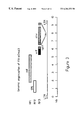
- FIG. 2 is a plot of reverse transcriptase activity in CrFK cells as a function of time after infection with FIV-Oma3.
- FIG. 3 is a schematic illustration of the genomic organization of FIV-Oma3.
- FIG. 4 is a representation of a western blot illustrating the presence of purified vif protein in eluted fractions.
- FIG. 5 is a representation of a western blot of vif protein indicating the presence of specific antibodies in rabbit sera immunized with vif protein.
- FIG. 6A is a schematic presentation of amino acid sequences of gag protein from FIV strains illustrating regions of similarity.
- FIG. 6B is a schematic presentation of the nucleotide sequence corresponding to the first boxed region of FIG. 6 A.
- FIGS. 7A, 7 B and 7 c are representations of western blots illustrating the detection of FIV using the gag protein from FIV-Oma3 and illustrating a comparison of FIV status as determined by gag western blot and by conventional methods.
- FIV is a feline immunodeficiency virus classified as a retrovirus and more specifically as a lentivirus, which is tropic for the T-lymphocytes of the host.
- FIV isolates from domestic and non-domestic cats exhibit heterogeneity at both cellular and molecular levels.
- the isolation and characterization of a highly cytopathic lentivirus from a young adult male Pallas' cat has been previously reported (Barr et al., 1995). The Pallas' cat ( Otocolobus manul ), was imported into the United States with three other Pallas' cats.
- the FIV-positive cat's CD4+/CD8+T-cell ratio was substantially lower than those of the three seronegative cats. Isolated FIV from the FIV-positive cat elicited a unique immune response in domestic cats (Barr et al. 1995). The cats seroconverted, with antibody levels peaking at 7-9 weeks post-infection, then decreased to low levels over the next 12 weeks. Although the initial response of these cats to FIV-Pallas was similar to that seen when cats are infected with domestic cat FIV, the subsequent loss of antibody was unique. Definitions
- chimeric virus for the purposes of specification and claims refers to a recombinant virus in the construction of which, portions of gene sequences, or minor modifications thereof that do not result in modified biological activity, from any of the FIV strains, have been used. Chimeric viruses may be formed by recombinations of gene sequences of two or more FIV strains.
- immunologically related for the purposes of specification and claims refers to various strains that display serological cross-reactivity with polypeptides expressed by the reconstructed viruses or variations thereof.
- Serological cross reactivity refers to the ability of an antiserum or antibody specific for the antigen(s) from a given strain to react with antigen(s) from other FIV strains.
- the FIV strains may include those both from domestic and non-domestic cats.
- Serological cross-reactivity may be determined by any standard immunoassay known in the art including, but not limited to ELISA, western blotting, and immunoblotting.
- polypeptide refers to a chain of amino acids, having a biological function, and does not refer to a specific length of chain.
- the polypeptide may be modified in vivo or in vitro, for example, by glycosylation, amidation, phosphorylation, carboxylation, or substitution without changing the primary biological function.
- the method of the present invention comprises first propagating and cultivating the Pallas's cat FIV (FIV-Oma) in established cell lines by methods well known in the art and as described in more details in the embodiments described herein.
- FIV Pallas's cat FIV
- a genomic DNA library can be constructed by standard methods well known in the art. Typically, following partial digestion of genomic DNA and reaction with Klenow polymerase in the presence of adenosine triphosphate (ATP) and guanosine triphosphate (GTP), fragments of 10-20 kb are isolated and ligated to lambda phage arms. The resultant phage is used to infect Escherichia coli ( E. coli ) and plated on tryptone broth to form plaques. Following hybridization of plaques with labeled oligonucleotides or gene fragments of FIV, positive clones can be identified. Each clone can be tested for infectivity by transfection into an established cell line. Restriction fragments of FIV-Oma subclones are cloned into a cloning vector and sequenced to determine nucleotide sequence. From the subclones, recombinant clones having desired biological properties can be constructed.
- ATP adenosine triphosphate
- a highly infectious and cytopathic recombinant clone (FIV-Oma3) was constructed from subclones that were not infectious or cytopathic.
- This clone serves as a basis for the other embodiments.
- the reconstructed clone is similar to the FIV-Oma virus in its cytopathic and infective abilities in tissue culture.
- the clone, FIV-Oma3 also serves as the basis for making chimeras having desired immunogenic and cytopathic properties.
- antigens from FIV-Oma and the reconstructed FIV-Oma3 are provided that have broad serological cross reactivity with related antigens from other species of FIV.
- the antigens of the present invention comprise polypeptides derived from the genes of FIV-Oma or FIV-Oma3. Polypeptides will be at least six amino acids found contiguously within one of the proteins of FIV-Oma or chimeric viruses. Polypeptides will generally correspond to at least one epitopic site which is characteristic of a FIV strain. By characteristic, it is meant that the epitopic site will allow immunological detection of the virus in a physiological sample. These antigens can be used in the detection of FIV infection of cats, both domestic and non-domestic, and for immunization of these cats.
- chimeric viruses are provided having desired biological properties. Chimeras have the advantage of reduced risk of recombination within the host cells with resultant production of infectious viral particles.
- the recombinant immunogenic virus is preferably one that has reduced pathogenicity.
- chimeric viruses can be constructed that are infectious in cell lines and have immunogenic properties. Such virus can be used for eliciting an antibody response in cats for the purpose of protection against FIV and could be easily propagated in established cell lines.
- the recombinant virus may contain gag, pol, vif, rev and other viral proteins or combinations thereof.
- a chimeric virus may be both pathogenic and infectious in cats. Such cats would then serve as a model system for evaluating antiviral compounds and a tool for investigating FIV infection.
- DNA constructs are provided which can be expressed using expression vectors well known in the art.
- An expression vector typically has a DNA construct linked to control sequences capable of expressing that DNA in a suitable host.
- control sequences include a transcriptional promoter, an optional operator sequence to control transcription, a sequence encoding suitable mRNA, and sequences which control the termination of transcription and translation.
- Suitable vectors include plasmids, viruses, including, but not limited to, vaccinia virus, adenovirus, baculocirus and cytomegalovirus and phages.
- any of the cell lines known in the art can be used, including cells in suspension and substrate-attached cells.
- Crandell feline kidney cells (CrFK) which grow as a monolayer or interleukin (IL)-2 dependent T-cell lines which grow as cells in suspension and require I1-2, can be used (Yamamoto et al. U.S. Pat. No. 5,510,106).
- IL-2 dependent T-cell lines which grow as cells in suspension and require I1-2
- Other T-cell lines that do not require IL-2 can also be used.
- virus cultures of FIV-Oma were inoculated onto CrFK cells at 50% confluency and cultured in the presence of complete CrFK growth medium (minimum essential growth medium with 20% Leibovitz's L-15 medium 4 mM L-glutamine, 1% gentamicin, and 5-fetal bovine serum). Numerous small syncytia were observed 3 to 4 days after inoculation of CrFK cells with FIV-Oma cultures. Following passage of the CrFK cells, numerous large syncytia (greater than 20 nuclei) and extensive cellular vacuolation were detected at confluency; and the CrFK monolayer was completely lysed 4 to 5 days after passage.
- complete CrFK growth medium minimum essential growth medium with 20% Leibovitz's L-15 medium 4 mM L-glutamine, 1% gentamicin, and 5-fetal bovine serum.
- the virus maintained this degree of cytopathy over subsequent passages through CrFK cells.
- This virus can also be propagated in peripheral blood mononuclear cells (PBMCs).
- PBMCs peripheral blood mononuclear cells
- cell-free medium from day 3-transfected CrFK cell culture was inoculated onto concanavalin A and IL-2 stimulated PBMCs.
- Supernatants from PBMC cultures can be assayed for reverse transcriptase activity, filtered through a 0.22 ⁇ m pore size filter and then used for further inoculation.
- a genomic library was constructed in a lambda phage vector.
- FIV-Oma infected CrFK cells were grown to confluency in CrFK growth medium as described in Example 1. The cells were divided 1:5 once after 3 days of cultivation and were monitored daily for syncytium formation. When large syncytia were noted, genomic DNA was isolated and a genomic DNA library constructed in accordance with standard procedures well known in the art (See W. Strauss, Preparation of genomic DNA from mammalian tissue, in Current Protocols in Molecular Biology, pp 2.2.1-2.2.3, F. Ausubel, R. Brent, R. Scientific, D. Moore, J. Seidman, J. Smith and K. Struhl, eds.
- the extract from the packaging extract was plated on E. coli (strain P2392), and plaques screened by using a 32 P-labeled FIV-Oma pol gene fragment (Barr et al., 1995).
- the pol gene fragment was made using degenerate lentivirus pol primers described in Gelman et al (1992, AIDS Res. Hum. Retroviruses 8:1981-1989. These primers amplified a 437 bp fragment from DNA isolated from CrFK culture infected with the Pallas FIV.
- the degenerate primer sequences used for amplification were
- LV1 CCGATCCDCAYCCNGSAGGAYTAMAA (SEQ ID NO:14), and
- LV2 GGTCTAGAYRYARTTCATAACCCAKCCA (SEQ ID NO:15)
- Bacteriophage from positive plaques were purified and amplified according to instructions of the manufacturer of the packaging extract, and bacteriophage preparations were banded on cesium chloride gradients (Sambrook et al. 1989). Following dialysis in 0.1 M Tris-HCl, pH 8.0, 0.05 M NaCl, 1 mM MgCl 2 , DNA was prepared from the FIV-Oma positive clones by proteinase K digestion (50 mM EDTA, pH 8.00, 50 SDS, 100 ug/ml proteinase K), phenol chloroform extraction and ethanol precipitation.
- FIV-Oma positive clones from Example 2 were digested with restriction enzymes BamH1, EcoR1, HindIII, KpnI, NheI, PstI, SacI, and SalI. Fragments were separated by agarose gel electrophoresis, and detected by Southern hybridization using 32 P-labeled FIV-Oma pol fragment. Restriction enzyme fragments of the clones were purified from agarose gels using a DNA recovery system (SpinBind system from FMS Bioproducts) and ligated into a phagemid cloning vector (pBlueScript II, SK-from Stratagene).
- Nucleotide sequencing was performed using standard primers to vector sequences and FIV-Oma-specific primers to obtain data for both strands of DNA. The sequencing was performed using automated DNA sequencing and conventional chain termination sequencing (Isotherm DNA sequencing kit; Epicentre Technologies). The sequence was analyzed using commercially available software.
- FIV-Oma ⁇ 5 and ⁇ 7 both of which have an internal Sac1 and Pst1 fragments, were used for nucleotide sequence analysis in a cloning vector using vector-specific and FIV specific primers. Subclones of partially overlapping restriction fragments were then sequenced. Based on this analysis, it was inferred that proviral clone 7 was truncated at the 3′ end while proviral clone 5 was integrated aberrantly (3′ gag, pol, env, partial 3′ LTR, 5′LTR, 5′ gag). Clone ⁇ 2 was the only full length proviral clone containing 5′ and 3′ LTRs in the correct position.
- each clone can be transfected into a cell line by methods well known in the art, for example using calcium phosphate, DEAE-Dextran and electroporation (see R. guitarist et al., in Current Protocols in Molecular Biology, supra pp 9.0.1-9.4.3).
- each clone from Example 3 was transfected into CrFK cells using commercial calcium phosphate reagent (CellPhect Transfection kit; Pharamcia).
- DNA was mixed with an equal volume of Buffer A (0.5 M CaCl 2 , 0.1 M HEPES, pH 7.), incubated at room temperature for 10 minutes, mixed with an equal volume of Buffer B (0.28 M NaCl, 0.05 M HEPES, 0.75 mM NaH 2 PO 4 , 0.75 mM Na 2 HPO 4 , pH 7.0), incubated at room temperature for another 15 minutes, and added to 50% confluent CrFK cells. The cells were incubated at 37° C. for 4-6 hours, then subjected to a glycerol shock.
- Buffer A 0.5 M CaCl 2 , 0.1 M HEPES, pH 7.
- the cells were washed once with HBSS and incubated with 20% glycerol in phosphate-buffered saline (PBS) for 1.5 minutes, then washed twice with HBSS. After glycerol shock, the cells were incubated in growth medium at 37° C. Supernatants from transfected cells were harvested daily and observed for virion production by assaying for reverse transcriptase activity according to the method of Heine et al., 1980, which method hereby incorporated by reference. Briefly, supernatant samples (10 ⁇ l/reaction) were incubated at 37° C.
- Sequences of ⁇ 2 were amplified by polymerase chain reaction (Saiki et al. 1988) by methods well known in the art and using commercially available reagents (GeneAmp; Perkin-Elmer Cetus). Briefly, 1 ⁇ g of DNA template, 1 ⁇ g of each primer, dNTPs (0.2 mM each), 2 mM MgCl 2 and 5 Units Taq DNA polymerase was subjected to a 2-min “heat-shock” step at 95° C. prior to 30 cycles of amplification at 94° C. for 1 minute, 55° C. for 2 minutes, 72° C. for 3 minutes and a final cycle of 72° C. for 10 minutes in a commercial thermocycler. The products were cloned into a cloning vector (pCRII vector, TA cloning kit; Invitrogen corp.) and the sequence was confirmed by nucleotide sequencing.
- pCRII vector TA cloning kit
- FIV-Oma specific primers were used to amplify the 5′ end region (1-3633 bases): forward primer 5′-GCGGCCGCTGGGAGGATTGGAGGTCCT-3′ (SEQ ID NO:1), corresponding to bases 1-19 with an added 5′ Not1 site, and reverse primer 5′-GCTCTTAAGGCTATGTCGCA-3′ (SEQ ID NO:2).
- the 1-3633 region was cut out of this subclone with NotI and AflII, and ligated into a NotI and AflII digested subclone of ⁇ 7 clone which contained the 7 kb region of the proviral genome in phagemid cloning vector (pBluescriptII SK-from Stratagene), to construct a 8.4 kb subclone.
- the 3′ end of the provirus genome was amplified by nucleic acid amplification techniques like polymerase chain reaction (PCR).
- PCR polymerase chain reaction
- the 3′ end was amplified from ⁇ 2 clone with primers 5′-TGTCCAGTGTTAGAGTCGGTAG-3′ (SEQ ID NO:3) corresponding to bases 7182-7203, and a reverse primer 5′-GTCGACTGCTAAGGTCTCCGTCCCGAATC-3′ (SEQ ID NO:4), corresponding to bases 9747-9725 of the FIV-Oma genome.
- a TAl clone was obtained by amplification of fragments from ⁇ 2 by PCR using primers having the sequence of SEQ ID NO:3 and SEQ ID NO:4.
- the amplified products were cloned into PCRTMII (Invitrogen), and then removed by restriction enzyme digestion. Then the 7182-9747 base region was cut out of TA1 with Eco47III and SalI, ligated to the EcoIII and SalI digested 8.4 kb subclone to construct a full length FIV-Oma1 in a proviral vector.
- Subclone TA5 was constructed by amplification of fragments from ⁇ 2 by PCR using primers having the sequence of SEQ ID NO:1 and SEQ ID NO:2. The amplified products were cloned into pCRTMII (Invitrogen), and then removed by restriction enzyme digestion. Subclones TA1 and TA5 were subjected to digestion with restriction enzymes, Eco47III and NotI. The region between Eco47III and NotI sites of the virus genome in subclone TA1 was replaced by that region in subclone TA5 to obtain a subclone TA3 containing 3′ end region of the virus genome with 7297 bases derived from ⁇ 5 clone. The 3′ end region (7182-9747 bases) digested from TA3 with Eco47III and SalI was cloned into similarly digested 8.4 kb subclone to construct an infectious clone FIV-Oma3.
- This embodiment is directed towards determining the infectivity and cytopathicity of the subclones constructed from the lambda clones in Example 5.
- CrFK cells were transfected with pFIV-Oma3 by the calcium phosphate method as described in Example 4 to determine if the proviral clone was infectious. Infectivity of virions derived from the CrFK cells transfected with pFIV-Oma3 was determined. One ml cell-free medium from transfected CrFK cells was inoculated onto about 60% confluent CrFK cells. The RT activity in the medium was about 1.37 ⁇ 10 8 cpm/ml. As shown in FIG. 2, infectious virions were produced in CrFK cells. Cytopathic effect of syncytium formation and vacuolization similar to those described for wild-type FIV-Oma (Barr et al. 1955) were visible by Day 5, and most cells were lysed by Day 11 following transfection. Additionally progeny virions were infectious and cytopathic for CrFK cells and primary feline PBMCs.
- the DNA sequence of the reconstructed clone can be determined by any of the standard methods known in the art. Using the dideoxynucleotide chain termination method in an automated DNA sequencer, the FIV-Oma3 clone was sequenced. The length of the infectious clone was found to be 9751 bp (SEQ ID NO:7). Potential regulatory and coding regions in the FIV-Oma3 provirus were identified by sequence analysis. As illustrated in FIG. 3, the genomic organization of the FIV-Oma3 provirus is typical of other lentiviruses (Narayan and Elements, 1989, J. Gen Virol.
- gag protein starts at nucleotide 684 and encodes a protein of 498 amino acids (SEQ ID NO:8).
- the open reading frame for the pol protein begins at nucleotide 1979 and encodes a protein of 1150 amino acids (SEA ID NO:9).
- the open reading frame for vif protein starts at nucleotide 5429 and encodes a protein of 252 amino acids (SEQ ID NO:10).
- the open reading frame for the env and rev proteins starts at nucleotide 6512 and encodes a rev protein of 863 amino acids (SEQ ID NO:11). Splicing of this protein at amino acid 103 results in the formation of the env protein.
- Four other open reading frames, orfA (SEQ ID NO:12), orfB (SEQ ID NO:13), orfC (SEQ ID NO:14) and orfE (SEQ ID NO:15) are also present starting at nucleotides 1100, 6387, 7827 and 9165 respectively, and encoding putative polypeptides of 51, 39, 38 and 65 amino acids respectively.
- FIV-Fca denotes domestic cat FIV strains
- TM2 and FIV-14 are sequenced viral isolates
- FIV-PPR is an infectious clone of a domestic cat isolate.
- FIV-Fco is a sequenced puma (nondomestic cat) FIV isolate (Langley et al., 1994, Virology 202:853-864).
- This embodiment is directed towards construction of chimeric viruses to determine which sequences are required for eliciting an immune response in cats.
- DNA isolated from FIV-Oma3 and a selected FIV can be treated with various restriction enzyme combinations, and specific portions ligated together in constructing the chimeras.
- a restriction enzyme or combination of restriction enzymes may be used to generate chimeric viruses that have the desired biological properties.
- a chimeric virus can be constructed which elicits an immune response in cats. Restriction enzyme selection may be done to reduce the cytopathicity of the immunogenic peptide.
- a chimeric virus for use as a vaccine can be constructed which is immunogenic, and is highly infectious in cell cultures so that it may be propagated easily to obtain high titres. Further, chimeric viruses can be constructed that are highly infectious and pathogenic in cats. Such chimeras can serve as models for evaluating potential anti-viral compounds and would provide a model system for studying FIV infection.
- chimeric viral clones can be introduced into CrFK cells and tested for immunogenicity by techniques well known in the art including immunoblotting and western blotting.
- This embodiment is directed towards methods for identification of sequences affecting cytopathicity.
- Chimeras from Example 8 can be individually introduced into CrFK cells. Transfected cells are passaged every three days and carried for 3-8 passages. Cells are monitored for syncytium formation and vacuolization from Day 4. Cytopathic activity can be correlated with the specific nucleotide sequence of the clones and thus polypeptides that affect or cause cytopathicity can be identified.
- DNA fragments from FIV-Oma3 or chimeric viruses constructed therefrom as in Example 8 may be incorporated in DNA constructs capable of introduction into expression vectors such as phage vectors or plasmids.
- the DNA construct should contain the necessary elements for transcription and translation.
- a variety of host systems may be utilized to express the Oma or chimeric peptides including, but not limited to, bacteria transformed with a bacteriophage vector, plasmid vector, or cosmid DNA; yeast containing yeast vectors, fungi containing fungal vectors, insect cell lines infected with virus (e.g.
- baculovirus avian cells transfected with plasmid or viral expression vectors, or infected with recombinant virus
- mammalian cell lines transfected with plasmid or viral expression vectors, or infected with recombinant virus (e.g. vaccinia virus, adenovirus, adeno-associated virus, retrovirus, etc.)
- promoters and enhancers can be incorporated in the DNA vector by methods well known in the art. A broad variety of suitable promoters are available. The selection of the promoter depends on the expression system used. Promoters vary in their ability to facilitate transcription.
- Suitable promoter systems in microbial expression vectors include the beta lactamase and lactose promoter systems, tryptophan promoter system and the tac promoter.
- Suitable promoters for yeast vectors include the promoters for metallothionein, 3-phosphoglycerate kinase or other glycolytic enzymes.
- the polyhedrin or p10 promoters of baculovirus can be used.
- promoters such as SV40 early or late promoters and the metallothionine-I promoter can be used.
- Enhancer sequences are DNA elements that appear to increase transcriptional efficiency in a manner relatively independent of their position and orientation with respect to a nearby gene. Thus, depending on the host cell expression vector system used, an enhancer may be placed upstream or downstream from the inserted nucleotide sequence.
- One or more regulatory elements, such as transcription or translation initiation signals may be used to regulate the expression of the nucleotide sequence encoding the recombinant polypeptide.
- polypeptides of the present invention may be modified using genetic engineering techniques. For, example, site directed mutagenesis may be used to change a single amino acid in the polypeptide to alter its immunogenic and/or its cytopathic activity.
- the vif gene was PCR amplified from lambda 2 clone using primers 5′-GCTACCGAGTGGTGAAGAGGATTGGCAG-3′ (SEQ ID NO:5) and 5′-GTCGACTTAACTCTTCATCCG-3′ (SEQ ID NO:6). DNA was ligated to PCRTMII vector (Invitrogen) to obtain a subclone containing the vif gene (without a start codon).
- Buffer A (6 M GuHCl, 0.1 M Na-phosphate, 0.01 M Tris/HCl, pH 8.0) at 5 ml per gram wet weight for 1 hour at room temperature.
- Buffer B 8 M urea, 0.1 M Na-phosphate, 0.01 M Tris/HCl, pH 8.0
- Buffer C 8 M urea, 0.1 M Na-phosphate, 0.01 M Tris/HCl, pH 6.3
- the eluted fractions were collected and analyzed on 15% SDS-PAGE by methods well known in the art. As shown in FIG. 4, SDS-PAGE analysis of purified His-tagged vif protein indicates the presence of approximately 22 kD protein in fractions 1-5.
- the gag gene was amplified by PCR using primers Bam-gag and gag-Xho from 1 ng of pOma3.
- the BamHI-XhoI digested product was cloned into pET28(a)(Novagen) that had been digested with BamHI and XhoI.
- the recombinant pET-Gag plasmid was transformed into BL21( ⁇ DE) cells.
- Induction of the gag polyprotein was accomplished by the addition of IPTG to the culture at 1 mM, with growth for an additional 4 hours at 37° C.
- Bacterial extract was prepared in and run on 8-20% Phast gels (Pharmacia) according to manufacturer's instructions.
- This embodiment is directed towards various vaccines useful for protecting cats against FIV.
- vaccines useful for protecting cats against FIV include live attenuated chimeric viruses, fixed whole viruses, host cells which express viral antigens, preparations of viral fragments, purified proteins from the viruses or expressed by host cells, and antigenic fragments of proteins.
- Live attenuated virus is made by serial passage of the virus in CrFK cells in culture, or genetically altering it in accordance with procedures well known in the art.
- live virus is contacted with a suitable fixative like formalin.
- Fixative like formalin. Fixed virus can then be used in vaccine formulations.
- polypeptides of FIV-Oma1 or FIV-Oma3 may be expressed in a suitable host system as described in Example 10.
- Recombinant polypeptides produced can be purified by methods well known in the art including detergent extraction, chromatography (e.g. ion exchange, affinity, immunoaffinity, or sizing columns), differential solubility, differential centrifugation, and the like.
- Immunopurification of the polypeptides from a host cell expression system preparation may be accomplished using methods known in the art for immunoaffinity chromatography.
- Specific antibodies for desired epitopes may be linked to a solid matrix to form an affinity matrix.
- the preparation containing the recombinant polypeptides is then incubated with the affinity matrix.
- the affinity matrix is washed to remove unbound material and the bound peptide is then eluted from the matrix.
- Transformed host cell preparations can also be used in vaccines.
- the lysate from the host cell preparation may be used in a crude form or the expressed polypeptides can be purified by conventional methods.
- host cells such as yeast cells may be transformed with vectors so that polypeptides of the present invention are expressed on the surface of the yeast cells. The yeast cells can then be used in a vaccine either as such or fixed in a suitable fixative.
- Vaccine formulations of the present invention comprise the antigen in a pharmaceutically acceptable carrier.
- the antigen is present in an amount sufficient to elicit an immune response.
- one or more adjuvants may also be present.
- the adjuvants aid in attaining a more durable and higher level immune response using smaller amounts of vaccine antigen or fewer doses than if the vaccine antigen were administered alone.
- Suitable adjuvants include Freund's adjuvant, Adjuvant 65 (containing peanut oil, mannide monooleate and aluminum monostearate), mineral gels such as aluminum hydroxide and aluminum phosphate, plant and animal oils and synthetic polymers.
- the vaccine formulation may also contain a stabilizer like a carbohydrate (sorbitol, mannitol, glucose, starch) or a protein (albumin or casein).
- the vaccines of the present invention may be administered by any conventional means.
- suitable administration routes are intramuscular, subcutaneous, oral, nasal, and intraperitoneally.
- the dosage of the immunogen depends upon the immunogen and the route of administration. Typically, a single dose has a total volume of between about 0.1 ml to 5.0 ml.
- the number of inoculations should be sufficient to elicit an immune response. Determination of the number and temporal spacing of the inoculations is well within the skill of those skilled in the art. In general, there are at least two inoculations spaced over a period of two to ten weeks.
- purified vif protein from Example 10 was used to produce polyclonal antibodies in rabbit by standard methods known in the art and as described in this embodiment.
- Antiserum from the immunized rabbit was collected. Western blotting was performed to detect the presence of vif antibodies in the rabbit serum.
- the rabbit antiserum contained antibodies that reacted specifically with purified vif protein. This demonstrated the efficacy of FIV-Oma3 polypeptides for the generation of antibodies.
- gag protein from FIV-Oma or from a chimeric virus from Example 9 can be used in the vaccination of cats.
- This embodiment is directed towards reagents and methods for detecting the presence of FIV antibodies in cats.
- the reagents can be used for detection of FIV strains of both domestic and non-domestic cats.
- Detection system of the present invention may involve detection by polynucleotide probes or by polypeptides.
- Detection of FIV in biological samples can be carried out using polynucleotide probes based on the sequence of the FIV genome.
- the length of the probe is not critical but is preferably at least 12 bases and is sufficiently complimentary to the viral genome.
- the probe may be DNA or RNA.
- DNA probes may be prepared synthetically or may be prepared by cleavage of the genome by restriction endonucleases followed by cloning of the DNA fragment to obtain large quantities by techniques well known in the art.
- the probes can be labeled according to standard techniques.
- the labels may be radioactive (e.g. 14 C, 32 P or 3 H) or non radioactive such as specific antibodies, fluorescers, chemiluminescers, and enzymes.
- Labeled probes can be purified on polyacrylamide gels with subsequent elution in distilled water. (Sambrook et al 1989).
- the probes are then used to detect the presence of FIV by hybridization techniques that are well known in the art.
- stringency conditions during hybridization can be varied to identify either closely related strains or a wide variety of strains. The more stringent the incubation condition, the higher the degree of complimentarily that is required between the probe and the sample DNA. Stringency can be controlled by temperature, probe concentration, probe length, ionic strength, time etc. A common way of varying stringency is to vary the concentration of formamide to change the polarity of the reactant solution.
- the diagnostic probes of the present invention include nucleotide sequences that hybridize to FIV-Oma3 genomic DNA or fragments thereof.
- the probes may be fragments of cDNA or oligonucleotide probes which hybridize to DNA sequences encoding FIV-Oma3 polypeptides.
- FIV isolates that are closely related to FIV-Oma can be identified.
- assays can be designed that will detect both domestic and non-domestic cat FIVs.
- conserved regions of the FIV gene sequences can be identified using commercially available software. Oligonucleotide probes can be designed taking into consideration the degeneracy of the genetic code, which is well known in the art. By varying the stringency of hybridization condition, various strains of FIV can be detected.
- FIG. 6A shows homologous regions of gag protein from different strains of FIVs. The regions are particularly suitable for designing oligonucleotide and polypeptide probes.
- FIG. 6B aligned amino acid residues are printed in lowercase. Other residues (uppercase) are not aligned. Boxed areas indicate conserved regions of highest similarity.
- the detection system of the present invention also includes polypeptide probes which can be used as antigens in immunoassays for the detection of FIV antibodies.
- Antigenic peptides can be generated from the recombinant polypeptides or can be synthesized by techniques well known in the art.
- the antigenic peptides can vary in size but generally consist of from 7 to 14 amino acids and can be synthesized by methods including solid peptide synthesis using tertbutyl oxycarbonyl amino acids (Mitchell et al., 1978, J. Org/Chem.
- the polypeptides and the antibodies can be labeled so as to provide a detectable signal.
- Suitable labels include radionucleotides, enzymes, substrates, cofactors, inhibitors, fluorescent and chemifluorescent materials, magnetic particles and the like. (see U.S. Pat. No. 5,510,106)
- the detection of FIV may be carried out in various biological specimens including, but not limited to, blood, plasma, serum, and urine.
- a diagnostic assay utilizing as an antigen, peptides or polypeptides of the present invention includes any of immunoassays known in the art including, but not limited to, radioimmunoassay, ELISAs, “sandwich” assay, immmunoblotting, fluorescent assay and chemiluminescence-based assays.
- gag protein from FIV-Oma3 prepared according to Example 8 can be used as an antigen in an ELISA test in which the gag protein is immobilized to a selected surface, followed by blocking of unbound areas of the surface, contacting the sample containing FIV with the selected surface having the attached antigen, washing the surface to remove unbound materials, and detection of the immune complexes by standard detection means like enzyme substrate complexes or fluorescent detection systems.
- a diagnostic kit for detecting FIV in domestic and nondomestic cats may comprise a peptide from FIV-Oma3 or a chimeric virus expressing a feline FIV protein that has a broad specificity, a means for facilitating contact between the sample containing FIV and the antigen (e.g. a microtitre plate) and a means for detecting presence of immune complexes formed.
- polypeptides are obtained in a substantially pure form and free of contaminants and interfering proteins.
- polypeptides of the present invention can be purified to at least 50% purity. Suitable means of purification of proteins include affinity columns, immunoadsorption and the like. Once the peptide is purified in sufficient amount, it can be used in the detection systems of the present invention.
- gag protein was used in western blotting analysis to detect the presence of different strains of FIV.
- Gag protein prepared according to Example 10 and run on 8-20% Phast gels (Pharmacia) was transferred to nitrocellulose by capillary blot. Nitrocellulose strips were incubated with cat serum which had been preincubated with approximately 0.5 mg/ml E.coli extract (Promega) at room temperature. Various Cat serum samples were diluted 1:12. Primary antibody binding was carried out overnight at room temperature. Horseradish peroxidase labeled secondary antibody (goat anti-cat IgG) binding was carried out at room temperature for 1 hour before the peroxidase color reaction.
- FIG. 7A, 7 B and 7 C shows a comparison of conventional tests using a combination of kinetic ELISA (KELA based on an ELISA kit, IDEXX) and western blots using FIV viral antigen prepared from a domestic cat FIV strain (Petaluma) or the FIV-Oma virus.
- the samples were classified as plus (+), minus ( ⁇ ), or equivocal (eq) based on the conventional tests.
- the results of the conventional tests is shown on top of each panel.
- the gels indicate the presence or absence of gag antibodies by western blot analysis using the gag protein and methods of the present invention. The presence (+), absence ( ⁇ ), or weak presence ((+)) of the antibodies is shown at the bottom.
- MW indicates molecular weight makers; TX cougar is Texas cougar; Af. lion is African lion; Bk.footed cat is Black footed cat; and ON is Ontario; These data clearly indicate that gag protein from the FIV-Oma3 clone detects a wide range of FIV strains and therefore, can be used to detect the presence of FIV in both domestic and nondomestic cats.
- a system for evaluating the inhibitory effects of potential therapeutics of FIV. Since the infectious clone FIV-Oma3 has been found to be highly cytopathic and infectious in culture, a culture of CrFK cells can be inoculated with FIV-Oma3. The cultures can be set up in any type of tissue culture plates, preferably in a multiwell plate. Once the cells have been infected with the virus, potential inhibitory agents can be added under sterile conditions and the cultures monitored for syncytium formation and vacuolization.
Landscapes
- Chemical & Material Sciences (AREA)
- Life Sciences & Earth Sciences (AREA)
- Organic Chemistry (AREA)
- Health & Medical Sciences (AREA)
- Virology (AREA)
- Biophysics (AREA)
- General Health & Medical Sciences (AREA)
- Genetics & Genomics (AREA)
- Medicinal Chemistry (AREA)
- Molecular Biology (AREA)
- Proteomics, Peptides & Aminoacids (AREA)
- Biochemistry (AREA)
- Immunology (AREA)
- Gastroenterology & Hepatology (AREA)
- Micro-Organisms Or Cultivation Processes Thereof (AREA)
- Measuring Or Testing Involving Enzymes Or Micro-Organisms (AREA)
- Peptides Or Proteins (AREA)
Abstract
Description
| TABLE 1 | ||
| % similarity with FIV-Oma | ||
| gag | pol | env | vif | ||
| (NA/AA) | (NA/AA) | (NA/AA) | (NA/AA) | ||
| FIV-Fca(TM2) | 55/63 | 72/71 | 44/26 | 60/54 |
| FIV-Fca(FIV-14) | 60/63 | 72/71 | 44/26 | 52/52 |
| FIV-Fca(PPR) | 62/63 | 72/71 | 43/25 | 52/52 |
| FIV-Fco(PLV-14) | 55/52 | 63/59 | 58/47 | 50/63 |
Claims (11)
Priority Applications (2)
| Application Number | Priority Date | Filing Date | Title |
|---|---|---|---|
| US09/238,303 US6284253B1 (en) | 1998-01-29 | 1999-01-28 | Feline immunodeficiency virus (FIV) nucleotide sequence |
| US09/946,239 US6579527B2 (en) | 1998-01-29 | 2001-09-04 | Feline immunodeficiency virus nucleotide sequence |
Applications Claiming Priority (2)
| Application Number | Priority Date | Filing Date | Title |
|---|---|---|---|
| US7292798P | 1998-01-29 | 1998-01-29 | |
| US09/238,303 US6284253B1 (en) | 1998-01-29 | 1999-01-28 | Feline immunodeficiency virus (FIV) nucleotide sequence |
Related Child Applications (1)
| Application Number | Title | Priority Date | Filing Date |
|---|---|---|---|
| US09/946,239 Division US6579527B2 (en) | 1998-01-29 | 2001-09-04 | Feline immunodeficiency virus nucleotide sequence |
Publications (1)
| Publication Number | Publication Date |
|---|---|
| US6284253B1 true US6284253B1 (en) | 2001-09-04 |
Family
ID=26753919
Family Applications (2)
| Application Number | Title | Priority Date | Filing Date |
|---|---|---|---|
| US09/238,303 Expired - Lifetime US6284253B1 (en) | 1998-01-29 | 1999-01-28 | Feline immunodeficiency virus (FIV) nucleotide sequence |
| US09/946,239 Expired - Fee Related US6579527B2 (en) | 1998-01-29 | 2001-09-04 | Feline immunodeficiency virus nucleotide sequence |
Family Applications After (1)
| Application Number | Title | Priority Date | Filing Date |
|---|---|---|---|
| US09/946,239 Expired - Fee Related US6579527B2 (en) | 1998-01-29 | 2001-09-04 | Feline immunodeficiency virus nucleotide sequence |
Country Status (1)
| Country | Link |
|---|---|
| US (2) | US6284253B1 (en) |
Cited By (8)
| Publication number | Priority date | Publication date | Assignee | Title |
|---|---|---|---|---|
| US20050058993A1 (en) * | 2003-09-11 | 2005-03-17 | Idexx Laboratories, Inc. | Method and device for detecting feline immunodeficiency virus |
| US20050136401A1 (en) * | 2003-12-18 | 2005-06-23 | Idexx Laboratories, Inc. | Method and device for detecting feline immunodeficiency virus |
| US20050208067A1 (en) * | 2004-02-19 | 2005-09-22 | Idexx Laboratories, Inc. | Method and device for detecting feline immunodeficiency virus |
| US20060003445A1 (en) * | 2004-06-30 | 2006-01-05 | Idexx Laboratories, Inc. | Method and device for detecting feline immunodeficiency virus |
| US20060004184A1 (en) * | 2004-06-30 | 2006-01-05 | Idexx Laboratories, Inc. | Method and device for detecting feline immunodeficiency virus |
| US20060002957A1 (en) * | 2004-06-30 | 2006-01-05 | Idexx Laboratories, Inc. | Method and device for detecting feline immunodeficiency virus |
| US20060205923A1 (en) * | 2005-03-09 | 2006-09-14 | Idexx Laboratories, Inc. | Method and device for detecting feline immunodeficiency virus |
| US8809004B2 (en) | 2010-04-02 | 2014-08-19 | Idexx Laboratories, Inc. | Detection of feline immunodeficiency virus |
Citations (1)
| Publication number | Priority date | Publication date | Assignee | Title |
|---|---|---|---|---|
| US5510106A (en) | 1987-08-26 | 1996-04-23 | The Regents Of The University Of California | Methods and compositions for vaccinating against feline immunodeficiency virus |
-
1999
- 1999-01-28 US US09/238,303 patent/US6284253B1/en not_active Expired - Lifetime
-
2001
- 2001-09-04 US US09/946,239 patent/US6579527B2/en not_active Expired - Fee Related
Patent Citations (1)
| Publication number | Priority date | Publication date | Assignee | Title |
|---|---|---|---|---|
| US5510106A (en) | 1987-08-26 | 1996-04-23 | The Regents Of The University Of California | Methods and compositions for vaccinating against feline immunodeficiency virus |
Non-Patent Citations (1)
| Title |
|---|
| Barr et al., "Isolation of a Highly Cytopathic Lentivirus from a Nondomestic Cat," Journal of Virology, Nov. 1995, pp. 7371-7374. |
Cited By (18)
| Publication number | Priority date | Publication date | Assignee | Title |
|---|---|---|---|---|
| US20050058992A1 (en) * | 2003-09-11 | 2005-03-17 | Idexx Laboratories, Inc. | Method and device for detecting feline immunodeficiency virus |
| US20050058993A1 (en) * | 2003-09-11 | 2005-03-17 | Idexx Laboratories, Inc. | Method and device for detecting feline immunodeficiency virus |
| US20110189651A1 (en) * | 2003-09-11 | 2011-08-04 | Idexx Laboratories, Inc. | Method and Device for Detecting Feline Immunodeficiency Virus |
| US7776546B2 (en) | 2003-09-11 | 2010-08-17 | Idexx Laboratories, Inc. | Method and device for detecting feline immunodeficiency virus |
| US7201903B2 (en) | 2003-09-11 | 2007-04-10 | Idexx Laboratories, Inc. | Method and device for detecting feline immunodeficiency virus |
| US7285272B2 (en) | 2003-12-18 | 2007-10-23 | Idexx Laboratories, Inc. | Method and device for detecting feline immunodeficiency virus |
| US20050136401A1 (en) * | 2003-12-18 | 2005-06-23 | Idexx Laboratories, Inc. | Method and device for detecting feline immunodeficiency virus |
| US20050208067A1 (en) * | 2004-02-19 | 2005-09-22 | Idexx Laboratories, Inc. | Method and device for detecting feline immunodeficiency virus |
| US7348136B2 (en) | 2004-02-19 | 2008-03-25 | Idexx Laboratories, Inc. | Method and device for detecting feline immunodeficiency virus |
| US20060003445A1 (en) * | 2004-06-30 | 2006-01-05 | Idexx Laboratories, Inc. | Method and device for detecting feline immunodeficiency virus |
| US7285278B2 (en) | 2004-06-30 | 2007-10-23 | Idexx Laboratories, Inc. | Method and device for detecting feline immunodeficiency virus |
| US7335360B2 (en) | 2004-06-30 | 2008-02-26 | Idexx Laboratories, Inc. | Method and device for detecting feline immunodeficiency virus |
| US20100092940A1 (en) * | 2004-06-30 | 2010-04-15 | Idexx Laboratories, Inc. | Method and Device for Detecting Feline Immunodeficiency Virus |
| US20060002957A1 (en) * | 2004-06-30 | 2006-01-05 | Idexx Laboratories, Inc. | Method and device for detecting feline immunodeficiency virus |
| US20060004184A1 (en) * | 2004-06-30 | 2006-01-05 | Idexx Laboratories, Inc. | Method and device for detecting feline immunodeficiency virus |
| US7291338B2 (en) | 2005-03-09 | 2007-11-06 | Idexx Laboratories, Inc. | Method and device for detecting feline immunodeficiency virus |
| US20060205923A1 (en) * | 2005-03-09 | 2006-09-14 | Idexx Laboratories, Inc. | Method and device for detecting feline immunodeficiency virus |
| US8809004B2 (en) | 2010-04-02 | 2014-08-19 | Idexx Laboratories, Inc. | Detection of feline immunodeficiency virus |
Also Published As
| Publication number | Publication date |
|---|---|
| US6579527B2 (en) | 2003-06-17 |
| US20020044945A1 (en) | 2002-04-18 |
Similar Documents
| Publication | Publication Date | Title |
|---|---|---|
| AU704309B2 (en) | Antigenically-marked non-infectious retrovirus-like particles | |
| JP5339687B2 (en) | Multi-subtype FIV vaccine | |
| US20030165535A1 (en) | Retrovirus-like particles made non-infectious by a plurality of mutations | |
| US6544752B1 (en) | Anigenically-marked non-infectious retrovirus-like particles | |
| OA10954A (en) | Hiv envelope polypeptides and vaccine | |
| Kappes et al. | Intracellular transport and virion incorporation of vpx requires interaction with other virus type-specific components | |
| US5736378A (en) | Molecular cloning and characterization of the feline immunodeficiency virus isolate PPR | |
| US6284253B1 (en) | Feline immunodeficiency virus (FIV) nucleotide sequence | |
| EP0651806B1 (en) | Anti-feline immunodeficiency virus (fiv) vaccines | |
| CA2525641A1 (en) | Materials and methods for immunizing against fiv infection | |
| JPH02211881A (en) | Recombinant hybrid hbsag particle having morphological feature of hbsag antigen with immunogen arrangement inducing neutral antibody or being recognized by such antibody, nucleotide arrangement which codes such particle, and vaccine containing nucleus particle | |
| US20030215793A1 (en) | Complete genome sequence of a simian immunodeficiency virus from a wild chimpanzee | |
| EP1007687A1 (en) | Fiv vaccine | |
| US20050064392A1 (en) | Compositions, methods and kits relating to deletion mutations of immunodeficiency virus gp120 hypervariable regions | |
| Hulskotte et al. | Antigenicity and immunogenicity of recombinant envelope glycoproteins of SIVmac32H with different in vivo passage histories | |
| US7700726B2 (en) | HIV antisense proteins | |
| US6270959B1 (en) | Human T-cell lymphotropic virus type II envelope protein and human monoclonal antibodies specific therefor | |
| CA2201419C (en) | Human t-cell lymphotropic virus type i envelope protein and human monoclonal antibodies specific therefor | |
| US20030118601A1 (en) | FIV vaccine |
Legal Events
| Date | Code | Title | Description |
|---|---|---|---|
| AS | Assignment |
Owner name: CORNELL RESEARCH FOUNDATION, INC., NEW YORK Free format text: ASSIGNMENT OF ASSIGNORS INTEREST;ASSIGNORS:BARR, MARGARET C.;AVERY, ROGER J.;SUTTON, CLAUDIA A.;REEL/FRAME:009732/0675;SIGNING DATES FROM 19990126 TO 19990127 |
|
| AS | Assignment |
Owner name: CORNELL RESEARCH FOUNDATION, INC., NEW YORK Free format text: ASSIGNMENT OF ASSIGNORS INTEREST;ASSIGNOR:ZOU, LILY;REEL/FRAME:011399/0345 Effective date: 20001208 Owner name: CORNELL RESEARCH FOUNDATION, INC., NEW YORK Free format text: ASSIGNMENT OF ASSIGNORS INTEREST;ASSIGNOR:LONG, FAN;REEL/FRAME:011402/0045 Effective date: 20001211 |
|
| STCF | Information on status: patent grant |
Free format text: PATENTED CASE |
|
| FPAY | Fee payment |
Year of fee payment: 4 |
|
| FEPP | Fee payment procedure |
Free format text: PAYER NUMBER DE-ASSIGNED (ORIGINAL EVENT CODE: RMPN); ENTITY STATUS OF PATENT OWNER: SMALL ENTITY Free format text: PAYOR NUMBER ASSIGNED (ORIGINAL EVENT CODE: ASPN); ENTITY STATUS OF PATENT OWNER: SMALL ENTITY |
|
| AS | Assignment |
Owner name: NATIONAL INSTITUTES OF HEALTH (NIH), U.S. DEPT. OF Free format text: EXECUTIVE ORDER 9424, CONFIRMATORY LICENSE;ASSIGNOR:CORNELL UNIVERSITY;REEL/FRAME:021372/0694 Effective date: 20010913 |
|
| REMI | Maintenance fee reminder mailed | ||
| FPAY | Fee payment |
Year of fee payment: 8 |
|
| SULP | Surcharge for late payment |
Year of fee payment: 7 |
|
| FPAY | Fee payment |
Year of fee payment: 12 |