US6263239B1 - Method and apparatus for determining the second phase of defibrillator devices - Google Patents

Method and apparatus for determining the second phase of defibrillator devices Download PDF

Info

Publication number
US6263239B1
US6263239B1 US09/383,561 US38356199A US6263239B1 US 6263239 B1 US6263239 B1 US 6263239B1 US 38356199 A US38356199 A US 38356199A US 6263239 B1 US6263239 B1 US 6263239B1
Authority
US
United States
Prior art keywords
patient
phase
resistance component
quantitative
circuit
Prior art date
Legal status (The legal status is an assumption and is not a legal conclusion. Google has not performed a legal analysis and makes no representation as to the accuracy of the status listed.)
Expired - Lifetime
Application number
US09/383,561
Inventor
James E. Brewer
Gary B. Stendahl
Kenneth F. Olson
Current Assignee (The listed assignees may be inaccurate. Google has not performed a legal analysis and makes no representation or warranty as to the accuracy of the list.)
Zoll Medical Corp
Original Assignee
Survivalink Corp
Priority date (The priority date is an assumption and is not a legal conclusion. Google has not performed a legal analysis and makes no representation as to the accuracy of the date listed.)
Filing date
Publication date
Application filed by Survivalink Corp filed Critical Survivalink Corp
Priority to US09/383,561 priority Critical patent/US6263239B1/en
Assigned to SURVIVALINK CORPORATION reassignment SURVIVALINK CORPORATION ASSIGNMENT OF ASSIGNORS INTEREST (SEE DOCUMENT FOR DETAILS). Assignors: STENDAHL, GARY B., OLSON, KENNETH F., BREWER, JAMES E.
Priority to US09/678,820 priority patent/US6411846B1/en
Application granted granted Critical
Publication of US6263239B1 publication Critical patent/US6263239B1/en
Priority to US10/078,735 priority patent/US20020138104A1/en
Assigned to HSBC BANK, USA reassignment HSBC BANK, USA SECURITY AGREEMENT Assignors: CARDIAC SCIENCE, INC.
Assigned to CARDIAC SCIENCE, INC. reassignment CARDIAC SCIENCE, INC. ASSIGNMENT OF ASSIGNORS INTEREST (SEE DOCUMENT FOR DETAILS). Assignors: SURVIVALINK CORPORATION
Priority to US10/760,040 priority patent/US7463923B2/en
Assigned to CARDIAC SCIENCE CORPORATION reassignment CARDIAC SCIENCE CORPORATION MERGER (SEE DOCUMENT FOR DETAILS). Assignors: CARDIAC SCIENCE, INC.
Assigned to SILICON VALLEY BANK reassignment SILICON VALLEY BANK SECURITY AGREEMENT Assignors: CARDIAC SCIENCE, INC.
Assigned to CARDIAC SCIENCE CORPORATION reassignment CARDIAC SCIENCE CORPORATION RELEASE BY SECURED PARTY (SEE DOCUMENT FOR DETAILS). Assignors: SILICON VALLEY BANK
Assigned to DBS BANK LTD., BANGALORE BRANCH reassignment DBS BANK LTD., BANGALORE BRANCH SECURITY AGREEMENT Assignors: CARDIAC SCIENCE CORPORATION
Assigned to CFS 915 LLC, AS SECURITY TRUSTEE AND SECURITY AGENT reassignment CFS 915 LLC, AS SECURITY TRUSTEE AND SECURITY AGENT ASSIGNMENT OF INTELLECTUAL PROPERTY SECURITY AGREEMENT Assignors: DBS BANK LTD, BANGALORE BRANCH
Assigned to CARD-SCI INC. reassignment CARD-SCI INC. ASSIGNMENT OF ASSIGNORS INTEREST (SEE DOCUMENT FOR DETAILS). Assignors: CARDIAC SCIENCE CORPORATION
Assigned to THE PRIVATEBANK AND TRUST COMPANY reassignment THE PRIVATEBANK AND TRUST COMPANY SECURITY INTEREST (SEE DOCUMENT FOR DETAILS). Assignors: CARD-SCI INC.
Assigned to CARD-SCI INC. reassignment CARD-SCI INC. CORRECTIVE ASSIGNMENT TO CORRECT THE PATENT NO. 6143233 THAT WAS INCORRECTLY ASSIGNED PREVIOUSLY RECORDED ON REEL 037627 FRAME 0418. ASSIGNOR(S) HEREBY CONFIRMS THE THE CORRECT PATENT NUMBER IS 6148233. Assignors: CARDIAC SCIENCE CORPORATION
Assigned to CARDIAC SCIENCE CORPORATION reassignment CARDIAC SCIENCE CORPORATION CHANGE OF NAME (SEE DOCUMENT FOR DETAILS). Assignors: CARD-SCI INC.
Anticipated expiration legal-status Critical
Assigned to CARDIAC SCIENCE CORPORATION reassignment CARDIAC SCIENCE CORPORATION RELEASE BY SECURED PARTY (SEE DOCUMENT FOR DETAILS). Assignors: CIBC BANK USA (FKA THE PRIVATEBANK AND TRUST COMPANY)
Assigned to ZOLL MEDICAL CORPORATION reassignment ZOLL MEDICAL CORPORATION ASSIGNMENT OF ASSIGNORS INTEREST (SEE DOCUMENT FOR DETAILS). Assignors: CARDIAC SCIENCE CORPORATION
Expired - Lifetime legal-status Critical Current

Links

Images

Classifications

    • AHUMAN NECESSITIES
    • A61MEDICAL OR VETERINARY SCIENCE; HYGIENE
    • A61NELECTROTHERAPY; MAGNETOTHERAPY; RADIATION THERAPY; ULTRASOUND THERAPY
    • A61N1/00Electrotherapy; Circuits therefor
    • A61N1/18Applying electric currents by contact electrodes
    • A61N1/32Applying electric currents by contact electrodes alternating or intermittent currents
    • A61N1/38Applying electric currents by contact electrodes alternating or intermittent currents for producing shock effects
    • A61N1/39Heart defibrillators
    • A61N1/3925Monitoring; Protecting
    • A61N1/3937Monitoring output parameters
    • AHUMAN NECESSITIES
    • A61MEDICAL OR VETERINARY SCIENCE; HYGIENE
    • A61NELECTROTHERAPY; MAGNETOTHERAPY; RADIATION THERAPY; ULTRASOUND THERAPY
    • A61N1/00Electrotherapy; Circuits therefor
    • A61N1/18Applying electric currents by contact electrodes
    • A61N1/32Applying electric currents by contact electrodes alternating or intermittent currents
    • A61N1/38Applying electric currents by contact electrodes alternating or intermittent currents for producing shock effects
    • A61N1/39Heart defibrillators
    • A61N1/3906Heart defibrillators characterised by the form of the shockwave

Definitions

  • This invention relates generally to an electrotherapy method and apparatus for delivering an electrical pulse to a patient's heart.
  • this invention relates to a method and apparatus for tailoring a second phase of biphasic waveform delivered by an external defibrillator, to random patients, by performing intelligent calculations and analysis to the results of a first phase segment of a biphasic defibrillation waveform and other parameters pertaining thereto based on theory and practice as disclosed herein.
  • Implantable defibrillators are well accepted by the medical community as effective tools to combat fibrillation for an identified segment of the population. A substantial amount of research in fibrillation and the therapy of defibrillation has been done. Much of the most recent research has concentrated on understanding the effects that a defibrillation shock pulse has on fibrillation to terminate such a condition.
  • a monophasic waveform is defined to be a single phase, capacitive-discharge, time-truncated, waveform with exponential decay.
  • a biphasic waveform is defined to comprise two monophasic waveforms, separated by time and of opposite polarity. The first phase is designated ⁇ 1 and the second phase is designated ⁇ 2 . The delivery of ⁇ 1 is completed before the delivery of ⁇ 2 is begun.
  • biphasic waveforms are more efficacious than monophasic waveforms.
  • ⁇ 1 defibrillates the heart and ⁇ 2 performs a stabilizing action that keeps the heart from refibrillating.
  • Biphasic defibrillation waveforms are now the standard of care in clinical use for defibrillation with implantable cardioverter-defibrillators (ICDs), due to the superior performance demonstrated over that of comparable monophasic waveforms.
  • ICD research has developed cardiac cell response models to defibrillation. Waveform design criteria have been derived from these first principles and have been applied to monophasic and biphasic waveforms to optimize their parameters. These principles-based design criteria have produced significant improvements over the current art of waveforms.
  • Kroll developed a biphasic model for the optimal design of ⁇ 2 for a biphasic defibrillation waveform.
  • Kroll, M. W. “A minimal model of the single capacitor biphasic defibrillation waveform.” PACE 1994; 17: 1782-1792.
  • Kroll proposed that the ⁇ 2 stabilizing action removed the charge deposited by ⁇ 1 from those cells not stimulated by ⁇ 1 . This has come to be known as “charge burping”.
  • Kroll supported his hypothesis with retrospective analysis of studies by Dixon, et al., Tang, et al., and Freese, et al. regarding single capacitor, biphasic waveform studies.
  • the charge burping hypothesis can be used to develop equations that describe the time course of a cell's membrane potential during a biphasic shock pulse. At the end of ⁇ 1 , those cells that were not stimulated by ⁇ 1 have a residual charge due to the action of ⁇ 1 on the cell.
  • the charge burping model hypothesizes that an optimal pulse duration for ⁇ 2 is that duration that removes as much of the ⁇ 1 residual charge from the cell as possible. Ideally, these unstimulated cells are set back to “relative ground.”
  • the charge burping model proposed by Kroll is based on the circuit model shown in FIG. 2 b which is adapted from the general model of a defibrillator illustrated in FIG. 2 a.
  • the charge burping model also accounts for removing the residual cell membrane potential at the end of a ⁇ 1 pulse that is independent of a ⁇ 2 . That is, ⁇ 2 is delivered by a set of capacitors separate from the set of capacitors used to deliver ⁇ 1 .
  • This charge burping model is constructed by adding a second set of capacitors, as illustrated in FIG. 3 .
  • C 1 represents the ⁇ 1 capacitor set
  • C 2 represents the ⁇ 2 capacitor set
  • R H represents the resistance of the heart
  • the pair C M and R M represent membrane series capacitance and resistance of a single cell.
  • the node V S represents the voltage between the electrodes
  • V M denotes the voltage across the cell membrane.
  • External defibrillators send electrical pulses to the patient's heart through electrodes applied to the patient's torso. External defibrillators are useful in any situation where there may be an unanticipated need to provide electrotherapy to a patient on short notice.
  • the advantage of external defibrillators is that they may be used on a patient as needed, then subsequently moved to be used with another patient.
  • external defibrillators do not have direct contact with the patient's heart. External defibrillators have traditionally delivered their electrotherapeutic pulses to the patient's heart from the surface of the patient's chest. This is known as the transthoracic defibrillation problem. Second, external defibrillators must be able to be used on patients having a variety of physiological differences. External defibrillators have traditionally operated according to pulse amplitude and duration parameters that can be effective in all patients. This is known as the patient variability problem.
  • defibrillation models that provide waveform design rules from first principles. These defibrillation models and their associated design rules for the development of defibrillation waveforms and their characteristics were first developed by Kroll and Irnich for monophasic waveforms using effective and rheobase current concepts.
  • Kroll, M. W. “A minimal model of the monophasic defibrillation pulse.” PACE 1993; 15: 769.
  • Irnich, W. “Optimal truncation of defibrillation pulses.” PACE 1995; 18: 673.
  • Kroll, Walcott, Cleland and others developed the passive cardiac cell membrane response model for monophasic and biphasic waveforms, herein called the cell response model.
  • Kroll, M. W. “A minimal model of the single capacitor biphasic defibrillation waveform.” PACE 1994; 17: 1782.
  • Walcott, G. P., Walker, R. G. Cates.
  • Block et al. has recently written a comprehensive survey of the new principles-based theories and their impact on optimizing internal defibrillation through improved waveforms.
  • the present invention relates to an external defibrillation method and apparatus that addresses the limitations in the prior art.
  • the present invention incorporates three singular practices that distinguish the practice of designing external defibrillators from the practice of designing implantable defibrillators. These practices are 1) designing multiphasic transthoracic shock pulse waveforms from principles based on cardiac electrophysiology, 2) designing multiphasic transthoracic shock pulse waveforms in which each phase of the waveform can be designed without implementation limitations placed on its charging and delivery means by such means for prior waveform phases, and 3) designing multiphasic transthoracic shock pulse waveforms to operate across a wide range of parameters determined by a large, heterogeneous population of patients.
  • the present invention provides for a method and apparatus for tailoring and reforming a second phase ( ⁇ 2 ) of a biphasic defibrillation waveform relative to a first phase ( ⁇ 1 ) of the waveform based on intelligent calculations.
  • the method includes the steps of determining and providing a quantitative description of the desired cardiac membrane response function.
  • a quantitative model of a defibrillator circuit for producing external defibrillation waveforms is then provided.
  • a quantitative model of a patient which includes a chest component, a heart component and a cell membrane component.
  • a quantitative description of a transchest external defibrillation waveform that will produce the desired cardiac membrane response function is then computed.
  • Intelligent calculations based on the phase 1 cell response is then computed to determine the desired phase 2 waveform. The computation is made as a function of the desired cardiac membrane response function, the patient model and the defibrillator circuit model.
  • FIGS. 1 a and 1 b are perspective views of an AED according to the present invention.
  • FIG. 2 a is a very simplified defibrillator model.
  • FIG. 2 b is a known monophasic defibrillation model.
  • FIG. 3 is a known biphasic defibrillation model.
  • FIG. 4 represents a monophasic or biphasic capacitive-discharge external defibrillation model according to the present invention.
  • FIG. 5 a represents a monophasic capacitor-inductor external defibrillator model according to the present invention.
  • FIG. 5 b represents an alternative embodiment of a biphasic capacitor-inductor external defibrillator model according to the present invention.
  • FIG. 6 illustrates a biphasic waveform generator utilizing the present invention.
  • FIG. 7 is a schematic diagram of a circuit which enables the implementation of the present invention.
  • FIG. 8 illustrates a biphasic waveform in relation to a cellular response curve.
  • the present invention provides a method and apparatus for tailoring a second phase ( ⁇ 2 ) of a biphasic waveform delivered by an external defibrillator, to random patients, by performing intelligent calculations and analysis to the results of a first phase ( ⁇ 1 ) segment of a biphasic defibrillation waveform and other parameters pertaining thereto.
  • ⁇ 2 a second phase of a biphasic waveform delivered by an external defibrillator
  • FIGS. 1 a and 1 b The apparatus of the present invention is an automated external defibrillator (AED) illustrated in FIGS. 1 a and 1 b .
  • FIG. 1 a illustrates an AED 10 , including a plastic case 12 with a carrying handle 14 .
  • a lid 16 is provided which covers an electrode compartment 18 .
  • An electrode connector 20 , a speaker 22 and a diagnostic panel (not shown) are located on case 12 within electrode compartment 18 .
  • FIG. 1 b illustrates AED 10 having a pair of electrodes 24 connected thereto. Electrodes 24 can be pre-connected to connector 20 and stored in compartment 18 .
  • a rescue mode of AED 10 is initiated when lid 16 is opened to access electrodes 24 .
  • the opening of lid 16 is detected by AED 10 to effectively turn on the device.
  • AED 10 then quickly runs a short test routine.
  • AED 10 senses patient specific parameters, such as impedance, voltage, current, charge or other measurable parameters of the patient. The patient specific parameters are then utilized in the design of optimal waveforms as will be described below.
  • AED 10 If a shockable condition is detected through electrodes 24 , a plurality of capacitors inside of AED 10 are charged from an energy source, typically a detachable battery pack. Based upon the patient specific parameters sensed, the duration and other characteristics of a discharge waveform are then calculated. The energy stored in AED 10 is then discharged to the patient through electrodes 24 .
  • an energy source typically a detachable battery pack.
  • both phases of a biphasic waveform are delivered using the same set of capacitors or that both phases of a biphasic waveform are delivered using the capacitor set in the same electrical configuration, although such an embodiment is considered within the spirit and scope of the present invention.
  • Transthoracic defibrillation is generally performed by placing electrodes on the apex and anterior positions of the chest wall. With this electrode arrangement, nearly all current passing through the heart is conducted by the lungs and the equipotential surfaces pass through the myocardium normal to the electrode axis.
  • the present invention uses the transthoracic charge burping model to develop design equations that describe the time course of a cell's membrane potential during a transthoracic biphasic shock pulse. These equations are then used to create equations that describe the design of monophasic and biphasic shock pulses for transchest defibrillation to optimize the design of ⁇ 1 for defibrillating and the design ⁇ 2 for stabilizing. These optimizing shock pulse design equations are called design rules.
  • the main series pathway for current is to pass through the chest wall, the lungs, and the heart. Additionally, there are two important shunting pathways in parallel with the current pathway through the heart. These shunting pathways must be taken into consideration.
  • the lungs shunt current around the heart through a parallel pathway.
  • the second shunting pathway is provided by the thoracic cage.
  • the resistivity of the thoracic cage and the skeletal muscle structure is low when compared to lungs.
  • the high resistivity of the lungs and the shunting pathways are characterizing elements of external defibrillation that distinguish the art from intracardiac defibrillation and implantable defibrillation technologies.
  • R S represents the resistance of the defibrillation system, including the resistance of the defibrillation electrodes.
  • R CW and R LS represent the resistances of the chest wall and the lungs, respectively, in series with resistance of the heart, R H .
  • R TC and R LP represent the resistances of the thoracic cage and the lungs, respectively, in parallel with the resistance of the heart.
  • the design rules for external defibrillation waveforms are determined in three steps.
  • the transchest forcing function is determined.
  • the transchest forcing function is the name that is given to the voltage that is applied across each cardiac cell during an external defibrillation shock.
  • the design equations for ⁇ 1 of a shock pulse are determined.
  • the design equations are the equations describing the cell's response to the ⁇ 1 transchest forcing function, the equation describing the optimal ⁇ 1 pulse duration, and the equation describing the optimal ⁇ 1 capacitor. Therefore, step two relates the cell response to the action of a monophasic shock pulse or the first phase of a biphasic shock pulse.
  • step two is not restricted to capacitor discharge shock pulses and their associated transchest forcing function.
  • Another common implementation of an external defibrillator incorporates a damped sine wave for a shock pulse and can be either a monophasic or biphasic waveform. This type of external defibrillator is modeled by the circuits shown in FIGS. 5 a and 5 b .
  • the design equations for ⁇ 2 of a shock pulse are determined.
  • the design equations are the equations describing the cell's response to the ⁇ 2 transchest forcing function, the equation describing the optimal ⁇ 2 pulse duration and the equation describing the optimal ⁇ 2 capacitor. These design equations are employed to determine the optimal design rules and thereby design parameters of ⁇ 2 of a biphasic shock pulse with respect to how the cell responds to the shock pulse.
  • An important element of this invention is to provide shock pulse waveforms that are designed from a cardiac cell response model developed from first principles and that correctly determines the effects of the chest and its components on the ability of a shock pulse to defibrillate.
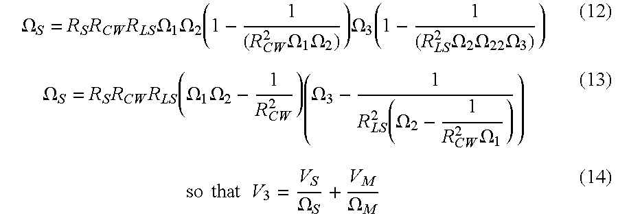
  • the transchest forcing function is determined by solving for the voltage found at node V 3 in FIG. 4 .
  • V 3 V S R S ⁇ R CW ⁇ R LS ⁇ ⁇ 1 ⁇ ⁇ 2 ⁇ ⁇ 22 ⁇ ⁇ 3 ⁇ ⁇ 33 + V M R M ⁇ ⁇ 3 ⁇ ⁇ 33 ⁇ ⁇
  • ⁇ 33 1 - 1 ( R LS 2 ⁇ ⁇ 2 ⁇ ⁇ 22 ⁇ ⁇ 3 ) ( 9 )
  • ⁇ S R S ⁇ R CW ⁇ R LS ⁇ ⁇ 1 ⁇ ⁇ 2 ⁇ ( 1 - 1 ( R CW 2 ⁇ ⁇ 1 ⁇ ⁇ 2 ) ) ⁇ ⁇ 3 ⁇ ( 1 - 1 ( R LS 2 ⁇ ⁇ 2 ⁇ ⁇ 22 ⁇ ⁇ 3 ) ) ( 12 )
  • Equation 14 encapsulates the transchest elements and their association between the forcing function V S (which models a defibrillation circuit and the shock pulse) and the cell membrane voltage V M . Therefore, this completes the first step.
  • V S may now be replaced with a more specific description of the defibrillation circuitry that implements a shock pulse.
  • Equation 19 is a general ordinary differential equation (ODE) that models the effects of any general forcing function V S that represents a phase of a shock pulse waveform applied across the chest.
  • the general ODE equation 19 models the effects of a general shock pulse phase V S on the myocardium, determining cardiac cell response to such a shock pulse phase.
  • R B body impedance (thoracic cage, chest wall, lungs (series, parallel), heart).
  • R B To determine body impedance, R B , we see that the series combination of R H and R LS yields R H +R LS . (FIG. 4 ). The parallel combination of R H +R LS and R LP yields: R LP ⁇ ( R LS + R H ) R LP + R LS + R H . ( 20 )
  • R B [ R TC [ R CW + R LP ⁇ ( R LS + R H ) ( R LP + R LS + R H ) R TC + R CW + R LP ⁇ ( R LS + R H ) ( R LP + R LS + R H ) ] ( 22 )
  • R B is the impedance of the body for this model.
  • V S V 1 e -t/ ⁇ 1 for an initial C 1 capacitor voltage of V 1 .
  • V S V 1 e -t/ ⁇ 1 for an initial C 1 capacitor voltage of V 1 .
  • ⁇ 1 which equals R S C 1 , represents the time constant of ⁇ 1 .
  • Equation 23 models the effects of each phase of a time-truncated, capacitor-discharged shock pulse waveform.
  • Equation 24 is an expression of cell membrane potential during ⁇ 1 of a shock pulse.
  • V M1 ⁇ ( t ) V G ⁇ ⁇ - ( t / ⁇ M ) ⁇ ⁇ ( 1 - 1 ⁇ M ) + ( V 1 ⁇ S ⁇ ) ⁇ ⁇ ( ⁇ 1 ⁇ 1 ⁇ ( 1 - 1 ⁇ M ) - ⁇ M ) ⁇ ( ⁇ - t / ⁇ 1 - ⁇ - ( t / ⁇ M ) ⁇ ⁇ ( 1 - 1 ⁇ M ) ) ( 26 )
  • Equation 26 describes the residual voltage found on a cell at the end of ⁇ 1 .
  • V M1 ⁇ ( t ) ( 1 ⁇ S ) ⁇ ⁇ ( ⁇ 1 ⁇ 1 ⁇ ( 1 - 1 ⁇ M ) - ⁇ M ) ⁇ ( ⁇ - t ⁇ 1 - ⁇ - ( t ⁇ M ) ⁇ ( 1 - 1 ⁇ M ) ) . ( 27 )
  • Equation 27 is differentiated and the resulting equation 27B is set to zero. Equation 27B is then solved for the time t, which represents shock pulse duration required to maximize cardiac cell response.
  • d ⁇ ⁇ ⁇ 1 ( ⁇ 1 ⁇ ⁇ M ⁇ 1 ⁇ ( 1 - 1 ⁇ M ) - ⁇ M ) ⁇ ⁇ ln ( ⁇ 1 ⁇ ( 1 - 1 ⁇ M ) ⁇ M ) , (27C)
  • Equation 29 is again a first-order linear ODE.
  • V M2 ⁇ ( t ) ( V 2 ⁇ S ) ⁇ ⁇ ( ⁇ 2 ⁇ 2 ⁇ ( 1 - 1 ⁇ M ) - ⁇ M ) ⁇ ⁇ ( ⁇ - ( t / ⁇ M ) ⁇ ⁇ ( 1 - 1 ⁇ M ) - ⁇ - t / ⁇ 2 ) + V ⁇ ⁇ ⁇ 1 ⁇ ⁇ - ( t / ⁇ M ) ⁇ ⁇ ( 1 - 1 ⁇ M ) . ( 33 )
  • Equation 33 provides a means to calculate the residual membrane potential at the end of ⁇ 2 for the cells that were not stimulated by ⁇ 1 .
  • the biphasic shock pulse removes the residual change placed on a cell by ⁇ 1 .
  • d ⁇ 2 ( ⁇ 2 ⁇ ⁇ M ⁇ 2 ⁇ ( 1 - 1 ⁇ M ) - ⁇ M ) ⁇ ln ⁇ ⁇ ( 1 + ( ⁇ 2 ⁇ ( 1 - 1 ⁇ M ) - ⁇ M ⁇ 2 ) ⁇ ( ⁇ S ⁇ V ⁇ ⁇ ⁇ 1 V 2 ) ) . ( 34 )
  • an optimal monophasic or biphasic defibrillation waveform may be calculated for an external defibrillator.
  • ⁇ 2 is independent from ⁇ 1 .
  • the only information necessary from ⁇ 1 is where the cell response was left when ⁇ 1 was truncated.
  • ⁇ 2 need not use the same or similar circuitry as that used for ⁇ 1 .
  • may use circuitry as illustrated in FIG. 4 where ⁇ 2 may use circuitry illustrated in FIG. 5 a , or vice-versa.
  • the corresponding design rules for a ⁇ 1 circuitry may be used in conjunction with the design rules for a ⁇ 2 circuitry, regardless of the specific circuitry used to implement each phase of a monophasic or biphasic shock pulse.
  • the present invention is based on the charge burping model hypothesis which postulates and defines an optimal pulse duration for ⁇ 2 as a duration that removes as much of the ⁇ 1 residual charge from the cell as possible. Ideally, the objective is to maintain unstimulated cells with no charge or set them back to relative ground.
  • a further objective of the present invention is to formulate a measurement by which the optimal duration of ⁇ s (cell time constant) and ⁇ m (membrane time constant) can be measured.
  • ⁇ s cell time constant
  • ⁇ m membrane time constant
  • the present invention is designed to correct for “range” of candidate ⁇ m values to fit an optimal duration for a fixed ⁇ 2 .
  • ⁇ 2 is selected so that the capacitance in the model is matched with measured R H to get a “soft landing” to thereby minimize error due to ⁇ m ⁇ E in charge burping ability of ⁇ 2 involving patient variability.
  • the technique of “soft landing” advanced by the present invention limits the error in ⁇ m and sets ⁇ 2 to dynamically adjust within a range of possible ⁇ m values.
  • optimizing solutions are used to determine parameters on which intelligent calculations could be made so that autonomous ⁇ 2 adjustments for variable R H are possible.
  • the charge burping model also accounts for removing the residual charge at the end of ⁇ 1 based on ⁇ 2 delivered by a separate set of capacitors other than those used to deliver ⁇ 1 .
  • C 1 represents the ⁇ 1 capacitor set and C 2 represents the ⁇ 2 capacitor
  • R H represents the resistance of the heart
  • the pair C m and R m represent the membrane series capacitance and resistance of a single cell.
  • the node V s represents the voltage between the electrodes, while V m denotes the voltage across the cell membrane.
  • AEDs have over ICDs, is that the implementation of a ⁇ 2 waveform may be completely independent of the implementation of ⁇ 1 . Specifically, the charging and discharging circuits for ⁇ 1 and ⁇ 2 do not need to be the same circuitry. Unlike ICDs, AEDs are not strictly constrained by space and volume requirements. Within practical limits, in AEDs the capacitance and voltage which characterize ⁇ 2 need not depend on the circuitry and the values of ⁇ 1 .
  • the Lerman-Deale model for AED's define the main series for current to pass through the chest wall, the lungs and the heart. Further, two shunting pathways in parallel with current pathway through the heart are defined. Another shunting pathway is provided by the thoracic cage. However, when compared to the resistivity of the lungs, the thoracic cage resistance is rather negligible.
  • R s represents the resistance of the defibrillation system, including the resistance of the electrodes.
  • R CW and R LS represent the resistances of the chest wall and the lungs, respectively, in series with resistance of the heart, R H .
  • R TC and R LP represent the resistances of the thoracic cage and the lungs, respectively, in parallel with the resistance of the heart.
  • V M ( t ) V O (1 ⁇ e - t/ ⁇ M ). (35)
  • the slope of the curve is zero, which means the terminal value of the time constant is determinable at this point.
  • ⁇ 1 , duration ( ⁇ m ) to ⁇ 2 duration ( ⁇ 2 ) should be ⁇ 1.
  • Charge burping theory postulates that the beneficial effects of ⁇ 2 are maximal when it completely removes the charge deposited on myocardial cell by ⁇ 1 . This theory predicts that ⁇ 1 / ⁇ 2 should be >1 when ⁇ s is >3 ms and ⁇ 1 when ⁇ s ⁇ 3 ms.
  • ⁇ s is defined as the product of the pathway resistance and capacitance.
  • FIG. 7 is a schematic of a circuit which enables the implementation of the theory developed in the present invention.
  • the circuit shows a plurality of double throw switches connecting a plurality of capacitors.
  • the capacitors and the switches are connected to a charge or potential source.
  • the voltage is discharged via electrodes.
  • One aspect of implementing the “soft landing” charge burping technique developed in the present invention is to fix C S for ⁇ 1 and fix C S for ⁇ 2 . Further, ⁇ m is fixed. Then a range of resistance values representing R H are selected.
  • the ⁇ m and R H ranges represent the patient variability problem.
  • capacitor bank values be determined for ⁇ 1 and ⁇ 2 .
  • a variable resistor is used to set R H thus providing a known but variable value and ⁇ m can be set within these practical ranges.
  • FIG. 8 depicts a biphasic defibrillation waveform 300 , generated using equations 35-42 above, in relation to a predicted patient's cellular response curve 304 ; the cellular response, as explained earlier is based on a patient's measured impedance.
  • the residual charge left on the cardiac cells after delivery of ⁇ 1 has been brought back to zero charge, i.e. charge balanced, after the delivery of ⁇ 2 . This charge balance has been achieved because the energy delivered has been allowed to vary.
  • V i is the initial voltage and V f is the final voltage.
  • the above equations make the assumption that the internal impedance of the defibrillator is 0 ohms. This is a good approximation for truncated exponential waveforms, but it is not a good approximation for damped sine waveforms. Damped sine waveforms typically have 10-13 ohms internal impedance. This internal impedance effects the delivered energy. The internal impedance of the defibrillator will absorb a portion of the stored energy in the capacitor, thus reducing the delivered energy. For low patient impedances, the absorbed energy can become quite significant, with on the order of 40% of the stored energy being absorbed by the internal resistances.
  • I is the current and R represents the patient's impedance.
  • R represents the patient's impedance.
  • the current is inversely proportional to the impedance. This means that there is lower current flow for high impedance patients which, in turn, means that it takes longer to deliver the energy to a high impedance patient.
  • V ⁇ ( t ) 1 2 ⁇ CV i ⁇ ⁇ - t RC ( 46 )
  • Equation 46 shows that patient impedance, R, is reflected within in the voltage equation which forms a part of the energy equation. Equation 46 shows that the duration of a defibrillation waveform extends passively with impedance, since it takes longer to reach the truncate voltage.
  • the energy must be allowed to vary.
  • the charge balance waveform of the present invention there is a desire to exactly terminate the defibrillation waveform when the cellular response curve returns to zero charge, see again FIG. 8 Failing to terminate the defibrillation waveform when the cellular response curve returns to zero charge, e.g., overshooting or undershooting the neutral condition, can promote refibrillation.
  • the current is not a system variable
  • the duration of the defibrillation pulse is controlled by the expected cellular response curve, i.e., duration is not a system variable.
  • items 1-3 are not system variables but rather are preset or set in accordance to the patient at hand, the energy must be allowed to vary or the defibrillation system is over constrained and charge balancing cannot be achieved over the range of patient impedances. To deliver fixed energy in a charge balanced system would require the ability to vary the charge voltage. Varying the charge voltage is typically not done since this requires knowing the exact impedance in advance of the shock delivery. Many defibrillators measure the impedance during a high voltage charge delivery since this is more accurate than low voltage measurements done prior to shock delivery. Varying charge voltage also requires a more expensive defibrillation circuitry since capacitor costs increase dramatically with voltage.
  • the duration of the defibrillation pulse does increase slightly for increases in patient impedance. This increased duration is of a much lesser effect than in a traditional truncated waveform.

Abstract

A method for determining an optimal transchest external defibrillation waveform which, when applied through a plurality of electrodes positioned on a patient's torso will produce a desired response in the patient's cardiac cell membranes. The method includes the steps of providing a quantitative model of a defibrillator circuit for producing external defibrillation waveforms, the quantitative model of a patient includes a chest component, a heart component, a cell membrane component and a quantitative description of the desired cardiac membrane response function. Finally, a quantitative description of a transchest external defibrillation waveform that will produce the desired cardiac membrane response function is computed. The computation is made as a function of the desired cardiac membrane response function, the patient model and the defibrillator circuit model.

Description

RELATED APPLICATIONS
This application claims priority to copending non-provisional application Ser. No. 08/886,736, filed Jul. 1, 1997, entitled METHOD AND APPARATUS FOR DETERMINING THE SECOND PHASE OF EXTERNAL DEFIBRILLATOR DEVICES, which is based on provisional patent application Ser. No. 60/021,161, filed Jul. 1, 1996 entitled DYNAMIC SECOND PHASE (φ2) WITH SELF-CORRECTING CHARGE BURPING FOR EXTERNAL DEFIBRILLATOR DEVICES, the contents of both applications of which are herein incorporated by reference.
FIELD OF THE INVENTION
This invention relates generally to an electrotherapy method and apparatus for delivering an electrical pulse to a patient's heart. In particular, this invention relates to a method and apparatus for tailoring a second phase of biphasic waveform delivered by an external defibrillator, to random patients, by performing intelligent calculations and analysis to the results of a first phase segment of a biphasic defibrillation waveform and other parameters pertaining thereto based on theory and practice as disclosed herein.
BACKGROUND OF THE INVENTION
Devices for defibrillating a heart have been known for sometime now. Implantable defibrillators are well accepted by the medical community as effective tools to combat fibrillation for an identified segment of the population. A substantial amount of research in fibrillation and the therapy of defibrillation has been done. Much of the most recent research has concentrated on understanding the effects that a defibrillation shock pulse has on fibrillation to terminate such a condition.
A monophasic waveform is defined to be a single phase, capacitive-discharge, time-truncated, waveform with exponential decay. A biphasic waveform is defined to comprise two monophasic waveforms, separated by time and of opposite polarity. The first phase is designated φ1 and the second phase is designated φ2. The delivery of φ1 is completed before the delivery of φ2 is begun.
After extensive testing, it has been determined that biphasic waveforms are more efficacious than monophasic waveforms. There is a wide debate regarding the reasons for the increased efficacy of biphasic waveforms over that of a monophasic waveforms. One hypothesis holds that φ1 defibrillates the heart and φ2 performs a stabilizing action that keeps the heart from refibrillating.
Biphasic defibrillation waveforms are now the standard of care in clinical use for defibrillation with implantable cardioverter-defibrillators (ICDs), due to the superior performance demonstrated over that of comparable monophasic waveforms. To better understand these significantly different outcomes, ICD research has developed cardiac cell response models to defibrillation. Waveform design criteria have been derived from these first principles and have been applied to monophasic and biphasic waveforms to optimize their parameters. These principles-based design criteria have produced significant improvements over the current art of waveforms.
In a two paper set, Blair developed a model for the optimal design of a monophasic waveform when used for electrical stimulation. (1) Blair, H. A., “On the Intensity-time relations for stimulation by electric currents.” I. J. Gen. Physiol. 1932; 15: 709-729. (2) Blair, H. A., “On the Intensity-time Relations for stimulation by electric currents. II. J. Gen. Physiol. 1932; 15: 731-755. Blair proposed and demonstrated that the optimal duration of a monophasic waveform is equal to the point in time at which the cell response to the stimulus is maximal. Duplicating Blair's model, Walcott extended Blair's analysis to defibrillation, where they obtained supporting experimental results. Walcott, et al., “Choosing the optimal monophasic and biphasic waveforms for ventricular defibrillation.” J. Cardiovasc Electrophysiol. 1995; 6: 737-750.
Independently, Kroll developed a biphasic model for the optimal design of φ2 for a biphasic defibrillation waveform. Kroll, M. W., “A minimal model of the single capacitor biphasic defibrillation waveform.” PACE 1994; 17: 1782-1792. Kroll proposed that the φ2 stabilizing action removed the charge deposited by φ1 from those cells not stimulated by φ1. This has come to be known as “charge burping”. Kroll supported his hypothesis with retrospective analysis of studies by Dixon, et al., Tang, et al., and Freese, et al. regarding single capacitor, biphasic waveform studies. Dixon, et al., “Improved defibrillation thresholds with large contoured epicardial electrodes and biphasic waveforms.” Circulation 1987; 76: 1176-1184; Tang, et al. “Ventricular defibrillation using biphasic waveforms: The Importance of Phasic duration.” J. Am. Coll. Cardiol. 1989; 13: 207-214; and Feeser, S. A., et al. “Strength-duration and probability of success curves for defibrillation with biphasic waveforms.” Circulation 1990; 82: 2128-2141. Again, the Walcott group retrospectively evaluated their extension of Blair's model to φ2 using the Tang and Feeser data sets. Their finding further supported Kroll's hypothesis regarding biphasic defibrillation waveforms. For further discussions on the development of these models, reference may be made to PCT publications WO 95/32020 and WO 95/09673 and to U.S. Pat. No. 5,431,686.
The charge burping hypothesis can be used to develop equations that describe the time course of a cell's membrane potential during a biphasic shock pulse. At the end of φ1, those cells that were not stimulated by φ1 have a residual charge due to the action of φ1 on the cell. The charge burping model hypothesizes that an optimal pulse duration for φ2 is that duration that removes as much of the φ1 residual charge from the cell as possible. Ideally, these unstimulated cells are set back to “relative ground.” The charge burping model proposed by Kroll is based on the circuit model shown in FIG. 2b which is adapted from the general model of a defibrillator illustrated in FIG. 2a.
The charge burping model also accounts for removing the residual cell membrane potential at the end of a φ1 pulse that is independent of a φ2. That is, φ2 is delivered by a set of capacitors separate from the set of capacitors used to deliver φ1. This charge burping model is constructed by adding a second set of capacitors, as illustrated in FIG. 3. In this figure, C1 represents the φ1 capacitor set, C2 represents the φ2 capacitor set RH represents the resistance of the heart, and the pair CM and RM represent membrane series capacitance and resistance of a single cell. The node VS represents the voltage between the electrodes, while VM denotes the voltage across the cell membrane.
External defibrillators send electrical pulses to the patient's heart through electrodes applied to the patient's torso. External defibrillators are useful in any situation where there may be an unanticipated need to provide electrotherapy to a patient on short notice. The advantage of external defibrillators is that they may be used on a patient as needed, then subsequently moved to be used with another patient.
However, this important advantage has two fundamental limitations. First, external defibrillators do not have direct contact with the patient's heart. External defibrillators have traditionally delivered their electrotherapeutic pulses to the patient's heart from the surface of the patient's chest. This is known as the transthoracic defibrillation problem. Second, external defibrillators must be able to be used on patients having a variety of physiological differences. External defibrillators have traditionally operated according to pulse amplitude and duration parameters that can be effective in all patients. This is known as the patient variability problem.
The prior art described above effectively models implantable defibrillators, however it does not fully addressed the transthoracic defibrillation problem nor the patient variability problem. In fact, these two limitations to external defibrillators are not fully appreciated by those in the art. For example, prior art disclosures of the use of truncated exponential monophasic or biphasic shock pulses in implantable or external defibrillators have provided little guidance for the design of an external defibrillator that will successfully defibrillate across a large, heterogeneous population of patients. In particular, an implantable defibrillator and an external defibrillator can deliver a shock pulse of similar form, and yet the actual implementation of the waveform delivery system is radically different.
In the past five years, new research in ICD therapy has developed and demonstrated defibrillation models that provide waveform design rules from first principles. These defibrillation models and their associated design rules for the development of defibrillation waveforms and their characteristics were first developed by Kroll and Irnich for monophasic waveforms using effective and rheobase current concepts. (1) Kroll, M. W., “A minimal model of the monophasic defibrillation pulse.” PACE 1993; 15: 769. (2) Irnich, W., “Optimal truncation of defibrillation pulses.” PACE 1995; 18: 673. Subsequently, Kroll, Walcott, Cleland and others developed the passive cardiac cell membrane response model for monophasic and biphasic waveforms, herein called the cell response model. (1) Kroll, M. W., “A minimal model of the single capacitor biphasic defibrillation waveform.” PACE 1994; 17: 1782. (2) Walcott, G. P., Walker, R. G., Cates. A. W., Krassowska, W., Smith, W. M, Ideker R E. “Choosing the optimal monophasic and biphasic waveforms for ventricular defibrillation.” J Cardiovasc Electrophysiol 1995; 6: 737; and Cleland B G. “A conceptual basis for defibrillation waveforms.” PACE 1996; 19: 1186).
A significant increase in the understanding of waveform design has occurred and substantial improvements have been made by using these newly developed design principles. Block et al. has recently written a comprehensive survey of the new principles-based theories and their impact on optimizing internal defibrillation through improved waveforms. Block M, Breithardt G., “Optimizing defibrillation through improved waveforms.” PACE 1995; 18: 526.
There have not been significant developments in external defibrillation waveforms beyond the two basic monophasic waveforms: the damped sine or the truncated exponential. To date, their design for transthoracic defibrillation has been based almost entirely on empirically derived data. It seems that the design of monophasic and biphasic waveforms for external defibrillation has not yet been generally influenced by the important developments in ICD research.
Recently there has been reported research on the development and validation of a biphasic truncated exponential waveform in which it was compared clinically to a damped sine waveform. For additional background, reference may be made to U.S. Pat. Nos. 5,593,427, 5,601,612 and 5,607,454. See also: Gliner B E, Lyster T E, Dillon S M, Bardy G H, “Transthoracic defibrillation of swine with monophasic and biphasic waveforms.” Circulation 1995; 92: 1634-1643; Bardy G H, Gliner B E, Kudenchuk P J, Poole J E, Dolack G L, Jones G K, Anderson J, Troutman C, Johnson G.; “Truncated biphasic pulses for transthoracic defibrillation.” Circulation 1995; 91: 1768-1774; and Bardy G H et al, “For the Transthoracic Investigators. Multicenter comparison of truncated biphasic shocks and standard damped sine wave monophasic shocks for transthoracic ventricular defibrillation.” Circulation 1996; 94: 2507-2514. Although the research determined a usable biphasic waveform, there was no new theoretical understanding determined for external waveform design. It appears that external waveform research may develop a “rules-of-thumb by trial and error” design approach much like that established in the early stages of theoretical ICD research. The noted limitations of the transthoracic biphasic waveform may be due in part to a lack of principles-based design rules to determine its waveform characteristics.
There is a continued need for a device designed to perform a quick and automatic adjustment of phase 2 relative to phase 1 if AED's are to be advantageously applied to random patients according to a cell response model. Further, the model and the device must be adaptable to patient variance and be able to provide automatic adjustment in a dynamic environment.
SUMMARY OF THE INVENTION
The present invention relates to an external defibrillation method and apparatus that addresses the limitations in the prior art. The present invention incorporates three singular practices that distinguish the practice of designing external defibrillators from the practice of designing implantable defibrillators. These practices are 1) designing multiphasic transthoracic shock pulse waveforms from principles based on cardiac electrophysiology, 2) designing multiphasic transthoracic shock pulse waveforms in which each phase of the waveform can be designed without implementation limitations placed on its charging and delivery means by such means for prior waveform phases, and 3) designing multiphasic transthoracic shock pulse waveforms to operate across a wide range of parameters determined by a large, heterogeneous population of patients.
In particular, the present invention provides for a method and apparatus for tailoring and reforming a second phase (φ2) of a biphasic defibrillation waveform relative to a first phase (φ1) of the waveform based on intelligent calculations. The method includes the steps of determining and providing a quantitative description of the desired cardiac membrane response function. A quantitative model of a defibrillator circuit for producing external defibrillation waveforms is then provided. Also provided is a quantitative model of a patient which includes a chest component, a heart component and a cell membrane component. A quantitative description of a transchest external defibrillation waveform that will produce the desired cardiac membrane response function is then computed. Intelligent calculations based on the phase 1 cell response is then computed to determine the desired phase 2 waveform. The computation is made as a function of the desired cardiac membrane response function, the patient model and the defibrillator circuit model.
BRIEF DESCRIPTION OF THE DRAWINGS
FIGS. 1a and 1 b are perspective views of an AED according to the present invention.
FIG. 2a is a very simplified defibrillator model.
FIG. 2b is a known monophasic defibrillation model.
FIG. 3 is a known biphasic defibrillation model.
FIG. 4 represents a monophasic or biphasic capacitive-discharge external defibrillation model according to the present invention.
FIG. 5a represents a monophasic capacitor-inductor external defibrillator model according to the present invention.
FIG. 5b represents an alternative embodiment of a biphasic capacitor-inductor external defibrillator model according to the present invention.
FIG. 6 illustrates a biphasic waveform generator utilizing the present invention.
FIG. 7 is a schematic diagram of a circuit which enables the implementation of the present invention.
FIG. 8 illustrates a biphasic waveform in relation to a cellular response curve.
DESCRIPTION OF THE PREFERRED EMBODIMENT
The present invention provides a method and apparatus for tailoring a second phase (φ2) of a biphasic waveform delivered by an external defibrillator, to random patients, by performing intelligent calculations and analysis to the results of a first phase (φ1) segment of a biphasic defibrillation waveform and other parameters pertaining thereto. Prior to describing the present invention, a discussion of the development of an external defibrillation model will be given.
External Defibrillator Model
The apparatus of the present invention is an automated external defibrillator (AED) illustrated in FIGS. 1a and 1 b. FIG. 1a illustrates an AED 10, including a plastic case 12 with a carrying handle 14. A lid 16 is provided which covers an electrode compartment 18. An electrode connector 20, a speaker 22 and a diagnostic panel (not shown) are located on case 12 within electrode compartment 18. FIG. 1b illustrates AED 10 having a pair of electrodes 24 connected thereto. Electrodes 24 can be pre-connected to connector 20 and stored in compartment 18.
The operation of AED 10 is described briefly below. A rescue mode of AED 10 is initiated when lid 16 is opened to access electrodes 24. The opening of lid 16 is detected by AED 10 to effectively turn on the device. AED 10 then quickly runs a short test routine. After electrodes 24 have been placed on the patient, AED 10 senses patient specific parameters, such as impedance, voltage, current, charge or other measurable parameters of the patient. The patient specific parameters are then utilized in the design of optimal waveforms as will be described below.
If a shockable condition is detected through electrodes 24, a plurality of capacitors inside of AED 10 are charged from an energy source, typically a detachable battery pack. Based upon the patient specific parameters sensed, the duration and other characteristics of a discharge waveform are then calculated. The energy stored in AED 10 is then discharged to the patient through electrodes 24.
For a more detailed description of the physical structure of AED 10 or the process involved in sensing, charging, shocking and testing, reference should be made to applicants co-pending application Ser. No. 08/512,441, filed Aug. 8, 1995 entitled AUTOMATED EXTERNAL DEFIBRILLATOR WITH SELF-TEST SYSTEM which is assigned to the assignee of the present invention, the disclosure of which is herein incorporated by reference.
In the present invention it is not assumed that both phases of a biphasic waveform are delivered using the same set of capacitors or that both phases of a biphasic waveform are delivered using the capacitor set in the same electrical configuration, although such an embodiment is considered within the spirit and scope of the present invention.
Transthoracic defibrillation is generally performed by placing electrodes on the apex and anterior positions of the chest wall. With this electrode arrangement, nearly all current passing through the heart is conducted by the lungs and the equipotential surfaces pass through the myocardium normal to the electrode axis. The present invention uses the transthoracic charge burping model to develop design equations that describe the time course of a cell's membrane potential during a transthoracic biphasic shock pulse. These equations are then used to create equations that describe the design of monophasic and biphasic shock pulses for transchest defibrillation to optimize the design of φ1 for defibrillating and the design φ2 for stabilizing. These optimizing shock pulse design equations are called design rules.
According to the present invention, the main series pathway for current is to pass through the chest wall, the lungs, and the heart. Additionally, there are two important shunting pathways in parallel with the current pathway through the heart. These shunting pathways must be taken into consideration. The lungs shunt current around the heart through a parallel pathway. The second shunting pathway is provided by the thoracic cage. The resistivity of the thoracic cage and the skeletal muscle structure is low when compared to lungs. The high resistivity of the lungs and the shunting pathways are characterizing elements of external defibrillation that distinguish the art from intracardiac defibrillation and implantable defibrillation technologies.
Therefore, in the transthoracic defibrillation model of the present invention illustrated in FIG. 4, there are several resistances in addition to those discussed for the charge burping model above. RS represents the resistance of the defibrillation system, including the resistance of the defibrillation electrodes. RCW and RLS represent the resistances of the chest wall and the lungs, respectively, in series with resistance of the heart, RH. RTC and RLP represent the resistances of the thoracic cage and the lungs, respectively, in parallel with the resistance of the heart.
The design rules for external defibrillation waveforms are determined in three steps. In the first step, the transchest forcing function is determined. The transchest forcing function is the name that is given to the voltage that is applied across each cardiac cell during an external defibrillation shock. In the second step, the design equations for φ1 of a shock pulse are determined. The design equations are the equations describing the cell's response to the φ1 transchest forcing function, the equation describing the optimal φ1 pulse duration, and the equation describing the optimal φ1 capacitor. Therefore, step two relates the cell response to the action of a monophasic shock pulse or the first phase of a biphasic shock pulse. This relation is used to determine the optimal design rules and thereby design parameters for the implementation of this phase in an external defibrillator. It will be clear to those in the art that step two is not restricted to capacitor discharge shock pulses and their associated transchest forcing function. Another common implementation of an external defibrillator incorporates a damped sine wave for a shock pulse and can be either a monophasic or biphasic waveform. This type of external defibrillator is modeled by the circuits shown in FIGS. 5a and 5 b. In the third step, the design equations for φ2 of a shock pulse are determined. The design equations are the equations describing the cell's response to the φ2 transchest forcing function, the equation describing the optimal φ2 pulse duration and the equation describing the optimal φ2 capacitor. These design equations are employed to determine the optimal design rules and thereby design parameters of φ2 of a biphasic shock pulse with respect to how the cell responds to the shock pulse. An important element of this invention is to provide shock pulse waveforms that are designed from a cardiac cell response model developed from first principles and that correctly determines the effects of the chest and its components on the ability of a shock pulse to defibrillate.
The transchest forcing function is determined by solving for the voltage found at node V3 in FIG. 4. The transchest forcing function is derived by solving for V3 using the following three nodal equations: V 1 - V S R S + V 1 R TC + V 1 - V 2 R CW = 0 , ( 1 ) V 2 - V 1 R CW + V 2 R LP + V 2 - V 3 R LS = 0 , and ( 2 ) V 3 - V 2 R LS + V 3 R H + V 3 - V M R M = 0. ( 3 )
Figure US06263239-20010717-M00001
Equation 1 can be rewritten as V 1 ( 1 R S + 1 R BC + 1 R CW ) = V S R S + V 2 R CW . (4A) V 1 = V S R S Ω 1 + V 2 R CW Ω 1 , where Ω 1 = 1 R S + 1 R TC + 1 R CW . (4B)
Figure US06263239-20010717-M00002
Rewriting equation 2, we have V 2 ( 1 R CW + 1 R LP + 1 R LS ) = V 1 R CW + V 3 R LS . (4C)
Figure US06263239-20010717-M00003
By substituting equation 4B for V1 into equation 4C, we can solve for V2 as an expression of VS and V3: V 2 = V S R S R CW Ω 1 Ω 2 Ω 22 + V 3 R LS Ω 2 Ω 22 , where Ω 2 = 1 R LS + 1 R LP + 1 R CW , and Ω 22 = 1 - 1 R CW 2 Ω 1 Ω 2 . ( 5 )
Figure US06263239-20010717-M00004
Now solving for V3 as an expression of VS and VM, equation 3 may be re-arranged as V 3 ( 1 R LS + 1 R H + 1 R M ) = V 2 R LS + V M R M so that ( 6 ) V 3 = V 2 R LS Ω 3 + V M R M Ω 3 where Ω 3 = 1 R LS + 1 R H + 1 R M . ( 7 )
Figure US06263239-20010717-M00005
Substituting equation 5 for V2 into equation 7, we can solve for V3 as an expression of VS and VM: V 3 = V S R S R CW R LS Ω 1 Ω 2 Ω 22 Ω 3 Ω 33 + V M R M Ω 3 Ω 33 where ( 8 ) Ω 33 = 1 - 1 ( R LS 2 Ω 2 Ω 22 Ω 3 ) ( 9 )
Figure US06263239-20010717-M00006
From equation 8 we define ΩM to be: Ω M = R M Ω 3 Ω 33 = R M Ω 3 ( 1 - 1 ( R LS 2 Ω 2 Ω 22 Ω 3 ) ) Ω M = R M ( Ω 3 - 1 R LS 2 ( Ω 2 - 1 R CW 2 Ω 1 ) ) . ( 10 )
Figure US06263239-20010717-M00007
From equation 8 we also define ΩS to be:
ΩS =R S R CW R LSΩ1Ω2Ω3Ω22Ω33  (11)
Ω S = R S R CW R LS Ω 1 Ω 2 ( 1 - 1 ( R CW 2 Ω 1 Ω 2 ) ) Ω 3 ( 1 - 1 ( R LS 2 Ω 2 Ω 22 Ω 3 ) ) ( 12 ) Ω S = R S R CW R LS ( Ω 1 Ω 2 - 1 R CW 2 ) ( Ω 3 - 1 R LS 2 ( Ω 2 - 1 R CW 2 Ω 1 ) ) ( 13 ) so that V 3 = V S Ω S + V M Ω M ( 14 )
Figure US06263239-20010717-M00008
is the general transchest transfer function as shown in FIG. 4 or FIGS. 5a and 5 b. Equation 14 encapsulates the transchest elements and their association between the forcing function VS (which models a defibrillation circuit and the shock pulse) and the cell membrane voltage VM. Therefore, this completes the first step.
The variable VS may now be replaced with a more specific description of the defibrillation circuitry that implements a shock pulse. For a first example, a monophasic time-truncated, capacitive-discharge circuit may be represented by VS=V1e-t/τ 1, where V1 is the leading-edge voltage for the shock pulse and τ1=RC1, with R determined below.
As shown in FIGS. 5a and 5 b, a second example would be a monophasic damped sine wave circuit, represented by V S = V 1 ( τ C1 τ C1 - τ L1 ) ( - t / τ C1 - - t / τ L1 ) (14B)
Figure US06263239-20010717-M00009
where V1 is the voltage on the charged capacitor C1τC1=RC1 and τL1=L1/R. Every step illustrated below may be performed with this and other similar transchest forcing functions which represent defibrillator circuitry.
To proceed with step two, from FIG. 4, nodal analysis provides an equation for VM: C M V M t + V M - V 3 R M = 0. ( 15 )
Figure US06263239-20010717-M00010
Rearranging equation 15, we have C M V M t + V M R M = V 3 R M . ( 16 )
Figure US06263239-20010717-M00011
Next, substituting equation 14 as an expression for V3 into equation 16, the cell membrane response is now calculated as follows: C M V M t + V M R M = 1 R M ( V S Ω S + V M Ω M ) ( 17 ) C M V M t + V M R M - V M R M Ω M = V S R M Ω S C M V M t + V M R M ( 1 - 1 Ω M ) = V S R M Ω S ( 18 )
Figure US06263239-20010717-M00012
Dividing through by CM, and setting τM=RMCM, then equation 18 becomes V M t + V M τ M ( 1 - 1 Ω M ) = V S τ M ( 1 Ω S ) . ( 19 )
Figure US06263239-20010717-M00013
Equation 19 is a general ordinary differential equation (ODE) that models the effects of any general forcing function VS that represents a phase of a shock pulse waveform applied across the chest. The general ODE equation 19 models the effects of a general shock pulse phase VS on the myocardium, determining cardiac cell response to such a shock pulse phase.
In the equations given below:
C1 equals the capacitance of the first capacitor bank and VS=V1e-t/τ 1 ;
C2 equals the capacitance of the second capacitor bank and VS=V2e-t/τ 2 ;
R=RS+RB, where RS=System impedance (device and electrodes);
RB=body impedance (thoracic cage, chest wall, lungs (series, parallel), heart).
To determine body impedance, RB, we see that the series combination of RH and RLS yields RH+RLS. (FIG. 4). The parallel combination of RH+RLS and RLP yields: R LP ( R LS + R H ) R LP + R LS + R H . ( 20 )
Figure US06263239-20010717-M00014
The series combination of equation 20 and RCW yields: R CW + R LP ( R LS + R H ) ( R LP + R LS + R H ) . ( 21 )
Figure US06263239-20010717-M00015
The parallel combination of equation 21 and RTC yields: R B = [ R TC [ R CW + R LP ( R LS + R H ) ( R LP + R LS + R H ) R TC + R CW + R LP ( R LS + R H ) ( R LP + R LS + R H ) ] ( 22 )
Figure US06263239-20010717-M00016
where RB is the impedance of the body for this model.
The discharge of a single capacitor is modeled by VS=V1e-t/τ 1 for an initial C1 capacitor voltage of V1. Placing VS into equation 19 gives: V M t + V M τ M ( 1 - 1 Ω M ) = V 1 - t / τ 1 τ M Ω S ( 23 )
Figure US06263239-20010717-M00017
where τM=RMCM represents the time constant of the myocardial cell in the circuit model, and τ1, which equals RSC1, represents the time constant of φ1. Such a standard linear ODE as equation 23 has the form y x + P ( X ) Y = Q ( x ) .
Figure US06263239-20010717-M00018
These linear ODEs have an integration factor that equals e∫pdx. The general solution to such equations is:
Y=e -∫pdx [∫e ∫pdx Qdx+c].
The ODE in equation 23 models the effects of each phase of a time-truncated, capacitor-discharged shock pulse waveform. Equation 23 is a first-order linear ODE, and may be solved using the method of integration factors, to get: V M1 ( t ) = k - ( t / τ M ) ( 1 - 1 Ω M ) + ( V 1 Ω S ) ( τ 1 τ 1 ( 1 - 2 Ω M ) - τ M ) - t / τ 1 . ( 24 )
Figure US06263239-20010717-M00019
Equation 24 is an expression of cell membrane potential during φ1 of a shock pulse. To determine the constant of integration k, the initial value of VM1 is assumed to be VM1(0)=VG (“cell ground”). Applying this initial condition to equation 24, k is found to be k = V G - ( V o Ω S ) ( τ 1 τ 1 ( 1 - 1 Ω M ) - τ M ) . ( 25 )
Figure US06263239-20010717-M00020
Assuming τ1=RC1, where R=RS+RB, then the solution to the initial-value problem for φ1 is: V M1 ( t ) = V G - ( t / τ M ) ( 1 - 1 Ω M ) + ( V 1 Ω S ) ( τ 1 τ 1 ( 1 - 1 Ω M ) - τ M ) ( - t / τ 1 - - ( t / τ M ) ( 1 - 1 Ω M ) ) ( 26 )
Figure US06263239-20010717-M00021
Equation 26 describes the residual voltage found on a cell at the end of φ1.
Assuming VG=0 and V1=1, the solution for cell response to an external shock pulse is V M1 ( t ) = ( 1 Ω S ) ( τ 1 τ 1 ( 1 - 1 Ω M ) - τ M ) ( - t τ 1 - - ( t τ M ) ( 1 - 1 Ω M ) ) . ( 27 )
Figure US06263239-20010717-M00022
We may now determine optimal durations for φ1 according to criteria for desired cell response. One such design role or criterion is that the φ1 duration is equal to the time required for the external defibrillator shock pulse to bring the cell response to its maximum possible level. To determine this duration, equation 27 is differentiated and the resulting equation 27B is set to zero. Equation 27B is then solved for the time t, which represents shock pulse duration required to maximize cardiac cell response. ( AB τ M ) - Bt / τ M - ( A τ 1 ) - t / τ 1 = 0 , where A = ( 1 Ω S ) ( τ 1 τ 1 ( 1 - 1 Ω M ) - τ M ) and B = 1 - 1 Ω M . (27B)
Figure US06263239-20010717-M00023
Solving for t, the optimal duration dφ1 for a monophasic shock pulse or φ1 of a biphasic shock pulse is found to be d φ 1 = ( τ 1 τ M τ 1 ( 1 - 1 Ω M ) - τ M ) ln ( τ 1 ( 1 - 1 Ω M ) τ M ) , (27C)
Figure US06263239-20010717-M00024
where “1n” represents the logarithm to the base e, the natural logarithm.
For φ2, an analysis almost identical to equations 20 through 27 above is derived. The differences are two-fold. First, a biphasic waveform reverses the flow of current through the myocardium during φ2. Reversing the flow of current in the circuit model changes the sign on the current. The sign changes on the right hand side of equation 23.
The second difference is the step taken to incorporate an independent φ2 into the charge burping model. Therefore, the φ2 ODE incorporates the C2 capacitor set and their associated leading-edge voltage, V2, for the φ2 portion of the pulse. Then τ2 represents the φ2 time constant; τ2=RC2, and VS=−V2e-t/τ 2 . Equation 23 now becomes: V M t + ( V M τ M ) ( 1 - 1 Ω M ) = - V 2 - t / τ 2 τ M Ω S . ( 29 )
Figure US06263239-20010717-M00025
Equation 29 is again a first-order linear ODE. In a similar manner, its general solution is determined to be: V M2 ( t ) = k ( - t / τ M ) ( 1 - 1 Ω M ) - ( V 2 Ω S ) ( τ 2 τ 2 ( 1 - 1 Ω M ) - τ M ) . ( 30 )
Figure US06263239-20010717-M00026
To determine the constant of integration k, the value of VM2 at the end of φ1 is
V M2(0)=V M1(d φ1)=V φ1′  (31)
where dφ1 is the overall time of discharge for φ1 and Vφ1 is the voltage left on the cell at the end of φ1. Applying the initial condition to equation 30 and solving for k: k = V φ 1 + ( V 2 Ω S ) ( τ 2 τ 2 ( 1 - 1 Ω M ) - τ M ) . ( 32 )
Figure US06263239-20010717-M00027
The solution to the initial-value problem for φ2 is V M2 ( t ) = ( V 2 Ω S ) ( τ 2 τ 2 ( 1 - 1 Ω M ) - τ M ) ( - ( t / τ M ) ( 1 - 1 Ω M ) - - t / τ 2 ) + V φ 1 - ( t / τ M ) ( 1 - 1 Ω M ) . ( 33 )
Figure US06263239-20010717-M00028
Equation 33 provides a means to calculate the residual membrane potential at the end of φ2 for the cells that were not stimulated by φ1. Setting Equation 33 equal to zero, we solve for t, thereby determining the duration of φ2, denoted dφ2, such that VM2(dφ2)=0. By designing φ2 with a duration dφ2, the biphasic shock pulse removes the residual change placed on a cell by φ1. We determine dφ2 to be: d φ2 = ( τ 2 τ M τ 2 ( 1 - 1 Ω M ) - τ M ) · ln ( 1 + ( τ 2 ( 1 - 1 Ω M ) - τ M τ 2 ) ( Ω S V φ 1 V 2 ) ) . ( 34 )
Figure US06263239-20010717-M00029
From the equations above, an optimal monophasic or biphasic defibrillation waveform may be calculated for an external defibrillator.
As an example, an external defibrillator may be designed as set forth below. Assume a monophasic truncated exponential shock pulse, a 200 μF capacitor, so that τ1=R·(200 μF). Suppose also that the external defibrillator is designed to apply the maximal cardiac cell response design rule (equation 27C) to determine the duration of the discharge. Suppose further that the human cardiac cell time constant is estimated to be 3±1 ms. Further assume that the external defibrillator energy source comprises five 1000 μF capacitors in series to implement a 200 μF capacitor bank. If each capacitor is charged to 400V, for a total of 2000V for the leading-edge voltage, this represents 400J of stored energy. The transchest elements are estimated at: 82% current through the thoracic cage; 14% through the chest wall and lungs in parallel; and 4% of applied current through the lung in series with the heart. Then the membrane resistance coefficient ΩM=5.9, and the system resistance coefficient ΩS=2.3. Then the table below illustrates the application of the design rule as the overall chest resistance ranges from 25Ω to 200Ω:
R (Ω) τ1 d(φ1) Vfinal Edelivered
25 5.2 5.05 757 343
50 10.2 6.90 1017 297
75 15.2 8.15 1170 263
100 20.2 9.10 1275 238
125 25.2 9.90 1350 216
150 30.2 10.55 1410 201
175 35.2 11.15 1457 186
200 40.2 11.65 1497 176
It should be noted and understood that the design of φ2 is independent from φ1. To design φ2, the only information necessary from φ1 is where the cell response was left when φ1 was truncated. Additionally, φ2 need not use the same or similar circuitry as that used for φ1. For example, φ may use circuitry as illustrated in FIG. 4 where φ2 may use circuitry illustrated in FIG. 5a, or vice-versa. The corresponding design rules for a φ1 circuitry may be used in conjunction with the design rules for a φ2 circuitry, regardless of the specific circuitry used to implement each phase of a monophasic or biphasic shock pulse.
Present Invention
The present invention is based on the charge burping model hypothesis which postulates and defines an optimal pulse duration for φ2 as a duration that removes as much of the φ1 residual charge from the cell as possible. Ideally, the objective is to maintain unstimulated cells with no charge or set them back to relative ground.
A further objective of the present invention is to formulate a measurement by which the optimal duration of τs (cell time constant) and τm (membrane time constant) can be measured. Although one can choose a proper φ2 (fixed) for a given cell response φ1, in transthoracic shock pulse applications, τm is not known and it varies across patients, waveforms and time. For a fixed φ2, therefore, the error in τm could be substantial. Realizing this, the present invention is designed to correct for “range” of candidate τm values to fit an optimal duration for a fixed φ2. In other words, φ2 is selected so that the capacitance in the model is matched with measured RH to get a “soft landing” to thereby minimize error due to τm±E in charge burping ability of φ2 involving patient variability.
The technique of “soft landing” advanced by the present invention limits the error in τm and sets φ2 to dynamically adjust within a range of possible τm values. As discussed hereinbelow, optimizing solutions are used to determine parameters on which intelligent calculations could be made so that autonomous φ2 adjustments for variable RH are possible.
The charge burping model also accounts for removing the residual charge at the end of φ1 based on φ2 delivered by a separate set of capacitors other than those used to deliver φ1. Referring now to FIG. 3, C1 represents the φ1 capacitor set and C2 represents the φ2 capacitor, RH represents the resistance of the heart, and the pair Cm and Rm represent the membrane series capacitance and resistance of a single cell. The node Vs represents the voltage between the electrodes, while Vm denotes the voltage across the cell membrane.
Accordingly, one of the advantages that AEDs have over ICDs, is that the implementation of a φ2 waveform may be completely independent of the implementation of φ1. Specifically, the charging and discharging circuits for φ1 and φ2 do not need to be the same circuitry. Unlike ICDs, AEDs are not strictly constrained by space and volume requirements. Within practical limits, in AEDs the capacitance and voltage which characterize φ2 need not depend on the circuitry and the values of φ1.
The Lerman-Deale model for AED's define the main series for current to pass through the chest wall, the lungs and the heart. Further, two shunting pathways in parallel with current pathway through the heart are defined. Another shunting pathway is provided by the thoracic cage. However, when compared to the resistivity of the lungs, the thoracic cage resistance is rather negligible.
Thus, considering the transthoracic defibrillation model of FIG. 4, there are several other resistances in addition to those discussed for the charge burping model hereinabove. Rs represents the resistance of the defibrillation system, including the resistance of the electrodes. RCW and RLS represent the resistances of the chest wall and the lungs, respectively, in series with resistance of the heart, RH. RTC and RLP represent the resistances of the thoracic cage and the lungs, respectively, in parallel with the resistance of the heart.
As discussed hereinabove, developing design equations which enable adjustments for variable resistances encountered in the transthoracic defibrillation model of FIG. 4 is one of the advances of the present invention. In order to adjust for variable RH both φ1 and φ2 are assumed fixed. Then φ2 is selected to have a range of capacitance values which permit to optimize the slope of the voltage curve at time t. In other words, CS2 for φ2 is chosen such that v t = 0
Figure US06263239-20010717-M00030
The design parameters of the present invention are derived from equation 35, as follows:
V M(t)=V O(1−e - t/τ M ).   (35)
Equation 35 can be rewritten as: V M2 ( t ) = ( V φ1 + { τ 2 τ 2 - τ M } V 2 ) e - τ τ M - ( τ 2 τ 2 - τ M ) V 2 - t / τ 2 = 0. Letting A = V φ 1 + B and B = τ 2 τ 2 - τ M V 2 , ( 36 )
Figure US06263239-20010717-M00031
equation 36 can be written as: V ( t ) = Ae - t τ M - Be - t τ 2 . ( 37 )
Figure US06263239-20010717-M00032
Differentiating equation 37 with respect to t, we have the following: v t = - A - t / τ M τ M + B τ 2 - t / τ 2 = 0. ( 38 )
Figure US06263239-20010717-M00033
Equation 38 is the profile of φ1 waveform and at v t = 0 ,
Figure US06263239-20010717-M00034
the slope of the curve is zero, which means the terminal value of the time constant is determinable at this point.
Thus, solving equation 38 for the value of t, we have: t = t 2 τ 2 - τ M τ M · ln { τ 2 τ M ( V φ 1 + τ 2 τ 2 - τ M V 2 ) ( τ 2 τ 2 - τ M ) V 2 } where ( 39 ) t 1 = ( τ 2 τ M τ 2 - τ M ) ln { 1 + ( τ 2 - τ M τ 2 ) ( V φ 1 V 2 ) } ( 40 ) t 1 = t 1 + ( τ 2 τ M τ 2 - τ M ln { τ 2 τ M } ) . ( 41 )
Figure US06263239-20010717-M00035
For biphasic defibrillation waveforms, it is generally accepted that the ratio of φ1, duration (τm) to φ2 duration (τ2) should be ≧1. Charge burping theory postulates that the beneficial effects of φ2 are maximal when it completely removes the charge deposited on myocardial cell by φ1. This theory predicts that φ12 should be >1 when τs is >3 ms and <1 when τs <3 ms. τs is defined as the product of the pathway resistance and capacitance. (See NASPE ABSTRACTS, Section 361 entitled Charge Burping Predicts Optimal Ratios of Phase Duration for Biphasic Defibrillation, by Charles D. Swerdlow, M.D., Wei Fan, M.D., James E. Brewer, M.S., Cedar-Sinai Medical Center, Los Angeles, Calif.
In light of the proposed duration ratio of φ1 and φ2, wherein the optimal solution is indicated to be at t1=t2 where t is the duration of φ1 and t2 is the duration of φ2 and superimposing this condition on equation 41 hereinabove, we have: t 2 = t 1 + ( τ 2 τ M τ 2 - τ M · ln { τ 2 τ M } ) setting t 2 = t 1 , remanaging terms we have: ( 42 ) t 2 - t 1 = 0 = τ 2 τ M τ 2 - τ M · ln { τ 2 τ M } or τ 2 τ M · ln ( τ 2 τ M ) = 0 or ln τ 2 τ M = 0 or τ 2 τ M = 1.
Figure US06263239-20010717-M00036
From the result of equation 42 we make the final conclusion that the optimal charge burping is obtained when τ2m. From prior definition, we have established that τm=RH·CS. Thus, in accordance with equation 42, τ2m=RH·CS.
Referring now to FIG. 6, a biphasic defibrillation waveform generated using the equations 35-42 is shown. At Vm=0 and dVm/dt=0, φ1 and φ2 are equal to zero.
FIG. 7 is a schematic of a circuit which enables the implementation of the theory developed in the present invention. The circuit shows a plurality of double throw switches connecting a plurality of capacitors. The capacitors and the switches are connected to a charge or potential source. The voltage is discharged via electrodes. One aspect of implementing the “soft landing” charge burping technique developed in the present invention is to fix CS for φ1 and fix CS for φ2. Further, τm is fixed. Then a range of resistance values representing RH are selected. The τm and RH ranges represent the patient variability problem. The objective is to enable corrective action such that CS values could range between 40 mf-200 mf and dv/dt=0 for τ2. As indicated hereinabove the error in changing burping is minimized for τ2 when dv/dt=0.
The implementation of the present invention requires that capacitor bank values be determined for φ1 and φ2. Specifically, the capacitor values for φ1 should be designed to realize dv/dt=0 and V=0 for φ2 to minimize charge burping error due to RH and τm. Where a variable resistor is used to set RH thus providing a known but variable value and τm can be set within these practical ranges.
FIG. 8 depicts a biphasic defibrillation waveform 300, generated using equations 35-42 above, in relation to a predicted patient's cellular response curve 304; the cellular response, as explained earlier is based on a patient's measured impedance. As shown, the residual charge left on the cardiac cells after delivery of φ1 has been brought back to zero charge, i.e. charge balanced, after the delivery of φ2. This charge balance has been achieved because the energy delivered has been allowed to vary.
Traditionally, all defibrillation waveforms have delivered a fixed energy. This has been primarily because therapy has been measured in joules. These fixed energy waveforms, which include monophasic damped sine, biphasic truncated exponential, and monophasic truncated exponential waveforms, only passively responded to patient and/or system impedance.
This passive response was by charging a capacitor to a fixed voltage, wherein the voltage was adjusted depending on amount of fixed energy to be delivered that was desired, and discharging it across the patient, whom acts as the load. The energy delivered is controlled by the simple equation: E = 1 2 CV 2 ( 43 )
Figure US06263239-20010717-M00037
where: E is the energy, C is the capacitance of the charging capacitor, and V is the voltage. In the case of the truncated exponential waveform, equation 43 is extended to: E = 1 2 C ( V i 2 - V f 2 ) ( 44 )
Figure US06263239-20010717-M00038
where: Vi is the initial voltage and Vf is the final voltage. Significantly, no form of patient impedance or system impedance appears in either of the above equations.
With respect to system impedance, the above equations make the assumption that the internal impedance of the defibrillator is 0 ohms. This is a good approximation for truncated exponential waveforms, but it is not a good approximation for damped sine waveforms. Damped sine waveforms typically have 10-13 ohms internal impedance. This internal impedance effects the delivered energy. The internal impedance of the defibrillator will absorb a portion of the stored energy in the capacitor, thus reducing the delivered energy. For low patient impedances, the absorbed energy can become quite significant, with on the order of 40% of the stored energy being absorbed by the internal resistances.
With respect to patient impedance, the patient acts as the load and, as such, the peak current is simply a function of the peak voltage via Ohm's law: I = V R ( 45 )
Figure US06263239-20010717-M00039
where: I is the current and R represents the patient's impedance. As equation 45 indicates, the current is inversely proportional to the impedance. This means that there is lower current flow for high impedance patients which, in turn, means that it takes longer to deliver the energy to a high impedance patient. This fact is further exemplified when considering the equation for a truncated exponential voltage at any point in time: V ( t ) = 1 2 CV i - t RC ( 46 )
Figure US06263239-20010717-M00040
Equation 46 shows that patient impedance, R, is reflected within in the voltage equation which forms a part of the energy equation. Equation 46 shows that the duration of a defibrillation waveform extends passively with impedance, since it takes longer to reach the truncate voltage.
To actively respond to system and patient impedances, as the present invention does with its charge burping model, the energy must be allowed to vary. To explain further, in the charge balance waveform of the present invention, there is a desire to exactly terminate the defibrillation waveform when the cellular response curve returns to zero charge, see again FIG. 8 Failing to terminate the defibrillation waveform when the cellular response curve returns to zero charge, e.g., overshooting or undershooting the neutral condition, can promote refibrillation.
In achieving this charge balanced waveform, as explained in detail in the specification above: (1) the charging capacitor is charged to a specific charge voltage, i.e., this specific charge voltage is not a system variable; (2) the current is controlled by the patient impedance ( I = V R ) ,
Figure US06263239-20010717-M00041
i.e., the current is not a system variable; and (3) the duration of the defibrillation pulse is controlled by the expected cellular response curve, i.e., duration is not a system variable. Because items 1-3 are not system variables but rather are preset or set in accordance to the patient at hand, the energy must be allowed to vary or the defibrillation system is over constrained and charge balancing cannot be achieved over the range of patient impedances. To deliver fixed energy in a charge balanced system would require the ability to vary the charge voltage. Varying the charge voltage is typically not done since this requires knowing the exact impedance in advance of the shock delivery. Many defibrillators measure the impedance during a high voltage charge delivery since this is more accurate than low voltage measurements done prior to shock delivery. Varying charge voltage also requires a more expensive defibrillation circuitry since capacitor costs increase dramatically with voltage.
It should be noted that due to the characteristics of the cellular response curve, the duration of the defibrillation pulse does increase slightly for increases in patient impedance. This increased duration is of a much lesser effect than in a traditional truncated waveform.
Although the present invention has been described with reference to preferred embodiments, workers skilled in the art will recognize that changes may be made in form and detail without departing from the spirit or scope of the present invention.

Claims (39)

What is claimed:
1. A method for determining the second phase of a biphasic defibrillation shock pulse, said second phase having variable energy, wherein upon application of said second phase of said biphasic defibrillation shock pulse a desired response is produced in a patient's cardiac cell membrane, comprising:
providing a quantitative model of a defibrillator circuit for producing said biphasic defibrillation shock pulse;
providing a quantitative model of a patient that includes a variable heart component;
providing a quantitative description of a predetermined response of said cardiac cell membrane to said shock pulse;
determining a quantitative description of a first phase of said biphasic defibrillation shock pulse that will produce said predetermined response of said cardiac cell membrane, wherein the determination is made as a function of said predetermined response of said cardiac cell membrane, said quantitative model of a defibrillator circuit, and said quantitative model of a patient; and
determining a quantitative description of a second phase of said biphasic defibrillation shock pulse based on said first phase, wherein said quantitative description provides for setting a time duration for said second phase based on said variable heart component whereby an amount of energy to be delivered by said second phase varies according to said time duration that is set.
2. The method of claim 1, wherein said predetermined response has a first instance of zero charge and a second instance of zero charge, and wherein said quantitative description of said first phase provides for initiating said first phase at a time of said first instance of zero charge and said quantitative description of said second phase provides for setting said time duration to end at a time of said second instance of zero charge.
3. The method of claim 1, wherein said quantitative model of a patient further includes a chest component, and a cardiac cell membrane component.
4. The method of claim 1, wherein the step of providing a quantitative model of a patient is carried out with a circuit having at least one defibrillation system resistance component.
5. The method of claim 1, wherein the step of providing a quantitative model of a patient is carried out with a circuit having at least one chest wall resistance component.
6. The method of claim 1, wherein the step of providing a quantitative model of a patient is carried out with a circuit having a thoracic cage resistance component.
7. The method of claim 1, wherein the step of providing a quantitative model of a patient is carried out with a circuit having a one lung series resistance component.
8. The method of claim 1, wherein the step of providing a quantitative model of a patient is carried out with a circuit having a one lung parallel resistance.
9. The method of claim 1, wherein the step of providing a quantitative model of a patient is carried out with a circuit having a chest wall resistance component, a thoracic cage resistance component, and a lung resistance component.
10. The method of claim 1, wherein the step of providing a quantitative model of a patient is carried out with a circuit having a lung parallel resistance component connected in parallel with said variable heart component.
11. The method of claim 1, wherein the step of providing a quantitative model of a patient is carried out with a circuit having a thoracic cage resistance component connected in parallel with said variable heart component.
12. The method of claim 1, wherein the step of providing a quantitative model of a defibrillator circuit is carried out with a circuit having first and second capacitors.
13. The method of claim 1, wherein the step of providing a quantitative model of a patient is carried out with a circuit having a lung parallel resistance component and a thoracic cage resistance component connected in parallel with said variable heart component.
14. A defibrillation apparatus for delivering a biphasic defibrillation shock pulse to produce a desired response in a patient's cardiac cell membrane, wherein a second phase of said biphasic defibrillation shock pulse has a variable energy, comprising:
a defibrillator circuit; and
a pair of electrodes operably coupled to said defibrillator circuit, wherein said pair of electrodes delivers said biphasic defibrillation shock pulse to said patient's cardiac cell membrane,
wherein said defibrillator circuit has been configured according to:
a quantitative model of a defibrillator circuit for producing said biphasic defibrillation shock pulse;
a quantitative model of a patient that includes a variable heart component; and
a quantitative description of a predetermined response of said cardiac cell membrane to said shock pulse;
a quantitative description of a first phase of said biphasic defibrillation shock pulse that will produce said predetermined response of said cardiac cell membrane, wherein said quantitative description of said first phase has been determined from said predetermined response of said cardiac cell membrane, said quantitative model of said defibrillator circuit, and said quantitative model of said patient; and
a quantitative description of a second phase of said biphasic defibrillation shock pulse wherein said quantitative description of said second phase has been determined based on said first phase and wherein said quantitative description of said second phase includes a time duration setting that has been based on said variable heart component whereby an amount of energy to be delivered by said second phase varies according to said time duration setting.
15. The apparatus of claim 14, wherein said predetermined response has a first instance of zero charge and a second instance of zero charge, and wherein said quantitative description of said first phase includes initiating said first phase at a time of said first instance of zero charge and said quantitative description of said second phase includes said time duration setting that ends said second phase at a time of said second instance of zero charge.
16. The apparatus of claim 14, wherein said quantitative model of a patient further includes a chest component and a cardiac cell membrane component.
17. The apparatus of claim 14, wherein said defibrillator circuit includes a defibrillation system resistance component.
18. The apparatus of claim 14, wherein said defibrillator circuit includes a chest wall resistance component.
19. The apparatus of claim 14, wherein said defibrillator circuit includes a thoracic cage resistance component.
20. The apparatus of claim 14, wherein said defibrillator circuit has a lung series resistance component.
21. The apparatus of claim 14, wherein said defibrillator circuit has a lung parallel resistance component.
22. The apparatus of claim 14, wherein said defibrillator circuit has a chest wall resistance component, a thoracic cage resistance component, and a lung resistance component.
23. The apparatus of claim 14, wherein said defibrillator circuit has a lung parallel resistance component connected with parallel with a variable heart resistance component.
24. The apparatus of claim 14, wherein said defibrillator circuit has a thoracic cage resistance component connected in parallel with a variable heart resistance component.
25. The apparatus of claim 14, wherein said defibrillator circuit has a first and second capacitor.
26. The apparatus of claim 14, wherein said defibrillator circuit has a lung parallel resistance component and a thoracic cage resistance component connected in parallel with a variable heart resistance component.
27. A defibrillation apparatus for delivering a biphasic defibrillation shock pulse to produce a desired response in a patient's cardiac cell membrane, wherein a second phase of said biphasic defibrillation shock pulse has a variable energy, comprising:
an electronic circuit having a defibrillator portion and a patient modeling portion; and
a pair of electrodes operably coupled to said electronic circuit, wherein said pair of electrodes delivers said biphasic defibrillation shock pulse to said patient's cardiac cell membrane,
wherein said electronic circuit has been configured according to:
a quantitative model of a defibrillator circuit for producing said biphasic defibrillation shock pulse;
a quantitative model of a patient that includes a variable heart component; and
a quantitative description of a predetermined response of said cardiac cell membrane to said shock pulse;
a quantitative description of a first phase of said biphasic defibrillation shock pulse that will produce said predetermined response of said cardiac cell membrane, wherein said quantitative description of said first phase has been determined from said predetermined response of said cardiac cell membrane, said quantitative model of said defibrillator circuit, and said quantitative model of said patient; and
a quantitative description of a second phase of said biphasic defibrillation shock pulse wherein said quantitative description of said second phase has been determined based on said first phase and wherein said quantitative description of said second phase includes a time duration setting that has been based on said variable heart component whereby an amount of energy to be delivered by said second phase varies according to said time duration setting.
28. The apparatus of claim 27, wherein said predetermined response has a first instance of zero charge and a second instance of zero charge, and wherein said quantitative description of said first phase includes initiating said first phase at a time of said first instance of zero charge and said quantitative description of said second phase includes said time duration setting that ends said second phase at a time of said second instance of zero charge.
29. The apparatus of claim 27, wherein said quantitative model of a patient further includes a chest component and a cardiac cell membrane component.
30. The apparatus of claim 27, wherein said patient modeling portion includes a defibrillation system resistance component.
31. The apparatus of claim 27, wherein said patient modeling portion includes a chest wall resistance component.
32. The apparatus of claim 27, wherein said patient modeling portion includes a thoracic cage resistance component.
33. The apparatus of claim 27, wherein said patient modeling portion has a lung series resistance component.
34. The apparatus of claim 27, wherein said patient modeling portion has a lung parallel resistance component.
35. The apparatus of claim 27, wherein said patient modeling portion has a chest wall resistance component, a thoracic cage resistance component, and a lung resistance component.
36. The apparatus of claim 27, wherein said patient modeling portion has a lung parallel resistance component connected with parallel with a variable heart resistance component.
37. The apparatus of claim 27, wherein said patient modeling portion has a thoracic cage resistance component connected in parallel with a variable heart resistance component.
38. The apparatus of claim 27, wherein said defibrillator portion has a first and second capacitor.
39. The apparatus of claim 27, wherein said patient modeling portion has a lung parallel resistance component and a thoracic cage resistance component connected in parallel with a variable heart resistance component.
US09/383,561 1996-07-01 1999-08-26 Method and apparatus for determining the second phase of defibrillator devices Expired - Lifetime US6263239B1 (en)

Priority Applications (4)

Application Number Priority Date Filing Date Title
US09/383,561 US6263239B1 (en) 1996-07-01 1999-08-26 Method and apparatus for determining the second phase of defibrillator devices
US09/678,820 US6411846B1 (en) 1999-08-26 2000-10-04 Method and apparatus for delivering a biphasic defibrillation pulse with variable energy
US10/078,735 US20020138104A1 (en) 1996-07-01 2002-02-19 Method and apparatus for delivering a biphasic defibrillation pulse with variable energy
US10/760,040 US7463923B2 (en) 1996-07-01 2004-01-16 Method and apparatus for delivering a biphasic defibrillation pulse with variable energy

Applications Claiming Priority (2)

Application Number Priority Date Filing Date Title
US2116196P 1996-07-01 1996-07-01
US09/383,561 US6263239B1 (en) 1996-07-01 1999-08-26 Method and apparatus for determining the second phase of defibrillator devices

Related Parent Applications (1)

Application Number Title Priority Date Filing Date
US08/886,736 Continuation-In-Part US5968080A (en) 1996-07-01 1997-07-01 Method for determining the second phase of external defibrillator devices

Related Child Applications (1)

Application Number Title Priority Date Filing Date
US09/678,820 Continuation-In-Part US6411846B1 (en) 1996-07-01 2000-10-04 Method and apparatus for delivering a biphasic defibrillation pulse with variable energy

Publications (1)

Publication Number Publication Date
US6263239B1 true US6263239B1 (en) 2001-07-17

Family

ID=26694343

Family Applications (1)

Application Number Title Priority Date Filing Date
US09/383,561 Expired - Lifetime US6263239B1 (en) 1996-07-01 1999-08-26 Method and apparatus for determining the second phase of defibrillator devices

Country Status (1)

Country Link
US (1) US6263239B1 (en)

Cited By (5)

* Cited by examiner, † Cited by third party
Publication number Priority date Publication date Assignee Title
US6456877B1 (en) * 2000-08-07 2002-09-24 Pacesetter, Inc. Multielectrode defibrillator or cardioverting device with simplified design
EP1328316A1 (en) * 2000-10-04 2003-07-23 Survivalink Corporation Method and apparatus for delivering a biphasic defibrillation pulse with variable energy
US20040158310A1 (en) * 2003-02-06 2004-08-12 Jan Weber Medical device with magnetic resonance visibility enhancing structure
US20080065162A1 (en) * 2006-06-29 2008-03-13 Pittaro Michael R Systems and methods for determining an optimal defibrillation shock waveform
US10946207B2 (en) 2017-05-27 2021-03-16 West Affum Holdings Corp. Defibrillation waveforms for a wearable cardiac defibrillator

Citations (38)

* Cited by examiner, † Cited by third party
Publication number Priority date Publication date Assignee Title
US3706313A (en) 1971-02-04 1972-12-19 Medical Research Lab Trapezoidal waveshape defibrillator
US3857398A (en) 1971-12-13 1974-12-31 L Rubin Electrical cardiac defibrillator
US3886950A (en) 1973-10-01 1975-06-03 Spacelabs Inc Defibrillator
US4050004A (en) 1970-04-29 1977-09-20 Wilson Greatbatch Ltd. Cardiac pacer including controlled voltage multiplier
US4504773A (en) 1981-09-10 1985-03-12 Kureha Kagaku Kogyo Kabushiki Kaisha Capacitor discharge circuit
US4566457A (en) 1982-08-04 1986-01-28 Gunter Stemple Defibrillator circuit and electrodes therefor
US4575810A (en) 1983-03-11 1986-03-11 Siemens Gammasonics, Inc. Method and circuit for processing pulses by applying the technique of weighted acquisition
US4619265A (en) 1984-03-08 1986-10-28 Physio-Control Corporation Interactive portable defibrillator including ECG detection circuit
US4637397A (en) 1985-05-30 1987-01-20 Case Western Reserve University Triphasic wave defibrillation
US4745923A (en) 1985-11-20 1988-05-24 Intermedics, Inc. Protection apparatus for patient-implantable device
US4768512A (en) 1986-05-13 1988-09-06 Mieczyslaw Mirowski Cardioverting system and method with high-frequency pulse delivery
EP0281219A1 (en) 1987-01-14 1988-09-07 Medtronic, Inc. Cardiac defibrillator
US4821723A (en) 1987-02-27 1989-04-18 Intermedics Inc. Biphasic waveforms for defibrillation
US4823796A (en) 1987-04-03 1989-04-25 Laerdal Manufacturing Corp. Defibrillator circuit for producing a trapezoidal defibrillation pulse
US4850357A (en) 1988-01-12 1989-07-25 Cardiac Pacemakers, Inc. Biphasic pulse generator for an implantable defibrillator
US4953551A (en) 1987-01-14 1990-09-04 Medtronic, Inc. Method of defibrillating a heart
US4998531A (en) 1990-03-28 1991-03-12 Cardiac Pacemakers, Inc. Implantable N-phasic defibrillator output bridge circuit
EP0445800A1 (en) 1990-03-07 1991-09-11 Müller, Gerhard Electric circuit for providing a high tension impulse, particularly for a defibrillator
US5083562A (en) 1988-01-19 1992-01-28 Telectronics Pacing Systems, Inc. Method and apparatus for applying asymmetric biphasic truncated exponential countershocks
EP0487776A1 (en) 1990-11-29 1992-06-03 Siemens Aktiengesellschaft Method and apparatus for determining a parameter during the delivery of an electric pulse to a biological tissue
EP0507504A1 (en) 1991-04-04 1992-10-07 Physio-Control Corporation Cardiac defibrillator
US5207219A (en) 1992-10-23 1993-05-04 Incontrol, Inc. Atrial defibrillator and method for providing interval timing prior to cardioversion
US5306291A (en) 1992-02-26 1994-04-26 Angeion Corporation Optimal energy steering for an implantable defibrillator
US5352239A (en) 1990-12-18 1994-10-04 Ventritex, Inc. Apparatus for producing configurable biphastic defibrillation waveforms
WO1994027674A1 (en) 1993-05-18 1994-12-08 Heartstream, Inc. Defibrillator with self-test features
US5372606A (en) 1993-10-07 1994-12-13 Cardiac Pacemakers, Inc. Method and apparatus for generating adaptive n-phasic defibrillation waveforms
US5385575A (en) 1992-03-24 1995-01-31 Angeion Corporation Implantable cardioverter defibrillator having variable output capacitance
US5391186A (en) 1993-12-13 1995-02-21 Angeion Corporation Method and apparatus for utilizing short tau capacitors in an implantable cardioverter defibrillator
WO1995005121A1 (en) 1993-08-12 1995-02-23 Vascular Technologies, Inc. Catheter introducer with suture capability
US5395395A (en) 1992-09-16 1995-03-07 Siemens Aktiengesellschaft Method and apparatus for increasing the energy output from a bank of capacitors
US5405361A (en) 1993-03-15 1995-04-11 Surviva Link Corporation External defibrillator circuit
WO1995009673A1 (en) 1993-10-06 1995-04-13 Duke University Method and apparatus for delivering a shock with an optimum duration in treating arrhythmias
US5411525A (en) 1992-01-30 1995-05-02 Cardiac Pacemakers, Inc. Dual capacitor biphasic defibrillator waveform generator employing selective connection of capacitors for each phase
US5411526A (en) 1992-03-24 1995-05-02 Angeion Corporation Improved implantable defibrillator system for producing true-voltage-pulse waveforms
US5431686A (en) 1992-02-18 1995-07-11 Angeion Corporation Method for optimal pulse defibrillation using an implantable defibrillator
US5593427A (en) 1993-08-06 1997-01-14 Heartstream, Inc. Electrotherapy method
US5607454A (en) 1993-08-06 1997-03-04 Heartstream, Inc. Electrotherapy method and apparatus
EP0813892A2 (en) 1996-06-20 1997-12-29 Hewlett-Packard Company Defibrillator with waveform selection circuitry

Patent Citations (40)

* Cited by examiner, † Cited by third party
Publication number Priority date Publication date Assignee Title
US4050004A (en) 1970-04-29 1977-09-20 Wilson Greatbatch Ltd. Cardiac pacer including controlled voltage multiplier
US3706313A (en) 1971-02-04 1972-12-19 Medical Research Lab Trapezoidal waveshape defibrillator
US3857398A (en) 1971-12-13 1974-12-31 L Rubin Electrical cardiac defibrillator
US3886950A (en) 1973-10-01 1975-06-03 Spacelabs Inc Defibrillator
US4504773A (en) 1981-09-10 1985-03-12 Kureha Kagaku Kogyo Kabushiki Kaisha Capacitor discharge circuit
US4566457A (en) 1982-08-04 1986-01-28 Gunter Stemple Defibrillator circuit and electrodes therefor
US4575810A (en) 1983-03-11 1986-03-11 Siemens Gammasonics, Inc. Method and circuit for processing pulses by applying the technique of weighted acquisition
US4619265A (en) 1984-03-08 1986-10-28 Physio-Control Corporation Interactive portable defibrillator including ECG detection circuit
US4637397A (en) 1985-05-30 1987-01-20 Case Western Reserve University Triphasic wave defibrillation
US4745923A (en) 1985-11-20 1988-05-24 Intermedics, Inc. Protection apparatus for patient-implantable device
US4768512A (en) 1986-05-13 1988-09-06 Mieczyslaw Mirowski Cardioverting system and method with high-frequency pulse delivery
EP0281219A1 (en) 1987-01-14 1988-09-07 Medtronic, Inc. Cardiac defibrillator
US4953551A (en) 1987-01-14 1990-09-04 Medtronic, Inc. Method of defibrillating a heart
US4821723A (en) 1987-02-27 1989-04-18 Intermedics Inc. Biphasic waveforms for defibrillation
US4823796A (en) 1987-04-03 1989-04-25 Laerdal Manufacturing Corp. Defibrillator circuit for producing a trapezoidal defibrillation pulse
US4850357A (en) 1988-01-12 1989-07-25 Cardiac Pacemakers, Inc. Biphasic pulse generator for an implantable defibrillator
US5083562A (en) 1988-01-19 1992-01-28 Telectronics Pacing Systems, Inc. Method and apparatus for applying asymmetric biphasic truncated exponential countershocks
EP0445800A1 (en) 1990-03-07 1991-09-11 Müller, Gerhard Electric circuit for providing a high tension impulse, particularly for a defibrillator
US4998531A (en) 1990-03-28 1991-03-12 Cardiac Pacemakers, Inc. Implantable N-phasic defibrillator output bridge circuit
EP0487776A1 (en) 1990-11-29 1992-06-03 Siemens Aktiengesellschaft Method and apparatus for determining a parameter during the delivery of an electric pulse to a biological tissue
US5352239A (en) 1990-12-18 1994-10-04 Ventritex, Inc. Apparatus for producing configurable biphastic defibrillation waveforms
EP0507504A1 (en) 1991-04-04 1992-10-07 Physio-Control Corporation Cardiac defibrillator
US5411525A (en) 1992-01-30 1995-05-02 Cardiac Pacemakers, Inc. Dual capacitor biphasic defibrillator waveform generator employing selective connection of capacitors for each phase
US5431686A (en) 1992-02-18 1995-07-11 Angeion Corporation Method for optimal pulse defibrillation using an implantable defibrillator
US5306291A (en) 1992-02-26 1994-04-26 Angeion Corporation Optimal energy steering for an implantable defibrillator
US5385575A (en) 1992-03-24 1995-01-31 Angeion Corporation Implantable cardioverter defibrillator having variable output capacitance
US5411526A (en) 1992-03-24 1995-05-02 Angeion Corporation Improved implantable defibrillator system for producing true-voltage-pulse waveforms
US5395395A (en) 1992-09-16 1995-03-07 Siemens Aktiengesellschaft Method and apparatus for increasing the energy output from a bank of capacitors
US5207219A (en) 1992-10-23 1993-05-04 Incontrol, Inc. Atrial defibrillator and method for providing interval timing prior to cardioversion
US5405361A (en) 1993-03-15 1995-04-11 Surviva Link Corporation External defibrillator circuit
WO1994027674A1 (en) 1993-05-18 1994-12-08 Heartstream, Inc. Defibrillator with self-test features
US5593427A (en) 1993-08-06 1997-01-14 Heartstream, Inc. Electrotherapy method
US5601612A (en) 1993-08-06 1997-02-11 Heartstream, Inc. Method for applying a multiphasic waveform
US5607454A (en) 1993-08-06 1997-03-04 Heartstream, Inc. Electrotherapy method and apparatus
WO1995005121A1 (en) 1993-08-12 1995-02-23 Vascular Technologies, Inc. Catheter introducer with suture capability
WO1995009673A1 (en) 1993-10-06 1995-04-13 Duke University Method and apparatus for delivering a shock with an optimum duration in treating arrhythmias
US5372606A (en) 1993-10-07 1994-12-13 Cardiac Pacemakers, Inc. Method and apparatus for generating adaptive n-phasic defibrillation waveforms
US5391186A (en) 1993-12-13 1995-02-21 Angeion Corporation Method and apparatus for utilizing short tau capacitors in an implantable cardioverter defibrillator
EP0813892A2 (en) 1996-06-20 1997-12-29 Hewlett-Packard Company Defibrillator with waveform selection circuitry
US5725560A (en) 1996-06-20 1998-03-10 Hewlett-Packard Company Defibrillator with waveform selection circuitry

Non-Patent Citations (15)

* Cited by examiner, † Cited by third party
Title
A Conceptual Basis for Defibrillation of Waveforms, by Brian G. Cleland, Pacing and Clinical Electrophysiology, Futura Publishing Co., vol. 19, No. 8, pp. 1141-1272, Aug. 1996.
A Minimal Model of the Monophasic Defibrillation Pulse, by Mark W. Kroll, Pacing and Clinical Electrophysiology, Futura Publishing Co., vol. 16, No. 4, Part I, pp. 693-827, Apr. 1993.
A Minimal Model of the Single Capacitor Biphasic Defibrillation Waveform, by Mark W. Kroll, Pacing and Clinical Electrophysiology, Futura Publishing Co., vol. 17, No. 11, Part I, pp. 1707-1836, Nov. 1994.
Charge Burping Predicts Optimal Ratios of Phase Duration for Biphasic Defibrillation, by C.D. Swerdlow, W. Fan, J.E. Brewer, NASPE Abstracts, Sec. 361, not dated.
Choosing the Optimal Monophasic and Biphasic Waveforms for Ventricular Defibrillation, by G.P. Walcott, R.G. Walker, A.W. Cates, W. Krassowska, W.M. Smith, R.E. Ideker, Journal of Cardiovascular Electrophysiology, Futura Publishing Co., vol. 6, No. 9, pp. 737-750,Sep. 1995.
Improved Defibrillation Thresholds With Large Contoured Epicardial Electrodes and Biphasic Waveforms, by S.A. Feeser, A.S.L. Tang, K.M. Kavanaugh, D.L. Rollins, W.M. Smith, P.D. Wolf, R.E. Ideker, Circulation, American Heart Association, vol. 76, No. 5, pp. 1176-1184, Nov. 1987.
Multicenter Comparison of Truncated Biphasic Shocks and Standard Damped Sine Wave Monophasic Shocks for Transthoracic Ventricular Defibrillation, G.H. Bardy, F.E. Marchlinkski, A.D. Sharma, S.J. Worley, R.M. Luceri, R. Yee, B.D. Halperin, C.L. Fellows, T.S. Ahern, D.A. Chilson, D.L. Packer, D.J. Wilber, T.A. Mattioni, R. Reddy, R.A. Kronmal, R. Lazzara, Circulation, American Heart Association, vol. 94, No. 10, pp. 2507-2514, Nov. 1995.
On The Intensity-Time Relations for Stimulation by Electric Currents I, by H.A. Blair, The Journal of General Physiology, Rockefeller Institute for Medical Research, vol. 15, pp. 708-729, 1932.
On The Intensity-Time Relations for Stimulation by Electric Currents II, H.A. Blair, The Journal of General Physiology, Rockefeller Institute for Medical Research, vol. 15, pp. 731-755, 1932.
Optimal Truncation of Defibrillation Pulses, by Werner Irnich, Pacing and Clinical Electrophysiology, Futura Publishing Co., vol. 18, No. 4, pp. 633-758, Apr. 1995.
Optimizing Defibrillation Through Improved Waveforms, by Michael Block and Günter Breithardt, Pacing and Clinical Electrophysiology, Futura Publishing Co., vol. 18, No. 3, Part II, pp. 505-631, Mar. 1995.
Strength-Duration and Probability of Success Curves for Defibrillation With Biphasic Waveforms, S.A. Feeser, A.S.L. Tang, K.M. Kavanaugh, D.L. Rollins, W.M. Smith, P.D. Wolf, R.E. Ideker, Circulation, American Heart Association, vol. 82, No. 6, pp. 2128-2141, Dec. 1990.
Transthoracic Defibrillation of Swine With Monophasic and Biphasic Waveforms, by B.E. Gliner, T. E. Lyster, S.M. Dillion, G.H. Bardy, Circulation, American Heart Association, vol. 92, No. 6, pp. 1634-1643, Sep. 1995.
Truncated Biphasic Pulses for Transthoracic Defibrillation, by G.H. Bardy, B.E. Gliner, P.J. Kudenchuk, J.E. Poole, G.L. Dolack, G.K. Jones, J. Anderson, C. Troutman, G. Johnson, Circulation, American Heart Association, vol. 91, No. 6, pp. 1768-1774, Mar. 1995.
Ventricular Defibrillation Using Biphasic Waveforms: The Importance of Phasic Duration, by A.S.L. Tang, S. Yabe, J.M. Wharton, M. Doker, W.M. Smith, R.E. Ideker, Journal of the American College of Cardiology, American College of Cardiology, vol. 13, No. 1, pp. 207-214, Jan. 1989.

Cited By (11)

* Cited by examiner, † Cited by third party
Publication number Priority date Publication date Assignee Title
US20040172072A1 (en) * 1996-07-01 2004-09-02 Cardiac Science Inc. Method and apparatus for delivering a biphasic defibrillation pulse with variable energy
US7463923B2 (en) 1996-07-01 2008-12-09 Cardiac Science Corporation Method and apparatus for delivering a biphasic defibrillation pulse with variable energy
US6456877B1 (en) * 2000-08-07 2002-09-24 Pacesetter, Inc. Multielectrode defibrillator or cardioverting device with simplified design
EP1328316A1 (en) * 2000-10-04 2003-07-23 Survivalink Corporation Method and apparatus for delivering a biphasic defibrillation pulse with variable energy
EP1328316A4 (en) * 2000-10-04 2007-12-19 Cardiac Science Inc Method and apparatus for delivering a biphasic defibrillation pulse with variable energy
US20040158310A1 (en) * 2003-02-06 2004-08-12 Jan Weber Medical device with magnetic resonance visibility enhancing structure
US20080065162A1 (en) * 2006-06-29 2008-03-13 Pittaro Michael R Systems and methods for determining an optimal defibrillation shock waveform
US8150511B2 (en) * 2006-06-29 2012-04-03 Pacesetter, Inc. Systems and methods for determining an optimal defibrillation shock waveform
US20120116472A1 (en) * 2006-06-29 2012-05-10 Pacesetter, Inc. Systems and methods for determining an optimal defibrillation shock waveform
US10946207B2 (en) 2017-05-27 2021-03-16 West Affum Holdings Corp. Defibrillation waveforms for a wearable cardiac defibrillator
US11648411B2 (en) 2017-05-27 2023-05-16 West Affum Holdings Dac Defibrillation waveforms for a wearable cardiac defibrillator

Similar Documents

Publication Publication Date Title
US5891173A (en) Method of designing external defibrillator waveforms
US8352033B2 (en) Apparatus and methods for measuring defibrillation lead impedance via a high magnitude, short duration current pulse
US6643545B2 (en) Method and apparatus for delivering an optimum shock duration in treating cardiac arrhythmias
US5534015A (en) Method and apparatus for generating biphasic waveforms in an implantable defibrillator
US6745073B1 (en) System and method of generating a low-pain multi-step defibrillation waveform for use in an implantable cardioverter/defibrillator (ICD)
US6484056B2 (en) System and method of generating a high efficiency biphasic defibrillation waveform for use in an implantable cardioverter/defibrillator (ICD)
US5871505A (en) Apparatus for generating biphasic waveforms in an implantable defibrillator
US6411846B1 (en) Method and apparatus for delivering a biphasic defibrillation pulse with variable energy
US6539255B1 (en) Full-tilt exponential defibrillation waveform
US7962207B2 (en) Method and apparatus for variable capacitance defibrillation
US5968080A (en) Method for determining the second phase of external defibrillator devices
US5902323A (en) Method and apparatus for external defibrillation using a device having a low capacitance and small time constant
EP0636041B1 (en) Short-pulse cardioversion
Bardy et al. Prospective comparison of sequential pulse and single pulse defibrillation with use of two different clinically available systems
US6263239B1 (en) Method and apparatus for determining the second phase of defibrillator devices
US5944742A (en) AAMI specification optimized truncated exponential waveform
Bardy et al. Intraoperative comparison of sequential-pulse and single-pulse defibrillation in candidates for automatic implantable defibrillators
WO1998044990A9 (en) Aami specification optimized truncated exponential waveform
Kroll et al. Lessons for the clinical implant

Legal Events

Date Code Title Description
AS Assignment

Owner name: SURVIVALINK CORPORATION, MINNESOTA

Free format text: ASSIGNMENT OF ASSIGNORS INTEREST;ASSIGNORS:BREWER, JAMES E.;STENDAHL, GARY B.;OLSON, KENNETH F.;REEL/FRAME:010466/0742;SIGNING DATES FROM 19991021 TO 19991103

STCF Information on status: patent grant

Free format text: PATENTED CASE

AS Assignment

Owner name: HSBC BANK, USA, NEW YORK

Free format text: SECURITY AGREEMENT;ASSIGNOR:CARDIAC SCIENCE, INC.;REEL/FRAME:013146/0001

Effective date: 20020530

AS Assignment

Owner name: CARDIAC SCIENCE, INC., MINNESOTA

Free format text: ASSIGNMENT OF ASSIGNORS INTEREST;ASSIGNOR:SURVIVALINK CORPORATION;REEL/FRAME:013280/0068

Effective date: 20020718

REMI Maintenance fee reminder mailed
FPAY Fee payment

Year of fee payment: 4

SULP Surcharge for late payment
AS Assignment

Owner name: CARDIAC SCIENCE CORPORATION, WASHINGTON

Free format text: MERGER;ASSIGNOR:CARDIAC SCIENCE, INC.;REEL/FRAME:018268/0219

Effective date: 20060224

FEPP Fee payment procedure

Free format text: PAT HOLDER NO LONGER CLAIMS SMALL ENTITY STATUS, ENTITY STATUS SET TO UNDISCOUNTED (ORIGINAL EVENT CODE: STOL); ENTITY STATUS OF PATENT OWNER: LARGE ENTITY

REFU Refund

Free format text: REFUND - PAYMENT OF MAINTENANCE FEE, 8TH YR, SMALL ENTITY (ORIGINAL EVENT CODE: R2552); ENTITY STATUS OF PATENT OWNER: LARGE ENTITY

FPAY Fee payment

Year of fee payment: 8

AS Assignment

Owner name: SILICON VALLEY BANK,CALIFORNIA

Free format text: SECURITY AGREEMENT;ASSIGNOR:CARDIAC SCIENCE, INC.;REEL/FRAME:024492/0931

Effective date: 20100607

Owner name: SILICON VALLEY BANK, CALIFORNIA

Free format text: SECURITY AGREEMENT;ASSIGNOR:CARDIAC SCIENCE, INC.;REEL/FRAME:024492/0931

Effective date: 20100607

AS Assignment

Owner name: CARDIAC SCIENCE CORPORATION, WASHINGTON

Free format text: RELEASE BY SECURED PARTY;ASSIGNOR:SILICON VALLEY BANK;REEL/FRAME:029389/0937

Effective date: 20121012

FPAY Fee payment

Year of fee payment: 12

AS Assignment

Owner name: DBS BANK LTD., BANGALORE BRANCH, INDIA

Free format text: SECURITY AGREEMENT;ASSIGNOR:CARDIAC SCIENCE CORPORATION;REEL/FRAME:029733/0363

Effective date: 20121228

AS Assignment

Owner name: CFS 915 LLC, AS SECURITY TRUSTEE AND SECURITY AGEN

Free format text: ASSIGNMENT OF INTELLECTUAL PROPERTY SECURITY AGREEMENT;ASSIGNOR:DBS BANK LTD, BANGALORE BRANCH;REEL/FRAME:036712/0001

Effective date: 20150929

AS Assignment

Owner name: CARD-SCI INC., CALIFORNIA

Free format text: ASSIGNMENT OF ASSIGNORS INTEREST;ASSIGNOR:CARDIAC SCIENCE CORPORATION;REEL/FRAME:037627/0418

Effective date: 20160125

AS Assignment

Owner name: THE PRIVATEBANK AND TRUST COMPANY, ILLINOIS

Free format text: SECURITY INTEREST;ASSIGNOR:CARD-SCI INC.;REEL/FRAME:037689/0592

Effective date: 20160125

AS Assignment

Owner name: CARD-SCI INC., CALIFORNIA

Free format text: CORRECTIVE ASSIGNMENT TO CORRECT THE PATENT NO. 6143233 THAT WAS INCORRECTLY ASSIGNED PREVIOUSLY RECORDED ON REEL 037627 FRAME 0418. ASSIGNOR(S) HEREBY CONFIRMS THE THE CORRECT PATENT NUMBER IS 6148233;ASSIGNOR:CARDIAC SCIENCE CORPORATION;REEL/FRAME:037783/0215

Effective date: 20160125

Owner name: CARDIAC SCIENCE CORPORATION, WISCONSIN

Free format text: CHANGE OF NAME;ASSIGNOR:CARD-SCI INC.;REEL/FRAME:037793/0106

Effective date: 20160126

AS Assignment

Owner name: CARDIAC SCIENCE CORPORATION, WISCONSIN

Free format text: CHANGE OF NAME;ASSIGNOR:CARD-SCI INC.;REEL/FRAME:037897/0952

Effective date: 20160126

AS Assignment

Owner name: CARDIAC SCIENCE CORPORATION, WISCONSIN

Free format text: RELEASE BY SECURED PARTY;ASSIGNOR:CIBC BANK USA (FKA THE PRIVATEBANK AND TRUST COMPANY);REEL/FRAME:050300/0804

Effective date: 20190826

AS Assignment

Owner name: ZOLL MEDICAL CORPORATION, MASSACHUSETTS

Free format text: ASSIGNMENT OF ASSIGNORS INTEREST;ASSIGNOR:CARDIAC SCIENCE CORPORATION;REEL/FRAME:056391/0439

Effective date: 20210409