US6034212A - SH3 kinase domain associated protein, a signalling domain therein, nucleic acids encoding the protein and the domain, and diagnostic and therapeutic uses thereof - Google Patents
SH3 kinase domain associated protein, a signalling domain therein, nucleic acids encoding the protein and the domain, and diagnostic and therapeutic uses thereof Download PDFInfo
- Publication number
- US6034212A US6034212A US08/476,509 US47650995A US6034212A US 6034212 A US6034212 A US 6034212A US 47650995 A US47650995 A US 47650995A US 6034212 A US6034212 A US 6034212A
- Authority
- US
- United States
- Prior art keywords
- pro
- seq
- protein
- ser
- domain
- Prior art date
- Legal status (The legal status is an assumption and is not a legal conclusion. Google has not performed a legal analysis and makes no representation as to the accuracy of the status listed.)
- Expired - Fee Related
Links
- 108090000623 proteins and genes Proteins 0.000 title claims abstract description 321
- 102000004169 proteins and genes Human genes 0.000 title claims abstract description 242
- 230000011664 signaling Effects 0.000 title abstract description 23
- 150000007523 nucleic acids Chemical class 0.000 title description 76
- 102000039446 nucleic acids Human genes 0.000 title description 72
- 108020004707 nucleic acids Proteins 0.000 title description 72
- 108091000080 Phosphotransferase Proteins 0.000 title description 14
- 102000020233 phosphotransferase Human genes 0.000 title description 14
- 230000001225 therapeutic effect Effects 0.000 title description 3
- 108090000765 processed proteins & peptides Proteins 0.000 claims description 167
- 102000004196 processed proteins & peptides Human genes 0.000 claims description 126
- 229920001184 polypeptide Polymers 0.000 claims description 116
- 102000005720 Glutathione transferase Human genes 0.000 claims description 30
- 108010070675 Glutathione transferase Proteins 0.000 claims description 30
- 125000003275 alpha amino acid group Chemical group 0.000 claims description 28
- 125000000539 amino acid group Chemical group 0.000 claims description 18
- 108010021833 Proto-Oncogene Proteins c-yes Proteins 0.000 claims description 11
- 102000007696 Proto-Oncogene Proteins c-yes Human genes 0.000 claims description 11
- 239000003446 ligand Substances 0.000 abstract description 88
- 230000000694 effects Effects 0.000 abstract description 33
- 238000003745 diagnosis Methods 0.000 abstract description 5
- 238000002560 therapeutic procedure Methods 0.000 abstract description 5
- 230000033228 biological regulation Effects 0.000 abstract description 3
- 102000033686 SH3 domain binding proteins Human genes 0.000 abstract description 2
- 108091009674 SH3 domain binding proteins Proteins 0.000 abstract description 2
- 230000033077 cellular process Effects 0.000 abstract description 2
- 235000018102 proteins Nutrition 0.000 description 222
- 108700038175 YAP-Signaling Proteins Proteins 0.000 description 196
- 230000027455 binding Effects 0.000 description 102
- 238000009739 binding Methods 0.000 description 102
- 108020004414 DNA Proteins 0.000 description 95
- 239000012634 fragment Substances 0.000 description 79
- 235000001014 amino acid Nutrition 0.000 description 75
- 229940024606 amino acid Drugs 0.000 description 74
- 239000002299 complementary DNA Substances 0.000 description 74
- 150000001413 amino acids Chemical class 0.000 description 73
- 210000004027 cell Anatomy 0.000 description 73
- 241000282414 Homo sapiens Species 0.000 description 65
- 238000000034 method Methods 0.000 description 62
- 230000014509 gene expression Effects 0.000 description 53
- -1 DNA molecules Chemical class 0.000 description 49
- 102000000395 SH3 domains Human genes 0.000 description 46
- 108050008861 SH3 domains Proteins 0.000 description 46
- 102000037865 fusion proteins Human genes 0.000 description 46
- 108020001507 fusion proteins Proteins 0.000 description 46
- 239000002253 acid Substances 0.000 description 45
- 150000007513 acids Chemical class 0.000 description 43
- 210000000349 chromosome Anatomy 0.000 description 43
- 239000000523 sample Substances 0.000 description 39
- 239000013598 vector Substances 0.000 description 38
- 239000000047 product Substances 0.000 description 29
- 235000013930 proline Nutrition 0.000 description 29
- ONIBWKKTOPOVIA-BYPYZUCNSA-N L-Proline Chemical compound OC(=O)[C@@H]1CCCN1 ONIBWKKTOPOVIA-BYPYZUCNSA-N 0.000 description 27
- ONIBWKKTOPOVIA-UHFFFAOYSA-N Proline Natural products OC(=O)C1CCCN1 ONIBWKKTOPOVIA-UHFFFAOYSA-N 0.000 description 27
- 108020004999 messenger RNA Proteins 0.000 description 24
- 241000287828 Gallus gallus Species 0.000 description 23
- 208000037265 diseases, disorders, signs and symptoms Diseases 0.000 description 21
- 238000009396 hybridization Methods 0.000 description 21
- 238000004458 analytical method Methods 0.000 description 20
- 239000013604 expression vector Substances 0.000 description 20
- 102000004022 Protein-Tyrosine Kinases Human genes 0.000 description 18
- 108090000412 Protein-Tyrosine Kinases Proteins 0.000 description 18
- 108091032973 (ribonucleotides)n+m Proteins 0.000 description 17
- 108091026890 Coding region Proteins 0.000 description 17
- 102100028279 WW domain-binding protein 1 Human genes 0.000 description 17
- 101710160563 WW domain-binding protein 1 Proteins 0.000 description 17
- 108010087924 alanylproline Proteins 0.000 description 17
- 239000002773 nucleotide Substances 0.000 description 17
- 125000003729 nucleotide group Chemical group 0.000 description 17
- 108091035707 Consensus sequence Proteins 0.000 description 16
- 108010069091 Dystrophin Proteins 0.000 description 16
- 102000001039 Dystrophin Human genes 0.000 description 16
- 241000699666 Mus <mouse, genus> Species 0.000 description 16
- 240000004808 Saccharomyces cerevisiae Species 0.000 description 16
- 238000000338 in vitro Methods 0.000 description 16
- 101710173353 Cytotoxicity-associated immunodominant antigen Proteins 0.000 description 15
- 235000014680 Saccharomyces cerevisiae Nutrition 0.000 description 15
- 230000019491 signal transduction Effects 0.000 description 15
- 210000001519 tissue Anatomy 0.000 description 15
- AJHCSUXXECOXOY-UHFFFAOYSA-N N-glycyl-L-tryptophan Natural products C1=CC=C2C(CC(NC(=O)CN)C(O)=O)=CNC2=C1 AJHCSUXXECOXOY-UHFFFAOYSA-N 0.000 description 14
- DBMJMQXJHONAFJ-UHFFFAOYSA-M Sodium laurylsulphate Chemical compound [Na+].CCCCCCCCCCCCOS([O-])(=O)=O DBMJMQXJHONAFJ-UHFFFAOYSA-M 0.000 description 14
- 238000003556 assay Methods 0.000 description 14
- 239000000499 gel Substances 0.000 description 14
- 230000026731 phosphorylation Effects 0.000 description 14
- 238000006366 phosphorylation reaction Methods 0.000 description 14
- 239000013612 plasmid Substances 0.000 description 14
- 230000001105 regulatory effect Effects 0.000 description 14
- 101150020580 yap1 gene Proteins 0.000 description 14
- WHUUTDBJXJRKMK-UHFFFAOYSA-N Glutamic acid Natural products OC(=O)C(N)CCC(O)=O WHUUTDBJXJRKMK-UHFFFAOYSA-N 0.000 description 13
- 230000000295 complement effect Effects 0.000 description 13
- 108010077112 prolyl-proline Proteins 0.000 description 13
- WSFSSNUMVMOOMR-UHFFFAOYSA-N Formaldehyde Chemical compound O=C WSFSSNUMVMOOMR-UHFFFAOYSA-N 0.000 description 12
- 230000001413 cellular effect Effects 0.000 description 12
- 238000004519 manufacturing process Methods 0.000 description 12
- 241000894007 species Species 0.000 description 12
- RLMISHABBKUNFO-WHFBIAKZSA-N Ala-Ala-Gly Chemical compound C[C@H](N)C(=O)N[C@@H](C)C(=O)NCC(O)=O RLMISHABBKUNFO-WHFBIAKZSA-N 0.000 description 11
- 241000283973 Oryctolagus cuniculus Species 0.000 description 11
- SBVPYBFMIGDIDX-SRVKXCTJSA-N Pro-Pro-Pro Chemical compound OC(=O)[C@@H]1CCCN1C(=O)[C@H]1N(C(=O)[C@H]2NCCC2)CCC1 SBVPYBFMIGDIDX-SRVKXCTJSA-N 0.000 description 11
- 102100039412 WW domain-binding protein 2 Human genes 0.000 description 11
- 108700039529 WW domain-binding protein 2 Proteins 0.000 description 11
- 239000002671 adjuvant Substances 0.000 description 11
- 238000010367 cloning Methods 0.000 description 11
- 201000010099 disease Diseases 0.000 description 11
- 230000006870 function Effects 0.000 description 11
- 230000005714 functional activity Effects 0.000 description 11
- 230000003993 interaction Effects 0.000 description 11
- 230000036961 partial effect Effects 0.000 description 11
- 102000053602 DNA Human genes 0.000 description 10
- 241001465754 Metazoa Species 0.000 description 10
- 108091028043 Nucleic acid sequence Proteins 0.000 description 10
- 238000013459 approach Methods 0.000 description 10
- 108010093581 aspartyl-proline Proteins 0.000 description 10
- 208000035475 disorder Diseases 0.000 description 10
- 108010089804 glycyl-threonine Proteins 0.000 description 10
- 238000001727 in vivo Methods 0.000 description 10
- 239000012528 membrane Substances 0.000 description 10
- 239000000203 mixture Substances 0.000 description 10
- KDXKERNSBIXSRK-UHFFFAOYSA-N Lysine Natural products NCCCCC(N)C(O)=O KDXKERNSBIXSRK-UHFFFAOYSA-N 0.000 description 9
- 239000000020 Nitrocellulose Substances 0.000 description 9
- 108700026244 Open Reading Frames Proteins 0.000 description 9
- FAPWRFPIFSIZLT-UHFFFAOYSA-M Sodium chloride Chemical compound [Na+].[Cl-] FAPWRFPIFSIZLT-UHFFFAOYSA-M 0.000 description 9
- 239000005557 antagonist Substances 0.000 description 9
- 210000001638 cerebellum Anatomy 0.000 description 9
- 238000002509 fluorescent in situ hybridization Methods 0.000 description 9
- VPZXBVLAVMBEQI-UHFFFAOYSA-N glycyl-DL-alpha-alanine Natural products OC(=O)C(C)NC(=O)CN VPZXBVLAVMBEQI-UHFFFAOYSA-N 0.000 description 9
- 230000002209 hydrophobic effect Effects 0.000 description 9
- 230000031146 intracellular signal transduction Effects 0.000 description 9
- 108010057821 leucylproline Proteins 0.000 description 9
- 210000004072 lung Anatomy 0.000 description 9
- 239000006166 lysate Substances 0.000 description 9
- 229920001220 nitrocellulos Polymers 0.000 description 9
- 210000001672 ovary Anatomy 0.000 description 9
- 238000012216 screening Methods 0.000 description 9
- 238000013519 translation Methods 0.000 description 9
- 101000650779 Boana raniceps Raniseptin-5 Proteins 0.000 description 8
- ZHNUHDYFZUAESO-UHFFFAOYSA-N Formamide Chemical compound NC=O ZHNUHDYFZUAESO-UHFFFAOYSA-N 0.000 description 8
- 238000000636 Northern blotting Methods 0.000 description 8
- 108091034117 Oligonucleotide Proteins 0.000 description 8
- MTCFGRXMJLQNBG-UHFFFAOYSA-N Serine Natural products OCC(N)C(O)=O MTCFGRXMJLQNBG-UHFFFAOYSA-N 0.000 description 8
- 108010040443 aspartyl-aspartic acid Proteins 0.000 description 8
- 238000002955 isolation Methods 0.000 description 8
- 210000004185 liver Anatomy 0.000 description 8
- 108010026333 seryl-proline Proteins 0.000 description 8
- 210000002027 skeletal muscle Anatomy 0.000 description 8
- 241000894006 Bacteria Species 0.000 description 7
- 108020004705 Codon Proteins 0.000 description 7
- 101001136981 Homo sapiens Proteasome subunit beta type-9 Proteins 0.000 description 7
- FFEARJCKVFRZRR-BYPYZUCNSA-N L-methionine Chemical compound CSCC[C@H](N)C(O)=O FFEARJCKVFRZRR-BYPYZUCNSA-N 0.000 description 7
- 241001529936 Murinae Species 0.000 description 7
- YBAFDPFAUTYYRW-UHFFFAOYSA-N N-L-alpha-glutamyl-L-leucine Natural products CC(C)CC(C(O)=O)NC(=O)C(N)CCC(O)=O YBAFDPFAUTYYRW-UHFFFAOYSA-N 0.000 description 7
- 102100035764 Proteasome subunit beta type-9 Human genes 0.000 description 7
- 102000001253 Protein Kinase Human genes 0.000 description 7
- 241000700159 Rattus Species 0.000 description 7
- 229920002684 Sepharose Polymers 0.000 description 7
- 238000002105 Southern blotting Methods 0.000 description 7
- 102000005890 Spectrin Human genes 0.000 description 7
- 108010019965 Spectrin Proteins 0.000 description 7
- 238000006243 chemical reaction Methods 0.000 description 7
- 239000003795 chemical substances by application Substances 0.000 description 7
- 108010078144 glutaminyl-glycine Proteins 0.000 description 7
- 108010049041 glutamylalanine Proteins 0.000 description 7
- 108010010147 glycylglutamine Proteins 0.000 description 7
- 108010050848 glycylleucine Proteins 0.000 description 7
- 238000001556 precipitation Methods 0.000 description 7
- 230000008569 process Effects 0.000 description 7
- 108060006633 protein kinase Proteins 0.000 description 7
- 238000001262 western blot Methods 0.000 description 7
- YBJHBAHKTGYVGT-ZKWXMUAHSA-N (+)-Biotin Chemical compound N1C(=O)N[C@@H]2[C@H](CCCCC(=O)O)SC[C@@H]21 YBJHBAHKTGYVGT-ZKWXMUAHSA-N 0.000 description 6
- 101100505161 Caenorhabditis elegans mel-32 gene Proteins 0.000 description 6
- 101150097297 Nedd4 gene Proteins 0.000 description 6
- LEIKGVHQTKHOLM-IUCAKERBSA-N Pro-Pro-Gly Chemical compound OC(=O)CNC(=O)[C@@H]1CCCN1C(=O)[C@H]1NCCC1 LEIKGVHQTKHOLM-IUCAKERBSA-N 0.000 description 6
- 108010009341 Protein Serine-Threonine Kinases Proteins 0.000 description 6
- 102000009516 Protein Serine-Threonine Kinases Human genes 0.000 description 6
- COYHRQWNJDJCNA-NUJDXYNKSA-N Thr-Thr-Thr Chemical compound C[C@@H](O)[C@H](N)C(=O)N[C@@H]([C@@H](C)O)C(=O)N[C@@H]([C@@H](C)O)C(O)=O COYHRQWNJDJCNA-NUJDXYNKSA-N 0.000 description 6
- 241000700605 Viruses Species 0.000 description 6
- 239000011543 agarose gel Substances 0.000 description 6
- 108010047495 alanylglycine Proteins 0.000 description 6
- 239000000427 antigen Substances 0.000 description 6
- 230000000890 antigenic effect Effects 0.000 description 6
- 108091007433 antigens Proteins 0.000 description 6
- 102000036639 antigens Human genes 0.000 description 6
- 108010077245 asparaginyl-proline Proteins 0.000 description 6
- 108010038633 aspartylglutamate Proteins 0.000 description 6
- 108010047857 aspartylglycine Proteins 0.000 description 6
- 230000002759 chromosomal effect Effects 0.000 description 6
- 239000013599 cloning vector Substances 0.000 description 6
- 230000004927 fusion Effects 0.000 description 6
- RWSXRVCMGQZWBV-WDSKDSINSA-N glutathione Chemical compound OC(=O)[C@@H](N)CCC(=O)N[C@@H](CS)C(=O)NCC(O)=O RWSXRVCMGQZWBV-WDSKDSINSA-N 0.000 description 6
- 108010084389 glycyltryptophan Proteins 0.000 description 6
- 230000001404 mediated effect Effects 0.000 description 6
- 229960004452 methionine Drugs 0.000 description 6
- 230000004048 modification Effects 0.000 description 6
- 238000012986 modification Methods 0.000 description 6
- 210000000056 organ Anatomy 0.000 description 6
- 125000002924 primary amino group Chemical group [H]N([H])* 0.000 description 6
- 108010093296 prolyl-prolyl-alanine Proteins 0.000 description 6
- 108010087846 prolyl-prolyl-glycine Proteins 0.000 description 6
- 108010031719 prolyl-serine Proteins 0.000 description 6
- 108010029020 prolylglycine Proteins 0.000 description 6
- 210000002307 prostate Anatomy 0.000 description 6
- 102000005962 receptors Human genes 0.000 description 6
- 108020003175 receptors Proteins 0.000 description 6
- 108091008146 restriction endonucleases Proteins 0.000 description 6
- 210000000813 small intestine Anatomy 0.000 description 6
- 239000000243 solution Substances 0.000 description 6
- 210000000952 spleen Anatomy 0.000 description 6
- 238000010561 standard procedure Methods 0.000 description 6
- 238000013518 transcription Methods 0.000 description 6
- 230000035897 transcription Effects 0.000 description 6
- 230000009466 transformation Effects 0.000 description 6
- 230000001131 transforming effect Effects 0.000 description 6
- 238000009281 ultraviolet germicidal irradiation Methods 0.000 description 6
- JBVSSSZFNTXJDX-YTLHQDLWSA-N Ala-Ala-Thr Chemical compound C[C@@H](O)[C@@H](C(O)=O)NC(=O)[C@H](C)NC(=O)[C@H](C)N JBVSSSZFNTXJDX-YTLHQDLWSA-N 0.000 description 5
- LVMUGODRNHFGRA-AVGNSLFASA-N Arg-Leu-Arg Chemical compound NC(N)=NCCC[C@H](N)C(=O)N[C@@H](CC(C)C)C(=O)N[C@@H](CCCN=C(N)N)C(O)=O LVMUGODRNHFGRA-AVGNSLFASA-N 0.000 description 5
- 102100024165 G1/S-specific cyclin-D1 Human genes 0.000 description 5
- 101001053946 Homo sapiens Dystrophin Proteins 0.000 description 5
- 101000980756 Homo sapiens G1/S-specific cyclin-D1 Proteins 0.000 description 5
- 241000701044 Human gammaherpesvirus 4 Species 0.000 description 5
- IIXDMJNYALIKGP-DJFWLOJKSA-N Ile-Asn-His Chemical compound CC[C@H](C)[C@@H](C(=O)N[C@@H](CC(=O)N)C(=O)N[C@@H](CC1=CN=CN1)C(=O)O)N IIXDMJNYALIKGP-DJFWLOJKSA-N 0.000 description 5
- SENJXOPIZNYLHU-UHFFFAOYSA-N L-leucyl-L-arginine Natural products CC(C)CC(N)C(=O)NC(C(O)=O)CCCN=C(N)N SENJXOPIZNYLHU-UHFFFAOYSA-N 0.000 description 5
- KAFOIVJDVSZUMD-UHFFFAOYSA-N Leu-Gln-Gln Natural products CC(C)CC(N)C(=O)NC(CCC(N)=O)C(=O)NC(CCC(N)=O)C(O)=O KAFOIVJDVSZUMD-UHFFFAOYSA-N 0.000 description 5
- 238000005481 NMR spectroscopy Methods 0.000 description 5
- 239000004677 Nylon Substances 0.000 description 5
- 238000012300 Sequence Analysis Methods 0.000 description 5
- SOUPNXUJAJENFU-SWRJLBSHSA-N Thr-Trp-Gln Chemical compound C[C@H]([C@@H](C(=O)N[C@@H](CC1=CNC2=CC=CC=C21)C(=O)N[C@@H](CCC(=O)N)C(=O)O)N)O SOUPNXUJAJENFU-SWRJLBSHSA-N 0.000 description 5
- HRKOLWXWQSDMSK-XIRDDKMYSA-N Trp-Glu-Met Chemical compound CSCC[C@@H](C(=O)O)NC(=O)[C@H](CCC(=O)O)NC(=O)[C@H](CC1=CNC2=CC=CC=C21)N HRKOLWXWQSDMSK-XIRDDKMYSA-N 0.000 description 5
- JHDZONWZTCKTJR-KJEVXHAQSA-N Tyr-Thr-Val Chemical compound CC(C)[C@@H](C(O)=O)NC(=O)[C@H]([C@@H](C)O)NC(=O)[C@@H](N)CC1=CC=C(O)C=C1 JHDZONWZTCKTJR-KJEVXHAQSA-N 0.000 description 5
- JLCPHMBAVCMARE-UHFFFAOYSA-N [3-[[3-[[3-[[3-[[3-[[3-[[3-[[3-[[3-[[3-[[3-[[5-(2-amino-6-oxo-1H-purin-9-yl)-3-[[3-[[3-[[3-[[3-[[3-[[5-(2-amino-6-oxo-1H-purin-9-yl)-3-[[5-(2-amino-6-oxo-1H-purin-9-yl)-3-hydroxyoxolan-2-yl]methoxy-hydroxyphosphoryl]oxyoxolan-2-yl]methoxy-hydroxyphosphoryl]oxy-5-(5-methyl-2,4-dioxopyrimidin-1-yl)oxolan-2-yl]methoxy-hydroxyphosphoryl]oxy-5-(6-aminopurin-9-yl)oxolan-2-yl]methoxy-hydroxyphosphoryl]oxy-5-(6-aminopurin-9-yl)oxolan-2-yl]methoxy-hydroxyphosphoryl]oxy-5-(6-aminopurin-9-yl)oxolan-2-yl]methoxy-hydroxyphosphoryl]oxy-5-(6-aminopurin-9-yl)oxolan-2-yl]methoxy-hydroxyphosphoryl]oxyoxolan-2-yl]methoxy-hydroxyphosphoryl]oxy-5-(5-methyl-2,4-dioxopyrimidin-1-yl)oxolan-2-yl]methoxy-hydroxyphosphoryl]oxy-5-(4-amino-2-oxopyrimidin-1-yl)oxolan-2-yl]methoxy-hydroxyphosphoryl]oxy-5-(5-methyl-2,4-dioxopyrimidin-1-yl)oxolan-2-yl]methoxy-hydroxyphosphoryl]oxy-5-(5-methyl-2,4-dioxopyrimidin-1-yl)oxolan-2-yl]methoxy-hydroxyphosphoryl]oxy-5-(6-aminopurin-9-yl)oxolan-2-yl]methoxy-hydroxyphosphoryl]oxy-5-(6-aminopurin-9-yl)oxolan-2-yl]methoxy-hydroxyphosphoryl]oxy-5-(4-amino-2-oxopyrimidin-1-yl)oxolan-2-yl]methoxy-hydroxyphosphoryl]oxy-5-(4-amino-2-oxopyrimidin-1-yl)oxolan-2-yl]methoxy-hydroxyphosphoryl]oxy-5-(4-amino-2-oxopyrimidin-1-yl)oxolan-2-yl]methoxy-hydroxyphosphoryl]oxy-5-(6-aminopurin-9-yl)oxolan-2-yl]methoxy-hydroxyphosphoryl]oxy-5-(4-amino-2-oxopyrimidin-1-yl)oxolan-2-yl]methyl [5-(6-aminopurin-9-yl)-2-(hydroxymethyl)oxolan-3-yl] hydrogen phosphate Polymers Cc1cn(C2CC(OP(O)(=O)OCC3OC(CC3OP(O)(=O)OCC3OC(CC3O)n3cnc4c3nc(N)[nH]c4=O)n3cnc4c3nc(N)[nH]c4=O)C(COP(O)(=O)OC3CC(OC3COP(O)(=O)OC3CC(OC3COP(O)(=O)OC3CC(OC3COP(O)(=O)OC3CC(OC3COP(O)(=O)OC3CC(OC3COP(O)(=O)OC3CC(OC3COP(O)(=O)OC3CC(OC3COP(O)(=O)OC3CC(OC3COP(O)(=O)OC3CC(OC3COP(O)(=O)OC3CC(OC3COP(O)(=O)OC3CC(OC3COP(O)(=O)OC3CC(OC3COP(O)(=O)OC3CC(OC3COP(O)(=O)OC3CC(OC3COP(O)(=O)OC3CC(OC3COP(O)(=O)OC3CC(OC3COP(O)(=O)OC3CC(OC3CO)n3cnc4c(N)ncnc34)n3ccc(N)nc3=O)n3cnc4c(N)ncnc34)n3ccc(N)nc3=O)n3ccc(N)nc3=O)n3ccc(N)nc3=O)n3cnc4c(N)ncnc34)n3cnc4c(N)ncnc34)n3cc(C)c(=O)[nH]c3=O)n3cc(C)c(=O)[nH]c3=O)n3ccc(N)nc3=O)n3cc(C)c(=O)[nH]c3=O)n3cnc4c3nc(N)[nH]c4=O)n3cnc4c(N)ncnc34)n3cnc4c(N)ncnc34)n3cnc4c(N)ncnc34)n3cnc4c(N)ncnc34)O2)c(=O)[nH]c1=O JLCPHMBAVCMARE-UHFFFAOYSA-N 0.000 description 5
- 239000000556 agonist Substances 0.000 description 5
- 210000004556 brain Anatomy 0.000 description 5
- 239000013592 cell lysate Substances 0.000 description 5
- 108010054813 diprotin B Proteins 0.000 description 5
- 230000028993 immune response Effects 0.000 description 5
- 238000003018 immunoassay Methods 0.000 description 5
- 238000001114 immunoprecipitation Methods 0.000 description 5
- 230000001965 increasing effect Effects 0.000 description 5
- 230000002401 inhibitory effect Effects 0.000 description 5
- 230000005764 inhibitory process Effects 0.000 description 5
- 210000003734 kidney Anatomy 0.000 description 5
- 108010000761 leucylarginine Proteins 0.000 description 5
- 108010054155 lysyllysine Proteins 0.000 description 5
- 230000031864 metaphase Effects 0.000 description 5
- 238000007899 nucleic acid hybridization Methods 0.000 description 5
- 229920001778 nylon Polymers 0.000 description 5
- 210000002826 placenta Anatomy 0.000 description 5
- 230000004850 protein–protein interaction Effects 0.000 description 5
- 238000000746 purification Methods 0.000 description 5
- 210000002966 serum Anatomy 0.000 description 5
- 239000011780 sodium chloride Substances 0.000 description 5
- 239000000758 substrate Substances 0.000 description 5
- 210000001550 testis Anatomy 0.000 description 5
- 125000000430 tryptophan group Chemical group [H]N([H])C(C(=O)O*)C([H])([H])C1=C([H])N([H])C2=C([H])C([H])=C([H])C([H])=C12 0.000 description 5
- 108010080629 tryptophan-leucine Proteins 0.000 description 5
- 102000040650 (ribonucleotides)n+m Human genes 0.000 description 4
- 101710122462 65 kDa protein Proteins 0.000 description 4
- 108010085238 Actins Proteins 0.000 description 4
- 102000007469 Actins Human genes 0.000 description 4
- 102100036439 Amyloid beta precursor protein binding family B member 1 Human genes 0.000 description 4
- 102000014914 Carrier Proteins Human genes 0.000 description 4
- 108010054576 Deoxyribonuclease EcoRI Proteins 0.000 description 4
- 102000007623 Dystroglycans Human genes 0.000 description 4
- 108010071885 Dystroglycans Proteins 0.000 description 4
- 102000002578 Dystrophin-Associated Proteins Human genes 0.000 description 4
- 108010093446 Dystrophin-Associated Proteins Proteins 0.000 description 4
- 102000004190 Enzymes Human genes 0.000 description 4
- 108090000790 Enzymes Proteins 0.000 description 4
- PNENQZWRFMUZOM-DCAQKATOSA-N Gln-Glu-Leu Chemical compound [H]N[C@@H](CCC(N)=O)C(=O)N[C@@H](CCC(O)=O)C(=O)N[C@@H](CC(C)C)C(O)=O PNENQZWRFMUZOM-DCAQKATOSA-N 0.000 description 4
- WLRYGVYQFXRJDA-DCAQKATOSA-N Gln-Pro-Pro Chemical compound NC(=O)CC[C@H](N)C(=O)N1CCC[C@H]1C(=O)N1[C@H](C(O)=O)CCC1 WLRYGVYQFXRJDA-DCAQKATOSA-N 0.000 description 4
- OLPPXYMMIARYAL-QMMMGPOBSA-N Gly-Gly-Val Chemical compound CC(C)[C@@H](C(O)=O)NC(=O)CNC(=O)CN OLPPXYMMIARYAL-QMMMGPOBSA-N 0.000 description 4
- 241000238631 Hexapoda Species 0.000 description 4
- OUYCCCASQSFEME-QMMMGPOBSA-N L-tyrosine Chemical compound OC(=O)[C@@H](N)CC1=CC=C(O)C=C1 OUYCCCASQSFEME-QMMMGPOBSA-N 0.000 description 4
- VIWUBXKCYJGNCL-SRVKXCTJSA-N Leu-Asn-His Chemical compound CC(C)C[C@H](N)C(=O)N[C@@H](CC(N)=O)C(=O)N[C@H](C(O)=O)CC1=CN=CN1 VIWUBXKCYJGNCL-SRVKXCTJSA-N 0.000 description 4
- WDTLNWHPIPCMMP-AVGNSLFASA-N Met-Arg-Leu Chemical compound [H]N[C@@H](CCSC)C(=O)N[C@@H](CCCNC(N)=N)C(=O)N[C@@H](CC(C)C)C(O)=O WDTLNWHPIPCMMP-AVGNSLFASA-N 0.000 description 4
- DBXMFHGGHMXYHY-DCAQKATOSA-N Met-Leu-Ser Chemical compound CSCC[C@H](N)C(=O)N[C@@H](CC(C)C)C(=O)N[C@@H](CO)C(O)=O DBXMFHGGHMXYHY-DCAQKATOSA-N 0.000 description 4
- KZNQNBZMBZJQJO-UHFFFAOYSA-N N-glycyl-L-proline Natural products NCC(=O)N1CCCC1C(O)=O KZNQNBZMBZJQJO-UHFFFAOYSA-N 0.000 description 4
- 206010028980 Neoplasm Diseases 0.000 description 4
- 229910019142 PO4 Inorganic materials 0.000 description 4
- DZZCICYRSZASNF-FXQIFTODSA-N Pro-Ala-Ala Chemical compound OC(=O)[C@H](C)NC(=O)[C@H](C)NC(=O)[C@@H]1CCCN1 DZZCICYRSZASNF-FXQIFTODSA-N 0.000 description 4
- KIZQGKLMXKGDIV-BQBZGAKWSA-N Pro-Ala-Gly Chemical compound OC(=O)CNC(=O)[C@H](C)NC(=O)[C@@H]1CCCN1 KIZQGKLMXKGDIV-BQBZGAKWSA-N 0.000 description 4
- BGWKULMLUIUPKY-BQBZGAKWSA-N Pro-Ser-Gly Chemical compound OC(=O)CNC(=O)[C@H](CO)NC(=O)[C@@H]1CCCN1 BGWKULMLUIUPKY-BQBZGAKWSA-N 0.000 description 4
- 108020004511 Recombinant DNA Proteins 0.000 description 4
- 102000007056 Recombinant Fusion Proteins Human genes 0.000 description 4
- 108010008281 Recombinant Fusion Proteins Proteins 0.000 description 4
- GJFYFGOEWLDQGW-GUBZILKMSA-N Ser-Leu-Gln Chemical compound CC(C)C[C@@H](C(=O)N[C@@H](CCC(=O)N)C(=O)O)NC(=O)[C@H](CO)N GJFYFGOEWLDQGW-GUBZILKMSA-N 0.000 description 4
- LKJCABTUFGTPPY-HJGDQZAQSA-N Thr-Pro-Gln Chemical compound C[C@@H](O)[C@H](N)C(=O)N1CCC[C@H]1C(=O)N[C@@H](CCC(N)=O)C(O)=O LKJCABTUFGTPPY-HJGDQZAQSA-N 0.000 description 4
- ZQGPWORGSNRQLN-NHCYSSNCSA-N Val-Asp-His Chemical compound CC(C)[C@@H](C(=O)N[C@@H](CC(=O)O)C(=O)N[C@@H](CC1=CN=CN1)C(=O)O)N ZQGPWORGSNRQLN-NHCYSSNCSA-N 0.000 description 4
- GBIUHAYJGWVNLN-UHFFFAOYSA-N Val-Ser-Pro Natural products CC(C)C(N)C(=O)NC(CO)C(=O)N1CCCC1C(O)=O GBIUHAYJGWVNLN-UHFFFAOYSA-N 0.000 description 4
- 230000004913 activation Effects 0.000 description 4
- OIRDTQYFTABQOQ-KQYNXXCUSA-N adenosine Chemical compound C1=NC=2C(N)=NC=NC=2N1[C@@H]1O[C@H](CO)[C@@H](O)[C@H]1O OIRDTQYFTABQOQ-KQYNXXCUSA-N 0.000 description 4
- 230000000692 anti-sense effect Effects 0.000 description 4
- 239000011324 bead Substances 0.000 description 4
- 108091008324 binding proteins Proteins 0.000 description 4
- 230000015572 biosynthetic process Effects 0.000 description 4
- 238000000975 co-precipitation Methods 0.000 description 4
- 239000013078 crystal Substances 0.000 description 4
- 230000007547 defect Effects 0.000 description 4
- 229940088598 enzyme Drugs 0.000 description 4
- 230000002068 genetic effect Effects 0.000 description 4
- 229960003180 glutathione Drugs 0.000 description 4
- XKUKSGPZAADMRA-UHFFFAOYSA-N glycyl-glycyl-glycine Natural products NCC(=O)NCC(=O)NCC(O)=O XKUKSGPZAADMRA-UHFFFAOYSA-N 0.000 description 4
- 210000002216 heart Anatomy 0.000 description 4
- 238000002744 homologous recombination Methods 0.000 description 4
- 230000006801 homologous recombination Effects 0.000 description 4
- 210000004408 hybridoma Anatomy 0.000 description 4
- 238000003780 insertion Methods 0.000 description 4
- 230000037431 insertion Effects 0.000 description 4
- 230000004068 intracellular signaling Effects 0.000 description 4
- 238000000021 kinase assay Methods 0.000 description 4
- 108010073472 leucyl-prolyl-proline Proteins 0.000 description 4
- 210000000265 leukocyte Anatomy 0.000 description 4
- 210000001161 mammalian embryo Anatomy 0.000 description 4
- 239000003550 marker Substances 0.000 description 4
- 108010056582 methionylglutamic acid Proteins 0.000 description 4
- 238000010369 molecular cloning Methods 0.000 description 4
- 201000006938 muscular dystrophy Diseases 0.000 description 4
- 229920002401 polyacrylamide Polymers 0.000 description 4
- 230000017854 proteolysis Effects 0.000 description 4
- 230000002285 radioactive effect Effects 0.000 description 4
- 230000012385 regulation of binding Effects 0.000 description 4
- 230000002441 reversible effect Effects 0.000 description 4
- 238000002741 site-directed mutagenesis Methods 0.000 description 4
- 238000012916 structural analysis Methods 0.000 description 4
- 238000006467 substitution reaction Methods 0.000 description 4
- 108010061238 threonyl-glycine Proteins 0.000 description 4
- 230000002103 transcriptional effect Effects 0.000 description 4
- OUYCCCASQSFEME-UHFFFAOYSA-N tyrosine Natural products OC(=O)C(N)CC1=CC=C(O)C=C1 OUYCCCASQSFEME-UHFFFAOYSA-N 0.000 description 4
- 108010003137 tyrosyltyrosine Proteins 0.000 description 4
- 241000701447 unidentified baculovirus Species 0.000 description 4
- 238000011144 upstream manufacturing Methods 0.000 description 4
- 229920000936 Agarose Polymers 0.000 description 3
- FEGOCLZUJUFCHP-CIUDSAMLSA-N Ala-Pro-Gln Chemical compound [H]N[C@@H](C)C(=O)N1CCC[C@H]1C(=O)N[C@@H](CCC(N)=O)C(O)=O FEGOCLZUJUFCHP-CIUDSAMLSA-N 0.000 description 3
- DCVYRWFAMZFSDA-ZLUOBGJFSA-N Ala-Ser-Ala Chemical compound [H]N[C@@H](C)C(=O)N[C@@H](CO)C(=O)N[C@@H](C)C(O)=O DCVYRWFAMZFSDA-ZLUOBGJFSA-N 0.000 description 3
- 102100024321 Alkaline phosphatase, placental type Human genes 0.000 description 3
- YFWTXMRJJDNTLM-LSJOCFKGSA-N Arg-Ala-His Chemical compound C[C@@H](C(=O)N[C@@H](CC1=CN=CN1)C(=O)O)NC(=O)[C@H](CCCN=C(N)N)N YFWTXMRJJDNTLM-LSJOCFKGSA-N 0.000 description 3
- PQAIOUVVZCOLJK-FXQIFTODSA-N Asn-Gln-Gln Chemical compound C(CC(=O)N)[C@@H](C(=O)N[C@@H](CCC(=O)N)C(=O)O)NC(=O)[C@H](CC(=O)N)N PQAIOUVVZCOLJK-FXQIFTODSA-N 0.000 description 3
- RQYMKRMRZWJGHC-BQBZGAKWSA-N Asp-Gly-Met Chemical compound CSCC[C@@H](C(=O)O)NC(=O)CNC(=O)[C@H](CC(=O)O)N RQYMKRMRZWJGHC-BQBZGAKWSA-N 0.000 description 3
- 101100315624 Caenorhabditis elegans tyr-1 gene Proteins 0.000 description 3
- XSELZJJGSKZZDO-UBHSHLNASA-N Cys-Trp-Asp Chemical compound C1=CC=C2C(=C1)C(=CN2)C[C@@H](C(=O)N[C@@H](CC(=O)O)C(=O)O)NC(=O)[C@H](CS)N XSELZJJGSKZZDO-UBHSHLNASA-N 0.000 description 3
- 102000004163 DNA-directed RNA polymerases Human genes 0.000 description 3
- 108090000626 DNA-directed RNA polymerases Proteins 0.000 description 3
- 101100015729 Drosophila melanogaster drk gene Proteins 0.000 description 3
- 102000018898 GTPase-Activating Proteins Human genes 0.000 description 3
- ZBKUIQNCRIYVGH-SDDRHHMPSA-N Gln-Leu-Pro Chemical compound CC(C)C[C@@H](C(=O)N1CCC[C@@H]1C(=O)O)NC(=O)[C@H](CCC(=O)N)N ZBKUIQNCRIYVGH-SDDRHHMPSA-N 0.000 description 3
- ITYRYNUZHPNCIK-GUBZILKMSA-N Glu-Ala-Leu Chemical compound [H]N[C@@H](CCC(O)=O)C(=O)N[C@@H](C)C(=O)N[C@@H](CC(C)C)C(O)=O ITYRYNUZHPNCIK-GUBZILKMSA-N 0.000 description 3
- 108010024636 Glutathione Proteins 0.000 description 3
- LCNXZQROPKFGQK-WHFBIAKZSA-N Gly-Asp-Ser Chemical compound NCC(=O)N[C@@H](CC(O)=O)C(=O)N[C@@H](CO)C(O)=O LCNXZQROPKFGQK-WHFBIAKZSA-N 0.000 description 3
- PABFFPWEJMEVEC-JGVFFNPUSA-N Gly-Gln-Pro Chemical compound C1C[C@@H](N(C1)C(=O)[C@H](CCC(=O)N)NC(=O)CN)C(=O)O PABFFPWEJMEVEC-JGVFFNPUSA-N 0.000 description 3
- PEDCQBHIVMGVHV-UHFFFAOYSA-N Glycerine Chemical compound OCC(O)CO PEDCQBHIVMGVHV-UHFFFAOYSA-N 0.000 description 3
- DHMQDGOQFOQNFH-UHFFFAOYSA-N Glycine Chemical compound NCC(O)=O DHMQDGOQFOQNFH-UHFFFAOYSA-N 0.000 description 3
- 101000756632 Homo sapiens Actin, cytoplasmic 1 Proteins 0.000 description 3
- UDLAWRKOVFDKFL-PEFMBERDSA-N Ile-Asp-Gln Chemical compound CC[C@H](C)[C@@H](C(=O)N[C@@H](CC(=O)O)C(=O)N[C@@H](CCC(=O)N)C(=O)O)N UDLAWRKOVFDKFL-PEFMBERDSA-N 0.000 description 3
- OWSWUWDMSNXTNE-GMOBBJLQSA-N Ile-Pro-Asp Chemical compound CC[C@H](C)[C@@H](C(=O)N1CCC[C@H]1C(=O)N[C@@H](CC(=O)O)C(=O)O)N OWSWUWDMSNXTNE-GMOBBJLQSA-N 0.000 description 3
- 102000001706 Immunoglobulin Fab Fragments Human genes 0.000 description 3
- 108010054477 Immunoglobulin Fab Fragments Proteins 0.000 description 3
- QNAYBMKLOCPYGJ-REOHCLBHSA-N L-alanine Chemical compound C[C@H](N)C(O)=O QNAYBMKLOCPYGJ-REOHCLBHSA-N 0.000 description 3
- 241000880493 Leptailurus serval Species 0.000 description 3
- WSGXUIQTEZDVHJ-GARJFASQSA-N Leu-Ala-Pro Chemical compound CC(C)C[C@H](N)C(=O)N[C@@H](C)C(=O)N1CCC[C@@H]1C(O)=O WSGXUIQTEZDVHJ-GARJFASQSA-N 0.000 description 3
- MMEDVBWCMGRKKC-GARJFASQSA-N Leu-Asp-Pro Chemical compound CC(C)C[C@@H](C(=O)N[C@@H](CC(=O)O)C(=O)N1CCC[C@@H]1C(=O)O)N MMEDVBWCMGRKKC-GARJFASQSA-N 0.000 description 3
- BIZNDKMFQHDOIE-KKUMJFAQSA-N Leu-Phe-Asn Chemical compound CC(C)C[C@H](N)C(=O)N[C@H](C(=O)N[C@@H](CC(N)=O)C(O)=O)CC1=CC=CC=C1 BIZNDKMFQHDOIE-KKUMJFAQSA-N 0.000 description 3
- NSGXXVIHCIAISP-CIUDSAMLSA-N Met-Asn-Gln Chemical compound [H]N[C@@H](CCSC)C(=O)N[C@@H](CC(N)=O)C(=O)N[C@@H](CCC(N)=O)C(O)=O NSGXXVIHCIAISP-CIUDSAMLSA-N 0.000 description 3
- 206010073150 Multiple endocrine neoplasia Type 1 Diseases 0.000 description 3
- 101100319891 Mus musculus Yap1 gene Proteins 0.000 description 3
- 108010079364 N-glycylalanine Proteins 0.000 description 3
- 108700020796 Oncogene Proteins 0.000 description 3
- CMHTUJQZQXFNTQ-OEAJRASXSA-N Phe-Leu-Thr Chemical compound C[C@H]([C@@H](C(=O)O)NC(=O)[C@H](CC(C)C)NC(=O)[C@H](CC1=CC=CC=C1)N)O CMHTUJQZQXFNTQ-OEAJRASXSA-N 0.000 description 3
- 101710114878 Phospholipase A-2-activating protein Proteins 0.000 description 3
- CGBYDGAJHSOGFQ-LPEHRKFASA-N Pro-Ala-Pro Chemical compound C[C@@H](C(=O)N1CCC[C@@H]1C(=O)O)NC(=O)[C@@H]2CCCN2 CGBYDGAJHSOGFQ-LPEHRKFASA-N 0.000 description 3
- ZPPVJIJMIKTERM-YUMQZZPRSA-N Pro-Gln-Gly Chemical compound OC(=O)CNC(=O)[C@H](CCC(=O)N)NC(=O)[C@@H]1CCCN1 ZPPVJIJMIKTERM-YUMQZZPRSA-N 0.000 description 3
- FMLRRBDLBJLJIK-DCAQKATOSA-N Pro-Leu-Ala Chemical compound OC(=O)[C@H](C)NC(=O)[C@H](CC(C)C)NC(=O)[C@@H]1CCCN1 FMLRRBDLBJLJIK-DCAQKATOSA-N 0.000 description 3
- KDBHVPXBQADZKY-GUBZILKMSA-N Pro-Pro-Ala Chemical compound OC(=O)[C@H](C)NC(=O)[C@@H]1CCCN1C(=O)[C@H]1NCCC1 KDBHVPXBQADZKY-GUBZILKMSA-N 0.000 description 3
- FYKUEXMZYFIZKA-DCAQKATOSA-N Pro-Pro-Gln Chemical compound [H]N1CCC[C@H]1C(=O)N1CCC[C@H]1C(=O)N[C@@H](CCC(N)=O)C(O)=O FYKUEXMZYFIZKA-DCAQKATOSA-N 0.000 description 3
- RMJZWERKFFNNNS-XGEHTFHBSA-N Pro-Thr-Ser Chemical compound [H]N1CCC[C@H]1C(=O)N[C@@H]([C@@H](C)O)C(=O)N[C@@H](CO)C(O)=O RMJZWERKFFNNNS-XGEHTFHBSA-N 0.000 description 3
- RHAPJNVNWDBFQI-BQBZGAKWSA-N Ser-Pro-Gly Chemical compound OC[C@H](N)C(=O)N1CCC[C@H]1C(=O)NCC(O)=O RHAPJNVNWDBFQI-BQBZGAKWSA-N 0.000 description 3
- PPCZVWHJWJFTFN-ZLUOBGJFSA-N Ser-Ser-Asp Chemical compound [H]N[C@@H](CO)C(=O)N[C@@H](CO)C(=O)N[C@@H](CC(O)=O)C(O)=O PPCZVWHJWJFTFN-ZLUOBGJFSA-N 0.000 description 3
- WUXCHQZLUHBSDJ-LKXGYXEUSA-N Ser-Thr-Asp Chemical compound OC[C@H](N)C(=O)N[C@@H]([C@H](O)C)C(=O)N[C@@H](CC(O)=O)C(O)=O WUXCHQZLUHBSDJ-LKXGYXEUSA-N 0.000 description 3
- 108091081024 Start codon Proteins 0.000 description 3
- NIEWSKWFURSECR-FOHZUACHSA-N Thr-Gly-Asp Chemical compound [H]N[C@@H]([C@@H](C)O)C(=O)NCC(=O)N[C@@H](CC(O)=O)C(O)=O NIEWSKWFURSECR-FOHZUACHSA-N 0.000 description 3
- WPSKTVVMQCXPRO-BWBBJGPYSA-N Thr-Ser-Ser Chemical compound [H]N[C@@H]([C@@H](C)O)C(=O)N[C@@H](CO)C(=O)N[C@@H](CO)C(O)=O WPSKTVVMQCXPRO-BWBBJGPYSA-N 0.000 description 3
- OBWQLWYNNZPWGX-QEJZJMRPSA-N Trp-Gln-Asp Chemical compound [H]N[C@@H](CC1=CNC2=C1C=CC=C2)C(=O)N[C@@H](CCC(N)=O)C(=O)N[C@@H](CC(O)=O)C(O)=O OBWQLWYNNZPWGX-QEJZJMRPSA-N 0.000 description 3
- PKUJMYZNJMRHEZ-XIRDDKMYSA-N Trp-Glu-Arg Chemical compound [H]N[C@@H](CC1=CNC2=C1C=CC=C2)C(=O)N[C@@H](CCC(O)=O)C(=O)N[C@@H](CCCNC(N)=N)C(O)=O PKUJMYZNJMRHEZ-XIRDDKMYSA-N 0.000 description 3
- CZWIHKFGHICAJX-BPUTZDHNSA-N Trp-Glu-Glu Chemical compound C1=CC=C2C(C[C@H](N)C(=O)N[C@@H](CCC(O)=O)C(=O)N[C@@H](CCC(O)=O)C(O)=O)=CNC2=C1 CZWIHKFGHICAJX-BPUTZDHNSA-N 0.000 description 3
- SVFRYKBZHUGKLP-QXEWZRGKSA-N Val-Met-Asn Chemical compound CC(C)[C@@H](C(=O)N[C@@H](CCSC)C(=O)N[C@@H](CC(=O)N)C(=O)O)N SVFRYKBZHUGKLP-QXEWZRGKSA-N 0.000 description 3
- 239000012190 activator Substances 0.000 description 3
- 235000004279 alanine Nutrition 0.000 description 3
- 108010044940 alanylglutamine Proteins 0.000 description 3
- KOSRFJWDECSPRO-UHFFFAOYSA-N alpha-L-glutamyl-L-glutamic acid Natural products OC(=O)CCC(N)C(=O)NC(CCC(O)=O)C(O)=O KOSRFJWDECSPRO-UHFFFAOYSA-N 0.000 description 3
- 230000003321 amplification Effects 0.000 description 3
- 125000003118 aryl group Chemical group 0.000 description 3
- 108010092854 aspartyllysine Proteins 0.000 description 3
- 108010068265 aspartyltyrosine Proteins 0.000 description 3
- 210000003719 b-lymphocyte Anatomy 0.000 description 3
- 230000001580 bacterial effect Effects 0.000 description 3
- 229960002685 biotin Drugs 0.000 description 3
- 235000020958 biotin Nutrition 0.000 description 3
- 239000011616 biotin Substances 0.000 description 3
- 238000012512 characterization method Methods 0.000 description 3
- 238000002983 circular dichroism Methods 0.000 description 3
- 238000010276 construction Methods 0.000 description 3
- 108010004073 cysteinylcysteine Proteins 0.000 description 3
- 230000007423 decrease Effects 0.000 description 3
- 239000003599 detergent Substances 0.000 description 3
- 230000029087 digestion Effects 0.000 description 3
- 238000005516 engineering process Methods 0.000 description 3
- 210000003527 eukaryotic cell Anatomy 0.000 description 3
- 210000002950 fibroblast Anatomy 0.000 description 3
- 108010072405 glycyl-aspartyl-glycine Proteins 0.000 description 3
- 108010037850 glycylvaline Proteins 0.000 description 3
- 101150098203 grb2 gene Proteins 0.000 description 3
- 108010025306 histidylleucine Proteins 0.000 description 3
- 108010085325 histidylproline Proteins 0.000 description 3
- 108010018006 histidylserine Proteins 0.000 description 3
- 210000003917 human chromosome Anatomy 0.000 description 3
- 230000002163 immunogen Effects 0.000 description 3
- 230000001939 inductive effect Effects 0.000 description 3
- 239000003112 inhibitor Substances 0.000 description 3
- 230000003834 intracellular effect Effects 0.000 description 3
- 238000002372 labelling Methods 0.000 description 3
- 108010009298 lysylglutamic acid Proteins 0.000 description 3
- 108010017391 lysylvaline Proteins 0.000 description 3
- 238000013507 mapping Methods 0.000 description 3
- 239000000463 material Substances 0.000 description 3
- 238000013508 migration Methods 0.000 description 3
- 230000005012 migration Effects 0.000 description 3
- 238000003199 nucleic acid amplification method Methods 0.000 description 3
- 239000003921 oil Substances 0.000 description 3
- 235000019198 oils Nutrition 0.000 description 3
- 230000037361 pathway Effects 0.000 description 3
- 108010012581 phenylalanylglutamate Proteins 0.000 description 3
- 108010073025 phenylalanylphenylalanine Proteins 0.000 description 3
- 239000010452 phosphate Substances 0.000 description 3
- 239000002953 phosphate buffered saline Substances 0.000 description 3
- BLFWHYXWBKKRHI-JYBILGDPSA-N plap Chemical compound N([C@@H](CC(C)C)C(=O)N[C@@H]([C@@H](C)CC)C(=O)N[C@@H](C)C(=O)N[C@@H](CCCCN)C(=O)N[C@@H](C(C)C)C(=O)N[C@@H](CC(C)C)C(=O)N[C@@H]([C@@H](C)O)C(=O)N[C@@H]([C@@H](C)O)C(=O)N[C@@H](CCC(O)=O)C(=O)N1[C@@H](CCC1)C(=O)N1[C@@H](CCC1)C(=O)N[C@@H]([C@@H](C)CC)C(=O)N[C@@H]([C@@H](C)CC)C(=O)N[C@@H]([C@@H](C)O)C(=O)N1[C@@H](CCC1)C(=O)N[C@@H](C(C)C)C(=O)N[C@@H](CCCNC(N)=N)C(=O)N[C@@H](CCCNC(N)=N)C(O)=O)C(=O)[C@@H]1CCCN1C(=O)[C@H](CO)NC(=O)[C@@H](N)CCC(O)=O BLFWHYXWBKKRHI-JYBILGDPSA-N 0.000 description 3
- 238000012545 processing Methods 0.000 description 3
- 108010079317 prolyl-tyrosine Proteins 0.000 description 3
- 108010090894 prolylleucine Proteins 0.000 description 3
- 238000000159 protein binding assay Methods 0.000 description 3
- 230000006337 proteolytic cleavage Effects 0.000 description 3
- 238000003259 recombinant expression Methods 0.000 description 3
- 230000006798 recombination Effects 0.000 description 3
- 125000003607 serino group Chemical group [H]N([H])[C@]([H])(C(=O)[*])C(O[H])([H])[H] 0.000 description 3
- 238000002415 sodium dodecyl sulfate polyacrylamide gel electrophoresis Methods 0.000 description 3
- 238000000527 sonication Methods 0.000 description 3
- 238000010186 staining Methods 0.000 description 3
- 238000003786 synthesis reaction Methods 0.000 description 3
- 238000011282 treatment Methods 0.000 description 3
- 108010073969 valyllysine Proteins 0.000 description 3
- ASWBNKHCZGQVJV-UHFFFAOYSA-N (3-hexadecanoyloxy-2-hydroxypropyl) 2-(trimethylazaniumyl)ethyl phosphate Chemical compound CCCCCCCCCCCCCCCC(=O)OCC(O)COP([O-])(=O)OCC[N+](C)(C)C ASWBNKHCZGQVJV-UHFFFAOYSA-N 0.000 description 2
- UFBJCMHMOXMLKC-UHFFFAOYSA-N 2,4-dinitrophenol Chemical compound OC1=CC=C([N+]([O-])=O)C=C1[N+]([O-])=O UFBJCMHMOXMLKC-UHFFFAOYSA-N 0.000 description 2
- QKNYBSVHEMOAJP-UHFFFAOYSA-N 2-amino-2-(hydroxymethyl)propane-1,3-diol;hydron;chloride Chemical compound Cl.OCC(N)(CO)CO QKNYBSVHEMOAJP-UHFFFAOYSA-N 0.000 description 2
- CZPAHAKGPDUIPJ-CIUDSAMLSA-N Ala-Gln-Pro Chemical compound C[C@H](N)C(=O)N[C@@H](CCC(N)=O)C(=O)N1CCC[C@H]1C(O)=O CZPAHAKGPDUIPJ-CIUDSAMLSA-N 0.000 description 2
- ZVFVBBGVOILKPO-WHFBIAKZSA-N Ala-Gly-Ala Chemical compound C[C@H](N)C(=O)NCC(=O)N[C@@H](C)C(O)=O ZVFVBBGVOILKPO-WHFBIAKZSA-N 0.000 description 2
- MQIGTEQXYCRLGK-BQBZGAKWSA-N Ala-Gly-Pro Chemical compound C[C@H](N)C(=O)NCC(=O)N1CCC[C@H]1C(O)=O MQIGTEQXYCRLGK-BQBZGAKWSA-N 0.000 description 2
- CBCCCLMNOBLBSC-XVYDVKMFSA-N Ala-His-Ser Chemical compound [H]N[C@@H](C)C(=O)N[C@@H](CC1=CNC=N1)C(=O)N[C@@H](CO)C(O)=O CBCCCLMNOBLBSC-XVYDVKMFSA-N 0.000 description 2
- OKIKVSXTXVVFDV-MMWGEVLESA-N Ala-Ile-Pro Chemical compound CC[C@H](C)[C@@H](C(=O)N1CCC[C@@H]1C(=O)O)NC(=O)[C@H](C)N OKIKVSXTXVVFDV-MMWGEVLESA-N 0.000 description 2
- YHKANGMVQWRMAP-DCAQKATOSA-N Ala-Leu-Arg Chemical compound C[C@H](N)C(=O)N[C@@H](CC(C)C)C(=O)N[C@H](C(O)=O)CCCN=C(N)N YHKANGMVQWRMAP-DCAQKATOSA-N 0.000 description 2
- OINVDEKBKBCPLX-JXUBOQSCSA-N Ala-Lys-Thr Chemical compound [H]N[C@@H](C)C(=O)N[C@@H](CCCCN)C(=O)N[C@@H]([C@@H](C)O)C(O)=O OINVDEKBKBCPLX-JXUBOQSCSA-N 0.000 description 2
- WEZNQZHACPSMEF-QEJZJMRPSA-N Ala-Phe-Lys Chemical compound NCCCC[C@@H](C(O)=O)NC(=O)[C@@H](NC(=O)[C@@H](N)C)CC1=CC=CC=C1 WEZNQZHACPSMEF-QEJZJMRPSA-N 0.000 description 2
- 102000007698 Alcohol dehydrogenase Human genes 0.000 description 2
- 108010021809 Alcohol dehydrogenase Proteins 0.000 description 2
- 241000224489 Amoeba Species 0.000 description 2
- YVTHEZNOKSAWRW-DCAQKATOSA-N Arg-Lys-Ala Chemical compound [H]N[C@@H](CCCNC(N)=N)C(=O)N[C@@H](CCCCN)C(=O)N[C@@H](C)C(O)=O YVTHEZNOKSAWRW-DCAQKATOSA-N 0.000 description 2
- BGINHSZTXRJIPP-FXQIFTODSA-N Asn-Asp-Met Chemical compound CSCC[C@@H](C(=O)O)NC(=O)[C@H](CC(=O)O)NC(=O)[C@H](CC(=O)N)N BGINHSZTXRJIPP-FXQIFTODSA-N 0.000 description 2
- XVBDDUPJVQXDSI-PEFMBERDSA-N Asn-Ile-Glu Chemical compound CC[C@H](C)[C@@H](C(=O)N[C@@H](CCC(=O)O)C(=O)O)NC(=O)[C@H](CC(=O)N)N XVBDDUPJVQXDSI-PEFMBERDSA-N 0.000 description 2
- SNYCNNPOFYBCEK-ZLUOBGJFSA-N Asn-Ser-Ser Chemical compound [H]N[C@@H](CC(N)=O)C(=O)N[C@@H](CO)C(=O)N[C@@H](CO)C(O)=O SNYCNNPOFYBCEK-ZLUOBGJFSA-N 0.000 description 2
- JXMREEPBRANWBY-VEVYYDQMSA-N Asn-Thr-Arg Chemical compound NC(=O)C[C@H](N)C(=O)N[C@@H]([C@H](O)C)C(=O)N[C@@H](CCCNC(N)=N)C(O)=O JXMREEPBRANWBY-VEVYYDQMSA-N 0.000 description 2
- JZLFYAAGGYMRIK-BYULHYEWSA-N Asn-Val-Asp Chemical compound [H]N[C@@H](CC(N)=O)C(=O)N[C@@H](C(C)C)C(=O)N[C@@H](CC(O)=O)C(O)=O JZLFYAAGGYMRIK-BYULHYEWSA-N 0.000 description 2
- OMMIEVATLAGRCK-BYPYZUCNSA-N Asp-Gly-Gly Chemical compound OC(=O)C[C@H](N)C(=O)NCC(=O)NCC(O)=O OMMIEVATLAGRCK-BYPYZUCNSA-N 0.000 description 2
- AYFVRYXNDHBECD-YUMQZZPRSA-N Asp-Leu-Gly Chemical compound [H]N[C@@H](CC(O)=O)C(=O)N[C@@H](CC(C)C)C(=O)NCC(O)=O AYFVRYXNDHBECD-YUMQZZPRSA-N 0.000 description 2
- LTCKTLYKRMCFOC-KKUMJFAQSA-N Asp-Phe-Leu Chemical compound [H]N[C@@H](CC(O)=O)C(=O)N[C@@H](CC1=CC=CC=C1)C(=O)N[C@@H](CC(C)C)C(O)=O LTCKTLYKRMCFOC-KKUMJFAQSA-N 0.000 description 2
- XUVTWGPERWIERB-IHRRRGAJSA-N Asp-Pro-Phe Chemical compound N[C@@H](CC(O)=O)C(=O)N1CCC[C@H]1C(=O)N[C@@H](Cc1ccccc1)C(O)=O XUVTWGPERWIERB-IHRRRGAJSA-N 0.000 description 2
- KGHLGJAXYSVNJP-WHFBIAKZSA-N Asp-Ser-Gly Chemical compound OC(=O)C[C@H](N)C(=O)N[C@@H](CO)C(=O)NCC(O)=O KGHLGJAXYSVNJP-WHFBIAKZSA-N 0.000 description 2
- ZQFRDAZBTSFGGW-SRVKXCTJSA-N Asp-Ser-Phe Chemical compound [H]N[C@@H](CC(O)=O)C(=O)N[C@@H](CO)C(=O)N[C@@H](CC1=CC=CC=C1)C(O)=O ZQFRDAZBTSFGGW-SRVKXCTJSA-N 0.000 description 2
- 102100026189 Beta-galactosidase Human genes 0.000 description 2
- 241000283690 Bos taurus Species 0.000 description 2
- 108091003079 Bovine Serum Albumin Proteins 0.000 description 2
- 208000005623 Carcinogenesis Diseases 0.000 description 2
- 108020004635 Complementary DNA Proteins 0.000 description 2
- 241000186216 Corynebacterium Species 0.000 description 2
- 206010013801 Duchenne Muscular Dystrophy Diseases 0.000 description 2
- KCXVZYZYPLLWCC-UHFFFAOYSA-N EDTA Chemical compound OC(=O)CN(CC(O)=O)CCN(CC(O)=O)CC(O)=O KCXVZYZYPLLWCC-UHFFFAOYSA-N 0.000 description 2
- 102100031780 Endonuclease Human genes 0.000 description 2
- 108010042407 Endonucleases Proteins 0.000 description 2
- 241000206602 Eukaryota Species 0.000 description 2
- 101000775103 Gallus gallus Transcriptional coactivator YAP1 Proteins 0.000 description 2
- SHERTACNJPYHAR-ACZMJKKPSA-N Gln-Ala-Ser Chemical compound OC[C@@H](C(O)=O)NC(=O)[C@H](C)NC(=O)[C@@H](N)CCC(N)=O SHERTACNJPYHAR-ACZMJKKPSA-N 0.000 description 2
- NSORZJXKUQFEKL-JGVFFNPUSA-N Gln-Gly-Pro Chemical compound C1C[C@@H](N(C1)C(=O)CNC(=O)[C@H](CCC(=O)N)N)C(=O)O NSORZJXKUQFEKL-JGVFFNPUSA-N 0.000 description 2
- LGIKBBLQVSWUGK-DCAQKATOSA-N Gln-Leu-Gln Chemical compound [H]N[C@@H](CCC(N)=O)C(=O)N[C@@H](CC(C)C)C(=O)N[C@@H](CCC(N)=O)C(O)=O LGIKBBLQVSWUGK-DCAQKATOSA-N 0.000 description 2
- KUBFPYIMAGXGBT-ACZMJKKPSA-N Gln-Ser-Ala Chemical compound [H]N[C@@H](CCC(N)=O)C(=O)N[C@@H](CO)C(=O)N[C@@H](C)C(O)=O KUBFPYIMAGXGBT-ACZMJKKPSA-N 0.000 description 2
- YRHZWVKUFWCEPW-GLLZPBPUSA-N Gln-Thr-Gln Chemical compound C[C@H]([C@@H](C(=O)N[C@@H](CCC(=O)N)C(=O)O)NC(=O)[C@H](CCC(=O)N)N)O YRHZWVKUFWCEPW-GLLZPBPUSA-N 0.000 description 2
- CLROYXHHUZELFX-FXQIFTODSA-N Glu-Gln-Asp Chemical compound [H]N[C@@H](CCC(O)=O)C(=O)N[C@@H](CCC(N)=O)C(=O)N[C@@H](CC(O)=O)C(O)=O CLROYXHHUZELFX-FXQIFTODSA-N 0.000 description 2
- MUSGDMDGNGXULI-DCAQKATOSA-N Glu-Glu-Leu Chemical compound CC(C)C[C@@H](C(O)=O)NC(=O)[C@H](CCC(O)=O)NC(=O)[C@@H](N)CCC(O)=O MUSGDMDGNGXULI-DCAQKATOSA-N 0.000 description 2
- GXMXPCXXKVWOSM-KQXIARHKSA-N Glu-Ile-Pro Chemical compound CC[C@H](C)[C@@H](C(=O)N1CCC[C@@H]1C(=O)O)NC(=O)[C@H](CCC(=O)O)N GXMXPCXXKVWOSM-KQXIARHKSA-N 0.000 description 2
- HVYWQYLBVXMXSV-GUBZILKMSA-N Glu-Leu-Ala Chemical compound [H]N[C@@H](CCC(O)=O)C(=O)N[C@@H](CC(C)C)C(=O)N[C@@H](C)C(O)=O HVYWQYLBVXMXSV-GUBZILKMSA-N 0.000 description 2
- IOUQWHIEQYQVFD-JYJNAYRXSA-N Glu-Leu-Tyr Chemical compound [H]N[C@@H](CCC(O)=O)C(=O)N[C@@H](CC(C)C)C(=O)N[C@@H](CC1=CC=C(O)C=C1)C(O)=O IOUQWHIEQYQVFD-JYJNAYRXSA-N 0.000 description 2
- BCYGDJXHAGZNPQ-DCAQKATOSA-N Glu-Lys-Glu Chemical compound OC(=O)CC[C@H](N)C(=O)N[C@@H](CCCCN)C(=O)N[C@@H](CCC(O)=O)C(O)=O BCYGDJXHAGZNPQ-DCAQKATOSA-N 0.000 description 2
- ZWMYUDZLXAQHCK-CIUDSAMLSA-N Glu-Met-Asp Chemical compound [H]N[C@@H](CCC(O)=O)C(=O)N[C@@H](CCSC)C(=O)N[C@@H](CC(O)=O)C(O)=O ZWMYUDZLXAQHCK-CIUDSAMLSA-N 0.000 description 2
- JWNZHMSRZXXGTM-XKBZYTNZSA-N Glu-Ser-Thr Chemical compound [H]N[C@@H](CCC(O)=O)C(=O)N[C@@H](CO)C(=O)N[C@@H]([C@@H](C)O)C(O)=O JWNZHMSRZXXGTM-XKBZYTNZSA-N 0.000 description 2
- DMYACXMQUABZIQ-NRPADANISA-N Glu-Ser-Val Chemical compound [H]N[C@@H](CCC(O)=O)C(=O)N[C@@H](CO)C(=O)N[C@@H](C(C)C)C(O)=O DMYACXMQUABZIQ-NRPADANISA-N 0.000 description 2
- PYTZFYUXZZHOAD-WHFBIAKZSA-N Gly-Ala-Ala Chemical compound OC(=O)[C@H](C)NC(=O)[C@H](C)NC(=O)CN PYTZFYUXZZHOAD-WHFBIAKZSA-N 0.000 description 2
- JRDYDYXZKFNNRQ-XPUUQOCRSA-N Gly-Ala-Val Chemical compound CC(C)[C@@H](C(O)=O)NC(=O)[C@H](C)NC(=O)CN JRDYDYXZKFNNRQ-XPUUQOCRSA-N 0.000 description 2
- XLFHCWHXKSFVIB-BQBZGAKWSA-N Gly-Gln-Gln Chemical compound NCC(=O)N[C@@H](CCC(N)=O)C(=O)N[C@@H](CCC(N)=O)C(O)=O XLFHCWHXKSFVIB-BQBZGAKWSA-N 0.000 description 2
- BEQGFMIBZFNROK-JGVFFNPUSA-N Gly-Glu-Pro Chemical compound C1C[C@@H](N(C1)C(=O)[C@H](CCC(=O)O)NC(=O)CN)C(=O)O BEQGFMIBZFNROK-JGVFFNPUSA-N 0.000 description 2
- CCQOOWAONKGYKQ-BYPYZUCNSA-N Gly-Gly-Ala Chemical compound OC(=O)[C@H](C)NC(=O)CNC(=O)CN CCQOOWAONKGYKQ-BYPYZUCNSA-N 0.000 description 2
- YWAQATDNEKZFFK-BYPYZUCNSA-N Gly-Gly-Ser Chemical compound NCC(=O)NCC(=O)N[C@@H](CO)C(O)=O YWAQATDNEKZFFK-BYPYZUCNSA-N 0.000 description 2
- ZKLYPEGLWFVRGF-IUCAKERBSA-N Gly-His-Gln Chemical compound [H]NCC(=O)N[C@@H](CC1=CNC=N1)C(=O)N[C@@H](CCC(N)=O)C(O)=O ZKLYPEGLWFVRGF-IUCAKERBSA-N 0.000 description 2
- OOCFXNOVSLSHAB-IUCAKERBSA-N Gly-Pro-Pro Chemical compound NCC(=O)N1CCC[C@H]1C(=O)N1[C@H](C(O)=O)CCC1 OOCFXNOVSLSHAB-IUCAKERBSA-N 0.000 description 2
- NIOPEYHPOBWLQO-KBPBESRZSA-N Gly-Trp-Glu Chemical compound NCC(=O)N[C@@H](Cc1c[nH]c2ccccc12)C(=O)N[C@@H](CCC(O)=O)C(O)=O NIOPEYHPOBWLQO-KBPBESRZSA-N 0.000 description 2
- NYHBQMYGNKIUIF-UUOKFMHZSA-N Guanosine Chemical compound C1=NC=2C(=O)NC(N)=NC=2N1[C@@H]1O[C@H](CO)[C@@H](O)[C@H]1O NYHBQMYGNKIUIF-UUOKFMHZSA-N 0.000 description 2
- SWSVTNGMKBDTBM-DCAQKATOSA-N His-Gln-Glu Chemical compound C1=C(NC=N1)C[C@@H](C(=O)N[C@@H](CCC(=O)N)C(=O)N[C@@H](CCC(=O)O)C(=O)O)N SWSVTNGMKBDTBM-DCAQKATOSA-N 0.000 description 2
- WYKXJGWSJUULSL-AVGNSLFASA-N His-Val-Arg Chemical compound CC(C)[C@H](NC(=O)[C@@H](N)Cc1cnc[nH]1)C(=O)N[C@@H](CCCNC(=N)N)C(=O)O WYKXJGWSJUULSL-AVGNSLFASA-N 0.000 description 2
- 101001130509 Homo sapiens Ras GTPase-activating protein 1 Proteins 0.000 description 2
- 241000725303 Human immunodeficiency virus Species 0.000 description 2
- 108010065920 Insulin Lispro Proteins 0.000 description 2
- PMGDADKJMCOXHX-UHFFFAOYSA-N L-Arginyl-L-glutamin-acetat Natural products NC(=N)NCCCC(N)C(=O)NC(CCC(N)=O)C(O)=O PMGDADKJMCOXHX-UHFFFAOYSA-N 0.000 description 2
- AYFVYJQAPQTCCC-GBXIJSLDSA-N L-threonine Chemical compound C[C@@H](O)[C@H](N)C(O)=O AYFVYJQAPQTCCC-GBXIJSLDSA-N 0.000 description 2
- UCOCBWDBHCUPQP-DCAQKATOSA-N Leu-Arg-Ser Chemical compound CC(C)C[C@H](N)C(=O)N[C@@H](CCCNC(N)=N)C(=O)N[C@@H](CO)C(O)=O UCOCBWDBHCUPQP-DCAQKATOSA-N 0.000 description 2
- STAVRDQLZOTNKJ-RHYQMDGZSA-N Leu-Arg-Thr Chemical compound [H]N[C@@H](CC(C)C)C(=O)N[C@@H](CCCNC(N)=N)C(=O)N[C@@H]([C@@H](C)O)C(O)=O STAVRDQLZOTNKJ-RHYQMDGZSA-N 0.000 description 2
- OXRLYTYUXAQTHP-YUMQZZPRSA-N Leu-Gly-Ala Chemical compound [H]N[C@@H](CC(C)C)C(=O)NCC(=O)N[C@@H](C)C(O)=O OXRLYTYUXAQTHP-YUMQZZPRSA-N 0.000 description 2
- GNRPTBRHRRZCMA-RWMBFGLXSA-N Leu-Met-Pro Chemical compound CC(C)C[C@@H](C(=O)N[C@@H](CCSC)C(=O)N1CCC[C@@H]1C(=O)O)N GNRPTBRHRRZCMA-RWMBFGLXSA-N 0.000 description 2
- BMVFXOQHDQZAQU-DCAQKATOSA-N Leu-Pro-Asp Chemical compound CC(C)C[C@@H](C(=O)N1CCC[C@H]1C(=O)N[C@@H](CC(=O)O)C(=O)O)N BMVFXOQHDQZAQU-DCAQKATOSA-N 0.000 description 2
- DAYQSYGBCUKVKT-VOAKCMCISA-N Leu-Thr-Lys Chemical compound CC(C)C[C@H](N)C(=O)N[C@@H]([C@@H](C)O)C(=O)N[C@@H](CCCCN)C(O)=O DAYQSYGBCUKVKT-VOAKCMCISA-N 0.000 description 2
- WQWZXKWOEVSGQM-DCAQKATOSA-N Lys-Ala-Met Chemical compound CSCC[C@@H](C(O)=O)NC(=O)[C@H](C)NC(=O)[C@@H](N)CCCCN WQWZXKWOEVSGQM-DCAQKATOSA-N 0.000 description 2
- MUXNCRWTWBMNHX-SRVKXCTJSA-N Lys-Leu-Asp Chemical compound [H]N[C@@H](CCCCN)C(=O)N[C@@H](CC(C)C)C(=O)N[C@@H](CC(O)=O)C(O)=O MUXNCRWTWBMNHX-SRVKXCTJSA-N 0.000 description 2
- WGILOYIKJVQUPT-DCAQKATOSA-N Lys-Pro-Asp Chemical compound [H]N[C@@H](CCCCN)C(=O)N1CCC[C@H]1C(=O)N[C@@H](CC(O)=O)C(O)=O WGILOYIKJVQUPT-DCAQKATOSA-N 0.000 description 2
- DNDVVILEHVMWIS-LPEHRKFASA-N Met-Asp-Pro Chemical compound CSCC[C@@H](C(=O)N[C@@H](CC(=O)O)C(=O)N1CCC[C@@H]1C(=O)O)N DNDVVILEHVMWIS-LPEHRKFASA-N 0.000 description 2
- MIXPUVSPPOWTCR-FXQIFTODSA-N Met-Ser-Ser Chemical compound [H]N[C@@H](CCSC)C(=O)N[C@@H](CO)C(=O)N[C@@H](CO)C(O)=O MIXPUVSPPOWTCR-FXQIFTODSA-N 0.000 description 2
- QYIGOFGUOVTAHK-ZJDVBMNYSA-N Met-Thr-Thr Chemical compound CSCC[C@H](N)C(=O)N[C@@H]([C@@H](C)O)C(=O)N[C@@H]([C@@H](C)O)C(O)=O QYIGOFGUOVTAHK-ZJDVBMNYSA-N 0.000 description 2
- SITLTJHOQZFJGG-UHFFFAOYSA-N N-L-alpha-glutamyl-L-valine Natural products CC(C)C(C(O)=O)NC(=O)C(N)CCC(O)=O SITLTJHOQZFJGG-UHFFFAOYSA-N 0.000 description 2
- 101100068676 Neurospora crassa (strain ATCC 24698 / 74-OR23-1A / CBS 708.71 / DSM 1257 / FGSC 987) gln-1 gene Proteins 0.000 description 2
- 108020004711 Nucleic Acid Probes Proteins 0.000 description 2
- 238000012408 PCR amplification Methods 0.000 description 2
- 241000237988 Patellidae Species 0.000 description 2
- 241000009328 Perro Species 0.000 description 2
- AAERWTUHZKLDLC-IHRRRGAJSA-N Phe-Pro-Asp Chemical compound [H]N[C@@H](CC1=CC=CC=C1)C(=O)N1CCC[C@H]1C(=O)N[C@@H](CC(O)=O)C(O)=O AAERWTUHZKLDLC-IHRRRGAJSA-N 0.000 description 2
- 108010089430 Phosphoproteins Proteins 0.000 description 2
- 102000007982 Phosphoproteins Human genes 0.000 description 2
- 229920000037 Polyproline Polymers 0.000 description 2
- ZCXQTRXYZOSGJR-FXQIFTODSA-N Pro-Asp-Ser Chemical compound [H]N1CCC[C@H]1C(=O)N[C@@H](CC(O)=O)C(=O)N[C@@H](CO)C(O)=O ZCXQTRXYZOSGJR-FXQIFTODSA-N 0.000 description 2
- JFNPBBOGGNMSRX-CIUDSAMLSA-N Pro-Gln-Ala Chemical compound [H]N1CCC[C@H]1C(=O)N[C@@H](CCC(N)=O)C(=O)N[C@@H](C)C(O)=O JFNPBBOGGNMSRX-CIUDSAMLSA-N 0.000 description 2
- FISHYTLIMUYTQY-GUBZILKMSA-N Pro-Gln-Gln Chemical compound NC(=O)CC[C@@H](C(O)=O)NC(=O)[C@H](CCC(N)=O)NC(=O)[C@@H]1CCCN1 FISHYTLIMUYTQY-GUBZILKMSA-N 0.000 description 2
- LGSANCBHSMDFDY-GARJFASQSA-N Pro-Glu-Pro Chemical compound C1C[C@H](NC1)C(=O)N[C@@H](CCC(=O)O)C(=O)N2CCC[C@@H]2C(=O)O LGSANCBHSMDFDY-GARJFASQSA-N 0.000 description 2
- CLNJSLSHKJECME-BQBZGAKWSA-N Pro-Gly-Ala Chemical compound OC(=O)[C@H](C)NC(=O)CNC(=O)[C@@H]1CCCN1 CLNJSLSHKJECME-BQBZGAKWSA-N 0.000 description 2
- AFXCXDQNRXTSBD-FJXKBIBVSA-N Pro-Gly-Thr Chemical compound [H]N1CCC[C@H]1C(=O)NCC(=O)N[C@@H]([C@@H](C)O)C(O)=O AFXCXDQNRXTSBD-FJXKBIBVSA-N 0.000 description 2
- MCWHYUWXVNRXFV-RWMBFGLXSA-N Pro-Leu-Pro Chemical compound CC(C)C[C@@H](C(=O)N1CCC[C@@H]1C(=O)O)NC(=O)[C@@H]2CCCN2 MCWHYUWXVNRXFV-RWMBFGLXSA-N 0.000 description 2
- WFIVLLFYUZZWOD-RHYQMDGZSA-N Pro-Lys-Thr Chemical compound [H]N1CCC[C@H]1C(=O)N[C@@H](CCCCN)C(=O)N[C@@H]([C@@H](C)O)C(O)=O WFIVLLFYUZZWOD-RHYQMDGZSA-N 0.000 description 2
- RFWXYTJSVDUBBZ-DCAQKATOSA-N Pro-Pro-Glu Chemical compound OC(=O)CC[C@@H](C(O)=O)NC(=O)[C@@H]1CCCN1C(=O)[C@H]1NCCC1 RFWXYTJSVDUBBZ-DCAQKATOSA-N 0.000 description 2
- KBUAPZAZPWNYSW-SRVKXCTJSA-N Pro-Pro-Val Chemical compound CC(C)[C@@H](C(O)=O)NC(=O)[C@@H]1CCCN1C(=O)[C@H]1NCCC1 KBUAPZAZPWNYSW-SRVKXCTJSA-N 0.000 description 2
- SEZGGSHLMROBFX-CIUDSAMLSA-N Pro-Ser-Gln Chemical compound [H]N1CCC[C@H]1C(=O)N[C@@H](CO)C(=O)N[C@@H](CCC(N)=O)C(O)=O SEZGGSHLMROBFX-CIUDSAMLSA-N 0.000 description 2
- AIOWVDNPESPXRB-YTWAJWBKSA-N Pro-Thr-Pro Chemical compound C[C@H]([C@@H](C(=O)N1CCC[C@@H]1C(=O)O)NC(=O)[C@@H]2CCCN2)O AIOWVDNPESPXRB-YTWAJWBKSA-N 0.000 description 2
- FIODMZKLZFLYQP-GUBZILKMSA-N Pro-Val-Ser Chemical compound [H]N1CCC[C@H]1C(=O)N[C@@H](C(C)C)C(=O)N[C@@H](CO)C(O)=O FIODMZKLZFLYQP-GUBZILKMSA-N 0.000 description 2
- 101710104624 Proline/betaine transporter Proteins 0.000 description 2
- 239000012083 RIPA buffer Substances 0.000 description 2
- 108050003452 SH2 domains Proteins 0.000 description 2
- 102000014400 SH2 domains Human genes 0.000 description 2
- BRKHVZNDAOMAHX-BIIVOSGPSA-N Ser-Ala-Pro Chemical compound C[C@@H](C(=O)N1CCC[C@@H]1C(=O)O)NC(=O)[C@H](CO)N BRKHVZNDAOMAHX-BIIVOSGPSA-N 0.000 description 2
- QEDMOZUJTGEIBF-FXQIFTODSA-N Ser-Arg-Asp Chemical compound [H]N[C@@H](CO)C(=O)N[C@@H](CCCNC(N)=N)C(=O)N[C@@H](CC(O)=O)C(O)=O QEDMOZUJTGEIBF-FXQIFTODSA-N 0.000 description 2
- OJPHFSOMBZKQKQ-GUBZILKMSA-N Ser-Gln-Leu Chemical compound CC(C)C[C@@H](C(O)=O)NC(=O)[C@H](CCC(N)=O)NC(=O)[C@@H](N)CO OJPHFSOMBZKQKQ-GUBZILKMSA-N 0.000 description 2
- FMDHKPRACUXATF-ACZMJKKPSA-N Ser-Gln-Ser Chemical compound OC[C@H](N)C(=O)N[C@@H](CCC(N)=O)C(=O)N[C@@H](CO)C(O)=O FMDHKPRACUXATF-ACZMJKKPSA-N 0.000 description 2
- SFTZWNJFZYOLBD-ZDLURKLDSA-N Ser-Gly-Thr Chemical compound C[C@@H](O)[C@@H](C(O)=O)NC(=O)CNC(=O)[C@@H](N)CO SFTZWNJFZYOLBD-ZDLURKLDSA-N 0.000 description 2
- CAOYHZOWXFFAIR-CIUDSAMLSA-N Ser-His-Ser Chemical compound [H]N[C@@H](CO)C(=O)N[C@@H](CC1=CNC=N1)C(=O)N[C@@H](CO)C(O)=O CAOYHZOWXFFAIR-CIUDSAMLSA-N 0.000 description 2
- BUYHXYIUQUBEQP-AVGNSLFASA-N Ser-Phe-Glu Chemical compound C1=CC=C(C=C1)C[C@@H](C(=O)N[C@@H](CCC(=O)O)C(=O)O)NC(=O)[C@H](CO)N BUYHXYIUQUBEQP-AVGNSLFASA-N 0.000 description 2
- ADJDNJCSPNFFPI-FXQIFTODSA-N Ser-Pro-Ala Chemical compound OC(=O)[C@H](C)NC(=O)[C@@H]1CCCN1C(=O)[C@@H](N)CO ADJDNJCSPNFFPI-FXQIFTODSA-N 0.000 description 2
- JLKWJWPDXPKKHI-FXQIFTODSA-N Ser-Pro-Asn Chemical compound C1C[C@H](N(C1)C(=O)[C@H](CO)N)C(=O)N[C@@H](CC(=O)N)C(=O)O JLKWJWPDXPKKHI-FXQIFTODSA-N 0.000 description 2
- FZXOPYUEQGDGMS-ACZMJKKPSA-N Ser-Ser-Gln Chemical compound [H]N[C@@H](CO)C(=O)N[C@@H](CO)C(=O)N[C@@H](CCC(N)=O)C(O)=O FZXOPYUEQGDGMS-ACZMJKKPSA-N 0.000 description 2
- SRSPTFBENMJHMR-WHFBIAKZSA-N Ser-Ser-Gly Chemical compound OC[C@H](N)C(=O)N[C@@H](CO)C(=O)NCC(O)=O SRSPTFBENMJHMR-WHFBIAKZSA-N 0.000 description 2
- VVKVHAOOUGNDPJ-SRVKXCTJSA-N Ser-Tyr-Ser Chemical compound [H]N[C@@H](CO)C(=O)N[C@@H](CC1=CC=C(O)C=C1)C(=O)N[C@@H](CO)C(O)=O VVKVHAOOUGNDPJ-SRVKXCTJSA-N 0.000 description 2
- JGUWRQWULDWNCM-FXQIFTODSA-N Ser-Val-Ser Chemical compound [H]N[C@@H](CO)C(=O)N[C@@H](C(C)C)C(=O)N[C@@H](CO)C(O)=O JGUWRQWULDWNCM-FXQIFTODSA-N 0.000 description 2
- 102000016266 T-Cell Antigen Receptors Human genes 0.000 description 2
- GCXFWAZRHBRYEM-NUMRIWBASA-N Thr-Gln-Asn Chemical compound C[C@H]([C@@H](C(=O)N[C@@H](CCC(=O)N)C(=O)N[C@@H](CC(=O)N)C(=O)O)N)O GCXFWAZRHBRYEM-NUMRIWBASA-N 0.000 description 2
- KBBRNEDOYWMIJP-KYNKHSRBSA-N Thr-Gly-Thr Chemical compound C[C@H]([C@@H](C(=O)NCC(=O)N[C@@H]([C@@H](C)O)C(=O)O)N)O KBBRNEDOYWMIJP-KYNKHSRBSA-N 0.000 description 2
- VTVVYQOXJCZVEB-WDCWCFNPSA-N Thr-Leu-Glu Chemical compound [H]N[C@@H]([C@@H](C)O)C(=O)N[C@@H](CC(C)C)C(=O)N[C@@H](CCC(O)=O)C(O)=O VTVVYQOXJCZVEB-WDCWCFNPSA-N 0.000 description 2
- VRUFCJZQDACGLH-UVOCVTCTSA-N Thr-Leu-Thr Chemical compound [H]N[C@@H]([C@@H](C)O)C(=O)N[C@@H](CC(C)C)C(=O)N[C@@H]([C@@H](C)O)C(O)=O VRUFCJZQDACGLH-UVOCVTCTSA-N 0.000 description 2
- NDXSOKGYKCGYKT-VEVYYDQMSA-N Thr-Pro-Asp Chemical compound C[C@@H](O)[C@H](N)C(=O)N1CCC[C@H]1C(=O)N[C@@H](CC(O)=O)C(O)=O NDXSOKGYKCGYKT-VEVYYDQMSA-N 0.000 description 2
- KERCOYANYUPLHJ-XGEHTFHBSA-N Thr-Pro-Ser Chemical compound C[C@@H](O)[C@H](N)C(=O)N1CCC[C@H]1C(=O)N[C@@H](CO)C(O)=O KERCOYANYUPLHJ-XGEHTFHBSA-N 0.000 description 2
- ABCLYRRGTZNIFU-BWAGICSOSA-N Thr-Tyr-His Chemical compound C[C@H]([C@@H](C(=O)N[C@@H](CC1=CC=C(C=C1)O)C(=O)N[C@@H](CC2=CN=CN2)C(=O)O)N)O ABCLYRRGTZNIFU-BWAGICSOSA-N 0.000 description 2
- IQFYYKKMVGJFEH-XLPZGREQSA-N Thymidine Chemical compound O=C1NC(=O)C(C)=CN1[C@@H]1O[C@H](CO)[C@@H](O)C1 IQFYYKKMVGJFEH-XLPZGREQSA-N 0.000 description 2
- 102000006601 Thymidine Kinase Human genes 0.000 description 2
- 108020004440 Thymidine kinase Proteins 0.000 description 2
- 108700009124 Transcription Initiation Site Proteins 0.000 description 2
- 239000013504 Triton X-100 Substances 0.000 description 2
- 229920004890 Triton X-100 Polymers 0.000 description 2
- PHNBFZBKLWEBJN-BPUTZDHNSA-N Trp-Glu-Gln Chemical compound C1=CC=C2C(=C1)C(=CN2)C[C@@H](C(=O)N[C@@H](CCC(=O)O)C(=O)N[C@@H](CCC(=O)N)C(=O)O)N PHNBFZBKLWEBJN-BPUTZDHNSA-N 0.000 description 2
- HJXOFWKCWLHYIJ-SZMVWBNQSA-N Trp-Lys-Glu Chemical compound [H]N[C@@H](CC1=CNC2=C1C=CC=C2)C(=O)N[C@@H](CCCCN)C(=O)N[C@@H](CCC(O)=O)C(O)=O HJXOFWKCWLHYIJ-SZMVWBNQSA-N 0.000 description 2
- KSCVLGXNQXKUAR-JYJNAYRXSA-N Tyr-Leu-Glu Chemical compound [H]N[C@@H](CC1=CC=C(O)C=C1)C(=O)N[C@@H](CC(C)C)C(=O)N[C@@H](CCC(O)=O)C(O)=O KSCVLGXNQXKUAR-JYJNAYRXSA-N 0.000 description 2
- SCZJKZLFSSPJDP-ACRUOGEOSA-N Tyr-Phe-Leu Chemical compound [H]N[C@@H](CC1=CC=C(O)C=C1)C(=O)N[C@@H](CC1=CC=CC=C1)C(=O)N[C@@H](CC(C)C)C(O)=O SCZJKZLFSSPJDP-ACRUOGEOSA-N 0.000 description 2
- DRTQHJPVMGBUCF-XVFCMESISA-N Uridine Chemical compound O[C@@H]1[C@H](O)[C@@H](CO)O[C@H]1N1C(=O)NC(=O)C=C1 DRTQHJPVMGBUCF-XVFCMESISA-N 0.000 description 2
- 241000700618 Vaccinia virus Species 0.000 description 2
- COYSIHFOCOMGCF-UHFFFAOYSA-N Val-Arg-Gly Natural products CC(C)C(N)C(=O)NC(C(=O)NCC(O)=O)CCCN=C(N)N COYSIHFOCOMGCF-UHFFFAOYSA-N 0.000 description 2
- CVUDMNSZAIZFAE-UHFFFAOYSA-N Val-Arg-Pro Natural products NC(N)=NCCCC(NC(=O)C(N)C(C)C)C(=O)N1CCCC1C(O)=O CVUDMNSZAIZFAE-UHFFFAOYSA-N 0.000 description 2
- VLOYGOZDPGYWFO-LAEOZQHASA-N Val-Asp-Glu Chemical compound CC(C)[C@H](N)C(=O)N[C@@H](CC(O)=O)C(=O)N[C@@H](CCC(O)=O)C(O)=O VLOYGOZDPGYWFO-LAEOZQHASA-N 0.000 description 2
- HURRXSNHCCSJHA-AUTRQRHGSA-N Val-Gln-Gln Chemical compound CC(C)[C@@H](C(=O)N[C@@H](CCC(=O)N)C(=O)N[C@@H](CCC(=O)N)C(=O)O)N HURRXSNHCCSJHA-AUTRQRHGSA-N 0.000 description 2
- XBRMBDFYOFARST-AVGNSLFASA-N Val-His-Val Chemical compound CC(C)[C@@H](C(=O)N[C@@H](CC1=CN=CN1)C(=O)N[C@@H](C(C)C)C(=O)O)N XBRMBDFYOFARST-AVGNSLFASA-N 0.000 description 2
- WBAJDGWKRIHOAC-GVXVVHGQSA-N Val-Lys-Gln Chemical compound [H]N[C@@H](C(C)C)C(=O)N[C@@H](CCCCN)C(=O)N[C@@H](CCC(N)=O)C(O)=O WBAJDGWKRIHOAC-GVXVVHGQSA-N 0.000 description 2
- YTNGABPUXFEOGU-SRVKXCTJSA-N Val-Pro-Arg Chemical compound CC(C)[C@H](N)C(=O)N1CCC[C@H]1C(=O)N[C@@H](CCCN=C(N)N)C(O)=O YTNGABPUXFEOGU-SRVKXCTJSA-N 0.000 description 2
- UGFMVXRXULGLNO-XPUUQOCRSA-N Val-Ser-Gly Chemical compound CC(C)[C@H](N)C(=O)N[C@@H](CO)C(=O)NCC(O)=O UGFMVXRXULGLNO-XPUUQOCRSA-N 0.000 description 2
- CEKSLIVSNNGOKH-KZVJFYERSA-N Val-Thr-Ala Chemical compound C[C@H]([C@@H](C(=O)N[C@@H](C)C(=O)O)NC(=O)[C@H](C(C)C)N)O CEKSLIVSNNGOKH-KZVJFYERSA-N 0.000 description 2
- 239000013543 active substance Substances 0.000 description 2
- 239000012082 adaptor molecule Substances 0.000 description 2
- 238000007818 agglutination assay Methods 0.000 description 2
- 125000003295 alanine group Chemical group N[C@@H](C)C(=O)* 0.000 description 2
- 108010086434 alanyl-seryl-glycine Proteins 0.000 description 2
- WNROFYMDJYEPJX-UHFFFAOYSA-K aluminium hydroxide Chemical compound [OH-].[OH-].[OH-].[Al+3] WNROFYMDJYEPJX-UHFFFAOYSA-K 0.000 description 2
- 230000003302 anti-idiotype Effects 0.000 description 2
- 108010008355 arginyl-glutamine Proteins 0.000 description 2
- 108010068380 arginylarginine Proteins 0.000 description 2
- 108010060035 arginylproline Proteins 0.000 description 2
- 108010005774 beta-Galactosidase Proteins 0.000 description 2
- 229940098773 bovine serum albumin Drugs 0.000 description 2
- 201000008275 breast carcinoma Diseases 0.000 description 2
- 239000000872 buffer Substances 0.000 description 2
- 201000011510 cancer Diseases 0.000 description 2
- 230000036952 cancer formation Effects 0.000 description 2
- 231100000504 carcinogenesis Toxicity 0.000 description 2
- 230000003197 catalytic effect Effects 0.000 description 2
- 238000005119 centrifugation Methods 0.000 description 2
- 210000002230 centromere Anatomy 0.000 description 2
- 230000002490 cerebral effect Effects 0.000 description 2
- 230000008859 change Effects 0.000 description 2
- 239000003153 chemical reaction reagent Substances 0.000 description 2
- 230000014107 chromosome localization Effects 0.000 description 2
- 238000003776 cleavage reaction Methods 0.000 description 2
- 210000001072 colon Anatomy 0.000 description 2
- 238000004440 column chromatography Methods 0.000 description 2
- 150000001875 compounds Chemical class 0.000 description 2
- 238000012258 culturing Methods 0.000 description 2
- 235000018417 cysteine Nutrition 0.000 description 2
- XUJNEKJLAYXESH-UHFFFAOYSA-N cysteine Natural products SCC(N)C(O)=O XUJNEKJLAYXESH-UHFFFAOYSA-N 0.000 description 2
- 108010016616 cysteinylglycine Proteins 0.000 description 2
- 210000000805 cytoplasm Anatomy 0.000 description 2
- 230000006735 deficit Effects 0.000 description 2
- 238000001514 detection method Methods 0.000 description 2
- 230000004069 differentiation Effects 0.000 description 2
- 229940042399 direct acting antivirals protease inhibitors Drugs 0.000 description 2
- 239000003937 drug carrier Substances 0.000 description 2
- 238000009510 drug design Methods 0.000 description 2
- 239000000839 emulsion Substances 0.000 description 2
- 230000002255 enzymatic effect Effects 0.000 description 2
- 238000002474 experimental method Methods 0.000 description 2
- 238000001415 gene therapy Methods 0.000 description 2
- 108010055341 glutamyl-glutamic acid Proteins 0.000 description 2
- 230000013595 glycosylation Effects 0.000 description 2
- 238000006206 glycosylation reaction Methods 0.000 description 2
- 230000012010 growth Effects 0.000 description 2
- 108060003552 hemocyanin Proteins 0.000 description 2
- 230000001900 immune effect Effects 0.000 description 2
- 239000012133 immunoprecipitate Substances 0.000 description 2
- 238000000099 in vitro assay Methods 0.000 description 2
- 238000010348 incorporation Methods 0.000 description 2
- 208000015181 infectious disease Diseases 0.000 description 2
- 230000000977 initiatory effect Effects 0.000 description 2
- 238000002347 injection Methods 0.000 description 2
- 239000007924 injection Substances 0.000 description 2
- 229910052500 inorganic mineral Inorganic materials 0.000 description 2
- 210000000936 intestine Anatomy 0.000 description 2
- BPHPUYQFMNQIOC-NXRLNHOXSA-N isopropyl beta-D-thiogalactopyranoside Chemical compound CC(C)S[C@@H]1O[C@H](CO)[C@H](O)[C@H](O)[C@H]1O BPHPUYQFMNQIOC-NXRLNHOXSA-N 0.000 description 2
- 108010045069 keyhole-limpet hemocyanin Proteins 0.000 description 2
- 108010003700 lysyl aspartic acid Proteins 0.000 description 2
- 108010025153 lysyl-alanyl-alanine Proteins 0.000 description 2
- 108010064235 lysylglycine Proteins 0.000 description 2
- 210000004962 mammalian cell Anatomy 0.000 description 2
- 230000007246 mechanism Effects 0.000 description 2
- 230000002503 metabolic effect Effects 0.000 description 2
- 229930182817 methionine Natural products 0.000 description 2
- 108010005942 methionylglycine Proteins 0.000 description 2
- 230000003278 mimic effect Effects 0.000 description 2
- 239000011707 mineral Substances 0.000 description 2
- 210000003205 muscle Anatomy 0.000 description 2
- 210000000663 muscle cell Anatomy 0.000 description 2
- 238000002703 mutagenesis Methods 0.000 description 2
- 231100000350 mutagenesis Toxicity 0.000 description 2
- 230000035772 mutation Effects 0.000 description 2
- 230000000869 mutational effect Effects 0.000 description 2
- 210000004165 myocardium Anatomy 0.000 description 2
- 238000010606 normalization Methods 0.000 description 2
- 239000002853 nucleic acid probe Substances 0.000 description 2
- 231100000590 oncogenic Toxicity 0.000 description 2
- 230000002246 oncogenic effect Effects 0.000 description 2
- 238000011275 oncology therapy Methods 0.000 description 2
- 230000008520 organization Effects 0.000 description 2
- 210000000496 pancreas Anatomy 0.000 description 2
- 239000000137 peptide hydrolase inhibitor Substances 0.000 description 2
- 210000005259 peripheral blood Anatomy 0.000 description 2
- 239000011886 peripheral blood Substances 0.000 description 2
- 108010084572 phenylalanyl-valine Proteins 0.000 description 2
- 108010079892 phosphoglycerol kinase Proteins 0.000 description 2
- DCWXELXMIBXGTH-UHFFFAOYSA-N phosphotyrosine Chemical compound OC(=O)C(N)CC1=CC=C(OP(O)(O)=O)C=C1 DCWXELXMIBXGTH-UHFFFAOYSA-N 0.000 description 2
- 229920001983 poloxamer Polymers 0.000 description 2
- 238000002264 polyacrylamide gel electrophoresis Methods 0.000 description 2
- 230000008488 polyadenylation Effects 0.000 description 2
- 229920000447 polyanionic polymer Polymers 0.000 description 2
- 238000003752 polymerase chain reaction Methods 0.000 description 2
- 229920005862 polyol Polymers 0.000 description 2
- 150000003077 polyols Chemical class 0.000 description 2
- 238000002360 preparation method Methods 0.000 description 2
- 150000003148 prolines Chemical class 0.000 description 2
- 125000001500 prolyl group Chemical group [H]N1C([H])(C(=O)[*])C([H])([H])C([H])([H])C1([H])[H] 0.000 description 2
- 108010020755 prolyl-glycyl-glycine Proteins 0.000 description 2
- 108010053725 prolylvaline Proteins 0.000 description 2
- 102000016914 ras Proteins Human genes 0.000 description 2
- 238000005215 recombination Methods 0.000 description 2
- 230000002829 reductive effect Effects 0.000 description 2
- 230000010076 replication Effects 0.000 description 2
- 238000011160 research Methods 0.000 description 2
- 230000004044 response Effects 0.000 description 2
- 230000001177 retroviral effect Effects 0.000 description 2
- 230000007017 scission Effects 0.000 description 2
- 150000003384 small molecules Chemical class 0.000 description 2
- 239000007787 solid Substances 0.000 description 2
- 108010087686 src-Family Kinases Proteins 0.000 description 2
- 102000009076 src-Family Kinases Human genes 0.000 description 2
- 239000000126 substance Substances 0.000 description 2
- 210000001587 telencephalon Anatomy 0.000 description 2
- 108010071097 threonyl-lysyl-proline Proteins 0.000 description 2
- 210000001541 thymus gland Anatomy 0.000 description 2
- 238000010361 transduction Methods 0.000 description 2
- 230000026683 transduction Effects 0.000 description 2
- 238000001890 transfection Methods 0.000 description 2
- 238000013385 tryptic peptide mapping Methods 0.000 description 2
- 125000001493 tyrosinyl group Chemical group [H]OC1=C([H])C([H])=C(C([H])=C1[H])C([H])([H])C([H])(N([H])[H])C(*)=O 0.000 description 2
- 241000701161 unidentified adenovirus Species 0.000 description 2
- 241001515965 unidentified phage Species 0.000 description 2
- 238000010200 validation analysis Methods 0.000 description 2
- 239000003981 vehicle Substances 0.000 description 2
- XLYOFNOQVPJJNP-UHFFFAOYSA-N water Substances O XLYOFNOQVPJJNP-UHFFFAOYSA-N 0.000 description 2
- 238000002424 x-ray crystallography Methods 0.000 description 2
- MTCFGRXMJLQNBG-REOHCLBHSA-N (2S)-2-Amino-3-hydroxypropansäure Chemical compound OC[C@H](N)C(O)=O MTCFGRXMJLQNBG-REOHCLBHSA-N 0.000 description 1
- GJLXVWOMRRWCIB-MERZOTPQSA-N (2S)-2-[[(2S)-2-[[(2S)-2-[[(2S)-2-[[(2S)-2-[[(2S)-2-[[(2S)-2-[[(2S)-2-[[(2S)-2-[[(2S)-2-[[(2S)-2-[[(2S)-2-acetamido-5-(diaminomethylideneamino)pentanoyl]amino]-3-(4-hydroxyphenyl)propanoyl]amino]-3-(4-hydroxyphenyl)propanoyl]amino]-5-(diaminomethylideneamino)pentanoyl]amino]-3-(1H-indol-3-yl)propanoyl]amino]-6-aminohexanoyl]amino]-6-aminohexanoyl]amino]-6-aminohexanoyl]amino]-6-aminohexanoyl]amino]-6-aminohexanoyl]amino]-6-aminohexanoyl]amino]-6-aminohexanamide Chemical compound C([C@H](NC(=O)[C@H](CCCN=C(N)N)NC(=O)C)C(=O)N[C@@H](CC=1C=CC(O)=CC=1)C(=O)N[C@@H](CCCN=C(N)N)C(=O)N[C@@H](CC=1C2=CC=CC=C2NC=1)C(=O)N[C@@H](CCCCN)C(=O)N[C@@H](CCCCN)C(=O)N[C@@H](CCCCN)C(=O)N[C@@H](CCCCN)C(=O)N[C@@H](CCCCN)C(=O)N[C@@H](CCCCN)C(=O)N[C@@H](CCCCN)C(N)=O)C1=CC=C(O)C=C1 GJLXVWOMRRWCIB-MERZOTPQSA-N 0.000 description 1
- BRPMXFSTKXXNHF-IUCAKERBSA-N (2s)-1-[2-[[(2s)-pyrrolidine-2-carbonyl]amino]acetyl]pyrrolidine-2-carboxylic acid Chemical compound OC(=O)[C@@H]1CCCN1C(=O)CNC(=O)[C@H]1NCCC1 BRPMXFSTKXXNHF-IUCAKERBSA-N 0.000 description 1
- NNRFRJQMBSBXGO-CIUDSAMLSA-N (3s)-3-[[2-[[(2s)-2-amino-5-(diaminomethylideneamino)pentanoyl]amino]acetyl]amino]-4-[[(1s)-1-carboxy-2-hydroxyethyl]amino]-4-oxobutanoic acid Chemical compound NC(N)=NCCC[C@H](N)C(=O)NCC(=O)N[C@@H](CC(O)=O)C(=O)N[C@@H](CO)C(O)=O NNRFRJQMBSBXGO-CIUDSAMLSA-N 0.000 description 1
- UHDGCWIWMRVCDJ-UHFFFAOYSA-N 1-beta-D-Xylofuranosyl-NH-Cytosine Natural products O=C1N=C(N)C=CN1C1C(O)C(O)C(CO)O1 UHDGCWIWMRVCDJ-UHFFFAOYSA-N 0.000 description 1
- YKBGVTZYEHREMT-KVQBGUIXSA-N 2'-deoxyguanosine Chemical compound C1=NC=2C(=O)NC(N)=NC=2N1[C@H]1C[C@H](O)[C@@H](CO)O1 YKBGVTZYEHREMT-KVQBGUIXSA-N 0.000 description 1
- CKTSBUTUHBMZGZ-SHYZEUOFSA-N 2'‐deoxycytidine Chemical compound O=C1N=C(N)C=CN1[C@@H]1O[C@H](CO)[C@@H](O)C1 CKTSBUTUHBMZGZ-SHYZEUOFSA-N 0.000 description 1
- BFSVOASYOCHEOV-UHFFFAOYSA-N 2-diethylaminoethanol Chemical compound CCN(CC)CCO BFSVOASYOCHEOV-UHFFFAOYSA-N 0.000 description 1
- QFVHZQCOUORWEI-UHFFFAOYSA-N 4-[(4-anilino-5-sulfonaphthalen-1-yl)diazenyl]-5-hydroxynaphthalene-2,7-disulfonic acid Chemical compound C=12C(O)=CC(S(O)(=O)=O)=CC2=CC(S(O)(=O)=O)=CC=1N=NC(C1=CC=CC(=C11)S(O)(=O)=O)=CC=C1NC1=CC=CC=C1 QFVHZQCOUORWEI-UHFFFAOYSA-N 0.000 description 1
- 241000224422 Acanthamoeba Species 0.000 description 1
- 108010063503 Actinin Proteins 0.000 description 1
- 102000010825 Actinin Human genes 0.000 description 1
- CXRCVCURMBFFOL-FXQIFTODSA-N Ala-Ala-Pro Chemical compound C[C@H](N)C(=O)N[C@@H](C)C(=O)N1CCC[C@H]1C(O)=O CXRCVCURMBFFOL-FXQIFTODSA-N 0.000 description 1
- ZFXQNADNEBRERM-BJDJZHNGSA-N Ala-Ala-Pro-Pro Chemical compound C[C@H](N)C(=O)N[C@@H](C)C(=O)N1CCC[C@H]1C(=O)N1[C@H](C(O)=O)CCC1 ZFXQNADNEBRERM-BJDJZHNGSA-N 0.000 description 1
- SHYYAQLDNVHPFT-DLOVCJGASA-N Ala-Asn-Phe Chemical compound C[C@H](N)C(=O)N[C@@H](CC(N)=O)C(=O)N[C@H](C(O)=O)CC1=CC=CC=C1 SHYYAQLDNVHPFT-DLOVCJGASA-N 0.000 description 1
- ZIBWKCRKNFYTPT-ZKWXMUAHSA-N Ala-Asn-Val Chemical compound [H]N[C@@H](C)C(=O)N[C@@H](CC(N)=O)C(=O)N[C@@H](C(C)C)C(O)=O ZIBWKCRKNFYTPT-ZKWXMUAHSA-N 0.000 description 1
- RXTBLQVXNIECFP-FXQIFTODSA-N Ala-Gln-Gln Chemical compound C[C@H](N)C(=O)N[C@@H](CCC(N)=O)C(=O)N[C@@H](CCC(N)=O)C(O)=O RXTBLQVXNIECFP-FXQIFTODSA-N 0.000 description 1
- OQCPATDFWYYDDX-HGNGGELXSA-N Ala-Gln-His Chemical compound C[C@H](N)C(=O)N[C@@H](CCC(N)=O)C(=O)N[C@@H](Cc1cnc[nH]1)C(O)=O OQCPATDFWYYDDX-HGNGGELXSA-N 0.000 description 1
- BLGHHPHXVJWCNK-GUBZILKMSA-N Ala-Gln-Leu Chemical compound [H]N[C@@H](C)C(=O)N[C@@H](CCC(N)=O)C(=O)N[C@@H](CC(C)C)C(O)=O BLGHHPHXVJWCNK-GUBZILKMSA-N 0.000 description 1
- LJFNNUBZSZCZFN-WHFBIAKZSA-N Ala-Gly-Cys Chemical compound N[C@@H](C)C(=O)NCC(=O)N[C@@H](CS)C(=O)O LJFNNUBZSZCZFN-WHFBIAKZSA-N 0.000 description 1
- BTBUEVAGZCKULD-XPUUQOCRSA-N Ala-Gly-His Chemical compound C[C@H](N)C(=O)NCC(=O)N[C@H](C(O)=O)CC1=CN=CN1 BTBUEVAGZCKULD-XPUUQOCRSA-N 0.000 description 1
- OBVSBEYOMDWLRJ-BFHQHQDPSA-N Ala-Gly-Thr Chemical compound C[C@@H](O)[C@@H](C(O)=O)NC(=O)CNC(=O)[C@H](C)N OBVSBEYOMDWLRJ-BFHQHQDPSA-N 0.000 description 1
- SOBIAADAMRHGKH-CIUDSAMLSA-N Ala-Leu-Ser Chemical compound [H]N[C@@H](C)C(=O)N[C@@H](CC(C)C)C(=O)N[C@@H](CO)C(O)=O SOBIAADAMRHGKH-CIUDSAMLSA-N 0.000 description 1
- FCXAUASCMJOFEY-NDKCEZKHSA-N Ala-Leu-Thr-Pro Chemical compound C[C@H](N)C(=O)N[C@@H](CC(C)C)C(=O)N[C@@H]([C@@H](C)O)C(=O)N1CCC[C@H]1C(O)=O FCXAUASCMJOFEY-NDKCEZKHSA-N 0.000 description 1
- SDZRIBWEVVRDQI-CIUDSAMLSA-N Ala-Lys-Asp Chemical compound [H]N[C@@H](C)C(=O)N[C@@H](CCCCN)C(=O)N[C@@H](CC(O)=O)C(O)=O SDZRIBWEVVRDQI-CIUDSAMLSA-N 0.000 description 1
- RAAWHFXHAACDFT-FXQIFTODSA-N Ala-Met-Asn Chemical compound CSCC[C@H](NC(=O)[C@H](C)N)C(=O)N[C@@H](CC(N)=O)C(O)=O RAAWHFXHAACDFT-FXQIFTODSA-N 0.000 description 1
- XSTZMVAYYCJTNR-DCAQKATOSA-N Ala-Met-Leu Chemical compound [H]N[C@@H](C)C(=O)N[C@@H](CCSC)C(=O)N[C@@H](CC(C)C)C(O)=O XSTZMVAYYCJTNR-DCAQKATOSA-N 0.000 description 1
- IPZQNYYAYVRKKK-FXQIFTODSA-N Ala-Pro-Ala Chemical compound C[C@H](N)C(=O)N1CCC[C@H]1C(=O)N[C@@H](C)C(O)=O IPZQNYYAYVRKKK-FXQIFTODSA-N 0.000 description 1
- OLVCTPPSXNRGKV-GUBZILKMSA-N Ala-Pro-Pro Chemical compound C[C@H](N)C(=O)N1CCC[C@H]1C(=O)N1[C@H](C(O)=O)CCC1 OLVCTPPSXNRGKV-GUBZILKMSA-N 0.000 description 1
- FFZJHQODAYHGPO-KZVJFYERSA-N Ala-Pro-Thr Chemical compound C[C@@H](O)[C@@H](C(O)=O)NC(=O)[C@@H]1CCCN1C(=O)[C@H](C)N FFZJHQODAYHGPO-KZVJFYERSA-N 0.000 description 1
- HOVPGJUNRLMIOZ-CIUDSAMLSA-N Ala-Ser-Leu Chemical compound CC(C)C[C@@H](C(O)=O)NC(=O)[C@H](CO)NC(=O)[C@H](C)N HOVPGJUNRLMIOZ-CIUDSAMLSA-N 0.000 description 1
- WQKAQKZRDIZYNV-VZFHVOOUSA-N Ala-Ser-Thr Chemical compound [H]N[C@@H](C)C(=O)N[C@@H](CO)C(=O)N[C@@H]([C@@H](C)O)C(O)=O WQKAQKZRDIZYNV-VZFHVOOUSA-N 0.000 description 1
- SAHQGRZIQVEJPF-JXUBOQSCSA-N Ala-Thr-Lys Chemical compound C[C@H](N)C(=O)N[C@@H]([C@H](O)C)C(=O)N[C@H](C(O)=O)CCCCN SAHQGRZIQVEJPF-JXUBOQSCSA-N 0.000 description 1
- IETUUAHKCHOQHP-KZVJFYERSA-N Ala-Thr-Val Chemical compound CC(C)[C@H](NC(=O)[C@@H](NC(=O)[C@H](C)N)[C@@H](C)O)C(O)=O IETUUAHKCHOQHP-KZVJFYERSA-N 0.000 description 1
- PGNNQOJOEGFAOR-KWQFWETISA-N Ala-Tyr-Gly Chemical compound OC(=O)CNC(=O)[C@@H](NC(=O)[C@@H](N)C)CC1=CC=C(O)C=C1 PGNNQOJOEGFAOR-KWQFWETISA-N 0.000 description 1
- MUGAESARFRGOTQ-IGNZVWTISA-N Ala-Tyr-Tyr Chemical compound C[C@@H](C(=O)N[C@@H](CC1=CC=C(C=C1)O)C(=O)N[C@@H](CC2=CC=C(C=C2)O)C(=O)O)N MUGAESARFRGOTQ-IGNZVWTISA-N 0.000 description 1
- NLYYHIKRBRMAJV-AEJSXWLSSA-N Ala-Val-Pro Chemical compound C[C@@H](C(=O)N[C@@H](C(C)C)C(=O)N1CCC[C@@H]1C(=O)O)N NLYYHIKRBRMAJV-AEJSXWLSSA-N 0.000 description 1
- REWSWYIDQIELBE-FXQIFTODSA-N Ala-Val-Ser Chemical compound [H]N[C@@H](C)C(=O)N[C@@H](C(C)C)C(=O)N[C@@H](CO)C(O)=O REWSWYIDQIELBE-FXQIFTODSA-N 0.000 description 1
- 102000002260 Alkaline Phosphatase Human genes 0.000 description 1
- 108020004774 Alkaline Phosphatase Proteins 0.000 description 1
- 108020000948 Antisense Oligonucleotides Proteins 0.000 description 1
- 108020005544 Antisense RNA Proteins 0.000 description 1
- OZNSCVPYWZRQPY-CIUDSAMLSA-N Arg-Asp-Glu Chemical compound [H]N[C@@H](CCCNC(N)=N)C(=O)N[C@@H](CC(O)=O)C(=O)N[C@@H](CCC(O)=O)C(O)=O OZNSCVPYWZRQPY-CIUDSAMLSA-N 0.000 description 1
- FEZJJKXNPSEYEV-CIUDSAMLSA-N Arg-Gln-Ala Chemical compound [H]N[C@@H](CCCNC(N)=N)C(=O)N[C@@H](CCC(N)=O)C(=O)N[C@@H](C)C(O)=O FEZJJKXNPSEYEV-CIUDSAMLSA-N 0.000 description 1
- VDBKFYYIBLXEIF-GUBZILKMSA-N Arg-Gln-Glu Chemical compound [H]N[C@@H](CCCNC(N)=N)C(=O)N[C@@H](CCC(N)=O)C(=O)N[C@@H](CCC(O)=O)C(O)=O VDBKFYYIBLXEIF-GUBZILKMSA-N 0.000 description 1
- BEXGZLUHRXTZCC-CIUDSAMLSA-N Arg-Gln-Ser Chemical compound C(C[C@@H](C(=O)N[C@@H](CCC(=O)N)C(=O)N[C@@H](CO)C(=O)O)N)CN=C(N)N BEXGZLUHRXTZCC-CIUDSAMLSA-N 0.000 description 1
- GXXWTNKNFFKTJB-NAKRPEOUSA-N Arg-Ile-Ser Chemical compound [H]N[C@@H](CCCNC(N)=N)C(=O)N[C@@H]([C@@H](C)CC)C(=O)N[C@@H](CO)C(O)=O GXXWTNKNFFKTJB-NAKRPEOUSA-N 0.000 description 1
- GMFAGHNRXPSSJS-SRVKXCTJSA-N Arg-Leu-Gln Chemical compound [H]N[C@@H](CCCNC(N)=N)C(=O)N[C@@H](CC(C)C)C(=O)N[C@@H](CCC(N)=O)C(O)=O GMFAGHNRXPSSJS-SRVKXCTJSA-N 0.000 description 1
- UZGFHWIJWPUPOH-IHRRRGAJSA-N Arg-Leu-Lys Chemical compound CC(C)C[C@@H](C(=O)N[C@@H](CCCCN)C(=O)O)NC(=O)[C@H](CCCN=C(N)N)N UZGFHWIJWPUPOH-IHRRRGAJSA-N 0.000 description 1
- DNBMCNQKNOKOSD-DCAQKATOSA-N Arg-Pro-Gln Chemical compound NC(N)=NCCC[C@H](N)C(=O)N1CCC[C@H]1C(=O)N[C@@H](CCC(N)=O)C(O)=O DNBMCNQKNOKOSD-DCAQKATOSA-N 0.000 description 1
- JKRPBTQDPJSQIT-RCWTZXSCSA-N Arg-Thr-Met Chemical compound C[C@H]([C@@H](C(=O)N[C@@H](CCSC)C(=O)O)NC(=O)[C@H](CCCN=C(N)N)N)O JKRPBTQDPJSQIT-RCWTZXSCSA-N 0.000 description 1
- ZJBUILVYSXQNSW-YTWAJWBKSA-N Arg-Thr-Pro Chemical compound C[C@H]([C@@H](C(=O)N1CCC[C@@H]1C(=O)O)NC(=O)[C@H](CCCN=C(N)N)N)O ZJBUILVYSXQNSW-YTWAJWBKSA-N 0.000 description 1
- UVTGNSWSRSCPLP-UHFFFAOYSA-N Arg-Tyr Natural products NC(CCNC(=N)N)C(=O)NC(Cc1ccc(O)cc1)C(=O)O UVTGNSWSRSCPLP-UHFFFAOYSA-N 0.000 description 1
- QMQZYILAWUOLPV-JYJNAYRXSA-N Arg-Tyr-Arg Chemical compound NC(N)=NCCC[C@H](N)C(=O)N[C@H](C(=O)N[C@@H](CCCN=C(N)N)C(O)=O)CC1=CC=C(O)C=C1 QMQZYILAWUOLPV-JYJNAYRXSA-N 0.000 description 1
- LFWOQHSQNCKXRU-UFYCRDLUSA-N Arg-Tyr-Phe Chemical compound C([C@H](NC(=O)[C@H](CCCN=C(N)N)N)C(=O)N[C@@H](CC=1C=CC=CC=1)C(O)=O)C1=CC=C(O)C=C1 LFWOQHSQNCKXRU-UFYCRDLUSA-N 0.000 description 1
- 239000004475 Arginine Substances 0.000 description 1
- QEYJFBMTSMLPKZ-ZKWXMUAHSA-N Asn-Ala-Val Chemical compound [H]N[C@@H](CC(N)=O)C(=O)N[C@@H](C)C(=O)N[C@@H](C(C)C)C(O)=O QEYJFBMTSMLPKZ-ZKWXMUAHSA-N 0.000 description 1
- HUZGPXBILPMCHM-IHRRRGAJSA-N Asn-Arg-Phe Chemical compound [H]N[C@@H](CC(N)=O)C(=O)N[C@@H](CCCNC(N)=N)C(=O)N[C@@H](CC1=CC=CC=C1)C(O)=O HUZGPXBILPMCHM-IHRRRGAJSA-N 0.000 description 1
- CUQUEHYSSFETRD-ACZMJKKPSA-N Asn-Asp-Gln Chemical compound C(CC(=O)N)[C@@H](C(=O)O)NC(=O)[C@H](CC(=O)O)NC(=O)[C@H](CC(=O)N)N CUQUEHYSSFETRD-ACZMJKKPSA-N 0.000 description 1
- KUYKVGODHGHFDI-ACZMJKKPSA-N Asn-Gln-Ser Chemical compound [H]N[C@@H](CC(N)=O)C(=O)N[C@@H](CCC(N)=O)C(=O)N[C@@H](CO)C(O)=O KUYKVGODHGHFDI-ACZMJKKPSA-N 0.000 description 1
- OLGCWMNDJTWQAG-GUBZILKMSA-N Asn-Glu-Lys Chemical compound NCCCC[C@@H](C(O)=O)NC(=O)[C@H](CCC(O)=O)NC(=O)[C@@H](N)CC(N)=O OLGCWMNDJTWQAG-GUBZILKMSA-N 0.000 description 1
- AITGTTNYKAWKDR-CIUDSAMLSA-N Asn-His-Ser Chemical compound [H]N[C@@H](CC(N)=O)C(=O)N[C@@H](CC1=CNC=N1)C(=O)N[C@@H](CO)C(O)=O AITGTTNYKAWKDR-CIUDSAMLSA-N 0.000 description 1
- ACKNRKFVYUVWAC-ZPFDUUQYSA-N Asn-Ile-Lys Chemical compound CC[C@H](C)[C@@H](C(=O)N[C@@H](CCCCN)C(=O)O)NC(=O)[C@H](CC(=O)N)N ACKNRKFVYUVWAC-ZPFDUUQYSA-N 0.000 description 1
- GKKUBLFXKRDMFC-BQBZGAKWSA-N Asn-Pro-Gly Chemical compound [H]N[C@@H](CC(N)=O)C(=O)N1CCC[C@H]1C(=O)NCC(O)=O GKKUBLFXKRDMFC-BQBZGAKWSA-N 0.000 description 1
- XTMZYFMTYJNABC-ZLUOBGJFSA-N Asn-Ser-Ala Chemical compound C[C@@H](C(=O)O)NC(=O)[C@H](CO)NC(=O)[C@H](CC(=O)N)N XTMZYFMTYJNABC-ZLUOBGJFSA-N 0.000 description 1
- HPBNLFLSSQDFQW-WHFBIAKZSA-N Asn-Ser-Gly Chemical compound NC(=O)C[C@H](N)C(=O)N[C@@H](CO)C(=O)NCC(O)=O HPBNLFLSSQDFQW-WHFBIAKZSA-N 0.000 description 1
- UGXYFDQFLVCDFC-CIUDSAMLSA-N Asn-Ser-Lys Chemical compound NCCCC[C@@H](C(O)=O)NC(=O)[C@H](CO)NC(=O)[C@@H](N)CC(N)=O UGXYFDQFLVCDFC-CIUDSAMLSA-N 0.000 description 1
- NCXTYSVDWLAQGZ-ZKWXMUAHSA-N Asn-Ser-Val Chemical compound CC(C)[C@@H](C(O)=O)NC(=O)[C@H](CO)NC(=O)[C@@H](N)CC(N)=O NCXTYSVDWLAQGZ-ZKWXMUAHSA-N 0.000 description 1
- PUUPMDXIHCOPJU-HJGDQZAQSA-N Asn-Thr-Lys Chemical compound C[C@H]([C@@H](C(=O)N[C@@H](CCCCN)C(=O)O)NC(=O)[C@H](CC(=O)N)N)O PUUPMDXIHCOPJU-HJGDQZAQSA-N 0.000 description 1
- CBWCQCANJSGUOH-ZKWXMUAHSA-N Asn-Val-Ala Chemical compound [H]N[C@@H](CC(N)=O)C(=O)N[C@@H](C(C)C)C(=O)N[C@@H](C)C(O)=O CBWCQCANJSGUOH-ZKWXMUAHSA-N 0.000 description 1
- MYRLSKYSMXNLLA-LAEOZQHASA-N Asn-Val-Glu Chemical compound [H]N[C@@H](CC(N)=O)C(=O)N[C@@H](C(C)C)C(=O)N[C@@H](CCC(O)=O)C(O)=O MYRLSKYSMXNLLA-LAEOZQHASA-N 0.000 description 1
- GHWWTICYPDKPTE-NGZCFLSTSA-N Asn-Val-Pro Chemical compound CC(C)[C@@H](C(=O)N1CCC[C@@H]1C(=O)O)NC(=O)[C@H](CC(=O)N)N GHWWTICYPDKPTE-NGZCFLSTSA-N 0.000 description 1
- HOQGTAIGQSDCHR-SRVKXCTJSA-N Asp-Asn-Phe Chemical compound [H]N[C@@H](CC(O)=O)C(=O)N[C@@H](CC(N)=O)C(=O)N[C@@H](CC1=CC=CC=C1)C(O)=O HOQGTAIGQSDCHR-SRVKXCTJSA-N 0.000 description 1
- JGDBHIVECJGXJA-FXQIFTODSA-N Asp-Asp-Arg Chemical compound [H]N[C@@H](CC(O)=O)C(=O)N[C@@H](CC(O)=O)C(=O)N[C@@H](CCCNC(N)=N)C(O)=O JGDBHIVECJGXJA-FXQIFTODSA-N 0.000 description 1
- SBHUBSDEZQFJHJ-CIUDSAMLSA-N Asp-Asp-Leu Chemical compound CC(C)C[C@@H](C(O)=O)NC(=O)[C@H](CC(O)=O)NC(=O)[C@@H](N)CC(O)=O SBHUBSDEZQFJHJ-CIUDSAMLSA-N 0.000 description 1
- SVFOIXMRMLROHO-SRVKXCTJSA-N Asp-Asp-Phe Chemical compound OC(=O)C[C@H](N)C(=O)N[C@@H](CC(O)=O)C(=O)N[C@H](C(O)=O)CC1=CC=CC=C1 SVFOIXMRMLROHO-SRVKXCTJSA-N 0.000 description 1
- LKIYSIYBKYLKPU-BIIVOSGPSA-N Asp-Asp-Pro Chemical compound C1C[C@@H](N(C1)C(=O)[C@H](CC(=O)O)NC(=O)[C@H](CC(=O)O)N)C(=O)O LKIYSIYBKYLKPU-BIIVOSGPSA-N 0.000 description 1
- PXLNPFOJZQMXAT-BYULHYEWSA-N Asp-Asp-Val Chemical compound CC(C)[C@@H](C(O)=O)NC(=O)[C@H](CC(O)=O)NC(=O)[C@@H](N)CC(O)=O PXLNPFOJZQMXAT-BYULHYEWSA-N 0.000 description 1
- KIJLEFNHWSXHRU-NUMRIWBASA-N Asp-Gln-Thr Chemical compound [H]N[C@@H](CC(O)=O)C(=O)N[C@@H](CCC(N)=O)C(=O)N[C@@H]([C@@H](C)O)C(O)=O KIJLEFNHWSXHRU-NUMRIWBASA-N 0.000 description 1
- DGKCOYGQLNWNCJ-ACZMJKKPSA-N Asp-Glu-Ser Chemical compound [H]N[C@@H](CC(O)=O)C(=O)N[C@@H](CCC(O)=O)C(=O)N[C@@H](CO)C(O)=O DGKCOYGQLNWNCJ-ACZMJKKPSA-N 0.000 description 1
- VIRHEUMYXXLCBF-WDSKDSINSA-N Asp-Gly-Glu Chemical compound [H]N[C@@H](CC(O)=O)C(=O)NCC(=O)N[C@@H](CCC(O)=O)C(O)=O VIRHEUMYXXLCBF-WDSKDSINSA-N 0.000 description 1
- DWOGMPWRQQWPPF-GUBZILKMSA-N Asp-Leu-Glu Chemical compound [H]N[C@@H](CC(O)=O)C(=O)N[C@@H](CC(C)C)C(=O)N[C@@H](CCC(O)=O)C(O)=O DWOGMPWRQQWPPF-GUBZILKMSA-N 0.000 description 1
- ZKAOJVJQGVUIIU-GUBZILKMSA-N Asp-Pro-Arg Chemical compound OC(=O)C[C@H](N)C(=O)N1CCC[C@H]1C(=O)N[C@@H](CCCNC(N)=N)C(O)=O ZKAOJVJQGVUIIU-GUBZILKMSA-N 0.000 description 1
- BRRPVTUFESPTCP-ACZMJKKPSA-N Asp-Ser-Glu Chemical compound OC(=O)C[C@H](N)C(=O)N[C@@H](CO)C(=O)N[C@H](C(O)=O)CCC(O)=O BRRPVTUFESPTCP-ACZMJKKPSA-N 0.000 description 1
- KBJVTFWQWXCYCQ-IUKAMOBKSA-N Asp-Thr-Ile Chemical compound [H]N[C@@H](CC(O)=O)C(=O)N[C@@H]([C@@H](C)O)C(=O)N[C@@H]([C@@H](C)CC)C(O)=O KBJVTFWQWXCYCQ-IUKAMOBKSA-N 0.000 description 1
- GCACQYDBDHRVGE-LKXGYXEUSA-N Asp-Thr-Ser Chemical compound OC[C@@H](C(O)=O)NC(=O)[C@H]([C@H](O)C)NC(=O)[C@@H](N)CC(O)=O GCACQYDBDHRVGE-LKXGYXEUSA-N 0.000 description 1
- NWAHPBGBDIFUFD-KKUMJFAQSA-N Asp-Tyr-Leu Chemical compound [H]N[C@@H](CC(O)=O)C(=O)N[C@@H](CC1=CC=C(O)C=C1)C(=O)N[C@@H](CC(C)C)C(O)=O NWAHPBGBDIFUFD-KKUMJFAQSA-N 0.000 description 1
- GIKOVDMXBAFXDF-NHCYSSNCSA-N Asp-Val-Leu Chemical compound [H]N[C@@H](CC(O)=O)C(=O)N[C@@H](C(C)C)C(=O)N[C@@H](CC(C)C)C(O)=O GIKOVDMXBAFXDF-NHCYSSNCSA-N 0.000 description 1
- QPDUWAUSSWGJSB-NGZCFLSTSA-N Asp-Val-Pro Chemical compound CC(C)[C@@H](C(=O)N1CCC[C@@H]1C(=O)O)NC(=O)[C@H](CC(=O)O)N QPDUWAUSSWGJSB-NGZCFLSTSA-N 0.000 description 1
- ZUNMTUPRQMWMHX-LSJOCFKGSA-N Asp-Val-Val Chemical compound [H]N[C@@H](CC(O)=O)C(=O)N[C@@H](C(C)C)C(=O)N[C@@H](C(C)C)C(O)=O ZUNMTUPRQMWMHX-LSJOCFKGSA-N 0.000 description 1
- DCXYFEDJOCDNAF-UHFFFAOYSA-N Asparagine Natural products OC(=O)C(N)CC(N)=O DCXYFEDJOCDNAF-UHFFFAOYSA-N 0.000 description 1
- 241000972773 Aulopiformes Species 0.000 description 1
- 241001203868 Autographa californica Species 0.000 description 1
- 241000271566 Aves Species 0.000 description 1
- 241001213911 Avian retroviruses Species 0.000 description 1
- 108090001008 Avidin Proteins 0.000 description 1
- 208000004736 B-Cell Leukemia Diseases 0.000 description 1
- 238000011725 BALB/c mouse Methods 0.000 description 1
- 208000026310 Breast neoplasm Diseases 0.000 description 1
- 241001598984 Bromius obscurus Species 0.000 description 1
- 239000002126 C01EB10 - Adenosine Substances 0.000 description 1
- 102000002110 C2 domains Human genes 0.000 description 1
- 108050009459 C2 domains Proteins 0.000 description 1
- 101150025841 CCND1 gene Proteins 0.000 description 1
- 101100512078 Caenorhabditis elegans lys-1 gene Proteins 0.000 description 1
- 101710132601 Capsid protein Proteins 0.000 description 1
- 239000004215 Carbon black (E152) Substances 0.000 description 1
- 108010078791 Carrier Proteins Proteins 0.000 description 1
- 241000282693 Cercopithecidae Species 0.000 description 1
- 101100358659 Chlorobium chlorochromatii (strain CaD3) rplY gene Proteins 0.000 description 1
- 108020004638 Circular DNA Proteins 0.000 description 1
- MIKUYHXYGGJMLM-GIMIYPNGSA-N Crotonoside Natural products C1=NC2=C(N)NC(=O)N=C2N1[C@H]1O[C@@H](CO)[C@H](O)[C@@H]1O MIKUYHXYGGJMLM-GIMIYPNGSA-N 0.000 description 1
- 108010058546 Cyclin D1 Proteins 0.000 description 1
- 102000006311 Cyclin D1 Human genes 0.000 description 1
- CVOZXIPULQQFNY-ZLUOBGJFSA-N Cys-Ala-Cys Chemical compound C[C@H](NC(=O)[C@@H](N)CS)C(=O)N[C@@H](CS)C(O)=O CVOZXIPULQQFNY-ZLUOBGJFSA-N 0.000 description 1
- KOHBWQDSVCARMI-BWBBJGPYSA-N Cys-Cys-Thr Chemical compound [H]N[C@@H](CS)C(=O)N[C@@H](CS)C(=O)N[C@@H]([C@@H](C)O)C(O)=O KOHBWQDSVCARMI-BWBBJGPYSA-N 0.000 description 1
- URDUGPGPLNXXES-WHFBIAKZSA-N Cys-Gly-Cys Chemical compound SC[C@H](N)C(=O)NCC(=O)N[C@@H](CS)C(O)=O URDUGPGPLNXXES-WHFBIAKZSA-N 0.000 description 1
- DZLQXIFVQFTFJY-BYPYZUCNSA-N Cys-Gly-Gly Chemical compound SC[C@H](N)C(=O)NCC(=O)NCC(O)=O DZLQXIFVQFTFJY-BYPYZUCNSA-N 0.000 description 1
- OXOQBEVULIBOSH-ZDLURKLDSA-N Cys-Gly-Thr Chemical compound [H]N[C@@H](CS)C(=O)NCC(=O)N[C@@H]([C@@H](C)O)C(O)=O OXOQBEVULIBOSH-ZDLURKLDSA-N 0.000 description 1
- RAGIABZNLPZBGS-FXQIFTODSA-N Cys-Pro-Cys Chemical compound N[C@@H](CS)C(=O)N1CCC[C@H]1C(=O)N[C@@H](CS)C(O)=O RAGIABZNLPZBGS-FXQIFTODSA-N 0.000 description 1
- KSMSFCBQBQPFAD-GUBZILKMSA-N Cys-Pro-Pro Chemical compound SC[C@H](N)C(=O)N1CCC[C@H]1C(=O)N1[C@H](C(O)=O)CCC1 KSMSFCBQBQPFAD-GUBZILKMSA-N 0.000 description 1
- ALNKNYKSZPSLBD-ZDLURKLDSA-N Cys-Thr-Gly Chemical compound [H]N[C@@H](CS)C(=O)N[C@@H]([C@@H](C)O)C(=O)NCC(O)=O ALNKNYKSZPSLBD-ZDLURKLDSA-N 0.000 description 1
- UHDGCWIWMRVCDJ-PSQAKQOGSA-N Cytidine Natural products O=C1N=C(N)C=CN1[C@@H]1[C@@H](O)[C@@H](O)[C@H](CO)O1 UHDGCWIWMRVCDJ-PSQAKQOGSA-N 0.000 description 1
- NYHBQMYGNKIUIF-UHFFFAOYSA-N D-guanosine Natural products C1=2NC(N)=NC(=O)C=2N=CN1C1OC(CO)C(O)C1O NYHBQMYGNKIUIF-UHFFFAOYSA-N 0.000 description 1
- 230000004544 DNA amplification Effects 0.000 description 1
- 230000004543 DNA replication Effects 0.000 description 1
- 238000001712 DNA sequencing Methods 0.000 description 1
- 108010014303 DNA-directed DNA polymerase Proteins 0.000 description 1
- 102000016928 DNA-directed DNA polymerase Human genes 0.000 description 1
- CKTSBUTUHBMZGZ-UHFFFAOYSA-N Deoxycytidine Natural products O=C1N=C(N)C=CN1C1OC(CO)C(O)C1 CKTSBUTUHBMZGZ-UHFFFAOYSA-N 0.000 description 1
- 229920002307 Dextran Polymers 0.000 description 1
- LTMHDMANZUZIPE-AMTYYWEZSA-N Digoxin Natural products O([C@H]1[C@H](C)O[C@H](O[C@@H]2C[C@@H]3[C@@](C)([C@@H]4[C@H]([C@]5(O)[C@](C)([C@H](O)C4)[C@H](C4=CC(=O)OC4)CC5)CC3)CC2)C[C@@H]1O)[C@H]1O[C@H](C)[C@@H](O[C@H]2O[C@@H](C)[C@H](O)[C@@H](O)C2)[C@@H](O)C1 LTMHDMANZUZIPE-AMTYYWEZSA-N 0.000 description 1
- 108090000204 Dipeptidase 1 Proteins 0.000 description 1
- 102000043859 Dynamin Human genes 0.000 description 1
- 102000001301 EGF receptor Human genes 0.000 description 1
- 108060006698 EGF receptor Proteins 0.000 description 1
- 101710126486 Envelope glycoprotein D Proteins 0.000 description 1
- 101710126496 Envelope glycoprotein I Proteins 0.000 description 1
- 241001492222 Epicoccum Species 0.000 description 1
- 241000588724 Escherichia coli Species 0.000 description 1
- 241000701959 Escherichia virus Lambda Species 0.000 description 1
- 101150106356 FPS gene Proteins 0.000 description 1
- 108010074860 Factor Xa Proteins 0.000 description 1
- 108010058643 Fungal Proteins Proteins 0.000 description 1
- 241000233866 Fungi Species 0.000 description 1
- 108091006027 G proteins Proteins 0.000 description 1
- 108700031836 GRB2 Adaptor Proteins 0.000 description 1
- 102000053333 GRB2 Adaptor Human genes 0.000 description 1
- 102000030782 GTP binding Human genes 0.000 description 1
- 108091000058 GTP-Binding Proteins 0.000 description 1
- 108010027920 GTPase-Activating Proteins Proteins 0.000 description 1
- 101710177291 Gag polyprotein Proteins 0.000 description 1
- 108010001515 Galectin 4 Proteins 0.000 description 1
- 102100039556 Galectin-4 Human genes 0.000 description 1
- 108700028146 Genetic Enhancer Elements Proteins 0.000 description 1
- 108700039691 Genetic Promoter Regions Proteins 0.000 description 1
- YJIUYQKQBBQYHZ-ACZMJKKPSA-N Gln-Ala-Ala Chemical compound [H]N[C@@H](CCC(N)=O)C(=O)N[C@@H](C)C(=O)N[C@@H](C)C(O)=O YJIUYQKQBBQYHZ-ACZMJKKPSA-N 0.000 description 1
- MWLYSLMKFXWZPW-ZPFDUUQYSA-N Gln-Arg-Ile Chemical compound CC[C@H](C)[C@@H](C(O)=O)NC(=O)[C@H](CCCN=C(N)N)NC(=O)[C@@H](N)CCC(N)=O MWLYSLMKFXWZPW-ZPFDUUQYSA-N 0.000 description 1
- MQANCSUBSBJNLU-KKUMJFAQSA-N Gln-Arg-Tyr Chemical compound [H]N[C@@H](CCC(N)=O)C(=O)N[C@@H](CCCNC(N)=N)C(=O)N[C@@H](CC1=CC=C(O)C=C1)C(O)=O MQANCSUBSBJNLU-KKUMJFAQSA-N 0.000 description 1
- OETQLUYCMBARHJ-CIUDSAMLSA-N Gln-Asn-Arg Chemical compound [H]N[C@@H](CCC(N)=O)C(=O)N[C@@H](CC(N)=O)C(=O)N[C@@H](CCCNC(N)=N)C(O)=O OETQLUYCMBARHJ-CIUDSAMLSA-N 0.000 description 1
- AAOBFSKXAVIORT-GUBZILKMSA-N Gln-Asn-Leu Chemical compound [H]N[C@@H](CCC(N)=O)C(=O)N[C@@H](CC(N)=O)C(=O)N[C@@H](CC(C)C)C(O)=O AAOBFSKXAVIORT-GUBZILKMSA-N 0.000 description 1
- CKNUKHBRCSMKMO-XHNCKOQMSA-N Gln-Asn-Pro Chemical compound C1C[C@@H](N(C1)C(=O)[C@H](CC(=O)N)NC(=O)[C@H](CCC(=O)N)N)C(=O)O CKNUKHBRCSMKMO-XHNCKOQMSA-N 0.000 description 1
- LPYPANUXJGFMGV-FXQIFTODSA-N Gln-Gln-Ala Chemical compound C[C@@H](C(=O)O)NC(=O)[C@H](CCC(=O)N)NC(=O)[C@H](CCC(=O)N)N LPYPANUXJGFMGV-FXQIFTODSA-N 0.000 description 1
- CITDWMLWXNUQKD-FXQIFTODSA-N Gln-Gln-Asn Chemical compound C(CC(=O)N)[C@@H](C(=O)N[C@@H](CCC(=O)N)C(=O)N[C@@H](CC(=O)N)C(=O)O)N CITDWMLWXNUQKD-FXQIFTODSA-N 0.000 description 1
- PKVWNYGXMNWJSI-CIUDSAMLSA-N Gln-Gln-Gln Chemical compound [H]N[C@@H](CCC(N)=O)C(=O)N[C@@H](CCC(N)=O)C(=O)N[C@@H](CCC(N)=O)C(O)=O PKVWNYGXMNWJSI-CIUDSAMLSA-N 0.000 description 1
- XFKUFUJECJUQTQ-CIUDSAMLSA-N Gln-Gln-Glu Chemical compound NC(=O)CC[C@H](N)C(=O)N[C@@H](CCC(N)=O)C(=O)N[C@@H](CCC(O)=O)C(O)=O XFKUFUJECJUQTQ-CIUDSAMLSA-N 0.000 description 1
- GHYJGDCPHMSFEJ-GUBZILKMSA-N Gln-Gln-Met Chemical compound CSCC[C@@H](C(=O)O)NC(=O)[C@H](CCC(=O)N)NC(=O)[C@H](CCC(=O)N)N GHYJGDCPHMSFEJ-GUBZILKMSA-N 0.000 description 1
- MCAVASRGVBVPMX-FXQIFTODSA-N Gln-Glu-Ala Chemical compound [H]N[C@@H](CCC(N)=O)C(=O)N[C@@H](CCC(O)=O)C(=O)N[C@@H](C)C(O)=O MCAVASRGVBVPMX-FXQIFTODSA-N 0.000 description 1
- CLPQUWHBWXFJOX-BQBZGAKWSA-N Gln-Gly-Gln Chemical compound [H]N[C@@H](CCC(N)=O)C(=O)NCC(=O)N[C@@H](CCC(N)=O)C(O)=O CLPQUWHBWXFJOX-BQBZGAKWSA-N 0.000 description 1
- IWUFOVSLWADEJC-AVGNSLFASA-N Gln-His-Leu Chemical compound [H]N[C@@H](CCC(N)=O)C(=O)N[C@@H](CC1=CNC=N1)C(=O)N[C@@H](CC(C)C)C(O)=O IWUFOVSLWADEJC-AVGNSLFASA-N 0.000 description 1
- XWIBVSAEUCAAKF-GVXVVHGQSA-N Gln-His-Val Chemical compound CC(C)[C@@H](C(=O)O)NC(=O)[C@H](CC1=CN=CN1)NC(=O)[C@H](CCC(=O)N)N XWIBVSAEUCAAKF-GVXVVHGQSA-N 0.000 description 1
- FFVXLVGUJBCKRX-UKJIMTQDSA-N Gln-Ile-Val Chemical compound CC[C@H](C)[C@@H](C(=O)N[C@@H](C(C)C)C(=O)O)NC(=O)[C@H](CCC(=O)N)N FFVXLVGUJBCKRX-UKJIMTQDSA-N 0.000 description 1
- CAXXTYYGFYTBPV-IUCAKERBSA-N Gln-Leu-Gly Chemical compound [H]N[C@@H](CCC(N)=O)C(=O)N[C@@H](CC(C)C)C(=O)NCC(O)=O CAXXTYYGFYTBPV-IUCAKERBSA-N 0.000 description 1
- MSHXWFKYXJTLEZ-CIUDSAMLSA-N Gln-Met-Asn Chemical compound CSCC[C@@H](C(=O)N[C@@H](CC(=O)N)C(=O)O)NC(=O)[C@H](CCC(=O)N)N MSHXWFKYXJTLEZ-CIUDSAMLSA-N 0.000 description 1
- QMVCEWKHIUHTSD-GUBZILKMSA-N Gln-Met-Glu Chemical compound CSCC[C@@H](C(=O)N[C@@H](CCC(=O)O)C(=O)O)NC(=O)[C@H](CCC(=O)N)N QMVCEWKHIUHTSD-GUBZILKMSA-N 0.000 description 1
- FNAJNWPDTIXYJN-CIUDSAMLSA-N Gln-Pro-Ala Chemical compound OC(=O)[C@H](C)NC(=O)[C@@H]1CCCN1C(=O)[C@@H](N)CCC(N)=O FNAJNWPDTIXYJN-CIUDSAMLSA-N 0.000 description 1
- PBYFVIQRFLNQCO-GUBZILKMSA-N Gln-Pro-Gln Chemical compound [H]N[C@@H](CCC(N)=O)C(=O)N1CCC[C@H]1C(=O)N[C@@H](CCC(N)=O)C(O)=O PBYFVIQRFLNQCO-GUBZILKMSA-N 0.000 description 1
- NYCVMJGIJYQWDO-CIUDSAMLSA-N Gln-Ser-Arg Chemical compound [H]N[C@@H](CCC(N)=O)C(=O)N[C@@H](CO)C(=O)N[C@@H](CCCNC(N)=N)C(O)=O NYCVMJGIJYQWDO-CIUDSAMLSA-N 0.000 description 1
- OKARHJKJTKFQBM-ACZMJKKPSA-N Gln-Ser-Asn Chemical compound C(CC(=O)N)[C@@H](C(=O)N[C@@H](CO)C(=O)N[C@@H](CC(=O)N)C(=O)O)N OKARHJKJTKFQBM-ACZMJKKPSA-N 0.000 description 1
- KVQOVQVGVKDZNW-GUBZILKMSA-N Gln-Ser-His Chemical compound C1=C(NC=N1)C[C@@H](C(=O)O)NC(=O)[C@H](CO)NC(=O)[C@H](CCC(=O)N)N KVQOVQVGVKDZNW-GUBZILKMSA-N 0.000 description 1
- SYZZMPFLOLSMHL-XHNCKOQMSA-N Gln-Ser-Pro Chemical compound C1C[C@@H](N(C1)C(=O)[C@H](CO)NC(=O)[C@H](CCC(=O)N)N)C(=O)O SYZZMPFLOLSMHL-XHNCKOQMSA-N 0.000 description 1
- JILRMFFFCHUUTJ-ACZMJKKPSA-N Gln-Ser-Ser Chemical compound [H]N[C@@H](CCC(N)=O)C(=O)N[C@@H](CO)C(=O)N[C@@H](CO)C(O)=O JILRMFFFCHUUTJ-ACZMJKKPSA-N 0.000 description 1
- BYKZWDGMJLNFJY-XKBZYTNZSA-N Gln-Ser-Thr Chemical compound C[C@H]([C@@H](C(=O)O)NC(=O)[C@H](CO)NC(=O)[C@H](CCC(=O)N)N)O BYKZWDGMJLNFJY-XKBZYTNZSA-N 0.000 description 1
- DUGYCMAIAKAQPB-GLLZPBPUSA-N Gln-Thr-Glu Chemical compound [H]N[C@@H](CCC(N)=O)C(=O)N[C@@H]([C@@H](C)O)C(=O)N[C@@H](CCC(O)=O)C(O)=O DUGYCMAIAKAQPB-GLLZPBPUSA-N 0.000 description 1
- STHSGOZLFLFGSS-SUSMZKCASA-N Gln-Thr-Thr Chemical compound [H]N[C@@H](CCC(N)=O)C(=O)N[C@@H]([C@@H](C)O)C(=O)N[C@@H]([C@@H](C)O)C(O)=O STHSGOZLFLFGSS-SUSMZKCASA-N 0.000 description 1
- HLRLXVPRJJITSK-IFFSRLJSSA-N Gln-Thr-Val Chemical compound [H]N[C@@H](CCC(N)=O)C(=O)N[C@@H]([C@@H](C)O)C(=O)N[C@@H](C(C)C)C(O)=O HLRLXVPRJJITSK-IFFSRLJSSA-N 0.000 description 1
- DITJVHONFRJKJW-BPUTZDHNSA-N Gln-Trp-Glu Chemical compound C1=CC=C2C(=C1)C(=CN2)C[C@@H](C(=O)N[C@@H](CCC(=O)O)C(=O)O)NC(=O)[C@H](CCC(=O)N)N DITJVHONFRJKJW-BPUTZDHNSA-N 0.000 description 1
- BETSEXMYBWCDAE-SZMVWBNQSA-N Gln-Trp-Lys Chemical compound C1=CC=C2C(=C1)C(=CN2)C[C@@H](C(=O)N[C@@H](CCCCN)C(=O)O)NC(=O)[C@H](CCC(=O)N)N BETSEXMYBWCDAE-SZMVWBNQSA-N 0.000 description 1
- NVHJGTGTUGEWCG-ZVZYQTTQSA-N Gln-Trp-Val Chemical compound [H]N[C@@H](CCC(N)=O)C(=O)N[C@@H](CC1=CNC2=C1C=CC=C2)C(=O)N[C@@H](C(C)C)C(O)=O NVHJGTGTUGEWCG-ZVZYQTTQSA-N 0.000 description 1
- AKDOUBMVLRCHBD-SIUGBPQLSA-N Gln-Tyr-Ile Chemical compound [H]N[C@@H](CCC(N)=O)C(=O)N[C@@H](CC1=CC=C(O)C=C1)C(=O)N[C@@H]([C@@H](C)CC)C(O)=O AKDOUBMVLRCHBD-SIUGBPQLSA-N 0.000 description 1
- OACPJRQRAHMQEQ-NHCYSSNCSA-N Gln-Val-Arg Chemical compound NC(=O)CC[C@H](N)C(=O)N[C@@H](C(C)C)C(=O)N[C@@H](CCCN=C(N)N)C(O)=O OACPJRQRAHMQEQ-NHCYSSNCSA-N 0.000 description 1
- RLZBLVSJDFHDBL-KBIXCLLPSA-N Glu-Ala-Ile Chemical compound [H]N[C@@H](CCC(O)=O)C(=O)N[C@@H](C)C(=O)N[C@@H]([C@@H](C)CC)C(O)=O RLZBLVSJDFHDBL-KBIXCLLPSA-N 0.000 description 1
- KKCUFHUTMKQQCF-SRVKXCTJSA-N Glu-Arg-Leu Chemical compound [H]N[C@@H](CCC(O)=O)C(=O)N[C@@H](CCCNC(N)=N)C(=O)N[C@@H](CC(C)C)C(O)=O KKCUFHUTMKQQCF-SRVKXCTJSA-N 0.000 description 1
- CKRUHITYRFNUKW-WDSKDSINSA-N Glu-Asn-Gly Chemical compound [H]N[C@@H](CCC(O)=O)C(=O)N[C@@H](CC(N)=O)C(=O)NCC(O)=O CKRUHITYRFNUKW-WDSKDSINSA-N 0.000 description 1
- RTOOAKXIJADOLL-GUBZILKMSA-N Glu-Asp-His Chemical compound C1=C(NC=N1)C[C@@H](C(=O)O)NC(=O)[C@H](CC(=O)O)NC(=O)[C@H](CCC(=O)O)N RTOOAKXIJADOLL-GUBZILKMSA-N 0.000 description 1
- MTAOBYXRYJZRGQ-WDSKDSINSA-N Glu-Gly-Asp Chemical compound OC(=O)CC[C@H](N)C(=O)NCC(=O)N[C@@H](CC(O)=O)C(O)=O MTAOBYXRYJZRGQ-WDSKDSINSA-N 0.000 description 1
- OGNJZUXUTPQVBR-BQBZGAKWSA-N Glu-Gly-Glu Chemical compound OC(=O)CC[C@H](N)C(=O)NCC(=O)N[C@@H](CCC(O)=O)C(O)=O OGNJZUXUTPQVBR-BQBZGAKWSA-N 0.000 description 1
- OAGVHWYIBZMWLA-YFKPBYRVSA-N Glu-Gly-Gly Chemical compound OC(=O)CC[C@H](N)C(=O)NCC(=O)NCC(O)=O OAGVHWYIBZMWLA-YFKPBYRVSA-N 0.000 description 1
- RAUDKMVXNOWDLS-WDSKDSINSA-N Glu-Gly-Ser Chemical compound OC(=O)CC[C@H](N)C(=O)NCC(=O)N[C@@H](CO)C(O)=O RAUDKMVXNOWDLS-WDSKDSINSA-N 0.000 description 1
- HPJLZFTUUJKWAJ-JHEQGTHGSA-N Glu-Gly-Thr Chemical compound [H]N[C@@H](CCC(O)=O)C(=O)NCC(=O)N[C@@H]([C@@H](C)O)C(O)=O HPJLZFTUUJKWAJ-JHEQGTHGSA-N 0.000 description 1
- ZSWGJYOZWBHROQ-RWRJDSDZSA-N Glu-Ile-Thr Chemical compound [H]N[C@@H](CCC(O)=O)C(=O)N[C@@H]([C@@H](C)CC)C(=O)N[C@@H]([C@@H](C)O)C(O)=O ZSWGJYOZWBHROQ-RWRJDSDZSA-N 0.000 description 1
- MWMJCGBSIORNCD-AVGNSLFASA-N Glu-Leu-Leu Chemical compound [H]N[C@@H](CCC(O)=O)C(=O)N[C@@H](CC(C)C)C(=O)N[C@@H](CC(C)C)C(O)=O MWMJCGBSIORNCD-AVGNSLFASA-N 0.000 description 1
- WNRZUESNGGDCJX-JYJNAYRXSA-N Glu-Leu-Phe Chemical compound [H]N[C@@H](CCC(O)=O)C(=O)N[C@@H](CC(C)C)C(=O)N[C@@H](CC1=CC=CC=C1)C(O)=O WNRZUESNGGDCJX-JYJNAYRXSA-N 0.000 description 1
- ZQYZDDXTNQXUJH-CIUDSAMLSA-N Glu-Met-Ala Chemical compound C[C@@H](C(=O)O)NC(=O)[C@H](CCSC)NC(=O)[C@H](CCC(=O)O)N ZQYZDDXTNQXUJH-CIUDSAMLSA-N 0.000 description 1
- YRMZCZIRHYCNHX-RYUDHWBXSA-N Glu-Phe-Gly Chemical compound [H]N[C@@H](CCC(O)=O)C(=O)N[C@@H](CC1=CC=CC=C1)C(=O)NCC(O)=O YRMZCZIRHYCNHX-RYUDHWBXSA-N 0.000 description 1
- BFEZQZKEPRKKHV-SRVKXCTJSA-N Glu-Pro-Lys Chemical compound C1C[C@H](N(C1)C(=O)[C@H](CCC(=O)O)N)C(=O)N[C@@H](CCCCN)C(=O)O BFEZQZKEPRKKHV-SRVKXCTJSA-N 0.000 description 1
- WIKMTDVSCUJIPJ-CIUDSAMLSA-N Glu-Ser-Arg Chemical compound OC(=O)CC[C@H](N)C(=O)N[C@@H](CO)C(=O)N[C@H](C(O)=O)CCCN=C(N)N WIKMTDVSCUJIPJ-CIUDSAMLSA-N 0.000 description 1
- MRWYPDWDZSLWJM-ACZMJKKPSA-N Glu-Ser-Asp Chemical compound [H]N[C@@H](CCC(O)=O)C(=O)N[C@@H](CO)C(=O)N[C@@H](CC(O)=O)C(O)=O MRWYPDWDZSLWJM-ACZMJKKPSA-N 0.000 description 1
- BXSZPACYCMNKLS-AVGNSLFASA-N Glu-Ser-Phe Chemical compound [H]N[C@@H](CCC(O)=O)C(=O)N[C@@H](CO)C(=O)N[C@@H](CC1=CC=CC=C1)C(O)=O BXSZPACYCMNKLS-AVGNSLFASA-N 0.000 description 1
- VNCNWQPIQYAMAK-ACZMJKKPSA-N Glu-Ser-Ser Chemical compound [H]N[C@@H](CCC(O)=O)C(=O)N[C@@H](CO)C(=O)N[C@@H](CO)C(O)=O VNCNWQPIQYAMAK-ACZMJKKPSA-N 0.000 description 1
- BDISFWMLMNBTGP-NUMRIWBASA-N Glu-Thr-Asp Chemical compound [H]N[C@@H](CCC(O)=O)C(=O)N[C@@H]([C@@H](C)O)C(=O)N[C@@H](CC(O)=O)C(O)=O BDISFWMLMNBTGP-NUMRIWBASA-N 0.000 description 1
- MWTGQXBHVRTCOR-GLLZPBPUSA-N Glu-Thr-Gln Chemical compound [H]N[C@@H](CCC(O)=O)C(=O)N[C@@H]([C@@H](C)O)C(=O)N[C@@H](CCC(N)=O)C(O)=O MWTGQXBHVRTCOR-GLLZPBPUSA-N 0.000 description 1
- RMWAOBGCZZSJHE-UMNHJUIQSA-N Glu-Val-Pro Chemical compound CC(C)[C@@H](C(=O)N1CCC[C@@H]1C(=O)O)NC(=O)[C@H](CCC(=O)O)N RMWAOBGCZZSJHE-UMNHJUIQSA-N 0.000 description 1
- WQZGKKKJIJFFOK-GASJEMHNSA-N Glucose Natural products OC[C@H]1OC(O)[C@H](O)[C@@H](O)[C@@H]1O WQZGKKKJIJFFOK-GASJEMHNSA-N 0.000 description 1
- BRFJMRSRMOMIMU-WHFBIAKZSA-N Gly-Ala-Asn Chemical compound NCC(=O)N[C@@H](C)C(=O)N[C@@H](CC(N)=O)C(O)=O BRFJMRSRMOMIMU-WHFBIAKZSA-N 0.000 description 1
- YMUFWNJHVPQNQD-ZKWXMUAHSA-N Gly-Ala-Ile Chemical compound CC[C@H](C)[C@@H](C(O)=O)NC(=O)[C@H](C)NC(=O)CN YMUFWNJHVPQNQD-ZKWXMUAHSA-N 0.000 description 1
- VSVZIEVNUYDAFR-YUMQZZPRSA-N Gly-Ala-Leu Chemical compound CC(C)C[C@@H](C(O)=O)NC(=O)[C@H](C)NC(=O)CN VSVZIEVNUYDAFR-YUMQZZPRSA-N 0.000 description 1
- QXPRJQPCFXMCIY-NKWVEPMBSA-N Gly-Ala-Pro Chemical compound C[C@@H](C(=O)N1CCC[C@@H]1C(=O)O)NC(=O)CN QXPRJQPCFXMCIY-NKWVEPMBSA-N 0.000 description 1
- OCQUNKSFDYDXBG-QXEWZRGKSA-N Gly-Arg-Ile Chemical compound CC[C@H](C)[C@@H](C(O)=O)NC(=O)[C@@H](NC(=O)CN)CCCN=C(N)N OCQUNKSFDYDXBG-QXEWZRGKSA-N 0.000 description 1
- VNBNZUAPOYGRDB-ZDLURKLDSA-N Gly-Cys-Thr Chemical compound C[C@H]([C@@H](C(=O)O)NC(=O)[C@H](CS)NC(=O)CN)O VNBNZUAPOYGRDB-ZDLURKLDSA-N 0.000 description 1
- DTRUBYPMMVPQPD-YUMQZZPRSA-N Gly-Gln-Arg Chemical compound [H]NCC(=O)N[C@@H](CCC(N)=O)C(=O)N[C@@H](CCCNC(N)=N)C(O)=O DTRUBYPMMVPQPD-YUMQZZPRSA-N 0.000 description 1
- BYYNJRSNDARRBX-YFKPBYRVSA-N Gly-Gln-Gly Chemical compound NCC(=O)N[C@@H](CCC(N)=O)C(=O)NCC(O)=O BYYNJRSNDARRBX-YFKPBYRVSA-N 0.000 description 1
- SOEATRRYCIPEHA-BQBZGAKWSA-N Gly-Glu-Glu Chemical compound [H]NCC(=O)N[C@@H](CCC(O)=O)C(=O)N[C@@H](CCC(O)=O)C(O)=O SOEATRRYCIPEHA-BQBZGAKWSA-N 0.000 description 1
- UPADCCSMVOQAGF-LBPRGKRZSA-N Gly-Gly-Trp Chemical compound C1=CC=C2C(C[C@H](NC(=O)CNC(=O)CN)C(O)=O)=CNC2=C1 UPADCCSMVOQAGF-LBPRGKRZSA-N 0.000 description 1
- COVXELOAORHTND-LSJOCFKGSA-N Gly-Ile-Val Chemical compound NCC(=O)N[C@@H]([C@@H](C)CC)C(=O)N[C@@H](C(C)C)C(O)=O COVXELOAORHTND-LSJOCFKGSA-N 0.000 description 1
- NNCSJUBVFBDDLC-YUMQZZPRSA-N Gly-Leu-Ser Chemical compound NCC(=O)N[C@@H](CC(C)C)C(=O)N[C@@H](CO)C(O)=O NNCSJUBVFBDDLC-YUMQZZPRSA-N 0.000 description 1
- ICUTTWWCDIIIEE-BQBZGAKWSA-N Gly-Met-Asn Chemical compound CSCC[C@@H](C(=O)N[C@@H](CC(=O)N)C(=O)O)NC(=O)CN ICUTTWWCDIIIEE-BQBZGAKWSA-N 0.000 description 1
- GGLIDLCEPDHEJO-BQBZGAKWSA-N Gly-Pro-Ala Chemical compound OC(=O)[C@H](C)NC(=O)[C@@H]1CCCN1C(=O)CN GGLIDLCEPDHEJO-BQBZGAKWSA-N 0.000 description 1
- NSVOVKWEKGEOQB-LURJTMIESA-N Gly-Pro-Gly Chemical compound NCC(=O)N1CCC[C@H]1C(=O)NCC(O)=O NSVOVKWEKGEOQB-LURJTMIESA-N 0.000 description 1
- HFPVRZWORNJRRC-UWVGGRQHSA-N Gly-Pro-Leu Chemical compound CC(C)C[C@@H](C(O)=O)NC(=O)[C@@H]1CCCN1C(=O)CN HFPVRZWORNJRRC-UWVGGRQHSA-N 0.000 description 1
- GLACUWHUYFBSPJ-FJXKBIBVSA-N Gly-Pro-Thr Chemical compound C[C@@H](O)[C@@H](C(O)=O)NC(=O)[C@@H]1CCCN1C(=O)CN GLACUWHUYFBSPJ-FJXKBIBVSA-N 0.000 description 1
- SOEGEPHNZOISMT-BYPYZUCNSA-N Gly-Ser-Gly Chemical compound NCC(=O)N[C@@H](CO)C(=O)NCC(O)=O SOEGEPHNZOISMT-BYPYZUCNSA-N 0.000 description 1
- FKESCSGWBPUTPN-FOHZUACHSA-N Gly-Thr-Asn Chemical compound [H]NCC(=O)N[C@@H]([C@@H](C)O)C(=O)N[C@@H](CC(N)=O)C(O)=O FKESCSGWBPUTPN-FOHZUACHSA-N 0.000 description 1
- ZKJZBRHRWKLVSJ-ZDLURKLDSA-N Gly-Thr-Cys Chemical compound C[C@H]([C@@H](C(=O)N[C@@H](CS)C(=O)O)NC(=O)CN)O ZKJZBRHRWKLVSJ-ZDLURKLDSA-N 0.000 description 1
- ZZWUYQXMIFTIIY-WEDXCCLWSA-N Gly-Thr-Leu Chemical compound [H]NCC(=O)N[C@@H]([C@@H](C)O)C(=O)N[C@@H](CC(C)C)C(O)=O ZZWUYQXMIFTIIY-WEDXCCLWSA-N 0.000 description 1
- LYZYGGWCBLBDMC-QWHCGFSZSA-N Gly-Tyr-Pro Chemical compound C1C[C@@H](N(C1)C(=O)[C@H](CC2=CC=C(C=C2)O)NC(=O)CN)C(=O)O LYZYGGWCBLBDMC-QWHCGFSZSA-N 0.000 description 1
- KSOBNUBCYHGUKH-UWVGGRQHSA-N Gly-Val-Val Chemical compound CC(C)[C@@H](C(O)=O)NC(=O)[C@H](C(C)C)NC(=O)CN KSOBNUBCYHGUKH-UWVGGRQHSA-N 0.000 description 1
- 239000004471 Glycine Substances 0.000 description 1
- 102000016285 Guanine Nucleotide Exchange Factors Human genes 0.000 description 1
- 108010067218 Guanine Nucleotide Exchange Factors Proteins 0.000 description 1
- QZAFGJNKLMNDEM-DCAQKATOSA-N His-Asn-Arg Chemical compound NC(N)=NCCC[C@@H](C(O)=O)NC(=O)[C@H](CC(N)=O)NC(=O)[C@@H](N)CC1=CN=CN1 QZAFGJNKLMNDEM-DCAQKATOSA-N 0.000 description 1
- MDBYBTWRMOAJAY-NHCYSSNCSA-N His-Asn-Val Chemical compound CC(C)[C@@H](C(=O)O)NC(=O)[C@H](CC(=O)N)NC(=O)[C@H](CC1=CN=CN1)N MDBYBTWRMOAJAY-NHCYSSNCSA-N 0.000 description 1
- NNBWMLHQXBTIIT-HVTMNAMFSA-N His-Gln-Ile Chemical compound CC[C@H](C)[C@@H](C(=O)O)NC(=O)[C@H](CCC(=O)N)NC(=O)[C@H](CC1=CN=CN1)N NNBWMLHQXBTIIT-HVTMNAMFSA-N 0.000 description 1
- DYKZGTLPSNOFHU-DEQVHRJGSA-N His-Ile-Pro Chemical compound CC[C@H](C)[C@@H](C(=O)N1CCC[C@@H]1C(=O)O)NC(=O)[C@H](CC2=CN=CN2)N DYKZGTLPSNOFHU-DEQVHRJGSA-N 0.000 description 1
- FHGVHXCQMJWQPK-SRVKXCTJSA-N His-Lys-Asn Chemical compound [H]N[C@@H](CC1=CNC=N1)C(=O)N[C@@H](CCCCN)C(=O)N[C@@H](CC(N)=O)C(O)=O FHGVHXCQMJWQPK-SRVKXCTJSA-N 0.000 description 1
- FHKZHRMERJUXRJ-DCAQKATOSA-N His-Ser-Arg Chemical compound NC(N)=NCCC[C@@H](C(O)=O)NC(=O)[C@H](CO)NC(=O)[C@@H](N)CC1=CN=CN1 FHKZHRMERJUXRJ-DCAQKATOSA-N 0.000 description 1
- XHQYFGPIRUHQIB-PBCZWWQYSA-N His-Thr-Asp Chemical compound OC(=O)C[C@@H](C(O)=O)NC(=O)[C@H]([C@H](O)C)NC(=O)[C@@H](N)CC1=CN=CN1 XHQYFGPIRUHQIB-PBCZWWQYSA-N 0.000 description 1
- 241000282412 Homo Species 0.000 description 1
- 101000572820 Homo sapiens MICOS complex subunit MIC60 Proteins 0.000 description 1
- 108010084873 Human Immunodeficiency Virus nef Gene Products Proteins 0.000 description 1
- XQFRJNBWHJMXHO-RRKCRQDMSA-N IDUR Chemical compound C1[C@H](O)[C@@H](CO)O[C@H]1N1C(=O)NC(=O)C(I)=C1 XQFRJNBWHJMXHO-RRKCRQDMSA-N 0.000 description 1
- 102000009438 IgE Receptors Human genes 0.000 description 1
- 108010073816 IgE Receptors Proteins 0.000 description 1
- TWYOYAKMLHWMOJ-ZPFDUUQYSA-N Ile-Leu-Asn Chemical compound CC[C@H](C)[C@H](N)C(=O)N[C@@H](CC(C)C)C(=O)N[C@@H](CC(N)=O)C(O)=O TWYOYAKMLHWMOJ-ZPFDUUQYSA-N 0.000 description 1
- GLYJPWIRLBAIJH-UHFFFAOYSA-N Ile-Lys-Pro Natural products CCC(C)C(N)C(=O)NC(CCCCN)C(=O)N1CCCC1C(O)=O GLYJPWIRLBAIJH-UHFFFAOYSA-N 0.000 description 1
- XLXPYSDGMXTTNQ-UHFFFAOYSA-N Ile-Phe-Leu Natural products CCC(C)C(N)C(=O)NC(C(=O)NC(CC(C)C)C(O)=O)CC1=CC=CC=C1 XLXPYSDGMXTTNQ-UHFFFAOYSA-N 0.000 description 1
- CAHCWMVNBZJVAW-NAKRPEOUSA-N Ile-Pro-Ser Chemical compound CC[C@H](C)[C@@H](C(=O)N1CCC[C@H]1C(=O)N[C@@H](CO)C(=O)O)N CAHCWMVNBZJVAW-NAKRPEOUSA-N 0.000 description 1
- XMYURPUVJSKTMC-KBIXCLLPSA-N Ile-Ser-Gln Chemical compound CC[C@H](C)[C@@H](C(=O)N[C@@H](CO)C(=O)N[C@@H](CCC(=O)N)C(=O)O)N XMYURPUVJSKTMC-KBIXCLLPSA-N 0.000 description 1
- ZYVTXBXHIKGZMD-QSFUFRPTSA-N Ile-Val-Asn Chemical compound CC[C@H](C)[C@@H](C(=O)N[C@@H](C(C)C)C(=O)N[C@@H](CC(=O)N)C(=O)O)N ZYVTXBXHIKGZMD-QSFUFRPTSA-N 0.000 description 1
- JCGMFFQQHJQASB-PYJNHQTQSA-N Ile-Val-His Chemical compound N[C@@H]([C@@H](C)CC)C(=O)N[C@@H](C(C)C)C(=O)N[C@@H](CC1=CNC=N1)C(=O)O JCGMFFQQHJQASB-PYJNHQTQSA-N 0.000 description 1
- 108060003951 Immunoglobulin Proteins 0.000 description 1
- 102000008394 Immunoglobulin Fragments Human genes 0.000 description 1
- 108010021625 Immunoglobulin Fragments Proteins 0.000 description 1
- 102000017727 Immunoglobulin Variable Region Human genes 0.000 description 1
- 108010067060 Immunoglobulin Variable Region Proteins 0.000 description 1
- 108091092195 Intron Proteins 0.000 description 1
- XUJNEKJLAYXESH-REOHCLBHSA-N L-Cysteine Chemical compound SC[C@H](N)C(O)=O XUJNEKJLAYXESH-REOHCLBHSA-N 0.000 description 1
- FADYJNXDPBKVCA-UHFFFAOYSA-N L-Phenylalanyl-L-lysin Natural products NCCCCC(C(O)=O)NC(=O)C(N)CC1=CC=CC=C1 FADYJNXDPBKVCA-UHFFFAOYSA-N 0.000 description 1
- UGTHTQWIQKEDEH-BQBZGAKWSA-N L-alanyl-L-prolylglycine zwitterion Chemical compound C[C@H](N)C(=O)N1CCC[C@H]1C(=O)NCC(O)=O UGTHTQWIQKEDEH-BQBZGAKWSA-N 0.000 description 1
- ODKSFYDXXFIFQN-BYPYZUCNSA-P L-argininium(2+) Chemical compound NC(=[NH2+])NCCC[C@H]([NH3+])C(O)=O ODKSFYDXXFIFQN-BYPYZUCNSA-P 0.000 description 1
- DCXYFEDJOCDNAF-REOHCLBHSA-N L-asparagine Chemical compound OC(=O)[C@@H](N)CC(N)=O DCXYFEDJOCDNAF-REOHCLBHSA-N 0.000 description 1
- CKLJMWTZIZZHCS-REOHCLBHSA-N L-aspartic acid Chemical compound OC(=O)[C@@H](N)CC(O)=O CKLJMWTZIZZHCS-REOHCLBHSA-N 0.000 description 1
- WHUUTDBJXJRKMK-VKHMYHEASA-N L-glutamic acid Chemical compound OC(=O)[C@@H](N)CCC(O)=O WHUUTDBJXJRKMK-VKHMYHEASA-N 0.000 description 1
- ZDXPYRJPNDTMRX-VKHMYHEASA-N L-glutamine Chemical compound OC(=O)[C@@H](N)CCC(N)=O ZDXPYRJPNDTMRX-VKHMYHEASA-N 0.000 description 1
- HNDVDQJCIGZPNO-YFKPBYRVSA-N L-histidine Chemical compound OC(=O)[C@@H](N)CC1=CN=CN1 HNDVDQJCIGZPNO-YFKPBYRVSA-N 0.000 description 1
- AGPKZVBTJJNPAG-WHFBIAKZSA-N L-isoleucine Chemical compound CC[C@H](C)[C@H](N)C(O)=O AGPKZVBTJJNPAG-WHFBIAKZSA-N 0.000 description 1
- ROHFNLRQFUQHCH-YFKPBYRVSA-N L-leucine Chemical compound CC(C)C[C@H](N)C(O)=O ROHFNLRQFUQHCH-YFKPBYRVSA-N 0.000 description 1
- KDXKERNSBIXSRK-YFKPBYRVSA-N L-lysine Chemical compound NCCCC[C@H](N)C(O)=O KDXKERNSBIXSRK-YFKPBYRVSA-N 0.000 description 1
- COLNVLDHVKWLRT-QMMMGPOBSA-N L-phenylalanine Chemical compound OC(=O)[C@@H](N)CC1=CC=CC=C1 COLNVLDHVKWLRT-QMMMGPOBSA-N 0.000 description 1
- QIVBCDIJIAJPQS-VIFPVBQESA-N L-tryptophane Chemical compound C1=CC=C2C(C[C@H](N)C(O)=O)=CNC2=C1 QIVBCDIJIAJPQS-VIFPVBQESA-N 0.000 description 1
- KZSNJWFQEVHDMF-BYPYZUCNSA-N L-valine Chemical compound CC(C)[C@H](N)C(O)=O KZSNJWFQEVHDMF-BYPYZUCNSA-N 0.000 description 1
- 101150113776 LMP1 gene Proteins 0.000 description 1
- YOZCKMXHBYKOMQ-IHRRRGAJSA-N Leu-Arg-Lys Chemical compound CC(C)C[C@@H](C(=O)N[C@@H](CCCN=C(N)N)C(=O)N[C@@H](CCCCN)C(=O)O)N YOZCKMXHBYKOMQ-IHRRRGAJSA-N 0.000 description 1
- OGCQGUIWMSBHRZ-CIUDSAMLSA-N Leu-Asn-Ser Chemical compound [H]N[C@@H](CC(C)C)C(=O)N[C@@H](CC(N)=O)C(=O)N[C@@H](CO)C(O)=O OGCQGUIWMSBHRZ-CIUDSAMLSA-N 0.000 description 1
- KAFOIVJDVSZUMD-DCAQKATOSA-N Leu-Gln-Gln Chemical compound [H]N[C@@H](CC(C)C)C(=O)N[C@@H](CCC(N)=O)C(=O)N[C@@H](CCC(N)=O)C(O)=O KAFOIVJDVSZUMD-DCAQKATOSA-N 0.000 description 1
- ZTLGVASZOIKNIX-DCAQKATOSA-N Leu-Gln-Glu Chemical compound CC(C)C[C@@H](C(=O)N[C@@H](CCC(=O)N)C(=O)N[C@@H](CCC(=O)O)C(=O)O)N ZTLGVASZOIKNIX-DCAQKATOSA-N 0.000 description 1
- LOLUPZNNADDTAA-AVGNSLFASA-N Leu-Gln-Leu Chemical compound CC(C)C[C@H](N)C(=O)N[C@@H](CCC(N)=O)C(=O)N[C@@H](CC(C)C)C(O)=O LOLUPZNNADDTAA-AVGNSLFASA-N 0.000 description 1
- AXZGZMGRBDQTEY-SRVKXCTJSA-N Leu-Gln-Met Chemical compound [H]N[C@@H](CC(C)C)C(=O)N[C@@H](CCC(N)=O)C(=O)N[C@@H](CCSC)C(O)=O AXZGZMGRBDQTEY-SRVKXCTJSA-N 0.000 description 1
- DZQMXBALGUHGJT-GUBZILKMSA-N Leu-Glu-Ala Chemical compound [H]N[C@@H](CC(C)C)C(=O)N[C@@H](CCC(O)=O)C(=O)N[C@@H](C)C(O)=O DZQMXBALGUHGJT-GUBZILKMSA-N 0.000 description 1
- KVMULWOHPPMHHE-DCAQKATOSA-N Leu-Glu-Gln Chemical compound [H]N[C@@H](CC(C)C)C(=O)N[C@@H](CCC(O)=O)C(=O)N[C@@H](CCC(N)=O)C(O)=O KVMULWOHPPMHHE-DCAQKATOSA-N 0.000 description 1
- NEEOBPIXKWSBRF-IUCAKERBSA-N Leu-Glu-Gly Chemical compound [H]N[C@@H](CC(C)C)C(=O)N[C@@H](CCC(O)=O)C(=O)NCC(O)=O NEEOBPIXKWSBRF-IUCAKERBSA-N 0.000 description 1
- VWHGTYCRDRBSFI-ZETCQYMHSA-N Leu-Gly-Gly Chemical compound CC(C)C[C@H](N)C(=O)NCC(=O)NCC(O)=O VWHGTYCRDRBSFI-ZETCQYMHSA-N 0.000 description 1
- HYMLKESRWLZDBR-WEDXCCLWSA-N Leu-Gly-Thr Chemical compound CC(C)C[C@H](N)C(=O)NCC(=O)N[C@@H]([C@@H](C)O)C(O)=O HYMLKESRWLZDBR-WEDXCCLWSA-N 0.000 description 1
- PDQDCFBVYXEFSD-SRVKXCTJSA-N Leu-Leu-Asp Chemical compound CC(C)C[C@H](N)C(=O)N[C@@H](CC(C)C)C(=O)N[C@@H](CC(O)=O)C(O)=O PDQDCFBVYXEFSD-SRVKXCTJSA-N 0.000 description 1
- ZGUMORRUBUCXEH-AVGNSLFASA-N Leu-Lys-Gln Chemical compound [H]N[C@@H](CC(C)C)C(=O)N[C@@H](CCCCN)C(=O)N[C@@H](CCC(N)=O)C(O)=O ZGUMORRUBUCXEH-AVGNSLFASA-N 0.000 description 1
- HDHQQEDVWQGBEE-DCAQKATOSA-N Leu-Met-Ser Chemical compound [H]N[C@@H](CC(C)C)C(=O)N[C@@H](CCSC)C(=O)N[C@@H](CO)C(O)=O HDHQQEDVWQGBEE-DCAQKATOSA-N 0.000 description 1
- WMIOEVKKYIMVKI-DCAQKATOSA-N Leu-Pro-Ala Chemical compound [H]N[C@@H](CC(C)C)C(=O)N1CCC[C@H]1C(=O)N[C@@H](C)C(O)=O WMIOEVKKYIMVKI-DCAQKATOSA-N 0.000 description 1
- GOFJOGXGMPHOGL-DCAQKATOSA-N Leu-Ser-Met Chemical compound CSCC[C@@H](C(O)=O)NC(=O)[C@H](CO)NC(=O)[C@@H](N)CC(C)C GOFJOGXGMPHOGL-DCAQKATOSA-N 0.000 description 1
- SBANPBVRHYIMRR-UHFFFAOYSA-N Leu-Ser-Pro Natural products CC(C)CC(N)C(=O)NC(CO)C(=O)N1CCCC1C(O)=O SBANPBVRHYIMRR-UHFFFAOYSA-N 0.000 description 1
- BRTVHXHCUSXYRI-CIUDSAMLSA-N Leu-Ser-Ser Chemical compound CC(C)C[C@H](N)C(=O)N[C@@H](CO)C(=O)N[C@@H](CO)C(O)=O BRTVHXHCUSXYRI-CIUDSAMLSA-N 0.000 description 1
- ODRREERHVHMIPT-OEAJRASXSA-N Leu-Thr-Phe Chemical compound CC(C)C[C@H](N)C(=O)N[C@@H]([C@@H](C)O)C(=O)N[C@H](C(O)=O)CC1=CC=CC=C1 ODRREERHVHMIPT-OEAJRASXSA-N 0.000 description 1
- ROHFNLRQFUQHCH-UHFFFAOYSA-N Leucine Natural products CC(C)CC(N)C(O)=O ROHFNLRQFUQHCH-UHFFFAOYSA-N 0.000 description 1
- FZIJIFCXUCZHOL-CIUDSAMLSA-N Lys-Ala-Ala Chemical compound OC(=O)[C@H](C)NC(=O)[C@H](C)NC(=O)[C@@H](N)CCCCN FZIJIFCXUCZHOL-CIUDSAMLSA-N 0.000 description 1
- BTSXLXFPMZXVPR-DLOVCJGASA-N Lys-Ala-His Chemical compound C[C@@H](C(=O)N[C@@H](CC1=CN=CN1)C(=O)O)NC(=O)[C@H](CCCCN)N BTSXLXFPMZXVPR-DLOVCJGASA-N 0.000 description 1
- ZQCVMVCVPFYXHZ-SRVKXCTJSA-N Lys-Asn-Lys Chemical compound NCCCC[C@H](N)C(=O)N[C@@H](CC(N)=O)C(=O)N[C@H](C(O)=O)CCCCN ZQCVMVCVPFYXHZ-SRVKXCTJSA-N 0.000 description 1
- SQXUUGUCGJSWCK-CIUDSAMLSA-N Lys-Asp-Cys Chemical compound C(CCN)C[C@@H](C(=O)N[C@@H](CC(=O)O)C(=O)N[C@@H](CS)C(=O)O)N SQXUUGUCGJSWCK-CIUDSAMLSA-N 0.000 description 1
- MQMIRLVJXQNTRJ-SDDRHHMPSA-N Lys-Gln-Pro Chemical compound C1C[C@@H](N(C1)C(=O)[C@H](CCC(=O)N)NC(=O)[C@H](CCCCN)N)C(=O)O MQMIRLVJXQNTRJ-SDDRHHMPSA-N 0.000 description 1
- DRCILAJNUJKAHC-SRVKXCTJSA-N Lys-Glu-Arg Chemical compound [H]N[C@@H](CCCCN)C(=O)N[C@@H](CCC(O)=O)C(=O)N[C@@H](CCCNC(N)=N)C(O)=O DRCILAJNUJKAHC-SRVKXCTJSA-N 0.000 description 1
- NKKFVJRLCCUJNA-QWRGUYRKSA-N Lys-Gly-Lys Chemical compound NCCCC[C@H](N)C(=O)NCC(=O)N[C@H](C(O)=O)CCCCN NKKFVJRLCCUJNA-QWRGUYRKSA-N 0.000 description 1
- YPLVCBKEPJPBDQ-MELADBBJSA-N Lys-Leu-Pro Chemical compound CC(C)C[C@@H](C(=O)N1CCC[C@@H]1C(=O)O)NC(=O)[C@H](CCCCN)N YPLVCBKEPJPBDQ-MELADBBJSA-N 0.000 description 1
- YUAXTFMFMOIMAM-QWRGUYRKSA-N Lys-Lys-Gly Chemical compound [H]N[C@@H](CCCCN)C(=O)N[C@@H](CCCCN)C(=O)NCC(O)=O YUAXTFMFMOIMAM-QWRGUYRKSA-N 0.000 description 1
- LUTDBHBIHHREDC-IHRRRGAJSA-N Lys-Pro-Lys Chemical compound NCCCC[C@H](N)C(=O)N1CCC[C@H]1C(=O)N[C@@H](CCCCN)C(O)=O LUTDBHBIHHREDC-IHRRRGAJSA-N 0.000 description 1
- LKDXINHHSWFFJC-SRVKXCTJSA-N Lys-Ser-His Chemical compound C1=C(NC=N1)C[C@@H](C(=O)O)NC(=O)[C@H](CO)NC(=O)[C@H](CCCCN)N LKDXINHHSWFFJC-SRVKXCTJSA-N 0.000 description 1
- MIFFFXHMAHFACR-KATARQTJSA-N Lys-Ser-Thr Chemical compound C[C@@H](O)[C@@H](C(O)=O)NC(=O)[C@H](CO)NC(=O)[C@@H](N)CCCCN MIFFFXHMAHFACR-KATARQTJSA-N 0.000 description 1
- PLOUVAYOMTYJRG-JXUBOQSCSA-N Lys-Thr-Ala Chemical compound [H]N[C@@H](CCCCN)C(=O)N[C@@H]([C@@H](C)O)C(=O)N[C@@H](C)C(O)=O PLOUVAYOMTYJRG-JXUBOQSCSA-N 0.000 description 1
- BDFHWFUAQLIMJO-KXNHARMFSA-N Lys-Thr-Pro Chemical compound C[C@H]([C@@H](C(=O)N1CCC[C@@H]1C(=O)O)NC(=O)[C@H](CCCCN)N)O BDFHWFUAQLIMJO-KXNHARMFSA-N 0.000 description 1
- UGCIQUYEJIEHKX-GVXVVHGQSA-N Lys-Val-Glu Chemical compound [H]N[C@@H](CCCCN)C(=O)N[C@@H](C(C)C)C(=O)N[C@@H](CCC(O)=O)C(O)=O UGCIQUYEJIEHKX-GVXVVHGQSA-N 0.000 description 1
- 239000004472 Lysine Substances 0.000 description 1
- 108700012928 MAPK14 Proteins 0.000 description 1
- 102100026639 MICOS complex subunit MIC60 Human genes 0.000 description 1
- 241000282560 Macaca mulatta Species 0.000 description 1
- 101710125418 Major capsid protein Proteins 0.000 description 1
- 241000124008 Mammalia Species 0.000 description 1
- PWHULOQIROXLJO-UHFFFAOYSA-N Manganese Chemical compound [Mn] PWHULOQIROXLJO-UHFFFAOYSA-N 0.000 description 1
- 102000018697 Membrane Proteins Human genes 0.000 description 1
- 108010052285 Membrane Proteins Proteins 0.000 description 1
- QAHFGYLFLVGBNW-DCAQKATOSA-N Met-Ala-Lys Chemical compound CSCC[C@H](N)C(=O)N[C@@H](C)C(=O)N[C@H](C(O)=O)CCCCN QAHFGYLFLVGBNW-DCAQKATOSA-N 0.000 description 1
- CAODKDAPYGUMLK-FXQIFTODSA-N Met-Asn-Ser Chemical compound CSCC[C@H](N)C(=O)N[C@@H](CC(N)=O)C(=O)N[C@@H](CO)C(O)=O CAODKDAPYGUMLK-FXQIFTODSA-N 0.000 description 1
- OHMKUHXCDSCOMT-QXEWZRGKSA-N Met-Asn-Val Chemical compound [H]N[C@@H](CCSC)C(=O)N[C@@H](CC(N)=O)C(=O)N[C@@H](C(C)C)C(O)=O OHMKUHXCDSCOMT-QXEWZRGKSA-N 0.000 description 1
- MCNGIXXCMJAURZ-VEVYYDQMSA-N Met-Asp-Thr Chemical compound C[C@H]([C@@H](C(=O)O)NC(=O)[C@H](CC(=O)O)NC(=O)[C@H](CCSC)N)O MCNGIXXCMJAURZ-VEVYYDQMSA-N 0.000 description 1
- HLQWFLJOJRFXHO-CIUDSAMLSA-N Met-Glu-Ser Chemical compound [H]N[C@@H](CCSC)C(=O)N[C@@H](CCC(O)=O)C(=O)N[C@@H](CO)C(O)=O HLQWFLJOJRFXHO-CIUDSAMLSA-N 0.000 description 1
- UZWMJZSOXGOVIN-LURJTMIESA-N Met-Gly-Gly Chemical compound CSCC[C@H](N)C(=O)NCC(=O)NCC(O)=O UZWMJZSOXGOVIN-LURJTMIESA-N 0.000 description 1
- BCRQJDMZQUHQSV-STQMWFEESA-N Met-Gly-Tyr Chemical compound [H]N[C@@H](CCSC)C(=O)NCC(=O)N[C@@H](CC1=CC=C(O)C=C1)C(O)=O BCRQJDMZQUHQSV-STQMWFEESA-N 0.000 description 1
- ZIIMORLEZLVRIP-SRVKXCTJSA-N Met-Leu-Gln Chemical compound [H]N[C@@H](CCSC)C(=O)N[C@@H](CC(C)C)C(=O)N[C@@H](CCC(N)=O)C(O)=O ZIIMORLEZLVRIP-SRVKXCTJSA-N 0.000 description 1
- JKXVPNCSAMWUEJ-GUBZILKMSA-N Met-Met-Asp Chemical compound [H]N[C@@H](CCSC)C(=O)N[C@@H](CCSC)C(=O)N[C@@H](CC(O)=O)C(O)=O JKXVPNCSAMWUEJ-GUBZILKMSA-N 0.000 description 1
- NHXXGBXJTLRGJI-GUBZILKMSA-N Met-Pro-Ser Chemical compound [H]N[C@@H](CCSC)C(=O)N1CCC[C@H]1C(=O)N[C@@H](CO)C(O)=O NHXXGBXJTLRGJI-GUBZILKMSA-N 0.000 description 1
- ZDJICAUBMUKVEJ-CIUDSAMLSA-N Met-Ser-Gln Chemical compound CSCC[C@H](N)C(=O)N[C@@H](CO)C(=O)N[C@H](C(O)=O)CCC(N)=O ZDJICAUBMUKVEJ-CIUDSAMLSA-N 0.000 description 1
- 108090000157 Metallothionein Proteins 0.000 description 1
- 108090000143 Mouse Proteins Proteins 0.000 description 1
- 241000699670 Mus sp. Species 0.000 description 1
- XMBSYZWANAQXEV-UHFFFAOYSA-N N-alpha-L-glutamyl-L-phenylalanine Natural products OC(=O)CCC(N)C(=O)NC(C(O)=O)CC1=CC=CC=C1 XMBSYZWANAQXEV-UHFFFAOYSA-N 0.000 description 1
- 108010002311 N-glycylglutamic acid Proteins 0.000 description 1
- 241000244206 Nematoda Species 0.000 description 1
- 101710149086 Nuclease S1 Proteins 0.000 description 1
- BZQFBWGGLXLEPQ-UHFFFAOYSA-N O-phosphoryl-L-serine Natural products OC(=O)C(N)COP(O)(O)=O BZQFBWGGLXLEPQ-UHFFFAOYSA-N 0.000 description 1
- 102000043276 Oncogene Human genes 0.000 description 1
- 108010058765 Oncogene Protein pp60(v-src) Proteins 0.000 description 1
- 108010078627 Oncogene Protein v-crk Proteins 0.000 description 1
- 108010058846 Ovalbumin Proteins 0.000 description 1
- 108091008606 PDGF receptors Proteins 0.000 description 1
- 108090000526 Papain Proteins 0.000 description 1
- 235000019483 Peanut oil Nutrition 0.000 description 1
- 102000057297 Pepsin A Human genes 0.000 description 1
- 108090000284 Pepsin A Proteins 0.000 description 1
- 108010067902 Peptide Library Proteins 0.000 description 1
- 108010002747 Pfu DNA polymerase Proteins 0.000 description 1
- JNRFYJZCMHHGMH-UBHSHLNASA-N Phe-Ala-Met Chemical compound CSCC[C@@H](C(O)=O)NC(=O)[C@H](C)NC(=O)[C@@H](N)CC1=CC=CC=C1 JNRFYJZCMHHGMH-UBHSHLNASA-N 0.000 description 1
- XWBJLKDCHJVKAK-KKUMJFAQSA-N Phe-Arg-Gln Chemical compound C1=CC=C(C=C1)C[C@@H](C(=O)N[C@@H](CCCN=C(N)N)C(=O)N[C@@H](CCC(=O)N)C(=O)O)N XWBJLKDCHJVKAK-KKUMJFAQSA-N 0.000 description 1
- QCHNRQQVLJYDSI-DLOVCJGASA-N Phe-Asn-Ala Chemical compound OC(=O)[C@H](C)NC(=O)[C@H](CC(N)=O)NC(=O)[C@@H](N)CC1=CC=CC=C1 QCHNRQQVLJYDSI-DLOVCJGASA-N 0.000 description 1
- KBVJZCVLQWCJQN-KKUMJFAQSA-N Phe-Leu-Asn Chemical compound [H]N[C@@H](CC1=CC=CC=C1)C(=O)N[C@@H](CC(C)C)C(=O)N[C@@H](CC(N)=O)C(O)=O KBVJZCVLQWCJQN-KKUMJFAQSA-N 0.000 description 1
- BNRFQGLWLQESBG-YESZJQIVSA-N Phe-Lys-Pro Chemical compound C1C[C@@H](N(C1)C(=O)[C@H](CCCCN)NC(=O)[C@H](CC2=CC=CC=C2)N)C(=O)O BNRFQGLWLQESBG-YESZJQIVSA-N 0.000 description 1
- LYCOGHUNJCETDK-JYJNAYRXSA-N Phe-Met-Met Chemical compound CSCC[C@@H](C(=O)N[C@@H](CCSC)C(=O)O)NC(=O)[C@H](CC1=CC=CC=C1)N LYCOGHUNJCETDK-JYJNAYRXSA-N 0.000 description 1
- OWSLLRKCHLTUND-BZSNNMDCSA-N Phe-Phe-Asn Chemical compound C1=CC=C(C=C1)C[C@@H](C(=O)N[C@@H](CC2=CC=CC=C2)C(=O)N[C@@H](CC(=O)N)C(=O)O)N OWSLLRKCHLTUND-BZSNNMDCSA-N 0.000 description 1
- 102000004160 Phosphoric Monoester Hydrolases Human genes 0.000 description 1
- 108090000608 Phosphoric Monoester Hydrolases Proteins 0.000 description 1
- 102000011653 Platelet-Derived Growth Factor Receptors Human genes 0.000 description 1
- 102100030264 Pleckstrin Human genes 0.000 description 1
- 241000276498 Pollachius virens Species 0.000 description 1
- 229920001213 Polysorbate 20 Polymers 0.000 description 1
- HFZNNDWPHBRNPV-KZVJFYERSA-N Pro-Ala-Thr Chemical compound [H]N1CCC[C@H]1C(=O)N[C@@H](C)C(=O)N[C@@H]([C@@H](C)O)C(O)=O HFZNNDWPHBRNPV-KZVJFYERSA-N 0.000 description 1
- QSKCKTUQPICLSO-AVGNSLFASA-N Pro-Arg-Lys Chemical compound C1C[C@H](NC1)C(=O)N[C@@H](CCCN=C(N)N)C(=O)N[C@@H](CCCCN)C(=O)O QSKCKTUQPICLSO-AVGNSLFASA-N 0.000 description 1
- ZSKJPKFTPQCPIH-RCWTZXSCSA-N Pro-Arg-Thr Chemical compound [H]N1CCC[C@H]1C(=O)N[C@@H](CCCNC(N)=N)C(=O)N[C@@H]([C@@H](C)O)C(O)=O ZSKJPKFTPQCPIH-RCWTZXSCSA-N 0.000 description 1
- ILMLVTGTUJPQFP-FXQIFTODSA-N Pro-Asp-Asp Chemical compound [H]N1CCC[C@H]1C(=O)N[C@@H](CC(O)=O)C(=O)N[C@@H](CC(O)=O)C(O)=O ILMLVTGTUJPQFP-FXQIFTODSA-N 0.000 description 1
- SGCZFWSQERRKBD-BQBZGAKWSA-N Pro-Asp-Gly Chemical compound OC(=O)CNC(=O)[C@H](CC(O)=O)NC(=O)[C@@H]1CCCN1 SGCZFWSQERRKBD-BQBZGAKWSA-N 0.000 description 1
- DEDANIDYQAPTFI-IHRRRGAJSA-N Pro-Asp-Tyr Chemical compound [H]N1CCC[C@H]1C(=O)N[C@@H](CC(O)=O)C(=O)N[C@@H](CC1=CC=C(O)C=C1)C(O)=O DEDANIDYQAPTFI-IHRRRGAJSA-N 0.000 description 1
- DIFXZGPHVCIVSQ-CIUDSAMLSA-N Pro-Gln-Ser Chemical compound [H]N1CCC[C@H]1C(=O)N[C@@H](CCC(N)=O)C(=O)N[C@@H](CO)C(O)=O DIFXZGPHVCIVSQ-CIUDSAMLSA-N 0.000 description 1
- UEHYFUCOGHWASA-HJGDQZAQSA-N Pro-Glu-Thr Chemical compound C[C@@H](O)[C@@H](C(O)=O)NC(=O)[C@H](CCC(O)=O)NC(=O)[C@@H]1CCCN1 UEHYFUCOGHWASA-HJGDQZAQSA-N 0.000 description 1
- QEWBZBLXDKIQPS-STQMWFEESA-N Pro-Gly-Tyr Chemical compound [H]N1CCC[C@H]1C(=O)NCC(=O)N[C@@H](CC1=CC=C(O)C=C1)C(O)=O QEWBZBLXDKIQPS-STQMWFEESA-N 0.000 description 1
- OFGUOWQVEGTVNU-DCAQKATOSA-N Pro-Lys-Ala Chemical compound [H]N1CCC[C@H]1C(=O)N[C@@H](CCCCN)C(=O)N[C@@H](C)C(O)=O OFGUOWQVEGTVNU-DCAQKATOSA-N 0.000 description 1
- ABSSTGUCBCDKMU-UWVGGRQHSA-N Pro-Lys-Gly Chemical compound NCCCC[C@@H](C(=O)NCC(O)=O)NC(=O)[C@@H]1CCCN1 ABSSTGUCBCDKMU-UWVGGRQHSA-N 0.000 description 1
- BARPGRUZBKFJMA-SRVKXCTJSA-N Pro-Met-Arg Chemical compound CSCC[C@@H](C(=O)N[C@@H](CCCN=C(N)N)C(=O)O)NC(=O)[C@@H]1CCCN1 BARPGRUZBKFJMA-SRVKXCTJSA-N 0.000 description 1
- HOTVCUAVDQHUDB-UFYCRDLUSA-N Pro-Phe-Tyr Chemical compound C([C@@H](C(=O)O)NC(=O)[C@H](CC=1C=CC=CC=1)NC(=O)[C@H]1NCCC1)C1=CC=C(O)C=C1 HOTVCUAVDQHUDB-UFYCRDLUSA-N 0.000 description 1
- RWCOTTLHDJWHRS-YUMQZZPRSA-N Pro-Pro Chemical compound OC(=O)[C@@H]1CCCN1C(=O)[C@H]1NCCC1 RWCOTTLHDJWHRS-YUMQZZPRSA-N 0.000 description 1
- FHZJRBVMLGOHBX-GUBZILKMSA-N Pro-Pro-Asp Chemical compound OC(=O)C[C@H](NC(=O)[C@@H]1CCCN1C(=O)[C@@H]1CCCN1)C(O)=O FHZJRBVMLGOHBX-GUBZILKMSA-N 0.000 description 1
- CGSOWZUPLOKYOR-AVGNSLFASA-N Pro-Pro-Leu Chemical compound CC(C)C[C@@H](C(O)=O)NC(=O)[C@@H]1CCCN1C(=O)[C@H]1NCCC1 CGSOWZUPLOKYOR-AVGNSLFASA-N 0.000 description 1
- QAAYIXYLEMRULP-SRVKXCTJSA-N Pro-Pro-Met Chemical compound CSCC[C@@H](C(O)=O)NC(=O)[C@@H]1CCCN1C(=O)[C@H]1NCCC1 QAAYIXYLEMRULP-SRVKXCTJSA-N 0.000 description 1
- FDMKYQQYJKYCLV-GUBZILKMSA-N Pro-Pro-Ser Chemical compound OC[C@@H](C(O)=O)NC(=O)[C@@H]1CCCN1C(=O)[C@H]1NCCC1 FDMKYQQYJKYCLV-GUBZILKMSA-N 0.000 description 1
- AJNGQVUFQUVRQT-JYJNAYRXSA-N Pro-Pro-Tyr Chemical compound C([C@@H](C(=O)O)NC(=O)[C@H]1N(CCC1)C(=O)[C@H]1NCCC1)C1=CC=C(O)C=C1 AJNGQVUFQUVRQT-JYJNAYRXSA-N 0.000 description 1
- CZCCVJUUWBMISW-FXQIFTODSA-N Pro-Ser-Cys Chemical compound C1C[C@H](NC1)C(=O)N[C@@H](CO)C(=O)N[C@@H](CS)C(=O)O CZCCVJUUWBMISW-FXQIFTODSA-N 0.000 description 1
- ZYJMLBCDFPIGNL-JYJNAYRXSA-N Pro-Tyr-Arg Chemical compound NC(=N)NCCC[C@H](NC(=O)[C@H](Cc1ccc(O)cc1)NC(=O)[C@@H]1CCCN1)C(O)=O ZYJMLBCDFPIGNL-JYJNAYRXSA-N 0.000 description 1
- QKWYXRPICJEQAJ-KJEVXHAQSA-N Pro-Tyr-Thr Chemical compound C[C@H]([C@@H](C(=O)O)NC(=O)[C@H](CC1=CC=C(C=C1)O)NC(=O)[C@@H]2CCCN2)O QKWYXRPICJEQAJ-KJEVXHAQSA-N 0.000 description 1
- XDKKMRPRRCOELJ-GUBZILKMSA-N Pro-Val-Ala Chemical compound OC(=O)[C@H](C)NC(=O)[C@H](C(C)C)NC(=O)[C@@H]1CCCN1 XDKKMRPRRCOELJ-GUBZILKMSA-N 0.000 description 1
- IIRBTQHFVNGPMQ-AVGNSLFASA-N Pro-Val-Lys Chemical compound CC(C)[C@@H](C(=O)N[C@@H](CCCCN)C(=O)O)NC(=O)[C@@H]1CCCN1 IIRBTQHFVNGPMQ-AVGNSLFASA-N 0.000 description 1
- 239000004365 Protease Substances 0.000 description 1
- 102000001708 Protein Isoforms Human genes 0.000 description 1
- 108010029485 Protein Isoforms Proteins 0.000 description 1
- 101710192141 Protein Nef Proteins 0.000 description 1
- 108010076504 Protein Sorting Signals Proteins 0.000 description 1
- 102000052575 Proto-Oncogene Human genes 0.000 description 1
- 108700020978 Proto-Oncogene Proteins 0.000 description 1
- 102000012500 Proto-Oncogene Proteins c-crk Human genes 0.000 description 1
- 230000004570 RNA-binding Effects 0.000 description 1
- 238000001069 Raman spectroscopy Methods 0.000 description 1
- 108090000231 Ras GTPase-activating proteins Proteins 0.000 description 1
- 102000003901 Ras GTPase-activating proteins Human genes 0.000 description 1
- 241000700157 Rattus norvegicus Species 0.000 description 1
- 101100269971 Rattus norvegicus Apbb1 gene Proteins 0.000 description 1
- 108020005091 Replication Origin Proteins 0.000 description 1
- 241000220010 Rhode Species 0.000 description 1
- 241000283984 Rodentia Species 0.000 description 1
- 241000714474 Rous sarcoma virus Species 0.000 description 1
- 102000038012 SFKs Human genes 0.000 description 1
- 108091008118 SFKs Proteins 0.000 description 1
- 101000820462 Saccharomyces cerevisiae (strain ATCC 204508 / S288c) Suppressor protein STM1 Proteins 0.000 description 1
- YQHZVYJAGWMHES-ZLUOBGJFSA-N Ser-Ala-Ser Chemical compound OC[C@H](N)C(=O)N[C@@H](C)C(=O)N[C@@H](CO)C(O)=O YQHZVYJAGWMHES-ZLUOBGJFSA-N 0.000 description 1
- HQTKVSCNCDLXSX-BQBZGAKWSA-N Ser-Arg-Gly Chemical compound [H]N[C@@H](CO)C(=O)N[C@@H](CCCNC(N)=N)C(=O)NCC(O)=O HQTKVSCNCDLXSX-BQBZGAKWSA-N 0.000 description 1
- BCKYYTVFBXHPOG-ACZMJKKPSA-N Ser-Asn-Gln Chemical compound C(CC(=O)N)[C@@H](C(=O)O)NC(=O)[C@H](CC(=O)N)NC(=O)[C@H](CO)N BCKYYTVFBXHPOG-ACZMJKKPSA-N 0.000 description 1
- RDFQNDHEHVSONI-ZLUOBGJFSA-N Ser-Asn-Ser Chemical compound OC[C@H](N)C(=O)N[C@@H](CC(N)=O)C(=O)N[C@@H](CO)C(O)=O RDFQNDHEHVSONI-ZLUOBGJFSA-N 0.000 description 1
- MESDJCNHLZBMEP-ZLUOBGJFSA-N Ser-Asp-Asp Chemical compound [H]N[C@@H](CO)C(=O)N[C@@H](CC(O)=O)C(=O)N[C@@H](CC(O)=O)C(O)=O MESDJCNHLZBMEP-ZLUOBGJFSA-N 0.000 description 1
- GHPQVUYZQQGEDA-BIIVOSGPSA-N Ser-Asp-Pro Chemical compound C1C[C@@H](N(C1)C(=O)[C@H](CC(=O)O)NC(=O)[C@H](CO)N)C(=O)O GHPQVUYZQQGEDA-BIIVOSGPSA-N 0.000 description 1
- IXUGADGDCQDLSA-FXQIFTODSA-N Ser-Gln-Gln Chemical compound C(CC(=O)N)[C@@H](C(=O)N[C@@H](CCC(=O)N)C(=O)O)NC(=O)[C@H](CO)N IXUGADGDCQDLSA-FXQIFTODSA-N 0.000 description 1
- ZOHGLPQGEHSLPD-FXQIFTODSA-N Ser-Gln-Glu Chemical compound [H]N[C@@H](CO)C(=O)N[C@@H](CCC(N)=O)C(=O)N[C@@H](CCC(O)=O)C(O)=O ZOHGLPQGEHSLPD-FXQIFTODSA-N 0.000 description 1
- BQWCDDAISCPDQV-XHNCKOQMSA-N Ser-Gln-Pro Chemical compound C1C[C@@H](N(C1)C(=O)[C@H](CCC(=O)N)NC(=O)[C@H](CO)N)C(=O)O BQWCDDAISCPDQV-XHNCKOQMSA-N 0.000 description 1
- DGPGKMKUNGKHPK-QEJZJMRPSA-N Ser-Gln-Trp Chemical compound C1=CC=C2C(=C1)C(=CN2)C[C@@H](C(=O)O)NC(=O)[C@H](CCC(=O)N)NC(=O)[C@H](CO)N DGPGKMKUNGKHPK-QEJZJMRPSA-N 0.000 description 1
- GYXVUTAOICLGKJ-ACZMJKKPSA-N Ser-Glu-Cys Chemical compound C(CC(=O)O)[C@@H](C(=O)N[C@@H](CS)C(=O)O)NC(=O)[C@H](CO)N GYXVUTAOICLGKJ-ACZMJKKPSA-N 0.000 description 1
- GZBKRJVCRMZAST-XKBZYTNZSA-N Ser-Glu-Thr Chemical compound [H]N[C@@H](CO)C(=O)N[C@@H](CCC(O)=O)C(=O)N[C@@H]([C@@H](C)O)C(O)=O GZBKRJVCRMZAST-XKBZYTNZSA-N 0.000 description 1
- UQFYNFTYDHUIMI-WHFBIAKZSA-N Ser-Gly-Ala Chemical compound OC(=O)[C@H](C)NC(=O)CNC(=O)[C@@H](N)CO UQFYNFTYDHUIMI-WHFBIAKZSA-N 0.000 description 1
- RJHJPZQOMKCSTP-CIUDSAMLSA-N Ser-His-Asn Chemical compound [H]N[C@@H](CO)C(=O)N[C@@H](CC1=CNC=N1)C(=O)N[C@@H](CC(N)=O)C(O)=O RJHJPZQOMKCSTP-CIUDSAMLSA-N 0.000 description 1
- ZFVFHHZBCVNLGD-GUBZILKMSA-N Ser-His-Gln Chemical compound [H]N[C@@H](CO)C(=O)N[C@@H](CC1=CNC=N1)C(=O)N[C@@H](CCC(N)=O)C(O)=O ZFVFHHZBCVNLGD-GUBZILKMSA-N 0.000 description 1
- CLKKNZQUQMZDGD-SRVKXCTJSA-N Ser-His-Lys Chemical compound NCCCC[C@@H](C(O)=O)NC(=O)[C@@H](NC(=O)[C@@H](N)CO)CC1=CN=CN1 CLKKNZQUQMZDGD-SRVKXCTJSA-N 0.000 description 1
- MOINZPRHJGTCHZ-MMWGEVLESA-N Ser-Ile-Pro Chemical compound CC[C@H](C)[C@@H](C(=O)N1CCC[C@@H]1C(=O)O)NC(=O)[C@H](CO)N MOINZPRHJGTCHZ-MMWGEVLESA-N 0.000 description 1
- UIPXCLNLUUAMJU-JBDRJPRFSA-N Ser-Ile-Ser Chemical compound [H]N[C@@H](CO)C(=O)N[C@@H]([C@@H](C)CC)C(=O)N[C@@H](CO)C(O)=O UIPXCLNLUUAMJU-JBDRJPRFSA-N 0.000 description 1
- FPCGZYMRFFIYIH-CIUDSAMLSA-N Ser-Lys-Ser Chemical compound [H]N[C@@H](CO)C(=O)N[C@@H](CCCCN)C(=O)N[C@@H](CO)C(O)=O FPCGZYMRFFIYIH-CIUDSAMLSA-N 0.000 description 1
- VIIJCAQMJBHSJH-FXQIFTODSA-N Ser-Met-Ser Chemical compound [H]N[C@@H](CO)C(=O)N[C@@H](CCSC)C(=O)N[C@@H](CO)C(O)=O VIIJCAQMJBHSJH-FXQIFTODSA-N 0.000 description 1
- WBAXJMCUFIXCNI-WDSKDSINSA-N Ser-Pro Chemical compound OC[C@H](N)C(=O)N1CCC[C@H]1C(O)=O WBAXJMCUFIXCNI-WDSKDSINSA-N 0.000 description 1
- DINQYZRMXGWWTG-GUBZILKMSA-N Ser-Pro-Pro Chemical compound OC[C@H](N)C(=O)N1CCC[C@H]1C(=O)N1[C@H](C(O)=O)CCC1 DINQYZRMXGWWTG-GUBZILKMSA-N 0.000 description 1
- WLJPJRGQRNCIQS-ZLUOBGJFSA-N Ser-Ser-Asn Chemical compound [H]N[C@@H](CO)C(=O)N[C@@H](CO)C(=O)N[C@@H](CC(N)=O)C(O)=O WLJPJRGQRNCIQS-ZLUOBGJFSA-N 0.000 description 1
- BMKNXTJLHFIAAH-CIUDSAMLSA-N Ser-Ser-Leu Chemical compound [H]N[C@@H](CO)C(=O)N[C@@H](CO)C(=O)N[C@@H](CC(C)C)C(O)=O BMKNXTJLHFIAAH-CIUDSAMLSA-N 0.000 description 1
- ILZAUMFXKSIUEF-SRVKXCTJSA-N Ser-Ser-Phe Chemical compound OC[C@H](N)C(=O)N[C@@H](CO)C(=O)N[C@H](C(O)=O)CC1=CC=CC=C1 ILZAUMFXKSIUEF-SRVKXCTJSA-N 0.000 description 1
- CUXJENOFJXOSOZ-BIIVOSGPSA-N Ser-Ser-Pro Chemical compound C1C[C@@H](N(C1)C(=O)[C@H](CO)NC(=O)[C@H](CO)N)C(=O)O CUXJENOFJXOSOZ-BIIVOSGPSA-N 0.000 description 1
- XQJCEKXQUJQNNK-ZLUOBGJFSA-N Ser-Ser-Ser Chemical compound OC[C@H](N)C(=O)N[C@@H](CO)C(=O)N[C@@H](CO)C(O)=O XQJCEKXQUJQNNK-ZLUOBGJFSA-N 0.000 description 1
- UQGAAZXSCGWMFU-UBHSHLNASA-N Ser-Trp-Asp Chemical compound C1=CC=C2C(=C1)C(=CN2)C[C@@H](C(=O)N[C@@H](CC(=O)O)C(=O)O)NC(=O)[C@H](CO)N UQGAAZXSCGWMFU-UBHSHLNASA-N 0.000 description 1
- ATEQEHCGZKBEMU-GQGQLFGLSA-N Ser-Trp-Ile Chemical compound CC[C@H](C)[C@@H](C(=O)O)NC(=O)[C@H](CC1=CNC2=CC=CC=C21)NC(=O)[C@H](CO)N ATEQEHCGZKBEMU-GQGQLFGLSA-N 0.000 description 1
- FVFUOQIYDPAIJR-XIRDDKMYSA-N Ser-Trp-Leu Chemical compound CC(C)C[C@@H](C(=O)O)NC(=O)[C@H](CC1=CNC2=CC=CC=C21)NC(=O)[C@H](CO)N FVFUOQIYDPAIJR-XIRDDKMYSA-N 0.000 description 1
- NERYDXBVARJIQS-JYBASQMISA-N Ser-Trp-Thr Chemical compound C[C@H]([C@@H](C(=O)O)NC(=O)[C@H](CC1=CNC2=CC=CC=C21)NC(=O)[C@H](CO)N)O NERYDXBVARJIQS-JYBASQMISA-N 0.000 description 1
- UKKROEYWYIHWBD-ZKWXMUAHSA-N Ser-Val-Asp Chemical compound [H]N[C@@H](CO)C(=O)N[C@@H](C(C)C)C(=O)N[C@@H](CC(O)=O)C(O)=O UKKROEYWYIHWBD-ZKWXMUAHSA-N 0.000 description 1
- 241000256251 Spodoptera frugiperda Species 0.000 description 1
- 208000037065 Subacute sclerosing leukoencephalitis Diseases 0.000 description 1
- 206010042297 Subacute sclerosing panencephalitis Diseases 0.000 description 1
- 230000024932 T cell mediated immunity Effects 0.000 description 1
- 108091008874 T cell receptors Proteins 0.000 description 1
- 108010092262 T-Cell Antigen Receptors Proteins 0.000 description 1
- 108020005038 Terminator Codon Proteins 0.000 description 1
- RYYWUUFWQRZTIU-UHFFFAOYSA-N Thiophosphoric acid Chemical class OP(O)(S)=O RYYWUUFWQRZTIU-UHFFFAOYSA-N 0.000 description 1
- IGROJMCBGRFRGI-YTLHQDLWSA-N Thr-Ala-Ala Chemical compound C[C@@H](O)[C@H](N)C(=O)N[C@@H](C)C(=O)N[C@@H](C)C(O)=O IGROJMCBGRFRGI-YTLHQDLWSA-N 0.000 description 1
- NJEMRSFGDNECGF-GCJQMDKQSA-N Thr-Ala-Asp Chemical compound C[C@@H](O)[C@H](N)C(=O)N[C@@H](C)C(=O)N[C@H](C(O)=O)CC(O)=O NJEMRSFGDNECGF-GCJQMDKQSA-N 0.000 description 1
- DDPVJPIGACCMEH-XQXXSGGOSA-N Thr-Ala-Gln Chemical compound [H]N[C@@H]([C@@H](C)O)C(=O)N[C@@H](C)C(=O)N[C@@H](CCC(N)=O)C(O)=O DDPVJPIGACCMEH-XQXXSGGOSA-N 0.000 description 1
- FQPQPTHMHZKGFM-XQXXSGGOSA-N Thr-Ala-Glu Chemical compound [H]N[C@@H]([C@@H](C)O)C(=O)N[C@@H](C)C(=O)N[C@@H](CCC(O)=O)C(O)=O FQPQPTHMHZKGFM-XQXXSGGOSA-N 0.000 description 1
- TYVAWPFQYFPSBR-BFHQHQDPSA-N Thr-Ala-Gly Chemical compound [H]N[C@@H]([C@@H](C)O)C(=O)N[C@@H](C)C(=O)NCC(O)=O TYVAWPFQYFPSBR-BFHQHQDPSA-N 0.000 description 1
- JMQUAZXYFAEOIH-XGEHTFHBSA-N Thr-Arg-Cys Chemical compound C[C@H]([C@@H](C(=O)N[C@@H](CCCN=C(N)N)C(=O)N[C@@H](CS)C(=O)O)N)O JMQUAZXYFAEOIH-XGEHTFHBSA-N 0.000 description 1
- JTEICXDKGWKRRV-HJGDQZAQSA-N Thr-Asn-Lys Chemical compound C[C@H]([C@@H](C(=O)N[C@@H](CC(=O)N)C(=O)N[C@@H](CCCCN)C(=O)O)N)O JTEICXDKGWKRRV-HJGDQZAQSA-N 0.000 description 1
- OJRNZRROAIAHDL-LKXGYXEUSA-N Thr-Asn-Ser Chemical compound [H]N[C@@H]([C@@H](C)O)C(=O)N[C@@H](CC(N)=O)C(=O)N[C@@H](CO)C(O)=O OJRNZRROAIAHDL-LKXGYXEUSA-N 0.000 description 1
- PQLXHSACXPGWPD-GSSVUCPTSA-N Thr-Asn-Thr Chemical compound [H]N[C@@H]([C@@H](C)O)C(=O)N[C@@H](CC(N)=O)C(=O)N[C@@H]([C@@H](C)O)C(O)=O PQLXHSACXPGWPD-GSSVUCPTSA-N 0.000 description 1
- PZVGOVRNGKEFCB-KKHAAJSZSA-N Thr-Asn-Val Chemical compound C[C@H]([C@@H](C(=O)N[C@@H](CC(=O)N)C(=O)N[C@@H](C(C)C)C(=O)O)N)O PZVGOVRNGKEFCB-KKHAAJSZSA-N 0.000 description 1
- OHAJHDJOCKKJLV-LKXGYXEUSA-N Thr-Asp-Ser Chemical compound [H]N[C@@H]([C@@H](C)O)C(=O)N[C@@H](CC(O)=O)C(=O)N[C@@H](CO)C(O)=O OHAJHDJOCKKJLV-LKXGYXEUSA-N 0.000 description 1
- WLDUCKSCDRIVLJ-NUMRIWBASA-N Thr-Gln-Asp Chemical compound C[C@H]([C@@H](C(=O)N[C@@H](CCC(=O)N)C(=O)N[C@@H](CC(=O)O)C(=O)O)N)O WLDUCKSCDRIVLJ-NUMRIWBASA-N 0.000 description 1
- MSIYNSBKKVMGFO-BHNWBGBOSA-N Thr-Gly-Pro Chemical compound C[C@H]([C@@H](C(=O)NCC(=O)N1CCC[C@@H]1C(=O)O)N)O MSIYNSBKKVMGFO-BHNWBGBOSA-N 0.000 description 1
- PAXANSWUSVPFNK-IUKAMOBKSA-N Thr-Ile-Asn Chemical compound CC[C@H](C)[C@@H](C(=O)N[C@@H](CC(=O)N)C(=O)O)NC(=O)[C@H]([C@@H](C)O)N PAXANSWUSVPFNK-IUKAMOBKSA-N 0.000 description 1
- NCXVJIQMWSGRHY-KXNHARMFSA-N Thr-Leu-Pro Chemical compound C[C@H]([C@@H](C(=O)N[C@@H](CC(C)C)C(=O)N1CCC[C@@H]1C(=O)O)N)O NCXVJIQMWSGRHY-KXNHARMFSA-N 0.000 description 1
- MCDVZTRGHNXTGK-HJGDQZAQSA-N Thr-Met-Glu Chemical compound [H]N[C@@H]([C@@H](C)O)C(=O)N[C@@H](CCSC)C(=O)N[C@@H](CCC(O)=O)C(O)=O MCDVZTRGHNXTGK-HJGDQZAQSA-N 0.000 description 1
- XKWABWFMQXMUMT-HJGDQZAQSA-N Thr-Pro-Glu Chemical compound [H]N[C@@H]([C@@H](C)O)C(=O)N1CCC[C@H]1C(=O)N[C@@H](CCC(O)=O)C(O)=O XKWABWFMQXMUMT-HJGDQZAQSA-N 0.000 description 1
- GVMXJJAJLIEASL-ZJDVBMNYSA-N Thr-Pro-Thr Chemical compound C[C@@H](O)[C@H](N)C(=O)N1CCC[C@H]1C(=O)N[C@@H]([C@@H](C)O)C(O)=O GVMXJJAJLIEASL-ZJDVBMNYSA-N 0.000 description 1
- DSGIVWSDDRDJIO-ZXXMMSQZSA-N Thr-Thr Chemical compound C[C@@H](O)[C@H](N)C(=O)N[C@@H]([C@@H](C)O)C(O)=O DSGIVWSDDRDJIO-ZXXMMSQZSA-N 0.000 description 1
- QYDKSNXSBXZPFK-ZJDVBMNYSA-N Thr-Thr-Arg Chemical compound [H]N[C@@H]([C@@H](C)O)C(=O)N[C@@H]([C@@H](C)O)C(=O)N[C@@H](CCCNC(N)=N)C(O)=O QYDKSNXSBXZPFK-ZJDVBMNYSA-N 0.000 description 1
- AAZOYLQUEQRUMZ-GSSVUCPTSA-N Thr-Thr-Asn Chemical compound C[C@@H](O)[C@H](N)C(=O)N[C@@H]([C@@H](C)O)C(=O)N[C@H](C(O)=O)CC(N)=O AAZOYLQUEQRUMZ-GSSVUCPTSA-N 0.000 description 1
- KHTIUAKJRUIEMA-HOUAVDHOSA-N Thr-Trp-Asp Chemical compound C1=CC=C2C(C[C@H](NC(=O)[C@@H](N)[C@H](O)C)C(=O)N[C@@H](CC(O)=O)C(O)=O)=CNC2=C1 KHTIUAKJRUIEMA-HOUAVDHOSA-N 0.000 description 1
- NLWDSYKZUPRMBJ-IEGACIPQSA-N Thr-Trp-Leu Chemical compound C[C@H]([C@@H](C(=O)N[C@@H](CC1=CNC2=CC=CC=C21)C(=O)N[C@@H](CC(C)C)C(=O)O)N)O NLWDSYKZUPRMBJ-IEGACIPQSA-N 0.000 description 1
- MYNYCUXMIIWUNW-IEGACIPQSA-N Thr-Trp-Lys Chemical compound C[C@H]([C@@H](C(=O)N[C@@H](CC1=CNC2=CC=CC=C21)C(=O)N[C@@H](CCCCN)C(=O)O)N)O MYNYCUXMIIWUNW-IEGACIPQSA-N 0.000 description 1
- IJKNKFJZOJCKRR-GBALPHGKSA-N Thr-Trp-Ser Chemical compound C1=CC=C2C(C[C@H](NC(=O)[C@@H](N)[C@H](O)C)C(=O)N[C@@H](CO)C(O)=O)=CNC2=C1 IJKNKFJZOJCKRR-GBALPHGKSA-N 0.000 description 1
- JNKAYADBODLPMQ-HSHDSVGOSA-N Thr-Trp-Val Chemical compound C1=CC=C2C(C[C@@H](C(=O)N[C@@H](C(C)C)C(O)=O)NC(=O)[C@@H](N)[C@@H](C)O)=CNC2=C1 JNKAYADBODLPMQ-HSHDSVGOSA-N 0.000 description 1
- QNXZCKMXHPULME-ZNSHCXBVSA-N Thr-Val-Pro Chemical compound C[C@H]([C@@H](C(=O)N[C@@H](C(C)C)C(=O)N1CCC[C@@H]1C(=O)O)N)O QNXZCKMXHPULME-ZNSHCXBVSA-N 0.000 description 1
- AYFVYJQAPQTCCC-UHFFFAOYSA-N Threonine Natural products CC(O)C(N)C(O)=O AYFVYJQAPQTCCC-UHFFFAOYSA-N 0.000 description 1
- 239000004473 Threonine Substances 0.000 description 1
- 108091023040 Transcription factor Proteins 0.000 description 1
- 102000040945 Transcription factor Human genes 0.000 description 1
- 239000007983 Tris buffer Substances 0.000 description 1
- XZLHHHYSWIYXHD-XIRDDKMYSA-N Trp-Gln-Arg Chemical compound [H]N[C@@H](CC1=CNC2=C1C=CC=C2)C(=O)N[C@@H](CCC(N)=O)C(=O)N[C@@H](CCCNC(N)=N)C(O)=O XZLHHHYSWIYXHD-XIRDDKMYSA-N 0.000 description 1
- KDWZQYUTMJSYRJ-BHYGNILZSA-N Trp-Glu-Pro Chemical compound C1C[C@@H](N(C1)C(=O)[C@H](CCC(=O)O)NC(=O)[C@H](CC2=CNC3=CC=CC=C32)N)C(=O)O KDWZQYUTMJSYRJ-BHYGNILZSA-N 0.000 description 1
- LYMVXFSTACVOLP-ZFWWWQNUSA-N Trp-Leu Chemical compound C1=CC=C2C(C[C@H]([NH3+])C(=O)N[C@@H](CC(C)C)C([O-])=O)=CNC2=C1 LYMVXFSTACVOLP-ZFWWWQNUSA-N 0.000 description 1
- NLLARHRWSFNEMH-NUTKFTJISA-N Trp-Lys-Ala Chemical compound C[C@@H](C(=O)O)NC(=O)[C@H](CCCCN)NC(=O)[C@H](CC1=CNC2=CC=CC=C21)N NLLARHRWSFNEMH-NUTKFTJISA-N 0.000 description 1
- SNWIAPVRCNYFNI-SZMVWBNQSA-N Trp-Met-Arg Chemical compound CSCC[C@@H](C(=O)N[C@@H](CCCN=C(N)N)C(=O)O)NC(=O)[C@H](CC1=CNC2=CC=CC=C21)N SNWIAPVRCNYFNI-SZMVWBNQSA-N 0.000 description 1
- KXFYAQUYJKOQMI-QEJZJMRPSA-N Trp-Ser-Gln Chemical compound C1=CC=C2C(C[C@H](N)C(=O)N[C@@H](CO)C(=O)N[C@@H](CCC(N)=O)C(O)=O)=CNC2=C1 KXFYAQUYJKOQMI-QEJZJMRPSA-N 0.000 description 1
- HIZDHWHVOLUGOX-BPUTZDHNSA-N Trp-Ser-Val Chemical compound [H]N[C@@H](CC1=CNC2=C1C=CC=C2)C(=O)N[C@@H](CO)C(=O)N[C@@H](C(C)C)C(O)=O HIZDHWHVOLUGOX-BPUTZDHNSA-N 0.000 description 1
- YCQXZDHDSUHUSG-FJHTZYQYSA-N Trp-Thr-Ala Chemical compound C1=CC=C2C(C[C@H](N)C(=O)N[C@@H]([C@H](O)C)C(=O)N[C@@H](C)C(O)=O)=CNC2=C1 YCQXZDHDSUHUSG-FJHTZYQYSA-N 0.000 description 1
- QIVBCDIJIAJPQS-UHFFFAOYSA-N Tryptophan Natural products C1=CC=C2C(CC(N)C(O)=O)=CNC2=C1 QIVBCDIJIAJPQS-UHFFFAOYSA-N 0.000 description 1
- PEVVXUGSAKEPEN-AVGNSLFASA-N Tyr-Asn-Glu Chemical compound [H]N[C@@H](CC1=CC=C(O)C=C1)C(=O)N[C@@H](CC(N)=O)C(=O)N[C@@H](CCC(O)=O)C(O)=O PEVVXUGSAKEPEN-AVGNSLFASA-N 0.000 description 1
- VTFWAGGJDRSQFG-MELADBBJSA-N Tyr-Asn-Pro Chemical compound C1C[C@@H](N(C1)C(=O)[C@H](CC(=O)N)NC(=O)[C@H](CC2=CC=C(C=C2)O)N)C(=O)O VTFWAGGJDRSQFG-MELADBBJSA-N 0.000 description 1
- ZNFPUOSTMUMUDR-JRQIVUDYSA-N Tyr-Asn-Thr Chemical compound [H]N[C@@H](CC1=CC=C(O)C=C1)C(=O)N[C@@H](CC(N)=O)C(=O)N[C@@H]([C@@H](C)O)C(O)=O ZNFPUOSTMUMUDR-JRQIVUDYSA-N 0.000 description 1
- QNJYPWZACBACER-KKUMJFAQSA-N Tyr-Asp-His Chemical compound C1=CC(=CC=C1C[C@@H](C(=O)N[C@@H](CC(=O)O)C(=O)N[C@@H](CC2=CN=CN2)C(=O)O)N)O QNJYPWZACBACER-KKUMJFAQSA-N 0.000 description 1
- JHORGUYURUBVOM-KKUMJFAQSA-N Tyr-His-Ser Chemical compound [H]N[C@@H](CC1=CC=C(O)C=C1)C(=O)N[C@@H](CC1=CNC=N1)C(=O)N[C@@H](CO)C(O)=O JHORGUYURUBVOM-KKUMJFAQSA-N 0.000 description 1
- FMXFHNSFABRVFZ-BZSNNMDCSA-N Tyr-Lys-Leu Chemical compound [H]N[C@@H](CC1=CC=C(O)C=C1)C(=O)N[C@@H](CCCCN)C(=O)N[C@@H](CC(C)C)C(O)=O FMXFHNSFABRVFZ-BZSNNMDCSA-N 0.000 description 1
- TYFLVOUZHQUBGM-IHRRRGAJSA-N Tyr-Ser-Val Chemical compound CC(C)[C@@H](C(O)=O)NC(=O)[C@H](CO)NC(=O)[C@@H](N)CC1=CC=C(O)C=C1 TYFLVOUZHQUBGM-IHRRRGAJSA-N 0.000 description 1
- NVJCMGGZHOJNBU-UFYCRDLUSA-N Tyr-Val-Phe Chemical compound CC(C)[C@@H](C(=O)N[C@@H](CC1=CC=CC=C1)C(=O)O)NC(=O)[C@H](CC2=CC=C(C=C2)O)N NVJCMGGZHOJNBU-UFYCRDLUSA-N 0.000 description 1
- 101710100170 Unknown protein Proteins 0.000 description 1
- 102000011856 Utrophin Human genes 0.000 description 1
- 108010075653 Utrophin Proteins 0.000 description 1
- GNWUWQAVVJQREM-NHCYSSNCSA-N Val-Asn-His Chemical compound CC(C)[C@@H](C(=O)N[C@@H](CC(=O)N)C(=O)N[C@@H](CC1=CN=CN1)C(=O)O)N GNWUWQAVVJQREM-NHCYSSNCSA-N 0.000 description 1
- QHDXUYOYTPWCSK-RCOVLWMOSA-N Val-Asp-Gly Chemical compound CC(C)[C@@H](C(=O)N[C@@H](CC(=O)O)C(=O)NCC(=O)O)N QHDXUYOYTPWCSK-RCOVLWMOSA-N 0.000 description 1
- APEBUJBRGCMMHP-HJWJTTGWSA-N Val-Ile-Phe Chemical compound CC(C)[C@H](N)C(=O)N[C@@H]([C@@H](C)CC)C(=O)N[C@H](C(O)=O)CC1=CC=CC=C1 APEBUJBRGCMMHP-HJWJTTGWSA-N 0.000 description 1
- CKTMJBPRVQWPHU-JSGCOSHPSA-N Val-Phe-Gly Chemical compound CC(C)[C@@H](C(=O)N[C@@H](CC1=CC=CC=C1)C(=O)NCC(=O)O)N CKTMJBPRVQWPHU-JSGCOSHPSA-N 0.000 description 1
- RYQUMYBMOJYYDK-NHCYSSNCSA-N Val-Pro-Glu Chemical compound CC(C)[C@@H](C(=O)N1CCC[C@H]1C(=O)N[C@@H](CCC(=O)O)C(=O)O)N RYQUMYBMOJYYDK-NHCYSSNCSA-N 0.000 description 1
- VSCIANXXVZOYOC-AVGNSLFASA-N Val-Pro-His Chemical compound CC(C)[C@@H](C(=O)N1CCC[C@H]1C(=O)N[C@@H](CC2=CN=CN2)C(=O)O)N VSCIANXXVZOYOC-AVGNSLFASA-N 0.000 description 1
- NHXZRXLFOBFMDM-AVGNSLFASA-N Val-Pro-Leu Chemical compound CC(C)C[C@@H](C(O)=O)NC(=O)[C@@H]1CCCN1C(=O)[C@@H](N)C(C)C NHXZRXLFOBFMDM-AVGNSLFASA-N 0.000 description 1
- WANVRBAZGSICCP-SRVKXCTJSA-N Val-Pro-Met Chemical compound CSCC[C@H](NC(=O)[C@@H]1CCCN1C(=O)[C@@H](N)C(C)C)C(O)=O WANVRBAZGSICCP-SRVKXCTJSA-N 0.000 description 1
- MIKHIIQMRFYVOR-RCWTZXSCSA-N Val-Pro-Thr Chemical compound C[C@H]([C@@H](C(=O)O)NC(=O)[C@@H]1CCCN1C(=O)[C@H](C(C)C)N)O MIKHIIQMRFYVOR-RCWTZXSCSA-N 0.000 description 1
- GBIUHAYJGWVNLN-AEJSXWLSSA-N Val-Ser-Pro Chemical compound CC(C)[C@@H](C(=O)N[C@@H](CO)C(=O)N1CCC[C@@H]1C(=O)O)N GBIUHAYJGWVNLN-AEJSXWLSSA-N 0.000 description 1
- PZTZYZUTCPZWJH-FXQIFTODSA-N Val-Ser-Ser Chemical compound CC(C)[C@@H](C(=O)N[C@@H](CO)C(=O)N[C@@H](CO)C(=O)O)N PZTZYZUTCPZWJH-FXQIFTODSA-N 0.000 description 1
- GVNLOVJNNDZUHS-RHYQMDGZSA-N Val-Thr-Lys Chemical compound [H]N[C@@H](C(C)C)C(=O)N[C@@H]([C@@H](C)O)C(=O)N[C@@H](CCCCN)C(O)=O GVNLOVJNNDZUHS-RHYQMDGZSA-N 0.000 description 1
- OWFGFHQMSBTKLX-UFYCRDLUSA-N Val-Tyr-Tyr Chemical compound CC(C)[C@@H](C(=O)N[C@@H](CC1=CC=C(C=C1)O)C(=O)N[C@@H](CC2=CC=C(C=C2)O)C(=O)O)N OWFGFHQMSBTKLX-UFYCRDLUSA-N 0.000 description 1
- SSKKGOWRPNIVDW-AVGNSLFASA-N Val-Val-His Chemical compound CC(C)[C@@H](C(=O)N[C@@H](C(C)C)C(=O)N[C@@H](CC1=CN=CN1)C(=O)O)N SSKKGOWRPNIVDW-AVGNSLFASA-N 0.000 description 1
- LLJLBRRXKZTTRD-GUBZILKMSA-N Val-Val-Ser Chemical compound CC(C)[C@@H](C(=O)N[C@@H](C(C)C)C(=O)N[C@@H](CO)C(=O)O)N LLJLBRRXKZTTRD-GUBZILKMSA-N 0.000 description 1
- JVGDAEKKZKKZFO-RCWTZXSCSA-N Val-Val-Thr Chemical compound C[C@H]([C@@H](C(=O)O)NC(=O)[C@H](C(C)C)NC(=O)[C@H](C(C)C)N)O JVGDAEKKZKKZFO-RCWTZXSCSA-N 0.000 description 1
- KZSNJWFQEVHDMF-UHFFFAOYSA-N Valine Natural products CC(C)C(N)C(O)=O KZSNJWFQEVHDMF-UHFFFAOYSA-N 0.000 description 1
- 108010067390 Viral Proteins Proteins 0.000 description 1
- 230000002378 acidificating effect Effects 0.000 description 1
- 238000007792 addition Methods 0.000 description 1
- 229960005305 adenosine Drugs 0.000 description 1
- 238000001042 affinity chromatography Methods 0.000 description 1
- 238000000246 agarose gel electrophoresis Methods 0.000 description 1
- 108010076324 alanyl-glycyl-glycine Proteins 0.000 description 1
- 108010024078 alanyl-glycyl-serine Proteins 0.000 description 1
- 108010005233 alanylglutamic acid Proteins 0.000 description 1
- 108010070944 alanylhistidine Proteins 0.000 description 1
- 208000026935 allergic disease Diseases 0.000 description 1
- 230000000172 allergic effect Effects 0.000 description 1
- 230000004075 alteration Effects 0.000 description 1
- 210000004102 animal cell Anatomy 0.000 description 1
- 239000003242 anti bacterial agent Substances 0.000 description 1
- 229940088710 antibiotic agent Drugs 0.000 description 1
- 239000000074 antisense oligonucleotide Substances 0.000 description 1
- 238000012230 antisense oligonucleotides Methods 0.000 description 1
- 239000007864 aqueous solution Substances 0.000 description 1
- ODKSFYDXXFIFQN-UHFFFAOYSA-N arginine Natural products OC(=O)C(N)CCCNC(N)=N ODKSFYDXXFIFQN-UHFFFAOYSA-N 0.000 description 1
- 108010013835 arginine glutamate Proteins 0.000 description 1
- 108010089975 arginyl-glycyl-aspartyl-serine Proteins 0.000 description 1
- 108010009111 arginyl-glycyl-glutamic acid Proteins 0.000 description 1
- 108010038850 arginyl-isoleucyl-tyrosine Proteins 0.000 description 1
- 108010062796 arginyllysine Proteins 0.000 description 1
- 235000009582 asparagine Nutrition 0.000 description 1
- 229960001230 asparagine Drugs 0.000 description 1
- 235000003704 aspartic acid Nutrition 0.000 description 1
- 208000010668 atopic eczema Diseases 0.000 description 1
- 230000035578 autophosphorylation Effects 0.000 description 1
- 239000013602 bacteriophage vector Substances 0.000 description 1
- 230000008901 benefit Effects 0.000 description 1
- WQZGKKKJIJFFOK-VFUOTHLCSA-N beta-D-glucose Chemical compound OC[C@H]1O[C@@H](O)[C@H](O)[C@@H](O)[C@@H]1O WQZGKKKJIJFFOK-VFUOTHLCSA-N 0.000 description 1
- DRTQHJPVMGBUCF-PSQAKQOGSA-N beta-L-uridine Natural products O[C@H]1[C@@H](O)[C@H](CO)O[C@@H]1N1C(=O)NC(=O)C=C1 DRTQHJPVMGBUCF-PSQAKQOGSA-N 0.000 description 1
- OQFSQFPPLPISGP-UHFFFAOYSA-N beta-carboxyaspartic acid Natural products OC(=O)C(N)C(C(O)=O)C(O)=O OQFSQFPPLPISGP-UHFFFAOYSA-N 0.000 description 1
- 102000006635 beta-lactamase Human genes 0.000 description 1
- 239000011230 binding agent Substances 0.000 description 1
- 230000004071 biological effect Effects 0.000 description 1
- 238000001574 biopsy Methods 0.000 description 1
- 210000004899 c-terminal region Anatomy 0.000 description 1
- 239000001506 calcium phosphate Substances 0.000 description 1
- 229910000389 calcium phosphate Inorganic materials 0.000 description 1
- 235000011010 calcium phosphates Nutrition 0.000 description 1
- 125000003178 carboxy group Chemical group [H]OC(*)=O 0.000 description 1
- 239000000969 carrier Substances 0.000 description 1
- 230000015556 catabolic process Effects 0.000 description 1
- 239000006143 cell culture medium Substances 0.000 description 1
- 230000007910 cell fusion Effects 0.000 description 1
- 230000010261 cell growth Effects 0.000 description 1
- 230000010307 cell transformation Effects 0.000 description 1
- 230000005754 cellular signaling Effects 0.000 description 1
- 210000003169 central nervous system Anatomy 0.000 description 1
- 150000005829 chemical entities Chemical class 0.000 description 1
- 239000003638 chemical reducing agent Substances 0.000 description 1
- 238000011210 chromatographic step Methods 0.000 description 1
- 238000004587 chromatography analysis Methods 0.000 description 1
- 239000013611 chromosomal DNA Substances 0.000 description 1
- 238000004891 communication Methods 0.000 description 1
- 230000002860 competitive effect Effects 0.000 description 1
- 239000003184 complementary RNA Substances 0.000 description 1
- 230000009918 complex formation Effects 0.000 description 1
- 239000000306 component Substances 0.000 description 1
- 238000005094 computer simulation Methods 0.000 description 1
- 230000001268 conjugating effect Effects 0.000 description 1
- 238000011109 contamination Methods 0.000 description 1
- 239000008358 core component Substances 0.000 description 1
- 238000004132 cross linking Methods 0.000 description 1
- 239000012531 culture fluid Substances 0.000 description 1
- 108010060199 cysteinylproline Proteins 0.000 description 1
- UHDGCWIWMRVCDJ-ZAKLUEHWSA-N cytidine Chemical compound O=C1N=C(N)C=CN1[C@H]1[C@H](O)[C@@H](O)[C@H](CO)O1 UHDGCWIWMRVCDJ-ZAKLUEHWSA-N 0.000 description 1
- 230000021953 cytokinesis Effects 0.000 description 1
- 230000001086 cytosolic effect Effects 0.000 description 1
- 230000003247 decreasing effect Effects 0.000 description 1
- 230000007850 degeneration Effects 0.000 description 1
- 238000006731 degradation reaction Methods 0.000 description 1
- 238000012217 deletion Methods 0.000 description 1
- 230000037430 deletion Effects 0.000 description 1
- 238000000326 densiometry Methods 0.000 description 1
- 229960003964 deoxycholic acid Drugs 0.000 description 1
- KXGVEGMKQFWNSR-LLQZFEROSA-N deoxycholic acid Chemical compound C([C@H]1CC2)[C@H](O)CC[C@]1(C)[C@@H]1[C@@H]2[C@@H]2CC[C@H]([C@@H](CCC(O)=O)C)[C@@]2(C)[C@@H](O)C1 KXGVEGMKQFWNSR-LLQZFEROSA-N 0.000 description 1
- 239000005549 deoxyribonucleoside Substances 0.000 description 1
- 230000001419 dependent effect Effects 0.000 description 1
- 238000011033 desalting Methods 0.000 description 1
- 229950006137 dexfosfoserine Drugs 0.000 description 1
- 239000008121 dextrose Substances 0.000 description 1
- LTMHDMANZUZIPE-PUGKRICDSA-N digoxin Chemical compound C1[C@H](O)[C@H](O)[C@@H](C)O[C@H]1O[C@@H]1[C@@H](C)O[C@@H](O[C@@H]2[C@H](O[C@@H](O[C@@H]3C[C@@H]4[C@]([C@@H]5[C@H]([C@]6(CC[C@@H]([C@@]6(C)[C@H](O)C5)C=5COC(=O)C=5)O)CC4)(C)CC3)C[C@@H]2O)C)C[C@@H]1O LTMHDMANZUZIPE-PUGKRICDSA-N 0.000 description 1
- 229960005156 digoxin Drugs 0.000 description 1
- LTMHDMANZUZIPE-UHFFFAOYSA-N digoxine Natural products C1C(O)C(O)C(C)OC1OC1C(C)OC(OC2C(OC(OC3CC4C(C5C(C6(CCC(C6(C)C(O)C5)C=5COC(=O)C=5)O)CC4)(C)CC3)CC2O)C)CC1O LTMHDMANZUZIPE-UHFFFAOYSA-N 0.000 description 1
- 239000003085 diluting agent Substances 0.000 description 1
- 238000006471 dimerization reaction Methods 0.000 description 1
- LOKCTEFSRHRXRJ-UHFFFAOYSA-I dipotassium trisodium dihydrogen phosphate hydrogen phosphate dichloride Chemical compound P(=O)(O)(O)[O-].[K+].P(=O)(O)([O-])[O-].[Na+].[Na+].[Cl-].[K+].[Cl-].[Na+] LOKCTEFSRHRXRJ-UHFFFAOYSA-I 0.000 description 1
- 208000002173 dizziness Diseases 0.000 description 1
- 238000004520 electroporation Methods 0.000 description 1
- 230000013020 embryo development Effects 0.000 description 1
- 239000003623 enhancer Substances 0.000 description 1
- 230000009144 enzymatic modification Effects 0.000 description 1
- ZMMJGEGLRURXTF-UHFFFAOYSA-N ethidium bromide Chemical compound [Br-].C12=CC(N)=CC=C2C2=CC=C(N)C=C2[N+](CC)=C1C1=CC=CC=C1 ZMMJGEGLRURXTF-UHFFFAOYSA-N 0.000 description 1
- 229960005542 ethidium bromide Drugs 0.000 description 1
- 230000001747 exhibiting effect Effects 0.000 description 1
- MHMNJMPURVTYEJ-UHFFFAOYSA-N fluorescein-5-isothiocyanate Chemical compound O1C(=O)C2=CC(N=C=S)=CC=C2C21C1=CC=C(O)C=C1OC1=CC(O)=CC=C21 MHMNJMPURVTYEJ-UHFFFAOYSA-N 0.000 description 1
- 108010006664 gamma-glutamyl-glycyl-glycine Proteins 0.000 description 1
- 108010063718 gamma-glutamylaspartic acid Proteins 0.000 description 1
- 230000002496 gastric effect Effects 0.000 description 1
- 238000001502 gel electrophoresis Methods 0.000 description 1
- 238000012817 gel-diffusion technique Methods 0.000 description 1
- 238000002523 gelfiltration Methods 0.000 description 1
- 238000003209 gene knockout Methods 0.000 description 1
- 238000010353 genetic engineering Methods 0.000 description 1
- 235000013922 glutamic acid Nutrition 0.000 description 1
- 239000004220 glutamic acid Substances 0.000 description 1
- ZDXPYRJPNDTMRX-UHFFFAOYSA-N glutamine Natural products OC(=O)C(N)CCC(N)=O ZDXPYRJPNDTMRX-UHFFFAOYSA-N 0.000 description 1
- 125000000404 glutamine group Chemical group N[C@@H](CCC(N)=O)C(=O)* 0.000 description 1
- 108010013768 glutamyl-aspartyl-proline Proteins 0.000 description 1
- 108010046775 glutamyl-isoleucyl-leucyl-aspartyl-valine Proteins 0.000 description 1
- 108010079547 glutamylmethionine Proteins 0.000 description 1
- XBGGUPMXALFZOT-UHFFFAOYSA-N glycyl-L-tyrosine hemihydrate Natural products NCC(=O)NC(C(O)=O)CC1=CC=C(O)C=C1 XBGGUPMXALFZOT-UHFFFAOYSA-N 0.000 description 1
- 108010027668 glycyl-alanyl-valine Proteins 0.000 description 1
- 108010084264 glycyl-glycyl-cysteine Proteins 0.000 description 1
- 108010067216 glycyl-glycyl-glycine Proteins 0.000 description 1
- 108010078326 glycyl-glycyl-valine Proteins 0.000 description 1
- 108010082286 glycyl-seryl-alanine Proteins 0.000 description 1
- 108010015792 glycyllysine Proteins 0.000 description 1
- 108010077515 glycylproline Proteins 0.000 description 1
- 108010087823 glycyltyrosine Proteins 0.000 description 1
- PCHJSUWPFVWCPO-UHFFFAOYSA-N gold Chemical compound [Au] PCHJSUWPFVWCPO-UHFFFAOYSA-N 0.000 description 1
- 229940029575 guanosine Drugs 0.000 description 1
- 230000035931 haemagglutination Effects 0.000 description 1
- 230000036541 health Effects 0.000 description 1
- 210000003630 histaminocyte Anatomy 0.000 description 1
- HNDVDQJCIGZPNO-UHFFFAOYSA-N histidine Natural products OC(=O)C(N)CC1=CN=CN1 HNDVDQJCIGZPNO-UHFFFAOYSA-N 0.000 description 1
- 108010028295 histidylhistidine Proteins 0.000 description 1
- 238000000265 homogenisation Methods 0.000 description 1
- 230000028996 humoral immune response Effects 0.000 description 1
- 210000004754 hybrid cell Anatomy 0.000 description 1
- 229930195733 hydrocarbon Natural products 0.000 description 1
- 150000002430 hydrocarbons Chemical class 0.000 description 1
- 238000003384 imaging method Methods 0.000 description 1
- 230000005934 immune activation Effects 0.000 description 1
- 210000002865 immune cell Anatomy 0.000 description 1
- 230000003832 immune regulation Effects 0.000 description 1
- 210000004201 immune sera Anatomy 0.000 description 1
- 229940042743 immune sera Drugs 0.000 description 1
- 210000000987 immune system Anatomy 0.000 description 1
- 230000003053 immunization Effects 0.000 description 1
- 238000002649 immunization Methods 0.000 description 1
- 230000000951 immunodiffusion Effects 0.000 description 1
- 238000000760 immunoelectrophoresis Methods 0.000 description 1
- 238000010166 immunofluorescence Methods 0.000 description 1
- 102000018358 immunoglobulin Human genes 0.000 description 1
- 230000006872 improvement Effects 0.000 description 1
- 238000007901 in situ hybridization Methods 0.000 description 1
- 238000003017 in situ immunoassay Methods 0.000 description 1
- 210000003000 inclusion body Anatomy 0.000 description 1
- 238000011534 incubation Methods 0.000 description 1
- 230000010354 integration Effects 0.000 description 1
- 230000035990 intercellular signaling Effects 0.000 description 1
- 238000005342 ion exchange Methods 0.000 description 1
- 238000001155 isoelectric focusing Methods 0.000 description 1
- AGPKZVBTJJNPAG-UHFFFAOYSA-N isoleucine Natural products CCC(C)C(N)C(O)=O AGPKZVBTJJNPAG-UHFFFAOYSA-N 0.000 description 1
- 229960000310 isoleucine Drugs 0.000 description 1
- 108010031424 isoleucyl-prolyl-proline Proteins 0.000 description 1
- 108010044311 leucyl-glycyl-glycine Proteins 0.000 description 1
- 108010034529 leucyl-lysine Proteins 0.000 description 1
- 230000000670 limiting effect Effects 0.000 description 1
- 238000001638 lipofection Methods 0.000 description 1
- 239000007788 liquid Substances 0.000 description 1
- 238000011068 loading method Methods 0.000 description 1
- 230000004807 localization Effects 0.000 description 1
- 230000002934 lysing effect Effects 0.000 description 1
- 210000003712 lysosome Anatomy 0.000 description 1
- 230000001868 lysosomic effect Effects 0.000 description 1
- 230000014759 maintenance of location Effects 0.000 description 1
- 229910052748 manganese Inorganic materials 0.000 description 1
- 239000011572 manganese Substances 0.000 description 1
- 238000010297 mechanical methods and process Methods 0.000 description 1
- 230000005226 mechanical processes and functions Effects 0.000 description 1
- 108020004084 membrane receptors Proteins 0.000 description 1
- 102000006240 membrane receptors Human genes 0.000 description 1
- 229910021645 metal ion Inorganic materials 0.000 description 1
- 244000005700 microbiome Species 0.000 description 1
- 238000000520 microinjection Methods 0.000 description 1
- 235000013336 milk Nutrition 0.000 description 1
- 239000008267 milk Substances 0.000 description 1
- 210000004080 milk Anatomy 0.000 description 1
- 235000010446 mineral oil Nutrition 0.000 description 1
- 239000002480 mineral oil Substances 0.000 description 1
- 230000011278 mitosis Effects 0.000 description 1
- 238000007479 molecular analysis Methods 0.000 description 1
- 230000004001 molecular interaction Effects 0.000 description 1
- 238000000302 molecular modelling Methods 0.000 description 1
- 210000004898 n-terminal fragment Anatomy 0.000 description 1
- 210000004897 n-terminal region Anatomy 0.000 description 1
- 230000007935 neutral effect Effects 0.000 description 1
- 230000003472 neutralizing effect Effects 0.000 description 1
- 238000001683 neutron diffraction Methods 0.000 description 1
- 230000009871 nonspecific binding Effects 0.000 description 1
- 238000002515 oligonucleotide synthesis Methods 0.000 description 1
- 230000006548 oncogenic transformation Effects 0.000 description 1
- 229940092253 ovalbumin Drugs 0.000 description 1
- 230000002611 ovarian Effects 0.000 description 1
- 229940055729 papain Drugs 0.000 description 1
- 235000019834 papain Nutrition 0.000 description 1
- 230000007170 pathology Effects 0.000 description 1
- 239000000312 peanut oil Substances 0.000 description 1
- 229940111202 pepsin Drugs 0.000 description 1
- 238000012510 peptide mapping method Methods 0.000 description 1
- 239000003208 petroleum Substances 0.000 description 1
- 239000000546 pharmaceutical excipient Substances 0.000 description 1
- COLNVLDHVKWLRT-UHFFFAOYSA-N phenylalanine Natural products OC(=O)C(N)CC1=CC=CC=C1 COLNVLDHVKWLRT-UHFFFAOYSA-N 0.000 description 1
- NBIIXXVUZAFLBC-UHFFFAOYSA-K phosphate Chemical compound [O-]P([O-])([O-])=O NBIIXXVUZAFLBC-UHFFFAOYSA-K 0.000 description 1
- 125000002467 phosphate group Chemical group [H]OP(=O)(O[H])O[*] 0.000 description 1
- 229910052698 phosphorus Inorganic materials 0.000 description 1
- BZQFBWGGLXLEPQ-REOHCLBHSA-N phosphoserine Chemical compound OC(=O)[C@@H](N)COP(O)(O)=O BZQFBWGGLXLEPQ-REOHCLBHSA-N 0.000 description 1
- USRGIUJOYOXOQJ-GBXIJSLDSA-N phosphothreonine Chemical compound OP(=O)(O)O[C@H](C)[C@H](N)C(O)=O USRGIUJOYOXOQJ-GBXIJSLDSA-N 0.000 description 1
- 210000005059 placental tissue Anatomy 0.000 description 1
- 108010026735 platelet protein P47 Proteins 0.000 description 1
- 239000000256 polyoxyethylene sorbitan monolaurate Substances 0.000 description 1
- 235000010486 polyoxyethylene sorbitan monolaurate Nutrition 0.000 description 1
- 108010026466 polyproline Proteins 0.000 description 1
- 210000002729 polyribosome Anatomy 0.000 description 1
- 229920000136 polysorbate Polymers 0.000 description 1
- 230000001323 posttranslational effect Effects 0.000 description 1
- 239000002244 precipitate Substances 0.000 description 1
- 239000002243 precursor Substances 0.000 description 1
- 238000004321 preservation Methods 0.000 description 1
- 230000037452 priming Effects 0.000 description 1
- 230000009465 prokaryotic expression Effects 0.000 description 1
- 230000035755 proliferation Effects 0.000 description 1
- 108010014614 prolyl-glycyl-proline Proteins 0.000 description 1
- 108010004914 prolylarginine Proteins 0.000 description 1
- 108010070643 prolylglutamic acid Proteins 0.000 description 1
- 230000000644 propagated effect Effects 0.000 description 1
- XJMOSONTPMZWPB-UHFFFAOYSA-M propidium iodide Chemical compound [I-].[I-].C12=CC(N)=CC=C2C2=CC=C(N)C=C2[N+](CCC[N+](C)(CC)CC)=C1C1=CC=CC=C1 XJMOSONTPMZWPB-UHFFFAOYSA-M 0.000 description 1
- 238000013197 protein A assay Methods 0.000 description 1
- 108020001580 protein domains Proteins 0.000 description 1
- 238000000734 protein sequencing Methods 0.000 description 1
- 230000002797 proteolythic effect Effects 0.000 description 1
- 239000001397 quillaja saponaria molina bark Substances 0.000 description 1
- 238000000163 radioactive labelling Methods 0.000 description 1
- 238000003127 radioimmunoassay Methods 0.000 description 1
- 230000022532 regulation of transcription, DNA-dependent Effects 0.000 description 1
- 210000005084 renal tissue Anatomy 0.000 description 1
- 230000003362 replicative effect Effects 0.000 description 1
- 239000011347 resin Substances 0.000 description 1
- 229920005989 resin Polymers 0.000 description 1
- 230000000717 retained effect Effects 0.000 description 1
- 239000002342 ribonucleoside Substances 0.000 description 1
- 235000019515 salmon Nutrition 0.000 description 1
- 229930182490 saponin Natural products 0.000 description 1
- 150000007949 saponins Chemical class 0.000 description 1
- 238000007423 screening assay Methods 0.000 description 1
- 238000000926 separation method Methods 0.000 description 1
- 239000008159 sesame oil Substances 0.000 description 1
- 235000011803 sesame oil Nutrition 0.000 description 1
- 230000007727 signaling mechanism Effects 0.000 description 1
- 238000004513 sizing Methods 0.000 description 1
- 238000010532 solid phase synthesis reaction Methods 0.000 description 1
- 238000005063 solubilization Methods 0.000 description 1
- 230000007928 solubilization Effects 0.000 description 1
- 230000003381 solubilizing effect Effects 0.000 description 1
- 239000003549 soybean oil Substances 0.000 description 1
- 235000012424 soybean oil Nutrition 0.000 description 1
- 238000004611 spectroscopical analysis Methods 0.000 description 1
- 230000009469 supplementation Effects 0.000 description 1
- 239000000725 suspension Substances 0.000 description 1
- 230000002381 testicular Effects 0.000 description 1
- 230000005030 transcription termination Effects 0.000 description 1
- 238000012546 transfer Methods 0.000 description 1
- 238000000844 transformation Methods 0.000 description 1
- 230000009261 transgenic effect Effects 0.000 description 1
- 230000014621 translational initiation Effects 0.000 description 1
- 108091005703 transmembrane proteins Proteins 0.000 description 1
- 102000035160 transmembrane proteins Human genes 0.000 description 1
- QORWJWZARLRLPR-UHFFFAOYSA-H tricalcium bis(phosphate) Chemical compound [Ca+2].[Ca+2].[Ca+2].[O-]P([O-])([O-])=O.[O-]P([O-])([O-])=O QORWJWZARLRLPR-UHFFFAOYSA-H 0.000 description 1
- LENZDBCJOHFCAS-UHFFFAOYSA-N tris Chemical compound OCC(N)(CO)CO LENZDBCJOHFCAS-UHFFFAOYSA-N 0.000 description 1
- 108010029384 tryptophyl-histidine Proteins 0.000 description 1
- 108010005834 tyrosyl-alanyl-glycine Proteins 0.000 description 1
- 108010020532 tyrosyl-proline Proteins 0.000 description 1
- DRTQHJPVMGBUCF-UHFFFAOYSA-N uracil arabinoside Natural products OC1C(O)C(CO)OC1N1C(=O)NC(=O)C=C1 DRTQHJPVMGBUCF-UHFFFAOYSA-N 0.000 description 1
- 229940045145 uridine Drugs 0.000 description 1
- 239000004474 valine Substances 0.000 description 1
- 108010009962 valyltyrosine Proteins 0.000 description 1
- 235000013311 vegetables Nutrition 0.000 description 1
- 230000003612 virological effect Effects 0.000 description 1
- 238000011179 visual inspection Methods 0.000 description 1
- 101150024474 yes gene Proteins 0.000 description 1
Images

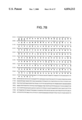
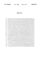

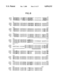

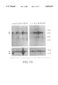

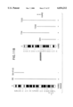
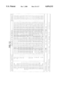

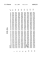

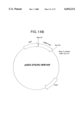

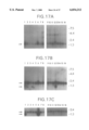
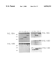
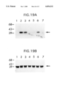

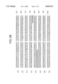
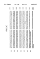


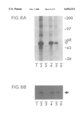
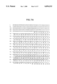
Classifications
-
- C—CHEMISTRY; METALLURGY
- C07—ORGANIC CHEMISTRY
- C07K—PEPTIDES
- C07K14/00—Peptides having more than 20 amino acids; Gastrins; Somatostatins; Melanotropins; Derivatives thereof
- C07K14/435—Peptides having more than 20 amino acids; Gastrins; Somatostatins; Melanotropins; Derivatives thereof from animals; from humans
- C07K14/46—Peptides having more than 20 amino acids; Gastrins; Somatostatins; Melanotropins; Derivatives thereof from animals; from humans from vertebrates
- C07K14/47—Peptides having more than 20 amino acids; Gastrins; Somatostatins; Melanotropins; Derivatives thereof from animals; from humans from vertebrates from mammals
-
- C—CHEMISTRY; METALLURGY
- C07—ORGANIC CHEMISTRY
- C07K—PEPTIDES
- C07K14/00—Peptides having more than 20 amino acids; Gastrins; Somatostatins; Melanotropins; Derivatives thereof
- C07K14/435—Peptides having more than 20 amino acids; Gastrins; Somatostatins; Melanotropins; Derivatives thereof from animals; from humans
- C07K14/46—Peptides having more than 20 amino acids; Gastrins; Somatostatins; Melanotropins; Derivatives thereof from animals; from humans from vertebrates
-
- A—HUMAN NECESSITIES
- A61—MEDICAL OR VETERINARY SCIENCE; HYGIENE
- A61K—PREPARATIONS FOR MEDICAL, DENTAL OR TOILETRY PURPOSES
- A61K38/00—Medicinal preparations containing peptides
-
- C—CHEMISTRY; METALLURGY
- C07—ORGANIC CHEMISTRY
- C07K—PEPTIDES
- C07K2319/00—Fusion polypeptide
Definitions
- the present invention relates to regulation and control of cellular processes by SH3-domain binding proteins, by putative signalling domains of such proteins, ligands of the signalling domain, and diagnosis and therapy based on the activity of such proteins, signalling domains, and ligands.
- Protein-protein interaction is one of the mechanisms of signal transduction processes.
- One such process involves non-receptor type protein-tyrosine kinases (PTKs) of the Src family, which signal in normal and transformed cells.
- PTKs protein-tyrosine kinases
- SH2 SH stands for Src homology
- SH3 PH
- PH pleckstrin homology
- the yes proto-oncogene encodes a member of the Src family of non-receptor type protein tyrosine kinases (PTKs) (Cooper, 1990; Sudol, 1993).
- the Src family kinases have been implicated in signal transduction processes because they physically associate with certain membrane receptors and functionally respond to the binding of cognate ligands or receptor crosslinking (Bolen, 1991).
- the functional response of receptor-associated PTKs is usually manifested by an increase in tyrosine phosphorylation of cellular proteins (Bolen, 1991).
- the Yes protein kinase was shown to be functionally associated with platelet-derived growth factor receptor in fibroblasts (Kypta et al., 1990), with glycoprotein IV (CD 36) in platelets (Huang et al., 1991), and with the high-affinity IgE receptor in mast cells (Eiseman & Bolen, 1992), but further signaling steps through these complexes are not known.
- SH2 and SH3 SH for Src homology
- SH2 and SH3 SH for Src homology
- the SH2 domains are known to interact specifically with phosphotyrosine-containing proteins (Pawson & Gish, 1992; Birge & Hanafusa, 1993; Pawson & Schlessinger, 1993) and the resulting complexes are involved in signal transduction events initiated by PTKs (Cantley et al, 1991).
- the SH2 domain of Src PTKs is involved in substrate recognition and in the regulation of kinase activity by maintaining a repressed conformation of PTKs (Kanner et al, 1991; Roussel et al., 1991).
- the precise role of SH3 domains in signal transduction has yet to be completely elucidated (Musacchio et al, 1992a; Mayer & Baltimore, 1993; Pawson & Schlessinger, 1993) but the accumulating genetic, biochemical, and structural data implicate these domains in mediating noncovalent protein-protein interactions essential for cellular and intercellular signaling (Clark et al., 1992; Musacchio et al., 1992b; Yu et al., 1992; Booker et al., 1993; Kohda et al., 1993; Koyama et al., 1993; Li et al., 1993; Noble et al., 1993; Rozakis-Adcock et al.,
- SH3 domains of Src PTKs interact with substrates (Kanner et al., 1991; Seidel-Dugan et al., 1992; Liu et al., 1993) and with other signaling molecules including unknown serine and/or threonine kinases (Weng et al., 1993).
- YAP65 A novel Yes-associated protein (YAP) of 65 kDa was identified in chicken by one of the inventors herein (Sudol, 1994, "Yes-associated protein (YAP65) is a proline-rich phosphoprotein that binds to the SH3 domain of the Yes proto-oncogene product," Oncogene 9:2145-52, which is incorporated herein by reference in its entirety, and which corresponds to the first Example disclosed infra).
- the present invention relates to proteins and polypeptides that are involved with intracellular signal transduction.
- the invention provides a novel polypeptide domain that appears to be involved in signalling.
- the invention further provides nucleic acids, particularly DNA molecules, encoding such proteins and polypeptides.
- the invention further relates to modulation of intracellular signal transduction by inducing or inhibiting the activity of the proteins and polypeptides of the invention.
- the invention provides an isolated nucleic acid molecule, which is hybridizable to a DNA molecule (or its complement) that has a nucleotide sequence shown in FIG. 2 (SEQ ID NO:1), or the sequence complementary thereto; or that has a nucleotide sequence shown in FIG. 7 (SEQ ID NO:3), or the sequence complementary thereto; or that encodes a Yes proto-oncogene associated protein or polypeptide (YAP).
- SEQ ID NO:1 nucleotide sequence shown in FIG. 2
- SEQ ID NO:3 nucleotide sequence shown in FIG. 7
- YAP Yes proto-oncogene associated protein or polypeptide
- the YAP protein or polypeptide of the invention is characterized by binding to the Src homology domain 3 (SH3), containing a proline-rich motif that is involved in binding between YAP and Yes kinase, containing a WW domain polypeptide, and being phosphorylated in vivo on serine.
- SH3 Src homology domain 3
- the WW domain which is a unique domain advantageously characterized herein, has from 30 to 50 amino acid residues; has a consensus sequence:
- h indicates a hydrophobic amino acid residue
- t indicates a turn-like or polar amino acid residue
- N indicates any amino acid
- a hyphen (-) indicates either no amino acid residue or any amino acid residue, and has a predicted secondary structure using a computer-assisted structural assignment program having the parameters characteristic of the PHD secondary structure prediction program as follows: loop-unassigned-beta strand-unassigned-loop-unassigned-beta strand-unassigned-loop-unassigned-loop, wherein the highly conserved tryptophan residues are located in the first and fifth unassigned segments.
- the nucleic acid of the invention hybridizes to a DNA molecule that encodes a YAP protein or polypeptide having an amino acid sequence selected from the group consisting of the amino acid sequence of chicken YAP in FIG. 2 (SEQ ID NO:2), human YAP in FIG. 7 (SEQ ID NO:4), or murine YAP in FIG. 8 (SEQ ID NO:5), or the sequence complementary thereto.
- the nucleic acid molecule encodes a protein or polypeptide having an amino acid sequence selected from the group consisting of the amino acid sequence of chicken YAP in FIG. 2 (SEQ ID NO:2), human YAP in FIG. 7 (SEQ ID NO:4), or murine YAP in FIG. 8 (SEQ ID NO:5).
- the nucleic acid molecule encodes a protein or polypeptide fragment, which protein or polypeptide fragment is functionally active.
- the functional activity of the protein or polypeptide is selected from the group consisting of binding to an SH3 domain, binding to an approximately 40 kDa intracellular ligand, binding to a dystrophin-associated protein, regulation of binding of ⁇ -dystroglycan to dystrophin, and modulation of intracellular signalling.
- the invention is especially directed to the nucleic acid molecule encoding a YAP fragment, wherein the protein or polypeptide fragment is the WW domain; however, with the exception of the novel YAP proteins of the invention, the nucleic acid molecule of the invention does not encode a full length, naturally occurring protein that includes a WW domain.
- the invention broadly provides a nucleic acid molecule which encodes a WW domain polypeptide, which WW domain polypeptide is characterized as described above.
- the WW domain polypeptide has an amino acid sequence selected from the group consisting of SEQ ID NOS:6-25.
- the nucleic acid molecule encoding the WW domain polypeptide encodes a chimeric polypeptide.
- the invention further provides a cloning vector comprising the nucleic acid molecule encoding a YAP, or a functionally active fragment thereof, and a cloning vector encoding a WW domain polypeptide, as well as a host cell, in particular a bacterial host cell, harboring such a cloning vector.
- the invention provides an expression vector comprising the nucleic acid molecule encoding a YAP protein or polypeptide or fragment thereof, or a WW domain polypeptide, operatively associated with an expression control sequence, as well as host cells, such as bacteria, yeast, insect cells, or mammalian cells, harboring such expression vectors.
- the invention further extends to a method for producing a YAP protein or polypeptide, comprising culturing the host cell harboring the expression vector encoding a YAP protein or polypeptide, or fragment thereof, under conditions that allow for expression of the YAP protein or polypeptide; and recovering the expressed YAP protein or polypeptide.
- the invention relates to a method for producing a WW domain polypeptide comprising culturing the host cell harboring the expression vector encoding the WW domain polypeptide under conditions that allow for expression of the WW domain polypeptide; and recovering the expressed WW domain polypeptide.
- the WW domain polypeptide is expressed as a GST-fusion protein.
- the invention provides an isolated Yes proto-oncogene associated protein or polypeptide (YAP), or functionally active fragment thereof, which protein or polypeptide binds to the Src homology domain 3 (SH3), contains a proline-rich motif that is involved in binding between YAP and Yes kinase, contains a WW domain, and is phosphorylated in vivo on serine.
- the functional activity of the fragment is selected from the group consisting of binding to an SH3 domain, binding to an approximately 40 kDa intracellular ligand, binding to a dystrophin-associated protein, regulation of binding of ⁇ -dystroglycan to dystrophin, and modulation of intracellular signalling.
- the protein has an amino acid sequence selected from the group consisting of the amino acid sequence of chicken YAP in FIG. 2 (SEQ ID NO:2), human YAP in FIG. 7 (SEQ ID NO:4), or murine YAP in FIG. 8 (SEQ ID NO:5).
- the invention further relates to a WW domain polypeptide, as characterized above.
- the WW domain polypeptide is not a naturally occurring full length protein (with the exception of the novel YAP proteins of the invention).
- the WW domain polypeptide has an amino acid sequence selected from the group consisting of SEQ ID NOS:6-25.
- the WW domain polypeptide is labeled.
- the WW domain polypeptide is a chimeric polypeptide.
- the protein can be a GST-chimeric protein.
- the invention extends to an antibody that binds to the protein or polypeptide of the invention.
- Such antibodies may be polyclonal or monoclonal, and are intended to include single chain, Fv fragments, F(ab) fragments, chimeric antibodies, humanized antibodies, bacterially expressed antibodies, etc.
- the antibody can inhibit the functional activity of the protein or polypeptide.
- the WW domain interacts with a proteinaceous ligand in the cytoplasm.
- This ligand has been identified by "Western" analysis (using labeled WW domain) as having an approximate molecular weight of 35-36 kDa.
- cDNAs encoding the ligand have also been identified, and the partial sequence information indicates that the ligand does not have any significant similarity with protein sequences available on Genbank.
- the invention is further directed to a method for identifying a ligand of a WW domain polypeptide, comprising contacting candidate ligands with the WW domain polypeptide, detecting binding of the WW domain polypeptide with a ligand; and determining the structure of the ligand.
- the invention naturally relates to the ligand identified by this method, and as characterized above.
- preferred ligands of the invention include those which contain a consensus sequence of XPPXY (SEQ ID NO:37).
- Especially preferred XPPXY-containing ligands include those which contain the consensus sequence PPPPY (SEQ ID NO:38) which has been termed a PY motif, and in particular, those termed WBP-1 and WBP-2.
- WBP-1 and WBP-2 polypeptides include those encoded by SEQ ID NOS:28 and 29, respectively. It should be appreciated that also within the scope of the invention are polypeptide ligands which share substantial homology to SEQ ID NOS:28 and 29 as defined below, and fragments and analogs of SEQ ID NOS:28 and 29 which retain the characteristic binding activity, or functional activity, as defined below, of the WBP ligands.
- the invention is also directed to nucleic acids encoding such polypeptide ligands, which can be synthesized based on the polypeptide sequence using a standard codon chart.
- the invention also relates to a method for identifying a nucleic acid encoding a ligand of the WW domain polypeptide comprising contacting cells transformed with candidate DNA believed to encode a ligand of the WW domain polypeptide with the WW domain polypeptide; detecting binding of the WW domain polypeptide with a ligand expressed by the transformed cells; selecting transformed cells in which binding of the WW domain polypeptide is detected; and determining the structure of a nucleic acid in the selected cells which corresponds to the transforming DNA which encodes the ligand. Accordingly, the invention further relates to the nucleic acid encoding a ligand of the WW domain polypeptide identified according to the method of claim 42.
- the proteins and polypeptides of the invention, and nucleic acids encoding the same, are useful for diagnosis and therapy of a disease or disorder associated with a defect in intracellular signal transduction.
- the invention relates to a method for treating a disease or disorder associated with a defect in intracellular signal transduction comprising administering an amount of the YAP protein or polypeptide, or a WW domain polypeptide, into cells of a subject believed to be suffering from a disease or disorder associated with a defect in intracellular signal transduction.
- the invention relates to introducing an expression vector that expresses the YAP protein or polypeptide, or functionally active fragment thereof, or a WW domain polypeptide, into cells of a subject believed to be suffering from a disease or disorder associated with a defect in intracellular signal transduction, wherein the expression control sequence of the expression vector provides for expression in the cell.
- the disease or disorder is muscular dystrophy.
- the present invention contemplates inhibiting the YAP protein or WW domain polypeptide, e.g., to decrease cellular activation associated with intracellular signalling.
- Such therapy may be important in the treatment of certain cancers and tumors.
- Inhibition can be achieved with neutralizing antibodies, by gene knockout, with antisense nucleic acids, and the use of small molecule antagonists (e.g., a competitive inhibitor such as PVKQPPPLAP (SEQ ID NO:26).
- a corollary object of the invention is to inhibit or reverse oncogenic transformation of a cell by inhibiting the signal transduction pathway within the cell.
- a related object is to treat a disease or disorder associated with an impairment of signal transduction mediated by dystrophin or dystrophin-binding proteins.
- FIG. 1 Identification of the 65 Da (YAP65) and a 120 kDa protein by anti-sera against anti-Yes IgG.
- Lanes 1-4 Immunoprecipitation from lysates of 35 S-methionine labeled CEFs with sera from two rabbits. Lanes 1 and 2 were precipitated with preimmune sera; lanes 3 and 4 were precipitated with immune sera.
- Lanes 5-9 Immune blot analysis.
- Immunoprecipitates with preimmune (lane 5) or anti-idiotypic immune (lane 6) serum, or total lysates of primary (lane 7), secondary (lane 8), and tertiary (lane 9) CEFs were resolved on polyacrylamide gel, transferred to a nitrocellulose membrane, and probed with anti-idiotypic serum and 125 I-labeled-protein A.
- Solid arrows indicate YAP65, and an open arrow shows 120 kDa protein. The 120 kDa protein was not detected on the immune blot. Molecular size markers are shown in kDa.
- FIG. 2 The sequence of the chicken YAP65 CDNA and the predicted protein product.
- the CDNA sequence of the original clone isolated with the anti-idiotypic antibodies is indicated with arrows.
- the sequence of a proline-rich motif implicated in the binding of YAP65 to the SH3 domain of Yes is underlined.
- the termination codon is shown as END.
- Four independent and overlapping CDNA clones were used to reconstruct the entire sequence.
- FIG. 3 Validation of the YAP65 cDNA (1-10) and phosphorylation of YAP 65 protein on serine residues (11-12). Immunoprecipitation of 35 S-labeled CEFs with preimmune (1) and anti-idiotypic IgG (2), or with preimmune IgG (3) and with IgG against TrpE-YAP65 fusion protein (4). Lanes 5 and 6 are as 3 and 4, respectively, but the immunoprecipitation was from CEFs labeled with [ 32 P i ].
- FIG. 4 Northern blot analysis of YAP65 and Yes mRNAs.
- Five micrograms of polyA + mRNA from telencephalon (1), or cerebellum (2), spleen (3), intestine (4), muscle (5), heart (6), liver (7), and kidney (8) of 2-week-old chicks were probed with radioactive YAP65 cDNA (A) or with Yes cDNA (B). Numbers to the right of the gel are sizes of mRNAs in kilobases.
- FIG. 5 Binding between YAP65 and Yes in vitro.
- TrpE-YAP65 fusion protein (2,4,5,6,7) or TrpE alone (1,3) were probed on Western blots with 35 S-methionine labeled GST-Yes-SH3 alone (2) or with radioactive GST-Yes-SH3 supplemented with 10 ⁇ M of cold GST-Yes-SH3 (4); Lane 5 is as in lane 2 but the incubation was in the presence of SPLAP peptide (200 ⁇ M); lanes 6 and 7, the binding was competed with 50 ⁇ M and 200 ⁇ M of the PLAP peptide, respectively.
- the arrow indicates partially purified TrpE-YAP65 fusion protein.
- TrpE-YAP65 Lower migrating protein bands represent products of proteolytic degradation. Even if an increased concentration of protease inhibitors and careful purification protocols were used, we always observed limited degradation of the TrpE-YAP65 protein. Equal amounts (8 ⁇ g) of TrpE-fusion protein or TrpE protein were loaded into each lane of the SDS-polyacrylamide gel and transferred to nitrocellulose.
- TrpE-fusion protein or TrpE protein were loaded into each lane of the SDS-polyacrylamide gel and transferred to nitrocellulose.
- B Differential binding of 35 S-labeled TrpE-YAP65 fusion protein to various fusion proteins containing SH3 domains. Before the Western transfer, we had loaded equal amounts (1 ⁇ g) of purified proteins into each lane of the SDS gel. An SDS-polyacrylamide gel was run in parallel and stained with Coomassie Blue to confirm equal concentrations of the purified proteins.
- Lane 1 GST protein itself; lane 2, GST-Nck; lane 3, GST-SH3-Yes; lane 4, GST-c-Crk; lane 5, GST-SH3-Src; lane 6, GST-SH3-Abl; and lane 7, GST-SH3-GAP.
- FIG. 6 Coprecipitation of Yes kinase with YAP65 coupled to Sepharose.
- A Lysates of CEFs were immunoprecipitated with anti-Yes IgG (2) or with YAP65-Sepharose (4,5,6) and subjected to an immune complex kinase assay. Preimmune IgG--lane 1; Sepharose-4B--lane 3. Lane 5 is immunoprecipitation with YAP65-Sepharose in the presence of 2 ⁇ M of GST-Yes-SH3 fusion protein; lane 6, in the presence of 10 ⁇ M of the GST-Yes-SH3 protein.
- the precipitated kinase activity shown in A may also be due to other kinases, in addition to Yes; we have shown here that in vitro YAP65 interacts with the SH3 domain of Src.
- FIG. 7 Nucleotide and deduced amino acid sequences of human YAP.
- the 5154-base pair human YAP cDNA encodes 493 amino acids and is terminated at nucleotide 1638 marked by an asterisk.
- a putative protein domain, termed the WW domain, is underlined.
- a proline-rich sequence implicated in binding between YAP and various SH3 domains is indicated with black dots.
- FIG. 8 Alignment of the human (HYAP), mouse (MYAP) and chicken (YAP) YAP amino acid sequences. Positions that differ in at least one amino acid are indicated in bold. Spaces in the alignment were introduced arbitrarily and are indicated with dots. The sequences corresponding to the putative WW domain are underlined. Note that in MYAP a second WW domain is present. Proline-rich sequences implicated in binding between YAP and various SH3 domains are conserved and indicated with #.
- FIG. 9 Southern blot analysis of genomic DNA from nine eukaryotic species. Genomic DNA (4 ⁇ g) was digested with EcoRI, resolved in 0.7% agarose gel, transferred to a charge-modified nylon membrane by blotting and fixed by UV irradiation. The DNA corresponding to the entire coding region of the HYAP CDNA was used as a probe. Left Panel (A-J) represents results of hybridization with the HYAP CDNA probe, Right Panel (K-T) shows results of staining the agarose gel with ethidium bromide to check for even DNA loading and clear satellite bands. Lanes A,K contain lambda Hind III DNA markers with sizes indicated in kilobases. Lanes B,L contain human; C,M monkey; D,N rat; E,O mouse; F,P dog; G,Q cow; H,R rabbit; I,S chicken and J,T yeast DNA. The exposure time was four days.
- FIG. 10 Northern blot analysis of poly A + RNA from sixteen different human tissues.
- Poly A + RNAs (2 ⁇ g each) from adult human tissues were run on a denaturing formaldehyde 1.2% agarose gel, transferred to a charge-modified nylon membrane by blotting and fixed by UV irradiation.
- the radiolabeled cDNA corresponding to the entire coding region of the HYAP was used as a probe (Upper Panel).
- the blot was hybridized with a radiolabeled cDNA encoding human beta-actin (Lower Panel).
- Lane A heart; B, brain; C, placenta; D, lung; E, liver; F, skeletal muscle; G, kidney; H, pancreas; I, spleen; J, thymus; K, prostate; L, testis; M, ovary; N, small intestine; O, colon; P, peripheral blood leukocytes.
- An open arrow indicates HYAP mRNA.
- Two arrows indicate beta actin mRNAs. Note that heart and skeletal muscle and to lesser degree prostate and small intestine contain an extra form of beta-actin mRNA that is of 1.6-1.8 kb. The exposure times were three days for HYAP, and 2 hours for beta-actin.
- YAP65 cDNA detects loci on human chromosomes 11 and 6.
- DNA ⁇ 10 ⁇ g/lane
- human hamster-human hybrid 7300 with human chromosomes 6, 8, 11 and X
- mouse-human hybrid N9 with chromosomes 6, 7, partial 17, and 21 lane 3
- hamster-human hybrid 10095 with a der 9 chromosome 9pter->9q34::Xq13->Xqter)
- mouse-human hybrid G5 with 6,10,12, 20, and X (lane 5)
- Hybrids with a four or five number designation are from the Coriell Institute.
- FIG. 11 Chromosomal localization of the HYAP gene.
- A Presence of the YAP65 loci in a panel of 17 rodent-human hybrids. DNA (10 ⁇ g) from various rodent-human hybrids was cleaved with restriction enzyme Sst I, electrophoresed, transferred to nitrocellulose filter and hybridized to radiolabeled HYAP cDNA probe. Indicates that the hybrid named in the left column contains the chromosome indicated in the upper row; indicates presence of the long arm of the chromosome 9 or part of the long arm represented by a smaller fraction of stippling); indicates presence of the short arm (or partial short arm) of the chromosome; indicates the absence of the chromosome listed above the column.
- the column of chromosomes 6 and 11 are boldly outlined and stippled to highlight correlation of the presence of these chromosomes (or region of the chromosomes) with the presence of the YAP65 loci.
- the patterns of retention of the loci in the panel are shown to the right of the figure where presence of a locus in a hybrid is indicated by a stippled box with a plus sign and absence of a locus indicated by an open box enclosing a minus sign.
- B Regional chromosomal localization of YAP65 loci.
- Chromosome 6 the portion of chromosome 6 present in specific hybrids is represented by the solid line to the right of the chromosome 6 idiogram.
- Hybrids were tested by filter hybridization as described in the Methods section. Presence or absence of the YAP65-related locus is indicated below the lines representing individual hybrids. The YAP65-related locus was present only in hybrids which retained chromosome region 6p21-6qter in common. Results of fluorescent in situ hybridization (FISH) to normal human metaphases is illustrated to the left of the chromosome 6 idiogram where each filled circle represents five fluorescent signals.
- FISH fluorescent in situ hybridization
- Chromosome 11 hybrids carrying partial fragments of chromosome 11 are illustrated to the right of the chromosome 11 idiogram with results of filter hybridization to the YAP65 cDNA shown below the lines representing hybrids; the YAP65 cognate locus is present only in hybrids which retain 11cen->11q13 in common.
- Hybrid CE4 retains a der 14 (14pter->14q32::11q13->11qter) from a B cell leukemia with a break in the BCL1 major breakpoint region and is negative for the YAP65 locus.
- the YAP65 gene is centromeric to the BCL1/CCND1 locus.
- results of FISH on normal human metaphases is illustrated to the left of the chromosome 11 idiogram where each filled circle represents two fluorescent signals.
- Idiograms are from "Idiogram Albums" and are used with permission of Dr. David Adler, Dept. of Pathology, University of Washington.
- FIG. 12 Alignment of selected WW domains.
- Protein codes are taken from the SWISSPROT data base if available (Yo61--hypothetical protein from C. elegans chromosome III; YKB2--hypothetical protein from yeast chromosome III; Dmd--dystrophin; Utro--utrophin; Amoe--hypothetical protein from Acanthamoeba).
- the consensus line displays conserved features (capitals--conserved amino acids, h--hydrophobic; t--turn-like or polar). Amino acids conserved in at least 60% of the sequences are shown in bold.
- the secondary structures were predicted using the program PHD (e--beta strand; 1--loop; not assigned--nearly equal preference for both beta-strand and loop) (Rost and Sander, 1994). All segments have a probability less than 10 -7 of matching the alignment by chance (computed using the MoST program (Tatusov et al., 1994)) except FE65.
- the WW domain in FE 65 is, however, 38% identical to that in YAP and is therefore included.
- Sequence Identification Information is as follows: Dmd/human (SEQ ID NO:6); Dmd/Ray (SEQ ID NO:7); Utro/Human (SEQ ID NO:8); Yap/Human (SEQ ID NO:9); Yap/Chick (SEQ ID NO:10); Yap/Mouse-1 (SEQ ID NO:11); Yap/Mouse-2 (SEQ ID NO:12); Nedd4/Mouse-1 (SEQ ID NO:13); Nedd4/Mouse-2 (SEQ ID NO:14); Nedd4/Mouse-3 (SEQ ID NO:15); Rsp5/Yeast-1 (SEQ ID NO:16); Rsp5/Yeast-2 (SEQ ID NO:17); Rsp5/Yeast-3 (SEQ ID NO:18); Ykb2-Yeast-1 (SEQ ID NO:19); Ykb2-Yeast-2 (SEQ ID NO:20); Yo61/Caeel-1 (SEQ ID NO:21); Yo61/C
- FIG. 13 Modular architecture of the proteins containing the WW domain. Dashed lines denote partial sequence. Note that the 24 spectrin repeats of dystrophin are not drawn to scale.
- FIGS. 14A and 14B Recombinant plasmid maps for expression the WW domains of human dystrophin and human YAP.
- FIG. 15 Co-precipitation of GST-WW-YAP and putative ligand from various rat organ lysates.
- a and B Lanes: 1, lung; 2, ovary; 3, cerebellum; 4, skeletal muscle.
- GST was used for precipitation and subsequently as a labelled probe
- B Precipitated and probed with GST-WW-YAP. No specific bands were seen in skeletal muscle, even when the blots were exposed to radiographic film for >1 wk. Notice the presence of diffuse bands representing the dimerization of GST and GST-WW-YAP fusion protein migrating at 26 kDa and 30 kDa respectively (open arrows).
- FIG. 16 Deduced amino acid sequence from the open reading frames of partial cDNA clones for WW-YAP ligands, WBP-1 (SEQ ID NO:28) and WBP-2 (SEQ ID NO:29).
- the PY motifs presumed to be the putative binding sites that are shared between the two independent clones, are shown in boldface and underlined.
- the open reading frames were set by the frame of the T7 gene 10 product in the pEXIox cloning vector.
- FIG. 17 Northern blot of human tissues probed with cDNA of cloned ligands.
- A-C Lanes: 1, heart; 2, brain; 3, placenta; 4, lung; 5, liver; 6, skeletal muscle; 7, kidney; 8, pancreas; 9, spleen; 10, thymus; 11, prostate; 12, testis; 13, ovary; 14, small intestine; 15, colon; 16, peripheral blood leukocytes.
- A Probed with labelled random primed fragments of WBP-1 CDNA. There also seems to be nonspecific hybridization of the probe to 18S and 28S rRNA (indicated by dots). The difference in the migration patterns of the rRNAs was caused by a variation in the size of the gels.
- FIG. 18 Binding assays with WBP-1 and putative binding domain. Two independent clones of each GST fusion construct were chosen and induced for protein expression in order to minimize the possibility of using a mutated and nonfunctional form of the ligand. The amount of protein in each lane (3-5 mg) did not vary significantly between each blot as confirmed by Coomassie stain (data not shown).
- A-E Lanes: 1, GST; 2 and 3, GST-WBP-1 (34-43); 4 and 5, GST-WBP-1 (1-74); 6 and 7, GST-WBP-1 (1-169).
- A Nitrocellulose blot of GST-ligands probed with labelled GST-WW-YAP.
- B Probed with labelled GST, as a control for background binding.
- FIG. 19 Mutational analysis of PY motif.
- the residues comprising the PY motif were each changed to alanine.
- (A and B) Lanes: 1, GST; 2, GST-GTPPPPYTVG (SEQ ID NO:30) (wild type); 3, GST-GTAPPPYTVG (SEQ ID NO:32); 4, GST-GTPAPPYTVG (SEQ ID NO:33); 5, GST-GTPPAPYTVG (SEQ ID NO:34); 6, GST-GTPPPAYTVG (SEQ ID NO:35); 7, GST-GTPPPPATVG (SEQ ID NO:36).
- FIG. 20 Binding specificity of the PY motif
- A Blots of the above purified fusion proteins were probed with 32P-labelled GST-GTPPPPYTVG (SEQ ID NO:26) protein.
- B A Coomassie stain of a replica gel confirmed equal amounts of protein in each lane (2 mg). Molecular weight shown in kDa.
- the present invention relates to proteins and polypeptides that are involved in intracellular signal transduction.
- the invention provides a novel polypeptide domain that appears to be involved in signalling.
- the invention provides nucleic acids, particularly DNA molecules, encoding such proteins and polypeptides.
- the invention relates to diagnosis of diseases or disorders, employing the polypeptides and nucleic acids of the invention.
- the invention further relates to modulation of intracellular signal transduction by inducing or inhibiting the activity of the proteins and polypeptides of the invention, by administration of a factor (protein or polypeptide, or nucleic acid) of the invention to a subject believed to be in need of modulation.
- the invention relates to methods for preparing the polypeptides and nucleic acids.
- protein is used herein to refer to the naturally occurring form of a gene product, both in a pre-processed form (if applicable), and in a post-processed form (also if applicable).
- polypeptide is inclusive of the term protein, but also encompasses minor modifications, such as deletions or N- or C-terminal additional amino acid residues to facilitate expression, purification, labeling, stability of the recombinant product, and the like.
- a “fusion protein” is a chimeric protein comprising YAP or a fragment thereof, or particularly a WW domain, and at least a portion of a non-YAP or non-WW domain protein.
- the portion of the fusion partner protein refers to a portion of a non-YAP or non-WW domain protein that is capable of (i) serving as a substrate for proteolytic cleavage (e.g., a Factor Xa sequence); (ii) binding to an antibody specific for the fusion partner protein; (iii) binding to a cognate receptor or a ligand; (iv) interacting ionically or hydrophobically with a chromatographic support; (v) catalyzing a reaction, i.e., enzymatic activity; or (vi) otherwise biologically active as assayed in vitro or in vivo.
- proteolytic cleavage e.g., a Factor Xa sequence
- a protein or polypeptide is said to have a "proline-rich motif" when a region of the protein or polypeptide has a disproportionate number of proline residues, including tow or more proline residues in tandem.
- the proline-rich motif of the invention (from chicken YAP) has the sequence PVKQPPPLAP (SEQ ID NO:26).
- consensus sequence is used herein to refer to a region or domain in a series of proteins or polypeptides that has features in common among all of the proteins or polypeptides.
- the consensus sequence can be defined by determination that putative consensus segments have a probability of less than 1 in 10 6 , and preferably less than 1 in 10 7 , of matching the alignment by chance as computed on the MoST program.
- a consensus sequence can be identified by a high degree of homology or sequence similarity between a candidate segment and a consensus segment as defined by the above criteria.
- the consensus sequence is characterized by the presence of two tryptophan residues in all of the sequences (hence the designation of the pertinent domain as the WW domain), additional conserved residues, positions in which amino acid residues with either hydrophobic or hydrophilic (or turn-like) amino acid residues, and positions having a significant probability of adopting a particular secondary structure.
- the consensus sequence of the WW domain of the invention has the sequence:
- Secondary structure refers to the first level of three-dimensional structure adopted by a protein or polypeptide. Secondary structural elements include the ⁇ -helix, ⁇ -sheet, ⁇ -turn, ⁇ -strand, and loop structures; the WW domain of the present invention is primarily concerned with the latter two structures, as these structures form the consensus sequence of the domain (see FIG. 12).
- the term "functionally active” refers to a polypeptide or protein having sufficient structure to mediate some activity. Such activity may be characteristic of the native protein, i.e., agonist activity. Alternatively, the functional activity may oppose that of the native protein, i.e., antagonist activity. Specific examples of functional activities of the invention include, but are not limited to, binding to an SH3 domain (or inhibition thereof), binding to an approximately 40 kDa intracellular ligand (or inhibition thereof), binding to a dystrophin-associated protein (or inhibition thereof), regulation of binding of ⁇ -dystroglycan to dystrophin, and modulation of intracellular signalling.
- a “vector” is a replicon, such as plasmid, phage or cosmid, to which another DNA segment may be attached so as to bring about the replication of the attached segment.
- a “replicon” is any genetic element (e.g., plasmid, chromosome, virus) that functions as an autonomous unit of DNA replication in vivo, i.e., capable of replication under its own control.
- the vector may be a cloning vector, e.g., to propagate the cloned gene, or it may be an expression vector, in which a foreign gene is inserted under control of expression control sequences contained in the vector for heterologous gene expression in a recombinant host cell.
- a "cassette” refers to a segment of DNA that can be inserted into a vector at specific restriction sites.
- the segment of DNA encodes a polypeptide of interest, and the cassette and restriction sites are designed to ensure insertion of the cassette in the proper reading frame for transcription and translation.
- a cell has been "transfected” by exogenous or heterologous DNA when such DNA has been introduced inside the cell.
- a cell has been "transformed” by exogenous or heterologous DNA when the transfected DNA effects a phenotypic change.
- the transforming DNA should be integrated (covalently linked) into chromosomal DNA making up the genome of the cell.
- stable transformation with plasmid (or cosmid) DNA is also possible.
- Heterologous DNA refers to DNA not naturally located in the cell, or in a chromosomal site of the cell.
- the heterologous DNA includes a gene foreign to the cell.
- a "clone” is a population of cells derived from a single cell or common ancestor by mitosis.
- nucleic acid molecule refers to the phosphate ester polymeric form of ribonucleosides (adenosine, guanosine, uridine or cytidine; "RNA molecules”) or deoxyribonucleosides (deoxyadenosine, deoxyguanosine, deoxythymidine, or deoxycytidine; "DNA molecules”) in either single stranded form, or a double-stranded helix. Double stranded DNA-DNA, DNA-RNA and RNA-RNA helices are possible.
- nucleic acid molecule refers only to the primary and secondary structure of the molecule, and does not limit it to any particular tertiary forms. Thus, this term includes double-stranded DNA found, inter alia, in linear or circular DNA molecules (e.g., restriction fragments), plasmids, and chromosomes. In discussing the structure of particular double-stranded DNA molecules, sequences may be described herein according to the normal convention of giving only the sequence in the 5' to 3' direction along the nontranscribed strand of DNA (i.e., the strand having a sequence homologous to the mRNA).
- a "recombinant DNA molecule” is a DNA molecule that has undergone a molecular biological manipulation.
- a "complementary" nucleic acid is the opposite strand, e.g., mRNA is complementary to the DNA template, antisense RNA is complementary to sense RNA, and each strand of double-stranded DNA is the complement of the other.
- a nucleic acid molecule is "hybridizable" to another nucleic acid molecule, such as a cDNA, genomic DNA, or RNA, when a single stranded form of the nucleic acid molecule can anneal to the other nucleic acid molecule under the appropriate conditions of temperature and solution ionic strength (see Sambrook et al., supra).
- the conditions of temperature and ionic strength determine the "stringency" of the hybridization.
- low stringency hybridization conditions corresponding to a T m of 55°, can be used, e.g., 5 ⁇ SSC, 0.1% SDS, 0.25% milk, and no formamide; or 30% formamide, 5 ⁇ SSC, 0.5% SDS).
- Moderate stringency hybridization conditions correspond to a higher Tm, e.g., 40% formamide, with 5 ⁇ or 6 ⁇ SCC.
- High stringency hybridization conditions correspond to the highest T m , e.g., 50% formamide, 5 ⁇ or 6 ⁇ SCC.
- Hybridization requires that the two nucleic acids contain complementary sequences, although depending on the stringency of the hybridization, mismatches between bases are possible.
- the appropriate stringency for hybridizing nucleic acids depends on the length of the nucleic acids and the degree of complementation, variables well known in the art. The greater the degree of similarity or homology between two nucleotide sequences, the greater the value of T m for hybrids of nucleic acids having those sequences.
- RNA:RNA, DNA:RNA, DNA:DNA The relative stability (corresponding to higher T m ) of nucleic acid hybridizations decreases in the following order: RNA:RNA, DNA:RNA, DNA:DNA.
- equations for calculating T m have been derived (see Sambrook et al., supra, 9.50-0.51).
- the position of mismatches becomes more important, and the length of the oligonucleotide determines its specificity (see Sambrook et al., supra, 11.7-11.8).
- a minimum length for a hybridizable nucleic acid is at least about 10 nucleotides; more preferably at least about 15 nucleotides; most preferably the length is at least about 20 nucleotides.
- oligonucleotides refers to short nucleic acids (including nucleic acids containing phosphate bond mimics, such as thiophosphates) that can be used as primers for PCR, labeled and used as probes, used for site directed mutagenesis, and for other techniques known in the art.
- Homologous recombination refers to the insertion of a foreign DNA sequence of a vector in a chromosome.
- the vector targets a specific chromosomal site for homologous recombination.
- the vector will contain sufficiently long regions of homology to sequences of the chromosome to allow complementary binding and incorporation of the vector into the chromosome. Longer regions of homology, and greater degrees of sequence similarity, may increase the efficiency of homologous recombination.
- a DNA "coding sequence” is a double-stranded DNA sequence which is transcribed and translated into a polypeptide in a cell in vitro or in vivo when placed under the control of appropriate regulatory sequences. The boundaries of the coding sequence are determined by a start codon at the 5' (amino) terminus and a translation stop codon at the 3' (carboxyl) terminus.
- a coding sequence can include, but is not limited to, prokaryotic sequences, cDNA from eukaryotic mRNA, genomic DNA sequences from eukaryotic (e.g., mammalian) DNA, and even synthetic DNA sequences. If the coding sequence is intended for expression in a eukaryotic cell, a polyadenylation signal and transcription termination sequence will usually be located 3' to the coding sequence.
- Transcriptional and translational control sequences are DNA regulatory sequences, such as promoters, enhancers, terminators, and the like, that provide for the expression of a coding sequence in a host cell.
- polyadenylation signals are control sequences.
- a “promoter sequence” is a DNA regulatory region capable of binding RNA polymerase in a cell and initiating transcription of a downstream (3' direction) coding sequence.
- the promoter sequence is bounded at its 3' terminus by the transcription initiation site and extends upstream (5' direction) to include the minimum number of bases or elements necessary to initiate transcription at levels detectable above background.
- a transcription initiation site (conveniently defined for example, by mapping with nuclease S1), as well as protein binding domains (consensus sequences) responsible for the binding of RNA polymerase.
- a coding sequence is "under the control" of transcriptional and translational control sequences in a cell when RNA polymerase transcribes the coding sequence into mRNA, which is then trans-RNA spliced and translated into the protein encoded by the coding sequence.
- a molecule is "antigenic" when it is capable of specifically interacting with an antigen recognition molecule of the immune system, such as an immunoglobulin (antibody) or T cell antigen receptor.
- An antigenic polypeptide contains at least about 5, and preferably at least about 10, amino acids.
- An antigenic portion of a molecule can be that portion that is immunodominant for antibody or T cell receptor recognition, or it can be a portion used to generate an antibody to the molecule by conjugating the antigenic portion to a carrier molecule for immunization.
- a molecule that is antigenic need not be itself immunogenic, i.e., capable of eliciting an immune response without a carrier.
- adjuvant refers to a compound or mixture that enhances the immune response to an antigen.
- An adjuvant can serve as a tissue depot that slowly releases the antigen and also as a lymphoid system activator that non-specifically enhances the immune response (Hood et al., Immunology, Second Ed., 1984, Benjamin/Cummings: Menlo Park, Calif., p. 384).
- a primary challenge with an antigen alone, in the absence of an adjuvant will fail to elicit a humoral or cellular immune response.
- Adjuvants include, but are not limited to, complete Freund's adjuvant, incomplete Freund's adjuvant, saponin, mineral gels such as aluminum hydroxide, surface active substances such as lysolecithin, pluronic polyols, polyanions, peptides, oil or hydrocarbon emulsions, keyhole limpet hemocyanins, dinitrophenol, and potentially useful human adjuvants such as BCG (bacille Calmette-Guerin) and Corynebacterium parvum.
- the adjuvant is pharmaceutically acceptable.
- a composition comprising "A” (where "A” is a single protein, DNA molecule, vector, recombinant host cell, etc.) is substantially free of “B” (where “B” comprises one or more contaminating proteins, DNA molecules, vectors, etc.) when at least about 75% by weight of the proteins, DNA, vectors (depending on the category of species to which A and B belong) in the composition is "A".
- "A” comprises at least about 90% by weight of the A+B species in the composition, most preferably at least about 99% by weight. It is also preferred that a composition, which is substantially free of contamination, contain only a single molecular weight species having the activity or characteristic of the species of interest.
- pharmaceutically acceptable refers to molecular entities and compositions that are physiologically tolerable and do not typically produce an allergic or similar untoward reaction, such as gastric upset, dizziness and the like, when administered to a human.
- pharmaceutically acceptable means approved by a regulatory agency of the Federal or a state government or listed in the U.S. Pharmacopeia or other generally recognized pharmacopeia for use in animals, and more particularly in humans.
- carrier refers to a diluent, adjuvant, excipient, or vehicle with which the compound is administered.
- Such pharmaceutical carriers can be sterile liquids, such as water and oils, including those of petroleum, animal, vegetable or synthetic origin, such as peanut oil, soybean oil, mineral oil, sesame oil and the like.
- Water or aqueous solution saline solutions and aqueous dextrose and glycerol solutions are preferably employed as carriers, particularly for injectable solutions. Suitable pharmaceutical carriers are described in "Remington's Pharmaceutical Sciences” by E. W. Martin.
- terapéuticaally effective amount is used herein to mean an amount sufficient to reduce by at least about 15 percent, preferably by at least 50 percent, more preferably by at least 90 percent, and most preferably prevent, a clinically significant deficit in the activity, function and response of the host. Alternatively, a therapeutically effective amount is sufficient to cause an improvement in a clinically significant condition in the host.
- spectrin repeat refers to a spectrin-like sequence, e.g., an identifiable consensus sequence found in proteins such as ⁇ -spectrin, ⁇ -spectrin, dystrophin, and ⁇ -actinin. Spectrin repeats of the sort identified herein have heretofore never been identified on phosphatases.
- the present invention is directed to YAP proteins, polypeptides comprising or consisting primarily of the WW domain, to nucleic acids encoding such proteins, to antibodies reactive with the proteins, and to methods of use of the proteins, polypeptides, and acids.
- the present invention is based, in part, on the isolation and characterization of a unique Yes-associated protein from chicken, based on screening with anti-idiotypic antibodies generated against Yes SH3-specific polyclonal antibodies.
- the chicken gene in hand, the human and murine orthologs (homologous genes in different species) were quickly recovered.
- Expression of YAP in various tissues and cells has been examined, and this protein has been found to be ubiquitous, with relatively high levels of expression in placenta, prostate, testis, ovaries, and small intestine, and relatively lower levels in brain, liver, and spleen. No YAP mRNA expression was detectable in human peritoneal leukocytes, even with overexposure of the blot.
- the invention is further based on the discovery of a motif in YAP that shares significant sequence and putative structure similarities with sequences found in various regulatory and signalling proteins.
- the present invention is divided into the following sections, which relate to YAP proteins, and nucleic acids encoding them; the WW domain polypeptide, and nucleic acids encoding them; ligands of the WW domain, and genes inducing them; isolating genes and expressing recombinant proteins; antibodies to the proteins; antisense nucleic acids; diagnostic applications; and therapeutic applications.
- the YAP proteins of the invention are characterized by binding to the Src homology 3 domain (SH3) of Yes and other SH3-containing proteins, including Hck, Crk, and Src.
- SH3 Src homology 3 domain
- the protein is a 65 kDa MW protein in chicken, and of comparable size in human.
- the murine protein includes an inserted sequence that represented an imperfect repeat of the upstream sequence, and which turned out to be a repeated WW domain.
- YAP contains a proline rich sequence (see FIG. 8, the portion of the sequence marked with # symbols).
- Human yap gene is located on the short arm on chromosome 11q13, that also harbors a gene for Multiple Endocrine Neoplasia type 1.
- the yap gene is highly conserved among higher eukaryotes, and expression of the gene is rather ubiquitous.
- Probes for identifying orthologs can be prepared from the coding sequence of any yap gene or cDNA, and used to probe genomic or CDNA from other species. Preferably, such probing is done under non-stringent conditions.
- chicken yap cDNA was used to select for human and mouse yap cDNA.
- the present invention further contemplates functionally active fragments of the protein, as defined above.
- Such fragments can be prepared by expression of a truncated nucleic acid, by chemical synthesis, or by proteolysis of the full length protein.
- the present invention is also directed to polypeptides that include the consensus sequence characteristic of the WW domain, as defined hereinabove.
- the WW domain has from 30 to 50 amino acid residues.
- WW domain polypeptides of the invention may further comprise additional amino acid residues, such as N-terminal or C-terminal extensions that facilitate expression or purification.
- a His-tag can be introduced, e.g., using pET vectors from Invitrogen.
- a GST domain-WW domain fusion protein can be expressed, as exemplified infra.
- the WW domain polypeptide can be prepared synthetically, or by proteolytic cleavage of a protein that comprises such a domain.
- the WW domain polypeptide is expressed in an expression vector that inserts a recognition sequence for the catalytic subunit of cAMP-dependent heart muscle protein kinase, i.e., a phosphorylation site, in reading frame with the heterologous protein (pGEX-2TK, Pharmacia), between the GST domain and the protein.
- the recombinant WW domain polypeptide (fusion protein) can be labeled with 32 p by reaction with the kinase.
- Nucleic acids encoding such WW domains polypeptides can be prepared by appropriate endonuclease cleavage of cDNAs encoding such proteins, e.g., the YAP proteins. More preferably, a portion of the cDNA encoding a protein that contains a WW domain can be cloned by PCR amplification of the WW domain region using standard techniques. Specific endonuclease sites can be introduced by engineering the primers. In another embodiment, the nucleic acid encoding a WW domain can be synthesized.
- the present invention is further directed to isolation and cloning of a gene that encodes a protein that appears to be a ligand for the WW domain.
- the 32 P-labeled GST-WW domain fusion (using the human dystrophin WW domain and human YAP WW domain) described above and specifically exemplified infra, can be used to detect the presence of a ligand to the WW domain. In this way, a 35-36 kDa protein has been identified as binding to the labeled WW domain protein was identified.
- the labeled WW domain polypeptide can also be used to screen an expression library for reactive clones, which are indicative of expression of the ligand. Two clones were isolated, one 1.6 kB and one 0.5 kB long.
- partial cDNA clones are obtained from the expression library, the full length cDNA can be obtained, and the sequence determined and analyzed. From this information, a putative amino acid sequence can be deduced, and characteristics about the gene and the polypeptide can be explored.
- the present invention contemplates isolation of a gene encoding a functional YAP, or portion thereof, a gene encoding a functional WW domain of the invention, or a gene encoding a ligand for the WW domain from any animal, particularly mammalian or avian, and more particularly human source.
- the term "gene” refers to an assembly of nucleotides that encode a polypeptide, and includes cDNA and genomic DNA nucleic acids.
- a gene encoding YAP, or a fragment thereof, or a WW domain, or a WW domain ligand, whether genomic DNA or cDNA, can be isolated from any source, particularly from a human cDNA or genomic library. Methods for obtaining the such genes are well known in the art, as described above (see, e.g., Sambrook et al., 1989, supra).
- any animal cell potentially can serve as the nucleic acid source for the molecular cloning of a gene.
- the DNA may be obtained by standard procedures known in the art from cloned DNA (e.g., a DNA "library”), by chemical synthesis, by cDNA cloning, or by the cloning of genomic DNA, or fragments thereof, purified from the desired cell (See, for example, Sambrook et al., 1989, supra; Glover, D. M. (ed.), 1985, DNA Cloning: A Practical Approach, MRL Press, Ltd., Oxford, U. K. Vol. I, II).
- Clones derived from genomic DNA may contain regulatory and intron DNA regions in addition to coding regions; clones derived from cDNA will not contain intron sequences. Whatever the source, the gene should be molecularly cloned into a suitable vector for propagation of the gene.
- DNA fragments are generated, some of which will encode the desired gene.
- the DNA may be cleaved at specific sites using various restriction enzymes.
- DNAse in the presence of manganese to fragment the DNA, or the DNA can be physically sheared, as for example, by sonication.
- the linear DNA fragments can then be separated according to size by standard techniques, including but not limited to, agarose and polyacrylamide gel electrophoresis and column chromatography.
- identification of the specific DNA fragment containing the desired gene may be accomplished in a number of ways. For example, if an amount of a portion of a gene or its specific RNA, or a fragment thereof, is available and can be purified and labeled, the generated DNA fragments may be screened by nucleic acid hybridization to the labeled probe (Benton and Davis, 1977, Science 196:180; Grunstein and Hogness, 1975, Proc. Natl. Acad. Sci. U.S.A. 72:3961).
- nucleic acid probes which can be conveniently prepared from the specific sequences disclosed herein, e.g., a probe having a nucleotide sequence corresponding to at least a 10, and preferably a 15, nucleotide fragment of the sequence depicted in FIG. 1 or 7 (SEQ ID NO:1 or 3, respectively), including flanking sequences.
- a fragment is selected that is highly unique to the gene of interest. Those DNA fragments with substantial homology to the probe will hybridize.
- the greater the degree of homology the more stringent hybridization conditions can be used.
- low stringency hybridization conditions are used to identify a homologous gene form another species (an ortholog).
- a nucleic acid encoding a gene of the invention will hybridize to a nucleic acid having a nucleotide sequence depicted in FIG. 2 or 7 (SEQ ID NO:1 or 3, respectively), or a hybridizable fragment thereof, under moderately stringent conditions; more preferably, it will hybridize under high stringency conditions. Further selection can be carried out on the basis of the properties of the gene, e.g., if the gene includes sequences encoding a proline-rich region or a WW domain (for yap gene), or binding to a WW domain ligand.
- the presence of the gene may be detected by assays based on the physical, chemical, or immunological properties of its expressed product.
- cDNA clones, or DNA clones which hybrid-select the proper mRNAs can be selected which produce a protein that, e.g., has similar or identical electrophoretic migration, isoelectric focusing behavior, proteolytic digestion maps, or antigenic properties as known for the gene produce, e.g., YAP, the WW domain, or the WW domain ligand.
- the antibodies of the instant invention can conveniently be used to screen for homologs of YAP from other sources, preferably human.
- a radiolabeled cDNA can be synthesized by PCR using the selected mRNA (from the adsorbed polysomes) as a template.
- the radiolabeled mRNA or cDNA may then be used as a probe to identify homologous DNA fragments from among other genomic DNA fragments.
- the present invention also relates to cloning vectors containing genes encoding analogs and derivatives of the of the invention, that have the same or homologous functional activity as the native protein or polypeptide, and homologs thereof from other species.
- the production and use of derivatives and analogs related to are within the scope of the present invention.
- the derivative or analog is functionally active, i.e., capable of exhibiting one or more functional activities associated with a full-length, wild-type protein of the invention.
- Derivatives of the protein or polypeptide can be made by altering encoding nucleic acid sequences by substitutions, additions or deletions that provide for functionally equivalent molecules.
- derivatives are made that have enhanced or increased functional activity relative to the native protein or polypeptide.
- nucleotide coding sequences which encode substantially the same amino acid sequence as a yap, WW domain, or WW domain ligand gene may be used in the practice of the present invention.
- nucleotide sequences comprising all or portions of genes which are altered by the substitution of different codons that encode the same amino acid residue within the sequence, thus producing a silent change.
- the derivatives of the invention include, but are not limited to, those containing, as a primary amino acid sequence, all or part of the amino acid sequence of a protein or polypeptide of the invention including altered sequences in which functionally equivalent amino acid residues are substituted for residues within the sequence resulting in a conservative amino acid substitution.
- one or more amino acid residues within the sequence can be substituted by another amino acid of a similar polarity, which acts as a functional equivalent, resulting in a silent alteration.
- Substitutes for an amino acid within the sequence may be selected from other members of the class to which the amino acid belongs.
- the nonpolar (hydrophobic) amino acids include alanine, leucine, isoleucine, valine, proline, phenylalanine, tryptophan and methionine.
- the polar neutral amino acids include glycine, serine, threonine, cysteine, tyrosine, asparagine, and glutamine.
- the positively charged (basic) amino acids include arginine, lysine and histidine.
- the negatively charged (acidic) amino acids include aspartic acid and glutamic acid. Such substitutions are particularly contemplated for the consensus sequence of the WW domain, which clearly identifies those positions that are immutable, those positions that are highly conserved, those positions that have a strong polar or non-polar character, and those positions for which it simply does not matter what amino acid is there.
- genes encoding derivatives and analogs of the invention can be produced by various methods known in the art. The manipulations which result in their production can occur at the gene or protein level.
- the cloned gene sequence can be modified by any of numerous strategies known in the art (Sambrook et al., 1989, supra). The sequence can be cleaved at appropriate sites with restriction endonuclease(s), followed by further enzymatic modification if desired, isolated, and ligated in vitro.
- the encoding nucleic acid sequence can be mutated in vitro or in vivo, to create and/or destroy translation, initiation, and/or termination sequences, or to create variations in coding regions and/or form new restriction endonuclease sites or destroy preexisting ones, to facilitate further in vitro modification.
- mutations enhance the functional activity of the mutated gene product.
- Any technique for mutagenesis known in the art can be used, including but not limited to, in vitro site-directed mutagenesis (Hutchinson, C., et al., 1978, J. Biol. Chem.
- nucleotide sequence coding for a YAP, WW domain polypeptide, or WW domain ligand, or functional fragment, derivative or analog thereof can be inserted into an appropriate expression vector, i.e., a vector which contains the necessary elements for the transcription and translation of the inserted protein-coding sequence. Such elements are termed herein a "promoter.”
- a promoter a vector which contains the necessary elements for the transcription and translation of the inserted protein-coding sequence.
- the nucleic acid encoding a polypeptide or protein of the invention is operationally (operably) associated with a promoter in an expression vector of the invention. Both CDNA and genomic sequences can be cloned and expressed under control of such regulatory sequences.
- An expression vector also preferably includes a replication origin.
- the necessary transcriptional and translational signals can be provided on a recombinant expression vector, or they may be supplied by the native gene encoding a and/or its flanking regions.
- Potential host-vector systems include but are not limited to mammalian cell systems infected with virus (e.g., vaccinia virus, adenovirus, etc.); insect cell systems infected with virus (e.g., baculovirus); microorganisms such as yeast containing yeast vectors; or bacteria transformed with bacteriophage, DNA, plasmid DNA, or cosmid DNA.
- virus e.g., vaccinia virus, adenovirus, etc.
- insect cell systems infected with virus e.g., baculovirus
- microorganisms such as yeast containing yeast vectors
- bacteria transformed with bacteriophage, DNA, plasmid DNA, or cosmid DNA e.g., bacteriophage, DNA, plasmid DNA, or cosmid DNA.
- the expression elements of vectors vary in their strengths and specificities. Depending on the host-vector system utilized, any one of a number of suitable transcription and translation elements may be used.
- a recombinant of the invention, or functional fragment, derivative or analog thereof, may be expressed chromosomally, after integration of the coding sequence by recombination.
- any of a number of amplification systems may be used to achieve high levels of stable gene expression (See Sambrook et al., 1989, supra) can be used.
- the cell into which the recombinant vector comprising the nucleic acid encoding the protein or polypeptide is cultured in an appropriate cell culture medium under conditions that provide for expression of the protein or polypeptide by the cell.
- Any of the methods previously described for the insertion of DNA fragments into a cloning vector may be used to construct expression vectors containing a gene consisting of appropriate transcriptional/translational control signals and the protein coding sequences. These methods may include in vitro recombinant DNA and synthetic techniques and in vivo recombination (genetic recombination).
- Promoters which may be used to control gene expression include, but are not limited to, the SV40 early promoter region (Benoist and Chambon, 1981, Nature 290:304-310), the promoter contained in the 3' long terminal repeat of Rous sarcoma virus (Yamamoto, et al., 1980, Cell 22:787-797), the herpes thymidine kinase promoter (Wagner et al., 1981, Proc. Natl. Acad. Sci. U.S.A.
- prokaryotic expression vectors such as the ⁇ -lactamase promoter (Villa-Kamaroff, et al., 1978, Proc. Natl. Acad. Sci. U.S.A. 75:3727-3731), or the tac promoter (DeBoer, et al., 1983, Proc. Natl. Acad. Sci. U.S.A.
- Expression vectors containing a nucleic acid encoding a of the invention can be identified by four general approaches: (a) PCR amplification of the desired plasmid DNA or specific mRNA, (b) nucleic acid hybridization, (c) presence or absence of "marker" gene functions, and (d) expression of inserted sequences.
- the nucleic acids can be amplified by PCR to provide for detection of the amplified product.
- the presence of a foreign gene inserted in an expression vector can be detected by nucleic acid hybridization using probes comprising sequences that are homologous to an inserted gene.
- the recombinant vector/host system can be identified and selected based upon the presence or absence of certain "marker" gene functions (e.g., ⁇ -galactosidase activity, thymidine kinase activity, resistance to antibiotics, transformation phenotype, occlusion body formation in baculovirus, etc.) caused by the insertion of foreign genes in the vector.
- certain "marker" gene functions e.g., ⁇ -galactosidase activity, thymidine kinase activity, resistance to antibiotics, transformation phenotype, occlusion body formation in baculovirus, etc.
- recombinant expression vectors can be identified by assaying for the activity of the gene product expressed by the recombinant, provided that the expressed protein assumes a functionally active conformation.
- assays can be based, for example, on the physical or functional properties of the gene product in in vitro assay systems, e.g., tyrosine phosphorylation, or alternatively binding with antibody.
- the expression vectors which can be used include, but are not limited to, the following vectors or their derivatives: human or animal viruses such as vaccinia virus or adenovirus; insect viruses such as baculovirus; yeast vectors; bacteriophage vectors (e.g., lambda), and plasmid and cosmid DNA vectors, to name but a few.
- a host cell strain may be chosen which modulates the expression of the inserted sequences, or modifies and processes the gene product in the specific fashion desired.
- Different host cells have characteristic and specific mechanisms for the translational and post-translational processing and modification (e.g., glycosylation, cleavage [e.g., of signal sequence]) of proteins.
- Appropriate cell lines or host systems can be chosen to ensure the desired modification and processing of the foreign protein expressed.
- expression in a bacterial system can be used to produce an nonglycosylated core protein product.
- the transmembrane protein expressed in bacteria may not be properly folded.
- Expression in yeast can produce a glycosylated product.
- Expression in eukaryotic cells can increase the likelihood of "native" glycosylation and folding of a heterologous protein.
- different vector/host expression systems may affect processing reactions, such as proteolytic cleavages, to a different extent.
- Vectors are introduced into the desired host cells by methods known in the art, e.g., transfection, electroporation, microinjection, transduction, cell fusion, DEAE dextran, calcium phosphate precipitation, lipofection (lysosome fusion), use of a gene gun, or a DNA vector transporter (see, e.g., Wu et al., 1992, J. Biol. Chem. 267:963-967; Wu and Wu, 1988, J. Biol. Chem. 263:14621-14624; Hartmut et al., Canadian Patent Application No. 2,012,311, filed Mar. 15, 1990).
- Recombinant protein can be isolated and purified by standard methods.
- the protein or polypeptide, which is expected to be expressed into the cytoplasm can be obtained by lysing the membrane with detergents, such as but not limited to, sodium dodecyl sulfate (SDS), Triton X-100, nonidet P-40 (NP-40), digoxin, sodium deoxycholate, and the like, including mixtures thereof.
- Solubilization can be enhanced by sonication of the suspension.
- Soluble forms of the protein can be obtained by collecting culture fluid, or solubilizing inclusion bodies, e.g., by treatment with detergent, and if desired sonication or other mechanical processes, as described above.
- the solubilized or soluble protein can be isolated using various techniques, such as polyacrylamide gel electrophoresis (PAGE), including chromatography (e.g., ion exchange, affinity, immunoaffinity, and sizing column chromatography), centrifugation, differential solubility, immunoprecipitation, or by any other standard technique for the purification of proteins.
- PAGE polyacrylamide gel electrophoresis
- chromatography e.g., ion exchange, affinity, immunoaffinity, and sizing column chromatography
- centrifugation e.g., ion exchange, affinity, immunoaffinity, and sizing column chromatography
- differential solubility e.g., differential solubility
- immunoprecipitation e.g., differential solubility, immunoprecipitation, or by any other standard technique for the purification of proteins.
- a protein or polypeptide, or fragment, derivative, or analog thereof can be expressed as a GST-fusion protein in a bacterial expression system.
- a fragment such as the WW domain, is expressed in such a system.
- a cDNA or gene fragment can be isolated, as described above, gel purified, blunt-ended with T4 DNA polymerase, and ligated with EcoRI-linearized, blunt ended PGEX-3X DNA (Smith and Johnson, 1988, Gene 67:31-40). The ligation mixture can then be transformed into E. coli and the clones obtained analyzed by restriction digestion and DNA sequencing. Products of resulting plasmids can be purified over glutathione-SEPHAROSE resin and eluted with free glutathione. The glutathione can be removed by passage through a PD10 desalting column.
- the invention specifically provides for infection of Sf9 (Spodoptera frugiperda) cells at a multiplicity of infection of 10, with a recombinant baculovirus (Autographa californica), made by subcloning cDNA into the pAcYM1 vector (Matsura et al., 1987, J. Gen. Virol. 68:1233-50). After 72 hours, cells can be lysed by Dounce homogenization in TNE buffer, and protein products purified by gel filtration, antibody affinity chromatography, or a combination of chromatography steps.
- Sf9 Spodoptera frugiperda
- Autographa californica Autographa californica
- the gene of the invention is expressed in an indicator cell line, which is discussed in detail, infra.
- isolation of the expressed protein is not desired, since the functional activity of the expressed protein in the indicator cell line is the property most of interest.
- the recombinant product can be analyzed. This is achieved by assays based on the physical or functional properties of the product, including radioactive labelling of the product followed by analysis by gel electrophoresis, immunoassay, etc.
- the ability of the expressed protein, or a fragment thereof, to function in an assay can be determined.
- the structure of a YAP protein, a WW domain polypeptide, or a WW domain ligand of the invention can be analyzed by various methods known in the art.
- the structure of the various domains, particularly the domain is analyzed.
- Structural analysis can be performed by identifying sequence similarity with other known proteins, as was performed in identifying the WW domain.
- the degree of similarity (or homology) can provide a basis for predicting structure and function of a similar domain.
- sequence comparisons can be performed with sequences found in GenBank, using, for example, the FASTA and FASTP programs (Pearson and Lipman, 1988, Proc. Natl. Acad. Sci. U.S.A. 85:2444-48).
- the protein sequence can be further characterized by a hydrophilicity analysis (e.g., Hopp and Woods, 1981, Proc. Natl. Acad. Sci. U.S.A. 78:3824).
- a hydrophilicity profile can be used to identify the hydrophobic and hydrophilic regions of the a protein.
- Manipulation, translation, and secondary structure prediction, as well as open reading frame prediction and plotting, can also be accomplished using computer software programs available in the art.
- the present invention enables quantitative structural determination of the protein, or domains thereof.
- enough material is provided for nuclear magnetic resonance (NMR), infrared (IR), Raman, and ultraviolet (UV), especially circular dichroism (CD), spectroscopic analysis.
- NMR nuclear magnetic resonance
- IR infrared
- UV ultraviolet
- CD circular dichroism
- spectroscopic analysis provides very powerful structural analysis of molecules in solution, which more closely approximates their native environment (Marion et al., 1983, Biochem. Biophys. Res. Comm. 113:967-974; Bar et al., 1985, J. Magn. Reson. 65:355-360; Kimura et al., 1980, Proc. Natl. Acad. Sci. U.S.A. 77:1681-1685).
- Other methods of structural analysis can also be employed. These include but are not limited to X-ray crystallography (Engstom, A., 1974, Biochem. Exp. Biol. 11:7-13).
- the crystal structure of human YAP and human dystrophin are being obtained and compared, to determine the molecular consequences of the observed similarity between these proteins, particularly at the level of the WW domain.
- co-crystals of Yes and YAP can be prepared, so that the exact nature of the binding reaction can be studied.
- co-crystals of the WW domain and the WW domain ligand can be prepared. Analysis of co-crystals provides detailed information about binding, which in turn allows for rational design of ligand agonists and antagonists.
- Computer modeling can also be used, especially in connection with NMR or X-ray methods (Fletterick, R. and Zoller, M. (eds.), 1986, Computer Graphics and Molecular Modeling, in Current Communications in Molecular Biology, Cold Spring Harbor Laboratory, Cold Spring Harbor, N.Y.).
- recombinant proteins or polypeptides, and fragments or other derivatives or analogs thereof, or cells expressing the foregoing may be used as an immunogen to generate antibodies which recognize the cognate protein or polypeptide.
- antibodies include but are not limited to polyclonal, monoclonal, chimeric, single chain, Fab fragments, and an Fab expression library.
- anti-idiotype antibodies can be generated to a binding partner of the protein or polypeptide, for example to anti-Yes antibodies, in order to obtain antibodies reactive, in this instance, with YAP.
- recombinant or derivative or analog thereof various procedures known in the art may be used for the production of polyclonal antibodies to a recombinant or derivative or analog thereof.
- various host animals can be immunized by injection with the recombinant, or a derivative (e.g., fragment) thereof, including but not limited to rabbits, mice, rats, etc.
- the recombinant or fragment thereof can be conjugated to an immunogenic carrier, e.g., bovine serum albumin (BSA) or keyhole limpet hemocyanin (KLH).
- BSA bovine serum albumin
- KLH keyhole limpet hemocyanin
- adjuvants may be used to increase the immunological response, depending on the host species, including but not limited to Freund's (complete and incomplete), mineral gels such as aluminum hydroxide, surface active substances such as lysolecithin, pluronic polyols, polyanions, peptides, oil emulsions, keyhole limpet hemocyanins, dinitrophenol, and potentially useful human adjuvants such as BCG (bacille Calmette-Guerin) and Corynebacterium parvum.
- BCG Bacille Calmette-Guerin
- Corynebacterium parvum bacille Calmette-Guerin
- any technique which provides for the production of antibody molecules by continuous cell lines in culture may be used. These include but are not limited to the hybridoma technique originally developed by Kohler and Milstein (1975, Nature 256:495-497), as well as the trioma technique, the human B-cell hybridoma technique (Kozbor et al., 1983, Immunology Today 4:72), and the EBV-hybridoma technique to produce human monoclonal antibodies (Cole et al., 1985, in Monoclonal Antibodies and Cancer Therapy, Alan R. Liss, Inc., pp. 77-96).
- monoclonal antibodies can be produced in germ-free animals utilizing recent technology (PCT/US90/02545).
- human antibodies may be used and can be obtained by using human hybridomas (Cote et al., 1983, Proc. Natl. Acad. Sci. U.S.A. 80:2026-2030) or by transforming human B cells with EBV virus in vitro (Cole et al., 1985, in Monoclonal Antibodies and Cancer Therapy, Alan R. Liss, pp. 77-96).
- human hybridomas Cote et al., 1983, Proc. Natl. Acad. Sci. U.S.A. 80:2026-2030
- transforming human B cells with EBV virus in vitro Cold-et al., 1985, in Monoclonal Antibodies and Cancer Therapy, Alan R. Liss, pp. 77-96.
- techniques developed for the production of "chimeric antibodies” (Morrison et al., 1984, J. Bacterio
- Antibody fragments which contain the idiotype of the antibody molecule can be generated by known techniques.
- such fragments include but are not limited to: the F(ab') 2 fragment which can be produced by pepsin digestion of the antibody molecule; the Fab' fragments which can be generated by reducing the disulfide bridges of the F(ab') 2 fragment; and the Fab fragments which can be generated by treating the antibody molecule with papain and a reducing agent.
- screening for the desired antibody can be accomplished by techniques known in the art, e.g., radioimmunoassay, ELISA (enzyme-linked immunosorbant assay), "sandwich” immunoassays, immunoradiometric assays, gel diffusion precipitin reactions, immunodiffusion assays, in situ immunoassays (using colloidal gold, enzyme or radioisotope labels, for example), western blots, precipitation reactions, agglutination assays (e.g., gel agglutination assays, hemagglutination assays), complement fixation assays, immunofluorescence assays, protein A assays, and immunoelectrophoresis assays, etc.
- radioimmunoassay e.g., ELISA (enzyme-linked immunosorbant assay), "sandwich” immunoassays, immunoradiometric assays, gel diffusion precipitin reactions, immunodiffusion assays, in situ immunoa
- antibody binding is detected by detecting a label on the primary antibody.
- the primary antibody is detected by detecting binding of a secondary antibody or reagent to the primary antibody.
- the secondary antibody is labeled.
- Many means are known in the art for detecting binding in an immunoassay and are within the scope of the present invention. For example, to select antibodies which recognize a specific epitope, one may assay generated hybridomas for a product which binds to a fragment containing such epitope. For selection of an antibody specific to an YAP, WW domain, or WW domain ligand from a particular species of animal, one can select on the basis of positive binding with the protein or polypeptide expressed by or isolated from cells of that species of animal.
- the foregoing antibodies can be used in methods known in the art relating to the localization and activity of their binding partners, e.g., for Western blotting, imaging, measuring levels thereof in appropriate physiological samples, etc.
- antibodies that agonize or antagonize the activity of can be generated. Such antibodies can be tested using the assays described infra for identifying ligands.
- Identification and isolation of a gene encoding YAP, a WW domain, or the WW domain ligand of the invention provides for expression of the protein in quantities greater than can be isolated from natural sources, or in indicator cells that are specially engineered to indicate the activity of a protein expressed after transfection or transformation of the cells. According, the present invention contemplates identifying specific ligands of using various screening assays known in the art.
- Any screening technique known in the art can be used to screen for agonists or antagonists.
- the present invention contemplates screens for small molecule ligands or ligand analogs and mimics, as well as screens for the native ligand that binds to and activates of the invention in vivo.
- synthetic libraries (Needels et al., 1993, "Generation and screening of an oligonucleotide encoded synthetic peptide library," Proc. Natl. Acad. Sci. U.S.A. 90:10700-4; Lam et al., International Patent Publication No. WO 92/00252, each of which is incorporated herein by reference in its entirety), and the like can be used to screen for ligands according to the present invention.
- the peptide PVKQPPPLAP (SEQ ID NO:26) is shown to inhibit binding of YAP to Yes.
- Protein binding is one means by which cells accomplish signal transduction, and thus control activation, proliferation, and differentiation. Therefore, the level of expression of YAP, proteins containing the WW domain, and the ability to modulate activity of such proteins and polypeptides of the invention, can be very important for the diagnosis and treatment of diseases of disorders, particularly cellular transformations that lead to cancer, and to disorders such as muscular dystrophy.
- the nucleic acid probes (enzyme or radio-labeled nucleotides) or antibodies of the invention can be used to detect expression, and measure the level of expression, of a YAP protein, or a protein carrying a WW consensus sequence of the invention in selected tissues.
- a YAP protein or a protein carrying a WW consensus sequence of the invention in selected tissues.
- the presence or absence of expression of YAP in cancer cells obtained in a tissue biopsy can be important in evaluating whether the normal cellular control machinery are operating.
- the presence or absence, and level of expression, of the invention in immune cells can provide information about the level of immune activation and regulation.
- the WW domain can be used as a probe for the presence or level of the ligand to which it binds.
- Information about the level of the ligand is informative with respect to the state of cellular activation, oncogenesis, and other indicia of metabolic state.
- the level of Yes and other SH3-containing proteins can be evaluated by detecting the level of binding of YAP protein to the sample being assayed.
- signal transduction can be evaluated by detecting the level of phosphorylation of the YAP protein in cells in vivo.
- antibodies generated to YAP, the WW domain or domains, or to the WW domain ligand can be used to evaluate the presence or level of activity of the proteins or polypeptides.
- Immunoassays can be performed by any of the standard techniques described above. The presence of low levels of YAP or particular proteins containing specific WW domains may be indicative of a disease or disorder characterized by a decrease in cellular metabolic activity, possibly resulting from the low level of YAP or WW domains. Conversely, increased levels of these proteins may be characteristic of cellular activation, e.g., such as accompanies oncogenesis.
- antisense oligonucleotides capable of hybridizing to yac or WW domain mRNA can be used to inhibit expression of either protein in a cell, and thus modulate signal transduction activity in a cell.
- Inhibition of signalling activity can be useful, e.g., to modulate the activity of various cells. For example, if testicular or ovarian cells become transformed, it may be desirable to inhibit signalling mediated by YAP interaction with Yes, or by inhibiting the signalling mediated by the WW domain, in order to inhibit or reverse the transformation.
- YAP or the WW domain can be introduced into cells, either directly or by gene therapy, to increase the level of signal transduction.
- the WW domain can be introduced into muscle cells of a subject suffering from a form of muscular dystrophy characterized by a mutation in the dystrophin WW domain. Supplementation of the activity of the dystrophin WW domain, by introduction of a functionally active WW domain may be used to reverse the degeneration of muscle cells that accompanies muscular dystrophy.
- ligand agonists or antagonists can be used to modulate cellular activity by increasing or decreasing the signalling activity of the either YAP or the WW domain-containing proteins, or both in cells.
- the ligand for the WW domain can be introduced into cells, either directly or by gene therapy, to increase the level of WW domain ligand activity in the cells.
- the present example relates to the identification of a novel gene and its deduced protein product.
- This protein was isolated by binding to anti-idiotype antibodies against the amino terminal domain of Yes, a member of the Src family of protein-tyrosine kinases involved in signaling.
- the results reported herein were previously published by Sudol (a co-inventor of this application) (1994, Oncogene 9:2145-51, which is hereby specifically incorporated herein by reference in its entirety).
- Antibodies against YAP65 were generated in rabbits against a portion of the YAP65 sequence (nucleotides 381-1298) expressed in bacteria using the TrpE-expression vector as previously described for Yrk (Sudol et al., 1993). Polymerase Chain Reaction was used to generate the YAP65 cDNA insert with appropriate cloning sites. The open reading frame of the betagalactosidase protein in the original lambda gt11 clone (1 kb long clone) indicated the reading frame of YAP65. Antibodies against the human GAP protein that recognize also the chicken GAP protein on Western blots were purchased from UBI (Lake Placid, N.Y.).
- GST-SH3-Yes fusion protein was obtained by subcloning a PCR amplified SH3 fragment of CDNA (Sudol et al., 1988) into pGEX-3X vector in frame using BamHI and EcoRI restriction sites engineered at the end of the amplified CDNA. Purification was on a glutathione-Sepharose column.
- Gst-Nck Chou et al., 1992
- Gst-Crk Birge et al., 1992
- Gst-SH3-Src Cicchetti et al., 1992
- Gst-SH3-Abl Cicchetti et al., 1992
- Gst-SH3-GAP Gst-SH3-GAP
- ⁇ PLAP ⁇ ISQSAPVKQPPPLAPQSPQGGV SEQ ID NO:39 corresponding to the YAP65 sequence, amino acids 233-254
- ⁇ SPLAP ⁇ VQPAQLSIPGPVSPQPKGQSPA SEQ ID NO:40, a scrambled version of ⁇ PLAP ⁇ without any consecutive prolines
- Cell lysates were prepared in 150 mM NaCl RIPA buffer with protease inhibitors (Sudol & Hanafusa, 1986). The autophosphorylation kinase assay and Western blot analyses were as previously described (Sudol & Hanafusa).
- Antisera generated in two rabbits against the affinity purified anti-Yes IgG fraction (Sudol & Hanafusa, 1986) precipitated a 65 KDa protein, as well as a 120 kDa protein, from CEFs metabolically labeled with [ 35 S]methionine (FIG. 1, lanes 3 and 4).
- the 65 kDa protein was also detected by immune blot analysis in total lysates of CEFs from various passages (FIG. 1, lanes 6-9).
- the even intensity of the 65 kDa band in primary and tertiary CEFs eliminated the possibility that the 65 kDa protein is derived from nonfibroblastic cells frequently contaminating primary cultures.
- the combined sequence of the original (1 kb long) clone and of four independently isolated overlapping clones is shown in FIG. 2.
- the cDNA predicted sequence of YAP65 did not show any significant similarity at the DNA or protein levels when compared with GenBank sequences.
- the YAP65 sequence's most prominent feature is the high content of proline and glutamine residues.
- YAP65 is a Phosphoprotein
- the 120 kDa protein differs from YAP65 (FIG. 3, lanes 8 and 9).
- our data do not exclude the possibility that the 120 kDa protein shares some epitopes with YAP65.
- YAP65 and the 120 kDa protein are phosphorylated constitutively and exclusively on serine residues (FIG. 3, lanes 5, 6 and 11, 12).
- the YAP65 cDNA On Northern blots, the YAP65 cDNA detected a single 4.2 kb transcript expressed ubiquitously in various chicken tissues including brain (telencephalon, cerebellum), heart, spleen, intestine, liver, kidney and muscle (FIG. 4). There was no quantitative correlation between the patterns of YAP65 and Yes mRNA expression except that both were expressed ubiquitously.
- Trp-E-YAP65 binding To perform reciprocal binding and to estimate the relative specificity of YAP65 binding to other proteins that contain SH3 domains, we probed the purified GST fusion proteins of Nck, Crk, Src, Abl and GAP with radioactively labeled Trp-E-YAP65 protein. The same amount of protein was analyzed in a membrane binding assay; TRPE-YAP-65 bound the strongest to Nck and Yes followed by Crk, and Src. Binding of TrpE-YAP65 to the GST-SH3 domains of Abl and GAP was relatively weak (FIG. 5b).
- both the anti-idiotypic antibodies and antibodies generated against bacterially expressed YAP65 cDNA recognized another protein of 120 kDa.
- the peptide mapping analysis showed that the 120 kDa protein is not a precursor of YAP65.
- the 120 kDa protein was not detected on Western blots, we cannot presently determine whether it shares epitopes with YAP65 or whether it is a YAP65 binding protein. The former possibility seems likely since we have recently isolated a cDNA clone sharing a 3' end coding sequence with YAP65 and at the 5' end it contains a novel sequence.
- the 120 kDa protein is not recognized by antibodies that recognize the human GAP protein (data not shown).
- the YAP65 cDNA contains one long open reading frame that ends with a stop codon followed by at least two other stop codons in alternate reading frames.
- the sequence preceding the proposed initiation codon (first methionine) conforms to Kozak's rules for translation initiation (Kozak, 1989).
- the predicted molecular mass of the YAP65 is at least 15 kDa shorter than the YAP65 molecular mass estimated from SDS-polyacrylamide gels. There are several possible explanations of this discrepancy.
- SH3 domain-ligand interactions are: (i) the Abl kinase and the 3BP1 protein that shows homology to GAP-rho, (ii) Grb2 protein that links epidermal growth factor receptor to guanine nucleotide exchange factor, SOS, (iii) GTPase dynamin that is activated by binding to various proteins containing SH-3 domain(s), and (iv) Abl protooncogene kinase that binds to the first SH3 domain of Crk proto-oncogene protein (Lowenstein et al., 1992; Oliver et al., 1993; Egan et al., 1993; Gout et al., 1993; Li et al., 1993; Ren et al., 1993; Rozakis-Adcock et al., 1993; Ren et al., 1994; Feller et al., 1994).
- cDNA Cloning and Sequencing--A cDNA of chicken YAP corresponding to the coding region was used as a probe to screen a lambda pCEV 15 cDNA library derived from M426 human lung embryonic fibroblast cells and a 16 day mouse embryo cDNA library in lambda ExloxTM (purchased from Novagen, Madison, Wis.).
- the low stringency conditions of hybridization were as follows: 5 ⁇ SSPE, 10 ⁇ Denhart, 2% SDS, 0.2 mg/ml of salmon sperm DNA and 106 cpm/ml of 32 P-labeled cDNA at 65° C. overnight. The filters were washed 2 ⁇ for 20 min.
- mouse YAP CDNA was reconstituted with two overlapping clones contained in pEXlox-MYAP6 (2.3 kb Eco RI-Hind III insert) and pEXlox-MYAP20 (Eco RI-Hind III insert). Both strands of the cDNA clones were analyzed by direct sequence analysis using the Sanger method.
- DNA sources were as follows: human, Rhesus monkey, Sprague-Dawley rat, BALB/c mouse, dog, cow, rabbit, chicken and Saccharomyces cerevisiae. Except for yeast DNA and human DNA, all genomic DNAs were isolated from kidney tissue. Human DNA was isolated from placental tissue. DNA was digested with EcoRI, run on a 0.7% agarose gel, transferred to a charge-modified nylon membrane by blotting and fixed by UV irradiation.
- cDNA inserts of the HYAP5 plasmid or human beta-actin cDNA control probe were radioactively labelled to a specific activity of approximately 2 ⁇ 108 cpm/ ⁇ g and were used as a probe for Southern (HYAP probe) and for Northern analysis (HYAP probe first, and after striping the probe for beta actin).
- Poly A + RNAs were isolated from 16 different human tissues. The age and sex of tissue donors varied but all tissues, as far as could be determined, were free of disease (Clontech Lab, Inc. Palo Alto, Calif.).
- RNA (2 ⁇ g per lane) were run on a denaturing formaldehyde 1.2% agarose gel, transferred to a charge-modified nylon membrane by blotting and fixed by UV irradiation.
- the hybridization conditions were: 5 ⁇ SSPE, 10 Denhardt's solution, 100 ⁇ g/ml of freshly denatured, sheared salmon sperm DNA, 50% formaldehyde and 2% SDS at 42° C. overnight.
- the blots were washed for 30 min. at room temperature in 2 ⁇ SSC, 0.05% SDS and for 1 hour at 50° C. in 0.1 ⁇ SSC, 0.1% SDS. Removal of the HYAP probe from the blot for subsequent hybridization with the human beta-actin probe was by incubating the blot for 10 min. in sterile H 2 O, 0.5% SDS that was heated to 90° C.
- the cDNA insert was isolated and radiolabeled by random priming to a specific activity of 10 8 cpm/0.1 ⁇ g and 10 8 cpm were used for each filter hybridization; for FISH, the entire cDNA containing plasmid was labeled with biotin by nick translation.
- Hybrid DNAs were from previously described rodent-human hybrid cell lines (Huebner et al., 1991; Lou et al., 1993, Nagarajan et al., 1986) or from the NIGMS Human Genetic Mutant Cell Repository (Coriell Institute, Camden, N.J.). Hybrids retaining partial chromosomes 11 and 6 have also been described (Lou et al., 1993; Nagarajan et al., 1986). Hybrid DNAs were tested for presence of YAP65 specific human Sst I and Pst I restriction fragments detected by radiolabeled YAP65 probe using standard Southern hybridization methods.
- FISH Chromosomal Fluorescence In situ Hybridization
- the fluorescent signal was observed with filter block 13 BP450-490/LP515; Leitz Orthoplan) on the background of red chromosomes stained with propidium iodide. Q-banding was observed with filter block A (BP340-380/LP430).
- HYAP cDNA Chromosomal localization--The HYAP cDNA detected two loci, one on chromosome 11 (11q13) and another on chromosome 6 (6q23-qter).
- human DNA was digested with Sst I restriction enzyme and probed with radioactive HYAP cDNA, two strongly hybridizing bands one of 16 kbp and another migrating above 23 the kbp mark were detected (not shown). In addition, we also observed less strongly hybridizing bands.
- rodent DNA digested with Sst I and probed with HYAP cDNA showed fainter bands distinguishable from the HYAP specific fragments (not shown).
- FISH fluorescent in situ hybridization
- the HYAP65 gene mapped to 11q13, centromeric to the BCL1 major breakpoint region, possibly within the chromosomal region which is amplified in a significant fraction of human mammary carcinomas, a panel of 17 mammary carcinoma cell line DNAs was tested for evidence of amplification of the HYAP65 gene. Four of these DNAs had shown amplification of the CCND1 gene (from 3 to 10-fold) but none showed evidence of amplified HYA65 gene (data not shown). Thus, the HYAP65 gene is most likely centromeric to the chromosome region commonly amplified at 11q13 in mammary carcinomas.
- the YAP65 gene maps to 11q13 centromeric to the BCL1 locus and could thus be near the locus for the multiple endocrine neoplasia type 1 familial gene (MEN1, cited in Schinzel et al., 1993); also, genes in the 11q13 band map to mouse chromosome 19 or 7 with the centromere proximal loci on mouse 19 (summarized in O'Brien, et al., 1993). Thus, the murine YAP65 gene is likely to map to mouse chromosome 19 but could be on mouse chromosome 7.
- Duchenne and Becker muscular dystrophies are degenerative diseases caused by mutations of a single locus, the dystrophin gene (for review, see Ahn and Kunkel, 1993, Nature Genetics 3:283-291).
- the gene encodes a large molecule that belongs to a family of cytoskeletal proteins including ⁇ -actinin and ⁇ -spectrin.
- flanking hinge regions are sufficiently long to form functionally independent domains, they have been subjected to sequence database searches.
- the segment following the spectrin repeats indeed showed significant sequence similarity to repeats in a nematode and in a mouse protein (Yo61 and Nedd-4 in FIG. 13): the probability of a chance match was less than 10 -6 , as computed using the program Blastp (Altschul et al., 1990, J. Mol. biol. 215:403-410).
- Subsequent database searches with profiles (Gribskov et al, 1987, Proc. Natl. Acad. Sci.
- the mouse YAP protein contains two WW domains compared to only one found in the human and chicken orthologs.
- the second WW domain in the mouse sequence appears to be the result of a recent duplication and thus allows the length of the domain to be estimated at about 40 amino acids.
- the WW domain is often flanked by histidine- or cysteine-rich regions that might bind metal ions, as in dystrophin (Ahn and Kunkel, 1993, supra).
- the domain itself appears to contain ⁇ -strands (FIG. 13) grouped around four conserved aromatic positions.
- the presence of both a hydrophobic core and numerous charged residues (FIG. 13) is reminiscent of well-characterized protein modules involved in protein-protein interactions.
- the WW domain occurs in a variety of molecules whose functions do not have a specific common denominator. Despite their functional diversity, all of the proteins listed in FIG. 13 seem to be involved in signalling (or regulatory) functions.
- Dystrophin and utrophin are more that 70% identical in sequence (Ahn and Kunkel, 1993, supra); they form tetramers via their spectrin-like repeats and are thought to have multiple functions including involvement in membrane stability, transduction of contractile forces to extracellular environment, and organization of membrane specialization (Ahn and Kunkel, 1993, supra).
- YAP is a substrate of an unknown serine kinase, and it binds to the SH3 domain of the Yes proto-oncogene product via a proline-rich region located downstream of the WW domain (see Example 1, supra) (FIG. 12).
- Mouse Nedd-4 plays a role in the embryonic development and differentiation of the central nervous system (Kumar et al., 1992, Bioch. Biophys.
- Yeast Rsp5 is similar to Nedd-4 in molecular organization and contains an N-terminal regulatory domain common to protein kinases C and synaptogamins (C2-domain in the PROSITE motif database).
- the yeast Essl protein appears to be essential for growth and may be involved in cytokinesis and or cell separation (Hanes et al., 1989, Yeast 5:55-72).
- Rat FE65 is a transcription factor activator expressed preferentially in liver. The activator factor domain is located within the first 232 residues of FE65 (Duilio et al., 1991, Nucl. Acid. Res. 19:5269-74). This region also contains the WW domain.
- the identification of the WW domain in dystrophin suggests a binding site for one of the many dystrophin-associated proteins (Tinsley et al., 1994, Proc. National Acad. Sci USA 91:8307-13). It is closely located to the ⁇ -dystroglycan binding site and may regulate the formation of this complex.
- cDNA clones for human dystrophin WW domain and human YAP WW domain were inserted in the pGEX-2TK vector (Pharmacia) and expressed as fusion proteins with a GST domain and a phosphorylation site introduced. These vector constructs are depicted in FIG. 14A and B, respectively.
- the recombinant WW domains were expressed, labeled with 32 P-ATP, and used to screen a 16 day old embryo mouse cDNA expression library.
- Primers corresponding to the 5' or 3' ends of the cDNA for the WW domain of human YAP flanked by a 5' Bam HI or 3' Eco RI site are as follows: 5'-dCTATACGGATCCCAGTCTTCTTTTGAGATACCT-3' (SEQ ID NO:42) and 5'dTACGACGAATTCGACTGGTGGGGGCTGTGACGTTCA-3' (SEQ ID NO:41).
- the primers were subsequently used to amplify the YAP WW domain gene fragment by polymerase chain reaction (PCR, Perkin-Elmer, Alameda, Calif.).
- the amplified fragments were subcloned in frame into the pGEX-2TK vector between the Bam HI and Eco RI sites (Pharmacia, Piscataway, N.J.).
- GST glutathione-S-transferase
- SDS-PAGE sodium dodecyl sulfate polyacrylamide gel electrophoresis
- Fusion proteins of the cloned WBP-1 and fragments therein were constructed similarly. PCR-amplified fragments (using Pfu polymerase, Stratagene, La Jolla, Calif.) of the full length WBP-1 (5'-dAGTATCGGATCCAGCCACGGGGCTGGCCCTGTT-3' (SEQ ID NO:45) and 5'-dGCATCCGAATTCGGTTCATGTCTCTTTAATGAG-3') (SEQ ID NO:44) and residues 1-74 N-terminal fragment (Ban HI primer: as above; Eco RI primer: 5'-dGCTATCGAATTCAAACACCTTCTACATTTGTCC-3') (SEQ ID NO:43) were subcloned into pGEX-2TK.
- Mutagenized PY in GST fusion constructs were similarly constructed with a pair (sense and antisense) of the above PY motif oligonucleotides, but each of the respective codons for the residues PPPPY (SEQ ID NO:38) were alternately replaced with the codon (GCA) for an alanine residue.
- Recombinant SURE cells (Stratagene, La Jolla, Calif.) were induced with 1.0 mM isopropyl-b-D-thiogalactoside (IPTG; Pharmacia, Piscataway, N.J.) for 3-4 hours and then sonicated in phosphate buffered saline (PBS: 137 mM NaCl, 3 mM KCl, 8 mM Na 2 HPO 4 -7H 2 O, 1.5 mM KH 2 PO 4 ) with 1% Triton X-100. Fusion proteins were then purified on a glutathione-agarose column as previously described (Smith et al (1988) Gene 67:31-40).
- fusion protein For labelling purposes, approximately 50 mg of fusion protein was bound to 50 ml of glutathione agarose. Labelling of the proteins was achieved as previously described (Kaelin et al (1992) Cell 70:351-364). Precipitation of Cell Lysates
- Cerebellum, lung, ovary, and skeletal muscle were dissected from rats, homogenized in RIPA buffer (10 mM Tris-HCl pH 7.4, 5 mM EDTA, 300 mM NaCl), and clarified by centrifugation.
- Agarose-glutathione beads (Pharmacia) bound with 50 mg of GST or GST-WW-YAP fusion protein were incubated with the above organ lysates (diluted 10 fold in Tris/Tween buffer-50 mM Tris-HCl pH 7.5, 100 mM NaCl, 1 mM EDTA, 0.1% Tween 20, 1% ovalbumin, and 1 mM DTT; Feller et al (1994) EMBO J.
- RNAs were isolated from 16 different human tissues from healthy donors of both sexes (Clontech Lab, Inc., Palo Alto, Calif.). The RNAs (2 mg per lane) were run on a denaturing formaldehyde 1.2% agarose gel, transferred to a charge-modified nylon membrane by blotting, and fixed by UV irradiation. The hybridization conditions were as previously described (Sudol et al (1995) J. Biol. Chem. in press). The blots were washed for 30 minutes at room temperature in 2 ⁇ SSC, 0.05% SDS and for 1 hour at 50° C. in 0.1 ⁇ SSC, 0.1% SDS. Removal of the WBP cDNA probes from the blot for subsequent hybridization with the human b-actin probe was achieved by incubating the blot for 10 minutes in sterile H 2 0 containing 0.5% SDS that was heated to 90° C.
- a mouse embryo library was screened with 32 P-labelled GST-WW-YAP fusion protein and three partial clones were obtained that were positive after four rounds of screening but which did not bind 32 P-labelled GST probe. Two of the clones proved to be identical.
- the invariant residues PPPPY (SEQ ID NO:38) comprise the PY motif of both WBP-1 and WBP-2.
- WBP-1 possesses only one such PY motif, but WBP-2 has two in the forward orientation and one in the reverse orientation.
- a survey of human organs revealed a transcript of approximately 1.5 kb (arrow) that is present in most organs, but at a significantly lower level in placenta, lung, liver, and kidney (FIG. 17A).
- the same blot probed with WBP-2 DNA revealed transcripts that were more ubiquitously present but migrated at a molecular weight of 2.0 kb (top arrow).
- the probe also hybridized to a 1.3 kb transcript in testis, possibly an alternatively spliced message (bottom arrow).
- WBP-1 was chosen for these studies because of the existence of only a single PY motif in this particular clone, as opposed to the three PY motifs in WBP-2, which may not all be functional.
- FIG. 18A Western ligand blot analysis probed with 32 P-labelled GST-WW-YAP showed binding to the GST-ligand fusion proteins but not to GST alone (FIG. 18A).
- the WW domain bound to the GST-GTPPPPYTVG (SEQ ID NO:30) fusion protein with slightly lower affinity (approximately 80% of the signal with N-terminal construct; FIG. 18A, lanes 2-5) despite the gel being slightly overloaded, suggesting that optimal binding may rely on more distant residues flanking the core PY motif.
- FIG. 18B Western ligand blot analysis probed with 32 P-labelled GST-WW-YAP showed binding to the GST-ligand fusion proteins but not to GST alone (FIG. 18A).
- the WW domain bound to the GST-GTPPPPYTVG (SEQ ID NO:30) fusion protein with slightly lower affinity (approximately 80% of the signal with N-terminal construct; FIG. 18A, lanes 2-5) despite the gel being slightly overloaded
- binding was virtually abolished in the fusion proteins GST-GTPAPPYTVG (SEQ ID NO:33), GST-GTPPAPYTVG (SEQ ID NO:34), and GST-GTPPPPATVG (SEQ ID NO:36).
- binding was reduced approximately two-fold in GST-GTAPPPYTVG (SEQ ID NO:32) and GST-GTPPPAYTVG (SEQ ID NO:35) as compared with wild-type PY. Therefore, all residues of the PY motif appear to be important in binding the WW domain, but P2 (amino acid numbered according to its position in the PY motif), P3, and Y5 are crucial although not entirely sufficient for optimal binding.
- the PY motif does not appear to conform to the PXXP (SEQ ID NO:49) consensus of SH3-binding proline-rich domains, but rather appears to require a consensus XPPXY (SEQ ID NO:37) sequence for binding.
- the SH3 domains of Yes (Sudol (1994) Oncogene 9:2145-2152), Fyn (a gift from G. Cheng and D. Baltimore), Abl, and Ras-GAP were expressed as GST fusion proteins utilizing the previously described method (the latter two kindly provided by B. Knudsen). Nitrocellulose blots of these fusion proteins were probed with 32 P-labelled GST-GTPPPPYTVG (SEQ ID NO:30) protein. The labelled probe bound to GST and the SH3 domains at the level of background binding as compared to the PY-WW interaction (FIG. 20A). Moreover, no significant interaction was evident between GST-WW-dystrophin (a gift of C. Bougeret) and the labelled probe. The binding region from WBP-1 thus seems to exhibit binding specificity for WW-YAP and not for the panel of SH3 domains and the WW domain of dystrophin. It is likely that the residues surrounding the PY motif may impart specificity for particular WW domain(s).
- WBP-1 and WBP-2 Two cDNA clones have been isolated that encode putative ligand proteins, named WBP-1 and WBP-2, which bind the WW domain of human YAP in vitro.
- the pentapeptide PPPPY (SEQ ID NO:38) was identified as being involved in protein-protein interactions and the specific amino acids in this PY motif that are required for binding to the WW domain of YAP were mapped.
- the structure of the WW domain appears to resemble that of SH3 domains in that a hydrophobic core of conserved aromatic residues is surrounded by beta loops containing charged amino acids.
- the SH3 domain binds to proline-rich motifs with the preliminary consensus of PXXP (SEQ ID NO:49), which contributes in the formation of a left-handed helical structure known as polyproline helix type II (PPII; Musacchio et al (1994) Prog. Biophys. Molec. Biol. 61:283-297). It is possible that the PY motif may bind in a similar manner to the hydrophobic pocket of the WW domain.
- PPPPY SEQ ID NO:38
- Gag Gag of avian retroviruses
- LMP2 Epstein-Barr virus
- both proteins are believed to affect cellular transformation and modulate protein-tyrosine kinases.
- the putative PY motif is present in the Gag protein fused to Fps, Yes, and Crk oncogene products.
- Crk transforms fibroblasts to a much lower extent (Jong et al (1990) J. Virology 64:5997-6009; Mayer et al (1990) J. Virology 64:3581-3589).
- LMP2 is an integral membrane protein encoded by the EBV genome (Foster et al (1985)Cell 42:105-115; Kieff et al (1990) in Virology, eds. Fields, B. N. and Knipe, D. M. (Raven Press, New York), pp.
- LMP1 the transforming protein of EBV
- LMP2 the transforming protein of EBV
- the amino terminal domain of LMP2 was shown to associate with two protein-tyrosine kinases of the Src family, Lyn and Fyn (Burkhardt et al (1992) J. Virology 66:5161-5167). Since there are two putative PY motifs located in the cytoplasmic part of the amino terminal domain of LMP2 (Sample et al (1989) J.
- the PPPPY (SEQ ID NO:38) sequences in Gag or in LMP2 constitute a functional protein binding domain
- the Nef protein of the human immunodeficiency virus (HIV) which contains a PXXP (SEQ ID NO:49) motif that binds to the SH3 domain of the tyrosine kinases Hck and Lyn to effect a higher replicative potential (Saksela et al (1995) EMBO J. 14:484-491). It is not surprising that viruses through evolution have managed to take advantage of existing host signalling pathways for enhanced self-preservation.
- the HIV Nef protein, and perhaps Gag and LMP2 apparently could be a few examples of many such strategies.
- YAP possesses not only a WW domain but also a proline-rich domain that binds to the SH3 domain of Yes. YAP may in fact represent another example of an adaptor molecule.
- the genetic manipulation of the WW module in dystrophin, a molecule that has been implicated in a specific disease phenotype (Duchenne's and Becker's muscular dystrophy), and other genetic approaches to analyze the WW domain of the yeast protein Rsp-5 should provide useful biological correlates.
Abstract
The present invention relates to regulation and control of cellular processes by SH3-domain binding proteins, by putative signalling domains of such proteins, ligands of the signalling domain, and diagnosis and therapy based on the activity of such proteins, signalling domains, and ligands.
Description
The research leading to the present invention was supported in part with Grant No. CA51083 from the National Institutes of Health, and Grant Nos. CA45757 and CA01605 from the National Cancer Institute. Accordingly, the Government may have certain rights in the invention.
This application is a continuation-in-part of U.S. application Ser. No. 08/348,518, filed Dec. 1, 1994.
The present invention relates to regulation and control of cellular processes by SH3-domain binding proteins, by putative signalling domains of such proteins, ligands of the signalling domain, and diagnosis and therapy based on the activity of such proteins, signalling domains, and ligands.
Protein-protein interaction is one of the mechanisms of signal transduction processes. One such process involves non-receptor type protein-tyrosine kinases (PTKs) of the Src family, which signal in normal and transformed cells. In recent years, much of the attention has been concentrated on certain specific regions of PTKs, in particular three structural domains, termed SH2 (SH stands for Src homology), SH3, and PH (pleckstrin homology). In addition to the Src family of proteins, these domains are present in a wide variety of proteins implicated in signal transduction processes. The yes proto-oncogene encodes a member of the Src family of non-receptor type protein tyrosine kinases (PTKs) (Cooper, 1990; Sudol, 1993). The Src family kinases have been implicated in signal transduction processes because they physically associate with certain membrane receptors and functionally respond to the binding of cognate ligands or receptor crosslinking (Bolen, 1991). The functional response of receptor-associated PTKs is usually manifested by an increase in tyrosine phosphorylation of cellular proteins (Bolen, 1991). The Yes protein kinase was shown to be functionally associated with platelet-derived growth factor receptor in fibroblasts (Kypta et al., 1990), with glycoprotein IV (CD 36) in platelets (Huang et al., 1991), and with the high-affinity IgE receptor in mast cells (Eiseman & Bolen, 1992), but further signaling steps through these complexes are not known.
Another clue pointing to the involvement of the Src family of PTKs in signaling processes comes from the identification of structural domains, termed SH2 and SH3 (SH for Src homology), which are present in the amino-terminal half of Src family members and are also found in a wide variety of proteins implicated in signal transduction processes (Margolis, 1992; Pawson & Gish, 1992). The SH2 domains are known to interact specifically with phosphotyrosine-containing proteins (Pawson & Gish, 1992; Birge & Hanafusa, 1993; Pawson & Schlessinger, 1993) and the resulting complexes are involved in signal transduction events initiated by PTKs (Cantley et al, 1991). The SH2 domain of Src PTKs is involved in substrate recognition and in the regulation of kinase activity by maintaining a repressed conformation of PTKs (Kanner et al, 1991; Roussel et al., 1991). The precise role of SH3 domains in signal transduction has yet to be completely elucidated (Musacchio et al, 1992a; Mayer & Baltimore, 1993; Pawson & Schlessinger, 1993) but the accumulating genetic, biochemical, and structural data implicate these domains in mediating noncovalent protein-protein interactions essential for cellular and intercellular signaling (Clark et al., 1992; Musacchio et al., 1992b; Yu et al., 1992; Booker et al., 1993; Kohda et al., 1993; Koyama et al., 1993; Li et al., 1993; Noble et al., 1993; Rozakis-Adcock et al., 1993; Ren et al., 1994; Feller et al., 1994). Studies with the SH3 domains of the Abl kinase and the Grb2 adaptor protein identified a 10 amino acid long proline-rich motifs that are present in the proteins that bind to the SH3 domains and mediate the protein-protein interaction (Cicchetti et al., 1992; Li et al., 1993; Ren et al., 1993; Rozakis-Adcock et al., 1993; Williamson, 1994; Yu et al., 1994). For Src and other members of the family, it is presumed that binding of specific proteins to their SH3 domains may result in the modulation of their enzymatic activity and thus could be a part of the signaling mechanism by cellular and oncogenic forms of the Src family PTKs (Kato et al., 1986, Potts et al., 1988; Nemeth et al., 1989; Hirai & Varmus, 1990; Seidel-Dugan et al., 1992; Wages et al., 1992; Cooper & Howell, 1993; Liu et al., 1993). It has also been reported that SH3 domains of Src PTKs interact with substrates (Kanner et al., 1991; Seidel-Dugan et al., 1992; Liu et al., 1993) and with other signaling molecules including unknown serine and/or threonine kinases (Weng et al., 1993).
Our functional studies of the Yes proto-oncogene started with the generation of polyclonal antibodies directed to the bacterially expressed fusion protein corresponding to the unique and SH3 domains of Yes (Sudol & Hanafusa, 1986). Interestingly, the resulting antibody showed strong immunoreactivity with the SH3 domain and weaker reaction with the unique domain. Based on this observation we used the original anti-Yes IgG to generate polyclonal anti-idiotypic antibodies (Jerne, 1974) expecting a reagent that would mimic a conformation of the SH3 domain of Yes and would allow us to isolate Yes binding proteins.
We report here the identification, characterization and cDNA cloning of a novel protein that binds to the SH3 domain of the Yes proto-oncogene product. Anti-idiotypic antibodies were used to identify the protein and to clone its cDNA from an expression library. The presence of serine phosphorylation along with a proline-rich motif involved in SH3 binding implicates YAP65 in signaling processes. It is possible that interaction between Yes and YAP65 represents a novel link between pathways transduced by protein tyrosine and serine kinases.
A novel Yes-associated protein (YAP) of 65 kDa was identified in chicken by one of the inventors herein (Sudol, 1994, "Yes-associated protein (YAP65) is a proline-rich phosphoprotein that binds to the SH3 domain of the Yes proto-oncogene product," Oncogene 9:2145-52, which is incorporated herein by reference in its entirety, and which corresponds to the first Example disclosed infra).
References cited herein only by author and year are listed at the end of the specification, after the last example. The citation of any reference herein is not an admission that such reference is available as prior art to the instant invention.
In broadest aspect, the present invention relates to proteins and polypeptides that are involved with intracellular signal transduction. In particular, the invention provides a novel polypeptide domain that appears to be involved in signalling. Accordingly, the invention further provides nucleic acids, particularly DNA molecules, encoding such proteins and polypeptides. The invention further relates to modulation of intracellular signal transduction by inducing or inhibiting the activity of the proteins and polypeptides of the invention.
In a first aspect, the invention provides an isolated nucleic acid molecule, which is hybridizable to a DNA molecule (or its complement) that has a nucleotide sequence shown in FIG. 2 (SEQ ID NO:1), or the sequence complementary thereto; or that has a nucleotide sequence shown in FIG. 7 (SEQ ID NO:3), or the sequence complementary thereto; or that encodes a Yes proto-oncogene associated protein or polypeptide (YAP). The YAP protein or polypeptide of the invention is characterized by binding to the Src homology domain 3 (SH3), containing a proline-rich motif that is involved in binding between YAP and Yes kinase, containing a WW domain polypeptide, and being phosphorylated in vivo on serine.
The WW domain, which is a unique domain advantageously characterized herein, has from 30 to 50 amino acid residues; has a consensus sequence:
LPtGWEXXXttt-Gt-YYhNH-TtTTtWNtPtNNt, (SEQ ID NO:27)
wherein capitals indicate conserved amino acids, and boldface indicates the highly conserved tryptophan residues characteristic of the domain, h indicates a hydrophobic amino acid residue; t indicates a turn-like or polar amino acid residue; N indicates any amino acid; and a hyphen (-) indicates either no amino acid residue or any amino acid residue, and has a predicted secondary structure using a computer-assisted structural assignment program having the parameters characteristic of the PHD secondary structure prediction program as follows: loop-unassigned-beta strand-unassigned-loop-unassigned-beta strand-unassigned-loop-unassigned-loop, wherein the highly conserved tryptophan residues are located in the first and fifth unassigned segments.
In a further embodiment, the nucleic acid of the invention hybridizes to a DNA molecule that encodes a YAP protein or polypeptide having an amino acid sequence selected from the group consisting of the amino acid sequence of chicken YAP in FIG. 2 (SEQ ID NO:2), human YAP in FIG. 7 (SEQ ID NO:4), or murine YAP in FIG. 8 (SEQ ID NO:5), or the sequence complementary thereto. In specific embodiments, the nucleic acid molecule encodes a protein or polypeptide having an amino acid sequence selected from the group consisting of the amino acid sequence of chicken YAP in FIG. 2 (SEQ ID NO:2), human YAP in FIG. 7 (SEQ ID NO:4), or murine YAP in FIG. 8 (SEQ ID NO:5).
In another embodiment, the nucleic acid molecule encodes a protein or polypeptide fragment, which protein or polypeptide fragment is functionally active. In particular, the functional activity of the protein or polypeptide is selected from the group consisting of binding to an SH3 domain, binding to an approximately 40 kDa intracellular ligand, binding to a dystrophin-associated protein, regulation of binding of β-dystroglycan to dystrophin, and modulation of intracellular signalling.
The invention is especially directed to the nucleic acid molecule encoding a YAP fragment, wherein the protein or polypeptide fragment is the WW domain; however, with the exception of the novel YAP proteins of the invention, the nucleic acid molecule of the invention does not encode a full length, naturally occurring protein that includes a WW domain. Moreover, the invention broadly provides a nucleic acid molecule which encodes a WW domain polypeptide, which WW domain polypeptide is characterized as described above. In specific embodiments, the WW domain polypeptide has an amino acid sequence selected from the group consisting of SEQ ID NOS:6-25. In a specific embodiment, infra, the nucleic acid molecule encoding the WW domain polypeptide encodes a chimeric polypeptide.
Naturally, in addition to the nucleic acids of the invention, the invention further provides a cloning vector comprising the nucleic acid molecule encoding a YAP, or a functionally active fragment thereof, and a cloning vector encoding a WW domain polypeptide, as well as a host cell, in particular a bacterial host cell, harboring such a cloning vector. Similarly, the invention provides an expression vector comprising the nucleic acid molecule encoding a YAP protein or polypeptide or fragment thereof, or a WW domain polypeptide, operatively associated with an expression control sequence, as well as host cells, such as bacteria, yeast, insect cells, or mammalian cells, harboring such expression vectors.
The invention further extends to a method for producing a YAP protein or polypeptide, comprising culturing the host cell harboring the expression vector encoding a YAP protein or polypeptide, or fragment thereof, under conditions that allow for expression of the YAP protein or polypeptide; and recovering the expressed YAP protein or polypeptide. Similarly, the invention relates to a method for producing a WW domain polypeptide comprising culturing the host cell harboring the expression vector encoding the WW domain polypeptide under conditions that allow for expression of the WW domain polypeptide; and recovering the expressed WW domain polypeptide. In specific examples, infra, the WW domain polypeptide is expressed as a GST-fusion protein.
In a further aspect, the invention provides an isolated Yes proto-oncogene associated protein or polypeptide (YAP), or functionally active fragment thereof, which protein or polypeptide binds to the Src homology domain 3 (SH3), contains a proline-rich motif that is involved in binding between YAP and Yes kinase, contains a WW domain, and is phosphorylated in vivo on serine. The functional activity of the fragment is selected from the group consisting of binding to an SH3 domain, binding to an approximately 40 kDa intracellular ligand, binding to a dystrophin-associated protein, regulation of binding of β-dystroglycan to dystrophin, and modulation of intracellular signalling.
In specific embodiments, infra, the protein has an amino acid sequence selected from the group consisting of the amino acid sequence of chicken YAP in FIG. 2 (SEQ ID NO:2), human YAP in FIG. 7 (SEQ ID NO:4), or murine YAP in FIG. 8 (SEQ ID NO:5).
The invention further relates to a WW domain polypeptide, as characterized above. After identification of the WW domain, based on the observation that this domain is repeated in murine YAP, it was discovered that this domain can be found in other known, naturally occurring proteins. Accordingly, the present invention relates to the WW domain isolated from such naturally occurring proteins, whether by chemical synthesis, proteolysis of full length natural protein, or recombinant technology. Thus, according to the invention, that the WW domain polypeptide is not a naturally occurring full length protein (with the exception of the novel YAP proteins of the invention). In specific embodiments, the WW domain polypeptide has an amino acid sequence selected from the group consisting of SEQ ID NOS:6-25. In a specific embodiment, the WW domain polypeptide is labeled. In another specific embodiment, the WW domain polypeptide is a chimeric polypeptide. In particular, the protein can be a GST-chimeric protein.
In addition to proteins, the invention extends to an antibody that binds to the protein or polypeptide of the invention. Such antibodies may be polyclonal or monoclonal, and are intended to include single chain, Fv fragments, F(ab) fragments, chimeric antibodies, humanized antibodies, bacterially expressed antibodies, etc. In a specific embodiment, the antibody can inhibit the functional activity of the protein or polypeptide.
It has been discovered that the WW domain interacts with a proteinaceous ligand in the cytoplasm. This ligand has been identified by "Western" analysis (using labeled WW domain) as having an approximate molecular weight of 35-36 kDa. cDNAs encoding the ligand have also been identified, and the partial sequence information indicates that the ligand does not have any significant similarity with protein sequences available on Genbank.
Accordingly, the invention is further directed to a method for identifying a ligand of a WW domain polypeptide, comprising contacting candidate ligands with the WW domain polypeptide, detecting binding of the WW domain polypeptide with a ligand; and determining the structure of the ligand. The invention naturally relates to the ligand identified by this method, and as characterized above. In particular, preferred ligands of the invention include those which contain a consensus sequence of XPPXY (SEQ ID NO:37). Especially preferred XPPXY-containing ligands include those which contain the consensus sequence PPPPY (SEQ ID NO:38) which has been termed a PY motif, and in particular, those termed WBP-1 and WBP-2. Among such WBP-1 and WBP-2 polypeptides include those encoded by SEQ ID NOS:28 and 29, respectively. It should be appreciated that also within the scope of the invention are polypeptide ligands which share substantial homology to SEQ ID NOS:28 and 29 as defined below, and fragments and analogs of SEQ ID NOS:28 and 29 which retain the characteristic binding activity, or functional activity, as defined below, of the WBP ligands. The invention is also directed to nucleic acids encoding such polypeptide ligands, which can be synthesized based on the polypeptide sequence using a standard codon chart.
The invention also relates to a method for identifying a nucleic acid encoding a ligand of the WW domain polypeptide comprising contacting cells transformed with candidate DNA believed to encode a ligand of the WW domain polypeptide with the WW domain polypeptide; detecting binding of the WW domain polypeptide with a ligand expressed by the transformed cells; selecting transformed cells in which binding of the WW domain polypeptide is detected; and determining the structure of a nucleic acid in the selected cells which corresponds to the transforming DNA which encodes the ligand. Accordingly, the invention further relates to the nucleic acid encoding a ligand of the WW domain polypeptide identified according to the method of claim 42.
The proteins and polypeptides of the invention, and nucleic acids encoding the same, are useful for diagnosis and therapy of a disease or disorder associated with a defect in intracellular signal transduction. For example, the invention relates to a method for treating a disease or disorder associated with a defect in intracellular signal transduction comprising administering an amount of the YAP protein or polypeptide, or a WW domain polypeptide, into cells of a subject believed to be suffering from a disease or disorder associated with a defect in intracellular signal transduction. Alternatively, the invention relates to introducing an expression vector that expresses the YAP protein or polypeptide, or functionally active fragment thereof, or a WW domain polypeptide, into cells of a subject believed to be suffering from a disease or disorder associated with a defect in intracellular signal transduction, wherein the expression control sequence of the expression vector provides for expression in the cell. In a specific embodiment, the disease or disorder is muscular dystrophy.
Conversely, the present invention contemplates inhibiting the YAP protein or WW domain polypeptide, e.g., to decrease cellular activation associated with intracellular signalling. Such therapy may be important in the treatment of certain cancers and tumors. Inhibition can be achieved with neutralizing antibodies, by gene knockout, with antisense nucleic acids, and the use of small molecule antagonists (e.g., a competitive inhibitor such as PVKQPPPLAP (SEQ ID NO:26).
Thus, it is a primary object of the present invention to provide factors for modulation of intracellular signal transduction.
It is a further object of the invention to provide modulators of SH3-mediated signal transduction.
A corollary object of the invention is to inhibit or reverse oncogenic transformation of a cell by inhibiting the signal transduction pathway within the cell.
It is another object of the invention to provide modulators of signal transduction mediated by dystrophin and dystrophin-binding proteins.
A related object is to treat a disease or disorder associated with an impairment of signal transduction mediated by dystrophin or dystrophin-binding proteins.
These and other objects of the present invention can be better appreciated and understood by reference to the following drawings and detailed description of the invention.
FIG. 1. Identification of the 65 Da (YAP65) and a 120 kDa protein by anti-sera against anti-Yes IgG. (A) Lanes 1-4: Immunoprecipitation from lysates of 35 S-methionine labeled CEFs with sera from two rabbits. Lanes 1 and 2 were precipitated with preimmune sera; lanes 3 and 4 were precipitated with immune sera. (B) Lanes 5-9: Immune blot analysis. Immunoprecipitates with preimmune (lane 5) or anti-idiotypic immune (lane 6) serum, or total lysates of primary (lane 7), secondary (lane 8), and tertiary (lane 9) CEFs were resolved on polyacrylamide gel, transferred to a nitrocellulose membrane, and probed with anti-idiotypic serum and 125 I-labeled-protein A. Solid arrows indicate YAP65, and an open arrow shows 120 kDa protein. The 120 kDa protein was not detected on the immune blot. Molecular size markers are shown in kDa.
FIG. 2. The sequence of the chicken YAP65 CDNA and the predicted protein product. The CDNA sequence of the original clone isolated with the anti-idiotypic antibodies is indicated with arrows. The sequence of a proline-rich motif implicated in the binding of YAP65 to the SH3 domain of Yes is underlined. The termination codon is shown as END. Four independent and overlapping CDNA clones were used to reconstruct the entire sequence.
FIG. 3. Validation of the YAP65 cDNA (1-10) and phosphorylation of YAP 65 protein on serine residues (11-12). Immunoprecipitation of 35 S-labeled CEFs with preimmune (1) and anti-idiotypic IgG (2), or with preimmune IgG (3) and with IgG against TrpE-YAP65 fusion protein (4). Lanes 5 and 6 are as 3 and 4, respectively, but the immunoprecipitation was from CEFs labeled with [32 Pi ]. One dimensional tryptic peptide mapping of YAP65 precipitated with IgG against TrpE-YAP65 fusion protein (7) or with anti-idiotypic antibody (8); lane 9, tryptic peptide map of the 120 kDa protein precipitated with anti TrpE-YAP65 or with anti-idiotypic antibody (lane 10). One dimensional phosphoamino acid analysis of YAP65 (lane 11) and 120 kDa protein (lane 12). O-origin of the sample application; P-Y, phosphotyrosine; P-T, phosphothreonine; P-S, phosphoserine; FP, free phosphate. Arrows are as in FIG. 1. Black triangle on right side of lane 6 indicates the position of 120 kDa protein.
FIG. 4. Northern blot analysis of YAP65 and Yes mRNAs. Five micrograms of polyA+ mRNA from telencephalon (1), or cerebellum (2), spleen (3), intestine (4), muscle (5), heart (6), liver (7), and kidney (8) of 2-week-old chicks were probed with radioactive YAP65 cDNA (A) or with Yes cDNA (B). Numbers to the right of the gel are sizes of mRNAs in kilobases.
FIG. 5. Binding between YAP65 and Yes in vitro. (A) TrpE-YAP65 fusion protein (2,4,5,6,7) or TrpE alone (1,3) were probed on Western blots with 35 S-methionine labeled GST-Yes-SH3 alone (2) or with radioactive GST-Yes-SH3 supplemented with 10 μM of cold GST-Yes-SH3 (4); Lane 5 is as in lane 2 but the incubation was in the presence of SPLAP peptide (200 μM); lanes 6 and 7, the binding was competed with 50 μM and 200 μM of the PLAP peptide, respectively. The arrow indicates partially purified TrpE-YAP65 fusion protein. Lower migrating protein bands represent products of proteolytic degradation. Even if an increased concentration of protease inhibitors and careful purification protocols were used, we always observed limited degradation of the TrpE-YAP65 protein. Equal amounts (8 μg) of TrpE-fusion protein or TrpE protein were loaded into each lane of the SDS-polyacrylamide gel and transferred to nitrocellulose. (B) Differential binding of 35 S-labeled TrpE-YAP65 fusion protein to various fusion proteins containing SH3 domains. Before the Western transfer, we had loaded equal amounts (1 μg) of purified proteins into each lane of the SDS gel. An SDS-polyacrylamide gel was run in parallel and stained with Coomassie Blue to confirm equal concentrations of the purified proteins. Lane 1, GST protein itself; lane 2, GST-Nck; lane 3, GST-SH3-Yes; lane 4, GST-c-Crk; lane 5, GST-SH3-Src; lane 6, GST-SH3-Abl; and lane 7, GST-SH3-GAP.
FIG. 6. Coprecipitation of Yes kinase with YAP65 coupled to Sepharose. (A) Lysates of CEFs were immunoprecipitated with anti-Yes IgG (2) or with YAP65-Sepharose (4,5,6) and subjected to an immune complex kinase assay. Preimmune IgG--lane 1; Sepharose-4B--lane 3. Lane 5 is immunoprecipitation with YAP65-Sepharose in the presence of 2 μM of GST-Yes-SH3 fusion protein; lane 6, in the presence of 10 μM of the GST-Yes-SH3 protein. The precipitated kinase activity shown in A may also be due to other kinases, in addition to Yes; we have shown here that in vitro YAP65 interacts with the SH3 domain of Src. The doublet of bands observed in the results of kinase assays, lanes 4 and 5, is characteristic for Yes kinase (for discussion see Sudol & Hanafusa, 1986). (B) Western blot analysis of samples shown in (A). Proteins transferred to nitrocellulose were probed with anti-Yes IgG and 128 I-labeled protein A. Open arrow indicates products of the in vitro kinase assay. Solid arrow indicates the Yes protein.
FIG. 7. Nucleotide and deduced amino acid sequences of human YAP. The 5154-base pair human YAP cDNA encodes 493 amino acids and is terminated at nucleotide 1638 marked by an asterisk. A putative protein domain, termed the WW domain, is underlined. A proline-rich sequence implicated in binding between YAP and various SH3 domains is indicated with black dots.
FIG. 8. Alignment of the human (HYAP), mouse (MYAP) and chicken (YAP) YAP amino acid sequences. Positions that differ in at least one amino acid are indicated in bold. Spaces in the alignment were introduced arbitrarily and are indicated with dots. The sequences corresponding to the putative WW domain are underlined. Note that in MYAP a second WW domain is present. Proline-rich sequences implicated in binding between YAP and various SH3 domains are conserved and indicated with #.
FIG. 9. Southern blot analysis of genomic DNA from nine eukaryotic species. Genomic DNA (4 μg) was digested with EcoRI, resolved in 0.7% agarose gel, transferred to a charge-modified nylon membrane by blotting and fixed by UV irradiation. The DNA corresponding to the entire coding region of the HYAP CDNA was used as a probe. Left Panel (A-J) represents results of hybridization with the HYAP CDNA probe, Right Panel (K-T) shows results of staining the agarose gel with ethidium bromide to check for even DNA loading and clear satellite bands. Lanes A,K contain lambda Hind III DNA markers with sizes indicated in kilobases. Lanes B,L contain human; C,M monkey; D,N rat; E,O mouse; F,P dog; G,Q cow; H,R rabbit; I,S chicken and J,T yeast DNA. The exposure time was four days.
FIG. 10. Northern blot analysis of poly A+ RNA from sixteen different human tissues. Poly A+ RNAs (2 μg each) from adult human tissues were run on a denaturing formaldehyde 1.2% agarose gel, transferred to a charge-modified nylon membrane by blotting and fixed by UV irradiation. The radiolabeled cDNA corresponding to the entire coding region of the HYAP was used as a probe (Upper Panel). For normalization and to ensure the intactness of the RNA the blot was hybridized with a radiolabeled cDNA encoding human beta-actin (Lower Panel). Lane A, heart; B, brain; C, placenta; D, lung; E, liver; F, skeletal muscle; G, kidney; H, pancreas; I, spleen; J, thymus; K, prostate; L, testis; M, ovary; N, small intestine; O, colon; P, peripheral blood leukocytes. An open arrow indicates HYAP mRNA. Two arrows indicate beta actin mRNAs. Note that heart and skeletal muscle and to lesser degree prostate and small intestine contain an extra form of beta-actin mRNA that is of 1.6-1.8 kb. The exposure times were three days for HYAP, and 2 hours for beta-actin.
YAP65 cDNA detects loci on human chromosomes 11 and 6. DNA (˜10 μg/lane) from human (lane 1), hamster-human hybrid 7300 with human chromosomes 6, 8, 11 and X (lane 2), mouse-human hybrid N9 with chromosomes 6, 7, partial 17, and 21 (lane 3), hamster-human hybrid 10095 with a der 9 chromosome (9pter->9q34::Xq13->Xqter) (lane 4), mouse-human hybrid G5 with 6,10,12, 20, and X (lane 5), hamster-human hybrid 7298 with 4, 14, 20, 21 and t(X; 1) (Xqter->cen->11qter) (lane 6) and mouse (lane 7) was cleaved with restriction enzyme Sst I, electrophoresed, transferred to filter and hybridized to radiolabeled YAP65 cDNA probe. Hybrids with a four or five number designation are from the Coriell Institute.
FIG. 11. Chromosomal localization of the HYAP gene. (A) Presence of the YAP65 loci in a panel of 17 rodent-human hybrids. DNA (10 μg) from various rodent-human hybrids was cleaved with restriction enzyme Sst I, electrophoresed, transferred to nitrocellulose filter and hybridized to radiolabeled HYAP cDNA probe. Indicates that the hybrid named in the left column contains the chromosome indicated in the upper row; indicates presence of the long arm of the chromosome 9 or part of the long arm represented by a smaller fraction of stippling); indicates presence of the short arm (or partial short arm) of the chromosome; indicates the absence of the chromosome listed above the column. The column of chromosomes 6 and 11 are boldly outlined and stippled to highlight correlation of the presence of these chromosomes (or region of the chromosomes) with the presence of the YAP65 loci. The patterns of retention of the loci in the panel are shown to the right of the figure where presence of a locus in a hybrid is indicated by a stippled box with a plus sign and absence of a locus indicated by an open box enclosing a minus sign. (B). Regional chromosomal localization of YAP65 loci. Chromosome 6: the portion of chromosome 6 present in specific hybrids is represented by the solid line to the right of the chromosome 6 idiogram. Hybrids were tested by filter hybridization as described in the Methods section. Presence or absence of the YAP65-related locus is indicated below the lines representing individual hybrids. The YAP65-related locus was present only in hybrids which retained chromosome region 6p21-6qter in common. Results of fluorescent in situ hybridization (FISH) to normal human metaphases is illustrated to the left of the chromosome 6 idiogram where each filled circle represents five fluorescent signals. Chromosome 11: hybrids carrying partial fragments of chromosome 11 are illustrated to the right of the chromosome 11 idiogram with results of filter hybridization to the YAP65 cDNA shown below the lines representing hybrids; the YAP65 cognate locus is present only in hybrids which retain 11cen->11q13 in common. Hybrid CE4 retains a der 14 (14pter->14q32::11q13->11qter) from a B cell leukemia with a break in the BCL1 major breakpoint region and is negative for the YAP65 locus. Thus, the YAP65 gene is centromeric to the BCL1/CCND1 locus. Results of FISH on normal human metaphases is illustrated to the left of the chromosome 11 idiogram where each filled circle represents two fluorescent signals. Idiograms are from "Idiogram Albums" and are used with permission of Dr. David Adler, Dept. of Pathology, University of Washington.
FIG. 12. Alignment of selected WW domains. Protein codes are taken from the SWISSPROT data base if available (Yo61--hypothetical protein from C. elegans chromosome III; YKB2--hypothetical protein from yeast chromosome III; Dmd--dystrophin; Utro--utrophin; Amoe--hypothetical protein from Acanthamoeba). The consensus line displays conserved features (capitals--conserved amino acids, h--hydrophobic; t--turn-like or polar). Amino acids conserved in at least 60% of the sequences are shown in bold. The secondary structures were predicted using the program PHD (e--beta strand; 1--loop; not assigned--nearly equal preference for both beta-strand and loop) (Rost and Sander, 1994). All segments have a probability less than 10-7 of matching the alignment by chance (computed using the MoST program (Tatusov et al., 1994)) except FE65. The WW domain in FE 65 is, however, 38% identical to that in YAP and is therefore included. Sequence Identification Information is as follows: Dmd/human (SEQ ID NO:6); Dmd/Ray (SEQ ID NO:7); Utro/Human (SEQ ID NO:8); Yap/Human (SEQ ID NO:9); Yap/Chick (SEQ ID NO:10); Yap/Mouse-1 (SEQ ID NO:11); Yap/Mouse-2 (SEQ ID NO:12); Nedd4/Mouse-1 (SEQ ID NO:13); Nedd4/Mouse-2 (SEQ ID NO:14); Nedd4/Mouse-3 (SEQ ID NO:15); Rsp5/Yeast-1 (SEQ ID NO:16); Rsp5/Yeast-2 (SEQ ID NO:17); Rsp5/Yeast-3 (SEQ ID NO:18); Ykb2-Yeast-1 (SEQ ID NO:19); Ykb2-Yeast-2 (SEQ ID NO:20); Yo61/Caeel-1 (SEQ ID NO:21); Yo61/Caeel-2 (SEQ ID NO:22); Amoe/Amoeba (SEQ ID NO:23); FE65/Rat (SEQ ID NO:24); and Ess1/Yeast (SEQ ID NO:25).
FIG. 13. Modular architecture of the proteins containing the WW domain. Dashed lines denote partial sequence. Note that the 24 spectrin repeats of dystrophin are not drawn to scale.
FIGS. 14A and 14B. Recombinant plasmid maps for expression the WW domains of human dystrophin and human YAP.
FIG. 15. Co-precipitation of GST-WW-YAP and putative ligand from various rat organ lysates. (A and B) Lanes: 1, lung; 2, ovary; 3, cerebellum; 4, skeletal muscle. (A) GST was used for precipitation and subsequently as a labelled probe (B) Precipitated and probed with GST-WW-YAP. No specific bands were seen in skeletal muscle, even when the blots were exposed to radiographic film for >1 wk. Notice the presence of diffuse bands representing the dimerization of GST and GST-WW-YAP fusion protein migrating at 26 kDa and 30 kDa respectively (open arrows). Both GST and GST-WW-YAP precipitate from lung and ovary a 28 kDa unknown protein which apparently does not bind specifically to WW-YAP. We were unable to discern any discrete bands above 70 kDa because of the high nonspecific binding in this region. Molecular weight indicated in kilodaltons (kDa).
FIG. 16. Deduced amino acid sequence from the open reading frames of partial cDNA clones for WW-YAP ligands, WBP-1 (SEQ ID NO:28) and WBP-2 (SEQ ID NO:29). The PY motifs, presumed to be the putative binding sites that are shared between the two independent clones, are shown in boldface and underlined. The open reading frames were set by the frame of the T7 gene 10 product in the pEXIox cloning vector.
FIG. 17. Northern blot of human tissues probed with cDNA of cloned ligands. (A-C) Lanes: 1, heart; 2, brain; 3, placenta; 4, lung; 5, liver; 6, skeletal muscle; 7, kidney; 8, pancreas; 9, spleen; 10, thymus; 11, prostate; 12, testis; 13, ovary; 14, small intestine; 15, colon; 16, peripheral blood leukocytes. (A) Probed with labelled random primed fragments of WBP-1 CDNA. There also seems to be nonspecific hybridization of the probe to 18S and 28S rRNA (indicated by dots). The difference in the migration patterns of the rRNAs was caused by a variation in the size of the gels. The apparent size of the ligand transcript, however, was aligned at 1.5 kb (arrow). (B) Probed with labelled random-primed WBP-2. (C) For normalization and to ensure the intactness of the RNA, the blot was hybridized with CDNA encoding human b-actin. Two arrows indicate b-actin mRNAs. Note that heart and skeletal muscle (and to a lesser degree, prostate and small intestine) contain another isoform of b-actin mRNA that is of 1.6-1.8 kb (lower arrow). Molecular weight shown in kilobases (kb).
FIG. 18. Binding assays with WBP-1 and putative binding domain. Two independent clones of each GST fusion construct were chosen and induced for protein expression in order to minimize the possibility of using a mutated and nonfunctional form of the ligand. The amount of protein in each lane (3-5 mg) did not vary significantly between each blot as confirmed by Coomassie stain (data not shown). (A-E) Lanes: 1, GST; 2 and 3, GST-WBP-1 (34-43); 4 and 5, GST-WBP-1 (1-74); 6 and 7, GST-WBP-1 (1-169). (A) Nitrocellulose blot of GST-ligands probed with labelled GST-WW-YAP. (B) Probed with labelled GST, as a control for background binding. (C) Probed with labelled GST-WW-YAP competed with 300 nM or (D) 300 mM of unlabelled decapeptide (biotin-GTPPPPYTVG (SEQ ID NO:30), Research Genetics, Inc., Huntsville, Ala.) containing the PY motif. (E) Competition with a scrambled decapeptide (biotin-GVYGPTPTPP (SEQ ID NO:27)). Molecular weight shown in kDa.
FIG. 19. Mutational analysis of PY motif. The residues comprising the PY motif were each changed to alanine. (A and B) Lanes: 1, GST; 2, GST-GTPPPPYTVG (SEQ ID NO:30) (wild type); 3, GST-GTAPPPYTVG (SEQ ID NO:32); 4, GST-GTPAPPYTVG (SEQ ID NO:33); 5, GST-GTPPAPYTVG (SEQ ID NO:34); 6, GST-GTPPPAYTVG (SEQ ID NO:35); 7, GST-GTPPPPATVG (SEQ ID NO:36). (A) GST fusion proteins expressing each of these mutated PY motif along with the five invariant flanking residues were then assayed for binding activity to labelled GST-WW-YAP (arrow). (B) The amount of protein loaded in each well (2 mg) was equivalent as confirmed by Coomassie stain of a replica gel. The lower band in each lane most likely represents a product of limited proteolysis of the fusion protein. Molecular weight shown in kDa.
FIG. 20. Binding specificity of the PY motif (A and B) Lanes: 1, GST; 2, GST-WW-YAP; 3, GST-WW-dystrophin; 4, GST-SH3-GAP; 5, GST-SH3-Abl; 6, GST-SH3-Fyn; 7, GST-SH3-Yes. (A) Blots of the above purified fusion proteins were probed with 32P-labelled GST-GTPPPPYTVG (SEQ ID NO:26) protein. (B) A Coomassie stain of a replica gel confirmed equal amounts of protein in each lane (2 mg). Molecular weight shown in kDa.
As noted above, the present invention relates to proteins and polypeptides that are involved in intracellular signal transduction. In particular, the invention provides a novel polypeptide domain that appears to be involved in signalling. Accordingly, the invention provides nucleic acids, particularly DNA molecules, encoding such proteins and polypeptides. In one aspect, the invention relates to diagnosis of diseases or disorders, employing the polypeptides and nucleic acids of the invention. The invention further relates to modulation of intracellular signal transduction by inducing or inhibiting the activity of the proteins and polypeptides of the invention, by administration of a factor (protein or polypeptide, or nucleic acid) of the invention to a subject believed to be in need of modulation. Thus, the invention relates to methods for preparing the polypeptides and nucleic acids.
Thus, in accordance with the present invention there may be employed conventional molecular biology, microbiology, and recombinant DNA techniques within the skill of the art. Such techniques are explained fully in the literature. See, e.g., Sambrook, Fritsch & Maniatis, Molecular Cloning: A Laboratory Manual, Second Edition (1989) Cold Spring Harbor Laboratory Press, Cold Spring Harbor, N.Y. (herein "Sambrook et al., 1989"); DNA Cloning: A Practical Approach, Volumes I and II (D. N. Glover ed. 1985); Oligonucleotide Synthesis (M. J. Gait ed. 1984); Nucleic Acid Hybridization [B. D. Hames & S. J. Higgins eds. (1985)]; Transcription And Translation [B. D. Hames & S. J. Higgins, eds. (1984)]; Animal Cell Culture [R. I. Freshney, ed. (1986)]; Immobilized Cells And Enzymes [IRL Press, (1986)]; B. Perbal, A Practical Guide To Molecular Cloning (1984).
To facilitate understanding of the invention, the following terms shall have the definitions set out below.
The term "protein" is used herein to refer to the naturally occurring form of a gene product, both in a pre-processed form (if applicable), and in a post-processed form (also if applicable). The term "polypeptide" is inclusive of the term protein, but also encompasses minor modifications, such as deletions or N- or C-terminal additional amino acid residues to facilitate expression, purification, labeling, stability of the recombinant product, and the like. A "fusion protein" is a chimeric protein comprising YAP or a fragment thereof, or particularly a WW domain, and at least a portion of a non-YAP or non-WW domain protein. Preferably, the portion of the fusion partner protein refers to a portion of a non-YAP or non-WW domain protein that is capable of (i) serving as a substrate for proteolytic cleavage (e.g., a Factor Xa sequence); (ii) binding to an antibody specific for the fusion partner protein; (iii) binding to a cognate receptor or a ligand; (iv) interacting ionically or hydrophobically with a chromatographic support; (v) catalyzing a reaction, i.e., enzymatic activity; or (vi) otherwise biologically active as assayed in vitro or in vivo.
A protein or polypeptide is said to have a "proline-rich motif" when a region of the protein or polypeptide has a disproportionate number of proline residues, including tow or more proline residues in tandem. In a specific embodiment, the proline-rich motif of the invention (from chicken YAP) has the sequence PVKQPPPLAP (SEQ ID NO:26).
The term "consensus sequence" is used herein to refer to a region or domain in a series of proteins or polypeptides that has features in common among all of the proteins or polypeptides. In a specific embodiment, the consensus sequence can be defined by determination that putative consensus segments have a probability of less than 1 in 106, and preferably less than 1 in 107, of matching the alignment by chance as computed on the MoST program. Alternatively, a consensus sequence can be identified by a high degree of homology or sequence similarity between a candidate segment and a consensus segment as defined by the above criteria. In the present invention, the consensus sequence is characterized by the presence of two tryptophan residues in all of the sequences (hence the designation of the pertinent domain as the WW domain), additional conserved residues, positions in which amino acid residues with either hydrophobic or hydrophilic (or turn-like) amino acid residues, and positions having a significant probability of adopting a particular secondary structure. The consensus sequence of the WW domain of the invention has the sequence:
LPtGWEXXXttt-Gt-YYhNH-TtTTtWNtPtNNt SEQ ID NO:27,
wherein capitals indicate conserved amino acids, and boldface indicates the highly conserved tryptophan residues characteristic of the domain; h indicates a hydrophobic amino acid residue; t indicates a turn-like or polar amino acid residue; N indicates any amino acid; and a hyphen (-) indicates either no amino acid residue or any amino acid residue.
The term "secondary structure" refers to the first level of three-dimensional structure adopted by a protein or polypeptide. Secondary structural elements include the α-helix, β-sheet, β-turn, β-strand, and loop structures; the WW domain of the present invention is primarily concerned with the latter two structures, as these structures form the consensus sequence of the domain (see FIG. 12).
As used herein, the term "functionally active" refers to a polypeptide or protein having sufficient structure to mediate some activity. Such activity may be characteristic of the native protein, i.e., agonist activity. Alternatively, the functional activity may oppose that of the native protein, i.e., antagonist activity. Specific examples of functional activities of the invention include, but are not limited to, binding to an SH3 domain (or inhibition thereof), binding to an approximately 40 kDa intracellular ligand (or inhibition thereof), binding to a dystrophin-associated protein (or inhibition thereof), regulation of binding of β-dystroglycan to dystrophin, and modulation of intracellular signalling.
A "vector" is a replicon, such as plasmid, phage or cosmid, to which another DNA segment may be attached so as to bring about the replication of the attached segment. A "replicon" is any genetic element (e.g., plasmid, chromosome, virus) that functions as an autonomous unit of DNA replication in vivo, i.e., capable of replication under its own control. The vector may be a cloning vector, e.g., to propagate the cloned gene, or it may be an expression vector, in which a foreign gene is inserted under control of expression control sequences contained in the vector for heterologous gene expression in a recombinant host cell.
A "cassette" refers to a segment of DNA that can be inserted into a vector at specific restriction sites. The segment of DNA encodes a polypeptide of interest, and the cassette and restriction sites are designed to ensure insertion of the cassette in the proper reading frame for transcription and translation.
A cell has been "transfected" by exogenous or heterologous DNA when such DNA has been introduced inside the cell. A cell has been "transformed" by exogenous or heterologous DNA when the transfected DNA effects a phenotypic change. Preferably, the transforming DNA should be integrated (covalently linked) into chromosomal DNA making up the genome of the cell. However, stable transformation with plasmid (or cosmid) DNA is also possible.
"Heterologous" DNA refers to DNA not naturally located in the cell, or in a chromosomal site of the cell. Preferably, the heterologous DNA includes a gene foreign to the cell.
A "clone" is a population of cells derived from a single cell or common ancestor by mitosis.
A "nucleic acid molecule" refers to the phosphate ester polymeric form of ribonucleosides (adenosine, guanosine, uridine or cytidine; "RNA molecules") or deoxyribonucleosides (deoxyadenosine, deoxyguanosine, deoxythymidine, or deoxycytidine; "DNA molecules") in either single stranded form, or a double-stranded helix. Double stranded DNA-DNA, DNA-RNA and RNA-RNA helices are possible. The term nucleic acid molecule, and in particular DNA or RNA molecule, refers only to the primary and secondary structure of the molecule, and does not limit it to any particular tertiary forms. Thus, this term includes double-stranded DNA found, inter alia, in linear or circular DNA molecules (e.g., restriction fragments), plasmids, and chromosomes. In discussing the structure of particular double-stranded DNA molecules, sequences may be described herein according to the normal convention of giving only the sequence in the 5' to 3' direction along the nontranscribed strand of DNA (i.e., the strand having a sequence homologous to the mRNA). A "recombinant DNA molecule" is a DNA molecule that has undergone a molecular biological manipulation. A "complementary" nucleic acid is the opposite strand, e.g., mRNA is complementary to the DNA template, antisense RNA is complementary to sense RNA, and each strand of double-stranded DNA is the complement of the other.
A nucleic acid molecule is "hybridizable" to another nucleic acid molecule, such as a cDNA, genomic DNA, or RNA, when a single stranded form of the nucleic acid molecule can anneal to the other nucleic acid molecule under the appropriate conditions of temperature and solution ionic strength (see Sambrook et al., supra). The conditions of temperature and ionic strength determine the "stringency" of the hybridization. For preliminary screening for homologous nucleic acids, low stringency hybridization conditions, corresponding to a Tm of 55°, can be used, e.g., 5× SSC, 0.1% SDS, 0.25% milk, and no formamide; or 30% formamide, 5× SSC, 0.5% SDS). Moderate stringency hybridization conditions correspond to a higher Tm, e.g., 40% formamide, with 5× or 6× SCC. High stringency hybridization conditions correspond to the highest Tm, e.g., 50% formamide, 5× or 6× SCC. Hybridization requires that the two nucleic acids contain complementary sequences, although depending on the stringency of the hybridization, mismatches between bases are possible. The appropriate stringency for hybridizing nucleic acids depends on the length of the nucleic acids and the degree of complementation, variables well known in the art. The greater the degree of similarity or homology between two nucleotide sequences, the greater the value of Tm for hybrids of nucleic acids having those sequences. The relative stability (corresponding to higher Tm) of nucleic acid hybridizations decreases in the following order: RNA:RNA, DNA:RNA, DNA:DNA. For hybrids of greater than 100 nucleotides in length, equations for calculating Tm have been derived (see Sambrook et al., supra, 9.50-0.51). For hybridization with shorter nucleic acids, i.e., oligonucleotides, the position of mismatches becomes more important, and the length of the oligonucleotide determines its specificity (see Sambrook et al., supra, 11.7-11.8). Preferably a minimum length for a hybridizable nucleic acid is at least about 10 nucleotides; more preferably at least about 15 nucleotides; most preferably the length is at least about 20 nucleotides.
The term "oligonucleotides" refers to short nucleic acids (including nucleic acids containing phosphate bond mimics, such as thiophosphates) that can be used as primers for PCR, labeled and used as probes, used for site directed mutagenesis, and for other techniques known in the art.
"Homologous recombination" refers to the insertion of a foreign DNA sequence of a vector in a chromosome. Preferably, the vector targets a specific chromosomal site for homologous recombination. For specific homologous recombination, the vector will contain sufficiently long regions of homology to sequences of the chromosome to allow complementary binding and incorporation of the vector into the chromosome. Longer regions of homology, and greater degrees of sequence similarity, may increase the efficiency of homologous recombination.
A DNA "coding sequence" is a double-stranded DNA sequence which is transcribed and translated into a polypeptide in a cell in vitro or in vivo when placed under the control of appropriate regulatory sequences. The boundaries of the coding sequence are determined by a start codon at the 5' (amino) terminus and a translation stop codon at the 3' (carboxyl) terminus. A coding sequence can include, but is not limited to, prokaryotic sequences, cDNA from eukaryotic mRNA, genomic DNA sequences from eukaryotic (e.g., mammalian) DNA, and even synthetic DNA sequences. If the coding sequence is intended for expression in a eukaryotic cell, a polyadenylation signal and transcription termination sequence will usually be located 3' to the coding sequence.
Transcriptional and translational control sequences are DNA regulatory sequences, such as promoters, enhancers, terminators, and the like, that provide for the expression of a coding sequence in a host cell. In eukaryotic cells, polyadenylation signals are control sequences.
A "promoter sequence" is a DNA regulatory region capable of binding RNA polymerase in a cell and initiating transcription of a downstream (3' direction) coding sequence. For purposes of defining the present invention, the promoter sequence is bounded at its 3' terminus by the transcription initiation site and extends upstream (5' direction) to include the minimum number of bases or elements necessary to initiate transcription at levels detectable above background. Within the promoter sequence will be found a transcription initiation site (conveniently defined for example, by mapping with nuclease S1), as well as protein binding domains (consensus sequences) responsible for the binding of RNA polymerase.
A coding sequence is "under the control" of transcriptional and translational control sequences in a cell when RNA polymerase transcribes the coding sequence into mRNA, which is then trans-RNA spliced and translated into the protein encoded by the coding sequence.
A molecule is "antigenic" when it is capable of specifically interacting with an antigen recognition molecule of the immune system, such as an immunoglobulin (antibody) or T cell antigen receptor. An antigenic polypeptide contains at least about 5, and preferably at least about 10, amino acids. An antigenic portion of a molecule can be that portion that is immunodominant for antibody or T cell receptor recognition, or it can be a portion used to generate an antibody to the molecule by conjugating the antigenic portion to a carrier molecule for immunization. A molecule that is antigenic need not be itself immunogenic, i.e., capable of eliciting an immune response without a carrier.
The term "adjuvant" refers to a compound or mixture that enhances the immune response to an antigen. An adjuvant can serve as a tissue depot that slowly releases the antigen and also as a lymphoid system activator that non-specifically enhances the immune response (Hood et al., Immunology, Second Ed., 1984, Benjamin/Cummings: Menlo Park, Calif., p. 384). Often, a primary challenge with an antigen alone, in the absence of an adjuvant, will fail to elicit a humoral or cellular immune response. Adjuvants include, but are not limited to, complete Freund's adjuvant, incomplete Freund's adjuvant, saponin, mineral gels such as aluminum hydroxide, surface active substances such as lysolecithin, pluronic polyols, polyanions, peptides, oil or hydrocarbon emulsions, keyhole limpet hemocyanins, dinitrophenol, and potentially useful human adjuvants such as BCG (bacille Calmette-Guerin) and Corynebacterium parvum. Preferably, the adjuvant is pharmaceutically acceptable.
A composition comprising "A" (where "A" is a single protein, DNA molecule, vector, recombinant host cell, etc.) is substantially free of "B" (where "B" comprises one or more contaminating proteins, DNA molecules, vectors, etc.) when at least about 75% by weight of the proteins, DNA, vectors (depending on the category of species to which A and B belong) in the composition is "A". Preferably, "A" comprises at least about 90% by weight of the A+B species in the composition, most preferably at least about 99% by weight. It is also preferred that a composition, which is substantially free of contamination, contain only a single molecular weight species having the activity or characteristic of the species of interest.
The phrase "pharmaceutically acceptable" refers to molecular entities and compositions that are physiologically tolerable and do not typically produce an allergic or similar untoward reaction, such as gastric upset, dizziness and the like, when administered to a human. Preferably, as used herein, the term "pharmaceutically acceptable" means approved by a regulatory agency of the Federal or a state government or listed in the U.S. Pharmacopeia or other generally recognized pharmacopeia for use in animals, and more particularly in humans. The term "carrier" refers to a diluent, adjuvant, excipient, or vehicle with which the compound is administered. Such pharmaceutical carriers can be sterile liquids, such as water and oils, including those of petroleum, animal, vegetable or synthetic origin, such as peanut oil, soybean oil, mineral oil, sesame oil and the like. Water or aqueous solution saline solutions and aqueous dextrose and glycerol solutions are preferably employed as carriers, particularly for injectable solutions. Suitable pharmaceutical carriers are described in "Remington's Pharmaceutical Sciences" by E. W. Martin.
The phrase "therapeutically effective amount" is used herein to mean an amount sufficient to reduce by at least about 15 percent, preferably by at least 50 percent, more preferably by at least 90 percent, and most preferably prevent, a clinically significant deficit in the activity, function and response of the host. Alternatively, a therapeutically effective amount is sufficient to cause an improvement in a clinically significant condition in the host.
As used herein, the term "spectrin repeat" refers to a spectrin-like sequence, e.g., an identifiable consensus sequence found in proteins such as α-spectrin, β-spectrin, dystrophin, and α-actinin. Spectrin repeats of the sort identified herein have heretofore never been identified on phosphatases.
In its primary aspect, the present invention is directed to YAP proteins, polypeptides comprising or consisting primarily of the WW domain, to nucleic acids encoding such proteins, to antibodies reactive with the proteins, and to methods of use of the proteins, polypeptides, and acids.
The present invention is based, in part, on the isolation and characterization of a unique Yes-associated protein from chicken, based on screening with anti-idiotypic antibodies generated against Yes SH3-specific polyclonal antibodies. With the chicken gene in hand, the human and murine orthologs (homologous genes in different species) were quickly recovered. Expression of YAP in various tissues and cells has been examined, and this protein has been found to be ubiquitous, with relatively high levels of expression in placenta, prostate, testis, ovaries, and small intestine, and relatively lower levels in brain, liver, and spleen. No YAP mRNA expression was detectable in human peritoneal leukocytes, even with overexposure of the blot.
The invention is further based on the discovery of a motif in YAP that shares significant sequence and putative structure similarities with sequences found in various regulatory and signalling proteins.
The present invention is divided into the following sections, which relate to YAP proteins, and nucleic acids encoding them; the WW domain polypeptide, and nucleic acids encoding them; ligands of the WW domain, and genes inducing them; isolating genes and expressing recombinant proteins; antibodies to the proteins; antisense nucleic acids; diagnostic applications; and therapeutic applications.
The YAP proteins of the invention are characterized by binding to the Src homology 3 domain (SH3) of Yes and other SH3-containing proteins, including Hck, Crk, and Src. The protein is a 65 kDa MW protein in chicken, and of comparable size in human. The murine protein includes an inserted sequence that represented an imperfect repeat of the upstream sequence, and which turned out to be a repeated WW domain. YAP contains a proline rich sequence (see FIG. 8, the portion of the sequence marked with # symbols).
Human yap gene is located on the short arm on chromosome 11q13, that also harbors a gene for Multiple Endocrine Neoplasia type 1. The yap gene is highly conserved among higher eukaryotes, and expression of the gene is rather ubiquitous.
Probes for identifying orthologs can be prepared from the coding sequence of any yap gene or cDNA, and used to probe genomic or CDNA from other species. Preferably, such probing is done under non-stringent conditions. In a specific embodiment, infra, chicken yap cDNA was used to select for human and mouse yap cDNA.
In addition to the full length, mature YAP protein, the present invention further contemplates functionally active fragments of the protein, as defined above. Such fragments can be prepared by expression of a truncated nucleic acid, by chemical synthesis, or by proteolysis of the full length protein.
The present invention is also directed to polypeptides that include the consensus sequence characteristic of the WW domain, as defined hereinabove. The WW domain has from 30 to 50 amino acid residues. WW domain polypeptides of the invention may further comprise additional amino acid residues, such as N-terminal or C-terminal extensions that facilitate expression or purification. For example, a His-tag can be introduced, e.g., using pET vectors from Invitrogen. Alternatively, a GST domain-WW domain fusion protein can be expressed, as exemplified infra. Alternatively, the WW domain polypeptide, can be prepared synthetically, or by proteolytic cleavage of a protein that comprises such a domain.
In a specific embodiment, the WW domain polypeptide is expressed in an expression vector that inserts a recognition sequence for the catalytic subunit of cAMP-dependent heart muscle protein kinase, i.e., a phosphorylation site, in reading frame with the heterologous protein (pGEX-2TK, Pharmacia), between the GST domain and the protein. The recombinant WW domain polypeptide (fusion protein) can be labeled with 32 p by reaction with the kinase.
Nucleic acids encoding such WW domains polypeptides can be prepared by appropriate endonuclease cleavage of cDNAs encoding such proteins, e.g., the YAP proteins. More preferably, a portion of the cDNA encoding a protein that contains a WW domain can be cloned by PCR amplification of the WW domain region using standard techniques. Specific endonuclease sites can be introduced by engineering the primers. In another embodiment, the nucleic acid encoding a WW domain can be synthesized.
The present invention is further directed to isolation and cloning of a gene that encodes a protein that appears to be a ligand for the WW domain.
In this regard, the 32 P-labeled GST-WW domain fusion (using the human dystrophin WW domain and human YAP WW domain) described above and specifically exemplified infra, can be used to detect the presence of a ligand to the WW domain. In this way, a 35-36 kDa protein has been identified as binding to the labeled WW domain protein was identified.
The labeled WW domain polypeptide can also be used to screen an expression library for reactive clones, which are indicative of expression of the ligand. Two clones were isolated, one 1.6 kB and one 0.5 kB long.
Once partial cDNA clones are obtained from the expression library, the full length cDNA can be obtained, and the sequence determined and analyzed. From this information, a putative amino acid sequence can be deduced, and characteristics about the gene and the polypeptide can be explored.
As noted above, the present invention contemplates isolation of a gene encoding a functional YAP, or portion thereof, a gene encoding a functional WW domain of the invention, or a gene encoding a ligand for the WW domain from any animal, particularly mammalian or avian, and more particularly human source. As used herein, the term "gene" refers to an assembly of nucleotides that encode a polypeptide, and includes cDNA and genomic DNA nucleic acids.
A gene encoding YAP, or a fragment thereof, or a WW domain, or a WW domain ligand, whether genomic DNA or cDNA, can be isolated from any source, particularly from a human cDNA or genomic library. Methods for obtaining the such genes are well known in the art, as described above (see, e.g., Sambrook et al., 1989, supra).
Accordingly, any animal cell potentially can serve as the nucleic acid source for the molecular cloning of a gene. The DNA may be obtained by standard procedures known in the art from cloned DNA (e.g., a DNA "library"), by chemical synthesis, by cDNA cloning, or by the cloning of genomic DNA, or fragments thereof, purified from the desired cell (See, for example, Sambrook et al., 1989, supra; Glover, D. M. (ed.), 1985, DNA Cloning: A Practical Approach, MRL Press, Ltd., Oxford, U. K. Vol. I, II). Clones derived from genomic DNA may contain regulatory and intron DNA regions in addition to coding regions; clones derived from cDNA will not contain intron sequences. Whatever the source, the gene should be molecularly cloned into a suitable vector for propagation of the gene.
In the molecular cloning of the gene from genomic DNA, DNA fragments are generated, some of which will encode the desired gene. The DNA may be cleaved at specific sites using various restriction enzymes. Alternatively, one may use DNAse in the presence of manganese to fragment the DNA, or the DNA can be physically sheared, as for example, by sonication. The linear DNA fragments can then be separated according to size by standard techniques, including but not limited to, agarose and polyacrylamide gel electrophoresis and column chromatography.
Once the DNA fragments are generated, identification of the specific DNA fragment containing the desired gene may be accomplished in a number of ways. For example, if an amount of a portion of a gene or its specific RNA, or a fragment thereof, is available and can be purified and labeled, the generated DNA fragments may be screened by nucleic acid hybridization to the labeled probe (Benton and Davis, 1977, Science 196:180; Grunstein and Hogness, 1975, Proc. Natl. Acad. Sci. U.S.A. 72:3961). The present invention provides such nucleic acid probes, which can be conveniently prepared from the specific sequences disclosed herein, e.g., a probe having a nucleotide sequence corresponding to at least a 10, and preferably a 15, nucleotide fragment of the sequence depicted in FIG. 1 or 7 (SEQ ID NO:1 or 3, respectively), including flanking sequences. Preferably, a fragment is selected that is highly unique to the gene of interest. Those DNA fragments with substantial homology to the probe will hybridize. As noted above, the greater the degree of homology, the more stringent hybridization conditions can be used. In a specific embodiment, low stringency hybridization conditions are used to identify a homologous gene form another species (an ortholog). However, in a preferred aspect, a nucleic acid encoding a gene of the invention will hybridize to a nucleic acid having a nucleotide sequence depicted in FIG. 2 or 7 (SEQ ID NO:1 or 3, respectively), or a hybridizable fragment thereof, under moderately stringent conditions; more preferably, it will hybridize under high stringency conditions. Further selection can be carried out on the basis of the properties of the gene, e.g., if the gene includes sequences encoding a proline-rich region or a WW domain (for yap gene), or binding to a WW domain ligand.
Alternatively, the presence of the gene may be detected by assays based on the physical, chemical, or immunological properties of its expressed product. For example, cDNA clones, or DNA clones which hybrid-select the proper mRNAs, can be selected which produce a protein that, e.g., has similar or identical electrophoretic migration, isoelectric focusing behavior, proteolytic digestion maps, or antigenic properties as known for the gene produce, e.g., YAP, the WW domain, or the WW domain ligand. For example, the antibodies of the instant invention can conveniently be used to screen for homologs of YAP from other sources, preferably human.
A radiolabeled cDNA can be synthesized by PCR using the selected mRNA (from the adsorbed polysomes) as a template. The radiolabeled mRNA or cDNA may then be used as a probe to identify homologous DNA fragments from among other genomic DNA fragments.
The present invention also relates to cloning vectors containing genes encoding analogs and derivatives of the of the invention, that have the same or homologous functional activity as the native protein or polypeptide, and homologs thereof from other species. The production and use of derivatives and analogs related to are within the scope of the present invention. In a specific embodiment, the derivative or analog is functionally active, i.e., capable of exhibiting one or more functional activities associated with a full-length, wild-type protein of the invention.
Derivatives of the protein or polypeptide can be made by altering encoding nucleic acid sequences by substitutions, additions or deletions that provide for functionally equivalent molecules. Preferably, derivatives are made that have enhanced or increased functional activity relative to the native protein or polypeptide.
Due to the degeneracy of nucleotide coding sequences, other DNA sequences which encode substantially the same amino acid sequence as a yap, WW domain, or WW domain ligand gene may be used in the practice of the present invention. These include but are not limited to nucleotide sequences comprising all or portions of genes which are altered by the substitution of different codons that encode the same amino acid residue within the sequence, thus producing a silent change. Likewise, the derivatives of the invention include, but are not limited to, those containing, as a primary amino acid sequence, all or part of the amino acid sequence of a protein or polypeptide of the invention including altered sequences in which functionally equivalent amino acid residues are substituted for residues within the sequence resulting in a conservative amino acid substitution. For example, one or more amino acid residues within the sequence can be substituted by another amino acid of a similar polarity, which acts as a functional equivalent, resulting in a silent alteration. Substitutes for an amino acid within the sequence may be selected from other members of the class to which the amino acid belongs. For example, the nonpolar (hydrophobic) amino acids include alanine, leucine, isoleucine, valine, proline, phenylalanine, tryptophan and methionine. The polar neutral amino acids include glycine, serine, threonine, cysteine, tyrosine, asparagine, and glutamine. The positively charged (basic) amino acids include arginine, lysine and histidine. The negatively charged (acidic) amino acids include aspartic acid and glutamic acid. Such substitutions are particularly contemplated for the consensus sequence of the WW domain, which clearly identifies those positions that are immutable, those positions that are highly conserved, those positions that have a strong polar or non-polar character, and those positions for which it simply does not matter what amino acid is there.
The genes encoding derivatives and analogs of the invention can be produced by various methods known in the art. The manipulations which result in their production can occur at the gene or protein level. For example, the cloned gene sequence can be modified by any of numerous strategies known in the art (Sambrook et al., 1989, supra). The sequence can be cleaved at appropriate sites with restriction endonuclease(s), followed by further enzymatic modification if desired, isolated, and ligated in vitro. In the production of the gene encoding a derivative or analog, such as with a fusion protein, care should be taken to ensure that the modified gene remains within the same translational reading frame as the gene, uninterrupted by translational stop signals, in the gene region where the desired activity is encoded.
Additionally, the encoding nucleic acid sequence can be mutated in vitro or in vivo, to create and/or destroy translation, initiation, and/or termination sequences, or to create variations in coding regions and/or form new restriction endonuclease sites or destroy preexisting ones, to facilitate further in vitro modification. Preferably, such mutations enhance the functional activity of the mutated gene product. Any technique for mutagenesis known in the art can be used, including but not limited to, in vitro site-directed mutagenesis (Hutchinson, C., et al., 1978, J. Biol. Chem. 253:6551; Zoller and Smith, 1984, DNA 3:479-488; Oliphant et al., 1986, Gene 44:177; Hutchinson et al., 1986, Proc. Natl. Acad. Sci. U.S.A. 83:710), use of TABS® linkers (Pharmacia), etc. PCR techniques are preferred for site directed mutagenesis (see Higuchi, 1989, "Using PCR to Engineer DNA", in PCR Technology: Principles and Applications for DNA Amplification, H. Erlich, ed., Stockton Press, Chapter 6, pp. 61-70).
The nucleotide sequence coding for a YAP, WW domain polypeptide, or WW domain ligand, or functional fragment, derivative or analog thereof, can be inserted into an appropriate expression vector, i.e., a vector which contains the necessary elements for the transcription and translation of the inserted protein-coding sequence. Such elements are termed herein a "promoter." Thus, the nucleic acid encoding a polypeptide or protein of the invention is operationally (operably) associated with a promoter in an expression vector of the invention. Both CDNA and genomic sequences can be cloned and expressed under control of such regulatory sequences.
An expression vector also preferably includes a replication origin.
The necessary transcriptional and translational signals can be provided on a recombinant expression vector, or they may be supplied by the native gene encoding a and/or its flanking regions.
Potential host-vector systems include but are not limited to mammalian cell systems infected with virus (e.g., vaccinia virus, adenovirus, etc.); insect cell systems infected with virus (e.g., baculovirus); microorganisms such as yeast containing yeast vectors; or bacteria transformed with bacteriophage, DNA, plasmid DNA, or cosmid DNA. The expression elements of vectors vary in their strengths and specificities. Depending on the host-vector system utilized, any one of a number of suitable transcription and translation elements may be used.
A recombinant of the invention, or functional fragment, derivative or analog thereof, may be expressed chromosomally, after integration of the coding sequence by recombination. In this regard, any of a number of amplification systems may be used to achieve high levels of stable gene expression (See Sambrook et al., 1989, supra) can be used.
The cell into which the recombinant vector comprising the nucleic acid encoding the protein or polypeptide is cultured in an appropriate cell culture medium under conditions that provide for expression of the protein or polypeptide by the cell.
Any of the methods previously described for the insertion of DNA fragments into a cloning vector may be used to construct expression vectors containing a gene consisting of appropriate transcriptional/translational control signals and the protein coding sequences. These methods may include in vitro recombinant DNA and synthetic techniques and in vivo recombination (genetic recombination).
Expression of a protein or polypeptide may be controlled by any promoter/enhancer element known in the art, but these regulatory elements must be functional in the host selected for expression. Promoters which may be used to control gene expression include, but are not limited to, the SV40 early promoter region (Benoist and Chambon, 1981, Nature 290:304-310), the promoter contained in the 3' long terminal repeat of Rous sarcoma virus (Yamamoto, et al., 1980, Cell 22:787-797), the herpes thymidine kinase promoter (Wagner et al., 1981, Proc. Natl. Acad. Sci. U.S.A. 78:1441-1445), the regulatory sequences of the metallothionein gene (Brinster et al., 1982, Nature 296:39-42); prokaryotic expression vectors such as the β-lactamase promoter (Villa-Kamaroff, et al., 1978, Proc. Natl. Acad. Sci. U.S.A. 75:3727-3731), or the tac promoter (DeBoer, et al., 1983, Proc. Natl. Acad. Sci. U.S.A. 80:21-25); see also "Useful proteins from recombinant bacteria" in Scientific American, 1980, 242:74-94; promoter elements from yeast or other fungi such as the Gal 4 promoter, the ADC (alcohol dehydrogenase) promoter, PGK (phosphoglycerol kinase) promoter, alkaline phosphatase promoter; and the animal transcriptional control regions, which exhibit tissue specificity and have been utilized in transgenic animals.
Expression vectors containing a nucleic acid encoding a of the invention can be identified by four general approaches: (a) PCR amplification of the desired plasmid DNA or specific mRNA, (b) nucleic acid hybridization, (c) presence or absence of "marker" gene functions, and (d) expression of inserted sequences. In the first approach, the nucleic acids can be amplified by PCR to provide for detection of the amplified product. In the second approach, the presence of a foreign gene inserted in an expression vector can be detected by nucleic acid hybridization using probes comprising sequences that are homologous to an inserted gene. In the third approach, the recombinant vector/host system can be identified and selected based upon the presence or absence of certain "marker" gene functions (e.g., β-galactosidase activity, thymidine kinase activity, resistance to antibiotics, transformation phenotype, occlusion body formation in baculovirus, etc.) caused by the insertion of foreign genes in the vector. In another example, if the nucleic acid encoding a is inserted within the marker gene sequence of the vector, recombinants containing the insert can be identified by the absence of the marker gene function. In the fourth approach, recombinant expression vectors can be identified by assaying for the activity of the gene product expressed by the recombinant, provided that the expressed protein assumes a functionally active conformation. Such assays can be based, for example, on the physical or functional properties of the gene product in in vitro assay systems, e.g., tyrosine phosphorylation, or alternatively binding with antibody.
Once a particular recombinant DNA molecule is identified and isolated, several methods known in the art may be used to propagate it. Once a suitable host system and growth conditions are established, recombinant expression vectors can be propagated and prepared in quantity. As previously explained, the expression vectors which can be used include, but are not limited to, the following vectors or their derivatives: human or animal viruses such as vaccinia virus or adenovirus; insect viruses such as baculovirus; yeast vectors; bacteriophage vectors (e.g., lambda), and plasmid and cosmid DNA vectors, to name but a few.
In addition, a host cell strain may be chosen which modulates the expression of the inserted sequences, or modifies and processes the gene product in the specific fashion desired. Different host cells have characteristic and specific mechanisms for the translational and post-translational processing and modification (e.g., glycosylation, cleavage [e.g., of signal sequence]) of proteins. Appropriate cell lines or host systems can be chosen to ensure the desired modification and processing of the foreign protein expressed. For example, expression in a bacterial system can be used to produce an nonglycosylated core protein product. However, the transmembrane protein expressed in bacteria may not be properly folded. Expression in yeast can produce a glycosylated product. Expression in eukaryotic cells can increase the likelihood of "native" glycosylation and folding of a heterologous protein. Furthermore, different vector/host expression systems may affect processing reactions, such as proteolytic cleavages, to a different extent.
Vectors are introduced into the desired host cells by methods known in the art, e.g., transfection, electroporation, microinjection, transduction, cell fusion, DEAE dextran, calcium phosphate precipitation, lipofection (lysosome fusion), use of a gene gun, or a DNA vector transporter (see, e.g., Wu et al., 1992, J. Biol. Chem. 267:963-967; Wu and Wu, 1988, J. Biol. Chem. 263:14621-14624; Hartmut et al., Canadian Patent Application No. 2,012,311, filed Mar. 15, 1990).
Recombinant protein can be isolated and purified by standard methods. Generally, the protein or polypeptide, which is expected to be expressed into the cytoplasm, can be obtained by lysing the membrane with detergents, such as but not limited to, sodium dodecyl sulfate (SDS), Triton X-100, nonidet P-40 (NP-40), digoxin, sodium deoxycholate, and the like, including mixtures thereof. Solubilization can be enhanced by sonication of the suspension. Soluble forms of the protein can be obtained by collecting culture fluid, or solubilizing inclusion bodies, e.g., by treatment with detergent, and if desired sonication or other mechanical processes, as described above. The solubilized or soluble protein can be isolated using various techniques, such as polyacrylamide gel electrophoresis (PAGE), including chromatography (e.g., ion exchange, affinity, immunoaffinity, and sizing column chromatography), centrifugation, differential solubility, immunoprecipitation, or by any other standard technique for the purification of proteins.
In yet another specific embodiment, a protein or polypeptide, or fragment, derivative, or analog thereof, can be expressed as a GST-fusion protein in a bacterial expression system. Preferably, a fragment, such as the WW domain, is expressed in such a system. A cDNA or gene fragment can be isolated, as described above, gel purified, blunt-ended with T4 DNA polymerase, and ligated with EcoRI-linearized, blunt ended PGEX-3X DNA (Smith and Johnson, 1988, Gene 67:31-40). The ligation mixture can then be transformed into E. coli and the clones obtained analyzed by restriction digestion and DNA sequencing. Products of resulting plasmids can be purified over glutathione-SEPHAROSE resin and eluted with free glutathione. The glutathione can be removed by passage through a PD10 desalting column.
For expression in insect cell, the invention specifically provides for infection of Sf9 (Spodoptera frugiperda) cells at a multiplicity of infection of 10, with a recombinant baculovirus (Autographa californica), made by subcloning cDNA into the pAcYM1 vector (Matsura et al., 1987, J. Gen. Virol. 68:1233-50). After 72 hours, cells can be lysed by Dounce homogenization in TNE buffer, and protein products purified by gel filtration, antibody affinity chromatography, or a combination of chromatography steps.
In another embodiment, the gene of the invention is expressed in an indicator cell line, which is discussed in detail, infra. In this embodiment, isolation of the expressed protein is not desired, since the functional activity of the expressed protein in the indicator cell line is the property most of interest.
Once a recombinant which expresses the gene sequence is identified, the recombinant product can be analyzed. This is achieved by assays based on the physical or functional properties of the product, including radioactive labelling of the product followed by analysis by gel electrophoresis, immunoassay, etc.
For example, the ability of the expressed protein, or a fragment thereof, to function in an assay, can be determined.
The structure of a YAP protein, a WW domain polypeptide, or a WW domain ligand of the invention can be analyzed by various methods known in the art. Preferably, the structure of the various domains, particularly the domain, is analyzed. Structural analysis can be performed by identifying sequence similarity with other known proteins, as was performed in identifying the WW domain. The degree of similarity (or homology) can provide a basis for predicting structure and function of a similar domain. In a specific embodiment, sequence comparisons can be performed with sequences found in GenBank, using, for example, the FASTA and FASTP programs (Pearson and Lipman, 1988, Proc. Natl. Acad. Sci. U.S.A. 85:2444-48).
The protein sequence can be further characterized by a hydrophilicity analysis (e.g., Hopp and Woods, 1981, Proc. Natl. Acad. Sci. U.S.A. 78:3824). A hydrophilicity profile can be used to identify the hydrophobic and hydrophilic regions of the a protein.
Secondary structural analysis (e.g., Chou and Fasman, 1974, Biochemistry 13:222) can also be done, to identify regions of a protein or polypeptide that assume specific secondary structures.
Manipulation, translation, and secondary structure prediction, as well as open reading frame prediction and plotting, can also be accomplished using computer software programs available in the art.
By providing an abundant source of recombinant proteins and polypeptides, the present invention enables quantitative structural determination of the protein, or domains thereof. In particular, enough material is provided for nuclear magnetic resonance (NMR), infrared (IR), Raman, and ultraviolet (UV), especially circular dichroism (CD), spectroscopic analysis. In particular NMR provides very powerful structural analysis of molecules in solution, which more closely approximates their native environment (Marion et al., 1983, Biochem. Biophys. Res. Comm. 113:967-974; Bar et al., 1985, J. Magn. Reson. 65:355-360; Kimura et al., 1980, Proc. Natl. Acad. Sci. U.S.A. 77:1681-1685). Other methods of structural analysis can also be employed. These include but are not limited to X-ray crystallography (Engstom, A., 1974, Biochem. Exp. Biol. 11:7-13).
In a specific embodiment, the crystal structure of human YAP and human dystrophin are being obtained and compared, to determine the molecular consequences of the observed similarity between these proteins, particularly at the level of the WW domain.
More preferably, co-crystals of Yes and YAP can be prepared, so that the exact nature of the binding reaction can be studied. Similarly, co-crystals of the WW domain and the WW domain ligand can be prepared. Analysis of co-crystals provides detailed information about binding, which in turn allows for rational design of ligand agonists and antagonists. Computer modeling can also be used, especially in connection with NMR or X-ray methods (Fletterick, R. and Zoller, M. (eds.), 1986, Computer Graphics and Molecular Modeling, in Current Communications in Molecular Biology, Cold Spring Harbor Laboratory, Cold Spring Harbor, N.Y.).
According to the invention, recombinant proteins or polypeptides, and fragments or other derivatives or analogs thereof, or cells expressing the foregoing may be used as an immunogen to generate antibodies which recognize the cognate protein or polypeptide. Such antibodies include but are not limited to polyclonal, monoclonal, chimeric, single chain, Fab fragments, and an Fab expression library. In another embodiment, infra, anti-idiotype antibodies can be generated to a binding partner of the protein or polypeptide, for example to anti-Yes antibodies, in order to obtain antibodies reactive, in this instance, with YAP. Moreover, it was a surprising result that such antibodies could in fact be obtained.
Various procedures known in the art may be used for the production of polyclonal antibodies to a recombinant or derivative or analog thereof. For the production of antibody, various host animals can be immunized by injection with the recombinant, or a derivative (e.g., fragment) thereof, including but not limited to rabbits, mice, rats, etc. In one embodiment, the recombinant or fragment thereof can be conjugated to an immunogenic carrier, e.g., bovine serum albumin (BSA) or keyhole limpet hemocyanin (KLH). Various adjuvants may be used to increase the immunological response, depending on the host species, including but not limited to Freund's (complete and incomplete), mineral gels such as aluminum hydroxide, surface active substances such as lysolecithin, pluronic polyols, polyanions, peptides, oil emulsions, keyhole limpet hemocyanins, dinitrophenol, and potentially useful human adjuvants such as BCG (bacille Calmette-Guerin) and Corynebacterium parvum.
For preparation of monoclonal antibodies directed toward an or analog thereof, any technique which provides for the production of antibody molecules by continuous cell lines in culture may be used. These include but are not limited to the hybridoma technique originally developed by Kohler and Milstein (1975, Nature 256:495-497), as well as the trioma technique, the human B-cell hybridoma technique (Kozbor et al., 1983, Immunology Today 4:72), and the EBV-hybridoma technique to produce human monoclonal antibodies (Cole et al., 1985, in Monoclonal Antibodies and Cancer Therapy, Alan R. Liss, Inc., pp. 77-96). In an additional embodiment of the invention, monoclonal antibodies can be produced in germ-free animals utilizing recent technology (PCT/US90/02545). According to the invention, human antibodies may be used and can be obtained by using human hybridomas (Cote et al., 1983, Proc. Natl. Acad. Sci. U.S.A. 80:2026-2030) or by transforming human B cells with EBV virus in vitro (Cole et al., 1985, in Monoclonal Antibodies and Cancer Therapy, Alan R. Liss, pp. 77-96). In fact, according to the invention, techniques developed for the production of "chimeric antibodies" (Morrison et al., 1984, J. Bacteriol. 159-870; Neuberger et al., 1984, Nature 312:604-608; Takeda et al., 1985, Nature 314:452-454) by splicing the genes from a mouse antibody molecule specific for a λ together with genes from a human antibody molecule of appropriate biological activity can be used; such antibodies are within the scope of this invention. Such human or humanized chimeric antibodies are preferred for use in therapy (described infra), since the human or humanized antibodies are much less likely than xenogenic antibodies to induce an immune response, in particular an allergic response, themselves.
According to the invention, techniques described for the production of single chain antibodies (U.S. Pat. No. 4,946,778) can be adapted to produce specific single chain antibodies. An additional embodiment of the invention utilizes the techniques described for the construction of Fab expression libraries (Huse et al., 1989, Science 246:1275-1281) to allow rapid and easy identification of monoclonal Fab fragments with the desired specificity.
Antibody fragments which contain the idiotype of the antibody molecule can be generated by known techniques. For example, such fragments include but are not limited to: the F(ab')2 fragment which can be produced by pepsin digestion of the antibody molecule; the Fab' fragments which can be generated by reducing the disulfide bridges of the F(ab')2 fragment; and the Fab fragments which can be generated by treating the antibody molecule with papain and a reducing agent.
In the production of antibodies, screening for the desired antibody can be accomplished by techniques known in the art, e.g., radioimmunoassay, ELISA (enzyme-linked immunosorbant assay), "sandwich" immunoassays, immunoradiometric assays, gel diffusion precipitin reactions, immunodiffusion assays, in situ immunoassays (using colloidal gold, enzyme or radioisotope labels, for example), western blots, precipitation reactions, agglutination assays (e.g., gel agglutination assays, hemagglutination assays), complement fixation assays, immunofluorescence assays, protein A assays, and immunoelectrophoresis assays, etc. In one embodiment, antibody binding is detected by detecting a label on the primary antibody. In another embodiment, the primary antibody is detected by detecting binding of a secondary antibody or reagent to the primary antibody. In a further embodiment, the secondary antibody is labeled. Many means are known in the art for detecting binding in an immunoassay and are within the scope of the present invention. For example, to select antibodies which recognize a specific epitope, one may assay generated hybridomas for a product which binds to a fragment containing such epitope. For selection of an antibody specific to an YAP, WW domain, or WW domain ligand from a particular species of animal, one can select on the basis of positive binding with the protein or polypeptide expressed by or isolated from cells of that species of animal.
The foregoing antibodies can be used in methods known in the art relating to the localization and activity of their binding partners, e.g., for Western blotting, imaging, measuring levels thereof in appropriate physiological samples, etc.
In a specific embodiment, antibodies that agonize or antagonize the activity of can be generated. Such antibodies can be tested using the assays described infra for identifying ligands.
Identification and isolation of a gene encoding YAP, a WW domain, or the WW domain ligand of the invention provides for expression of the protein in quantities greater than can be isolated from natural sources, or in indicator cells that are specially engineered to indicate the activity of a protein expressed after transfection or transformation of the cells. According, the present invention contemplates identifying specific ligands of using various screening assays known in the art.
Any screening technique known in the art can be used to screen for agonists or antagonists. The present invention contemplates screens for small molecule ligands or ligand analogs and mimics, as well as screens for the native ligand that binds to and activates of the invention in vivo.
Knowledge of the primary sequence of the protein, and the similarity of that sequence with proteins of known function, can provide an initial clue as the inhibitors or antagonists of the protein. Identification and screening of antagonists is further facilitated by determining structural features of the protein, e.g., using X-ray crystallography, neutron diffraction, nuclear magnetic resonance spectrometry, and other techniques for structure determination. These techniques provide for the rational design or identification of inhibitors and antagonists.
Another approach uses recombinant bacteriophage to produce large libraries. Using the "phage method" (Scott and Smith, 1990, Science 249:386-390; Cwirla, et al., 1990, Proc. Natl. Acad. Sci., 87:6378-6382; Devlin et al., 1990, Science, 249:404-406), very large libraries can be constructed (106 -108 chemical entities). A second approach uses primarily chemical methods, of which the Geysen method (Geysen et al., 1986, Molecular Immunology 23:709-715; Geysen et al. 1987, J. Immunologic Method 102:259-274) and the recent method of Fodor et al. (1991, Science 251, 767-773) are examples. Furka et al. (1988, 14th International Congress of Biochemistry, Volume 5, Abstract FR:013; Furka, 1991, Int. J. Peptide Protein Res. 37:487-493), Houghton (U.S. Pat. No. 4,631,211, issued December 1986) and Rutter et al. (U.S. Pat. No. 5,010,175, issued Apr. 23, 1991) describe methods to produce a mixture of peptides.
In another aspect, synthetic libraries (Needels et al., 1993, "Generation and screening of an oligonucleotide encoded synthetic peptide library," Proc. Natl. Acad. Sci. U.S.A. 90:10700-4; Lam et al., International Patent Publication No. WO 92/00252, each of which is incorporated herein by reference in its entirety), and the like can be used to screen for ligands according to the present invention.
In a specific embodiment, infra, the peptide PVKQPPPLAP (SEQ ID NO:26) is shown to inhibit binding of YAP to Yes.
Protein binding is one means by which cells accomplish signal transduction, and thus control activation, proliferation, and differentiation. Therefore, the level of expression of YAP, proteins containing the WW domain, and the ability to modulate activity of such proteins and polypeptides of the invention, can be very important for the diagnosis and treatment of diseases of disorders, particularly cellular transformations that lead to cancer, and to disorders such as muscular dystrophy.
Thus, the nucleic acid probes (enzyme or radio-labeled nucleotides) or antibodies of the invention can be used to detect expression, and measure the level of expression, of a YAP protein, or a protein carrying a WW consensus sequence of the invention in selected tissues. For example, the presence or absence of expression of YAP in cancer cells obtained in a tissue biopsy can be important in evaluating whether the normal cellular control machinery are operating. Similarly, the presence or absence, and level of expression, of the invention in immune cells can provide information about the level of immune activation and regulation.
In another embodiment, the WW domain can be used as a probe for the presence or level of the ligand to which it binds. Information about the level of the ligand is informative with respect to the state of cellular activation, oncogenesis, and other indicia of metabolic state.
In another embodiment, the level of Yes and other SH3-containing proteins can be evaluated by detecting the level of binding of YAP protein to the sample being assayed. In a further aspect, signal transduction can be evaluated by detecting the level of phosphorylation of the YAP protein in cells in vivo.
In a further embodiment, antibodies generated to YAP, the WW domain or domains, or to the WW domain ligand can be used to evaluate the presence or level of activity of the proteins or polypeptides. Immunoassays can be performed by any of the standard techniques described above. The presence of low levels of YAP or particular proteins containing specific WW domains may be indicative of a disease or disorder characterized by a decrease in cellular metabolic activity, possibly resulting from the low level of YAP or WW domains. Conversely, increased levels of these proteins may be characteristic of cellular activation, e.g., such as accompanies oncogenesis.
In another aspect of the invention, antisense oligonucleotides capable of hybridizing to yac or WW domain mRNA can be used to inhibit expression of either protein in a cell, and thus modulate signal transduction activity in a cell. Inhibition of signalling activity can be useful, e.g., to modulate the activity of various cells. For example, if testicular or ovarian cells become transformed, it may be desirable to inhibit signalling mediated by YAP interaction with Yes, or by inhibiting the signalling mediated by the WW domain, in order to inhibit or reverse the transformation.
In a further aspect of the invention, YAP or the WW domain can be introduced into cells, either directly or by gene therapy, to increase the level of signal transduction. In a specific embodiment, the WW domain can be introduced into muscle cells of a subject suffering from a form of muscular dystrophy characterized by a mutation in the dystrophin WW domain. Supplementation of the activity of the dystrophin WW domain, by introduction of a functionally active WW domain may be used to reverse the degeneration of muscle cells that accompanies muscular dystrophy.
In a further embodiment, ligand agonists or antagonists can be used to modulate cellular activity by increasing or decreasing the signalling activity of the either YAP or the WW domain-containing proteins, or both in cells. In a particular aspect, the ligand for the WW domain can be introduced into cells, either directly or by gene therapy, to increase the level of WW domain ligand activity in the cells.
The present invention may be better understood by reference to the following non-limiting example, which is provided by way of exemplification.
The present example relates to the identification of a novel gene and its deduced protein product. This protein was isolated by binding to anti-idiotype antibodies against the amino terminal domain of Yes, a member of the Src family of protein-tyrosine kinases involved in signaling. The results reported herein were previously published by Sudol (a co-inventor of this application) (1994, Oncogene 9:2145-51, which is hereby specifically incorporated herein by reference in its entirety).
Materials and Methods
Cells and Antibodies
All passages of CEFs were prepared and maintained as previously described (Sudol & Hanafusa, 1986). Anti-Yes serum was generated in rabbits against a portion of bacterially express Yes protein that contains its entire unique and SH3 domains (Sudol & Hanafusa, 1986). Anti-idiotypic antibodies (Jerne, 1974) were raised in rabbits following a published protocol (Strosberg, 1989). Two rabbits were injected with 500 μg affinity purified anti-Yes IgG. Five boosts, 200 μg each, were stared one and a half months after the initial injection and continued in 2 week intervals. After the second boost the serum showed immunoreactivity. Antibodies against YAP65 were generated in rabbits against a portion of the YAP65 sequence (nucleotides 381-1298) expressed in bacteria using the TrpE-expression vector as previously described for Yrk (Sudol et al., 1993). Polymerase Chain Reaction was used to generate the YAP65 cDNA insert with appropriate cloning sites. The open reading frame of the betagalactosidase protein in the original lambda gt11 clone (1 kb long clone) indicated the reading frame of YAP65. Antibodies against the human GAP protein that recognize also the chicken GAP protein on Western blots were purchased from UBI (Lake Placid, N.Y.).
Fusion Proteins and Peptides
GST-SH3-Yes fusion protein was obtained by subcloning a PCR amplified SH3 fragment of CDNA (Sudol et al., 1988) into pGEX-3X vector in frame using BamHI and EcoRI restriction sites engineered at the end of the amplified CDNA. Purification was on a glutathione-Sepharose column. Purified fusion proteins encoding Gst-Nck (Chou et al., 1992), Gst-Crk (Birge et al., 1992), Gst-SH3-Src (Cicchetti et al., 1992), Gst-SH3-Abl (Cicchetti et al., 1992), and Gst-SH3-GAP were a gift from Stephan Feller and Beatrice Knudsen, The Rockefeller University. Two peptides used in the competition studies: `PLAP` (ISQSAPVKQPPPLAPQSPQGGV SEQ ID NO:39 corresponding to the YAP65 sequence, amino acids 233-254) and `SPLAP` (VQPAQLSIPGPVSPQPKGQSPA SEQ ID NO:40, a scrambled version of `PLAP` without any consecutive prolines) were synthesized by the Rockefeller Protein Sequencing Facility following standard protocols of the solid phase synthesis (Merrifield, 1963).
Immunoassays
Cell lysates were prepared in 150 mM NaCl RIPA buffer with protease inhibitors (Sudol & Hanafusa, 1986). The autophosphorylation kinase assay and Western blot analyses were as previously described (Sudol & Hanafusa).
Results
Immunoreactivity of Anti-anti-Yes Sera
Antisera generated in two rabbits against the affinity purified anti-Yes IgG fraction (Sudol & Hanafusa, 1986) precipitated a 65 KDa protein, as well as a 120 kDa protein, from CEFs metabolically labeled with [35 S]methionine (FIG. 1, lanes 3 and 4). The 65 kDa protein was also detected by immune blot analysis in total lysates of CEFs from various passages (FIG. 1, lanes 6-9). The even intensity of the 65 kDa band in primary and tertiary CEFs eliminated the possibility that the 65 kDa protein is derived from nonfibroblastic cells frequently contaminating primary cultures.
Isolation of CDNA for YAP65
High levels of Yes expression in cerebellum (Sudol et al., 1989) and the detection of YAP65 in cerebellum by immune blot (data not shown) pointed to a source of RNA for the isolation of YAP65 cDNA. mRNA from the cerebella of 2-week-old chicks was used for production of the cDNA and the construction of a cerebellar cDNA library in lambda gt11 phage (Young & Davis, 1983). Using anti-idiotypic sera, we screened the cerebellar cDNA library. The screen resulted in on clone containing a 1 kb long insert (FIG. 2, arrows). The combined sequence of the original (1 kb long) clone and of four independently isolated overlapping clones is shown in FIG. 2. The cDNA predicted sequence of YAP65 did not show any significant similarity at the DNA or protein levels when compared with GenBank sequences. The YAP65 sequence's most prominent feature is the high content of proline and glutamine residues. In addition, we have identified a motif--PVKQPPPLAP--(FIG. 2) that is similar to the sequence identified by Ren and colleagues in proteins that bind in vitro to the SH3 domain of Abl (Ren et al., 1993). This motif agrees well with the SH3-binding site consensus proposed by Schreiber and his colleagues (Chen et al., 1993; Yu et al., 1994).
Validation of the cDNA Sequence; YAP65 is a Phosphoprotein
To show that the cloned cDNA corresponds tot he YAP65 identified by anti-idiotypic sera, we have expressed a part of the cDNA in bacteria using a TrpE operon based vector. Polyclonal antibodies generated in rabbits against the Trp-E-YAP65 fusion protein precipitated from CEFs a 65 kD protein that comigrated with YAP65 identified by anti-idiotypic sera (FIGS. 3, lanes 7 and 8. By the same method, we have also determined that the 120 kDa protein precipitated with anti-idiotypic antibodies and with antibodies generated against the bacterially expressed YAP65 protein are identical (FIG. 3, lanes 9 and 10). By the criterion of the tryptic peptide mapping, the 120 kDa protein differs from YAP65 (FIG. 3, lanes 8 and 9). However, our data do not exclude the possibility that the 120 kDa protein shares some epitopes with YAP65. Interestingly, in CEFs, YAP65 and the 120 kDa protein are phosphorylated constitutively and exclusively on serine residues (FIG. 3, lanes 5, 6 and 11, 12).
On Northern blots, the YAP65 cDNA detected a single 4.2 kb transcript expressed ubiquitously in various chicken tissues including brain (telencephalon, cerebellum), heart, spleen, intestine, liver, kidney and muscle (FIG. 4). There was no quantitative correlation between the patterns of YAP65 and Yes mRNA expression except that both were expressed ubiquitously.
YAP65 Binds to Yes In Vitro and in Cell Lysates
We assayed for binding between the bacterially expressed fusion proteins of YAP65 and Yes. As shown in FIG. 5a, lanes 1 and 2, 35 S-methionine labeled GST-YES-SH3 protein bound to TrpE-YAP65 immobilized on nitrocellulose (lane 2) but not to TrpE alone (lane 1). GST alone or GST-YES-SH2 did not show any binding to TrpE-YAP65 (data not shown). To show binding specificity, we used cold GST-YES-SH3 protein in a competition assay (FIG. 5a, lane 4). In order to evaluate the involvement of the proposed proline-rich motif of YAP65 in binding to the SH3 domain of YES, we incubated 35 S-methionine labeled GST-YES-SH3 protein with TrpE-YAP65 immobilized on nitrocellulose in the presence of SPLAP peptide (a scrambled peptide) or PLA peptide (amino acids 233-254 corresponding to the proline rich motif). Only the PLA peptide competed in binding between Trp-E-YAP65 and GST-YES-SH3 fusion proteins (FIG. 5a, lanes 5-7).
To perform reciprocal binding and to estimate the relative specificity of YAP65 binding to other proteins that contain SH3 domains, we probed the purified GST fusion proteins of Nck, Crk, Src, Abl and GAP with radioactively labeled Trp-E-YAP65 protein. The same amount of protein was analyzed in a membrane binding assay; TRPE-YAP-65 bound the strongest to Nck and Yes followed by Crk, and Src. Binding of TrpE-YAP65 to the GST-SH3 domains of Abl and GAP was relatively weak (FIG. 5b).
To document direct interaction between YAP65 and Yes we attempted to coprecipitate Yes with YAP65 antibodies and YAP65 with Yes antibodies. The results were negative. However, when we partially purified YAP65 protein from CEFs and coupled it covalently to Sepharose beads, we were able to precipitate the Yes protein from CEF lysates (FIG. 6).
Discussion
Using polyclonal antibodies raised in rabbits against affinity purified polyclonal antibodies recognizing the unique and SH3 domain of the Yes protein, we detected a 65 kDa protein (YAP65) that form a complex with th Yes proto-oncogene product in in vitro assays. With thus generated antibodies, we cloned the YAP65 CDNA from an expression library. By a number of criteria, we showed that YAP65 interacts specifically with the SH3 domain of the Yes protein and at differing affinities, it also binds to other signaling molecules that contain SH3 domains including Nck and Crk. Based on previous findings (Ren et al., 1993), we identified a short proline-rich sequence within YAP65--PVKQPPPLAP SEQ ID NO:26--and showed its involvement in binding to the SH3 domain of Yes in vitro. We also documented coprecipitation of the Yes kinase with the YAP65 protein in cell lysates prepared in buffers containing non-ionic detergents.
The following aspects of the work deserve brief comment: (i) the use of polyclonal antibodies in the generation of anti-idiotypic antibodies; (ii) the identity of the 120 kDa protein that is found in YAP65 immunoprecipitates; (iii) the hallmarks and subtle features of the YAP65 cDNA and the predicted protein product, and (iv) the potential biological significance of the YAP65-Yes interaction.
The decision to generate polyclonal anti-idiotypic antibodies against polyclonal antibodies, rather than to use monoclonal antibodies as antigens, stemmed from two observations. (i) The primary anti-Yes serum was generated against a portion of the Yes protein (Sudol & Hanafusa, 1986) and recognized strongly the Yes SH3 domain and only weakly the unique domain, although both regions were represented in the antigen in equivalent molar amounts (Sudol, unpublished). (ii) Mapping of binding domains for the monoclonal antibodies generated against another closely related kinase, Src, provided suggestive evidence on the `immunodominance` of epitopes within the SH3 domain (Parsons et al., 1986). Based on these two observations, we argued that by using polyclonal antibodies (first antibody, anti-Yes) directed to the apparently dominant epitope(s) (Yes SH3), we may obtain anti-idiotypic antibodies (Jerne, 1974) that would mimic the Yes SH3 domain and bind to its putative cellular targets.
In addition to YAP65, both the anti-idiotypic antibodies and antibodies generated against bacterially expressed YAP65 cDNA recognized another protein of 120 kDa. The peptide mapping analysis showed that the 120 kDa protein is not a precursor of YAP65. Although the 120 kDa protein was not detected on Western blots, we cannot presently determine whether it shares epitopes with YAP65 or whether it is a YAP65 binding protein. The former possibility seems likely since we have recently isolated a cDNA clone sharing a 3' end coding sequence with YAP65 and at the 5' end it contains a novel sequence. However, more work is required to ascertain whether it is a chimeric clone or whether it corresponds to a novel CDNA. The 120 kDa protein is not recognized by antibodies that recognize the human GAP protein (data not shown). The YAP65 cDNA contains one long open reading frame that ends with a stop codon followed by at least two other stop codons in alternate reading frames. The sequence preceding the proposed initiation codon (first methionine) conforms to Kozak's rules for translation initiation (Kozak, 1989). However, the predicted molecular mass of the YAP65 is at least 15 kDa shorter than the YAP65 molecular mass estimated from SDS-polyacrylamide gels. There are several possible explanations of this discrepancy. One is that the present open reading frame of the cDNA is not complete and an alternative 5' upstream initiation codon is used. We have tried to prime the mRNA with YAP65 specific oligonucleotides to isolate the putative 5' sequences from the YAP65 cDNA without success. The molecular mass discrepancy maybe also due to serine phosphorylation and to the unusually high content of prolines that could affect the relative migration of the YAP65 protein.
The constitutive phosphorylation of YAP65 on serine residues is a noteworthy aspect of this protein. Although a number of consensus sites for various serine kinases exist within the YAP65 protein sequence, frequently repeated X-S-P-X SEQ ID NO:50 sequences could be phosphorylated by a proline-directed serine protein kinase (Vulliet et al., 1989; Kemp & Pearson, 1990). Mapping of the serine phosphorylation site(s) on YAP65 is in progress.
By a number of criteria, we showed that YAP65 expressed in bacteria binds in vitro to the bacterially expressed SH3 domain of Yes. We were also able to document the precipitation of the Yes kinase with purified YAP65 coupled to Sepharose beads (FIG. 6). However, we were not able to coprecipitate Yes and YAP65 using available antibodies. It is likely that these antibodies prevent complex formation by binding at or near the domains involved in the interaction.
Using synthetic peptides and bacterially expressed fusion proteins we have shown the involvement of a short proline-rich sequence of YAP65 in binding to the Yes-SH3 domain. A synthetic peptide corresponding to the proline-rich domain of YAP65 (PLAP peptide) was also able to block the recognition of YAP65 by the original anti-idiotypic antibody (not shown). In view of the fact that a large concentration (200 μM) of the PLAP peptide was required to partially compete for binding, we cannot eliminate the role of other sequences of YAP65 in binding to Yes. Especially, with the recent data on the consensus sequence for SH3 binders (Yu et al., 1994) one could identify more proline-rich sites within the YAP65 sequence, which could be involved in binding. A more obvious explanation of the inefficient competition with peptides vs efficient competition with bacterially expressed proteins is that peptides may lack the required conformation for optimal binding.
With respect to the biological significance of the molecular interaction between YAP65 and Yes proteins, it seems that the most important aspect of this finding my relate to the possible signaling link between the Yes tyrosine kinase and YAP65 as a substrate of a serine kinase. Therefore it would be important to map the phosphorylation site(s) on YAP65, to identify a kinase responsible for the modification, and to assess the potential role of phosphorylation in the regulation of binding. Among more basic questions that remain to be answered is whether the formation of the YAP65-Yes complex affects Yes kinase activity and whether YAP65 binds to the oncogenic forms of Yes.
The four examples of SH3 domain-ligand interactions are: (i) the Abl kinase and the 3BP1 protein that shows homology to GAP-rho, (ii) Grb2 protein that links epidermal growth factor receptor to guanine nucleotide exchange factor, SOS, (iii) GTPase dynamin that is activated by binding to various proteins containing SH-3 domain(s), and (iv) Abl protooncogene kinase that binds to the first SH3 domain of Crk proto-oncogene protein (Lowenstein et al., 1992; Oliver et al., 1993; Egan et al., 1993; Gout et al., 1993; Li et al., 1993; Ren et al., 1993; Rozakis-Adcock et al., 1993; Ren et al., 1994; Feller et al., 1994). These and other recent data (Barford et al., 1993) support the suggestion that the SH3 domain is frequently involved in the control of small, Ras-like G proteins (Pawson & Gish, 1992). Since non-receptor type protein-tyrosine kinases are known to signal through Ras (Smith et al., 1986; Gibbs et al., 1990), it would be important to reevaluate the Yes-YAP65 interaction in terms of the Ras pathway.
Barfod, E. T., Zhenz, Y., Kugan, W. J. Hat, M. J. Evans, T. Cerione, R. A. & Ashkenazi, A. (1993), J. Biol. Chem., 268, 26059-26062.
Birge, R. B. & Hanafusa, H. (1993), Science, 262, 1522-1524. Birge, R. B., Fajardo, J. E., Mayer, B. J. & Hanafusa, H. (1992). J. Biol. Chem., 267, 10588-10595.
Cantley, L. C. Auger, K. R., Carpenter, C. Duckworth, B., Graziani, A., Kapellar R. & Soltoff, S. (1991) Cell, 64, 281-302.
Chen, J. K., Lane, W. S., Brauer, A. W., Tanaka, A. & Schreiber, S. L. (1993). J. Am. Chem. Sco., 115, 12591-12592.
Chou, M. M. Fajardo, E. J. & Hanafusa, H. (1992). Mol. Cell. Biol., 12, 5834-5842.
Chicchetti, P., Mayer, B. J., Thiel, G. & Baltimore, D. (1992). Science, 267, 803-806.
Clark, S. G., Stern, M. J. & Horvitz, H. R. (1992). Nature, 356, 340-344.
Cooper, J. A. (1990). Peptides & Protein Phosphorylation. Kemp, B. (ed) CRC: Boca Raton, Fla. pp. 85-113.
Cooper, J. A. & Howell, B. (1993). Cell, 73, 1051-1054.
Eagan, S. E., Giddings, B. W. Brooks, M. W., Buday, L., Sizeland, A. M. & Weinberg, R. A. (1993). Nature, 363, 45-51.
Eiseman, E. & Bolen, J. B. (1992). Nature, 355, 78-80.
Feller, S. M., Knudsen, B. & Hanafusa H. (1994). EMBO J., in press.
Gibbs, J. B., Marshall, M. S. Scolnivk, R. M., Dixon, R. A. F. & Vogel, U. S. (1990). J. Biol. Chem., 265, 20437-20442.
Gout, I., Dhand, R., Hiles, I. D., Fry, M. J., Panayotou, G., Das, P., Truong, O., Totty, F. N. Hsuan, J. Booker, G. W., Campbell, I. D. & Waterfield, M. D. (1993), Cell, 75, 25-36.
Hagen, F. S. & Young, E. T. (1975). Biochemistry, 13, 3394-3400.
Hirai, h. & Varmus, H. E. (1990). Mol. Cell. Biol., 10, 1307-1318.
Huang, M. M., Bolen, J. B., Barnwell, J. W. Shatill, S. J. & Brugge, J. S. (1991). Proc. Natl. Acad. Sci USA, 88, 7844-7848.
Jerne, N. K. (1974). Ann. Immunol. (Inst. Pastuer), 125C, 373-389.
Kanner, S. B., Reynolds, A. B., Wang, H. C. R., Vines, R. R. & Parsons, J. T. (1991) EMBO J, 10, 1689-1698.
Kato, J. Y., Takeya, T., Grandori, C., Iba, H., Levy, J. B. & Hanafusa, H. (1986). Mol. Cell. Biol., 6, 4155-4160.
Kemp, B. E. & Pearson, R. B. (1990). TIBS, 15, 342-346.
Kohda, D. Hatanaka, H., Okada, M., Mandiyan, V., Ullrich, A., Schlessinger, J. & Inagaki, F. (1993). Cell, 72, 953-960.
Koyama, S., Yu, H. Dalgarno, D. C., Shin, T. B. Zydowsky, L. D. & Schreiber, S. L. (1993). Cell, 72, 945-952.
Kozak, M. (1989). J. Cell. Biol., 108, 229-241.
Kypta, R. M., Goldberg, Yl, Ulug, E. T. & Courtneidge, S. A. (1990). Cell, 62, 481-492.
Li, N., Batzer A., Daly, R., Yajnik, V., Skolnik, E., Chardin, P., Bar-Sagi, D. Margolis, B. & Schlessinger, J. (1993). Nature, 363, 85-88.
Liu, Y., Marehgere, E. M., Koch, C. A. & Pawson, T. (1993). Mol. Cell. Biol., 13, 5225-5232.
Lowenstein, E. J., Daly, R. J., Batzer, A. G., Li, W., Margolis, B., Lammers, R., Ullrich, A., Skolnik, E. Y., Bar, S. D. & Schesslinger, J. (1992). Cell, 70, 431-442.
Margolis, B. (1992) Cell Growth Diff., 3, 73-80.
Merrifield, R. B. (1963), Science, 85, 2149-2154.
Musacchio, A., Gibson T., Lehto, V. P. & Saraste, M. (1992a). FEBS lett., 307, 55-61.
Musacchio, A., Noble, Pauptit, R., Wierenga, R. & Saraste, M. (1992b). Nature, 359, 851-855.
Nemeth, S. P., Fox, L. C., DeMarco, M. & Brugge, J. S. (1989), Mol. Cell. Biol., 9, 1109-1119.
Noble, M. E., Musacchio, a. Saraste, M., Courtneidge, S. A. & Wierenga, R. K. (1993). EMBO j., 12, 2617-2624.
Olivier, J. P., Raabe, T., Henemeyer, M., Dickson, B., Mbamalu, G., Margolis, B. & Schlessinger, J., Hafen, E. & Pawson, T. (1993). Cell, 73, 179-191.
Parsons, S. J., McCarley, D. J., Raymond, V. W. & Parsons, T. J. (1986) J. Virol., 59, 755-758.
Pawson, T. & Gish, G. D. (1992). Cell, 71, 359-362.
Pawson, T. & Schlessinger, J. (1993). Curr. Biol., 3, 434-442.
Potts, W. M., Reynolds, A. B., Lansing, T. J. & Parsons, J. T. (1988). Oncogene Res., 3, 343-355.
Ren, R., Ye, Z. S. & Baltimore, D. (1994). Genes & Develop, in press.
Ren, R., Mayer, B. J., Cicchetti, P. & Baltimore, D. (1993). Science, 259, 1157-1161.
Roussel, R. R., Brodeur, S. R., Shalloway, D. & Laudano, A. P. (1991). Proc. Natl. Acad Sci. USA, 88, 10696-10700.
Rozaksi-Adcock, M., Fernley, R. Wase, J., Pawson, T. & Bowtell, D. (1993). Nature, 363, 83-85.
Sanger, F., Niklen, S. & Coulson, A. R. (1977). Proc. Natl. Acad. Sci., USA, 74, 5463-5467.
Seidel-Dugan, C., Meyer, B. E., Thomas, S. M. & Brugge, J. S. (1992). Mol. Cell. Biol., 12, 1835-1845.
Smith, M. R., DeGudicibus, J. S. & Stacy, D. W. (1986). Nature, 320, 540-543.
Strosberg, A. D. (1989) Methods In Enzymol., 178, 265-275.
Sudol, M. (1993). The Molecular Basis of Cancer, Neel, B. & Kumar, R. (eds). Futura: N.Y., pp. 203-224.
Sudol, M., Greulich, H., Newman, L., Sarkar, A., Sukegawa, J. & Yamamoto, T. (1993). Oncogene, 8, 823-831.
Sudol, M. (1989). J. Neurosci. Res., 24, 1-8.
Sudol, M., Kuo, F. C. Shigemitsu, L. & Alvarez-Buylla, A. (1989). Mol. Cell. Biol., 9, 4545-4549.
Sudol, M. Kieswetter, C., Zhao, Y-H, Dorai, T. Wang, L-H & Hanafusa, H. (1988). Nucl. Acid. Res., 16, 9876.
Sudol, M. & Reich, E. (1984). Biochem. J., 219, 971-978.
Vulliet, P. R., Hall, F. L., Mitchell, J. P. & Hardie, D. G. (1989), J. Biol. Chem., 264, 16292-16298.
Wages, D. S., Keefer, J., Rall, T. B. & Weber, M. J. (1992), J. Virol, 66, 1866-1874.
Weng, Z., Taylor, J. A. Turner, C. E., Brugge, J. S. & Seidel-Dugan, C. (1993). J. Biol. Chem., 268, 14956-14963.
Willott, E., Balda, M. S. Fanning, A. S., Jameson, B., Itallie, C. V. & Anderson, J. M. (1993). Proc. Natl. Acad. Sci., USA, 90, 7834-7838.
Williamson, M. P. (1994). Biochem. J., 297, 249-260.
Young, R. A. & Davis, R. W. (1983). Proc. Natl. Acad. Sci. USA, 80, 1194-1198.
Yu, H., Rosen, M. K. Shin, T. B. Seidel-Dugan, C. Brugge, J. S. & Schreiber, S. L. (1992), Science, 258, 1665-1668.
Yu, H. Chen, J. K., Feng, S. Dalgarno, D. C., Brauer, A. W. & Schreber, S. L. (1994), Cell, 76, 933-945.
cDNA Cloning and Sequencing--A cDNA of chicken YAP corresponding to the coding region was used as a probe to screen a lambda pCEV 15 cDNA library derived from M426 human lung embryonic fibroblast cells and a 16 day mouse embryo cDNA library in lambda Exlox™ (purchased from Novagen, Madison, Wis.). The low stringency conditions of hybridization were as follows: 5× SSPE, 10× Denhart, 2% SDS, 0.2 mg/ml of salmon sperm DNA and 106 cpm/ml of 32 P-labeled cDNA at 65° C. overnight. The filters were washed 2× for 20 min. at room temperature with 2× SSC, 0.05% SDS, and 2× at 60° C. for 20 min. with 0.1SSC, 0.1% SDS. Both libraries contained phages with a plasmid part that carried the insert. The plasmids with inserts were easily rescued from the lambda genome following published protocols. The apparently complete sequence of the human YAP CDNA was contained in one recombinant plasmid pCEV 15-HYAP6 with a Sal1-Sal1 insert of Skb pairs. The sequence of mouse YAP CDNA was reconstituted with two overlapping clones contained in pEXlox-MYAP6 (2.3 kb Eco RI-Hind III insert) and pEXlox-MYAP20 (Eco RI-Hind III insert). Both strands of the cDNA clones were analyzed by direct sequence analysis using the Sanger method.
Southern and Northern Blot Analysis--southern blot on genomic DNA from nine eukaryotic species was performed using the same conditions as for cDNA library screening. DNA sources were as follows: human, Rhesus monkey, Sprague-Dawley rat, BALB/c mouse, dog, cow, rabbit, chicken and Saccharomyces cerevisiae. Except for yeast DNA and human DNA, all genomic DNAs were isolated from kidney tissue. Human DNA was isolated from placental tissue. DNA was digested with EcoRI, run on a 0.7% agarose gel, transferred to a charge-modified nylon membrane by blotting and fixed by UV irradiation. The cDNA inserts of the HYAP5 plasmid or human beta-actin cDNA control probe were radioactively labelled to a specific activity of approximately 2×10⁸ cpm/μg and were used as a probe for Southern (HYAP probe) and for Northern analysis (HYAP probe first, and after striping the probe for beta actin). Poly A+ RNAs were isolated from 16 different human tissues. The age and sex of tissue donors varied but all tissues, as far as could be determined, were free of disease (Clontech Lab, Inc. Palo Alto, Calif.). The RNA (2 μg per lane) were run on a denaturing formaldehyde 1.2% agarose gel, transferred to a charge-modified nylon membrane by blotting and fixed by UV irradiation. The hybridization conditions were: 5× SSPE, 10 Denhardt's solution, 100 μg/ml of freshly denatured, sheared salmon sperm DNA, 50% formaldehyde and 2% SDS at 42° C. overnight. The blots were washed for 30 min. at room temperature in 2×SSC, 0.05% SDS and for 1 hour at 50° C. in 0.1×SSC, 0.1% SDS. Removal of the HYAP probe from the blot for subsequent hybridization with the human beta-actin probe was by incubating the blot for 10 min. in sterile H2 O, 0.5% SDS that was heated to 90° C.
Chromosomal Localization--For Southern blot hybridization, the cDNA insert was isolated and radiolabeled by random priming to a specific activity of 108 cpm/0.1 υg and 108 cpm were used for each filter hybridization; for FISH, the entire cDNA containing plasmid was labeled with biotin by nick translation.
Hybrid DNAs were from previously described rodent-human hybrid cell lines (Huebner et al., 1991; Lou et al., 1993, Nagarajan et al., 1986) or from the NIGMS Human Genetic Mutant Cell Repository (Coriell Institute, Camden, N.J.). Hybrids retaining partial chromosomes 11 and 6 have also been described (Lou et al., 1993; Nagarajan et al., 1986). Hybrid DNAs were tested for presence of YAP65 specific human Sst I and Pst I restriction fragments detected by radiolabeled YAP65 probe using standard Southern hybridization methods.
Chromosomal Fluorescence In Situ Hybridization (FISH). The procedure used in this study has been described in detail (Lou et al., 1993). Probes were prepared by nick translation using biotin-labeled 11-dUTP (Bionick kit, BRL). Hybridization of biotin-labeled probes was detected with fluorescein isothiocyanate-conjugated avidin. Metaphase chromosomes were identified by Hoechst-33528 staining and UV irradiation (365 nm), followed by 4', g-diamidino-2-phenylindole (DAPI) staining to produce the banding pattern. The fluorescent signal was observed with filter block 13 BP450-490/LP515; Leitz Orthoplan) on the background of red chromosomes stained with propidium iodide. Q-banding was observed with filter block A (BP340-380/LP430).
Computer-Aided Analysis of Protein Sequences--Searches of sequence homology were performed through the FASTA and FASTP programs in GenBank. The secondary structures of the polypeptides were predicted using the program PHD. The probability of matching the alignment by chance was computed using the MoST program.
Cloning of Human and Chicken YAPs--Using a cDNA fragment encoding the chicken YAP as a probe, we screened lambda phage plaques of a human lung embryonic fibroblast cDNA library. Of 13 positive clones, two (HYAP5 and HYAP6) with the longest inserts (approximately 3 and 5 kb long, respectively) were analyzed further. Initial analysis of the DNA sequence showed that HYAP5 cDNA is included with the HYAP6 clone. The result of direct sequence analysis of both strands of the HYAP6 cDNA is shown in FIG. 7. The longest open reading frame predicted a protein product of 493 amino acids with significant sequence similarity to the chicken YAP (FIG. 8). In parallel experiments, we isolated a mouse ortholog of YAP using the same chicken YAP cDNA as a probe. We screened a mouse embryo (16 day) cDNA library in lambda Exlox vector. Of 7 positive clones, two (MYAP6 and MYAP20) were shown to contain long inserts (approximately 2.3 and 3.6 kb long, respectively) and the clones overlapped giving rise to a 4 kb long cDNA sequence terminating with a polyA stretch. Similar to the human YAP, the longest open reading frame predicted a protein product with significant sequence similarity to the chicken YAP. However, an extra insert sequence of approximately 40 amino acids was present in the middle of the sequence (FIG. 8). Visual inspection of the insert sequence suggested that it is an imperfect duplication of a sequence found upstream (see underlined sequences in FIGS. 7 and 8).
We have subjected this sequence to a more extensive analysis and found that the motif shares significant sequence and putative structure similarities with sequences found in various regulatory and signalling proteins (see below). The alignment of the chicken YAP, MYAP and HYAP also revealed long stretches of amino acid sequences that were perfectly conserved. Interestingly, the proline-rich sequence (FIG. 8, indicated with #), implicated in binding between chicken YAP and SH3 domain of Yes, is conserved among the three sequences.
Southern Blot with Various Eukaryotic DNAs--A high degree of sequence similarity between HYAP, MYAP and chicken YAP was confirmed by Southern blot analysis of the genomic DNAs digested with EcoRI enzyme (FIG. 9). Genomic DNA from other higher eukaryotes also showed hybridization with the HYAP radioactive probe. However, no specific signal was detected in yeast Saccharomyces cerevisiae.
Northern Blot Analysis--A major band of approximately 5 kb was detected in various human tissues. In addition a band migrating below the 2.4 kb mark was also detected in some of the tissues (see FIG. 10, lanes K, M and O for example). The expression of HYAP mRNA is rather ubiquitous, being relatively high in placenta, prostate, testis, ovary and small intestine (FIG. 10, lanes C, K, L, M, N). Relatively lower levels of the message were found in the brain, liver and spleen (FIG. 10, lanes B, E, I). We could not detect HYAP mRNA in the preparation of human peritoneal blood leukocytes even if the blot was overexposed (FIG. 10, lane P).
Chromosomal localization--The HYAP cDNA detected two loci, one on chromosome 11 (11q13) and another on chromosome 6 (6q23-qter). When human DNA was digested with Sst I restriction enzyme and probed with radioactive HYAP cDNA, two strongly hybridizing bands one of 16 kbp and another migrating above 23 the kbp mark were detected (not shown). In addition, we also observed less strongly hybridizing bands. In the same analysis, rodent DNA digested with Sst I and probed with HYAP cDNA showed fainter bands distinguishable from the HYAP specific fragments (not shown). When DNAs from a panel of rodent-human hybrids, each carrying a few human chromosomes, were tested for the presence of HYAP locus, it was observed that the two strongly hybridizing bands segregated independently and thus were on different chromosomes (not shown). The results of the more extensive analysis of the rodent-human hybrid panel are summarized in FIG. 11A. These data illustrate that one HYAP specific locus maps to chromosome 11 and another to chromosome 6. The less strongly hybridizing bands did not seem segregated with either of the two major bands. The locus on chromosome 11q was the most intensely hybridizing band and was thus presumed to represent the cognate YAP65 gene. To define the chromosome positions of these loci more narrowly, small panels of DNAs from hybrids carrying partial chromosomes 11 or 6 were also tested for the presence of the YAP65 loci with the results summarized in FIG. 11B. Because the HYAP cognate locus is present in hybrid 7298 but absent in hybrid Cer, the gene maps between the centromere and the BCL1 locus whereas the HYAP related locus maps to 6p21 to 6qter.
To confirm the above data, fluorescent in situ hybridization (FISH) with the HYAP65 cDNA probe to normal human metaphases was performed. Using FISH we detected 51 signals at 11q13 on 27 metaphases and only 12 signals on the q terminal 1/3 of chromosome 6. The FISH results are summarized to the left of the chromosome idiogram shown in FIG. 11B.
Since the HYAP65 gene mapped to 11q13, centromeric to the BCL1 major breakpoint region, possibly within the chromosomal region which is amplified in a significant fraction of human mammary carcinomas, a panel of 17 mammary carcinoma cell line DNAs was tested for evidence of amplification of the HYAP65 gene. Four of these DNAs had shown amplification of the CCND1 gene (from 3 to 10-fold) but none showed evidence of amplified HYA65 gene (data not shown). Thus, the HYAP65 gene is most likely centromeric to the chromosome region commonly amplified at 11q13 in mammary carcinomas.
The YAP65 gene maps to 11q13 centromeric to the BCL1 locus and could thus be near the locus for the multiple endocrine neoplasia type 1 familial gene (MEN1, cited in Schinzel et al., 1993); also, genes in the 11q13 band map to mouse chromosome 19 or 7 with the centromere proximal loci on mouse 19 (summarized in O'Brien, et al., 1993). Thus, the murine YAP65 gene is likely to map to mouse chromosome 19 but could be on mouse chromosome 7.
References for Example 2
Benovic, J. L., Stone, W. C., Huebner, K., Croce, C., Caron, M. G. and Lefkowitz, R. J.
cDNA cloning and chromosomal localization of the human β-adrenergic receptor kinase. FEBs, Letters 283:(1) 122-126, 1991.
Huebner, K., Druck, T., Croce, C. M. and Thiesen, H.-J. Twenty-seven nonoverlapping zinc finger cDNAs from human T-cells map to nine different human chromosomes with apparent clustering. Am. J. Hum. Genet. 48:726-740, 1991.
Lou, Z., Kastury, K., Crilley, P., Lasota, J., Druck, T., Croce, C. M. and Huebner, K. Characterization of bone marrow derived closed circular DNA clones. Genes, Chrom. Cancer 7:15-27, 1993.
Nagarajan, L., Louie, E., Tsujimoto, Y., Balduzzi, P. C., Huebner, K. and Croce, C. M. The human c-ros gene (ROS) is located at chromosome region 6q16-6q22. Proc. Natl. Acad. Sci. USA, 83:6568-6572, 1986.
O'Brien, S. J., Peters, J., Searle, A., Womack, J. and Marshall-Gram, J. Report of the committee on comparative gene mapping, 758-809, Chromosome Coordinating Meeting (1992); Cuticehia, A. J., Pearson, P. L., Klinger, A. P., (eds); Genome Priority Reports, Vol 1 Basel, Karger, 1993.
Schinzel, A., Frezal, J. and McKusick, V. A. Report of the committee for clinical disorders, chromosome aberrations and uniparental dismay, 658-699, Chromosome Coordinating Meeting (1992); Cuticehia, A. J., Pearson, P. L., Klinger, A. P>(eds); Genome Priority Reports, Vol. 1 Basel, Karger, 1993.
Miki, T., Matsui, T., Heidaran, M. A. and Aaronson, S. A. (1989) Gene, 83, 137-146.
Palazzolo, M. J., Hamilton, B. A., Ding, D., Martin, C. A., Raghavan, K. V., Mierendorf, R. C., Mead, D. A., Meyerowitz, E. M., and Lipschitz, H. D. (1990) Gene 88, 25-36.
Duchenne and Becker muscular dystrophies are degenerative diseases caused by mutations of a single locus, the dystrophin gene (for review, see Ahn and Kunkel, 1993, Nature Genetics 3:283-291). The gene encodes a large molecule that belongs to a family of cytoskeletal proteins including α-actinin and β-spectrin. Different splicing forms exist; the longest form consists of four domains: i) an N-terminal, globular actin-binding domain common to other cytoskeletal proteins; ii) twenty-four spectrin-like repeats forming a long rod in the middle of the molecule; iii) a cysteine-rich calcium-binding domain; and iv) a C-terminal globular domain (Ahn and Kunkel, 1993, supra) (FIG. 12). Molecular analysis of the central rod-like portion of human dystrophin revealed two interruptions of the spectrin repeats and two flanking segments which appear to be hinge regions (Koenig and Kunkel, 1990, J. Biol. Chem. 265:4560-66).
Since the flanking hinge regions are sufficiently long to form functionally independent domains, they have been subjected to sequence database searches. The segment following the spectrin repeats indeed showed significant sequence similarity to repeats in a nematode and in a mouse protein (Yo61 and Nedd-4 in FIG. 13): the probability of a chance match was less than 10-6, as computed using the program Blastp (Altschul et al., 1990, J. Mol. biol. 215:403-410). Subsequent database searches with profiles (Gribskov et al, 1987, Proc. Natl. Acad. Sci. USA 84:4355-58) and patterns (Rhode and Bork, 1993, CABIOS 9:183-189) derived from these regions identified several other proteins which contain this novel domain. Some of the proteins have as many as three copies (FIG. 13). Since the two strictly conserved tryptophans (W in the single letter amino acid code) give the strongest signal at the sequence level, this domain was termed the WW domain. Instrumental to delineation of the WW domain was the cloning of the YAP gene.
As noted above, the mouse YAP protein contains two WW domains compared to only one found in the human and chicken orthologs. The second WW domain in the mouse sequence appears to be the result of a recent duplication and thus allows the length of the domain to be estimated at about 40 amino acids.
The WW domain is often flanked by histidine- or cysteine-rich regions that might bind metal ions, as in dystrophin (Ahn and Kunkel, 1993, supra). The domain itself appears to contain β-strands (FIG. 13) grouped around four conserved aromatic positions. The presence of both a hydrophobic core and numerous charged residues (FIG. 13) is reminiscent of well-characterized protein modules involved in protein-protein interactions. Like the SH2, SH3, and PH domains, the WW domain occurs in a variety of molecules whose functions do not have a specific common denominator. Despite their functional diversity, all of the proteins listed in FIG. 13 seem to be involved in signalling (or regulatory) functions.
Dystrophin and utrophin are more that 70% identical in sequence (Ahn and Kunkel, 1993, supra); they form tetramers via their spectrin-like repeats and are thought to have multiple functions including involvement in membrane stability, transduction of contractile forces to extracellular environment, and organization of membrane specialization (Ahn and Kunkel, 1993, supra). YAP is a substrate of an unknown serine kinase, and it binds to the SH3 domain of the Yes proto-oncogene product via a proline-rich region located downstream of the WW domain (see Example 1, supra) (FIG. 12). Mouse Nedd-4 plays a role in the embryonic development and differentiation of the central nervous system (Kumar et al., 1992, Bioch. Biophys. Res. Comm. 185:1155-61). Yeast Rsp5 is similar to Nedd-4 in molecular organization and contains an N-terminal regulatory domain common to protein kinases C and synaptogamins (C2-domain in the PROSITE motif database). The yeast Essl protein appears to be essential for growth and may be involved in cytokinesis and or cell separation (Hanes et al., 1989, Yeast 5:55-72). Rat FE65 is a transcription factor activator expressed preferentially in liver. The activator factor domain is located within the first 232 residues of FE65 (Duilio et al., 1991, Nucl. Acid. Res. 19:5269-74). This region also contains the WW domain.
The identification of the WW domain in dystrophin suggests a binding site for one of the many dystrophin-associated proteins (Tinsley et al., 1994, Proc. National Acad. Sci USA 91:8307-13). It is closely located to the β-dystroglycan binding site and may regulate the formation of this complex.
cDNA clones for human dystrophin WW domain and human YAP WW domain were inserted in the pGEX-2TK vector (Pharmacia) and expressed as fusion proteins with a GST domain and a phosphorylation site introduced. These vector constructs are depicted in FIG. 14A and B, respectively. The recombinant WW domains were expressed, labeled with 32 P-ATP, and used to screen a 16 day old embryo mouse cDNA expression library.
To date, two clones have been isolated. One is 1.6 kB, the other is 0.5 kB. Preliminary and partial sequence data suggest that these clones encode two novel proteins. Sequence analysis indicates that they are not related to each other, and there is no significant degree of sequence similarity with any of the sequences available in Genbank.
Materials and Methods
Construction of Fusion Proteins
Primers corresponding to the 5' or 3' ends of the cDNA for the WW domain of human YAP flanked by a 5' Bam HI or 3' Eco RI site (underlined) are as follows: 5'-dCTATACGGATCCCAGTCTTCTTTTGAGATACCT-3' (SEQ ID NO:42) and 5'dTACGACGAATTCGACTGGTGGGGGCTGTGACGTTCA-3' (SEQ ID NO:41). The primers were subsequently used to amplify the YAP WW domain gene fragment by polymerase chain reaction (PCR, Perkin-Elmer, Alameda, Calif.). The amplified fragments were subcloned in frame into the pGEX-2TK vector between the Bam HI and Eco RI sites (Pharmacia, Piscataway, N.J.). Expression of a glutathione-S-transferase (GST) fusion protein migrating at the predicted molecular weight was verified by sodium dodecyl sulfate polyacrylamide gel electrophoresis (SDS-PAGE), and the nucleic acid sequence of the construct was confirmed by direct sequence analysis using the Sanger method (Sanger et al, (1977) Proc. Natl. Acad. Sci. U.S.A. 74:5463-5467). Fusion proteins of the cloned WBP-1 and fragments therein were constructed similarly. PCR-amplified fragments (using Pfu polymerase, Stratagene, La Jolla, Calif.) of the full length WBP-1 (5'-dAGTATCGGATCCAGCCACGGGGCTGGCCCTGTT-3' (SEQ ID NO:45) and 5'-dGCATCCGAATTCGGTTCATGTCTCTTTAATGAG-3') (SEQ ID NO:44) and residues 1-74 N-terminal fragment (Ban HI primer: as above; Eco RI primer: 5'-dGCTATCGAATTCAAACACCTTCTACATTTGTCC-3') (SEQ ID NO:43) were subcloned into pGEX-2TK. An oligonucleotide and its complementary strand coding for residues 34-43 (including the PY motif and flanking Bam HI and Eco RI sites) were annealed and then subcloned in frame into the vector between the same two restriction sites (5'-dTACGTCGGATCCGGCACACCGCCACCTCCTTACACTGTGGGCCGAATTCGTCTGC-3' (SEQ ID NO:46) and its complementary strand). Mutagenized PY in GST fusion constructs were similarly constructed with a pair (sense and antisense) of the above PY motif oligonucleotides, but each of the respective codons for the residues PPPPY (SEQ ID NO:38) were alternately replaced with the codon (GCA) for an alanine residue.
Purification and Labelling Offusion Proteins
Recombinant SURE cells (Stratagene, La Jolla, Calif.) were induced with 1.0 mM isopropyl-b-D-thiogalactoside (IPTG; Pharmacia, Piscataway, N.J.) for 3-4 hours and then sonicated in phosphate buffered saline (PBS: 137 mM NaCl, 3 mM KCl, 8 mM Na2 HPO4 -7H2 O, 1.5 mM KH2 PO4) with 1% Triton X-100. Fusion proteins were then purified on a glutathione-agarose column as previously described (Smith et al (1988) Gene 67:31-40). For labelling purposes, approximately 50 mg of fusion protein was bound to 50 ml of glutathione agarose. Labelling of the proteins was achieved as previously described (Kaelin et al (1992) Cell 70:351-364). Precipitation of Cell Lysates
Cerebellum, lung, ovary, and skeletal muscle were dissected from rats, homogenized in RIPA buffer (10 mM Tris-HCl pH 7.4, 5 mM EDTA, 300 mM NaCl), and clarified by centrifugation. Agarose-glutathione beads (Pharmacia) bound with 50 mg of GST or GST-WW-YAP fusion protein were incubated with the above organ lysates (diluted 10 fold in Tris/Tween buffer-50 mM Tris-HCl pH 7.5, 100 mM NaCl, 1 mM EDTA, 0.1% Tween 20, 1% ovalbumin, and 1 mM DTT; Feller et al (1994) EMBO J. 13:2341-2351) with a final protein concentration of 1 mg/ml for 24 hrs at 4° C. The beads were then washed twice in 10 volumes PBS and then separated by SDS-PAGE, which was subsequently transferred to nitrocellulose paper. The blots of GST or GST-WW-YAP precipitated complexes were probed with 32 P-labelled GST or GST-WW-YAP fusion protein, respectively (Western ligand blotting).
Molecular Cloning of Ligands
A 16 day mouse embryo cDNA library (Novagen, Madison, Wis.) plaque-lifted unto IPTG-saturated nitrocellulose filters was probed with the labelled GST-WW-YAP fusion protein. Four rounds of screening were performed, and the clones were finally purified by cre-mediated excision and incorporation into the pEXlox vector as previously described (Palazzolo et al (1990) Gene 88:25-36). Both strands of the cDNA clones were analyzed by direct sequence analysis using the Sanger method (Sanger et al, 1977).
Northern Blot Analysis
Poly A+ RNAs were isolated from 16 different human tissues from healthy donors of both sexes (Clontech Lab, Inc., Palo Alto, Calif.). The RNAs (2 mg per lane) were run on a denaturing formaldehyde 1.2% agarose gel, transferred to a charge-modified nylon membrane by blotting, and fixed by UV irradiation. The hybridization conditions were as previously described (Sudol et al (1995) J. Biol. Chem. in press). The blots were washed for 30 minutes at room temperature in 2× SSC, 0.05% SDS and for 1 hour at 50° C. in 0.1× SSC, 0.1% SDS. Removal of the WBP cDNA probes from the blot for subsequent hybridization with the human b-actin probe was achieved by incubating the blot for 10 minutes in sterile H2 0 containing 0.5% SDS that was heated to 90° C.
Results
Precipitation of WW-Domain Ligand From Cell Lysates
To show the possible existence of specific protein ligands to the WW domain of YAP, co-precipitation studies were conducted by incubating GST-WW-YAP fusion proteins with lysates from various rat organs and established cell lines. Previous studies have shown that YAP is present at high levels in lung, ovary, cerebellum, and skeletal muscle, thus increasing the likelihood of detecting the cognate ligand(s) in those organs as well (Sudol et al, 1995). Western ligand blot analysis revealed a band of approximately 38 kDa in size in lung, ovary, and cerebellum and an additional 34 kDa band present only in cerebellum when probed with 32 P-labelled fusion protein (FIG. 15B) but not with labelled GST (FIG. 15A). In addition, precipitation with lysates of HeLa, A431, 3Y1, and v-src transformed 3Y1 cell lines yielded a 38 kDa band as well (data not shown).
Cloning of Ligands for the WW Domain
A mouse embryo library was screened with 32 P-labelled GST-WW-YAP fusion protein and three partial clones were obtained that were positive after four rounds of screening but which did not bind 32 P-labelled GST probe. Two of the clones proved to be identical. The predicted gene products of the two different clones, 700 bp and 1.8 kb in length, were named WBP-1 and WBP-2 respectively (FIG. 16). Searches through Genbank and the EMBL database with the predicted amino acid sequences revealed no significant homologies with known proteins. However, the sequences of WBP-1 and WBP-2 were then compared with each other and a short proline-rich region of homology was found, which was named the PY motif. The invariant residues PPPPY (SEQ ID NO:38) comprise the PY motif of both WBP-1 and WBP-2. WBP-1 possesses only one such PY motif, but WBP-2 has two in the forward orientation and one in the reverse orientation. These data pointed to the PY motif as a possible region of binding between the WBPs and the WW domain of YAP. A preliminary consensus sequence of XPPXY (SEQ ID NO:37) appears to be critical for binding.
Northern Blot Analysis of the Ligands
When probed with random primed fragments of the WBP-1 coding region, a survey of human organs revealed a transcript of approximately 1.5 kb (arrow) that is present in most organs, but at a significantly lower level in placenta, lung, liver, and kidney (FIG. 17A). The same blot probed with WBP-2 DNA (FIG. 17B) revealed transcripts that were more ubiquitously present but migrated at a molecular weight of 2.0 kb (top arrow). The probe also hybridized to a 1.3 kb transcript in testis, possibly an alternatively spliced message (bottom arrow). These experiments indicated that the putative ligands are encoded by distinct mRNAs, their protein products are likely to be at a low molecular weight, and they are well conserved in mammals at the level of nucleic acid sequence.
Binding Assay of Cloned Ligands
The ability of the cloned proteins to bind to WW-YAP was subsequently confirmed by constructing GST fusion proteins of the WBP-1 clone (residues 1-169), the N-terminal region of WBP-1 (residues 1-74), and the ten amino acid sequence (residues 34-43), GTPPPPYTVG (SEQ ID NO:30), which includes the PY motif. WBP-1 was chosen for these studies because of the existence of only a single PY motif in this particular clone, as opposed to the three PY motifs in WBP-2, which may not all be functional. Western ligand blot analysis probed with 32 P-labelled GST-WW-YAP showed binding to the GST-ligand fusion proteins but not to GST alone (FIG. 18A). The WW domain bound to the GST-GTPPPPYTVG (SEQ ID NO:30) fusion protein with slightly lower affinity (approximately 80% of the signal with N-terminal construct; FIG. 18A, lanes 2-5) despite the gel being slightly overloaded, suggesting that optimal binding may rely on more distant residues flanking the core PY motif. When the ligands were probed with labelled GST only low background binding was seen (FIG. 18B). The binding specificity of three GST-ligand fusion proteins to labelled GST-WW-YAP probe was shown by competing the blots with unlabelled peptides harboring the PY motif at concentrations of 300 nM or 300 mM (FIGS. 18C and D, respectively). In addition, competition with a scrambled peptide at the 300 mM (FIG. 18E) showed no interference with binding. By competing blots of the GST-GTPPPPYTVG (SEQ ID NO:30) protein with varying doses of unlabelled PY peptides (GTPPPPYTVG; SEQ ID NO:30) against a fixed concentration of labelled GST-WW-YAP, we estimated the binding affinity of the WW domain to its ligand from the observed IC50. At a competing peptide concentration of 750+250 nM, the binding of GST-WW-YAP to GST-GTPPPPYTVG (SEQ ID NO:30) is reduced 50% from maximum binding (i.e., without any competing peptide) as measured by densitometry (data not shown).
Mutagenesis of PY Motif Sequence
Each of the residues in the motif, PPPPY (SEQ ID NO:38), was systematically changed to alanine (A) by constructing the appropriate coding oligonucleotides and then expressing them as GST fusion proteins, as previously described (Knudsen et al (1995) EMBO J., in press). Binding to WW-YAP domain was then assayed by probing blots of the mutant ligand proteins with 32 P-labelled GST-WW-YAP (FIG. 19A). Binding was virtually abolished in the fusion proteins GST-GTPAPPYTVG (SEQ ID NO:33), GST-GTPPAPYTVG (SEQ ID NO:34), and GST-GTPPPPATVG (SEQ ID NO:36). In addition, binding was reduced approximately two-fold in GST-GTAPPPYTVG (SEQ ID NO:32) and GST-GTPPPAYTVG (SEQ ID NO:35) as compared with wild-type PY. Therefore, all residues of the PY motif appear to be important in binding the WW domain, but P2 (amino acid numbered according to its position in the PY motif), P3, and Y5 are crucial although not entirely sufficient for optimal binding. In summary, the PY motif does not appear to conform to the PXXP (SEQ ID NO:49) consensus of SH3-binding proline-rich domains, but rather appears to require a consensus XPPXY (SEQ ID NO:37) sequence for binding.
Specificity of WW-PY Binding
The SH3 domains of Yes (Sudol (1994) Oncogene 9:2145-2152), Fyn (a gift from G. Cheng and D. Baltimore), Abl, and Ras-GAP were expressed as GST fusion proteins utilizing the previously described method (the latter two kindly provided by B. Knudsen). Nitrocellulose blots of these fusion proteins were probed with 32 P-labelled GST-GTPPPPYTVG (SEQ ID NO:30) protein. The labelled probe bound to GST and the SH3 domains at the level of background binding as compared to the PY-WW interaction (FIG. 20A). Moreover, no significant interaction was evident between GST-WW-dystrophin (a gift of C. Bougeret) and the labelled probe. The binding region from WBP-1 thus seems to exhibit binding specificity for WW-YAP and not for the panel of SH3 domains and the WW domain of dystrophin. It is likely that the residues surrounding the PY motif may impart specificity for particular WW domain(s).
Discussion
Two cDNA clones have been isolated that encode putative ligand proteins, named WBP-1 and WBP-2, which bind the WW domain of human YAP in vitro. Within the sequence of the ligands, the pentapeptide PPPPY (SEQ ID NO:38) was identified as being involved in protein-protein interactions and the specific amino acids in this PY motif that are required for binding to the WW domain of YAP were mapped. The structure of the WW domain appears to resemble that of SH3 domains in that a hydrophobic core of conserved aromatic residues is surrounded by beta loops containing charged amino acids. This observation, along with the fact that the WW ligand contains a polyproline sequence, suggests that the WW domain-cognate ligand interaction may be a variant of the paradigm set by the SH3 domain and its proline-rich ligand. The SH3 domain binds to proline-rich motifs with the preliminary consensus of PXXP (SEQ ID NO:49), which contributes in the formation of a left-handed helical structure known as polyproline helix type II (PPII; Musacchio et al (1994) Prog. Biophys. Molec. Biol. 61:283-297). It is possible that the PY motif may bind in a similar manner to the hydrophobic pocket of the WW domain. Furthermore, it remains to be seen whether the aromatic residues are essential for binding as they are sometimes partially absent in a few SH3 domains (e.g., Crk SH3(2) and Grb2 SH3 domains). It is not clear whether the PY motif typifies a variant of the PPII structures that bind to SH3 domains. However, since the PY motif does not conform to the PXXP (SEQ ID NO:49) motif of SH3-binding ligands in our mutational studies nor does it bind to the SH3 domains of GAP, Abl, Fyn, and Yes, it may in fact represent another type of modular protein binding sequence. It is conceivable that residues flanking the PY motif exert limitations on binding, while the PY motif itself acts as the core component needed to interact with a WW domain. Furthermore, the presence of the tyrosine residue in the PY motif suggests a role for its phosphorylation in regulating the binding to the WW domain. Studies performed on bacterially expressed proteins, suggest that tyrosine phosphorylation of the PY motif is not required for binding. A negatively-charged phosphate group on the tyrosine residue could in fact disrupt the WW-PY interaction in vivo as a means of regulating the process of signal propagation.
A database search revealed that a PPPPY (SEQ ID NO:38) sequence is present in two viral proteins: Gag of avian retroviruses and LMP2 of the Epstein-Barr virus (EBV). Interestingly, both proteins are believed to affect cellular transformation and modulate protein-tyrosine kinases. The putative PY motif is present in the Gag protein fused to Fps, Yes, and Crk oncogene products. When overexpressed in retroviral constructs without the Gag portion, in contrast, Crk transforms fibroblasts to a much lower extent (Jong et al (1990) J. Virology 64:5997-6009; Mayer et al (1990) J. Virology 64:3581-3589). Numerous studies have implicated the viral Gag sequences as modulators of transforming potential rather than simple vehicles for the stable retroviral expression of a cellular gene (e.g., Foster et al (1985) Cell 42:105-115). It would be interesting to investigate the transforming potential of various Gag-oncogene fusion proteins in which the PY motif is rendered inactive by site-directed mutagenesis. LMP2 is an integral membrane protein encoded by the EBV genome (Foster et al (1985)Cell 42:105-115; Kieff et al (1990) in Virology, eds. Fields, B. N. and Knipe, D. M. (Raven Press, New York), pp. 1889-1920), and it interacts biochemically with LMP1, the transforming protein of EBV (Longnecker et al (1990) J. Virology 64:2319-2326). The amino terminal domain of LMP2 was shown to associate with two protein-tyrosine kinases of the Src family, Lyn and Fyn (Burkhardt et al (1992) J. Virology 66:5161-5167). Since there are two putative PY motifs located in the cytoplasmic part of the amino terminal domain of LMP2 (Sample et al (1989) J. Virology 63:933-937), it is conceivable that a molecule, perhaps similar to YAP, could serve as an adaptor to bring LMP2 and the Lyn or Fyn kinases together to stimulate cell growth and transformation of B-lymphocytes (Burkhardt et al, 1992). Although it remains to be proven that the PPPPY (SEQ ID NO:38) sequences in Gag or in LMP2 constitute a functional protein binding domain, one parallel to keep in mind is the Nef protein of the human immunodeficiency virus (HIV), which contains a PXXP (SEQ ID NO:49) motif that binds to the SH3 domain of the tyrosine kinases Hck and Lyn to effect a higher replicative potential (Saksela et al (1995) EMBO J. 14:484-491). It is not surprising that viruses through evolution have managed to take advantage of existing host signalling pathways for enhanced self-preservation. The HIV Nef protein, and perhaps Gag and LMP2, apparently could be a few examples of many such strategies.
With the identification of Crk, a new group of proteins with multiple modular protein binding domains and no apparent catalytic domain has gained considerable interest. These proteins, including Grb2, Nck, and Shc act as adaptor molecules, charged with the responsibility of bringing together various components of a pathway by virtue of protein-binding domains such as SH2 and SH3 in order to propagate the signal. YAP possesses not only a WW domain but also a proline-rich domain that binds to the SH3 domain of Yes. YAP may in fact represent another example of an adaptor molecule. The genetic manipulation of the WW module in dystrophin, a molecule that has been implicated in a specific disease phenotype (Duchenne's and Becker's muscular dystrophy), and other genetic approaches to analyze the WW domain of the yeast protein Rsp-5 should provide useful biological correlates.
The present invention is not to be limited in scope by the specific embodiments described herein, since such embodiments are intended as but single illustrations of one aspect of the invention and any functionally equivalent embodiments are within the scope of this invention. Indeed, various modifications of the invention in addition to those shown and described herein will become apparent to those skilled in the art from the foregoing description and accompanying drawings. Such modifications are intended to fall within the scope of the appended claims.
It is also to be understood that all base pair sizes given for nucleotides are approximate and are used for the purpose of description.
Various references are cited herein, the disclosures of which are incorporated by reference herein in their entirety.
__________________________________________________________________________
# SEQUENCE LISTING
- - - - (1) GENERAL INFORMATION:
- - (iii) NUMBER OF SEQUENCES: 50
- - - - (2) INFORMATION FOR SEQ ID NO:1:
- - (i) SEQUENCE CHARACTERISTICS:
(A) LENGTH: 1512 base - #pairs
(B) TYPE: nucleic acid
(C) STRANDEDNESS: double
(D) TOPOLOGY: linear
- - (ii) MOLECULE TYPE: cDNA
- - (iii) HYPOTHETICAL: NO
- - (ix) FEATURE:
(A) NAME/KEY: CDS
(B) LOCATION: 66..1409
- - (xi) SEQUENCE DESCRIPTION: SEQ ID NO:1:
- - GAATTCCCCG AGCACACAGA GCCATCGAGC CCCGCGAGGA AGCGCCAGGG GG -
#TCCCGCCG 60
- - CAGCC ATG GAT CCC GGG CAG CCT CAG CCG CAG - #CAG CCG CCG CAG GCG
107
Met Asp Pro Gly Gln Pro Gln - #Pro Gln Gln Pro Pro Gln Ala
1 - # 5 - # 10
- - GCG CAG CCC CCG GCC CCG CAG CAG GCG GCC CC - #G CAG CCC CCG GGC GCG
155
Ala Gln Pro Pro Ala Pro Gln Gln Ala Ala Pr - #o Gln Pro Pro Gly Ala
15 - # 20 - # 25 - # 30
- - GGG TCG GGA GCT CCG GGA GGC GCC GCG CAG CC - #G CCG GGC GCG GGG CCC
203
Gly Ser Gly Ala Pro Gly Gly Ala Ala Gln Pr - #o Pro Gly Ala Gly Pro
35 - # 40 - # 45
- - CCT CCG GCG GGG CAC CAG ATC GTC CAT GTG CG - #G GGC GAC TCC GAG ACC
251
Pro Pro Ala Gly His Gln Ile Val His Val Ar - #g Gly Asp Ser Glu Thr
50 - # 55 - # 60
- - GAC CTG GAG GCT CTC TTC AAC GCC GTG ATG AA - #C CCC AAG GGC GCC AAC
299
Asp Leu Glu Ala Leu Phe Asn Ala Val Met As - #n Pro Lys Gly Ala Asn
65 - # 70 - # 75
- - GTG CCG CAC ACG CTG CCC ATG CGG CTC CGC AA - #G CTG CCG GAC TCC TTC
347
Val Pro His Thr Leu Pro Met Arg Leu Arg Ly - #s Leu Pro Asp Ser Phe
80 - # 85 - # 90
- - TTC AAG CCG CCC GAG CCC AAA GCT CAC TCC CG - #C CAG GCC AGC ACT GAC
395
Phe Lys Pro Pro Glu Pro Lys Ala His Ser Ar - #g Gln Ala Ser Thr Asp
95 - #100 - #105 - #110
- - GCA GGG ACA GCA GGA GCC CTG ACC CCT CAG CA - #T GTT CGT GCT CAT TCC
443
Ala Gly Thr Ala Gly Ala Leu Thr Pro Gln Hi - #s Val Arg Ala His Ser
115 - # 120 - # 125
- - TCT CCA GCA TCA CTG CAG CTG GGG GCC GTC TC - #C CCT GGG ACG CTC ACA
491
Ser Pro Ala Ser Leu Gln Leu Gly Ala Val Se - #r Pro Gly Thr Leu Thr
130 - # 135 - # 140
- - CCC TCC GGA GTA GTG ACC GGA CCC GGA GCT CC - #G TCT TCT CAG CAT CTC
539
Pro Ser Gly Val Val Thr Gly Pro Gly Ala Pr - #o Ser Ser Gln His Leu
145 - # 150 - # 155
- - CGC CAG TCT TCA TTT GAG ATC CCT GAT GAT GT - #A CCT CTG CCA CCG GGC
587
Arg Gln Ser Ser Phe Glu Ile Pro Asp Asp Va - #l Pro Leu Pro Pro Gly
160 - # 165 - # 170
- - TGG GAG ATG GCC AAA ACA CCA TCT GGA CAG AG - #A TAC TTC CTT AAT CAT
635
Trp Glu Met Ala Lys Thr Pro Ser Gly Gln Ar - #g Tyr Phe Leu Asn His
175 1 - #80 1 - #85 1 -
#90
- - ATT GAT CAA ACA ACA ACA TGG CAA GAT CCC AG - #G AAG GCC ATG CTT
TCC 683
Ile Asp Gln Thr Thr Thr Trp Gln Asp Pro Ar - #g Lys Ala Met Leu Ser
195 - # 200 - # 205
- - CAG ATG AAC GTT ACA GCT CCC ACC AGT CCT CC - #C GTG CAA CAG AAC TTA
731
Gln Met Asn Val Thr Ala Pro Thr Ser Pro Pr - #o Val Gln Gln Asn Leu
210 - # 215 - # 220
- - ATG AAC TCA GCA TCA GCC ATG AAT CAG CGC AT - #C AGC CAA AGT GCT CCA
779
Met Asn Ser Ala Ser Ala Met Asn Gln Arg Il - #e Ser Gln Ser Ala Pro
225 - # 230 - # 235
- - GTG AAA CAG CCA CCC CCT CTG GCT CCT CAG AG - #T CCC CAA GGT GGT GTC
827
Val Lys Gln Pro Pro Pro Leu Ala Pro Gln Se - #r Pro Gln Gly Gly Val
240 - # 245 - # 250
- - ATG GGT GGG AGT AGC TCC AAT CAG CAA CAA CA - #G ATG AGA CTT CAG CAG
875
Met Gly Gly Ser Ser Ser Asn Gln Gln Gln Gl - #n Met Arg Leu Gln Gln
255 2 - #60 2 - #65 2 -
#70
- - CTA CAG ATG GAG AAG GAA AGG CTG AGA CTG AA - #G CAT CAA GAA CTG
CTT 923
Leu Gln Met Glu Lys Glu Arg Leu Arg Leu Ly - #s His Gln Glu Leu Leu
275 - # 280 - # 285
- - CGG CAG GAA TTG GCT CTC CGT AGC CAG CTT CC - #A ACG ATG GAA CAA GAT
971
Arg Gln Glu Leu Ala Leu Arg Ser Gln Leu Pr - #o Thr Met Glu Gln Asp
290 - # 295 - # 300
- - GGT GGA TCT CAA AAT CCC GTA TCA TCT CCT GG - #A ATG TCT CAG GAA CTG
1019
Gly Gly Ser Gln Asn Pro Val Ser Ser Pro Gl - #y Met Ser Gln Glu Leu
305 - # 310 - # 315
- - AGG ACT ATG ACT ACA AAT AGT TCT GAT CCC TT - #T CTT AAC AGT GGA ACA
1067
Arg Thr Met Thr Thr Asn Ser Ser Asp Pro Ph - #e Leu Asn Ser Gly Thr
320 - # 325 - # 330
- - TAT CAC TCC AGA GAT GAA AGC ACA GAT AGC GG - #A CTT AGC ATG AGC AGT
1115
Tyr His Ser Arg Asp Glu Ser Thr Asp Ser Gl - #y Leu Ser Met Ser Ser
335 3 - #40 3 - #45 3 -
#50
- - TAC AGC GTA CCC AGA ACC CCC GAT GAC TTC CT - #G AAC AGT GTT GAT
GAG 1163
Tyr Ser Val Pro Arg Thr Pro Asp Asp Phe Le - #u Asn Ser Val Asp Glu
355 - # 360 - # 365
- - ATG GAT ACA GGT GAC AGT ATC AGC CAA AGT AA - #C ATA CCG TCC CAT CAG
1211
Met Asp Thr Gly Asp Ser Ile Ser Gln Ser As - #n Ile Pro Ser His Gln
370 - # 375 - # 380
- - AAC CGA TTC CCA GAC TAC CTT GAA GCC ATT CC - #A GGG ACA AAT GTG GAC
1259
Asn Arg Phe Pro Asp Tyr Leu Glu Ala Ile Pr - #o Gly Thr Asn Val Asp
385 - # 390 - # 395
- - CTT GGG ACA CTG GAA GGA GAT GGG ATG AAT AT - #A GAA GGA GAA GAA CTG
1307
Leu Gly Thr Leu Glu Gly Asp Gly Met Asn Il - #e Glu Gly Glu Glu Leu
400 - # 405 - # 410
- - ATG CCA AGT CTG CAA GAG GCT TTG AGC TCT GA - #C ATC CTA AAT GAC ATG
1355
Met Pro Ser Leu Gln Glu Ala Leu Ser Ser As - #p Ile Leu Asn Asp Met
415 4 - #20 4 - #25 4 -
#30
- - GAA TCT GTC TTG GCA GCC ACC AAG CCA GAT AA - #A GAG AGT TTT CTT
ACT 1403
Glu Ser Val Leu Ala Ala Thr Lys Pro Asp Ly - #s Glu Ser Phe Leu Thr
435 - # 440 - # 445
- - TGG TTA TAG GGGCCTC AGGGAGACTG AATTCAATCT GTCTTGGCAG - #CCACCAAGCC
1459
Trp Leu
- - AGATAAAGAG AGTTTTCTTA CTTGGTTATA GGGGCCTCAG GGAGACTGAA TT - #C
1512
- - - - (2) INFORMATION FOR SEQ ID NO:2:
- - (i) SEQUENCE CHARACTERISTICS:
(A) LENGTH: 448 amino - #acids
(B) TYPE: amino acid
(D) TOPOLOGY: linear
- - (ii) MOLECULE TYPE: protein
- - (xi) SEQUENCE DESCRIPTION: SEQ ID NO:2:
- - Met Asp Pro Gly Gln Pro Gln Pro Gln Gln Pr - #o Pro Gln Ala Ala Gln
1 5 - # 10 - # 15
- - Pro Pro Ala Pro Gln Gln Ala Ala Pro Gln Pr - #o Pro Gly Ala Gly Ser
20 - # 25 - # 30
- - Gly Ala Pro Gly Gly Ala Ala Gln Pro Pro Gl - #y Ala Gly Pro Pro Pro
35 - # 40 - # 45
- - Ala Gly His Gln Ile Val His Val Arg Gly As - #p Ser Glu Thr Asp Leu
50 - # 55 - # 60
- - Glu Ala Leu Phe Asn Ala Val Met Asn Pro Ly - #s Gly Ala Asn Val Pro
65 - # 70 - # 75 - # 80
- - His Thr Leu Pro Met Arg Leu Arg Lys Leu Pr - #o Asp Ser Phe Phe Lys
85 - # 90 - # 95
- - Pro Pro Glu Pro Lys Ala His Ser Arg Gln Al - #a Ser Thr Asp Ala Gly
100 - # 105 - # 110
- - Thr Ala Gly Ala Leu Thr Pro Gln His Val Ar - #g Ala His Ser Ser Pro
115 - # 120 - # 125
- - Ala Ser Leu Gln Leu Gly Ala Val Ser Pro Gl - #y Thr Leu Thr Pro Ser
130 - # 135 - # 140
- - Gly Val Val Thr Gly Pro Gly Ala Pro Ser Se - #r Gln His Leu Arg Gln
145 1 - #50 1 - #55 1 -
#60
- - Ser Ser Phe Glu Ile Pro Asp Asp Val Pro Le - #u Pro Pro Gly Trp
Glu
165 - # 170 - # 175
- - Met Ala Lys Thr Pro Ser Gly Gln Arg Tyr Ph - #e Leu Asn His Ile Asp
180 - # 185 - # 190
- - Gln Thr Thr Thr Trp Gln Asp Pro Arg Lys Al - #a Met Leu Ser Gln Met
195 - # 200 - # 205
- - Asn Val Thr Ala Pro Thr Ser Pro Pro Val Gl - #n Gln Asn Leu Met Asn
210 - # 215 - # 220
- - Ser Ala Ser Ala Met Asn Gln Arg Ile Ser Gl - #n Ser Ala Pro Val Lys
225 2 - #30 2 - #35 2 -
#40
- - Gln Pro Pro Pro Leu Ala Pro Gln Ser Pro Gl - #n Gly Gly Val Met
Gly
245 - # 250 - # 255
- - Gly Ser Ser Ser Asn Gln Gln Gln Gln Met Ar - #g Leu Gln Gln Leu Gln
260 - # 265 - # 270
- - Met Glu Lys Glu Arg Leu Arg Leu Lys His Gl - #n Glu Leu Leu Arg Gln
275 - # 280 - # 285
- - Glu Leu Ala Leu Arg Ser Gln Leu Pro Thr Me - #t Glu Gln Asp Gly Gly
290 - # 295 - # 300
- - Ser Gln Asn Pro Val Ser Ser Pro Gly Met Se - #r Gln Glu Leu Arg Thr
305 3 - #10 3 - #15 3 -
#20
- - Met Thr Thr Asn Ser Ser Asp Pro Phe Leu As - #n Ser Gly Thr Tyr
His
325 - # 330 - # 335
- - Ser Arg Asp Glu Ser Thr Asp Ser Gly Leu Se - #r Met Ser Ser Tyr Ser
340 - # 345 - # 350
- - Val Pro Arg Thr Pro Asp Asp Phe Leu Asn Se - #r Val Asp Glu Met Asp
355 - # 360 - # 365
- - Thr Gly Asp Ser Ile Ser Gln Ser Asn Ile Pr - #o Ser His Gln Asn Arg
370 - # 375 - # 380
- - Phe Pro Asp Tyr Leu Glu Ala Ile Pro Gly Th - #r Asn Val Asp Leu Gly
385 3 - #90 3 - #95 4 -
#00
- - Thr Leu Glu Gly Asp Gly Met Asn Ile Glu Gl - #y Glu Glu Leu Met
Pro
405 - # 410 - # 415
- - Ser Leu Gln Glu Ala Leu Ser Ser Asp Ile Le - #u Asn Asp Met Glu Ser
420 - # 425 - # 430
- - Val Leu Ala Ala Thr Lys Pro Asp Lys Glu Se - #r Phe Leu Thr Trp Leu
435 - # 440 - # 445
- - - - (2) INFORMATION FOR SEQ ID NO:3:
- - (i) SEQUENCE CHARACTERISTICS:
(A) LENGTH: 5115 base - #pairs
(B) TYPE: nucleic acid
(C) STRANDEDNESS: single
(D) TOPOLOGY: linear
- - (ii) MOLECULE TYPE: cDNA
- - (iii) HYPOTHETICAL: NO
- - (iv) ANTI-SENSE: NO
- - (vi) ORIGINAL SOURCE:
(A) ORGANISM: Human
- - (vii) IMMEDIATE SOURCE:
(B) CLONE: YAP
- - (ix) FEATURE:
(A) NAME/KEY: CDS
(B) LOCATION: 275..1637
- - (xi) SEQUENCE DESCRIPTION: SEQ ID NO:3:
- - GTCGACGGCC ATTATGGATG GATGGCCGAG TGCCTCGCAG CCCCTCCCGA GG -
#CGCAGCCG 60
- - CCAGACCAGT GGAGCCGGGG CGCAGGGCGG GGGCGGAGGC GCCGGGGCGG GG -
#GATGCGGG 120
- - GCCGCGGCGC AGCCCCCCGG CCCTGAGAGC GAGGACAGCG CCGCCCGGCC CG -
#CAGCCGTC 180
- - GCCGCTTCTC CACCTCGGCC CGTGGAGCCG GGGCGTCCGG GCGTAGCCCT CG -
#CTCGCCTG 240
- - GGTCAGGGGG TGCGCGTCGG GGGAGGCAGA AGCC ATG GAT CCC G - #GG CAG CAG
292
- # - # Met Asp Pro Gly Gln Gln
- # - # 1 - # 5
- - CCG CCG CCT CAA CCG GCC CCC CAG GGC CAA GG - #G CAG CCG CCT TCG CAG
340
Pro Pro Pro Gln Pro Ala Pro Gln Gly Gln Gl - #y Gln Pro Pro Ser Gln
10 - # 15 - # 20
- - CCC CCG CAG GGG CAG GGC CCG CCG TCC GGA CC - #C GGG CAA CCG GCA CCC
388
Pro Pro Gln Gly Gln Gly Pro Pro Ser Gly Pr - #o Gly Gln Pro Ala Pro
25 - # 30 - # 35
- - GCG GCG ACC CAG GCG GCG CCG CAG GCA CCC CC - #C GCC GGG CAT CAG ATC
436
Ala Ala Thr Gln Ala Ala Pro Gln Ala Pro Pr - #o Ala Gly His Gln Ile
40 - # 45 - # 50
- - GTG CAC GTC CGC GGG GAC TCG GAG ACC GAC CT - #G GAG GCG CTC TTC AAC
484
Val His Val Arg Gly Asp Ser Glu Thr Asp Le - #u Glu Ala Leu Phe Asn
55 - # 60 - # 65 - # 70
- - GCC GTC ATG AAC CCC AAG ACG GCC AAC GTG CC - #C CAG ACC GTG CCC ATG
532
Ala Val Met Asn Pro Lys Thr Ala Asn Val Pr - #o Gln Thr Val Pro Met
75 - # 80 - # 85
- - AGG CTC CGG AAG CTG CCC GAC TCC TTC TTC AA - #G CCG CCG GAG CCC AAA
580
Arg Leu Arg Lys Leu Pro Asp Ser Phe Phe Ly - #s Pro Pro Glu Pro Lys
90 - # 95 - # 100
- - TCC CAC TCC CGA CAG GCC AGT ACT GAT GCA GG - #C ACT GCA GGA GCC CTG
628
Ser His Ser Arg Gln Ala Ser Thr Asp Ala Gl - #y Thr Ala Gly Ala Leu
105 - # 110 - # 115
- - ACT CCA CAG CAT GTT CGA GCT CAT TCC TCT CC - #A GCT TCT CTG CAG TTG
676
Thr Pro Gln His Val Arg Ala His Ser Ser Pr - #o Ala Ser Leu Gln Leu
120 - # 125 - # 130
- - GGA GCT GTT TCT CCT GGG ACA CTG ACC CCC AC - #T GGA GTA GTC TCT GGC
724
Gly Ala Val Ser Pro Gly Thr Leu Thr Pro Th - #r Gly Val Val Ser Gly
135 1 - #40 1 - #45 1 -
#50
- - CCA GCA GCT ACA CCC ACA GCT CAG CAT CTT CG - #A CAG TCT TCT TTT
GAG 772
Pro Ala Ala Thr Pro Thr Ala Gln His Leu Ar - #g Gln Ser Ser Phe Glu
155 - # 160 - # 165
- - ATA CCT GAT GAT GTA CCT CTG CCA GCA GGT TG - #G GAG ATG GCA AAG ACA
820
Ile Pro Asp Asp Val Pro Leu Pro Ala Gly Tr - #p Glu Met Ala Lys Thr
170 - # 175 - # 180
- - TCT TCT GGT CAG AGA TAC TTC TTA AAT CAC AT - #C GAT CAG ACA ACA ACA
868
Ser Ser Gly Gln Arg Tyr Phe Leu Asn His Il - #e Asp Gln Thr Thr Thr
185 - # 190 - # 195
- - TGG CAG GAC CCC AGG AAG GCC ATG CTG TCC CA - #G ATG AAC GTC ACA GCC
916
Trp Gln Asp Pro Arg Lys Ala Met Leu Ser Gl - #n Met Asn Val Thr Ala
200 - # 205 - # 210
- - CCC ACC AGT CCA CCA GTG CAG CAG AAT ATG AT - #G AAC TCG GCT TCA GCC
964
Pro Thr Ser Pro Pro Val Gln Gln Asn Met Me - #t Asn Ser Ala Ser Ala
215 2 - #20 2 - #25 2 -
#30
- - ATG AAC CAG AGA ATC AGT CAG AGT GCT CCA GT - #G AAA CAG CCA CCA
CCC 1012
Met Asn Gln Arg Ile Ser Gln Ser Ala Pro Va - #l Lys Gln Pro Pro Pro
235 - # 240 - # 245
- - CTG GCT CCC CAG AGC CCA CAG GGA GGC GTC AT - #G GGT GGC AGC AAC TCC
1060
Leu Ala Pro Gln Ser Pro Gln Gly Gly Val Me - #t Gly Gly Ser Asn Ser
250 - # 255 - # 260
- - AAC CAG CAG CAA CAG ATG CGA CTG CAG CAA CT - #G CAG ATG GAG AAG GAG
1108
Asn Gln Gln Gln Gln Met Arg Leu Gln Gln Le - #u Gln Met Glu Lys Glu
265 - # 270 - # 275
- - AGG CTG CGG CTG AAA CAG CAA GAA CTG CTT CG - #G CAG GTG AGG CCA CAG
1156
Arg Leu Arg Leu Lys Gln Gln Glu Leu Leu Ar - #g Gln Val Arg Pro Gln
280 - # 285 - # 290
- - GAG TTA GCC CTG CGT AGC CAG TTA CCA ACA CT - #G GAG CAG GAT GGT GGG
1204
Glu Leu Ala Leu Arg Ser Gln Leu Pro Thr Le - #u Glu Gln Asp Gly Gly
295 3 - #00 3 - #05 3 -
#10
- - ACT CAA AAT CCA GTG TCT TCT CCC GGG ATG TC - #T CAG GAA TTG AGA
ACA 1252
Thr Gln Asn Pro Val Ser Ser Pro Gly Met Se - #r Gln Glu Leu Arg Thr
315 - # 320 - # 325
- - ATG ACG ACC AAT AGC TCA GAT CCT TTC CTT AA - #C AGT GGC ACC TAT CAC
1300
Met Thr Thr Asn Ser Ser Asp Pro Phe Leu As - #n Ser Gly Thr Tyr His
330 - # 335 - # 340
- - TCT CGA GAT GAG AGT ACA GAC AGT GGA CTA AG - #C ATG AGC AGC TAC AGT
1348
Ser Arg Asp Glu Ser Thr Asp Ser Gly Leu Se - #r Met Ser Ser Tyr Ser
345 - # 350 - # 355
- - GTC CCT CGA ACC CCA GAT GAC TTC CTG AAC AG - #T GTG GAT GAG ATG GAT
1396
Val Pro Arg Thr Pro Asp Asp Phe Leu Asn Se - #r Val Asp Glu Met Asp
360 - # 365 - # 370
- - ACA GGT GAT ACT ATC AAC CAA AGC ACC CTG CC - #C TCA CAG CAG AAC CGT
1444
Thr Gly Asp Thr Ile Asn Gln Ser Thr Leu Pr - #o Ser Gln Gln Asn Arg
375 3 - #80 3 - #85 3 -
#90
- - TTC CCA GAC TAC CTT GAA GCC ATT CCT GGG AC - #A AAT GTG GAC CTT
GGA 1492
Phe Pro Asp Tyr Leu Glu Ala Ile Pro Gly Th - #r Asn Val Asp Leu Gly
395 - # 400 - # 405
- - ACA CTG GAA GGA GAT GGA ATG AAC ATA GAA GG - #A GAG GAG CTG ATG CCA
1540
Thr Leu Glu Gly Asp Gly Met Asn Ile Glu Gl - #y Glu Glu Leu Met Pro
410 - # 415 - # 420
- - AGT CTG CAG GAA GCT TTG AGT TCT GAC ATC CT - #T AAT GAC ATG GAG TCT
1588
Ser Leu Gln Glu Ala Leu Ser Ser Asp Ile Le - #u Asn Asp Met Glu Ser
425 - # 430 - # 435
- - GTT TTG GCT GCC ACC AAG CTA GAT AAA GAA AG - #C TTT CTT ACA TGG TTA
T 1637
Val Leu Ala Ala Thr Lys Leu Asp Lys Glu Se - #r Phe Leu Thr Trp Leu
440 - # 445 - # 450
- - AGAGCCCTCA GGCAGACTGA ATTCTAAATC TGTGAAGGAT CTAAGGAGAC AC -
#ATGCACCG 1697
- - GAAATTTCCA TAAGCCAGTT GCAGTTTTCA GGCTAATACA GAAAAAGATG AA -
#CAAACGTC 1757
- - CAGCAAGATA CTTTAATCCT CTATTTTGCT CTTCCTTGTC CATTGCTGCT GT -
#TAATGTAT 1817
- - TGCTGACCTC TTTCACAGTT GGCTCTAAAG AATCAAAAGA AAAAAACTTT TT -
#ATTTCTTT 1877
- - TGCTATTAAA ACTACTGTTC ATTTTGGGGG CTGGGGGAAG TGAGCCTGTT TG -
#GATGATGG 1937
- - ATGCCATTCC TTTTGCCCAG TTAAATGTTC ACCAATCATT TTAACTAAAT AC -
#TCAGACTT 1997
- - AGAAGTCAGA TGCTTCATGT CACAGCATTT AGTTTGTTCA ACAGTTGTTT CT -
#TCAGCTTC 2057
- - CTTTGTCCAG TGGAAAAACA TGATTTACTG GTCTGACAAG CCAAAAATGT TA -
#TATCTGAT 2117
- - ATTAAATACT TAATGCTGAT TTGAAGAGAT AGCTGAAACC AAGGCTGAAG AC -
#TGTTTTAC 2177
- - TTTCAGTATT TTCTTTTCCT CCTAGTGCTA TCATTAGTCA CATAATGACC TT -
#GATTTTAT 2237
- - TTTAGGAGCT TATAAGGCAT GAGACAATTT CCATATAAAT ATATTAATTA TT -
#GCCACATA 2297
- - CTCTAATATA GATTTTGGTG GATAATTTTG TGGGTGTGCA TTTTGTTCTG TT -
#TTGTTGGG 2357
- - TTTTTTGTTT TTTTTGTTTT TGGCAGGGTC GGTGGGGGGG TTGGTTGGTT GG -
#TTGGTTTT 2417
- - GTCGGAACCT AGGCAAATGA CCATATTAGT GAATCTGTTA ATAGTTGTAG CT -
#TGGGATGG 2477
- - TTATTGTAGT TGTTTTGGTA AAATCTTCAT TTCCTGGTTT TTTTTACCAC CT -
#TATTTAAA 2537
- - TCTCGATTAT CTGCTCTCTC TTTTATATAC ATACACACAC CCAAACATAA CA -
#TTTATAAT 2597
- - AGTGTGGTAG TGGAATGTAT CCTTTTTTAG GTTTCCCTGC TTTCCAGTTA AT -
#TTTTAAAA 2657
- - TGGTAGCGCT TTGTATGCAT TTAGAATACA TGACTAGTAG TTTATATTTC AC -
#TGGTAGTT 2717
- - TAAATCTGGT TGGGGCAGTC TGCAGATGTT TGAAGTAGTT TAGTGTTCTA GA -
#AAGAGCTA 2777
- - TTACTGTGGA TAGTGCCTAG GGGAGTGCTC CACGCCCTCT GGGCATACGG TA -
#GATATTAT 2837
- - CTGATGAATT GGAAAGGAGC AAACCAGAAA TGGCTTTATT TTCTCCCTTG GA -
#CTAATTTT 2897
- - TAAGTCTCGA TTGGAAATCA GTGAGTAGGT TCATAATGTG CATGACAGAA AT -
#AAGCTTTA 2957
- - TAGTGGTTTA CCTTCATTTA GCTTTGGAAG TTTTCTTTGC CTTAGTTTTG GA -
#AGTAAATT 3017
- - CTAGTTTGTA GTTCTCATTT GTAATGAACA CATTAACGAC TAGATTAAAA TA -
#TTGCCTTC 3077
- - AAGATTGTTC TTACTTACAA GACTTGCTCC TACTTCTATG CTGAAAATTG AC -
#CCTGGATA 3137
- - GAATACTATA AGGTTTTGAG TTAGCTGGAA AAGTGATCAG ATTAATAAAT GT -
#ATATTGGT 3197
- - AGTTGAATTT AGCAAAGAAA TAGAGATAAT CATGATTATA CCTTTATTTT TA -
#CAGGAAGA 3257
- - GATGATGTAA CTAGAGTATG TGTCTACAGG AGTAATAATG GTTTCCAAAG AG -
#TATTTTTT 3317
- - AAAGGAACAA AACGAGCATG AATTAACTCT TCAATATAAG CTATGAAGTA AT -
#AGTTGGTT 3377
- - GTGAATTAAA GTGGCACCAG CTAGCACCTC TGTGTTTTAA GGGTCTTTCA AT -
#GTTTCTAG 3437
- - AATAAGCCCT TATTTTCAAG GGTTCATAAC AGGCATAAAA TCTCTTCTCC TG -
#GCAAAAGC 3497
- - TGCTATGAAA AGCCTCAGCT TGGGAAGATA GATTTTTTTC CCCCCAATTA CA -
#AAATCTAA 3557
- - GTATTTTGGC CCTTCAATTT GGAGGAGGGC AAAAGTTGGA AGTAAGAAGT TT -
#TATTTTAA 3617
- - GTACTTTCAG TGCTCAAAAA AATGCAATCA CTGTGTTGTA TATAATAGTT CA -
#TAGGTTGA 3677
- - TCACTCATAA TAATTGACTC TAAGGCTTTT ATTAAGAAAA CAGCAGAAAG AT -
#TAAATCTT 3737
- - GAATTAAGTC TGGGGGGAAA TGGCCACTGC AGATGGAGTT TTAGAGTAGT AA -
#TGAAATTC 3797
- - TACCTAGAAT GCAAAATTGG GTATATGAAT TACATAGCAT GTTGTTGGGA TT -
#TTTTTTAA 3857
- - TGTGCAGAAG ATCAAAGCTA CTTGGAAGGA GTGCCTATAA TTTGCCAGTA GC -
#CACAGATT 3917
- - AAGATTATAT CTTATATATC AGCAGATTAG CTTTAGCTTA GGGGGAGGGT GG -
#GAAAGTTT 3977
- - GGGGGGGGGG TTGTGAAGAT TTAGGGGGAC CTTGATAGAG AACTTTATAA AC -
#TTCTTTCT 4037
- - CTTTAATAAA GACTTGTCTT ACACCGTGCT GCCATTAAAG GCAGCTGTTC TA -
#GAGTTTCA 4097
- - GTCACCTAAG TACACCCACA AAACAATATG AATATGGAGA TCTTCCTTTA CC -
#CCTCAACT 4157
- - TTAATTTGCC CAGTTATACC TCAGTGTTGT AGCAGTACTG TGATACCTGG CA -
#CAGTGCTT 4217
- - TGATCTTACG ATGCCCTCTG TACTGACCTG AAGGAGACCT AAGAGTCCTT TC -
#CCTTTTTG 4277
- - AGTTTGAATC ATAGCCTTGA TGTGGTCTCT TGTTTTATGT CCTTGTTCCT AA -
#TGTAAAAG 4337
- - TGCTTAACTG CTTCTTGGTT GTATTGGGTA GCATTGGGAT AAGATTTTAA CT -
#GGGTATTC 4397
- - TTGAATTGCT TTTACAATAA ACCAATTTTA TAATCTTTAA ATTTATCAAC TT -
#TTTACATT 4457
- - TGTGTTATTT TCAGTCAGGG CTTCTTAGAT CTACTTATGG TTGATGGAGC AC -
#ATTGATTT 4517
- - GGAGTTTCAG ATCTTCCAAA GCACTATTTG TTGTAATAAC TTTTCTAAAT AT -
#AGTGCCTT 4577
- - TAAAGGAAAA ATGAACACAG GGAAGTGACT TTGCTACAAA TAATGTTGCT GT -
#GTTAAGTA 4637
- - TTCATATTAA ATACATGCCT TCTATATGGA ACATGGCAGA AAGACTGAAA AA -
#TAACAGTA 4697
- - ATTAATTGTG TAATTCAGAA TTCATACCAA TCAGTGTTGA AACTCAAACA TT -
#GCAAAAGT 4757
- - GGGTGGCAAT ATTCAGTGCT TAACACTTTT CTAGCGTTGG TACATCTGAG AA -
#ATGAGTGC 4817
- - TCAGGTGGAT TTTATCCTCG CAAGCATGTT GTTATAAGAA TTGTGGGTGT GC -
#CTATCATA 4877
- - ACAATTGTTT TCTGTATCTT GAAAAAGTAT TCTCCACATT TTAAATGTTT TA -
#TATTAGAG 4937
- - AATTCTTTAA TGCACACTTG TCAAATATAT ATATATAGTA CCAATGTTAC CT -
#TTTTATTT 4997
- - TTTGTTTTAG ATGTAAGAGC ATGCTCATAT GTTAGGTACT TACATAAATT GT -
#TACATTAT 5057
- - TTTTTCTTAT GTAATACCTT TTTGTTTGTT TATGTGGTTC AAATATATTC TT -
#TCCTTA 5115
- - - - (2) INFORMATION FOR SEQ ID NO:4:
- - (i) SEQUENCE CHARACTERISTICS:
(A) LENGTH: 454 amino - #acids
(B) TYPE: amino acid
(D) TOPOLOGY: linear
- - (ii) MOLECULE TYPE: protein
- - (xi) SEQUENCE DESCRIPTION: SEQ ID NO:4:
- - Met Asp Pro Gly Gln Gln Pro Pro Pro Gln Pr - #o Ala Pro Gln Gly
Gln
1 5 - # 10 - # 15
- - Gly Gln Pro Pro Ser Gln Pro Pro Gln Gly Gl - #n Gly Pro Pro Ser Gly
20 - # 25 - # 30
- - Pro Gly Gln Pro Ala Pro Ala Ala Thr Gln Al - #a Ala Pro Gln Ala Pro
35 - # 40 - # 45
- - Pro Ala Gly His Gln Ile Val His Val Arg Gl - #y Asp Ser Glu Thr Asp
50 - # 55 - # 60
- - Leu Glu Ala Leu Phe Asn Ala Val Met Asn Pr - #o Lys Thr Ala Asn Val
65 - # 70 - # 75 - # 80
- - Pro Gln Thr Val Pro Met Arg Leu Arg Lys Le - #u Pro Asp Ser Phe Phe
85 - # 90 - # 95
- - Lys Pro Pro Glu Pro Lys Ser His Ser Arg Gl - #n Ala Ser Thr Asp Ala
100 - # 105 - # 110
- - Gly Thr Ala Gly Ala Leu Thr Pro Gln His Va - #l Arg Ala His Ser Ser
115 - # 120 - # 125
- - Pro Ala Ser Leu Gln Leu Gly Ala Val Ser Pr - #o Gly Thr Leu Thr Pro
130 - # 135 - # 140
- - Thr Gly Val Val Ser Gly Pro Ala Ala Thr Pr - #o Thr Ala Gln His Leu
145 1 - #50 1 - #55 1 -
#60
- - Arg Gln Ser Ser Phe Glu Ile Pro Asp Asp Va - #l Pro Leu Pro Ala
Gly
165 - # 170 - # 175
- - Trp Glu Met Ala Lys Thr Ser Ser Gly Gln Ar - #g Tyr Phe Leu Asn His
180 - # 185 - # 190
- - Ile Asp Gln Thr Thr Thr Trp Gln Asp Pro Ar - #g Lys Ala Met Leu Ser
195 - # 200 - # 205
- - Gln Met Asn Val Thr Ala Pro Thr Ser Pro Pr - #o Val Gln Gln Asn Met
210 - # 215 - # 220
- - Met Asn Ser Ala Ser Ala Met Asn Gln Arg Il - #e Ser Gln Ser Ala Pro
225 2 - #30 2 - #35 2 -
#40
- - Val Lys Gln Pro Pro Pro Leu Ala Pro Gln Se - #r Pro Gln Gly Gly
Val
245 - # 250 - # 255
- - Met Gly Gly Ser Asn Ser Asn Gln Gln Gln Gl - #n Met Arg Leu Gln Gln
260 - # 265 - # 270
- - Leu Gln Met Glu Lys Glu Arg Leu Arg Leu Ly - #s Gln Gln Glu Leu Leu
275 - # 280 - # 285
- - Arg Gln Val Arg Pro Gln Glu Leu Ala Leu Ar - #g Ser Gln Leu Pro Thr
290 - # 295 - # 300
- - Leu Glu Gln Asp Gly Gly Thr Gln Asn Pro Va - #l Ser Ser Pro Gly Met
305 3 - #10 3 - #15 3 -
#20
- - Ser Gln Glu Leu Arg Thr Met Thr Thr Asn Se - #r Ser Asp Pro Phe
Leu
325 - # 330 - # 335
- - Asn Ser Gly Thr Tyr His Ser Arg Asp Glu Se - #r Thr Asp Ser Gly Leu
340 - # 345 - # 350
- - Ser Met Ser Ser Tyr Ser Val Pro Arg Thr Pr - #o Asp Asp Phe Leu Asn
355 - # 360 - # 365
- - Ser Val Asp Glu Met Asp Thr Gly Asp Thr Il - #e Asn Gln Ser Thr Leu
370 - # 375 - # 380
- - Pro Ser Gln Gln Asn Arg Phe Pro Asp Tyr Le - #u Glu Ala Ile Pro Gly
385 3 - #90 3 - #95 4 -
#00
- - Thr Asn Val Asp Leu Gly Thr Leu Glu Gly As - #p Gly Met Asn Ile
Glu
405 - # 410 - # 415
- - Gly Glu Glu Leu Met Pro Ser Leu Gln Glu Al - #a Leu Ser Ser Asp Ile
420 - # 425 - # 430
- - Leu Asn Asp Met Glu Ser Val Leu Ala Ala Th - #r Lys Leu Asp Lys Glu
435 - # 440 - # 445
- - Ser Phe Leu Thr Trp Leu
450
- - - - (2) INFORMATION FOR SEQ ID NO:5:
- - (i) SEQUENCE CHARACTERISTICS:
(A) LENGTH: 472 amino - #acids
(B) TYPE: amino acid
(C) STRANDEDNESS:
(D) TOPOLOGY: linear
- - (ii) MOLECULE TYPE: protein
- - (iii) HYPOTHETICAL: NO
- - (vi) ORIGINAL SOURCE:
(A) ORGANISM: Mouse
- - (vii) IMMEDIATE SOURCE:
(B) CLONE: YAP
- - (xi) SEQUENCE DESCRIPTION: SEQ ID NO:5:
- - Met Glu Pro Ala Gln Gln Pro Pro Pro Gln Pr - #o Ala Pro Gln Gly Pro
1 5 - # 10 - # 15
- - Ala Pro Pro Ser Val Ser Pro Ala Gly Thr Pr - #o Ala Ala Pro Pro Ala
20 - # 25 - # 30
- - Pro Pro Ala Gly His Gln Val Val His Val Ar - #g Gly Asp Ser Glu Thr
35 - # 40 - # 45
- - Asp Leu Glu Ala Leu Phe Asn Ala Val Met As - #n Pro Lys Thr Ala Asn
50 - # 55 - # 60
- - Val Pro Gln Thr Val Pro Met Arg Leu Arg Ly - #s Leu Pro Asp Ser Phe
65 - #70 - #75 - #80
- - Phe Lys Pro Pro Glu Pro Lys Ser His Ser Ar - #g Gln Ala Ser Thr Asp
85 - # 90 - # 95
- - Ala Gly Thr Ala Gly Ala Leu Thr Pro Gln Hi - #s Val Arg Ala His Ser
100 - # 105 - # 110
- - Ser Pro Ala Ser Leu Gln Leu Gly Ala Val Se - #r Pro Gly Thr Leu Thr
115 - # 120 - # 125
- - Ala Ser Gly Val Val Ser Gly Pro Ala Ala Al - #a Pro Ala Ala Gln His
130 - # 135 - # 140
- - Leu Arg Gln Ser Ser Phe Glu Ile Pro Asp As - #p Val Pro Leu Pro Ala
145 1 - #50 1 - #55 1 -
#60
- - Gly Trp Glu Met Ala Lys Thr Ser Ser Gly Gl - #n Arg Tyr Phe Leu
Asn
165 - # 170 - # 175
- - His Asn Asp Gln Thr Thr Thr Trp Gln Asp Pr - #o Arg Lys Ala Met Leu
180 - # 185 - # 190
- - Ser Gln Leu Asn Val Pro Ala Pro Ala Ser Pr - #o Ala Val Pro Gln Thr
195 - # 200 - # 205
- - Leu Met Asn Ser Ala Ser Gly Pro Leu Pro As - #p Gly Trp Glu Gln Ala
210 - # 215 - # 220
- - Met Thr Gln Asp Gly Glu Val Tyr Tyr Ile As - #n His Lys Asn Lys Thr
225 2 - #30 2 - #35 2 -
#40
- - Thr Ser Trp Leu Asp Pro Arg Leu Asp Pro Ar - #g Phe Ala Met Asn
Gln
245 - # 250 - # 255
- - Arg Ile Thr Gln Ser Ala Pro Val Lys Gln Pr - #o Pro Pro Leu Ala Pro
260 - # 265 - # 270
- - Gln Ser Pro Gln Gly Gly Val Leu Gly Gly Gl - #y Ser Ser Asn Gln Gln
275 - # 280 - # 285
- - Gln Gln Ile Gln Leu Gln Gln Leu Gln Met Gl - #u Lys Glu Arg Leu Arg
290 - # 295 - # 300
- - Leu Lys Gln Gln Glu Leu Phe Arg Gln Glu Le - #u Ala Leu Arg Ser Gln
305 3 - #10 3 - #15 3 -
#20
- - Leu Pro Thr Leu Glu Gln Asp Gly Gly Thr Pr - #o Asn Ala Val Ser
Ser
325 - # 330 - # 335
- - Pro Gly Met Ser Gln Glu Leu Arg Thr Met Th - #r Thr Asn Ser Ser Asp
340 - # 345 - # 350
- - Pro Phe Leu Asn Ser Gly Thr Tyr His Ser Ar - #g Asp Glu Ser Thr Asp
355 - # 360 - # 365
- - Ser Gly Leu Ser Met Ser Ser Tyr Ser Ile Pr - #o Arg Thr Pro Asp Asp
370 - # 375 - # 380
- - Phe Leu Asn Ser Val Asp Glu Met Asp Thr Gl - #y Asp Thr Ile Ser Gln
385 3 - #90 3 - #95 4 -
#00
- - Ser Thr Leu Pro Ser Gln Gln Ser Arg Phe Pr - #o Asp Tyr Leu Glu
Ala
405 - # 410 - # 415
- - Leu Pro Gly Thr Asn Val Asp Leu Gly Thr Le - #u Glu Gly Asp Ala Met
420 - # 425 - # 430
- - Asn Ile Glu Gly Glu Glu Leu Met Pro Ser Le - #u Gln Glu Ala Leu Ser
435 - # 440 - # 445
- - Ser Glu Ile Leu Asp Val Glu Ser Val Leu Al - #a Ala Thr Lys Leu Asp
450 - # 455 - # 460
- - Lys Glu Ser Phe Leu Thr Trp Leu
465 4 - #70
- - - - (2) INFORMATION FOR SEQ ID NO:6:
- - (i) SEQUENCE CHARACTERISTICS:
(A) LENGTH: 38 amino - #acids
(B) TYPE: amino acid
(C) STRANDEDNESS:
(D) TOPOLOGY: linear
- - (ii) MOLECULE TYPE: protein
- - (iii) HYPOTHETICAL: NO
- - (v) FRAGMENT TYPE: internal
- - (vi) ORIGINAL SOURCE:
(A) ORGANISM: Human
- - (vii) IMMEDIATE SOURCE:
(B) CLONE: Dmd
- - (xi) SEQUENCE DESCRIPTION: SEQ ID NO:6:
- - Thr Ser Val Gln Gly Pro Trp Glu Arg Ala Il - #e Ser Pro Asn Lys Val
1 5 - # 10 - # 15
- - Pro Tyr Tyr Ile Asn His Glu Thr Gln Thr Th - #r Cys Trp Asp His Pro
20 - # 25 - # 30
- - Lys Met Thr Glu Leu Tyr
35
- - - - (2) INFORMATION FOR SEQ ID NO:7:
- - (i) SEQUENCE CHARACTERISTICS:
(A) LENGTH: 38 amino - #acids
(B) TYPE: amino acid
(D) TOPOLOGY: linear
- - (ii) MOLECULE TYPE: protein
- - (iii) HYPOTHETICAL: NO
- - (v) FRAGMENT TYPE: internal
- - (vi) ORIGINAL SOURCE:
(A) ORGANISM: Ray
- - (vii) IMMEDIATE SOURCE:
(B) CLONE: Dmd
- - (xi) SEQUENCE DESCRIPTION: SEQ ID NO:7:
- - Thr Ser Val Gln Gly Pro Trp Glu Arg Ala Il - #e Ser Pro Asn Lys Val
1 5 - # 10 - # 15
- - Pro Tyr Tyr Ile Asn His Gln Thr Gln Thr Th - #r Cys Trp Asp His Pro
20 - # 25 - # 30
- - Lys Met Thr Glu Leu Tyr
35
- - - - (2) INFORMATION FOR SEQ ID NO:8:
- - (i) SEQUENCE CHARACTERISTICS:
(A) LENGTH: 38 amino - #acids
(B) TYPE: amino acid
(C) STRANDEDNESS:
(D) TOPOLOGY: linear
- - (ii) MOLECULE TYPE: protein
- - (iii) HYPOTHETICAL: NO
- - (v) FRAGMENT TYPE: internal
- - (vi) ORIGINAL SOURCE:
(A) ORGANISM: Human
- - (vii) IMMEDIATE SOURCE:
(B) CLONE: Utro
- - (xi) SEQUENCE DESCRIPTION: SEQ ID NO:8:
- - Thr Ser Val Gln Leu Pro Trp Gln Arg Ser Il - #e Ser His Asn Lys Val
1 5 - # 10 - # 15
- - Pro Tyr Tyr Ile Asn His Gln Thr Gln Thr Th - #r Cys Trp Asp His Pro
20 - # 25 - # 30
- - Lys Met Thr Glu Leu Phe
35
- - - - (2) INFORMATION FOR SEQ ID NO:9:
- - (i) SEQUENCE CHARACTERISTICS:
(A) LENGTH: 38 amino - #acids
(B) TYPE: amino acid
(D) TOPOLOGY: linear
- - (ii) MOLECULE TYPE: protein
- - (iii) HYPOTHETICAL: NO
- - (v) FRAGMENT TYPE: internal
- - (vi) ORIGINAL SOURCE:
(A) ORGANISM: Human
- - (vii) IMMEDIATE SOURCE:
(B) CLONE: Yap
- - (xi) SEQUENCE DESCRIPTION: SEQ ID NO:9:
- - Val Pro Leu Pro Ala Gly Trp Glu Met Ala Ly - #s Thr Ser Ser Gly Gln
1 5 - # 10 - # 15
- - Arg Tyr Phe Leu Asn His Ile Asp Gln Thr Th - #r Thr Trp Gln Asp Pro
20 - # 25 - # 30
- - Arg Lys Ala Met Leu Ser
35
- - - - (2) INFORMATION FOR SEQ ID NO:10:
- - (i) SEQUENCE CHARACTERISTICS:
(A) LENGTH: 38 amino - #acids
(B) TYPE: amino acid
(D) TOPOLOGY: linear
- - (ii) MOLECULE TYPE: protein
- - (iii) HYPOTHETICAL: NO
- - (v) FRAGMENT TYPE: internal
- - (vi) ORIGINAL SOURCE:
(A) ORGANISM: Chick
- - (vii) IMMEDIATE SOURCE:
(B) CLONE: Yap
- - (xi) SEQUENCE DESCRIPTION: SEQ ID NO:10:
- - Val Pro Leu Pro Pro Gly Trp Glu Met Ala Ly - #s Thr Pro Ser Gly Gln
1 5 - # 10 - # 15
- - Arg Tyr Phe Leu Asn His Ile Asp Gln Thr Th - #r Thr Trp Gln Asp Pro
20 - # 25 - # 30
- - Arg Lys Ala Met Leu Ser
35
- - - - (2) INFORMATION FOR SEQ ID NO:11:
- - (i) SEQUENCE CHARACTERISTICS:
(A) LENGTH: 38 amino - #acids
(B) TYPE: amino acid
(D) TOPOLOGY: linear
- - (ii) MOLECULE TYPE: protein
- - (iii) HYPOTHETICAL: NO
- - (v) FRAGMENT TYPE: internal
- - (vi) ORIGINAL SOURCE:
(A) ORGANISM: Mouse-1
- - (vii) IMMEDIATE SOURCE:
(B) CLONE: Yap
- - (xi) SEQUENCE DESCRIPTION: SEQ ID NO:11:
- - Val Pro Leu Pro Ala Gly Trp Glu Met Ala Ly - #s Thr Ser Ser Gly Gln
1 5 - # 10 - # 15
- - Arg Tyr Phe Leu Asn His Asn Asp Gln Thr Th - #r Thr Trp Gln Asp Pro
20 - # 25 - # 30
- - Arg Lys Ala Met Leu Ser
35
- - - - (2) INFORMATION FOR SEQ ID NO:12:
- - (i) SEQUENCE CHARACTERISTICS:
(A) LENGTH: 38 amino - #acids
(B) TYPE: amino acid
(D) TOPOLOGY: linear
- - (ii) MOLECULE TYPE: protein
- - (iii) HYPOTHETICAL: NO
- - (v) FRAGMENT TYPE: internal
- - (vi) ORIGINAL SOURCE:
(A) ORGANISM: Mouse-2
- - (vii) IMMEDIATE SOURCE:
(B) CLONE: Yap
- - (xi) SEQUENCE DESCRIPTION: SEQ ID NO:12:
- - Gly Pro Leu Pro Asp Gly Trp Glu Gln Ala Me - #t Thr Gln Asp Gly Glu
1 5 - # 10 - # 15
- - Val Tyr Tyr Ile Asn His Lys Asn Lys Thr Th - #r Ser Trp Leu Asp Pro
20 - # 25 - # 30
- - Arg Leu Asp Pro Arg Phe
35
- - - - (2) INFORMATION FOR SEQ ID NO:13:
- - (i) SEQUENCE CHARACTERISTICS:
(A) LENGTH: 38 amino - #acids
(B) TYPE: amino acid
(D) TOPOLOGY: linear
- - (ii) MOLECULE TYPE: protein
- - (iii) HYPOTHETICAL: NO
- - (v) FRAGMENT TYPE: internal
- - (vi) ORIGINAL SOURCE:
(A) ORGANISM: Mouse-1
- - (vii) IMMEDIATE SOURCE:
(B) CLONE: Nedd4
- - (xi) SEQUENCE DESCRIPTION: SEQ ID NO:13:
- - Ser Pro Leu Pro Pro Gly Trp Glu Glu Arg Gl - #n Asp Val Leu Gly Arg
1 5 - # 10 - # 15
- - Thr Tyr Tyr Val Asn His Glu Ser Arg Arg Th - #r Gln Trp Lys Arg Pro
20 - # 25 - # 30
- - Ser Pro Asp Asp Asp Leu
35
- - - - (2) INFORMATION FOR SEQ ID NO:14:
- - (i) SEQUENCE CHARACTERISTICS:
(A) LENGTH: 38 amino - #acids
(B) TYPE: amino acid
(D) TOPOLOGY: linear
- - (ii) MOLECULE TYPE: protein
- - (iii) HYPOTHETICAL: NO
- - (v) FRAGMENT TYPE: internal
- - (vi) ORIGINAL SOURCE:
(A) ORGANISM: Mouse-2
- - (vii) IMMEDIATE SOURCE:
(B) CLONE: Nedd4
- - (xi) SEQUENCE DESCRIPTION: SEQ ID NO:14:
- - Ser Gly Leu Pro Pro Gly Trp Glu Glu Lys Gl - #n Asp Asp Arg Gly Arg
1 5 - # 10 - # 15
- - Ser Tyr Tyr Val Asp His Asn Ser Lys Thr Th - #r Thr Trp Ser Lys Pro
20 - # 25 - # 30
- - Thr Met Gln Asp Asp Pro
35
- - - - (2) INFORMATION FOR SEQ ID NO:15:
- - (i) SEQUENCE CHARACTERISTICS:
(A) LENGTH: 38 amino - #acids
(B) TYPE: amino acid
(D) TOPOLOGY: linear
- - (ii) MOLECULE TYPE: protein
- - (iii) HYPOTHETICAL: NO
- - (v) FRAGMENT TYPE: internal
- - (vi) ORIGINAL SOURCE:
(A) ORGANISM: Mouse-3
- - (vii) IMMEDIATE SOURCE:
(B) CLONE: Nedd4
- - (xi) SEQUENCE DESCRIPTION: SEQ ID NO:15:
- - Gly Pro Leu Pro Pro Gly Trp Glu Glu Arg Th - #r His Thr Asp Gly Arg
1 5 - # 10 - # 15
- - Val Phe Phe Ile Asn His Asn Ile Lys Lys Th - #r Gln Trp Glu Asp Pro
20 - # 25 - # 30
- - Arg Leu Gln Asn Val Ala
35
- - - - (2) INFORMATION FOR SEQ ID NO:16:
- - (i) SEQUENCE CHARACTERISTICS:
(A) LENGTH: 38 amino - #acids
(B) TYPE: amino acid
(D) TOPOLOGY: linear
- - (ii) MOLECULE TYPE: protein
- - (iii) HYPOTHETICAL: NO
- - (v) FRAGMENT TYPE: internal
- - (vi) ORIGINAL SOURCE:
(A) ORGANISM: Yeast-1
- - (vii) IMMEDIATE SOURCE:
(B) CLONE: Rsp5
- - (xi) SEQUENCE DESCRIPTION: SEQ ID NO:16:
- - Gly Arg Leu Pro Pro Gly Trp Glu Arg Arg Th - #r Asp Asn Phe Gly Arg
1 5 - # 10 - # 15
- - Thr Tyr Tyr Val Asp His Asn Thr Arg Thr Th - #r Thr Trp Lys Arg Pro
20 - # 25 - # 30
- - Thr Leu Asp Gln Thr Glu
35
- - - - (2) INFORMATION FOR SEQ ID NO:17:
- - (i) SEQUENCE CHARACTERISTICS:
(A) LENGTH: 38 amino - #acids
(B) TYPE: amino acid
(D) TOPOLOGY: linear
- - (ii) MOLECULE TYPE: protein
- - (iii) HYPOTHETICAL: NO
- - (v) FRAGMENT TYPE: internal
- - (vi) ORIGINAL SOURCE:
(A) ORGANISM: Yeast-2
- - (vii) IMMEDIATE SOURCE:
(B) CLONE: Rsp5
- - (xi) SEQUENCE DESCRIPTION: SEQ ID NO:17:
- - Gly Glu Leu Pro Ser Gly Trp Glu Gln Arg Ph - #e Thr Pro Glu Gly Arg
1 5 - # 10 - # 15
- - Ala Tyr Phe Val Asp His Asn Thr Arg Thr Th - #r Thr Trp Val Asp Pro
20 - # 25 - # 30
- - Arg Arg Gln Gln Tyr Ile
35
- - - - (2) INFORMATION FOR SEQ ID NO:18:
- - (i) SEQUENCE CHARACTERISTICS:
(A) LENGTH: 38 amino - #acids
(B) TYPE: amino acid
(D) TOPOLOGY: linear
- - (ii) MOLECULE TYPE: protein
- - (iii) HYPOTHETICAL: NO
- - (v) FRAGMENT TYPE: internal
- - (vi) ORIGINAL SOURCE:
(A) ORGANISM: Yeast-3
- - (vii) IMMEDIATE SOURCE:
(B) CLONE: Rsp5
- - (xi) SEQUENCE DESCRIPTION: SEQ ID NO:18:
- - Gly Pro Leu Pro Ser Gly Trp Glu Met Arg Le - #u Thr Asn Thr Ala Arg
1 5 - # 10 - # 15
- - Val Tyr Phe Val Asp His Asn Thr Lys Thr Th - #r Thr Trp Asp Asp Pro
20 - # 25 - # 30
- - Arg Leu Pro Ser Ser Leu
35
- - - - (2) INFORMATION FOR SEQ ID NO:19:
- - (i) SEQUENCE CHARACTERISTICS:
(A) LENGTH: 35 amino - #acids
(B) TYPE: amino acid
(D) TOPOLOGY: linear
- - (ii) MOLECULE TYPE: protein
- - (iii) HYPOTHETICAL: NO
- - (v) FRAGMENT TYPE: internal
- - (vi) ORIGINAL SOURCE:
(A) ORGANISM: Yeast-1
- - (vii) IMMEDIATE SOURCE:
(B) CLONE: Ykb2
- - (xi) SEQUENCE DESCRIPTION: SEQ ID NO:19:
- - Met Ser Ile Trp Lys Glu Ala Lys Asp Ala Se - #r Gly Arg Ile Tyr Tyr
1 5 - # 10 - # 15
- - Tyr Asn Thr Leu Thr Lys Lys Ser Thr Trp Gl - #u Lys Pro Lys Glu Leu
20 - # 25 - # 30
- - Ile Ser Gln
35
- - - - (2) INFORMATION FOR SEQ ID NO:20:
- - (i) SEQUENCE CHARACTERISTICS:
(A) LENGTH: 38 amino - #acids
(B) TYPE: amino acid
(D) TOPOLOGY: linear
- - (ii) MOLECULE TYPE: protein
- - (iii) HYPOTHETICAL: NO
- - (v) FRAGMENT TYPE: internal
- - (vi) ORIGINAL SOURCE:
(A) ORGANISM: Yeast-2
- - (vii) IMMEDIATE SOURCE:
(B) CLONE: Ykb2
- - (xi) SEQUENCE DESCRIPTION: SEQ ID NO:20:
- - Leu Leu Arg Glu Asn Gly Trp Lys Ala Ala Ly - #s Thr Ala Asp Gly Lys
1 5 - # 10 - # 15
- - Val Tyr Tyr Tyr Asn Pro Thr Thr Arg Glu Th - #r Ser Trp Thr Ile Pro
20 - # 25 - # 30
- - Phe Glu Lys Lys Val Glu
35
- - - - (2) INFORMATION FOR SEQ ID NO:21:
- - (i) SEQUENCE CHARACTERISTICS:
(A) LENGTH: 38 amino - #acids
(B) TYPE: amino acid
(D) TOPOLOGY: linear
- - (ii) MOLECULE TYPE: protein
- - (iii) HYPOTHETICAL: NO
- - (v) FRAGMENT TYPE: internal
- - (vi) ORIGINAL SOURCE:
(A) ORGANISM: Caeel-1
- - (vii) IMMEDIATE SOURCE:
(B) CLONE: Yo61
- - (xi) SEQUENCE DESCRIPTION: SEQ ID NO:21:
- - Pro Ser Val Glu Ser Asp Trp Ser Val His Th - #r Asn Glu Lys Gly Thr
1 5 - # 10 - # 15
- - Pro Tyr Tyr His Asn Arg Val Thr Lys Gln Th - #r Ser Trp Ile Lys Pro
20 - # 25 - # 30
- - Asp Val Leu Lys Thr Pro
35
- - - - (2) INFORMATION FOR SEQ ID NO:22:
- - (i) SEQUENCE CHARACTERISTICS:
(A) LENGTH: 38 amino - #acids
(B) TYPE: amino acid
(D) TOPOLOGY: linear
- - (ii) MOLECULE TYPE: protein
- - (iii) HYPOTHETICAL: NO
- - (v) FRAGMENT TYPE: internal
- - (vi) ORIGINAL SOURCE:
(A) ORGANISM: Caeel-2
- - (vii) IMMEDIATE SOURCE:
(B) CLONE: Yo61
- - (xi) SEQUENCE DESCRIPTION: SEQ ID NO:22:
- - Gln Pro Gln Gln Gly Gln Trp Lys Glu Phe Me - #t Ser Asp Asp Gly Lys
1 5 - # 10 - # 15
- - Pro Tyr Tyr Tyr Asn Thr Leu Thr Lys Lys Th - #r Gln Trp Val Lys Pro
20 - # 25 - # 30
- - Asp Gly Glu Glu Ile Thr
35
- - - - (2) INFORMATION FOR SEQ ID NO:23:
- - (i) SEQUENCE CHARACTERISTICS:
(A) LENGTH: 38 amino - #acids
(B) TYPE: amino acid
(D) TOPOLOGY: linear
- - (ii) MOLECULE TYPE: protein
- - (iii) HYPOTHETICAL: NO
- - (v) FRAGMENT TYPE: internal
- - (vi) ORIGINAL SOURCE:
(A) ORGANISM: Amoeba
- - (vii) IMMEDIATE SOURCE:
(B) CLONE: Amoe
- - (xi) SEQUENCE DESCRIPTION: SEQ ID NO:23:
- - Lys Met Ser Val Asp Gly Trp Ser Gln Tyr Ph - #e Thr Ala Glu Gly Asn
1 5 - # 10 - # 15
- - Ala Tyr Tyr Tyr Asn Glu Val Ser Gly Glu Th - #r Ser Trp Asp Pro Pro
20 - # 25 - # 30
- - Ser Ser Leu Gln Ser His
35
- - - - (2) INFORMATION FOR SEQ ID NO:24:
- - (i) SEQUENCE CHARACTERISTICS:
(A) LENGTH: 37 amino - #acids
(B) TYPE: amino acid
(D) TOPOLOGY: linear
- - (ii) MOLECULE TYPE: protein
- - (iii) HYPOTHETICAL: NO
- - (v) FRAGMENT TYPE: internal
- - (vi) ORIGINAL SOURCE:
(A) ORGANISM: Rat
- - (vii) IMMEDIATE SOURCE:
(B) CLONE: FE65
- - (xi) SEQUENCE DESCRIPTION: SEQ ID NO:24:
- - Ser Asp Leu Pro Ala Gly Trp Met Arg Val Gl - #n Asp Thr Ser Gly Thr
1 5 - # 10 - # 15
- - Tyr Tyr Trp His Ile Pro Thr Gly Thr Thr Gl - #n Trp Glu Pro Pro Gly
20 - # 25 - # 30
- - Arg Ala Ser Pro Ser
35
- - - - (2) INFORMATION FOR SEQ ID NO:25:
- - (i) SEQUENCE CHARACTERISTICS:
(A) LENGTH: 39 amino - #acids
(B) TYPE: amino acid
(D) TOPOLOGY: linear
- - (ii) MOLECULE TYPE: protein
- - (iii) HYPOTHETICAL: NO
- - (v) FRAGMENT TYPE: internal
- - (vi) ORIGINAL SOURCE:
(A) ORGANISM: Yeast
- - (vii) IMMEDIATE SOURCE:
(B) CLONE: Ess1
- - (xi) SEQUENCE DESCRIPTION: SEQ ID NO:25:
- - Thr Gly Leu Pro Thr Pro Trp Thr Ala Arg Ty - #r Ser Lys Ser Lys Lys
1 5 - # 10 - # 15
- - Arg Glu Tyr Phe Phe Asn Pro Glu Thr Lys Hi - #s Ser Gln Trp Glu Glu
20 - # 25 - # 30
- - Pro Glu Gly Thr Asn Lys Asp
35
- - - - (2) INFORMATION FOR SEQ ID NO:26:
- - (i) SEQUENCE CHARACTERISTICS:
(A) LENGTH: 10 amino - #acids
(B) TYPE: amino acid
(D) TOPOLOGY: linear
- - (ii) MOLECULE TYPE: protein
- - (iii) HYPOTHETICAL: NO
- - (v) FRAGMENT TYPE: internal
- - (xi) SEQUENCE DESCRIPTION: SEQ ID NO:26:
- - Pro Val Lys Gln Pro Pro Pro Leu Ala Pro
1 5 - # 10
- - - - (2) INFORMATION FOR SEQ ID NO:27:
- - (i) SEQUENCE CHARACTERISTICS:
(A) LENGTH: 35 amino - #acids
(B) TYPE: amino acid
(C) STRANDEDNESS: single
(D) TOPOLOGY: linear
- - (ii) MOLECULE TYPE: peptide
(A) DESCRIPTION: Xaa's - #at Positions 3,10,11,12,15,24,27,30,3
2,
and 35 - #are turn-like or polar amino acids; Xaa at
Position - #19 is a hydrophobic amino acid; Xaa at
position - #s
13, 16, - #and 22 are any amino acid or no amino acid; -
#Xaa
at positi - #ons 20, 29, 33, and 34 indicate any amino ac
- #id.
- - (iii) HYPOTHETICAL: YES
- - (v) FRAGMENT TYPE: internal
- - (xi) SEQUENCE DESCRIPTION: SEQ ID NO:27:
- - Leu Pro Xaa Gly Trp Glu Xaa Xaa Xaa Xaa Xa - #a Xaa Xaa Gly Xaa Xaa
1 5 - # 10 - # 15
- - Tyr Tyr Xaa Xaa His Xaa Thr Xaa Thr Thr Xa - #a Trp Xaa Xaa Pro Xaa
20 - # 25 - # 30
- - Xaa Xaa Xaa
35
- - - - (2) INFORMATION FOR SEQ ID NO:28:
- - (i) SEQUENCE CHARACTERISTICS:
(A) LENGTH: 169 amino - #acids
(B) TYPE: amino acid
(C) STRANDEDNESS: single
(D) TOPOLOGY: linear
- - (ii) MOLECULE TYPE: peptide
- - (iii) HYPOTHETICAL: NO
- - (xi) SEQUENCE DESCRIPTION: SEQ ID NO:28:
- - Ser His Gly Ala Gly Pro Val Pro Thr Gly Se - #r Leu Leu Asp Leu Arg
1 5 - # 10 - # 15
- - Leu Leu Ser Ala Phe Lys Pro Pro Ala Tyr Gl - #u Asp Val Val His His
20 - # 25 - # 30
- - Pro Gly Thr Pro Pro Pro Pro Tyr Thr Val Gl - #y Pro Gly Tyr Pro Trp
35 - # 40 - # 45
- - Thr Thr Ser Ser Glu Cys Thr Arg Cys Ser Se - #r Glu Ser Ser Cys Ser
50 - # 55 - # 60
- - Ala His Leu Glu Gly Thr Asn Val Glu Gly Va - #l Ser Ser Gln Gln Ser
65 - #70 - #75 - #80
- - Ala Leu Pro His Gln Glu Gly Glu Pro Arg Al - #a Gly Leu Ser Pro Val
85 - # 90 - # 95
- - His Ile Pro Pro Ser Cys Arg Tyr Arg Arg Le - #u Thr Gly Asp Ser Asp
100 - # 105 - # 110
- - Ile Glu Leu Cys Pro Cys Pro Asp Ser Ser Gl - #u Gly Glu Pro Leu Lys
115 - # 120 - # 125
- - Glu Ala Arg Ala Ser Ala Ser Gln Pro Asp Le - #u Glu Asp His Ser Pro
130 - # 135 - # 140
- - Cys Ala Leu Pro Pro Asp Ser Val Ser Gln Va - #l Pro Pro Met Gly Leu
145 1 - #50 1 - #55 1 -
#60
- - Ala Ser Ser Cys Gly Thr Ser His Lys
165
- - - - (2) INFORMATION FOR SEQ ID NO:29:
- - (i) SEQUENCE CHARACTERISTICS:
(A) LENGTH: 259 amino - #acids
(B) TYPE: amino acid
(C) STRANDEDNESS: single
(D) TOPOLOGY: linear
- - (ii) MOLECULE TYPE: peptide
- - (iii) HYPOTHETICAL: NO
- - (xi) SEQUENCE DESCRIPTION: SEQ ID NO:29:
- - Ser Ser Lys Asn His Ser Glu Gly Gly Gly Va - #l Ile Val Asn Asn
Thr
1 5 - # 10 - # 15
- - Glu Ser Ile Leu Met Ser Tyr Asp His Val Gl - #u Leu Thr Phe Asn Asp
20 - # 25 - # 30
- - Met Lys Asn Val Pro Glu Ala Phe Lys Gly Th - #r Lys Lys Gly Thr Val
35 - # 40 - # 45
- - Tyr Leu Thr Pro Tyr Arg Val Ile Phe Leu Se - #r Lys Gly Lys Asp Ala
50 - # 55 - # 60
- - Met Gln Ser Phe Met Met Pro Phe Tyr Leu Me - #t Lys Asp Cys Glu Ile
65 - #70 - #75 - #80
- - Lys Gln Pro Val Phe Gly Ala Asn Phe Ile Ly - #s Gly Ile Val Lys Ala
85 - # 90 - # 95
- - Glu Ala Gly Gly Gly Trp Glu Gly Ser Ala Se - #r Tyr Lys Leu Thr Phe
100 - # 105 - # 110
- - Thr Ala Gly Gly Ala Ile Glu Phe Gly Gln Ar - #g Met Leu Gln Val Ala
115 - # 120 - # 125
- - Ser Gln Ala Ser Arg Gly Glu Val Pro Asn Gl - #y Ala Tyr Gly Tyr Pro
130 - # 135 - # 140
- - Tyr Met Pro Ser Gly Ala Tyr Val Phe Pro Pr - #o Pro Val Ala Asn Gly
145 1 - #50 1 - #55 1 -
#60
- - Met Tyr Pro Cys Pro Pro Gly Tyr Pro Tyr Pr - #o Pro Pro Pro Pro
Glu
165 - # 170 - # 175
- - Phe Tyr Ala Gly Pro Pro Met Met Asp Gly Al - #a Met Gly Tyr Val Gln
180 - # 185 - # 190
- - Pro Pro Pro Pro Pro Tyr Pro Gly Pro Met Gl - #u Pro Pro Val Ser Gly
195 - # 200 - # 205
- - Pro Ser Ala Pro Ala Thr Pro Ala Ala Glu Al - #a Lys Ala Ala Glu Ala
210 - # 215 - # 220
- - Ala Ala Ser Ala Tyr Tyr Asn Pro Gly Asn Pr - #o His Asn Val Tyr Met
225 2 - #30 2 - #35 2 -
#40
- - Pro Thr Ser Gln Pro Pro Pro Pro Pro Tyr Ty - #r Pro Pro Glu Asp
Lys
245 - # 250 - # 255
- - Lys Thr Gln
- - - - (2) INFORMATION FOR SEQ ID NO:30:
- - (i) SEQUENCE CHARACTERISTICS:
(A) LENGTH: 10 amino - #acids
(B) TYPE: amino acid
(C) STRANDEDNESS: single
(D) TOPOLOGY: linear
- - (ii) MOLECULE TYPE: peptide
- - (iii) HYPOTHETICAL: NO
- - (xi) SEQUENCE DESCRIPTION: SEQ ID NO:30:
- - Gly Thr Pro Pro Pro Pro Tyr Thr Val Gly
1 5 - # 10
- - - - (2) INFORMATION FOR SEQ ID NO:31:
- - (i) SEQUENCE CHARACTERISTICS:
(A) LENGTH: 10 amino - #acids
(B) TYPE: amino acid
(C) STRANDEDNESS: single
(D) TOPOLOGY: linear
- - (ii) MOLECULE TYPE: peptide
- - (iii) HYPOTHETICAL: NO
- - (xi) SEQUENCE DESCRIPTION: SEQ ID NO:31:
- - Gly Val Tyr Gly Pro Thr Pro Thr Pro Pro
1 5 - # 10
- - - - (2) INFORMATION FOR SEQ ID NO:32:
- - (i) SEQUENCE CHARACTERISTICS:
(A) LENGTH: 10 amino - #acids
(B) TYPE: amino acid
(C) STRANDEDNESS: single
(D) TOPOLOGY: linear
- - (ii) MOLECULE TYPE: peptide
- - (iii) HYPOTHETICAL: NO
- - (xi) SEQUENCE DESCRIPTION: SEQ ID NO:32:
- - Gly Thr Ala Pro Pro Pro Tyr Thr Val Gly
1 5 - # 10
- - - - (2) INFORMATION FOR SEQ ID NO:33:
- - (i) SEQUENCE CHARACTERISTICS:
(A) LENGTH: 10 amino - #acids
(B) TYPE: amino acid
(C) STRANDEDNESS: single
(D) TOPOLOGY: linear
- - (ii) MOLECULE TYPE: peptide
- - (iii) HYPOTHETICAL: NO
- - (xi) SEQUENCE DESCRIPTION: SEQ ID NO:33:
- - Gly Thr Pro Ala Pro Pro Tyr Thr Val Gly
1 5 - # 10
- - - - (2) INFORMATION FOR SEQ ID NO:34:
- - (i) SEQUENCE CHARACTERISTICS:
(A) LENGTH: 10 amino - #acids
(B) TYPE: amino acid
(C) STRANDEDNESS: single
(D) TOPOLOGY: linear
- - (ii) MOLECULE TYPE: peptide
- - (iii) HYPOTHETICAL: NO
- - (xi) SEQUENCE DESCRIPTION: SEQ ID NO:34:
- - Gly Thr Pro Pro Ala Pro Tyr Thr Val Gly
1 5 - # 10
- - - - (2) INFORMATION FOR SEQ ID NO:35:
- - (i) SEQUENCE CHARACTERISTICS:
(A) LENGTH: 10 amino - #acids
(B) TYPE: amino acid
(C) STRANDEDNESS: single
(D) TOPOLOGY: linear
- - (ii) MOLECULE TYPE: peptide
- - (iii) HYPOTHETICAL: NO
- - (xi) SEQUENCE DESCRIPTION: SEQ ID NO:35:
- - Gly Thr Pro Pro Pro Ala Tyr Thr Val Gly
1 5 - # 10
- - - - (2) INFORMATION FOR SEQ ID NO:36:
- - (i) SEQUENCE CHARACTERISTICS:
(A) LENGTH: 10 amino - #acids
(B) TYPE: amino acid
(C) STRANDEDNESS: single
(D) TOPOLOGY: linear
- - (ii) MOLECULE TYPE: peptide
- - (iii) HYPOTHETICAL: NO
- - (xi) SEQUENCE DESCRIPTION: SEQ ID NO:36:
- - Gly Thr Pro Pro Pro Pro Ala Thr Val Gly
1 5 - # 10
- - - - (2) INFORMATION FOR SEQ ID NO:37:
- - (i) SEQUENCE CHARACTERISTICS:
(A) LENGTH: 5 amino - #acids
(B) TYPE: amino acid
(C) STRANDEDNESS: single
(D) TOPOLOGY: linear
- - (ii) MOLECULE TYPE: peptide
- - (iii) HYPOTHETICAL: NO
- - (xi) SEQUENCE DESCRIPTION: SEQ ID NO:37:
- - Xaa Pro Pro Xaa Tyr
1 5
- - - - (2) INFORMATION FOR SEQ ID NO:38:
- - (i) SEQUENCE CHARACTERISTICS:
(A) LENGTH: 5 amino - #acids
(B) TYPE: amino acid
(C) STRANDEDNESS: single
(D) TOPOLOGY: linear
- - (ii) MOLECULE TYPE: peptide
- - (iii) HYPOTHETICAL: NO
- - (xi) SEQUENCE DESCRIPTION: SEQ ID NO:38:
- - Pro Pro Pro Pro Tyr
1 5
- - - - (2) INFORMATION FOR SEQ ID NO:39:
- - (i) SEQUENCE CHARACTERISTICS:
(A) LENGTH: 22 amino - #acids
(B) TYPE: amino acid
(C) STRANDEDNESS: single
(D) TOPOLOGY: linear
- - (ii) MOLECULE TYPE: peptide
- - (iii) HYPOTHETICAL: NO
- - (xi) SEQUENCE DESCRIPTION: SEQ ID NO:39:
- - Ile Ser Gln Ser Ala Pro Val Lys Gln Pro Pr - #o Pro Leu Ala Pro Gln
1 5 - # 10 - # 15
- - Ser Pro Gln Gly Gly Val
20
- - - - (2) INFORMATION FOR SEQ ID NO:40:
- - (i) SEQUENCE CHARACTERISTICS:
(A) LENGTH: 22 amino - #acids
(B) TYPE: amino acid
(C) STRANDEDNESS: single
(D) TOPOLOGY: linear
- - (ii) MOLECULE TYPE: peptide
- - (iii) HYPOTHETICAL: NO
- - (xi) SEQUENCE DESCRIPTION: SEQ ID NO:40:
- - Val Gln Pro Ala Gln Leu Ser Ile Pro Gly Pr - #o Val Ser Pro Gln Pro
1 5 - # 10 - # 15
- - Lys Gly Gln Ser Pro Ala
20
- - - - (2) INFORMATION FOR SEQ ID NO:41:
- - (i) SEQUENCE CHARACTERISTICS:
(A) LENGTH: 36 base - #pairs
(B) TYPE: nucleic acid
(C) STRANDEDNESS: single
(D) TOPOLOGY: linear
- - (ii) MOLECULE TYPE: other nucleic acid
(A) DESCRIPTION: /desc - #= "PRIMER"
- - (iii) HYPOTHETICAL: NO
- - (xi) SEQUENCE DESCRIPTION: SEQ ID NO:41:
- - TACGACGAAT TCGACTGGTG GGGGCTGTGA CGTTCA - # -
# 36
- - - - (2) INFORMATION FOR SEQ ID NO:42:
- - (i) SEQUENCE CHARACTERISTICS:
(A) LENGTH: 33 base - #pairs
(B) TYPE: nucleic acid
(C) STRANDEDNESS: single
(D) TOPOLOGY: linear
- - (ii) MOLECULE TYPE: other nucleic acid
(A) DESCRIPTION: /desc - #= "PRIMER"
- - (iii) HYPOTHETICAL: NO
- - (xi) SEQUENCE DESCRIPTION: SEQ ID NO:42:
- - CTATACGGAT CCCAGTCTTC TTTTGAGATA CCT - # - #
33
- - - - (2) INFORMATION FOR SEQ ID NO:43:
- - (i) SEQUENCE CHARACTERISTICS:
(A) LENGTH: 33 base - #pairs
(B) TYPE: nucleic acid
(C) STRANDEDNESS: single
(D) TOPOLOGY: linear
- - (ii) MOLECULE TYPE: other nucleic acid
(A) DESCRIPTION: /desc - #= "PRIMER"
- - (iii) HYPOTHETICAL: NO
- - (xi) SEQUENCE DESCRIPTION: SEQ ID NO:43:
- - GCTATCGAAT TCAAACACCT TCTACATTTG TCC - # - #
33
- - - - (2) INFORMATION FOR SEQ ID NO:44:
- - (i) SEQUENCE CHARACTERISTICS:
(A) LENGTH: 33 base - #pairs
(B) TYPE: nucleic acid
(C) STRANDEDNESS: single
(D) TOPOLOGY: linear
- - (ii) MOLECULE TYPE: other nucleic acid
(A) DESCRIPTION: /desc - #= "PRIMER"
- - (iii) HYPOTHETICAL: NO
- - (xi) SEQUENCE DESCRIPTION: SEQ ID NO:44:
- - GCATCCGAAT TCGGTTCATG TCTCTTTAAT GAG - # - #
33
- - - - (2) INFORMATION FOR SEQ ID NO:45:
- - (i) SEQUENCE CHARACTERISTICS:
(A) LENGTH: 33 base - #pairs
(B) TYPE: nucleic acid
(C) STRANDEDNESS: single
(D) TOPOLOGY: linear
- - (ii) MOLECULE TYPE: other nucleic acid
(A) DESCRIPTION: /desc - #= "PRIMER"
- - (iii) HYPOTHETICAL: NO
- - (xi) SEQUENCE DESCRIPTION: SEQ ID NO:45:
- - AGTATCGGAT CCAGCCACGG GGCTGGCCCT GTT - # - #
33
- - - - (2) INFORMATION FOR SEQ ID NO:46:
- - (i) SEQUENCE CHARACTERISTICS:
(A) LENGTH: 55 amino - #acids
(B) TYPE: amino acid
(C) STRANDEDNESS: single
(D) TOPOLOGY: linear
- - (ii) MOLECULE TYPE: other nucleic acid
(A) DESCRIPTION: /desc - #= "PRIMER"
- - (iii) HYPOTHETICAL: NO
- - (xi) SEQUENCE DESCRIPTION: SEQ ID NO:46:
- - Thr Ala Cys Gly Thr Cys Gly Gly Ala Thr Cy - #s Cys Gly Gly Cys Ala
1 5 - # 10 - # 15
- - Cys Ala Cys Cys Gly Cys Cys Ala Cys Cys Th - #r Cys Cys Thr Thr Ala
20 - # 25 - # 30
- - Cys Ala Cys Thr Gly Thr Gly Gly Gly Cys Cy - #s Gly Ala Ala Thr Thr
35 - # 40 - # 45
- - Cys Gly Thr Cys Thr Gly Cys
50 - # 55
- - - - (2) INFORMATION FOR SEQ ID NO:47:
- - (i) SEQUENCE CHARACTERISTICS:
(A) LENGTH: 20 amino - #acids
(B) TYPE: amino acid
(C) STRANDEDNESS: single
(D) TOPOLOGY: linear
- - (ii) MOLECULE TYPE: other nucleic acid
(A) DESCRIPTION: /desc - #= "PRIMER"
- - (iii) HYPOTHETICAL: NO
- - (xi) SEQUENCE DESCRIPTION: SEQ ID NO:47:
- - Cys Cys Cys Thr Ala Ala Gly Cys Thr Ala Al - #a Ala Gly Cys Thr Ala
1 5 - # 10 - # 15
- - Ala Thr Cys Thr
20
- - - - (2) INFORMATION FOR SEQ ID NO:48:
- - (i) SEQUENCE CHARACTERISTICS:
(A) LENGTH: 18 base - #pairs
(B) TYPE: nucleic acid
(C) STRANDEDNESS: single
(D) TOPOLOGY: linear
- - (ii) MOLECULE TYPE: other nucleic acid
(A) DESCRIPTION: /desc - #= "PRIMER"
- - (iii) HYPOTHETICAL: NO
- - (xi) SEQUENCE DESCRIPTION: SEQ ID NO:48:
- - GGAAATGGCC ACTGCAGA - # - #
- # 18
- - - - (2) INFORMATION FOR SEQ ID NO:49:
- - (i) SEQUENCE CHARACTERISTICS:
(A) LENGTH: 4 amino - #acids
(B) TYPE: amino acid
(C) STRANDEDNESS: single
(D) TOPOLOGY: linear
- - (ii) MOLECULE TYPE: peptide
- - (iii) HYPOTHETICAL: NO
- - (xi) SEQUENCE DESCRIPTION: SEQ ID NO:49:
- - Pro Xaa Xaa Pro
1
- - - - (2) INFORMATION FOR SEQ ID NO:50:
- - (i) SEQUENCE CHARACTERISTICS:
(A) LENGTH: 4 amino - #acids
(B) TYPE: amino acid
(C) STRANDEDNESS: single
(D) TOPOLOGY: linear
- - (ii) MOLECULE TYPE: peptide
- - (iii) HYPOTHETICAL: NO
- - (xi) SEQUENCE DESCRIPTION: SEQ ID NO:50:
- - Xaa Ser Pro Xaa
__________________________________________________________________________
Claims (12)
1. An isolated Yes proto-oncogene associated protein or polypeptide (YAP) that comprises the amino acid sequence of SEQ ID NO:2.
2. An isolated Yes proto-oncogene associated protein or polypeptide (YAP) that comprises the amino acid sequence of SEQ ID NO:4.
3. An isolated Yes proto-oncogene associated protein or polypeptide (YAP) that comprises the amino acid sequence of SEQ ID NO:5.
4. A peptide consisting of 30 to 50 amino acid residues comprising a WW domain that comprises an amino acid sequence selected from the group consisting of SEQ ID NO:6, SEQ ID NO:7, SEQ ID NO:8, SEQ ID NO:9, SEQ ID NO:10, SEQ ID NO;11, SEQ ID NO:12, SEQ ID NO:13, SEQ ID NO:14, SEQ ID NO:15, SEQ ID NO:16, SEQ ID NO:17, SEQ ID NO:18, SEQ ID NO:19, SEQ ID NO:20, SEQ ID NO:21, SEQ ID NO:22, SEQ ID NO:23, SEQ ID NO:24, and SEQ ID NO:25.
5. The peptide of claim 4 that comprises the amino acid sequence of SEQ ID NO:6.
6. The peptide of claim 4 that comprises the amino acid sequence of SEQ ID NO:9.
7. The peptide of claim 4 that comprises the amino acid sequence of SEQ ID NO:10.
8. The peptide of claim 4 that comprises the amino acid sequence of SEQ ID NO:11.
9. The peptide of claim 4 that comprises the amino acid sequence of SEQ ID NO:12.
10. The peptide of claim 4 which is labeled.
11. A chimeric polypeptide consisting of a protein and a peptide; wherein the protein contains at least a portion of a non-YAP or non-WW domain protein; and wherein the peptide consists of 30 to 50 amino acid residues; and wherein said peptide comprises an amino acid sequence selected from the group consisting of SEQ ID NO:6, SEQ ID NO:7, SEQ ID NO:8, SEQ ID NO:9, SEQ ID NO:10, SEQ ID NO:11, SEQ ID NO:12, SEQ ID NO:13, SEQ ID NO:14, SEQ ID NO:15, SEQ ID NO:16, SEQ ID NO:17, SEQ ID NO:18, SEQ ID NO:19, SEQ ID NO:20, SEQ ID NO:21, SEQ ID NO:22, SEQ ID NO:23, SEQ ID NO:24, and SEQ ID NO:25.
12. The chimeric polypeptide of claim 11 wherein the protein is glutathione-S-transferase.
Priority Applications (2)
| Application Number | Priority Date | Filing Date | Title |
|---|---|---|---|
| US08/476,509 US6034212A (en) | 1994-12-01 | 1995-06-07 | SH3 kinase domain associated protein, a signalling domain therein, nucleic acids encoding the protein and the domain, and diagnostic and therapeutic uses thereof |
| PCT/US1995/015512 WO1996017061A1 (en) | 1994-12-01 | 1995-11-30 | An sh3 kinase domain associated protein, a signalling domain therein, nucleic acids encoding the protein and the domain, and diagnostic and therapeutic uses thereof |
Applications Claiming Priority (2)
| Application Number | Priority Date | Filing Date | Title |
|---|---|---|---|
| US08/348,518 US6022740A (en) | 1994-12-01 | 1994-12-01 | SH3 kinase domain associated protein, a signalling domain therein, nucleic acids encoding the protein and the domain, and diagnostic and therapeutic uses thereof |
| US08/476,509 US6034212A (en) | 1994-12-01 | 1995-06-07 | SH3 kinase domain associated protein, a signalling domain therein, nucleic acids encoding the protein and the domain, and diagnostic and therapeutic uses thereof |
Related Parent Applications (1)
| Application Number | Title | Priority Date | Filing Date |
|---|---|---|---|
| US08/348,518 Continuation-In-Part US6022740A (en) | 1994-12-01 | 1994-12-01 | SH3 kinase domain associated protein, a signalling domain therein, nucleic acids encoding the protein and the domain, and diagnostic and therapeutic uses thereof |
Publications (1)
| Publication Number | Publication Date |
|---|---|
| US6034212A true US6034212A (en) | 2000-03-07 |
Family
ID=26995764
Family Applications (1)
| Application Number | Title | Priority Date | Filing Date |
|---|---|---|---|
| US08/476,509 Expired - Fee Related US6034212A (en) | 1994-12-01 | 1995-06-07 | SH3 kinase domain associated protein, a signalling domain therein, nucleic acids encoding the protein and the domain, and diagnostic and therapeutic uses thereof |
Country Status (2)
| Country | Link |
|---|---|
| US (1) | US6034212A (en) |
| WO (1) | WO1996017061A1 (en) |
Cited By (3)
| Publication number | Priority date | Publication date | Assignee | Title |
|---|---|---|---|---|
| US6415224B1 (en) * | 2001-02-06 | 2002-07-02 | Alpine Electronics, Inc. | Display method and apparatus for navigation system |
| WO2001090185A3 (en) * | 2000-05-25 | 2002-09-19 | Univ Kingston | Pt32 sperm protein, sperm c-yes, oocyte cytoplasmic c-yes, and uses thereof |
| US7216113B1 (en) * | 2000-03-24 | 2007-05-08 | Symyx Technologies, Inc. | Remote Execution of Materials Library Designs |
Families Citing this family (1)
| Publication number | Priority date | Publication date | Assignee | Title |
|---|---|---|---|---|
| CA2432327A1 (en) * | 2000-12-26 | 2002-07-04 | Molecular Engines Laboratories | Screening method based on tsap6 binding partners |
Citations (1)
| Publication number | Priority date | Publication date | Assignee | Title |
|---|---|---|---|---|
| WO1989006286A2 (en) * | 1987-12-22 | 1989-07-13 | The Children's Medical Center Corporation | Probes for and methods of diagnosis for muscular dystrophy |
-
1995
- 1995-06-07 US US08/476,509 patent/US6034212A/en not_active Expired - Fee Related
- 1995-11-30 WO PCT/US1995/015512 patent/WO1996017061A1/en active Application Filing
Patent Citations (1)
| Publication number | Priority date | Publication date | Assignee | Title |
|---|---|---|---|---|
| WO1989006286A2 (en) * | 1987-12-22 | 1989-07-13 | The Children's Medical Center Corporation | Probes for and methods of diagnosis for muscular dystrophy |
Non-Patent Citations (62)
| Title |
|---|
| Ahn and Kunkel, 1993, Nature Genetics 3:283 91. * |
| Ahn and Kunkel, 1993, Nature Genetics 3:283-91. |
| Birge and Hanafusa, 1993, Science 262:1552 24. * |
| Birge and Hanafusa, 1993, Science 262:1552-24. |
| Booker et al., 1993, Cell 73:813 22. * |
| Booker et al., 1993, Cell 73:813-22. |
| Cantley et al., 1991, Cell 64:281 302. * |
| Cantley et al., 1991, Cell 64:281-302. |
| Clark et al., 1992, Nature 356:340 44. * |
| Clark et al., 1992, Nature 356:340-44. |
| Dickinson, TIG, vol. 11, 4:119 120, column 2 on p. 120, 1995. * |
| Dickinson, TIG, vol. 11, 4:119-120, column 2 on p. 120, 1995. |
| Duliliio et al., 1991, Nucl. Acids Res. 19:5269 74. * |
| Duliliio et al., 1991, Nucl. Acids Res. 19:5269-74. |
| Feller et al., 1994, EMBO J. 13:2341 51. * |
| Feller et al., 1994, EMBO J. 13:2341-51. |
| Kohda et al., 1993, Cell 72:953 60. * |
| Kohda et al., 1993, Cell 72:953-60. |
| Koyama et al., 1993, Cell 72:945 52. * |
| Koyama et al., 1993, Cell 72:945-52. |
| Kumar et al., 1992, Bioch. Biophys. Res. Comm. 185:1155 61. * |
| Kumar et al., 1992, Bioch. Biophys. Res. Comm. 185:1155-61. |
| Li et al., Nature 363:85 88. * |
| Li et al., Nature 363:85-88. |
| Margolis, 1992, Cell Growth Diff. 3:73 80. * |
| Margolis, 1992, Cell Growth Diff. 3:73-80. |
| Mayer and Baltimore, 1993, Trends Cell Biol. 3:8 13. * |
| Mayer and Baltimore, 1993, Trends Cell Biol. 3:8-13. |
| Mikkelsen, (TIG, vol. 9, 1993). * |
| Musacchio et al., 1992, FEBS Lett. 307:55 61. * |
| Musacchio et al., 1992, FEBS Lett. 307:55-61. |
| Musacchio et al., 1992, Nature 359:851 55. * |
| Musacchio et al., 1992, Nature 359:851-55. |
| Noble et al., 1993, EMBO J. 12:2617 24. * |
| Noble et al., 1993, EMBO J. 12:2617-24. |
| Pawson and Gish, 1992, Cell 71:359 62. * |
| Pawson and Gish, 1992, Cell 71:359-62. |
| Pawson and Schlessinger, 1993, Curr. Biol. 3:434 42. * |
| Pawson and Schlessinger, 1993, Curr. Biol. 3:434-42. |
| Ren et al., 1993, Science 259:1157 61. * |
| Ren et al., 1993, Science 259:1157-61. |
| Ren et al., 1994, Genes and Develop. 8:783 95. * |
| Ren et al., 1994, Genes and Develop. 8:783-95. |
| Rozakis Adcock et al., 1993, Nature 363:83 85. * |
| Rozakis-Adcock et al., 1993, Nature 363:83-85. |
| Sudol and Hanafusa, 1986, Mol. Cell. Biol. 6:2839 46. * |
| Sudol and Hanafusa, 1986, Mol. Cell. Biol. 6:2839-46. |
| Sudol et al. reference (FEBS Letters, 369:67 71, 1995). * |
| Sudol et al. reference (FEBS Letters, 369:67-71, 1995). |
| Sudol et al., 1988, Nucl. Acids Res. 16:9876. * |
| Sudol, 1989, J. Neurosci. Res.24:1 8. * |
| Sudol, 1989, J. Neurosci. Res.24:1-8. |
| Sudol, 1994, Oncogene 9:2145 52. * |
| Sudol, 1994, Oncogene 9:2145-52. |
| Sudol, Prog. Biophys. Molecular Biology, vol. 65, No. 1/2, pp. 113 132, 1996. * |
| Sudol, Prog. Biophys. Molecular Biology, vol. 65, No. 1/2, pp. 113-132, 1996. |
| Tinsley et al, 1994, Proc. Natl. Acad. Sci. USA 91:8307 13. * |
| Tinsley et al, 1994, Proc. Natl. Acad. Sci. USA 91:8307-13. |
| Yu et al., 1992, Science 258:1665 68. * |
| Yu et al., 1992, Science 258:1665-68. |
| Yu et al., 1994, Cell 76:933 45. * |
| Yu et al., 1994, Cell 76:933-45. |
Cited By (5)
| Publication number | Priority date | Publication date | Assignee | Title |
|---|---|---|---|---|
| US7216113B1 (en) * | 2000-03-24 | 2007-05-08 | Symyx Technologies, Inc. | Remote Execution of Materials Library Designs |
| WO2001090185A3 (en) * | 2000-05-25 | 2002-09-19 | Univ Kingston | Pt32 sperm protein, sperm c-yes, oocyte cytoplasmic c-yes, and uses thereof |
| US6995252B2 (en) | 2000-05-25 | 2006-02-07 | Queen's University At Kingston | PT32 sperm protein, sperm c-Yes, oocyte cytoplasmic c-Yes and uses thereof |
| US7384913B2 (en) | 2000-05-25 | 2008-06-10 | Queen's University At Kingston | PT32 sperm protein, sperm c-Yes, oocyte cytoplasmic c-Yes, and uses thereof |
| US6415224B1 (en) * | 2001-02-06 | 2002-07-02 | Alpine Electronics, Inc. | Display method and apparatus for navigation system |
Also Published As
| Publication number | Publication date |
|---|---|
| WO1996017061A1 (en) | 1996-06-06 |
Similar Documents
| Publication | Publication Date | Title |
|---|---|---|
| Chen et al. | The WW domain of Yes-associated protein binds a proline-rich ligand that differs from the consensus established for Src homology 3-binding modules. | |
| US6096515A (en) | NF-AT polynucleotides | |
| US6069231A (en) | PR domain peptides | |
| Haass et al. | Pantophysin is a ubiquitously expressed synaptophysin homologue and defines constitutive transport vesicles. | |
| KR100404011B1 (en) | Cell-cycle regulating proteins and how to use them | |
| US6103869A (en) | Smad2 phosphorylation and interaction with Smad4 | |
| US6077686A (en) | Shc proteins | |
| WO1997032020A9 (en) | Shc proteins | |
| US6034212A (en) | SH3 kinase domain associated protein, a signalling domain therein, nucleic acids encoding the protein and the domain, and diagnostic and therapeutic uses thereof | |
| GB2282814A (en) | Human transcription factor E2F-2 | |
| US6022740A (en) | SH3 kinase domain associated protein, a signalling domain therein, nucleic acids encoding the protein and the domain, and diagnostic and therapeutic uses thereof | |
| AU6661298A (en) | Parg, a gtpase activating protein which interacts with ptpl1 | |
| US5969101A (en) | ABL-interactor protein | |
| US6133428A (en) | Gab1, a Grb2 binding protein, and compositions for making and methods of using the same | |
| US6043030A (en) | Cell-cycle regulatory proteins, and uses related thereto | |
| WO1996017061A9 (en) | An sh3 kinase domain associated protein, a signalling domain therein, nucleic acids encoding the protein and the domain, and diagnostic and therapeutic uses thereof | |
| WO1999055728A2 (en) | Ese genes and proteins | |
| WO2000024768A2 (en) | Genes integrating signal transduction pathways | |
| US20050069557A1 (en) | Vertebrate globin | |
| US6297019B1 (en) | Recombinant polynucleotides encoding CYP7 promoter-binding factors | |
| US6100384A (en) | Antibodies immunologically specific for a signal mediator protein that induces cellular morphological alterations | |
| JPH10201491A (en) | Protein r5 binding protein phosphatase 1 | |
| WO1995026983A2 (en) | Treatment or diagnosis of abnormal signal transduction disorders | |
| JP2000506733A (en) | Tyrosine phosphatase-related proteins | |
| US6013767A (en) | Brain-specific adapter molecule, gene thereof, and antibody thereto |
Legal Events
| Date | Code | Title | Description |
|---|---|---|---|
| FPAY | Fee payment |
Year of fee payment: 4 |
|
| REMI | Maintenance fee reminder mailed | ||
| LAPS | Lapse for failure to pay maintenance fees | ||
| STCH | Information on status: patent discontinuation |
Free format text: PATENT EXPIRED DUE TO NONPAYMENT OF MAINTENANCE FEES UNDER 37 CFR 1.362 |
|
| FP | Lapsed due to failure to pay maintenance fee |
Effective date: 20080307 |