US6013642A - Use of estrone derivatives as steroid sulphatase inhibitors - Google Patents
Use of estrone derivatives as steroid sulphatase inhibitors Download PDFInfo
- Publication number
- US6013642A US6013642A US08/721,987 US72198797A US6013642A US 6013642 A US6013642 A US 6013642A US 72198797 A US72198797 A US 72198797A US 6013642 A US6013642 A US 6013642A
- Authority
- US
- United States
- Prior art keywords
- dhea
- dheas
- sensitisation
- group
- inhibitor
- Prior art date
- Legal status (The legal status is an assumption and is not a legal conclusion. Google has not performed a legal analysis and makes no representation as to the accuracy of the status listed.)
- Expired - Fee Related
Links
- 239000003112 inhibitor Substances 0.000 title claims abstract description 110
- 102000009134 Steryl-Sulfatase Human genes 0.000 title description 13
- 108010087999 Steryl-Sulfatase Proteins 0.000 title description 13
- 150000002167 estrones Chemical class 0.000 title 1
- FMGSKLZLMKYGDP-USOAJAOKSA-N dehydroepiandrosterone Chemical compound C1[C@@H](O)CC[C@]2(C)[C@H]3CC[C@](C)(C(CC4)=O)[C@@H]4[C@@H]3CC=C21 FMGSKLZLMKYGDP-USOAJAOKSA-N 0.000 claims abstract description 112
- 230000028709 inflammatory response Effects 0.000 claims abstract description 21
- 230000001766 physiological effect Effects 0.000 claims abstract description 21
- CZWCKYRVOZZJNM-USOAJAOKSA-N dehydroepiandrosterone sulfate Chemical compound C1[C@@H](OS(O)(=O)=O)CC[C@]2(C)[C@H]3CC[C@](C)(C(CC4)=O)[C@@H]4[C@@H]3CC=C21 CZWCKYRVOZZJNM-USOAJAOKSA-N 0.000 claims description 125
- 238000000034 method Methods 0.000 claims description 27
- 150000005845 steroid sulfates Chemical group 0.000 claims description 27
- 241000282414 Homo sapiens Species 0.000 claims description 19
- QADHLRWLCPCEKT-UHFFFAOYSA-N Androstenediol Natural products C1C(O)CCC2(C)C3CCC(C)(C(CC4)O)C4C3CC=C21 QADHLRWLCPCEKT-UHFFFAOYSA-N 0.000 claims description 14
- QADHLRWLCPCEKT-LOVVWNRFSA-N androst-5-ene-3beta,17beta-diol Chemical compound C1[C@@H](O)CC[C@]2(C)[C@H]3CC[C@](C)([C@H](CC4)O)[C@@H]4[C@@H]3CC=C21 QADHLRWLCPCEKT-LOVVWNRFSA-N 0.000 claims description 14
- 229950009148 androstenediol Drugs 0.000 claims description 14
- -1 AED-S Chemical compound 0.000 claims description 7
- GUGSXATYPSGVAY-DHKQUUGRSA-N 5-Androstenetriol Chemical compound C1[C@@H](O)CC[C@]2(C)[C@H]3CC[C@](C)([C@H]([C@H](O)C4)O)[C@@H]4[C@@H]3CC=C21 GUGSXATYPSGVAY-DHKQUUGRSA-N 0.000 claims description 5
- DNXHEGUUPJUMQT-UHFFFAOYSA-N (+)-estrone Natural products OC1=CC=C2C3CCC(C)(C(CC4)=O)C4C3CCC2=C1 DNXHEGUUPJUMQT-UHFFFAOYSA-N 0.000 claims description 3
- 229960003399 estrone Drugs 0.000 claims description 3
- 208000027866 inflammatory disease Diseases 0.000 claims description 3
- 239000002207 metabolite Substances 0.000 claims description 3
- IIACRCGMVDHOTQ-UHFFFAOYSA-M sulfamate Chemical compound NS([O-])(=O)=O IIACRCGMVDHOTQ-UHFFFAOYSA-M 0.000 claims description 2
- 125000003719 estrone group Chemical group 0.000 claims 1
- 150000003431 steroids Chemical class 0.000 abstract description 82
- QAOWNCQODCNURD-UHFFFAOYSA-L Sulfate Chemical compound [O-]S([O-])(=O)=O QAOWNCQODCNURD-UHFFFAOYSA-L 0.000 abstract description 2
- 229910021653 sulphate ion Inorganic materials 0.000 abstract 1
- CZWCKYRVOZZJNM-UHFFFAOYSA-N Prasterone sodium sulfate Natural products C1C(OS(O)(=O)=O)CCC2(C)C3CCC(C)(C(CC4)=O)C4C3CC=C21 CZWCKYRVOZZJNM-UHFFFAOYSA-N 0.000 description 120
- 206010070834 Sensitisation Diseases 0.000 description 104
- FMGSKLZLMKYGDP-UHFFFAOYSA-N Dehydroepiandrosterone Natural products C1C(O)CCC2(C)C3CCC(C)(C(CC4)=O)C4C3CC=C21 FMGSKLZLMKYGDP-UHFFFAOYSA-N 0.000 description 98
- 229960002847 prasterone Drugs 0.000 description 98
- 230000000694 effects Effects 0.000 description 57
- 210000004027 cell Anatomy 0.000 description 43
- 230000004044 response Effects 0.000 description 40
- 102000004190 Enzymes Human genes 0.000 description 32
- 108090000790 Enzymes Proteins 0.000 description 32
- UREBDLICKHMUKA-CXSFZGCWSA-N dexamethasone Chemical compound C1CC2=CC(=O)C=C[C@]2(C)[C@]2(F)[C@@H]1[C@@H]1C[C@@H](C)[C@@](C(=O)CO)(O)[C@@]1(C)C[C@@H]2O UREBDLICKHMUKA-CXSFZGCWSA-N 0.000 description 29
- 229960003957 dexamethasone Drugs 0.000 description 29
- 230000028993 immune response Effects 0.000 description 27
- 238000002474 experimental method Methods 0.000 description 25
- 239000004006 olive oil Substances 0.000 description 23
- 235000008390 olive oil Nutrition 0.000 description 23
- 230000005764 inhibitory process Effects 0.000 description 21
- 241001465754 Metazoa Species 0.000 description 20
- 241000699666 Mus <mouse, genus> Species 0.000 description 18
- 210000001744 T-lymphocyte Anatomy 0.000 description 18
- IAZDPXIOMUYVGZ-UHFFFAOYSA-N Dimethylsulphoxide Chemical compound CS(C)=O IAZDPXIOMUYVGZ-UHFFFAOYSA-N 0.000 description 16
- 238000003556 assay Methods 0.000 description 16
- 210000003819 peripheral blood mononuclear cell Anatomy 0.000 description 16
- 239000003981 vehicle Substances 0.000 description 16
- 238000006243 chemical reaction Methods 0.000 description 14
- 239000000758 substrate Substances 0.000 description 14
- 239000003862 glucocorticoid Substances 0.000 description 13
- LFQSCWFLJHTTHZ-UHFFFAOYSA-N Ethanol Chemical compound CCO LFQSCWFLJHTTHZ-UHFFFAOYSA-N 0.000 description 12
- 102000000588 Interleukin-2 Human genes 0.000 description 12
- 108010002350 Interleukin-2 Proteins 0.000 description 12
- 230000003314 glucocorticoidlike Effects 0.000 description 12
- 241000699670 Mus sp. Species 0.000 description 11
- 229960001760 dimethyl sulfoxide Drugs 0.000 description 11
- 208000027930 type IV hypersensitivity disease Diseases 0.000 description 11
- 206010053613 Type IV hypersensitivity reaction Diseases 0.000 description 10
- 238000004458 analytical method Methods 0.000 description 10
- 230000001506 immunosuppresive effect Effects 0.000 description 10
- 239000013641 positive control Substances 0.000 description 10
- 108010068815 steroid hormone 7-alpha-hydroxylase Proteins 0.000 description 10
- 230000005951 type IV hypersensitivity Effects 0.000 description 10
- 206010014025 Ear swelling Diseases 0.000 description 9
- 238000010790 dilution Methods 0.000 description 9
- 239000012895 dilution Substances 0.000 description 9
- 238000011534 incubation Methods 0.000 description 9
- 210000002540 macrophage Anatomy 0.000 description 9
- 108020003175 receptors Proteins 0.000 description 9
- 102000005962 receptors Human genes 0.000 description 9
- PMATZTZNYRCHOR-CGLBZJNRSA-N Cyclosporin A Chemical compound CC[C@@H]1NC(=O)[C@H]([C@H](O)[C@H](C)C\C=C\C)N(C)C(=O)[C@H](C(C)C)N(C)C(=O)[C@H](CC(C)C)N(C)C(=O)[C@H](CC(C)C)N(C)C(=O)[C@@H](C)NC(=O)[C@H](C)NC(=O)[C@H](CC(C)C)N(C)C(=O)[C@H](C(C)C)NC(=O)[C@H](CC(C)C)N(C)C(=O)CN(C)C1=O PMATZTZNYRCHOR-CGLBZJNRSA-N 0.000 description 8
- 108010036949 Cyclosporine Proteins 0.000 description 8
- 239000000427 antigen Substances 0.000 description 8
- 108091007433 antigens Proteins 0.000 description 8
- 102000036639 antigens Human genes 0.000 description 8
- 238000001727 in vivo Methods 0.000 description 8
- UCSJYZPVAKXKNQ-HZYVHMACSA-N streptomycin Chemical compound CN[C@H]1[C@H](O)[C@@H](O)[C@H](CO)O[C@H]1O[C@@H]1[C@](C=O)(O)[C@H](C)O[C@H]1O[C@@H]1[C@@H](NC(N)=N)[C@H](O)[C@@H](NC(N)=N)[C@H](O)[C@H]1O UCSJYZPVAKXKNQ-HZYVHMACSA-N 0.000 description 8
- OMFXVFTZEKFJBZ-UHFFFAOYSA-N Corticosterone Natural products O=C1CCC2(C)C3C(O)CC(C)(C(CC4)C(=O)CO)C4C3CCC2=C1 OMFXVFTZEKFJBZ-UHFFFAOYSA-N 0.000 description 7
- 102000004127 Cytokines Human genes 0.000 description 7
- 108090000695 Cytokines Proteins 0.000 description 7
- 102000004388 Interleukin-4 Human genes 0.000 description 7
- 108090000978 Interleukin-4 Proteins 0.000 description 7
- OMFXVFTZEKFJBZ-HJTSIMOOSA-N corticosterone Chemical compound O=C1CC[C@]2(C)[C@H]3[C@@H](O)C[C@](C)([C@H](CC4)C(=O)CO)[C@@H]4[C@@H]3CCC2=C1 OMFXVFTZEKFJBZ-HJTSIMOOSA-N 0.000 description 7
- 230000001419 dependent effect Effects 0.000 description 7
- 210000004698 lymphocyte Anatomy 0.000 description 7
- 230000001404 mediated effect Effects 0.000 description 7
- 239000000203 mixture Substances 0.000 description 7
- 230000003169 placental effect Effects 0.000 description 7
- CURLTUGMZLYLDI-UHFFFAOYSA-N Carbon dioxide Chemical compound O=C=O CURLTUGMZLYLDI-UHFFFAOYSA-N 0.000 description 6
- MUMGGOZAMZWBJJ-DYKIIFRCSA-N Testostosterone Chemical compound O=C1CC[C@]2(C)[C@H]3CC[C@](C)([C@H](CC4)O)[C@@H]4[C@@H]3CCC2=C1 MUMGGOZAMZWBJJ-DYKIIFRCSA-N 0.000 description 6
- 230000016396 cytokine production Effects 0.000 description 6
- 201000010099 disease Diseases 0.000 description 6
- 208000037265 diseases, disorders, signs and symptoms Diseases 0.000 description 6
- 230000002441 reversible effect Effects 0.000 description 6
- 210000002966 serum Anatomy 0.000 description 6
- 239000002904 solvent Substances 0.000 description 6
- 229940037128 systemic glucocorticoids Drugs 0.000 description 6
- 229960000814 tetanus toxoid Drugs 0.000 description 6
- 101150106931 IFNG gene Proteins 0.000 description 5
- 241000700159 Rattus Species 0.000 description 5
- 238000000540 analysis of variance Methods 0.000 description 5
- 230000003110 anti-inflammatory effect Effects 0.000 description 5
- 210000000612 antigen-presenting cell Anatomy 0.000 description 5
- 229910002092 carbon dioxide Inorganic materials 0.000 description 5
- 229920001525 carrageenan Polymers 0.000 description 5
- 230000001413 cellular effect Effects 0.000 description 5
- 210000005069 ears Anatomy 0.000 description 5
- 239000012530 fluid Substances 0.000 description 5
- 238000002347 injection Methods 0.000 description 5
- 239000007924 injection Substances 0.000 description 5
- 230000003993 interaction Effects 0.000 description 5
- 238000007799 mixed lymphocyte reaction assay Methods 0.000 description 5
- 238000002360 preparation method Methods 0.000 description 5
- 239000000047 product Substances 0.000 description 5
- 230000028327 secretion Effects 0.000 description 5
- 229930105110 Cyclosporin A Natural products 0.000 description 4
- HTTJABKRGRZYRN-UHFFFAOYSA-N Heparin Chemical compound OC1C(NC(=O)C)C(O)OC(COS(O)(=O)=O)C1OC1C(OS(O)(=O)=O)C(O)C(OC2C(C(OS(O)(=O)=O)C(OC3C(C(O)C(O)C(O3)C(O)=O)OS(O)(=O)=O)C(CO)O2)NS(O)(=O)=O)C(C(O)=O)O1 HTTJABKRGRZYRN-UHFFFAOYSA-N 0.000 description 4
- 206010062016 Immunosuppression Diseases 0.000 description 4
- 229930182555 Penicillin Natural products 0.000 description 4
- JGSARLDLIJGVTE-MBNYWOFBSA-N Penicillin G Chemical compound N([C@H]1[C@H]2SC([C@@H](N2C1=O)C(O)=O)(C)C)C(=O)CC1=CC=CC=C1 JGSARLDLIJGVTE-MBNYWOFBSA-N 0.000 description 4
- 239000012980 RPMI-1640 medium Substances 0.000 description 4
- FAPWRFPIFSIZLT-UHFFFAOYSA-M Sodium chloride Chemical compound [Na+].[Cl-] FAPWRFPIFSIZLT-UHFFFAOYSA-M 0.000 description 4
- 230000001919 adrenal effect Effects 0.000 description 4
- 210000004369 blood Anatomy 0.000 description 4
- 239000008280 blood Substances 0.000 description 4
- 244000309466 calf Species 0.000 description 4
- 229960001265 ciclosporin Drugs 0.000 description 4
- 150000001875 compounds Chemical class 0.000 description 4
- 229930182912 cyclosporin Natural products 0.000 description 4
- 238000000432 density-gradient centrifugation Methods 0.000 description 4
- 239000002158 endotoxin Substances 0.000 description 4
- 239000011521 glass Substances 0.000 description 4
- ZDXPYRJPNDTMRX-UHFFFAOYSA-N glutamine Natural products OC(=O)C(N)CCC(N)=O ZDXPYRJPNDTMRX-UHFFFAOYSA-N 0.000 description 4
- 238000003306 harvesting Methods 0.000 description 4
- 229960002897 heparin Drugs 0.000 description 4
- 229920000669 heparin Polymers 0.000 description 4
- 230000002440 hepatic effect Effects 0.000 description 4
- 230000036737 immune function Effects 0.000 description 4
- 230000002401 inhibitory effect Effects 0.000 description 4
- 210000001165 lymph node Anatomy 0.000 description 4
- 239000002609 medium Substances 0.000 description 4
- 229940049954 penicillin Drugs 0.000 description 4
- 210000001986 peyer's patch Anatomy 0.000 description 4
- 239000008194 pharmaceutical composition Substances 0.000 description 4
- 239000000546 pharmaceutical excipient Substances 0.000 description 4
- 230000008569 process Effects 0.000 description 4
- 230000035755 proliferation Effects 0.000 description 4
- 239000011780 sodium chloride Substances 0.000 description 4
- 229960005322 streptomycin Drugs 0.000 description 4
- 239000006228 supernatant Substances 0.000 description 4
- 229940104230 thymidine Drugs 0.000 description 4
- CSCPPACGZOOCGX-UHFFFAOYSA-N Acetone Chemical compound CC(C)=O CSCPPACGZOOCGX-UHFFFAOYSA-N 0.000 description 3
- DNXHEGUUPJUMQT-CBZIJGRNSA-N Estrone Chemical class OC1=CC=C2[C@H]3CC[C@](C)(C(CC4)=O)[C@@H]4[C@@H]3CCC2=C1 DNXHEGUUPJUMQT-CBZIJGRNSA-N 0.000 description 3
- 108010002616 Interleukin-5 Proteins 0.000 description 3
- 102000000743 Interleukin-5 Human genes 0.000 description 3
- OKKJLVBELUTLKV-UHFFFAOYSA-N Methanol Chemical compound OC OKKJLVBELUTLKV-UHFFFAOYSA-N 0.000 description 3
- 230000006044 T cell activation Effects 0.000 description 3
- 108091008874 T cell receptors Proteins 0.000 description 3
- 102000016266 T-Cell Antigen Receptors Human genes 0.000 description 3
- YXFVVABEGXRONW-UHFFFAOYSA-N Toluene Chemical compound CC1=CC=CC=C1 YXFVVABEGXRONW-UHFFFAOYSA-N 0.000 description 3
- 230000004913 activation Effects 0.000 description 3
- 239000004480 active ingredient Substances 0.000 description 3
- 230000002800 anti-glucocorticoid effect Effects 0.000 description 3
- 206010003246 arthritis Diseases 0.000 description 3
- 230000003416 augmentation Effects 0.000 description 3
- 230000003190 augmentative effect Effects 0.000 description 3
- 239000000679 carrageenan Substances 0.000 description 3
- 235000010418 carrageenan Nutrition 0.000 description 3
- 229940113118 carrageenan Drugs 0.000 description 3
- 230000008859 change Effects 0.000 description 3
- 239000003085 diluting agent Substances 0.000 description 3
- 239000003814 drug Substances 0.000 description 3
- 239000000428 dust Substances 0.000 description 3
- 238000001952 enzyme assay Methods 0.000 description 3
- JKKFKPJIXZFSSB-CBZIJGRNSA-N estrone 3-sulfate Chemical compound OS(=O)(=O)OC1=CC=C2[C@H]3CC[C@](C)(C(CC4)=O)[C@@H]4[C@@H]3CCC2=C1 JKKFKPJIXZFSSB-CBZIJGRNSA-N 0.000 description 3
- 229950008385 estrone sulphate Drugs 0.000 description 3
- 210000000416 exudates and transudate Anatomy 0.000 description 3
- 238000009472 formulation Methods 0.000 description 3
- 230000003308 immunostimulating effect Effects 0.000 description 3
- 238000000338 in vitro Methods 0.000 description 3
- 230000002757 inflammatory effect Effects 0.000 description 3
- 230000004073 interleukin-2 production Effects 0.000 description 3
- 230000003228 microsomal effect Effects 0.000 description 3
- 230000037361 pathway Effects 0.000 description 3
- 230000002093 peripheral effect Effects 0.000 description 3
- 230000001817 pituitary effect Effects 0.000 description 3
- 208000008423 pleurisy Diseases 0.000 description 3
- 239000011541 reaction mixture Substances 0.000 description 3
- 230000002829 reductive effect Effects 0.000 description 3
- 230000003248 secreting effect Effects 0.000 description 3
- 241000894007 species Species 0.000 description 3
- 210000004989 spleen cell Anatomy 0.000 description 3
- 230000004936 stimulating effect Effects 0.000 description 3
- 230000008961 swelling Effects 0.000 description 3
- 239000003826 tablet Substances 0.000 description 3
- 239000008399 tap water Substances 0.000 description 3
- 235000020679 tap water Nutrition 0.000 description 3
- 229960003604 testosterone Drugs 0.000 description 3
- 210000001519 tissue Anatomy 0.000 description 3
- 230000007723 transport mechanism Effects 0.000 description 3
- 230000003442 weekly effect Effects 0.000 description 3
- UHVMMEOXYDMDKI-JKYCWFKZSA-L zinc;1-(5-cyanopyridin-2-yl)-3-[(1s,2s)-2-(6-fluoro-2-hydroxy-3-propanoylphenyl)cyclopropyl]urea;diacetate Chemical compound [Zn+2].CC([O-])=O.CC([O-])=O.CCC(=O)C1=CC=C(F)C([C@H]2[C@H](C2)NC(=O)NC=2N=CC(=CC=2)C#N)=C1O UHVMMEOXYDMDKI-JKYCWFKZSA-L 0.000 description 3
- 101100387135 Caenorhabditis elegans dex-1 gene Proteins 0.000 description 2
- 108090000943 Cholesterol 7-alpha-monooxygenases Proteins 0.000 description 2
- 102000004410 Cholesterol 7-alpha-monooxygenases Human genes 0.000 description 2
- 241001529936 Murinae Species 0.000 description 2
- 108090000854 Oxidoreductases Proteins 0.000 description 2
- 102000004316 Oxidoreductases Human genes 0.000 description 2
- VYPSYNLAJGMNEJ-UHFFFAOYSA-N Silicium dioxide Chemical compound O=[Si]=O VYPSYNLAJGMNEJ-UHFFFAOYSA-N 0.000 description 2
- 108010004978 Steroid 16-alpha-Hydroxylase Proteins 0.000 description 2
- 210000000577 adipose tissue Anatomy 0.000 description 2
- 230000000947 anti-immunosuppressive effect Effects 0.000 description 2
- 208000010668 atopic eczema Diseases 0.000 description 2
- 230000005784 autoimmunity Effects 0.000 description 2
- 230000000903 blocking effect Effects 0.000 description 2
- 210000004556 brain Anatomy 0.000 description 2
- 238000004364 calculation method Methods 0.000 description 2
- 239000006143 cell culture medium Substances 0.000 description 2
- 230000003915 cell function Effects 0.000 description 2
- 238000005119 centrifugation Methods 0.000 description 2
- 238000004590 computer program Methods 0.000 description 2
- 230000001517 counterregulatory effect Effects 0.000 description 2
- 239000002552 dosage form Substances 0.000 description 2
- 231100000673 dose–response relationship Toxicity 0.000 description 2
- 239000003937 drug carrier Substances 0.000 description 2
- 238000009547 dual-energy X-ray absorptiometry Methods 0.000 description 2
- 239000000284 extract Substances 0.000 description 2
- 239000012634 fragment Substances 0.000 description 2
- 210000002149 gonad Anatomy 0.000 description 2
- 230000004727 humoral immunity Effects 0.000 description 2
- 210000002865 immune cell Anatomy 0.000 description 2
- 210000004969 inflammatory cell Anatomy 0.000 description 2
- 230000004054 inflammatory process Effects 0.000 description 2
- 239000007788 liquid Substances 0.000 description 2
- 210000004072 lung Anatomy 0.000 description 2
- HQKMJHAJHXVSDF-UHFFFAOYSA-L magnesium stearate Chemical compound [Mg+2].CCCCCCCCCCCCCCCCCC([O-])=O.CCCCCCCCCCCCCCCCCC([O-])=O HQKMJHAJHXVSDF-UHFFFAOYSA-L 0.000 description 2
- 238000004519 manufacturing process Methods 0.000 description 2
- 210000003071 memory t lymphocyte Anatomy 0.000 description 2
- 210000001589 microsome Anatomy 0.000 description 2
- 230000031990 negative regulation of inflammatory response Effects 0.000 description 2
- 230000003389 potentiating effect Effects 0.000 description 2
- 229950009829 prasterone sulfate Drugs 0.000 description 2
- 239000002243 precursor Substances 0.000 description 2
- 230000000770 proinflammatory effect Effects 0.000 description 2
- 230000001105 regulatory effect Effects 0.000 description 2
- 238000012216 screening Methods 0.000 description 2
- 210000003491 skin Anatomy 0.000 description 2
- 239000000243 solution Substances 0.000 description 2
- 210000000952 spleen Anatomy 0.000 description 2
- 239000000375 suspending agent Substances 0.000 description 2
- 239000000725 suspension Substances 0.000 description 2
- 230000002381 testicular Effects 0.000 description 2
- XLYOFNOQVPJJNP-UHFFFAOYSA-N water Substances O XLYOFNOQVPJJNP-UHFFFAOYSA-N 0.000 description 2
- SJHPCNCNNSSLPL-CSKARUKUSA-N (4e)-4-(ethoxymethylidene)-2-phenyl-1,3-oxazol-5-one Chemical compound O1C(=O)C(=C/OCC)\N=C1C1=CC=CC=C1 SJHPCNCNNSSLPL-CSKARUKUSA-N 0.000 description 1
- DDMOUSALMHHKOS-UHFFFAOYSA-N 1,2-dichloro-1,1,2,2-tetrafluoroethane Chemical compound FC(F)(Cl)C(F)(F)Cl DDMOUSALMHHKOS-UHFFFAOYSA-N 0.000 description 1
- 102100031126 6-phosphogluconolactonase Human genes 0.000 description 1
- 108010029731 6-phosphogluconolactonase Proteins 0.000 description 1
- 108700028369 Alleles Proteins 0.000 description 1
- 102000009133 Arylsulfatases Human genes 0.000 description 1
- 206010003645 Atopy Diseases 0.000 description 1
- 206010009900 Colitis ulcerative Diseases 0.000 description 1
- 102000008186 Collagen Human genes 0.000 description 1
- 108010035532 Collagen Proteins 0.000 description 1
- 102000000503 Collagen Type II Human genes 0.000 description 1
- 108010041390 Collagen Type II Proteins 0.000 description 1
- 229920002261 Corn starch Polymers 0.000 description 1
- 241000699800 Cricetinae Species 0.000 description 1
- 208000011231 Crohn disease Diseases 0.000 description 1
- 108010015742 Cytochrome P-450 Enzyme System Proteins 0.000 description 1
- 102000002004 Cytochrome P-450 Enzyme System Human genes 0.000 description 1
- 201000004624 Dermatitis Diseases 0.000 description 1
- 206010012442 Dermatitis contact Diseases 0.000 description 1
- 239000004338 Dichlorodifluoromethane Substances 0.000 description 1
- 239000004150 EU approved colour Substances 0.000 description 1
- 241000792859 Enema Species 0.000 description 1
- 208000009386 Experimental Arthritis Diseases 0.000 description 1
- 102000003676 Glucocorticoid Receptors Human genes 0.000 description 1
- 108090000079 Glucocorticoid Receptors Proteins 0.000 description 1
- 108010018962 Glucosephosphate Dehydrogenase Proteins 0.000 description 1
- 208000009329 Graft vs Host Disease Diseases 0.000 description 1
- 101000611183 Homo sapiens Tumor necrosis factor Proteins 0.000 description 1
- 108060003951 Immunoglobulin Proteins 0.000 description 1
- 102000001706 Immunoglobulin Fab Fragments Human genes 0.000 description 1
- 108010054477 Immunoglobulin Fab Fragments Proteins 0.000 description 1
- 206010061218 Inflammation Diseases 0.000 description 1
- 206010022489 Insulin Resistance Diseases 0.000 description 1
- 108010002352 Interleukin-1 Proteins 0.000 description 1
- 108090000174 Interleukin-10 Proteins 0.000 description 1
- GUBGYTABKSRVRQ-QKKXKWKRSA-N Lactose Natural products OC[C@H]1O[C@@H](O[C@H]2[C@H](O)[C@@H](O)C(O)O[C@@H]2CO)[C@H](O)[C@@H](O)[C@H]1O GUBGYTABKSRVRQ-QKKXKWKRSA-N 0.000 description 1
- 108090001090 Lectins Proteins 0.000 description 1
- 102000004856 Lectins Human genes 0.000 description 1
- 235000019759 Maize starch Nutrition 0.000 description 1
- 229920000168 Microcrystalline cellulose Polymers 0.000 description 1
- 206010028980 Neoplasm Diseases 0.000 description 1
- 241000276498 Pollachius virens Species 0.000 description 1
- 108010029485 Protein Isoforms Proteins 0.000 description 1
- 102000001708 Protein Isoforms Human genes 0.000 description 1
- 201000004681 Psoriasis Diseases 0.000 description 1
- 241000700157 Rattus norvegicus Species 0.000 description 1
- 241000283984 Rodentia Species 0.000 description 1
- 239000006146 Roswell Park Memorial Institute medium Substances 0.000 description 1
- DBMJMQXJHONAFJ-UHFFFAOYSA-M Sodium laurylsulphate Chemical compound [Na+].CCCCCCCCCCCCOS([O-])(=O)=O DBMJMQXJHONAFJ-UHFFFAOYSA-M 0.000 description 1
- 239000004141 Sodium laurylsulphate Substances 0.000 description 1
- 108010085012 Steroid Receptors Proteins 0.000 description 1
- 229930006000 Sucrose Natural products 0.000 description 1
- CZMRCDWAGMRECN-UGDNZRGBSA-N Sucrose Chemical compound O[C@H]1[C@H](O)[C@@H](CO)O[C@@]1(CO)O[C@@H]1[C@H](O)[C@@H](O)[C@H](O)[C@@H](CO)O1 CZMRCDWAGMRECN-UGDNZRGBSA-N 0.000 description 1
- 230000024932 T cell mediated immunity Effects 0.000 description 1
- 230000005867 T cell response Effects 0.000 description 1
- 239000007983 Tris buffer Substances 0.000 description 1
- 229920004890 Triton X-100 Polymers 0.000 description 1
- 239000013504 Triton X-100 Substances 0.000 description 1
- 102100040247 Tumor necrosis factor Human genes 0.000 description 1
- 201000006704 Ulcerative Colitis Diseases 0.000 description 1
- 206010047115 Vasculitis Diseases 0.000 description 1
- 230000009471 action Effects 0.000 description 1
- 239000000654 additive Substances 0.000 description 1
- 230000001464 adherent effect Effects 0.000 description 1
- 239000002671 adjuvant Substances 0.000 description 1
- 210000004404 adrenal cortex Anatomy 0.000 description 1
- 239000000443 aerosol Substances 0.000 description 1
- 230000000172 allergic effect Effects 0.000 description 1
- 239000003098 androgen Substances 0.000 description 1
- 230000001548 androgenic effect Effects 0.000 description 1
- 229940121363 anti-inflammatory agent Drugs 0.000 description 1
- 239000002260 anti-inflammatory agent Substances 0.000 description 1
- 239000012223 aqueous fraction Substances 0.000 description 1
- 239000008135 aqueous vehicle Substances 0.000 description 1
- 239000012131 assay buffer Substances 0.000 description 1
- 208000006673 asthma Diseases 0.000 description 1
- 239000011230 binding agent Substances 0.000 description 1
- 230000004071 biological effect Effects 0.000 description 1
- 230000037396 body weight Effects 0.000 description 1
- 239000000872 buffer Substances 0.000 description 1
- 239000000337 buffer salt Substances 0.000 description 1
- FUFJGUQYACFECW-UHFFFAOYSA-L calcium hydrogenphosphate Chemical compound [Ca+2].OP([O-])([O-])=O FUFJGUQYACFECW-UHFFFAOYSA-L 0.000 description 1
- 201000011510 cancer Diseases 0.000 description 1
- 239000002775 capsule Substances 0.000 description 1
- 239000001569 carbon dioxide Substances 0.000 description 1
- 229960004424 carbon dioxide Drugs 0.000 description 1
- 239000000969 carrier Substances 0.000 description 1
- 238000004113 cell culture Methods 0.000 description 1
- 230000007969 cellular immunity Effects 0.000 description 1
- 210000003169 central nervous system Anatomy 0.000 description 1
- 239000003795 chemical substances by application Substances 0.000 description 1
- 229940110456 cocoa butter Drugs 0.000 description 1
- 235000019868 cocoa butter Nutrition 0.000 description 1
- 229920001436 collagen Polymers 0.000 description 1
- 238000004040 coloring Methods 0.000 description 1
- 208000010247 contact dermatitis Diseases 0.000 description 1
- 238000013270 controlled release Methods 0.000 description 1
- 238000012937 correction Methods 0.000 description 1
- 239000000287 crude extract Substances 0.000 description 1
- 238000007405 data analysis Methods 0.000 description 1
- 230000007423 decrease Effects 0.000 description 1
- 230000003247 decreasing effect Effects 0.000 description 1
- CZWCKYRVOZZJNM-USOAJAOKSA-M dehydroepiandrosterone sulfate(1-) Chemical compound C1[C@@H](OS([O-])(=O)=O)CC[C@]2(C)[C@H]3CC[C@](C)(C(CC4)=O)[C@@H]4[C@@H]3CC=C21 CZWCKYRVOZZJNM-USOAJAOKSA-M 0.000 description 1
- 239000003405 delayed action preparation Substances 0.000 description 1
- 230000003111 delayed effect Effects 0.000 description 1
- 230000001627 detrimental effect Effects 0.000 description 1
- 238000011161 development Methods 0.000 description 1
- 206010012601 diabetes mellitus Diseases 0.000 description 1
- 238000010586 diagram Methods 0.000 description 1
- 235000019700 dicalcium phosphate Nutrition 0.000 description 1
- PXBRQCKWGAHEHS-UHFFFAOYSA-N dichlorodifluoromethane Chemical compound FC(F)(Cl)Cl PXBRQCKWGAHEHS-UHFFFAOYSA-N 0.000 description 1
- 235000019404 dichlorodifluoromethane Nutrition 0.000 description 1
- 229940042935 dichlorodifluoromethane Drugs 0.000 description 1
- 229940087091 dichlorotetrafluoroethane Drugs 0.000 description 1
- 239000007884 disintegrant Substances 0.000 description 1
- 239000002270 dispersing agent Substances 0.000 description 1
- 229940079593 drug Drugs 0.000 description 1
- 239000003995 emulsifying agent Substances 0.000 description 1
- 239000000839 emulsion Substances 0.000 description 1
- 239000007920 enema Substances 0.000 description 1
- 229940079360 enema for constipation Drugs 0.000 description 1
- 231100000655 enterotoxin Toxicity 0.000 description 1
- 238000001704 evaporation Methods 0.000 description 1
- 230000008020 evaporation Effects 0.000 description 1
- 239000000945 filler Substances 0.000 description 1
- 239000000796 flavoring agent Substances 0.000 description 1
- 239000012909 foetal bovine serum Substances 0.000 description 1
- 235000003599 food sweetener Nutrition 0.000 description 1
- 239000007789 gas Substances 0.000 description 1
- 230000002068 genetic effect Effects 0.000 description 1
- 125000005456 glyceride group Chemical group 0.000 description 1
- 208000024908 graft versus host disease Diseases 0.000 description 1
- 229940088597 hormone Drugs 0.000 description 1
- 239000005556 hormone Substances 0.000 description 1
- 210000005260 human cell Anatomy 0.000 description 1
- 230000008348 humoral response Effects 0.000 description 1
- 229920003088 hydroxypropyl methyl cellulose Polymers 0.000 description 1
- 230000001900 immune effect Effects 0.000 description 1
- 102000018358 immunoglobulin Human genes 0.000 description 1
- 230000016784 immunoglobulin production Effects 0.000 description 1
- 229960001438 immunostimulant agent Drugs 0.000 description 1
- 239000003022 immunostimulating agent Substances 0.000 description 1
- 239000003018 immunosuppressive agent Substances 0.000 description 1
- 229940125721 immunosuppressive agent Drugs 0.000 description 1
- 238000002513 implantation Methods 0.000 description 1
- 238000005462 in vivo assay Methods 0.000 description 1
- 230000004941 influx Effects 0.000 description 1
- 238000001802 infusion Methods 0.000 description 1
- 230000017307 interleukin-4 production Effects 0.000 description 1
- 230000017306 interleukin-6 production Effects 0.000 description 1
- 238000010255 intramuscular injection Methods 0.000 description 1
- 239000007927 intramuscular injection Substances 0.000 description 1
- 230000002147 killing effect Effects 0.000 description 1
- 239000008101 lactose Substances 0.000 description 1
- 239000002523 lectin Substances 0.000 description 1
- 230000021633 leukocyte mediated immunity Effects 0.000 description 1
- 238000011694 lewis rat Methods 0.000 description 1
- 238000012417 linear regression Methods 0.000 description 1
- 210000004185 liver Anatomy 0.000 description 1
- 239000007937 lozenge Substances 0.000 description 1
- 239000000314 lubricant Substances 0.000 description 1
- 210000003563 lymphoid tissue Anatomy 0.000 description 1
- 230000002934 lysing effect Effects 0.000 description 1
- 235000019359 magnesium stearate Nutrition 0.000 description 1
- 230000014759 maintenance of location Effects 0.000 description 1
- 238000005259 measurement Methods 0.000 description 1
- 230000004060 metabolic process Effects 0.000 description 1
- 235000019813 microcrystalline cellulose Nutrition 0.000 description 1
- 239000008108 microcrystalline cellulose Substances 0.000 description 1
- 229940016286 microcrystalline cellulose Drugs 0.000 description 1
- 239000003226 mitogen Substances 0.000 description 1
- 210000001616 monocyte Anatomy 0.000 description 1
- 201000006417 multiple sclerosis Diseases 0.000 description 1
- 206010028417 myasthenia gravis Diseases 0.000 description 1
- 239000013642 negative control Substances 0.000 description 1
- 239000002687 nonaqueous vehicle Substances 0.000 description 1
- 210000000056 organ Anatomy 0.000 description 1
- 239000012074 organic phase Substances 0.000 description 1
- 238000007911 parenteral administration Methods 0.000 description 1
- 230000001575 pathological effect Effects 0.000 description 1
- 230000007170 pathology Effects 0.000 description 1
- 230000000144 pharmacologic effect Effects 0.000 description 1
- 230000004962 physiological condition Effects 0.000 description 1
- 230000035790 physiological processes and functions Effects 0.000 description 1
- 210000003281 pleural cavity Anatomy 0.000 description 1
- 239000001267 polyvinylpyrrolidone Substances 0.000 description 1
- 229920000036 polyvinylpyrrolidone Polymers 0.000 description 1
- 235000013855 polyvinylpyrrolidone Nutrition 0.000 description 1
- 229920001592 potato starch Polymers 0.000 description 1
- 239000000843 powder Substances 0.000 description 1
- 239000003755 preservative agent Substances 0.000 description 1
- 108090000765 processed proteins & peptides Proteins 0.000 description 1
- 230000002062 proliferating effect Effects 0.000 description 1
- 230000001737 promoting effect Effects 0.000 description 1
- 239000003380 propellant Substances 0.000 description 1
- 150000003180 prostaglandins Chemical class 0.000 description 1
- 238000003127 radioimmunoassay Methods 0.000 description 1
- 238000011084 recovery Methods 0.000 description 1
- 230000009467 reduction Effects 0.000 description 1
- 238000011160 research Methods 0.000 description 1
- 206010039073 rheumatoid arthritis Diseases 0.000 description 1
- 210000005212 secondary lymphoid organ Anatomy 0.000 description 1
- 239000012679 serum free medium Substances 0.000 description 1
- 239000000377 silicon dioxide Substances 0.000 description 1
- 210000002027 skeletal muscle Anatomy 0.000 description 1
- 208000017520 skin disease Diseases 0.000 description 1
- VILMUCRZVVVJCA-UHFFFAOYSA-M sodium glycolate Chemical compound [Na+].OCC([O-])=O VILMUCRZVVVJCA-UHFFFAOYSA-M 0.000 description 1
- 235000019333 sodium laurylsulphate Nutrition 0.000 description 1
- 210000004988 splenocyte Anatomy 0.000 description 1
- 239000007921 spray Substances 0.000 description 1
- 230000003019 stabilising effect Effects 0.000 description 1
- 239000003381 stabilizer Substances 0.000 description 1
- 238000010561 standard procedure Methods 0.000 description 1
- 102000005969 steroid hormone receptors Human genes 0.000 description 1
- 230000000638 stimulation Effects 0.000 description 1
- 239000007929 subcutaneous injection Substances 0.000 description 1
- 238000010254 subcutaneous injection Methods 0.000 description 1
- 239000005720 sucrose Substances 0.000 description 1
- 108060007951 sulfatase Proteins 0.000 description 1
- 231100000617 superantigen Toxicity 0.000 description 1
- 239000000829 suppository Substances 0.000 description 1
- 239000002511 suppository base Substances 0.000 description 1
- 239000003765 sweetening agent Substances 0.000 description 1
- 239000006188 syrup Substances 0.000 description 1
- 235000020357 syrup Nutrition 0.000 description 1
- 201000000596 systemic lupus erythematosus Diseases 0.000 description 1
- 239000000454 talc Substances 0.000 description 1
- 229910052623 talc Inorganic materials 0.000 description 1
- 238000012360 testing method Methods 0.000 description 1
- 230000001225 therapeutic effect Effects 0.000 description 1
- 238000002560 therapeutic procedure Methods 0.000 description 1
- 206010043778 thyroiditis Diseases 0.000 description 1
- 238000002054 transplantation Methods 0.000 description 1
- CYRMSUTZVYGINF-UHFFFAOYSA-N trichlorofluoromethane Chemical compound FC(Cl)(Cl)Cl CYRMSUTZVYGINF-UHFFFAOYSA-N 0.000 description 1
- 229940029284 trichlorofluoromethane Drugs 0.000 description 1
- LENZDBCJOHFCAS-UHFFFAOYSA-N tris Chemical compound OCC(N)(CO)CO LENZDBCJOHFCAS-UHFFFAOYSA-N 0.000 description 1
- 239000003656 tris buffered saline Substances 0.000 description 1
- GPRLSGONYQIRFK-MNYXATJNSA-N triton Chemical compound [3H+] GPRLSGONYQIRFK-MNYXATJNSA-N 0.000 description 1
- 239000000080 wetting agent Substances 0.000 description 1
Images


Classifications
-
- A—HUMAN NECESSITIES
- A61—MEDICAL OR VETERINARY SCIENCE; HYGIENE
- A61K—PREPARATIONS FOR MEDICAL, DENTAL OR TOILETRY PURPOSES
- A61K31/00—Medicinal preparations containing organic active ingredients
- A61K31/56—Compounds containing cyclopenta[a]hydrophenanthrene ring systems; Derivatives thereof, e.g. steroids
- A61K31/565—Compounds containing cyclopenta[a]hydrophenanthrene ring systems; Derivatives thereof, e.g. steroids not substituted in position 17 beta by a carbon atom, e.g. estrane, estradiol
-
- A—HUMAN NECESSITIES
- A61—MEDICAL OR VETERINARY SCIENCE; HYGIENE
- A61K—PREPARATIONS FOR MEDICAL, DENTAL OR TOILETRY PURPOSES
- A61K31/00—Medicinal preparations containing organic active ingredients
Definitions
- This invention relates to a new medical use for certain compounds.
- it relates to the use of inhibitors which prevent the normal physiological effect of DHEA or related steroids on immune and/or inflammatory responses and most especially to the use of inhibitors of a steroid sulphate sulphatase in revealing an endogenous glucocorticoid effect.
- DHEA Dehydroepiandrosterone
- DHEA Dehydroepiandrosterone
- DHEA is a mild androgenic steroid produced by the adrenal cortex and gonads in men and by the gonads in rodents. In all species it is partly under hypothalamo-pituitary control and formed from common steroid precursors. Although DHEA may act as a precursor for more physiologically active androgenic steroids (e.g. testosterone) it is normally secreted as a hormone in its own right. DHEA is rapidly sulphated to DHEA sulphate (DHEAS) before release into the circulation and it is this latter species which constitutes the main circulating form of the steroid. Outside of the CNS DHEAS is itself inactive. To exert physiological effects it must be desulphated by a steroid sulphate sulphatase at the target site and therefore is able to influence local biological events.
- DHEA sulphate sulphatase In
- the immune response to antigen is generally either cell mediated (T-cell mediated killing via processed antigens) or humoral (antibody production via recognition of whole antigen).
- T H cells T-cell mediated killing via processed antigens
- humoral antibody production via recognition of whole antigen.
- T H1 cell mediated immunity
- T H2 humoral immunity
- the secretory pattern is modulated at the level of the secondary lymphoid organ then pharmacological manipulation of the specific TH cytokine pattern can influence the type and extent of the immune response generated.
- DHEA and dexamethasone are co-administered, the DHEA effect predominates which is consistent with DHEA acting as an anti-glucocorticoid in this and other systems (Daynes et al (1990) Eur. J. Immunol. 20 793-802), Browne et al (1992) The American Journal of the Medical Sciences 303 No. 6, 366-371 and Blauer et al (1991) Endocrinology 129 No. 6, 3174-3179). These differing responses of glucocorticoids and DHEA are indicative of T H2 and T H1 type cytokine patterns respectively, suggesting that they can differentially influence immune responses.
- the immunostimulatory response to biologically inactive DHEAS indicates that there is conversion by steroid sulphate sulphatase within the confines of the secondary lymphoid tissue to the active DHEA.
- the location of the converting enzyme is within the antigen presenting cells (APC) since in purified T cell preparations treated with anti-CD3 antibody.
- APC antigen presenting cells
- DHEA but not DHEAS could enhance IL-2 production.
- macrophages were added to the system both steroid types could elicit the increased IL-2 response (Daynes et al (1990) J. Exp. Med. 171 979-996).
- T H1 cell mediated
- T H1 and T H2 -dependent responses show some degree of anatomical compartmentalisation; those lymphoid areas draining non-mucosal sites (e.g. spleen, peripheral lymph nodes) generate predominantly T H1 cytokine secreting patterns and those draining mucosal sites (eg Peyer's patches) show T H2 type responses.
- lymphoid areas draining non-mucosal sites e.g. spleen, peripheral lymph nodes
- those draining mucosal sites eg Peyer's patches
- anti-CD3-treated cells taken from spleen, peripheral lymph nodes or Peyer's patches from sensitised mice treated with DHEA show typical increased IL-2 and IFNg secretion but only spleen cells or peripheral lymph node cells do so when DHEAS was the treatment.
- T cell cytokine production is influenced by locally produced DHEA whereas in areas governing humoral responses.
- DHEA has less influence allowing effects of circulating glucocorticoids to predominate. Any effects of DHEA are relatively discrete since it is rapidly resulphated on the first-pass through the liver. It follows therefore that in diseases where increased cellular immunity to specific antigens is pathological (e.g. autoimmunity) then a reduction in the influence of DHEA on these responses should lead to a state of relative immunosuppression with humoral immunity being spared.
- Inhibitors which prevent the normal physiological effect of DHEA or related steroids thereby prevent DHEA or related steroids acting as an anti-glucocorticoid and consequently reveal an endogenous glucocorticoid-like effect. Since glucocorticoids can act on inflammatory processes as well as immune processes this endogenous glucocorticoid-like effect may therefore be manifested as an anti-inflammatory response.
- the invention provides a therapeutic method for revealing an endogenous glucocorticoid-like effect in a human being which comprises administering an effective amount of an inhibitor which prevents the normal physiological effect of DHEA or related steroids on immune and/or inflammatory responses to said human being.
- DHEA or related steroids ⁇ is used to denote DHEA and physiologically active metabolites of DHEA such as, for example, 70H-DHEA. Androstenediol (AED), Androstenetriol (AET) and 160H-DHEA.
- AED Androstenediol
- AET Androstenetriol
- the invention provides an inhibitor which prevents the normal physiological effect of DHEA or related steroids on immune and/or inflammatory responses for use in revealing an endogenous glucocorticoid-like effect.
- the invention provides an inhibitor which prevents the normal physiological effect of DHEA or related steroids on immune and/or inflammatory responses for use in a method of treatment of a human subject said method of treatment comprising revealing an endogenous glucocorticoid-like effect.
- the invention further provides the use of an inhibitor which prevents the normal physiological effect of DHEA or related steroids or immune and/or inflammatory responses in the manufacture of a medicament for revealing an endogenous glucocorticoid-like effect.
- the inhibitors are believed to achieve their therapeutic effect by removing a counter regulatory pathway which exists in vivo via the effect of DHEA or related steroids thereby revealing an endogenous glucocorticoid-like effect.
- the endogenous glucocorticoid-like effect may have an anti-inflammatory and immunosuppressive component.
- the inhibitors By acting to reveal an endogenous glucocorticoid-like effect the inhibitors produce indirectly immunosuppression and/or an anti-inflammatory response.
- the medicament manufactured according to the method of the present invention may be used to prevent or treat any physiological conditions which are generally associated with inflammatory diseases and/or diseases requiring immunosuppression.
- the diseases suitable for treatment with the inhibitor which prevents the normal physiological effect of DHEA or related steroids on immune and/or inflammatory responses are preferably those resulting from autoimmunity, including for example, rheumatoid arthritis, type I and II diabetes, systemic lupus erythematosus, multiple sclerosis, myasthenia gravis, thyroiditis, vasculitis, ulcerative colitis and Crohn's disease, skin disorders e.g. psoriasis and contact dermatitis; graft versus host disease; eczema, asthma and organ rejection following transplantation.
- autoimmunity including for example, rheumatoid arthritis, type I and II diabetes, systemic lupus erythematosus, multiple sclerosis, myasthenia gravis, thyroiditis, vasculitis, ulcerative colitis and Crohn's disease, skin disorders e.g. psoriasis and contact dermatitis; graft versus
- the effect of the inhibitor in revealing an endogenous glucocorticoid-like effect may be determined by measuring anti-inflammatory and/or immunosuppressive responses.
- the inhibitors which prevent the normal physiological effect of DHEA or related steroids on immune and/or inflammatory responses for use according to the invention may for example be a steroid analogue, a steroid derivative or fragment, a chemical compound or an antibody against DHEA or a related steroid, or a receptor therefor.
- the inhibitors which prevent the normal physiological effect of DHEA or related steroids on immune and/or inflammatory responses may be inhibitors of a steroid sulphate sulphatase (E.C. 3.1.6.2).
- a steroid sulphate sulphatase ⁇ denotes an enzyme which can remove a sulphate group from a sulphated steroid.
- Such an enzyme may be for example aryl sulphatase C (E.C. 3.1.6.1) or may desirably be one specific for DHEAS or related sulphated steroids i.e. sulphated metabolites derivable from DHEAS.
- the inhibitors for use in the present invention are preferably steroid sulphate sulphatase inhibitors and most preferably are specific for DHEAS (or related sulphated steroids) sulphatase.
- the inhibitors which prevent the normal physiological action of DHEA on immune and/or inflammatory responses via its physiologically active metabolites may be for example inhibitors of the enzymes 17-ketosteroid reductase, 16- ⁇ hydroxylase and 7-hydroxylase preferably 7- ⁇ -hydroxylase.
- the 7-hydroxylase is preferably 7 ⁇ -hydroxylase.
- the 7 ⁇ -hydroxylase is cytochrome P450 dependent (Khalil et al J. Steroid Biochem. Mol. Biol. 48, 5/6 545-552, (1994)) and is widely distributed i.e. brain, pituitary, adrenal skin, adipose tissue, lymphocytes and lung and is distinct from the testicular and hepatic testosterone/androstenedione 7 ⁇ -hydroxylases, or the known hepatic cholesterol 7 ⁇ -hydroxylases.
- the 7 ⁇ -hydroxylase inhibitors for use according to the invention are therefore specific for the 7 ⁇ -hydroxylases derived for example, from brain, pituitary, adrenal, adipose tissue, lymphocytes, skin and lung tisse and do not inhibit the testicular and hepatic testosterone/androstenedine 7 ⁇ -hydroxylases or the known hepatic cholesterol 7 ⁇ -hydroxylase.
- 7-hydroxylase inhibitors may be assayed for example in an enzyme assay for 7 ⁇ -OH dehydroepiandrosterone and dehydroepiandrosterone or dehydroepiandrosterone sulphate and 7 ⁇ -OH dehydroepiandrosterone sulphate.
- enzyme assays are known in the art, see for example Khalil et al (1994) (J. Steroid Biochem. Molec. Biol. 48, 5/6 545-552).
- Inhibitors of 7-hydroxylase may be evaluated by adding at various concentrations to the above assay.
- the biological activity of the 7-hydroxylase inhibitors identified as above to inhibit ongoing immune responses in cellular (eg MLR) and in vivo (e.g. DTH) systems will be studied as described more fully herein.
- 16 ⁇ -hydroxylase and inhibition thereof may be assayed by following the above teaching for steroid sulphate sulphatase and 7 ⁇ -hydroxylase and as described by Chang et al; (1993) (Biochem J. 291, 429-434) after Waxman D. J. (1991) (Methods in Enzymol 206 462-476).
- 17 ketosteroid reductase and inhibition thereof would be assayed by following the above teaching for steroid sulphate sulphatase and 7 ⁇ -hydroxylase and as described by Labrie et al (1992) Cancer Res 52 (3) 610-615 using a suitable source of enzyme activity.
- DHEA Interconversion of DHEA, AED, 7OH, DEA, AET and 160H DHEA may be assayed by following the teaching of Khalil et al (1994) (J. Steroid Biochem. Molec. Biol. 48 5/6 454-552) or by Alena et al (1992) (J. Steroid Biochem and Molec. Biol. 288 959-964). A suitable source of the appropriate enzyme activity would be used.
- Assay systems such as radioimmunoassay kits for measurement of DHEA and DHEA-S are commercially available (see for example Diagnostic Systems Laboratories, 445 Medical Centre Boulevard, Webster, Tex. 77598).
- the anti-glucocorticoid effect could be achieved by for example blocking the target receptor for DHEA or related steroid or transport mechanism for DHEAS or related sulphated steroids.
- inhibitors may work by blocking the DHEA or related steroid receptor present in a T-cell or inhibiting the transport mechanism of DHEAS into cells and may be for example a peptide or antibody molecule.
- Inhibitors which inhibit the uptake of DHEAS and related sulphated steroids into cells for example via a transport mechanism may be identified by an assay which would measure the uptake of labelled DHEAS (or related sulphated steroid) into a cell and the effects of inhibitors on such uptake as described more fully herein.
- the effect of an inhibitor on the ability of DHEA or related steroids to bind to a receptor such as a T-cell receptor may be measured using techniques well known in the art. For example binding inhibition studies may be carried out measuring the effect of an inhibitor on the ability of the steroid to bind to its receptor.
- the inhibitor for use in the invention is an antibody molecule it may in general belong to any immunoglobulin class. It may be of animal, for example mammalian, e.g. murine, rat, hamster, or human origin.
- the antibody may be a whole immunogloblin or a fragment thereof, for example a F(ab') 2 or Fab fragment.
- An antibody against an enzyme, receptor, or steroid may be prepared using well-known immunological techniques employing the enzyme, receptor or steroid as antigen.
- Any suitable host may, for example, be immunised with the enzyme, receptor or steroid and splenocytes or lymphocytes recovered and immortalised using for example, the method of Kohler et al Eur. J. Immunol. 6 511 (1976).
- the resulting cells are diluted and cloned to obtain a single genetic line producing antibodies in accordance with conventional practice. Where it is desired to produce recombinant antibodies these may be produced using methods well known in the art.
- the antibody may comprise an engineered human antibody e.g. a chimeric antibody or a CDR-grafted antibody as described in our International Patent Application WO-A-91/09967, EP 0120694 and EP 125023 the disclosures of which are incorporated herein by reference.
- an engineered human antibody e.g. a chimeric antibody or a CDR-grafted antibody as described in our International Patent Application WO-A-91/09967, EP 0120694 and EP 125023 the disclosures of which are incorporated herein by reference.
- the invention provides a drug for revealing an endogenous glucocorticoid effect comprising an inhibitor which prevents the normal physiological effect of DHEA or related steroids on immune and/or inflammatory responses as an active ingredient.
- the invention provides a method of treatment of a human subject suffering from a disease said method of treatment comprising revealing an endogenous glucocorticoid-like by administering an effective amount of an inhibitor which prevents the normal physiological effect of DHEA or related steroids on immune and/or inflammatory responses.
- a pharmaceutical composition which comprises an inhibitor which prevents the normal physiological effect of DHEA or related steroids on immune and/or inflammatory responses for use in revealing an endogenous glucocorticoid-like effect in admixture with one or more pharmaceutically acceptable carriers, excipients or diluents.
- a method for the manufacture of a pharmaceutical composition for use in revealing an endogenous glucocorticoid-effect effect which comprises admixing an inhibitor which prevents the normal physiological effect of DHEA or related steroids on immune and/or inflammatory responses and one or more pharmaceutically acceptable carriers, excipients or diluents.
- compositions for use according to the present invention may be formulated in conventional manner, optionally with one or more physiologically acceptable carriers, diluents or excipients.
- Inhibitors which prevent the normal physiological effect of DHEA or related steroids on immune and/or inflammatory responses for use according to the present invention may be formulated for oral, buccal, parenteral or rectal administration or in a form suitable for nasal administration or administration by inhalation or insufflation.
- the pharmaceutical compositions may take the form of, for example, tablets or capsules prepared by conventional means with pharmaceutically acceptable excipients such as binding agents (e.g. pregelatinised maize starch, polyvinylpyrrolidone or hydroxypropyl methycellulose); fillers (e.g. lactose, microcrystalline cellulose or calcium hydrogen phosphate); lubricants (e.g. magnesium stearate, talc or silica); disintegrants (e.g. potato starch or sodium glycollate); or wetting agents (e.g. sodium lauryl sulphate).
- binding agents e.g. pregelatinised maize starch, polyvinylpyrrolidone or hydroxypropyl methycellulose
- fillers e.g. lactose, microcrystalline cellulose or calcium hydrogen phosphate
- lubricants e.g. magnesium stearate, talc or silica
- disintegrants e.g. potato starch or sodium glycollate
- Liquid preparations for oral administration may take the form of, for example, solutions, syrups or suspension, or they may be presented as a dry product for constitution with water or other suitable vehicle before use.
- Such liquid preparations may be prepared by conventional means with pharmaceutically acceptable additives such as suspending agents; emulsifying agents; non-aqueous vehicles; and preservatives.
- the preparations may also contain buffer salts, flavouring, colouring and sweetening agents as appropriate.
- Preparations for oral administration may be suitably formulated to give controlled release of the active compound.
- compositions may take the form of tablets or lozenges formulated in conventional manner.
- the inhibitor may be formulated for parenteral administration by injection e.g. by bolus injection or continuous infusion.
- Formulations for injection may be presented in unit dosage form.
- the compositions may take such forms as suspensions, solutions or emulsions in oily or aqueous vehicles, and may contain formulatory agents such as suspending, stabilising and/or dispersing agents.
- the active ingredient may be in powder form for constitution with a suitable vehicle, e.g. sterile pyrogen-free water, before use.
- the inhibitor may also be formulated in rectal compositions such as suppositories or retention enemas, e.g. containing conventional suppository bases such as cocoa butter or other glycerides.
- the inhibitor may also be formulated as a depot preparation. Such long acting formulations may be administered by implantation or by intramuscular injection.
- the compounds for use according to the present invention are conveniently delivered in the form of an aerosol spray presentation from pressurised packs or a nebuliser, with the use of a suitable propellant, e.g. dichlorodifluoromethane, trichlorofluoromethane, dichlorotetrafluoroethane, carbon dioxide or other suitable gas.
- a suitable propellant e.g. dichlorodifluoromethane, trichlorofluoromethane, dichlorotetrafluoroethane, carbon dioxide or other suitable gas.
- compositions may, if desired, be presented in a pack or dispenser device which may contain one or more unit dosage forms containing the active ingredient.
- the pack or dispenser device may be accompanied by instructions for administration.
- the dose at which the inhibitor which prevents the normal physiological effect of DHEA or related steroids on immune and/or inflammatory responses will be administered will depend on the nature of the inhibitor and the route of administration, the potency of inhibitor and the body weight and pathology of the patient. Dosages may, for example, be in the range 1 ⁇ g/kg to 100 mg/kg, preferably 0.1 mg to 10 mg/kg and most preferably 0.1 mg to 1 mg/kg.
- the inhibitor is preferably an inhibitor of a steroid sulphate sulphatase and preferably of DHEAS (or a related sulphated steroid) sulphatase.
- Steroid sulphate sulphatase is a microsomal enzyme found in many tissues but predominantly in lymphoid (presumably macrophage) and pituitary tissues (Milewich et al, (1984) J. Steroid, Biochem 21 No. 5, 529-538). It is as yet unclear whether isoforms of this enzyme exist or whether the macrophage enzyme is distinct from that of other tissues.
- the steroid sulphatase inhibitor is most preferably specific for DHEAS sulphatase found in macrophages or other antigen presenting cells.
- the invention provides a method of screening for an inhibitor which prevents the normal physiological effect of DHEA or related steroids on immune and/or inflammatory responses comprising measuring the ability of said inhibitor to inhibit ongoing immune and/or inflammatory response in cellular and/or in vivo systems.
- inhibitors according to the present invention on inflammation may be measured in vitro using methods known in the art such as, for example, by measuring inflammatory mediator release e.g. cytokine, prostaglandin, thromboxin or PAF release, and in vivo using inflammatory dependent processes such as for example carageenan pleurlsy described more fully herein.
- inflammatory mediator release e.g. cytokine, prostaglandin, thromboxin or PAF release
- inflammatory dependent processes such as for example carageenan pleurlsy described more fully herein.
- the effect of the inhibitors on immune function may be measured in vitro using methods well known in the art such as in Mixed Lymphocyte Reaction and in vivo in, for example, a Delayed Type Hypersensitivity Reaction.
- the screening method is particularly suitable for identifying those inhibitors which prevent the normal physiological effect of DHEA or related steroids on immune and/or inflammatory responses suitable for use in revealing an endogenous glucocorticoid effect.
- FIG. 1 shows a schematic representation of the influence of adrenal steroids on T H cell cytokine production.
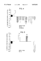
- FIG. 2 shows a graphic representation of CT2211 inhibition of DHEAS conversion by U937 sulphatase.
- FIG. 3 shows a graphic representation of CT2251 inhibition of DHEAS and ES conversion by U937 sulphatase.
- FIG. 4 shows a histogram analysis of contact sensitisation results.
- timepoint 48 hr.
- FIG. 5 shows a histogram analysis of contact sensitisation results.
- timepoint 24 hr.
- FIG. 6 shows a histogram analysis of contact sensitisation results_________________________________________ ⁇ DHEAS (50 mg/kg) ⁇ DHEAS (5 mg/kg) DHEAS (50 mg/kg) and corticosterone (5 mg/kg) DHEAS (5 mg/kg) and corticosterone (5 mg/kg) DHEAS (0.5 mg/kg) and corticosterone (5 mg/kg) DHEAS (0.05 mg/kg) and corticosterone (5 mg/kg) DHEAS (0.005 mg/kg) and corticosterone (5 mg/kg) DHEAS (0.0005 mg/kg) and corticosterone (5 mg/kg) corticosterone (5 mg/kg)________________________________________
- steroids given on day 0 and 4 sub.cut in dmso 20%: olive oil 80% sensitised with 2.5% oxazalone challenged with 0.25%
- FIG. 7 shows a histogram analysis of contact sensitisation results.
- _____________________________________________A dexamthasone 5 mg/kg ⁇ DHEA 5 mg/kg DHEA 5 mg/kg and Ct 2251 0.1 mg/kg DHEA 5 mg/kg and Ct 2251 10 mg/kg Ct 2251 0.1 mg/kg Ct 2251 10 mg/kgB ⁇ dexamthasone 5 mg/kg ⁇ DHEAS 5 mg/kg DHEAS 5 mg/kg and Ct 2251 0.1 mg/kg DHEAS 5 mg/kg and Ct 2251 10 mg/kg Ct 2251 0.1 mg/kg Ct 2251 10 mg/kg.
- steroids were given subcutaneously on days 0 and 4 sensitised with 2.5% oxazalone, challenged with 0.25%.
- FIG. 8 shows a histogram analysis of contact sensitisation results_______________________________________ ⁇ dexamethasone 5 mg/kg Ct 2251 0.01 mg/kg ⁇ Ct 2251 0.3 mg/kg Ct 2251 0.003 mg/kg Ct 2251 0.1 mg/kg Ct 2251 0.001 mg/kg Ct 2251 0.03 mg/kg.
- time point 24 hours, steroids given on day 0, 4 subcutaneously. Sensitised with 2.5% oxazalone, challenged with 0.25%.
- FIG. 9 shows a histogram analysis of contact sensitisation results_______________________________________ ⁇ dexamthasone 5 mg/kg ⁇ dheas 50 mg/kg dheas 15 mg/kg dheas 5 mg/kg dheas 50 mg/kg and Ct 2251 0.1 mg/kg dheas 15 mg/kg and Ct 2251 0.1 mg/kg dheas 5 mg/kg and Ct 2251 0.1 mg/kg Ct 2251 0.1 mg/kg Ct 2251 0.1 mg/kg.___
- time point 24 hours, steroids given on day 0,4 subcutaneously. Sensitised with 2.5% oxazalone, challenged with 0.25%.
- FIG. 10 shows effect of Ct 2251 on macrophage and T-cell numbers in contact sensitisation experiments in mice_______________________________________A vehicle control ⁇ Ct 2251 10 mg/kg DHEAS DEX 5 mg/kg DHEAS and Ct 2251 10 mg/kgB ⁇ negative control ⁇ Ct 2251 10 mg/kg vehicle control Dex 5 mg/kg DHEAS normal ears DHEAS and Ct 2251 10 mg/kg____
- FIG. 11 shows a histogram analysis of the effect of Ct 2251 on Delayed Type Hypersensitivity________________________________A ⁇ DHEA 5 mg/kg ⁇ DHEAS 5 mg/kg DEXA 5 mg/kg DHEA 5 mg/kg and Ct 2251 10 mg/kg DHEA 5 mg/kg and Ct 2251 0.1 mg/kg Ct 2251 10 mg/kg Ct 2251 0.1 mg/kgB ⁇ DHEA 5 mg/kg ⁇ DHEAS 5 mg/kg DEXA 5 mg/kg DHEAS 5 mg/kg and Ct 2251 10 mg/kg DHEAS 5 mg/kg and Ct 2251 0.1 mg/kg Ct 2251 10 mg/kg Ct 2251 0.1 mg/kg.
- steroids given on day 0 and 3 subcutaneously. Response at 24 h post challenge.
- FIG. 12 shows a graph of the effect of Ct 2251 on DTH in rats.
- CT 2251 given on days 0, 3 and 4 subcutaneously_______________________________________ ⁇ challenge only ⁇ Ct 2251 1 mg/kg ⁇ vehicle control ⁇ Ct 2251 0.1 mg/kg ⁇ Ct 2251 10 mg/kg.
- FIG. 13 shows effect of Ct 2251 on collagen induced arthritis in mice________________________________________ ⁇ paws involved severity________________________________________
- FIG. 14 shows
- FIG. 15 shows a histogram analysis of contact sensitisation results________________________________________ ⁇ DHEA (5 mg/kg) ⁇ DHEA (0.5 mg/kg) DHEA (0.05 mg/kg) DHEA (0.005 mg/kg) AED (5 mg/kg) AED (0.5 mg/kg) AED (0.05 mg/kg)______________________________________
- FIG. 16 shows a histogram analysis of contact sensitisation results________________________________________ ⁇ DHEAS (5 mg/kg) ⁇ DHEAS (0.5 mg/kg) DHEAS (0.05 mg/kg) DHEAS (0.005 mg/kg) AEDS (5 mg/kg) AEDS (0.5 mg/kg) AEDS (0.05 mg/kg) AEDS (0.005 mg/kg) AEDS (0.0005 mg/kg)_______________________________________
- FIG. 17 shows a histogram analysis of contact sensitisation results_______________________________________ ⁇ AEDS (5 mg/kg) ⁇ (5 mg/kg) and CT 2251 (0.1 mg/kg) DHEAS (5 mg/kg) DHEAS (5 mg/kg) and CT 2251 (0.1 mg/kg) CT 2251 (0.1 mg/kg) DEX (5 mg/kg)______________________________________
- FIG. 18 shows a diagram of the pathways of DHEA metabolism.
- ENZYME Human placental microsomes prepared as described in (Santner, S. J., et al (1984) J. Clin. Endocrinol. Metab. 59 29).
- ASSAY All assays were carried out in (at least) duplicate.
- the reaction mixture consisted of either PBS+250 mM sucrose (placental microsomes), containing 1 ⁇ mol/1 DHEAS [concentration chosen because approx. 10° ⁇ Km(app)] with 0.05% (v/v) [ 3 H] substrate and 0.05% (v/v) [ 14 C] DHEA) in 0.5% (v/v DMSO.
- the reaction was initiated by addition of appropriately diluted) such that 10% substrate conversion occurred during the assay) enzyme and incubated for 30 min at 30° C.
- Reactions were stopped by addition of 1000 ⁇ l toluene to the reaction mixture, vortexed (2*40 sec on max setting of multitube vortexer), then allow to settle for 5 min.
- 500 ⁇ l top (solvent) layer removed to scintillation vial.
- 10 ml scintillation fluid (Ultima Gold XR) then counted for radioactivity on a scintillation counter.
- enzyme was excluded from some reaction mixtures and 50% solvent and aqueous layers removed and counted for radioactivity in order to determine background (in solvent layer) and total (in aqueous layer) [ 3 H].
- DHEA is included to enable back correction to allow for product (DHEA) recovery (normally >98%).
- Inhibitors were included in the above reaction at varying concentrations in 0.5% (v/v) DMSO [or 5% (v/v) DMSO if necessary due to poor solubility of inhibitor] bringing the final DMSO concentration in the assay to 1% (v/v) [or 5.5% (v/v)].
- the enzyme would be assayed for estrone sulphate sulphatase activity similarly except with ES in the assay instead of DHEAS.
- ENZYME human recombinant placental steroid sulphatase (expressed in NSO cells). Either crude extracts, partially pure or pure enzyme may be used. The enzyme will be assayed as described for human placental steroid sulphatase as described above, except the assay buffer will be 50 mM Tris/HCl of appropriate pH (-7.5), containing 0.1% (v/v Triton ⁇ 100.
- Net 3H/14C values were divided by total, then result multiplied by substrate concentration (1 ⁇ mol/l) to give concentration of product.
- the computer program MULTICALC (Pharmacia) was used to fit a 4 parameter, unweighted curve to % inhibition (y axis) vs log (concentration of inhibitor) (x axis) and to determine the concentration of inhibitor which reduces the enzyme activity (in the absence of inhibitor) by 50% (IC50) from the graph.
- the enzyme activity would be determined at a variety of substrate concentrations then K' m calculated from these data using the computer program Enzfitter (Leatherbarrow R. A. (1987) Enzfitter; a non-linear regression data analysis programme for the IBM PC. Elsevier Biosoft, Cambridge U.K.). This would be repeated at a variety of inhibitor concentrations and K i determined from the K' m values obtained according to the equation:
- the inhibitors Ct 2211 and Ct 2251 were dissolved in 100% ethanol and 100% dimethyl sulphoxide (DMSO) respectively at stock concentrations of 20 mM. These were titrated in their respective solvents over a range of assay concentrations.
- the tritiated substrates DHEA sulphate and estrone sulphate were obtained from New England Nuclear and diluted in ethanol to give a final concentration of 70 nM for use in the assay.
- U937 cells were grown in RPLI containing 10% foetal bovine serum and harvested at a cell concentration of 1 ⁇ 10 6 /ml. These were washed in serum free medium stored at -70° C. at 1 ⁇ 10 8 /ml in Tris buffered saline pH7.4+1% triton X-100 and sonicated prior to use in the assay.
- FIG. 2 shows the ability of Ct 2211 to inhibit the sulphatase derived from U937 cells using DHEAS as a substrate.
- FIG. 3 shows the ability of Ct 2251 to inhibit the same enzyme using both DHEAS and estrone sulphate as substrates.
- IC 50 determined as described previously.
- An appropriate cell line i.e. one which takes up [ 3 H] DHEAS above background levels
- RPMI serum free cell culture medium
- [ 3 H] in cells--[ 3 H] in a suitable cell line known not to take up DHEAS e.g. U937
- DHEAS e.g. U937
- the principle of the experiment is that when leucocytes from one individual are mixed with those of another who expresses different HLA alleles, they will recognise each other as foreign and the lymphocytes will become activated. This activation is dependent, primarily, on interactions between the CD3/TcR complex on T cells and the MHC-II molecule on antigen presenting cells. Antibodies that bind to MHC-II are known to inhibit this reaction.
- PBMC Peripheral blood mononuclear cells
- PBMC Peripheral blood mononuclear cells
- PBMC are adjusted to 2 ⁇ 10 6 cells/ml in RPMI 1640 medium (Gibco UK) containing 2 mM Glutamine (Gibco UK), 100 ⁇ /ml/100 ⁇ g/ml Penicillin/Streptomycin (Gibco) and 10% foetal calf serum (Sigma UK), in which all manipulations, dilutions and incubations are done.
- PBMC from one individual are irradiated with 3000 rads. These cells will be stimulate a response from the other individual.
- Serial inhibitor dilutions are prepared in triplicate in sterile U-bottom 96 well microtitre plates (Falcon UK) in 100 ⁇ l. Control wells containing medium only and optimal Cyclosporin (Sandimmun, Sandoz) levels (100 nM) are also prepared to establish the maximum response and maximum inhibition, respectively. Equal numbers of irradiated stimulators and responders are mixed together and 100 ⁇ l are added to each well ⁇ DHEA-s added in 10 ⁇ l. Wells of stimulator alone and responders alone are also set up as controls. The experiment is incubated at 37° C. in 100% humidity and 5% CO 2 for 5 days. Response is measured by assessing proliferation during the last 18 hours of culture by incubation with 1 ⁇ Ci/well 3 H-Thymidine (Amersham UK), harvesting on to glass filter mattes and counting using a beta counter.
- T lymphocytes from an individual previously immunised with Tetanus Toxoid will respond to TT when re-exposed ex vivo.
- This activation is dependent on the interaction between the CD3/TcR complex on T cells and the MHC-II molecule on cells which process and present the antigen.
- Antibodies that bind to MHC-II are known to inhibit this reaction.
- Lymphocytes are prepared fresh for each experiment. Human venous blood is drawn into endotoxin free tubes containing heparin.
- Peripheral blood mononuclear cells are prepared by density gradient centrifugation according to the manufacturers instructions (Pharmacia), PBMC are adjusted to 2 ⁇ 10 6 cells/ml in RPMI 1640 medium (Gibco UK) containing 2 mM Glutamine (Gibco UK), 100 ⁇ /ml/100 ⁇ g/ml Penicillin/Streptomycin (Gibco) and 10% foetal calf serum (Sigma UK), in which all manipulations, dilutions and incubations are done.
- Serial inhibitor dilutions are prepared in triplicate in sterile U-bottom 96 well microtitre plates (Falcon UK) in 100 ⁇ l. 50 ⁇ l containing an optimal concentration of TT, previously determined by experimentation, is added to all wells. Control wells containing medium only or Cyclosporin (Sandimmun, Sandoz) (100 nM) are also prepared to establish the maximum response and maximum inhibition, respectively. 50 ⁇ l PBMC are then added to each well ⁇ DHEA-s added in 10 ⁇ l. The experiment is incubated at 37° C. in 100% humidity and 5% CO 2 for 7 days. Response is measured by assessing proliferation during the last 18 hours of culture by incubation with 1 ⁇ Ci/well 3 H-Thymidine, harvesting on to glass filter mattes and counting using a beta counter.
- T lymphocytes from an atopic individual known to be allergic to house dust mite will respond to extracts of house dust mite when re-exposed ex vivo. This activation is dependent on the interaction between the CD3/TcR complex on T cells and the MHC-II molecule on cells which process and present the antigen.
- Lymphocytes are prepared fresh for each experiment. Human venous blood is drawn into endotoxin free tubes containing heparin.
- Peripheral blood mononuclear cells are prepared by density gradient centrifugation according to the manufacturers instructions (Pharmacia). PBMC are adjusted to 2 ⁇ 10 6 cells/ml in RPMI 1640 medium (Gibco UK) containing 2 mM Glutamine (Gibco UK), 100 ⁇ /ml/100 ⁇ g/ml Penicillin/Streptomycin (Gibco) and 10% foetal calf serum (Sigma UK), in which all manipulations dilutions and incubations are done.
- Serial inhibitor dilutions are prepared in triplicate in sterile U-bottom 96 well microtitre plates (Falcon UK) in 100 ⁇ l. 50 ⁇ l containing an optimal concentration of HDM, previously determined by experimentation, is added to all wells. Control wells containing medium only or Cyclosporin (Sandimmun, Sandoz) (100 nM) are also prepared to establish the maximum response and maximum inhibition, respectively. 50 ⁇ l PBMC are then added to each well ⁇ DHEA-s. The experiment is incubated at 37° C. in 100% humidity and 5% CO 2 for 7 days. Response is measured by assessing proliferation during the last 18 hours of culture by incubation with 1 ⁇ Ci/well 3 H-Thymidine, harvesting on to glass filter mattes and counting using a beta counter.
- the mitogens PHA, Superantigens and OKT3 are used.
- T cells When mixed with the lectin PHA staphylococcal enterotoxins, or the anti- CD3 Ab OKT3, T cells will respond by becoming activated, proliferating and secreting cytokines.
- Lymphocytes are prepared fresh for each experiment. Human venous blood is drawn into endotoxin free tubes containing heparin.
- Peripheral blood mononuclear cells are prepared by density gradient centrifugation according to the manufacturers instructions (Pharmacia). PBMC are adjusted to 2 ⁇ 10 6 cells/ml in RPMI 1640 medium (Gibco UK) containing 2 mM Glutamine (Gibco UK), 100 ⁇ /ml/100 ⁇ g/ml Penicillin/Streptomycin (Gibco) and 10% foetal calf serum (Sigma UK), in which all manipulations, dilutions and incubations are done.
- Serial inhibitor dilutions are prepared in triplicate in sterile U-bottom 96 well microtitre plates (Falcon UK) in 100 ⁇ l. 50 ⁇ l containing an optimal concentrations of mutogens, previously determined by experimentation, are added to all wells. Control wells containing medium only or Cyclosporin (Sandimmun, Sandoz) (100 nM) are also prepared to establish the maximum response and maximum inhibition, respectively, 50 ⁇ l PBMC are then added to each well ⁇ DHEA-s in 10 ⁇ l. The experiment is incubated at 37° C. in 100% humidity and 5% CO 2 for 7 days. Response is measured by assessing proliferation during the last 18 hours of culture by incubation with 1 ⁇ Ci/well 3 H-Thymidine, harvesting on to glass filter mattes and counting using a beta counter.
- mice were painted on their shaved right flank with 50 ⁇ l 2.5% oxazalone or vehicle (acetone 4:1 olive oil).
- animals were challenged on their dorsal right ear surface with 25 ⁇ l of either 0.75% or 0.25% oxazalone.
- Prior to ear challenge, and 24, 48 and 72 hour post challenge ear thickness was measured using an engineer's micrometers. Ear swelling is determined as the difference in thickness prior and post challenge. The data are expressed as percentage change from the vehicle treated groups given sensitisation and challenge.
- mice On day 0, animals were injected intravenously with 10 6 washed SRBC in 0.1 ml saline. Four days later animals were challenged in the right hind footpad with 10 6 washed SRBC in 50 ⁇ l saline. Footpad thickness is measured prior to challenge, 24, 48 and 72 hours post challenge using calipers. Footpad swelling is determined as the difference in thickness prior and post challenge. The data are expressed as percentage change from the vehicle treated groups given sensitisation and challenge.
- DHEA, DHEAS were administered at various times, generally at time of sensitisation (day 0) and time of challenge (day 3 or 4-DTH model, day 4 or 5-(CS-model) in order to obtain an augmentation of the immune response above non-steroid treated animals.
- the inhibitors tested were:
- CT 2210 Estrone-3-0-(N,N-dimethyl)sulphamate
- CT2210 and CT2211 were able to reverse the stimulatory effects of either DHEA and DHEAS and furthermore were able to attenuate the ear swelling seen in the absence of DHEA and DHEAS. These data indicate that CT2210 and 2211 both inhibitors of steroid sulphate sulphatase, are immunosuppressive in this model.
- Group 1 sensitisation and challenge plus olive oil (positive control).
- Group 2 sensitisation and challenge only.
- Group 3 sensitisation and challenge plus DHEA (100 ⁇ g/mouse).
- Group 4 sensitisation and challenge plus DHEAS (100 ⁇ g/mouse).
- Group 5 sensitisation and challenge plus DEX (100 ⁇ g/mouse).
- the data shown in FIG. 6 indicate that by increasing the administered dose of DHEAS (and thereby its conversion to DHEA) a functional anti-glucocorticoid effect of DHEA exists in this model.
- Group 1 sensitisation and challenge plus olive oil (positive control)
- Group 4 sensitisation and challenge plus DHEA (5 mg/kg) and CT2251 (0.1 mg/kg)
- Group 5 sensitisation and challenge plus DHEA (5 mg/kg) and CT2251 (10 mg/kg)
- Group 9 sensitisation and challenge plus DHEAS (5 mg/kg) and CT2251 (0.1 mg/kg)
- Group 10 sensitisation and challenge plus DHEAS (5 mg/kg) and CT2251 (10 mg/kg)
- Group 1 sensitisation and challenge plus olive oil (positive control)
- Dexamethasone inhibited the degree of ear swelling. This was also inhibited by CT2251 treatment in a dose-dependent manner with doses of 0.1 and above being effective.
- Group 1 sensitisation and challenge plus olive oil (positive control)
- Group 3 sensitisation and challenge plus DHEAS (50 mg/kg)
- Group 6 sensitisation and challenge plus DHEAS (50 mg/kg) and CT2251 (0.1 mg/kg)
- Group 7 sensitisation and challenge plus DHEAS (15 mg/kg) and CT2251 (0.1 mg/kg)
- Group 8 sensitisation and challenge plus DHEAS (5 mg/kg) and CT2251 (0.1 mg/kg)
- ears were removed, fixed and stained with either an anti-CD3 (T-cells) or anti-Mac-1 (monocytes/macrophages) antibody and the number of positive cells counted in 10 random fields.
- Group 2 sensitisation and challenge plus olive oil (vehicle control)
- Group 4 sensitisation and challenge plus DHEAS (5 mg/kg) and CT2251 (10 mg/kg)
- Group 1 sensitisation and challenge plus olive oil (positive control)
- Group 5 sensitisation and challenge plus DHEA (5 mg/kg) and CT2251 (10 mg/kg)
- Group 6 sensitisation and challenge plus DHEA (5 mg/kg) and CT2251 (0.1 mg/kg)
- Group 7 sensitisation and challenge plus DHEAS (5 mg/kg) and CT2251 (10 mg/kg)
- Group 8 sensitisation and challenge plus DHEAS (5 mg/kg) and CT2251 (0.1 mg/kg)
- the data shown in FIG. (11) are consistent with the observations made in the mouse contact sensitisation model in terms of how DHEA/S, DEX and CT2251 affect the readout.
- DHEA and DHEAS are clearly pro-immune (or pro-inflammatory) and CT2251 can effectively reverse the DHEAS-mediated augmentation.
- CT2251 on its own can markedly reduce the foot swelling response.
- Group 2 sensitisation and challenge plus vehicle
- Group 1 tap water p.o. on day -1 and weekly
- Group 2 olive oil p.o. on day -1 and weekly
- FIG. (13) shows an incidence of 60 and 40% in the tap water and olive oil treated groups with those getting the disease showing severe signs. Treatment with CT2251 prevented the development of any disease.
- Wistar Rats were anaesthetised and given intra-pleural injections of 0.5% carrageenan in saline at time 0. Six hours later the animals were killed and pleural cavity lavaged with 1 ml. citrated saline. Volume of fluid exudate and exudate cell concentration were measured.
- Group 1 olive oil p.o. only
- Group 1 sensitisation and challenge plus vehicle (positive control)
- Group 3 sensitisation and challenge plus DHEA (0.5 mg/kg)
- Group 7 sensitisation and challenge plus AED (0.5 mg/kg)
- Group 8 sensitisation and challenge plus AED (0.05 mg/kg)
- the data shown in FIG. 15 indicate that AED and DHEA can augment the ear swelling response in this mode.
- the effect of AED can be seen down to a dose of 0.05 mg/kg or less.
- Group 1 sensitisation and challenge plus vehicle (positive control)
- Group 3 sensitisation and challenge plus DHEAS (0.5 mg/kg)
- Group 5 sensitisation and challenge plus DHEAS (0.005 mg/kg)
- Group 7 sensitisation and challenge plus AEDS (0.5 mg/kg)
- Group 8 sensitisation and challenge plus AEDS (0.05 mg/kg)
- Group 9 sensitisation and challenge plus AEDS (0.005 mg/kg)
- Group 10 sensitisation and challenge plus AEDS (0.0005 mg/kg)
- FIG. 16 show that AEDS can augment the ear swelling response in this model. This is achieved at doses at least 1000-fold less than seen with DHEAS.
- Group 1 sensitisation and challenge plus vehicle (positive control)
- Group 3 sensitisation and challenge plus AEDS (5 mg/kg) and CT 2251 (0.1 mg/kg)
- Group 5 sensitisation and challenge plus DHEAS (5 mg/kg) and Ct 2251 (0.1 mg/kg)
- the data presented in FIG. 17 indicate that the stimulatory response seen with AEDS (and DHEAS) can be inhibited by a sulphatase inhibitor. This shows that AEDS can serve as a physiological substrate for the sulphatase enzyme in vivo.
Landscapes
- Health & Medical Sciences (AREA)
- Chemical & Material Sciences (AREA)
- Medicinal Chemistry (AREA)
- Pharmacology & Pharmacy (AREA)
- Epidemiology (AREA)
- Life Sciences & Earth Sciences (AREA)
- Animal Behavior & Ethology (AREA)
- General Health & Medical Sciences (AREA)
- Public Health (AREA)
- Veterinary Medicine (AREA)
- Pharmaceuticals Containing Other Organic And Inorganic Compounds (AREA)
- Steroid Compounds (AREA)
Abstract
This invention relates to the use of stroid sulphate sulphatase inhibitors which prevent the normal physiological effect of DHEA or related steroids on inflammatory responses.
Description
This is a 371 of PCT/GB95/00780 filed 05 Apr., 1995.
This invention relates to a new medical use for certain compounds. In particular it relates to the use of inhibitors which prevent the normal physiological effect of DHEA or related steroids on immune and/or inflammatory responses and most especially to the use of inhibitors of a steroid sulphate sulphatase in revealing an endogenous glucocorticoid effect.
Dehydroepiandrosterone (DHEA) is a mild androgenic steroid produced by the adrenal cortex and gonads in men and by the gonads in rodents. In all species it is partly under hypothalamo-pituitary control and formed from common steroid precursors. Although DHEA may act as a precursor for more physiologically active androgenic steroids (e.g. testosterone) it is normally secreted as a hormone in its own right. DHEA is rapidly sulphated to DHEA sulphate (DHEAS) before release into the circulation and it is this latter species which constitutes the main circulating form of the steroid. Outside of the CNS DHEAS is itself inactive. To exert physiological effects it must be desulphated by a steroid sulphate sulphatase at the target site and therefore is able to influence local biological events.
Circulating DHEAS levels decline with age (unlike other steroids) and there is some evidence suggesting that exogenous DHEA administration in mice can reverse some of the physiological events associated with the aged phenotype (Daynes et al (1993) The Journal of Immunology, Vol 150: 12, 5219-5230). For example, administration of DHEAS can reverse the increase in IL-6 production in aged mice (Daynes et al (1993) The Journal of Immunology. Vol 150: 12, 5219-5230). In addition, DHEA may regulate insulin sensitivity of adlpocytes and skeletal muscle (Cleary, (1991) P.S.E.B.M. Vol 196: 8-17) However, its major physiological role may be its ability to influence immune responses.
The immune response to antigen is generally either cell mediated (T-cell mediated killing via processed antigens) or humoral (antibody production via recognition of whole antigen). The pattern of cytokine production by TH cells involved in the immune response can influence which of these response types predominates: cell mediated immunity (TH1) is characterised by high IL-2 and IFNg but low IL-4 production, whereas in humoral immunity (TH2) the pattern is low IL-2 and IFNg but high IL-4, IL-5, IL-10. Since the secretory pattern is modulated at the level of the secondary lymphoid organ then pharmacological manipulation of the specific TH cytokine pattern can influence the type and extent of the immune response generated.
It is well established that administration of exogenous glucocorticoids to ovalbumin-sensitised mice (and other species) induces immunosuppression in which spleen cells show a reduced capacity to secrete IL-2 but are able to enhance IL-4 secretion upon antigen challenge. In contrast, DHEA or DHEAS administration markedly augments IL-2 and IFNg secretion without affecting IL-4 release (Daynes et al (1990) Eur. J. Immunol. 20 793-802). When DHEA and dexamethasone are co-administered, the DHEA effect predominates which is consistent with DHEA acting as an anti-glucocorticoid in this and other systems (Daynes et al (1990) Eur. J. Immunol. 20 793-802), Browne et al (1992) The American Journal of the Medical Sciences 303 No. 6, 366-371 and Blauer et al (1991) Endocrinology 129 No. 6, 3174-3179). These differing responses of glucocorticoids and DHEA are indicative of TH2 and TH1 type cytokine patterns respectively, suggesting that they can differentially influence immune responses. Similar effects are seen if spleen cells from sensitized animals are treated with anti-CD3 in the presence of either DHEA, DHEAS or dexamethasone (Daynes et al (1990) J. Exp. Med. 171 979-996).
The immunostimulatory response to biologically inactive DHEAS indicates that there is conversion by steroid sulphate sulphatase within the confines of the secondary lymphoid tissue to the active DHEA. The location of the converting enzyme (steroid sulphate sulphatase) is within the antigen presenting cells (APC) since in purified T cell preparations treated with anti-CD3 antibody. DHEA but not DHEAS could enhance IL-2 production. However, if macrophages were added to the system both steroid types could elicit the increased IL-2 response (Daynes et al (1990) J. Exp. Med. 171 979-996). Hence the pattern of cytokine secretion of TH cells can be regulated within the secondary lymphoid microenvironment under the influence of adrenal steroids with DHEA promoting cell mediated (TH1) type responses. This effect would be counter-regulatory to the normal effect of endogenous glucocorticoids (inhibiting cell-mediated type responses). Indeed, a high affinity binding site for DHEA has been described in murine T cells that is distinct from the T-cell glucocorticoid receptor (Melkle et al (1992) J. Steroid Biochem. Molec. Biol. 42 No. 3/4, 293-304). Furthermore, many of these observations of steroid effects on immune responses in mice have also been demonstrated using human cells (Suzuki et al (1991) Clinical Immunology and Immunopathology 61, 202-211).
TH1 and TH2 -dependent responses show some degree of anatomical compartmentalisation; those lymphoid areas draining non-mucosal sites (e.g. spleen, peripheral lymph nodes) generate predominantly TH1 cytokine secreting patterns and those draining mucosal sites (eg Peyer's patches) show TH2 type responses. Indeed, anti-CD3-treated cells taken from spleen, peripheral lymph nodes or Peyer's patches from sensitised mice treated with DHEA show typical increased IL-2 and IFNg secretion but only spleen cells or peripheral lymph node cells do so when DHEAS was the treatment. In fact cells from Peyer's patches do not increase IL-2 production following DHEAS treatment but show a high IL-4 and low IL-2 secretion typical of a TH2 (and glucocorticoid) type response. This differential influence of DHEA on TH cell cytokine production correlates with the presence of DHEAS sulphatase in these areas; for example, brachial and axially lymph nodes (non-mucosal) have 6× the DHEAS sulphatase activity than Peyer's patches (mucosal; (Daynes et al (1990) J. Exp. Med. 171 979-996). Hence, in lymphoid areas where generally cell mediated immune responses take place then T cell cytokine production is influenced by locally produced DHEA whereas in areas governing humoral responses. DHEA has less influence allowing effects of circulating glucocorticoids to predominate. Any effects of DHEA are relatively discrete since it is rapidly resulphated on the first-pass through the liver. It follows therefore that in diseases where increased cellular immunity to specific antigens is pathological (e.g. autoimmunity) then a reduction in the influence of DHEA on these responses should lead to a state of relative immunosuppression with humoral immunity being spared.
We believe that inhibition of steroid sulphate sulphatase within the macrophage or other antigen presenting cells would lead to a decreased ability of sensitised T cells to mount a TH1 (high IL-2, IFNg low IL-4) response. The normal regulatory influence of other steroids such as glucocorticoids would therefore predominate.
Inhibitors which prevent the normal physiological effect of DHEA or related steroids thereby prevent DHEA or related steroids acting as an anti-glucocorticoid and consequently reveal an endogenous glucocorticoid-like effect. Since glucocorticoids can act on inflammatory processes as well as immune processes this endogenous glucocorticoid-like effect may therefore be manifested as an anti-inflammatory response.
In a first aspect the invention provides a therapeutic method for revealing an endogenous glucocorticoid-like effect in a human being which comprises administering an effective amount of an inhibitor which prevents the normal physiological effect of DHEA or related steroids on immune and/or inflammatory responses to said human being.
As used herein the term `DHEA or related steroids` is used to denote DHEA and physiologically active metabolites of DHEA such as, for example, 70H-DHEA. Androstenediol (AED), Androstenetriol (AET) and 160H-DHEA.
In a second aspect the invention provides an inhibitor which prevents the normal physiological effect of DHEA or related steroids on immune and/or inflammatory responses for use in revealing an endogenous glucocorticoid-like effect.
In a third aspect the invention provides an inhibitor which prevents the normal physiological effect of DHEA or related steroids on immune and/or inflammatory responses for use in a method of treatment of a human subject said method of treatment comprising revealing an endogenous glucocorticoid-like effect.
In a fourth aspect the invention further provides the use of an inhibitor which prevents the normal physiological effect of DHEA or related steroids or immune and/or inflammatory responses in the manufacture of a medicament for revealing an endogenous glucocorticoid-like effect.
The inhibitors are believed to achieve their therapeutic effect by removing a counter regulatory pathway which exists in vivo via the effect of DHEA or related steroids thereby revealing an endogenous glucocorticoid-like effect.
The endogenous glucocorticoid-like effect may have an anti-inflammatory and immunosuppressive component. By acting to reveal an endogenous glucocorticoid-like effect the inhibitors produce indirectly immunosuppression and/or an anti-inflammatory response.
The medicament manufactured according to the method of the present invention may be used to prevent or treat any physiological conditions which are generally associated with inflammatory diseases and/or diseases requiring immunosuppression.
The diseases suitable for treatment with the inhibitor which prevents the normal physiological effect of DHEA or related steroids on immune and/or inflammatory responses according to all aspects of the invention disclosed herein are preferably those resulting from autoimmunity, including for example, rheumatoid arthritis, type I and II diabetes, systemic lupus erythematosus, multiple sclerosis, myasthenia gravis, thyroiditis, vasculitis, ulcerative colitis and Crohn's disease, skin disorders e.g. psoriasis and contact dermatitis; graft versus host disease; eczema, asthma and organ rejection following transplantation.
The effect of the inhibitor in revealing an endogenous glucocorticoid-like effect may be determined by measuring anti-inflammatory and/or immunosuppressive responses.
The inhibitors which prevent the normal physiological effect of DHEA or related steroids on immune and/or inflammatory responses for use according to the invention may for example be a steroid analogue, a steroid derivative or fragment, a chemical compound or an antibody against DHEA or a related steroid, or a receptor therefor.
The inhibitors which prevent the normal physiological effect of DHEA or related steroids on immune and/or inflammatory responses may be inhibitors of a steroid sulphate sulphatase (E.C. 3.1.6.2). As used herein the term `a steroid sulphate sulphatase` denotes an enzyme which can remove a sulphate group from a sulphated steroid. Such an enzyme may be for example aryl sulphatase C (E.C. 3.1.6.1) or may desirably be one specific for DHEAS or related sulphated steroids i.e. sulphated metabolites derivable from DHEAS.
The inhibitors for use in the present invention are preferably steroid sulphate sulphatase inhibitors and most preferably are specific for DHEAS (or related sulphated steroids) sulphatase.
Examples of steroid sulphate sulphatase inhibitors are described in Howarth et al (J. Med. Chem (1994) 37, 219-221; and Bioorganic and Medicinal Chemistry Letters (1993) 3 (2) 313-318), Endocrinology (1991) 129 No. 6, 3174-3179) Birnbock and von Angerer (1990) (Biochemical Pharmacology 39 (11), 1709-1713); U.S. Pat. No. 5,281,587; Duncan et al (1993) (Cancer Research 53, 298-303); Li et al (1993) (Steroids 58, 106-111); Dibbelt et al (1994) (J. Steroid Biochem Molec. Biol. 50, 5/6, 261-266).
The inhibitors which prevent the normal physiological action of DHEA on immune and/or inflammatory responses via its physiologically active metabolites may be for example inhibitors of the enzymes 17-ketosteroid reductase, 16-α hydroxylase and 7-hydroxylase preferably 7-α -hydroxylase.
As indicated above the 7-hydroxylase is preferably 7α-hydroxylase. The 7α-hydroxylase is cytochrome P450 dependent (Khalil et al J. Steroid Biochem. Mol. Biol. 48, 5/6 545-552, (1994)) and is widely distributed i.e. brain, pituitary, adrenal skin, adipose tissue, lymphocytes and lung and is distinct from the testicular and hepatic testosterone/androstenedione 7α-hydroxylases, or the known hepatic cholesterol 7α-hydroxylases. The 7α-hydroxylase inhibitors for use according to the invention are therefore specific for the 7α-hydroxylases derived for example, from brain, pituitary, adrenal, adipose tissue, lymphocytes, skin and lung tisse and do not inhibit the testicular and hepatic testosterone/androstenedine 7α-hydroxylases or the known hepatic cholesterol 7α-hydroxylase.
7-hydroxylase inhibitors may be assayed for example in an enzyme assay for 7α-OH dehydroepiandrosterone and dehydroepiandrosterone or dehydroepiandrosterone sulphate and 7α-OH dehydroepiandrosterone sulphate. Such enzyme assays are known in the art, see for example Khalil et al (1994) (J. Steroid Biochem. Molec. Biol. 48, 5/6 545-552). Inhibitors of 7-hydroxylase may be evaluated by adding at various concentrations to the above assay. The biological activity of the 7-hydroxylase inhibitors identified as above to inhibit ongoing immune responses in cellular (eg MLR) and in vivo (e.g. DTH) systems will be studied as described more fully herein.
Similarly 16α-hydroxylase and inhibition thereof may be assayed by following the above teaching for steroid sulphate sulphatase and 7α-hydroxylase and as described by Chang et al; (1993) (Biochem J. 291, 429-434) after Waxman D. J. (1991) (Methods in Enzymol 206 462-476). 17 ketosteroid reductase and inhibition thereof would be assayed by following the above teaching for steroid sulphate sulphatase and 7α-hydroxylase and as described by Labrie et al (1992) Cancer Res 52 (3) 610-615 using a suitable source of enzyme activity.
Interconversion of DHEA, AED, 7OH, DEA, AET and 160H DHEA may be assayed by following the teaching of Khalil et al (1994) (J. Steroid Biochem. Molec. Biol. 48 5/6 454-552) or by Alena et al (1992) (J. Steroid Biochem and Molec. Biol. 288 959-964). A suitable source of the appropriate enzyme activity would be used.
Assay systems such as radioimmunoassay kits for measurement of DHEA and DHEA-S are commercially available (see for example Diagnostic Systems Laboratories, 445 Medical Centre Blvd, Webster, Tex. 77598).
Alternatively the anti-glucocorticoid effect could be achieved by for example blocking the target receptor for DHEA or related steroid or transport mechanism for DHEAS or related sulphated steroids. For example inhibitors may work by blocking the DHEA or related steroid receptor present in a T-cell or inhibiting the transport mechanism of DHEAS into cells and may be for example a peptide or antibody molecule.
Inhibitors which inhibit the uptake of DHEAS and related sulphated steroids into cells for example via a transport mechanism may be identified by an assay which would measure the uptake of labelled DHEAS (or related sulphated steroid) into a cell and the effects of inhibitors on such uptake as described more fully herein.
The effect of an inhibitor on the ability of DHEA or related steroids to bind to a receptor such as a T-cell receptor may be measured using techniques well known in the art. For example binding inhibition studies may be carried out measuring the effect of an inhibitor on the ability of the steroid to bind to its receptor.
Where the inhibitor for use in the invention is an antibody molecule it may in general belong to any immunoglobulin class. It may be of animal, for example mammalian, e.g. murine, rat, hamster, or human origin. The antibody may be a whole immunogloblin or a fragment thereof, for example a F(ab')2 or Fab fragment.
An antibody against an enzyme, receptor, or steroid (DHEA or DHEAS or related steroid) may be prepared using well-known immunological techniques employing the enzyme, receptor or steroid as antigen. Any suitable host may, for example, be immunised with the enzyme, receptor or steroid and splenocytes or lymphocytes recovered and immortalised using for example, the method of Kohler et al Eur. J. Immunol. 6 511 (1976). The resulting cells are diluted and cloned to obtain a single genetic line producing antibodies in accordance with conventional practice. Where it is desired to produce recombinant antibodies these may be produced using methods well known in the art.
The antibody may comprise an engineered human antibody e.g. a chimeric antibody or a CDR-grafted antibody as described in our International Patent Application WO-A-91/09967, EP 0120694 and EP 125023 the disclosures of which are incorporated herein by reference.
In a fifth aspect the invention provides a drug for revealing an endogenous glucocorticoid effect comprising an inhibitor which prevents the normal physiological effect of DHEA or related steroids on immune and/or inflammatory responses as an active ingredient.
In a sixth aspect the invention provides a method of treatment of a human subject suffering from a disease said method of treatment comprising revealing an endogenous glucocorticoid-like by administering an effective amount of an inhibitor which prevents the normal physiological effect of DHEA or related steroids on immune and/or inflammatory responses.
According to a seventh aspect there is provided a pharmaceutical composition which comprises an inhibitor which prevents the normal physiological effect of DHEA or related steroids on immune and/or inflammatory responses for use in revealing an endogenous glucocorticoid-like effect in admixture with one or more pharmaceutically acceptable carriers, excipients or diluents.
In a eighth aspect of the invention we provide a method for the manufacture of a pharmaceutical composition for use in revealing an endogenous glucocorticoid-effect effect which comprises admixing an inhibitor which prevents the normal physiological effect of DHEA or related steroids on immune and/or inflammatory responses and one or more pharmaceutically acceptable carriers, excipients or diluents.
Pharmaceutical compositions for use according to the present invention may be formulated in conventional manner, optionally with one or more physiologically acceptable carriers, diluents or excipients.
Inhibitors which prevent the normal physiological effect of DHEA or related steroids on immune and/or inflammatory responses for use according to the present invention may be formulated for oral, buccal, parenteral or rectal administration or in a form suitable for nasal administration or administration by inhalation or insufflation.
For oral administration, the pharmaceutical compositions may take the form of, for example, tablets or capsules prepared by conventional means with pharmaceutically acceptable excipients such as binding agents (e.g. pregelatinised maize starch, polyvinylpyrrolidone or hydroxypropyl methycellulose); fillers (e.g. lactose, microcrystalline cellulose or calcium hydrogen phosphate); lubricants (e.g. magnesium stearate, talc or silica); disintegrants (e.g. potato starch or sodium glycollate); or wetting agents (e.g. sodium lauryl sulphate). The tablets may be coated by methods well known in the art. Liquid preparations for oral administration may take the form of, for example, solutions, syrups or suspension, or they may be presented as a dry product for constitution with water or other suitable vehicle before use. Such liquid preparations may be prepared by conventional means with pharmaceutically acceptable additives such as suspending agents; emulsifying agents; non-aqueous vehicles; and preservatives. The preparations may also contain buffer salts, flavouring, colouring and sweetening agents as appropriate.
Preparations for oral administration may be suitably formulated to give controlled release of the active compound.
For buccal administration the compositions may take the form of tablets or lozenges formulated in conventional manner.
The inhibitor may be formulated for parenteral administration by injection e.g. by bolus injection or continuous infusion. Formulations for injection may be presented in unit dosage form. The compositions may take such forms as suspensions, solutions or emulsions in oily or aqueous vehicles, and may contain formulatory agents such as suspending, stabilising and/or dispersing agents. Alternatively, the active ingredient may be in powder form for constitution with a suitable vehicle, e.g. sterile pyrogen-free water, before use.
The inhibitor may also be formulated in rectal compositions such as suppositories or retention enemas, e.g. containing conventional suppository bases such as cocoa butter or other glycerides.
In addition to the formulations described previously, the inhibitor may also be formulated as a depot preparation. Such long acting formulations may be administered by implantation or by intramuscular injection.
For nasal administration or administration by inhalation the compounds for use according to the present invention are conveniently delivered in the form of an aerosol spray presentation from pressurised packs or a nebuliser, with the use of a suitable propellant, e.g. dichlorodifluoromethane, trichlorofluoromethane, dichlorotetrafluoroethane, carbon dioxide or other suitable gas.
The compositions may, if desired, be presented in a pack or dispenser device which may contain one or more unit dosage forms containing the active ingredient. The pack or dispenser device may be accompanied by instructions for administration.
The dose at which the inhibitor which prevents the normal physiological effect of DHEA or related steroids on immune and/or inflammatory responses will be administered will depend on the nature of the inhibitor and the route of administration, the potency of inhibitor and the body weight and pathology of the patient. Dosages may, for example, be in the range 1 μg/kg to 100 mg/kg, preferably 0.1 mg to 10 mg/kg and most preferably 0.1 mg to 1 mg/kg.
As indicated above the inhibitor is preferably an inhibitor of a steroid sulphate sulphatase and preferably of DHEAS (or a related sulphated steroid) sulphatase. Steroid sulphate sulphatase is a microsomal enzyme found in many tissues but predominantly in lymphoid (presumably macrophage) and pituitary tissues (Milewich et al, (1984) J. Steroid, Biochem 21 No. 5, 529-538). It is as yet unclear whether isoforms of this enzyme exist or whether the macrophage enzyme is distinct from that of other tissues. The steroid sulphatase inhibitor is most preferably specific for DHEAS sulphatase found in macrophages or other antigen presenting cells.
As a simple first step to test our hypothesis a couple of structural derivatives of oestrone have been made which inhibit steroid sulphate sulphatase. It will be evident to a man skilled in the art that structural derivatives of DHEA could also be made and assayed as described below. These have been assayed against the enzyme present in macrophage cell lines and isolated human placental sulphatase and then their ability to inhibit ongoing immune response in cellular (eg. Mixed Lymphocyte Reaction [MLR]) and in vivo (eg Delayed type Hypersensitivity [DTH]) systems has been studied. In addition their ability to interfere with DHEAS-mediated T cell IL-2 production in the presence and absence of macrophages has been assayed.
According to a ninth aspect the invention provides a method of screening for an inhibitor which prevents the normal physiological effect of DHEA or related steroids on immune and/or inflammatory responses comprising measuring the ability of said inhibitor to inhibit ongoing immune and/or inflammatory response in cellular and/or in vivo systems.
The effects of inhibitors according to the present invention on inflammation may be measured in vitro using methods known in the art such as, for example, by measuring inflammatory mediator release e.g. cytokine, prostaglandin, thromboxin or PAF release, and in vivo using inflammatory dependent processes such as for example carageenan pleurlsy described more fully herein.
The effect of the inhibitors on immune function may be measured in vitro using methods well known in the art such as in Mixed Lymphocyte Reaction and in vivo in, for example, a Delayed Type Hypersensitivity Reaction.
The screening method is particularly suitable for identifying those inhibitors which prevent the normal physiological effect of DHEA or related steroids on immune and/or inflammatory responses suitable for use in revealing an endogenous glucocorticoid effect.
The invention is further illustrated with reference to the following Examples and Figures in which:
FIG. 1 shows a schematic representation of the influence of adrenal steroids on TH cell cytokine production.
FIG. 2 shows a graphic representation of CT2211 inhibition of DHEAS conversion by U937 sulphatase.
FIG. 3 shows a graphic representation of CT2251 inhibition of DHEAS and ES conversion by U937 sulphatase.
FIG. 4 shows a histogram analysis of contact sensitisation results.
______________________________________
▪
     dhea 100 μg         dheas 100 μg = 2210 2 mg
□
     dheas 100 μg        dheas 100 μg = 2211 2 mg
     dexamethasone 100 μg
                            Ct2210 2 mg
     dhea 100 μg = 2210 2 mg
                            Ct 2211 2 mg
     dhea 100 μg = 2211 2 mg
______________________________________
timepoint=48 hr. Steroids given on day 0, 4 and 5 subcutaneously, sensitised with 2.5% oxazalone, challenged with 0.25%.
FIG. 5 shows a histogram analysis of contact sensitisation results.______________________________________▪ olive oil dheas 100 μg□ dhea 100 μg dexamethasone 100 μg______________________________________
timepoint=24 hr. Steroids given on day 0 and 5 subcutaneously, sensitised with 2.5% oxazalone, challenged with 0.25%.
by anova ** p<0.05 vs control+dexamethasone
$$p<0.05 vs control dhea+dheas
FIG. 6 shows a histogram analysis of contact sensitisation results______________________________________□ DHEAS (50 mg/kg)▪ DHEAS (5 mg/kg) DHEAS (50 mg/kg) and corticosterone (5 mg/kg) DHEAS (5 mg/kg) and corticosterone (5 mg/kg) DHEAS (0.5 mg/kg) and corticosterone (5 mg/kg) DHEAS (0.05 mg/kg) and corticosterone (5 mg/kg) DHEAS (0.005 mg/kg) and corticosterone (5 mg/kg) DHEAS (0.0005 mg/kg) and corticosterone (5 mg/kg) corticosterone (5 mg/kg)______________________________________
steroids given on day 0 and 4 sub.cut in dmso 20%: olive oil 80% sensitised with 2.5% oxazalone challenged with 0.25%
Results are given as mean±sem, n=10
FIG. 7 shows a histogram analysis of contact sensitisation results.______________________________________A ▪ dexamthasone 5 mg/kg □ DHEA 5 mg/kg DHEA 5 mg/kg and Ct 2251 0.1 mg/kg DHEA 5 mg/kg and Ct 2251 10 mg/kg Ct 2251 0.1 mg/kg Ct 2251 10 mg/kgB ▪ dexamthasone 5 mg/kg □ DHEAS 5 mg/kg DHEAS 5 mg/kg and Ct 2251 0.1 mg/kg DHEAS 5 mg/kg and Ct 2251 10 mg/kg Ct 2251 0.1 mg/kg Ct 2251 10 mg/kg______________________________________
steroids were given subcutaneously on days 0 and 4 sensitised with 2.5% oxazalone, challenged with 0.25%.
results given as mean±sem, n=7
FIG. 8 shows a histogram analysis of contact sensitisation results______________________________________▪ dexamethasone 5 mg/kg Ct 2251 0.01 mg/kg□ Ct 2251 0.3 mg/kg Ct 2251 0.003 mg/kg Ct 2251 0.1 mg/kg Ct 2251 0.001 mg/kg Ct 2251 0.03 mg/kg______________________________________
time point=24 hours, steroids given on day 0, 4 subcutaneously. Sensitised with 2.5% oxazalone, challenged with 0.25%.
results given as mean±sem, n=7-14
FIG. 9 shows a histogram analysis of contact sensitisation results______________________________________▪ dexamthasone 5 mg/kg□ dheas 50 mg/kg dheas 15 mg/kg dheas 5 mg/kg dheas 50 mg/kg and Ct 2251 0.1 mg/kg dheas 15 mg/kg and Ct 2251 0.1 mg/kg dheas 5 mg/kg and Ct 2251 0.1 mg/kg Ct 2251 0.1 mg/kg______________________________________
time point=24 hours, steroids given on day 0,4 subcutaneously. Sensitised with 2.5% oxazalone, challenged with 0.25%.
results given as mean±sem, n=7-14
FIG. 10 shows effect of Ct 2251 on macrophage and T-cell numbers in contact sensitisation experiments in mice______________________________________A vehicle control ▪ Ct 2251 10 mg/kg DHEAS DEX 5 mg/kg DHEAS and Ct 2251 10 mg/kgB □ negative control ▪ Ct 2251 10 mg/kg vehicle control Dex 5 mg/kg DHEAS normal ears DHEAS and Ct 2251 10 mg/kg______________________________________
results given as mean±sem.
*=p<0.05 vs DHEAS, *=p<0.05 vs CT2251
$=p<0.05 vs vehicle control
FIG. 11 shows a histogram analysis of the effect of Ct 2251 on Delayed Type Hypersensitivity______________________________________A ▪ DHEA 5 mg/kg □ DHEAS 5 mg/kg DEXA 5 mg/kg DHEA 5 mg/kg and Ct 2251 10 mg/kg DHEA 5 mg/kg and Ct 2251 0.1 mg/kg Ct 2251 10 mg/kg Ct 2251 0.1 mg/kgB ▪ DHEA 5 mg/kg □ DHEAS 5 mg/kg DEXA 5 mg/kg DHEAS 5 mg/kg and Ct 2251 10 mg/kg DHEAS 5 mg/kg and Ct 2251 0.1 mg/kg Ct 2251 10 mg/kg Ct 2251 0.1 mg/kg______________________________________
steroids given on day 0 and 3 subcutaneously. Response at 24 h post challenge.
Results given as mean±sem, n=7-8
$, *p<0.05 ANOVA
FIG. 12 shows a graph of the effect of Ct 2251 on DTH in rats. CT 2251 given on days 0, 3 and 4 subcutaneously______________________________________◯ challenge only Ct 2251 1 mg/kg vehicle control Ct 2251 0.1 mg/kg▴ Ct 2251 10 mg/kg______________________________________
results given as mean±sem, n=6-7
FIG. 13 shows effect of Ct 2251 on collagen induced arthritis in mice______________________________________ □ paws involved severity______________________________________
data are mean±sem, n=10. Incidence of arthritis was 60% and 40% in tap water and olive oil groups respectively.
FIG. 14 shows
A the effect of Ct 2251 and DHEAS on carrageenan pleurisy --exudate
B the effect of Ct 2251 and DHEAS on carageenan pleurisy --Total Cells
Results given as mean±sem. *=sig. diff vs control by ANOVA.
FIG. 15 shows a histogram analysis of contact sensitisation results______________________________________▪ DHEA (5 mg/kg)□ DHEA (0.5 mg/kg) DHEA (0.05 mg/kg) DHEA (0.005 mg/kg) AED (5 mg/kg) AED (0.5 mg/kg) AED (0.05 mg/kg)______________________________________
Sensitised with 2.5% oxazalone, challenged with 0.25%.
Steroids given on day 0 and 4 subcutaneously, 20% dmso: 80% olive oil.
Results given as mean±sem, n=10.
FIG. 16 shows a histogram analysis of contact sensitisation results______________________________________▪ DHEAS (5 mg/kg)□ DHEAS (0.5 mg/kg) DHEAS (0.05 mg/kg) DHEAS (0.005 mg/kg) AEDS (5 mg/kg) AEDS (0.5 mg/kg) AEDS (0.05 mg/kg) AEDS (0.005 mg/kg) AEDS (0.0005 mg/kg)______________________________________
Sensitised with 2.5% oxazalone, challenged with 0.25%.
Steroids given on day 0 and 4 subcutaneously, 20% dmso: 80% olive oil.
Results given as mean±sem, n=10.
FIG. 17 shows a histogram analysis of contact sensitisation results______________________________________▪ AEDS (5 mg/kg)□ (5 mg/kg) and CT 2251 (0.1 mg/kg) DHEAS (5 mg/kg) DHEAS (5 mg/kg) and CT 2251 (0.1 mg/kg) CT 2251 (0.1 mg/kg) DEX (5 mg/kg)______________________________________
Sensitised with 2.5% oxazalone, challenged with 0.25%.
Steroids given on day 0 and 4 subcutaneously, 20% dmso: 80% olive oil.
Results given as mean±sem, n=10-11.
FIG. 18 shows a diagram of the pathways of DHEA metabolism.
1. Identification of Potent Steroid Sulphatase Inhibitors
a) Human placental microsomal steroid sulphatase
ENZYME: Human placental microsomes prepared as described in (Santner, S. J., et al (1984) J. Clin. Endocrinol. Metab. 59 29).
ASSAY: All assays were carried out in (at least) duplicate. The reaction mixture consisted of either PBS+250 mM sucrose (placental microsomes), containing 1 μmol/1 DHEAS [concentration chosen because approx. 10°<Km(app)] with 0.05% (v/v) [3 H] substrate and 0.05% (v/v) [14 C] DHEA) in 0.5% (v/v DMSO. The reaction was initiated by addition of appropriately diluted) such that 10% substrate conversion occurred during the assay) enzyme and incubated for 30 min at 30° C. Reactions were stopped by addition of 1000 μl toluene to the reaction mixture, vortexed (2*40 sec on max setting of multitube vortexer), then allow to settle for 5 min. 500 μl top (solvent) layer removed to scintillation vial. add 10 ml scintillation fluid (Ultima Gold XR) then counted for radioactivity on a scintillation counter. As controls, enzyme was excluded from some reaction mixtures and 50% solvent and aqueous layers removed and counted for radioactivity in order to determine background (in solvent layer) and total (in aqueous layer) [3 H]. [14 C] DHEA is included to enable back correction to allow for product (DHEA) recovery (normally >98%).
EFFECT OF INHIBITORS: Inhibitors were included in the above reaction at varying concentrations in 0.5% (v/v) DMSO [or 5% (v/v) DMSO if necessary due to poor solubility of inhibitor] bringing the final DMSO concentration in the assay to 1% (v/v) [or 5.5% (v/v)].
The enzyme would be assayed for estrone sulphate sulphatase activity similarly except with ES in the assay instead of DHEAS.
b) Recombinant steroid sulphatase (as Stein et al J. Biol. Chem. 264, 13865-13872, 1990; Yen et al Cell, 49, 443-454, 1987).
ENZYME: human recombinant placental steroid sulphatase (expressed in NSO cells). Either crude extracts, partially pure or pure enzyme may be used. The enzyme will be assayed as described for human placental steroid sulphatase as described above, except the assay buffer will be 50 mM Tris/HCl of appropriate pH (-7.5), containing 0.1% (v/v Triton×100.
1. Of rate of reaction of steroid sulphate sulphatase.
3H/14C dpm ratioed for all samples including controls i.e. (ratioed 3H dpm bottom layer "no enzyme" controls to 14C top layer "no enzyme controls" to give total 3H/14C).
3H/14C "no enzyme" controls (background) were subtracted from 3H/14C samples (not total) to give net 3H/14C values.
Net 3H/14C values were divided by total, then result multiplied by substrate concentration (1 μmol/l) to give concentration of product.
Concentration of product was divided by time of incubation (30 min) to give rate (V) substrate conversion (in μmol/l/min).
2. % inhibition in the presence of inhibitor.
[1-((rate+inhibitor)/(rate-inhibitor))]×100=% inhibition. where rate+inhibitor =rate in the presence of inhibitor
and where rate-inhibitor=rate in the absence of inhibitor
3. IC50
The computer program MULTICALC (Pharmacia) was used to fit a 4 parameter, unweighted curve to % inhibition (y axis) vs log (concentration of inhibitor) (x axis) and to determine the concentration of inhibitor which reduces the enzyme activity (in the absence of inhibitor) by 50% (IC50) from the graph.
4. Km (app) and Ki (app)
The enzyme activity would be determined at a variety of substrate concentrations then K'm calculated from these data using the computer program Enzfitter (Leatherbarrow R. A. (1987) Enzfitter; a non-linear regression data analysis programme for the IBM PC. Elsevier Biosoft, Cambridge U.K.). This would be repeated at a variety of inhibitor concentrations and Ki determined from the K'm values obtained according to the equation:
K.sub.m '=K.sub.m (1+[l]K.sub.i) where K.sub.m '=K.sub.m in presence of inhibitor at concentration [l].
The median Ki would be taken as a representative figure.
c) DHEAS sulphatase activity in U937 cell extracts.
The inhibitors Ct 2211 and Ct 2251 were dissolved in 100% ethanol and 100% dimethyl sulphoxide (DMSO) respectively at stock concentrations of 20 mM. These were titrated in their respective solvents over a range of assay concentrations. The tritiated substrates DHEA sulphate and estrone sulphate were obtained from New England Nuclear and diluted in ethanol to give a final concentration of 70 nM for use in the assay. U937 cells were grown in RPLI containing 10% foetal bovine serum and harvested at a cell concentration of 1×10⁶ /ml. These were washed in serum free medium stored at -70° C. at 1×10⁸ /ml in Tris buffered saline pH7.4+1% triton X-100 and sonicated prior to use in the assay.
50 μl of substrate in ethanol was added to each assay tube and the solvent removed in a rotary vacuum evaporator. For Ct 2211 dissolved in ethanol. 25 μl of each dilution was added to an assay tube and the solvent again removed by evaporation. With Ct 2251 dissolved in DMSO, 25 μl of U937 cells sonicate, diluted to a cell equivalent of 1.25×10⁷ /ml, was added to each tube. With Ct 2251 225 μl of U937 cell solicate was added to each tube giving a final concentration of 10% DMSO. The tubes were incubated at 37° C. for 4 hours and the reaction stopped with methanol. After centrifugation scintillation fluid was added to extract the desulphated substrate and the CPM present in an aliquot of the organic phase were counted using a beta counter.
FIG. 2 shows the ability of Ct 2211 to inhibit the sulphatase derived from U937 cells using DHEAS as a substrate. FIG. 3 shows the ability of Ct 2251 to inhibit the same enzyme using both DHEAS and estrone sulphate as substrates.
In order to obtain total counts 10 μl of the aqueous fraction of the duplicate tubes containing only hot DHEA-s were counted after addition of scintillant and the total cpm calculated as follows: ##EQU1## where vo +inhibitor-rate in the presence of inhibitor and vo -inhibitor=rate in the absence of inhibitor.
IC50 determined as described previously.
d) Other, as yet unknown DHEAS sulphatase activities will be assayed broadly as described above using a suitable buffer (i.e. one in which the enzyme has measurable activity).
2. Selectivity of Potent Steroid Sulphatase Inhibitors for Steroid Sulphatase only
The effect of steroid sulphatase inhibitors would be examined on other related sulphatase activities i.e. aryl sulphatases A and B.
The effects of steroid sulphatases directly on enzymes/receptors of the glucocorticoid pathway would be examined.
The effects of steroid sulphatase inhibitors on glucose-6-phosphate dehydrogenase would be examined.
Effects on these enzymes/receptors would be considered detrimental and to be avoided as far as possible.
3. Identification of Inhibitors of DHEAS and Related Sulphated Steroids Uptake into Cells
An assay to identify such inhibitors would measure uptake of [3H]DHEAS or a related sulphated steroid into an appropriate cell line and the effects of inhibitors on such an uptake. This is described for [3 H] DHEAS but it will be apparent to one skilled in the art that this could be modified using standard techniques for identifying inhibitors of uptake of related sulphated steroids.
1. An appropriate cell line (i.e. one which takes up [3 H] DHEAS above background levels) would be incubated with 1 μM DHEAS containing 1% (v/v) [3 H] DHEAS at 37° C. for a given time (>1 h) in a serum free cell culture medium (RPMI)±various inhibitor concentrations.
2. At the end of that time cells would be harvested (if adherent, first trypsinised off cell culture plate) by centrifugation, washed 3 times in excess (10 ml) of the serum free cell culture medium and counted for radioactivity in a scintillation counter (all cells lysing in scintillation fluid).
3. Calculation
1. Net [3 H] DHEAS uptake
[3 H] in cells--[3 H] in a suitable cell line known not to take up DHEAS (e.g. U937), i.e. background.
=net [3 H] uptake.
2. % Inhibition ##EQU2## where net [3 H] uptake+inhibitor=uptake in the presence of inhibitor and net [3 H] uptake-inhibitor=uptake in the absence of inhibitor.
3. IC50 Values
--determined as described for inhibition of sulphatase activity.
Results obtained to date:
1. vs U937 DHEAS sulphatase activity
______________________________________
CT No.      Apparent IC50 (μM)
______________________________________
2211        84
2251        0.04
______________________________________
2. vs human placental microsomal DHEAS sulphatase activity
______________________________________
CT No.      Apparent IC50 (μM)
______________________________________
2211        152
2251        0.005
______________________________________
2. IN VITRO CELL IMMUNOLOGY IMMUNE FUNCTION
Ex vivo T cell function experiments are performed where an interaction between MHC-II and the T cell receptor is an obligatory requirement for T cell activation. Steroid sulphate sulphatase inhibitors are tested in mixed lymphocyte reactions, which measure both naive and memory T cell activation, and recall responses to tetanus toxoid and to housedust mite which measure memory T cell responses of Th1+Th2 type respectively.
The principle of the experiment is that when leucocytes from one individual are mixed with those of another who expresses different HLA alleles, they will recognise each other as foreign and the lymphocytes will become activated. This activation is dependent, primarily, on interactions between the CD3/TcR complex on T cells and the MHC-II molecule on antigen presenting cells. Antibodies that bind to MHC-II are known to inhibit this reaction.
Leucocytes are prepared fresh for each experiment. Human venous blood from two individuals is drawn into endotoxin free tubes containing heparin. Peripheral blood mononuclear cells (PBMC) are prepared by density gradient centrifugation according to the manufacturers instructions (Pharmacia). PBMC are adjusted to 2×10⁶ cells/ml in RPMI 1640 medium (Gibco UK) containing 2 mM Glutamine (Gibco UK), 100 μ/ml/100 μg/ml Penicillin/Streptomycin (Gibco) and 10% foetal calf serum (Sigma UK), in which all manipulations, dilutions and incubations are done. PBMC from one individual are irradiated with 3000 rads. These cells will be stimulate a response from the other individual.
Serial inhibitor dilutions are prepared in triplicate in sterile U-bottom 96 well microtitre plates (Falcon UK) in 100 μl. Control wells containing medium only and optimal Cyclosporin (Sandimmun, Sandoz) levels (100 nM) are also prepared to establish the maximum response and maximum inhibition, respectively. Equal numbers of irradiated stimulators and responders are mixed together and 100 μl are added to each well±DHEA-s added in 10 μl. Wells of stimulator alone and responders alone are also set up as controls. The experiment is incubated at 37° C. in 100% humidity and 5% CO2 for 5 days. Response is measured by assessing proliferation during the last 18 hours of culture by incubation with 1 μCi/well 3 H-Thymidine (Amersham UK), harvesting on to glass filter mattes and counting using a beta counter.
Results are plotted as CPM against inhibitor concentration.
Supernatants are removed and assayed for cytokine production IL-2, IFNγ, TNF and LT.
The principle of the experiment is that T lymphocytes from an individual previously immunised with Tetanus Toxoid (TT) will respond to TT when re-exposed ex vivo. This activation is dependent on the interaction between the CD3/TcR complex on T cells and the MHC-II molecule on cells which process and present the antigen. Antibodies that bind to MHC-II are known to inhibit this reaction.
Lymphocytes are prepared fresh for each experiment. Human venous blood is drawn into endotoxin free tubes containing heparin. Peripheral blood mononuclear cells (PBMC) are prepared by density gradient centrifugation according to the manufacturers instructions (Pharmacia), PBMC are adjusted to 2×10⁶ cells/ml in RPMI 1640 medium (Gibco UK) containing 2 mM Glutamine (Gibco UK), 100 μ/ml/100 μg/ml Penicillin/Streptomycin (Gibco) and 10% foetal calf serum (Sigma UK), in which all manipulations, dilutions and incubations are done.
Serial inhibitor dilutions are prepared in triplicate in sterile U-bottom 96 well microtitre plates (Falcon UK) in 100 μl. 50 μl containing an optimal concentration of TT, previously determined by experimentation, is added to all wells. Control wells containing medium only or Cyclosporin (Sandimmun, Sandoz) (100 nM) are also prepared to establish the maximum response and maximum inhibition, respectively. 50 μl PBMC are then added to each well±DHEA-s added in 10 μl. The experiment is incubated at 37° C. in 100% humidity and 5% CO2 for 7 days. Response is measured by assessing proliferation during the last 18 hours of culture by incubation with 1 μCi/well 3 H-Thymidine, harvesting on to glass filter mattes and counting using a beta counter.
Results are plotted as CPM against inhibitor concentration.
Supernatants are also removed and tested for cytokine production IL-2, IL-4, IL-5, TNF, LT, IFNγ.
The principle of the experiment is that T lymphocytes from an atopic individual known to be allergic to house dust mite will respond to extracts of house dust mite when re-exposed ex vivo. This activation is dependent on the interaction between the CD3/TcR complex on T cells and the MHC-II molecule on cells which process and present the antigen.
Lymphocytes are prepared fresh for each experiment. Human venous blood is drawn into endotoxin free tubes containing heparin. Peripheral blood mononuclear cells (PBMC) are prepared by density gradient centrifugation according to the manufacturers instructions (Pharmacia). PBMC are adjusted to 2×10⁶ cells/ml in RPMI 1640 medium (Gibco UK) containing 2 mM Glutamine (Gibco UK), 100 μ/ml/100 μg/ml Penicillin/Streptomycin (Gibco) and 10% foetal calf serum (Sigma UK), in which all manipulations dilutions and incubations are done.
Serial inhibitor dilutions are prepared in triplicate in sterile U-bottom 96 well microtitre plates (Falcon UK) in 100 μl. 50 μl containing an optimal concentration of HDM, previously determined by experimentation, is added to all wells. Control wells containing medium only or Cyclosporin (Sandimmun, Sandoz) (100 nM) are also prepared to establish the maximum response and maximum inhibition, respectively. 50 μl PBMC are then added to each well±DHEA-s. The experiment is incubated at 37° C. in 100% humidity and 5% CO2 for 7 days. Response is measured by assessing proliferation during the last 18 hours of culture by incubation with 1 μCi/well 3 H-Thymidine, harvesting on to glass filter mattes and counting using a beta counter.
Supernatants are also removed and tested for IL-2, IL-4, IL-5, IFNγ, TNF and LT.
Results are plotted as CPM against inhibitor concentration.
Ex Vivo T cell function experiments are performed where an interaction between MHC-II and the T cell receptor is not an obligatory requirement for T cell activation.
The mitogens PHA, Superantigens and OKT3 are used.
When mixed with the lectin PHA staphylococcal enterotoxins, or the anti- CD3 Ab OKT3, T cells will respond by becoming activated, proliferating and secreting cytokines.
Lymphocytes are prepared fresh for each experiment. Human venous blood is drawn into endotoxin free tubes containing heparin. Peripheral blood mononuclear cells (PBMC) are prepared by density gradient centrifugation according to the manufacturers instructions (Pharmacia). PBMC are adjusted to 2×10⁶ cells/ml in RPMI 1640 medium (Gibco UK) containing 2 mM Glutamine (Gibco UK), 100 μ/ml/100 μg/ml Penicillin/Streptomycin (Gibco) and 10% foetal calf serum (Sigma UK), in which all manipulations, dilutions and incubations are done.
Serial inhibitor dilutions are prepared in triplicate in sterile U-bottom 96 well microtitre plates (Falcon UK) in 100 μl. 50 μl containing an optimal concentrations of mutogens, previously determined by experimentation, are added to all wells. Control wells containing medium only or Cyclosporin (Sandimmun, Sandoz) (100 nM) are also prepared to establish the maximum response and maximum inhibition, respectively, 50 μl PBMC are then added to each well±DHEA-s in 10 μl. The experiment is incubated at 37° C. in 100% humidity and 5% CO2 for 7 days. Response is measured by assessing proliferation during the last 18 hours of culture by incubation with 1 μCi/well 3 H-Thymidine, harvesting on to glass filter mattes and counting using a beta counter.
Results are plotted as CPM against inhibitor concentration.
Supernatants are removed for cytokine assay IL-2, 4, 5, LT, TNF, IFNγ and IL-1.
3. IN VIVO PHARMACOLOGY FOR DHEAS SULPHATASE, INHIBITORS
The effect of DHEA, DHEAS and their inhibitors have been tested in 2 models of immune function in mice.
1. Contact Sensitisation (CS)
On day 0, mice were painted on their shaved right flank with 50 μl 2.5% oxazalone or vehicle (acetone 4:1 olive oil). On day 5 animals were challenged on their dorsal right ear surface with 25 μl of either 0.75% or 0.25% oxazalone. Prior to ear challenge, and 24, 48 and 72 hour post challenge ear thickness was measured using an engineer's micrometers. Ear swelling is determined as the difference in thickness prior and post challenge. The data are expressed as percentage change from the vehicle treated groups given sensitisation and challenge.
2. Delayed Type Hypersensitivity (DTH)
On day 0, animals were injected intravenously with 106 washed SRBC in 0.1 ml saline. Four days later animals were challenged in the right hind footpad with 106 washed SRBC in 50 μl saline. Footpad thickness is measured prior to challenge, 24, 48 and 72 hours post challenge using calipers. Footpad swelling is determined as the difference in thickness prior and post challenge. The data are expressed as percentage change from the vehicle treated groups given sensitisation and challenge.
DHEA, DHEAS were administered at various times, generally at time of sensitisation (day 0) and time of challenge (day 3 or 4-DTH model, day 4 or 5-(CS-model) in order to obtain an augmentation of the immune response above non-steroid treated animals.
Different doses of the inhibitors have been used, and compared to the effect of dexamethasone as potential immunosuppressive/anti-inflammatory agents.
Three inhibitors of steroid sulphate sulphatase prepared as described in Howarth et al J. Med. Chem. (1994) 37, 219-221 and Howarth et al (1993) Bioorganic & Medicinal Chemistry Letters 3 (2) 313-318 have been tested for their immunosuppressive ability in a mouse contact sensitisation model. The basic protocol is outlined above. Two separated experiments have been performed.
The inhibitors tested were:
CT 2210: Estrone-3-0-(N,N-dimethyl)sulphamate
CT 2211: Estrone 3-methylphosphonate
CT 2251: Estrone 3-0-sulphamate
Animals were sensitised with 2.5% oxazolone on day 0 and ear challenged with 0.25% oxazalone on day 5. Steroids (DHEA, DHEAS, dexamethasone and inhibitors CT 2210 and CT 2211) were given on days 0, 4 and 5 as subcutaneous injections dissolved in olive oil. The data are presented as percentage change from the vehicle treated group given sensitisation and challenge (positive control). Animals were randomised into 10 treatment groups (n=7-8 per group).
______________________________________
Group 1:
        sensitisation and challenge plus olive oil (positive control)
Group 2:
        sensitisation and challenge plus DHEA 100 μg/mouse
Group 3:
        sensitisation and challenge plus DHEAS 100 μg/mouse
Group 4:
        sensitisation and challenge plus dexamethasone
        100 μg/mouse
Group 5:
        sensitisation and challenge plus DHEA 100 μg/mouse +
        CT 2210 2 mg/mouse
Group 6:
        sensitisation and challenge plus DHEA 100 μg/mouse +
        CT 2211 2 mg/mouse
Group 7:
        sensitisation and challenge plus DHEAS 100 μg/mouse +
        CT 2210 2 mg/mouse
Group 8:
        sensitisation and challenge plus DHEAS 100 μg/mouse +
        CT 2211 2 mg/mouse
Groups 9/10
        sensitisation and challenge plus CT2210 or CT2211
        2 mg/mouse
______________________________________
The data are shown in FIG. 4 measured at 48 post challenge. Both DHEA and DHEAS alone augmented the normal increase in ear thickness seen with the procedure indicating an immunostimulatory effect. Dexamethasone reduced the response indicating an immunosuppressive (or anti-inflammatory) effect. Both CT2210 and CT2211 were able to reverse the stimulatory effects of either DHEA and DHEAS and furthermore were able to attenuate the ear swelling seen in the absence of DHEA and DHEAS. These data indicate that CT2210 and 2211 both inhibitors of steroid sulphate sulphatase, are immunosuppressive in this model.
Animals were randomised into 5 groups (n=7-8) and steroids given on days 0 and 5 subcutaneously, sensitised with 2.5% oxazalone challenged with 0.25%.
Group 1: sensitisation and challenge plus olive oil (positive control).
Group 2: sensitisation and challenge only.
Group 3: sensitisation and challenge plus DHEA (100 μg/mouse).
Group 4: sensitisation and challenge plus DHEAS (100 μg/mouse).
Group 5: sensitisation and challenge plus DEX (100 μg/mouse).
The data are shown in FIG. (5) measured at 24 h post-challenge. Both DHEA and DHEAS significantly augmented the response whereas it was inhibited by DEX. These data are consistent with the role of DHEA and DHEAS as immunostimulants/pro-inflammatories in vivo and DEX as immunosuppressive/anti-inflammatory.
By ANOVA **p<0.05 vs control+dexamethasone
$$p<0.05 vs control dhea+dheas
The data shown in FIG. 6 indicate that by increasing the administered dose of DHEAS (and thereby its conversion to DHEA) a functional anti-glucocorticoid effect of DHEA exists in this model.
Steroids given on day 0 and 4 subcutaneously in dmso 20%: olive oil 80%.
Sensitised with 2.5% oxazalone challenged with 0.25%. Results presented as mean±sem, n=10.
Animals were randomised into 9 groups (n=7) and steroids given on days 0 and 4 subcutaneously sensitised with 2.5% oxazalone challenged with 0.25%. Results given as mean±sem, n=7.
Group 1: sensitisation and challenge plus olive oil (positive control)
Group 2: sensitisation and challenge plus DEX (5 mg/kg)
Group 3: sensitisation and challenge plus DHEA (5 mg/kg)
Group 4: sensitisation and challenge plus DHEA (5 mg/kg) and CT2251 (0.1 mg/kg)
Group 5: sensitisation and challenge plus DHEA (5 mg/kg) and CT2251 (10 mg/kg)
Group 6: sensitisation and challenge plus CT2251 (0.1 mg/kg)
Group 7: sensitisation and challenge plus CT2251 (10 mg/kg)
Group 8: sensitisation and challenge plus DHEAS (5 mg/kg)
Group 9: sensitisation and challenge plus DHEAS (5 mg/kg) and CT2251 (0.1 mg/kg)
Group 10: sensitisation and challenge plus DHEAS (5 mg/kg) and CT2251 (10 mg/kg)
The data using these relatively weak inhibitors are shown in FIG. (7). Both DHEA and DHEAS augmented the normal ear swelling whereas it was inhibited by DEX. CT2251 (at both doses) reversed the augmentation seen with DHEAS but not that seen with DHEA. CT2251 on its own (at both doses) significantly inhibited the normal ear swelling response. These data show that inhibition of steroid sulphate sulphatase can reverse the stimulatory effect of DHEAS, a substrate for the enzyme. Furthermore, the ear swelling response itself is at least partially dependent on a product of endogenous sulphatase activity such as DHEA since CT2251 administration alone causes an immuno-suppressive or anti-inflammatory effect.
Animals were randomised into 8 groups (n=7-14) and steroids given in days 0 and 4, (see FIG. 8). Sensitised with 2.5% oxazalone challenged with 0.25%. Results presented as mean±sem, n=7-14. Timepoint=24 h.
Group 1: sensitisation and challenge plus olive oil (positive control)
Group 2: sensitisation and challenge plus DEX (5 mg/kg)
Group 3: sensitisation and challenge plus CT2251 (0.3 mg/kg)
Group 4: sensitisation and challenge plus CT2251 (0.1 mg/kg)
Group 5: sensitisation and challenge plus CT2251 (0.03 mg/kg)
Group 6: sensitisation and challenge plus CT2251 (0.01 mg/kg)
Group 7: sensitisation and challenge plus CT2251 (0.003 mg/kg)
Group 8: sensitisation and challenge plus CT2251 (0.001 mg/kg)
Dexamethasone inhibited the degree of ear swelling. This was also inhibited by CT2251 treatment in a dose-dependent manner with doses of 0.1 and above being effective.
Animals were randomised into 9 groups (n=7-14) and steroids given on days 0 and 4 (see FIG. 9). Sensitised with 2.5% oxazalone challenged with 0.25%. Results presented as mean±sem. Timepoint=24 h.
Group 1: sensitisation and challenge plus olive oil (positive control)
Group 2: sensitisation and challenge plus DEX (5 mg/kg)
Group 3: sensitisation and challenge plus DHEAS (50 mg/kg)
Group 4: sensitisation and challenge plus DHEAS (15 mg/kg)
Group 5: sensitisation and challenge plus DHEAS (5 mg/kg)
Group 6: sensitisation and challenge plus DHEAS (50 mg/kg) and CT2251 (0.1 mg/kg)
Group 7: sensitisation and challenge plus DHEAS (15 mg/kg) and CT2251 (0.1 mg/kg)
Group 8: sensitisation and challenge plus DHEAS (5 mg/kg) and CT2251 (0.1 mg/kg)
Group 9: sensitisation and challenge plus CT2251 (0.1 mg/kg)
The data indicate that the inhibitory effect of CT2251 on the ear swelling response can be reverse by increasing doses of DHEAS, the substrate for the sulphatase enzyme.
Animals were randomised into 7 groups (n=7-10) and steroids given on days 0 and 5. At 24 h following challenge, ears were removed, fixed and stained with either an anti-CD3 (T-cells) or anti-Mac-1 (monocytes/macrophages) antibody and the number of positive cells counted in 10 random fields.
Group 1: olive oil only
Group 2: sensitisation and challenge plus olive oil (vehicle control)
Group 3: sensitisation and challenge plus DHEAS (5 mg/kg)
Group 4: sensitisation and challenge plus DHEAS (5 mg/kg) and CT2251 (10 mg/kg)
Group 5: sensitisation and challenge plus CT2251 (10 mg/kg)
Group 6: sensitisation and challenge plus DEX (5 mg/kg)
Group 7: normal ears
The data in FIG. (10) show that DHEAS can augment both immune and inflammatory cell infiltrate into challenged ears. This can be effectively reversed by concomitant treatment with CT2251. CT2251 on its own was able to further reduce the number of infiltrating CD3 positive cells. These observations indicate that stimulation or inhibition of steroid sulphate sulphatase can augment or attenuate the movement of both immune and inflammatory cell types into the ears of sensitised mice.
*=p<0.05 vs DHEAS
$=p<0.05 vs vehicle control
C=p<0.05 vs CT2251.
Animals were randomised into 10 groups (n=7-8) and steroids given on day 0 and 4.
Steroids were given on day 0 and 3 subcutaneously. Response at 24 h post challenge. Results given as mean±sem. $, *p<0.05 ANOVA.
Group 1: sensitisation and challenge plus olive oil (positive control)
Group 2: sensitisation and challenge plus DHEA (5 mg/kg)
Group 3: sensitisation and challenge plus DHEAS (5 mg/kg)
Group 4: sensitisation and challenge plus DEX (5 mg/kg)
Group 5: sensitisation and challenge plus DHEA (5 mg/kg) and CT2251 (10 mg/kg)
Group 6: sensitisation and challenge plus DHEA (5 mg/kg) and CT2251 (0.1 mg/kg)
Group 7: sensitisation and challenge plus DHEAS (5 mg/kg) and CT2251 (10 mg/kg)
Group 8: sensitisation and challenge plus DHEAS (5 mg/kg) and CT2251 (0.1 mg/kg)
Group 9: sensitisation and challenge plus CT2251 (10 mg/kg)
Group 10: sensitisation and challenge plus CT2251 (0.1 mg/kg)
The data shown in FIG. (11) are consistent with the observations made in the mouse contact sensitisation model in terms of how DHEA/S, DEX and CT2251 affect the readout. DHEA and DHEAS are clearly pro-immune (or pro-inflammatory) and CT2251 can effectively reverse the DHEAS-mediated augmentation. Also CT2251 on its own can markedly reduce the foot swelling response.
Lewis rats were sensitised with SRBC IV and challenged 4 days later with 108 SRBC into the footpad. The resulting swelling response was measured 24 and 48 h later. Rats were randomised into 5 groups (n=6-7) and steroids given on days 0, 3 and 4, subcutaneously. Results given as mean±sem.
Group 1: challenge only
Group 2: sensitisation and challenge plus vehicle
Group 3: sensitisation and challenge plus CT2251 (10 mg/kg)
Group 4: sensitisation and challenge plus CT2251 (1 mg/kg)
Group 5: sensitisation and challenge plus CT2251 (0.1 mg/kg)
The data shown in FIG. (12) indicate that CT2251 can also suppress a DTH response in rats.
Male DBA/1 mice were sensitised with type II collagen into the base of the tail in FCA in day 0. These animals received a further injection of CII in FICA (Freunds Incomplete Adjuvant) on day 18 and incidence and severity of arthritis assessed from day 25. Mice were randomised into 3 groups (n=10).
Group 1: tap water p.o. on day -1 and weekly
Group 2: olive oil p.o. on day -1 and weekly
Group 3: CT2251 (10 mg/kg) on day -1 and weekly
The data shown in FIG. (13) shows an incidence of 60 and 40% in the tap water and olive oil treated groups with those getting the disease showing severe signs. Treatment with CT2251 prevented the development of any disease.
Wistar Rats were anaesthetised and given intra-pleural injections of 0.5% carrageenan in saline at time 0. Six hours later the animals were killed and pleural cavity lavaged with 1 ml. citrated saline. Volume of fluid exudate and exudate cell concentration were measured.
Animals were randomised to 5 treatment groups (n=6).
Results presented as mean±sem, *=sig. diff vs control by ANOVA.
Group 1: olive oil p.o. only
Group 2: DEX (1 mg/kg) p.o. at T=-1 h
Group 3: CT2251 (10 mg/kg) p.o. at T=-24 h and -1 h
Group 4: CT2251 (10 mg/kg) p.o. at t -1h
Group 5: DHEAS (5 mg/kg) p.o. at T=-24 h and -1 h
The data shown in FIG. (14) indicate that DEX can completely inhibit fluid leakage and markedly reduce cellular influx (68%). Treatment with CT2251 at -24 h and -1 h significantly reduced both exudate volume (43%) and cell infiltrate (28%) indicating that inhibition of steroid sulphate sulphatase can reduce an inflammatory-based response.
Animals were randomised to 8 groups (n=10) and steroids given on days 0 and 4 subcutaneously 20% DMSO: 80% olive oil. Sensitised with 2.5% oxazalone challenged with 0.25%.
Group 1: sensitisation and challenge plus vehicle (positive control)
Group 2: sensitisation and challenge plus DHEA (5 mg/kg)
Group 3: sensitisation and challenge plus DHEA (0.5 mg/kg)
Group 4: sensitisation and challenge plus DHEA (0.05 mg/kg)
Group 5: sensitisation and challenge plus DHEA (0.005 mg/kg)
Group 6: sensitisation and challenge plus AED (5 mg/kg)
Group 7: sensitisation and challenge plus AED (0.5 mg/kg)
Group 8: sensitisation and challenge plus AED (0.05 mg/kg)
The data shown in FIG. 15 indicate that AED and DHEA can augment the ear swelling response in this mode. The effect of AED can be seen down to a dose of 0.05 mg/kg or less.
Animals were randomised into 10 groups (n=10) and steroids given on days 0 and 4 subcutaneously in DMSO 20%: olive oil 80%. Sensitised with 2.5% oxazalone challenged with 0.25%. Results given as mean±sem.
Group 1: sensitisation and challenge plus vehicle (positive control)
Group 2: sensitisation and challenge plus DHEAS (5 mg/kg)
Group 3: sensitisation and challenge plus DHEAS (0.5 mg/kg)
Group 4: sensitisation and challenge plus DHEAS (0.05 mg/kg)
Group 5: sensitisation and challenge plus DHEAS (0.005 mg/kg)
Group 6: sensitisation and challenge plus AEDS (5 mg/kg)
Group 7: sensitisation and challenge plus AEDS (0.5 mg/kg)
Group 8: sensitisation and challenge plus AEDS (0.05 mg/kg)
Group 9: sensitisation and challenge plus AEDS (0.005 mg/kg)
Group 10: sensitisation and challenge plus AEDS (0.0005 mg/kg)
The data presented in FIG. 16 show that AEDS can augment the ear swelling response in this model. This is achieved at doses at least 1000-fold less than seen with DHEAS.
Animals were randomised into groups (n=10-11) and steroids given in days 0 and 4 subcutaneously in DMSO 20%: olive oil 80%. Sensitised with 2.5% oxazalone challenged with 0.25%. Results given as mean±sem.
Group 1: sensitisation and challenge plus vehicle (positive control)
Group 2: sensitisation and challenge plus AEDS (5 mg/kg)
Group 3: sensitisation and challenge plus AEDS (5 mg/kg) and CT 2251 (0.1 mg/kg)
Group 4: sensitisation and challenge plus DHEAS (5 mg/kg)
Group 5: sensitisation and challenge plus DHEAS (5 mg/kg) and Ct 2251 (0.1 mg/kg)
Group 6: sensitisation and challenge plus CT 2251 (0.1 mg/kg)
Group 7: sensitisation and challenge plus DEX (5 mg/kg)
The data presented in FIG. 17 indicate that the stimulatory response seen with AEDS (and DHEAS) can be inhibited by a sulphatase inhibitor. This shows that AEDS can serve as a physiological substrate for the sulphatase enzyme in vivo.
Claims (5)
1. A method of treating a human subject having an inflammatory disease comprising administering an effective amount of an inhibitor which prevents the normal physiological effect of DHEA or a physiologically active metabolite of DHEA on inflammatory responses, wherein the inhibitor is a steroid sulphate inhibitor, thereby treating the subject.
2. A method of treating a human subject having an inflammatory disease comprising an effective amount of an inhibitor which prevents the normal physiological effect of DHEA or a physiologically active metabolite of DHEA, wherein the physiologically active metabolite is selected from the group consisting of 70H-DHEA, 70-DHEA-S, 160-DHEA, 160-DHEA-S, androstenediol (AED), AED-S, androstenetriol (AET), AET-S, and DHEA-S on inflammatory responses, wherein the inhibotor is a steroid sulphate sulphatase inhibitor, thereby treating the subject.
3. A method according to claim 1 wherein said steroid sulphate sulphase inhibitor is estrone-3O-N,N-dimethyl)sulphamate.
4. A method according to claim 1, wherein said steroid sulphate sulphatase inhibitor is estrone-3-methylphosphonate.
5. A method according to claim 1, wherein said steroid sulphate sulphatase inhibitor is estrone 3-o-sulphamate.
Applications Claiming Priority (7)
| Application Number | Priority Date | Filing Date | Title |
|---|---|---|---|
| GB9406715 | 1994-04-05 | ||
| GB9406715A GB9406715D0 (en) | 1994-04-05 | 1994-04-05 | Medicaments |
| GB941062 | 1994-05-26 | ||
| GB9410621A GB9410621D0 (en) | 1994-05-26 | 1994-05-26 | Medicaments |
| GBGB9425759.9A GB9425759D0 (en) | 1994-12-20 | 1994-12-20 | Medicaments |
| GB9425759 | 1994-12-20 | ||
| PCT/GB1995/000780 WO1995026717A1 (en) | 1994-04-05 | 1995-04-05 | Use of estrone derivatives as steroid sulphatase inhibitors |
Publications (1)
| Publication Number | Publication Date |
|---|---|
| US6013642A true US6013642A (en) | 2000-01-11 |
Family
ID=27267136
Family Applications (1)
| Application Number | Title | Priority Date | Filing Date |
|---|---|---|---|
| US08/721,987 Expired - Fee Related US6013642A (en) | 1994-04-05 | 1995-04-05 | Use of estrone derivatives as steroid sulphatase inhibitors |
Country Status (6)
| Country | Link |
|---|---|
| US (1) | US6013642A (en) |
| EP (1) | EP0758230B1 (en) |
| AU (1) | AU2112995A (en) |
| DE (1) | DE69531048T2 (en) |
| ES (1) | ES2201101T3 (en) |
| WO (1) | WO1995026717A1 (en) |
Cited By (11)
| Publication number | Priority date | Publication date | Assignee | Title |
|---|---|---|---|---|
| WO2002085374A1 (en) * | 2001-04-25 | 2002-10-31 | The Regents Of The University Of California | Estriol therapy for multiple sclerosis and other autoimmune diseases |
| US20030083231A1 (en) * | 1998-11-24 | 2003-05-01 | Ahlem Clarence N. | Blood cell deficiency treatment method |
| US20040220114A1 (en) * | 1999-03-23 | 2004-11-04 | Ahlem Clarence Nathaniel | Pharmaceutical compositions and treatment methods |
| US20050209208A1 (en) * | 2001-04-25 | 2005-09-22 | The Regents Of The University Of California | Use of estriol and other estranes, estrogens and estrogen receptor active compositions in the treatment of psoriasis and other autoimmune disorders |
| US20060079492A1 (en) * | 1999-10-25 | 2006-04-13 | Ahlem Clarence N | Compositions and treatment methods |
| US20100203016A1 (en) * | 2001-04-25 | 2010-08-12 | Rhonda Voskuhl | Pregnancy hormone combination for treatment of autoimmune diseases |
| US20110236350A1 (en) * | 2001-04-25 | 2011-09-29 | Rhonda R Voskuhl | Estrogen receptor ligand treatment for neurodegenerative diseases |
| WO2018221966A1 (en) * | 2017-06-01 | 2018-12-06 | 연세대학교 산학협력단 | Pharmaceutical composition for prevention or treatment of bone-related disease |
| US10610535B2 (en) | 2005-09-26 | 2020-04-07 | The Regents Of The University Of California | Diarylpropionitrile therapy for treatment of multiple sclerosis |
| US11253498B2 (en) | 2017-06-01 | 2022-02-22 | Nexyon Biotech Co., Ltd. | Pharmaceutical composition for treatment of bone-related disease |
| WO2025079965A1 (en) * | 2023-10-10 | 2025-04-17 | 주식회사 넥시온바이오텍 | Composition for prevention or treatment of periodontal diseases |
Families Citing this family (5)
| Publication number | Priority date | Publication date | Assignee | Title |
|---|---|---|---|---|
| GB9422777D0 (en) * | 1994-11-11 | 1995-01-04 | Imperial College | Assay |
| US6046186A (en) * | 1997-12-24 | 2000-04-04 | Sri International | Estrone sulfamate inhibitors of estrone sulfatase, and associated pharmaceutical compositions and methods of use |
| AU763307B2 (en) * | 1998-02-04 | 2003-07-17 | Charlotte-Mecklenburg Hospital Authority | Androsterone derivatives for inhibiting DNA binding of AP-1 and airway smooth muscle proliferation |
| GB9807779D0 (en) | 1998-04-09 | 1998-06-10 | Ciba Geigy Ag | Organic compounds |
| ES2514140B1 (en) | 2013-03-26 | 2015-08-03 | Universidad Pablo De Olavide | Use of the STX64 steroid sulfatase inhibitor for the treatment of aging |
Citations (2)
| Publication number | Priority date | Publication date | Assignee | Title |
|---|---|---|---|---|
| WO1993005064A1 (en) * | 1991-08-29 | 1993-03-18 | Imperial College Of Science, Technology And Medicine | Steroid sulphatase inhibitors |
| US5281587A (en) * | 1990-02-21 | 1994-01-25 | Imperial College Of Science, Technology And Medicine | Steroid sulphatase inhibitors |
-
1995
- 1995-04-05 WO PCT/GB1995/000780 patent/WO1995026717A1/en not_active Ceased
- 1995-04-05 US US08/721,987 patent/US6013642A/en not_active Expired - Fee Related
- 1995-04-05 DE DE69531048T patent/DE69531048T2/en not_active Expired - Fee Related
- 1995-04-05 ES ES95913919T patent/ES2201101T3/en not_active Expired - Lifetime
- 1995-04-05 AU AU21129/95A patent/AU2112995A/en not_active Abandoned
- 1995-04-05 EP EP95913919A patent/EP0758230B1/en not_active Expired - Lifetime
Patent Citations (2)
| Publication number | Priority date | Publication date | Assignee | Title |
|---|---|---|---|---|
| US5281587A (en) * | 1990-02-21 | 1994-01-25 | Imperial College Of Science, Technology And Medicine | Steroid sulphatase inhibitors |
| WO1993005064A1 (en) * | 1991-08-29 | 1993-03-18 | Imperial College Of Science, Technology And Medicine | Steroid sulphatase inhibitors |
Non-Patent Citations (38)
| Title |
|---|
| Akwa et al. (1992) Biochem. J. 288:959 64. * |
| Akwa et al. (1992) Biochem. J. 288:959-64. |
| Birnbock et al. (1990) Biochem. Pharmcol. 39:1709 13. * |
| Birnbock et al. (1990) Biochem. Pharmcol. 39:1709-13. |
| Blauer et al. (1991) Endocrinology 129:3174 9. * |
| Blauer et al. (1991) Endocrinology 129:3174-9. |
| Browne et al. (1992) Am. J. Med. Sci. 303:366 71. * |
| Browne et al. (1992) Am. J. Med. Sci. 303:366-71. |
| Chang et al. (1993) Biochem. J. 291:429 34. * |
| Chang et al. (1993) Biochem. J. 291:429-34. |
| Daynes et al. (1990) Eur. J. Immunol. 20:793 802. * |
| Daynes et al. (1990) Eur. J. Immunol. 20:793-802. |
| Daynes et al. (1990) J. Exp. Med. 171:979 96. * |
| Daynes et al. (1990) J. Exp. Med. 171:979-96. |
| Dibbelt et al. (1994) J. Steroid Biochem Mol. Biol. 50:261 6. * |
| Dibbelt et al. (1994) J. Steroid Biochem Mol. Biol. 50:261-6. |
| Duncan et al. (1993) Cancer Res. 53:298 303. * |
| Duncan et al. (1993) Cancer Res. 53:298-303. |
| Hennebold et al. (1994) Endocrinology 135:67 75. * |
| Hennebold et al. (1994) Endocrinology 135:67-75. |
| Howarth et al. (1993) Bioorganic Med. Chem. Lett. 3:313 8. * |
| Howarth et al. (1993) Bioorganic Med. Chem. Lett. 3:313-8. |
| Howarth et al. (1994) J. Med Chem. 37:219 21. * |
| Howarth et al. (1994) J. Med Chem. 37:219-21. |
| Howarth et al.; Estrone Sulfamates; Potent Inhibitors of Estrone Sulfatase with Therapeutic Potential; J. Med. Chem. vol. 37, pp. 219 221, Jan. 1997. * |
| Howarth et al.; Estrone Sulfamates; Potent Inhibitors of Estrone Sulfatase with Therapeutic Potential; J. Med. Chem. vol. 37, pp. 219-221, Jan. 1997. |
| Khalil et al. (1994) J. Steroid Biochem. Mol. Bio. 48:545 52. * |
| Khalil et al. (1994) J. Steroid Biochem. Mol. Bio. 48:545-52. |
| Labrie et al. (1992) Cancer Res. 52:610 5. * |
| Labrie et al. (1992) Cancer Res. 52:610-5. |
| Li et al. (1993) Steroids 58:106 111. * |
| Li et al. (1993) Steroids 58:106-111. |
| Meikle et al. (1992) J. Steroid Biochem. Mol. Biol. 42:293 304. * |
| Meikle et al. (1992) J. Steroid Biochem. Mol. Biol. 42:293-304. |
| Suzuki et al. (1991) Clinical Immunology Immunopathol. 61:202 11. * |
| Suzuki et al. (1991) Clinical Immunology Immunopathol. 61:202-11. |
| Waxman, D.J. (1991) Methods in Enzymol. 206:462 76. * |
| Waxman, D.J. (1991) Methods in Enzymol. 206:462-76. |
Cited By (19)
| Publication number | Priority date | Publication date | Assignee | Title |
|---|---|---|---|---|
| US20030083231A1 (en) * | 1998-11-24 | 2003-05-01 | Ahlem Clarence N. | Blood cell deficiency treatment method |
| US20040220114A1 (en) * | 1999-03-23 | 2004-11-04 | Ahlem Clarence Nathaniel | Pharmaceutical compositions and treatment methods |
| US20060079492A1 (en) * | 1999-10-25 | 2006-04-13 | Ahlem Clarence N | Compositions and treatment methods |
| US7396827B2 (en) | 2000-03-16 | 2008-07-08 | Hollis-Eden Pharmaceuticals, Inc. | Pharmaceutical compositions and treatment methods |
| US20100203016A1 (en) * | 2001-04-25 | 2010-08-12 | Rhonda Voskuhl | Pregnancy hormone combination for treatment of autoimmune diseases |
| US8658627B2 (en) | 2001-04-25 | 2014-02-25 | The Regents Of The University Of California | Pregnancy hormone combination for treatment of autoimmune diseases |
| US20050209208A1 (en) * | 2001-04-25 | 2005-09-22 | The Regents Of The University Of California | Use of estriol and other estranes, estrogens and estrogen receptor active compositions in the treatment of psoriasis and other autoimmune disorders |
| US6936599B2 (en) | 2001-04-25 | 2005-08-30 | The Regents Of The University Of California | Estriol therapy for multiple sclerosis and other autoimmune diseases |
| WO2002085374A1 (en) * | 2001-04-25 | 2002-10-31 | The Regents Of The University Of California | Estriol therapy for multiple sclerosis and other autoimmune diseases |
| US20110236350A1 (en) * | 2001-04-25 | 2011-09-29 | Rhonda R Voskuhl | Estrogen receptor ligand treatment for neurodegenerative diseases |
| US8372826B2 (en) | 2001-04-25 | 2013-02-12 | The Regents Of The University Of California | Estriol therapy for multiple sclerosis and other autoimmune diseases |
| US20050239762A1 (en) * | 2001-04-25 | 2005-10-27 | Voskuhl Rhonda R | Estriol therapy for multiple sclerosis and other autoimmune diseases |
| US8895539B2 (en) | 2001-04-25 | 2014-11-25 | The Regents Of The University Of California | Pregnancy hormone combination for treatment of autoimmune diseases |
| US9168262B2 (en) | 2001-04-25 | 2015-10-27 | The Regents Of The University Of California | Estriol therapy for multiple sclerosis and other autoimmune diseases |
| US10610535B2 (en) | 2005-09-26 | 2020-04-07 | The Regents Of The University Of California | Diarylpropionitrile therapy for treatment of multiple sclerosis |
| US9452175B2 (en) | 2007-06-04 | 2016-09-27 | The Regents Of The University Of California | Pregnancy hormone combination for treatment of autoimmune diseases |
| WO2018221966A1 (en) * | 2017-06-01 | 2018-12-06 | 연세대학교 산학협력단 | Pharmaceutical composition for prevention or treatment of bone-related disease |
| US11253498B2 (en) | 2017-06-01 | 2022-02-22 | Nexyon Biotech Co., Ltd. | Pharmaceutical composition for treatment of bone-related disease |
| WO2025079965A1 (en) * | 2023-10-10 | 2025-04-17 | 주식회사 넥시온바이오텍 | Composition for prevention or treatment of periodontal diseases |
Also Published As
| Publication number | Publication date |
|---|---|
| DE69531048T2 (en) | 2004-05-06 |
| EP0758230A1 (en) | 1997-02-19 |
| DE69531048D1 (en) | 2003-07-17 |
| EP0758230B1 (en) | 2003-06-11 |
| WO1995026717A1 (en) | 1995-10-12 |
| ES2201101T3 (en) | 2004-03-16 |
| AU2112995A (en) | 1995-10-23 |
Similar Documents
| Publication | Publication Date | Title |
|---|---|---|
| US6013642A (en) | Use of estrone derivatives as steroid sulphatase inhibitors | |
| Floman et al. | Mechanism of steroid action in ocular inflammation: Inhibition of prostaglandin production. | |
| HILLS JR et al. | The effect of adrenocorticotropin and cortisone on thyroid function: thyroidadrenocortical interrelationships | |
| US6432970B2 (en) | Inhibitors of hedgehog signaling pathways, compositions and uses related thereto | |
| EP0923376B1 (en) | Use of steroids for treatment of asthma and airway diseases | |
| Schluger et al. | Altered HPA axis responsivity to metyrapone testing in methadone maintained former heroin addicts with ongoing cocaine addiction | |
| Calogero et al. | The muscarinic cholinergic agonist arecoline stimulates the rat hypothalamic-pituitary-adrenal axis through a centrally-mediated corticotropin-releasing hormone-dependent mechanism | |
| Lloyd et al. | Plasma testosterone and urinary 17-ketosteroids in women with hirsutism and polycystic ovaries | |
| Casper et al. | Progestins increase endogenous opioid peptide activity in postmenopausal women | |
| AU3486099A (en) | Use of steroidal alkaloid derivatives as inhibitors of hedgehog signaling pathways | |
| Asano et al. | Suppressive effects of Tripterygium wilfordii Hook f., a traditional Chinese medicine, on collagen arthritis in mice | |
| Chmielewski et al. | Dexamethasone‐induced apoptosis of mouse thymocytes: Prevention by native 7α‐hydroxysteroids | |
| Tasaka et al. | Involvement of stimulation of α7 nicotinic acetylcholine receptors in the suppressive effect of tropisetron on dextran sulfate sodium-induced colitis in mice | |
| Munck et al. | Glucocorticoid-induced lymphocyte death | |
| Boscaro et al. | Effect of bromocriptine in pituitary‐dependent Cushing's syndrome | |
| Van der Spuy et al. | INHIBITION OF 3‐BETA‐HYDROXY STEROID DEHYDROGENASE ACTIVITY IN FIRST TRIMESTER HUMAN PREGNANCY WITH TRILOSTANE AND WIN 32729 | |
| Ulrich et al. | Pharmacokinetics/pharmacodynamics of ketoconazole-prednisolone interaction. | |
| YOUNG et al. | Maintenance of Adrenal Cortical Responsiveness During Prolonged Corticoid Therapy: Evaluation of Intermittent Administration of Repository Corticotropin Injection | |
| KUPPERMAN et al. | THE COMPARATIVE EFFECTS OF METACORTANDRACIN, 9-α-FLU0R0HYDR0C0RTIS0NE AND HYDROCORTISONE UPON ACTH SECRETION IN MAN | |
| Rubin | Adrenocortical hormones and drugs affecting the adrenal cortex | |
| Sierra-Honigmann et al. | T cell receptor-independent immunosuppression induced by dexamethasone in murine T helper cells. | |
| Longhi et al. | Can we cure autoimmune hepatitis? | |
| AU726909B2 (en) | Therapeutic uses for an aminosterol compound | |
| Hassman et al. | The influence of cyclosporin A on the induction of experimental autoimmune thyroid disease in the PVG/c rat | |
| Criscuolo et al. | Effects of a new glucocorticoid, deflazacort, on pituitary-adrenal function in man |
Legal Events
| Date | Code | Title | Description |
|---|---|---|---|
| FEPP | Fee payment procedure |
Free format text: PAYOR NUMBER ASSIGNED (ORIGINAL EVENT CODE: ASPN); ENTITY STATUS OF PATENT OWNER: LARGE ENTITY |
|
| CC | Certificate of correction | ||
| FPAY | Fee payment |
Year of fee payment: 4 |
|
| FPAY | Fee payment |
Year of fee payment: 8 |
|
| REMI | Maintenance fee reminder mailed | ||
| LAPS | Lapse for failure to pay maintenance fees | ||
| STCH | Information on status: patent discontinuation |
Free format text: PATENT EXPIRED DUE TO NONPAYMENT OF MAINTENANCE FEES UNDER 37 CFR 1.362 |
|
| FP | Lapsed due to failure to pay maintenance fee |
Effective date: 20120111 |