US5948952A - Xeroderma pigmentosum-deficient mouse - Google Patents
Xeroderma pigmentosum-deficient mouse Download PDFInfo
- Publication number
- US5948952A US5948952A US08/708,958 US70895896A US5948952A US 5948952 A US5948952 A US 5948952A US 70895896 A US70895896 A US 70895896A US 5948952 A US5948952 A US 5948952A
- Authority
- US
- United States
- Prior art keywords
- xpc
- mouse
- cell
- mutation
- gene
- Prior art date
- Legal status (The legal status is an assumption and is not a legal conclusion. Google has not performed a legal analysis and makes no representation as to the accuracy of the status listed.)
- Expired - Lifetime
Links
- 230000002950 deficient Effects 0.000 title abstract description 27
- 201000006083 Xeroderma Pigmentosum Diseases 0.000 title abstract description 8
- 238000000034 method Methods 0.000 claims abstract description 28
- 230000000254 damaging effect Effects 0.000 claims abstract description 17
- 238000012216 screening Methods 0.000 claims abstract description 16
- 210000004027 cell Anatomy 0.000 claims description 135
- 101150042620 Xpc gene Proteins 0.000 claims description 82
- 230000035772 mutation Effects 0.000 claims description 54
- 108700028369 Alleles Proteins 0.000 claims description 34
- 210000002950 fibroblast Anatomy 0.000 claims description 28
- 108700024394 Exon Proteins 0.000 claims description 8
- 238000011144 upstream manufacturing Methods 0.000 claims description 2
- 230000008832 photodamage Effects 0.000 claims 8
- 238000009877 rendering Methods 0.000 claims 2
- 239000012623 DNA damaging agent Substances 0.000 abstract description 17
- 239000000126 substance Substances 0.000 abstract description 16
- 230000000381 tumorigenic effect Effects 0.000 abstract description 9
- 201000008551 xeroderma pigmentosum group C Diseases 0.000 description 66
- 239000013598 vector Substances 0.000 description 61
- 241000699666 Mus <mouse, genus> Species 0.000 description 50
- 241000699670 Mus sp. Species 0.000 description 48
- 230000008685 targeting Effects 0.000 description 38
- 108020004414 DNA Proteins 0.000 description 33
- 108090000623 proteins and genes Proteins 0.000 description 28
- 239000003550 marker Substances 0.000 description 27
- 238000012217 deletion Methods 0.000 description 24
- 230000037430 deletion Effects 0.000 description 24
- 239000012634 fragment Substances 0.000 description 24
- 101100103135 Mus musculus Xpc gene Proteins 0.000 description 23
- 230000000694 effects Effects 0.000 description 17
- 108020004999 messenger RNA Proteins 0.000 description 16
- 239000000523 sample Substances 0.000 description 14
- YHQDZJICGQWFHK-UHFFFAOYSA-N 4-nitroquinoline N-oxide Chemical compound C1=CC=C2C([N+](=O)[O-])=CC=[N+]([O-])C2=C1 YHQDZJICGQWFHK-UHFFFAOYSA-N 0.000 description 12
- 108091034117 Oligonucleotide Proteins 0.000 description 12
- 150000001413 amino acids Chemical group 0.000 description 12
- 238000003752 polymerase chain reaction Methods 0.000 description 11
- 230000035945 sensitivity Effects 0.000 description 11
- 210000003491 skin Anatomy 0.000 description 11
- 241001465754 Metazoa Species 0.000 description 10
- 102000004169 proteins and genes Human genes 0.000 description 10
- 239000003795 chemical substances by application Substances 0.000 description 9
- 238000002105 Southern blotting Methods 0.000 description 8
- 238000010367 cloning Methods 0.000 description 8
- 238000003780 insertion Methods 0.000 description 8
- 230000037431 insertion Effects 0.000 description 8
- 239000002773 nucleotide Substances 0.000 description 8
- 125000003729 nucleotide group Chemical group 0.000 description 8
- 108091026890 Coding region Proteins 0.000 description 7
- 101150003028 Hprt1 gene Proteins 0.000 description 7
- 108091028043 Nucleic acid sequence Proteins 0.000 description 7
- 108020004707 nucleic acids Proteins 0.000 description 7
- 102000039446 nucleic acids Human genes 0.000 description 7
- 150000007523 nucleic acids Chemical class 0.000 description 7
- LFQSCWFLJHTTHZ-UHFFFAOYSA-N Ethanol Chemical compound CCO LFQSCWFLJHTTHZ-UHFFFAOYSA-N 0.000 description 6
- 210000002459 blastocyst Anatomy 0.000 description 6
- 210000002257 embryonic structure Anatomy 0.000 description 6
- 230000037433 frameshift Effects 0.000 description 6
- 230000003902 lesion Effects 0.000 description 6
- 230000020520 nucleotide-excision repair Effects 0.000 description 6
- 239000013612 plasmid Substances 0.000 description 6
- 238000009281 ultraviolet germicidal irradiation Methods 0.000 description 6
- 108020004705 Codon Proteins 0.000 description 5
- 206010028980 Neoplasm Diseases 0.000 description 5
- 239000002299 complementary DNA Substances 0.000 description 5
- 230000029087 digestion Effects 0.000 description 5
- 210000001671 embryonic stem cell Anatomy 0.000 description 5
- 238000010363 gene targeting Methods 0.000 description 5
- 238000009650 gentamicin protection assay Methods 0.000 description 5
- 238000002744 homologous recombination Methods 0.000 description 5
- 230000006801 homologous recombination Effects 0.000 description 5
- 238000006467 substitution reaction Methods 0.000 description 5
- 230000005740 tumor formation Effects 0.000 description 5
- 208000005623 Carcinogenesis Diseases 0.000 description 4
- 240000005702 Galium aparine Species 0.000 description 4
- 235000014820 Galium aparine Nutrition 0.000 description 4
- 101000618535 Homo sapiens DNA repair protein complementing XP-C cells Proteins 0.000 description 4
- 101710163270 Nuclease Proteins 0.000 description 4
- JGSARLDLIJGVTE-MBNYWOFBSA-N Penicillin G Chemical compound N([C@H]1[C@H]2SC([C@@H](N2C1=O)C(O)=O)(C)C)C(=O)CC1=CC=CC=C1 JGSARLDLIJGVTE-MBNYWOFBSA-N 0.000 description 4
- 101150003725 TK gene Proteins 0.000 description 4
- 210000001142 back Anatomy 0.000 description 4
- 230000036952 cancer formation Effects 0.000 description 4
- 231100000504 carcinogenesis Toxicity 0.000 description 4
- 230000000295 complement effect Effects 0.000 description 4
- 230000007812 deficiency Effects 0.000 description 4
- 201000010099 disease Diseases 0.000 description 4
- 208000037265 diseases, disorders, signs and symptoms Diseases 0.000 description 4
- 230000002962 histologic effect Effects 0.000 description 4
- 102000046965 human XPC Human genes 0.000 description 4
- FDGQSTZJBFJUBT-UHFFFAOYSA-N hypoxanthine Chemical compound O=C1NC=NC2=C1NC=N2 FDGQSTZJBFJUBT-UHFFFAOYSA-N 0.000 description 4
- 208000003154 papilloma Diseases 0.000 description 4
- 239000013600 plasmid vector Substances 0.000 description 4
- 239000013615 primer Substances 0.000 description 4
- 238000012163 sequencing technique Methods 0.000 description 4
- 210000001519 tissue Anatomy 0.000 description 4
- 241000701959 Escherichia virus Lambda Species 0.000 description 3
- 239000002253 acid Substances 0.000 description 3
- 150000007513 acids Chemical class 0.000 description 3
- 238000010171 animal model Methods 0.000 description 3
- 230000008859 change Effects 0.000 description 3
- 230000002759 chromosomal effect Effects 0.000 description 3
- 239000013599 cloning vector Substances 0.000 description 3
- 238000010276 construction Methods 0.000 description 3
- 231100000135 cytotoxicity Toxicity 0.000 description 3
- 230000003013 cytotoxicity Effects 0.000 description 3
- 210000004602 germ cell Anatomy 0.000 description 3
- 238000001727 in vivo Methods 0.000 description 3
- 238000004519 manufacturing process Methods 0.000 description 3
- 238000013507 mapping Methods 0.000 description 3
- 230000036285 pathological change Effects 0.000 description 3
- 230000007170 pathology Effects 0.000 description 3
- 239000002243 precursor Substances 0.000 description 3
- 238000011160 research Methods 0.000 description 3
- 108091008146 restriction endonucleases Proteins 0.000 description 3
- 238000003757 reverse transcription PCR Methods 0.000 description 3
- 238000012353 t test Methods 0.000 description 3
- 230000002103 transcriptional effect Effects 0.000 description 3
- 238000011269 treatment regimen Methods 0.000 description 3
- BRPMXFSTKXXNHF-IUCAKERBSA-N (2s)-1-[2-[[(2s)-pyrrolidine-2-carbonyl]amino]acetyl]pyrrolidine-2-carboxylic acid Chemical compound OC(=O)[C@@H]1CCCN1C(=O)CNC(=O)[C@H]1NCCC1 BRPMXFSTKXXNHF-IUCAKERBSA-N 0.000 description 2
- WEEMDRWIKYCTQM-UHFFFAOYSA-N 2,6-dimethoxybenzenecarbothioamide Chemical compound COC1=CC=CC(OC)=C1C(N)=S WEEMDRWIKYCTQM-UHFFFAOYSA-N 0.000 description 2
- RBTBFTRPCNLSDE-UHFFFAOYSA-N 3,7-bis(dimethylamino)phenothiazin-5-ium Chemical compound C1=CC(N(C)C)=CC2=[S+]C3=CC(N(C)C)=CC=C3N=C21 RBTBFTRPCNLSDE-UHFFFAOYSA-N 0.000 description 2
- LRFVTYWOQMYALW-UHFFFAOYSA-N 9H-xanthine Chemical compound O=C1NC(=O)NC2=C1NC=N2 LRFVTYWOQMYALW-UHFFFAOYSA-N 0.000 description 2
- 102000006822 Agouti Signaling Protein Human genes 0.000 description 2
- 108010072151 Agouti Signaling Protein Proteins 0.000 description 2
- XYTNPQNAZREREP-XQXXSGGOSA-N Ala-Glu-Thr Chemical compound [H]N[C@@H](C)C(=O)N[C@@H](CCC(O)=O)C(=O)N[C@@H]([C@@H](C)O)C(O)=O XYTNPQNAZREREP-XQXXSGGOSA-N 0.000 description 2
- WVNFNPGXYADPPO-BQBZGAKWSA-N Arg-Gly-Ser Chemical compound NC(N)=NCCC[C@H](N)C(=O)NCC(=O)N[C@@H](CO)C(O)=O WVNFNPGXYADPPO-BQBZGAKWSA-N 0.000 description 2
- CUQDCPXNZPDYFQ-ZLUOBGJFSA-N Asp-Ser-Asp Chemical compound [H]N[C@@H](CC(O)=O)C(=O)N[C@@H](CO)C(=O)N[C@@H](CC(O)=O)C(O)=O CUQDCPXNZPDYFQ-ZLUOBGJFSA-N 0.000 description 2
- 108010006654 Bleomycin Proteins 0.000 description 2
- 241000484025 Cuniculus Species 0.000 description 2
- DGQJGBDBFVGLGL-ZKWXMUAHSA-N Cys-Val-Asp Chemical compound CC(C)[C@@H](C(=O)N[C@@H](CC(=O)O)C(=O)O)NC(=O)[C@H](CS)N DGQJGBDBFVGLGL-ZKWXMUAHSA-N 0.000 description 2
- UOEYKPDDHSFMLI-DCAQKATOSA-N Cys-Val-His Chemical compound CC(C)[C@@H](C(=O)N[C@@H](CC1=CN=CN1)C(=O)O)NC(=O)[C@H](CS)N UOEYKPDDHSFMLI-DCAQKATOSA-N 0.000 description 2
- 102000004594 DNA Polymerase I Human genes 0.000 description 2
- 108010017826 DNA Polymerase I Proteins 0.000 description 2
- 239000003298 DNA probe Substances 0.000 description 2
- 239000006144 Dulbecco’s modified Eagle's medium Substances 0.000 description 2
- 241000588724 Escherichia coli Species 0.000 description 2
- 241001646716 Escherichia coli K-12 Species 0.000 description 2
- 241000206602 Eukaryota Species 0.000 description 2
- WSFSSNUMVMOOMR-UHFFFAOYSA-N Formaldehyde Chemical compound O=C WSFSSNUMVMOOMR-UHFFFAOYSA-N 0.000 description 2
- IIMZHVKZBGSEKZ-SZMVWBNQSA-N Gln-Trp-Leu Chemical compound [H]N[C@@H](CCC(N)=O)C(=O)N[C@@H](CC1=CNC2=C1C=CC=C2)C(=O)N[C@@H](CC(C)C)C(O)=O IIMZHVKZBGSEKZ-SZMVWBNQSA-N 0.000 description 2
- PABFFPWEJMEVEC-JGVFFNPUSA-N Gly-Gln-Pro Chemical compound C1C[C@@H](N(C1)C(=O)[C@H](CCC(=O)N)NC(=O)CN)C(=O)O PABFFPWEJMEVEC-JGVFFNPUSA-N 0.000 description 2
- VEPBEGNDJYANCF-QWRGUYRKSA-N Gly-Lys-Lys Chemical compound NCCCC[C@@H](C(O)=O)NC(=O)[C@@H](NC(=O)CN)CCCCN VEPBEGNDJYANCF-QWRGUYRKSA-N 0.000 description 2
- OHUKZZYSJBKFRR-WHFBIAKZSA-N Gly-Ser-Asp Chemical compound [H]NCC(=O)N[C@@H](CO)C(=O)N[C@@H](CC(O)=O)C(O)=O OHUKZZYSJBKFRR-WHFBIAKZSA-N 0.000 description 2
- POJJAZJHBGXEGM-YUMQZZPRSA-N Gly-Ser-Lys Chemical compound C(CCN)C[C@@H](C(=O)O)NC(=O)[C@H](CO)NC(=O)CN POJJAZJHBGXEGM-YUMQZZPRSA-N 0.000 description 2
- RVKIPWVMZANZLI-UHFFFAOYSA-N H-Lys-Trp-OH Natural products C1=CC=C2C(CC(NC(=O)C(N)CCCCN)C(O)=O)=CNC2=C1 RVKIPWVMZANZLI-UHFFFAOYSA-N 0.000 description 2
- WZUVPPKBWHMQCE-UHFFFAOYSA-N Haematoxylin Chemical compound C12=CC(O)=C(O)C=C2CC2(O)C1C1=CC=C(O)C(O)=C1OC2 WZUVPPKBWHMQCE-UHFFFAOYSA-N 0.000 description 2
- 208000028782 Hereditary disease Diseases 0.000 description 2
- UGQMRVRMYYASKQ-UHFFFAOYSA-N Hypoxanthine nucleoside Natural products OC1C(O)C(CO)OC1N1C(NC=NC2=O)=C2N=C1 UGQMRVRMYYASKQ-UHFFFAOYSA-N 0.000 description 2
- IBMVEYRWAWIOTN-UHFFFAOYSA-N L-Leucyl-L-Arginyl-L-Proline Natural products CC(C)CC(N)C(=O)NC(CCCN=C(N)N)C(=O)N1CCCC1C(O)=O IBMVEYRWAWIOTN-UHFFFAOYSA-N 0.000 description 2
- IBMVEYRWAWIOTN-RWMBFGLXSA-N Leu-Arg-Pro Chemical compound CC(C)C[C@H](N)C(=O)N[C@@H](CCCN=C(N)N)C(=O)N1CCC[C@@H]1C(O)=O IBMVEYRWAWIOTN-RWMBFGLXSA-N 0.000 description 2
- SVJRVFPSHPGWFF-DCAQKATOSA-N Lys-Cys-Arg Chemical compound [H]N[C@@H](CCCCN)C(=O)N[C@@H](CS)C(=O)N[C@@H](CCCNC(N)=N)C(O)=O SVJRVFPSHPGWFF-DCAQKATOSA-N 0.000 description 2
- CRIODIGWCUPXKU-AVGNSLFASA-N Lys-Pro-Met Chemical compound [H]N[C@@H](CCCCN)C(=O)N1CCC[C@H]1C(=O)N[C@@H](CCSC)C(O)=O CRIODIGWCUPXKU-AVGNSLFASA-N 0.000 description 2
- 208000024556 Mendelian disease Diseases 0.000 description 2
- NWIBSHFKIJFRCO-WUDYKRTCSA-N Mytomycin Chemical compound C1N2C(C(C(C)=C(N)C3=O)=O)=C3[C@@H](COC(N)=O)[C@@]2(OC)[C@@H]2[C@H]1N2 NWIBSHFKIJFRCO-WUDYKRTCSA-N 0.000 description 2
- 108010002311 N-glycylglutamic acid Proteins 0.000 description 2
- 108010087066 N2-tryptophyllysine Proteins 0.000 description 2
- 208000012641 Pigmentation disease Diseases 0.000 description 2
- 108010079005 RDV peptide Proteins 0.000 description 2
- 238000010240 RT-PCR analysis Methods 0.000 description 2
- 240000004808 Saccharomyces cerevisiae Species 0.000 description 2
- 235000014680 Saccharomyces cerevisiae Nutrition 0.000 description 2
- 208000000453 Skin Neoplasms Diseases 0.000 description 2
- KRDSCBLRHORMRK-JXUBOQSCSA-N Thr-Lys-Ala Chemical compound [H]N[C@@H]([C@@H](C)O)C(=O)N[C@@H](CCCCN)C(=O)N[C@@H](C)C(O)=O KRDSCBLRHORMRK-JXUBOQSCSA-N 0.000 description 2
- IQFYYKKMVGJFEH-XLPZGREQSA-N Thymidine Chemical compound O=C1NC(=O)C(C)=CN1[C@@H]1O[C@H](CO)[C@@H](O)C1 IQFYYKKMVGJFEH-XLPZGREQSA-N 0.000 description 2
- NOXKHHXSHQFSGJ-FQPOAREZSA-N Tyr-Ala-Thr Chemical compound C[C@@H](O)[C@@H](C(O)=O)NC(=O)[C@H](C)NC(=O)[C@@H](N)CC1=CC=C(O)C=C1 NOXKHHXSHQFSGJ-FQPOAREZSA-N 0.000 description 2
- 208000025865 Ulcer Diseases 0.000 description 2
- NMANTMWGQZASQN-QXEWZRGKSA-N Val-Arg-Asp Chemical compound CC(C)[C@@H](C(=O)N[C@@H](CCCN=C(N)N)C(=O)N[C@@H](CC(=O)O)C(=O)O)N NMANTMWGQZASQN-QXEWZRGKSA-N 0.000 description 2
- ISERLACIZUGCDX-ZKWXMUAHSA-N Val-Asp-Ala Chemical compound C[C@@H](C(=O)O)NC(=O)[C@H](CC(=O)O)NC(=O)[C@H](C(C)C)N ISERLACIZUGCDX-ZKWXMUAHSA-N 0.000 description 2
- PMDOQZFYGWZSTK-LSJOCFKGSA-N Val-Gly-Ile Chemical compound CC[C@H](C)[C@@H](C(O)=O)NC(=O)CNC(=O)[C@@H](N)C(C)C PMDOQZFYGWZSTK-LSJOCFKGSA-N 0.000 description 2
- HWNYVQMOLCYHEA-IHRRRGAJSA-N Val-Ser-Tyr Chemical compound CC(C)[C@@H](C(=O)N[C@@H](CO)C(=O)N[C@@H](CC1=CC=C(C=C1)O)C(=O)O)N HWNYVQMOLCYHEA-IHRRRGAJSA-N 0.000 description 2
- UQMPYVLTQCGRSK-IFFSRLJSSA-N Val-Thr-Gln Chemical compound C[C@H]([C@@H](C(=O)N[C@@H](CCC(=O)N)C(=O)O)NC(=O)[C@H](C(C)C)N)O UQMPYVLTQCGRSK-IFFSRLJSSA-N 0.000 description 2
- 230000005856 abnormality Effects 0.000 description 2
- 208000009621 actinic keratosis Diseases 0.000 description 2
- 108010024078 alanyl-glycyl-serine Proteins 0.000 description 2
- 108010047495 alanylglycine Proteins 0.000 description 2
- 239000003242 anti bacterial agent Substances 0.000 description 2
- 229940088710 antibiotic agent Drugs 0.000 description 2
- 238000013459 approach Methods 0.000 description 2
- 108010069926 arginyl-glycyl-serine Proteins 0.000 description 2
- 108010084758 arginyl-tyrosyl-aspartic acid Proteins 0.000 description 2
- 108010036533 arginylvaline Proteins 0.000 description 2
- 210000001106 artificial yeast chromosome Anatomy 0.000 description 2
- 108010069205 aspartyl-phenylalanine Proteins 0.000 description 2
- 108010047857 aspartylglycine Proteins 0.000 description 2
- 230000008901 benefit Effects 0.000 description 2
- 230000005540 biological transmission Effects 0.000 description 2
- 229960001561 bleomycin Drugs 0.000 description 2
- OYVAGSVQBOHSSS-UAPAGMARSA-O bleomycin A2 Chemical compound N([C@H](C(=O)N[C@H](C)[C@@H](O)[C@H](C)C(=O)N[C@@H]([C@H](O)C)C(=O)NCCC=1SC=C(N=1)C=1SC=C(N=1)C(=O)NCCC[S+](C)C)[C@@H](O[C@H]1[C@H]([C@@H](O)[C@H](O)[C@H](CO)O1)O[C@@H]1[C@H]([C@@H](OC(N)=O)[C@H](O)[C@@H](CO)O1)O)C=1N=CNC=1)C(=O)C1=NC([C@H](CC(N)=O)NC[C@H](N)C(N)=O)=NC(N)=C1C OYVAGSVQBOHSSS-UAPAGMARSA-O 0.000 description 2
- 238000006243 chemical reaction Methods 0.000 description 2
- 230000005757 colony formation Effects 0.000 description 2
- 239000013601 cosmid vector Substances 0.000 description 2
- 108010069495 cysteinyltyrosine Proteins 0.000 description 2
- OPTASPLRGRRNAP-UHFFFAOYSA-N cytosine Chemical compound NC=1C=CNC(=O)N=1 OPTASPLRGRRNAP-UHFFFAOYSA-N 0.000 description 2
- 230000007547 defect Effects 0.000 description 2
- 238000011161 development Methods 0.000 description 2
- 210000002615 epidermis Anatomy 0.000 description 2
- 210000000744 eyelid Anatomy 0.000 description 2
- 230000002068 genetic effect Effects 0.000 description 2
- 206010020718 hyperplasia Diseases 0.000 description 2
- 206010023332 keratitis Diseases 0.000 description 2
- 230000002503 metabolic effect Effects 0.000 description 2
- 229960000907 methylthioninium chloride Drugs 0.000 description 2
- 238000010369 molecular cloning Methods 0.000 description 2
- 230000009826 neoplastic cell growth Effects 0.000 description 2
- 229940056360 penicillin g Drugs 0.000 description 2
- 230000019612 pigmentation Effects 0.000 description 2
- 238000007747 plating Methods 0.000 description 2
- 229920001184 polypeptide Polymers 0.000 description 2
- 125000002924 primary amino group Chemical group [H]N([H])* 0.000 description 2
- 108090000765 processed proteins & peptides Proteins 0.000 description 2
- 102000004196 processed proteins & peptides Human genes 0.000 description 2
- 230000002062 proliferating effect Effects 0.000 description 2
- 230000035755 proliferation Effects 0.000 description 2
- 108010014614 prolyl-glycyl-proline Proteins 0.000 description 2
- 230000006798 recombination Effects 0.000 description 2
- 238000005215 recombination Methods 0.000 description 2
- 230000008439 repair process Effects 0.000 description 2
- 201000000849 skin cancer Diseases 0.000 description 2
- 201000008261 skin carcinoma Diseases 0.000 description 2
- 206010041823 squamous cell carcinoma Diseases 0.000 description 2
- 229960002385 streptomycin sulfate Drugs 0.000 description 2
- 239000000758 substrate Substances 0.000 description 2
- 108010071097 threonyl-lysyl-proline Proteins 0.000 description 2
- 238000011282 treatment Methods 0.000 description 2
- 108091032973 (ribonucleotides)n+m Proteins 0.000 description 1
- IPVFGAYTKQKGBM-BYPJNBLXSA-N 1-[(2r,3s,4r,5r)-3-fluoro-4-hydroxy-5-(hydroxymethyl)oxolan-2-yl]-5-iodopyrimidine-2,4-dione Chemical compound F[C@H]1[C@H](O)[C@@H](CO)O[C@H]1N1C(=O)NC(=O)C(I)=C1 IPVFGAYTKQKGBM-BYPJNBLXSA-N 0.000 description 1
- CUJMXIQZWPZMNQ-XYYGWQPLSA-N 13,14-dihydro-15-oxo-prostaglandin E2 Chemical class CCCCCC(=O)CC[C@H]1[C@H](O)CC(=O)[C@@H]1C\C=C/CCCC(O)=O CUJMXIQZWPZMNQ-XYYGWQPLSA-N 0.000 description 1
- FWMNVWWHGCHHJJ-SKKKGAJSSA-N 4-amino-1-[(2r)-6-amino-2-[[(2r)-2-[[(2r)-2-[[(2r)-2-amino-3-phenylpropanoyl]amino]-3-phenylpropanoyl]amino]-4-methylpentanoyl]amino]hexanoyl]piperidine-4-carboxylic acid Chemical compound C([C@H](C(=O)N[C@H](CC(C)C)C(=O)N[C@H](CCCCN)C(=O)N1CCC(N)(CC1)C(O)=O)NC(=O)[C@H](N)CC=1C=CC=CC=1)C1=CC=CC=C1 FWMNVWWHGCHHJJ-SKKKGAJSSA-N 0.000 description 1
- TVZGACDUOSZQKY-LBPRGKRZSA-N 4-aminofolic acid Chemical compound C1=NC2=NC(N)=NC(N)=C2N=C1CNC1=CC=C(C(=O)N[C@@H](CCC(O)=O)C(O)=O)C=C1 TVZGACDUOSZQKY-LBPRGKRZSA-N 0.000 description 1
- WYWHKKSPHMUBEB-UHFFFAOYSA-N 6-Mercaptoguanine Natural products N1C(N)=NC(=S)C2=C1N=CN2 WYWHKKSPHMUBEB-UHFFFAOYSA-N 0.000 description 1
- 206010058820 Acantholysis Diseases 0.000 description 1
- FJVAQLJNTSUQPY-CIUDSAMLSA-N Ala-Ala-Lys Chemical compound C[C@H](N)C(=O)N[C@@H](C)C(=O)N[C@H](C(O)=O)CCCCN FJVAQLJNTSUQPY-CIUDSAMLSA-N 0.000 description 1
- VWEWCZSUWOEEFM-WDSKDSINSA-N Ala-Gly-Ala-Gly Chemical compound C[C@H](N)C(=O)NCC(=O)N[C@@H](C)C(=O)NCC(O)=O VWEWCZSUWOEEFM-WDSKDSINSA-N 0.000 description 1
- MDNAVFBZPROEHO-UHFFFAOYSA-N Ala-Lys-Val Natural products CC(C)C(C(O)=O)NC(=O)C(NC(=O)C(C)N)CCCCN MDNAVFBZPROEHO-UHFFFAOYSA-N 0.000 description 1
- VJVQKGYHIZPSNS-FXQIFTODSA-N Ala-Ser-Arg Chemical compound C[C@H](N)C(=O)N[C@@H](CO)C(=O)N[C@H](C(O)=O)CCCN=C(N)N VJVQKGYHIZPSNS-FXQIFTODSA-N 0.000 description 1
- QAODJPUKWNNNRP-DCAQKATOSA-N Arg-Glu-Arg Chemical compound NC(N)=NCCC[C@H](N)C(=O)N[C@@H](CCC(O)=O)C(=O)N[C@@H](CCCN=C(N)N)C(O)=O QAODJPUKWNNNRP-DCAQKATOSA-N 0.000 description 1
- YVTHEZNOKSAWRW-DCAQKATOSA-N Arg-Lys-Ala Chemical compound [H]N[C@@H](CCCNC(N)=N)C(=O)N[C@@H](CCCCN)C(=O)N[C@@H](C)C(O)=O YVTHEZNOKSAWRW-DCAQKATOSA-N 0.000 description 1
- FSNVAJOPUDVQAR-AVGNSLFASA-N Arg-Lys-Arg Chemical compound NC(=N)NCCC[C@H](N)C(=O)N[C@@H](CCCCN)C(=O)N[C@@H](CCCNC(N)=N)C(O)=O FSNVAJOPUDVQAR-AVGNSLFASA-N 0.000 description 1
- KSHJMDSNSKDJPU-QTKMDUPCSA-N Arg-Thr-His Chemical compound NC(N)=NCCC[C@H](N)C(=O)N[C@@H]([C@H](O)C)C(=O)N[C@H](C(O)=O)CC1=CN=CN1 KSHJMDSNSKDJPU-QTKMDUPCSA-N 0.000 description 1
- SDHFVYLZFBDSQT-DCAQKATOSA-N Asp-Arg-Lys Chemical compound C(CCN)C[C@@H](C(=O)O)NC(=O)[C@H](CCCN=C(N)N)NC(=O)[C@H](CC(=O)O)N SDHFVYLZFBDSQT-DCAQKATOSA-N 0.000 description 1
- UWOPETAWXDZUJR-ACZMJKKPSA-N Asp-Cys-Glu Chemical compound [H]N[C@@H](CC(O)=O)C(=O)N[C@@H](CS)C(=O)N[C@@H](CCC(O)=O)C(O)=O UWOPETAWXDZUJR-ACZMJKKPSA-N 0.000 description 1
- VAWNQIGQPUOPQW-ACZMJKKPSA-N Asp-Glu-Ala Chemical compound [H]N[C@@H](CC(O)=O)C(=O)N[C@@H](CCC(O)=O)C(=O)N[C@@H](C)C(O)=O VAWNQIGQPUOPQW-ACZMJKKPSA-N 0.000 description 1
- DTNUIAJCPRMNBT-WHFBIAKZSA-N Asp-Gly-Ala Chemical compound [H]N[C@@H](CC(O)=O)C(=O)NCC(=O)N[C@@H](C)C(O)=O DTNUIAJCPRMNBT-WHFBIAKZSA-N 0.000 description 1
- FAUPLTGRUBTXNU-FXQIFTODSA-N Asp-Pro-Ser Chemical compound [H]N[C@@H](CC(O)=O)C(=O)N1CCC[C@H]1C(=O)N[C@@H](CO)C(O)=O FAUPLTGRUBTXNU-FXQIFTODSA-N 0.000 description 1
- BRRPVTUFESPTCP-ACZMJKKPSA-N Asp-Ser-Glu Chemical compound OC(=O)C[C@H](N)C(=O)N[C@@H](CO)C(=O)N[C@H](C(O)=O)CCC(O)=O BRRPVTUFESPTCP-ACZMJKKPSA-N 0.000 description 1
- 206010003694 Atrophy Diseases 0.000 description 1
- 244000063299 Bacillus subtilis Species 0.000 description 1
- DWRXFEITVBNRMK-UHFFFAOYSA-N Beta-D-1-Arabinofuranosylthymine Natural products O=C1NC(=O)C(C)=CN1C1C(O)C(O)C(CO)O1 DWRXFEITVBNRMK-UHFFFAOYSA-N 0.000 description 1
- 241000908115 Bolivar Species 0.000 description 1
- 108091003079 Bovine Serum Albumin Proteins 0.000 description 1
- 238000011740 C57BL/6 mouse Methods 0.000 description 1
- 201000009030 Carcinoma Diseases 0.000 description 1
- 208000009458 Carcinoma in Situ Diseases 0.000 description 1
- 108020004638 Circular DNA Proteins 0.000 description 1
- DZIGZIIJIGGANI-FXQIFTODSA-N Cys-Glu-Gln Chemical compound SC[C@H](N)C(=O)N[C@@H](CCC(O)=O)C(=O)N[C@@H](CCC(N)=O)C(O)=O DZIGZIIJIGGANI-FXQIFTODSA-N 0.000 description 1
- SBORMUFGKSCGEN-XHNCKOQMSA-N Cys-Glu-Pro Chemical compound C1C[C@@H](N(C1)C(=O)[C@H](CCC(=O)O)NC(=O)[C@H](CS)N)C(=O)O SBORMUFGKSCGEN-XHNCKOQMSA-N 0.000 description 1
- 102000012410 DNA Ligases Human genes 0.000 description 1
- 108010061982 DNA Ligases Proteins 0.000 description 1
- 230000005778 DNA damage Effects 0.000 description 1
- 231100000277 DNA damage Toxicity 0.000 description 1
- 230000009946 DNA mutation Effects 0.000 description 1
- 108010008286 DNA nucleotidylexotransferase Proteins 0.000 description 1
- 239000003155 DNA primer Substances 0.000 description 1
- 230000033616 DNA repair Effects 0.000 description 1
- 108010014303 DNA-directed DNA polymerase Proteins 0.000 description 1
- 102000016928 DNA-directed DNA polymerase Human genes 0.000 description 1
- 102100029764 DNA-directed DNA/RNA polymerase mu Human genes 0.000 description 1
- 206010059866 Drug resistance Diseases 0.000 description 1
- 206010058314 Dysplasia Diseases 0.000 description 1
- YQYJSBFKSSDGFO-UHFFFAOYSA-N Epihygromycin Natural products OC1C(O)C(C(=O)C)OC1OC(C(=C1)O)=CC=C1C=C(C)C(=O)NC1C(O)C(O)C2OCOC2C1O YQYJSBFKSSDGFO-UHFFFAOYSA-N 0.000 description 1
- 241001452028 Escherichia coli DH1 Species 0.000 description 1
- MLZRSFQRBDNJON-GUBZILKMSA-N Gln-Ala-Lys Chemical compound C[C@@H](C(=O)N[C@@H](CCCCN)C(=O)O)NC(=O)[C@H](CCC(=O)N)N MLZRSFQRBDNJON-GUBZILKMSA-N 0.000 description 1
- MXOODARRORARSU-ACZMJKKPSA-N Glu-Ala-Ser Chemical compound C[C@@H](C(=O)N[C@@H](CO)C(=O)O)NC(=O)[C@H](CCC(=O)O)N MXOODARRORARSU-ACZMJKKPSA-N 0.000 description 1
- VAIWPXWHWAPYDF-FXQIFTODSA-N Glu-Asp-Gln Chemical compound [H]N[C@@H](CCC(O)=O)C(=O)N[C@@H](CC(O)=O)C(=O)N[C@@H](CCC(N)=O)C(O)=O VAIWPXWHWAPYDF-FXQIFTODSA-N 0.000 description 1
- JVSBYEDSSRZQGV-GUBZILKMSA-N Glu-Asp-Leu Chemical compound CC(C)C[C@@H](C(O)=O)NC(=O)[C@H](CC(O)=O)NC(=O)[C@@H](N)CCC(O)=O JVSBYEDSSRZQGV-GUBZILKMSA-N 0.000 description 1
- LGYZYFFDELZWRS-DCAQKATOSA-N Glu-Glu-Lys Chemical compound NCCCC[C@@H](C(O)=O)NC(=O)[C@H](CCC(O)=O)NC(=O)[C@@H](N)CCC(O)=O LGYZYFFDELZWRS-DCAQKATOSA-N 0.000 description 1
- WRNAXCVRSBBKGS-BQBZGAKWSA-N Glu-Gly-Gln Chemical compound [H]N[C@@H](CCC(O)=O)C(=O)NCC(=O)N[C@@H](CCC(N)=O)C(O)=O WRNAXCVRSBBKGS-BQBZGAKWSA-N 0.000 description 1
- ZQYZDDXTNQXUJH-CIUDSAMLSA-N Glu-Met-Ala Chemical compound C[C@@H](C(=O)O)NC(=O)[C@H](CCSC)NC(=O)[C@H](CCC(=O)O)N ZQYZDDXTNQXUJH-CIUDSAMLSA-N 0.000 description 1
- BIYNPVYAZOUVFQ-CIUDSAMLSA-N Glu-Pro-Ser Chemical compound [H]N[C@@H](CCC(O)=O)C(=O)N1CCC[C@H]1C(=O)N[C@@H](CO)C(O)=O BIYNPVYAZOUVFQ-CIUDSAMLSA-N 0.000 description 1
- KKBWDNZXYLGJEY-UHFFFAOYSA-N Gly-Arg-Pro Natural products NCC(=O)NC(CCNC(=N)N)C(=O)N1CCCC1C(=O)O KKBWDNZXYLGJEY-UHFFFAOYSA-N 0.000 description 1
- LGQZOQRDEUIZJY-YUMQZZPRSA-N Gly-Cys-Lys Chemical compound NCCCC[C@H](NC(=O)[C@H](CS)NC(=O)CN)C(O)=O LGQZOQRDEUIZJY-YUMQZZPRSA-N 0.000 description 1
- UTYGDAHJBBDPBA-BYULHYEWSA-N Gly-Ile-Asp Chemical compound CC[C@H](C)[C@@H](C(=O)N[C@@H](CC(=O)O)C(=O)O)NC(=O)CN UTYGDAHJBBDPBA-BYULHYEWSA-N 0.000 description 1
- DNVDEMWIYLVIQU-RCOVLWMOSA-N Gly-Val-Asp Chemical compound NCC(=O)N[C@@H](C(C)C)C(=O)N[C@@H](CC(O)=O)C(O)=O DNVDEMWIYLVIQU-RCOVLWMOSA-N 0.000 description 1
- VIJMRAIWYWRXSR-CIUDSAMLSA-N His-Ser-Ser Chemical compound OC[C@@H](C(O)=O)NC(=O)[C@H](CO)NC(=O)[C@@H](N)CC1=CN=CN1 VIJMRAIWYWRXSR-CIUDSAMLSA-N 0.000 description 1
- 241000282412 Homo Species 0.000 description 1
- 206010020649 Hyperkeratosis Diseases 0.000 description 1
- 206010020751 Hypersensitivity Diseases 0.000 description 1
- ZQISRDCJNBUVMM-UHFFFAOYSA-N L-Histidinol Natural products OCC(N)CC1=CN=CN1 ZQISRDCJNBUVMM-UHFFFAOYSA-N 0.000 description 1
- ZQISRDCJNBUVMM-YFKPBYRVSA-N L-histidinol Chemical compound OC[C@@H](N)CC1=CNC=N1 ZQISRDCJNBUVMM-YFKPBYRVSA-N 0.000 description 1
- LINKCQUOMUDLKN-KATARQTJSA-N Leu-Thr-Cys Chemical compound C[C@H]([C@@H](C(=O)N[C@@H](CS)C(=O)O)NC(=O)[C@H](CC(C)C)N)O LINKCQUOMUDLKN-KATARQTJSA-N 0.000 description 1
- PNPYKQFJGRFYJE-GUBZILKMSA-N Lys-Ala-Glu Chemical compound [H]N[C@@H](CCCCN)C(=O)N[C@@H](C)C(=O)N[C@@H](CCC(O)=O)C(O)=O PNPYKQFJGRFYJE-GUBZILKMSA-N 0.000 description 1
- WXJKFRMKJORORD-DCAQKATOSA-N Lys-Arg-Ala Chemical compound NC(=N)NCCC[C@@H](C(=O)N[C@@H](C)C(O)=O)NC(=O)[C@@H](N)CCCCN WXJKFRMKJORORD-DCAQKATOSA-N 0.000 description 1
- NQCJGQHHYZNUDK-DCAQKATOSA-N Lys-Arg-Ser Chemical compound NCCCC[C@H](N)C(=O)N[C@H](C(=O)N[C@@H](CO)C(O)=O)CCCN=C(N)N NQCJGQHHYZNUDK-DCAQKATOSA-N 0.000 description 1
- YEIYAQQKADPIBJ-GARJFASQSA-N Lys-Asp-Pro Chemical compound C1C[C@@H](N(C1)C(=O)[C@H](CC(=O)O)NC(=O)[C@H](CCCCN)N)C(=O)O YEIYAQQKADPIBJ-GARJFASQSA-N 0.000 description 1
- UWHCKWNPWKTMBM-WDCWCFNPSA-N Lys-Thr-Gln Chemical compound [H]N[C@@H](CCCCN)C(=O)N[C@@H]([C@@H](C)O)C(=O)N[C@@H](CCC(N)=O)C(O)=O UWHCKWNPWKTMBM-WDCWCFNPSA-N 0.000 description 1
- SQUTUWHAAWJYES-GUBZILKMSA-N Met-Asp-Arg Chemical compound [H]N[C@@H](CCSC)C(=O)N[C@@H](CC(O)=O)C(=O)N[C@@H](CCCNC(N)=N)C(O)=O SQUTUWHAAWJYES-GUBZILKMSA-N 0.000 description 1
- RMHHNLKYPOOKQN-FXQIFTODSA-N Met-Cys-Ser Chemical compound [H]N[C@@H](CCSC)C(=O)N[C@@H](CS)C(=O)N[C@@H](CO)C(O)=O RMHHNLKYPOOKQN-FXQIFTODSA-N 0.000 description 1
- GMMLGMFBYCFCCX-KZVJFYERSA-N Met-Thr-Ala Chemical compound CSCC[C@H](N)C(=O)N[C@@H]([C@@H](C)O)C(=O)N[C@@H](C)C(O)=O GMMLGMFBYCFCCX-KZVJFYERSA-N 0.000 description 1
- ZBLSZPYQQRIHQU-RCWTZXSCSA-N Met-Thr-Val Chemical compound CSCC[C@H](N)C(=O)N[C@@H]([C@@H](C)O)C(=O)N[C@@H](C(C)C)C(O)=O ZBLSZPYQQRIHQU-RCWTZXSCSA-N 0.000 description 1
- 108010021466 Mutant Proteins Proteins 0.000 description 1
- 102000008300 Mutant Proteins Human genes 0.000 description 1
- 206010029098 Neoplasm skin Diseases 0.000 description 1
- 108700020796 Oncogene Proteins 0.000 description 1
- KJJROSNFBRWPHS-JYJNAYRXSA-N Phe-Glu-Leu Chemical compound [H]N[C@@H](CC1=CC=CC=C1)C(=O)N[C@@H](CCC(O)=O)C(=O)N[C@@H](CC(C)C)C(O)=O KJJROSNFBRWPHS-JYJNAYRXSA-N 0.000 description 1
- CSDMCMITJLKBAH-SOUVJXGZSA-N Phe-Glu-Pro Chemical compound C1C[C@@H](N(C1)C(=O)[C@H](CCC(=O)O)NC(=O)[C@H](CC2=CC=CC=C2)N)C(=O)O CSDMCMITJLKBAH-SOUVJXGZSA-N 0.000 description 1
- 101100124346 Photorhabdus laumondii subsp. laumondii (strain DSM 15139 / CIP 105565 / TT01) hisCD gene Proteins 0.000 description 1
- CGBYDGAJHSOGFQ-LPEHRKFASA-N Pro-Ala-Pro Chemical compound C[C@@H](C(=O)N1CCC[C@@H]1C(=O)O)NC(=O)[C@@H]2CCCN2 CGBYDGAJHSOGFQ-LPEHRKFASA-N 0.000 description 1
- ONPFOYPPPOHMNH-UVBJJODRSA-N Pro-Ala-Trp Chemical compound C[C@@H](C(=O)N[C@@H](CC1=CNC2=CC=CC=C21)C(=O)O)NC(=O)[C@@H]3CCCN3 ONPFOYPPPOHMNH-UVBJJODRSA-N 0.000 description 1
- PEYNRYREGPAOAK-LSJOCFKGSA-N Pro-His-Ala Chemical compound C([C@@H](C(=O)N[C@@H](C)C([O-])=O)NC(=O)[C@H]1[NH2+]CCC1)C1=CN=CN1 PEYNRYREGPAOAK-LSJOCFKGSA-N 0.000 description 1
- VWXGFAIZUQBBBG-UWVGGRQHSA-N Pro-His-Gly Chemical compound C([C@@H](C(=O)NCC(=O)[O-])NC(=O)[C@H]1[NH2+]CCC1)C1=CN=CN1 VWXGFAIZUQBBBG-UWVGGRQHSA-N 0.000 description 1
- MTMJNKFZDQEVSY-BZSNNMDCSA-N Pro-Val-Trp Chemical compound [H]N1CCC[C@H]1C(=O)N[C@@H](C(C)C)C(=O)N[C@@H](CC1=CNC2=C1C=CC=C2)C(O)=O MTMJNKFZDQEVSY-BZSNNMDCSA-N 0.000 description 1
- 102100029812 Protein S100-A12 Human genes 0.000 description 1
- 101710110949 Protein S100-A12 Proteins 0.000 description 1
- 101150024879 RAD4 gene Proteins 0.000 description 1
- 108020004511 Recombinant DNA Proteins 0.000 description 1
- 108010039491 Ricin Proteins 0.000 description 1
- 241000283984 Rodentia Species 0.000 description 1
- 238000012300 Sequence Analysis Methods 0.000 description 1
- BRKHVZNDAOMAHX-BIIVOSGPSA-N Ser-Ala-Pro Chemical compound C[C@@H](C(=O)N1CCC[C@@H]1C(=O)O)NC(=O)[C@H](CO)N BRKHVZNDAOMAHX-BIIVOSGPSA-N 0.000 description 1
- VQBCMLMPEWPUTB-ACZMJKKPSA-N Ser-Glu-Ser Chemical compound [H]N[C@@H](CO)C(=O)N[C@@H](CCC(O)=O)C(=O)N[C@@H](CO)C(O)=O VQBCMLMPEWPUTB-ACZMJKKPSA-N 0.000 description 1
- UIGMAMGZOJVTDN-WHFBIAKZSA-N Ser-Gly-Ser Chemical compound OC[C@H](N)C(=O)NCC(=O)N[C@@H](CO)C(O)=O UIGMAMGZOJVTDN-WHFBIAKZSA-N 0.000 description 1
- VMLONWHIORGALA-SRVKXCTJSA-N Ser-Leu-Leu Chemical compound CC(C)C[C@@H](C([O-])=O)NC(=O)[C@H](CC(C)C)NC(=O)[C@@H]([NH3+])CO VMLONWHIORGALA-SRVKXCTJSA-N 0.000 description 1
- KCGIREHVWRXNDH-GARJFASQSA-N Ser-Leu-Pro Chemical compound CC(C)C[C@@H](C(=O)N1CCC[C@@H]1C(=O)O)NC(=O)[C@H](CO)N KCGIREHVWRXNDH-GARJFASQSA-N 0.000 description 1
- RWDVVSKYZBNDCO-MELADBBJSA-N Ser-Phe-Pro Chemical compound C1C[C@@H](N(C1)C(=O)[C@H](CC2=CC=CC=C2)NC(=O)[C@H](CO)N)C(=O)O RWDVVSKYZBNDCO-MELADBBJSA-N 0.000 description 1
- QUGRFWPMPVIAPW-IHRRRGAJSA-N Ser-Pro-Phe Chemical compound OC[C@H](N)C(=O)N1CCC[C@H]1C(=O)N[C@H](C(O)=O)CC1=CC=CC=C1 QUGRFWPMPVIAPW-IHRRRGAJSA-N 0.000 description 1
- OZPDGESCTGGNAD-CIUDSAMLSA-N Ser-Ser-Lys Chemical compound NCCCC[C@@H](C(O)=O)NC(=O)[C@H](CO)NC(=O)[C@@H](N)CO OZPDGESCTGGNAD-CIUDSAMLSA-N 0.000 description 1
- JURQXQBJKUHGJS-UHFFFAOYSA-N Ser-Ser-Ser-Ser Chemical compound OCC(N)C(=O)NC(CO)C(=O)NC(CO)C(=O)NC(CO)C(O)=O JURQXQBJKUHGJS-UHFFFAOYSA-N 0.000 description 1
- FHDLKMFZKRUQCE-HJGDQZAQSA-N Thr-Glu-Arg Chemical compound [H]N[C@@H]([C@@H](C)O)C(=O)N[C@@H](CCC(O)=O)C(=O)N[C@@H](CCCNC(N)=N)C(O)=O FHDLKMFZKRUQCE-HJGDQZAQSA-N 0.000 description 1
- WOCYUGQDXPTQPY-FXQIFTODSA-N Val-Ala-Cys Chemical compound C[C@@H](C(=O)N[C@@H](CS)C(=O)O)NC(=O)[C@H](C(C)C)N WOCYUGQDXPTQPY-FXQIFTODSA-N 0.000 description 1
- PZTZYZUTCPZWJH-FXQIFTODSA-N Val-Ser-Ser Chemical compound CC(C)[C@@H](C(=O)N[C@@H](CO)C(=O)N[C@@H](CO)C(=O)O)N PZTZYZUTCPZWJH-FXQIFTODSA-N 0.000 description 1
- 230000001594 aberrant effect Effects 0.000 description 1
- 230000002159 abnormal effect Effects 0.000 description 1
- 230000035508 accumulation Effects 0.000 description 1
- 238000009825 accumulation Methods 0.000 description 1
- 108010087924 alanylproline Proteins 0.000 description 1
- 208000026935 allergic disease Diseases 0.000 description 1
- KOSRFJWDECSPRO-UHFFFAOYSA-N alpha-L-glutamyl-L-glutamic acid Natural products OC(=O)CCC(N)C(=O)NC(CCC(O)=O)C(O)=O KOSRFJWDECSPRO-UHFFFAOYSA-N 0.000 description 1
- 230000004075 alteration Effects 0.000 description 1
- 229960003896 aminopterin Drugs 0.000 description 1
- 238000004458 analytical method Methods 0.000 description 1
- 231100000659 animal toxin Toxicity 0.000 description 1
- 238000000137 annealing Methods 0.000 description 1
- 108010001271 arginyl-glutamyl-arginine Proteins 0.000 description 1
- 238000003556 assay Methods 0.000 description 1
- 230000037444 atrophy Effects 0.000 description 1
- 230000003190 augmentative effect Effects 0.000 description 1
- 208000025341 autosomal recessive disease Diseases 0.000 description 1
- IQFYYKKMVGJFEH-UHFFFAOYSA-N beta-L-thymidine Natural products O=C1NC(=O)C(C)=CN1C1OC(CO)C(O)C1 IQFYYKKMVGJFEH-UHFFFAOYSA-N 0.000 description 1
- 230000015572 biosynthetic process Effects 0.000 description 1
- 244000309466 calf Species 0.000 description 1
- 230000000711 cancerogenic effect Effects 0.000 description 1
- 231100000357 carcinogen Toxicity 0.000 description 1
- 239000003183 carcinogenic agent Substances 0.000 description 1
- 230000007910 cell fusion Effects 0.000 description 1
- 230000010261 cell growth Effects 0.000 description 1
- 210000003850 cellular structure Anatomy 0.000 description 1
- 210000000349 chromosome Anatomy 0.000 description 1
- 239000003086 colorant Substances 0.000 description 1
- 238000012790 confirmation Methods 0.000 description 1
- 238000007796 conventional method Methods 0.000 description 1
- 210000004748 cultured cell Anatomy 0.000 description 1
- 229940104302 cytosine Drugs 0.000 description 1
- 231100000433 cytotoxic Toxicity 0.000 description 1
- 230000001472 cytotoxic effect Effects 0.000 description 1
- 230000006378 damage Effects 0.000 description 1
- 230000003247 decreasing effect Effects 0.000 description 1
- 230000007850 degeneration Effects 0.000 description 1
- 230000005786 degenerative changes Effects 0.000 description 1
- 230000002939 deleterious effect Effects 0.000 description 1
- 229940079593 drug Drugs 0.000 description 1
- 239000003814 drug Substances 0.000 description 1
- 230000008482 dysregulation Effects 0.000 description 1
- 210000005069 ears Anatomy 0.000 description 1
- 238000005516 engineering process Methods 0.000 description 1
- YQGOJNYOYNNSMM-UHFFFAOYSA-N eosin Chemical compound [Na+].OC(=O)C1=CC=CC=C1C1=C2C=C(Br)C(=O)C(Br)=C2OC2=C(Br)C(O)=C(Br)C=C21 YQGOJNYOYNNSMM-UHFFFAOYSA-N 0.000 description 1
- 238000011156 evaluation Methods 0.000 description 1
- 230000001747 exhibiting effect Effects 0.000 description 1
- 238000002474 experimental method Methods 0.000 description 1
- 239000012894 fetal calf serum Substances 0.000 description 1
- 230000001605 fetal effect Effects 0.000 description 1
- 229960004413 flucytosine Drugs 0.000 description 1
- 231100000221 frame shift mutation induction Toxicity 0.000 description 1
- 229960002963 ganciclovir Drugs 0.000 description 1
- 238000010448 genetic screening Methods 0.000 description 1
- 231100000024 genotoxic Toxicity 0.000 description 1
- 230000001738 genotoxic effect Effects 0.000 description 1
- 108010057083 glutamyl-aspartyl-leucine Proteins 0.000 description 1
- 108010055341 glutamyl-glutamic acid Proteins 0.000 description 1
- 108010073628 glutamyl-valyl-phenylalanine Proteins 0.000 description 1
- UYTPUPDQBNUYGX-UHFFFAOYSA-N guanine Chemical group O=C1NC(N)=NC2=C1N=CN2 UYTPUPDQBNUYGX-UHFFFAOYSA-N 0.000 description 1
- 101150113423 hisD gene Proteins 0.000 description 1
- 230000003118 histopathologic effect Effects 0.000 description 1
- 229920001519 homopolymer Polymers 0.000 description 1
- 210000005260 human cell Anatomy 0.000 description 1
- 238000009396 hybridization Methods 0.000 description 1
- 230000009610 hypersensitivity Effects 0.000 description 1
- 201000004933 in situ carcinoma Diseases 0.000 description 1
- 238000000338 in vitro Methods 0.000 description 1
- 230000006698 induction Effects 0.000 description 1
- 108010017391 lysylvaline Proteins 0.000 description 1
- 230000013011 mating Effects 0.000 description 1
- 239000000203 mixture Substances 0.000 description 1
- 238000012986 modification Methods 0.000 description 1
- 230000004048 modification Effects 0.000 description 1
- 238000012544 monitoring process Methods 0.000 description 1
- 238000010172 mouse model Methods 0.000 description 1
- 239000003471 mutagenic agent Substances 0.000 description 1
- 231100000707 mutagenic chemical Toxicity 0.000 description 1
- 230000003505 mutagenic effect Effects 0.000 description 1
- 230000001613 neoplastic effect Effects 0.000 description 1
- 230000007935 neutral effect Effects 0.000 description 1
- 239000012188 paraffin wax Substances 0.000 description 1
- 231100000915 pathological change Toxicity 0.000 description 1
- 230000001575 pathological effect Effects 0.000 description 1
- 239000013641 positive control Substances 0.000 description 1
- 230000000750 progressive effect Effects 0.000 description 1
- 108010031719 prolyl-serine Proteins 0.000 description 1
- 108010090894 prolylleucine Proteins 0.000 description 1
- 108010053725 prolylvaline Proteins 0.000 description 1
- 238000000746 purification Methods 0.000 description 1
- 230000001105 regulatory effect Effects 0.000 description 1
- 230000004044 response Effects 0.000 description 1
- 238000002741 site-directed mutagenesis Methods 0.000 description 1
- 210000001626 skin fibroblast Anatomy 0.000 description 1
- 238000001228 spectrum Methods 0.000 description 1
- 208000028647 spindle cell neoplasm Diseases 0.000 description 1
- 230000002269 spontaneous effect Effects 0.000 description 1
- 238000010186 staining Methods 0.000 description 1
- 210000000130 stem cell Anatomy 0.000 description 1
- 210000002536 stromal cell Anatomy 0.000 description 1
- 230000036561 sun exposure Effects 0.000 description 1
- 230000004083 survival effect Effects 0.000 description 1
- 208000024891 symptom Diseases 0.000 description 1
- 238000003786 synthesis reaction Methods 0.000 description 1
- 208000001608 teratocarcinoma Diseases 0.000 description 1
- 229940104230 thymidine Drugs 0.000 description 1
- 229960003087 tioguanine Drugs 0.000 description 1
- 231100000167 toxic agent Toxicity 0.000 description 1
- 239000003440 toxic substance Substances 0.000 description 1
- 238000013518 transcription Methods 0.000 description 1
- 230000035897 transcription Effects 0.000 description 1
- 238000001890 transfection Methods 0.000 description 1
- 238000012546 transfer Methods 0.000 description 1
- 230000036269 ulceration Effects 0.000 description 1
- 241001430294 unidentified retrovirus Species 0.000 description 1
- 230000035899 viability Effects 0.000 description 1
- 238000001262 western blot Methods 0.000 description 1
- 229940075420 xanthine Drugs 0.000 description 1
Images


Classifications
-
- C—CHEMISTRY; METALLURGY
- C12—BIOCHEMISTRY; BEER; SPIRITS; WINE; VINEGAR; MICROBIOLOGY; ENZYMOLOGY; MUTATION OR GENETIC ENGINEERING
- C12N—MICROORGANISMS OR ENZYMES; COMPOSITIONS THEREOF; PROPAGATING, PRESERVING, OR MAINTAINING MICROORGANISMS; MUTATION OR GENETIC ENGINEERING; CULTURE MEDIA
- C12N15/00—Mutation or genetic engineering; DNA or RNA concerning genetic engineering, vectors, e.g. plasmids, or their isolation, preparation or purification; Use of hosts therefor
- C12N15/09—Recombinant DNA-technology
- C12N15/63—Introduction of foreign genetic material using vectors; Vectors; Use of hosts therefor; Regulation of expression
- C12N15/79—Vectors or expression systems specially adapted for eukaryotic hosts
- C12N15/85—Vectors or expression systems specially adapted for eukaryotic hosts for animal cells
- C12N15/8509—Vectors or expression systems specially adapted for eukaryotic hosts for animal cells for producing genetically modified animals, e.g. transgenic
-
- A—HUMAN NECESSITIES
- A01—AGRICULTURE; FORESTRY; ANIMAL HUSBANDRY; HUNTING; TRAPPING; FISHING
- A01K—ANIMAL HUSBANDRY; AVICULTURE; APICULTURE; PISCICULTURE; FISHING; REARING OR BREEDING ANIMALS, NOT OTHERWISE PROVIDED FOR; NEW BREEDS OF ANIMALS
- A01K67/00—Rearing or breeding animals, not otherwise provided for; New or modified breeds of animals
- A01K67/027—New or modified breeds of vertebrates
- A01K67/0275—Genetically modified vertebrates, e.g. transgenic
- A01K67/0276—Knock-out vertebrates
-
- C—CHEMISTRY; METALLURGY
- C07—ORGANIC CHEMISTRY
- C07K—PEPTIDES
- C07K14/00—Peptides having more than 20 amino acids; Gastrins; Somatostatins; Melanotropins; Derivatives thereof
- C07K14/435—Peptides having more than 20 amino acids; Gastrins; Somatostatins; Melanotropins; Derivatives thereof from animals; from humans
- C07K14/705—Receptors; Cell surface antigens; Cell surface determinants
- C07K14/72—Receptors; Cell surface antigens; Cell surface determinants for hormones
- C07K14/721—Steroid/thyroid hormone superfamily, e.g. GR, EcR, androgen receptor, oestrogen receptor
-
- A—HUMAN NECESSITIES
- A01—AGRICULTURE; FORESTRY; ANIMAL HUSBANDRY; HUNTING; TRAPPING; FISHING
- A01K—ANIMAL HUSBANDRY; AVICULTURE; APICULTURE; PISCICULTURE; FISHING; REARING OR BREEDING ANIMALS, NOT OTHERWISE PROVIDED FOR; NEW BREEDS OF ANIMALS
- A01K2217/00—Genetically modified animals
- A01K2217/07—Animals genetically altered by homologous recombination
- A01K2217/075—Animals genetically altered by homologous recombination inducing loss of function, i.e. knock out
-
- A—HUMAN NECESSITIES
- A01—AGRICULTURE; FORESTRY; ANIMAL HUSBANDRY; HUNTING; TRAPPING; FISHING
- A01K—ANIMAL HUSBANDRY; AVICULTURE; APICULTURE; PISCICULTURE; FISHING; REARING OR BREEDING ANIMALS, NOT OTHERWISE PROVIDED FOR; NEW BREEDS OF ANIMALS
- A01K2227/00—Animals characterised by species
- A01K2227/10—Mammal
- A01K2227/105—Murine
-
- A—HUMAN NECESSITIES
- A01—AGRICULTURE; FORESTRY; ANIMAL HUSBANDRY; HUNTING; TRAPPING; FISHING
- A01K—ANIMAL HUSBANDRY; AVICULTURE; APICULTURE; PISCICULTURE; FISHING; REARING OR BREEDING ANIMALS, NOT OTHERWISE PROVIDED FOR; NEW BREEDS OF ANIMALS
- A01K2267/00—Animals characterised by purpose
- A01K2267/03—Animal model, e.g. for test or diseases
- A01K2267/0306—Animal model for genetic diseases
Definitions
- the present invention relates to, inter alia, an Xeroderma pigmentosum-deficient mouse, as well as methods for screening for the damaging and tumorigenic effect of ultraviolet light or a chemical DNA damaging agent.
- NER Nucleotide excision repair
- Xeroderma pigmentosum is a rare autosomal recessive disease.
- Cells cultured from XP patients have an increased sensitivity to ultraviolet light (UV), and a phenotype related to defective NER (Cleaver, Nature, 218:652 (1968)).
- UV ultraviolet light
- NER NER
- This disease is characterized primarily by hypersensitivity of the skin to sunlight, with cutaneous symptoms that may include pigmentation abnormalities, and a greater than 1000-fold increased risk of skin cancers in sun-exposed parts of the body (Cleaver, In: The Metabolic Basis of Inherited Disease, McGraw Hill, New York (1995)).
- XP is a heterogenous disease, with some patients exhibiting progressive neurologic degeneration, as well as sensitivity to sunlight.
- Cell fusion complementation studies have revealed eight complementation groups in XP (A-G, and an XP-variant form) (Cleaver, supra).
- XP group C is one of the most common forms of the disease, and is characterized by an NER defect in transcription-independent repair, with apparently normal rates of repair of the transcribed strand of active genes (Venema et al, Mol. Cell. Biol., 11:4128 (1991)). In rodents afflicted with XPC, the large untranscribed regions of the genome remain unrepaired, while small actively transcribed regions are repaired (Bohr, Carcinogenesis, 12:1983 (1991)).
- the human xpc gene has been cloned by complementation of the NER defect, and found to contain a region of homology to the yeast RAD4 gene (Legerski et al, Nature, 359:70 (1992)).
- the human xpc gene has been found to be mutated in cell lines from XPC patients (Li et al, Nature Genetics, 5:413 (1993)).
- the cloning of the human xpc gene has allowed for genetic screening of XPC in humans.
- the present invention has provided for the first time such an animal model for XPC.
- An object of the present invention is to provide an animal model for XPC.
- An additional object of the present invention is to provide mouse cells which are XPC-deficient.
- Another object of the present invention is to provide a mouse which is XPC-deficient.
- Still another object of the present invention is provide a method for screening for the damaging and tumorigenic effect of ultraviolet light and chemical DNA damaging agents using mouse cells which are XPC-deficient.
- Yet another object of the present invention is provide a method for screening for the damaging and tumorigenic effect of ultraviolet light and chemical DNA damaging agents using a mouse which is XPC-deficient.
- a mouse cell comprising two chromosomal alleles of the xpc gene, wherein at least one of said alleles contains a mutation such that said cell produces less than wild-type levels of XPC activity.
- the above-described objects have been met by a mutant mouse which produces less than wild-type levels of XPC activity as a result of a mutation in the xpc gene.
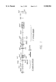
- FIG. 1 is a partial restriction map of the genomic locus of the mouse xpc gene useful in the construction of a targeting vector to knockout the xpc gene. The position of putative exons are also shown.
- FIG. 2 shows a comparison of the deduced amino acid sequence of the mouse XPC (mXPC) protein (SEQ ID NO:1) from part of a single large exon in the mouse xpc genomic locus, which is located at the 3' end of the 3' holomology region shown in FIG. 1, with that of the corresponding deduced amino acid sequence of the human XPC (hXPC) protein (SEQ ID NO:2).
- mXPC mouse XPC
- hXPC human XPC
- FIG. 3 illustrates the homologous recombinational event between the targeting vector, containing a mutation in the xpc gene, as well as a tk gene and a hprt gene; and the mouse xpc genomic locus used in Example 3 below to facilitate the production of XPC-deficient mouse cells. The position of putative exons is also shown.
- FIG. 4 shows the mutant xpc mRNA which is made as a result of the mutation in the xpc gene described in Example 3.
- the mutation causes a frame-shift which results in stop codons in the reading frame of the XPC mRNA which in turn results in less than wild-type levels of XPC mRNA in xpc m1 /xpc m1 cells.
- FIG. 5 shows the proliferative ability of XPC-deficient mouse cells.
- ( ⁇ ) represents +/+ wild-type primary embryonic fibroblasts
- ( ⁇ ) represents xpc m1 /xpc m1 cells.
- FIG. 6 shows the cytotoxic effects on XPC-deficient mouse cells after exposure to UV light in a colony survival assay.
- ( ⁇ ) represents +/+ wild-type primary embryonic fibroblasts
- ( ⁇ ) represents xpc m1 /xpc m1 cells.
- FIG. 7 shows the results of DNA damage in XPC-deficient mouse cells after exposure to the genotoxic agent 4NQO in a colony survival assay.
- ( ⁇ ) represents +/+ wild-type primary embryonic fibroblasts
- ( ⁇ ) represents xpc m1 /xpc m1 cells.
- FIG. 8 shows the time to tumor formation in XPC-deficient mice chronically exposed to UV light.
- ( ⁇ ) represents +/+ wild-type mice
- ( ⁇ ) represents xpc m1 /+ mice
- ( ⁇ ) represents xpc m1 /xpc m1 cells.
- a mouse cell comprising two chromosomal alleles of the xpc gene, wherein at least one of said alleles contains a mutation such that said cell produces less than wild-type levels of XPC activity.
- XPC-deficient means that at least one of the two wild-type XPC chromosomal alleles has been mutated such that less than wild-type levels of the XPC activity are produced.
- XPC-deficient genotypes include a homozygous, as well as a heterozygous genotype, although a homozygous genotype is preferable.
- XPC activity can be easily measured using several methods. For example, one can measure for a deficiency in XPC mRNA levels by Northern analysis (Sambrook et al, Molecular Cloning, A Laboratory Manual, Vol. 1-3, (1989)), or by using a reverse transcriptase polymerase chain reaction (RT PCR) (Sambrook et al, supra). Alternatively, one can measure protein levels of XPC by Western Blot (Sambrook et al, supra) using an antibody to XPC protein.
- the mutation in the xpc gene is preferably a deletion mutation, although substitution mutations and/or insertion mutations are also included within the scope of the present invention.
- the mutants of the present invention preferably lack part of the DNA sequence coding for XPC so that a defective xpc allele is more likely made.
- Deletion mutations can be introduced within the xpc gene taking advantage of the convenient restriction sites therein, such as any of the exonic BglII or HindIII restrictions sites or other sites which are easily identified by exonic sequencing of the xpc gene and restriction mapping (see FIG. 1), and the techniques described by Hasty et al, Mol. Cell. Biol., 11:4509-4517 (1991); and Joyner et al, Nature, 338:153-156 (1989).
- Substitution mutations can be prepared by site-directed mutagenesis, as described by Hasty et al, Nature, 350:243-246 (1991), so as to introduce a stop codon or other mutation near the 5' end of the xpc gene so as to give rise to abortive production of XPC protein or production of a mutant protein of deficient XPC activity.
- Insertion mutations can be introduced within the xpc gene taking advantage of the convenient restriction sites therein, such as any of the exonic BglII or HindIII restrictions sites or other sites which are easily identified by exonic sequencing of the xpc gene and restriction mapping (see FIG. 1), and the techniques described by Hasty et al, Mol. Cell. Biol., 11:4509-4517 (1991); and Joyner et al, Nature, 338:153-156 (1989).
- Another method of introducing a mutation in the xpc allele consists of infecting cells with a retrovirus which integrates in the xpc locus, using the techniques described by von Melchner et al, Genes & Dev., 6:919-927 (1992); Friedrich et al, Genes & Dev., 5:1513-1523 (1991); and Friedrich et al, Methods in Enzymology, 225:681-701 (1993), thereby creating a mutated xpc allele.
- the coding region of the xpc gene is approximately 2500 bp in size.
- Deletion mutants can be produced by eliminating a DNA fragment from a coding region of the xpc gene so that proper folding or substrate binding of the XPC protein is prevented.
- the size of the deletion may vary, but in general a larger deletion, is preferable to a smaller deletion, since the larger deletions are more likely to result in a greater deficiency in XPC activity.
- deleting a single base pair or two base pairs or any number of base pairs not divisible by 3 from the coding region will result in a frame-shift mutation which will be deleterious to making a functional XPC protein.
- a truncated polypeptide may be produced because polypeptide synthesis is aborted due to a frame-shift-induced stop codon. Still, changing a single base pair in the coding region of the xpc gene which results in an amino acid change can alter the proper folding of the XPC protein, and thereby create an XPC-deficiency. A single amino acid change so generated can also alter the affinity of XPC for its substrate, and thereby result in a deficiency of XPC activity.
- the preferred size of the deletion is about several hundred nucleotides near the 5' end of the gene.
- such a deletion will eliminate a number of nucleotides from the coding region not evenly divisible by 3, thereby creating a frame-shift mutation as well.
- mouse cells of the present invention can be prepared by the following steps:
- a targeting vector comprising a cloning vector and a DNA fragment containing at least one positive selectable marker gene flanked by two contiguous region of the mouse xpc gene or genomic locus which are in the same 5' to 3' orientation to one another (in the case of substitution mutants or insertions mutants) or non-contiguous regions of the mouse xpc gene or genomic locus which are in the same 5' to 3' orientation to one another (in the case of deletion mutants);
- the target vector of step (1) may also include a negative selectable marker gene adjacent to one of the non-contiguous regions. This negative selectable marker can increase the likelihood of recovering the desired homologous recombination event, i.e., mutation in the xpc gene.
- xpc gene or genomic locus sequences which must be present in the targeting vector of step (1) will depend on the site chosen for the insertion or the sequences chosen for the substitution or deletion, and (2) the restriction nucleases to be employed in the engineering of the insertion or deletion mutants.
- regions of homology The specific contiguous regions (hereinafter referred to as "regions of homology") required in step (1) depend on the specifics of the insertion or substitution in the targeting vector. Similarly, the specific non-contiguous regions (also hereinafter referred to as “regions of homology") required in step (1) depend on the specifics of the deletion in the targeting vector. In general, the size of the regions of homology used in the targeting vector will be at least about 400 bp, although longer or shorter regions can be used. In general, it is preferable to use regions of homology of approximately 1.5 kb or greater to insure a high degree of targeting efficiency. In the targeting vector described in detail in FIG. 3, the 5' and 3' homology regions on both sides of the deletion are 1.8 kb and 3.0 kb in length, respectively.
- the size of the insertion may also vary and depends on the regions of homology used in the targeting vector. Generally, the size of the insertion will be about 4.0 kb.
- the size of the deletion may also vary and depends on the regions of homology used in the targeting vector. That is, since non-contiguous regions of homology are used in the targeting vector, that region in the wild-type allele which is located between the regions of homology constitutes the region to be deleted upon homologous recombination with the targeting vector.
- the region deleted in Example 3 below is approximately 4.7 kb in length, although that size is not critical and either more or less can be deleted from the locus and still result in XPC-deficiency. It is preferable that the deletion include at least one exon or a portion of an exon of the xpc gene so as to result in mutant XPC mRNA.
- Another alternative is to generate a deletion or other mutation in the non-coding splicing region of the xpc gene which affects the proper splicing of the XPC mRNA.
- Such a mutation can create a mutant XPC transcript which is missing an entire exon or several exons as compared to the wild-type XPC mRNA.
- Another alternative is to delete a non-coding regulatory region to decrease expression of the xpc gene.
- the particular positive and negative selectable markers employed in the present invention are not critical thereto. Examples of preferred positive and negative selectable markers are listed in Table 1 below.
- the positive selectable marker should be located between the regions of homology, and the negative marker, if one is used, is preferably located outside of the regions of homology, either 5' or 3' to those regions as shown in FIG. 3.
- the regions of homology are preferably in the same 5' to 3' orientation to one another while the orientation of the positive and negative selectable markers are not critical.
- Positive and/or negative selectable marker genes are functional in the transfected cells if the phenotype expressed by the DNA sequences encoding such selectable markers is capable of conferring either a positive or negative selectable characteristic for the cell that is expressing the sequence.
- the positive selectable marker is preferably engineered to be functional in the transformed cells in which the gene targeting is being performed.
- the means by which the positive selectable marker gene is made functional is not critical to the present invention. Positive selection is accomplished by exposing the cells to an appropriate agent which kills or otherwise selects against cells not containing an integrated positive selectable marker. Examples of such agents are listed in Table 1 above.
- the positive selectable marker gene may have a promoter driving its expression (Donehower et al, Nature, 356:215-21 (1992)), or it may be driven by the juxtaposition of transcriptional elements at the target locus with the positive selectable marker (Le-Mouellic et al, Proc. Natl. Acad.
- a negative selectable marker is included in the targeting vector, it should be functional in the transformed cells and it may be located either 5' or 3' to the regions of homology. Its transcriptional orientation relative to the regions of homology is not important.
- the mutation engineered in the targeting vector can contain DNA sequences between the regions of homology, in addition to a positive selectable marker, e.g., an oligonucleotide linker, in place of the deleted xpc DNA.
- a positive selectable marker e.g., an oligonucleotide linker
- the oligonucleotide linker is generally 8-10 nucleotides in length, but can be longer, e.g., about 50 nucleotides, or shorter, e.g. 4, 5 or 7 nucleotides.
- the preferred length of the oligonucleotide linker is about 20 to 40 nucleotides in length.
- the DNA sequence of the oligonucleotide linker is not critical.
- oligonucleotide linker The method of inserting the oligonucleotide between the regions of homology in the targeting vector DNA will depend upon the type of oligonucleotide linker used. Palindromic double stranded linkers containing one or more restriction nuclease sites in the oligonucleotide sequence (New England Biolabs) may be inserted by well-known procedures (Maniatis et al, Molecular Cloning, Cold Spring Harbor Laboratory (1982)). Oligonucleotide linkers may also be inserted into deletions in plasmid DNA by tailing ends with complementary homopolymers using terminal transferase (Maniatis et al, supra).
- an oligonucleotide linker may be inserted into a deletion in a plasmid by bridging, through annealing of oligonucleotide containing ends complementary, to a cleaved plasmid's 5'-recessed and 3'-protruding cohesive ends, followed by filling in of the gap complementary to the oligonucleotide sequence with DNA polymerase (Klenow fragment). After subsequent ligation with T4 DNA ligase, closed circular DNA molecules can be regenerated.
- the targeting vector is designed such that the deleted region interrupts an exon, by the judicious choice of oligonucleotide linker length and sequence, frame-shift mutations and/or stop codons may be produced in the mouse xpc gene, augmenting the effect of deletions within the mouse xpc gene.
- the mutation engineered in the targeting vector can contain DNA sequences between the regions of homology in addition to the positive selectable marker, e.g., splice acceptor sequences. Such sequences have been shown to facilitate aberrant splicing to create mutant message (Friedrich et al, Genes & Dev., 5:1513-1523 (1991)).
- the DNA used as regions of homology is preferably derived from genomic DNA of the mouse xpc genomic locus.
- the strain of mouse from which the DNA is derived is not critical to the present invention, but preferably it should be the same as the strain of mouse from which the cells in which the gene targeting will be performed, are derived. Using DNA for the regions of homology which is isogenic to the cells in which the gene targeting is performed may enhance the efficiency with which gene targeting is accomplished (te Riele et al, Proc. Natl. Acad. Sci., USA, 89:5128-5132 (1992)).
- the regions of homology may be derived from genomic libraries of mouse DNA which may be cloned into a variety of library vectors, such as lambda phage vectors, cosmid vectors, plasmid vectors, p1 phage vectors, yeast artificial chromosome vectors, or other vectors (Sambrook et al, supra).
- the regions of homology to be used in the targeting vector can also be derived directly from genomic DNA using the polymerase chain reaction (PCR). This method can rely on knowledge of the published sequence of the human xpc gene (Legerski et al, Nature, 359:70-73 (1992)).
- the regions of homology so derived can be subcloned directly into the targeting vector.
- the particular cloning vector employed in the present invention to construct the targeting vector comprising the regions of homology separated by a positive selectable marker gene and an optional flanking negative selectable marker is not critical, as long as the cloning vector contains a gene coding for a selective trait, e.g., drug resistance.
- cloning vectors examples include pBR322 and pBR322-based vectors (Sekiguchi et al, Gene, 21:267 (1983)), pMB9, pBR325, pKH47 (Bethesda Research Laboratories), pBR328, pHC79, phage Charon 28 (Bethesda Research Laboratories, Boehringer Manneheim Biochemicals), pKB11, pKSV-10 (P-L Biochemicals), pMAR420 (Otsuka et al, Virol., 113:196 (1981)), oligo (dG)-tailed pBR322 (Bethesda Research Laboratories), pBluescript or similar plasmids (Stratagene), puc19 or similar plasmids (New England Biolabs).
- the targeting vector comprising the regions of homology separated by a positive selectable marker gene and an optional flanking negative selectable marker can be cloned into other cloning vectors, such as such as lambda phage vectors, cosmid vectors, plasmid vectors, p1 phage vectors, yeast artificial chromosome vectors, or other vectors (Sambrook et al, supra).
- cloning vectors such as such as lambda phage vectors, cosmid vectors, plasmid vectors, p1 phage vectors, yeast artificial chromosome vectors, or other vectors (Sambrook et al, supra).
- Another option is to prepare the components of the targeting vector synthetically by PCR, and simply ligate each component into its proper position by choosing restriction endonuclease sites for ligation which insure proper orientation of the regions of homology relative to each other, and to insure that the positive selectable marker is located between the regions of homology.
- cloning vectors containing unique cloning sites which are useful in the present invention can be determined upon evaluation of restriction nucleases other than EcoRV and BamHI for the 5' homology region and HindIII for the 3' homology region, which were used in the Examples herein (see FIG. 1).
- restriction nucleases which can be employed to produce fragments containing the mouse xpc gene, and thus other cloning vectors which can be useful in the present invention, are readily apparent from the mouse xpc gene restriction map and targeting vector shown in FIGS. 1 and 3, and which are discussed more fully below.
- regions of homology can be derived from restriction digests according to FIG. 1, such as XbaI and HindIII for the 5' homology region and a BglII fragment for the 3' homology region.
- a HindIII-EcoRI fragment can be used for the 5' homology region and a BamHI fragment for the 3' homology region.
- Many combinations of restriction endonucleases can be used to generate a targeting vector to mutate the xpc gene.
- regions of homology can be cloned into any of a large number of commercially available plasmids, such as the pBluescript series (Stratagene), the puc series (New England Biolabs), or the PGEM series (Promega).
- XPC-deficient mouse cells can be screened for mutations in the xpc gene, e.g., by Southern blotting using DNA probes for said mutation, or by PCR.
- Southern blotting using a probe 5' to the mutated locus was used to identify cell lines heterozygous for the engineered mutation by the presence of an 8.5 kb mutant DNA fragment and a 6.0 kb wild-type DNA fragment (FIG. 3).
- the specific host employed for growing the targeting vectors of the present invention is not critical. Examples of such hosts include E. coli K12 RR1 (Bolivar et al, Gene, 2:95 (1977)); E. coli K12 HB101 (ATCC No. 33694); E. coli MM21 (ATCC No. 336780); and E. coli DH1 (ATCC No. 33849).
- the preferred host in the present invention is DH5a (Life Technologies).
- alternative vector/cloning systems can be employed, such as targeting vectors which grow in E. coli or Saccharomyces cervisiae, or both, or plasmid vectors which grow in B. subtillis (Ure et al, Methods in Enzymology "Recombinant DNA", Vol. 101, Part C, Academic Press, N.Y. (1983)).
- the specific xpc + mouse cell which is mutated in the present invention is not critical thereto, and is preferably a precursor pluripotent cell.
- the term precursor means that the pluripotent cell is a precursor of the desired transfected pluripotent cell which is prepared in accordance with the present invention.
- the pluripotent cell may be cultured in vivo to form a mutant mouse (Evans et al, Nature, 292:154 (1981)).
- Examples of xpc + mouse cells which can be employed in the present invention include embryonic stem (ES) cells.
- ES cells Primary isolates of ES cells may also be used, and can be obtained directly from embryos, such as described for the EK.CCE cell line (Evans et al, Nature, 292:292-156 (1981)) or for ES cells in general (Robertson, In: Tetracarcinomas and Embryonic Stem Cells: A Practical Approach, Ed. Robertson, IRL Press, Oxford, pages 71-112 (1987)).
- the particular ES cell employed in the present invention is not critical thereto.
- ES cells examples include AB 2.1 and AB 2.2, hprt - cell lines (Matzuk et al, Nature, 360:313-319 (1992)), and AB 1.0, an hprt + cell line (McMahon et al, Cell, 62:1073-1085 (1989)).
- hprt - cell line when the positive selectable marker is an hprt gene.
- Other selectable markers such as those outlined in Table 1 can be used in other stem cell lines.
- the ES cells are preferably cultured on stromal cells, e.g., STO cells, and/or primary embryonic fibroblast cells, as described by Robertson, supra.
- stromal cells e.g., STO cells, and/or primary embryonic fibroblast cells, as described by Robertson, supra.
- the stromal (and/or fibroblast) cells serve to reduce the clonal outgrowth of abnormal ES cells.
- the mutant embryonic stems cells are injected into mouse blastocysts as described by Bradley, In: Teratocarcinomas and Embryonic Stem Cells: A Practical Approach, pages 113-151 (1987)).
- mice employed in the present invention are not critical thereto.
- blastocysts include those derived from C57BL6 mice, C57BL6 Albino mice, Swiss outbred mice, CFLP mice and MFI mice (Bradley, supra).
- mice heterozygous for the xpc mutant allele generated from the injected blastocyst can be screened for mutations in the xpc gene, e.g., by Southern blotting using DNA probes for said mutation, or by PCR (Sambrook et al, supra). For example, in Example 4 below, Southern blotting using a probe 5' to the mutated locus identified mice heterozygous for the engineered mutation by the presence of an 8.5 kb mutant DNA fragment and a 6.0 kb wild-type DNA fragment (FIG. 3).
- the mutant mice of the present invention can be intercrossed to obtain mice homozygous for the mutation in the xpc gene, and/or can be crossed with other mice strains to transfer the xpc mutation into these other strains.
- mice homozygous for the engineered mutation by the presence of only an 8.5 kb mutant DNA fragment and no 6.0 kb wild-type DNA fragment (FIG. 3).
- mouse cells are not critical to the present invention.
- examples of such mouse cells include embryonic fibroblasts and skin fibroblasts.
- Embryonic fibroblasts are the preferred cells to be employed in the present invention because of their high proliferative capacity and high plating efficiencies which allow for rapid experimentation on UV sensitivity.
- the amount of exposure to UV is not critical to the present invention, but is typically in a range of incrementally increasing values from 0 J/m 2 to 10 J/m 2 .
- the amount of exposure to chemical DNA damaging agents may vary, depending on the particular agent used.
- the particular chemical DNA damaging agent employed is not critical to the present invention. Examples of such agents include 4-nitroquinoline 1-oxide (4NQO), mitomycin c and bleomycin.
- 4NQO 4-nitroquinoline 1-oxide
- the dose of exposure to cells in culture will be in a range to establish a kill curve that reflects the relative sensitivity of the XPC-deficient cells to the various agents.
- the dose range typically used in such kill curve experiments is between 1.0 and 5.0 ⁇ M for 4NQO, and from 0.2 to 1.6 ⁇ M for mitomycin c and bleomycin.
- Cytotoxicity can be evaluated by counting the number of colonies of cells surviving after exposure to the toxic agents.
- the amount of exposure to UV light is not critical to the present invention, but is typically delivered in a range from about 500 to 2500 J/m 2 , which approximates the amount of sun exposure during 3 to 20 min, depending on clear weather conditions, in Houston, Tex. in July at 2:00 pm.
- Pathological changes in the exposed skin can be evaluated visually as tumors, or by histological staining of tissue specimens, as described by Sands et al, Nature, 377:166 (1995), which is incorporated by reference herein.
- the mouse homologue of the human xpc gene was cloned from a mouse 129-strain genomic library.
- a DNA fragment of the human xpc gene was obtained using a reverse transcriptase polymerase chain reaction (PCR) on RNA obtained from human cells (Sambrook et al, supra).
- PCR reverse transcriptase polymerase chain reaction
- the following oligonucleotide primers were used in the PCR: 5'-GAAGAGAGGGGGTACCATGAATGAAG-3' (SEQ ID NO: 3), and 5'-GAGGAGCCTCCTGGATCCGCAGTC-3' (SEQ ID NO: 4), and were based on the human xpc gene sequence (Legerski et al, Nature, 359:70-73 (1992)).
- the DNA fragment of the human xpc gene so obtained was subcloned into the HindIII site of plasmid vector pBluescript SK + (Stratagene). A radiolabeled probe was then made using the subcloned DNA fragment of the human xpc gene.
- the radiolabelled probe was used to screen a mouse 129-strain genomic lambda phage library (Stratagene) to identify phage containing the homologous mouse gene.
- Three positive phage were isolated, grown, and restriction mapping was performed on the DNA inserts, as described by Sambrook et al, supra, so to produce a map of the xpc genomic locus (see FIG. 1).
- One clone containing the mouse xpc genomic locus was designated mXPC clone #9.
- the DNA sequence of a portion of mouse xpc CDNA (SEQ ID NO:5) has substantial DNA sequence similarity to the human xpc gene. That is, 78% identity was found at the nucleotide level.
- the human cDNA sequence was used as a probe.
- the mouse cDNA sequence could also be radiolabeled, and used as a probe to clone the genomic locus of the mouse xpc gene for knockout vector construction.
- FIG. 2 One large contiguous coding region (exon), which is bisected by a HindIII site, and identified in the mouse xpc genomic clone in comparison to the deduced amino acid sequence of the human xpc cDNA sequence, is shown in FIG. 2.
- identical amino acids are indicated by a vertical line between the mouse and human sequences, conservative amino acid changes are shown with two dots, and moderately conservative amino acid changes are shown with one dot, using threshold values of 1.5, >0.5, and >0.1, respectively (Gribskov et al, Nuc. Acids Res., 14:6745 (1986)).
- mouse XPC protein was found to have extensive sequence similarity to the human XPC protein. That is, 79% identity was found at the amino acid level between mouse exonic regions and the human XPC CDNA sequence.
- a targeting vector which contains 1.8 kb of DNA homologous to the 5' end of the mouse xpc gene, and 3.0 kb of DNA homologous to the 3' end of the mouse xpc gene, was constructed (see FIG. 3).
- This vector also contains a marker for positive selection (the hprt mini-gene) (Matzuk et al, Nature, 360:313 (1992)), and a marker for negative selection (the tk gene) (Mansour et al, Nature, 226:348 (1988)) (see FIG. 3).
- 5' and 3' homology regions were selected.
- the 5' homology region used was located near the 5' end of the xpc genomic locus and was isolated by EcoRV and BamHI digestion of the phage clone obtained in Example 1 above (the sites are underlined on the restriction map shown in FIG. 1), which produced a DNA fragment of approximately 1.8 kb.
- this fragment was subcloned into Bluescript SK + vector (Stratagene) which had been prepared by cutting with XbaI, treating the XbaI end with Klenow fragment and nucleotides to make a blunt end (Sambrook et al, supra), and then cutting with BamHI. This effectively produced a vector that had complementary ends to the insert for directed ligation into the vector.
- the 3' homology region was isolated by HindIII digestion of the phage clone obtained in Example 1 above, (the sites are underlined on the restriction map shown in FIG. 1), which produced a DNA fragment of approximately 3.0 kb.
- the 3' homology region was subcloned into the HindIII site of Bluescript SK + vector (stratagene). This allowed for a subsequent restriction digestion with EcoRI and SalI to release the entire fragment from the subcloning vector, with the effective change of restriction sites on the ends of the 3' homology to EcoRI and SalI.
- the positive selectable marker used was the hprt mini-gene (Matzuk et al, supra), which was digested with BamHI and EcoRI to release a DNA fragment of approximately 3.6 kb containing the marker gene.
- the subclone containing the 5' homology region was used as the base vector for inserting the other required elements.
- the 5' homology vector was digested with BamHI and XhoI.
- the positive selectable marker was digested to produce sticky ends with BamHI and EcoRI.
- the 3' homology region subclone was digested with EcoRI and SalI. These three fragments were ligated together in a single directed ligation reaction (FIG. 3).
- the negative selectable tk gene (U.S. Pat. No. 5,464,764) was added upstream of the 5' homology region at the unique NotI site (FIG. 3).
- Example 2 Homologous recombination of the targeting vector obtained in Example 2 with the xpc genomic locus was effected in mouse embryonic stem cells (see FIG. 3).
- the ES cell were grown on a feeder layer of STO cells as described by Robertson, supra.
- 10 Mg of the positive-negative targeting vector obtained in Example 2 above was transfected into 1.0 ⁇ 10 7 AB 2.1 mouse 129 strain embryonic stem cells (Matzuk et al, Nature, 360:313 (1992)), and the resulting cells were grown in HAT (hypoxanthine, aminopterine and thymidine) positive selection media so as to select for those cells which were transfected with the targeting construct.
- HAT hypoxanthine, aminopterine and thymidine
- Negative selection against the tk gene was also carried out using the drug FIAU (McMahon et al, Cell, 62:1073-1085 (1989)) so as to enhance for selection of those cells which had undergone a homologous recombination event at the xpc locus (Capecchi et al, supra).
- ES cell genomic DNA for the mini-southern was digested with restriction enzyme EcoRI.
- the desired recombination event was detected using the 5' probe derived from an EcoRV-EcoRI digestion of the mouse xpc genomic clone as indicated in FIG. 1, which revealed a mutant allele of 8.5 kb as compared to the wild-type allele of 6.0 kb.
- the desired recombination event was confirmed using the 3' probe derived from an XhoI-HindIII digestion of mouse xpc genomic clone, as indicated in FIG. 1, which revealed a mutant allele of 10.7 kb as compared to the wild-type allele of 15 kb (see FIG. 3)
- Ten positive ES cells clones were identified as correct replacement events, with an approximate 4.7 kb genomic deletion. The targeting frequency was approximately 1/20.
- One of the clones was designated ES cell line xpc m1 -202.
- Chimeric male mice were mated to wild-type C57BL6 Albino females, and agouti pups were born, indicating successful germline transmission of the ES cell component of the chimeric mouse, and resulting in C57BL6 Albino/129 hybrids (referred to as C567BL6/129 hybrids).
- C567BL6/129 hybrids C57BL6 Albino/129 hybrids
- Genomic DNA was isolated from the resulting mice as described by Sambrook et al, supra. Then, 10 ⁇ g of the resulting genomic DNA was digested with EcoRI, and subjected to Southern blot analysis using the 5' and 3' probes as described above for the mini-Southerns. ES cell clone xpc m1 -202 was found to have transmitted the mutant allele through the germline having an approximate deletion of 4.5 kb.
- a male and female mouse were identified as heterozygous for the mutant allele, and then intercrossed.
- the chimeric mouse which had demonstrated germline transmission was also crossed with a wild-type 129 strain mouse, in order to place the mutant allele on the 129 strain background.
- Genomic DNA was isolated from the resulting mice, and 10 ⁇ g of the genomic DNA was digested with EcoRI, and subjected to Southern blot analysis, using the 5' and 3' probes described above.
- a single 6.0 kb band indicated a +/+ animal
- a single 8.5 kb band indicated an animal homozygous for the targeted mutant allele (xpc m1 /xpc m1 ).
- the presence of both bands indicated a heterozygous animal (xpc m1 /+).
- mice were viable and fertile, and no difference was noted in behavior or continued viability over a 1.5-year period of monitoring a population of approximately 100 wild-type, (xpc m1 /+), and (xpc m1 /xpc m1 ) mice.
- Wild-type and xpc m1 /xpc m1 primary embryonic fibroblasts were generated to measure XPC activity.
- mRNA was obtained using the QuickPrep Micro mRNA purification kit (Pharamacia) from mouse embryonic fibroblasts that were isolated from 13.5 day embryos derived from wild-type and xpc m1 /xpc m1 mice as described by Harvey et al, Oncogene, 8:2457-2467 (1993). Then, 1.2 ⁇ g of the mRNA was used to make 1st strand cDNA using oligo dT primers (Superscript RT II kit, Gibco BRL).
- PCR was then carried out using the following xpc primers 5'-GCTGCAGTGATCCAGGGGAC-3' (SEQ ID NO:6); and 5'-TGGACGGTTCAAAGTCAGAG-3' (SEQ ID NO:7).
- the PCR conditions were 93° C. for 40 sec, 72° C. for 1 min 30 sec for 34 cycles.
- the RT-PCR for XPC mRNA from xpc m1 /xpx m1 and wild-type embryonic fibroblasts revealed a predicted 1150 bp fragment with wild-type mRNA as template and a 665 bp fragment with mRNA from mutant cells (FIG. 4).
- RT-PCR positive controls using primers to the Mgf gene i.e., 5'-AGCTGCAGCTGGATCGCAGCGCTGCCTTTCC-3' (SEQ ID NO:8); and 5'-TCGAGCTCTGTTGATACGTCCACAATTACACCTC-3' (SEQ ID NO:9) (Anderson et al, Cell, 63:235-241 (1990)), indicated that the reaction conditions were optimized for both wild-type and mutant mRNA templates.
- the PCR products obtained with the xpc primers were subcloned into a T-Vector (Promega) and sequenced.
- the sequencing results of the mutant and wild-type bands revealed a 485 bp deletion in the mutant mRNA resulting in a frameshift mutation and stop codons 151 bp downstream from the deletion point (FIG. 4).
- Wild-type and xpc m1 /xpc m1 primary embryonic fibroblasts were generated to measure the effect of the xpc m1 /xpc m1 genotype on the sensitivity of cultured cells to DNA damaging agents, i.e., UV irradiation and 4-nitro-quinoline 1 oxide.
- DNA damaging agents i.e., UV irradiation and 4-nitro-quinoline 1 oxide.
- the proliferation of the XPC-deficient and wild-type fibroblasts were compared. As shown in FIG. 5, in the absence of UV irradiation, no differences were apparent in the proliferation between the two genotypes.
- primary embryonic fibroblasts were isolated from 13.5 day embryos derived from a cross of xpc m1 /+ mice as described by Harvey et al, supra. Then, cell lines having the xpc m1 /xpc m1 and +/+genotypes were plated at a low density, i.e., approximately 20,000 cells per 100 mm tissue culture plate, and exposed to increasing doses of UV light using a short wavelength (maximal emission approximately 250 nM-280 nM). The doses used were from 0 J/m 2 to 10 J/m 2 .
- xpc m1 /xpc m1 cells were able to proliferate in culture as rapidly as +/+ wild-type cells, and their plating efficiencies were comparable.
- xpc m1 /xpc m1 cells upon exposure to UV light, xpc m1 /xpc m1 cells demonstrated a significantly decreased ability to form colonies compared to wild-type cells (p ⁇ 0.01, bars represent standard deviation).
- Embryonic fibroblasts heterozygous for the mutant allele, i.e., xpc m1 /+ cells could not be distinguished from wild-type cells, i.e., +/+ cells, in this assay.
- cells homozygous for the mutant allele demonstrated a marked sensitivity to UV light in a colony survival assay, consistent with data from cells derived from XPC patients, which demonstrate a deficiency in "global genome” NER (Venema et al, Mol. Cell. Biol., 11:4128 (1991)).
- primary embryonic fibroblasts were isolated from 13.5 day embryos derived from a cross of xpc m1 /+ mice as described by Harvey et al, supra. Then, cell lines having the xpc m1 /xpc m1 and +/+ genotypes were plated at a low density, i.e., approximately 20,000 cells per 100 mm tissue culture plate, and exposed to increasing doses of 4NQO ranging from 0 ⁇ M to 0.8 ⁇ M.
- the cells were incubated for 10 days in DMEM supplemented with 15% (v/v) fetal calf and the antibiotics streptomycin sulfate (50 ⁇ g/ml) and penicillin G (50 units/ml), in order to allow for colony formation.
- the colonies were then fixed in 70% (v/v) ethanol, and stained with methylene blue. The average number of colonies per plate of three independent cell lines of each genotype was calculated for each UV dose; standard deviation (bars) and statistical significance were determined (t test). The results are shown in FIG. 7.
- the sensitivity of the xpc m1 /xpc m1 primary embryonic fibroblasts to 4NQO was not found to be statistically significant (t test) at the dose points tested, compared to the +/+ wild-type cells.
- XPC-deficiency significantly altered cell survival in response to the type of damage caused by UV light, but not that associated with 4NQO exposure.
- mice 8-12 week-old mice were exposed to an average of 2500 J/m 2 per day (suberythemic dose, maximal emission in the UVB wavelength range) for a 20 week period. The mice were shaved once per week during the 20 week exposure period, and were observed regularly for pathologic changes in the skin. Lesions that were identified grossly as tumors were noted. Histopathologic confirmation of carcinomas was obtained after the animals were sacrificed by placing tissue specimens in neutral buffered 10% (v/v) formalin, and embedding in paraffin. 5.0 ⁇ m sections were stained with hematoxylin and eosin by conventional methods. The time to tumor formation is shown in FIG. 8.
- all of the xpc m1 /xpc m1 mice had developed at least one skin carcinoma, which was confirmed by histologic sections. The results of the histology are summarized in Table 2 below.
- mice heterozygous for the mutant xpc allele and wild-type controls did not develop any tumors by the end of the treatment regimen.
- the histologic examination of the skin of the UV irradiated xpc m1 /xpc m1 mice showed marked hyperplasia of the epidermis, with focal areas of hyperkeratosis and varying degrees of dysplasia, acantholysis and/or dyskeratosis. These lesions have been previously described in UV-irradiated hairless mice (Gallagher et al, J. Invest.
- UV treatment produced frequent abnormalities in eyes of xpc m1 /xpc m1 mice; histologic sections revealed severe keratitis and comeal ulceration.
- the +/+ and xpc m1 /+ mice showed only moderate hyperplasia in the skin, and infrequent cases of mild keratitis during the UV treatment regimen.
- Four weeks after the UV treatment regimen was completed one of +/+ mice developed a spindle cell tumor and one of the xpc m1 /+ mice developed a papilloma.
- mice homozygous for the xpc mutant allele were viable, and did not exhibit an increased susceptibility to spontaneous tumorigenesis at one year of age.
- xpc m1 /xpc m1 mice were found to be highly susceptible to UV-induced carcinogenesis as compared to mice heterozygous for the mutant allele (xpc m1 /+) and wild-type controls. This is consistent with XPC patients who are predisposed to skin tumor formation in UV exposed areas of the body (Cleaver, In: The Metabolic Basis of Inherited Disease, McGraw Hill, New York (1995)).
- xpc m1 /xpc m1 mice also display a spectrum of UV exposure-related pathologic skin and eye changes consistent with the human disease XPC, and confirming their applicability as a mouse model for XPC.
Landscapes
- Health & Medical Sciences (AREA)
- Life Sciences & Earth Sciences (AREA)
- Chemical & Material Sciences (AREA)
- Genetics & Genomics (AREA)
- Organic Chemistry (AREA)
- Zoology (AREA)
- Engineering & Computer Science (AREA)
- General Health & Medical Sciences (AREA)
- Biotechnology (AREA)
- Veterinary Medicine (AREA)
- Biomedical Technology (AREA)
- Biochemistry (AREA)
- Environmental Sciences (AREA)
- Wood Science & Technology (AREA)
- Molecular Biology (AREA)
- Biophysics (AREA)
- Bioinformatics & Cheminformatics (AREA)
- General Engineering & Computer Science (AREA)
- Proteomics, Peptides & Aminoacids (AREA)
- Microbiology (AREA)
- Physics & Mathematics (AREA)
- Gastroenterology & Hepatology (AREA)
- Toxicology (AREA)
- Plant Pathology (AREA)
- Endocrinology (AREA)
- Immunology (AREA)
- Medicinal Chemistry (AREA)
- Animal Behavior & Ethology (AREA)
- Animal Husbandry (AREA)
- Biodiversity & Conservation Biology (AREA)
- Cell Biology (AREA)
- Micro-Organisms Or Cultivation Processes Thereof (AREA)
Abstract
Description
              TABLE 1
______________________________________
Selectable Markers for Use in Gene Targeting
                           Preferred
                           Concentration
               Selective   of
Gene    Type   Agents      Selective Agent
                                     Organism
______________________________________
neo     +      G418          50-1000 μg/ml
                                     Eukaryotes
hyg     +      Hygromycin    10-1000 μg/ml
                                     Eukaryotes
hisD    +      Histidinol    5-500 μg/ml
                                     Animals
gpt     +      Xanthine      50-500 μg/ml
                                     Animals
hprt    +      Hypoxanthine
                           0.01-10 mM
                                     All
HSV-tk  -      Gancyclovir 0.05-200 μM
                                     Animals
        -      FIAU        0.02-100 μM
                                     Animals
hprt    -      6-thioguanine
                            0.1-100 μg/ml
                                     All
gpt     -      6-thioxanthine
                            0.1-100 μg/ml
                                     Animals
Diptheria
        -      None        None      Animals
toxin
Ricin Toxin
        -      None        None      Animals
cytosine
        -      5-fluorocytosine
                             10-500 μg/ml
                                     All
deaminase
______________________________________
              TABLE 2
______________________________________
UV-induced Skin Neoplasia in XPC-deficient Mice
Mouse     Location of Lesion
                         Histopathology
______________________________________
 9        Ear            WD SCC
          Dorsum         WD SCC (3)
                         MD SCC (2)
11        Ear            WD SCC, MD SCC
12        Ear            WD SCC, Papilloma
14        Ear            Carcinoma in situ,
                         WM SCC
          Dorsum         WD SCC, MD SCC
23        Ear            Papilloma, WD SCC
          Eye lid        MD SCC
24        Ear            MD SCC
28        Ear            WD SCC (2)
          Dorsum         WD SCC (3)
188       Ear            Papilloma (2), WD SCC
          Eye lid        WD SCC
191       Ear            WD SCC, MD SCC
          Dorsum         MD SCC
______________________________________
 WD SCC = Well Differentiated Squamous Cell Carcinoma
 MD SSC = Moderately Differentiated Squamous Cell Carcinoma
 The numbers in parentheses indicate the number of lesions identified in a
 survey of histologic sections.
__________________________________________________________________________
#             SEQUENCE LISTING
- (1) GENERAL INFORMATION:
-    (iii) NUMBER OF SEQUENCES:  9
- (2) INFORMATION FOR SEQ ID NO: 1:
-      (i) SEQUENCE CHARACTERISTICS:
#acids    (A) LENGTH: 211 amino
          (B) TYPE: amino acid
          (D) TOPOLOGY: linear
-     (ii) MOLECULE TYPE: protein
#1:   (xi) SEQUENCE DESCRIPTION: SEQ ID NO:
- Gln Gly Arg Pro His Ala Arg Lys Arg Arg Va - #l Ala Ala Lys Val Ser
Tyr
#                 15
- Lys Glu Glu Ser Glu Ser Asp Gly Ala Gly Se - #r Gly Ser Asp Phe Glu
Pro
#        30
- Ser Ser Gly Glu Gly Gln His Ser Ser Asp Gl - #u Asp Cys Glu Pro Gly
Pro
#50
- Cys Lys Gln Lys Arg Ala Ser Ala Pro Gln Ar - #g Thr Lys Ala Gly Ser
Lys
#            65
- Ser Ala Ser Lys Thr Gln Arg Gly Ser Gln Cy - #s Glu Pro Ser Ser Phe
Pro
#    85
- Glu Ala Ser Ser Ser Ser Ser Gly Cys Lys Ar - #g Gly Lys Lys Val Ser
Ser
#                100
- Gly Ala Glu Glu Met Ala Asp Arg Lys Pro Al - #a Gly Val Asp Gln Trp
Leu
#       115
- Glu Val Tyr Cys Glu Pro Gln Ala Lys Trp Va - #l Cys Val Asp Cys Val
His
120                 1 - #25                 1 - #30                 1 -
#35
- Gly Val Val Gly Gln Pro Val Ala Cys Tyr Ly - #s Tyr Ala Thr Lys Pro
Met
#           150
- Thr Tyr Val Val Gly Ile Asp Ser Asp Gly Tr - #p Val Arg Asp Val Thr
Gln
#   170
- Arg Tyr Asp Pro Ala Trp Met Thr Ala Thr Ar - #g Lys Cys Arg Val Asp
Ala
#               185
- Glu Trp Trp Ala Glu Thr Leu Arg Pro Tyr Ar - #g Ser Leu Leu Thr Glu
Arg
#       200
- Glu Lys Lys Glu Asp Gln Glu
205                 2 - #10
- (2) INFORMATION FOR SEQ ID NO: 2:
-      (i) SEQUENCE CHARACTERISTICS:
#acids    (A) LENGTH: 211 amino
          (B) TYPE: amino acid
          (D) TOPOLOGY: linear
-     (ii) MOLECULE TYPE: protein
#2:   (xi) SEQUENCE DESCRIPTION: SEQ ID NO:
- Gln Arg Arg Pro His Gly Arg Glu Arg Arg Va - #l Ala Ser Arg Val Ser
Tyr
#                 15
- Lys Glu Glu Ser Gly Ser Asp Glu Ala Gly Se - #r Gly Ser Asp Phe Glu
Leu
#        30
- Ser Ser Gly Glu Ala Ser Asp Pro Ser Asp Gl - #u Asp Ser Glu Pro Gly
Pro
#50
- Pro Lys Gln Arg Lys Ala Pro Ala Pro Gln Ar - #g Thr Lys Ala Gly Ser
Lys
#            65
- Ser Ala Ser Arg Thr His Arg Gly Ser His Ar - #g Lys Asp Pro Ser Leu
Pro
#    85
- Ala Ala Ser Ser Ser Ser Ser Ser Ser Lys Ar - #g Gly Lys Lys Met Cys
Ser
#                100
- Asp Gly Glu Lys Ala Glu Lys Arg Ser Ile Al - #a Gly Ile Asp Gln Trp
Leu
#       115
- Glu Val Phe Cys Glu Gln Glu Glu Lys Trp Va - #l Cys Val Asp Cys Val
His
120                 1 - #25                 1 - #30                 1 -
#35
- Gly Val Val Gly Gln Pro Leu Thr Cys Tyr Ly - #s Tyr Ala Thr Lys Pro
Met
#           150
- Thr Tyr Val Val Gly Ile Asp Ser Asp Gly Tr - #p Val Arg Asp Val Thr
Gln
#   170
- Arg Tyr Asp Pro Val Trp Met Thr Val Thr Ar - #g Lys Cys Arg Val Asp
Ala
#               185
- Glu Trp Trp Ala Glu Thr Leu Arg Pro Tyr Gl - #n Ser Pro Phe Met Asp
Arg
#       200
- Glu Lys Lys Glu Asp Leu Glu
205                 2 - #10
- (2) INFORMATION FOR SEQ ID NO: 3:
-      (i) SEQUENCE CHARACTERISTICS:
#pairs    (A) LENGTH: 26 base
          (B) TYPE: nucleic acid
          (C) STRANDEDNESS: single
          (D) TOPOLOGY: linear
-     (ii) MOLECULE TYPE: genomic DNA
-    (iii) HYPOTHETICAL: NO
#3:   (xi) SEQUENCE DESCRIPTION: SEQ ID NO:
#              26  ATGA ATGAAG
- (2) INFORMATION FOR SEQ ID NO: 4:
-      (i) SEQUENCE CHARACTERISTICS:
#pairs    (A) LENGTH: 24 base
          (B) TYPE: nucleic acid
          (C) STRANDEDNESS: single
          (D) TOPOLOGY: linear
-     (ii) MOLECULE TYPE: synthetic
-    (iii) HYPOTHETICAL: NO
#4:   (xi) SEQUENCE DESCRIPTION: SEQ ID NO:
#                24TGGATCCGC AGTC
- (2) INFORMATION FOR SEQ ID NO: 5:
-      (i) SEQUENCE CHARACTERISTICS:
#pairs    (A) LENGTH: 1679 base
          (B) TYPE: nucleic acid
          (C) STRANDEDNESS: single
          (D) TOPOLOGY: linear
-     (ii) MOLECULE TYPE: cDNA
-    (iii) HYPOTHETICAL: NO
#5:   (xi) SEQUENCE DESCRIPTION: SEQ ID NO:
#              50GGGGAC CCCACAAATG GTGCAGCAAA AAAGAAAGTG
#             100TAAATC CAAGAATCTC AAGGTTCTGA AGGAGGAAGC
#             150ATGACT TCCGGGACTC ACCAGCTGAC TGCAAGAAGG
#             200AAAAGC AAGGTGGTGG ACCAAGGCAC TGATGAAGAT
#             250CTGGGA GGAGGTGGAA GAGCTTACTG AACCTGTGCT
#             300ATTCTG CCACCTCACC GTCTGACATG CCTGTGAAGG
#             350ATTGAA ACACCACAGC AGGCGAANNN ANGNGAAAGA
#             400NNTGGA GTTTGAGACA TANCTCCGGA GGNNGATGAA
#             450GGGTCC ATGAGGACAC ACACAAGGTT CACCTTCTCT
#             500GGCTTC TATCGAAATA ACATCTGCAG CCAGCTCGAT
#             550CCTCTC CATCATCCCA ATTCGCTTTA CCAAGGTGCC
#             600ATGCCT ACTACCTTTC AAACCTGGTA AAGTGGTTCA
#             650GTCAAC GCTGACCTTT CAGCCAGCGA GCAGGACGAC
#             700GGAAAG GAGGATTGCC ATTTACTCTG CGAGGGATAA
#             750ATATAT TTCTTCTGAT TCTTCGGGCT CTGCAGCTGC
#             800TTGTCT CTGCAGCCCA TTCCACTGAA GTCAGCTGTG
#             850ATCTTC CAAGGAGACA TCTGTAGAGG GTCCTGGAGG
#             900CTAGTA ACAGTCCAGA AAGCCACAAC AAACCTACGA
#             950AAAGAA GAAGAAACCT TGTCTGAGGG CAGAGCCAAA
#            1000GAAGAG AGGCACAGGC ACTGCGGGCA GCAGGCAGCG
#            1050GCAGCG AGGGAGAGGA GGCCGAGCAG AAAGTCCAGG
#            1100CGGAAG CGGCGTGTGG CTGCCAAGGT GTCATACAAA
#            1150CGATGG GGCAGGCAGC GGCTCTGACT TTGAACCGTC
#            1200AGCATT CCTCTGATGA GGATTGTGAG CCTGGCCCTT
#            1250GCCTCA GCTCCTCAGA GGACAAAGGC TGGGTCTAAG
#            1300CCAACG CGGAAGCCAG TGTGAGCCGT CAAGCTTTCC
#            1350GCTCTT CAGGCTGTAA GAGAGGCAAG AAGGTTTCCA
#            1400ATGGCA GATAGGAAAC CTGCTGGTGT AGACCAGTGG
#            1450TGAGCC ACAGGCAAAG TGGGTGTGTG TGGACTGTGT
#            1500GCCAGC CTGTGGCCTG TTACAAATAT GCCACCAAAC
#            1550GTAGGC ATTGACAGTG ATGGCTGGGT CCGAGATGTT
#            1600CCCAGC CTGGATGACC GCAACCCGCA AGTGCCGGGT
#            1650GGGCTG AGACCTTGAG ACCCTATCGG AGCCTACTTA
#          1679    GGAA GACCAGGAG
- (2) INFORMATION FOR SEQ ID NO: 6:
-      (i) SEQUENCE CHARACTERISTICS:
#pairs    (A) LENGTH: 20 base
          (B) TYPE: nucleic acid
          (C) STRANDEDNESS: single
          (D) TOPOLOGY: linear
-     (ii) MOLECULE TYPE: synthetic
-    (iii) HYPOTHETICAL: NO
#6:   (xi) SEQUENCE DESCRIPTION: SEQ ID NO:
# 20               GGAC
- (2) INFORMATION FOR SEQ ID NO: 7:
-      (i) SEQUENCE CHARACTERISTICS:
#pairs    (A) LENGTH: 20 base
          (B) TYPE: nucleic acid
          (C) STRANDEDNESS: single
          (D) TOPOLOGY: linear
-     (ii) MOLECULE TYPE: synthetic
-    (iii) HYPOTHETICAL: NO
#7:   (xi) SEQUENCE DESCRIPTION: SEQ ID NO:
# 20               AGAG
- (2) INFORMATION FOR SEQ ID NO: 8:
-      (i) SEQUENCE CHARACTERISTICS:
#pairs    (A) LENGTH: 31 base
          (B) TYPE: nucleic acid
          (C) STRANDEDNESS: single
          (D) TOPOLOGY: linear
-     (ii) MOLECULE TYPE: synthetic
-    (iii) HYPOTHETICAL: NO
#8:   (xi) SEQUENCE DESCRIPTION: SEQ ID NO:
#          31      CAGC GCTGCCTTTC C
- (2) INFORMATION FOR SEQ ID NO: 9:
-      (i) SEQUENCE CHARACTERISTICS:
#pairs    (A) LENGTH: 34 base
          (B) TYPE: nucleic acid
          (C) STRANDEDNESS: single
          (D) TOPOLOGY: linear
-     (ii) MOLECULE TYPE: synthetic
-    (iii) HYPOTHETICAL: NO
#9:   (xi) SEQUENCE DESCRIPTION: SEQ ID NO:
#        34        CGTC CACAATTACA CCTC
__________________________________________________________________________
Claims (14)
Priority Applications (1)
| Application Number | Priority Date | Filing Date | Title |
|---|---|---|---|
| US08/708,958 US5948952A (en) | 1996-09-06 | 1996-09-06 | Xeroderma pigmentosum-deficient mouse |
Applications Claiming Priority (1)
| Application Number | Priority Date | Filing Date | Title |
|---|---|---|---|
| US08/708,958 US5948952A (en) | 1996-09-06 | 1996-09-06 | Xeroderma pigmentosum-deficient mouse |
Publications (1)
| Publication Number | Publication Date |
|---|---|
| US5948952A true US5948952A (en) | 1999-09-07 |
Family
ID=24847878
Family Applications (1)
| Application Number | Title | Priority Date | Filing Date |
|---|---|---|---|
| US08/708,958 Expired - Lifetime US5948952A (en) | 1996-09-06 | 1996-09-06 | Xeroderma pigmentosum-deficient mouse |
Country Status (1)
| Country | Link |
|---|---|
| US (1) | US5948952A (en) |
Cited By (2)
| Publication number | Priority date | Publication date | Assignee | Title |
|---|---|---|---|---|
| US20040253727A1 (en) * | 2001-10-22 | 2004-12-16 | Athersys, Inc. | Compositions and methods for making mutations in cell lines and animals |
| WO2007098954A1 (en) * | 2006-03-03 | 2007-09-07 | Ciemat | A mouse model comprising an engrafted human skin having hypersensitivity to uv-light |
-
1996
- 1996-09-06 US US08/708,958 patent/US5948952A/en not_active Expired - Lifetime
Non-Patent Citations (16)
| Title |
|---|
| Cheo et al, J. Cell. Biochem., Abstract Supplement 21A :309, Abstract No. C5 328, 24th Annual Keystone (1995). * |
| Cheo et al, J. Cell. Biochem., Abstract Supplement 21A:309, Abstract No. C5-328, 24th Annual Keystone (1995). |
| Cheo et al. Characterization of defective nucleotide excision repair in XPC mutant mice. Mutation Research, vol. 374, pp. 1 9, 1997. * |
| Cheo et al. Characterization of defective nucleotide excision repair in XPC mutant mice. Mutation Research, vol. 374, pp. 1-9, 1997. |
| Cleaver et al, The Metabolic Basis of Inherited Disease , pp. 4393 4419 (McGraw Hill, New York (1995)). * |
| Cleaver et al, The Metabolic Basis of Inherited Disease, pp. 4393-4419 (McGraw-Hill, New York (1995)). |
| Cleaver, Cell , 76:1 4 (1994). * |
| Cleaver, Cell, 76:1-4 (1994). |
| Fassler et al. Knockout Mice: How to Make them and Why. Int. Arch. Allergy Immunol., vol. 106, pp. 323 334, 1995. * |
| Fassler et al. Knockout Mice: How to Make them and Why. Int. Arch. Allergy Immunol., vol. 106, pp. 323-334, 1995. |
| Legerski et al, Nature , 359:70 73 (1992). * |
| Legerski et al, Nature, 359:70-73 (1992). |
| Li et al, Nature Genetics , 5:413 147 (1993). * |
| Li et al, Nature Genetics, 5:413-147 (1993). |
| Sands et al, Nature , 377:162 165 (1995). * |
| Sands et al, Nature, 377:162-165 (1995). |
Cited By (4)
| Publication number | Priority date | Publication date | Assignee | Title |
|---|---|---|---|---|
| US20040253727A1 (en) * | 2001-10-22 | 2004-12-16 | Athersys, Inc. | Compositions and methods for making mutations in cell lines and animals |
| US8153431B2 (en) * | 2001-10-22 | 2012-04-10 | Abt Holding Company | Compositions and methods for making mutations in cell lines and animals |
| WO2007098954A1 (en) * | 2006-03-03 | 2007-09-07 | Ciemat | A mouse model comprising an engrafted human skin having hypersensitivity to uv-light |
| JP2009532020A (en) * | 2006-03-03 | 2009-09-10 | セ・イ・エ・エメ・ア・テ | A mouse model containing transplanted human skin sensitive to ultraviolet light |
Similar Documents
| Publication | Publication Date | Title |
|---|---|---|
| Bronson et al. | Single-copy transgenic mice with chosen-site integration. | |
| Vries et al. | Increased susceptibility to ultraviolet-B and carcinogens of mice lacking the DNA excision repair gene XPA | |
| Matzuk et al. | Functional analysis of activins during mammalian development | |
| US5569824A (en) | Transgenic mice containing a disrupted p53 gene | |
| AU2001267342A1 (en) | Novel chromosomal vectors and uses thereof | |
| Eisenberger et al. | Differential regulation of the rat phosphoenolpyruvate carboxykinase gene expression in several tissues of transgenic mice | |
| US5907079A (en) | MSH2 disrupted mice develop lymphomas | |
| AU1236092A (en) | Tumor susceptible non-human animals | |
| US20040171824A1 (en) | A Lac operator-repressor system | |
| US5948952A (en) | Xeroderma pigmentosum-deficient mouse | |
| Greene et al. | A Murine Model for Juvenile NCL: Gene Targeting of MouseCln3 | |
| Lallemand et al. | Generation and characterization of a tamoxifen inducible Msx1CreERT2 knock‐in allele | |
| EP0771348A1 (en) | Bradykinin b2 receptor modified transgenic non-human animals | |
| WO1997035967A2 (en) | Transgenic organisms with altered telomerase activity | |
| US6821759B1 (en) | Methods of performing homologous recombination based modification of nucleic acids in recombination deficient cells and use of the modified nucleic acid products thereof | |
| US6069297A (en) | Glucose-6-phosphate dehydrogenase deficient mice and methods of using same | |
| CA2301871A1 (en) | Impaired brca2 function in cells and non-human transgenic animals | |
| Camus et al. | Unexpected behavior of a gene trap vector comprising a fusion between the Sh ble and the lacZ genes | |
| US5994620A (en) | Induced chromosomal deletion | |
| EP1208200B1 (en) | Transgenic animal model for neurodegenerative diseases | |
| EP0787179A1 (en) | INTERLEUKIN-1$g(b) DEFICIENT TRANSGENIC ANIMALS | |
| EP0786007A1 (en) | Transgenic nonhuman animal having functionally disrupted interleukin-1-beta converting enzyme gene | |
| Lan et al. | Generation of a germ cell nuclear factor conditional allele in mice | |
| US6057489A (en) | MmRad51-deficient cells and transgenic mice | |
| US5850002A (en) | Animal models for loss and gain of Hox11 function |
Legal Events
| Date | Code | Title | Description |
|---|---|---|---|
| AS | Assignment |
Owner name: BAYLOR COLLEGE OF MEDICINE, TEXAS Free format text: ASSIGNMENT OF ASSIGNORS INTEREST;ASSIGNORS:SANDS, ARTHUR T.;ABUIN, ALEJANDRO;BRADLEY, ALLAN;REEL/FRAME:008275/0654 Effective date: 19960905 |
|
| AS | Assignment |
Owner name: NATIONAL INSTITUTES OF HEATH, THE, MARYLAND Free format text: CONFIRMATORY LICENSE;ASSIGNOR:BAYLOR COLLEGE OF MEDICINE;REEL/FRAME:009351/0760 Effective date: 19980131 |
|
| STCF | Information on status: patent grant |
Free format text: PATENTED CASE |
|
| FEPP | Fee payment procedure |
Free format text: PAT HOLDER NO LONGER CLAIMS SMALL ENTITY STATUS, ENTITY STATUS SET TO UNDISCOUNTED (ORIGINAL EVENT CODE: STOL); ENTITY STATUS OF PATENT OWNER: LARGE ENTITY |
|
| FPAY | Fee payment |
Year of fee payment: 4 |
|
| FPAY | Fee payment |
Year of fee payment: 8 |
|
| AS | Assignment |
Owner name: NATIONAL INSTITUTES OF HEALTH (NIH), U.S. DEPT. OF Free format text: CONFIRMATORY LICENSE;ASSIGNOR:BAYLOR COLLEGE OF MEDICINE;REEL/FRAME:020877/0528 Effective date: 19980131 |
|
| FPAY | Fee payment |
Year of fee payment: 12 |
|
| AS | Assignment |
Owner name: BIOPHARMA CREDIT PLC, UNITED KINGDOM Free format text: SECURITY INTEREST;ASSIGNOR:LEXICON PHARMACEUTICALS, INC.;REEL/FRAME:044958/0377 Effective date: 20171204 |
|
| AS | Assignment |
Owner name: LEXICON PHARMACEUTICALS, INC., TEXAS Free format text: RELEASE BY SECURED PARTY;ASSIGNOR:BIOPHARMA CREDIT PLC;REEL/FRAME:053767/0445 Effective date: 20200908 |