US5869622A - Monoclonal antibodies to the pur protein - Google Patents
Monoclonal antibodies to the pur protein Download PDFInfo
- Publication number
- US5869622A US5869622A US08/486,809 US48680995A US5869622A US 5869622 A US5869622 A US 5869622A US 48680995 A US48680995 A US 48680995A US 5869622 A US5869622 A US 5869622A
- Authority
- US
- United States
- Prior art keywords
- pur
- protein
- dna
- purα
- seq
- Prior art date
- Legal status (The legal status is an assumption and is not a legal conclusion. Google has not performed a legal analysis and makes no representation as to the accuracy of the status listed.)
- Expired - Fee Related
Links
Images


Classifications
-
- C—CHEMISTRY; METALLURGY
- C07—ORGANIC CHEMISTRY
- C07K—PEPTIDES
- C07K16/00—Immunoglobulins [IG], e.g. monoclonal or polyclonal antibodies
- C07K16/18—Immunoglobulins [IG], e.g. monoclonal or polyclonal antibodies against material from animals or humans
-
- C—CHEMISTRY; METALLURGY
- C07—ORGANIC CHEMISTRY
- C07K—PEPTIDES
- C07K14/00—Peptides having more than 20 amino acids; Gastrins; Somatostatins; Melanotropins; Derivatives thereof
- C07K14/435—Peptides having more than 20 amino acids; Gastrins; Somatostatins; Melanotropins; Derivatives thereof from animals; from humans
- C07K14/46—Peptides having more than 20 amino acids; Gastrins; Somatostatins; Melanotropins; Derivatives thereof from animals; from humans from vertebrates
- C07K14/47—Peptides having more than 20 amino acids; Gastrins; Somatostatins; Melanotropins; Derivatives thereof from animals; from humans from vertebrates from mammals
-
- A—HUMAN NECESSITIES
- A61—MEDICAL OR VETERINARY SCIENCE; HYGIENE
- A61K—PREPARATIONS FOR MEDICAL, DENTAL OR TOILETRY PURPOSES
- A61K38/00—Medicinal preparations containing peptides
-
- C—CHEMISTRY; METALLURGY
- C07—ORGANIC CHEMISTRY
- C07K—PEPTIDES
- C07K2319/00—Fusion polypeptide
-
- C—CHEMISTRY; METALLURGY
- C07—ORGANIC CHEMISTRY
- C07K—PEPTIDES
- C07K2319/00—Fusion polypeptide
- C07K2319/20—Fusion polypeptide containing a tag with affinity for a non-protein ligand
- C07K2319/23—Fusion polypeptide containing a tag with affinity for a non-protein ligand containing a GST-tag
-
- C—CHEMISTRY; METALLURGY
- C07—ORGANIC CHEMISTRY
- C07K—PEPTIDES
- C07K2319/00—Fusion polypeptide
- C07K2319/80—Fusion polypeptide containing a DNA binding domain, e.g. Lacl or Tet-repressor
Definitions
- the present invention relates to the PUR protein, to nucleotide sequences and expression vectors encoding PUR, and to methods for inhibiting PUR activity.
- the PUR protein binds specifically to single stranded DNA in regions that coincide with eukaryotic origins of DNA replication, and the 5' flanking regions of a number of cellular oncogenes that are frequently found amplified in cancer. It was also found that the PUR protein is associated with the retinoblastoma suppressor protein, which plays a critical role in the regulation of cell proliferation. Mapping studies localize the pura gene to a region of the genome that is frequently deleted in various proliferative disorders, further indicating a role for PUR in regulation of cell proliferation. Additionally, the PUR protein binds to viral transcriptional elements which demonstrates a role for PUR protein in the regulation of viral gene expression.
- reagents that inhibit PUR activity include reagents that inhibit PUR activity. These reagents may be useful for treatment of viral diseases or treatment of hyper-proliferative diseases such as cancers that result from amplification and/or overexpression of cellular oncogenes.
- Cell division is a carefully regulated process that involves two events, the duplication of genomic DNA and the physical division of the two daughter cells. Before each new cell division cycle, a decision must be made by the cell of whether to proceed through a new round of DNA replication or withdraw from the cell cycle into a quiescent nonproliferating state. When regulation of this process breaks down the result is uncontrolled cell proliferation which may lead to diseases such as cancer.
- ARS elements autonomous replicating sequences
- yeast ARS sequences have a significantly higher A+T content than average chromosomal DNA.
- ACBP ARS-consensus binding protein
- SV40 Simian Virus 40
- RP-Ab a single-stranded DNA-binding protein referred to as RP-Ab is required for replication initiated at the SV40 origin in vitro (Wobbe et al., 1987 Proc. Natl. Acad. Sci. 84:1834-1838; Erdile et al., 1991 J. Biol. Chem. 266:12090-12098). No sequence specificity has been reported for DNA binding by RP-A.
- pRB retinoblastoma tumor suppressor gene
- pRB binds to a number of cellular proteins and the association of pRB with these cellular proteins has been shown to be important in the regulation of pRB.
- pRB is differentially phosphorylated during different phases of the cell cycle. The phosphorylation of pRB alters its activity, causing it to release several cellular proteins with which it is associated, and allowing the cell to progress from the G1 to the S-phase of the cell cycle (Cobrinik et al., 1992, Trends Biochem. Sci. 17:312-315).
- E2F transcription factor Chobrippan et al., 1991, Cell 65:1053-1061. Phosphorylation of pRB leads to release of E2F, thereby enabling E2F to transactivate genes, the transcription of which it controls.
- pRB When pRB fails to undergo phosphorylation, progression through the cell cycle is blocked, highlighting the critical role pRB plays in the regulation of cell proliferation.
- the importance of pRB is further illustrated by the diverse body of evidence that indicates that disruption of retinoblastoma function is responsible for the pathogenesis of many human tumors. For example, in retinoblastomas, in small lung carcinomas, and in many sarcomas and bladder carcinomas, retinoblastoma function is lost through mutations in the retinoblastoma tumor suppressor gene (Horowitz et al., 1990, PNAS USA 87:2775-2779).
- Identification of cellular proteins that associate with pRB and regulate its activity during the cell cycle will provide useful targets for use in screening assays designed to identify therapeutic reagents that regulate cell proliferation.
- HIV-1 infection can lead to severe immunosuppression, in addition to depletion of CD4-positive T-lymphocytes, and other clinical syndromes. More than 60% of all HIV-1 infected individuals suffer from a series of devastating clinical disorders of the central nervous system (CNS) caused by direct infection and/or reactivation of other opportunistic pathogens in the cells of brain.
- CNS central nervous system
- JC virus is a DNA papovavirus that is widespread in latent form in humans.
- JCV is activated, leading to the progressive multifocal leukoencephalopathy (PML), a formerly rare disease that almost exclusively occurred in persons with underlying lymphoma and chronic lymphocytic leukemia (Berger et al., 1995, J. Neurovirology 1:5-18).
- PML progressive multifocal leukoencephalopathy
- This neurodegenerative disease is now seen with increasing frequency and is estimated to arise in more than 8% of all HIV-1 infected individuals.
- the tat gene of the human immunodeficiency virus type 1 encodes a protein that transactivates transcription of the HIV-1 LTR promoter and is required for viral replication (Arya, S. K., 1989, Science 229:69-73).
- the Tat protein binds a cis-acting RNA target sequence termed TAR, positioned in the transcribed 5' leader (+1 to +59) (Berkhout, B., 1989, Cell 59:273-282).
- TAR cis-acting RNA target sequence
- Tat comprises a transactivation domain (Southgate, C. et al., 1991, Genes Dev. 5:2496-2507).
- RNA-binding domain of Tat is not required for transactivation. This implies that the major function of TAR is to bring Tat in contact with the promoter, most likely in conjunction with a cellular TAR-binding protein.
- Tat binds a U-rich bulge in the secondary structure of TAR
- sequences in a nearby RNA loop are also essential for transactivation, and it is thought that the loop region may bind cellular factor(s) involved in activation.
- the 5' leaders of certain of the late JCV transcripts contain elements homologous to TAR, and transcription of these mRNAs is strongly stimulated by Tat (Kenney et al., 1986, J. Virol. 58:210-219; Tada, H. et al., 1990, Proc. Natl. Acad. Sci. USA 87:3479-3483). Furthermore, JCV transcriptional activation by Tat requires interaction with a cellular protein and the Tat-responsive element located in the JCV late region promoter upstream of the transcriptional start sites (Chowdhury, M. et al., 1993, Oncogene 8:887-892).
- Identification of cellular protein(s) that interact with Tat protein to activate viral transcription will facilitate the screening and identification of therapeutic molecules designed to inhibit this interaction. Such molecules will have therapeutic value in the treatment of viral disease.
- the present invention relates to the PUR gene and the biologically active polypeptide coded for by the PUR DNA sequence.
- the present invention also relates to inhibitors of PUR activity which may include neutralizing anti-PUR antibodies, anti-sense RNA and ribozyme molecules that are specifically targeted to prevent translation of PUR mRNA and to derivatives, analogues and PUR related polypeptides that inhibit PUR activity.
- Also included in the invention are reagents that interfere with the specific DNA/protein interaction between the PUR protein and PUR element, such as oligonucleotides that bind and form triplex helical structures at the PUR element.
- the invention is based, in part, on the discovery and characterization of the PUR element consensus DNA sequence.
- the location of PUR elements is found to coincide with regions of DNA that are believed to represent origins of replication.
- the PUR element is also found 5' to a number of cellular genes including the frequently amplified c-myc, int-2 and lck oncogenes suggesting that PUR elements may also function to regulate gene expression.
- the invention is also based on the isolation and characterization of a cDNA clone coding for a cellular factor, referred to as the PUR protein, which binds in a sequence specific manner to single-stranded PUR element DNA sequence.
- the invention is based on the discovery that the PUR protein binds specifically to the retinoblastoma protein, herein referred to as pRB protein.
- the retinoblastoma gene is perhaps the most extensively studied of the tumor suppressor genes.
- Current interest in pRB is based on observations that inactivation of pRB frees cells from the normal growth constraints imposed by a functional pRB. Inactivation of pRB results in uncontrolled cell proliferation and tumor cell growth.
- the invention also relates to methods for controlling the cell cycle by manipulating the interaction of PUR and/or pRB so that their activity is regulated. Inhibition of PUR protein activity may be of therapeutic value in the treatment of hyper-proliferative diseases such as cancers which result from amplification or over expression of cellular oncogenes.
- Gene mapping experiments have localized the pura gene to the 5q31 region of the genome. Interestingly, this region of the genome is frequently deleted in a number of proliferative disorders. Included in the present invention are diagnostic methods developed to detect mutations in the purgene and/or aberrant expression of the PUR protein.
- reagents that interfere with the specific association between PUR and the HIV encoded Tat protein are included in the invention.
- reagents that inhibit the binding of PUR to Tat responsive viral transcriptional elements are included in the invention.
- the invention is based on the discovery that the PUR protein binds specifically and with high affinity to the virally encoded HIV Tat protein.
- the Pur-Tat complex was found to bind specifically to a region of the JCV late promoter, thereby indicating a role for PUR protein in the regulation of viral gene expression.
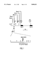
- FIG. 1 Structural features in a region of DNA bending upstream of the human c-myc gene. Positions of DNase 1-hypersensitive sites and CTF/NF-1 consensus binding sites are indicated by solid arrows at the top (Siebenlist et al., 1984, Cell 37:381-391). The open arrow (RIC) denotes the center of a zone of initiation of DNA replication (Vassilev, L., and E. M. Johnson, 1990, Mol. Cell. Biol. 10:4899-4904). The position of the 467-bp insert of plasmid pMYC47 is indicated and expanded below, showing the bend center as identified in FIG. 2. pMYC47 contains a tandem duplicate of this fragment. Restriction endonuclease cleavage sites are abbreviated as follows: HIII, HindIII; P, PstI; Spl, Spel;
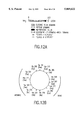
- FIGS. 2A-2B Location of an intrinsically bent DNA segment upstream of the c-myc gene.
- the tandem repeat was treated with single-site restriction enzymes to yield circular permutations of the 467-bp fragment. Enzymes are abbreviated as in FIG. 1.
- Lane M1 contains markers from the Bethesda Research Laboratories 1-kb ladder; lane M2 contains the 481-bp Bgl1-Xmn1 fragment of pUC19. Asterisks indicate two marker bands which are themselves more than 100% retarded due to bending (Stellwagen, N. C., 1983, Biochemistry 22:6186-6193).
- A Polyacrylamide gel electrophoresis showing anomalous migration of 467-bp fragments at 4° C. (I, bracket at left). (The top band in lane S is a partial digestion product.) In lane D (Ddel digest), two fragments of 252 and 254 bp, are also produced from the original 973-bp plus polylinker sequences.
- FIG. 3 Specificity of protein binding to the purine-rich strand of an element near the center of DNA bending.
- the probe is end-labeled single-stranded 24-mer MF0677, described in Section 6.1.1, corresponding to the purine-rich strand of nucleotides -1648 to -1625 upstream of the human c-myc P1 transcription start site.
- the left lane is the standard gel band shift binding reaction, described in Section 6.1.3, with no added competitor; the right lane is the reaction with an added HeLa nuclear extract. Unlabeled competitors are at 100 ⁇ excess.
- FIG. 4A-4B Specific UV cross-linking of a polypeptide with the PUR element.
- end-labeled 24-mer oligonucleotide MF0677 corresponding to the G-A strand of the c-myc PUR element, was either subjected or not subjected to UV cross-linking in the presence or absence of a HeLa nuclear extract as described in Section 6.2.3. The mixture was then subjected to SDS polyacrylamide gel electrophoresis in a 10% gel and autoradiographed.
- A Time course of UV cross-linking. Lanes were reacted without and with HeLa nuclear extract, as indicated. Cross-linking was performed for the times indicated.
- B bound label
- U unbound label.
- FIGS. 5A-5B Methylation interference analysis of purine contact points by protein specifically binding the purine-rich strand of the PUR element. After derivatization 32 P-end-labeled 24-mer oligonucleotide MF0677, containing the PUR element, with dimethyl sulfate, the single-stranded probe was allowed to react with protein in HeLa nuclear extract. (A) Gel band shift following derivatization with two levels of dimethyl sulfate as described in Section 6.2.4.
- Lanes - reaction with 53 mM dimethyl sulfate, calculated to derivatize approximately one purine base per oligonucleotide molecule; +, reaction with fivefold-higher dimethyl sulfate; T, reaction without added protein.
- Each half of the gel represents a single methylation reaction from which bands were purified for further sequence analysis as in panel B.
- FIG. 6 Mutation analysis of nucleotides essential for specific binding to the PUR element.
- Gel retardation of single-stranded probes by protein from HeLa nuclear extract was performed as described in 6.1.4. The four oligonucleotides listed at the top were used as probes.
- Pur G-A is the oligonucleotide MF0677
- Mut. I, II, and III are mutated oligonucleotides MM0677 (GGG>AAA in FIG. 3), MA0677, and MB0677, respectively. Sequences are listed in 6.1.1. Lanes: a, standard binding reaction with no added competitor; b to e, reactions with added 30-fold excess of unlabeled competitor oligonucleotides PUR G-A, MM0677, MA0677, and MB0677, respectively.
- FIG. 7 Affinity of the PUR factor for single-stranded versus double-stranded PUR element.
- Single-strand PUR oligonucleotide MF0677 was used as a probe in the standard gel band shift assay described in Section 6.1.3. Unlabeled single-or double-stranded oligonucleotides were added as competitors. Lane 1 has no added protein; lane 2 is the standard reaction with no added competitor.
- Excesses (3-, 30-, and 300-fold) of the following oligonucleotides were added: SS, single-stranded MF0677, DS, double-stranded version of the 24-mer containing the PUR element made by annealing MF0677 and MR0700; NS-SS, nonspecific single-stranded oligonucleotide MR0740.
- FIG. 8 Sequence of the PUR element region upstream of the human c-myc gene (SEQ ID NO:1). Numbering at the left is relative to the HindIII cleavage site 2,325 bp upstream of the P1 transcription start site. Numbering at the right is relative to the P1 start site. Indicated are two repeats of the yeast ARS consensus element each with 10-of-11-bp homology (solid arrows), two repeats of the PUR consensus element of Table 1, the first with 100% homology and the second with 14 of-16-bp homology (dotted area), and the sequence of oligonucleotide MF0677, used in binding studies (open box). Additional repeated elements within this region (GAGGGA and ATTATAT) are also indicated.
- FIG. 9 Specific oligonucleotide binding by ⁇ abs. ⁇ AB2 phage were plated, induced and transferred to nitrocellulose membranes. Each membrane was then cut in half, and the halves incubated separately in the presence of MF0677 oligonucleotide, 32 P-end-labeled as described in Section 7.1.2. The left half was incubated in the presence of an excess of unlabeled non-specific competitor MF0562, while the right half was incubated in the presence of excess unlabeled specific competitor (MF0677).
- FIG. 10 Nucleotide sequence of Pur ⁇ (SEQ ID NO:2).
- the nucleotide sequence shown is derived from phage clones ⁇ AB6 and ⁇ HE1.
- the amino acid sequence (SEQ ID NO:3). of the open reading frame is indicated beneath the nucleotide sequence (SEQ ID NO:2). Numbering begins with the first methionine.
- the line beneath the sequence at base -9 indicates the first base of the ⁇ AB6 clone.
- Singly-underlined amino acid residues in bold type indicate class I repeats.
- Doubly-underlined amino acid residues in bold type indicate class II repeats.
- FIG. 11 Two repeat motifs in Pur ⁇ and Pur ⁇ .
- the class I repeat motifs (three from Pur ⁇ and one from Pur ⁇ ) are aligned at top ⁇ Ia: (SEQ ID NO:4); ⁇ Ib: (SEQ ID NO:5); ⁇ Ic: (SEQ ID NO:6); ⁇ I: (SEQ ID NO:7)! and the class II repeat motifs (two from Pur ⁇ ) are aligned at bottom ⁇ IIa: (SEQ ID NO:8); ⁇ IIb: (SEQ ID NO:9)!.
- Solid boxes indicate identical amino acid residues, dotted boxes indicate conservative changes.
- FIGS. 12A-12B Arrangement of amino sequence domains in Pur ⁇ .
- FIG. 13 Gel Shift assay of protein extracts from ⁇ AB clones. Gel shift assays using labeled MF0677 probe were performed as described in Section 6.1.3.
- Lane 1 displays probe in the absence of protein.
- Lane 2 displays probe in the presence of a control protein extract prepared from Y1090 cells infected with ⁇ ovalb, a chicken ovalbumin gene clone in ⁇ gt11 (Clontech).
- Lane 3 displays probe in the presence of protein extract from Y1090 cells infected with ⁇ AB4.
- Lane 4 displays signal with 5-fold dilution ⁇ AB4 extract.
- Lanes 5 through 8 represent reactions similar to that of lane 3, but with cold competitor added; lane 5, 5-fold excess of MF0677: lane 6, 5-fold excess of polyA: lane 7, 20-fold excess of MF0677: lane 8, 20-fold excess of polyA.
- Filled arrows indicate bands specific to the clone, open arrows indicate bands present normally in E. coli.
- FIG. 14 Blot hybridization of human mRNAs with Pur ⁇ probe.
- Poly A + RNA prepared from tissue or culture cells was subjected to electrophoresis and blotted as described in Section 7.1.5. Membranes were probed with labeled-Pur ⁇ cDNA. Lanes are: pPUR6 ⁇ BamHI--pPUR6 DNA digested with BamHI; NCI-H82--lung tumor cell line mRNA; HepG2--liver hepatoma mRNA.
- the lower box displays the results of hybridization of the same membrane to a glyceraldehyde phosphate dehydrogenase probe as a loading control.
- FIG. 15 5' and 3' RACE extension of Pur ⁇ cDNA.
- Products of the RACE reactions outlined in Section 7.1.6 were subjected to electrophoresis in alkaline denaturating agarose gels, Southern blotted to GENE-SCREEN PLUS membranes and hybridized to a Pur ⁇ probe.
- numbers on right indicate molecular weight markers in kb.
- Lane 1 in each case indicates the reaction containing both primers.
- Lane 2 in each case represents a control reaction in which one primer was omitted.
- EX-270 SEQ ID NO:16
- EX-174 SEQ ID NO:17
- TTCTAAGCTTCGTCTCGTGCTGCAGCCC TTCTAAGCTTCGTCTCGTGCTGCAGCCC
- Pur-specific primers used were EX-695 (SEQ ID NO:18)(TCTTCGATGTGGGCTCCAAC), corresponding to nucleotides 695 to 714, for the first amplification reaction, and EX-990 (SEQ ID NO:19)(ACACACACACACATGCATAC), corresponding to nucleotides 990 to 1009, for the second amplification reaction.
- FIG. 16 Delay in the onset of DNA synthesis in serum-stimulated human fibroblasts, by PUR element oligonucleotides.
- FIG. 17 Regions of homologies between PUR protein and SV40 Large T-antigen Pur ⁇ : (SEQ ID NO:10); ERG-B: (SEQ ID NO:11); HAGEMON FACTOR: (SEQ ID NO:12); T-Antigen: (SEQ ID NO:13); Pur ⁇ : (SEQ ID NO:14)!.
- FIG. 18 Expression of GST-PUR protein in E. coli. SDS polyacrylamide gel of various GST-fusion proteins eluded from a glutathione-linked agarose column. The last two lanes represent PUR-GST fusion protein prepared form two different strains of E. coli, DH5 and BL21-LYS.
- FIG. 19 PUR binds the pRB protein in WR2E3 cell extracts as determined by passage of extracts over PUR-GST columns. Column bound proteins were subjected to SDS-polyacyralmide gel electrophoresis and blotted onto nitrocellulose membrane. The pRB protein was detected using anti-pRB antibodies. PUR preferentially binds the unphosphorylated form of pRB. Lane 1 is Retinoblastoma protein; Lane 2 is GST alone; Lane 3 is GST-T antigen fusion protein; Lane 4 is GST-ARP fusion protein which is another protein that interacts with the retinoblastoma protein and Lane 5 is GST-PUR fusion protein.
- FIG. 20A Detection of a Tat-Pur ⁇ complex in human glioblastoma cells expressing the HIV-1 tat gene.
- Glioblastoma cell line 5-10 constitutively synthesizing Tat expressed under control of the SV40 late promoter, was cultured in 75 cm 2 plates to 5 ⁇ 10 5 cells per plate. The left lane represents 25 ⁇ L of untreated lysate.
- immunoprecipitation was carried out using magnetic beads alone (No first antibody), mouse non-immune serum (Non-imm. ser.), an irrelevant first antibody, anti-influenza virus hemagglutinin epitope monoclonal antibody 12CA5 (Irr. 1st ab) or anti-Pur ⁇ antibody 5B11 (Anti-Pur mab 5B11).
- FIG. 20B Complex formation between Tat and GST-Pur ⁇ in glioblastoma cell lysates.
- the left lane represents purified Tat protein (400 ng.)
- Lanes labeled Columns represent protein eluted from columns consisting of beads alone (B), Beads coupled to GST (B-GST) or beads coupled to GST-Pur ⁇ (B-GST-Pur).
- B-GST beads alone
- B-GST-Pur ⁇ B-GST-Pur
- For the lane labeled 5-10 cell lysate 20 ⁇ L of clarified lysate were passed over the column.
- the four lanes at left were exposed to film for equal time, and the lane at far right 20 times longer.
- the position 18 kDa represents migration of a prestained lysozyme marker.
- FIG. 21A Specificity of binding of Pur ⁇ to the JCV upTAR element.
- the different oligonucleotide probes used for the indicated reactions are: JCV+, the purine-rich single strand of the upTAR element; JCV ds, the double-stranded upTAR element, only the pyrimidine-rich strand of which is labeled; JCV-, the pyrimidine-rich strand of the upTAR element.
- FIG. 21B Effect of Tat upon binding of a series of Pur ⁇ deletion mutants to the JCV upTAR element.
- Tat refers to GST-Tat.
- GST-PurA refers to full-length GST-Pur ⁇ , in which Pur ⁇ is 322 aa long. Numbers given for deletion mutants refer to the Pur ⁇ aa's remaining in the construct, beginning with the N-terminus of Pur ⁇ , except for PurA del 55-314, in which the indicated internal aa's have been deleted.
- FIG. 22 Association of Tat and Pur ⁇ proteins in the presence or absence of the JCV upTAR element. Binding of labeled upTAR indicates the position of GST-Pur ⁇ on the filter. The right portion filter, containing lanes run in parallel to those on the left, was probed with rabbit anti-Tat antibody 705, as described for FIG. 20 to indicate the position of Tat.
- FIG. 23 Fluorescence image of a metaphase chromosome spread hybridized with a human pura genomic probe. FITC fluorescence from the hybridization probe has been pseudocolored red for best photographic contrast. Isolated chromosomes 5 and 6 with hybridization signals are shown at the bottom on the right. The chromosomes are counterstained with DAPI.
- FIG. 24 Double-label fluorescence image of a chromosome 5 homologous pair. Green-yellow signal denotes R-like banding generated by cohybridization in situ with Alu repeat sequences, and red signal denotes hybridization loci of pur ⁇ at 5q31. Genomic clone EMhPur 1 was used as a probe for pur ⁇ . Each chromosome is counterstained with DAPI (blue). FITC and rhodamine images were recorded sequentially and subsequently aligned and merged as previously described using image-processing programs Gene Join Layer and Gene Join Maxpix.
- FIG. 25A Localization of pur ⁇ to human chromosome 5 by hybridization of cDNA fragment to DNA of two human/hamster hybrid cell lines containing individual chromosomes 5 or 6.
- DNA was prepared from hybrid cell lines NA10114, bearing human chromosome 5, and NA11580, bearing human chromosome 6, treated with restriction enzymes HindIII (lanes at left) or Eco RI (lanes at right), subjected to agarose gel electrophoresis alongside control DNAs, blotted and hybridized.
- the probe was a 777 bp PstI fragment of Pur ⁇ cDNA, pPUR6, labeled with 32 P-phosphate. Hybridization was for 16 hrs at 68°-36° C.
- FIG. 25B Restriction map of the puru locus at 5q31.
- the position of the pur ⁇ gene is indicated by a box.
- the cDNA is oriented as shown, 5' to 3' indicating the direction of the transcription.
- the 11.5 kb HindIII fragment of ⁇ phage EMhPur1, used as probe for FIGS. 1 and 2 and the 777 bp PstI fragment of plasmid pPUR6, used as probe for FIG. 30A are shown at bottom.
- a purine rich 24 nucleotide DNA sequence referred to as the PUR element, is believed to play a role in initiation of DNA replication and regulation of gene expression.
- the PUR element occurs at a major site of DNA bending located 1.6 kB upstream of the transcription start site of the human c-myc gene, near the center of a reported zone of initiation of DNA replication.
- the PUR protein a 27 kD HeLa cell nuclear factor, was initially identified by its ability to bind in a sequence specific manner to single-stranded PUR element nucleotide sequences indicating a role for PUR protein in DNA replication and/or regulation of gene expression.
- a cDNA clone encoding a polypeptide with PUR element binding activity has been isolated and sequenced and that clone is described herein.
- the invention involves the expression of PUR, or fragments thereof, to evaluate and screen for drugs that may regulate the activity of PUR.
- Such regulators of PUR may be used therapeutically to treat hyperproliferative diseases such as cancers.
- regulators of PUR may be used to treat viral disease where activation of viral gene expression is regulated by PUR.
- the invention is based on results indicating that the PUR protein binds specifically and with high affinity to the HIV encoded transcriptional activator protein (Tat).
- the Tat protein transactivates transcription from the HIV-1 LTR promoter and is required for viral replication.
- the JC virus (JCV) contains a DNA sequence in the late promoter region that binds the Tat-Pur ⁇ complex thereby indicating a role for PUR ⁇ in regulation of viral transcription.
- the present invention also relates to the use of PUR DNA or PUR antibodies for diagnostic purposes to detect aberrant expression of the PUR protein and/or mutations in the PUR gene.
- This aspect of the invention is based on in situ hybridization studies that localize the PUR ⁇ gene to chromosome 5 in the 5q31 region. Loss of heterozygosity at 5q31 is frequently associated with myelodysplastic syndrome and, particularly, with myeloid leukemias. Therefore, the diagnostic methods of the invention may be utilized to detect proliferative disorders.
- the present invention also relates to methods of treating proliferative disorders, such as leukemias, where gene therapy may be utilized to compensate for loss of PUR activity.
- gene therapies may, for example, utilize liposomes containing PUR protein for delivery of PUR to target tissues.
- recombinant viral vectors may be generated to express PUR protein in targeted tissues.
- Bending of DNA is a structural feature conserved at origins of replication in both eukaryotic and prokaryotic organisms.
- a major site of DNA bending is located 1.6 kB upstream of the transcription start site of the human c-myc gene.
- Gel-band shift assays and DNA methylation interference assays have more precisely defined the nucleotide sequences in this region of DNA that are important for DNA/protein interactions.
- This region of DNA is referred to as the PUR element and has the following nucleotide sequence, (SEQ ID No:20): GGAGGTGGTGGAGGGAGAAAAG.
- the PUR protein was initially identified as a 27 kD HeLa cell nuclear factor that bound in a sequence specific manner to single-stranded PUR element nucleotide sequences.
- a function for PUR protein in DNA replication and/or gene expression is indicated by the binding of PUR protein to specific regions of DNA involved in initiation of DNA replication and/or regulation of gene expression.
- a function for PUR in regulation of viral gene expression is indicated by the association between PUR and the HIV transactivating Tat protein.
- the Tat-PUR complex is also found bound to viral Tat responsive expression elements.
- a cDNA clone encoding a polypeptide with PUR binding activity has been isolated and sequenced.
- the nucleotide coding sequence and deduced amino acid sequence of the human PUR protein is depicted in FIG. 10.
- the nucleotide coding sequence for human PUR protein or its functional equivalent can be used to generate recombinant molecules which direct the expression of the PUR protein.
- the coding sequence of PUR may be obtained by cDNA cloning of RNA isolated and purified from cell sources that express PUR protein or by genomic cloning.
- cDNA libraries of clones may be prepared from DNA fragments generated using techniques known in the art, including but not limited to use of restriction enzymes.
- the clones that contain the gene for PUR may be identified in a number of ways know in the art. For example, a portion of the PUR amino acid sequence can be used to deduce nucleotide sequence. The DNA sequence may then be chemically synthesized, radioactively end-labeled and used as a hybridization probe. Other methods which can be used include but are not limited to chemically synthesizing the gene sequence from derived amino acid sequence of PUR. Alternatively, in-vitro translation of selected mRNA followed by a functional or immunological assay of translation products can be used.
- the PUR gene was cloned by constructing a cDNA library into the bacteriophage expression system ⁇ gt11.
- the library was screened with radioactively labeled oligonucleotide MF0677 representing sequences found in the core of the PUR element.
- nucleotide PUR sequences which encode PUR, peptide fragments of PUR, PUR fusion proteins or functional equivalents thereof may be used to generate recombinant DNA molecules that direct the expression of the PUR protein or a functionally active peptide, fusion protein or functional equivalent thereof, in appropriate host cells.
- DNA sequences which encode substantially the same or a functionally equivalent amino acid sequence, e.g., shown in FIG. 10, may be used in the practice of the invention for the cloning and expression of the PUR protein.
- DNA sequences include those which are capable of hybridizing to the PUR sequence under stringent conditions, or those which would be capable of hybridizing under stringent conditions but for the degeneracy of the genetic code.
- the stringency conditions may be adjusted in a number of ways. For example, when performing polymerase chain reactions (PCR), the temperature at which annealing of primers to template takes place or the concentration of MgCl 2 in the reaction buffer may be adjusted. When using radioactively labeled DNA fragments or oligonucleotides in hybridization reactions, the stringency may be adjusted by changes in the ionic strength of the wash solutions or by careful control of the temperature at which the washes are carried out.
- PCR polymerase chain reactions
- Altered nucleotide sequences which may be used in accordance with the invention include deletions, additions or substitutions of different nucleotides resulting in a sequence that encodes the same or a functionally equivalent gene product.
- the gene product may contain deletions, additions or substitutions of amino acid residues within the sequence which result in silent changes thus producing a bioactive product.
- Such amino acid substitutions may be made on the basis of similarity in polarity, charge, solubility, hydrophobicity, hydrophilicity and/or the amphipathic nature of the residues involved.
- negatively charged amino acids include aspartic acid and glutamic acid; positively charged amino acids include lysine and arginine; amino acids with uncharged polar head groups or nonpolar head groups having similar hydrophilicity values include the following: leucine, isoleucine, valine; glycine, alanine; asparagine, glutamine; serine, threonine; phenylalanine, tyrosine.
- a functionally equivalent PUR protein refers to a peptide, polypeptide, protein or fusion protein that binds to the PUR element, but not necessarily with the same binding affinity of its counterpart native PUR protein. The design and engineering of constructs encoding PUR fusion proteins is described infra (Section 5.2.2.)
- the coding sequence of the PUR protein could be synthesized, in whole or in part, using chemical methods well known in the art. See, for example, Caruthers, et al., 1980, Nuc. Acids. Res. Symp. Ser. 7: 215-233; Crea & Itrin, 1980, Nuc. Acids. Res. 9(10): 2331; Matteucci & Caruthers, 1980, Tetrahedron Letters 21: 719; and Chow & Kempe, 1981, Nuc. Acids. Res. 9(12): 2807-2817.
- the protein itself could be produced using chemical methods to synthesize the PUR protein amino acid sequence in whole or in part.
- peptides can be synthesized by solid phase techniques, cleaved from the resin and purified by preparative high performance liquid chromatography. (E.g., see Creighton, 1983, Proteins, Structures And Molecular Principles, W. H. Freeman & Co., N.Y., pp. 50-60). The composition of the synthetic peptides may be confirmed by amino acid analysis or sequencing (e.g., the Edman degradation procedure; see Creighton, 1983, Proteins, Structures And Molecular Principles, W. H. Freeman & Co., N.Y., pp. 34-49).
- the PUR ⁇ cDNA can be used as a probe to detect the expression of PUR RNA.
- Northern blot analysis using mRNA prepared from human fetal liver tissue, HeLa cells, NCI-H82 cells and HepG2 cells reveal a similar pattern of multiple transcripts. Two major transcripts of 5.5 kB and 2.1 kB, and two minor transcripts of 3.3 kB and 2.8 kB were detected suggesting alternative splicing of a single PUR gene or the existence of a number of related genes.
- the PUR ⁇ cDNA sequence may be used to isolate PUR ⁇ related genes.
- a HeLa cell cDNA library was screened with a radioactively end-labeled fragment of the PUR cDNA clone. A number of clones were isolated, and one in particular was chosen for further sequence analysis. The clone, designated PUR- ⁇ , was found to be similar but not identical to the initially purified PUR cDNA clone demonstrating the existence of a family of related PUR proteins (FIGS. 11 and 12).
- the nucleotide sequence coding for the PUR protein, or a functional equivalent including PUR fusion proteins, as described in Section 5.2.1, supra, is inserted into an appropriate expression vector, i.e., a vector which contains the necessary elements for the transcription and translation of the inserted coding sequences.
- an appropriate expression vector i.e., a vector which contains the necessary elements for the transcription and translation of the inserted coding sequences.
- host expression vector systems i.e.--vectors which contain the necessary elements for directing the replication, transcription, and translation of PUR coding sequence
- host expression vector systems i.e.--vectors which contain the necessary elements for directing the replication, transcription, and translation of PUR coding sequence
- These include but are not limited to microorganisms such as bacteria transformed with recombinant bacteriophage DNA, plasmid DNA or cosmid DNA expression vectors containing the PUR coding sequence; yeast transformed with recombinant yeast expression vectors containing the PUR coding sequence; insect cell systems infected with recombinant virus expression vectors (e.g., baculovirus) containing the PUR coding sequence; plant cell systems infected with recombinant virus expression vectors (e.g., cauliflower mosaic virus CaMV; tobacco mosaic virus, TMV) or transformed with recombinant plasmid expression vectors (e.g., Ti plasmid) containing the PUR coding sequence;
- the expression elements of these vectors vary in their strength and specificities. Depending on the host/vector system utilized, any one of a number of suitable transcription and translation elements may be used. For instance, when cloning in mammalian cell systems, promoters isolated from the genome of mammalian cells, (e.g., mouse metallothionine promoter) or from viruses that grow in these cells, (e.g., vaccinia virus 7.5K promoter or Moloney murine sarcoma virus long terminal repeat) may be used. Promoters produced by recombinant DNA or synthetic techniques may also be used to provide for transcription of the inserted sequences.
- promoters isolated from the genome of mammalian cells e.g., mouse metallothionine promoter
- viruses that grow in these cells e.g., vaccinia virus 7.5K promoter or Moloney murine sarcoma virus long terminal repeat
- Promoters produced by recombinant DNA or synthetic techniques may also be used to provide for transcription of
- Specific initiation signals are also required for sufficient translation of inserted protein coding sequences. These signals include the ATG initiation codon and adjacent sequences. In cases where the entire PUR gene including its own initiation codon and adjacent sequences are inserted into the appropriate expression vectors, no additional translational control signals may be needed. However, in cases where only a portion of the coding sequence is inserted, exogenous translational control signals, including the ATG initiation codon must be provided. Furthermore, the initiation codon must be in phase with the reading frame of the PUR coding sequences to ensure translation of the entire insert. These exogenous translational control signals and initiation codons can be of a variety of origins, both natural and synthetic. The efficiency of expression may be enhanced by the inclusion of transcription attenuation sequences, enhancer elements, etc.
- the PUR coding sequence may be ligated to an adenovirus transcription/translation control complex, e.g., the late promoter and tripartite ladder sequence.
- This chimeric gene may then be inserted in the adenovirus genome by in vitro or in vivo recombination. Insertion in a non-essential region of the viral genome (e.g., region E3 or E4) will result in a recombinant virus that is viable and capable of expressing PUR in infected hosts.
- the vaccinia 7.5K promoter may be used.
- An alternative expression system which could be used to express PUR is an insect system.
- Autographa californica nuclear polyhidrosis virus (AcNPV) is used as a vector to express foreign genes.
- the virus grows in Spodoptera frugiperda cells.
- the PUR coding sequence may be cloned into non-essential regions (for example the polyhedrin gene) of the virus and placed under control of an AcNPV promoter (for example the polyhedrin promoter).
- Successful insertion of the PUR coding sequence will result in inactivation of the polyhedrin gene and production of non-occluded recombinant virus (i.e., virus lacking the proteinaceous coat coded for by the polyhedrin gene).
- These recombinant viruses are then used to infect Spodoptera frugiperda cells in which the inserted gene is expressed.
- Retroviral vectors prepared in amphotropic packaging cell lines permit high efficiency expression in numerous cells types. This method allows one to assess cell-type specific processing, regulation or function of the inserted protein coding sequence.
- a host cell strain may be chosen which modulates the expression of the inserted sequences, or modifies and processes the gene product in the specific fashion desired. Expression from certain promotes can be elevated in the presence of certain inducers. (e.g., zinc and cadmium ions for metallothionein promoters). Therefore, expression of the genetically engineered PUR may be controlled. This is important if the protein product of the cloned foreign gene is lethal to host cells. Furthermore, modifications (e.g., phosphorylation) and processing (e.g., cleavage) of protein products are important for the function of the protein. Different host cells have characteristic and specific mechanisms for the post-translational processing and modification of protein. Appropriate cell lines or host systems can be chosen to ensure the correct modification and processing of the foreign protein expressed.
- inducers e.g., zinc and cadmium ions for metallothionein promoters. Therefore, expression of the genetically engineered PUR may be controlled. This is important if the protein product of
- Fusion protein vectors may be used to express PUR fusion protein.
- the purified PUR fusion protein may be used to raise antisera against the PUR protein to study the biochemical properties of the PUR protein and/or to engineer PUR fusion proteins with different binding affinities for the PUR element, and/or for the pRB protein.
- Possible expression vectors include but are not limited to, vectors that express ⁇ -galactosidase and trpE fusions, maltose-binding protein fusions and glutathione-S-transferase fusions (carrier regions). Methods which are well known to those skilled in the art can be used to construct expression vectors containing PUR protein coding sequences.
- the carrier region of the fusion protein may be used for purification of the PUR fusion protein.
- antibodies against the carrier protein may be used in affinity chromatography for purification of the fusion protein.
- amylose resin may be used for purification of maltose binding protein fusions or glutathione-agarose beads may be used for purification of glutathione-S-transferase fusion proteins.
- the expression vectors may also contain polylinker sequences that encode specific protease cleavage sites so that any cloned protein may be released from its carrier protein by treatment with a specific protease.
- DNA sequences encoding the thrombin or factor Xa cleavage sites may be included in the fusion protein vectors.
- the PUR coding sequence was inserted into the pGEX-1 ⁇ T expression vector containing the tac IPTG inducible promoter region and the coding region for the amino terminus of glutathione-S-transferase. After induction with IPTG, the GST-PUR fusion protein was purified from lysed cells using glutathione-linked garose beads.
- NLS nuclear localization sequences
- the coding region of PUR protein, or fragments thereof may be linked to DNA sequences encoding nuclear localization sequences (NLS).
- NLS nuclear localization sequences
- the NLS consists either of a short division of basic amino acids, for example as shown for the NLS of SV40 T antigen (SEQ ID No:33) (PKKKRKV).
- the NLS may have a bipartite structure comprised of two stretches of basic residues separated by a spacer of about 10 amino acids. (Dingwell et al., 1991, Trends Biochem. Sci. 16:478).
- any NLS sequences that functions to direct the localization of PUR to the nucleus may be incorporated into PUR expression vectors. Inclusion of these signals in PUR expression vectors will ensure that recombinantly expressed PUR protein, fragments of PUR proteins, or PUR fusion proteins are properly localized to the nucleus.
- nuclear localization signals are well known to those skilled in the art and may be genetically engineered into PUR expression vectors using routine methods.
- the host cells which contain the recombinant PUR coding sequence and which express the biologically active, mature product may be identified by at least four general approaches: (a) DNA--DNA, DNA-RNA or RNA-antisense RNA hybridization; (b) the presence or absence of "marker" gene functions; (c) assessing the level of transcription as measured by the expression of PUR mRNA transcripts in the host cell; and (d) detection of the mature gene product as measured by immunoassay and, ultimately, by its biological activity.
- the presence of the human PUR coding sequence inserted in the expression vector can be detected by DNA--DNA hybridization using probes comprising nucleotide sequences that are homologous to the human PUR coding sequence.
- the recombinant expression vector/host system can be identified and selected based upon the presence or absence of certain "marker" gene functions (e.g., thymidine kinase activity, resistance to antibiotics, resistance to methotrexate, transformation phenotype, occlusion body formation in baculovirus, etc.).
- certain "marker" gene functions e.g., thymidine kinase activity, resistance to antibiotics, resistance to methotrexate, transformation phenotype, occlusion body formation in baculovirus, etc.
- a marker gene can be place in tandem with the PUR sequence under the control of the same or different promoter used to control the expression of the PUR coding sequence. Expression of the marker in response to induction or selection indicates expression of the PUR coding sequence.
- transcriptional activity of the PUR coding region can be assessed by hybridization assays.
- polyadenylated RNA can be isolated and analyzed by Northern blot using a probe homologous to the PUR coding sequence or particular portions thereof.
- total nucleic acids of the host cell may be extracted and assayed for hybridization in such probes.
- the expression of the mature protein product can be assessed immunologically, for example by Western blots, immunoassays such as radioimmuno-precipitation, enzyme-linked immunoassays and the like.
- the ultimate test of the success of the expression system involves the detection of the biologically active PUR gene product.
- One of the properties associated with PUR protein is its sequence specific affinity for the PUR element.
- a possible method for detection of PUR protein activity might involve the use of gel-band shift assays.
- the PUR protein was initially characterized as a factor present in HeLa cell nuclear extracts, that bound in a sequence specific manner to single-stranded PUR element sequence. Screening of a ⁇ gtll expression library, with radioactively end-labeled PUR element sequences, represented by the oligonucleotide MF0677, resulted in the isolation of a cDNA clone encoding a protein with single-stranded binding activity matching that of the PUR factor.
- the deduced amino acid sequence of PUR ⁇ reveals a modular repeat structure unique among known DNA-binding proteins.
- the class I repeats are shown by single underlining in the sequence of FIG. 10, and the class II repeats are shown by double underlining. While the sequence between these repeats is not conserved, the distance between the class I repeats is highly regular.
- the repeats themselves are not identical but preserve a number of strictly conserved amino acids of fixed distances along the repeats, indicated by solid boxing in FIG. 11, and a high percentage of conservatively-substituted amino acids, indicated by dotted-line boxing in FIG. 11.
- PUR ⁇ contains several notable structural features denoted in FIG. 12A. Near the amino-terminal end of PUR ⁇ there is a prominent sequence of 28 glycine residues broken only by a single serine residue. Similar glycine stretches are present in proteins serving a wide variety of functions, including helix-destabilizing proteins (Haynes et al., 1987 Proc. Natl. Acad. Sci. US 79:4083-4087). Carboxyl terminal to all of the repeat modules there is a region (residues 261 through 274) of alpha helix (Chou and Fasman, 1974 Biochemistry 13:222-245; Levin et al., 1986 FEBS Lett.
- the amphipathic helix is ordered with opposing basic and aromatic side chains, as presented in the helical wheel of FIG. 12B. Similar amphipathic helices are present in several DNA-binding proteins thought to play a role in transcriptional activation (Ptashne, 1988, Nature 335:683-689).
- the carboxyl terminus of the PUR ⁇ molecule consists of glutamine-glutamate-rich domain. The entire sequence from residue 276 through 321 is 50% glutamine and glutamate residues. There is one sequence of 7 consecutive glutamine residues, and near the carboxyl terminus there is a sequence of 5 glutamate residues broken by a single glycine.
- Glutamine-rich domains have been implicated as transcriptional activation regions in several DNA-binding proteins (Courey et al., 1989, Cell 59:827-836). At the border between the amphipathic helix and the glutamine-glutamate-rich domain there is the motif Ser-Glu-Glu-Met (residues 275 through 278). The serine in this motif is a potential phosphorylation site for casein kinase II (Kennelly and Krebs, 1991, J. Biol. chem. 266:15555-15558), although it is not known whether the motif serves this function in Pur ⁇ .
- the PUR ⁇ molecule shares a region of protein homology with the DNA tumor virus protein SV40 large T-antigen (FIG. 17).
- the region of SV40 T-antigen, sharing homology with PUR ⁇ , is of particular interest as it is the region of T-antigen involved in the protein/protein interaction between SV40 large T-antigen and the retinoblastoma (pRB) gene product.
- PUR ⁇ could function as SV40 large T-antigen and bind to pRB protein.
- a PUR-GST protein was expressed in E. coli followed by immobilization of the fusion protein on a glutathione-linked agarose column. WR2E3 cell extracts were passed over the PUR-GST column and bound proteins were eluted with excess glutathione. The eluted proteins were subjected to SDS polyacrylamide gel electrophoresis and blotted onto nitrocellulose membrane. The presence of pRB protein was detected using anti-Rb antibodies. As illustrated in FIG. 17, the PUR-GST fusion protein is able to bind cellular pRB with the same affinity as SV40 large T-antigen. In addition, the PUR-GST fusion protein seems to preferentially bind the unphosphorylated form of pRB.
- a Tat-PUR ⁇ complex can be detected in glioblastomas cells expressing the HIV-1 tat gene (FIG. 20A). Moreover, purified TAT and PUR ⁇ can be shown to associate specifically and with high affinity. Analysis of PUR deletion mutants indicates that the first 72 amino acids of Tat and the first 85 amino acids of PUR ⁇ are required for the Tat-PUR protein/protein interaction (FIG. 21B).
- a purine rich element (upTAR) in the late promoter region of JCV DNA mediates transcriptional activation by the HIV-1 Tat protein, which by itself does not bind this element.
- the purine rich element contains recognition sites for PUR ⁇ .
- PUR ⁇ binds to the purine-rich strand of the upTAR element of the JCV virus.
- the binding of PUR ⁇ to the upTAR element is enhanced by the binding of Tat indicating a functional role for the Tat-PUR ⁇ complex in the activation of viral gene expression.
- PUR protein may play a role in regulation of cell proliferation.
- the observed protein/protein interaction between the PUR protein and the pRB protein further supports the view that the PUR protein is involved in regulation of cell proliferation.
- the PUR protein also binds 5' to a number of cellular oncogenes that include c-myc, int-2 and lick, indicating that PUR may also regulate gene expression.
- the observed protein/protein interaction between PUR ⁇ and the HIV encoded Tat protein suggests a role for PUR in regulation of viral transcription. This is further supported by the demonstration that the Tat-pur ⁇ complex binds to Tat responsive viral transcriptional elements.
- Inhibitors of PUR protein may function to selectively inhibit the replication and/or gene expression of specific genetic loci associated with PUR elements or viral transcriptional elements.
- Antibodies to PUR may be useful as diagnostic and therapeutic agents. More specifically, antibodies that bind PUR protein and which neutralize PUR activity may be of particular therapeutic value. For example, antibodies that bind PUR protein and in doing so, prevent PUR binding to pRB or to PUR element DNA sequences may be useful in therapies designed to inhibit cell proliferation. Alternatively, antibodies that bind PUR protein and in doing so prevent PUR from binding to HIV Tat, or Tat responsive viral transcriptional elements, may be useful in treating viral diseases.
- various host animals may be immunized by injection with the PUR protein including but not limited to rabbits, mice, rats, etc.
- Various adjuvants may be used to increase the immunological response, depending on the host species, including but not limited to Freund's (complete and incomplete), mineral gels such as aluminum hydroxide, surface active substances such as lysolecithin, pluronic polyols, polyanions, peptides, oil emulsions, keyhole limpet hemocyanin, dinitrophenol, and potentially useful human adjuvants such as BCG (bacille Calmette-Guerin) and corynebacterium parvum.
- BCG Bacille Calmette-Guerin
- Monoclonal antibodies to the PUR protein may be prepared by using any technique which provides for the production of antibody molecules by continuous cell lines in culture. These include but are not limited to the hybridoma technique originally described by Kohler and Milstein, (Nature, 1975, 256:495-497), the human B-cell hybridoma technique (Kosbor et al., 1983, Immunology Today, 4:72; Cote et al., 1983, Proc. Natl. Acad. Sci., 80:2026-2030) and the EBV-hybridoma technique (Cole et al., 1985, Monoclonal Antibodies and Cancer Therapy, Alan R. Liss, Inc., pp. 77-96).
- Antibody fragments which contain specific binding sites for the PUR protein may be generated by known techniques.
- such fragments include but are not limited to: the F(ab') 2 fragments which can be produced by pepsin digestion of the antibody molecule and the Fab fragments which can be generated by reducing the disulfide bridges of the F(ab') 2 fragments.
- Fab expression libraries may be constructed (Huse et al., 1989, Science, 246:1275-1281) to allow rapid and easy identification of monoclonal Fab fragments with the desired specificity to PUR protein.
- oligoribonucleotide sequences that include anti-sense RNA molecules and ribozymes that function to inhibit the translation of PUR mRNA.
- Anti-sense RNA molecules act to directly block the translation of mRNA by binding to targeted mRNA and preventing protein translation.
- Ribozymes are enzymatic RNA molecules capable of catalyzing the specific cleavage of RNA.
- the mechanism of ribozyme action involves sequence specific hybridization of the ribozyme molecule to complementary target RNA, followed by a endonucleolytic cleavage.
- engineered hammerhead motif ribozyme molecules that specifically and efficiently catalyze endonucleolytic cleavage of PUR RNA sequences.
- ribozyme cleavage sites within any potential RNA target are initially identified by scanning the target molecule for ribozyme cleavage sites which include the following sequences, GUA, GUU and GUC. Once identified, short RNA sequences of between 15 and 20 ribonucleotides corresponding to the region of the target gene containing the cleavage site may be evaluated for predicted structural features such as secondary structure that may render the oligonucleotide sequence unsuitable. The suitability of candidate targets may also be evaluated by testing their accessibility to hybridization with complementary oligonucleotides, using ribonuclease protection assays.
- RNA molecules and ribozymes of the invention may be prepared by any method known in the art for the synthesis of RNA molecules. These include techniques for chemically synthesizing oligoribonucleotides well known in the art such as for example solid phase phosphoamite chemical synthesis.
- RNA molecules may be generated by in-vitro and in-vivo transcription of DNA sequences encoding the RNA molecule. Such DNA sequences may be incorporated into a wide variety of vectors which incorporate suitable RNA polymerase promoters such as the T7 or SP6 polymerase promoters.
- RNA molecules may be introduced as a means of increasing intracellular stability and half-life. Possible modifications include but are not limited to the addition of flanking sequences of ribo- or deoxy-nucleotides to the 5' and/or 3' ends of the molecule or the use of phosphorothioate or 2' O-methyl rather than phosphodiesterase linkages within the oligoribonucleotide backbone.
- Oligodeoxyribonucleotides can form sequence-specific triple helices by hydrogen bonding to specific complementary sequences in duplexed DNA. Interest in triple helices has focused on the potential biological and therapeutic applications of these structures. Formation of specific triple helices may selectively inhibit the replication and/or gene expression of targeted genes by prohibiting the specific binding of functional trans-acting factors.
- the PUR element is comprised predominantly of long repeats of G/A nucleotide sequences which are characteristic of target sequences favoring DNA triplex formation.
- Triple helix formation, at the site of PUR elements, may function to inhibit DNA replication and/or transcription of DNA sequences found adjacent to the element by preventing the binding of trans-acting factors such as PUR protein. Support for this is provided by experimental data demonstrating a delay in onset of DNA replication in cells exposed to PUR element oligonucleotides (FIG. 16).
- derivatives, analogues and peptides related to PUR are also envisioned and are within the scope of the invention.
- Such derivatives, analogues and peptides may be used to compete with full length wild-type PUR protein for binding to the PUR consensus element and in doing so inhibit PUR protein activity.
- the inhibition of PUR protein function may be utilized in several applications, including but not limited to, the treatment of hyperproliferative diseases such as cancer.
- Derivatives, analogues and peptides related to PUR protein may also be used to compete with full length wild type PUR protein for binding to the HIV encoded Tat protein, or Tat responsive viral transcription elements. Such inhibitors may be utilized to treat virally infected patients.
- the present invention also includes methods for identifying the specific site(s) of PUR that interact with (i) PUR elements; (ii) pRB; (iii) HIV Tat; or (iv) Tat responsive viral transcriptional elements.
- identifying the specific site(s) of PUR that interact with (i) PUR elements; (ii) pRB; (iii) HIV Tat; or (iv) Tat responsive viral transcriptional elements Using the methods described herein, and biochemical and molecular biological methods well-known in the art, it is possible to identify the corresponding portions of PUR involved in these interactions. For example, site-directed mutagenesis of DNA encoding the PUR protein may be used to destroy or inhibit the interaction between the molecules and PUR.
- a series of deletion mutants in the PUR coding region may be constructed and analyzed to determine the minimum amino acid sequence requirements for binding to the PUR consensus element.
- Deletion mutants of the PUR coding sequence may be constructed using methods known in the art which include but are not limited to use of nucleases and/or restriction enzymes; site-directed mutagenesis techniques, PCR, etc.
- the mutated polypeptides may be assayed for their ability to bind to the PUR element by gel-band shift assays.
- the mutated polypeptides may be assayed for their ability to bind the virally encoded HIV Tat protein and/or the viral TAT responsive transcriptional elements.
- Biophysical methods such as X-ray crystallography and nuclear magnetic resonance may also be used to map and study these sites of interaction. Once these sites have been identified, the present invention provides means for promoting or inhibiting this interaction, depending upon the desired biological outcome. Based on the foregoing, given the physical information on the sites of interaction, compounds that modulate PUR activity may be elaborated by standard methods well known in the field of rational drug design.
- Recombinantly expressed PUR protein may be used to screen for molecules that modulate PUR activity.
- molecules may include small organic or inorganic compounds, antibodies, peptides, or other molecules that modulate PUR's ability to bind to (i) PUR elements; (ii) TAT responsive viral transcriptional elements; (iii) pRB; or (iv) HIV encoded Tat protein.
- Synthetic compounds, natural products, and other sources of potentially biologically active materials can be screened in a number of ways.
- test molecule to modulate the activity of PUR may be measured using standard biochemical techniques, such as gel shift assays, pRB binding assays or HIV Tat binding assays.
- standard biochemical techniques such as gel shift assays, pRB binding assays or HIV Tat binding assays.
- Various embodiments are described below for screening, identification and evaluation of compounds that interact with PUR protein, which compounds may affect various cellular processes under the control of the PUR protein.
- the invention includes a method whereby a molecule capable of binding to PUR in a chemical or biological preparation may be identified comprising:
- step (b) contacting the chemical or biological preparation with the solid phase matrix produced in step (a), for an interval sufficient to allow the compound to bind;
- the above method may further include the step of:
- fragment thereof refers to peptide fragments of PUR corresponding to as few as 5 contiguous amino acids.
- the peptide fragments may correspond to function domains of the PUR protein such as those domains of the PUR protein that bind to pRB, HIV encoded Tat or Tat responsive transcriptional elements.
- compound capable of binding to PUR refers to a naturally occurring or synthetically produced molecule which interacts PUR. Such a compound may directly or indirectly modulate PUR activity and may include molecules that are natively associated with PUR inside a cell.
- the present invention provides an assay for identifying a compound, which can block the interaction of PUR with: (i) PUR elements; (ii) pRB; (iii) HIV Tat; or (iv) Tat responsive viral transcriptional elements.
- a cell transfected to coexpress PUR and HIV Tat in which the two proteins interact to form a complex can be incubated with an agent suspected of being able to inhibit this interaction, and the effect on the interaction can be measured. Any of a number of means for measuring the interaction and its disruption such as coimmunoprecipitation or gel shift analysis are available.
- the present invention also provides an assay method to identify and test a compound which stabilizes and promotes the interaction, using the same approach described above for a potential inhibitor.
- Random peptide libraries consisting of all possible combinations of amino acids may be used to identify peptides that are able to bind to the binding sites of PUR, or other functional domains of PUR. Identification of molecules that are able to bind to PUR may be accomplished by screening a peptide library with recombinant PUR proteins or recombinant soluble forms of PUR protein. Alternatively, the binding domains of PUR may be separately expressed and used to screen peptide libraries.
- PUR proteins may be conjugated to enzymes such as alkaline phosphatase or horseradish peroxidase or to other reagents such as fluorescent labels which may include fluorescein isothyiocynate (FITC), phycoerythrin (PE) or rhodamine. Conjugation of any given label to PUR may be performed using techniques that are routine in the art.
- PUR expression vectors may be engineered to express a chimeric PUR protein containing an epitope for which a commercially available antibody exists. The epitope- specific antibody may be tagged using methods well known in the art including labeling with enzymes, fluorescent dyes or colored or magnetic beads.
- the DNA sequence encoding the desired polypeptide may then be cloned into an appropriate expression vector for overexpression in either bacteria or eukaryotic cells.
- Peptides may be purified from cell extracts in a number of ways including but not limited to ion-exchange chromatography or affinity chromatography. Alternatively, polypeptides may be synthesized by solid phase techniques followed by cleavage from resin and purification by high performance liquid chromatography.
- a search of the Genebank nucleotide sequence data base for homologies to the 24 nucleotide PUR element reveals a number of matches. Among the matches identified were those mapping to regions of the genome previously reported to be zones for initiation of DNA replication suggesting a role for PUR in initiation of DNA replication. Homologies were also observed in the 5' region of a number of cellular oncogenes including c-myc, int-2, and lck suggesting a potential role for PUR in regulation of gene expression.
- PUR protein shares a number of features in common with transactivating domains of many transcription factors and the region of PUR binding 5' to the myc gene, is a region previously reported to contain positively-acting transcriptional control elements (Hay et al. 1985, Genes Dev. 1:659-671). A number of factors are involved in both replication and transcription in prokaryotic, lower eukaryotic and viral systems.
- Inhibitors of PUR activity may be useful for inhibiting cellular DNA replication and proliferation of specifically targeted cells.
- the therapeutic value of anti-PUR reagents for treatment of hyper-proliferative diseases resulting from gene amplification is supported by the association of PUR elements with regions 5' to a number of oncogenes frequently found amplified in tissue derived from tumors. For example, using Southern and/or slot blot techniques, it has been shown that int-2 is amplified in cases of T-lymphoblastic leukemias (Tycko et al., J. Exp. Med. 174:867-73).
- c-myc, int-2 and lck have also been shown to be amplified and in one particular study in which 49 cases of breast cell carcinomas were studied a correlation between tumor progression (i.e. metastatic vs. non-metastatic) and the level of c-myc and int-2 amplification was observed (Donovan-Peluso et al., 1991, Am. J. Pathol. 138:835-45). Studies have also detected overexpression of c-myc and int-2 in tumors of the bladder, esophagus and kidney (Tsutsumi et al. 1988, Jpn J. Cancer Res, 79:428-32).
- anti-PUR antibodies capable of neutralizing the activity of PUR protein may be used to inhibit PUR activity.
- Small peptide fragments representative of regions of the PUR protein that competitively bind to the PUR element or the pRB protein, and in doing so block the wild type protein from binding and carrying out its function may also be used to inhibit PUR activity.
- antisense or ribozyme molecules designed on the basis of PUR DNA sequence, may be utilized to block translation and expression of PUR gene product.
- oligonucleotides complementary to the PUR element may be designed to form a triplex helical structure at the PUR element thereby preventing the binding of PUR protein.
- the pur ⁇ DNA may have a number of uses for the diagnosis of diseases resulting from mutations in the pur gene, and/or aberrant expression of PUR protein.
- the pur DNA sequence may be used in hybridization assays of biopsies or autopsies to detect mutations in the pur gene and/or abnormalities in expression of the pur gene; e.g., in situ hybridization assays, Southern or Northern analysis.
- antibodies that bind to epitopes of the PUR protein may be utilized diagnostically to assay for abnormalities in the levels of expression of the PUR protein; e.g., in situ hybridization assays, immunoprecipitations and Western analysis.
- the pur ⁇ gene is represented as a single copy gene in the human genome.
- the chromosomal localization of the human pur ⁇ gene was determined using fluorescence in-situ hybridization studies using a genomic pur ⁇ probe. Results presented in Section 9.2. indicate that pur ⁇ is present on chromosome 5.
- a more precise localization of the pur ⁇ gene places the gene at 5q31. Loss of heterozygosity at 5q31 is frequently associated with myelodysplastic syndrome and, particularly, with myloid leukemias. It has been speculated that a leukemia tumor suppressor gene is located at 5q31. Extensive deletions in the 5q region are also frequently observed in lung cancer. Therefore, in a specific embodiment of the invention diagnostic methods may be used to detect chromosomal abnormalties in the region of the genome that encodes PUR ⁇ .
- gene therapy to replace mutated or deleted PUR ⁇ with a wild type complement of the gene.
- a number of proliferative disorders such as myloid leukemias and lung cancers, have deletions in the regions of the genome encoding PUR ⁇ .
- Methods for transferring the wild type PUR ⁇ gene into the targeted tissue may include reconstitution of recombinant PUR ⁇ molecules into liposomes for delivery into target cells.
- recombinant viral vectors may be engineered to express wild type PUR ⁇ .
- Expression vectors derived from viruses such as retroviruses, vaccinia virus, adeno-associated virus, herpes virus or bovine papilloma virus, may be used to deliver wild type PUR ⁇ into the targeted cell population. Methods which are well known to those skilled in the art can be used to construct recombinant viral vectors containing PUR ⁇ coding sequence.
- the subsection below describes the characterization of a sequence element, referred to as the PUR element, found 1.6 Kb upstream of the cellular c-myc gene.
- the PUR element is located in a region of DNA bending activity which is a structural feature frequently associated with origins of DNA replication. Also described below is the identification of a polypeptide, having a molecular weight of approximately 37,000 and referred to as the PUR protein, that binds specifically to single-stranded DNA containing the PUR element sequence.
- Plasmid pMYC47 (FIG. 1) was constructed by cloning the 467-bp Sau3A1 fragment of the c-myc upstream region into the BamHI site of pUC19 and screening for clones containing two copies of the fragment in the same orientation.
- the sequences of oligonucleotides used are as follows:
- Nuclear extracts were prepared from HeLa cells according to the procedure of Dignam et al. (1983, Nucleic Acids Res. 11:1475-1489). Gel shift assays were performed as described by Ausubel et al. (1989, Current Protocols in Molecular Biology 2:12.2.1-12.2.10).
- Binding reaction mixtures contained 0.5 ⁇ g of poly(dI-dC), 4.5 ⁇ g of bovine serum albumin, 1 to 3 ⁇ g of nuclear extract protein, and 0.5 ng of oligonucleotide probe (end-labeled with T4 polynucleotide kinase and ⁇ - 32 P!ATP to approximately 60 Ci/mmol) in a total of 20 ⁇ l of binding buffer (9 mM N-2-hydroxethylpiperazine-N'-2-ethanesulfonic acid HEPES, pH 7.9!, 9% glycerol, 45 mM KCl, 0.25 mM dithiothreitol, 0.25 mM phenylmethysulfonyl fluoride) unless specified otherwise.
- binding buffer 9 mM N-2-hydroxethylpiperazine-N'-2-ethanesulfonic acid HEPES, pH 7.9!, 9% glycerol
- Binding was carried out for 15 min at 30° C. Electrophoresis was conducted in TBE buffer at 150 V for 2 h at 4° C. Gels were dried onto Schleicher & Schuell GB002 paper and autoradiographed on Kodak XAR5 film.
- the molecular weight of the PUR DNA-binding factor was determined by using a UV cross-linking technique (Chodosh et al., 1986, Mol. Cell. Biol. 6:4723-4733). Aliquots of a standard binding reaction mixture, using as a probe end-labeled single-stranded oligonucleotide MF0677 as outlined for the gel shift assay, were spotted onto plastic wrap covering a UV transilluminator filter (Fotodyne model 3-4500) and exposed to two 15-W, 252-nm UV lamps for various lengths of time.
- a UV cross-linking technique Chodosh et al., 1986, Mol. Cell. Biol. 6:4723-4733.
- sample buffer 4% sodium dodecyl sulfate SDS!, 20% glycerol, 0.001% bromophenol blue, 0.28M 2-mercaptoethanol, 125 mM Tris-HC1 pH 6.6!
- electrophoresis the gel was dried and autoradiographed as described above.
- Methylation interference analysis was conducted as described previously (Postel et al., 1989, Mol. Cell. Biol. 9:5123-5133). MF0677 was end labeled with polynucleotide kinase and ⁇ - 32 P!ATP and partially methylated with dimethyl sulfate as described by Maxam and Gilbert (1980, Methods Enzymol. 65:499-560). Methylated probe was precipitated twice, suspended in Tris-EDTA, and used in standard gel shift assays. After autoradiography, bands for free and bound probe were excised, case in 1% agarose gels, and transferred by electrophoresis to NA-45 paper (Schleicher & Schuell).
- DNA fragments were recovered from the paper pieces by incubation at 68° C. for 30 min in 200 ⁇ l of elution buffer (10 mM Tris-C1 pH 8.0!, 1 mM EDTA, 1M NaCl) and ethanol precipitated. The dried pellet was suspended in 100 ⁇ l of 1M piperidine and incubated at 95° C. for 30 min. After lyophilization, the pellets were suspended in formamide stop buffer (USB Scientific), and aliquots containing equal counts per minute (as measured by Cerenkov counting) were analyzed by electrophoresis on a 20% sequencing gel, dried, and autoradiographed as described above.
- elution buffer 10 mM Tris-C1 pH 8.0!, 1 mM EDTA, 1M NaCl
- the human fibroblast cell line GM2522 was allowed to grow to confluence over a period of 7-8 days at 37° C. After 7 days in culture, the confluent monolayers have depleted the medium of serum growth factors and the cells have growth arrested. The cells were then stimulated to progress synchronously through the cell cycle by removing the depleted medium and replacing it with fresh medium containing 15% FCS. Sixteen hours prior to serum stimulation the cells were exposed to 3 MM concentrations of S-oligonucleotides that were designed to mimic the pyrimidine-rich strand of the PUR element. The pools of degenerate 14-mer oligonucleotides were based on the following sequence: 5'-CCCTTCGCCGCCTC-3'.
- the controls for the experiment included plates containing non-specific oligonucleotides and plates in which no oligonucleotides had been added.
- the rate of DNA synthesis was determined by measuring the amount of 3 H-thymidine incorporated into newly synthesized cellular DNA. Each time point represents the average of 3 plates.
- Plasmid pMYC47 was created by introducing two copies of the 467-bp Sau3A fragment of the c-myc locus, spanning the region from bp -1970 to -1504 upstream of the c-myc P1 transcription start site, into the BamHI site of pUC19.
- This region includes much of the potential initiation zone as well as possible transcriptional control sequences (FIG. 1).
- the two insert copies are in the same orientation.
- Cyclic permutation experiments were performed by isolating the 973-bp PstI-EcoRI fragment (which contains both copies of the c-myc fragment) from agarose gels and then digesting aliquots with several different restriction endonucleases, each with a unique cleavage site in the 467-bp segment, prior to electrophoresis on polyacrylamide gels (FIG. 2). Each digest generates a 467-bp fragment plus two smaller end fragments. Reduced gel mobility is considered indicative of bending, greatest reduction in mobility occurring when the bend is near the center of a given fragment.
- the degree to which the bend retards migration of the slower fragment relative to the unbent fragment is approximately 15%.
- the mobility of the 341-bp fragment generated by HaeIII digestion is also reduced at 4° C. relative to molecular weight standards.
- the degrees of retardation of the various fragments are consistent with a bend centered between bp -1667 and -1587 upstream of the P1 start site.
- the binding is sequence specific, since an excess of the unlabeled oligonucleotide effectively prevents formation of the complex in competition experiments, whereas excesses of oligonucleotides with unrelated sequences taken from the c-myc locus do not (FIG. 3).
- the unlabeled oligonucleotide similarly inhibits binding to the labeled one when an excess of single-stranded M13 DNA is substituted for poly(dI-dC).
- a nonspecific single-stranded oligonucleotide (MR0740) does not inhibit binding to the labeled probe in the presence of M13 DNA (data not shown).
- oligonucleotide in which the three consecutive guanosine residues in MF0677 have been substituted with adenosine residues (MM0677; GGG>AAA in FIG. 3) competes poorly for binding of the factor.
- an oligonucleotide with a sequence occurring within the origin of DNA replication near the CHO dhfr gene (DR3529; dhfr G-A in FIG. 3) is an effective competitor.
- the complement of DR3529, DF3506 (dhfr C-T in FIG. 3), does not compete.
- Partial methylation was performed by using dimethyl sulfate with the aim of methylating approximately one purine residue per oligonucleotide molecule (FIG. 5, lanes -). Use of fivefold-greater dimethyl sulfate in the methylation reaction reduces subsequent binding of the factor to MF0677 by more than 80% (FIG. 5A, lanes +). Bands representing bound, unbound, and total DNA from the partial methylation experiment of FIG. 5A were excised and subjected to specific depurination using a piperidine reaction (Maxam, A. M. and W. Gilbert, 1980, Methods in Enzymol. 65:499-560). The vast majority of bound oligonucleotide is unmethylated (FIG.
- FIG. 5B, lane B as uncleaved material remaining near the gel origin.
- the residual protein binding (FIG. 5A, lanes +) and cleavage bands (FIG. 5B, lane B) may reflect the greater flexibility of the sugar-phosphate backbone in single-stranded DNA molecules, presumably reducing the steric interference due to the methyl group.
- the vast majority of unbound probe (FIG. 5B, lane U) is methylated, and thus cleaved by the piperidine reaction, and migrates into the gel.
- Comparison of lanes B and U clearly shows a pattern of guanosine bases that are under represented in lane B and over represented in lane U. Methylation of these G residues interferes with protein binding.
- G residues highlighted by stars in the sequence in FIG. 5B, are implicated as contact points with Pur.
- One of these G residues is present in the G triplet motif. This is consistent with results of FIG. 3, showing that altering GGG to AAA reduces protein binding.
- No adenosine bases are implicated by this experiment as contact points.
- A is somewhat less susceptible than G to cleavage in this protocol.
- Thymidine bases are not methylated. Additional experiments will be required to document clearly any contact points with T in this single-stranded oligonucleotide.
- Methylation interference results reveal guanosine-protein contact points within the PUR element. It is conceivable that Pur can bind a subcombination of G residues and that more than one such combination exists in the PUR element. For example, either GNGG or GGNGG could be a binding motif, and each is represented three times in the PUR 24-mer. We tested this possibility by using synthetic oligonucleotides with G bases selectively mutated in three combinations (FIG. 6). In each of the combinations, competition for binding to the PUR element is nearly abolished. All three mutations bind protein weakly (lanes a). It is important to note that the gel shown was run at 4° C.
- Mut. I in FIG. 6 is the GGG>AAA mutation also shown in FIG. 3.
- Mut. III has three of the G contact points shown in FIG. 5 disrupted, and it is the least able to compete with added unlabeled DNA.
- Mut. II is nearly at the level of Mut. III in this regard, and it has only two contact points disrupted, but at the 5' end of the molecule.
- double-stranded version of the PUR 24-mer unlabeled, in competition for protein binding with the labeled single-stranded, purine-rich version.
- double-stranded oligonucleotides were gel purified from annealed single strands.
- the double-stranded oligonucleotide for PUR does inhibit binding to the single-stranded version (FIG. 7, lanes 3 to 5), but only at molar concentrations nearly 10-fold higher than does the single-stranded version.
- a nonspecific single-stranded oligonucleotide does not inhibit binding at any concentration tested (lanes 6 to 8).
- nonspecific double-stranded oligonucleotides poly(dI-dC)! do not inhibit binding, even at greater than 1,000-fold excess over the labeled probe (lane 12).
- Pur has an affinity for the double-stranded PUR element. The possibility cannot be excluded that some generation of single-stranded PUR element occurs in the binding reaction and that this is responsible for part of the inhibition. It is unlikely to be responsible for all of the inhibition, however, since that would involve unwinding nearly 10% of the added double-stranded oligonucleotide. In any case, it is clear that affinity for the double-stranded element is at least 10-fold lower than its affinity for the single-stranded, purine-rich PUR sequence.
- the dhfr PUR element is located approximately 17 kb 3' to the dhfr gene, a position not known to harbor any transcriptional regulatory sequences.
- the PUR element is also located 5' to the mouse c-myc gene and to both human and mouse N-myc genes. At least four of the best matches, noted in Table I, are near genes known to be selectively amplified in cells of their respective organisms.
- Oligonucleotides designed to mimic the pyrimidine rich strand of the PUR element delay the onset of DNA synthesis in serum-stimulated human fibroblast when compared to plates containing either no oligonucleotides or non-specific oligonucleotides (FIG. 16). These results provide further evidence that the PUR element and the PUR protein play a role in the regulation of cell proliferation and DNA replication.
- Oligonucleotide MF0677 (SEQ ID No:20) (GGAGGTGGTGGAGGGAGAAAAG) is a 24-mer representing the c-myc sequence element initially established as binding to Pur. The oligonucleotides used as competitors with MF0677 in binding experiments are listed in Table II.
- Oligonucleotides used as primers in 3' and 5' RACE experiments are listed in the legend to FIG. 15.
- a human fetal liver library (Clontech Cat. No. HL1005, prepared in vector ⁇ gt11) was screened for clones which bind the purine-rich sequence from upstream of the c-myc gene (oligonucleotide MF0677), essentially as has been described previously for double-stranded oligonucleotides (Singh et al., 1988 Cell 52:415-423; Singh, 1989, In Current Protocols in Molecular Biology, Volume 2, Ausubel et al. (Greene Publishing Associates and Wiley Interscience) p. 12.7.1-12.7.10.
- the library was plated on E. coli strain Y1090 on a total of six 150 mm plates at a density of 5 ⁇ 10 4 PFU per plate.
- Filters were then washed for five minutes at room temperature three times in binding buffer. Filters were then shaken gently for 60 minutes at room temperature in 15 ml of binding buffer containing 10 6 cpm of probe (MF0677 labeled by polynucleotide kinase) and 5 ⁇ g/ml of each of MF0562 and MR0740. Filters were washed four times in binding buffer (50 ml per filter) at room temperature for 7 minutes and then blotted dry before autoradiography. Potential positives were pooled and rescreened.
- Y1090 clones lysogenized with ⁇ AB5 and ⁇ AB6 were isolated by infection at a multiplicity of infection of 10, followed by plating on agar containing Luria broth and incubating overnight at 32° C., as has been previously described (Singh, 1989, In Current Protocols in Molecular Biology, V12 Ausubel et al. (Greene Publishing Associates and Wiley Interscience) p. 12.7.1-12.7.10). Lysogens were then identified by their sensitivity to growth at 42° C. To prepare protein extracts, lysogens were grown at room temperature with shaking in Luria broth until they reached mid-log phase. IPTG was then added to 5 mM and the cultures were incubated at 37° C. for 2 hours with shaking.
- Phage were plated on 150 mm plates at a density of 10 5 per plate and incubated at 37° C. for 9 hours. The top agar was collected, mixed with 10 ml of extraction buffer and incubated at 4° C. for 1 hour. The agar was then removed by centrifugation at 12000 ⁇ g for 10 minutes and the supernatant dialyzed against extraction buffer for 6 hours at 4° C. Aliquots (2 ⁇ l) of each protein extract were used in gel shift assays, as have previously been described for nuclear extracts (Bergemann and Johnson, 1992 Mol. Cell. Biol. 12:1257-1265).
- HeLa cells were grown in suspension in one litre Corning spinner flasks.
- HeLa cells were grown in Dulbecco's Modified Eagle Medium (GIBCO)
- human hepatoma cells HepG2
- Joklik Modified Eagle Medium Sigma
- small cell lung carcinoma cells NCI-NH2
- All media were supplemented with 10% fetal calf serum, 50 U/mL penicillin G and 50 ⁇ g/mL streptomycin.
- Cells were pelleted at 1,600 ⁇ g for 5 minutes.
- Human fetal liver tissue, second trimester was kindly supplied by Sunkara Rao, Renata Dische and Stave Kohtz.
- RNA samples were passed twice through oligo dT-cellulose columns to purify poly(A) + RNA (Aviv and Leder, 1972 Proc. Natl. Acad. Sci. 69:1408-1412). The RNA concentration of each sample was determined by absorbance at 260 nm.
- RNA was transferred to GENE SCREEN PLUS, membranes (Du Pont), exposed to UV for 2 minutes and based at 80° C. in a vacuum oven for 2 hours. Filters were prehybridized for 3 hours at 70.5° C.
- the probe used was the 777 bp PstI fragment of the Pur ⁇ cDNA (nucleotides 165 through 941), labeled with 32 P-phosphate by a random priming procedure (Feinberg and Vogelstein, 1983, Anal. Biochem. 137:266-267).
- filters were washed twice for 5 minutes each in 2 ⁇ SSC+SDS (SSC is 150 mM NaCl, 15 mM sodium citrate, pH 7.0) at 70.5° C. and once for 30 minutes in 0.1 ⁇ SSC+1% SDS at 70.5° C. Filters were then rinsed in 2 ⁇ SSC and autoradiographed on Kodak XAR film.
- HepG2 cell poly(A) + RNA prepared as described was employed for rapid amplification of cDNA ends (RACE). Rapid amplification of the 3' cDNA end of Pur was carried out essentially as described previously (Frohman et al., 1988 Proc. Natl. Acad. Sci. USA 85:8998-9002). Positions of the primers used are detailed in the legend to FIG. 7.
- One microgram of HepG2 cell poly(A) + RNA in 13 ⁇ l of distilled water was denatured at 70° C. for 5 minutes and immediately chilled on ice.
- the first strand cDNA was synthesized with 20 ⁇ M primer PDT-01 and 200 U of reverse transcriptase from the SUPERSCRIPT PREAMPLIFICATION SYSTEM (BRL) according to the manufacturer's instructions, except that 20 U of RNasin (Promega) was included in the reaction.
- One microliter of cDNA from the reverse transcription reaction mix was then PCR amplified in 100 ⁇ l reaction volume with addition of 2.5 U of Taq polymerase using two primers (PDT-01 and EX-695, each 100 ⁇ M).
- the PCR profile was as follows: denaturation of 94° C. for 1 minute, annealing reaction of 54° C. for 2 minutes, and extension at 72° C. for 5 minutes.
- One microliter of the first amplification reaction was used for the second amplification of 3' cDNA end of Pur.
- the primer EX-990 100 AM was substituted for primer EX-695.
- the PCR reaction was performed for 20 cycles in the same reaction buffer, performed as described at 65° C. for 6 hours.
- the membranes were then washed twice in 2 ⁇ SSC+1% SDS at 65° C. each for 5 minutes, twice in 0.1 ⁇ SSC+1% SDS at 65° C. each for 30 minutes, once for 5 minutes in 0.1 ⁇ SSC and autoradiographed as described above.
- a ⁇ ZAP II library of HeLa cell cDNAs was kindly supplied by J. L. Manley. The library was screened for clones of Pur using previously described techniques (Benton and Davis, 1977 Science 196:180-182; Quertermous, 1989 In Current Protocols in Molecular Biology, Vol. 2 Ausubul et al. (Green Publishing Associates and Wiley Interscience); Strauss, 1989 In Current Protocols in Molecular Biology, Vol. 2 Ausubul et al. (Green Publishing Associates and Wiley Interscience). The 777 bp PstI fragment, outlined for use in Northern analysis, was labeled by priming of the Klenow polymerase reaction with random hexanucleotides (Feinberg and Vogelstein, 1983 Anal.
- Competent E. coli cells were transformed with recombinant DNA and transformants were selected for on LB/ampicillin plates.
- transformants were selected for on LB/ampicillin plates.
- transformed colonies were inoculated into LB/ampicillin media.
- the expression of the fusion protein was induced by addition of IPTG to 0.1 mM into the culture media.
- a PUR-GST fusion protein was expressed in E. coli followed by immobilization of the fusion protein on a glutathione-linked agarose column.
- WR2E3 cell extracts were passed over the PUR-GST fusion protein column and the bound proteins were eluted with a buffer containing excess glutathione.
- the eluted proteins were subjected to SDS polyacrylamide gel electrophoresis and blotted onto nitrocellulose membrane.
- the presence of pRB protein was detected using anti-pRB antibodies.
- Protein extracts derived from either phage lysates or lysogens of ⁇ AB5 and ⁇ AB6 display the presence of three bands in gel shift assays, using labeled MF0677 as probe, which are not present in controls (solid arrows in FIGS. 13, A and B).
- the controls employed are either uninfected Y1090 bacteria or lysogen uninduced by IPTG.
- the clone-specific bands only appear after induction by IPTG, indicating that they result from the fusion of the open reading frame indicated for ⁇ AB6 in FIG. 2 to the ⁇ -galactosidase gene.
- the bands are clearly competed by a 20-fold excess of MF0677, but not by a 20-fold excess of polyA oligonucleotide, indicating the sequence-specific nature of the DNA-binding by the fusion proteins.
- the band nearest the top of the gel is most likely the intact fusion protein since that molecule would be approximately 140 kD in size and would migrate slowly.
- the two more rapidly-migrating bands generated by each clone are most likely proteolytic products of this larger molecule.
- There are at least two lower bands in each gel lane that are contributed by E. coli (open arrows in FIGS. 13, A and B), and these proteins are not induced by IPTG (FIG. 13B, lanes 4 and 5).
- the E. coli proteins also possess some specific affinity for the Pur element.
- the central G residue of a G triplet makes a prominent contact in methylation interference studies, and it is essential for binding by the cloned and expressed Pur protein (Table II, oligonucleotide MG0677).
- Pur protein Table II, oligonucleotide MG0677.
- the results of 3' RACE are shown in FIG. 14B.
- FIG. 6C Three primary bands can be seen, indicating either that there are three transcripts hybridizing to the Pur ⁇ primer, that there are three major splicing products of a Pur ⁇ transcript, or that there are three major 3' termination sites for a Pur ⁇ transcript. Further studies of genomic Pur genes will distinguish between these possibilities.
- the results of 5' RACE are shown in FIG. 6C.
- Two primary bands can be seen at 200-300 nucleotides and a diffuse range of much less intense bands at 500-700 nucleotides.
- the smaller band represents artifactual stopping of either reverse transcriptase or Taq polymerase in the G-rich polyglycine sequence near the 5' end of the Pur ⁇ mRNA.
- the larger major band corresponds with the 5' end of the sequence shown in FIG.
- a PUR-GST fusion protein was express in E. coli (FIG. 18).
- the fusion protein was bound to a glutathione-linked agarose column followed by passage of WR2E3 cell extracts over the PUR-GST column.
- the bound proteins were eluted and separated on an SDS polyacrylamide gel.
- the separated proteins were transferred to a nitrocellulose membrane and probed with anti-pRB antibody.
- Results indicate that the PUR-GST fusion protein binds to the pRB protein with the same affinity as SV40 Large T-antigen and that the PUR protein with the same affinity preferentially to the unphosphorylated form of the pRB protein (FIG. 19).
- the subsection below describes the binding of PUR protein to the HIV encoded Tat protein.
- the PUR protein was shown to bind specifically to Tat responsive viral expression elements.
- Glioblastoma cell line 5-10 constitutively synthesizing Tat expressed under control of the SV40 lat promoter, was cultured in 75 cm 2 plates to 5 ⁇ 10 5 cells per plate. Cells were washed with 150 mM NaCl and lysed in the following buffer at 10 6 cells/ml: 50 mM Tris-HCl, pH 7.4,250 mM NaCl, 5.0 mMJ EDTA, 0.1% NP-40, 50 mM NaF, 1.0 mM PMSF, 1 mg/ml leupeptin, 1 mg/ml aprotinin. After 2 min in lysis buffer, extracts were clarified by centrifugation at 14,000 ⁇ g for 4 min in an Eppendorf centrifuge.
- IMMOBILON membrane After blotting to an IMMOBILON membrane (Millipore), the membrane was probed with rabbit anti-Tat antibody 705 (NIAID) followed by peroxidase-conjugated goat anti-rabbit antibody using the Dupont RENAISSANCE SYSTEM.
- IPP immunoprecipitation was carried out using magnetic beads alone (No first antibody), mouse non-immune serum (Non-imm. ser.), an irrelevant first antibody, anti-influenza virus hemagglutinin epitope monoclonal antibody 12CA5 (Irr. 1st ab) or anti-Pur ⁇ antibody 5B11 (Anti-Pur mab 5B11).
- GST glutathione-S-transferase
- GST-Pur ⁇ was expressed in E. coli strain BL21LysS and purified on a column containing glutathione-agarose beads (Pharmacia).
- the expression vector for each was pGEX-1 ⁇ T (Pharmacia) either containing or containing the EcoRI Pur ⁇ cDNA insert from plasmid pPUR6.
- the amounts of proteins bound to glutathione agarose beads were quantified by Coomassie blue staining after SDS gel electrophoresis. Proteins to be tested for binding were passed over columns of beads containing 2 mg of protein.
- the gel was transferred to an IMMOBILON membrane and the membrane probed with anti-Tat antibody and the RENAISSANCE SYSTEM as described for panel A.
- the four lanes at left were exposed to film for equal time, and the lane at the far right 20 times longer.
- the position 18 kDa represents migration of a prestained lysozyme marker.
- the different oligonucleotide probes used for the indicated reactions are: JCV+, the purine-rich single strand of the upTAR element; JCV ds, the double-stranded upTAR element, only the pyrimidine-rich strand of which is labeled; JCV-, the pyrimidine-rich strand of the upTAR element.
- Tat refers to GST-Tat.
- GST-PurA refers to full-length GST-Pur ⁇ , in which Pur ⁇ is 322aa long. Numbers given for deletion mutants refer to the Pur ⁇ aa's remaining in the construct, beginning with the N-terminus of Pur ⁇ , except for PurA del 55-314, in which the identical internal aa's have been deleted.
- This method of measuring protein-protein interaction is based on the ability of Pur ⁇ to bind its recognition element while attached to a nitrocellulose filter.
- Proteins thus separated were electroblotted to a nitrocellulose filter.
- the filter for the gel presented was cut into portions shown here on the left and right.
- the left portion filter was blocked with 5% Carnation powdered milk in TNE buffer (50 mM Tris-HCl, pH 7.5, 590 mM NaCl, 1 mM EDTA) for 12 hrs. It was then incubated with 32 P-end-labeled upTAR oligonucleotide (60 Ci/mmole; 200 ng/ml) in TNE containing 1% powdered milk and poly-dI-dC (2.0 ⁇ g/ml) for 3 hrs at 22° C.
- the filter was washed 8 times with TNE buffer, air dried and autoradiographed on Dupont REFLECTION film. Binding of labeled upTAR indicates the position of GST-Pur ⁇ on the filter.
- the human glioblastoma cell line, 5-10 has been derived from line U87-MG transfected with a vector for expression of the tat gene linked to an SV40 promoter and synthesizes Tat constitutively. These cells also contain Pur ⁇ , as does every mammalian tissue thus far tested.
- FIG. 20A a Western blot performed with anti-Tat antibody, shows that Tat and Pur ⁇ can be immunoextracted as a complex from lysates of 5-10 cells using a monoclonal antibody to Pur ⁇ . Lysates of 5-10 cells possess a protein of approximately 11,000 kDa which reacts with the anti-Tat antibody (left lane).
- FIG. 20B shows that Tat in a cell lysate specifically reacts with Pur ⁇ fused to glutathione-S-transferase (GST) and coupled to glutathioneagarose beads.
- GST glutathione-S-transferase
- the left lane of FIG. 20B shows reaction of the anti-Tat antibody with purified Tat.
- B beads alone
- B-GST beads coupled to GST alone
- B-GST-Pur B-GST-Pur
- FIG. 21 shows that presence of Tat at a 1:1 molar ratio to Pur ⁇ , enhances the binding of Pur ⁇ to its recognition element nearly 10-fold, as seen by comparing JCV + , the purine-rich strand of the PUR element, in the presence of GST-Pur ⁇ alone (FIG. 21A, second lane; FIG. 21B, second lane) with JCV + in the presence of Pur ⁇ and Tat (FIG. 21A, second lane; FIG. 21B, third lane).
- FIG. 21A shows that Pur ⁇ has little or no affinity for either the double-stranded PUR element (JVC ds) or for the pyrimidine-rich strand of the element (JCV-).
- 21B shows effects of Tat on DNA binding by a series of Pur ⁇ deletion mutants.
- the enhancing effect of Tat on Pur ⁇ DNA binding is abolished when the amino-terminal 85 amino acids of Pur ⁇ are removed.
- the enhancing effect of Tat is dependent upon Tat concentration and is maximal at a Tat:Pur ⁇ molar ratio of 1:1.
- Tat does not affect the rate of DNA binding by Pur ⁇ but increases the amount of DNA bound at each individual time point. This indicates that Tat's effect is to influence the DNA binding of individual Pur ⁇ molecules rather than to increase the number of Pur ⁇ molecules participating in binding.
- Pur ⁇ binds DNA in monomeric form (FIG. 21B, fastest-migrating gel-shift band) and as a series of multimeric aggregates.
- the amino terminus of Pur ⁇ affects the association of Pur ⁇ molecules, allowing more of the protein to exist in monomeric form. As more of the terminus id deleted, only higher-order multimers of Pur ⁇ are seen (FIG. 21B).
- Tat enhances the DNA binding of all multimeric forms of Pur ⁇ >10-fold but does not shift the position of any of these bands, suggesting that Tat is not present in a ternary Tat-Pur ⁇ -DNA complex.
- FIG. 22 shows that presence of the single-stranded PUR recognition element dissociates the Tat-Pur ⁇ complex.
- This figure which is not a standard gel-shift, utilizes a new procedure to examine protein-protein interaction in the presence or absence of DNA.
- Tat and Pur ⁇ are subjected to non-denaturing gel electrophoresis either alone or together.
- the proteins are blotted to a nitrocellulose filter and the filter probed with either the labeled JCV PUR recognition element, upTAR, (FIG. 22, left) or with an anti-Tat antibody (FIG. 22, right).
- the lanes for FIG. 22, left were run on the same gel as, in parallel to, those for FIG. 22, right, and the filter cut prior to probing.
- FIG. 22 left shows that Pur alone migrates as a series of multimers and that these are only slightly shifted by presence of the JCV PUR element.
- Pur ⁇ is shifted to near the top of the gel and, as in FIG. 21, the overall level of DNA binding is enhanced several fold.
- blotting may affect the Tat-Pur ⁇ association so that the two proteins may no longer be together with Pur ⁇ binds the probe in its position near the top of the gel. Addition of the unlabeled PUR element during electrophoresis disrupts the Tat-Pur ⁇ complex, as seen in the lane + Tat+Pur.
- Pur ⁇ is unique in its interaction with Tat.
- Pur ⁇ an activator of cellular gene transcription, specifically binds to its JCV recognition element, upTAR, which has been demonstrated to confer Tat responsiveness to late gene transcriptional activation. Since Tat does not bind the upTAR element itself, this earlier work implied that Tat might stimulate late gee transcription by interacting, either directly or indirectly, with Pur ⁇ .
- Tat not only interacts directly with Pur ⁇ , but that the interaction has functional consequences for DNA binding.
- Various cellular proteins have been reported to bind the HIV-1 Tat protein, including TBP-1, SPI, NF- ⁇ B and TAP. Pur ⁇ shares no extensive homology with any of these proteins. The role of any Tat-associated protein in transcriptional activation by Tat remains to be determined.
- Tat and Pur ⁇ influence transcription may not be determined. Any mechanism by which Tat and Pur ⁇ influence transcription may not be limited to the JCV late promoter.
- the sequence GGGAG is a minimal binding site for Pur ⁇ , and it is present in the loop structure of TAR RNA near nucleotides reported to be important for Tat binding. In preliminary experiments we have demonstrated that Pur ⁇ binds to this TAR loop element.
- Pur ⁇ contains a region with 6 of 7 D or # residues, and it was conceivable that this could ironically bind to the region of Tat containing 8 of 9 K or R residues. This is not the case, however, since a Pur ⁇ mutant lacking the acidic region, Pur ⁇ 1-314, still shows DNA-binding enhancement by Tat.
- Pur ⁇ has a complex structure with two series' of interspersed repeats, a glutamine-rich potential transactivation domain, a region of amphipathic helix and a glycine-rich domain. It would be worthwhile to determine precisely the amino acids involved in Tat binding since these may be targeted with potential inhibitors of the interaction.
- a human placental genomic DNA library in phage vector EMBL3 (Stratagene) was screened using the pur ⁇ cDNA insert from pPUR6 (Bergemann et al. 1992).
- a clone bearing a 16 kb insert was obtained (EMhPur1), and it was determined that this contained an 11.5kb HindIII fragment identified on genomic restriction fragment blots as contained the pur ⁇ gene.
- the 16kb pur ⁇ genomic clone was used for fluorescence in situ hybridization (FISH).
- the kb pur ⁇ genomic clone was labeled with biotin-11-dUTP (Sigma, St. Louis) and hybridized to metaphase chromosomes as previously described (Najfeld et al. 1992a; Najfeld et al. 1992b) using metaphase spreads obtained from a healthy individual.
- Hybridization to only chromosome band 5q31 was recorded in 14 of 52 metaphases, and hybridization to only 6q14 in 18. Hybridization to both 5q31 and 6q14 was recorded in an additional 20 metaphases (FIG. 1). Additional hybridization signals were recorded at 1q13, in a single metaphase, and at 7p13 in two metaphases, to both homologs in each case. Hybridization signals were recorded on one homolog of 15q13 in two metaphases.
- the hybridization loci were identified by combining the fluorescence images of the probe signals with fluorescence banding patterns of an Alu-specific oligonucleotide probe (R-like banding; Matera and Ward, 1992) and DAPI staining (G-banding), as shown for chromosome 5 in FIG. 2.
- Hybridization was subsequently performed in the presence of unlabeled, competing Pur ⁇ cDNA (406bp Eco RI fragment as described in Materials and Methods) at a 300-fold excess of competitor over probe.
- the Pur ⁇ cDNA is >80% homologous to pur ⁇ over approximately 300 bp of the pur ⁇ probe (Bergemann et al. 1992).
- the genomic DNAs of cell lines NA10114 and NA11580 were individually digested with either HindIII or EcoRI and hybridized with the 777-bp PstI fragment from human Pur ⁇ cDNA (FIG. 3A).
- Three bands are visualized in the CHO control lane representing hamster pur genes, one at approximately 7 kb and two at 9-11 kb. Only two of these, at approximately 7 and 9 kb, are seen in the human-hamster cell hybrid DNAs. This represents a polymorphism in the hamster pur genes but does not affect interpretation of hybridization to the human chromosomes.
Landscapes
- Chemical & Material Sciences (AREA)
- Health & Medical Sciences (AREA)
- Organic Chemistry (AREA)
- Life Sciences & Earth Sciences (AREA)
- Medicinal Chemistry (AREA)
- Biophysics (AREA)
- General Health & Medical Sciences (AREA)
- Genetics & Genomics (AREA)
- Biochemistry (AREA)
- Molecular Biology (AREA)
- Proteomics, Peptides & Aminoacids (AREA)
- Immunology (AREA)
- Toxicology (AREA)
- Zoology (AREA)
- Gastroenterology & Hepatology (AREA)
- Peptides Or Proteins (AREA)
- Medicines That Contain Protein Lipid Enzymes And Other Medicines (AREA)
- Preparation Of Compounds By Using Micro-Organisms (AREA)
Abstract
Description
                                  TABLE 1
__________________________________________________________________________
Conservation of the PUR element at eukaryotic origins of
DNA replication and upstream promoter regions.sup.a
Gene              Sequence     Location
__________________________________________________________________________
c-myc gene, human.sup.b (SEQ ID NO: 21)
                  GGTGGAGGGAGAGAAA
                               In 5' flanking
                               sequences, within
                               replication
                               initiation zone
c-myc gene, mouse (SEQ ID NO: 22)
                  GGGGAAGGGAGAAAGA
                               In 5' flanking
                               sequences
N-myc gene, human.sup.b
                  GGGGGAGGGAGAAAGG
                               In 5' flanking
and mouse (SEQ ID NO: 23)      sequences
dhfr gene, hamster.sup.b (SEQ ID NO: 24)
                  GGGAGAGGGAGAAGGG
                               In 3' flanking
                               sequences, within
                               replication
                               initiation zone
int-2 proto-      GGGAGAGGGAGAGGGA
                               In 5' flanking
oncogene, human.sup.b (SEQ ID NO: 25)
                               sequences
lck proto-oncogene,
                  GGCAGAGGGAGAGGGA
                               In 5' flanking
human (SEQ ID NO: 26)          sequences
Histone H4 gene,  GGAGGAGGGAGAGGAA
                               In 5' flanking
human (SEQ ID NO: 27)          sequences, within
                               region hyper-
                               sensitive to S1
                               nuclease during S
                               phase
β-Globin gene,
                  GGAGCAGGGAGGGCAG
                               In 5' flanking
human (SEQ ID NO: 28)          sequences
C2G1 ARS, yeast (SEQ ID NO: 29)
                  GGACGAGTGAGTTGGA
                               In origin of
                               replication,
                               chromosome III
Tubulin gene,     AGGTGAGGGAGAAGAA
                               In 3' flanking
Stylonychia.sup.b (SEQ ID NO: 30)
                               sequences, at
                               origin of
                               replication of
                               genome fragment
Direct repeat,    AGGGGAGAGAGATAAT
                               In region
Entamoeba (SEQ ID NO: 31)      displaying ARS
                               activity
PUR consensus (SEQ ID NO: 32)
                  GGNNGAGGGAGARRRR
__________________________________________________________________________
 .sup.a The GenBank nucleotide sequence data base screened by using the
 24nucleotide sequence of MF0677. Numerous homologies were found to a 16bp
 element within this sequence. Twelve of the best homologies are presented
 along with a 16bp consensus element. Underlining indicates conserved
 nucleotides.
 .sup.b Sequences in chromosome regions or segments for which selective
 amplification has been reported.
______________________________________
(SEQ ID NO: 20)
          MF0677,  GGAGGTGGTGGAGGGAGAGAAAAG
(SEQ ID NO: 34)
          MM0677,  GGAGGTGGTGGAAAAAGAGAAAAG
(SEQ ID NO: 35)
          DR3529,  TGATGAGGGAGAGGGAGAAGGGAT
(SEQ ID NO: 36)
          DF3506,  ATCCCTTCTCCCTCTCCCTCATCA
(SEQ ID NO: 37)
          MF0562,  TACTGAATTCACTTAACACT
(SEQ ID NO: 38)
          MR0700,  CTTTTCTCTCCCTCCACCACCTCC
(SEQ ID NO: 39)
          MR0740,  TCTCAAGCTTGGTCCCTCAC
(SEQ ID NO: 40)
          MA0677,  GGAGATAGTGGAGGGAAAGAAAAG
(SEQ ID NO: 41)
          MB0677,  GGAGATAGTAGAGGGAGAGAAAAG
______________________________________
                                  TABLE II
__________________________________________________________________________
Competitor.sup.a
            Sequence          5-fold.sup.b
                                  20-fold.sup.b
__________________________________________________________________________
MF0677 (SEQ ID NO: 20)
            GGAGGTGGTGGAGGGAGAGAAAAG
                              45% 16%
MC0677 (SEQ ID NO: 43)
            GGAGGTGGTGGAGGGTTTTTTTTT
                              61% 10%
ME0677 (SEQ ID NO: 44)
            GGAGGTGGTGGAGGTTTTTTTTTT
                              67% 44%
MG0677 (SEQ ID NO: 45)
            GGAGGTGGTGGAGTTTTTTTTTTT
                              100%
                                  76%
MH0677 (SEQ ID NO: 46)
            TTTTTTTTTGGAGGGTTTTTTTTT
                              82% 48%
MI0677 (SEQ ID NO: 47)
            TTTTTTGGTGGAGGGTTTTTTTTT
                              76% 35%
MJ0677 (SEQ ID NO: 48)
            TTTGGTGGTGGAGGGTTTTTTTTT
                              55% 15%
DR3529 (SEQ ID NO: 35)
            TGATGAGGGAGAGGGAGAAGGGAT
                              85% 74%
POLYG (SEQ ID NO: 49)
            GGGGGGGGGGGGGGGGGGGGGGGG
                              93% 85%
POLYA (SEQ ID NO: 50)
            AAAAAAAAAAAAAAAAAAAAAAAA
                              97% 99%
__________________________________________________________________________
 Binding of various synthetic oligonucleotides to protein extract of
 λAB6 lysogen. Binding reactions were performed as gelshift assays
 as described in Section 6.1.2. The labeled probe in each reaction was
 oligonucleotide MF0677. Competing oligonucleotides were added in either
 5fold or 20fold excess.
 .sup.a Singlestranded oligonucleotides used were the following: MF0677,
 the 24mer representing the PUR element site originally detected upstream
 of the human cmyc gene; MC0677, ME0677, MG0677, MH0677, MI0677, and
 MJ0677, mutants of oligonucleotide MF0677 in which indicated residues hav
 been replaced by T; DR3529, a 24mer representing the hamster dhfr version
 of the PUR element; POLYG and POLYA, homopolymers representing the two
 most common bases in the PUR element.
 .sup.b Following autoradiography of the gelshift assay results, bands wer
 scanned with a densitometer. Values given represent the percent of bindin
 activity remaining.
__________________________________________________________________________
SEQUENCE LISTING
(1) GENERAL INFORMATION:
(iii) NUMBER OF SEQUENCES: 51
(2) INFORMATION FOR SEQ ID NO:1:
(i) SEQUENCE CHARACTERISTICS:
(A) LENGTH: 270 base pairs
(B) TYPE: nucleic acid
(C) STRANDEDNESS: single
(D) TOPOLOGY: unknown
(ii) MOLECULE TYPE: DNA (genomic)
(xi) SEQUENCE DESCRIPTION: SEQ ID NO:1:
TACTATATTCACTTAACACTTGAACGCTGAGCTGCAAACTCAACGGGTAATAACCCATCT60
TGAACAGCGTACATGCTATACACACACCCCTTTCCCCCGAATTGTTTTCTCTTTTGGAGG120
TGGTGGAGGGAGAGAAAAGTTTACTTAAAATGCCTTTGGGTGAGGGACCAAGGATGAGAA180
GAATGTTTTTTGTTTTTCATGCCGTGGAATAACACAAAATAAAAAATCCCGAGGGAATAT240
ACATTATATATTAAATATAGATCATTTCAG270
(2) INFORMATION FOR SEQ ID NO:2:
(i) SEQUENCE CHARACTERISTICS:
(A) LENGTH: 1144 base pairs
(B) TYPE: nucleic acid
(C) STRANDEDNESS: single
(D) TOPOLOGY: unknown
(ii) MOLECULE TYPE: DNA (genomic)
(ix) FEATURE:
(A) NAME/KEY: CDS
(B) LOCATION: 60..1028
(xi) SEQUENCE DESCRIPTION: SEQ ID NO:2:
CGACTGAGGCGGCGGGCGGAGCGGCAGGCGGCGGCGGCGCGGCAGCGGAGCGCAGCATC59
ATGGCGGACCGAGACAGCGGCAGCGAGCAGGGTGGTGCGGCGCTGGGT107
MetAlaAspArgAspSerGlySerGluGlnGlyGlyAlaAlaLeuGly
151015
TCGGGCGGCTCCCTGGGGCACCCCGGCTCGGGCTCAGGCTCCGGCGGG155
SerGlyGlySerLeuGlyHisProGlySerGlySerGlySerGlyGly
202530
GGCGGTGGTGGCGGCGGGGGCGGCGGCGGCAGTGGCGGCGGCGGCGGC203
GlyGlyGlyGlyGlyGlyGlyGlyGlyGlySerGlyGlyGlyGlyGly
354045
GGGGCCCCAGGGGGGCTGCAGCACGAGACGCAGGAGCTGGCCTCCAAG251
GlyAlaProGlyGlyLeuGlnHisGluThrGlnGluLeuAlaSerLys
505560
CGGGTGGACATCCAGAACAAGCGCTTCTACCTGGACGTGAAGCAGAAC299
ArgValAspIleGlnAsnLysArgPheTyrLeuAspValLysGlnAsn
65707580
GCCAAGGGCCGCTTCCTGAAGATCGCCGAGGTGGGCGCGGGCGGCAAC347
AlaLysGlyArgPheLeuLysIleAlaGluValGlyAlaGlyGlyAsn
859095
AAGAGCCGCCTTACTCTCTCCATGTCAGTGGCCGTGGAGTTCCGCGAC395
LysSerArgLeuThrLeuSerMetSerValAlaValGluPheArgAsp
100105110
TACCTGGGCGACTTCATCGAGCACTACGCGCAGCTGGGCCCCAGCCAG443
TyrLeuGlyAspPheIleGluHisTyrAlaGlnLeuGlyProSerGln
115120125
CCGCCGGACCTGGCCCAGGCGCAGGACGAGCCGCGCCGGGCGCTCAAA491
ProProAspLeuAlaGlnAlaGlnAspGluProArgArgAlaLeuLys
130135140
AGCGAGTTCCTGGTGCGCGAGAACCGCAAGTACTACATGGATCTCAAG539
SerGluPheLeuValArgGluAsnArgLysTyrTyrMetAspLeuLys
145150155160
GAGAACCAGCGCGGCCGCTTCCTGCGCATCCGCCAGACGGTCAACCGG587
GluAsnGlnArgGlyArgPheLeuArgIleArgGlnThrValAsnArg
165170175
GGGCCTGGCCTGGGCTCCACGCAGGGCCAGACCATTGCGCTGCCCGCG635
GlyProGlyLeuGlySerThrGlnGlyGlnThrIleAlaLeuProAla
180185190
CAGGGGCTCATCGAGTTCCGTGACGCTCTGGCCAAGCTCATCGACGAC683
GlnGlyLeuIleGluPheArgAspAlaLeuAlaLysLeuIleAspAsp
195200205
TACGGAGTGGAGGAGGAGCCGGCCGAGCTGCCCGAGGGCACCTCCTTG731
TyrGlyValGluGluGluProAlaGluLeuProGluGlyThrSerLeu
210215220
ACTGTGGACAACAAGCGCTTCTTCTTCGATGTGGGCTCCAACAAGTAC779
ThrValAspAsnLysArgPhePhePheAspValGlySerAsnLysTyr
225230235240
GGCGTGTTTATGCGAGTGAGCGAGGTGAAGCCCACCTATCGCAACTCC827
GlyValPheMetArgValSerGluValLysProThrTyrArgAsnSer
245250255
ATCACCGTCCCCTACAAGGTGTGGGCCAAGTTCGGACACACCTTCTGC875
IleThrValProTyrLysValTrpAlaLysPheGlyHisThrPheCys
260265270
AAGTACTCGGAGGAGATGAAGAAGATTCAAGAGAAGCAGAGGGAGAAG923
LysTyrSerGluGluMetLysLysIleGlnGluLysGlnArgGluLys
275280285
CGGGCTGCCTGTGAGCAGCTTCACCAGCAGCAACAGCAGCAGCAGGAG971
ArgAlaAlaCysGluGlnLeuHisGlnGlnGlnGlnGlnGlnGlnGlu
290295300
GAGACCGCCGCTGCCACTCTGCTACTGCAGGGTGAGGAAGAAGGGGAA1019
GluThrAlaAlaAlaThrLeuLeuLeuGlnGlyGluGluGluGlyGlu
305310315320
GAAGATTGATCAAACAGAATGAAACCCCCACACACACACACATGCATAC1068
GluAsp*
ACACACACACACAGCCACACACACAGAAAATATACTGTAAAGAAAGAGAGAAAATAAAAA1128
GTTAAAAAGTTAAAAA1144
(2) INFORMATION FOR SEQ ID NO:3:
(i) SEQUENCE CHARACTERISTICS:
(A) LENGTH: 322 amino acids
(B) TYPE: amino acid
(D) TOPOLOGY: unknown
(ii) MOLECULE TYPE: protein
(xi) SEQUENCE DESCRIPTION: SEQ ID NO:3:
MetAlaAspArgAspSerGlySerGluGlnGlyGlyAlaAlaLeuGly
151015
SerGlyGlySerLeuGlyHisProGlySerGlySerGlySerGlyGly
202530
GlyGlyGlyGlyGlyGlyGlyGlyGlyGlySerGlyGlyGlyGlyGly
354045
GlyAlaProGlyGlyLeuGlnHisGluThrGlnGluLeuAlaSerLys
505560
ArgValAspIleGlnAsnLysArgPheTyrLeuAspValLysGlnAsn
65707580
AlaLysGlyArgPheLeuLysIleAlaGluValGlyAlaGlyGlyAsn
859095
LysSerArgLeuThrLeuSerMetSerValAlaValGluPheArgAsp
100105110
TyrLeuGlyAspPheIleGluHisTyrAlaGlnLeuGlyProSerGln
115120125
ProProAspLeuAlaGlnAlaGlnAspGluProArgArgAlaLeuLys
130135140
SerGluPheLeuValArgGluAsnArgLysTyrTyrMetAspLeuLys
145150155160
GluAsnGlnArgGlyArgPheLeuArgIleArgGlnThrValAsnArg
165170175
GlyProGlyLeuGlySerThrGlnGlyGlnThrIleAlaLeuProAla
180185190
GlnGlyLeuIleGluPheArgAspAlaLeuAlaLysLeuIleAspAsp
195200205
TyrGlyValGluGluGluProAlaGluLeuProGluGlyThrSerLeu
210215220
ThrValAspAsnLysArgPhePhePheAspValGlySerAsnLysTyr
225230235240
GlyValPheMetArgValSerGluValLysProThrTyrArgAsnSer
245250255
IleThrValProTyrLysValTrpAlaLysPheGlyHisThrPheCys
260265270
LysTyrSerGluGluMetLysLysIleGlnGluLysGlnArgGluLys
275280285
ArgAlaAlaCysGluGlnLeuHisGlnGlnGlnGlnGlnGlnGlnGlu
290295300
GluThrAlaAlaAlaThrLeuLeuLeuGlnGlyGluGluGluGlyGlu
305310315320
GluAsp
(2) INFORMATION FOR SEQ ID NO:4:
(i) SEQUENCE CHARACTERISTICS:
(A) LENGTH: 23 amino acids
(B) TYPE: amino acid
(D) TOPOLOGY: unknown
(ii) MOLECULE TYPE: peptide
(xi) SEQUENCE DESCRIPTION: SEQ ID NO:4:
ValAspIleGlnAsnLysArgPheTyrLeuAspValLysGlnAsnAla
151015
LysGlyArgPheLeuLysIle
20
(2) INFORMATION FOR SEQ ID NO:5:
(i) SEQUENCE CHARACTERISTICS:
(A) LENGTH: 23 amino acids
(B) TYPE: amino acid
(D) TOPOLOGY: unknown
(ii) MOLECULE TYPE: peptide
(xi) SEQUENCE DESCRIPTION: SEQ ID NO:5:
LeuValArgGlnAsnArgLysTyrTyrMetAspLeuLysGluAsnGln
151015
ArgGlyArgPheLeuArgIle
20
(2) INFORMATION FOR SEQ ID NO:6:
(i) SEQUENCE CHARACTERISTICS:
(A) LENGTH: 23 amino acids
(B) TYPE: amino acid
(D) TOPOLOGY: unknown
(ii) MOLECULE TYPE: peptide
(xi) SEQUENCE DESCRIPTION: SEQ ID NO:6:
LeuThrValAspAsnLysArgPhePhePheAspValGlySerAsnLys
151015
TyrGlyValPheMetArgVal
20
(2) INFORMATION FOR SEQ ID NO:7:
(i) SEQUENCE CHARACTERISTICS:
(A) LENGTH: 23 amino acids
(B) TYPE: amino acid
(D) TOPOLOGY: unknown
(ii) MOLECULE TYPE: peptide
(xi) SEQUENCE DESCRIPTION: SEQ ID NO:7:
IleThrValAspSerLysArgPhePhePheAspValGlyCysAsnLys
151015
TyrGlyValPheLeuArgLeu
20
(2) INFORMATION FOR SEQ ID NO:8:
(i) SEQUENCE CHARACTERISTICS:
(A) LENGTH: 25 amino acids
(B) TYPE: amino acid
(D) TOPOLOGY: unknown
(ii) MOLECULE TYPE: peptide
(xi) SEQUENCE DESCRIPTION: SEQ ID NO:8:
AlaValGluPheArgAspTyrLeuGlyAspPheIleGluHisTyrAla
151015
GlnLeuGlyProSerGlnProProAsp
2025
(2) INFORMATION FOR SEQ ID NO:9:
(i) SEQUENCE CHARACTERISTICS:
(A) LENGTH: 26 amino acids
(B) TYPE: amino acid
(D) TOPOLOGY: unknown
(ii) MOLECULE TYPE: peptide
(xi) SEQUENCE DESCRIPTION: SEQ ID NO:9:
LeuIleGluPheArgAspAlaLeuAlaLysLeuIleAspAspTyrGly
151015
ValGluGluGluProAlaGluLeuProGlu
2025
(2) INFORMATION FOR SEQ ID NO:10:
(i) SEQUENCE CHARACTERISTICS:
(A) LENGTH: 28 amino acids
(B) TYPE: amino acid
(D) TOPOLOGY: unknown
(ii) MOLECULE TYPE: peptide
(xi) SEQUENCE DESCRIPTION: SEQ ID NO:10:
ProSerTyrArgAsnAlaIleThrValProPheLysAlaTrpGlyLys
151015
PheGlyGlyAlaPheCysArgTyrAlaAspGluMet
2025
(2) INFORMATION FOR SEQ ID NO:11:
(i) SEQUENCE CHARACTERISTICS:
(A) LENGTH: 22 amino acids
(B) TYPE: amino acid
(D) TOPOLOGY: unknown
(ii) MOLECULE TYPE: peptide
(xi) SEQUENCE DESCRIPTION: SEQ ID NO:11:
ProSerTyrAspSerValArgArgGlyAlaTrpGlyAsnAsnMetAsn
151015
SerGlyLeuAsnLysSer
20
(2) INFORMATION FOR SEQ ID NO:12:
(i) SEQUENCE CHARACTERISTICS:
(A) LENGTH: 22 amino acids
(B) TYPE: amino acid
(D) TOPOLOGY: unknown
(ii) MOLECULE TYPE: peptide
(xi) SEQUENCE DESCRIPTION: SEQ ID NO:12:
AlaThrTyrArgAsnValThrAlaGluGlnAlaArgAsnTrpGlyLeu
151015
GlyGlyHisAlaPheCys
20
(2) INFORMATION FOR SEQ ID NO:13:
(i) SEQUENCE CHARACTERISTICS:
(A) LENGTH: 26 amino acids
(B) TYPE: amino acid
(D) TOPOLOGY: unknown
(ii) MOLECULE TYPE: peptide
(xi) SEQUENCE DESCRIPTION: SEQ ID NO:13:
ProThrTyrGlyThrAspGluTrpGluGlnTrpTrpAsnAlaPheAsn
151015
GluGluAsnLeuPheCysSerGluGluMet
2025
(2) INFORMATION FOR SEQ ID NO:14:
(i) SEQUENCE CHARACTERISTICS:
(A) LENGTH: 28 amino acids
(B) TYPE: amino acid
(D) TOPOLOGY: unknown
(ii) MOLECULE TYPE: peptide
(xi) SEQUENCE DESCRIPTION: SEQ ID NO:14:
ProThrTyrArgAsnSerIleThrValArgTyrLysValTrpAlaLys
151015
PheGlyHisThrPheCysLysTyrSerGluGluMet
2025
(2) INFORMATION FOR SEQ ID NO:15:
(i) SEQUENCE CHARACTERISTICS:
(A) LENGTH: 26 base pairs
(B) TYPE: nucleic acid
(C) STRANDEDNESS: single
(D) TOPOLOGY: linear
(ii) MOLECULE TYPE: DNA
(xi) SEQUENCE DESCRIPTION: SEQ ID NO:15:
TATCTGCAGTTTTTTTTTTTTTTTTT26
(2) INFORMATION FOR SEQ ID NO:16:
(i) SEQUENCE CHARACTERISTICS:
(A) LENGTH: 18 base pairs
(B) TYPE: nucleic acid
(C) STRANDEDNESS: single
(D) TOPOLOGY: linear
(ii) MOLECULE TYPE: DNA
(xi) SEQUENCE DESCRIPTION: SEQ ID NO:16:
CTCGGCGATCTTCAGGAA18
(2) INFORMATION FOR SEQ ID NO:17:
(i) SEQUENCE CHARACTERISTICS:
(A) LENGTH: 28 base pairs
(B) TYPE: nucleic acid
(C) STRANDEDNESS: single
(D) TOPOLOGY: linear
(ii) MOLECULE TYPE: DNA
(xi) SEQUENCE DESCRIPTION: SEQ ID NO:17:
TTCTAAGCTTCGTCTCGTGCTGCAGCCC28
(2) INFORMATION FOR SEQ ID NO:18:
(i) SEQUENCE CHARACTERISTICS:
(A) LENGTH: 20 base pairs
(B) TYPE: nucleic acid
(C) STRANDEDNESS: single
(D) TOPOLOGY: linear
(ii) MOLECULE TYPE: DNA
(xi) SEQUENCE DESCRIPTION: SEQ ID NO:18:
TCTTCGATGTGGGCTCCAAC20
(2) INFORMATION FOR SEQ ID NO:19:
(i) SEQUENCE CHARACTERISTICS:
(A) LENGTH: 20 base pairs
(B) TYPE: nucleic acid
(C) STRANDEDNESS: single
(D) TOPOLOGY: linear
(ii) MOLECULE TYPE: DNA
(xi) SEQUENCE DESCRIPTION: SEQ ID NO:19:
ACACACACACACATGCATAC20
(2) INFORMATION FOR SEQ ID NO:20:
(i) SEQUENCE CHARACTERISTICS:
(A) LENGTH: 24 base pairs
(B) TYPE: nucleic acid
(C) STRANDEDNESS: single
(D) TOPOLOGY: unknown
(ii) MOLECULE TYPE: DNA (genomic)
(xi) SEQUENCE DESCRIPTION: SEQ ID NO:20:
GGAGGTGGTGGAGGGAGAGAAAAG24
(2) INFORMATION FOR SEQ ID NO:21:
(i) SEQUENCE CHARACTERISTICS:
(A) LENGTH: 16 base pairs
(B) TYPE: nucleic acid
(C) STRANDEDNESS: single
(D) TOPOLOGY: unknown
(ii) MOLECULE TYPE: DNA (genomic)
(xi) SEQUENCE DESCRIPTION: SEQ ID NO:21:
GGTGGAGGGAGAGAAA16
(2) INFORMATION FOR SEQ ID NO:22:
(i) SEQUENCE CHARACTERISTICS:
(A) LENGTH: 16 base pairs
(B) TYPE: nucleic acid
(C) STRANDEDNESS: single
(D) TOPOLOGY: unknown
(ii) MOLECULE TYPE: DNA (genomic)
(xi) SEQUENCE DESCRIPTION: SEQ ID NO:22:
GGGGAAGGGAGAAAGA16
(2) INFORMATION FOR SEQ ID NO:23:
(i) SEQUENCE CHARACTERISTICS:
(A) LENGTH: 16 base pairs
(B) TYPE: nucleic acid
(C) STRANDEDNESS: single
(D) TOPOLOGY: unknown
(ii) MOLECULE TYPE: DNA (genomic)
(xi) SEQUENCE DESCRIPTION: SEQ ID NO:23:
GGGGGAGGGAGAAAGG16
(2) INFORMATION FOR SEQ ID NO:24:
(i) SEQUENCE CHARACTERISTICS:
(A) LENGTH: 16 base pairs
(B) TYPE: nucleic acid
(C) STRANDEDNESS: single
(D) TOPOLOGY: unknown
(ii) MOLECULE TYPE: DNA (genomic)
(xi) SEQUENCE DESCRIPTION: SEQ ID NO:24:
GGGAGAGGGAGAAGGG16
(2) INFORMATION FOR SEQ ID NO:25:
(i) SEQUENCE CHARACTERISTICS:
(A) LENGTH: 16 base pairs
(B) TYPE: nucleic acid
(C) STRANDEDNESS: single
(D) TOPOLOGY: unknown
(ii) MOLECULE TYPE: DNA (genomic)
(xi) SEQUENCE DESCRIPTION: SEQ ID NO:25:
GGGAGAGGGAGAGGGA16
(2) INFORMATION FOR SEQ ID NO:26:
(i) SEQUENCE CHARACTERISTICS:
(A) LENGTH: 16 base pairs
(B) TYPE: nucleic acid
(C) STRANDEDNESS: single
(D) TOPOLOGY: unknown
(ii) MOLECULE TYPE: DNA (genomic)
(xi) SEQUENCE DESCRIPTION: SEQ ID NO:26:
GGCAGAGGGAGAGGGA16
(2) INFORMATION FOR SEQ ID NO:27:
(i) SEQUENCE CHARACTERISTICS:
(A) LENGTH: 16 base pairs
(B) TYPE: nucleic acid
(C) STRANDEDNESS: single
(D) TOPOLOGY: unknown
(ii) MOLECULE TYPE: DNA (genomic)
(xi) SEQUENCE DESCRIPTION: SEQ ID NO:27:
GGAGGAGGGAGAGGAA16
(2) INFORMATION FOR SEQ ID NO:28:
(i) SEQUENCE CHARACTERISTICS:
(A) LENGTH: 16 base pairs
(B) TYPE: nucleic acid
(C) STRANDEDNESS: single
(D) TOPOLOGY: unknown
(ii) MOLECULE TYPE: DNA (genomic)
(xi) SEQUENCE DESCRIPTION: SEQ ID NO:28:
GGAGCAGGGAGGGCAG16
(2) INFORMATION FOR SEQ ID NO:29:
(i) SEQUENCE CHARACTERISTICS:
(A) LENGTH: 16 base pairs
(B) TYPE: nucleic acid
(C) STRANDEDNESS: single
(D) TOPOLOGY: unknown
(ii) MOLECULE TYPE: DNA (genomic)
(xi) SEQUENCE DESCRIPTION: SEQ ID NO:29:
GGACGAGTGAGTTGGA16
(2) INFORMATION FOR SEQ ID NO:30:
(i) SEQUENCE CHARACTERISTICS:
(A) LENGTH: 16 base pairs
(B) TYPE: nucleic acid
(C) STRANDEDNESS: single
(D) TOPOLOGY: unknown
(ii) MOLECULE TYPE: DNA (genomic)
(xi) SEQUENCE DESCRIPTION: SEQ ID NO:30:
AGGTGAGGGAGAAGAA16
(2) INFORMATION FOR SEQ ID NO:31:
(i) SEQUENCE CHARACTERISTICS:
(A) LENGTH: 16 base pairs
(B) TYPE: nucleic acid
(C) STRANDEDNESS: single
(D) TOPOLOGY: unknown
(ii) MOLECULE TYPE: DNA (genomic)
(xi) SEQUENCE DESCRIPTION: SEQ ID NO:31:
AGGGGAGAGAGATAAT16
(2) INFORMATION FOR SEQ ID NO:32:
(i) SEQUENCE CHARACTERISTICS:
(A) LENGTH: 16 base pairs
(B) TYPE: nucleic acid
(C) STRANDEDNESS: single
(D) TOPOLOGY: unknown
(ii) MOLECULE TYPE: DNA (genomic)
(xi) SEQUENCE DESCRIPTION: SEQ ID NO:32:
GGNNGAGGGAGARRRR16
(2) INFORMATION FOR SEQ ID NO:33:
(i) SEQUENCE CHARACTERISTICS:
(A) LENGTH: 7 amino acids
(B) TYPE: amino acid
(D) TOPOLOGY: unknown
(ii) MOLECULE TYPE: peptide
(xi) SEQUENCE DESCRIPTION: SEQ ID NO:33:
ProLysLysLysArgLysVal
15
(2) INFORMATION FOR SEQ ID NO:34:
(i) SEQUENCE CHARACTERISTICS:
(A) LENGTH: 24 base pairs
(B) TYPE: nucleic acid
(C) STRANDEDNESS: single
(D) TOPOLOGY: linear
(ii) MOLECULE TYPE: DNA
(xi) SEQUENCE DESCRIPTION: SEQ ID NO:34:
GGAGGTGGTGGAAAAAGAGAAAAG24
(2) INFORMATION FOR SEQ ID NO:35:
(i) SEQUENCE CHARACTERISTICS:
(A) LENGTH: 24 base pairs
(B) TYPE: nucleic acid
(C) STRANDEDNESS: single
(D) TOPOLOGY: linear
(ii) MOLECULE TYPE: DNA
(xi) SEQUENCE DESCRIPTION: SEQ ID NO:35:
TGATGAGGGAGAGGGAGAAGGGAT24
(2) INFORMATION FOR SEQ ID NO:36:
(i) SEQUENCE CHARACTERISTICS:
(A) LENGTH: 24 base pairs
(B) TYPE: nucleic acid
(C) STRANDEDNESS: single
(D) TOPOLOGY: linear
(ii) MOLECULE TYPE: DNA
(xi) SEQUENCE DESCRIPTION: SEQ ID NO:36:
ATCCCTTCTCCCTCTCCCTCATCA24
(2) INFORMATION FOR SEQ ID NO:37:
(i) SEQUENCE CHARACTERISTICS:
(A) LENGTH: 20 base pairs
(B) TYPE: nucleic acid
(C) STRANDEDNESS: single
(D) TOPOLOGY: linear
(ii) MOLECULE TYPE: DNA
(xi) SEQUENCE DESCRIPTION: SEQ ID NO:37:
TACTGAATTCACTTAACACT20
(2) INFORMATION FOR SEQ ID NO:38:
(i) SEQUENCE CHARACTERISTICS:
(A) LENGTH: 24 base pairs
(B) TYPE: nucleic acid
(C) STRANDEDNESS: single
(D) TOPOLOGY: linear
(ii) MOLECULE TYPE: DNA
(xi) SEQUENCE DESCRIPTION: SEQ ID NO:38:
CTTTTCTCTCCCTCCACCACCTCC24
(2) INFORMATION FOR SEQ ID NO:39:
(i) SEQUENCE CHARACTERISTICS:
(A) LENGTH: 20 base pairs
(B) TYPE: nucleic acid
(C) STRANDEDNESS: single
(D) TOPOLOGY: linear
(ii) MOLECULE TYPE: DNA
(xi) SEQUENCE DESCRIPTION: SEQ ID NO:39:
TCTCAAGCTTGGTCCCTCAC20
(2) INFORMATION FOR SEQ ID NO:40:
(i) SEQUENCE CHARACTERISTICS:
(A) LENGTH: 24 base pairs
(B) TYPE: nucleic acid
(C) STRANDEDNESS: single
(D) TOPOLOGY: linear
(ii) MOLECULE TYPE: DNA
(xi) SEQUENCE DESCRIPTION: SEQ ID NO:40:
GGAGATAGTGGAGGGAAAGAAAAG24
(2) INFORMATION FOR SEQ ID NO:41:
(i) SEQUENCE CHARACTERISTICS:
(A) LENGTH: 24 base pairs
(B) TYPE: nucleic acid
(C) STRANDEDNESS: single
(D) TOPOLOGY: linear
(ii) MOLECULE TYPE: DNA
(xi) SEQUENCE DESCRIPTION: SEQ ID NO:41:
GGAGATAGTAGAGGGAGAGAAAAG24
(2) INFORMATION FOR SEQ ID NO:42:
(i) SEQUENCE CHARACTERISTICS:
(A) LENGTH: 14 base pairs
(B) TYPE: nucleic acid
(C) STRANDEDNESS: single
(D) TOPOLOGY: linear
(ii) MOLECULE TYPE: DNA
(xi) SEQUENCE DESCRIPTION: SEQ ID NO:42:
CCCTTCGCCGCCTC14
(2) INFORMATION FOR SEQ ID NO:43:
(i) SEQUENCE CHARACTERISTICS:
(A) LENGTH: 24 base pairs
(B) TYPE: nucleic acid
(C) STRANDEDNESS: single
(D) TOPOLOGY: linear
(ii) MOLECULE TYPE: DNA
(xi) SEQUENCE DESCRIPTION: SEQ ID NO:43:
GGAGGTGGTGGAGGGTTTTTTTTT24
(2) INFORMATION FOR SEQ ID NO:44:
(i) SEQUENCE CHARACTERISTICS:
(A) LENGTH: 24 base pairs
(B) TYPE: nucleic acid
(C) STRANDEDNESS: single
(D) TOPOLOGY: linear
(ii) MOLECULE TYPE: DNA
(xi) SEQUENCE DESCRIPTION: SEQ ID NO:44:
GGAGGTGGTGGAGGTTTTTTTTTT24
(2) INFORMATION FOR SEQ ID NO:45:
(i) SEQUENCE CHARACTERISTICS:
(A) LENGTH: 24 base pairs
(B) TYPE: nucleic acid
(C) STRANDEDNESS: single
(D) TOPOLOGY: linear
(ii) MOLECULE TYPE: DNA
(xi) SEQUENCE DESCRIPTION: SEQ ID NO:45:
GGAGGTGGTGGAGTTTTTTTTTTT24
(2) INFORMATION FOR SEQ ID NO:46:
(i) SEQUENCE CHARACTERISTICS:
(A) LENGTH: 24 base pairs
(B) TYPE: nucleic acid
(C) STRANDEDNESS: single
(D) TOPOLOGY: linear
(ii) MOLECULE TYPE: DNA
(xi) SEQUENCE DESCRIPTION: SEQ ID NO:46:
TTTTTTTTTGGAGGGTTTTTTTTT24
(2) INFORMATION FOR SEQ ID NO:47:
(i) SEQUENCE CHARACTERISTICS:
(A) LENGTH: 24 base pairs
(B) TYPE: nucleic acid
(C) STRANDEDNESS: single
(D) TOPOLOGY: linear
(ii) MOLECULE TYPE: DNA
(xi) SEQUENCE DESCRIPTION: SEQ ID NO:47:
TTTTTTGGTGGAGGGTTTTTTTTT24
(2) INFORMATION FOR SEQ ID NO:48:
(i) SEQUENCE CHARACTERISTICS:
(A) LENGTH: 24 base pairs
(B) TYPE: nucleic acid
(C) STRANDEDNESS: single
(D) TOPOLOGY: linear
(ii) MOLECULE TYPE: DNA
(xi) SEQUENCE DESCRIPTION: SEQ ID NO:48:
TTTGGTGGTGGAGGGTTTTTTTTT24
(2) INFORMATION FOR SEQ ID NO:49:
(i) SEQUENCE CHARACTERISTICS:
(A) LENGTH: 24 base pairs
(B) TYPE: nucleic acid
(C) STRANDEDNESS: single
(D) TOPOLOGY: linear
(ii) MOLECULE TYPE: DNA
(xi) SEQUENCE DESCRIPTION: SEQ ID NO:49:
GGGGGGGGGGGGGGGGGGGGGGGG24
(2) INFORMATION FOR SEQ ID NO:50:
(i) SEQUENCE CHARACTERISTICS:
(A) LENGTH: 24 base pairs
(B) TYPE: nucleic acid
(C) STRANDEDNESS: single
(D) TOPOLOGY: linear
(ii) MOLECULE TYPE: DNA
(xi) SEQUENCE DESCRIPTION: SEQ ID NO:50:
AAAAAAAAAAAAAAAAAAAAAAAA24
(2) INFORMATION FOR SEQ ID NO:51:
(i) SEQUENCE CHARACTERISTICS:
(A) LENGTH: 24 base pairs
(B) TYPE: nucleic acid
(C) STRANDEDNESS: single
(D) TOPOLOGY: linear
(ii) MOLECULE TYPE: DNA
(xi) SEQUENCE DESCRIPTION: SEQ ID NO:51:
GGAGGCGGAGGCGGAGGCGGAGGC24
__________________________________________________________________________
Claims (3)
Priority Applications (1)
| Application Number | Priority Date | Filing Date | Title |
|---|---|---|---|
| US08/486,809 US5869622A (en) | 1992-08-28 | 1995-06-07 | Monoclonal antibodies to the pur protein |
Applications Claiming Priority (4)
| Application Number | Priority Date | Filing Date | Title |
|---|---|---|---|
| US93818992A | 1992-08-28 | 1992-08-28 | |
| US08/014,943 US5545551A (en) | 1992-08-28 | 1993-02-02 | Cloning and expression of pur protein |
| US08/470,911 US5756684A (en) | 1992-08-28 | 1995-06-06 | Cloning and expression of PUR protein |
| US08/486,809 US5869622A (en) | 1992-08-28 | 1995-06-07 | Monoclonal antibodies to the pur protein |
Related Parent Applications (1)
| Application Number | Title | Priority Date | Filing Date |
|---|---|---|---|
| US08/470,911 Division US5756684A (en) | 1992-08-28 | 1995-06-06 | Cloning and expression of PUR protein |
Publications (1)
| Publication Number | Publication Date |
|---|---|
| US5869622A true US5869622A (en) | 1999-02-09 |
Family
ID=26686742
Family Applications (4)
| Application Number | Title | Priority Date | Filing Date |
|---|---|---|---|
| US08/014,943 Expired - Lifetime US5545551A (en) | 1992-08-28 | 1993-02-02 | Cloning and expression of pur protein |
| US08/470,911 Expired - Fee Related US5756684A (en) | 1992-08-28 | 1995-06-06 | Cloning and expression of PUR protein |
| US08/486,421 Expired - Fee Related US5672479A (en) | 1992-08-28 | 1995-06-07 | Methods for identifying compounds that bind to PUR protein |
| US08/486,809 Expired - Fee Related US5869622A (en) | 1992-08-28 | 1995-06-07 | Monoclonal antibodies to the pur protein |
Family Applications Before (3)
| Application Number | Title | Priority Date | Filing Date |
|---|---|---|---|
| US08/014,943 Expired - Lifetime US5545551A (en) | 1992-08-28 | 1993-02-02 | Cloning and expression of pur protein |
| US08/470,911 Expired - Fee Related US5756684A (en) | 1992-08-28 | 1995-06-06 | Cloning and expression of PUR protein |
| US08/486,421 Expired - Fee Related US5672479A (en) | 1992-08-28 | 1995-06-07 | Methods for identifying compounds that bind to PUR protein |
Country Status (5)
| Country | Link |
|---|---|
| US (4) | US5545551A (en) |
| EP (1) | EP0659191A4 (en) |
| AU (1) | AU4840293A (en) |
| CA (1) | CA2143611A1 (en) |
| WO (1) | WO1994005689A1 (en) |
Cited By (1)
| Publication number | Priority date | Publication date | Assignee | Title |
|---|---|---|---|---|
| EP2933123A1 (en) | 2002-05-10 | 2015-10-21 | Compagnie Générale des Etablissements Michelin | System for generating electric power from a rotating tire's mechanical energy using reinforced piezoelectric materials |
Families Citing this family (26)
| Publication number | Priority date | Publication date | Assignee | Title |
|---|---|---|---|---|
| US6288042B1 (en) | 1993-04-23 | 2001-09-11 | Aronex Pharmaceuticals, Inc. | Anti-viral guanosine-rich tetrad forming oligonucleotides |
| US5616500A (en) * | 1993-04-30 | 1997-04-01 | The United States Of America As Represented By The Department Of Health & Human Services | Trichohyalin and transglutaminase-3 and methods of using same |
| US5670347A (en) | 1994-05-11 | 1997-09-23 | Amba Biosciences Llc | Peptide-mediated gene transfer |
| US5972643A (en) * | 1994-06-17 | 1999-10-26 | Fred Hutchinson Cancer Research Center | Isolated polynucleotide molecules encoding CTCF, a CCCTC-binding factor |
| US5912168A (en) * | 1996-08-30 | 1999-06-15 | Genesis Research & Development Corporation Limited | CD95 regulatory gene sequences |
| US6326157B1 (en) * | 1998-03-05 | 2001-12-04 | The Regents Of The University Of California | Recombinant fluorescent protein microsphere calibration standard |
| US20020199218A1 (en) * | 1999-08-19 | 2002-12-26 | Daphne Goring | Proline-rich extensin-like receptor kinases |
| US20040082509A1 (en) | 1999-10-12 | 2004-04-29 | Christophe Bonny | Cell-permeable peptide inhibitors of the JNK signal transduction pathway |
| US8183339B1 (en) | 1999-10-12 | 2012-05-22 | Xigen S.A. | Cell-permeable peptide inhibitors of the JNK signal transduction pathway |
| WO2001053488A2 (en) * | 2000-01-19 | 2001-07-26 | University Of Virginia Patent Foundation | Transcription factors regulating sp-10 gene expression |
| US7034109B2 (en) * | 2000-10-13 | 2006-04-25 | Christophe Bonny | Intracellular delivery of biological effectors |
| CA2364983A1 (en) * | 2000-12-13 | 2002-06-13 | John R. Coleman | Nucleic acid molecules and polypeptides for catabolism of abscisic acid |
| US7066301B2 (en) * | 2002-03-20 | 2006-06-27 | Invensys Building Systems, Inc. | Linear actuator having manual override and locking mechanism |
| US8080517B2 (en) * | 2005-09-12 | 2011-12-20 | Xigen Sa | Cell-permeable peptide inhibitors of the JNK signal transduction pathway |
| WO2007031098A1 (en) * | 2005-09-12 | 2007-03-22 | Xigen S.A. | Cell-permeable peptide inhibitors of the jnk signal transduction pathway |
| WO2009143865A1 (en) * | 2008-05-30 | 2009-12-03 | Xigen S.A. | Use of cell-permeable peptide inhibitors of the jnk signal transduction pathway for the treatment of various diseases |
| WO2009143864A1 (en) * | 2008-05-30 | 2009-12-03 | Xigen S.A. | Use of cell-permeable peptide inhibitors of the jnk signal transduction pathway for the treatment of chronic or non-chronic inflammatory digestive diseases |
| WO2010072228A1 (en) | 2008-12-22 | 2010-07-01 | Xigen S.A. | Novel transporter constructs and transporter cargo conjugate molecules |
| JP5457813B2 (en) * | 2009-12-16 | 2014-04-02 | ルネサスエレクトロニクス株式会社 | ADPLL circuit, semiconductor device and portable information device |
| WO2011160653A1 (en) | 2010-06-21 | 2011-12-29 | Xigen S.A. | Novel jnk inhibitor molecules |
| WO2012048721A1 (en) | 2010-10-14 | 2012-04-19 | Xigen S.A. | Use of cell-permeable peptide inhibitors of the jnk signal transduction pathway for the treatment of chronic or non-chronic inflammatory eye diseases |
| WO2013091670A1 (en) | 2011-12-21 | 2013-06-27 | Xigen S.A. | Novel jnk inhibitor molecules for treatment of various diseases |
| WO2014055800A1 (en) * | 2012-10-05 | 2014-04-10 | Eastern Virginia Medical School | A family of synthetic polynucleotide-binding peptides and uses thereof |
| KR20160023669A (en) | 2013-06-26 | 2016-03-03 | 자이겐 인플라메이션 리미티드 | New use of cell-permeable peptide inhibitors of the JNK signal transduction pathway for the treatment of various diseases |
| WO2014206427A1 (en) | 2013-06-26 | 2014-12-31 | Xigen Inflammation Ltd. | New use of cell-permeable peptide inhibitors of the jnk signal transduction pathway for the treatment of various diseases |
| WO2015197097A1 (en) | 2014-06-26 | 2015-12-30 | Xigen Inflammation Ltd. | New use for jnk inhibitor molecules for treatment of various diseases |
-
1993
- 1993-02-02 US US08/014,943 patent/US5545551A/en not_active Expired - Lifetime
- 1993-08-27 EP EP93921231A patent/EP0659191A4/en not_active Withdrawn
- 1993-08-27 AU AU48402/93A patent/AU4840293A/en not_active Abandoned
- 1993-08-27 WO PCT/US1993/008102 patent/WO1994005689A1/en not_active Ceased
- 1993-08-27 CA CA002143611A patent/CA2143611A1/en not_active Abandoned
-
1995
- 1995-06-06 US US08/470,911 patent/US5756684A/en not_active Expired - Fee Related
- 1995-06-07 US US08/486,421 patent/US5672479A/en not_active Expired - Fee Related
- 1995-06-07 US US08/486,809 patent/US5869622A/en not_active Expired - Fee Related
Non-Patent Citations (26)
| Title |
|---|
| Bergemann, A.D. and Johnson, E.M., 1992, "Sequence of cDNA Comprising the Human pur Gene and Sequence-Specific-Single-Stranded-DNA-Binding Properties of the Encoded Protein" Mol. Cell. Biol. 12:5673-5682. |
| Bergemann, A.D. and Johnson, E.M., 1992, "The HeLa PUR Factor Binds Single-Stranded DNA at a Specific Element Conserved in Gene Flanking Regions and Origins of DNA Replication" Mol. Cell. Biol. 12:1257-1265. |
| Bergemann, A.D. and Johnson, E.M., 1992, Sequence of cDNA Comprising the Human pur Gene and Sequence Specific Single Stranded DNA Binding Properties of the Encoded Protein Mol. Cell. Biol. 12:5673 5682. * |
| Bergemann, A.D. and Johnson, E.M., 1992, The HeLa PUR Factor Binds Single Stranded DNA at a Specific Element Conserved in Gene Flanking Regions and Origins of DNA Replication Mol. Cell. Biol. 12:1257 1265. * |
| Caddle, M.S. et al, 1990, "Intramolecular DNA Triplexes, Bent DNA and DNA Unwinding Elements in the Initiation Region of an Amplified Dihydrofolate Reductase Replicon" J. Mol. Biol. 211:19-33 |
| Caddle, M.S. et al, 1990, Intramolecular DNA Triplexes, Bent DNA and DNA Unwinding Elements in the Initiation Region of an Amplified Dihydrofolate Reductase Replicon J. Mol. Biol. 211:19 33 * |
| Caddle, M.S. et al., 1990, "RIP60, a Mammalian Origin-Binding Protein, Enhances DNA Bending Near the Dihydrofolate Reductase Origin of Replication" Mol. Cell. Biol. 10:6236-6243. |
| Caddle, M.S. et al., 1990, RIP60, a Mammalian Origin Binding Protein, Enhances DNA Bending Near the Dihydrofolate Reductase Origin of Replication Mol. Cell. Biol. 10:6236 6243. * |
| Campbell AM, 1984. Monoclonal antibody technology, Amsterdam: ELsevier pp. 29 30, 1984. * |
| Campbell AM, 1984. Monoclonal antibody technology, Amsterdam: ELsevier pp. 29-30, 1984. |
| Dailey, L. et al., 1990, "Purification of RIP60 and RIP100, Mammalian Proteins with Origin Specific DNA-binding and ATP-Dependent DNA Helicase Activity" Mol. Cell. Biol. 10:6225-6235. |
| Dailey, L. et al., 1990, Purification of RIP60 and RIP100, Mammalian Proteins with Origin Specific DNA binding and ATP Dependent DNA Helicase Activity Mol. Cell. Biol. 10:6225 6235. * |
| Erdile et al., 1991, "Characterization of a cDNA Encoding the 70-kDa Single-stranded DNA-binding Subunit of Human Replication Protein A and the Role of the Protein in DNA Replication" J. Biol. Chem., 266:12090-12098. |
| Erdile et al., 1991, Characterization of a cDNA Encoding the 70 kDa Single stranded DNA binding Subunit of Human Replication Protein A and the Role of the Protein in DNA Replication J. Biol. Chem., 266:12090 12098. * |
| Gutierrez et al., 1991, "Mechanism of Stimulation of DNA Replication by Bacteriophage .o slashed.29 Single-Standed DNA-binding Protein p5" J. Biol. Chem., 266:2104-2111. |
| Gutierrez et al., 1991, Mechanism of Stimulation of DNA Replication by Bacteriophage 29 Single Standed DNA binding Protein p5 J. Biol. Chem., 266:2104 2111. * |
| Hofmann, J. F. X. and Gasser, S.M., 1991, Identification and Purification of a Protein that Binds the Yeast ARS Consensus Sequence Cell 64:951 960. * |
| Hofmann, J. F.-X. and Gasser, S.M., 1991, "Identification and Purification of a Protein that Binds the Yeast ARS Consensus Sequence" Cell 64:951-960. |
| Traut, W. and Fanning, E., 1988, "Sequence Specific Interactions Between a Cellular DNA-binding Protein and the Simian Virus 40 Origin of DNA Replication" Mol. Cell. Biol. 8:903-911. |
| Traut, W. and Fanning, E., 1988, Sequence Specific Interactions Between a Cellular DNA binding Protein and the Simian Virus 40 Origin of DNA Replication Mol. Cell. Biol. 8:903 911. * |
| Vassilev, L. and Johnson, E.M. 1989, "Mapping Initiation Sites of DNA Replication in vivo Using Polymerase Chain Reaction Amplification of Nascent Strand Segments" Nucl. Acids Res. 17:7693-7705. |
| Vassilev, L. and Johnson, E.M. 1989, Mapping Initiation Sites of DNA Replication in vivo Using Polymerase Chain Reaction Amplification of Nascent Strand Segments Nucl. Acids Res. 17:7693 7705. * |
| Vassilev, L. and Johnson, E.M., 1990, "An Initiation Zone of Chromosomal DNA Replication Located Upstream of the c-myc Gene in Proliferating HeLa Cells" Mol. Cell. Biol. 10:4899-4904. |
| Vassilev, L. and Johnson, E.M., 1990, An Initiation Zone of Chromosomal DNA Replication Located Upstream of the c myc Gene in Proliferating HeLa Cells Mol. Cell. Biol. 10:4899 4904. * |
| Wobbe et al., 1987, "Replication of Simian Virus 40 Origin-containing DNA vitro Purified Protein"Proc. Natl. Acad. Sci. USA 84:1834-1838. |
| Wobbe et al., 1987, Replication of Simian Virus 40 Origin containing DNA in vitro Purified Protein Proc. Natl. Acad. Sci. USA 84:1834 1838. * |
Cited By (1)
| Publication number | Priority date | Publication date | Assignee | Title |
|---|---|---|---|---|
| EP2933123A1 (en) | 2002-05-10 | 2015-10-21 | Compagnie Générale des Etablissements Michelin | System for generating electric power from a rotating tire's mechanical energy using reinforced piezoelectric materials |
Also Published As
| Publication number | Publication date |
|---|---|
| CA2143611A1 (en) | 1994-03-17 |
| US5672479A (en) | 1997-09-30 |
| EP0659191A1 (en) | 1995-06-28 |
| EP0659191A4 (en) | 1997-03-19 |
| WO1994005689A1 (en) | 1994-03-17 |
| US5545551A (en) | 1996-08-13 |
| US5756684A (en) | 1998-05-26 |
| AU4840293A (en) | 1994-03-29 |
Similar Documents
| Publication | Publication Date | Title |
|---|---|---|
| US5869622A (en) | Monoclonal antibodies to the pur protein | |
| US5302519A (en) | Method of producing a Mad polypeptide | |
| US5811304A (en) | Nucleic acid molecules encoding retinoblastoma protein-interacting zinc finger proteins | |
| US5688511A (en) | Cellular protein TDP-43 and regulation of HIV-1 gene expression | |
| US5658784A (en) | Nucleic acid encoding transcription factor p300 and uses of p300 | |
| US6238918B1 (en) | DNA sequence encoding the tumor suppressor gene ING1 | |
| Luckow et al. | Cloning, expression, and chromosomal localization of the 140-kilodalton subunit of replication factor C from mice and humans | |
| US6716964B1 (en) | CtIP, a novel protein that interacts with CtBP and uses therefor | |
| US5624818A (en) | Nucleic acids encoding regulatory proteins that dimerize with Mad or Max | |
| Shimizu et al. | The G1/S boundary-specific enhancer of the rat cdc2 promoter | |
| US6323335B1 (en) | Retinoblastoma protein-interacting zinc finger proteins | |
| AU709319B2 (en) | Transcription factor E2F-4 | |
| US6818744B1 (en) | p53 regulatory protein called RB18A and uses thereof | |
| JP2002503466A (en) | Retinoblastoma protein complex and retinoblastoma interacting protein | |
| WO1998000695A2 (en) | Tat-sf: cofactor for stimulation of transcriptional elongation by hiv-1 tat | |
| US5965398A (en) | DNA sequence encoding a tumor suppressor gene | |
| US6747133B1 (en) | Antibodies against the tumor suppressor gene ING1 | |
| AU6715298A (en) | Methods of modulating p33ing1 mediated apoptosis | |
| WO1996029089A1 (en) | A novel ras p21-interacting protein and uses thereof |
Legal Events
| Date | Code | Title | Description |
|---|---|---|---|
| AS | Assignment |
Owner name: NATIONAL INSTITUTES OF HEALTH, THE, MARYLAND Free format text: CONFIRMATORY LICENSE;ASSIGNOR:NEW YORK UNIVERSITY;REEL/FRAME:010216/0502 Effective date: 19990722 |
|
| FPAY | Fee payment |
Year of fee payment: 4 |
|
| REMI | Maintenance fee reminder mailed | ||
| FPAY | Fee payment |
Year of fee payment: 8 |
|
| SULP | Surcharge for late payment |
Year of fee payment: 7 |
|
| REMI | Maintenance fee reminder mailed | ||
| LAPS | Lapse for failure to pay maintenance fees | ||
| STCH | Information on status: patent discontinuation |
Free format text: PATENT EXPIRED DUE TO NONPAYMENT OF MAINTENANCE FEES UNDER 37 CFR 1.362 |
|
| FP | Lapsed due to failure to pay maintenance fee |
Effective date: 20110209 |
|
| AS | Assignment |
Owner name: NIH-DEITR, MARYLAND Free format text: CONFIRMATORY LICENSE;ASSIGNOR:ICAHN SCHOOL OF MEDICINE AT MOUNT SINAI;REEL/FRAME:043248/0152 Effective date: 20170809 Owner name: NIH-DEITR, MARYLAND Free format text: CONFIRMATORY LICENSE;ASSIGNOR:ICAHN SCHOOL OF MEDICINE AT MOUNT SINAI;REEL/FRAME:043248/0109 Effective date: 20170809 |
|
| AS | Assignment |
Owner name: NIH-DEITR, MARYLAND Free format text: CONFIRMATORY LICENSE;ASSIGNOR:ICAHN SCHOOL OF MEDICINE AT MOUNT SINAI;REEL/FRAME:043562/0593 Effective date: 20170815 Owner name: NIH-DEITR, MARYLAND Free format text: CONFIRMATORY LICENSE;ASSIGNOR:ICAHN SCHOOL OF MEDICINE AT MOUNT SINAI;REEL/FRAME:043564/0195 Effective date: 20170815 |