US5830855A - Lipodepsipeptides as antifungal and fungicidal agents - Google Patents
Lipodepsipeptides as antifungal and fungicidal agents Download PDFInfo
- Publication number
- US5830855A US5830855A US08/713,996 US71399696A US5830855A US 5830855 A US5830855 A US 5830855A US 71399696 A US71399696 A US 71399696A US 5830855 A US5830855 A US 5830855A
- Authority
- US
- United States
- Prior art keywords
- lipodepsipeptides
- fungi
- antifungal
- dab
- clps
- Prior art date
- Legal status (The legal status is an assumption and is not a legal conclusion. Google has not performed a legal analysis and makes no representation as to the accuracy of the status listed.)
- Expired - Lifetime
Links
- 230000000843 anti-fungal effect Effects 0.000 title description 30
- 229940121375 antifungal agent Drugs 0.000 title description 24
- 239000003429 antifungal agent Substances 0.000 title description 13
- 239000000417 fungicide Substances 0.000 title description 8
- ZQVJBRJGDVZANE-MTGUCJNSSA-N syringomycin E Chemical group N1C(=O)[C@H](CCCNC(N)=N)NC(=O)[C@@H](CCN)NC(=O)[C@H](CCN)NC(=O)[C@@H](CO)NC(=O)[C@@H](NC(=O)C[C@H](O)CCCCCCCCC)COC(=O)[C@H]([C@H](O)CCl)NC(=O)[C@H]([C@H](O)C(O)=O)NC(=O)\C(=C\C)NC(=O)[C@@H]1CC1=CC=CC=C1 ZQVJBRJGDVZANE-MTGUCJNSSA-N 0.000 claims description 42
- 108010074498 syringomycin E Proteins 0.000 claims description 42
- MVODIEIJDZHZTJ-UHFFFAOYSA-N syringomycin E Natural products CCCCCCCCCCC(O)CC(=O)NC1COC(=O)C(NC(=O)C(NC(=O)C(=CCC)NC(=O)C(Cc2ccccc2)NC(=O)C(CCCN=C(N)N)NC(=O)C(CCN)NC(=O)C(CCN)NC(=O)C(CO)NC1=O)C(O)C(=O)O)C(O)CCl MVODIEIJDZHZTJ-UHFFFAOYSA-N 0.000 claims description 42
- 241000233866 Fungi Species 0.000 claims description 32
- FXAAMFXCTWRSIJ-UHFFFAOYSA-N Syringostatin B Natural products CCCCCCCCCCC(O)C(O)CC(=O)NC1COC(=O)C(NC(=O)C(NC(=O)C(=C/C)NC(=O)C(NC(=O)C(CCCN)NC(=O)C(CCO)NC(=O)C(CCN)NC(=O)C(CCN)NC1=O)C(C)O)C(O)C(=O)O)C(O)CCl FXAAMFXCTWRSIJ-UHFFFAOYSA-N 0.000 claims description 24
- 238000000034 method Methods 0.000 claims description 24
- 241000196324 Embryophyta Species 0.000 claims description 13
- 150000001413 amino acids Chemical group 0.000 claims description 10
- 244000053095 fungal pathogen Species 0.000 claims description 9
- 150000001875 compounds Chemical class 0.000 claims description 8
- 230000001717 pathogenic effect Effects 0.000 claims description 8
- ZQVJBRJGDVZANE-MXDMHAPNSA-N (2s)-2-[(3s,6s,9z,12s,15s,18s,21r,24r,27s)-18,21-bis(2-aminoethyl)-12-benzyl-3-[(1s)-2-chloro-1-hydroxyethyl]-15-[3-(diaminomethylideneamino)propyl]-9-ethylidene-27-[[(3s)-3-hydroxydodecanoyl]amino]-24-(hydroxymethyl)-2,5,8,11,14,17,20,23,26-nonaoxo-1-oxa Chemical compound N1C(=O)[C@H](CCCN=C(N)N)NC(=O)[C@H](CCN)NC(=O)[C@@H](CCN)NC(=O)[C@@H](CO)NC(=O)[C@@H](NC(=O)C[C@@H](O)CCCCCCCCC)COC(=O)[C@H]([C@H](O)CCl)NC(=O)[C@H]([C@H](O)C(O)=O)NC(=O)\C(=C\C)NC(=O)[C@@H]1CC1=CC=CC=C1 ZQVJBRJGDVZANE-MXDMHAPNSA-N 0.000 claims description 7
- ZQVJBRJGDVZANE-UHFFFAOYSA-N Syringomycin Natural products N1C(=O)C(CCCN=C(N)N)NC(=O)C(CCN)NC(=O)C(CCN)NC(=O)C(CO)NC(=O)C(NC(=O)CC(O)CCCCCCCCC)COC(=O)C(C(O)CCl)NC(=O)C(C(O)C(O)=O)NC(=O)C(=CC)NC(=O)C1CC1=CC=CC=C1 ZQVJBRJGDVZANE-UHFFFAOYSA-N 0.000 claims description 7
- 108010078552 syringomycin Proteins 0.000 claims description 7
- SOKGGVHELUKAFO-SDYNXESYSA-N 2-[(9z)-21,24-bis(2-aminoethyl)-15-(3-aminopropyl)-3-(2-chloro-1-hydroxyethyl)-9-ethylidene-12-(1-hydroxyethyl)-18-(2-hydroxyethyl)-27-(3-hydroxytetradecanoylamino)-2,5,8,11,14,17,20,23,26-nonaoxo-1-oxa-4,7,10,13,16,19,22,25-octazacyclooctacos-6-yl]-2-hyd Chemical group CCCCCCCCCCCC(O)CC(=O)NC1COC(=O)C(C(O)CCl)NC(=O)C(C(O)C(O)=O)NC(=O)\C(=C\C)NC(=O)C(C(C)O)NC(=O)C(CCCN)NC(=O)C(CCO)NC(=O)C(CCN)NC(=O)C(CCN)NC1=O SOKGGVHELUKAFO-SDYNXESYSA-N 0.000 claims description 6
- 244000052769 pathogen Species 0.000 claims description 6
- SOKGGVHELUKAFO-UHFFFAOYSA-N syringostatin A Natural products CCCCCCCCCCCC(O)CC(=O)NC1COC(=O)C(C(O)CCl)NC(=O)C(C(O)C(O)=O)NC(=O)C(=CC)NC(=O)C(C(C)O)NC(=O)C(CCCN)NC(=O)C(CCO)NC(=O)C(CCN)NC(=O)C(CCN)NC1=O SOKGGVHELUKAFO-UHFFFAOYSA-N 0.000 claims description 6
- 108010030788 syringostatin A Proteins 0.000 claims description 6
- GFIQMTUSPKSFSY-XNUQMYNASA-N 2-[(9z)-24-(2-aminoethyl)-15-(3-aminopropyl)-3-(2-chloro-1-hydroxyethyl)-9-ethylidene-12-(1-hydroxyethyl)-18-(2-hydroxyethyl)-27-(3-hydroxytetradecanoylamino)-2,5,8,11,14,17,20,23,26-nonaoxo-1-oxa-4,7,10,13,16,19,22,25-octazacyclooctacos-6-yl]-2-hydroxyac Chemical compound CCCCCCCCCCCC(O)CC(=O)NC1COC(=O)C(C(O)CCl)NC(=O)C(C(O)C(O)=O)NC(=O)\C(=C\C)NC(=O)C(C(C)O)NC(=O)C(CCCN)NC(=O)C(CCO)NC(=O)CNC(=O)C(CCN)NC1=O GFIQMTUSPKSFSY-XNUQMYNASA-N 0.000 claims description 4
- 241000894006 Bacteria Species 0.000 claims description 4
- 108010013435 syringotoxin B Proteins 0.000 claims description 4
- 125000004122 cyclic group Chemical group 0.000 claims description 3
- 229930185586 syringostatin Natural products 0.000 claims description 2
- 229930194835 syringotoxin Natural products 0.000 claims description 2
- APKFDSVGJQXUKY-INPOYWNPSA-N amphotericin B Chemical compound O[C@H]1[C@@H](N)[C@H](O)[C@@H](C)O[C@H]1O[C@H]1/C=C/C=C/C=C/C=C/C=C/C=C/C=C/[C@H](C)[C@@H](O)[C@@H](C)[C@H](C)OC(=O)C[C@H](O)C[C@H](O)CC[C@@H](O)[C@H](O)C[C@H](O)C[C@](O)(C[C@H](O)[C@H]2C(O)=O)O[C@H]2C1 APKFDSVGJQXUKY-INPOYWNPSA-N 0.000 description 27
- APKFDSVGJQXUKY-KKGHZKTASA-N Amphotericin-B Natural products O[C@H]1[C@@H](N)[C@H](O)[C@@H](C)O[C@H]1O[C@H]1C=CC=CC=CC=CC=CC=CC=C[C@H](C)[C@@H](O)[C@@H](C)[C@H](C)OC(=O)C[C@H](O)C[C@H](O)CC[C@@H](O)[C@H](O)C[C@H](O)C[C@](O)(C[C@H](O)[C@H]2C(O)=O)O[C@H]2C1 APKFDSVGJQXUKY-KKGHZKTASA-N 0.000 description 26
- 229960003942 amphotericin b Drugs 0.000 description 26
- 230000000855 fungicidal effect Effects 0.000 description 21
- 210000003743 erythrocyte Anatomy 0.000 description 20
- 210000004698 lymphocyte Anatomy 0.000 description 19
- 239000003226 mitogen Substances 0.000 description 18
- 108010047620 Phytohemagglutinins Proteins 0.000 description 16
- 108010033737 Pokeweed Mitogens Proteins 0.000 description 16
- 230000001885 phytohemagglutinin Effects 0.000 description 16
- 240000004808 Saccharomyces cerevisiae Species 0.000 description 15
- 235000014680 Saccharomyces cerevisiae Nutrition 0.000 description 15
- 230000035755 proliferation Effects 0.000 description 14
- 230000000694 effects Effects 0.000 description 12
- 231100000419 toxicity Toxicity 0.000 description 11
- 230000001988 toxicity Effects 0.000 description 11
- XMAYWYJOQHXEEK-OZXSUGGESA-N (2R,4S)-ketoconazole Chemical compound C1CN(C(=O)C)CCN1C(C=C1)=CC=C1OC[C@@H]1O[C@@](CN2C=NC=C2)(C=2C(=CC(Cl)=CC=2)Cl)OC1 XMAYWYJOQHXEEK-OZXSUGGESA-N 0.000 description 10
- 208000031888 Mycoses Diseases 0.000 description 10
- 241000235395 Mucor Species 0.000 description 9
- 229940079593 drug Drugs 0.000 description 9
- 239000003814 drug Substances 0.000 description 9
- 239000003018 immunosuppressive agent Substances 0.000 description 9
- 229960004125 ketoconazole Drugs 0.000 description 9
- 230000004044 response Effects 0.000 description 9
- 239000000725 suspension Substances 0.000 description 9
- 238000012360 testing method Methods 0.000 description 9
- 241000221204 Cryptococcus neoformans Species 0.000 description 8
- 206010017533 Fungal infection Diseases 0.000 description 8
- 239000003795 chemical substances by application Substances 0.000 description 8
- 230000002538 fungal effect Effects 0.000 description 8
- 230000001506 immunosuppresive effect Effects 0.000 description 8
- 241001225321 Aspergillus fumigatus Species 0.000 description 7
- 241001480037 Microsporum Species 0.000 description 7
- 210000001744 T-lymphocyte Anatomy 0.000 description 7
- 241000223238 Trichophyton Species 0.000 description 7
- 125000002252 acyl group Chemical group 0.000 description 7
- 210000004027 cell Anatomy 0.000 description 7
- 210000001519 tissue Anatomy 0.000 description 7
- 239000012980 RPMI-1640 medium Substances 0.000 description 6
- 238000002474 experimental method Methods 0.000 description 6
- 235000013305 food Nutrition 0.000 description 6
- 229960003444 immunosuppressant agent Drugs 0.000 description 6
- 238000010348 incorporation Methods 0.000 description 6
- 229930000184 phytotoxin Natural products 0.000 description 6
- 108090000765 processed proteins & peptides Proteins 0.000 description 6
- 241000589623 Pseudomonas syringae pv. syringae Species 0.000 description 5
- 238000002815 broth microdilution Methods 0.000 description 5
- LOKCTEFSRHRXRJ-UHFFFAOYSA-I dipotassium trisodium dihydrogen phosphate hydrogen phosphate dichloride Chemical compound P(=O)(O)(O)[O-].[K+].P(=O)(O)([O-])[O-].[Na+].[Na+].[Cl-].[K+].[Cl-].[Na+] LOKCTEFSRHRXRJ-UHFFFAOYSA-I 0.000 description 5
- 230000012010 growth Effects 0.000 description 5
- 238000011534 incubation Methods 0.000 description 5
- 239000002609 medium Substances 0.000 description 5
- 239000012528 membrane Substances 0.000 description 5
- 210000000056 organ Anatomy 0.000 description 5
- 239000002953 phosphate buffered saline Substances 0.000 description 5
- 241000222178 Candida tropicalis Species 0.000 description 4
- 229930105110 Cyclosporin A Natural products 0.000 description 4
- PMATZTZNYRCHOR-CGLBZJNRSA-N Cyclosporin A Chemical compound CC[C@@H]1NC(=O)[C@H]([C@H](O)[C@H](C)C\C=C\C)N(C)C(=O)[C@H](C(C)C)N(C)C(=O)[C@H](CC(C)C)N(C)C(=O)[C@H](CC(C)C)N(C)C(=O)[C@@H](C)NC(=O)[C@H](C)NC(=O)[C@H](CC(C)C)N(C)C(=O)[C@H](C(C)C)NC(=O)[C@H](CC(C)C)N(C)C(=O)CN(C)C1=O PMATZTZNYRCHOR-CGLBZJNRSA-N 0.000 description 4
- 108010036949 Cyclosporine Proteins 0.000 description 4
- 241000124008 Mammalia Species 0.000 description 4
- 241001465754 Metazoa Species 0.000 description 4
- QJJXYPPXXYFBGM-LFZNUXCKSA-N Tacrolimus Chemical compound C1C[C@@H](O)[C@H](OC)C[C@@H]1\C=C(/C)[C@@H]1[C@H](C)[C@@H](O)CC(=O)[C@H](CC=C)/C=C(C)/C[C@H](C)C[C@H](OC)[C@H]([C@H](C[C@H]2C)OC)O[C@@]2(O)C(=O)C(=O)N2CCCC[C@H]2C(=O)O1 QJJXYPPXXYFBGM-LFZNUXCKSA-N 0.000 description 4
- 230000009471 action Effects 0.000 description 4
- 229960001265 ciclosporin Drugs 0.000 description 4
- 125000002887 hydroxy group Chemical group [H]O* 0.000 description 4
- 210000000987 immune system Anatomy 0.000 description 4
- 239000002054 inoculum Substances 0.000 description 4
- 230000010534 mechanism of action Effects 0.000 description 4
- ZAHRKKWIAAJSAO-UHFFFAOYSA-N rapamycin Natural products COCC(O)C(=C/C(C)C(=O)CC(OC(=O)C1CCCCN1C(=O)C(=O)C2(O)OC(CC(OC)C(=CC=CC=CC(C)CC(C)C(=O)C)C)CCC2C)C(C)CC3CCC(O)C(C3)OC)C ZAHRKKWIAAJSAO-UHFFFAOYSA-N 0.000 description 4
- 229960002930 sirolimus Drugs 0.000 description 4
- QFJCIRLUMZQUOT-HPLJOQBZSA-N sirolimus Chemical compound C1C[C@@H](O)[C@H](OC)C[C@@H]1C[C@@H](C)[C@H]1OC(=O)[C@@H]2CCCCN2C(=O)C(=O)[C@](O)(O2)[C@H](C)CC[C@H]2C[C@H](OC)/C(C)=C/C=C/C=C/[C@@H](C)C[C@@H](C)C(=O)[C@H](OC)[C@H](O)/C(C)=C/[C@@H](C)C(=O)C1 QFJCIRLUMZQUOT-HPLJOQBZSA-N 0.000 description 4
- 239000000126 substance Substances 0.000 description 4
- QJJXYPPXXYFBGM-SHYZHZOCSA-N tacrolimus Natural products CO[C@H]1C[C@H](CC[C@@H]1O)C=C(C)[C@H]2OC(=O)[C@H]3CCCCN3C(=O)C(=O)[C@@]4(O)O[C@@H]([C@H](C[C@H]4C)OC)[C@@H](C[C@H](C)CC(=C[C@@H](CC=C)C(=O)C[C@H](O)[C@H]2C)C)OC QJJXYPPXXYFBGM-SHYZHZOCSA-N 0.000 description 4
- 231100000331 toxic Toxicity 0.000 description 4
- 230000002588 toxic effect Effects 0.000 description 4
- XLYOFNOQVPJJNP-UHFFFAOYSA-N water Chemical compound O XLYOFNOQVPJJNP-UHFFFAOYSA-N 0.000 description 4
- 235000001674 Agaricus brunnescens Nutrition 0.000 description 3
- 244000300657 Alchornea rugosa Species 0.000 description 3
- 241000222120 Candida <Saccharomycetales> Species 0.000 description 3
- 241000222122 Candida albicans Species 0.000 description 3
- 201000007336 Cryptococcosis Diseases 0.000 description 3
- 108090000790 Enzymes Proteins 0.000 description 3
- 102000004190 Enzymes Human genes 0.000 description 3
- 102000001554 Hemoglobins Human genes 0.000 description 3
- 108010054147 Hemoglobins Proteins 0.000 description 3
- 102000010789 Interleukin-2 Receptors Human genes 0.000 description 3
- 108010038453 Interleukin-2 Receptors Proteins 0.000 description 3
- 241001494479 Pecora Species 0.000 description 3
- 241000235645 Pichia kudriavzevii Species 0.000 description 3
- 241000589615 Pseudomonas syringae Species 0.000 description 3
- MTCFGRXMJLQNBG-UHFFFAOYSA-N Serine Natural products OCC(N)C(O)=O MTCFGRXMJLQNBG-UHFFFAOYSA-N 0.000 description 3
- FAPWRFPIFSIZLT-UHFFFAOYSA-M Sodium chloride Chemical compound [Na+].[Cl-] FAPWRFPIFSIZLT-UHFFFAOYSA-M 0.000 description 3
- HEMHJVSKTPXQMS-UHFFFAOYSA-M Sodium hydroxide Chemical compound [OH-].[Na+] HEMHJVSKTPXQMS-UHFFFAOYSA-M 0.000 description 3
- 229930182558 Sterol Natural products 0.000 description 3
- 238000002835 absorbance Methods 0.000 description 3
- 239000003242 anti bacterial agent Substances 0.000 description 3
- 229940088710 antibiotic agent Drugs 0.000 description 3
- 229940091771 aspergillus fumigatus Drugs 0.000 description 3
- 210000004899 c-terminal region Anatomy 0.000 description 3
- 210000000170 cell membrane Anatomy 0.000 description 3
- 238000005119 centrifugation Methods 0.000 description 3
- -1 cytotoxic therapy Substances 0.000 description 3
- 238000010790 dilution Methods 0.000 description 3
- 239000012895 dilution Substances 0.000 description 3
- 239000012153 distilled water Substances 0.000 description 3
- 208000015181 infectious disease Diseases 0.000 description 3
- 230000005764 inhibitory process Effects 0.000 description 3
- 230000007246 mechanism Effects 0.000 description 3
- 210000005087 mononuclear cell Anatomy 0.000 description 3
- 239000003123 plant toxin Substances 0.000 description 3
- 239000000047 product Substances 0.000 description 3
- 125000003607 serino group Chemical group [H]N([H])[C@]([H])(C(=O)[*])C(O[H])([H])[H] 0.000 description 3
- 239000011780 sodium chloride Substances 0.000 description 3
- 150000003432 sterols Chemical class 0.000 description 3
- 235000003702 sterols Nutrition 0.000 description 3
- 230000000638 stimulation Effects 0.000 description 3
- 239000006228 supernatant Substances 0.000 description 3
- 229940104230 thymidine Drugs 0.000 description 3
- 241000251468 Actinopterygii Species 0.000 description 2
- 241001480043 Arthrodermataceae Species 0.000 description 2
- 241000222173 Candida parapsilosis Species 0.000 description 2
- 241001508813 Clavispora lusitaniae Species 0.000 description 2
- 241000222175 Diutina rugosa Species 0.000 description 2
- 206010018910 Haemolysis Diseases 0.000 description 2
- 241000282412 Homo Species 0.000 description 2
- 206010061598 Immunodeficiency Diseases 0.000 description 2
- 206010062016 Immunosuppression Diseases 0.000 description 2
- 102000000588 Interleukin-2 Human genes 0.000 description 2
- 108010002350 Interleukin-2 Proteins 0.000 description 2
- 244000285963 Kluyveromyces fragilis Species 0.000 description 2
- 235000014663 Kluyveromyces fragilis Nutrition 0.000 description 2
- 241000133231 Marshallia caespitosa Species 0.000 description 2
- 241000222341 Polyporaceae Species 0.000 description 2
- UIIMBOGNXHQVGW-UHFFFAOYSA-M Sodium bicarbonate Chemical compound [Na+].OC([O-])=O UIIMBOGNXHQVGW-UHFFFAOYSA-M 0.000 description 2
- 230000024932 T cell mediated immunity Effects 0.000 description 2
- 241000748245 Villanova Species 0.000 description 2
- 239000012190 activator Substances 0.000 description 2
- 238000002827 antifungal susceptibility testing Methods 0.000 description 2
- 238000003556 assay Methods 0.000 description 2
- 210000003719 b-lymphocyte Anatomy 0.000 description 2
- 230000005540 biological transmission Effects 0.000 description 2
- 230000015572 biosynthetic process Effects 0.000 description 2
- 235000008429 bread Nutrition 0.000 description 2
- 229940095731 candida albicans Drugs 0.000 description 2
- 229940055022 candida parapsilosis Drugs 0.000 description 2
- 125000003178 carboxy group Chemical group [H]OC(*)=O 0.000 description 2
- 230000003915 cell function Effects 0.000 description 2
- 238000002512 chemotherapy Methods 0.000 description 2
- 230000009089 cytolysis Effects 0.000 description 2
- 231100000433 cytotoxic Toxicity 0.000 description 2
- 230000001472 cytotoxic effect Effects 0.000 description 2
- 230000006378 damage Effects 0.000 description 2
- 230000037304 dermatophytes Effects 0.000 description 2
- 230000006870 function Effects 0.000 description 2
- 230000036541 health Effects 0.000 description 2
- 230000008588 hemolysis Effects 0.000 description 2
- BJRNKVDFDLYUGJ-RMPHRYRLSA-N hydroquinone O-beta-D-glucopyranoside Chemical compound O[C@@H]1[C@@H](O)[C@H](O)[C@@H](CO)O[C@H]1OC1=CC=C(O)C=C1 BJRNKVDFDLYUGJ-RMPHRYRLSA-N 0.000 description 2
- 150000002460 imidazoles Chemical class 0.000 description 2
- 229940125721 immunosuppressive agent Drugs 0.000 description 2
- 238000000338 in vitro Methods 0.000 description 2
- 239000003112 inhibitor Substances 0.000 description 2
- 239000007788 liquid Substances 0.000 description 2
- 230000002101 lytic effect Effects 0.000 description 2
- 150000003904 phospholipids Chemical class 0.000 description 2
- 150000004291 polyenes Chemical class 0.000 description 2
- 102000004196 processed proteins & peptides Human genes 0.000 description 2
- 230000009467 reduction Effects 0.000 description 2
- 238000007430 reference method Methods 0.000 description 2
- 239000011550 stock solution Substances 0.000 description 2
- 230000001629 suppression Effects 0.000 description 2
- JCPXCMQOIGTFAM-UHFFFAOYSA-N syringotoxin B Natural products CCCCCCCCCCCC(O)CC(=O)NC1COC(=O)C(NC(=O)C(NC(=O)C(=CC)NC(=O)C(NC(=O)C(CCCN)NC(=O)C(CCO)NC(=O)CNC(CCN)(NC1=O)C(=O)O)C(C)O)C(O)C(=O)O)C(O)CCl JCPXCMQOIGTFAM-UHFFFAOYSA-N 0.000 description 2
- 238000002560 therapeutic procedure Methods 0.000 description 2
- 238000002054 transplantation Methods 0.000 description 2
- VFXHWENMNWQOTJ-MXPVUDIXSA-N (2S)-2-[(3S,6S,9Z,12S,15S,18R,21R,24R,27S)-18,21-bis(2-aminoethyl)-12-benzyl-3-[(1S)-2-chloro-1-hydroxyethyl]-15-[3-(diaminomethylideneamino)propyl]-9-ethylidene-27-[[(3R)-3-hydroxydecanoyl]amino]-24-(hydroxymethyl)-2,5,8,11,14,17,20,23,26-nonaoxo-1-oxa-4,7,10,13,16,19,22,25-octazacyclooctacos-6-yl]-2-hydroxyacetic acid Chemical compound CCCCCCC[C@@H](O)CC(=O)N[C@H]1COC(=O)[C@@H](NC(=O)[C@@H](NC(=O)\C(NC(=O)[C@H](Cc2ccccc2)NC(=O)[C@H](CCCN=C(N)N)NC(=O)[C@@H](CCN)NC(=O)[C@@H](CCN)NC(=O)[C@@H](CO)NC1=O)=C\C)[C@H](O)C(O)=O)[C@H](O)CCl VFXHWENMNWQOTJ-MXPVUDIXSA-N 0.000 description 1
- JCVDICFLPGDHAT-DJLDLDEBSA-N 1-[(2r,4s,5r)-4-hydroxy-5-(hydroxymethyl)oxolan-2-yl]-3,5-dimethylpyrimidine-2,4-dione Chemical compound O=C1N(C)C(=O)C(C)=CN1[C@@H]1O[C@H](CO)[C@@H](O)C1 JCVDICFLPGDHAT-DJLDLDEBSA-N 0.000 description 1
- PIGCSKVALLVWKU-UHFFFAOYSA-N 2-Aminoacridone Chemical compound C1=CC=C2C(=O)C3=CC(N)=CC=C3NC2=C1 PIGCSKVALLVWKU-UHFFFAOYSA-N 0.000 description 1
- YWJUOOVZJWOIRO-YQJDSCBESA-N 2-[(9z)-18,21-bis(2-aminoethyl)-12-benzyl-3-(2-chloro-1-hydroxyethyl)-15-[3-(diaminomethylideneamino)propyl]-9-ethylidene-24-(hydroxymethyl)-27-(3-hydroxytetradecanoylamino)-2,5,8,11,14,17,20,23,26-nonaoxo-1-oxa-4,7,10,13,16,19,22,25-octazacyclooctacos-6- Chemical compound N1C(=O)C(CCCN=C(N)N)NC(=O)C(CCN)NC(=O)C(CCN)NC(=O)C(CO)NC(=O)C(NC(=O)CC(O)CCCCCCCCCCC)COC(=O)C(C(O)CCl)NC(=O)C(C(O)C(O)=O)NC(=O)\C(=C\C)NC(=O)C1CC1=CC=CC=C1 YWJUOOVZJWOIRO-YQJDSCBESA-N 0.000 description 1
- 208000030507 AIDS Diseases 0.000 description 1
- 208000004998 Abdominal Pain Diseases 0.000 description 1
- 229920001817 Agar Polymers 0.000 description 1
- 241000228212 Aspergillus Species 0.000 description 1
- 241000271566 Aves Species 0.000 description 1
- 235000021537 Beetroot Nutrition 0.000 description 1
- 208000003508 Botulism Diseases 0.000 description 1
- 108091003079 Bovine Serum Albumin Proteins 0.000 description 1
- 241000030451 Byssochlamys fulva Species 0.000 description 1
- OKTJSMMVPCPJKN-UHFFFAOYSA-N Carbon Chemical compound [C] OKTJSMMVPCPJKN-UHFFFAOYSA-N 0.000 description 1
- 206010008631 Cholera Diseases 0.000 description 1
- 206010010904 Convulsion Diseases 0.000 description 1
- 102400000739 Corticotropin Human genes 0.000 description 1
- 101800000414 Corticotropin Proteins 0.000 description 1
- 206010059866 Drug resistance Diseases 0.000 description 1
- 229930091371 Fructose Natural products 0.000 description 1
- RFSUNEUAIZKAJO-ARQDHWQXSA-N Fructose Chemical compound OC[C@H]1O[C@](O)(CO)[C@@H](O)[C@@H]1O RFSUNEUAIZKAJO-ARQDHWQXSA-N 0.000 description 1
- 239000005715 Fructose Substances 0.000 description 1
- 208000010496 Heart Arrest Diseases 0.000 description 1
- 101000637792 Homo sapiens Solute carrier family 35 member G5 Proteins 0.000 description 1
- UFHFLCQGNIYNRP-UHFFFAOYSA-N Hydrogen Chemical compound [H][H] UFHFLCQGNIYNRP-UHFFFAOYSA-N 0.000 description 1
- 208000001953 Hypotension Diseases 0.000 description 1
- 208000037026 Invasive Fungal Infections Diseases 0.000 description 1
- ZDXPYRJPNDTMRX-VKHMYHEASA-N L-glutamine Chemical compound OC(=O)[C@@H](N)CCC(N)=O ZDXPYRJPNDTMRX-VKHMYHEASA-N 0.000 description 1
- 229930182816 L-glutamine Natural products 0.000 description 1
- 206010067125 Liver injury Diseases 0.000 description 1
- 239000007993 MOPS buffer Substances 0.000 description 1
- 240000002129 Malva sylvestris Species 0.000 description 1
- 235000006770 Malva sylvestris Nutrition 0.000 description 1
- 241001480000 Microsporum audouinii Species 0.000 description 1
- 241000893980 Microsporum canis Species 0.000 description 1
- 231100000678 Mycotoxin Toxicity 0.000 description 1
- 206010028813 Nausea Diseases 0.000 description 1
- 208000001388 Opportunistic Infections Diseases 0.000 description 1
- 229930182555 Penicillin Natural products 0.000 description 1
- JGSARLDLIJGVTE-MBNYWOFBSA-N Penicillin G Chemical compound N([C@H]1[C@H]2SC([C@@H](N2C1=O)C(O)=O)(C)C)C(=O)CC1=CC=CC=C1 JGSARLDLIJGVTE-MBNYWOFBSA-N 0.000 description 1
- 102000001253 Protein Kinase Human genes 0.000 description 1
- 239000012979 RPMI medium Substances 0.000 description 1
- 241000223254 Rhodotorula mucilaginosa Species 0.000 description 1
- 101100386054 Saccharomyces cerevisiae (strain ATCC 204508 / S288c) CYS3 gene Proteins 0.000 description 1
- 102100032019 Solute carrier family 35 member G5 Human genes 0.000 description 1
- 206010042938 Systemic candida Diseases 0.000 description 1
- 102000016266 T-Cell Antigen Receptors Human genes 0.000 description 1
- 108010092262 T-Cell Antigen Receptors Proteins 0.000 description 1
- 206010047700 Vomiting Diseases 0.000 description 1
- 239000000654 additive Substances 0.000 description 1
- 239000008272 agar Substances 0.000 description 1
- 230000000735 allogeneic effect Effects 0.000 description 1
- 208000007502 anemia Diseases 0.000 description 1
- 229960000271 arbutin Drugs 0.000 description 1
- 230000001580 bacterial effect Effects 0.000 description 1
- 235000013405 beer Nutrition 0.000 description 1
- 230000009286 beneficial effect Effects 0.000 description 1
- 230000008901 benefit Effects 0.000 description 1
- 230000031018 biological processes and functions Effects 0.000 description 1
- 210000004369 blood Anatomy 0.000 description 1
- 239000008280 blood Substances 0.000 description 1
- 210000001185 bone marrow Anatomy 0.000 description 1
- 201000003984 candidiasis Diseases 0.000 description 1
- 229910052799 carbon Inorganic materials 0.000 description 1
- 230000007969 cellular immunity Effects 0.000 description 1
- 210000003850 cellular structure Anatomy 0.000 description 1
- 235000013351 cheese Nutrition 0.000 description 1
- 238000006243 chemical reaction Methods 0.000 description 1
- 235000019804 chlorophyll Nutrition 0.000 description 1
- 238000011109 contamination Methods 0.000 description 1
- 230000036461 convulsion Effects 0.000 description 1
- IDLFZVILOHSSID-OVLDLUHVSA-N corticotropin Chemical compound C([C@@H](C(=O)N[C@@H](CO)C(=O)N[C@@H](CCSC)C(=O)N[C@@H](CCC(O)=O)C(=O)N[C@@H](CC=1NC=NC=1)C(=O)N[C@@H](CC=1C=CC=CC=1)C(=O)N[C@@H](CCCNC(N)=N)C(=O)N[C@@H](CC=1C2=CC=CC=C2NC=1)C(=O)NCC(=O)N[C@@H](CCCCN)C(=O)N1[C@@H](CCC1)C(=O)N[C@@H](C(C)C)C(=O)NCC(=O)N[C@@H](CCCCN)C(=O)N[C@@H](CCCCN)C(=O)N[C@@H](CCCNC(N)=N)C(=O)N[C@@H](CCCNC(N)=N)C(=O)N1[C@@H](CCC1)C(=O)N[C@@H](C(C)C)C(=O)N[C@@H](CCCCN)C(=O)N[C@@H](C(C)C)C(=O)N[C@@H](CC=1C=CC(O)=CC=1)C(=O)N1[C@@H](CCC1)C(=O)N[C@@H](CC(N)=O)C(=O)NCC(=O)N[C@@H](C)C(=O)N[C@@H](CCC(O)=O)C(=O)N[C@@H](CC(O)=O)C(=O)N[C@@H](CCC(O)=O)C(=O)N[C@@H](CO)C(=O)N[C@@H](C)C(=O)N[C@@H](CCC(O)=O)C(=O)N[C@@H](C)C(=O)N[C@@H](CC=1C=CC=CC=1)C(=O)N1[C@@H](CCC1)C(=O)N[C@@H](CC(C)C)C(=O)N[C@@H](CCC(O)=O)C(=O)N[C@@H](CC=1C=CC=CC=1)C(O)=O)NC(=O)[C@@H](N)CO)C1=CC=C(O)C=C1 IDLFZVILOHSSID-OVLDLUHVSA-N 0.000 description 1
- 229960000258 corticotropin Drugs 0.000 description 1
- 230000034994 death Effects 0.000 description 1
- 238000000432 density-gradient centrifugation Methods 0.000 description 1
- KXGVEGMKQFWNSR-LLQZFEROSA-N deoxycholic acid Chemical compound C([C@H]1CC2)[C@H](O)CC[C@]1(C)[C@@H]1[C@@H]2[C@@H]2CC[C@H]([C@@H](CCC(O)=O)C)[C@@]2(C)[C@@H](O)C1 KXGVEGMKQFWNSR-LLQZFEROSA-N 0.000 description 1
- 229960003964 deoxycholic acid Drugs 0.000 description 1
- 230000001419 dependent effect Effects 0.000 description 1
- 238000011161 development Methods 0.000 description 1
- 235000005911 diet Nutrition 0.000 description 1
- 230000037213 diet Effects 0.000 description 1
- 206010013023 diphtheria Diseases 0.000 description 1
- 230000002900 effect on cell Effects 0.000 description 1
- 239000002158 endotoxin Substances 0.000 description 1
- 230000007613 environmental effect Effects 0.000 description 1
- BEFDCLMNVWHSGT-UHFFFAOYSA-N ethenylcyclopentane Chemical compound C=CC1CCCC1 BEFDCLMNVWHSGT-UHFFFAOYSA-N 0.000 description 1
- 239000002095 exotoxin Substances 0.000 description 1
- 231100000776 exotoxin Toxicity 0.000 description 1
- 239000012091 fetal bovine serum Substances 0.000 description 1
- 235000019688 fish Nutrition 0.000 description 1
- XRECTZIEBJDKEO-UHFFFAOYSA-N flucytosine Chemical compound NC1=NC(=O)NC=C1F XRECTZIEBJDKEO-UHFFFAOYSA-N 0.000 description 1
- 229960004413 flucytosine Drugs 0.000 description 1
- 239000012530 fluid Substances 0.000 description 1
- 235000021022 fresh fruits Nutrition 0.000 description 1
- 235000012055 fruits and vegetables Nutrition 0.000 description 1
- 231100000162 fungitoxic Toxicity 0.000 description 1
- 239000005337 ground glass Substances 0.000 description 1
- 239000001963 growth medium Substances 0.000 description 1
- 231100000234 hepatic damage Toxicity 0.000 description 1
- 238000004128 high performance liquid chromatography Methods 0.000 description 1
- 230000028996 humoral immune response Effects 0.000 description 1
- 230000004727 humoral immunity Effects 0.000 description 1
- 229910052739 hydrogen Inorganic materials 0.000 description 1
- 239000001257 hydrogen Substances 0.000 description 1
- 230000002209 hydrophobic effect Effects 0.000 description 1
- 230000036543 hypotension Effects 0.000 description 1
- 230000028993 immune response Effects 0.000 description 1
- 230000016784 immunoglobulin production Effects 0.000 description 1
- 229940124589 immunosuppressive drug Drugs 0.000 description 1
- 230000036512 infertility Effects 0.000 description 1
- 229910052500 inorganic mineral Inorganic materials 0.000 description 1
- 210000003734 kidney Anatomy 0.000 description 1
- 231100001231 less toxic Toxicity 0.000 description 1
- 230000008818 liver damage Effects 0.000 description 1
- 150000007931 macrolactones Chemical group 0.000 description 1
- 230000014759 maintenance of location Effects 0.000 description 1
- 238000004519 manufacturing process Methods 0.000 description 1
- 235000013372 meat Nutrition 0.000 description 1
- 230000001404 mediated effect Effects 0.000 description 1
- 239000011707 mineral Substances 0.000 description 1
- 235000010755 mineral Nutrition 0.000 description 1
- 238000002156 mixing Methods 0.000 description 1
- 239000000203 mixture Substances 0.000 description 1
- 210000001616 monocyte Anatomy 0.000 description 1
- 239000002636 mycotoxin Substances 0.000 description 1
- 230000008693 nausea Effects 0.000 description 1
- 239000000712 neurohormone Substances 0.000 description 1
- 102000008434 neuropeptide hormone activity proteins Human genes 0.000 description 1
- 108040002669 neuropeptide hormone activity proteins Proteins 0.000 description 1
- 201000005541 opportunistic mycosis Diseases 0.000 description 1
- BJRNKVDFDLYUGJ-UHFFFAOYSA-N p-hydroxyphenyl beta-D-alloside Natural products OC1C(O)C(O)C(CO)OC1OC1=CC=C(O)C=C1 BJRNKVDFDLYUGJ-UHFFFAOYSA-N 0.000 description 1
- 239000008188 pellet Substances 0.000 description 1
- 229940049954 penicillin Drugs 0.000 description 1
- 210000003819 peripheral blood mononuclear cell Anatomy 0.000 description 1
- 230000035699 permeability Effects 0.000 description 1
- 230000026731 phosphorylation Effects 0.000 description 1
- 238000006366 phosphorylation reaction Methods 0.000 description 1
- 230000000704 physical effect Effects 0.000 description 1
- 231100000614 poison Toxicity 0.000 description 1
- 239000002574 poison Substances 0.000 description 1
- 235000010482 polyoxyethylene sorbitan monooleate Nutrition 0.000 description 1
- 229920001184 polypeptide Polymers 0.000 description 1
- 229920000053 polysorbate 80 Polymers 0.000 description 1
- 239000001965 potato dextrose agar Substances 0.000 description 1
- 238000002360 preparation method Methods 0.000 description 1
- 235000013324 preserved food Nutrition 0.000 description 1
- 238000012545 processing Methods 0.000 description 1
- 108060006633 protein kinase Proteins 0.000 description 1
- 238000004064 recycling Methods 0.000 description 1
- 231100000916 relative toxicity Toxicity 0.000 description 1
- 230000035945 sensitivity Effects 0.000 description 1
- 235000017557 sodium bicarbonate Nutrition 0.000 description 1
- 229910000030 sodium bicarbonate Inorganic materials 0.000 description 1
- JXKPEJDQGNYQSM-UHFFFAOYSA-M sodium propionate Chemical compound [Na+].CCC([O-])=O JXKPEJDQGNYQSM-UHFFFAOYSA-M 0.000 description 1
- 235000010334 sodium propionate Nutrition 0.000 description 1
- 239000004324 sodium propionate Substances 0.000 description 1
- 229960003212 sodium propionate Drugs 0.000 description 1
- 239000002689 soil Substances 0.000 description 1
- 239000000243 solution Substances 0.000 description 1
- 235000010199 sorbic acid Nutrition 0.000 description 1
- 239000004334 sorbic acid Substances 0.000 description 1
- 229940075582 sorbic acid Drugs 0.000 description 1
- 241000894007 species Species 0.000 description 1
- 238000002798 spectrophotometry method Methods 0.000 description 1
- 230000004936 stimulating effect Effects 0.000 description 1
- 101150035983 str1 gene Proteins 0.000 description 1
- 239000003053 toxin Substances 0.000 description 1
- 231100000765 toxin Toxicity 0.000 description 1
- 108700012359 toxins Proteins 0.000 description 1
- 150000003852 triazoles Chemical class 0.000 description 1
- 231100000925 very toxic Toxicity 0.000 description 1
- 230000035899 viability Effects 0.000 description 1
- 230000008673 vomiting Effects 0.000 description 1
- 235000014101 wine Nutrition 0.000 description 1
- 210000005253 yeast cell Anatomy 0.000 description 1
Images


Classifications
-
- A—HUMAN NECESSITIES
- A61—MEDICAL OR VETERINARY SCIENCE; HYGIENE
- A61K—PREPARATIONS FOR MEDICAL, DENTAL OR TOILETRY PURPOSES
- A61K38/00—Medicinal preparations containing peptides
- A61K38/04—Peptides having up to 20 amino acids in a fully defined sequence; Derivatives thereof
- A61K38/15—Depsipeptides; Derivatives thereof
-
- A—HUMAN NECESSITIES
- A61—MEDICAL OR VETERINARY SCIENCE; HYGIENE
- A61K—PREPARATIONS FOR MEDICAL, DENTAL OR TOILETRY PURPOSES
- A61K38/00—Medicinal preparations containing peptides
- A61K38/04—Peptides having up to 20 amino acids in a fully defined sequence; Derivatives thereof
- A61K38/08—Peptides having 5 to 11 amino acids
-
- C—CHEMISTRY; METALLURGY
- C07—ORGANIC CHEMISTRY
- C07K—PEPTIDES
- C07K7/00—Peptides having 5 to 20 amino acids in a fully defined sequence; Derivatives thereof
- C07K7/04—Linear peptides containing only normal peptide links
- C07K7/06—Linear peptides containing only normal peptide links having 5 to 11 amino acids
Definitions
- the present invention is related to peptides from the plant bacterium Pseudomonas syringae pv. syringae with antifungal and immunosuppressive activity. More specifically, the present invention relates to the use of lipodepsipeptides, and derivative of lipodepsipeptides, as antifungal and immunosuppressive agents.
- Fungi are eukaryotic organisms which are wide spread in nature and grow well in a broad range of environmental conditions.
- the term fungi generally includes mushrooms, puffballs, woody bracket fungi, molds and yeast. Thus, fungi may be single-celled or multicellular organisms.
- Fungi are heterotrophic because they do not contain chlorophyl. Fungi depend on organic products as a source of energy. Many fungi actively produce enzymes which enable them to break down complex substances for energy. Production of these enzymes has numerous beneficial effects, including recycling products in the soil thus enabling some plants to obtain minerals. Furthermore, fungi play a critical role in the processing of many foods, including leavening bread and fermenting beer, wine and cheeses. In addition, they produce antibiotics, such as penicillin, and are a valuable food source, as in the case of mushrooms.
- fungi produce poisons known as mycotoxins which are very toxic to many animal species and historically have had devastating effects on fish, birds and humans. Most fungal infections, are not life-threatening. Recently, however, fungal infections once dismissed as a nuisance have become a major health concern. Opportunistic fungal infections are increasingly important causes of morbidity and mortality in hospitalized patients. This is especially true for patients with AIDS and other immunocompromised conditions, those receiving broad-spectrum antibiotics, cytotoxic therapy, immunosuppressants or chemotherapy, and patients with intravascular catheters.
- Efforts to combat these infections are hampered by lack of fungal specific drugs, increasing drug resistance, and a growing list of pathogens. Because both fungi and higher organisms are eukaryotic, and thus share many biological processes, developing drugs that are specific to fungi has been extremely difficult.
- the most important antifungal agents are the polyenes such as amphotericin B (AmB), imidazoles such as triazoles, and RNA and DNA inhibitors such as flucytosine.
- fungi have developed resistance against many polyenes, imidazoles and RNA and DNA inhibitors. Moreover, these agents produce numerous side effects. Amphotericin B when used to treat systemic Candidiasis, for example, may cause kidney damage, convulsions, hypotension, nausea, vomiting, abdominal pain, cardiac arrest and anemia. Accordingly, increases in the incidence of fungal infections have prompted a search for new antifungal agents with broad antifungal and, fungicidal activity, and minimal plant and animal toxicity.
- Immunosuppressants are particularly important in tissue and organ grafts, cross-species transplantations, blood transfusions, and bone marrow transplants.
- One area of particular concern is donors and recipients with different histocompatibility.
- the donor's mature immunocompetent T cells are transfused, the allogeneic recipient is unable to identify and reject the donor's T cells.
- the donor's T cells react against the tissues of the host in a graft-versus-host response. This response triggers many reactions in the host, including skin, gut and liver damage which often leads to death. Therefore, it is critical to the art of organ and tissue transplantation to have effective immunosuppressant agents, which reduce organ and tissue rejection.
- X-rays were used to suppress the immune response to foreign organs and tissues.
- immunosuppressive drugs available. These include, for example, cyclosporin A , FK506, and rapamycin. Structures and a brief description of the agents are described in Kunz, et al. "Cyclosporin A, FK506, and rapamycin: more than just immunosuppression" which is hereby incorporated by reference.
- cyclosporin A, FK506, and rapamycin are also antifungal agents.
- these drugs simultaneously combat opportunistic fungal infection while the host's immune system is being suppressed.
- cyclosporin A, FK506, and rapamycin all are structurally and chemically related. It is possible, therefore, that a given host or fungi could become resistant to the immunosuppressive effects or antifungal activity of these agents. Given the rapid development of organ and tissue transplants and the risk for opportunistic mycosis infection, it would be advantageous to the art to have immunosuppressants that are chemically and structurally distinct, and hence are more likely to be effective against opportunistic fungi that are resistant to current immunosuppressants.
- the medical profession is currently facing a growing epidemic of opportunistic fungal infection, particularly among persons with immunocompromised conditions, those receiving broad-spectrum antibiotics, cytotoxic therapy, or chemotherapy and patients with intravascular catheters.
- Fungi are often classified into subgroups based on their method of reproduction, mycelial formation, cellular structure and formation, and biochemical and physical properties.
- the term fungi as used herein encompasses all forms of fungi including mushrooms, puffballs, woody bracket fungi, molds and yeast. Many fungi are becoming resistant to conventional antifungal and fungicidal agents. Therefore, there is a critical need in the art for new antifungal and fungicidal agents that to combat mycoses infections.
- CLPs cyclic lipodepsipeptides
- Pseudomonas syringae A class of molecules referred to as cyclic lipodepsipeptides possess antifungal and fungicidal activity.
- CLPs are naturally produced by certain bacteria such as Pseudomonas syringae. While the activity of CLPs has been known for over a decade, scientists in the field believed the bacteria that produced these toxins were pathovars (pathogenic to plants) and that CLPs were phytotoxins (toxic to plants). As a result, the use of CLPs as antifungal and fungicidal agents in plants and animals was not contemplated by scientist in the field.
- CLPs Like other bacterial endotoxins and exotoxins which cause diphtheria, botulism and cholera, CLPs were thought to be highly toxic and therefore useless as antifungal or fungicidal agents. Moreover, the mechanism of action of CLPs is similar to amphotericin B. Both compounds disrupt the permeability of cell membranes. Given the similarity in the two compounds' mechanisms of action, CLPs were not expected to be useful against fungi that had become resistant to amphotericin B and other agents which similarly disrupt fungi membrane.
- CLP are not phytotoxins as once believed.
- CLP producing bacteria protect plants from fungal infection and disease.
- CLPs are effective against fungal strains which are resistant to current agents such as amphotericin B.
- the present invention is directed to a novel method for treating cutaneous and invasive fungal disease in mammals using CLPs.
- CLPs may be used to preserve food and protect crops and vegetation from fungal pathogens.
- three CLPs were tested for antifungal and fungicidal activity.
- Syringomycin-E SR-E
- ST-B syringostatin-B
- S S-A syringostatin-A from Pseudomonas syringae pv. syringae all displayed fungicidal activities.
- All three CLPs were more active against yeasts than against filamentous fungi. This difference could be due to differences in membrane sterol and phospholipid composition of yeasts and filamentous fungi.
- Sterols and phospholipids are important for SR-E action.
- C. neoformans was particularly sensitive to ST-B.
- CLPs were lytic to erythrocytes.
- the lytic activity profile of the three CLPs paralleled their antifungal activities.
- SR-E and SS-A were more active than ST-B.
- the more positive net charge of SR-E and SS-A imparted by three basic amino acids verses two of ST-B could account for this difference and also ST-B's higher fungicidal activity against C. neoformans.
- CLPs also possess significant immunosuppressive properties.
- the ability of syringomycin-E to inhibit mitogen-induced lymphocyte proliferation was tested. Based on 3 H!-thymidine incorporation, SR-E had no effect on cell proliferation in the absence of mitogens. However, lymphocyte proliferation induced by pokeweed mitogen (PWM), phytohemagglutinin (PHA) and monoclonal antibody to CD3 (anti-CD3) was significantly suppressed in the presence of SR-E. SR-E's suppressive effect was more pronounced with PWM as compared with PHA or anti-CD3.
- PWM pokeweed mitogen
- PHA phytohemagglutinin
- anti-CD3 monoclonal antibody to CD3
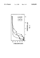
- FIG. 1 is a graph illustrating the erythrocyte toxicity of AmB, SR-E, SS-A, or ST-B as measured by hemoglobin release. Sheep erythrocytes were used and were from a single lot and each point represents the mean of three experiments.
- FIG. 2 is a graph illustrating the erythrocyte toxicity of AmB, SR-E, SS-A, or ST-B as measured by hemoglobin release. Sheep erythrocytes were used and were from different lots and each point represents the mean of three experiments.
- FIG. 3 is a graph illustrating the effect of syringomycin-E on lymphocyte proliferation in the absence of mitogens. The data represent the mean of two different experiments each with triplicate assays.
- FIG. 4 is a graph illustrating the effect of SR-E on mitogen-activated lymphocyte proliferation. Lymphocytes were cultured in the presence of mitogens PHA (10 ⁇ g/well), PWM (20 ⁇ g/well) or anti-CD3 (0.4 ⁇ g/well). The data represent the mean of three experiments each with triplicate assays.
- the present invention is directed to a novel method of combating human and plant fungal pathogens using lipodepsipeptides.
- the present invention is directed to a novel method of suppressing the immune system in mammals using lipodepsipeptides.
- this section is divided into five sub-sections: structure of lipodepsipeptides, antifungal activity of lipodepsipeptides, fungicidal activity of lipodepsipeptides, erythrocyte toxicity of lipodepsipeptides, and immunosuppressive activity.
- CLPs are composed of a peptide moiety and a hydroxylated acyl chain.
- the core structure of some lipodepsipeptides is shown below: ##STR1##
- the peptide is generally nine amino acids and modified amino acids in length. Natural occurring CLPs with less than nine amino acids, such as syringopeptins have been reported and also possess antifungal and fungicidal activity.
- the peptide is comprised of four amino acids which are conserved between CLPs. These are the N-terminal serine and the C-terminal 2,3-dehydroaminobutryl (Dhb)-3-hydroxyl aspartyl (Asp(3-OH))-4-chlorothreonyl (Thr(4-Cl)) residues (shaded above).
- the five amino acids between the N-terminal Ser and the C-terminal tripeptide form the variable region of the peptide moiety.
- these amino acids and modified amino acids may be varied significantly without altering the CLPs antifungal and fungicidal activity.
- the carboxyl of the clorothreonine is covalently bonded to the hydroxyl group of the N-terminal serine to form a macrocyclic ring.
- the N-terminal serine is N-acylated by a long-chain unbranched acyl chain.
- the acyl chain is O-acylated by the C-terminal carboxyl of the acyl chain to form a macrolactone ring.
- the length of the acyl chain can vary without significantly altering the CLPs antifungal and fungicidal activity.
- syringomycin-A 1 , syringomycin-E and syringomycin-G have n values of 6, 8 and 10, respectively.
- the acyl chain is also hydroxylated.
- the hydroxy group is at the C-3 position of the acyl chain.
- CLPs may have multiple hydroxyl groups without losing antifungal and fungicidal activity.
- syringostatin A has a hydrogen at the R position and syringostatin B as a hydroxyl group at the R position.
- three CLPs were tested for antifungal activity.
- the CLPs showed a broad range of antifungal activity against the fungal isolates (Table 1).
- SR-E and SS-A had similar activity profiles (except against Microsporum spp.) and were overall more active than ST-B.
- One strain of C. neoformans did not follow this pattern and was more susceptible to ST-B than either SR-E or SS-A. This strain was very sensitive to ST-B (0.8 g/ml) and somewhat resistant to AmB (1.25 g/ml). This differential sensitivity to ST-B also occurred with C. tropicalis and C. rugosa.
- SR-E, SS-A, and ST-B were more active against yeasts (MICs ranging from 0.8 to 25 g/ml) and were least active against the filamentous fungi, A. fumigatus (5-40 g/ml) and Mucor spp. (6.25-100 g/ml).
- ST-B was not as active against the dermatophytes, Microsporum spp. and Trichophyton spp. (25-200 g/ml).
- three CLPs were tested for fungicidal activity. All three CLPs showed fungicidal activity against most of the organisms tested (Table 2). The MFC values were within two-fold dilutions of the respective MICs, except Mucor spp. AmB, which is known for its fungicidal action, also showed fungicidal activity against most of the strains tested. Ketoconazole, which is not considered fungicidal, showed fungicidal activity only against C. krusei.
- the erythrocyte toxicity of three CLPs was tested. As illustrated in FIG. 1, all three CLPs caused lysis of the erythrocytes and in most cases were more toxic to the erythrocytes than AmB. However, as illustrated in FIG. 2, the relative toxicity varied with different lots of erythrocytes. In all studies, ST-B was the least toxic of the three CLPs. The kinetics of hemolysis differed between the CLPs and AmB.
- lymphocyte stimulation with mitogens is a measure of cell-mediated immunity, and that it can be used to assess the subset of lymphocytes affected.
- Inhibition of PHA- and anti-CD3-induced lymphocyte proliferation suggests suppression of cellular immunity (T cell function).
- Both PHA and anti-CD3 are well known activators of T lymphocytes, although through different mechanisms.
- Anti-CD3 activates T cells via the T cell antigen receptor (TCR/CD3) complex whereas PHA induces the interleukin-2 (IL-2)/IL-2 receptor (IL-2R) complex.
- IL-2 interleukin-2
- IL-2R interleukin-2 receptor
- B cell function humoral immunity
- SR-E at concentrations of 5.72 ⁇ M and lower had no effect in the absence of mitogens based on 3 H!-thymidine incorporation.
- mitogen-activated proliferation was suppressed by SR-E at various concentrations.
- SR-E's degree of inhibition varied according to the mitogen used to induce proliferation. For example, the PWM response was inhibited to a greater degree than the PHA and anti-CD3 responses.
- Lipodepsipeptides for Antifungal Experiments were produced from cultures of Pseudomonas syringae pv. syringae strains B301D, PS268, and SY12, respectively. Strains B301D and PS268 were grown in potato dextrose broth as described by Zhang, L., and J. Y. Takemoto. 1987. Effects of Pseudomonas syringae phytotoxin, syringomycin, on plasma membrane functions of Rhodotorula pilimanae. Phytopathol. 77(2):297-303.
- Strain SY12 was grown in syringomycin minimal medium supplemented with 100M arbutin (Sigma Chemical Co., A 4256; St. Louis, Mo.) and 0.1% fructose (SRMAF) (19, 23).
- SR-E, ST-B, and SS-A were purified by high performance liquid chromatography as described previously by Bidwai, A. P., and J. Y. Takemoto. 1987.
- Bacterial phytotoxin, syringomycin induces a protein kinase-mediated phosphorylation of red beet plasma membrane polypeptides. Proc. Natl. Acad. Sci. USA. 84:6755-6759.
- Solubilized AmB containing 35% sodium deoxycholate (Sigma Chemical Co., A 9528; St. Louis, Mo.) and ketoconazole (Sigma Chemical Co., K-1003; St. Louis, Mo.) were used as test standards.
- RPMI 1640 (RPMI) medium with L-glutamine and without sodium bicarbonate (Sigma Chemical Co., R-6504; St. Louis, Mo.) buffered with 0.165M MOPS (34.54 g/liter) was used for in vitro antifungal tests.
- the medium was adjusted to pH 7.0 with 10M NaOH and filter sterilized.
- Lipodepsipeptide Dilutions All stock drug solution concentrations were at least 10-fold higher than the highest concentration tested. AmB, ketoconazole, and the test compounds (SR-E, ST-B, and SS-A) were dissolved in sterile distilled water, 0.2N HCl, and 0.001N HCl, respectively. Each stock solution was diluted to 2 ⁇ the highest concentration of drug tested with RPMI. Two-fold dilutions were made with RPMI medium.
- the turbidity of each mixed suspension was measured at 530 nm and adjusted to 1 ⁇ 10 6 to 5 ⁇ 10 6 CFU/ml by the NCCLS spectrophotometric method described by National Committee for Clinical Laboratory Standards. 1992. Reference method for broth dilution antifungal susceptibility testing of yeasts; proposed standard, NCCLS document M27-P (ISBN 1-56238-186-5). NCCLS, Villanova. The final transmission of each yeast suspension ranged from 85-87%.
- Aspergillus fumigatus and Mucor spp. were grown for 1 wk at 35° C.
- the dermatophytes, Microsporum spp. and Trichophyton spp. were grown for two wk at 30° C.
- Fungal spores were washed from the plates of all good spore formers (A. fumigatus, Mucor spp., Microsporum canis and Trichophyton spp.) by placing 5 ml of 0.85% saline with 0.2% Tween 80 on the plates and mixing with an inoculating loop.
- the spore/mycelium suspension was drawn off with a pipette and allowed to settle for at least 5 min.
- the procedure was the same except the spore/mycelial suspension was homogenized with a ground glass tissue homogenizer and then allowed to settle for at least 5 min. A small amount of the supernatant was transferred to 5 ml of sterile saline and vortexed. The stock suspensions of all filamentous fungi were measured at 530 nm and adjusted to 85-87% transmission. The concentration (1 ⁇ 10 6 to 5 ⁇ 10 6 CFU/ml) was determined by hemacytometer count or plate count on SDA. All stock suspensions were diluted 1000-fold with RPMI, or 2 ⁇ the final desired test inocula. The 2 ⁇ inoculum was diluted 1:1 when the wells were inoculated, and the desired inoculum size was achieved.
- Broth Microdilution Test Broth microdilution tests were performed using sterile, disposable, multiwell microdilution plates (96 round U-bottom wells) (Falcon 3077; Becton and Dickinson Labware, Lincoln Park, N.J.). The drugs (2 ⁇ concentrations) were dispensed into the wells of rows number 1 to 10 of the microdilution plates in 100 ⁇ l volumes from highest drug concentration to lowest drug concentration. Each well was inoculated with 100 ⁇ l of the corresponding 2 ⁇ concentrated fungal suspension. The wells of row 11 contained the inoculum with drug-free media as a positive growth control and the wells of row 12 contained uninoculated-drug-free medium as a sterility control.
- Incubation and Scoring of MIC All cultures were incubated without shaking at the temperature used during subculture. Incubation times were 48 hr for Candida spp., S. cerevisiae, A. fumigatus, and Mucor spp; 72 hr for C. neoformans; and 7 days for Microsporum and Trichophyton spp.
- the microdilution wells were scored from 0, no growth; 1, slightly hazy; 2, prominent decrease in turbidity; 3, slight reduction in turbidity; and 4, no reduction in turbidity when compared to the growth control (drug-free) well.
- the MICs for SR-E, ST-B, SS-A, and AmB were defined as the lowest concentrations at which scores of 0 were observed.
- the MIC for ketoconazole was described as the lowest concentrations at which a score of 2 was observed.
- MFC Minimum Fungicidal Concentration
- Erythrocyte Toxicity Sheep red blood cell (RBC) hemolysis was used to assess erythrocyte toxicity of the CLPs and AmB.
- RBCs (MicroBio Products, Inc., Salt Lake City, Utah) were washed four times with phosphate-buffered-saline (PBS) by centrifugation at 800 ⁇ g for 10 min and adjusted to 1 ⁇ 10 8 cells/ml as previously described by Chavanet, P., V. Joly, D. Rigaud, J. Bolard, C. Carbon, and P. Yeni. 1994. Influence of diet on experimental toxicity of amphotericin B deoxycholate. Antimicrob. Agents Chemother. 38(5):963-968.
- RBCs and a 2 ⁇ concentration of SR-E, ST-B or SS-A in PBS were mixed in a 1:1 ratio and incubated at 37° C. for 1 hr. After incubation, cells were pelleted by centrifugation at 800 ⁇ g for 10 min and the supernatant was collected and the absorbance at 550 nm was determined. To verify that the compound did not affect the absorbance reading, the pellet was washed with PBS, lysed with distilled water, and the supernatant absorbance determined after centrifugation. Distilled water and PBS were used as lysis and hemoglobin retention controls, respectfully, as described by National Committee for Clinical Laboratory Standards. 1992. Reference method for broth dilution antifungal susceptibility testing of yeasts; proposed standard, NCCLS document M27-P (ISBN 1-56238-186-5). NCCLS, Villanova.
- SR-E Lipodepsipeptide for Immunosuppression Experiments: SR-E was purified as previously described in Bidwai, A. P., Bachmann, R. C. and Takemoto, J. Y., "Mechanism of action of Pseudomonas syringae phytotoxin syringomycin". Plant Physiol. 83:39-43 (1987). A stock solution (10 mg per ml) was prepared in RPMI1640 culture media, aliquoted and stored frozen at -20°. SR-E was thawed on ice, diluted appropriately and used fresh in lymphocyte cultures at the concentrated indicated in FIGS. 3 and 4.
- Lymphocyte stimulation was performed with three different mitogens: phytohemagglutinin (PHA), pokeweed mitogen (PWM), and monoclonal antibody to CD3 (anti-CD3).
- PHA phytohemagglutinin
- PWM pokeweed mitogen
- anti-CD3 monoclonal antibody to CD3
- Mitogen-induced lymphocyte proliferation Peripheral blood mononuclear cells (MNC) (containing lymphocytes and monocytes) were separated by Ficoll-Hypaque density-gradient centrifugation using techniques well known in the art. Mitogen-induced lymphocyte proliferation was determined as previously described in Singh, V. K., "Stimulatory effect of corticotropin-releasing neurohormone on human lymphocyte proliferation and interleukin-2 receptor expression". J.Neuroimmunol.23.257-262 (1989). Briefly, MNC were cultured in triplicate in 96-well microtiter plates (Corning, Inc.).
- the cultures (final volume of 200 ⁇ l) contained 10 5 cells, various mitogens, and different concentrations of SR-E in RPMI1640 containing fetal bovine serum at a final concentration of 5% (V/V).
- the blank cultures did not contain mitogen or SR-E. Cultures were incubated for 4 days in a humidified chamber with 5% CO 2 at 37° C.
- strains of Pseudomonas syringae pv. syringae produce CLPs with antifungal activity.
- In vitro antifungal and fungicidal activities of three CLPs syringomycin E, syringotoxin B, and syringostatin A were evaluated using a standard broth microdilution susceptibility method against medically important isolates. Erythrocyte toxicity was also evaluated. All three compounds showed broad antifungal activity and fungicidal action against most of the fungi tested.
- the cyclic lipodepspeptides were more effective against yeasts than against the filamentous fungi.
- Syringomycin E and syringostatin A had very similar antifungal activity (2.5->40 g/ml) and erythrocyte toxicity.
- Syringotoxin B was generally less active (0.8-200 g/ml) than syringomycin E and syringostatin A against most fungi and less toxic to erythrocytes.
- CLPs displayed significant immunosuppressive activity.
- One CLP, syringomycin-E had no effect on 3 H!-thymadine incorporation in the absence of mitogens.
- syringomycin-E significantly inhibited 3 H!-thymadine incorporation in mitogen-induced lymphocyte.
- Syringomycin-E's suppressive effect was more pronounced with pokeweed mitogen (PWM) as compared to phytohemagglutinin (PHA) or monoclonal antibody to CD3 (anti-CD3). Given the mechanism of action of these mitogens, syringomycin-E suppresses both cellular and humoral immune responses.
- PWM pokeweed mitogen
- PHA phytohemagglutinin
- anti-CD3 monoclonal antibody to CD3
Landscapes
- Health & Medical Sciences (AREA)
- Life Sciences & Earth Sciences (AREA)
- Chemical & Material Sciences (AREA)
- Medicinal Chemistry (AREA)
- Proteomics, Peptides & Aminoacids (AREA)
- General Health & Medical Sciences (AREA)
- Organic Chemistry (AREA)
- Epidemiology (AREA)
- Veterinary Medicine (AREA)
- Public Health (AREA)
- Animal Behavior & Ethology (AREA)
- Gastroenterology & Hepatology (AREA)
- Engineering & Computer Science (AREA)
- Bioinformatics & Cheminformatics (AREA)
- Immunology (AREA)
- Pharmacology & Pharmacy (AREA)
- Genetics & Genomics (AREA)
- Biochemistry (AREA)
- Biophysics (AREA)
- Molecular Biology (AREA)
- Peptides Or Proteins (AREA)
- Medicines That Contain Protein Lipid Enzymes And Other Medicines (AREA)
Abstract
Lipodepsipeptides from Pseudomonas syringae pv. syringae were evaluated for antifungal activity. Specifically, the in vitro antifungal and fungicidal activities of three cyclic lipodepsinonapeptides syringomycin E, syringotoxin B, and syringostatin A against medically important isolates were evaluated using a standard broth microdilution susceptibility method. Erythrocyte toxicity was also evaluated. All three compounds showed broad antifungal activity and fungicidal action against most of the fungi tested. In addition, the present invention relates to a novel method for suppressing the immune system of mammals using cyclic lipodepsipeptides. The ability of cyclic lipodepsipeptides to suppress the immune system was evaluated in mitogen-induced lymphocytes. One cyclic lipodepsipeptide, syringomycin-E significantly inhibited mitogen--induced lymphocytes stimulation by pokeweed mitogen, phytohemagglutinin, and monoclonal antibodies to CD3. These results indicate that lipodepsipeptides are useful at suppressing both cellular and humoral immune responses.
Description
This application is related to United States Provisional patent application Ser. No. 60/003,604, filed Sep. 12, 1995.
1. The Field of the Invention
The present invention is related to peptides from the plant bacterium Pseudomonas syringae pv. syringae with antifungal and immunosuppressive activity. More specifically, the present invention relates to the use of lipodepsipeptides, and derivative of lipodepsipeptides, as antifungal and immunosuppressive agents.
2. Technical Background
Fungi are eukaryotic organisms which are wide spread in nature and grow well in a broad range of environmental conditions. The term fungi generally includes mushrooms, puffballs, woody bracket fungi, molds and yeast. Thus, fungi may be single-celled or multicellular organisms.
Fungi are heterotrophic because they do not contain chlorophyl. Fungi depend on organic products as a source of energy. Many fungi actively produce enzymes which enable them to break down complex substances for energy. Production of these enzymes has numerous beneficial effects, including recycling products in the soil thus enabling some plants to obtain minerals. Furthermore, fungi play a critical role in the processing of many foods, including leavening bread and fermenting beer, wine and cheeses. In addition, they produce antibiotics, such as penicillin, and are a valuable food source, as in the case of mushrooms.
These enzymes, however, can also have undesirable effects on plants and animals, including human. Each year, millions of dollars worth of fruit and vegetables are lost due to fungal damage. Fungi such as Byssochlamys fulva, Aspergillus ssp., Mucor ssp., and Saccharomyces cerevisiae are responsible for spoiling canned foods, fresh fruits and jams, breads, fish, and meats. Traditionally, chemical additives such as sorbic acid and sodium propionate have been used to control fugal growth on food products or to control contamination of equipment used to handle food products.
Other types of fungi produce poisons known as mycotoxins which are very toxic to many animal species and historically have had devastating effects on fish, birds and humans. Most fungal infections, are not life-threatening. Recently, however, fungal infections once dismissed as a nuisance have become a major health concern. Opportunistic fungal infections are increasingly important causes of morbidity and mortality in hospitalized patients. This is especially true for patients with AIDS and other immunocompromised conditions, those receiving broad-spectrum antibiotics, cytotoxic therapy, immunosuppressants or chemotherapy, and patients with intravascular catheters.
Efforts to combat these infections are hampered by lack of fungal specific drugs, increasing drug resistance, and a growing list of pathogens. Because both fungi and higher organisms are eukaryotic, and thus share many biological processes, developing drugs that are specific to fungi has been extremely difficult. The most important antifungal agents are the polyenes such as amphotericin B (AmB), imidazoles such as triazoles, and RNA and DNA inhibitors such as flucytosine.
These agents have several shortcomings. Most notably, fungi have developed resistance against many polyenes, imidazoles and RNA and DNA inhibitors. Moreover, these agents produce numerous side effects. Amphotericin B when used to treat systemic Candidiasis, for example, may cause kidney damage, convulsions, hypotension, nausea, vomiting, abdominal pain, cardiac arrest and anemia. Accordingly, increases in the incidence of fungal infections have prompted a search for new antifungal agents with broad antifungal and, fungicidal activity, and minimal plant and animal toxicity.
At the same time, there is a need in the art for immunosuppressants. Immunosuppressants are particularly important in tissue and organ grafts, cross-species transplantations, blood transfusions, and bone marrow transplants. One area of particular concern is donors and recipients with different histocompatibility. When the donor's mature immunocompetent T cells are transfused, the allogeneic recipient is unable to identify and reject the donor's T cells. As a result, the donor's T cells react against the tissues of the host in a graft-versus-host response. This response triggers many reactions in the host, including skin, gut and liver damage which often leads to death. Therefore, it is critical to the art of organ and tissue transplantation to have effective immunosuppressant agents, which reduce organ and tissue rejection.
Traditionally, X-rays were used to suppress the immune response to foreign organs and tissues. Currently, there are several immunosuppressive drugs available. These include, for example, cyclosporin A , FK506, and rapamycin. Structures and a brief description of the agents are described in Kunz, et al. "Cyclosporin A, FK506, and rapamycin: more than just immunosuppression" which is hereby incorporated by reference. Advantageously, cyclosporin A, FK506, and rapamycin are also antifungal agents. Thus, these drugs simultaneously combat opportunistic fungal infection while the host's immune system is being suppressed.
One concern in the art is that cyclosporin A, FK506, and rapamycin all are structurally and chemically related. It is possible, therefore, that a given host or fungi could become resistant to the immunosuppressive effects or antifungal activity of these agents. Given the rapid development of organ and tissue transplants and the risk for opportunistic mycosis infection, it would be advantageous to the art to have immunosuppressants that are chemically and structurally distinct, and hence are more likely to be effective against opportunistic fungi that are resistant to current immunosuppressants.
From the foregoing, it will be appreciated that it would be an advancement in the art to provide methods for combating cutaneous and invasive fungal infections in mammals. It would be a further advancement in the art if the methods were effective against a broad range of fungi, including those pathogenic to humans. It would be yet another advancement in the art to provide methods for preserving food, and protecting crops and vegetation from fungal pathogens. It would also be an advancement in the art to provide methods for suppressing the immune system of mammals. Finally, it would be an advancement in the art if the immunosuppressive agent was also an antifungal and fungicidal agent and thus could simultaneously combat opportunistic infections during the time when the immune system is suppressed.
Such methods are disclosed and claimed herein.
The medical profession is currently facing a growing epidemic of opportunistic fungal infection, particularly among persons with immunocompromised conditions, those receiving broad-spectrum antibiotics, cytotoxic therapy, or chemotherapy and patients with intravascular catheters. Fungi are often classified into subgroups based on their method of reproduction, mycelial formation, cellular structure and formation, and biochemical and physical properties. The term fungi as used herein encompasses all forms of fungi including mushrooms, puffballs, woody bracket fungi, molds and yeast. Many fungi are becoming resistant to conventional antifungal and fungicidal agents. Therefore, there is a critical need in the art for new antifungal and fungicidal agents that to combat mycoses infections.
A class of molecules referred to as cyclic lipodepsipeptides (CLPs) possess antifungal and fungicidal activity. CLPs are naturally produced by certain bacteria such as Pseudomonas syringae. While the activity of CLPs has been known for over a decade, scientists in the field believed the bacteria that produced these toxins were pathovars (pathogenic to plants) and that CLPs were phytotoxins (toxic to plants). As a result, the use of CLPs as antifungal and fungicidal agents in plants and animals was not contemplated by scientist in the field. Like other bacterial endotoxins and exotoxins which cause diphtheria, botulism and cholera, CLPs were thought to be highly toxic and therefore useless as antifungal or fungicidal agents. Moreover, the mechanism of action of CLPs is similar to amphotericin B. Both compounds disrupt the permeability of cell membranes. Given the similarity in the two compounds' mechanisms of action, CLPs were not expected to be useful against fungi that had become resistant to amphotericin B and other agents which similarly disrupt fungi membrane.
It has been discovered that CLP are not phytotoxins as once believed. In fact, CLP producing bacteria protect plants from fungal infection and disease. Furthermore, in spite of the similarities between the biochemistry of CLPs and current antifungal and fungicidal agents, CLPs are effective against fungal strains which are resistant to current agents such as amphotericin B. Accordingly, the present invention is directed to a novel method for treating cutaneous and invasive fungal disease in mammals using CLPs. In addition, because CLPs are not phytotoxins, CLPs may be used to preserve food and protect crops and vegetation from fungal pathogens.
In one embodiment, three CLPs were tested for antifungal and fungicidal activity. Syringomycin-E (SR-E), syringostatin-B (ST-B), and syringostatin-A (S S-A ) from Pseudomonas syringae pv. syringae all displayed fungicidal activities. There was some variability in susceptibility between fungal species. All three CLPs were more active against yeasts than against filamentous fungi. This difference could be due to differences in membrane sterol and phospholipid composition of yeasts and filamentous fungi. Sterols and phospholipids are important for SR-E action. Although inhibited by all three CLPs, C. neoformans was particularly sensitive to ST-B.
Besides their antifungal properties, like other antifungal agents, CLPs were lytic to erythrocytes. The lytic activity profile of the three CLPs paralleled their antifungal activities. SR-E and SS-A were more active than ST-B. Conceivably, the more positive net charge of SR-E and SS-A imparted by three basic amino acids verses two of ST-B could account for this difference and also ST-B's higher fungicidal activity against C. neoformans.
A significant finding was that AmB-resistant C. rugosa was sensitive to CLPs. This is likely due to differences in mechanism of action between AmB and CLPs though, as discussed above, both agents bind membrane sterols and perturb membrane function. Chemical differences between the two classes of compounds probably account for their distinctive actions on membranes. For example, CLPs are water-soluble whereas AmB, a cyclic polyene, is significantly hydrophobic. In short, P. syringae pv. syringae CLPs are effective antifungal and fungicidal agents. They are fungicidal against important human pathogenic yeasts, water-soluble, and have unique mechanisms of action.
In addition, CLPs also possess significant immunosuppressive properties. In one embodiment, the ability of syringomycin-E to inhibit mitogen-induced lymphocyte proliferation was tested. Based on 3 H!-thymidine incorporation, SR-E had no effect on cell proliferation in the absence of mitogens. However, lymphocyte proliferation induced by pokeweed mitogen (PWM), phytohemagglutinin (PHA) and monoclonal antibody to CD3 (anti-CD3) was significantly suppressed in the presence of SR-E. SR-E's suppressive effect was more pronounced with PWM as compared with PHA or anti-CD3.
These and other objects and advantages of the present invention will become apparent upon reference to the accompanying drawings and graphs and upon reading the following detailed description and appended claims.
A more particular description of the invention briefly described above will be rendered by reference to the appended drawings and graphs. These drawings and graphs only provide information concerning typical embodiments of the invention and are not to be considered limiting of its scope.
FIG. 1 is a graph illustrating the erythrocyte toxicity of AmB, SR-E, SS-A, or ST-B as measured by hemoglobin release. Sheep erythrocytes were used and were from a single lot and each point represents the mean of three experiments.
FIG. 2 is a graph illustrating the erythrocyte toxicity of AmB, SR-E, SS-A, or ST-B as measured by hemoglobin release. Sheep erythrocytes were used and were from different lots and each point represents the mean of three experiments.
FIG. 3 is a graph illustrating the effect of syringomycin-E on lymphocyte proliferation in the absence of mitogens. The data represent the mean of two different experiments each with triplicate assays.
FIG. 4 is a graph illustrating the effect of SR-E on mitogen-activated lymphocyte proliferation. Lymphocytes were cultured in the presence of mitogens PHA (10 μg/well), PWM (20 μg/well) or anti-CD3 (0.4 μg/well). The data represent the mean of three experiments each with triplicate assays.
The present invention is directed to a novel method of combating human and plant fungal pathogens using lipodepsipeptides. In addition, the present invention is directed to a novel method of suppressing the immune system in mammals using lipodepsipeptides. To better understand the details of the present invention, this section is divided into five sub-sections: structure of lipodepsipeptides, antifungal activity of lipodepsipeptides, fungicidal activity of lipodepsipeptides, erythrocyte toxicity of lipodepsipeptides, and immunosuppressive activity.
5.1. Structure of Lipodepsipeptides
CLPs are composed of a peptide moiety and a hydroxylated acyl chain. The core structure of some lipodepsipeptides is shown below: ##STR1##
As illustrated above, the peptide is generally nine amino acids and modified amino acids in length. Natural occurring CLPs with less than nine amino acids, such as syringopeptins have been reported and also possess antifungal and fungicidal activity. The peptide is comprised of four amino acids which are conserved between CLPs. These are the N-terminal serine and the C-terminal 2,3-dehydroaminobutryl (Dhb)-3-hydroxyl aspartyl (Asp(3-OH))-4-chlorothreonyl (Thr(4-Cl)) residues (shaded above). The five amino acids between the N-terminal Ser and the C-terminal tripeptide form the variable region of the peptide moiety. As illustrated by the chemical structures above, these amino acids and modified amino acids may be varied significantly without altering the CLPs antifungal and fungicidal activity. The carboxyl of the clorothreonine is covalently bonded to the hydroxyl group of the N-terminal serine to form a macrocyclic ring.
The N-terminal serine is N-acylated by a long-chain unbranched acyl chain. The acyl chain is O-acylated by the C-terminal carboxyl of the acyl chain to form a macrolactone ring. It will be appreciated by one skilled in the art that the length of the acyl chain can vary without significantly altering the CLPs antifungal and fungicidal activity. For example, in the core structure of syringomycin above, syringomycin-A1, syringomycin-E and syringomycin-G have n values of 6, 8 and 10, respectively.
The acyl chain is also hydroxylated. In syringomycin and syringotoxin, the hydroxy group is at the C-3 position of the acyl chain. However, CLPs may have multiple hydroxyl groups without losing antifungal and fungicidal activity. For example, in the core structure of syringostatin above, syringostatin A has a hydrogen at the R position and syringostatin B as a hydroxyl group at the R position.
5.2 Antifungal Activity of Lipodepsipeptides
In one embodiment, three CLPs were tested for antifungal activity. The CLPs showed a broad range of antifungal activity against the fungal isolates (Table 1). SR-E and SS-A had similar activity profiles (except against Microsporum spp.) and were overall more active than ST-B. One strain of C. neoformans did not follow this pattern and was more susceptible to ST-B than either SR-E or SS-A. This strain was very sensitive to ST-B (0.8 g/ml) and somewhat resistant to AmB (1.25 g/ml). This differential sensitivity to ST-B also occurred with C. tropicalis and C. rugosa. One strain of C. tropicalis and one strain of C. rugosa showed resistance to AmB, but were still sensitive to the CLPs. SR-E, SS-A, and ST-B were more active against yeasts (MICs ranging from 0.8 to 25 g/ml) and were least active against the filamentous fungi, A. fumigatus (5-40 g/ml) and Mucor spp. (6.25-100 g/ml). In addition, ST-B was not as active against the dermatophytes, Microsporum spp. and Trichophyton spp. (25-200 g/ml). The control organism, S. cerevisiae ATCC 36375, gave expected MIC values for AmB and ketoconazole. AmB and ketoconazole, which were used as test standards, had MICs that were within, or close to, the range of the expected values for the clinical isolates (20, 25). These clinical isolates showed a wide range of susceptibility to AmB (0.02-1.25 g/ml) and ketoconazole (0.02->10 g/ml). MICs for AmB and ketoconazole were generally lower than for the CLPs. One strain of C. albicans showed resistance (MIC=10) to ketoconazole, but was sensitive to the other compounds.
                                  TABLE 1
__________________________________________________________________________
            No. of
                MFC (μg/ml) range
ORGANISM    Isolates
                SR-E ST-B SS-A
                              AmB  Ktz
__________________________________________________________________________
Candida albicans
            (20)
                2.5-5
                     3.2-12.5
                          2.5-5
                              0.04-0.3
                                   0.02-10
Candida kefyr
            (1) 2.5  3.2  2.5 0.3  0.02
Candida krusei
            (2) 10   12.5-25
                          10  0.3-0.6
                                   0.15
Candida lusitaniae
            (2) 2.5  6.25 5   0.3  0.02
Candida parapsilosis
            (2) 2.5  6.25-12.5
                          2.5-5
                              0.6  0.02
Candida rugosa
            (2) 5-20 3.2-25
                          10-20
                              0.3-1.25
                                   0.02
Candida tropicalis
            (2) 2.5-5
                     3.2  2.5-5
                              0.3-1.25
                                   0.08-0.6
Cryptococcus neoformans
            (14)
                2.5-10
                     0.8-6.25
                          2.5-10
                              0.08-1.25
                                   0.04-0.6
Saccharomyces cerevisiae
            (1).sup.a
                2.5  6.25 2.5 0.3  0.15
Aspergillus fumigatus
            (16)
                10-20
                     6.25-25
                          5-40
                              0.15-1.25
                                   0.3->10
Mucor spp.  (5) 10->40
                     6.25-100
                          10->40
                              0.02-0.15
                                   0.6->10
Microsporum spp.
            (2) 6.25-12.5
                     25-200
                          2.5-5
                              0.04-0.3
                                   0.8-1.6
Trichophyton spp.
            (3) 3.1-6.25
                     25-200
                          2.5-5
                              0.3-0.6
                                   0.4-3.1
__________________________________________________________________________
 .sup.a Strain was tested more than once on different days.
5.3 Fungicidal Activity of Lipodepsipeptides
In one embodiment, three CLPs were tested for fungicidal activity. All three CLPs showed fungicidal activity against most of the organisms tested (Table 2). The MFC values were within two-fold dilutions of the respective MICs, except Mucor spp. AmB, which is known for its fungicidal action, also showed fungicidal activity against most of the strains tested. Ketoconazole, which is not considered fungicidal, showed fungicidal activity only against C. krusei.
                                  TABLE 2
__________________________________________________________________________
            No. of
                MFC (μg/ml) range
ORGANISM    Isolates
                SR-E ST-B SS-A
                              AmB  Ktz
__________________________________________________________________________
Candida albicans
            (20)
                2.5-5
                     3.2-12.5
                          2.5-10
                              015-0.3
                                   0.3->10
Candida kefyr
            (1) 2.5  3.2  2.5 0.3  1.25
Candida krusei
            (2) 10   12.5-50
                          10-20
                              0.6  0.15
Candida lusitaniae
            (2) 2.5-5
                     6.25-12.5
                          5   0.6  ≦0.02
Candida parapsilosis
            (2) 2.5  12.5-25
                          2.5-10
                              1.25 0.04
Candida rugosa
            (2) 10-20
                     6.25-50
                          10->20
                              0.6-2.5
                                   ≦0.02-0.08
Candida tropicalis
            (2) 5    12.5 5   0.6-1.25
                                   2.5-10
Cryptococcus neoformans
            (14)
                2.5-10
                     0.8-12.5
                          2.5-10
                              0.15-1.25
                                   0.08->10
Saccharomyces cerevisiae
            (1).sup.a
                2.5  12.5-25
                          5   1.25 0.6-1.25
Aspergillus fumigatus
            (16)
                10->20
                     12.5->50
                          5-40
                              0.6-2.5
                                   2.5->10
Mucor spp.  (5) 20->40
                     25->100
                          40->40
                              ≦0.02-0.3
                                   2.5->10
Microsporum spp.
            (2) 6.25-12.5
                     25-200
                          5   0.08-0.3
                                   25->25
Trichophyton spp.
            (3) 6.25-25
                     25-200
                          5   0.3-0.6
                                   12.5->25
__________________________________________________________________________
 .sup.a Strain was tested more than once on different days.
5.4 Erythrocyte Toxicity of Lipodepsipeptides
In one embodiment, the erythrocyte toxicity of three CLPs was tested. As illustrated in FIG. 1, all three CLPs caused lysis of the erythrocytes and in most cases were more toxic to the erythrocytes than AmB. However, as illustrated in FIG. 2, the relative toxicity varied with different lots of erythrocytes. In all studies, ST-B was the least toxic of the three CLPs. The kinetics of hemolysis differed between the CLPs and AmB.
5.5 Immunosuppressive Activity
It is well known in the art that lymphocyte stimulation with mitogens is a measure of cell-mediated immunity, and that it can be used to assess the subset of lymphocytes affected. Inhibition of PHA- and anti-CD3-induced lymphocyte proliferation suggests suppression of cellular immunity (T cell function). Both PHA and anti-CD3 are well known activators of T lymphocytes, although through different mechanisms. Anti-CD3 activates T cells via the T cell antigen receptor (TCR/CD3) complex whereas PHA induces the interleukin-2 (IL-2)/IL-2 receptor (IL-2R) complex. In addition, because PWM is a polyclonal activator of T cell-dependent antibody production by B cells, inhibition of PWM-induced proliferation implies suppression of humoral immunity (B cell function). Accordingly, in one embodiment the ability of SR-E to suppress PHA, PWM and anti-CD3-induced lymphocyte proliferation was tested.
As expected, PHA, PWM and anti-CD3 each stimulated lymphocyte proliferation several fold above the baseline control (no mitogen). As illustrated in FIG. 3, SR-E at concentrations of 5.72 μM and lower had no effect in the absence of mitogens based on 3 H!-thymidine incorporation. However, as illustrated in FIG. 4, mitogen-activated proliferation was suppressed by SR-E at various concentrations. SR-E's degree of inhibition varied according to the mitogen used to induce proliferation. For example, the PWM response was inhibited to a greater degree than the PHA and anti-CD3 responses. At a concentration of 5.72 μM, SR-E suppressed PWM response by 60% (p=0.017), PHA response by 25% (p=0.026) and anti-CD3 response by 30% (p=0.02). Moreover, SR-E at a concentration of 1.14 μM significantly inhibited the PWM response but not PHA and anti-CD3 responses.
The following examples are given to illustrate various embodiments which have been made with the present invention. It is to be understood that the following examples are not comprehensive or exhaustive of the many types of embodiments which can be prepared in accordance with the present invention.
Lipodepsipeptides for Antifungal Experiments: SR-E, ST-B, and SS-A were produced from cultures of Pseudomonas syringae pv. syringae strains B301D, PS268, and SY12, respectively. Strains B301D and PS268 were grown in potato dextrose broth as described by Zhang, L., and J. Y. Takemoto. 1987. Effects of Pseudomonas syringae phytotoxin, syringomycin, on plasma membrane functions of Rhodotorula pilimanae. Phytopathol. 77(2):297-303. Strain SY12 was grown in syringomycin minimal medium supplemented with 100M arbutin (Sigma Chemical Co., A 4256; St. Louis, Mo.) and 0.1% fructose (SRMAF) (19, 23). SR-E, ST-B, and SS-A were purified by high performance liquid chromatography as described previously by Bidwai, A. P., and J. Y. Takemoto. 1987. Bacterial phytotoxin, syringomycin, induces a protein kinase-mediated phosphorylation of red beet plasma membrane polypeptides. Proc. Natl. Acad. Sci. USA. 84:6755-6759. Solubilized AmB containing 35% sodium deoxycholate (Sigma Chemical Co., A 9528; St. Louis, Mo.) and ketoconazole (Sigma Chemical Co., K-1003; St. Louis, Mo.) were used as test standards.
Most of the fungal strains used in the tests were clinical isolates obtained from the University of Texas Health Science Center at San Antonio Fungus Testing Laboratory (San Antonio, Tex.) and the remaining isolates were ATCC strains.
Medium: Liquid RPMI 1640 (RPMI) medium with L-glutamine and without sodium bicarbonate (Sigma Chemical Co., R-6504; St. Louis, Mo.) buffered with 0.165M MOPS (34.54 g/liter) was used for in vitro antifungal tests. The medium was adjusted to pH 7.0 with 10M NaOH and filter sterilized.
Lipodepsipeptide Dilutions: All stock drug solution concentrations were at least 10-fold higher than the highest concentration tested. AmB, ketoconazole, and the test compounds (SR-E, ST-B, and SS-A) were dissolved in sterile distilled water, 0.2N HCl, and 0.001N HCl, respectively. Each stock solution was diluted to 2× the highest concentration of drug tested with RPMI. Two-fold dilutions were made with RPMI medium.
Preparation of Inoculum Suspensions: On Sabouraud dextose agar (SDA; BBL, Becton and Dickinson and Co., Cockeysville, Md.) all isolates were subcultured at least twice before use to ensure purity and viability. Candida spp. and Saccharomyces cerevisiae were grown for 24 hr at 35° C. Isolates of Cryptococcus neoformans were grown for 48 hr at 35° C. Yeast cells from at least five 1-mm-diameter colonies were suspended in 5 ml of sterile 0.85% saline. The resulting suspension was vortexed for 15 sec. The turbidity of each mixed suspension was measured at 530 nm and adjusted to 1×10⁶ to 5×10⁶ CFU/ml by the NCCLS spectrophotometric method described by National Committee for Clinical Laboratory Standards. 1992. Reference method for broth dilution antifungal susceptibility testing of yeasts; proposed standard, NCCLS document M27-P (ISBN 1-56238-186-5). NCCLS, Villanova. The final transmission of each yeast suspension ranged from 85-87%.
Aspergillus fumigatus and Mucor spp. were grown for 1 wk at 35° C. The dermatophytes, Microsporum spp. and Trichophyton spp., were grown for two wk at 30° C. Fungal spores were washed from the plates of all good spore formers (A. fumigatus, Mucor spp., Microsporum canis and Trichophyton spp.) by placing 5 ml of 0.85% saline with 0.2% Tween 80 on the plates and mixing with an inoculating loop. The spore/mycelium suspension was drawn off with a pipette and allowed to settle for at least 5 min. For the poor spore-forming Microsporum audouinii, the procedure was the same except the spore/mycelial suspension was homogenized with a ground glass tissue homogenizer and then allowed to settle for at least 5 min. A small amount of the supernatant was transferred to 5 ml of sterile saline and vortexed. The stock suspensions of all filamentous fungi were measured at 530 nm and adjusted to 85-87% transmission. The concentration (1×10⁶ to 5×10⁶ CFU/ml) was determined by hemacytometer count or plate count on SDA. All stock suspensions were diluted 1000-fold with RPMI, or 2× the final desired test inocula. The 2× inoculum was diluted 1:1 when the wells were inoculated, and the desired inoculum size was achieved.
Broth Microdilution Test: Broth microdilution tests were performed using sterile, disposable, multiwell microdilution plates (96 round U-bottom wells) (Falcon 3077; Becton and Dickinson Labware, Lincoln Park, N.J.). The drugs (2× concentrations) were dispensed into the wells of rows number 1 to 10 of the microdilution plates in 100 μl volumes from highest drug concentration to lowest drug concentration. Each well was inoculated with 100 μl of the corresponding 2× concentrated fungal suspension. The wells of row 11 contained the inoculum with drug-free media as a positive growth control and the wells of row 12 contained uninoculated-drug-free medium as a sterility control.
Incubation and Scoring of MIC: All cultures were incubated without shaking at the temperature used during subculture. Incubation times were 48 hr for Candida spp., S. cerevisiae, A. fumigatus, and Mucor spp; 72 hr for C. neoformans; and 7 days for Microsporum and Trichophyton spp. The microdilution wells were scored from 0, no growth; 1, slightly hazy; 2, prominent decrease in turbidity; 3, slight reduction in turbidity; and 4, no reduction in turbidity when compared to the growth control (drug-free) well. The MICs for SR-E, ST-B, SS-A, and AmB were defined as the lowest concentrations at which scores of 0 were observed. The MIC for ketoconazole was described as the lowest concentrations at which a score of 2 was observed.
Minimum Fungicidal Concentration (MFC): MFC values were determined by subculturing 10 μl from each well with a drug concentration higher than the MIC, equal to the MIC, and one well lower than the MIC on drug-free SDA when the MICs were read. Incubation temperatures were the same as the MICs. Incubation times were 24 hr for Candida spp., S. cerevisiae, and Mucor spp; 48 hr for C. neoformans and A. fumigatus; and 7 d for Microsporum and Trichophyton spp. The MFC was the lowest concentration with three or fewer colonies/plate for the yeasts and no growth for the filamentous fungi.
Erythrocyte Toxicity: Sheep red blood cell (RBC) hemolysis was used to assess erythrocyte toxicity of the CLPs and AmB. RBCs (MicroBio Products, Inc., Salt Lake City, Utah) were washed four times with phosphate-buffered-saline (PBS) by centrifugation at 800× g for 10 min and adjusted to 1×10⁸ cells/ml as previously described by Chavanet, P., V. Joly, D. Rigaud, J. Bolard, C. Carbon, and P. Yeni. 1994. Influence of diet on experimental toxicity of amphotericin B deoxycholate. Antimicrob. Agents Chemother. 38(5):963-968. RBCs and a 2× concentration of SR-E, ST-B or SS-A in PBS were mixed in a 1:1 ratio and incubated at 37° C. for 1 hr. After incubation, cells were pelleted by centrifugation at 800× g for 10 min and the supernatant was collected and the absorbance at 550 nm was determined. To verify that the compound did not affect the absorbance reading, the pellet was washed with PBS, lysed with distilled water, and the supernatant absorbance determined after centrifugation. Distilled water and PBS were used as lysis and hemoglobin retention controls, respectfully, as described by National Committee for Clinical Laboratory Standards. 1992. Reference method for broth dilution antifungal susceptibility testing of yeasts; proposed standard, NCCLS document M27-P (ISBN 1-56238-186-5). NCCLS, Villanova.
Lipodepsipeptide for Immunosuppression Experiments: SR-E was purified as previously described in Bidwai, A. P., Bachmann, R. C. and Takemoto, J. Y., "Mechanism of action of Pseudomonas syringae phytotoxin syringomycin". Plant Physiol. 83:39-43 (1987). A stock solution (10 mg per ml) was prepared in RPMI1640 culture media, aliquoted and stored frozen at -20°. SR-E was thawed on ice, diluted appropriately and used fresh in lymphocyte cultures at the concentrated indicated in FIGS. 3 and 4.
Lymphocyte stimulation: Lymphocyte stimulation was performed with three different mitogens: phytohemagglutinin (PHA), pokeweed mitogen (PWM), and monoclonal antibody to CD3 (anti-CD3). Using methods well known in the art, PHA and PWM (both from Sigma Chemical Co.) and anti-CD3 (AMAC, Inc.) were administered at final concentrations of 10 μg, 20μ and 0.4 μg per ml, respectively, on cultured lymphocytes.
Mitogen-induced lymphocyte proliferation: Peripheral blood mononuclear cells (MNC) (containing lymphocytes and monocytes) were separated by Ficoll-Hypaque density-gradient centrifugation using techniques well known in the art. Mitogen-induced lymphocyte proliferation was determined as previously described in Singh, V. K., "Stimulatory effect of corticotropin-releasing neurohormone on human lymphocyte proliferation and interleukin-2 receptor expression". J.Neuroimmunol.23.257-262 (1989). Briefly, MNC were cultured in triplicate in 96-well microtiter plates (Corning, Inc.). The cultures (final volume of 200 μl) contained 105 cells, various mitogens, and different concentrations of SR-E in RPMI1640 containing fetal bovine serum at a final concentration of 5% (V/V). The blank cultures did not contain mitogen or SR-E. Cultures were incubated for 4 days in a humidified chamber with 5% CO2 at 37° C.
3 H!-thymadine Incorporation: Cells were pulse-labeled for 4 hours with 0.5 μCi/well of 3 H!methyl-thymidine (spec. act.=25 Ci/mmol from Amersham Intl.). The cells were collected using a semi-automatic cell harvester (Skatron, Inc.) onto filter paper discs, air-dried and transferred to vials. To each vial, 2 ml of CytoScint fluid (ICN Biomedicals) was dispensed and the radioactivity determined using a liquid scintillation counter (Tri-Carb, Packard). The rate of 3 H!-thymidine incorporation by MNC was calculated as counts per minute per 10 5 cells.
In summary, strains of Pseudomonas syringae pv. syringae produce CLPs with antifungal activity. In vitro antifungal and fungicidal activities of three CLPs syringomycin E, syringotoxin B, and syringostatin A were evaluated using a standard broth microdilution susceptibility method against medically important isolates. Erythrocyte toxicity was also evaluated. All three compounds showed broad antifungal activity and fungicidal action against most of the fungi tested. Overall, the cyclic lipodepspeptides were more effective against yeasts than against the filamentous fungi. Syringomycin E and syringostatin A had very similar antifungal activity (2.5->40 g/ml) and erythrocyte toxicity. Syringotoxin B was generally less active (0.8-200 g/ml) than syringomycin E and syringostatin A against most fungi and less toxic to erythrocytes.
In addition, CLPs displayed significant immunosuppressive activity. One CLP, syringomycin-E, had no effect on 3 H!-thymadine incorporation in the absence of mitogens. However, syringomycin-E significantly inhibited 3 H!-thymadine incorporation in mitogen-induced lymphocyte. Syringomycin-E's suppressive effect was more pronounced with pokeweed mitogen (PWM) as compared to phytohemagglutinin (PHA) or monoclonal antibody to CD3 (anti-CD3). Given the mechanism of action of these mitogens, syringomycin-E suppresses both cellular and humoral immune responses.
Claims (11)
1. A method for combating plant and mammalian pathogenic fungi comprising contacting the fungi with a cyclic lipodepsipeptide said lipodepsipeptide selected from the group consisting of syringomycin, syringostatin and syringotoxin.
2. The method for combating pathogenic fungi of claim 1 wherein the lipodepsipeptide is isolated from a plant bacterium Psuedomonas stringae pv. syringae.
3. The method for combating pathogenic fungi of claim 1 wherein the lipodepsipeptide is syringomycin-E.
4. The method for combating pathogenic fungi of claim 1 wherein the lipodepsipeptide is syringostatin-B.
5. The method for combating pathogenic fungi of claim 1 wherein the lipodepsipeptide is syringostatin-A.
6. A method for combating plant and mammalian pathogenic fungi comprising contacting the fungi with a compound comprising a lipodepsipeptide having a formula: ##STR2## wherein R1 is an amino acid or a modified amino acid; and
R2 is
H.sub.3 C--(CH.sub.2).sub.n --,
wherein n is an even integer from 6-12.
7. The method for combating pathogenic pathogens of claim 6 wherein R1 is selected from the group consisting of Ser-Dab-Dab-Arg-Phe, Dab-Dab-HomoSer-Orn-Thr and Dab-Gly-Homoser-Orn-Thr.
8. The method for combating pathogenic pathogens of claim 6 wherein R2 is CH3 (CH2)9 --CH(OH).
9. A method for combating pathogenic pathogens of claim 6 wherein R1 is Dab-Gly-HomoSer-Orn-Thr.
10. A method for combating pathogenic pathogens of claim 6 wherein R1 is Ser-Dab-Dab-Arg-Phe.
11. A method for combating pathogenic pathogens of claim 8 wherein R1 is Dab-Dab-HomoSer-Orn-Thr.
Priority Applications (1)
| Application Number | Priority Date | Filing Date | Title |
|---|---|---|---|
| US08/713,996 US5830855A (en) | 1996-09-12 | 1996-09-12 | Lipodepsipeptides as antifungal and fungicidal agents |
Applications Claiming Priority (1)
| Application Number | Priority Date | Filing Date | Title |
|---|---|---|---|
| US08/713,996 US5830855A (en) | 1996-09-12 | 1996-09-12 | Lipodepsipeptides as antifungal and fungicidal agents |
Publications (1)
| Publication Number | Publication Date |
|---|---|
| US5830855A true US5830855A (en) | 1998-11-03 |
Family
ID=24868399
Family Applications (1)
| Application Number | Title | Priority Date | Filing Date |
|---|---|---|---|
| US08/713,996 Expired - Lifetime US5830855A (en) | 1996-09-12 | 1996-09-12 | Lipodepsipeptides as antifungal and fungicidal agents |
Country Status (1)
| Country | Link |
|---|---|
| US (1) | US5830855A (en) |
Cited By (17)
| Publication number | Priority date | Publication date | Assignee | Title |
|---|---|---|---|---|
| WO2000062793A3 (en) * | 1999-04-15 | 2001-01-18 | Lilly Co Eli | Pseudomycin antifungal compositions and methods for their use |
| WO2001007589A3 (en) * | 1999-07-27 | 2001-09-27 | Inst Genomic Res Tigr | Dna sequences encoding lipopeptide synthases |
| US6310037B1 (en) | 1999-04-26 | 2001-10-30 | Utah State University | Method of controlling protozoan infections using syringomycin-family lipodepsipeptides |
| US20010038824A1 (en) * | 2000-01-20 | 2001-11-08 | Ikuo Horii | Intranasal cyclic peptide formulations |
| US20020176818A1 (en) * | 1999-07-11 | 2002-11-28 | Fritzberg Alan R. | High dose radionuclide complexes for bone marrow suppression |
| US6639076B1 (en) | 1998-08-18 | 2003-10-28 | Eli Lilly And Company | Growth hormone secretagogues |
| US20040058971A1 (en) * | 2001-10-09 | 2004-03-25 | Dodge Jeffrey Alan | Substituted dipeptides as growth hormone secretagogues |
| US20040063625A1 (en) * | 2001-02-19 | 2004-04-01 | Toru Kurome | Cyclic peptide |
| US20040096393A1 (en) * | 2001-01-08 | 2004-05-20 | Fritzberg Alan R. | Therapeutic and diagnostic compounds, compositions, and methods |
| US20040126317A1 (en) * | 1999-07-11 | 2004-07-01 | Fritzberg Alan R. | Skeletal-targeted radiation to treat bone-associated pathologies |
| US6828331B1 (en) | 1999-02-19 | 2004-12-07 | Eli Lilly And Company | Growth hormone secretagogues |
| US20050240001A1 (en) * | 2002-04-09 | 2005-10-27 | Eli Lilly And Company | Growth hormone secretagogues |
| US7045116B2 (en) | 2001-12-13 | 2006-05-16 | Dow Global Technologies Inc. | Treatment of osteomyelitis with radiopharmaceuticals |
| US20060167268A1 (en) * | 2002-04-09 | 2006-07-27 | Eli Lilly And Company, Patent Division, | Growth hormone secretagogues |
| US20070191292A1 (en) * | 2006-02-10 | 2007-08-16 | Gandhi N R | Antimycotic rhamnolipid compositions and related methods of use |
| WO2012173942A3 (en) * | 2011-06-11 | 2013-05-10 | Utah State University | Organic syringomycin methods and compositions |
| WO2020046190A1 (en) * | 2018-08-28 | 2020-03-05 | Ultupharma Ab | Peptide compounds |
Citations (1)
| Publication number | Priority date | Publication date | Assignee | Title |
|---|---|---|---|---|
| US5576298A (en) * | 1992-11-30 | 1996-11-19 | Research And Development Institute, Inc. At Montana State University | Peptides from pseudomonas syringae possessing broad-spectrum antibiotic activity |
-
1996
- 1996-09-12 US US08/713,996 patent/US5830855A/en not_active Expired - Lifetime
Patent Citations (1)
| Publication number | Priority date | Publication date | Assignee | Title |
|---|---|---|---|---|
| US5576298A (en) * | 1992-11-30 | 1996-11-19 | Research And Development Institute, Inc. At Montana State University | Peptides from pseudomonas syringae possessing broad-spectrum antibiotic activity |
Non-Patent Citations (10)
| Title |
|---|
| "Bacterial Phytotoxin Syringomycin and its Interactions with Host Membranes", Takemoto, Molecular Signals in Plant-Microbe Communications, 1991, pp. 247-260. |
| "Properties of Voltage-Gated Ion Channels Formed by Syringomycin E in Planar Lipid Bilayers", Feigin et al., The Journal of Membrane Biology, 1996, pp. 41-47. |
| Bacterial Phytotoxin Syringomycin and its Interactions with Host Membranes , Takemoto, Molecular Signals in Plant Microbe Communications, 1991, pp. 247 260. * |
| Grgurina et al, Experientia 50/2; pp. 130 133, 1994. * |
| Grgurina et al, Experientia 50/2; pp. 130-133, 1994. |
| Harrison, et al, J. Gen Microbiology., (1991) vol. 137, 12, (2857 2865). * |
| Harrison, et al, J. Gen Microbiology., (1991) vol. 137, 12, (2857-2865). |
| Properties of Voltage Gated Ion Channels Formed by Syringomycin E in Planar Lipid Bilayers , Feigin et al., The Journal of Membrane Biology, 1996, pp. 41 47. * |
| Sorensen et al Antimicrobial Agents and Chemothearpy 40(12) 2710 2713, 1996. * |
| Sorensen et al Antimicrobial Agents and Chemothearpy 40(12) 2710-2713, 1996. |
Cited By (42)
| Publication number | Priority date | Publication date | Assignee | Title |
|---|---|---|---|---|
| US6992097B2 (en) | 1998-08-18 | 2006-01-31 | Eli Lilly And Company | Growth hormone secretagogues |
| US6639076B1 (en) | 1998-08-18 | 2003-10-28 | Eli Lilly And Company | Growth hormone secretagogues |
| US6828331B1 (en) | 1999-02-19 | 2004-12-07 | Eli Lilly And Company | Growth hormone secretagogues |
| US20050143318A1 (en) * | 1999-02-19 | 2005-06-30 | Dodge Jeffrey A. | Growth hormone secretagogues |
| WO2000062793A3 (en) * | 1999-04-15 | 2001-01-18 | Lilly Co Eli | Pseudomycin antifungal compositions and methods for their use |
| US6310037B1 (en) | 1999-04-26 | 2001-10-30 | Utah State University | Method of controlling protozoan infections using syringomycin-family lipodepsipeptides |
| US7070759B2 (en) | 1999-07-11 | 2006-07-04 | Neorx Corporation | High dose radionuclide complexes for bone marrow suppression |
| US7097823B2 (en) | 1999-07-11 | 2006-08-29 | Neorx Corporation | High dose radionuclide complexes for bone marrow suppression |
| US7696331B2 (en) | 1999-07-11 | 2010-04-13 | Poniard Pharmaceuticals, Inc. | High dose radionuclide complexes for bone marrow treatment |
| US7691985B2 (en) | 1999-07-11 | 2010-04-06 | Poniard Pharmaceuticals, Inc. | Skeletal-targeted radiation to treat bone-associated pathologies |
| US20040126317A1 (en) * | 1999-07-11 | 2004-07-01 | Fritzberg Alan R. | Skeletal-targeted radiation to treat bone-associated pathologies |
| US6767531B2 (en) | 1999-07-11 | 2004-07-27 | Neorx Corporation | High dose radionuclide complexes for bone marrow suppression |
| US7605239B2 (en) | 1999-07-11 | 2009-10-20 | Poniard Pharmaceuticals, Inc. | Skeletal-targeted radiation to treat bone-associated pathologies |
| US20050063905A1 (en) * | 1999-07-11 | 2005-03-24 | Neorx Corporation | High dose radionuclide complexes for bone marrow suppression |
| US20050129667A1 (en) * | 1999-07-11 | 2005-06-16 | Neorx Corporation | High dose radionuclide complexes for bone marrow suppression |
| US20020176818A1 (en) * | 1999-07-11 | 2002-11-28 | Fritzberg Alan R. | High dose radionuclide complexes for bone marrow suppression |
| US20050238578A1 (en) * | 1999-07-11 | 2005-10-27 | Neorx Corporation | High dose radionuclide complexes for bone marrow suppression |
| US7408046B2 (en) | 1999-07-11 | 2008-08-05 | Poniard Pharmaceuticals, Inc. | Treatment of bone-associated cancers |
| US7378077B2 (en) | 1999-07-11 | 2008-05-27 | Poniard Pharmaceuticals, Inc. | High dose radionuclide complexes for bone marrow suppression |
| US20070053833A1 (en) * | 1999-07-11 | 2007-03-08 | Neorx Corporation | High dose radionuclide complexes for bone marrow suppression |
| US7094885B2 (en) | 1999-07-11 | 2006-08-22 | Neorx Corporation | Skeletal-targeted radiation to treat bone-associated pathologies |
| WO2001007589A3 (en) * | 1999-07-27 | 2001-09-27 | Inst Genomic Res Tigr | Dna sequences encoding lipopeptide synthases |
| US20010038824A1 (en) * | 2000-01-20 | 2001-11-08 | Ikuo Horii | Intranasal cyclic peptide formulations |
| US7115720B2 (en) | 2001-01-08 | 2006-10-03 | Neorx Corporation | Therapeutic and diagnostic compounds, compositions, and methods |
| US20040096393A1 (en) * | 2001-01-08 | 2004-05-20 | Fritzberg Alan R. | Therapeutic and diagnostic compounds, compositions, and methods |
| US7385042B2 (en) | 2001-01-08 | 2008-06-10 | Poniard Pharmaceuticals, Inc. | Therapeutic and diagnostic compounds, compositions, and methods |
| US7442683B2 (en) * | 2001-02-19 | 2008-10-28 | Takara Bio Inc. | Cyclic peptide |
| US20040063625A1 (en) * | 2001-02-19 | 2004-04-01 | Toru Kurome | Cyclic peptide |
| EP1362865A4 (en) * | 2001-02-19 | 2004-05-06 | Takara Bio Inc | Cyclic peptide |
| US7125840B2 (en) | 2001-10-09 | 2006-10-24 | Eli Lilly And Company | Substituted dipeptides as growth hormone secretagogues |
| US20040058971A1 (en) * | 2001-10-09 | 2004-03-25 | Dodge Jeffrey Alan | Substituted dipeptides as growth hormone secretagogues |
| US7045116B2 (en) | 2001-12-13 | 2006-05-16 | Dow Global Technologies Inc. | Treatment of osteomyelitis with radiopharmaceuticals |
| US20050240001A1 (en) * | 2002-04-09 | 2005-10-27 | Eli Lilly And Company | Growth hormone secretagogues |
| US7396846B2 (en) | 2002-04-09 | 2008-07-08 | Eli Lilly And Company | Growth hormone secretagogues |
| US20060167268A1 (en) * | 2002-04-09 | 2006-07-27 | Eli Lilly And Company, Patent Division, | Growth hormone secretagogues |
| WO2007095259A3 (en) * | 2006-02-10 | 2008-03-20 | Jeneil Biotech Inc | Antimycotic rhamnolipid compositions and related methods of use |
| US20070191292A1 (en) * | 2006-02-10 | 2007-08-16 | Gandhi N R | Antimycotic rhamnolipid compositions and related methods of use |
| WO2012173942A3 (en) * | 2011-06-11 | 2013-05-10 | Utah State University | Organic syringomycin methods and compositions |
| WO2020046190A1 (en) * | 2018-08-28 | 2020-03-05 | Ultupharma Ab | Peptide compounds |
| KR20210049837A (en) * | 2018-08-28 | 2021-05-06 | 울투파마 에이비 | Peptide compound |
| CN112912383A (en) * | 2018-08-28 | 2021-06-04 | 优拓制药公司 | Peptide compounds |
| US11981755B2 (en) | 2018-08-28 | 2024-05-14 | Ultupharma Ab | Peptide compounds |
Similar Documents
| Publication | Publication Date | Title |
|---|---|---|
| US5830855A (en) | Lipodepsipeptides as antifungal and fungicidal agents | |
| Sorensen et al. | In vitro antifungal and fungicidal activities and erythrocyte toxicities of cyclic lipodepsinonapeptides produced by Pseudomonas syringae pv. syringae | |
| Tian et al. | Biocontrol and the mechanisms of Bacillus sp. w176 against postharvest green mold in citrus | |
| Wan et al. | Iturin: cyclic lipopeptide with multifunction biological potential | |
| Filonow | Role of competition for sugars by yeasts in the biocontrol of gray mold of apple | |
| Endo et al. | Fungicidal action of aureobasidin A, a cyclic depsipeptide antifungal antibiotic, against Saccharomyces cerevisiae | |
| Liu et al. | Antifungal activity of Artemisia annua endophyte cultures against phytopathogenic fungi | |
| Bills et al. | of Pharmacologically Active Metabolites from Tropical Fungi | |
| Kannabiran | Bioactivity of Pyrrolo [1, 2-a] pyrazine-1, 4-dione, hexahydro-3-(phenylmethyl)-Extracted from Streptomyces sp. VITPK9 Isolated from the Salt Spring Habitat of Manipur, India | |
| Burkholder et al. | Antimicrobial activity of some marine sponges | |
| Oide et al. | Natural roles of nonribosomal peptide metabolites in fungi | |
| CN113142245B (en) | Synergistic prevention and treatment of common thrips by Beauveria bassiana SB038 and spinetoram | |
| CN113142244A (en) | Synergistic prevention and treatment of common thrips by Beauveria bassiana SB063 and spinetoram | |
| Schneemann et al. | Host defense mechanism in Aspergillus fumigatus infections | |
| Zhang et al. | Massoia lactone displays strong antifungal property against many crop pathogens and its potential application | |
| Lachance | Yeast selection in nature | |
| Agrillo et al. | An alternative biocontrol agent of soil-borne phytopathogens: A new antifungal compound produced by a plant growth promoting bacterium isolated from North Algeria | |
| Diao et al. | Toxicity of crude toxin protein produced by Cordyceps fumosorosea IF-1106 against Myzus persicae (Sulze) | |
| Izgu et al. | Inhibition of Penicillium digitatum and Penicillium italicum in vitro and in planta with Panomycocin, a novel exo-β-1, 3-glucanase isolated from Pichia anomala NCYC 434 | |
| Li et al. | Biological control of avocado branch blight caused by Lasiodiplodia theobromae using Bacillus velezensis | |
| Kudeken et al. | Mechanisms of the in vitro fungicidal effects of human neutrophils against Penicillium marneffei induced by granulocyte-macrophage colony-stimulating factor (GM-CSF) | |
| CN108017698A (en) | A kind of garlic cecropin A R117 and its application | |
| Fulgêncio et al. | In vitro antifungal activity of pelgipeptins against human pathogenic fungi and Candida albicans biofilms | |
| Jiang et al. | Effects of simulated microgravity by RCCS on the biological features of Candida albicans | |
| Hassan et al. | Investigation of bioactivity of surfactant-like biomolecules against bacteria and fungi |
Legal Events
| Date | Code | Title | Description |
|---|---|---|---|
| AS | Assignment |
Owner name: UTAH STATE UNIVERSITY TECHNOLOGY COMMERCIALIZATION Free format text: ASSIGNMENT OF ASSIGNORS INTEREST;ASSIGNORS:TAKEMOTO, JON Y.;SINGH, VIJENDRA K.;REEL/FRAME:008286/0308 Effective date: 19961206 |
|
| STCF | Information on status: patent grant |
Free format text: PATENTED CASE |
|
| FPAY | Fee payment |
Year of fee payment: 4 |
|
| FPAY | Fee payment |
Year of fee payment: 8 |
|
| FPAY | Fee payment |
Year of fee payment: 12 |