US5421733A - Synthesis of Lex ; dimeric Lex (difucosyl Y2 ; III3 FucV3 FucnLc6 Cer); sialylated forms thereof; and analogues thereof - Google Patents
Synthesis of Lex ; dimeric Lex (difucosyl Y2 ; III3 FucV3 FucnLc6 Cer); sialylated forms thereof; and analogues thereof Download PDFInfo
- Publication number
- US5421733A US5421733A US07/705,671 US70567191A US5421733A US 5421733 A US5421733 A US 5421733A US 70567191 A US70567191 A US 70567191A US 5421733 A US5421733 A US 5421733A
- Authority
- US
- United States
- Prior art keywords
- backbone
- dimeric
- preparing
- fucose
- analogues
- Prior art date
- Legal status (The legal status is an assumption and is not a legal conclusion. Google has not performed a legal analysis and makes no representation as to the accuracy of the status listed.)
- Expired - Fee Related
Links
- 238000003786 synthesis reaction Methods 0.000 title description 24
- 230000015572 biosynthetic process Effects 0.000 title description 20
- 238000000034 method Methods 0.000 claims abstract description 38
- 230000008569 process Effects 0.000 claims abstract description 20
- 125000005630 sialyl group Chemical group 0.000 claims abstract description 17
- 238000004519 manufacturing process Methods 0.000 claims abstract description 11
- 210000003743 erythrocyte Anatomy 0.000 claims description 20
- 241000283690 Bos taurus Species 0.000 claims description 17
- LQEBEXMHBLQMDB-JGQUBWHWSA-N GDP-beta-L-fucose Chemical compound O[C@H]1[C@H](O)[C@H](O)[C@H](C)O[C@@H]1OP(O)(=O)OP(O)(=O)OC[C@@H]1[C@@H](O)[C@@H](O)[C@H](N2C3=C(C(NC(N)=N3)=O)N=C2)O1 LQEBEXMHBLQMDB-JGQUBWHWSA-N 0.000 claims description 16
- LQEBEXMHBLQMDB-UHFFFAOYSA-N GDP-L-fucose Natural products OC1C(O)C(O)C(C)OC1OP(O)(=O)OP(O)(=O)OCC1C(O)C(O)C(N2C3=C(C(N=C(N)N3)=O)N=C2)O1 LQEBEXMHBLQMDB-UHFFFAOYSA-N 0.000 claims description 15
- 210000004027 cell Anatomy 0.000 claims description 15
- 210000002826 placenta Anatomy 0.000 claims description 12
- 108010019236 Fucosyltransferases Proteins 0.000 claims description 10
- 102000006471 Fucosyltransferases Human genes 0.000 claims description 10
- 230000000694 effects Effects 0.000 claims description 8
- 108010001671 galactoside 3-fucosyltransferase Proteins 0.000 claims description 8
- GUBGYTABKSRVRQ-QKKXKWKRSA-N Lactose Natural products OC[C@H]1O[C@@H](O[C@H]2[C@H](O)[C@@H](O)C(O)O[C@@H]2CO)[C@H](O)[C@@H](O)[C@H]1O GUBGYTABKSRVRQ-QKKXKWKRSA-N 0.000 claims description 7
- 239000008101 lactose Substances 0.000 claims description 6
- 210000001519 tissue Anatomy 0.000 claims description 6
- 201000010897 colon adenocarcinoma Diseases 0.000 claims description 5
- 241000283973 Oryctolagus cuniculus Species 0.000 claims description 4
- 238000002955 isolation Methods 0.000 claims description 3
- 210000003205 muscle Anatomy 0.000 claims description 3
- 239000012620 biological material Substances 0.000 claims 2
- 210000000056 organ Anatomy 0.000 claims 2
- 239000000427 antigen Substances 0.000 abstract description 44
- 102000036639 antigens Human genes 0.000 abstract description 44
- 108091007433 antigens Proteins 0.000 abstract description 44
- WQZGKKKJIJFFOK-FPRJBGLDSA-N beta-D-galactose Chemical group OC[C@H]1O[C@@H](O)[C@H](O)[C@@H](O)[C@H]1O WQZGKKKJIJFFOK-FPRJBGLDSA-N 0.000 abstract description 2
- 108010005774 beta-Galactosidase Proteins 0.000 abstract description 2
- 206010028980 Neoplasm Diseases 0.000 description 26
- 238000006243 chemical reaction Methods 0.000 description 23
- 239000011541 reaction mixture Substances 0.000 description 21
- 238000006911 enzymatic reaction Methods 0.000 description 19
- 239000000370 acceptor Substances 0.000 description 16
- QTBSBXVTEAMEQO-UHFFFAOYSA-N Acetic acid Chemical compound CC(O)=O QTBSBXVTEAMEQO-UHFFFAOYSA-N 0.000 description 15
- 230000033581 fucosylation Effects 0.000 description 14
- -1 lactosyl Chemical group 0.000 description 14
- 102000004190 Enzymes Human genes 0.000 description 13
- 108090000790 Enzymes Proteins 0.000 description 13
- 229940106189 ceramide Drugs 0.000 description 13
- 229940088598 enzyme Drugs 0.000 description 13
- OIPPWFOQEKKFEE-UHFFFAOYSA-N orcinol Chemical compound CC1=CC(O)=CC(O)=C1 OIPPWFOQEKKFEE-UHFFFAOYSA-N 0.000 description 13
- YDNKGFDKKRUKPY-JHOUSYSJSA-N C16 ceramide Natural products CCCCCCCCCCCCCCCC(=O)N[C@@H](CO)[C@H](O)C=CCCCCCCCCCCCCC YDNKGFDKKRUKPY-JHOUSYSJSA-N 0.000 description 12
- IEQCXFNWPAHHQR-YKLSGRGUSA-N beta-D-Gal-(1->4)-beta-D-GlcNAc-(1->3)-beta-D-Gal-(1->4)-D-Glc Chemical compound O([C@H]1[C@H](O)[C@H]([C@@H](O[C@@H]1CO)O[C@@H]1[C@H]([C@H](O[C@@H]2[C@H](OC(O)[C@H](O)[C@H]2O)CO)O[C@H](CO)[C@@H]1O)O)NC(=O)C)[C@@H]1O[C@H](CO)[C@H](O)[C@H](O)[C@H]1O IEQCXFNWPAHHQR-YKLSGRGUSA-N 0.000 description 12
- ZVEQCJWYRWKARO-UHFFFAOYSA-N ceramide Natural products CCCCCCCCCCCCCCC(O)C(=O)NC(CO)C(O)C=CCCC=C(C)CCCCCCCCC ZVEQCJWYRWKARO-UHFFFAOYSA-N 0.000 description 12
- VVGIYYKRAMHVLU-UHFFFAOYSA-N newbouldiamide Natural products CCCCCCCCCCCCCCCCCCCC(O)C(O)C(O)C(CO)NC(=O)CCCCCCCCCCCCCCCCC VVGIYYKRAMHVLU-UHFFFAOYSA-N 0.000 description 12
- CRJGESKKUOMBCT-VQTJNVASSA-N N-acetylsphinganine Chemical compound CCCCCCCCCCCCCCC[C@@H](O)[C@H](CO)NC(C)=O CRJGESKKUOMBCT-VQTJNVASSA-N 0.000 description 11
- 150000004044 tetrasaccharides Chemical class 0.000 description 11
- 229930186217 Glycolipid Natural products 0.000 description 10
- OKKJLVBELUTLKV-UHFFFAOYSA-N methanol Substances OC OKKJLVBELUTLKV-UHFFFAOYSA-N 0.000 description 10
- 230000002255 enzymatic effect Effects 0.000 description 9
- 238000010186 staining Methods 0.000 description 9
- HVCOBJNICQPDBP-UHFFFAOYSA-N 3-[3-[3,5-dihydroxy-6-methyl-4-(3,4,5-trihydroxy-6-methyloxan-2-yl)oxyoxan-2-yl]oxydecanoyloxy]decanoic acid;hydrate Chemical compound O.OC1C(OC(CC(=O)OC(CCCCCCC)CC(O)=O)CCCCCCC)OC(C)C(O)C1OC1C(O)C(O)C(O)C(C)O1 HVCOBJNICQPDBP-UHFFFAOYSA-N 0.000 description 8
- SHZGCJCMOBCMKK-DHVFOXMCSA-N L-fucopyranose Chemical compound C[C@@H]1OC(O)[C@@H](O)[C@H](O)[C@@H]1O SHZGCJCMOBCMKK-DHVFOXMCSA-N 0.000 description 8
- 238000012744 immunostaining Methods 0.000 description 8
- 230000007935 neutral effect Effects 0.000 description 8
- 238000002360 preparation method Methods 0.000 description 8
- 239000000758 substrate Substances 0.000 description 8
- SHZGCJCMOBCMKK-UHFFFAOYSA-N D-mannomethylose Natural products CC1OC(O)C(O)C(O)C1O SHZGCJCMOBCMKK-UHFFFAOYSA-N 0.000 description 7
- LFQSCWFLJHTTHZ-UHFFFAOYSA-N EtOH Substances CCO LFQSCWFLJHTTHZ-UHFFFAOYSA-N 0.000 description 7
- PNNNRSAQSRJVSB-SLPGGIOYSA-N Fucose Natural products C[C@H](O)[C@@H](O)[C@H](O)[C@H](O)C=O PNNNRSAQSRJVSB-SLPGGIOYSA-N 0.000 description 7
- ZMXDDKWLCZADIW-UHFFFAOYSA-N N,N-Dimethylformamide Chemical compound CN(C)C=O ZMXDDKWLCZADIW-UHFFFAOYSA-N 0.000 description 7
- 150000001720 carbohydrates Chemical class 0.000 description 7
- 238000004128 high performance liquid chromatography Methods 0.000 description 7
- XLYOFNOQVPJJNP-UHFFFAOYSA-N water Substances O XLYOFNOQVPJJNP-UHFFFAOYSA-N 0.000 description 7
- PEDCQBHIVMGVHV-UHFFFAOYSA-N Glycerine Chemical compound OCC(O)CO PEDCQBHIVMGVHV-UHFFFAOYSA-N 0.000 description 6
- 229910003556 H2 SO4 Inorganic materials 0.000 description 6
- 238000013459 approach Methods 0.000 description 6
- 201000011510 cancer Diseases 0.000 description 6
- SIHHLZPXQLFPMC-UHFFFAOYSA-N chloroform;methanol;hydrate Chemical group O.OC.ClC(Cl)Cl SIHHLZPXQLFPMC-UHFFFAOYSA-N 0.000 description 6
- 150000001875 compounds Chemical class 0.000 description 6
- 150000002482 oligosaccharides Chemical class 0.000 description 6
- RZZPDXZPRHQOCG-OJAKKHQRSA-M CDP-choline(1-) Chemical compound O[C@@H]1[C@H](O)[C@@H](COP([O-])(=O)OP([O-])(=O)OCC[N+](C)(C)C)O[C@H]1N1C(=O)N=C(N)C=C1 RZZPDXZPRHQOCG-OJAKKHQRSA-M 0.000 description 5
- 239000007983 Tris buffer Substances 0.000 description 5
- WQZGKKKJIJFFOK-PHYPRBDBSA-N alpha-D-galactose Chemical compound OC[C@H]1O[C@H](O)[C@H](O)[C@@H](O)[C@H]1O WQZGKKKJIJFFOK-PHYPRBDBSA-N 0.000 description 5
- 230000013595 glycosylation Effects 0.000 description 5
- 238000006206 glycosylation reaction Methods 0.000 description 5
- NGISWXJYJXIYEF-UHFFFAOYSA-N hexane;propan-2-ol;hydrate Chemical compound O.CC(C)O.CCCCCC NGISWXJYJXIYEF-UHFFFAOYSA-N 0.000 description 5
- 239000000203 mixture Substances 0.000 description 5
- 229920001542 oligosaccharide Polymers 0.000 description 5
- 239000000047 product Substances 0.000 description 5
- LENZDBCJOHFCAS-UHFFFAOYSA-N tris Chemical compound OCC(N)(CO)CO LENZDBCJOHFCAS-UHFFFAOYSA-N 0.000 description 5
- GQHTUMJGOHRCHB-UHFFFAOYSA-N 2,3,4,6,7,8,9,10-octahydropyrimido[1,2-a]azepine Chemical compound C1CCCCN2CCCN=C21 GQHTUMJGOHRCHB-UHFFFAOYSA-N 0.000 description 4
- CSJLBAMHHLJAAS-UHFFFAOYSA-N diethylaminosulfur trifluoride Chemical compound CCN(CC)S(F)(F)F CSJLBAMHHLJAAS-UHFFFAOYSA-N 0.000 description 4
- 210000003714 granulocyte Anatomy 0.000 description 4
- 230000007062 hydrolysis Effects 0.000 description 4
- 238000006460 hydrolysis reaction Methods 0.000 description 4
- 206010039073 rheumatoid arthritis Diseases 0.000 description 4
- SUBJHSREKVAVAR-UHFFFAOYSA-N sodium;methanol;methanolate Chemical compound [Na+].OC.[O-]C SUBJHSREKVAVAR-UHFFFAOYSA-N 0.000 description 4
- JOXIMZWYDAKGHI-UHFFFAOYSA-N toluene-4-sulfonic acid Chemical compound CC1=CC=C(S(O)(=O)=O)C=C1 JOXIMZWYDAKGHI-UHFFFAOYSA-N 0.000 description 4
- 229960005486 vaccine Drugs 0.000 description 4
- OWMXULOUTAAVIX-HNZIOFRCSA-N (2s,4r,5s,6s)-5-acetamido-2-[(2r,3r,4s,5s,6r)-2-[(2s,3r,4r,5r,6r)-3-acetamido-2-[(2s,3r,4s,5s,6r)-2-[(2r,3s,4r,5r,6r)-4,5-dihydroxy-2-(hydroxymethyl)-6-[(z)-3-hydroxy-2-(octadecanoylamino)octadec-4-enoxy]oxan-3-yl]oxy-3,5-dihydroxy-6-(hydroxymethyl)oxan-4 Chemical compound O[C@@H]1[C@@H](O)[C@H](OCC(NC(=O)CCCCCCCCCCCCCCCCC)C(O)\C=C/CCCCCCCCCCCCC)O[C@H](CO)[C@H]1O[C@H]1[C@H](O)[C@@H](O[C@H]2[C@@H]([C@@H](O[C@H]3[C@@H]([C@@H](O[C@]4(O[C@@H]([C@@H](NC(C)=O)[C@H](O)C4)C(O)C(O)CO)C(O)=O)[C@@H](O)[C@@H](CO)O3)O)[C@@H](O)[C@@H](CO)O2)NC(C)=O)[C@@H](O)[C@@H](CO)O1 OWMXULOUTAAVIX-HNZIOFRCSA-N 0.000 description 3
- 108010083651 3-galactosyl-N-acetylglucosaminide 4-alpha-L-fucosyltransferase Proteins 0.000 description 3
- WFDIJRYMOXRFFG-UHFFFAOYSA-N Acetic anhydride Chemical compound CC(=O)OC(C)=O WFDIJRYMOXRFFG-UHFFFAOYSA-N 0.000 description 3
- WEVYAHXRMPXWCK-UHFFFAOYSA-N Acetonitrile Chemical compound CC#N WEVYAHXRMPXWCK-UHFFFAOYSA-N 0.000 description 3
- UHOVQNZJYSORNB-UHFFFAOYSA-N Benzene Chemical compound C1=CC=CC=C1 UHOVQNZJYSORNB-UHFFFAOYSA-N 0.000 description 3
- 206010009944 Colon cancer Diseases 0.000 description 3
- YMWUJEATGCHHMB-UHFFFAOYSA-N Dichloromethane Chemical compound ClCCl YMWUJEATGCHHMB-UHFFFAOYSA-N 0.000 description 3
- OVRNDRQMDRJTHS-UHFFFAOYSA-N N-acelyl-D-glucosamine Natural products CC(=O)NC1C(O)OC(CO)C(O)C1O OVRNDRQMDRJTHS-UHFFFAOYSA-N 0.000 description 3
- OVRNDRQMDRJTHS-RTRLPJTCSA-N N-acetyl-D-glucosamine Chemical compound CC(=O)N[C@H]1C(O)O[C@H](CO)[C@@H](O)[C@@H]1O OVRNDRQMDRJTHS-RTRLPJTCSA-N 0.000 description 3
- MBLBDJOUHNCFQT-LXGUWJNJSA-N N-acetylglucosamine Natural products CC(=O)N[C@@H](C=O)[C@@H](O)[C@H](O)[C@H](O)CO MBLBDJOUHNCFQT-LXGUWJNJSA-N 0.000 description 3
- 229910019142 PO4 Inorganic materials 0.000 description 3
- KDLHZDBZIXYQEI-UHFFFAOYSA-N Palladium on carbon Substances [Pd] KDLHZDBZIXYQEI-UHFFFAOYSA-N 0.000 description 3
- 210000004369 blood Anatomy 0.000 description 3
- 239000008280 blood Substances 0.000 description 3
- 210000001185 bone marrow Anatomy 0.000 description 3
- 208000029742 colonic neoplasm Diseases 0.000 description 3
- 230000008878 coupling Effects 0.000 description 3
- 238000010168 coupling process Methods 0.000 description 3
- 238000005859 coupling reaction Methods 0.000 description 3
- 238000001514 detection method Methods 0.000 description 3
- 238000010828 elution Methods 0.000 description 3
- 125000002887 hydroxy group Chemical group [H]O* 0.000 description 3
- 230000002757 inflammatory effect Effects 0.000 description 3
- 238000005580 one pot reaction Methods 0.000 description 3
- 239000008188 pellet Substances 0.000 description 3
- 239000010452 phosphate Substances 0.000 description 3
- 238000000926 separation method Methods 0.000 description 3
- SQVRNKJHWKZAKO-OQPLDHBCSA-N sialic acid Chemical class CC(=O)N[C@@H]1[C@@H](O)C[C@@](O)(C(O)=O)OC1[C@H](O)[C@H](O)CO SQVRNKJHWKZAKO-OQPLDHBCSA-N 0.000 description 3
- 125000006850 spacer group Chemical group 0.000 description 3
- HDPNBNXLBDFELL-UHFFFAOYSA-N 1,1,1-trimethoxyethane Chemical group COC(C)(OC)OC HDPNBNXLBDFELL-UHFFFAOYSA-N 0.000 description 2
- HEWZVZIVELJPQZ-UHFFFAOYSA-N 2,2-dimethoxypropane Chemical compound COC(C)(C)OC HEWZVZIVELJPQZ-UHFFFAOYSA-N 0.000 description 2
- BFSVOASYOCHEOV-UHFFFAOYSA-N 2-diethylaminoethanol Chemical compound CCN(CC)CCO BFSVOASYOCHEOV-UHFFFAOYSA-N 0.000 description 2
- WQZGKKKJIJFFOK-GASJEMHNSA-N Glucose Chemical compound OC[C@H]1OC(O)[C@H](O)[C@@H](O)[C@@H]1O WQZGKKKJIJFFOK-GASJEMHNSA-N 0.000 description 2
- 229910021380 Manganese Chloride Inorganic materials 0.000 description 2
- GLFNIEUTAYBVOC-UHFFFAOYSA-L Manganese chloride Chemical compound Cl[Mn]Cl GLFNIEUTAYBVOC-UHFFFAOYSA-L 0.000 description 2
- 241000699660 Mus musculus Species 0.000 description 2
- JUJWROOIHBZHMG-UHFFFAOYSA-N Pyridine Chemical compound C1=CC=NC=C1 JUJWROOIHBZHMG-UHFFFAOYSA-N 0.000 description 2
- 229920005654 Sephadex Polymers 0.000 description 2
- 239000012507 Sephadex™ Substances 0.000 description 2
- FAPWRFPIFSIZLT-UHFFFAOYSA-M Sodium chloride Chemical compound [Na+].[Cl-] FAPWRFPIFSIZLT-UHFFFAOYSA-M 0.000 description 2
- 229920004890 Triton X-100 Polymers 0.000 description 2
- 239000013504 Triton X-100 Substances 0.000 description 2
- 125000000217 alkyl group Chemical group 0.000 description 2
- HBBOZFUQJDYASD-QGTNPELVSA-N alpha-L-Fucp-(1->3)-[beta-D-Galp-(1->4)]-D-GlcpNAc Chemical compound O[C@H]1[C@H](O)[C@H](O)[C@H](C)O[C@H]1O[C@H]1[C@H](O[C@H]2[C@@H]([C@@H](O)[C@@H](O)[C@@H](CO)O2)O)[C@@H](CO)OC(O)[C@@H]1NC(C)=O HBBOZFUQJDYASD-QGTNPELVSA-N 0.000 description 2
- 238000000211 autoradiogram Methods 0.000 description 2
- SQVRNKJHWKZAKO-UHFFFAOYSA-N beta-N-Acetyl-D-neuraminic acid Natural products CC(=O)NC1C(O)CC(O)(C(O)=O)OC1C(O)C(O)CO SQVRNKJHWKZAKO-UHFFFAOYSA-N 0.000 description 2
- 235000014633 carbohydrates Nutrition 0.000 description 2
- XMPZTFVPEKAKFH-UHFFFAOYSA-P ceric ammonium nitrate Chemical compound [NH4+].[NH4+].[Ce+4].[O-][N+]([O-])=O.[O-][N+]([O-])=O.[O-][N+]([O-])=O.[O-][N+]([O-])=O.[O-][N+]([O-])=O.[O-][N+]([O-])=O XMPZTFVPEKAKFH-UHFFFAOYSA-P 0.000 description 2
- WORJEOGGNQDSOE-UHFFFAOYSA-N chloroform;methanol Chemical compound OC.ClC(Cl)Cl WORJEOGGNQDSOE-UHFFFAOYSA-N 0.000 description 2
- 238000004587 chromatography analysis Methods 0.000 description 2
- 230000000112 colonic effect Effects 0.000 description 2
- 239000003599 detergent Substances 0.000 description 2
- 150000002016 disaccharides Chemical class 0.000 description 2
- 239000012467 final product Substances 0.000 description 2
- 125000002446 fucosyl group Chemical group C1([C@@H](O)[C@H](O)[C@H](O)[C@@H](O1)C)* 0.000 description 2
- 150000002270 gangliosides Chemical class 0.000 description 2
- 230000003053 immunization Effects 0.000 description 2
- 238000002649 immunization Methods 0.000 description 2
- 239000012535 impurity Substances 0.000 description 2
- 238000010348 incorporation Methods 0.000 description 2
- 230000004054 inflammatory process Effects 0.000 description 2
- 150000002632 lipids Chemical class 0.000 description 2
- 239000011565 manganese chloride Substances 0.000 description 2
- 239000012528 membrane Substances 0.000 description 2
- 230000004048 modification Effects 0.000 description 2
- 238000012986 modification Methods 0.000 description 2
- 210000004877 mucosa Anatomy 0.000 description 2
- 238000011580 nude mouse model Methods 0.000 description 2
- 238000005192 partition Methods 0.000 description 2
- 102000004169 proteins and genes Human genes 0.000 description 2
- 108090000623 proteins and genes Proteins 0.000 description 2
- 125000004085 sialosyl group Chemical group 0.000 description 2
- 239000000243 solution Substances 0.000 description 2
- 239000002904 solvent Substances 0.000 description 2
- 239000007858 starting material Substances 0.000 description 2
- 239000000126 substance Substances 0.000 description 2
- 239000006228 supernatant Substances 0.000 description 2
- 230000002194 synthesizing effect Effects 0.000 description 2
- WBWWGRHZICKQGZ-HZAMXZRMSA-N taurocholic acid Chemical compound C([C@H]1C[C@H]2O)[C@H](O)CC[C@]1(C)[C@@H]1[C@@H]2[C@@H]2CC[C@H]([C@@H](CCC(=O)NCCS(O)(=O)=O)C)[C@@]2(C)[C@@H](O)C1 WBWWGRHZICKQGZ-HZAMXZRMSA-N 0.000 description 2
- 125000004001 thioalkyl group Chemical group 0.000 description 2
- 210000004881 tumor cell Anatomy 0.000 description 2
- NWUYHJFMYQTDRP-UHFFFAOYSA-N 1,2-bis(ethenyl)benzene;1-ethenyl-2-ethylbenzene;styrene Chemical compound C=CC1=CC=CC=C1.CCC1=CC=CC=C1C=C.C=CC1=CC=CC=C1C=C NWUYHJFMYQTDRP-UHFFFAOYSA-N 0.000 description 1
- UPQQXPKAYZYUKO-UHFFFAOYSA-N 2,2,2-trichloroacetamide Chemical compound OC(=N)C(Cl)(Cl)Cl UPQQXPKAYZYUKO-UHFFFAOYSA-N 0.000 description 1
- PZUPAGRIHCRVKN-UHFFFAOYSA-N 5-[5-[3,4-dihydroxy-6-[(3,4,5-trihydroxyoxan-2-yl)oxymethyl]-5-[3,4,5-trihydroxy-6-[(3,4,5-trihydroxyoxan-2-yl)oxymethyl]oxan-2-yl]oxyoxan-2-yl]oxy-3,4-dihydroxy-6-[(3,4,5-trihydroxyoxan-2-yl)oxymethyl]oxan-2-yl]oxy-6-(hydroxymethyl)oxane-2,3,4-triol Chemical compound OCC1OC(O)C(O)C(O)C1OC1C(O)C(O)C(OC2C(C(O)C(OC3C(C(O)C(O)C(COC4C(C(O)C(O)CO4)O)O3)O)C(COC3C(C(O)C(O)CO3)O)O2)O)C(COC2C(C(O)C(O)CO2)O)O1 PZUPAGRIHCRVKN-UHFFFAOYSA-N 0.000 description 1
- USFZMSVCRYTOJT-UHFFFAOYSA-N Ammonium acetate Chemical compound N.CC(O)=O USFZMSVCRYTOJT-UHFFFAOYSA-N 0.000 description 1
- 239000005695 Ammonium acetate Substances 0.000 description 1
- UXVMQQNJUSDDNG-UHFFFAOYSA-L Calcium chloride Chemical compound [Cl-].[Cl-].[Ca+2] UXVMQQNJUSDDNG-UHFFFAOYSA-L 0.000 description 1
- 229920002271 DEAE-Sepharose Polymers 0.000 description 1
- KRHYYFGTRYWZRS-UHFFFAOYSA-M Fluoride anion Chemical compound [F-] KRHYYFGTRYWZRS-UHFFFAOYSA-M 0.000 description 1
- 241001529936 Murinae Species 0.000 description 1
- OVRNDRQMDRJTHS-FMDGEEDCSA-N N-acetyl-beta-D-glucosamine Chemical compound CC(=O)N[C@H]1[C@H](O)O[C@H](CO)[C@@H](O)[C@@H]1O OVRNDRQMDRJTHS-FMDGEEDCSA-N 0.000 description 1
- 229910002651 NO3 Inorganic materials 0.000 description 1
- 208000010359 Newcastle Disease Diseases 0.000 description 1
- 101100238516 Rattus norvegicus Mrgprx1 gene Proteins 0.000 description 1
- 229930182475 S-glycoside Natural products 0.000 description 1
- 101150108015 STR6 gene Proteins 0.000 description 1
- 101100386054 Saccharomyces cerevisiae (strain ATCC 204508 / S288c) CYS3 gene Proteins 0.000 description 1
- VMHLLURERBWHNL-UHFFFAOYSA-M Sodium acetate Chemical compound [Na+].CC([O-])=O VMHLLURERBWHNL-UHFFFAOYSA-M 0.000 description 1
- 208000005718 Stomach Neoplasms Diseases 0.000 description 1
- 102000004357 Transferases Human genes 0.000 description 1
- 108090000992 Transferases Proteins 0.000 description 1
- 241000700605 Viruses Species 0.000 description 1
- LNUFLCYMSVYYNW-ZPJMAFJPSA-N [(2r,3r,4s,5r,6r)-2-[(2r,3r,4s,5r,6r)-6-[(2r,3r,4s,5r,6r)-6-[(2r,3r,4s,5r,6r)-6-[[(3s,5s,8r,9s,10s,13r,14s,17r)-10,13-dimethyl-17-[(2r)-6-methylheptan-2-yl]-2,3,4,5,6,7,8,9,11,12,14,15,16,17-tetradecahydro-1h-cyclopenta[a]phenanthren-3-yl]oxy]-4,5-disulfo Chemical compound O([C@@H]1[C@@H](COS(O)(=O)=O)O[C@@H]([C@@H]([C@H]1OS(O)(=O)=O)OS(O)(=O)=O)O[C@@H]1[C@@H](COS(O)(=O)=O)O[C@@H]([C@@H]([C@H]1OS(O)(=O)=O)OS(O)(=O)=O)O[C@@H]1[C@@H](COS(O)(=O)=O)O[C@H]([C@@H]([C@H]1OS(O)(=O)=O)OS(O)(=O)=O)O[C@@H]1C[C@@H]2CC[C@H]3[C@@H]4CC[C@@H]([C@]4(CC[C@@H]3[C@@]2(C)CC1)C)[C@H](C)CCCC(C)C)[C@H]1O[C@H](COS(O)(=O)=O)[C@@H](OS(O)(=O)=O)[C@H](OS(O)(=O)=O)[C@H]1OS(O)(=O)=O LNUFLCYMSVYYNW-ZPJMAFJPSA-N 0.000 description 1
- 125000002777 acetyl group Chemical group [H]C([H])([H])C(*)=O 0.000 description 1
- 239000002253 acid Substances 0.000 description 1
- 230000009471 action Effects 0.000 description 1
- 230000003213 activating effect Effects 0.000 description 1
- 208000009956 adenocarcinoma Diseases 0.000 description 1
- 229940043376 ammonium acetate Drugs 0.000 description 1
- 235000019257 ammonium acetate Nutrition 0.000 description 1
- 230000001093 anti-cancer Effects 0.000 description 1
- 230000003110 anti-inflammatory effect Effects 0.000 description 1
- 239000007864 aqueous solution Substances 0.000 description 1
- 238000003556 assay Methods 0.000 description 1
- 125000003236 benzoyl group Chemical group [H]C1=C([H])C([H])=C(C([H])=C1[H])C(*)=O 0.000 description 1
- 125000001797 benzyl group Chemical group [H]C1=C([H])C([H])=C(C([H])=C1[H])C([H])([H])* 0.000 description 1
- 239000000872 buffer Substances 0.000 description 1
- 239000001110 calcium chloride Substances 0.000 description 1
- 229910001628 calcium chloride Inorganic materials 0.000 description 1
- 229940022399 cancer vaccine Drugs 0.000 description 1
- 239000003054 catalyst Substances 0.000 description 1
- 150000001768 cations Chemical class 0.000 description 1
- 125000001549 ceramide group Chemical group 0.000 description 1
- 230000008859 change Effects 0.000 description 1
- 229920001429 chelating resin Polymers 0.000 description 1
- 239000007795 chemical reaction product Substances 0.000 description 1
- 239000003153 chemical reaction reagent Substances 0.000 description 1
- 229940125782 compound 2 Drugs 0.000 description 1
- 238000007796 conventional method Methods 0.000 description 1
- 229960003964 deoxycholic acid Drugs 0.000 description 1
- KXGVEGMKQFWNSR-LLQZFEROSA-N deoxycholic acid Chemical compound C([C@H]1CC2)[C@H](O)CC[C@]1(C)[C@@H]1[C@@H]2[C@@H]2CC[C@H]([C@@H](CCC(O)=O)C)[C@@]2(C)[C@@H](O)C1 KXGVEGMKQFWNSR-LLQZFEROSA-N 0.000 description 1
- 238000010586 diagram Methods 0.000 description 1
- 229940079919 digestives enzyme preparation Drugs 0.000 description 1
- 239000003085 diluting agent Substances 0.000 description 1
- 239000003937 drug carrier Substances 0.000 description 1
- 238000001035 drying Methods 0.000 description 1
- 239000012636 effector Substances 0.000 description 1
- 210000002919 epithelial cell Anatomy 0.000 description 1
- 239000000469 ethanolic extract Substances 0.000 description 1
- 239000000284 extract Substances 0.000 description 1
- 239000000706 filtrate Substances 0.000 description 1
- 229930182830 galactose Natural products 0.000 description 1
- 206010017758 gastric cancer Diseases 0.000 description 1
- 239000008103 glucose Substances 0.000 description 1
- 150000002339 glycosphingolipids Chemical class 0.000 description 1
- 238000000265 homogenisation Methods 0.000 description 1
- 230000028993 immune response Effects 0.000 description 1
- 238000010166 immunofluorescence Methods 0.000 description 1
- 238000003364 immunohistochemistry Methods 0.000 description 1
- 238000000338 in vitro Methods 0.000 description 1
- 238000001727 in vivo Methods 0.000 description 1
- 239000007924 injection Substances 0.000 description 1
- 238000002347 injection Methods 0.000 description 1
- 238000011081 inoculation Methods 0.000 description 1
- 239000013067 intermediate product Substances 0.000 description 1
- 210000000936 intestine Anatomy 0.000 description 1
- 238000005342 ion exchange Methods 0.000 description 1
- 239000003456 ion exchange resin Substances 0.000 description 1
- 229920003303 ion-exchange polymer Polymers 0.000 description 1
- 210000003734 kidney Anatomy 0.000 description 1
- 239000002502 liposome Substances 0.000 description 1
- 239000000463 material Substances 0.000 description 1
- 239000002808 molecular sieve Substances 0.000 description 1
- 229950006780 n-acetylglucosamine Drugs 0.000 description 1
- 239000002773 nucleotide Substances 0.000 description 1
- 125000003729 nucleotide group Chemical group 0.000 description 1
- 239000013618 particulate matter Substances 0.000 description 1
- 239000000546 pharmaceutical excipient Substances 0.000 description 1
- 108090000765 processed proteins & peptides Proteins 0.000 description 1
- UMJSCPRVCHMLSP-UHFFFAOYSA-N pyridine Natural products COC1=CC=CN=C1 UMJSCPRVCHMLSP-UHFFFAOYSA-N 0.000 description 1
- 230000009257 reactivity Effects 0.000 description 1
- 230000009467 reduction Effects 0.000 description 1
- 238000010992 reflux Methods 0.000 description 1
- 238000011160 research Methods 0.000 description 1
- 239000001632 sodium acetate Substances 0.000 description 1
- 235000017281 sodium acetate Nutrition 0.000 description 1
- URGAHOPLAPQHLN-UHFFFAOYSA-N sodium aluminosilicate Chemical compound [Na+].[Al+3].[O-][Si]([O-])=O.[O-][Si]([O-])=O URGAHOPLAPQHLN-UHFFFAOYSA-N 0.000 description 1
- 239000011780 sodium chloride Substances 0.000 description 1
- JAJWGJBVLPIOOH-IZYKLYLVSA-M sodium taurocholate Chemical compound [Na+].C([C@H]1C[C@H]2O)[C@H](O)CC[C@]1(C)[C@@H]1[C@@H]2[C@@H]2CC[C@H]([C@@H](CCC(=O)NCCS([O-])(=O)=O)C)[C@@]2(C)[C@@H](O)C1 JAJWGJBVLPIOOH-IZYKLYLVSA-M 0.000 description 1
- 230000009870 specific binding Effects 0.000 description 1
- 238000003756 stirring Methods 0.000 description 1
- 201000011549 stomach cancer Diseases 0.000 description 1
- 101150035983 str1 gene Proteins 0.000 description 1
- 238000006467 substitution reaction Methods 0.000 description 1
- 208000024891 symptom Diseases 0.000 description 1
- 238000010189 synthetic method Methods 0.000 description 1
- 125000004014 thioethyl group Chemical group [H]SC([H])([H])C([H])([H])* 0.000 description 1
- 150000003569 thioglycosides Chemical class 0.000 description 1
- 125000004055 thiomethyl group Chemical group [H]SC([H])([H])* 0.000 description 1
- 125000002088 tosyl group Chemical group [H]C1=C([H])C(=C([H])C([H])=C1C([H])([H])[H])S(*)(=O)=O 0.000 description 1
- 230000009466 transformation Effects 0.000 description 1
- PIILXFBHQILWPS-UHFFFAOYSA-N tributyltin Chemical compound CCCC[Sn](CCCC)CCCC PIILXFBHQILWPS-UHFFFAOYSA-N 0.000 description 1
- 150000004043 trisaccharides Chemical class 0.000 description 1
- 210000005239 tubule Anatomy 0.000 description 1
Images


Classifications
-
- C—CHEMISTRY; METALLURGY
- C12—BIOCHEMISTRY; BEER; SPIRITS; WINE; VINEGAR; MICROBIOLOGY; ENZYMOLOGY; MUTATION OR GENETIC ENGINEERING
- C12P—FERMENTATION OR ENZYME-USING PROCESSES TO SYNTHESISE A DESIRED CHEMICAL COMPOUND OR COMPOSITION OR TO SEPARATE OPTICAL ISOMERS FROM A RACEMIC MIXTURE
- C12P19/00—Preparation of compounds containing saccharide radicals
- C12P19/04—Polysaccharides, i.e. compounds containing more than five saccharide radicals attached to each other by glycosidic bonds
Definitions
- the present invention relates to a process for synthesizing Le x , dimeric Le x (i.e., difucosyl Y 2 ; III 3 FucV 3 FucnLc 6 Cer), higher analogues thereof and sialylated forms thereof.
- Di- and trimeric Le x are especially important tumor antigens and are useful for developing vaccines against human cancer and specific effectors to dampen inflammatory processes of rheumatoid arthritis.
- the invention relates to a process for the synthesis of di- and trifucosyl Le x , higher analogues thereof and sialylated forms thereof by means of a one-step reaction which results in unexpectedly superior yields.
- the present invention also relates to a process for synthesis of Le y antigen analogues by means of a one-step reaction which results in unexpectedly superior yields.
- glycolipids dimeric Le x (difucosyl Y 2 ; III 3 FucV 3 FucnLc 6 Cer) and trimeric Le x (III 3 FucV 3 FucVII 3 FucnLc 8 Cer) are major antigens that are found in various human adenocarcinomas but are absent in corresponding normal tissue (Hakomori et al., 1984, J. Biol. Chem., 259, 4672-2680).
- a monoclonal antibody directed to those structures, but not cross-reacting with simple Le x is an important reagent for detecting the presence of the antigens in tumor cells (Fukushi et al., 1984, J. Biol.
- RA rheumatoid arthritis
- the antigens are expected to be useful components for developing anti-cancer and anti-inflammatory vaccines.
- reconstituted Newcastle's Disease virus membrane including the dimeric Le x antigen induced an immune response that suppressed growth of murine tumors bearing Le x .
- Le y antigens including extended Le y (Le y octasaccharide ceramide) and trifucosyl Le y , are also important human cancer antigens and are expected to be useful components for developing anti-cancer vaccines.
- Le x dimeric Le x and sialylated forms thereof and for various types of Le y antigen for use in active immunization has been increasing.
- it has been available only from human cancer tissue or via chemical synthesis as described by Nilsson & Norberg (1987, Glycoconjugate J., 4, 219-223; and 1988, Carbohydr. Res., 183, 71-82), Sato et al. (1987, Carbohydr. Res., 167, 197-210; 1988, Tetrahedron Lett., 29, 5267-5270) and Nicolaou et al. (1990, J. Am. Chem. Soc., 112, 3693-3695).
- Preparations from tumor cells provide limited quantities which often contain impurities.
- pure chemical synthesis involves at least 50 steps, is extremely laborious and results in a poor final yield.
- the principal object of the present invention is to provide a low-cost, simplified process for the synthesis of dimeric or trimeric Le x antigens having improved greater yields.
- Another object of the present invention is to provide a low cost, simplified process for the synthesis of sialyl-Le x , sialyl-dimeric Le x , trifucosyl Le y (VI 2 FucV 3 FucIII 3 FucnLc 6 ) and its higher homologs.
- a process for preparing difucosyl Y 2 antigen comprising: (1) preparing a lactonorhexaosylceramide backbone or a lactonorhexaosylsaccharide backbone linked to a carrier molecule; and (2) enzymatically fucosylating the backbone at the III 3 and V 3 positions through an ⁇ 1 ⁇ 3 linkage.
- the present invention also provides a process for preparing Le y antigen analogues, the process comprising:
- steps (2) and (3) enzymatically fucosylating the backbone at one or more positions through an ⁇ 1 ⁇ 3 linkage, provided that steps (2) and (3) can be conducted simultaneously or in any order.
- the present invention also provides a process for preparing an ⁇ 1 ⁇ 2 and/or ⁇ 1 ⁇ 3 fucosylated lactonorhexaosylceramide, lactonorhexaosylsaccharide linked to a carrier molecule or higher homologues thereof, said process comprising: (1) preparing a lactonorhexaosylceramide backbone, a lactonorhexaosylsaccharide backbone linked to a carrier molecule or backbones of higher homologues thereof; and (2) enzymatically fucosylating one or more residues of said backbone.
- the invention also provides a process for preparing sialylated forms of Le x and dimeric Le x using sialylated starting materials such as sialylparagloboside and sialylnorhexaosylceramide, respectively, or using synthetically prepared sialyllactonortetraosyl and sialyllactonorhexaosyl (sialyltetraosyl saccharide and sialylhexaosyl saccharide attached to a carrier molecule (spacer arm), respectively.
- sialylated starting materials such as sialylparagloboside and sialylnorhexaosylceramide, respectively
- synthetically prepared sialyllactonortetraosyl and sialyllactonorhexaosyl sialyltetraosyl saccharide and sialylhexaosyl saccharide attached to a carrier molecule (spacer arm
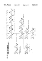
- FIGS. 1A and 1B outline the synthetic plan for lactonorhexaosyl structure (6 ) by a (2+2+2) stepwise approach (mode I).
- FIGS. 2A and 2B outline the synthetic plan for lactonorhexaosyl structure (6) by a (4+2) block approach (mode II).
- FIG. 3 describes a known synthetic route for O-(2,3,4,6-tetra-O-acetyl- ⁇ -D-galactopyranosyl)-(1 ⁇ 4)-1,3,6-tri-O-acetyl-2-deoxy-2-phthalimido- ⁇ / ⁇ -D-gluocopyranose (2).
- FIGS. 4A and 4B describe the known transformation of (2) into N-phthaloyl-lactosaminyl donors (3a, 3b and 3c).
- FIG. 5 describes the known synthesis of lactosyl acceptors(4a and 4b).
- FIG. 6 describes the entire procedure for synthesis of lactonorhexaosyl structure (6) by a stepwise approach (mode I) (detailed scheme).
- Gal represents galactose
- GlcNAc represents N-acetylglucosamine
- Glc glucose
- Ac represents an acetyl group
- Bn represents a benzyl group
- Bz represents a benzoyl group
- Pd-C represents palladium on carbon
- Ts represents a tosyl group
- CH 2 Cl 2 is methylene chloride (also known as dicholoromethane)
- Nphth represents an N-phthalyl group
- SMe represents a thiomethyl group
- Ac 2 O is acetic anhydride
- Py represents pyridine
- DMF is N,N-dimethylformamide
- MS AW-300 is molecular sieve AW-300
- CH 3 CN is acetonitrile
- Ce(NH 4 ) 2 (NO 3 ) 6 is ceric ammonium nit
- FIG. 7 shows the results of subjecting the monosialoganglioside fraction of bovine red blood cells (RBC) to high performance liquid chromatography (HPLC).
- FIG. 7A shows the elution pattern of the monosialyl gangliosides through an Iatrobead column. The arrow indicates sialylnorhexosyl ceramide.
- FIG. 7B shows the thin layer chromatograph of fractions 51 to 59. Arrow 1 indicates sialyl lactonoroctaosylceramide and arrow 2 indicates sialyl lactoisooctaosylceramide.
- FIG. 7C shows the thin layer chromatograph of several pooled fractions.
- FIG. 7D shows the separation pattern after the pooled fractions 55-58 in FIG. 7C were subjected to a second HPLC.
- FIG. 8 shows immunostained thin layer chromatographs of enzymatically synthesized dimeric Le x .
- FIG. 8A shows the pattern after staining with orcinol and H 2 SO 4 , which detects carbohydrate.
- FIG. 8B shows the pattern after staining with monoclonal antibody SH1, which detects long and short chain Le x glycolipid.
- FIG. 8C shows the pattern after staining with monoclonal antibody SH2, which detects only long chain Le x (dimeric Le x ).
- FIGS. 8A shows the pattern after staining with orcinol and H 2 SO 4 , which detects carbohydrate.
- FIG. 8B shows the pattern after staining with monoclonal antibody SH1, which detects long and short chain Le x glycolipid.
- FIG. 8C shows the pattern after staining with monoclonal antibody SH2, which detects only long chain Le x (dimeric Le x ).
- the lanes are as follows: lane 1, lactonoroctylceramide (nLc 6 ), standard from bovine RBC; lane 2, enzyme reaction mixture without nLc 6 ; lane 3, enzyme reaction mixture with nLc 6 ; lane 4, standard dimeric Le x from human tumor; lane 5, standard "O" upper Folch neutral from human RBC; lane 6, tumor upper Folch neutral.
- nLc 6 lactonoroctylceramide
- FIG. 9 is an autoradiogram showing the results of GDP- 14 C-fucose incorporation into nLc 4 and nLc 6 from bovine and human placenta lactosyl substrates.
- Lane A bovine RBC lactoneotetraosylceramide (nLc 4 ) and GDP- 14 C-fucose enzyme reaction mixture
- Lane B human placenta nLc 4 and GDP- 14 C-fucose enzyme reaction mixture
- Lane C bovine RBC nLc 6 and GDP- 14 C-fucose enzyme reaction mixture
- Lane D numan placenta nLc 6 and GDP- 14 C-fucose enzyme reaction mixture.
- FIG. 10 shows TLC immunostaining of nLc 4 conversion to Le x .
- FIG. 10A shows the pattern after staining with orcinol-H 2 SO 4 .
- FIG. 10B shows the pattern after staining with anti-Le x antibody SH1.
- FIG. 10C shows the pattern after staining with anti-dimeric Le x antibody SH2.
- the lanes are as follows: lane 1, standard "O" upper Folch neutral from human RBC; lane 2, tumor upper Folch neutral; lane 3, tumor Le x standard; lane 4, enzyme reaction mixture with nLc 4 from human placenta; lane 5, enzyme reaction mixture with nLc 4 from bovine RBC.
- FIG. 11 depicts a plan for synthesizing sialyl-Le x using sialic acid derivatives (18, 19) and disaccharide substrates (4 and 12).
- FIG. 12 depicts a plan for preparing lactosaminyl acceptor (12) which can be used to prepare sialyl-Le x .
- SEt is thioethyl and IR-120 is Amberlite.sup.• ion exchange resin 120.
- FIG. 13 depicts a plan for preparing sialyl dimeric Le x using, for example, the sialylated substrate (8) of FIG. 11 and a tetrasaccharide.
- FIG. 14 depicts a plan for preparing tetrasaccharide useful for making sialyl dimeric Le x .
- the present inventors have found the following system highly efficient for synthesis of dimeric Le x antigen (difucosyl Y 2 ).
- the backbone structure i.e., lactonorhexaosyl saccharide or its ceramide conjugate, is synthesized by organic chemical reaction or easily prepared from bovine red blood cells with high yield. Subsequent ⁇ 1 ⁇ 3 fucosylation at the III 3 and V 3 positions is carried out by ⁇ 1 ⁇ 3 fucosyltransferase and GDP-fucose.
- the enzyme ⁇ 1 ⁇ 3 fucosyltransferase is found with high activity in the Colo205 human colon cancer cell line, which has no ⁇ 1 ⁇ 2 fucosyltransferase activity.
- the final product, dimeric Le x antigen can be obtained from the backbone structure by a one-step reaction, with 70-80% yield.
- the overall cost of preparing the antigen in this manner is 1% or less compared to the multi-step chemical synthetic method.
- the method can be used to prepare fucosylated analogues of the lactonorhexaosyl structure such as lactonoroctaosyl, lactonordecaosyl or higher homologues (polylactosamines).
- lactonorhexaosyl structure such as lactonoroctaosyl, lactonordecaosyl or higher homologues (polylactosamines).
- Examples of compounds, other than the dimeric Le x antigen, that can be synthesized by the process of the present invention include the Y 2 antigen, the Z 1 antigen, the Z 2 antigen and the Z 3 antigen (trimeric Le x ). The structures of these compounds along with their short chain analogues are shown in Table I below.
- Le y octasaccharide ceramide, trifucosyl Le y , and Le y hexasaccharide ceramide are also important human tumor antigens (Abe et al., 1983, J. Biol. Chem. 258, 11793-11797; Nudelman et al., 1986, J. Biol. Chem. 261, 11247-11253; Kaizu et al., 1986, J. Biol. Chem. 261, 11254-11258) and the structures of these compounds are shown in Table II below.
- Synthesis of Le y antigen analogues as in Table II can be made by treating the core carbohydrate chains with ⁇ 1 ⁇ 2 fucosyltransferase and GDP-fucose followed by ⁇ 1 ⁇ 3 fucosyltransferase and GDP-fucose, or a combination of these two enzyme reactions.
- ⁇ 1 ⁇ 2 fucosyltransferase can be enriched from colonic cancer cell line MKN74 according to our preliminary experience. Other cell lines that may have higher enzyme activity are also available.
- Lactonorhexaosylceramide which has the structure, Ga1 ⁇ 1 ⁇ 4GlcNAc ⁇ 1 ⁇ 3Gal ⁇ 1 ⁇ 4GlcNAc ⁇ 1.fwdarw.3Gal ⁇ 1 ⁇ 4Glc ⁇ 1 ⁇ 1Cer, or its longer chain analogues, such as lactonoroctaosyl, lactonordecaosyl or higher homologues, can be prepared as glycolipid from a natural source, e.g., from human placenta, bovine red blood cells or rabbit muscle.
- a natural source e.g., from human placenta, bovine red blood cells or rabbit muscle.
- Lactonorhexaosylceramide or its longer chain analogues can be derived from the sialyl counterpart by hydrolysis of the sialic acid with a weak acid (e.g. 1% acetic acid, 80°-90° C. for 1 hr.).
- a weak acid e.g. 1% acetic acid, 80°-90° C. for 1 hr.
- lactonorhexaosylsaccharide or its longer chain analogues linked to carrier molecules such as ceramide or to a protein such as BSA through Lemieux's arm can be synthesized chemically.
- carrier molecules such as ceramide
- BSA protein
- Lemieux's arm Such procedures have been described previously (Ogawa, 1987, Carbohydr. Res., 163, 189-208; 167, 197-210; Lemieux et al., 1975, J. Amer. Chem. Soc., 97, 4076-4083).
- FIGS. 1 to 6 Various synthetic routes starting from lactose and N-phthalyllactosamine are shown in FIGS. 1 to 6.
- FIG. 1 shows a synthetic plan for the lactonorhexaosyl backbone 6 by a (2+2+2) stepwise approach.
- the hexasaccharide 6 can be synthesized by the coupling of an N-phthalyl lactosaminyl donor (3a, 3b, 3c or 3d) and a glycotetraosyl acceptor (5), which in turn can be synthesized by the coupling of an N-phthalyl lactosaminyl donor with glycobiosyl acceptor 4.
- the different lactosaminyl donors 3a (Arnap & Lonngren, 1981, J. Chem. Soc.
- the glycosylation of 3b with 4a preferentially gives the tetrasaccharide 7a with its (1 ⁇ 4) analogue 8a as a minor product (FIG. 6)(Paulsen & Michael, 1987, Carbohydr. Res., 169, 105-125; Ito & Ogawa, 1986, Agric. Biol. Chem., 50, 3231-3233).
- the glycosylation of 4b with 3b gives only one tetrasaccharide 7b in much better yield.
- Both of the tetrasaccharides 7a and 7b can be converted either to 5a by the reaction steps: (i) sodium methoxide-methanol (NaOMe-MeOH); ii) dimethoxypropane and p-toluenesulfonic acid (p-TsOH); iii) BnBr--NaH-DMF; and iv) AcOH--H 2 O, 80° C.; or to 5b by the reaction steps: (i) NaOMe-MeOH; ii) trimethylorthoacetate/p-TsOH/benzene; iii) BnBr--NaH-DMF; and iv) AcOH--H 2 O, 80° C.
- NaOMe-MeOH sodium methoxide-methanol
- p-TsOH dimethoxypropane and p-toluenesulfonic acid
- p-TsOH dimethoxypropan
- glycosylation of the glycotetraosyl acceptor 5a with 3b then gives the desired protected hexasaccharide 9a and its (1 ⁇ 4) analogue 10.
- the glycosylation of 5b with 4b is expected to give only one product 9b in better yield.
- Each of the protected hexasaccharides 9a or 9b can be converted to deblocked lactonorhexaosyl 6 by the following reactions [(i) NaOMe-MeOH, ii) NH 2 NH 2 --H 2 O-EtOH, reflux, iii) Ac 2 O-MeOH, iv) 10% Pd--C, H 2 ].
- FIG. 2 an alternative synthetic route (4+2 block approach, mode II) to 6 is shown, which differs from the (2+2+2) stepwise approach mode I (FIG. 1) in the sense that two N-phthalyl lactosaminyl disaccharides can be coupled to get a tetrasaccharide and then that tetrasaccharide can be coupled to the lactosyl acceptor to give the hexasaccharide.
- the thioglycoside 3c can be transformed into a lactosaminyl acceptor 11 (FIG. 14) and then glycosylated with another lactosaminyl donor 3 (FIG. 4) to give a tetrasaccharide 12.
- the tetrasaccharide thus obtained can be used as such as a donor by activating the anomeric thioalkyl group by the use of an appropriate catalyst and coupled to known lactosyl acceptor 4 to give the protected hexasaccharide 13.
- the protected hexasaccharide can be transformed into the free 6 in the same manner as described for 9 (FIG. 6).
- lactonorhexaosylceramide or analogues thereof derived from natural sources or chemically synthesized lactonorhexaosylsaccharide or analogues thereof can be converted quantitatively to their ⁇ 1 ⁇ 3 fucosyl derivatives as shown below: ##STR10## where R represents ceramide or a carrier molecule.
- Any source of ⁇ 1 ⁇ 3 fucosyltransferase can be used. Some of the preparations, depending on the source, may contain ⁇ 1 ⁇ 4 fucosyltransferase or a non-specific enzyme that has both ⁇ 1 ⁇ 3 and ⁇ 1 ⁇ 4 transferase activities.
- lactonorhexaosyl and longer lactonorhexaosyl structures have no free OH group at the C4 position of GlcNAc, and therefore, the presence of ⁇ 1 ⁇ 4 fucosyltransferase activity does not change the quality of the product, i.e., the product has exclusively ⁇ 1 ⁇ 3 fucosyl substitution.
- Sources other than Colo205 include human colonic mucosa and human colonic adenocarcinoma cell lines.
- Example II describes the reaction using the enzyme from the human colonic adenocarcinoma Colo205 cell line.
- This cell line contains ⁇ 1 ⁇ 3 and ⁇ 1 ⁇ 4 fucosyltransferase, but does not contain ⁇ 1 ⁇ 2 fucosyltransferase. Any of the other cell lines or tissues can be used if appropriate enzymatic activities are present.
- the Y 2 antigen can be prepared by ⁇ 1 ⁇ 3 fucosylation at the V 3 position of the lactonorhexaosyl backbone
- the Z 1 antigen, Z 2 antigen and Z 3 antigen can be prepared by ⁇ 1 ⁇ 3 fucosylation at the VII 3 positions only, the V 3 and VII 3 positions and the III 3 , V 3 and VII 3 positions, respectively, of the lactonoroctaosyl backbone.
- Further enzymatic synthesis could also include ⁇ 1 ⁇ 3 fucosylation at the V 3 position and ⁇ 1 ⁇ 2 fucosylation at the terminal galactose residue of lactonorhexaosyl backbone to give the Le Y structure and this could be further converted to trifucosyl Le Y by ⁇ 1 ⁇ 3 fucosylation at the III 3 position.
- Enzymatic synthesis could also include ⁇ 1 ⁇ 4 fucosylation etc., so long as a useful structure is produced.
- the ⁇ 1 ⁇ 3 fucotransferase and ⁇ 1 ⁇ 4 fucotransferase enzyme preparations can be made from known sources as described above, by known methods such as described in Example II. (Holmes et al., 1985, J. Biol. Chem., 260, 7619-7627).
- Le y antigens can be synthesized by addition of fucose at the terminal Gal residue with ⁇ 1 ⁇ 2 fucosyltransferase and GDP-fucose followed by the action of ⁇ 1 ⁇ 3 fucosyltransferase and GDP-fucose by analogous procedures to those followed for synthesis of dimeric Le x antigen.
- the enzyme ⁇ 1 ⁇ 2 fucotransferase capable of fucosylating a terminal galactose residue has been found in the MKN74 colonic cancer cell line although its activity is relatively weak and concentration of the enzyme is necessary. Since ⁇ 1 ⁇ 2 fucosyl transferase is widely found in various cells, better sources of ⁇ 1 ⁇ 2 fucosyl transferase are highly plausible. Thus a possibility for the following synthesis of Le y or Le y octasaccharide ceramide is envisioned to be within the scope of the invention. ##
- GDP-fucose can be synthesized from fucose ⁇ 1-phosphate by known methods (Nunez et al., 1981, Can, J. Chem., 59, 2086-2095; Michelson, 1964, Biochim. Biophys, Acta, 91, 1-13).
- Sialylated forms of dimeric Le x and derivatives thereof can be synthesized essentially in the same fashion as described above except that sialylated forms of the initial glycolipid substrates are used.
- Enzymatic synthesis of sialyl-Le x and sialyl-dimeric Le x is performed as described in Example II, however, the glycolipid substrates used are sialylparagloboside (IV 3 NeuAcnLc 4 ) and sialynorhexaosylceramide (VI 3 NeuAcnLc 6 ), respectively. Both substrates are obtained from bovine red blood cells as described in Example I, with the exception that step 3, "Hydrolysis with Acetic Acid", is omitted .
- Sialylparagloboside can be obtained in fractions 37-39 as shown in FIG. 7A. All procedures are performed as described for the bio-organic synthesis of dimeric Le x and its analogues with the exception of the aforementioned substrate modifications.
- sialyl Le x 14 can be synthesized by enzymatic fucosylation of sialosyl lactoneotetraose 15, which can be synthesized chemically by the coupling of a sialic acid containing trisaccharide donor 16 with a disaccharide acceptor 17.
- compound 18 as a synthetic equivalent of sialolactosaminyl donor 16 and compound 4 (FIG. 1) as a synthetic equivalent of lactosyl acceptor 17 could be used.
- the glycotriosyl donor 18 can be obtained by glycosylation of lactosaminyl acceptor 12 (FIG.
- Sialyl dimeric Le x 20 in FIG. 13 can be synthesized via enzymatic fucosylation of, for example, heptasaccharide 21 which can be synthesized chemically.
- sialolactosaminyl donor 18 (FIG. 11) is coupled to the tetrasaccharide acceptor 5a, 5b or 5c which can be synthesized as depicted in FIG. 14.
- the reaction scheme to make the tetrasaccharide acceptor is similar to the scheme presented in FIG. 6.
- sialyl-dimeric Le x oligosaccharides also may be accomplished conveniently using the hexasaccharide, Gal ⁇ 1 ⁇ 4GlcNAc ⁇ 1 ⁇ 3Gal ⁇ 1 ⁇ 4GlcNAc ⁇ 1.fwdarw.3Gal ⁇ 1 ⁇ 4Glc (6), which is synthesized as shown in FIG. 1 or as in FIG. 2 or FIG. 6.
- the terminal sialyl residue may be attached according to the reaction shown on the next page: ##STR12##
- Sialyl Le x may be synthesized according to the two enzymatic reactions: ##STR14##
- the stroma pellet was homogenized in three liters of ethanol for three minutes and then brought to slow boil in an 80° C. water bath for five minutes.
- the hot ethanol extract was filtered over a Buchner funnel and the residue rehomogenized in the same manner an additional two times.
- the three filtrates were pooled and evaporated to dryness by rotovap.
- the organic residue was transferred to a gallon jug in three liters of chloroform-methanol (2:1, v:v) and 500 ml of H 2 O were added. The solution was shakened ten times to form Folch's partition.
- a 300 ml bed volume of DEAE Sephadex (Sigma) was used in a column 5 cm ⁇ 25 cm.
- the Sephadex was pre-equilibrated with chloroform-methanol-water (30:60:8, v:v:v; 0.8M sodium acetate) overnight and washed in chloroform-methanol-water (30:60:8, v:v:v).
- the dialyzed upper phase was evaporated to dryness by rotovap and brought up in 500 ml chloroform-methanol-water (30:60:8, v:v:v, Sol A). This was applied to DEAE and washed with 2 liters Sol A.
- the column was washed with 500 ml methanol, and the monosialoganglioside fraction (containing sialylnorhexaosylceramide) was eluted in 0.04M ammonium acetate (1.5 liters).
- the monosialyl fraction was evaporated to dryness, dialyzed for three days against water, evaporated by rotovap and transferred to a 15 ml screw cap tube.
- the sample was applied in two injections of 2 ml in chloroform-methanol-water (2:1:0.1, v:v:v) at a starting composition of 55:40:5 (v:v:v) at 0.5 ml/min.
- fractions containing sialyllactonorhexaosylceramide were pooled and further purified by chromatography on a long Iatrobead 6RS-8010 column (0.5 cm ⁇ 100 cm) pre-equilibrated with isopropanol-hexane-water (55:40:5, v:v:v).
- the column was eluted by gradient elution using isopropanol-hexane-water (55:40:3 to 55:25:20, v:v:v) over 300 minutes, then eluted with the same solvent for 400 minutes.
- Essentially pure sialyllactonorhexaosylceramide was obtained.
- FIG. 7A is a thin layer chromatogram showing the elution pattern of monosialyl gangliosides through the Iatrobead column. The arrow indicates sialylnorhexaosyl ceramide.
- FIG. 7B shows a thin layer chromatographic pattern of fractions 51-59.
- Arrow 1 indicates sialyl lactonoroctaosylceramide and arrow 2 indicates sialyl lactoisooctaosylceramide.
- the fractions noted are the major sources for the preparation of lactonorhexaosylceramide (Gal ⁇ 1 ⁇ 4GlcNAcB1 ⁇ 3Gal ⁇ 1 ⁇ 4GlcNAc ⁇ 1.fwdarw.3Gal ⁇ 1 ⁇ 4Glc ⁇ 1 ⁇ 1Cer) and lactoisooctaosyl-ceramide (I antigen), respectively.
- FIG. 7C shows the thin layer chromatographic pattern of several pooled fractions.
- the pooled fraction (55-58) in FIG. 7C was subjected to a second HPLC run and the separation pattern is shown in FIG. 7D.
- sialyllactonorhexaosylceramide Two milligrams of sialyllactonorhexaosylceramide was evaporated to dryness in an 8 ml screw cap tube and 1 ml of 1.0% acetic acid added. The tube was placed in a 100° C. heat block for 1 hr. After hydrolysis, the sample was evaporated to dryness by adding 2 ml ethanol and drying under an N 2 stream. The dried glycolipid was dissolved in 2 ml of water and applied to a Sep-Pak C-18 reverse phase column, washed intensively with water and eluted with methanol. The eluate was evaporated. The reaction was quantitative.
- GDP-fucose the sugar donor
- fucose ⁇ 1-phosphate Funez et al. , 1981, Can. J. Chem., 59, 2086-2095; Michelson, 1964, Biochem. Biophys. Acta, 91, 1-13).
- lactonorhexaosylceramide (nLc 6 ) or lactonorhexaosyl oligosaccharide was mixed with 2 mg of sodium taurocholate in a 100 ml round bottom flask, codissolved in a suitable solvent, thoroughly mixed and evaporated to dryness. The dried residue was dissolved in 1 ml of water, 50 ⁇ l of 0.4M MnCl 2 and 500 ⁇ l of 1.0M Tris buffer pH 7.4, sonicated well and mixed with 500 ⁇ l of aqueous solution containing 50 mg CDP-choline (Sigma) and 10 mg GDP-fucose.
- the reaction mixture when starting from lactonorhexaosyl oligosaccharide linked to a carrier peptide or protein, was placed on an affinity column containing an antibody which reacts with dimeric Le x (an example of such an antibody is SH2 available from The Biomembrane Institute, Seattle, Wash.).
- the compound was diluted with 1-2M NaCl. Under these conditions, about 60-70% of the norhexaosyl structure was converted to dimeric Le x , based on orcinol detection of resultant glycolipid product.
- a TLC plate was chromatographed and detected by 0.5% oricinol in 10% H 2 SO 4 , heated at 150° C. for 3 minutes. Percentage conversion was based upon densitometer readings giving area units for each band. About 10% was not converted and about 20-30% was converted to the monofucosyl derivative.
- GDP-fucose can be obtained commercially or produced from fucose ⁇ 1-phosphate as described above.
- reaction was allowed to proceed overnight at 37° C. with shaking. After 18 hours, CDP-choline and GDP-fucose were added (Doubling amount added in each case) and reaction left another 24 hours. At this point about 60-70% of nLc 6 had been converted to dimeric Le x . About 10% was unconverted and about 20-30% was converted to monofucose glycolipid.
- TLC was carried out according to known methods, as was orcinal staining and immunostaining with antibodies that bind to Le x and to dimeric Le x (such as SH1 which is available from The Biomembrane Institute, Seattle, Wash.) and that bind to dimeric Le x only (such as SH2 which is available from The Biomembrane Institute, Seattle, Wash.).
- antibodies that bind to Le x and to dimeric Le x such as SH1 which is available from The Biomembrane Institute, Seattle, Wash.
- SH2 which is available from The Biomembrane Institute, Seattle, Wash.
- reaction mixtures were spotted at 2000 cpm/lane and the film was esposed for 18 hours.
- FIG. 8 shows thin layer chromatographs immunostained for enzymatically synthesized dimeric Le x using primary antibodies as shown, secondary rabbit anti-mouse antibodies and finally iodinated 125 I-protein A according to known methods.
- FIG. 8A shows the pattern after staining with 0.5% orcinol, 10% H 2 SO 4 by known methods. Orcinol staining detects carbohydrate.
- FIG. 8B shows the TLC pattern after immunostaining with monoclonal antibody SH1, detecting long and short chain Le x glycolipid.
- FIG. 8C shows the TLC pattern after immunostaining with SH2 which detects only long chain Le x (dimeric Le x ).
- Lane 1 nLc 6 standard from bovine RBC; lane 2, enzyme reaction mixture without nLc 6 (the fast migrating ban is sodium deoxycholate, the lower band is derived from impurity in the enzyme preparation); lane 3, enzyme reaction mixture with nLc 6 ; lane 4, standard dimeric Le x from human tumor; lane 5, standard "O" upper Folch neutral from human RBC; lane 6, tumor upper Folch neutral. Note conversion of nLc 6 to dimeric Le x in lane 3 as seen by orcinol detection (FIG. 8A) as well as immunostaining with anti-Le x (FIGS. 8B and 8C).
- FIG. 9 is an audioradiogram showing the results of the incorporation of 14 C-fucose into nLc 4 and nLc 6 from bovine and human placenta lactosyl substrates.
- Lane A depicts the bovine RBC nLc 4 and 14 C-fucose enzyme reaction mixture
- Lane B the human placenta nLc 4 and 14 C-fucose enzyme reaction mixture
- Lane C the bovine RBC nLc 6 and 14 C-fucose enzyme reaction mixture
- Lane D the human placenta nLc 6 and 14 C-fucose enzyme reaction mixture.
- FIG. 10 shows TLC immunostaining of nLc 4 conversion of Le x .
- FIG. 10A shows detection with orcinol-H 2 SO 4 as in FIG. 8A.
- FIG. 10B shows the immunostaining pattern with anti-Le x antibody, SH1.
- FIG. 10C shows immunostaining with anti-dimeric Le x antibody, SH2.
- Lane 1 standard "O" upper Folch neutral; lane 2, tumor upper Folch neutral; lane 3, tumor Le x standard; lane 4, enzyme reaction mixture with nLc 4 from human placenta; and lane 5, enzyme reaction mixture with nLc 4 from bovine RBC.
- FIG. 10B, lanes 4 and 5 show SH1-positive reaction demonstrating production of the Le x epitope.
- SH2 does not stain short-chain Le x .
- fucosylated structures produced according to the present invention are useful as active vaccines for tumors.
- the skilled artisan can readily determine which structures are useful for which tumors.
- Further suitable pharmaceutically acceptable carriers, diluents or excipients for the vaccine, suitable methods of administration and doses can be determined readily by the skilled artisan.
- Dimeric Le x has been shown to be useful to suppress the appearance of inflammatory granulocytes in the bone marrow of RA-affected joints.
- monoclonal antibodies with specific binding for that substance.
- Such monoclonal antibodies can be used for passive immunization against tumors and for in vitro and in vivo methods of detecting tumors. Such methods are readily known to and conducted by the skilled artisan.
- a series of Le y antigens can be prepared by a similar enzymatic fucosylation as that applied for synthesis of dimeric Le x described above.
- Lactonorhexaosylceramide or lactonorhexaosyl saccharides linked to carrier molecules or their higher analogues are the starting materials.
- the material is dissolved in Tris buffer in the presence of bivalent cation and appropriate detergent such as taurocholate, as described above.
- To the solution is added GDP-fucose, CDP-choline, and ⁇ 1 ⁇ 2 fucosyltransferase, which is isolated from human gastric cancer cell line MKN74 or the same enzyme isolated from any other source.
- the reaction can be similarly processed as described above and subsequently ⁇ 1 ⁇ 2 fucosylated derivatives will be isolated, which can be monitored by positive reaction with monoclonal antibody BE2 (Young et al., 1981, J. Biol. Chem., 256, 10967-10972).
- the H-active compound as an intermediate can be further processed for ⁇ 1 ⁇ 3 fucosylation by ⁇ 1 ⁇ 3 fucosyltransferase of Colo205 and GDP-fucose under the same conditions as described above for dimeric Le x .
- the final product can be monitored by positive reaction with monoclonal antibody AH6 defining the Le y structure (Abe et al., 1983, J. Biol.
Landscapes
- Organic Chemistry (AREA)
- Chemical & Material Sciences (AREA)
- Engineering & Computer Science (AREA)
- Life Sciences & Earth Sciences (AREA)
- Zoology (AREA)
- Wood Science & Technology (AREA)
- Health & Medical Sciences (AREA)
- General Engineering & Computer Science (AREA)
- General Chemical & Material Sciences (AREA)
- Chemical Kinetics & Catalysis (AREA)
- Biotechnology (AREA)
- Biochemistry (AREA)
- Bioinformatics & Cheminformatics (AREA)
- Microbiology (AREA)
- General Health & Medical Sciences (AREA)
- Genetics & Genomics (AREA)
- Saccharide Compounds (AREA)
- Preparation Of Compounds By Using Micro-Organisms (AREA)
- Medicines That Contain Protein Lipid Enzymes And Other Medicines (AREA)
- Medicines Containing Antibodies Or Antigens For Use As Internal Diagnostic Agents (AREA)
- Pharmaceuticals Containing Other Organic And Inorganic Compounds (AREA)
- Polysaccharides And Polysaccharide Derivatives (AREA)
Abstract
A process for preparing difucosyl Y2 antigen (dimeric Lex) , said process comprising: (1) preparing a lactonorhexaosylceramide backbone or a lactonorhexaosylsaccharide backbone linked to a carrier molecule; and (2) enzymatically fucosylating said backbone at the III3 and V3 positions through an α1→3 linkage. A process for preparing Ley antigen analogues, said process comprising: (1) preparing a lactonorhexaosyl-ceramide backbone or a lactonorhexaosylsaccharide backbone linked to a carrier molecule; and (2) enzymatically fucosylating said backbone at the terminal β-Gal through an α1→2 linkage; and (3) enzymatically fucosylating said backbone at one or more positions through an α1→3 linkage, provided that steps (2) and (3) can be conducted simultaneously or in any order. A process for preparing a fucosylated lactonorhexaosylceramide, lactonorhexaosylsaccharide linked to a carrier molecule or higher analogues thereof, said process comprising: (1) preparing a lactonorhexaosylceramide backbone, a lactonorhexaosylsaccharide backbone linked to a carrier molecule or backbones of higher analogues thereof; and (2) enzymatically fucosylating one or more residues of said backbone. A process for preparing sialyl Lex and sialyl dimeric Lex.
Description
This is a continuation-in-part of U.S. application Ser. No. 07/344,628, filed on 28 Apr. 1989, now abandoned.
The present invention relates to a process for synthesizing Lex, dimeric Lex (i.e., difucosyl Y2 ; III3 FucV3 FucnLc6 Cer), higher analogues thereof and sialylated forms thereof. Di- and trimeric Lex are especially important tumor antigens and are useful for developing vaccines against human cancer and specific effectors to dampen inflammatory processes of rheumatoid arthritis.
More particularly, the invention relates to a process for the synthesis of di- and trifucosyl Lex, higher analogues thereof and sialylated forms thereof by means of a one-step reaction which results in unexpectedly superior yields.
The present invention also relates to a process for synthesis of Ley antigen analogues by means of a one-step reaction which results in unexpectedly superior yields.
The glycolipids dimeric Lex (difucosyl Y2 ; III3 FucV3 FucnLc6 Cer) and trimeric Lex (III3 FucV3 FucVII3 FucnLc8 Cer) are major antigens that are found in various human adenocarcinomas but are absent in corresponding normal tissue (Hakomori et al., 1984, J. Biol. Chem., 259, 4672-2680). A monoclonal antibody directed to those structures, but not cross-reacting with simple Lex (see Table I), is an important reagent for detecting the presence of the antigens in tumor cells (Fukushi et al., 1984, J. Biol. Chem., 259, 4681-4685; Fukushi et al. , 1984, J. Exp. Med., 159, 506-520) and in sera of patients with cancer. The antibody, however, reacts on immunohistochemistry or immunofluorescence with selected normal cells, such as epithelial cells of proximal convoluted tubules of kidney and weakly with some subpopulations of granulocytes (Fukushi et al., 1984, Cancer Res., 45, 3711-1717).
More recently, the antibody was found to react with granulocytes of inflammatory bone marrow adjacent to joints affected with rheumatoid arthritis (RA) (Ochi et al, 1988, J. Rheumatol., 15, 1609-1615). Intradermal inoculation of liposomes containing the glycoplipids suppresses the appearance of inflammatory granulocytes in the bone marrow of RA-affected joints with a subsequent reduction of RA symptoms.
Because of the presence of the antigens in high concentration in various types of human cancer and inflammatory processes, the antigens are expected to be useful components for developing anti-cancer and anti-inflammatory vaccines. To support this idea, reconstituted Newcastle's Disease virus membrane including the dimeric Lex antigen induced an immune response that suppressed growth of murine tumors bearing Lex.
Ley antigens, including extended Ley (Ley octasaccharide ceramide) and trifucosyl Ley, are also important human cancer antigens and are expected to be useful components for developing anti-cancer vaccines.
Thus the demand for Lex dimeric Lex and sialylated forms thereof and for various types of Ley antigen for use in active immunization has been increasing. However, it has been available only from human cancer tissue or via chemical synthesis as described by Nilsson & Norberg (1987, Glycoconjugate J., 4, 219-223; and 1988, Carbohydr. Res., 183, 71-82), Sato et al. (1987, Carbohydr. Res., 167, 197-210; 1988, Tetrahedron Lett., 29, 5267-5270) and Nicolaou et al. (1990, J. Am. Chem. Soc., 112, 3693-3695).
Preparations from tumor cells provide limited quantities which often contain impurities. On the other hand, pure chemical synthesis involves at least 50 steps, is extremely laborious and results in a poor final yield.
It is toward the objective of providing a low-cost, simplified and greater-yielding synthesis of Lex ; dimeric and trimeric Lex antigens; sialylated forms thereof; and Ley antigen that the present invention is directed.
The principal object of the present invention is to provide a low-cost, simplified process for the synthesis of dimeric or trimeric Lex antigens having improved greater yields.
Another object of the present invention is to provide a low cost, simplified process for the synthesis of sialyl-Lex, sialyl-dimeric Lex, trifucosyl Ley (VI2 FucV3 FucIII3 FucnLc6) and its higher homologs.
These and other objects of the invention have been achieved by providing a process for preparing difucosyl Y2 antigen (dimeric Lex), the process comprising: (1) preparing a lactonorhexaosylceramide backbone or a lactonorhexaosylsaccharide backbone linked to a carrier molecule; and (2) enzymatically fucosylating the backbone at the III3 and V3 positions through an α1→3 linkage.
The present invention also provides a process for preparing Ley antigen analogues, the process comprising:
(1) preparing a lactonorhexaosylceramide backbone or a lactonorhexaosysaccharide backbone linked to a carrier molecule;
(2) enzymatically fucosylating the backbone at the terminal β-Gal through an α1→2 linkage; and
(3) enzymatically fucosylating the backbone at one or more positions through an α1→3 linkage, provided that steps (2) and (3) can be conducted simultaneously or in any order.
The present invention also provides a process for preparing an α1→2 and/or α1→3 fucosylated lactonorhexaosylceramide, lactonorhexaosylsaccharide linked to a carrier molecule or higher homologues thereof, said process comprising: (1) preparing a lactonorhexaosylceramide backbone, a lactonorhexaosylsaccharide backbone linked to a carrier molecule or backbones of higher homologues thereof; and (2) enzymatically fucosylating one or more residues of said backbone.
The invention also provides a process for preparing sialylated forms of Lex and dimeric Lex using sialylated starting materials such as sialylparagloboside and sialylnorhexaosylceramide, respectively, or using synthetically prepared sialyllactonortetraosyl and sialyllactonorhexaosyl (sialyltetraosyl saccharide and sialylhexaosyl saccharide attached to a carrier molecule (spacer arm), respectively.
FIGS. 1A and 1B outline the synthetic plan for lactonorhexaosyl structure (6 ) by a (2+2+2) stepwise approach (mode I).
FIGS. 2A and 2B outline the synthetic plan for lactonorhexaosyl structure (6) by a (4+2) block approach (mode II).
FIG. 3 describes a known synthetic route for O-(2,3,4,6-tetra-O-acetyl-β-D-galactopyranosyl)-(1→4)-1,3,6-tri-O-acetyl-2-deoxy-2-phthalimido-α/β-D-gluocopyranose (2).
FIGS. 4A and 4B describe the known transformation of (2) into N-phthaloyl-lactosaminyl donors (3a, 3b and 3c).
FIG. 5 describes the known synthesis of lactosyl acceptors(4a and 4b).
FIG. 6 describes the entire procedure for synthesis of lactonorhexaosyl structure (6) by a stepwise approach (mode I) (detailed scheme).
(In FIGS. 1-6, Gal represents galactose, GlcNAc represents N-acetylglucosamine, Glc represents glucose, Ac represents an acetyl group, Bn represents a benzyl group, Bz represents a benzoyl group, Pd-C represents palladium on carbon, Ts represents a tosyl group, CH2 Cl2 is methylene chloride (also known as dicholoromethane), Nphth represents an N-phthalyl group, SMe represents a thiomethyl group, Bu3 SnSR wherein R=alkyl represents thioalkyltributyltin, Ac2 O is acetic anhydride, Py represents pyridine, DMF is N,N-dimethylformamide, MS AW-300 is molecular sieve AW-300, CH3 CN is acetonitrile, Ce(NH4)2 (NO3)6 is ceric ammonium nitrate, (CH3 O )2 C(Me)2 is dimethoxypropane, BnOSnBu3 is tributyltin benoxide, DBU is 1,8-diazabicyclo[5.4.0]undec-7-ene, DAST is diethylaminosulfur trifluoride and (CH3 O)3 CMe is trimethylorthoacetate.)
FIG. 7 shows the results of subjecting the monosialoganglioside fraction of bovine red blood cells (RBC) to high performance liquid chromatography (HPLC). FIG. 7A shows the elution pattern of the monosialyl gangliosides through an Iatrobead column. The arrow indicates sialylnorhexosyl ceramide. FIG. 7B shows the thin layer chromatograph of fractions 51 to 59. Arrow 1 indicates sialyl lactonoroctaosylceramide and arrow 2 indicates sialyl lactoisooctaosylceramide. FIG. 7C shows the thin layer chromatograph of several pooled fractions. FIG. 7D shows the separation pattern after the pooled fractions 55-58 in FIG. 7C were subjected to a second HPLC.
FIG. 8 shows immunostained thin layer chromatographs of enzymatically synthesized dimeric Lex. FIG. 8A shows the pattern after staining with orcinol and H2 SO4, which detects carbohydrate. FIG. 8B shows the pattern after staining with monoclonal antibody SH1, which detects long and short chain Lex glycolipid. FIG. 8C shows the pattern after staining with monoclonal antibody SH2, which detects only long chain Lex (dimeric Lex). In FIGS. 8A, 8B and 8C the lanes are as follows: lane 1, lactonoroctylceramide (nLc6), standard from bovine RBC; lane 2, enzyme reaction mixture without nLc6 ; lane 3, enzyme reaction mixture with nLc6 ; lane 4, standard dimeric Lex from human tumor; lane 5, standard "O" upper Folch neutral from human RBC; lane 6, tumor upper Folch neutral.
FIG. 9 is an autoradiogram showing the results of GDP-14 C-fucose incorporation into nLc4 and nLc6 from bovine and human placenta lactosyl substrates. Lane A, bovine RBC lactoneotetraosylceramide (nLc4) and GDP-14 C-fucose enzyme reaction mixture; Lane B, human placenta nLc4 and GDP-14 C-fucose enzyme reaction mixture; Lane C, bovine RBC nLc6 and GDP-14 C-fucose enzyme reaction mixture; Lane D, numan placenta nLc6 and GDP-14 C-fucose enzyme reaction mixture.
FIG. 10 shows TLC immunostaining of nLc4 conversion to Lex. FIG. 10A shows the pattern after staining with orcinol-H2 SO4. FIG. 10B shows the pattern after staining with anti-Lex antibody SH1. FIG. 10C shows the pattern after staining with anti-dimeric Lex antibody SH2. In FIGS. 10A, 10B and 10C the lanes are as follows: lane 1, standard "O" upper Folch neutral from human RBC; lane 2, tumor upper Folch neutral; lane 3, tumor Lex standard; lane 4, enzyme reaction mixture with nLc4 from human placenta; lane 5, enzyme reaction mixture with nLc4 from bovine RBC.
FIG. 11 depicts a plan for synthesizing sialyl-Lex using sialic acid derivatives (18, 19) and disaccharide substrates (4 and 12).
FIG. 12 depicts a plan for preparing lactosaminyl acceptor (12) which can be used to prepare sialyl-Lex. SEt is thioethyl and IR-120 is Amberlite.sup.• ion exchange resin 120.
FIG. 13 depicts a plan for preparing sialyl dimeric Lex using, for example, the sialylated substrate (8) of FIG. 11 and a tetrasaccharide.
FIG. 14 depicts a plan for preparing tetrasaccharide useful for making sialyl dimeric Lex.
The present inventors have found the following system highly efficient for synthesis of dimeric Lex antigen (difucosyl Y2). The backbone structure, i.e., lactonorhexaosyl saccharide or its ceramide conjugate, is synthesized by organic chemical reaction or easily prepared from bovine red blood cells with high yield. Subsequent α1→3 fucosylation at the III3 and V3 positions is carried out by α1→3 fucosyltransferase and GDP-fucose.
The enzyme α1→3 fucosyltransferase is found with high activity in the Colo205 human colon cancer cell line, which has no α1→2 fucosyltransferase activity. Thus, the final product, dimeric Lex antigen, can be obtained from the backbone structure by a one-step reaction, with 70-80% yield. The overall cost of preparing the antigen in this manner is 1% or less compared to the multi-step chemical synthetic method.
Further, the method can be used to prepare fucosylated analogues of the lactonorhexaosyl structure such as lactonoroctaosyl, lactonordecaosyl or higher homologues (polylactosamines). Examples of compounds, other than the dimeric Lex antigen, that can be synthesized by the process of the present invention include the Y2 antigen, the Z1 antigen, the Z2 antigen and the Z3 antigen (trimeric Lex). The structures of these compounds along with their short chain analogues are shown in Table I below.
Ley octasaccharide ceramide, trifucosyl Ley, and Ley hexasaccharide ceramide are also important human tumor antigens (Abe et al., 1983, J. Biol. Chem. 258, 11793-11797; Nudelman et al., 1986, J. Biol. Chem. 261, 11247-11253; Kaizu et al., 1986, J. Biol. Chem. 261, 11254-11258) and the structures of these compounds are shown in Table II below.
Synthesis of Ley antigen analogues as in Table II can be made by treating the core carbohydrate chains with α1→2 fucosyltransferase and GDP-fucose followed by α1→3 fucosyltransferase and GDP-fucose, or a combination of these two enzyme reactions. Although a practical experimental example is not given in the application, α1→2 fucosyltransferase can be enriched from colonic cancer cell line MKN74 according to our preliminary experience. Other cell lines that may have higher enzyme activity are also available.
                                  TABLE I
__________________________________________________________________________
MONO- AND DIMERIC Le.sup.x ANTIGEN AND RELATED STRUCTURES
__________________________________________________________________________
Le.sup.x pentasaccharide ceramide
 ##STR1##
Y.sub.2 antigen
 ##STR2##
difucosyl Y.sub.2 (dimeric Le.sup.x)
 ##STR3##
Z.sub.1 antigen
 ##STR4##
4Glcβ1→1Cer
Z.sub.2 antigen
 ##STR5##
4Glcβ1→1Cer
Z.sub.3 antigen (trimeric Le.sup.x)
 ##STR6##
4Glcβ1→1Cer
__________________________________________________________________________
                                  TABLE II
__________________________________________________________________________
Le.sup.y ANTIGEN AND ANALOGUES
__________________________________________________________________________
Le.sup.y hexasaccharide ceramide (IV.sup.2 FucIII.sup.3 FucnLc.sub.4)
 ##STR7##
Le.sup.y octasaccharide ceramide (VI.sup.2 FucV.sup.3 FucnLc.sub.6)
 ##STR8##
trifucosyl Le.sup.Y (VI.sup.2 FucV.sup.3 FucIII.sup.3 FucnLc.sub.6)
 ##STR9##
__________________________________________________________________________
Conditions of synthesis for the method of the present invention are described below.
Lactonorhexaosylceramide, which has the structure, Ga1β1→4GlcNAcβ1→3Galβ1→4GlcNAcβ1.fwdarw.3Galβ1→4Glcβ1→1Cer, or its longer chain analogues, such as lactonoroctaosyl, lactonordecaosyl or higher homologues, can be prepared as glycolipid from a natural source, e.g., from human placenta, bovine red blood cells or rabbit muscle. These tissues and cells contain a large quantity of sialyl 2→6 or 2→3 lactonorhexaosylceramide and longer chain analogues thereof, which can be isolated easily by Folch's partition of total lipid extract followed by DEAE-Sepharose chromatography and high performance liquid chromatography in a buffer comprising isopropanol-hexane-water (for detailed procedure see: Hakomori "Chemistry of Glycosphingolipids" In: Handbook of Lipid Research 3: Sphingolipd Biochemistry (Kanfer & Hakomori, eds.), Plenum Publishing, New York, pp. 1-165, 1983). Lactonorhexaosylceramide or its longer chain analogues can be derived from the sialyl counterpart by hydrolysis of the sialic acid with a weak acid (e.g. 1% acetic acid, 80°-90° C. for 1 hr.).
As an alternative to isolation from natural sources, lactonorhexaosylsaccharide or its longer chain analogues linked to carrier molecules such as ceramide or to a protein such as BSA through Lemieux's arm can be synthesized chemically. Such procedures have been described previously (Ogawa, 1987, Carbohydr. Res., 163, 189-208; 167, 197-210; Lemieux et al., 1975, J. Amer. Chem. Soc., 97, 4076-4083). Various synthetic routes starting from lactose and N-phthalyllactosamine are shown in FIGS. 1 to 6.
FIG. 1 shows a synthetic plan for the lactonorhexaosyl backbone 6 by a (2+2+2) stepwise approach. According to this plan the hexasaccharide 6 can be synthesized by the coupling of an N-phthalyl lactosaminyl donor (3a, 3b, 3c or 3d) and a glycotetraosyl acceptor (5), which in turn can be synthesized by the coupling of an N-phthalyl lactosaminyl donor with glycobiosyl acceptor 4. The different lactosaminyl donors 3a (Arnap & Lonngren, 1981, J. Chem. Soc. Perkin Trans., 1, 2070-2074), 3b wherein the X group is 2,2,2-trichloroacetamide (Grundler & Schmidt, 1985, Carbohydr. Res., 135, 203-218; Sadozai et al., 1985, Carbohydr. Res., 157, 101-123), 3c wherein the X group is thioalkyl (Sadozai et al., 1985, supra) and 3d wherein the X group is fluoride (Sadozai et al., manuscript in preparation) can be prepared readily from 2 as described in FIG. 4, which in turn can be synthesized from commercially available lactose in seven steps as described in FIG. 3 (Haworth et al., 1930, J. Chem. Soc., 1930, 2644-2653; Arnap & Lonngren, 1981, J. Chem. Soc. Perkin Trans., 1, 2070-2074). The lactosyl acceptor 4a (R=H, R'=Bn), which has free hydroxyl groups at C-3 and C-4 of the galactose unit (Koike et al., 1987, Carbohydr. Res., 163, 189-208; Paulsen & Paal, 1985, Carbohydr. Res., 137, 39-62) or 4b (R=Ac, R'=Bn), which has a free hydroxyl group only at C-3 of the galactose unit (Yoshino et al., 1988, Glycoconjugate J., 5, 377-384) can also be prepared from lactose 1 by known procedures as described for example in FIG. 5.
Due to the higher reactivity of the 3'-OH groups, the glycosylation of 3b with 4a preferentially gives the tetrasaccharide 7a with its (1→4) analogue 8a as a minor product (FIG. 6)(Paulsen & Michael, 1987, Carbohydr. Res., 169, 105-125; Ito & Ogawa, 1986, Agric. Biol. Chem., 50, 3231-3233). Alternatively, the glycosylation of 4b with 3b gives only one tetrasaccharide 7b in much better yield. Both of the tetrasaccharides 7a and 7b can be converted either to 5a by the reaction steps: (i) sodium methoxide-methanol (NaOMe-MeOH); ii) dimethoxypropane and p-toluenesulfonic acid (p-TsOH); iii) BnBr--NaH-DMF; and iv) AcOH--H2 O, 80° C.; or to 5b by the reaction steps: (i) NaOMe-MeOH; ii) trimethylorthoacetate/p-TsOH/benzene; iii) BnBr--NaH-DMF; and iv) AcOH--H2 O, 80° C. Glycosylation of the glycotetraosyl acceptor 5a with 3b then gives the desired protected hexasaccharide 9a and its (1→4) analogue 10. On the other hand, the glycosylation of 5b with 4b is expected to give only one product 9b in better yield. Each of the protected hexasaccharides 9a or 9b can be converted to deblocked lactonorhexaosyl 6 by the following reactions [(i) NaOMe-MeOH, ii) NH2 NH2 --H2 O-EtOH, reflux, iii) Ac2 O-MeOH, iv) 10% Pd--C, H2 ].
In FIG. 2, an alternative synthetic route (4+2 block approach, mode II) to 6 is shown, which differs from the (2+2+2) stepwise approach mode I (FIG. 1) in the sense that two N-phthalyl lactosaminyl disaccharides can be coupled to get a tetrasaccharide and then that tetrasaccharide can be coupled to the lactosyl acceptor to give the hexasaccharide. According to this possible plan, the thioglycoside 3c can be transformed into a lactosaminyl acceptor 11 (FIG. 14) and then glycosylated with another lactosaminyl donor 3 (FIG. 4) to give a tetrasaccharide 12. The tetrasaccharide thus obtained can be used as such as a donor by activating the anomeric thioalkyl group by the use of an appropriate catalyst and coupled to known lactosyl acceptor 4 to give the protected hexasaccharide 13. The protected hexasaccharide can be transformed into the free 6 in the same manner as described for 9 (FIG. 6).
Subsequently, the lactonorhexaosylceramide or analogues thereof derived from natural sources or chemically synthesized lactonorhexaosylsaccharide or analogues thereof can be converted quantitatively to their α1→3 fucosyl derivatives as shown below: ##STR10## where R represents ceramide or a carrier molecule.
Any source of α1→3 fucosyltransferase can be used. Some of the preparations, depending on the source, may contain α1→4 fucosyltransferase or a non-specific enzyme that has both α1→3 and α1→4 transferase activities. However, lactonorhexaosyl and longer lactonorhexaosyl structures have no free OH group at the C4 position of GlcNAc, and therefore, the presence of α1→4 fucosyltransferase activity does not change the quality of the product, i.e., the product has exclusively α1→3 fucosyl substitution.
Sources other than Colo205 include human colonic mucosa and human colonic adenocarcinoma cell lines.
Example II describes the reaction using the enzyme from the human colonic adenocarcinoma Colo205 cell line. This cell line contains α1→3 and α1→4 fucosyltransferase, but does not contain α1→2 fucosyltransferase. Any of the other cell lines or tissues can be used if appropriate enzymatic activities are present.
In an analogous manner, structures related to the Lex antigen can also be prepared.
For example, the Y2 antigen can be prepared by α1→3 fucosylation at the V3 position of the lactonorhexaosyl backbone, and the Z1 antigen, Z2 antigen and Z3 antigen can be prepared by α1→3 fucosylation at the VII3 positions only, the V3 and VII3 positions and the III3, V3 and VII3 positions, respectively, of the lactonoroctaosyl backbone.
Further enzymatic synthesis could also include α1→3 fucosylation at the V3 position and α1→2 fucosylation at the terminal galactose residue of lactonorhexaosyl backbone to give the LeY structure and this could be further converted to trifucosyl LeY by α1→3 fucosylation at the III3 position.
Enzymatic synthesis could also include α1→4 fucosylation etc., so long as a useful structure is produced.
The α1→3 fucotransferase and α1→4 fucotransferase enzyme preparations can be made from known sources as described above, by known methods such as described in Example II. (Holmes et al., 1985, J. Biol. Chem., 260, 7619-7627).
Although actual examples of synthesis of Ley antigen are not included in this application, Ley antigens can be synthesized by addition of fucose at the terminal Gal residue with α1→2 fucosyltransferase and GDP-fucose followed by the action of α1→3 fucosyltransferase and GDP-fucose by analogous procedures to those followed for synthesis of dimeric Lex antigen.
The enzyme α1→2 fucotransferase capable of fucosylating a terminal galactose residue has been found in the MKN74 colonic cancer cell line although its activity is relatively weak and concentration of the enzyme is necessary. Since α1→2 fucosyl transferase is widely found in various cells, better sources of α1→2 fucosyl transferase are highly plausible. Thus a possibility for the following synthesis of Ley or Ley octasaccharide ceramide is envisioned to be within the scope of the invention. ##STR11##
GDP-fucose can be synthesized from fucose β1-phosphate by known methods (Nunez et al., 1981, Can, J. Chem., 59, 2086-2095; Michelson, 1964, Biochim. Biophys, Acta, 91, 1-13).
Sialylated forms of dimeric Lex and derivatives thereof can be synthesized essentially in the same fashion as described above except that sialylated forms of the initial glycolipid substrates are used.
Enzymatic synthesis of sialyl-Lex and sialyl-dimeric Lex is performed as described in Example II, however, the glycolipid substrates used are sialylparagloboside (IV3 NeuAcnLc4) and sialynorhexaosylceramide (VI3 NeuAcnLc6), respectively. Both substrates are obtained from bovine red blood cells as described in Example I, with the exception that step 3, "Hydrolysis with Acetic Acid", is omitted . Sialylparagloboside can be obtained in fractions 37-39 as shown in FIG. 7A. All procedures are performed as described for the bio-organic synthesis of dimeric Lex and its analogues with the exception of the aforementioned substrate modifications.
Alternatively, as shown in FIG. 11, sialyl Le x 14 can be synthesized by enzymatic fucosylation of sialosyl lactoneotetraose 15, which can be synthesized chemically by the coupling of a sialic acid containing trisaccharide donor 16 with a disaccharide acceptor 17. To achieve this chemical synthesis, compound 18 as a synthetic equivalent of sialolactosaminyl donor 16 and compound 4 (FIG. 1) as a synthetic equivalent of lactosyl acceptor 17 could be used. The glycotriosyl donor 18 can be obtained by glycosylation of lactosaminyl acceptor 12 (FIG. 11) with sialosyl donor 19 (Kuhn et al., 1966, Chem. Ber., 99:611). The lactosaminyl acceptor 12 is synthesized from known compound 2 (FIG. 3) (Arnap & Lonngren 1981, J. Chem. Soc. Perkin Trans., 1:2070-2074) according to the reaction sequence described in FIG. 12.
Sialyl Lex linked to a spacer arm (or linker arm) can also be synthesized by the same strategy using a lactosyl acceptor 4c (R=(CH2)n COOCH3) having a spacer arm already attached to the lactose.
Sialyl dimeric Le x 20 in FIG. 13 can be synthesized via enzymatic fucosylation of, for example, heptasaccharide 21 which can be synthesized chemically. To synthesize 21, sialolactosaminyl donor 18 (FIG. 11) is coupled to the tetrasaccharide acceptor 5a, 5b or 5c which can be synthesized as depicted in FIG. 14. The reaction scheme to make the tetrasaccharide acceptor is similar to the scheme presented in FIG. 6.
The synthesis of large quantities (gram amounts) of sialyl-dimeric Lex oligosaccharides also may be accomplished conveniently using the hexasaccharide, Galβ1→4GlcNAcβ1→3Galβ1→4GlcNAcβ1.fwdarw.3Galβ1→4Glc (6), which is synthesized as shown in FIG. 1 or as in FIG. 2 or FIG. 6. Once the oligosaccharide is obtained, the terminal sialyl residue may be attached according to the reaction shown on the next page: ##STR12##
Addition of fucose to the GlcNAc residues of sialylated oligosaccharide (22) is quantitative according to the reaction: ##STR13##
Similarly, Sialyl Lex may be synthesized according to the two enzymatic reactions: ##STR14##
The present invention will now be described by reference to specific examples, but the examples are not to be construed as limiting the invention.
Unless otherwise specified, all percents, ratios, parts etc. are by weight.
Five gallons of fresh bovine red blood cells were obtained from a local slaughterhouse in the form of unlysed, citrated whole blood. The five gallons were diluted to fifteen gallons by adding deionized H2 O at 4° C. and acetic acid was added to a final concentration of 0.1%. After stirring, the red blood cells were allowed to lyse overnight at 4° C. The following morning, the lysed blood was centrifuged in a preparative rotor (Beckman JCF-Z), and the pellet was washed with H2 O. From five gallons whole blood, a 1.8 liter pellet of pinkish stroma was obtained.
1. Homogenization
The stroma pellet was homogenized in three liters of ethanol for three minutes and then brought to slow boil in an 80° C. water bath for five minutes. The hot ethanol extract was filtered over a Buchner funnel and the residue rehomogenized in the same manner an additional two times. The three filtrates were pooled and evaporated to dryness by rotovap. The organic residue was transferred to a gallon jug in three liters of chloroform-methanol (2:1, v:v) and 500 ml of H2 O were added. The solution was shakened ten times to form Folch's partition. After the upper phase cleared, it was suctioned off, and the volume was replaced with chloroform-methanol-water (1:10:10, v:v:v; 0.1% KCI). After the phases separated, the second upper phase was drawn off. The two pooled upper phases were evaporated to dryness by rotovap and dialyzed in a Spectrapor membrane (S.P.) 3 (M.W cutoff=3500) against water for two days.
A 300 ml bed volume of DEAE Sephadex (Sigma) was used in a column 5 cm×25 cm. The Sephadex was pre-equilibrated with chloroform-methanol-water (30:60:8, v:v:v; 0.8M sodium acetate) overnight and washed in chloroform-methanol-water (30:60:8, v:v:v). The dialyzed upper phase was evaporated to dryness by rotovap and brought up in 500 ml chloroform-methanol-water (30:60:8, v:v:v, Sol A). This was applied to DEAE and washed with 2 liters Sol A.
The column was washed with 500 ml methanol, and the monosialoganglioside fraction (containing sialylnorhexaosylceramide) was eluted in 0.04M ammonium acetate (1.5 liters). The monosialyl fraction was evaporated to dryness, dialyzed for three days against water, evaporated by rotovap and transferred to a 15 ml screw cap tube.
2. Separation of Monosialyl Fraction by HPLC
A 50 cm×1 cm column of Iatrobeads (10 μm; Iatron 6RS-8010) was pre-equilibrated as follows. A starting composition of 2-propanol-hexane-water (55:25:20, v:v:v) at 2.0 ml/min was applied and a linear gradient to a final composition of 55:40:5 (v:v:v) was applied over 120 min. The sample was applied in two injections of 2 ml in chloroform-methanol-water (2:1:0.1, v:v:v) at a starting composition of 55:40:5 (v:v:v) at 0.5 ml/min. The gradient was changed to 55:25:20 (v:v:v) after 400 min and remained at this composition for a final 100 min (t=500 min). Fractions were collected in 100 tubes, 2.5 ml/tube. Fractions were chromatographed in chloroform-methanol-water (50:40:10, v:v:v) containing 0.05% CaCl2 (Merck HPTLC plates) and detected by 10% H2 SO4 -0.5% orcinol. The fractions containing sialyllactonorhexaosylceramide (fractions 55-58) were pooled and further purified by chromatography on a long Iatrobead 6RS-8010 column (0.5 cm×100 cm) pre-equilibrated with isopropanol-hexane-water (55:40:5, v:v:v). The column was eluted by gradient elution using isopropanol-hexane-water (55:40:3 to 55:25:20, v:v:v) over 300 minutes, then eluted with the same solvent for 400 minutes. Essentially pure sialyllactonorhexaosylceramide was obtained.
The results are shown in FIG. 7.
In FIG. 7, TLC was performed by conventional methods. FIG. 7A is a thin layer chromatogram showing the elution pattern of monosialyl gangliosides through the Iatrobead column. The arrow indicates sialylnorhexaosyl ceramide.
FIG. 7B shows a thin layer chromatographic pattern of fractions 51-59. Arrow 1 indicates sialyl lactonoroctaosylceramide and arrow 2 indicates sialyl lactoisooctaosylceramide. The fractions noted are the major sources for the preparation of lactonorhexaosylceramide (Galβ1→4GlcNAcB1→3Galβ1→4GlcNAcβ1.fwdarw.3Galβ1→4Glcβ1→1Cer) and lactoisooctaosyl-ceramide (I antigen), respectively.
FIG. 7C shows the thin layer chromatographic pattern of several pooled fractions. The pooled fraction (55-58) in FIG. 7C was subjected to a second HPLC run and the separation pattern is shown in FIG. 7D.
3. Hydrolysis with Acetic Acid
Two milligrams of sialyllactonorhexaosylceramide was evaporated to dryness in an 8 ml screw cap tube and 1 ml of 1.0% acetic acid added. The tube was placed in a 100° C. heat block for 1 hr. After hydrolysis, the sample was evaporated to dryness by adding 2 ml ethanol and drying under an N2 stream. The dried glycolipid was dissolved in 2 ml of water and applied to a Sep-Pak C-18 reverse phase column, washed intensively with water and eluted with methanol. The eluate was evaporated. The reaction was quantitative.
Fifty grams of solid tumors in nude mice (40-50 tumors combined) were homogenized on ice in 100 ml of 50 mM Tris buffer pH 7.4, 1% Triton X-100, 2 mM MnCl2, and 25% glycerol and particulate matter was separated. The supernatant was used as the enzyme source (Preparation I).
GDP-fucose, the sugar donor, is available commercially or can be produced from fucose β1-phosphate (Nunez et al. , 1981, Can. J. Chem., 59, 2086-2095; Michelson, 1964, Biochem. Biophys. Acta, 91, 1-13).
The enzymatic reaction was carried out in various scales. The following description indicates one example. Ten milligrams of lactonorhexaosylceramide (nLc6) or lactonorhexaosyl oligosaccharide was mixed with 2 mg of sodium taurocholate in a 100 ml round bottom flask, codissolved in a suitable solvent, thoroughly mixed and evaporated to dryness. The dried residue was dissolved in 1 ml of water, 50 μl of 0.4M MnCl2 and 500 μl of 1.0M Tris buffer pH 7.4, sonicated well and mixed with 500 μl of aqueous solution containing 50 mg CDP-choline (Sigma) and 10 mg GDP-fucose.
The total mixture was then added to 10 ml of the enzyme "Preparation I". The reaction then proceeded at 37° C. with shaking. After 18 hr, 100 mg CDP-choline and 20 mg GDP fucose in 1 ml H2 O were added and the reaction was left another 24 hrs.
The entire reaction mixture, when starting from lactonorhexaosylceramide, was then applied to a C18 HPLC column and washed extensively with water to eliminate all water-soluble components in the reaction mixture. Glycolipids were eluted from the column with methanol followed by chloroform-methanol (2:1, v:v), then further purified on HPLC with an isopropanol-hexane-water system using known methods.
The reaction mixture, when starting from lactonorhexaosyl oligosaccharide linked to a carrier peptide or protein, was placed on an affinity column containing an antibody which reacts with dimeric Lex (an example of such an antibody is SH2 available from The Biomembrane Institute, Seattle, Wash.). The compound was diluted with 1-2M NaCl. Under these conditions, about 60-70% of the norhexaosyl structure was converted to dimeric Lex, based on orcinol detection of resultant glycolipid product. A TLC plate was chromatographed and detected by 0.5% oricinol in 10% H2 SO4, heated at 150° C. for 3 minutes. Percentage conversion was based upon densitometer readings giving area units for each band. About 10% was not converted and about 20-30% was converted to the monofucosyl derivative.
Thus, the method of fucosylation can be summarized as presented in flow diagram on the next page.
______________________________________
Summary of Enzymatic α1→3 Fucosylation
______________________________________
1.    Enzyme Sources:
a)      Human Colonic Mucosa
b)      Human Colonic Adenocarcinoma Cell Lines
        Colo205, human intestine, etc.
        Crude homogenate
        Golgi-enriched fraction
        Detergent soluble fraction
        Semi-purified (ion exchange, GDP
        hexanolamine, etc.)
50 gm solid tumors grown in nude mice (40-50 tumors).
Place in 100 ml of 50 mM Tris pH 7.4, 1% Triton X-100,
2 mM MnCl.sub.2 and 25% glycerol and homogenize on ice.
  ↓
Spin 100,000 g
Supernatant is enzyme prep I.
______________________________________
2. Nucleotide sugar
GDP-fucose can be obtained commercially or produced from fucose β1-phosphate as described above.
3. Reaction Mixture
10 mg of nLc6 from human placenta (100% pure) was dried with 2 mg taurocholate 100 ml round bottom by rotovap. To this was added:
______________________________________ 500 μl of 0.4 m MnCl.sub.2 (200 μ moles) 500 μl of 1.0 m Tris pH 7.4 1.0 ml H.sub.2 O sonicated and mixed ↓Add 50 mg CDP-choline in 500 μl H.sub.2O 10 mg GDP-fucose (Biocarb) in 500 μl H.sub.2 O ↓ Plus 10 mls of Colo205 tumor enzyme prep I. ______________________________________
Reaction was allowed to proceed overnight at 37° C. with shaking. After 18 hours, CDP-choline and GDP-fucose were added (Doubling amount added in each case) and reaction left another 24 hours. At this point about 60-70% of nLc6 had been converted to dimeric Lex. About 10% was unconverted and about 20-30% was converted to monofucose glycolipid.
Various assays were then performed to characterize the reaction products.
The results are shown in FIGS. 8, 9 and 10.
In FIGS. 8, 9 and 10, TLC was carried out according to known methods, as was orcinal staining and immunostaining with antibodies that bind to Lex and to dimeric Lex (such as SH1 which is available from The Biomembrane Institute, Seattle, Wash.) and that bind to dimeric Lex only (such as SH2 which is available from The Biomembrane Institute, Seattle, Wash.).
To obtain the autoradiograms of FIG. 9, reaction mixtures were spotted at 2000 cpm/lane and the film was esposed for 18 hours.
FIG. 8 shows thin layer chromatographs immunostained for enzymatically synthesized dimeric Lex using primary antibodies as shown, secondary rabbit anti-mouse antibodies and finally iodinated 125 I-protein A according to known methods. FIG. 8A shows the pattern after staining with 0.5% orcinol, 10% H2 SO4 by known methods. Orcinol staining detects carbohydrate. FIG. 8B shows the TLC pattern after immunostaining with monoclonal antibody SH1, detecting long and short chain Lex glycolipid. FIG. 8C shows the TLC pattern after immunostaining with SH2 which detects only long chain Lex (dimeric Lex). Lane 1, nLc6 standard from bovine RBC; lane 2, enzyme reaction mixture without nLc6 (the fast migrating ban is sodium deoxycholate, the lower band is derived from impurity in the enzyme preparation); lane 3, enzyme reaction mixture with nLc6 ; lane 4, standard dimeric Lex from human tumor; lane 5, standard "O" upper Folch neutral from human RBC; lane 6, tumor upper Folch neutral. Note conversion of nLc6 to dimeric Lex in lane 3 as seen by orcinol detection (FIG. 8A) as well as immunostaining with anti-Lex (FIGS. 8B and 8C).
FIG. 9 is an audioradiogram showing the results of the incorporation of 14 C-fucose into nLc4 and nLc6 from bovine and human placenta lactosyl substrates. Lane A depicts the bovine RBC nLc4 and 14 C-fucose enzyme reaction mixture; Lane B, the human placenta nLc4 and 14 C-fucose enzyme reaction mixture; Lane C, the bovine RBC nLc6 and 14 C-fucose enzyme reaction mixture; and Lane D, the human placenta nLc6 and 14 C-fucose enzyme reaction mixture. The appearance of two doublets was noted in conversion of nLc6 denoting the addition of one and two fucose residues, respectively. The intermediate product, with only one fucose added, is not reactive with SH1 (FIGS 10B and 10C, lane 3) and therefore, fucose is added internally and not at the terminal GlcNac (i.e., Lex epitope).
FIG. 10 shows TLC immunostaining of nLc4 conversion of Lex. FIG. 10A shows detection with orcinol-H2 SO4 as in FIG. 8A. FIG. 10B shows the immunostaining pattern with anti-Lex antibody, SH1. FIG. 10C shows immunostaining with anti-dimeric Lex antibody, SH2. Lane 1, standard "O" upper Folch neutral; lane 2, tumor upper Folch neutral; lane 3, tumor Lex standard; lane 4, enzyme reaction mixture with nLc4 from human placenta; and lane 5, enzyme reaction mixture with nLc4 from bovine RBC. FIG. 10B, lanes 4 and 5 show SH1-positive reaction demonstrating production of the Lex epitope. SH2 does not stain short-chain Lex.
As already discussed above, many of the fucosylated structures produced according to the present invention are useful as active vaccines for tumors. The skilled artisan can readily determine which structures are useful for which tumors. Further suitable pharmaceutically acceptable carriers, diluents or excipients for the vaccine, suitable methods of administration and doses can be determined readily by the skilled artisan.
Dimeric Lex has been shown to be useful to suppress the appearance of inflammatory granulocytes in the bone marrow of RA-affected joints.
Many of the structures will be useful for producing monoclonal antibodies with specific binding for that substance. Such monoclonal antibodies can be used for passive immunization against tumors and for in vitro and in vivo methods of detecting tumors. Such methods are readily known to and conducted by the skilled artisan.
A series of Ley antigens can be prepared by a similar enzymatic fucosylation as that applied for synthesis of dimeric Lex described above. Lactonorhexaosylceramide or lactonorhexaosyl saccharides linked to carrier molecules or their higher analogues are the starting materials. The material is dissolved in Tris buffer in the presence of bivalent cation and appropriate detergent such as taurocholate, as described above. To the solution is added GDP-fucose, CDP-choline, and α1→2 fucosyltransferase, which is isolated from human gastric cancer cell line MKN74 or the same enzyme isolated from any other source. The reaction can be similarly processed as described above and subsequently α1→2 fucosylated derivatives will be isolated, which can be monitored by positive reaction with monoclonal antibody BE2 (Young et al., 1981, J. Biol. Chem., 256, 10967-10972). The H-active compound as an intermediate can be further processed for α1→3 fucosylation by α1→3 fucosyltransferase of Colo205 and GDP-fucose under the same conditions as described above for dimeric Lex. The final product can be monitored by positive reaction with monoclonal antibody AH6 defining the Ley structure (Abe et al., 1983, J. Biol. Chem., 258, 11793-11797) and monoclonal antibody directed to trifucosyl Ley and extended Ley (Kaizu et al., 1983, J. Biol. Chem., 261, 11254-11258).
While the invention has been described in detail and with reference to specific embodiments thereof, it will be apparent to one skilled in the art that various changes and modifications can be made therein without departing from the spirit and scope thereof.
Claims (10)
1. A process for preparing sialyl dimeric Lex ##STR15## with a yield of at least about 70% comprising the steps of: (1) preparing a sialylnorhexaosylceramide backbone
(SAα2→3Galβ1→4GlcNAcβ1→3Galβ1.fwdarw.4GlcNAcβ1→3Galβ1→4Glcβ1→1Cer)
or a sialylnorhexaosylsaccharide backbone linked to a carrier molecule; and
(2) treating said backbone with an isolated fucosyltransferase lacking α1→2 activity and GDP-fucose to produce a backbone wherein the III and V positions are fucosylated through an α1→3 linkage.
2. The process of claim 1 wherein said backbone is synthesized from lactose.
3. The process of claim 1 wherein said backbone is prepared by isolation from a biologic material selected from an organ, tissue or cell.
4. The process of claim 3 wherein said natural source is bovine red blood cells, human placenta or rabbit muscle.
5. The process of claim 1 wherein said step (2) is performed by the α1→3 fucosyltransferase of human colonic adenocarcinoma Colo205.
6. A process for preparing sialyl Lex ##STR16## about 70% comprising the steps of: (1) preparing a sialylnortetraosylceramide backbone
(SAα2→3Galβ1→4GlcNAcβ1→3Galβ1.fwdarw.4Glcβ1→1Cer)
or a sialylnortetraosylsaccharide backbone linked to a carrier molecule; and
(2) treating said backbone with an isolated fucosyltransferase lacking α1→2 activity and GDP-fucose to produce a backbone wherein the III position is fucosylated through an α1→3 linkage.
7. The process of claim 6 wherein said backbone is synthesized from lactose.
8. The process of claim 6 wherein said backbone is prepared by isolation from a biologic material selected from an organ, tissue or cell.
9. The process of claim 8 wherein said natural source is bovine red blood cells, human placenta or rabbit muscle.
10. The process of claim 6 wherein said step (2) is performed by the α1→3 fucosyltransferase of human colonic adenocarcinoma Colo205.
Priority Applications (1)
| Application Number | Priority Date | Filing Date | Title |
|---|---|---|---|
| US07/705,671 US5421733A (en) | 1989-04-28 | 1991-05-24 | Synthesis of Lex ; dimeric Lex (difucosyl Y2 ; III3 FucV3 FucnLc6 Cer); sialylated forms thereof; and analogues thereof |
Applications Claiming Priority (2)
| Application Number | Priority Date | Filing Date | Title |
|---|---|---|---|
| US34462889A | 1989-04-28 | 1989-04-28 | |
| US07/705,671 US5421733A (en) | 1989-04-28 | 1991-05-24 | Synthesis of Lex ; dimeric Lex (difucosyl Y2 ; III3 FucV3 FucnLc6 Cer); sialylated forms thereof; and analogues thereof |
Related Parent Applications (1)
| Application Number | Title | Priority Date | Filing Date |
|---|---|---|---|
| US34462889A Continuation-In-Part | 1989-04-28 | 1989-04-28 |
Publications (1)
| Publication Number | Publication Date |
|---|---|
| US5421733A true US5421733A (en) | 1995-06-06 |
Family
ID=23351300
Family Applications (1)
| Application Number | Title | Priority Date | Filing Date |
|---|---|---|---|
| US07/705,671 Expired - Fee Related US5421733A (en) | 1989-04-28 | 1991-05-24 | Synthesis of Lex ; dimeric Lex (difucosyl Y2 ; III3 FucV3 FucnLc6 Cer); sialylated forms thereof; and analogues thereof |
Country Status (4)
| Country | Link |
|---|---|
| US (1) | US5421733A (en) |
| EP (1) | EP0395217A3 (en) |
| JP (1) | JPH04179493A (en) |
| CA (1) | CA2015675A1 (en) |
Cited By (10)
| Publication number | Priority date | Publication date | Assignee | Title |
|---|---|---|---|---|
| WO1999048515A1 (en) * | 1998-03-25 | 1999-09-30 | Sloan-Kettering Institute For Cancer Research | Trimeric antigenic o-linked glycopeptide conjugates, methods of preparation and uses thereof |
| US6540999B1 (en) * | 1996-01-31 | 2003-04-01 | President And Fellows Of Harvard College | Immunomodulatory methods using Lewisx oligosacchardies |
| US20030083235A1 (en) * | 1997-04-16 | 2003-05-01 | Danishefsky Samuel J. | Alpha-o-linked glycoconjugates, methods of preparation and uses thereof |
| US20030153492A1 (en) * | 1999-08-20 | 2003-08-14 | Danishefsky Samuel J. | Novel glycoconjugates, glycoamino acids, intermediates thereto, and uses thereof |
| US20040001844A1 (en) * | 2002-04-22 | 2004-01-01 | Jan Holgersson | Lewis Y epitope-containing mucin fusion polypeptide vaccines, compositions and methods of use thereof |
| US20040142425A1 (en) * | 1998-11-13 | 2004-07-22 | Neose Technologies, Inc. | UDP-galactose: beta-N-acetyl-glucosamine beta1,3 galactosyltransferases, beta3gal-T5 |
| US20050222398A1 (en) * | 1997-04-16 | 2005-10-06 | Danishefsky Samuel J | Glycopeptide conjugates and uses thereof |
| US20060099699A1 (en) * | 1998-11-13 | 2006-05-11 | Glycozym Aps | UDP-galactose:beta-N-acetyl-glucosamine beta-1,3-galactosyl transferases, beta-3Gal-T5 |
| US7824687B2 (en) | 1999-08-20 | 2010-11-02 | Sloan-Kettering Institute For Cancer Research | Clustered multi-antigenic carbohydrate constructs, methods for their preparation, and uses thereof |
| US7879335B1 (en) | 1999-08-20 | 2011-02-01 | Sloan-Kettering Institute For Cancer Research | Glycoconjugates, glycoamino acids, intermediates thereto, and uses thereof |
Families Citing this family (5)
| Publication number | Priority date | Publication date | Assignee | Title |
|---|---|---|---|---|
| EP0588852A1 (en) * | 1991-06-10 | 1994-03-30 | Glycomed Incorporated | Immunosuppressive and tolerogenic oligosaccharide derivatives |
| US5352670A (en) * | 1991-06-10 | 1994-10-04 | Alberta Research Council | Methods for the enzymatic synthesis of alpha-sialylated oligosaccharide glycosides |
| US5374655A (en) * | 1991-06-10 | 1994-12-20 | Alberta Research Council | Methods for the synthesis of monofucosylated oligosaccharides terminating in di-N-acetyllactosaminyl structures |
| US5646123A (en) * | 1991-06-10 | 1997-07-08 | Alberta Research Council | Time dependent administration of oligosaccharide glycosides related to blood group determinants having a type I or type II core structure in reducing inflammation in a sensitized mammal arising form exposure to an antigen |
| DE19735994A1 (en) * | 1997-08-19 | 1999-02-25 | Piepersberg Wolfgang Prof Dr | Synthesizing guanosine |
Citations (3)
| Publication number | Priority date | Publication date | Assignee | Title |
|---|---|---|---|---|
| US4876199A (en) * | 1985-04-04 | 1989-10-24 | Fred Hutchinson Cancer Research Center | Hybridomas producing monoclonal antibodies to mono-, di-, and trifucosylated type 2 chain |
| US4904596A (en) * | 1985-08-07 | 1990-02-27 | Fred Hutchinson Cancer Research Center | Hybridoma antibody (FH6) defining a human cancer-associated difucoganglioside |
| US5030723A (en) * | 1988-05-31 | 1991-07-09 | The Biomembrane Institute | Long-chain glycolipid structure |
-
1990
- 1990-03-21 EP EP19900303034 patent/EP0395217A3/en not_active Withdrawn
- 1990-04-27 CA CA002015675A patent/CA2015675A1/en not_active Abandoned
- 1990-04-27 JP JP2115028A patent/JPH04179493A/en active Pending
-
1991
- 1991-05-24 US US07/705,671 patent/US5421733A/en not_active Expired - Fee Related
Patent Citations (3)
| Publication number | Priority date | Publication date | Assignee | Title |
|---|---|---|---|---|
| US4876199A (en) * | 1985-04-04 | 1989-10-24 | Fred Hutchinson Cancer Research Center | Hybridomas producing monoclonal antibodies to mono-, di-, and trifucosylated type 2 chain |
| US4904596A (en) * | 1985-08-07 | 1990-02-27 | Fred Hutchinson Cancer Research Center | Hybridoma antibody (FH6) defining a human cancer-associated difucoganglioside |
| US5030723A (en) * | 1988-05-31 | 1991-07-09 | The Biomembrane Institute | Long-chain glycolipid structure |
Non-Patent Citations (6)
| Title |
|---|
| Fukushi et al., J. Biol. Chem, vol. 259 (16), Aug. 1984, pp. 10511 10517. * |
| Fukushi et al., J. Biol. Chem, vol. 259 (16), Aug. 1984, pp. 10511-10517. |
| Hakomori et al., J. Biol. Chem., vol. 258 (19), Oct. 1983, pp. 11819 11822. * |
| Hakomori et al., J. Biol. Chem., vol. 258 (19), Oct. 1983, pp. 11819-11822. |
| Hakomori et al., J. Biol. Chem., vol. 259 (7), Apr. 1984, pp. 4672 4680. * |
| Hakomori et al., J. Biol. Chem., vol. 259 (7), Apr. 1984, pp. 4672-4680. |
Cited By (22)
| Publication number | Priority date | Publication date | Assignee | Title |
|---|---|---|---|---|
| US6540999B1 (en) * | 1996-01-31 | 2003-04-01 | President And Fellows Of Harvard College | Immunomodulatory methods using Lewisx oligosacchardies |
| US7550146B2 (en) | 1997-04-16 | 2009-06-23 | Sloan-Kettering Institute For Cancer Research | Glycopeptide conjugates and uses thereof |
| US20050222398A1 (en) * | 1997-04-16 | 2005-10-06 | Danishefsky Samuel J | Glycopeptide conjugates and uses thereof |
| US7160856B2 (en) | 1997-04-16 | 2007-01-09 | Sloan-Kettering Institute For Cancer Research | α-O-linked glycoconjugates, methods of preparation and uses thereof |
| US6660714B1 (en) | 1997-04-16 | 2003-12-09 | Sloan Kettering Institute For Cancer Research | α-O-linked glycoconjugates, methods of preparation and uses thereof |
| US20030083235A1 (en) * | 1997-04-16 | 2003-05-01 | Danishefsky Samuel J. | Alpha-o-linked glycoconjugates, methods of preparation and uses thereof |
| US20040102607A1 (en) * | 1998-03-25 | 2004-05-27 | Danishefsky Samuel J. | Trimeric antigenic O-linked glycopeptide conjugates, methods of preparation and uses thereof |
| WO1999048515A1 (en) * | 1998-03-25 | 1999-09-30 | Sloan-Kettering Institute For Cancer Research | Trimeric antigenic o-linked glycopeptide conjugates, methods of preparation and uses thereof |
| US20060099699A1 (en) * | 1998-11-13 | 2006-05-11 | Glycozym Aps | UDP-galactose:beta-N-acetyl-glucosamine beta-1,3-galactosyl transferases, beta-3Gal-T5 |
| US20040142425A1 (en) * | 1998-11-13 | 2004-07-22 | Neose Technologies, Inc. | UDP-galactose: beta-N-acetyl-glucosamine beta1,3 galactosyltransferases, beta3gal-T5 |
| US6800468B1 (en) | 1998-11-13 | 2004-10-05 | Henrik Clausen | UDP-galactose: β-N-acetyl-glucosamine β1,3galactosyltransferases, β3Gal-T5 |
| US7332279B2 (en) | 1998-11-13 | 2008-02-19 | Glycozym Aps | UDP-galactose: β-N-acetyl-glucosamine β1,3 galactosyltransferases, β3gal-T5 |
| US7476527B2 (en) | 1998-11-13 | 2009-01-13 | Glycozym Aps | UDP-galactose:β-N-acetyl-glucosamine β-1,3-galactosyl transferases, β-3Gal-T5 |
| US7824687B2 (en) | 1999-08-20 | 2010-11-02 | Sloan-Kettering Institute For Cancer Research | Clustered multi-antigenic carbohydrate constructs, methods for their preparation, and uses thereof |
| US20030153492A1 (en) * | 1999-08-20 | 2003-08-14 | Danishefsky Samuel J. | Novel glycoconjugates, glycoamino acids, intermediates thereto, and uses thereof |
| US7854934B2 (en) | 1999-08-20 | 2010-12-21 | Sloan-Kettering Institute For Cancer Research | Glycoconjugates, glycoamino acids, intermediates thereto, and uses thereof |
| US7879335B1 (en) | 1999-08-20 | 2011-02-01 | Sloan-Kettering Institute For Cancer Research | Glycoconjugates, glycoamino acids, intermediates thereto, and uses thereof |
| US20110206757A1 (en) * | 1999-08-20 | 2011-08-25 | Sloan Kettering Institute For Cancer Research | Novel glycoconjugates, glycoamino acids, intermediates thereto, and uses thereof |
| US8623378B2 (en) | 1999-08-20 | 2014-01-07 | Sloan-Kettering Institute For Cancer Research | Glycoconjugates, glycoamino acids, intermediates thereto, and uses thereof |
| US9464116B2 (en) | 1999-08-20 | 2016-10-11 | Sloan-Kettering Institute For Cancer Research | Glycoconjugates, glycoamino acids, intermediates thereto, and uses thereof |
| US20040001844A1 (en) * | 2002-04-22 | 2004-01-01 | Jan Holgersson | Lewis Y epitope-containing mucin fusion polypeptide vaccines, compositions and methods of use thereof |
| WO2003088996A3 (en) * | 2002-04-22 | 2004-03-25 | Absorber Ab | Lewis y epitope modified polypeptide, or mucin fusion polypetide, tumor vaccines |
Also Published As
| Publication number | Publication date |
|---|---|
| CA2015675A1 (en) | 1990-10-28 |
| EP0395217A2 (en) | 1990-10-31 |
| JPH04179493A (en) | 1992-06-26 |
| EP0395217A3 (en) | 1991-01-23 |
Similar Documents
| Publication | Publication Date | Title |
|---|---|---|
| Sabesan et al. | Combined chemical and enzymatic synthesis of sialyloligosaccharides and characterization by 500-MHz and proton and carbon-13 NMR spectroscopy | |
| US5421733A (en) | Synthesis of Lex ; dimeric Lex (difucosyl Y2 ; III3 FucV3 FucnLc6 Cer); sialylated forms thereof; and analogues thereof | |
| US5591835A (en) | Substituted lactose derivatives | |
| DE69217491T2 (en) | METHOD FOR THE ENZYMATIC PRODUCTION OF ALPHA-SIALYLATED OLIGOSACCHARIDE GLYCOSIDES | |
| Niemann et al. | Blood group i and I activities of “lacto-N-norhexaosylceramide” and its analogues: the structural requirements for i-specificities | |
| Finne et al. | Carbohydrate changes in glycoproteins of a poorly metastasizing wheat germ agglutinin-resistant melanoma clone | |
| US5580858A (en) | Immunosuppressive and tolerogenic modified Lewisx compounds | |
| US5646123A (en) | Time dependent administration of oligosaccharide glycosides related to blood group determinants having a type I or type II core structure in reducing inflammation in a sensitized mammal arising form exposure to an antigen | |
| JPH10279589A (en) | Sialic acid glycosides, antigens, immunosorbents and methods for their preparation | |
| US5939290A (en) | Modified sialyl Lewisx compounds | |
| Fukuda et al. | Structure of a novel sialylated fucosyl lacto-N-norhexaosylceramide isolated from chronic myelogenous leukemia cells. | |
| Sabharwal et al. | Oligosaccharides from feces of preterm infants fed on breast milk | |
| CA2110582C (en) | Modified sialyl lewis a compounds | |
| US5344870A (en) | Sialic acid glycosides, antigens, immunoadsorbents, and methods for their preparation | |
| DeGasperi et al. | Isolation and characterization of gangliosides with hybrid neolacto-ganglio-type sugar chains. | |
| JPH06510661A (en) | Synthesis method of monofucosylated oligosaccharides ending with di-N-acetyllactosaminyl structure | |
| EP0650492A1 (en) | Reducing inflammation by time dependent administration of oligosaccharides glycosides related to blood group determinants | |
| US5620864A (en) | Acceptor for fucosyl transferase | |
| CA2118405A1 (en) | Immunosuppressive and tolerogenic modified lewisc and lacnac compounds | |
| EP0830365B1 (en) | Modified kojibiosides analogues | |
| US20030186935A1 (en) | Myeloglycan | |
| AU675657C (en) | Oligosaccharide acceptor for fucosyltransferase | |
| US20030064956A1 (en) | Myeloglycan | |
| WO1992014151A1 (en) | Purified lam and synthetic analogs thereof | |
| US20050245479A1 (en) | Myeloglycan |
Legal Events
| Date | Code | Title | Description |
|---|---|---|---|
| AS | Assignment |
Owner name: BIOMEMBRANE INSTITUTE, THE, WASHINGTON Free format text: ASSIGNMENT OF ASSIGNORS INTEREST.;ASSIGNORS:NUDELMAN, EDWARD;SADOZAI, KHALID;HAKOMORI, SEN-ITIROH;AND OTHERS;REEL/FRAME:005921/0131;SIGNING DATES FROM 19910416 TO 19910506 |
|
| CC | Certificate of correction | ||
| REMI | Maintenance fee reminder mailed | ||
| LAPS | Lapse for failure to pay maintenance fees | ||
| FP | Lapsed due to failure to pay maintenance fee |
Effective date: 19990606 |
|
| STCH | Information on status: patent discontinuation |
Free format text: PATENT EXPIRED DUE TO NONPAYMENT OF MAINTENANCE FEES UNDER 37 CFR 1.362 |