US5094835A - Diagnosis of tumors with 5-(123 I)iodo-2'-deoxyuridine - Google Patents
Diagnosis of tumors with 5-(123 I)iodo-2'-deoxyuridine Download PDFInfo
- Publication number
- US5094835A US5094835A US07/651,785 US65178591A US5094835A US 5094835 A US5094835 A US 5094835A US 65178591 A US65178591 A US 65178591A US 5094835 A US5094835 A US 5094835A
- Authority
- US
- United States
- Prior art keywords
- tumor
- iudr
- tumors
- brain
- deoxyuridine
- Prior art date
- Legal status (The legal status is an assumption and is not a legal conclusion. Google has not performed a legal analysis and makes no representation as to the accuracy of the status listed.)
- Expired - Lifetime
Links
- 206010028980 Neoplasm Diseases 0.000 title claims abstract description 62
- 238000003745 diagnosis Methods 0.000 title claims abstract description 5
- XQFRJNBWHJMXHO-FANCZFNKSA-N 1-[(2r,4s,5r)-4-hydroxy-5-(hydroxymethyl)oxolan-2-yl]-5-iodanylpyrimidine-2,4-dione Chemical compound C1[C@H](O)[C@@H](CO)O[C@H]1N1C(=O)NC(=O)C([123I])=C1 XQFRJNBWHJMXHO-FANCZFNKSA-N 0.000 title claims 2
- 238000000034 method Methods 0.000 claims abstract description 11
- 238000002347 injection Methods 0.000 claims description 29
- 239000007924 injection Substances 0.000 claims description 29
- 241000124008 Mammalia Species 0.000 claims description 3
- 238000003384 imaging method Methods 0.000 claims 1
- 238000011282 treatment Methods 0.000 abstract description 8
- 239000002718 pyrimidine nucleoside Substances 0.000 abstract description 4
- 150000003230 pyrimidines Chemical class 0.000 abstract description 4
- 230000000259 anti-tumor effect Effects 0.000 abstract description 3
- 210000004556 brain Anatomy 0.000 description 19
- 241001465754 Metazoa Species 0.000 description 18
- 210000004027 cell Anatomy 0.000 description 15
- 210000004881 tumor cell Anatomy 0.000 description 14
- 230000000694 effects Effects 0.000 description 13
- 238000001802 infusion Methods 0.000 description 12
- FAPWRFPIFSIZLT-UHFFFAOYSA-M Sodium chloride Chemical compound [Na+].[Cl-] FAPWRFPIFSIZLT-UHFFFAOYSA-M 0.000 description 11
- 238000007912 intraperitoneal administration Methods 0.000 description 11
- 230000004083 survival effect Effects 0.000 description 11
- 241000699670 Mus sp. Species 0.000 description 9
- 239000000203 mixture Substances 0.000 description 9
- 210000001519 tissue Anatomy 0.000 description 9
- XLYOFNOQVPJJNP-UHFFFAOYSA-N water Substances O XLYOFNOQVPJJNP-UHFFFAOYSA-N 0.000 description 8
- 210000002784 stomach Anatomy 0.000 description 7
- IQFYYKKMVGJFEH-XLPZGREQSA-N Thymidine Chemical class O=C1NC(=O)C(C)=CN1[C@@H]1O[C@H](CO)[C@@H](O)C1 IQFYYKKMVGJFEH-XLPZGREQSA-N 0.000 description 6
- NLKNQRATVPKPDG-UHFFFAOYSA-M potassium iodide Chemical compound [K+].[I-] NLKNQRATVPKPDG-UHFFFAOYSA-M 0.000 description 6
- 239000000243 solution Substances 0.000 description 6
- 108020004414 DNA Proteins 0.000 description 5
- 210000003169 central nervous system Anatomy 0.000 description 5
- 238000006243 chemical reaction Methods 0.000 description 5
- 239000003795 chemical substances by application Substances 0.000 description 5
- 239000011780 sodium chloride Substances 0.000 description 5
- XQFRJNBWHJMXHO-RRKCRQDMSA-N IDUR Chemical compound C1[C@H](O)[C@@H](CO)O[C@H]1N1C(=O)NC(=O)C(I)=C1 XQFRJNBWHJMXHO-RRKCRQDMSA-N 0.000 description 4
- 206010061535 Ovarian neoplasm Diseases 0.000 description 4
- PNDPGZBMCMUPRI-XXSWNUTMSA-N [125I][125I] Chemical compound [125I][125I] PNDPGZBMCMUPRI-XXSWNUTMSA-N 0.000 description 4
- 238000002474 experimental method Methods 0.000 description 4
- 210000001652 frontal lobe Anatomy 0.000 description 4
- 230000005855 radiation Effects 0.000 description 4
- 230000001225 therapeutic effect Effects 0.000 description 4
- 208000003174 Brain Neoplasms Diseases 0.000 description 3
- 239000002246 antineoplastic agent Substances 0.000 description 3
- 150000001875 compounds Chemical class 0.000 description 3
- 230000005264 electron capture Effects 0.000 description 3
- 210000003736 gastrointestinal content Anatomy 0.000 description 3
- 210000003128 head Anatomy 0.000 description 3
- 238000000338 in vitro Methods 0.000 description 3
- 238000010348 incorporation Methods 0.000 description 3
- 238000011081 inoculation Methods 0.000 description 3
- 238000007913 intrathecal administration Methods 0.000 description 3
- 238000007914 intraventricular administration Methods 0.000 description 3
- 229940044173 iodine-125 Drugs 0.000 description 3
- 210000004962 mammalian cell Anatomy 0.000 description 3
- 210000000056 organ Anatomy 0.000 description 3
- 230000002285 radioactive effect Effects 0.000 description 3
- 238000001959 radiotherapy Methods 0.000 description 3
- 210000003625 skull Anatomy 0.000 description 3
- FVAUCKIRQBBSSJ-UHFFFAOYSA-M sodium iodide Chemical compound [Na+].[I-] FVAUCKIRQBBSSJ-UHFFFAOYSA-M 0.000 description 3
- 238000002560 therapeutic procedure Methods 0.000 description 3
- 210000001685 thyroid gland Anatomy 0.000 description 3
- 231100000419 toxicity Toxicity 0.000 description 3
- 230000001988 toxicity Effects 0.000 description 3
- 210000002700 urine Anatomy 0.000 description 3
- FJQZXCPWAGYPSD-UHFFFAOYSA-N 1,3,4,6-tetrachloro-3a,6a-diphenylimidazo[4,5-d]imidazole-2,5-dione Chemical compound ClN1C(=O)N(Cl)C2(C=3C=CC=CC=3)N(Cl)C(=O)N(Cl)C12C1=CC=CC=C1 FJQZXCPWAGYPSD-UHFFFAOYSA-N 0.000 description 2
- MXHRCPNRJAMMIM-SHYZEUOFSA-N 2'-deoxyuridine Chemical compound C1[C@H](O)[C@@H](CO)O[C@H]1N1C(=O)NC(=O)C=C1 MXHRCPNRJAMMIM-SHYZEUOFSA-N 0.000 description 2
- WOVKYSAHUYNSMH-RRKCRQDMSA-N 5-bromodeoxyuridine Chemical compound C1[C@H](O)[C@@H](CO)O[C@H]1N1C(=O)NC(=O)C(Br)=C1 WOVKYSAHUYNSMH-RRKCRQDMSA-N 0.000 description 2
- NJCXGFKPQSFZIB-RRKCRQDMSA-N 5-chloro-1-[(2r,4s,5r)-4-hydroxy-5-(hydroxymethyl)oxolan-2-yl]pyrimidine-2,4-dione Chemical compound C1[C@H](O)[C@@H](CO)O[C@H]1N1C(=O)NC(=O)C(Cl)=C1 NJCXGFKPQSFZIB-RRKCRQDMSA-N 0.000 description 2
- ZCYVEMRRCGMTRW-UHFFFAOYSA-N 7553-56-2 Chemical compound [I] ZCYVEMRRCGMTRW-UHFFFAOYSA-N 0.000 description 2
- 206010003445 Ascites Diseases 0.000 description 2
- IJGRMHOSHXDMSA-UHFFFAOYSA-N Atomic nitrogen Chemical compound N#N IJGRMHOSHXDMSA-UHFFFAOYSA-N 0.000 description 2
- DWRXFEITVBNRMK-UHFFFAOYSA-N Beta-D-1-Arabinofuranosylthymine Natural products O=C1NC(=O)C(C)=CN1C1C(O)C(O)C(CO)O1 DWRXFEITVBNRMK-UHFFFAOYSA-N 0.000 description 2
- DLGOEMSEDOSKAD-UHFFFAOYSA-N Carmustine Chemical compound ClCCNC(=O)N(N=O)CCCl DLGOEMSEDOSKAD-UHFFFAOYSA-N 0.000 description 2
- 206010018338 Glioma Diseases 0.000 description 2
- DGAQECJNVWCQMB-PUAWFVPOSA-M Ilexoside XXIX Chemical compound C[C@@H]1CC[C@@]2(CC[C@@]3(C(=CC[C@H]4[C@]3(CC[C@@H]5[C@@]4(CC[C@@H](C5(C)C)OS(=O)(=O)[O-])C)C)[C@@H]2[C@]1(C)O)C)C(=O)O[C@H]6[C@@H]([C@H]([C@@H]([C@H](O6)CO)O)O)O.[Na+] DGAQECJNVWCQMB-PUAWFVPOSA-M 0.000 description 2
- 241001529936 Murinae Species 0.000 description 2
- 241000699666 Mus <mouse, genus> Species 0.000 description 2
- 108091093105 Nuclear DNA Proteins 0.000 description 2
- 241000700159 Rattus Species 0.000 description 2
- 210000001015 abdomen Anatomy 0.000 description 2
- 229940034982 antineoplastic agent Drugs 0.000 description 2
- 238000013459 approach Methods 0.000 description 2
- QVGXLLKOCUKJST-UHFFFAOYSA-N atomic oxygen Chemical compound [O] QVGXLLKOCUKJST-UHFFFAOYSA-N 0.000 description 2
- IQFYYKKMVGJFEH-UHFFFAOYSA-N beta-L-thymidine Natural products O=C1NC(=O)C(C)=CN1C1OC(CO)C(O)C1 IQFYYKKMVGJFEH-UHFFFAOYSA-N 0.000 description 2
- 210000004369 blood Anatomy 0.000 description 2
- 239000008280 blood Substances 0.000 description 2
- 210000000988 bone and bone Anatomy 0.000 description 2
- 210000005013 brain tissue Anatomy 0.000 description 2
- 201000011510 cancer Diseases 0.000 description 2
- 208000025997 central nervous system neoplasm Diseases 0.000 description 2
- 210000001072 colon Anatomy 0.000 description 2
- 229940127089 cytotoxic agent Drugs 0.000 description 2
- 238000005695 dehalogenation reaction Methods 0.000 description 2
- 230000001419 dependent effect Effects 0.000 description 2
- MXHRCPNRJAMMIM-UHFFFAOYSA-N desoxyuridine Natural products C1C(O)C(CO)OC1N1C(=O)NC(=O)C=C1 MXHRCPNRJAMMIM-UHFFFAOYSA-N 0.000 description 2
- 239000003651 drinking water Substances 0.000 description 2
- 235000020188 drinking water Nutrition 0.000 description 2
- 239000003814 drug Substances 0.000 description 2
- 238000001914 filtration Methods 0.000 description 2
- 210000002216 heart Anatomy 0.000 description 2
- 238000004128 high performance liquid chromatography Methods 0.000 description 2
- XMBWDFGMSWQBCA-UHFFFAOYSA-N hydrogen iodide Chemical compound I XMBWDFGMSWQBCA-UHFFFAOYSA-N 0.000 description 2
- 238000001727 in vivo Methods 0.000 description 2
- 238000001361 intraarterial administration Methods 0.000 description 2
- 229910052740 iodine Inorganic materials 0.000 description 2
- 239000011630 iodine Substances 0.000 description 2
- QWTDNUCVQCZILF-UHFFFAOYSA-N isopentane Chemical compound CCC(C)C QWTDNUCVQCZILF-UHFFFAOYSA-N 0.000 description 2
- 230000004807 localization Effects 0.000 description 2
- 230000014759 maintenance of location Effects 0.000 description 2
- BRMYZIKAHFEUFJ-UHFFFAOYSA-L mercury diacetate Chemical compound CC(=O)O[Hg]OC(C)=O BRMYZIKAHFEUFJ-UHFFFAOYSA-L 0.000 description 2
- 125000002496 methyl group Chemical group [H]C([H])([H])* 0.000 description 2
- 231100000252 nontoxic Toxicity 0.000 description 2
- 230000003000 nontoxic effect Effects 0.000 description 2
- 239000001301 oxygen Substances 0.000 description 2
- 229910052760 oxygen Inorganic materials 0.000 description 2
- 238000004393 prognosis Methods 0.000 description 2
- 230000002062 proliferating effect Effects 0.000 description 2
- 239000011541 reaction mixture Substances 0.000 description 2
- 230000000717 retained effect Effects 0.000 description 2
- 229910052708 sodium Inorganic materials 0.000 description 2
- 239000011734 sodium Substances 0.000 description 2
- 239000000725 suspension Substances 0.000 description 2
- 229940104230 thymidine Drugs 0.000 description 2
- 238000002627 tracheal intubation Methods 0.000 description 2
- PDWUPXJEEYOOTR-UHFFFAOYSA-N 2-[(3-iodophenyl)methyl]guanidine Chemical compound NC(=N)NCC1=CC=CC(I)=C1 PDWUPXJEEYOOTR-UHFFFAOYSA-N 0.000 description 1
- 125000001340 2-chloroethyl group Chemical group [H]C([H])(Cl)C([H])([H])* 0.000 description 1
- WOVKYSAHUYNSMH-UHFFFAOYSA-N BROMODEOXYURIDINE Natural products C1C(O)C(CO)OC1N1C(=O)NC(=O)C(Br)=C1 WOVKYSAHUYNSMH-UHFFFAOYSA-N 0.000 description 1
- 238000011735 C3H mouse Methods 0.000 description 1
- 102000014150 Interferons Human genes 0.000 description 1
- 108010050904 Interferons Proteins 0.000 description 1
- ZCYVEMRRCGMTRW-AHCXROLUSA-N Iodine-123 Chemical compound [123I] ZCYVEMRRCGMTRW-AHCXROLUSA-N 0.000 description 1
- YQEZLKZALYSWHR-UHFFFAOYSA-N Ketamine Chemical compound C=1C=CC=C(Cl)C=1C1(NC)CCCCC1=O YQEZLKZALYSWHR-UHFFFAOYSA-N 0.000 description 1
- FBOZXECLQNJBKD-ZDUSSCGKSA-N L-methotrexate Chemical compound C=1N=C2N=C(N)N=C(N)C2=NC=1CN(C)C1=CC=C(C(=O)N[C@@H](CCC(O)=O)C(O)=O)C=C1 FBOZXECLQNJBKD-ZDUSSCGKSA-N 0.000 description 1
- 206010060862 Prostate cancer Diseases 0.000 description 1
- 208000000236 Prostatic Neoplasms Diseases 0.000 description 1
- 208000019155 Radiation injury Diseases 0.000 description 1
- 230000003187 abdominal effect Effects 0.000 description 1
- 230000009471 action Effects 0.000 description 1
- 230000005875 antibody response Effects 0.000 description 1
- 229940045719 antineoplastic alkylating agent nitrosoureas Drugs 0.000 description 1
- 238000000376 autoradiography Methods 0.000 description 1
- 230000004071 biological effect Effects 0.000 description 1
- 230000036770 blood supply Effects 0.000 description 1
- 230000037396 body weight Effects 0.000 description 1
- 210000001185 bone marrow Anatomy 0.000 description 1
- 238000002725 brachytherapy Methods 0.000 description 1
- 229950004398 broxuridine Drugs 0.000 description 1
- 239000002775 capsule Substances 0.000 description 1
- 210000001159 caudate nucleus Anatomy 0.000 description 1
- 230000022534 cell killing Effects 0.000 description 1
- 230000006364 cellular survival Effects 0.000 description 1
- 238000002512 chemotherapy Methods 0.000 description 1
- 208000012191 childhood neoplasm Diseases 0.000 description 1
- DQLATGHUWYMOKM-UHFFFAOYSA-L cisplatin Chemical compound N[Pt](N)(Cl)Cl DQLATGHUWYMOKM-UHFFFAOYSA-L 0.000 description 1
- 229960004316 cisplatin Drugs 0.000 description 1
- 210000000805 cytoplasm Anatomy 0.000 description 1
- 239000002254 cytotoxic agent Substances 0.000 description 1
- 231100000599 cytotoxic agent Toxicity 0.000 description 1
- 238000011161 development Methods 0.000 description 1
- 239000000032 diagnostic agent Substances 0.000 description 1
- 229940039227 diagnostic agent Drugs 0.000 description 1
- AFABGHUZZDYHJO-UHFFFAOYSA-N dimethyl butane Natural products CCCC(C)C AFABGHUZZDYHJO-UHFFFAOYSA-N 0.000 description 1
- 201000010099 disease Diseases 0.000 description 1
- 208000037265 diseases, disorders, signs and symptoms Diseases 0.000 description 1
- 238000002224 dissection Methods 0.000 description 1
- 239000002552 dosage form Substances 0.000 description 1
- 231100000673 dose–response relationship Toxicity 0.000 description 1
- 238000012377 drug delivery Methods 0.000 description 1
- 230000005284 excitation Effects 0.000 description 1
- 230000029142 excretion Effects 0.000 description 1
- 230000002349 favourable effect Effects 0.000 description 1
- ODKNJVUHOIMIIZ-RRKCRQDMSA-N floxuridine Chemical compound C1[C@H](O)[C@@H](CO)O[C@H]1N1C(=O)NC(=O)C(F)=C1 ODKNJVUHOIMIIZ-RRKCRQDMSA-N 0.000 description 1
- 208000005017 glioblastoma Diseases 0.000 description 1
- 208000002409 gliosarcoma Diseases 0.000 description 1
- PCHJSUWPFVWCPO-OUBTZVSYSA-N gold-198 Chemical compound [198Au] PCHJSUWPFVWCPO-OUBTZVSYSA-N 0.000 description 1
- 210000002767 hepatic artery Anatomy 0.000 description 1
- 208000029824 high grade glioma Diseases 0.000 description 1
- 230000006872 improvement Effects 0.000 description 1
- 239000002054 inoculum Substances 0.000 description 1
- 229940079322 interferon Drugs 0.000 description 1
- 210000000936 intestine Anatomy 0.000 description 1
- 239000007928 intraperitoneal injection Substances 0.000 description 1
- GKOZUEZYRPOHIO-IGMARMGPSA-N iridium-192 Chemical compound [192Ir] GKOZUEZYRPOHIO-IGMARMGPSA-N 0.000 description 1
- 229960003299 ketamine Drugs 0.000 description 1
- 210000003734 kidney Anatomy 0.000 description 1
- 210000002429 large intestine Anatomy 0.000 description 1
- 239000007788 liquid Substances 0.000 description 1
- 210000004185 liver Anatomy 0.000 description 1
- 210000004072 lung Anatomy 0.000 description 1
- 230000036210 malignancy Effects 0.000 description 1
- 201000011614 malignant glioma Diseases 0.000 description 1
- 239000011159 matrix material Substances 0.000 description 1
- 229960000485 methotrexate Drugs 0.000 description 1
- 210000003205 muscle Anatomy 0.000 description 1
- 230000001613 neoplastic effect Effects 0.000 description 1
- 210000004498 neuroglial cell Anatomy 0.000 description 1
- 229910052757 nitrogen Inorganic materials 0.000 description 1
- 238000009377 nuclear transmutation Methods 0.000 description 1
- 239000002777 nucleoside Substances 0.000 description 1
- 125000003835 nucleoside group Chemical group 0.000 description 1
- 238000011275 oncology therapy Methods 0.000 description 1
- 210000001672 ovary Anatomy 0.000 description 1
- 239000002245 particle Substances 0.000 description 1
- 230000002688 persistence Effects 0.000 description 1
- 239000008194 pharmaceutical composition Substances 0.000 description 1
- 238000002360 preparation method Methods 0.000 description 1
- 230000002685 pulmonary effect Effects 0.000 description 1
- 238000011158 quantitative evaluation Methods 0.000 description 1
- 239000012217 radiopharmaceutical Substances 0.000 description 1
- 229910052705 radium Inorganic materials 0.000 description 1
- HCWPIIXVSYCSAN-UHFFFAOYSA-N radium atom Chemical compound [Ra] HCWPIIXVSYCSAN-UHFFFAOYSA-N 0.000 description 1
- 230000009467 reduction Effects 0.000 description 1
- 230000004044 response Effects 0.000 description 1
- 210000004761 scalp Anatomy 0.000 description 1
- 208000011581 secondary neoplasm Diseases 0.000 description 1
- 210000003491 skin Anatomy 0.000 description 1
- 210000000813 small intestine Anatomy 0.000 description 1
- 150000003384 small molecules Chemical class 0.000 description 1
- 235000009518 sodium iodide Nutrition 0.000 description 1
- 239000002904 solvent Substances 0.000 description 1
- 210000000278 spinal cord Anatomy 0.000 description 1
- 210000000952 spleen Anatomy 0.000 description 1
- 230000001954 sterilising effect Effects 0.000 description 1
- 238000004659 sterilization and disinfection Methods 0.000 description 1
- 239000000126 substance Substances 0.000 description 1
- 230000008685 targeting Effects 0.000 description 1
- 229940124597 therapeutic agent Drugs 0.000 description 1
- 231100001274 therapeutic index Toxicity 0.000 description 1
- 231100000331 toxic Toxicity 0.000 description 1
- 230000002588 toxic effect Effects 0.000 description 1
- 238000012546 transfer Methods 0.000 description 1
- 238000002054 transplantation Methods 0.000 description 1
- 238000011277 treatment modality Methods 0.000 description 1
- 210000003932 urinary bladder Anatomy 0.000 description 1
- 210000004291 uterus Anatomy 0.000 description 1
- BPICBUSOMSTKRF-UHFFFAOYSA-N xylazine Chemical compound CC1=CC=CC(C)=C1NC1=NCCCS1 BPICBUSOMSTKRF-UHFFFAOYSA-N 0.000 description 1
- 229960001600 xylazine Drugs 0.000 description 1
Images


Classifications
-
- A—HUMAN NECESSITIES
- A61—MEDICAL OR VETERINARY SCIENCE; HYGIENE
- A61K—PREPARATIONS FOR MEDICAL, DENTAL OR TOILETRY PURPOSES
- A61K51/00—Preparations containing radioactive substances for use in therapy or testing in vivo
- A61K51/02—Preparations containing radioactive substances for use in therapy or testing in vivo characterised by the carrier, i.e. characterised by the agent or material covalently linked or complexing the radioactive nucleus
- A61K51/04—Organic compounds
- A61K51/0491—Sugars, nucleosides, nucleotides, oligonucleotides, nucleic acids, e.g. DNA, RNA, nucleic acid aptamers
-
- A—HUMAN NECESSITIES
- A61—MEDICAL OR VETERINARY SCIENCE; HYGIENE
- A61K—PREPARATIONS FOR MEDICAL, DENTAL OR TOILETRY PURPOSES
- A61K2121/00—Preparations for use in therapy
-
- A—HUMAN NECESSITIES
- A61—MEDICAL OR VETERINARY SCIENCE; HYGIENE
- A61K—PREPARATIONS FOR MEDICAL, DENTAL OR TOILETRY PURPOSES
- A61K2123/00—Preparations for testing in vivo
Definitions
- the present invention relates to methods for the treatment of tumors in mammals by injecting or infusing an effective anti-tumor amount of radiohalogenated pyrimidine nucleosides such as 5-halo-2'-deoxypyrimidine and typically 5-iodo-2'-deoxyuridine in a pharmaceutically acceptable vehicle directly to the affected site.
- radiohalogenated pyrimidine nucleosides such as 5-halo-2'-deoxypyrimidine and typically 5-iodo-2'-deoxyuridine in a pharmaceutically acceptable vehicle directly to the affected site.
- nucleosides include for example, 5-[ 123 I 125 I]iodo-2'-deoxyuridine which are hereinafter abbreviated as 123 IUdR or 125 IUdR.
- the present invention also includes within its scope methods for diagnosing tumors and or predicting their progress by intratumor administration of 123 IUdR or 125 IUdR.
- Tumors of the central nervous system are estimated to cause the death of 90,000 patients in the United States each year.
- One-fourth of the annual 4 billion dollar cost for care of cancer patients in the United States is allocated for patients inflicted with such neoplasms.
- the incidence of secondary neoplasms is much greater than that of primary neoplasms.
- CNS tumors comprise the most common group of solid tumors and account for 20% of all pediatric neoplasms. These tumors are different in histology and behavior from those seen in adults [50-70 years].
- Gliomas comprise about 60% of all primary CNS tumors and they constitute the bulk of the intrinsic intraparenchymal tumors of both brain and spinal cord. These tumors arise from distinct types of glial cells. Regardless of the location of the malignant glioma, the prognosis has not changed greatly in the last 20 years. Following treatment, recurrence is usually observed within 6 months and 80% of these patients die within 6 to 12 months.
- Efforts to improve prognosis for this malignancy have included, among others, the development of microsurgical techniques; improvement in drug delivery systems; high dose radiotherapy alone or in combination with nitrosoureas such as N,N-bis(2-chloroethyl)-N-nitrosourea [BCNU]; radiotherapy trials of implanted radiation sources [brachytherapy] with seeds of iodine-125, iridium-192, or gold-198; local arterial infusions of BCNU or cisplatin; intrathecal administration of chemotherapeutic agents; use of interferon; administration of radiosensitizers such as IUdR and bromodeoxyuridine [BrUdR]; and most recently the use of 131 I-labeled m-iodobenzylguanidine.
- nitrosoureas such as N,N-bis(2-chloroethyl)-N-nitrosourea [BCNU]
- brachytherapy implanted
- radionuclides that decay by electron capture [EC] and/or internal conversion [IC] demonstrate an Auger effect in which extremely low energy [ ⁇ 1 KeV], short range electrons are produced which dissipate their energy typically within nanometer distances from the decay site.
- the Auger-electron-emitting radionuclide investigated most extensively is iodine-125. Because of its predominant [93%] IC decay following EC, this radionuclide is a prolific emitter of Auger electrons [mean of 20 per decay]. The electrons most frequently produced dissipate their energy in the immediate vicinity of the decaying atom and deposit 10 5 -10 9 rad/decay within 20-to-60-nanometer spheres around the decaying atom (20-22). The radiotoxicity of this Auger electron emitter was demonstrated following the in vitro incorporation of the thymidine [TdR] analog 125 IUdR into the DNA of dividing mammalian cells.
- TdR thymidine
- 5-Iodo-2'-deoxyuridine is a thymidine analog in which the 5-methyl group of thymidine (TdR) is replaced by iodine.
- TdR 5-methyl group of thymidine
- iodine thymidine
- 2'-deoxyuridine [0.50 g, 2.20 mmol] is dissolved in 2 ml water and the solution is heated to 50° C.
- mercuric acetate [0.74 g, 2.32 mmol] in 3 ml of water is added.
- the reaction is allowed to proceed for 2.5 h at 50° C., the vial cooled down to 40° C., and sodium chloride [0.32 mg, 5.45 mmol] in 1 ml of water is added.
- the reaction mixture is stirred for 1 h, and the suspension is filtered, washed and dried.
- the target is approximately within an area that can be easily accessed
- the agent (a) freely diffuses throughout all the tissues, (b) is innocuous outside the cell, and (c) is selectively taken up (passively/actively) and indefinitely retained by each and every cancerous cell but not by noncancerous cells;
- the biologic behavior of the agent is not altered by repeated injection, i.e., it lends itself to repeat/continuous injections.
- IUdR is the agent that meets most of the above requirements when it is injected/infused intracerebrally, intraventricularly, or intraarterially. Being a low-molecular-weight molecule, it diffuses readily within tissues when radiolabeled with an Auger electron emitter i.e., 123 I, 125 I, 124 I, 131 I, 77 Br, 80m Br, ( 123 I or 125 I being preferred) it is innocuous outside the cell and ineffective at killing cells when within the cytoplasm; it is taken up selectively by dividing cancerous cells located within nondividing cells of the CNS for the most part, it is indefinitely retained following DNA incorporation; by far, the majority of the cells within the CNS are nondividing and will not incorporate IUdR into their DNA; most of the IUdR that will escape from the CNS will be catabolized/dehalogenated rapidly [t 1/2 of min] and thus will not incorporate into the DNA of distant noncancerous dividing cells; and being a small
- the present invention relates to methods for the treatment of tumors which are directly accessible by injecting or infusing an effective anti-tumor amount of a radiohalogenated pyrimidine nucleosides in a pharmaceutically acceptable vehicle directly to the affected site.
- radiohalogenated compounds include for example, UdR labelled with 123 I, 125 I, 124 I, 77 Br, 80m Br, and in particular radioiodinated pyrimidine nucleoside, such as 5-iodo-2'-deoxyuridine.
- the selected radionucleoside e.g., 123 IUdR and 125 IUdR prepared according to the method of U.S. Pat. No. 4,851,520 is dissolved in a pharmaceutically acceptable vehicle such as sterile normal saline yielding an effective diagnostic or therapeutic amounts per dose unit.
- a pharmaceutically acceptable vehicle such as sterile normal saline yielding an effective diagnostic or therapeutic amounts per dose unit.
- each dose contains about 1-5 mCi (diagnosis) and 10-500 mCi (therapy) of the selected compound.
- composition is administered as follows:
- prostate cancer Following initial direct intratumor administration, single/multiple injection or infusion is administered.
- composition is administered directly into the lumen of the stomach following intubation, or following direct intratumor administration, single/multiple injection or infusion is administered.
- composition is administered directly into the lumen of the colon or following direct intratumor administration, single/multiple injection or infusion is administered.
- composition is administered directly into the bladder following intubation, or following direct intratumor administration, single/multiple injection or infusion is administered.
- ovarian cancers Following initial intraperitoneal administration, single/multiple injection or infusion is administered.
- intrahepatic tumors Following initial intraarterial administration via a hepatic artery catheter, single/multiple injection or infusion is administered.
- compositions for administration of the 123 IUdR 125 IUdR for intra-cerebral, intraventricular, intra-carotid or intra-tumor maybe formulated by methods known to the pharmacist art, using suitable non-toxic, parenterally acceptable solvent such as normal saline, Ringer's solution and formulating into sterile dosage forms for these administrations.
- the specific dose level and the particular dosage regimen for any particular patient will depend upon a variety of factors including for example, the age, body weight, sex and severity of the particular condition of the host undergoing therapy.
- the dosage regimen therefore needs to be individualized by the clinician based on clinical response.
- Exponentially growing 9L gliosarcoma cells were stereotactically implanted into the right caudate nucleus of 3-week-old CDF [Fisher 344] rats. Briefly, the rats were anesthetized via an i.p. injection of ketamine [40 mg/kg] and xylazine [10 mg/kg] and placed in a small animal stereotactic frame [Kopf Instruments]. A sagittal incision through the scalp exposed the skull and a small burr hole was made 1.3 mm posterior and 4 mm to the right of the bregma.
- Tumor cells [2 ⁇ 10 4 /10 ⁇ l PBS], were then injected slowly [within 30 sec] at a depth of 4 mm using a 701 Hamilton syringe. The needle was left in place for 1 min and then withdrawn slowly. The hole was plugged with bone wax and the incision closed. The animals developed sizable tumors [0.1-4 mm in diameter] within 16 days and died by day 20 ⁇ 2. Control animals were sham-operated with the injection of normal saline.
- 5-Iodo-2'-deoxyuridine was simultaneously radiolabeled with a mixture of 123 I/ 125 I]sodium iodide by the method according to U.S. Pat. No. 4,851,500. Briefly, 2'-deoxyuridine [0.5 g, 2.20 mmol] was dissolved in 2 ml water and the solution is heated to 50° C. To this solution, mercuric acetate [0.74 g, 2.32 mmol] in 3 ml of water was added. The reaction was allowed to proceed for 2.5 h at 50° C., the vial cooled down to 40° C., and sodium chloride [0.32 mg, 5.45 mmol] in 1 ml of water was added. The reaction mixture was stirred for 1 h, and the suspension was filtered, washed and dried.
- 123 IUdR [150-400 ⁇ Ci 123 IUdR in 10 ⁇ l] was stereotactically injected directly into the brain 15 to 17 days post tumor or saline inoculation using the same coordinates used to introduce the tumor cell or normal saline inoculum.
- Scintigraphic images [ 123 IUdR] were obtained 1 to 38 h post 123 IUdR injection using a gamma camera [Starcam] equipped with a medium energy collimator [anterior views, 128 ⁇ 128 matrix, 2.67 magnification, 10 min acquisition]. Biodistribution of radioactivity was determined 40 h after 123 IUdR injection.
- the following samples and tissues were obtained, rinsed, blotted, weighted, and their 123 I radioactive content determined in a gamma counter: tumor-containing or sham-operated right brain, left brain, frontal lobes, skin, muscle, small intestine, large intestine, spleen, liver, kidney, heart, lung, right skull, left skull, bone, thyroid, bladder, urine, stomach, stomach contents, and blood.
- the frontal lobes were dissected away from the rest of the brain and counted separately.
- a coronal section of the brain was made through the plane of the injection site, and one-half of this tissue was immediately frozen in isopentane using liquid nitrogen for later sectioning [6 ⁇ m] for histopathology and autoradiography.
- Regions of interest were drawn around the head of all animals. Even 1 h after injection, the men counts per pixel in the tumor-bearing animals were at least twice that of the control animals. This ratio increased with time to a maximum of 3.8 by 38 h.
- samples obtained from the "left brain” [uninjected side] or the frontal lobes in tumor-bearing and control animals had similar amounts of activity [FIG. 2].
- samples obtained from the "right brain” [injected side] in tumor-bearing animals contained 0.36 ⁇ 0.14% of the injected dose per gram [%ID/g, mean ⁇ SD] as opposed to 0.09 ⁇ 0.02% ID/g from the same side of the brain in sham-operated controls (P ⁇ 0.05).
- tumor to normal tissue ratios were calculated and found to be equal to or greater than eight for all the tissues [FIG. 3].
- T/N ratios range of 53 to 488] were obtained in an animal where the brain tumor mass was sufficiently large [about 3 ⁇ 4 mm] to be excised and where the radioactivity per gram of tumor could be accurately assessed.
- mice The murine ovarian tumor (MOT) used in these experiments arose spontaneously in the ovary of a C3H mouse and is maintained in our laboratories by serial intraperitoneal [i.p.] transplantation in female C3HeB/FeJ mice.
- i.p. serial intraperitoneal transplantation in female C3HeB/FeJ mice.
- mice were injected with 10 6 tumor cells 24 h prior to the i.p. administration of 125 IUdR [5 injections, 4 h apart].
- Tumor-to-normal-tissue ratios derived from the biodistribution results ranged from 20 for organs with actively proliferating cells (for example uterus, intestine, stomach) to over 400 for organs with nondividing cells (brain, heart).
- the tumor used in these experiments is the same murine ovarian tumor described above.
- mice The relatively long survival of tumor-bearing mice facilitates quantitative evaluation of tumor cell killing after treatment with 125 IUdR and can be used to calculate a cellular survival fraction.
- mice When mice are treated with four doses of 125 IUdR at 4-h intervals and the survival fraction plotted as a function of the dose per treatment, a rapid decrease in the tumor cell survival fraction [10 -3 ] is observed at doses of 20 ⁇ Ci per treatment with the curve being flat at higher levels [FIG. 6].
- a rapid decrease in the tumor cell survival fraction [10 -3 ] is observed at doses of 20 ⁇ Ci per treatment with the curve being flat at higher levels [FIG. 6].
- seven consecutive injections of 125 IUdR are given, a similar steep reduction in tumor cell survival is also observed; the plateau in this regimen occurs at a survival fraction of 10 -5 .
- treatment with equivalent doses of IUdR radiolabeled with 131 I (a negatron emitter whose decay is not associated with any significant yield of Auger electron emissions] does not result in any decrease in survival.
Landscapes
- Health & Medical Sciences (AREA)
- Life Sciences & Earth Sciences (AREA)
- Chemical & Material Sciences (AREA)
- Engineering & Computer Science (AREA)
- Biochemistry (AREA)
- Biotechnology (AREA)
- Molecular Biology (AREA)
- Proteomics, Peptides & Aminoacids (AREA)
- Physics & Mathematics (AREA)
- Medicinal Chemistry (AREA)
- Optics & Photonics (AREA)
- Pharmacology & Pharmacy (AREA)
- Epidemiology (AREA)
- Animal Behavior & Ethology (AREA)
- General Health & Medical Sciences (AREA)
- Public Health (AREA)
- Veterinary Medicine (AREA)
- Medicines That Contain Protein Lipid Enzymes And Other Medicines (AREA)
- Medicines Containing Antibodies Or Antigens For Use As Internal Diagnostic Agents (AREA)
Abstract
A method for the treatment and diagnosis of tumors is disclosed. This method comprises the direct administration of an effective anti-tumor amount of a radiohalogenated pyrimidine nucleoside such as 123 IUdR to the affected site.
Description
This invention was supported under NIH Grant RO1-CA 15523 and the U.S. Government has certain rights to the invention.
This application is a division of application Ser. No. 07/502,759, filed Mar. 30, 1990, now U.S. Pat. No. 5,077,034.
The present invention relates to methods for the treatment of tumors in mammals by injecting or infusing an effective anti-tumor amount of radiohalogenated pyrimidine nucleosides such as 5-halo-2'-deoxypyrimidine and typically 5-iodo-2'-deoxyuridine in a pharmaceutically acceptable vehicle directly to the affected site. These nucleosides include for example, 5-[123 I 125 I]iodo-2'-deoxyuridine which are hereinafter abbreviated as 123 IUdR or 125 IUdR.
The present invention also includes within its scope methods for diagnosing tumors and or predicting their progress by intratumor administration of 123 IUdR or 125 IUdR.
It has been demonstrated that the Auger effect accompanying the decay of iodine-125[125 I] or iodine-123[123 I] is extremely toxic to cultured mammalian cells when these are incorporated into nuclear DNA in the form of the corresponding thymidine analog i.e., 5-[123 I/125 I]iodo-2'-deoxyuridine [123/125 IUdR]. Further in vitro studies indicated that these and other Auger electron emitters have also shown the ineffectiveness of this decay mode when it occurs at a distance from the nuclear DNA.
Tumors of the central nervous system are estimated to cause the death of 90,000 patients in the United States each year. One-fourth of the annual 4 billion dollar cost for care of cancer patients in the United States is allocated for patients inflicted with such neoplasms. The incidence of secondary neoplasms is much greater than that of primary neoplasms. In the young patient [3-12 years], CNS tumors comprise the most common group of solid tumors and account for 20% of all pediatric neoplasms. These tumors are different in histology and behavior from those seen in adults [50-70 years].
Gliomas comprise about 60% of all primary CNS tumors and they constitute the bulk of the intrinsic intraparenchymal tumors of both brain and spinal cord. These tumors arise from distinct types of glial cells. Regardless of the location of the malignant glioma, the prognosis has not changed greatly in the last 20 years. Following treatment, recurrence is usually observed within 6 months and 80% of these patients die within 6 to 12 months. Efforts to improve prognosis for this malignancy have included, among others, the development of microsurgical techniques; improvement in drug delivery systems; high dose radiotherapy alone or in combination with nitrosoureas such as N,N-bis(2-chloroethyl)-N-nitrosourea [BCNU]; radiotherapy trials of implanted radiation sources [brachytherapy] with seeds of iodine-125, iridium-192, or gold-198; local arterial infusions of BCNU or cisplatin; intrathecal administration of chemotherapeutic agents; use of interferon; administration of radiosensitizers such as IUdR and bromodeoxyuridine [BrUdR]; and most recently the use of 131 I-labeled m-iodobenzylguanidine. Despite these therapeutic approaches, progress in the therapy of high-grade brain tumors, particularly glioblastoma multiform, has been modest at best. The fundamental problem lies in the impossibility of total removal or effective sterilization e.g., radiation, chemotherapy, etc. of the tumor. This impass motivates the search for alternate treatment modalities that will show preferential uptake and selective killing of these tumors.
For a number of years, the scientific and medical communities have been continually exploring the possibility of using radionuclides for cancer therapy. The use of sealed radioactive sources [e.g., radium needles and capsules] is now commonplace. However, with the exception of a select number of applications, the hopes of employing unsealed sources for the radiotherapy of a neoplastic disease remain largely unrealized. The problem has two components: (a) the paucity of appropriate radionuclides, and (b) the scarcity of carrier molecules that can (i) bring the radionuclide into the vicinity of cancerous cells and (ii) achieve high therapeutic ratios between tumor cells and normal tissues.
The biological toxicity of internally deposited radionuclides can be attributed to radiation-induced ionizations and excitations, nuclear recoil, chemical transmutations, and local charge effects. Gamma and x-ray photons, energetic negatrons and positions have (i) a range of action equivalent to many cell diameters, (ii) are characterized by a low linear energy transfer [LET] and oxygen-dependent biological effects. On the other hand, radionuclides that decay by electron capture [EC] and/or internal conversion [IC] demonstrate an Auger effect in which extremely low energy [<1 KeV], short range electrons are produced which dissipate their energy typically within nanometer distances from the decay site. Consequently, the biological toxicity of these radionuclides resembles that of high LET radiations and is critically dependent on their intranuclear localization. Furthermore, the oxygen enhancement ratios [OER] obtained following their decay are smaller than those seen with x-irradiation and energetic particles.
The Auger-electron-emitting radionuclide investigated most extensively is iodine-125. Because of its predominant [93%] IC decay following EC, this radionuclide is a prolific emitter of Auger electrons [mean of 20 per decay]. The electrons most frequently produced dissipate their energy in the immediate vicinity of the decaying atom and deposit 105 -109 rad/decay within 20-to-60-nanometer spheres around the decaying atom (20-22). The radiotoxicity of this Auger electron emitter was demonstrated following the in vitro incorporation of the thymidine [TdR] analog 125 IUdR into the DNA of dividing mammalian cells.
5-Iodo-2'-deoxyuridine is a thymidine analog in which the 5-methyl group of thymidine (TdR) is replaced by iodine. The preparation of this compound as well as the iodinated 123 I and 125 I are fully described in U.S. Pat. No. 4,851,500 the teachings of which are incorporated herein by reference.
Briefly, 2'-deoxyuridine [0.50 g, 2.20 mmol] is dissolved in 2 ml water and the solution is heated to 50° C. To this solution, mercuric acetate [0.74 g, 2.32 mmol] in 3 ml of water is added. The reaction is allowed to proceed for 2.5 h at 50° C., the vial cooled down to 40° C., and sodium chloride [0.32 mg, 5.45 mmol] in 1 ml of water is added. The reaction mixture is stirred for 1 h, and the suspension is filtered, washed and dried.
To 6 mg [8.6 μmol] of the thus prepared 5-chloro-2'-deoxyuridine, 4 mg of Iodogen [9.3 μmol] and sodium [123 I/125 I]iodide [1-10 mCi] in 0.3 ml of water are added. The mixture is stirred in a closed 2-ml reaction vial at room temperature for 2 h, filtered through a 0.22 μm Millex filter, and injected into the HPLC [C18 column]. Fractions from the peak with a retention time [RT ] of 7.1 min [corresponding to that of an authentic cold IUdR sample] are pooled, the eluant [H2 O/CH3 OH,80/20 by volume] evaporated, and the 123 IUdR or 125 IUdR resuspended in saline and sterilized e.g., by filtration, prior to administration into the mammals.
Despite the fact that various pharmaceuticals that exhibit high in vitro toxicity to mammalian cells have been identified over the years, none of these have demonstrated any "magic bullet" characteristics in vivo. To facilitate targeting to tumors, investigators have relied on the direct introduction of the therapeutic/diagnostic agents either into the target area or into an arterial blood supply that immediately precedes the target. Inherent to the absolute success of such approaches are four main assumptions:
1. the target is approximately within an area that can be easily accessed;
2. once within the vicinity of the tumor-containing tissues, the agent (a) freely diffuses throughout all the tissues, (b) is innocuous outside the cell, and (c) is selectively taken up (passively/actively) and indefinitely retained by each and every cancerous cell but not by noncancerous cells;
3. once the agent has diffused out of the target area, it must either be converted quickly into an inactive, i.e., nontoxic, form and/or excreted be from the body;
4. the biologic behavior of the agent is not altered by repeated injection, i.e., it lends itself to repeat/continuous injections.
We have found that IUdR is the agent that meets most of the above requirements when it is injected/infused intracerebrally, intraventricularly, or intraarterially. Being a low-molecular-weight molecule, it diffuses readily within tissues when radiolabeled with an Auger electron emitter i.e., 123 I, 125 I, 124 I, 131 I, 77 Br, 80m Br, (123 I or 125 I being preferred) it is innocuous outside the cell and ineffective at killing cells when within the cytoplasm; it is taken up selectively by dividing cancerous cells located within nondividing cells of the CNS for the most part, it is indefinitely retained following DNA incorporation; by far, the majority of the cells within the CNS are nondividing and will not incorporate IUdR into their DNA; most of the IUdR that will escape from the CNS will be catabolized/dehalogenated rapidly [t1/2 of min] and thus will not incorporate into the DNA of distant noncancerous dividing cells; and being a small molecule, IUdR will not induce an antibody response and as such will lend itself to repeated injections/continuous infusion.
Accordingly, the present invention relates to methods for the treatment of tumors which are directly accessible by injecting or infusing an effective anti-tumor amount of a radiohalogenated pyrimidine nucleosides in a pharmaceutically acceptable vehicle directly to the affected site. These radiohalogenated compounds include for example, UdR labelled with 123 I, 125 I, 124 I, 77 Br, 80m Br, and in particular radioiodinated pyrimidine nucleoside, such as 5-iodo-2'-deoxyuridine. These methods as well as the pharmaceutical composition will become more apparent from the following detailed description.
According to the present invention, the selected radionucleoside e.g., 123 IUdR and 125 IUdR prepared according to the method of U.S. Pat. No. 4,851,520 is dissolved in a pharmaceutically acceptable vehicle such as sterile normal saline yielding an effective diagnostic or therapeutic amounts per dose unit. Generally speaking, each dose contains about 1-5 mCi (diagnosis) and 10-500 mCi (therapy) of the selected compound.
The resulting composition is administered as follows:
1. For tumors of the central nervous system: Following initial direct intracerebral administration single/multiple injection or infusion of radiolabeled IUdR is administered directly into the tumor site; or direct intraventricular administration single/multiple injection or infusion of the radiolabeled IUdR; or intracarotid administration followed by single/multiple injection or infusion of radiolabeled IUdR; or direct intrathecal administration followed by single/multiple injection or infusion; or the above four routes following the administration of other cytotoxic agents such as fluorodeoxyuridine and/or methotrexate or similar anti metabolities to enhance IUdR uptake by tumor cells.
2. In prostate cancer: Following initial direct intratumor administration, single/multiple injection or infusion is administered.
3. For tumors within the stomach wall: The composition is administered directly into the lumen of the stomach following intubation, or following direct intratumor administration, single/multiple injection or infusion is administered.
4. For cancers within the colon wall: The composition is administered directly into the lumen of the colon or following direct intratumor administration, single/multiple injection or infusion is administered.
5. For tumors within the bladder wall: The composition is administered directly into the bladder following intubation, or following direct intratumor administration, single/multiple injection or infusion is administered.
6. In ovarian cancers: Following initial intraperitoneal administration, single/multiple injection or infusion is administered.
7. In intrahepatic tumors: Following initial intraarterial administration via a hepatic artery catheter, single/multiple injection or infusion is administered.
8. Any tumor that is accessible via direct intratumor, intraarterial, intraventricular, intrathecal, intralymphatic, intraorgan containing tumor, intratissue containing tumor, intracavitary e.g., pulmonary, positioned, bone marrow, injection i.e., single or multiple.
The pharmaceutically acceptable compositions for administration of the 123 IUdR 125 IUdR for intra-cerebral, intraventricular, intra-carotid or intra-tumor maybe formulated by methods known to the pharmacist art, using suitable non-toxic, parenterally acceptable solvent such as normal saline, Ringer's solution and formulating into sterile dosage forms for these administrations.
It is to be understood that the specific dose level and the particular dosage regimen for any particular patient will depend upon a variety of factors including for example, the age, body weight, sex and severity of the particular condition of the host undergoing therapy. The dosage regimen therefore needs to be individualized by the clinician based on clinical response.
In order to illustrate further the practice of this invention, the following examples are included:
Exponentially growing 9L gliosarcoma cells were stereotactically implanted into the right caudate nucleus of 3-week-old CDF [Fisher 344] rats. Briefly, the rats were anesthetized via an i.p. injection of ketamine [40 mg/kg] and xylazine [10 mg/kg] and placed in a small animal stereotactic frame [Kopf Instruments]. A sagittal incision through the scalp exposed the skull and a small burr hole was made 1.3 mm posterior and 4 mm to the right of the bregma. Tumor cells [2×10⁴ /10 μl PBS], were then injected slowly [within 30 sec] at a depth of 4 mm using a 701 Hamilton syringe. The needle was left in place for 1 min and then withdrawn slowly. The hole was plugged with bone wax and the incision closed. The animals developed sizable tumors [0.1-4 mm in diameter] within 16 days and died by day 20±2. Control animals were sham-operated with the injection of normal saline.
5-Iodo-2'-deoxyuridine was simultaneously radiolabeled with a mixture of 123 I/125 I]sodium iodide by the method according to U.S. Pat. No. 4,851,500. Briefly, 2'-deoxyuridine [0.5 g, 2.20 mmol] was dissolved in 2 ml water and the solution is heated to 50° C. To this solution, mercuric acetate [0.74 g, 2.32 mmol] in 3 ml of water was added. The reaction was allowed to proceed for 2.5 h at 50° C., the vial cooled down to 40° C., and sodium chloride [0.32 mg, 5.45 mmol] in 1 ml of water was added. The reaction mixture was stirred for 1 h, and the suspension was filtered, washed and dried.
To 6 mg [8.6 μmol] of the prepared 5-chloro-2'-deoxyuridine, 4 mg of Iodogen [9.3 μmol] and sodium [123 I/125 I]iodide [1-10 mCi] in 0.3 ml of water were added. The mixture was stirred in a closed 2-ml reaction vial at room temperature for 2 h, filtered through a 9.22 μm Millex filter, and injected into the HPLC [c18 column]. Fractions from the peak corresponding to that of an authentic cold IUdR sample [retention time=7.1 min] were pooled, the eluant [H2 O/CH3 OH, 80/20 by volume] evaporated, and the 123 IUdR/125 IUdR mixture resuspended in saline and sterilized by Millipore filtration.
123 IUdR [150-400 μCi 123 IUdR in 10 μl] was stereotactically injected directly into the brain 15 to 17 days post tumor or saline inoculation using the same coordinates used to introduce the tumor cell or normal saline inoculum. Scintigraphic images [123 IUdR] were obtained 1 to 38 h post 123 IUdR injection using a gamma camera [Starcam] equipped with a medium energy collimator [anterior views, 128×128 matrix, 2.67 magnification, 10 min acquisition]. Biodistribution of radioactivity was determined 40 h after 123 IUdR injection. The following samples and tissues were obtained, rinsed, blotted, weighted, and their 123 I radioactive content determined in a gamma counter: tumor-containing or sham-operated right brain, left brain, frontal lobes, skin, muscle, small intestine, large intestine, spleen, liver, kidney, heart, lung, right skull, left skull, bone, thyroid, bladder, urine, stomach, stomach contents, and blood. The frontal lobes were dissected away from the rest of the brain and counted separately. A coronal section of the brain was made through the plane of the injection site, and one-half of this tissue was immediately frozen in isopentane using liquid nitrogen for later sectioning [6 μm] for histopathology and autoradiography. Examination of the other half indicated that in the few instances where the tumor mass was macroscopically visible, its delineation from the normal brain tissue was difficult. For these reasons, this part of the brain was cut in half through the midline to obtain a "right brain" sample [containing the tumor site and/or injection site], and a "left brain" sample [uninjected side representing the activity in the contralateral "normal" brain].
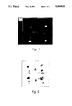
The scintigraphic images obtained 1 h after 123 IUdR injection showed activity in the head of both tumor-bearing [n=16] and sham-operated control [n=8] animals. Activity in the stomach and the bladder was also evident suggesting the rapid dehalogenation and excretion of free iodine. No activity was seen in the thyroid [0.1% potassium iodide solution had been added to the drinking water 48 h prior to the administration of radioactive IUdR]. Images obtained at subsequent intervals [12-38 h] demonstrated clearance of the activity from the head of all control animals by 12 h [FIG. 1-1 and 1-3] and persistence of the activity within the same region in all tumor-bearing animals [FIG. 1-2 and 1-4]. Bladder and stomach activities were still observed in both groups [these radioactivities were mainly associated with the stomach contents and with urine, see FIG. 2].
Regions of interest were drawn around the head of all animals. Even 1 h after injection, the men counts per pixel in the tumor-bearing animals were at least twice that of the control animals. This ratio increased with time to a maximum of 3.8 by 38 h.
The biodistribution data [40 h after 123 IUdR injection] indicated that samples obtained from the "left brain" [uninjected side] or the frontal lobes in tumor-bearing and control animals had similar amounts of activity [FIG. 2]. On the other hand, samples obtained from the "right brain" [injected side] in tumor-bearing animals contained 0.36±0.14% of the injected dose per gram [%ID/g, mean±SD] as opposed to 0.09±0.02% ID/g from the same side of the brain in sham-operated controls (P<0.05). Since a large proportion of the weighed "tumor" sample is, in fact, uninvolved brain tissue [some of the tumors were not visible macroscopically at the time of dissection, i.e., <0.5 mm in diameter], these % ID/g values underestimate the actual tumor uptake. This is further emphasized by the high uptake that was observed in two animals, one in which 12% of the ID was found to be associated with a tumor that could be precisely excised, and another in which 25% of the ID was found in a tumor that occupied a large portion of the "right brain" specimen. As suggested by the scintigraphic studies, the activity in all other normal tissues was low with the exception of the stomach and the bladder. However, examination of these organs indicated that the high activities observed were mainly associated with the stomach contents and with urine.
Using the biodistribution data shown in FIG. 2, tumor to normal tissue ratios were calculated and found to be equal to or greater than eight for all the tissues [FIG. 3]. Of particular interest, in the tumor-bearing animals right brain/left brain=22, right brain/frontal lobes=71, right brain/blood=9. Again, much higher T/N ratios [range of 53 to 488] were obtained in an animal where the brain tumor mass was sufficiently large [about 3×4 mm] to be excised and where the radioactivity per gram of tumor could be accurately assessed.
1. Intraperitoneal Injection of 125 IUdR leads to High Tumor to Nontumor Ratios
The murine ovarian tumor (MOT) used in these experiments arose spontaneously in the ovary of a C3H mouse and is maintained in our laboratories by serial intraperitoneal [i.p.] transplantation in female C3HeB/FeJ mice. We have examined the appropriateness of the i.p. route for IUdR administration as a means to (i) bypass the rapid intrahepatic dehalogenation of this agent, and (ii) obtain high tumor to nontumor ratios. In these experiments, mice were injected with 106 tumor cells 24 h prior to the i.p. administration of 125 IUdR [5 injections, 4 h apart]. Biodistribution studies 24 h following the last 125 IUdR injection have shown extremely favorable tumor to non-tumor ratios [FIG. 4]. Tumor-to-normal-tissue ratios derived from the biodistribution results ranged from 20 for organs with actively proliferating cells (for example uterus, intestine, stomach) to over 400 for organs with nondividing cells (brain, heart).
Analogous results were obtained from the scintigraphic images acquired 1, 2, 16 and 24 hr following a single injection of 300 μCi 123 IUdR [FIG. 5]. At 1 h post radiopharmaceutical injection, focal localization of radioactivity was observed in the abdomen of both tumor-bearing mice and control animals. However, at later time points, the focal area of abdominal activity persisted only in MOT-bearing mice while it cleared from the abdomen of animals without tumor, confirming biodistribution results.
2. 125 IUdR Is an Effective Antineoplastic Agent In A Mouse Ascites Tumor
The tumor used in these experiments is the same murine ovarian tumor described above. We have determined the median survival of mice after i.p. challenge with various tumor cell inocula. The results indicate that the median survival of these mice is proportional to the number of tumor cells inoculated into the mice.
The relatively long survival of tumor-bearing mice facilitates quantitative evaluation of tumor cell killing after treatment with 125 IUdR and can be used to calculate a cellular survival fraction. We have, therefore, studied tumor cell survival as a function of the dose of 125 IUdR administered i.p. at 4 h intervals beginning 24 h after tumor cell i.p. inoculation [105 -106 cells]. Because IUdR dehalogenates rapidly in vivo, potassium iodide is added to the animals' drinking water to block thyroid uptake of the released radionuclides.
When mice are treated with four doses of 125 IUdR at 4-h intervals and the survival fraction plotted as a function of the dose per treatment, a rapid decrease in the tumor cell survival fraction [10-3 ] is observed at doses of 20 μCi per treatment with the curve being flat at higher levels [FIG. 6]. When seven consecutive injections of 125 IUdR are given, a similar steep reduction in tumor cell survival is also observed; the plateau in this regimen occurs at a survival fraction of 10-5. Finally, treatment with equivalent doses of IUdR radiolabeled with 131 I [a negatron emitter whose decay is not associated with any significant yield of Auger electron emissions] does not result in any decrease in survival.
3. 123 IUdR Is An Effective Antineoplastic Agent In A Mouse Ascites Tumor
Recently, we have repeated the experiments described above using 123 IUdR [5 i.p. injections, 4 h intervals, 24 h post i.p. tumor inoculation]. Our results indicate that the incorporation of this Auger electron emitter into the DNA of these tumor cells also prolongs median survival of the tumor-bearing animals [FIG. 7] in a dose-dependent fashion. When the survival fraction of tumor cells is plotted as a function of dose, an exponential decrease is obtained similar to that observed with the 125 IUdR data [FIG. 8].
Claims (1)
1. A method for the diagnosis of tumors in a live mammal which comprises the direct injection of 1-5 mCi of 5-(123 I)iodo-2'-deoxyuridine in a pharmaceutically acceptable vehicle into the tumor, and thereafter imaging the tumor scintigraphically.
Priority Applications (1)
| Application Number | Priority Date | Filing Date | Title |
|---|---|---|---|
| US07/651,785 US5094835A (en) | 1990-03-30 | 1991-02-07 | Diagnosis of tumors with 5-(123 I)iodo-2'-deoxyuridine |
Applications Claiming Priority (2)
| Application Number | Priority Date | Filing Date | Title |
|---|---|---|---|
| US07/502,759 US5077034A (en) | 1990-03-30 | 1990-03-30 | Treatment of tumors with 5-radioiodo-2'-deoxyuridine |
| US07/651,785 US5094835A (en) | 1990-03-30 | 1991-02-07 | Diagnosis of tumors with 5-(123 I)iodo-2'-deoxyuridine |
Related Parent Applications (1)
| Application Number | Title | Priority Date | Filing Date |
|---|---|---|---|
| US07/502,759 Division US5077034A (en) | 1990-03-30 | 1990-03-30 | Treatment of tumors with 5-radioiodo-2'-deoxyuridine |
Publications (1)
| Publication Number | Publication Date |
|---|---|
| US5094835A true US5094835A (en) | 1992-03-10 |
Family
ID=27054273
Family Applications (1)
| Application Number | Title | Priority Date | Filing Date |
|---|---|---|---|
| US07/651,785 Expired - Lifetime US5094835A (en) | 1990-03-30 | 1991-02-07 | Diagnosis of tumors with 5-(123 I)iodo-2'-deoxyuridine |
Country Status (1)
| Country | Link |
|---|---|
| US (1) | US5094835A (en) |
Cited By (5)
| Publication number | Priority date | Publication date | Assignee | Title |
|---|---|---|---|---|
| US5308605A (en) * | 1992-08-27 | 1994-05-03 | The President And Fellows Of Harvard College | Diagnosis of tumors with 5-radioiodo-2'-deoxyuridine |
| WO1996023806A1 (en) * | 1995-02-02 | 1996-08-08 | President And Fellows Of Harvard College | Rapid synthesis of radiolabeled pyrimidine nucleosides or nucleotides |
| US5720935A (en) * | 1995-06-09 | 1998-02-24 | President & Fellows Of Harvard College | Rapid synthesis of radiolabeled pyrimidine nucleosides or nucleotides from stannyl precursors |
| US5811073A (en) * | 1995-06-19 | 1998-09-22 | President And Fellows Of Harvard College | Method for radioisotopic detection and localization of inflammation in a host |
| US6207111B1 (en) * | 1997-12-31 | 2001-03-27 | Pem Technologies, Inc. | System for describing the physical distribution of an agent in a patient |
-
1991
- 1991-02-07 US US07/651,785 patent/US5094835A/en not_active Expired - Lifetime
Non-Patent Citations (4)
| Title |
|---|
| Mercer, J. R. et al., "Synthesis and Tumor Uptake of . . . Uracils", J. Med. Chem., 32, 1989, pp. 1289-1294. |
| Mercer, J. R. et al., Synthesis and Tumor Uptake of . . . Uracils , J. Med. Chem. , 32, 1989, pp. 1289 1294. * |
| Robins et al., "Iodine-123-Iododeoxyuridine: . . . ", J. Nucl. Med. Biol., vol. 8, 1981, pp. 53-63. |
| Robins et al., Iodine 123 Iododeoxyuridine: . . . , J. Nucl. Med. Biol. , vol. 8, 1981, pp. 53 63. * |
Cited By (7)
| Publication number | Priority date | Publication date | Assignee | Title |
|---|---|---|---|---|
| US5308605A (en) * | 1992-08-27 | 1994-05-03 | The President And Fellows Of Harvard College | Diagnosis of tumors with 5-radioiodo-2'-deoxyuridine |
| WO1996023806A1 (en) * | 1995-02-02 | 1996-08-08 | President And Fellows Of Harvard College | Rapid synthesis of radiolabeled pyrimidine nucleosides or nucleotides |
| US5574148A (en) * | 1995-02-02 | 1996-11-12 | President U Fellows Of Harvard College | Rapid synthesis of radiolabeled pyrimidine nucleosides or nucleotides |
| US5720935A (en) * | 1995-06-09 | 1998-02-24 | President & Fellows Of Harvard College | Rapid synthesis of radiolabeled pyrimidine nucleosides or nucleotides from stannyl precursors |
| US5811073A (en) * | 1995-06-19 | 1998-09-22 | President And Fellows Of Harvard College | Method for radioisotopic detection and localization of inflammation in a host |
| US6207111B1 (en) * | 1997-12-31 | 2001-03-27 | Pem Technologies, Inc. | System for describing the physical distribution of an agent in a patient |
| US6555380B2 (en) | 1997-12-31 | 2003-04-29 | Pem Technologies, Inc. | Method for describing the physical distribution of an agent in a patient |
Similar Documents
| Publication | Publication Date | Title |
|---|---|---|
| US5077034A (en) | Treatment of tumors with 5-radioiodo-2'-deoxyuridine | |
| Lee et al. | Treatment of intracranial human glioma xenografts with 131I-labeled anti-tenascin monoclonal antibody 81C6 | |
| JP4247110B2 (en) | Novel metalloporphyrins and their use as radiosensitizers for radiotherapy | |
| Kassis et al. | 5-[125I] iodo-2′-deoxyuridine in the radiotherapy of brain tumors in rats | |
| Kassis et al. | Radiolabeled nucleoside analogs in cancer diagnosis and therapy | |
| Kassis et al. | Specific uptake of the Auger electron-emitting thymidine analogue 5-[123I/125I] iodo-2′-deoxyuridine in rat brain tumors: diagnostic and therapeutic implications in humans | |
| Baranowska-Kortylewicz et al. | 5-[123I] iodo-2′-deoxyuridine in the radiotherapy of an early ascites tumor model | |
| US7803350B2 (en) | Radioactive arsenic-containing compounds and their uses in the treatment of tumors | |
| Fairchild et al. | Thiouracil distribution in mice carrying transplantable melanoma | |
| Kassis et al. | In vivo therapy of neoplastic meningitis with methotrexate and 5-[125I] iodo-2'-deoxyuridine | |
| Appelbaum et al. | Specific marrow ablation before marrow transplantation using an aminophosphonic acid conjugate 166Ho-EDTMP | |
| Coderre et al. | Iodothiouracil as a melanoma localizing agent | |
| US5094835A (en) | Diagnosis of tumors with 5-(123 I)iodo-2'-deoxyuridine | |
| US6685913B1 (en) | Lipid soluble radioactive metal chelates for tumor therapy | |
| Wheldon | Targeting radiation to tumours | |
| Hofer | Radiation effects on death and migration of tumor cells in mice | |
| US5308605A (en) | Diagnosis of tumors with 5-radioiodo-2'-deoxyuridine | |
| US5612017A (en) | Halogenated sulfidohydroboranes for nuclear medicine and boron neutron capture therapy | |
| Lin et al. | Evaluation of three rhenium-188 candidates for intravascular radiation therapy with liquid-filled balloons to prevent restenosis | |
| KR20000022136A (en) | Novel radiopharmaceutical compositions and matrices and uses thereof | |
| KR102621851B1 (en) | Porphyrin derivatives and Composition for imaging, diagnosing, or treating cancers | |
| US20120251442A1 (en) | Methods for Treatment of Tumors by Direct Administration of a Radioisotope | |
| Laster et al. | Analysis of 5-iodo-2′-deoxyuridine incorporation in murine melanoma for photon activation therapy | |
| Kassis et al. | 5-[125I] iodo-2'-deoxyuridine in the radiotherapy of solid CNS tumors in rats | |
| Dence et al. | Radiochemical synthesis, rodent biodistribution and tumor uptake, and dosimetry calculations of [11C] methylated LY2181308 |
Legal Events
| Date | Code | Title | Description |
|---|---|---|---|
| STCF | Information on status: patent grant |
Free format text: PATENTED CASE |
|
| FEPP | Fee payment procedure |
Free format text: PAYOR NUMBER ASSIGNED (ORIGINAL EVENT CODE: ASPN); ENTITY STATUS OF PATENT OWNER: SMALL ENTITY |
|
| FPAY | Fee payment |
Year of fee payment: 4 |
|
| FEPP | Fee payment procedure |
Free format text: PAT HOLDER CLAIMS SMALL ENTITY STATUS - SMALL BUSINESS (ORIGINAL EVENT CODE: SM02); ENTITY STATUS OF PATENT OWNER: SMALL ENTITY |
|
| FPAY | Fee payment |
Year of fee payment: 8 |
|
| FPAY | Fee payment |
Year of fee payment: 12 |